Embed Size (px)
Citation preview

www.elsevier.com/locate/intimp
International Immunopharmac
Treatment with Flt3 ligand plasmid reverses allergic airway
inflammation in ovalbumin-sensitized and -challenged mice
Jehad H. Edwana, James E. Talmadgeb, Devendra K. Agrawala,c,d,*
aDepartment of Medical Microbiology and Immunology, Creighton University School of Medicine, CRISS I Room 131,
2500 California Plaza, Omaha, NE 68178, United StatesbDepartment of Pathology and Microbiology, University of Nebraska Medical Center, Omaha, NE 68198, United States
cDepartment of Biomedical Sciences, Creighton University School of Medicine, Omaha, NE 68178, United StatesdDepartment of Medicine, Creighton University School of Medicine, Omaha, NE 68178, United States
Received 13 October 2004; accepted 13 October 2004
Abstract
We have previously reported that fms-like tyrosine kinase 3 ligand (Flt3-L) prevents and reverses established allergic airway
inflammation in an ovalbumin (OVA) induced mouse model of asthma. In this study, we investigated the effect of pUMVC3-
hFLex, a plasmid, mammalian expression vector for the secretion of Flt3-L on the same mouse model as well as the duration of
the effect of the treatment. Allergic airway inflammation to OVA was established in BALB/c mice. OVA-sensitized mice
received three intramuscular (i.m.) injections of 200 Ag pUMVC3-hFLex over 10 days. The response to pUMVC3-hFLex
therapy was assessed based on airway hyperresponsiveness (AHR) to methacholine and inflammation, measured as serum
cytokine and immunoglobulins (Ig) levels, and the total and differential cells in bronchoalveolar lavage fluid (BALF).
pUMVC3-hFLex treatment completely reversed established AHR (Pb0.01) and this effect lasted for at least 24 days after the
last treatment injection (Pb0.001). pUMVC3-hFLex treatment significantly increased BALF interferon-gamma (IFN-g)
(Pb0.01), serum interleukin (IL)-10 (Pb0.01) and anti-OVA IgG2a levels (Pb0.01). In contrast, serum IL-4 and IgE levels
were significantly reduced (Pb0.05). Total BALF cellularity, eosinophiles counts and BALF IL-5 levels were also reduced
1567-5769/$ - s
doi:10.1016/j.in
Abbreviation
cells; DC, dendr
Flt3-L, fms-like
intraperitoneal;
helper.
* Correspon
School of Medic
E-mail addr
ology 5 (2005) 345–357
ee front matter D 2004 Published by Elsevier B.V.
timp.2004.10.002
s: Ab, antibody; Ag, antigen; AHR, airway hyperresponsiveness; ACT, ammonium chloride Tris; APCs, antigen presenting
itic cells; BAL, bronchoalveolar lavage; BALF, bronchoalveolar lavage fluid; ELISA, enzyme-linked immunosorbent assay;
tyrosine kinase 3 ligand; IFN-g, interferon-gamma; Ig, immunoglobulin; IL, interleukin; i.m., intramuscular; i.p.,
OVA, ovalbumin; pDCs, plasmacytoid DCs; PBS, phosphate buffer saline; TGF-h, transforming growth factor-beta; TH, T
ding author. Department of Biomedical Sciences, Medicine, and Medical Microbiology and Immunology, Creighton University
ine, CRISS I Room 131, 2500 California Plaza Omaha, NE 68178, United States. Tel.: +1 402 280 2938; fax: +1 402 280 1421.
ess: [email protected] (D.K. Agrawal).

J.H. Edwan et al. / International Immunopharmacology 5 (2005) 345–357346
(Pb0.01). pUMVC3-hFLex treatment can reverse established experimental asthma and might provide a novel approach for
treating asthma.
D 2004 Published by Elsevier B.V.
Keywords: Allergy; Flt3-L; TH1/TH2 cells; OVA mouse model; Asthma; pUMVC3-hFLex
1. Introduction
Asthma is a multifactorial disease characterized by
episodes of reversible airway inflammation and
edema, as well as mucus secretion, which result in
bronchial constriction and airway obstruction. Several
immune cells and mediators are responsible for the
development of allergy and asthma [1]. As a result of
this heterogeneity, there are many treatment modal-
ities used clinically [2–7]. Overall, recent therapeutic
approaches have focused on modulating antigen (Ag)
presentation to T cells, inhibition of Ag-specific T
helper (TH) 2 responses and/or increasing the TH1
response [8]. In recent years, various immunomodu-
lators have shown therapeutic activity for allergen-
induced asthma, including anti-IgE [9], anti-cytokine
antibodies (Abs) [10,11], probiotics [12], CpG [13],
synthetic YpG, CpR and YpR immunostimulatory
motifs [14], and mycobacterial Ags [15], Vaccination
using DNA-encoded Ags induce cellular and humoral
immunity against microbial pathogens [16–18]. Plas-
mid vectors encoding fms-like tyrosine kinase 3
ligand (Flt3-L), the novel human hematopoietic
growth factor, have been shown to provide adjuvant
activity for the induction of Ag-specific immunity in
mice [19–21]. The adjuvant activity is due, in part, to
the ability of Flt3-L to expand dendritic cell (DC)
numbers in the circulation and organs. [22] It has
recently been demonstrated that hypervolemic injec-
tions of a Flt3-L plasmid vector could result in the
expansion of DCs [23]. In contrast, Flt3-L can also
promote tolerance to orally administered Ags [24].
Thus, DCs have a regulatory role in the induction of
both tolerance and immunity, although both processes
can be regulated by other cell types [25].
Recently, we reported that treatment with Flt3-L
could prevent the development [26] and reverse [27]
asthma-like conditions in a mouse model, resulting in
the complete abolition of airway hyperresponsiveness
(AHR) to methacholine. However, in these studies,
mice were injected daily with recombinant Flt3-L for
10 days. Because of the adjuvant activity by Flt3-L
protein, plasmids and viral vectors for vaccines
[28,29], and our finding of therapy for asthma with
Flt3-L protein, we wished to examine the therapeutic
activity of Flt3-L cDNA for asthma as well as the
duration of the effect of the treatment. Further,
because of the therapeutic activity of CpG against
asthma, there is the potential for multiple mechanisms
with a Flt3-L plasmid, including Flt3-L protein
activity and the CpG motif. Thus, we compared the
ability of Flt3-L secreting plasmid (pUMVC3-hFLex)
to a control plasmid using mice with preexisting AHR
and allergic airway inflammation.
2. Methods
2.1. Animals
Four- to five-week-old female BALB/c mice were
obtained from Harlan Laboratories (Indianapolis, IN)
and were housed in separate cages according to
treatment. Food and water were provided ad libitum.
According to the National Institutes of Health guide-
lines, the research protocol of this study was approved
by the Animal Research Committee of Creighton
University (Omaha, NE).
2.2. Plasmid DNA
The pUMVC3-hFLex plasmid contains the extrac-
ellular domain (secreted form) of the human Flt3-L
gene. This vector as well as the control plasmid,
pUMVC3, were obtained from the Vector Core
Laboratory at the University of Michigan (Ann Arbor,
MI, USA) and has been described previously [30].
2.3. Sensitization and treatment
Allergic airway inflammation to ovalbumin (OVA)
was induced intraperitoneal (i.p.) injection of 20 Ag

J.H. Edwan et al. / International Immunopharmacology 5 (2005) 345–357 347
grade V chicken egg OVA (Sigma-Aldrich, St. Louis,
MO) emulsified in 2.25 mg imject Alum (Pierce,
Rockford, IL) in total volume of 100 Al on days 1 and
14 (Fig. 1), followed by aerosol sensitization with 1%
OVA for 20 min using an Ultra-Neb 90 nebulizer (De
Villbiss, Somerset, PA) on days 28, 29 and 30.
Starting on day 33, OVA-sensitized mice were
randomized into three groups: two of the groups
received injection into the muscle interior tibialis of
200 Ag pUMVC3-hFLex or pUMVC3 treatment three
times on days 33, 38 and 41, and the OVA group
received phosphate buffer solution (PBS) only. On
day 44, mice were challenged with 5% aerosolized
OVA. After methacholine challenge on day 45, half
the mice in each group were sacrificed. On day 64,
mice were challenged with 5% aerosolized OVA and
sacrificed after the last methacholine challenge on day
65. Non-sensitized control mice were treated only
with the vehicle (PBS).
2.4. Noninvasive method for measuring pulmonary
function
Single-chamber whole-body plethysmograph
(Buxco Electronics, Troy, NY), without the use of
anesthesia or restraint, were used to measure
pulmonary functions. This method has been dem-
Fig. 1. OVA murine model of asthma and immunotherapy protocol. Balb
challenged with OVA by aerosolization on days 28, 29 and 30. Starting on d
of the groups received injection into the muscle interior tibialis 200 Ag pUM
41, while the OVA group received PBS only. On day 44, mice were chall
sacrificed on day 45. On day 64, mice were challenged with 5% aerosolized
Non-sensitized control mice were treated only with the vehicle (PBS).
onstrated to accurately reflect airway resistance
[5,31], expressed as the Penh units [32]. Twenty-
four hours following OVA challenge, mice were
placed within the chambers and challenged with
increasing doses of methacholine (Sigma-Aldrich) to
measure AHR, and Penh in response to methacholine
challenge were recorded and expressed as the
absolute Penh units.
2.5. Serum IgE analysis
Blood collected after sacrifice on day 45 was
immediately centrifuged and serum was collected and
stored at �80 8C for later analysis. Enzyme-linked
immunosorbent assay (ELISA) for total IgE was
conducted as previously described [33] and according
to the manufacturer’s recommendations using rat anti-
mouse IgE (BD PharMingen, San Diego, CA),
standard IgE (BD PharMingen) and rat anti-mouse
IgE-HRP (Southern Biotechnology Associates, Bir-
mingham, AL) for the total IgE assay. Both cytokine
and IgE assays were developed with 3,3V,5,5V-tetra-methylbenzidine (TMB) substrate (BD PharMingen),
reactions were stopped with 2 N H2SO4, and read at
450 nm using a Bio-Rad microplate reader and
software (Bio-Rad, Hercules, CA). Sensitivity for
total IgE was 1 ng/ml.
/c mice were sensitized to OVA by i.p. injection and subsequently
ay 33, OVA-sensitized mice were randomized into three groups: two
VC3-hFLex or pUMVC3 treatment three times on days 33, 38 and
enged with 5% aerosolized OVA. Half the mice in each group were
OVA and sacrificed after the last methacholine challenge on day 65.

J.H. Edwan et al. / International Immunopharmacology 5 (2005) 345–357348
2.6. Serum total and anti-OVA IgG isotype analysis
Total IgG were determined using rat anti-mouse
IgG2a or rat anti-mouse IgG1 (BD PharMingen),
IgG2a or IgG1 standard (BD PharMingen) and rat
anti-mouse IgG2a-HRP or rat anti-mouse IgG1-HRP
(BD PharMingen). Anti-OVA IgGs serum levels were
determined as previously described [34]. Briefly,
microtiter plates were coated with100 Ag/ml chicken
egg OVA. The coated plates were washed several
times with PBS containing 0.05% Tween (PBS-T)
and blocked with 10% FBS for 2 h at room
temperature. Diluted serum was incubated in dupli-
cates overnight, washed with PBS-T, incubated with
anti-mouse avidin conjugates (IgG1 or IgG2a, BD
PharMingen) for 2 h, and then washed several times
with PBS-T. Assays were developed with TMB
substrate reagent, reactions were stopped with 2 N
H2SO4 and read at 450 nm using a Bio-Rad micro-
plate reader and software.
2.7. Bronchoalveolar lavage (BAL) collection
Immediately after methacholine challenge, mice
were euthanized with a lethal dose of pentobarbital.
Bronchoalveolar lavage fluid (BALF) was collected
from each animal via cannulation of the exposed
trachea and gentle flushing of the lungs with 1 ml
warm PBS (0.8 ml) was recovered, which was
centrifuged and the supernatant was collected.
2.8. Cytokine assays
Serum and BALF cytokines were measured by
sandwich ELISA with capture and biotinylated detec-
tion antibody (Ab) pairs for interferon-gamma (IFN-
g), interleukin (IL)-4, IL-5, IL-10 and IL-12 and
avidin-horseradish peroxidase and TMB substrate
(BD PharMingen).
2.9. Phenotype of lung dendritic cells
DCs were isolated from the lungs of animals. After
removing the blood, lungs were excised, washed with
PBS, and digested with 5 ml of 1 mg/ml collagenase
D in RPMI media, digested lungs were then centri-
fuged and incubated with ammonium chloride Tris
(ACT) to lyses red blood cells. Cells were incubated
in 4% FBS to block FcgR non-specific binding. Cells
were stained with anti CD11c-PE, anti CD11b-FITC,
anti CD8a PerCP and anti CD45R–biotin–antigen
presenting cell (APC). The cell profiles were gated on
DCs (based on high forward and 908 light scatter).
The DCs gate was then sub-gated on CD11c+ cells,
further on the CD11c+ gate CD11b, CD8a and B220
profiles were examined.
2.10. Data analysis
Data were analyzed using GraphPad PRISM
statistical analysis and graphing software. One-way
ANOVA Bonferroni’s multiple comparison test was
used to compare multiple groups. A p-value of b0.05
was considered significant.
3. Results
3.1. Effect of pUMVC3-hFLex treatment on AHR in
OVA-presensitized and -challenged mice
On day 33 of the sensitization protocol (Fig. 1),
mice were placed in a whole body plethysmograph
and enhanced pause response ( Penh) readings
recorded to plot baseline dose response curves to
increasing methacholine concentrations and to con-
firm that OVA-sensitized mice have established AHR
(data not shown). Treatment with pUMVC3-hFLex
significantly reduced airway hyperresponsiveness in
OVA-sensitized mice to levels comparable to PBS-
treated mice (Fig. 2A). This effect lasted for at least 24
days after the last pUMVC3-hFLex injection (Fig.
2B). In contrast, none of the OVA-sensitized
pUMVC3-treated and -non-treated mice exhibited
changes in AHR.
3.2. Effect of treatment on serum cytokines, total IgE
and total and anti-OVA IgG subclasses in
OVA-presensitized and -challenged mice
In the serum samples collected on day 45, OVA
sensitization significantly increased serum IL-12 and
total IgE levels. Further, these levels were signifi-
cantly reduced after treatment with pUMVC3-hFLex
and pUMVC (Fig. 3C,D). Additionally, serum IL-4
levels were significantly reduced after treatment with

Fig. 3. Effect of pUMVC3-hFLex treatment on serum cytokines and total IgE levels in mice. On day 45, after recording pulmonary functions for
AHR, blood was collected to measure serum IL-4 (A), serum IL-10 (B), serum IL-12 (C) and serum total IgE concentration (D). Data is shown
as meanFS.E.M. (n=8). (*pb0.05, ***pb0.001 compared with OVA group; zpb0.05, zzzpb0.001 compared with PBS group), serum IL-5 and
IFN-g were not detected. pUMVC3-hFLex, a plasmid, mammalian expression vector for the secretion of Flt3-L, pUMVC3 the backbone
plasmid without the Flt3-L insertion.
Fig. 2. AHR to methacholine in unrestrained mice. Following OVA sensitization and challenge, AHR to methacholine was established (day 33)
(see Fig. 1) followed by treatment with pUMVC3-hFLex (200 Ag/day, i.p.) three times. On day 45, AHR to methacholine was again measured
and Penh values were recorded. pUMVC3-hFLex treatment abolishes AHR to methacholine in established allergic inflammatory mice (left panel
A). On day 65, AHR to methacholine was again measured and Penh values were recorded. pUMVC3-hFLex treatment abolishes AHR to
methacholine and this effect lasts for at least 24 days after final dose of treatment of established allergic inflammation in mice (right panel B).
Data is shown as meanFS.E.M. (N=8 in each group). **pb0.01, ***pb0.001 compared with OVA group.
J.H. Edwan et al. / International Immunopharmacology 5 (2005) 345–357 349

J.H. Edwan et al. / International Immunopharmacology 5 (2005) 345–357350
pUMVC3-hFLex and pUMVC (Fig. 3A). In contrast,
OVA sensitization significantly decreased serum IL-
10 levels; however, there was no effect of pUMVC3-
hFLex or pUMVC3 on serum IL-10 level (Fig. 3E).
On day 65, serum IL-4 levels were significantly
higher than PBS control mice (Fig. 4A) and serum IL-
10 levels in OVA-presensitized mice were still
significantly lower than PBS control mice (Fig. 4B).
In contrast, serum IL-12 levels in OVA-presensitized
mice were significantly lower than PBS control mice
(Fig. 4C).
3.3. Effect of pUMVC3-hFLex treatment on BALF
cytokines in OVA-presensitized and -challenged mice
On day 45, OVA sensitization significantly
increased BALF IL-5 and IL-12 levels, which were
significantly reduced after treatment with pUMVC3-
hFLex and pUMVC (Fig. 5B,C). In contrast, OVA
sensitization significantly decreased BALF IL-10 and
IFN-g levels (Fig. 5D,E). Further, the BALF levels of
IL-4 were unchanged, which differed from the serum
IL-4 levels that were increased by pUMVC3-hFLex
injections. On day 65, BALF IL-5 levels were
significantly higher than PBS control mice (Table
2), in contrast to levels on day 45, BALF IL-5 levels
increased to levels comparable to those in OVA-
sensitized mice. However, BALF IL-10 levels in
OVA-presensitized mice were still significantly
Fig. 4. Effect of pUMVC3-hFLex treatment on serum cytokines in mice. O
collected to measure serum IL-4 (A), serum IL-10 (B) and serum IL-12 (C).
OVA group; zpb0.05, zzpb0.01, zzzpb0.001 compared with PBS group),
plasmid, mammalian expression vector for the secretion of Flt3-L, pUMV
decreased and treatment with pUMVC3-hFLex has
increased that levels (Table 2).
3.4. Effect of treatment on serum total and anti-OVA
IgG isotypes in OVA-presensitized and -challenged
mice
OVA sensitization significantly increased total and
anti-OVA serum IgG1 and IgG2a levels. The injection
of pUMVC3-hFlex had no effect on total serum
IgGa2 and IgG1 levels (Fig. 6A,B). However, in
serum samples collected on day 45 treatment with
pUMVC3-hFLex induced a significant increase in the
serum levels of anti-OVA IgGa2 (Fig. 6C). In
addition, unlike most other Ag responses, there was
no significant effect of pUMCV3-hFlex injection on
anti-OVA IgG1 levels (Fig. 6D). In contrast to the
initial increase in anti-OVA IgGa2 levels, on day 65,
pUMVC3-hFlex caused a significant reduction in
anti-OVA IgGa2 levels (Fig. 7C).
3.5. Effect of treatment on BALF inflammatory cells in
OVA-presensitized and -challenged mice
On days 45 and 65, sensitization and challenge
with OVA induced a significant influx of cells into the
airways (Tables 1 and 2). Treatment with pUMVC3-
hFLex significantly reduced the total cellular infiltra-
tion (Tables 1 and 2). The reduction in the frequency
n day 65, after recording pulmonary functions for AHR, blood was
Data is shown as meanFS.E.M. (n=8). (***pb0.001 compared with
serum IL-5, IFN-g and IgE were not detected. pUMVC3-hFLex, a
C3 the backbone plasmid without the Flt3-L insertion.

Fig. 5. Effect of pUMVC3-hFLex treatment on BALF cytokines levels in mice. On day 45, after recording pulmonary functions for AHR,
BALF samples were immediately centrifuged and cytokines in supernatants were measured: BALF IL-4 (A), BALF IL-5 (B), BALF IL-12 (C),
BALF IL-10 (D) and BALF IFN-g (E). Data is shown as meanFS.E.M. (n=8). (*pb0.05, **pb0.01, ***pb0.001 compared with OVA group;zpb0.05, zzzpb0.001 compared with PBS group).
J.H. Edwan et al. / International Immunopharmacology 5 (2005) 345–357 351
of infiltrating eosinophils was also significant. Total
number of macrophages was significantly increased in
both pUMVC3-hFLex-treated and pUMVC3-treated
animals (Tables 1 and 2). There was no significant
effect on the number of neutrophils or lymphocytes in
the BALF.
3.6. Effect of pUMVC3-hFLex administration on lung
DCs
pUMVC3-hFLex treatment significantly altered
the profile of CD11c+ in the lungs with an increase
in the total number of CD11c+ cells, including
CD11c+ CD11b+ ( pb0.05) and CD11c+ CD8a+ cells
(Table 3). In addition, there was a significant increase
in CD11c+ B220+ ( pb0.05) as well as (CD11c+
CD8a+) ( pb0.01) cells as compared to the PBS-
treated OVA-sensitized group (Table 3).
3.7. Discussion
This report presents evidence that plasmid contain-
ing Flt3-L gene can shape and protect against the
immune allergic response to OVA in OVA-presensi-
tized and -challenged mice with established AHR. This
model of allergic airway inflammation has been
reported to elicit type 2 T-cell responses and is
characterized by eosinophilic infiltration of the airways
and nonspecific AHR. Previously, we demonstrated
that Flt3-L, administered prior to Ag sensitization, was
capable of preventing [26] and reversing [27] airway
inflammation and hyperresponsiveness.
In this study, we report the therapeutic activity of a
plasmid with the Flt3-L transgene (pUMVC3-hFLex).
We report for the first time that the injection of this
plasmid can reverse established AHR and this
reversal lasts for at least 24 days after the final

Fig. 6. Effect of pUMVC3-hFLex treatment and Ag sensitization and challenge on IgG subclasses in mice serum. On day 45, after recording
pulmonary functions for AHR, blood was collected to measure: total IgG2a (A), total IgG1 (B), anti-OVA IgG2a (C) and anti-OVA IgG1 (D).
Data are shown as meanFS.E.M. from eight animals in each group. (*pb0.05, ***pb0.001 compared with OVA group; zzzpb0.001 compared
with PBS group).
J.H. Edwan et al. / International Immunopharmacology 5 (2005) 345–357352
pUMVC3-hFLex injection. BALF IL-5 and eosino-
phil levels, and serum IL-4 and IgE levels were all
reduced. In contrast, the therapeutic effect of Flt3-L
on allergic airway inflammation and pulmonary
function was associated with a significant increase
in BALF IFN-g and IL-10 levels, and serum anti-
OVA IgG2a levels.
The development of allergen-induced asthma is
mediated by the production of TH2 cytokines such as
IL-4, IL-5 and IL-13. Several studies have shown that
type 1 immunomodulators have therapeutic activity
for allergen-induced asthma. Some of these not only
boost the production of TH1 cytokines, but also block
the induction of TH2 immunity. One of these
cytokines, IL-4, has been identified as important to
the development of allergic inflammation. The role of
IL-4 in asthma is supported by the augmented levels
of IL-4 in BALF [35] and increased expression of IL-
4 mRNA in broncho-alveolar lavage cells [36,37]
reported in patients with allergic inflammation. In
murine models, IL-4 has been demonstrated to play a
crucial role in allergic airway inflammation [38–40]
and AHR [41,42]. These observations are supported
by our findings that the i.m. delivery of pUMVC3-
hFLex results in a significant decrease in serum IL-4
levels. Interestingly, injections of the control vector,
pUMVC3, also decreased serum IL-4 levels, suggest-
ing that the reversal of AHR might be independent of
a reduction in IL-4 levels. It is also noteworthy that
the pUMVC3 backbone is a pUC19 plasmid contain-
ing methylated CpG motifs [43], which might be
responsible for the decreased IL-4 levels. Alone,
however, it appears not to be able to reverse the
AHR, which requires the Flt3-L transgene. Even
though trials with IL-4 neutralizing agents in patients
with asthma have been reported to be non-therapeutic,

Fig. 7. Effect of pUMVC3-hFLex treatment and Ag sensitization and challenge on IgG subclasses in mice serum. On day 65, after recording
pulmonary functions for AHR, blood was collected to measure: total IgG2a (A), total IgG1 (B), anti-OVA IgG2a (C) and anti-OVA IgG1 (D).
Data are shown as meanFS.E.M. from eight animals in each group. (*pb0.05, **pb0.01 compared with OVA group; zzzpb0.001 compared with
PBS group).
J.H. Edwan et al. / International Immunopharmacology 5 (2005) 345–357 353
it has raised the question of the importance of this
cytokine in established asthma, although the signifi-
cance of this cytokine cannot be ruled out and further
studies are warranted.
IL-5 knockout mice have been reported to be unable
to develop airway eosinophilia and hyperresponsive-
ness [44]. Similarly, Ab neutralization of systemic IL-5
reduces airway inflammation and hyperresponsiveness
[45]. In addition, as IL-5 is able to recruit eosinophils to
the lung; the observation that treating asthmatics with
an anti-IL-5 Ab results in decreased airway eosinophil
numbers is expected. Therefore, the effect of
pUMVC3-hFLex injections on IL-5 levels and the
recruitment of eosinophils to the airways are expected.
We report that pUMVC3-hFLex significantly inhibited
IL-5 levels and inflammatory cell infiltration as well as
lung eosinophilia. We suggest that the ability of
pUMVC3-hFLex to reduce IL-5 secretion may directly
reduce the recruitment of eosinophils into the lungs;
thereby, regulating, at least, the inflammatory cellular
components of asthma. The decrease in AHR observed
in pUMVC3-hFLex-treated mice is probably due to the
combined result of lowering IL-4 and IL-5 production.
One mechanism of vaccine adjuvant activity is the
recruitment of professional Ag presenting cells (APCs)
to the site(s) of infection, and by directly stimulating
these APCs to express costimulatory molecules
[46,47]. In the lungs, immature DCs act as a sentinel
against infectious diseases. After interacting with a
foreign Ag, they migrate to the draining lymph nodes,
where they present the captured and processed Ag in
the context of co-stimulatory molecules to Ag specific
naRve T cells, and initiate the development of a primary
effector T cell response [48].

Table 1
BALF total and differential cell counts (�10�3) on day 45
Treatment group
PBS OVA OVA/pUMVC3-hFLex OVA/pUMVC3
Total cells 65.00F35.06 515.00F86.72zzz 176.25F65.43**,## 475F49.35zzz
Macrophages 54.58F28.43 63.73F11.58 133.51F15.13z 130.63F11.06z
Eosinophils 10.38F7.28 436.46F14.16zzz 37.45F13.96*** 331.31F6.25***,zzz
Neutrophils 0.00F0.00 11.59F6.78 2.20F0.84 10.69F6.25
Lymphocytes 0.05F0.05 3.22F1.93 3.08F1.11 2.38F1.37
BAL fluid (0.8 ml) was collected from each animal and centrifuged. Recovered total cells were counted (cell/ml�10�3) and differential analysis
was performed using standard morphological criteria on cytospin slides. At least 300 cells were examined in each cytospin slide and absolute
cell numbers were calculated per milliliter of the BALF based on the percentage of individual cell in a slide. Shown are meansFS.E.M. for six
animals in each group. (*pb0.05 compared with OVA group; ###pb0.001 compared with pUMVC3 group). pUMVC3-hFLex, a plasmid,
mammalian expression vector for the secretion of Flt3-L, pUMVC3 the backbone plasmid without the Flt3-L insertion.
** pb0.01 compared with OVA group.
*** pb0.001 compared with OVA group.z pb0.05 compared with PBS group.zzz pb0.001 compared with PBS group.## pb0.01 compared with pUMVC3 group.
J.H. Edwan et al. / International Immunopharmacology 5 (2005) 345–357354
It has recently been demonstrated that a distinct
subset of DCs with a CD11c+ B220+ phenotype, which
are identified as plasmacytoid DCs (pDCs), have the
ability to secrete IL-10 following activation and
regulate T regulatory (Treg) cell differentiation [49]
Table 2
BALF total and differential cell counts (�10�3) and cytokines on day 65
Treatment group
PBS OVA
Total cells 64F13.7 209F36.6z
Macrophages 50.9F2.9 25.9F4.7
Eosinophils 11.8F2.7 177.1F5.7zzz
Neutrophils 0.8F0.5 4.7F2.7
Lymphocytes 0.5F0.3 1.3F0.8
IL-4 43.4F4.0 37.4F1.9
IL-5 75.5F6.1 365.8F19.4zzz
IL-12 2354.2F95.7 3130F217.2
IL-10 1473.9F98.7 211.5F17.8zzz
IFN-g 293.4F15.2 37.2F10.4zzz
BAL fluid (0.8 ml) was collected and the total cells (cells/ml�10�3)
differentiate cell types. Differential cell counts were calculated as a produc
milliliter. Samples were centrifuged and supernatants were analyzed for
meanFS.E.M. pUMVC3-hFLex, a plasmid, mammalian expression vector
the Flt3-L insertion.
* pb0.05 compared with OVA group.
** pb0.01 compared with OVA group.
*** pb0.001 compared with OVA group.z pb0.05 compared with PBS group.zz pb0.01 compared with PBS group.zzz pb0.001 compared with PBS group.# pb0.05 compared with pUMVC3 group.## pb0.001 compared with pUMVC3 group.
and expansion [50–52]. In addition, pDCs do not
express co-stimulatory molecules, which can contrib-
ute to their tolerance inducing properties [53]. Treg
cells produce immunosuppressive cytokines, including
IL-10 and transforming growth factor-beta (TGF-h)
OVA/pUMVC3-hFLex OVA/pUMVC3
112F19.4 204F36.5z
84.8F9.6z,***,# 56.1F4.7*
23.8F8.9***,## 142.3F2.7zzz,**
1.4F0.5 4.6F2.7
1.96F0.7 1.0F0.6
31.9F3.4 46.0F4.6
427.5F15.3zzz 502.7F18.7zzz
3261.7F183.7z 3550.4F135.1zz
423.3F68.1zzz,* 357.4F23.5zzz
71.0F15.9zzz 74.5F30.9zzz
were counted. One hundred microliters of BALF was stained to
t of the percentage of each cell type and the total number of cells per
cytokines by ELISA (pg/ml), n=6 per group, data are shown as
for the secretion of Flt3-L, pUMVC3 the backbone plasmid without
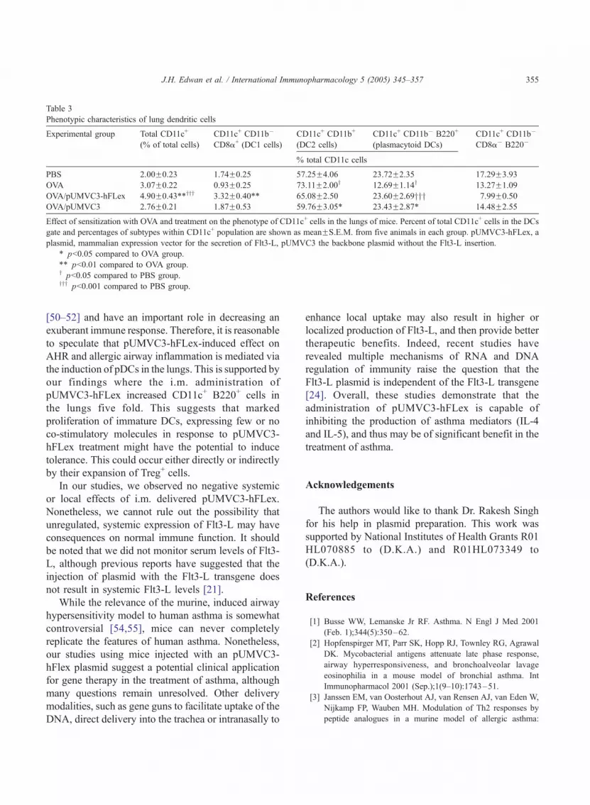
Table 3
Phenotypic characteristics of lung dendritic cells
Experimental group Total CD11c+
(% of total cells)
CD11c+ CD11b�
CD8a+ (DC1 cells)
CD11c+ CD11b+
(DC2 cells)
CD11c+ CD11b� B220+
(plasmacytoid DCs)
CD11c+ CD11b�
CD8a� B220�
% total CD11c cells
PBS 2.00F0.23 1.74F0.25 57.25F4.06 23.72F2.35 17.29F3.93
OVA 3.07F0.22 0.93F0.25 73.11F2.00y 12.69F1.14y 13.27F1.09
OVA/pUMVC3-hFLex 4.90F0.43**yyy 3.32F0.40** 65.08F2.50 23.60F2.69yyy 7.99F0.50
OVA/pUMVC3 2.76F0.21 1.87F0.53 59.76F3.05* 23.43F2.87* 14.48F2.55
Effect of sensitization with OVA and treatment on the phenotype of CD11c+ cells in the lungs of mice. Percent of total CD11c+ cells in the DCs
gate and percentages of subtypes within CD11c+ population are shown as meanFS.E.M. from five animals in each group. pUMVC3-hFLex, a
plasmid, mammalian expression vector for the secretion of Flt3-L, pUMVC3 the backbone plasmid without the Flt3-L insertion.
* pb0.05 compared to OVA group.
** pb0.01 compared to OVA group.y pb0.05 compared to PBS group.yyy pb0.001 compared to PBS group.
J.H. Edwan et al. / International Immunopharmacology 5 (2005) 345–357 355
[50–52] and have an important role in decreasing an
exuberant immune response. Therefore, it is reasonable
to speculate that pUMVC3-hFLex-induced effect on
AHR and allergic airway inflammation is mediated via
the induction of pDCs in the lungs. This is supported by
our findings where the i.m. administration of
pUMVC3-hFLex increased CD11c+ B220+ cells in
the lungs five fold. This suggests that marked
proliferation of immature DCs, expressing few or no
co-stimulatory molecules in response to pUMVC3-
hFLex treatment might have the potential to induce
tolerance. This could occur either directly or indirectly
by their expansion of Treg+ cells.
In our studies, we observed no negative systemic
or local effects of i.m. delivered pUMVC3-hFLex.
Nonetheless, we cannot rule out the possibility that
unregulated, systemic expression of Flt3-L may have
consequences on normal immune function. It should
be noted that we did not monitor serum levels of Flt3-
L, although previous reports have suggested that the
injection of plasmid with the Flt3-L transgene does
not result in systemic Flt3-L levels [21].
While the relevance of the murine, induced airway
hypersensitivity model to human asthma is somewhat
controversial [54,55], mice can never completely
replicate the features of human asthma. Nonetheless,
our studies using mice injected with an pUMVC3-
hFlex plasmid suggest a potential clinical application
for gene therapy in the treatment of asthma, although
many questions remain unresolved. Other delivery
modalities, such as gene guns to facilitate uptake of the
DNA, direct delivery into the trachea or intranasally to
enhance local uptake may also result in higher or
localized production of Flt3-L, and then provide better
therapeutic benefits. Indeed, recent studies have
revealed multiple mechanisms of RNA and DNA
regulation of immunity raise the question that the
Flt3-L plasmid is independent of the Flt3-L transgene
[24]. Overall, these studies demonstrate that the
administration of pUMVC3-hFLex is capable of
inhibiting the production of asthma mediators (IL-4
and IL-5), and thus may be of significant benefit in the
treatment of asthma.
Acknowledgements
The authors would like to thank Dr. Rakesh Singh
for his help in plasmid preparation. This work was
supported by National Institutes of Health Grants R01
HL070885 to (D.K.A.) and R01HL073349 to
(D.K.A.).
References
[1] Busse WW, Lemanske Jr RF. Asthma. N Engl J Med 2001
(Feb. 1);344(5):350–62.
[2] Hopfenspirger MT, Parr SK, Hopp RJ, Townley RG, Agrawal
DK. Mycobacterial antigens attenuate late phase response,
airway hyperresponsiveness, and bronchoalveolar lavage
eosinophilia in a mouse model of bronchial asthma. Int
Immunopharmacol 2001 (Sep.);1(9–10):1743–51.
[3] Janssen EM, van Oosterhout AJ, van Rensen AJ, van Eden W,
Nijkamp FP, Wauben MH. Modulation of Th2 responses by
peptide analogues in a murine model of allergic asthma:

J.H. Edwan et al. / International Immunopharmacology 5 (2005) 345–357356
amelioration or deterioration of the disease process depends on
the Th1 or Th2 skewing characteristics of the therapeutic
peptide. J Immunol 2000 (Jan. 15);164(2):580–8.
[4] Kato Y, Manabe T, Tanaka Y, Mochizuki H. Effect of an
orally active Th1/Th2 balance modulator, M50367, on IgE
production, eosinophilia, and airway hyperresponsiveness in
mice. J Immunol 1999 (Jun. 15);162(12):7470–9.
[5] Hopfenspirger M, Parr SK, Townley RG, Agrawal DK.
Attenuation of allergic airway inflammation and associated
pulmonary functions by mycobacterial antigens is independent
of IgE in a mouse model of asthma. Allergol Int 2002 (Jan.
3);51(1):21–32.
[6] Tomkinson A, Duez C, Cieslewicz G, Pratt JC, Joetham A,
Shanafelt MC, et al. A murine IL-4 receptor antagonist that
inhibits IL-4- and IL-13-induced responses prevents antigen-
induced airway eosinophilia and airway hyperresponsiveness.
J Immunol 2001 (May 1);166(9):5792–800.
[7] Zuany-Amorim C, Haile S, Leduc D, Dumarey C, Huerre M,
Vargaftig BB, et al. Interleukin-10 inhibits antigen-induced
cellular recruitment into the airways of sensitized mice. J Clin
Invest 1995 (Jun.);95(6):2644–51.
[8] O’Garra A, Murphy K. Role of cytokines in determining T-
lymphocyte function. Curr Opin Immunol 1994 (Jun.);6(3):
458–66.
[9] Johansson SG, Haahtela T, O’Byrne PM. Omalizumab and
the immune system: an overview of preclinical and clinical
data. Ann Allergy Asthma Immunol 2002 (Aug.);89(2):
132–8.
[10] Mauser PJ, Pitman AM, Fernandez X, Foran SK, Adams III
GK, Foran SK, Kreutner W, et al. Effects of an antibody to
interleukin-5 in a monkey model of asthma. Am J Respir Crit
Care Med 1995 (Aug.);152(2):467–72.
[11] Cheng G, Arima M, Honda K, Hirata H, Eda F, Yoshida N, et
al. Anti-interleukin-9 antibody treatment inhibits airway
inflammation and hyperreactivity in mouse asthma model.
Am J Respir Crit Care Med 2002 (Aug. 1);166(3):409–16.
[12] Isolauri E, Arvola T, Sutas Y, Moilanen E, Salminen S.
Probiotics in the management of atopic eczema. Clin Exp
Allergy 2000 (Nov.);30(11):1604–10.
[13] Krieg AM. CpG motifs in bacterial DNA and their immune
effects. Annu Rev Immunol 2002;20:709–60.
[14] Agrawal DK, Edwan J, Kandimalla ER, Yu D, Bhagat L,
Wang D, et al. Novel immunomodulatory oligonucleotides
prevent development of allergic airway inflammation and
airway hyperresponsiveness in asthma. Int Immunopharmacol
2004 (Jan.);4(1):127–38.
[15] Nahori MA, Lagranderie M, Lefort J, Thouron F, Joseph D,
Winter N, et al. Effects of Mycobacterium bovis BCG on the
development of allergic inflammation and bronchial hyper-
responsiveness in hyper-IgE BP2 mice vaccinated as new-
borns. Vaccine 2001 (Jan. 8);19(11–12):1484–95.
[16] Tang DC, DeVit M, Johnston SA. Genetic immunization is a
simple method for eliciting an immune response. Nature 1992
(Mar. 12);356(6365):152–4.
[17] Watts AM, Kennedy RC. DNA vaccination strategies
against infectious diseases. Int J Parasitol 1999 (Aug.);
29(8):1149–63.
[18] Gurunathan S, Klinman DM, Seder RA. DNA vaccines:
immunology, application, and optimization. Annu Rev Immu-
nol 2000;18:927–74.
[19] Fong CL, Hui KM. Generation of potent and specific cellular
immune responses via in vivo stimulation of dendritic cells by
pNGVL3-hFLex plasmid DNA and immunogenic peptides.
Gene Ther 2002 (Sep.);9(17):1127–38.
[20] Peretz Y, Zhou ZF, Halwani F, Prud’homme GJ. In vivo
generation of dendritic cells by intramuscular codelivery of
FLT3 ligand and GM-CSF plasmids. Mol Ther 2002
(Sep.);6(3):407–14.
[21] Sang H, Pisarev VM, Munger C, Robinson S, Chavez J,
Hatcher L, et al. Regional, but not systemic recruitment/
expansion of dendritic cells by a pluronic-formulated Flt3-
ligand plasmid with vaccine adjuvant activity. Vaccine 2003
(Jun. 20);21:3019–29.
[22] Maraskovsky E, Brasel K, Teepe M, Roux ER, Lyman SD,
Shortman K, et al. Dramatic increase in the numbers of
functionally mature dendritic cells in Flt3 ligand-treated mice:
multiple dendritic cell subpopulations identified. J Exp Med
1996 (Nov. 1);184(5):1953–62.
[23] Wu X, He Y, Falo Jr LD, Hui KM, Huang L. Regression of
human mammary adenocarcinoma by systemic administration
of a recombinant gene encoding the hFlex-TRAIL fusion
protein. Mol Ther 2001 (Mar.);3(3):368–74.
[24] He Y, Pimenov AA, Nayak JV, Plowey J, Falo Jr LD, Huang
L. Intravenous injection of naked DNA encoding secreted flt3
ligand dramatically increases the number of dendritic cells and
natural killer cells in vivo. Hum Gene Ther 2000 (Mar.
1);11(4):547–54.
[25] Williamson E, Westrich GM, Viney JL. Modulating dendritic
cells to optimize mucosal immunization protocols. J Immunol
1999 (Oct. 1);163(7):3668–75.
[26] Agrawal DK, Hopfenspirger MT, Chavez J, Talmadge JE. Flt3
ligand: a novel cytokine prevents allergic asthma in a mouse
model. Int Immunopharmacol 2001 (Nov.);1(12):2081–9.
[27] Edwan JH, Perry G, Talmadge JE, Agrawal DK. Flt-3 ligand
reverses late allergic response and airway hyper-responsive-
ness in a mouse model of allergic inflammation. J Immunol
2004 (Apr. 15);172(8):5016–23.
[28] Parajuli P, Pisarev V, Sublet J, Steffel A, Varney M, Singh R,
et al. Immunization with wild-type p53 gene sequences
coadministered with Flt3 ligand induces an antigen-specific
type 1 T-cell response. Cancer Res 2001 (Nov. 15);61(22):
8227–34.
[29] Pisarev VM, Parajuli P, Mosley RL, Chavez J, Zimmerman D,
Winship D, et al. Flt3 ligand and conjugation to IL-1beta
peptide as adjuvants for a type 1, T-cell response to an HIV
p17 gag vaccine. Vaccine 2002 (May 22);20(17–18):2358–68.
[30] Lyman SD, James L, Johnson L, Brasel K, de Vries P, Escobar
SS, et al. Cloning of the human homologue of the murine flt3
ligand: a growth factor for early hematopoietic progenitor
cells. Blood 1994 (May 15);83(10):2795–801.
[31] Chong BT, Agrawal DK, Romero FA, Townley RG. Measure-
ment of bronchoconstriction using whole-body plethysmo-
graph: comparison of freely moving versus restrained guinea
pigs. J Pharmacol Toxicol Methods 1998 (Apr.);39(3):163–8.

J.H. Edwan et al. / International Immunopharmacology 5 (2005) 345–357 357
[32] Hamelmann E, Schwarze J, Takeda K, Oshiba A, Larsen GL,
Irvin CG, et al. Noninvasive measurement of airway respon-
siveness in allergic mice using barometric plethysmography.
Am J Respir Crit Care Med 1997 (Sep.);156(3 Pt. 1):766–75.
[33] Dohi M, Tsukamoto S, Nagahori T, Shinagawa K, Saitoh K,
Tanaka Y, et al. Noninvasive system for evaluating the
allergen-specific airway response in a murine model of
asthma. Lab Invest 1999 (Dec);79(12):1559–71.
[34] Saloga J, Renz H, Lack G, Bradley KL, Greenstein JL, Larsen
G, et al. Development and transfer of immediate cutaneous
hypersensitivity in mice exposed to aerosolized antigen. J Clin
Invest 1993 (Jan.);91(1):133–40.
[35] Walker C, Bode E, Boer L, Hansel TT, Blaser K, Virchow Jr
JC. Allergic and nonallergic asthmatics have distinct patterns
of T-cell activation and cytokine production in peripheral
blood and bronchoalveolar lavage. Am Rev Respir Dis 1992
(Jul.);146(1):109–15.
[36] Robinson DS, Hamid Q, Ying S, Tsicopoulos A, Barkans J,
Bentley AM, et al. Predominant TH2-like bronchoalveolar T-
lymphocyte population in atopic asthma. N Engl J Med 1992
(Jan. 30);326(5):298–304.
[37] Robinson DS, Hamid Q, Jacobson M, Ying S, Kay AB,
Durham SR. Evidence for Th2-type T helper cell control of
allergic disease in vivo. Springer Semin Immunopathol
1993;15(1):17–27.
[38] Brusselle G, Kips J, Joos G, Bluethmann H, Pauwels R.
Allergen-induced airway inflammation and bronchial respon-
siveness in wild-type and interleukin-4-deficient mice. Am J
Respir Cell Mol Biol 1995 (Mar.);12(3):254–9.
[39] Brusselle GG, Kips JC, Tavernier JH, van der Heyden JG,
Cuvelier CA, Pauwels RA, et al. Attenuation of allergic airway
inflammation in IL-4 deficient mice. Clin Exp Allergy 1994
(Jan.);24(1):73–80.
[40] Coyle AJ, Bertrand C, Tsuyuki S, Pircher H, Walti S, Le Gros
G, et al. IL-4 differentiates naive CD8+ T cells to a bTh2-likeQphenotype: a link between viral infections and bronchial
asthma. Ann N Y Acad Sci 1996 (Oct. 31);796:97–103.
[41] Venkayya R, Lam M, Willkom M, Grunig G, Corry DB, Erle
DJ. The Th2 lymphocyte products IL-4 and IL-13 rapidly
induce airway hyperresponsiveness through direct effects on
resident airway cells. Am J Respir Cell Mol Biol 2002
(Feb.);26(2):202–8.
[42] Kips JC, Brusselle GG, Joos GF, Peleman RA, Devos RR,
Tavernier JH, et al. Importance of interleukin-4 and interleu-
kin-12 in allergen-induced airway changes in mice. Int Arch
Allergy Immunol 1995 (May);107(1–3):115–8.
[43] Martin-Orozco E, Kobayashi H, Van Uden J, Nguyen MD,
Kornbluth RS, Raz E. Enhancement of antigen-presenting cell
surface molecules involved in cognate interactions by immu-
nostimulatory DNA sequences. Int Immunol 1999 (Jul.);
11(7):1111–8.
[44] Foster PS, Hogan SP, Ramsay AJ, Matthaei KI, Young IG.
Interleukin 5 deficiency abolishes eosinophilia, airways hyper-
reactivity, and lung damage in a mouse asthma model. J Exp
Med 1996 (Jan. 1);183(1):195–201.
[45] Hamelmann E, Oshiba A, Loader J, Larsen GL, Gleich G, Lee
J, et al. Antiinterleukin-5 antibody prevents airway hyper-
responsiveness in a murine model of airway sensitization. Am
J Respir Crit Care Med 1997 (Mar.);155(3):819–25.
[46] Pape KA, Khoruts A, Mondino A, Jenkins MK. Inflammatory
cytokines enhance the in vivo clonal expansion and differ-
entiation of antigen-activated CD4+ T cells. J Immunol 1997
(Jul. 15);159(2):591–8.
[47] Warren HS, Vogel FR, Chedid LA. Current status of
immunological adjuvants. Annu Rev Immunol 1986;4:
369–88.
[48] Banchereau J, Steinman RM. Dendritic cells and the control of
immunity. Nature 1998 (Mar. 19);392(6673):245–52.
[49] Wakkach A, Fournier N, Brun V, Breittmayer JP, Cottrez F,
Groux H. Characterization of dendritic cells that induce
tolerance and T regulatory 1 cell differentiation in vivo.
Immunity 2003 (May);18(5):605–17.
[50] Faria AM, Maron R, Ficker SM, Slavin AJ, Spahn T, Weiner
HL. Oral tolerance induced by continuous feeding: enhanced
up-regulation of transforming growth factor-beta/interleukin-
10 and suppression of experimental autoimmune encephalo-
myelitis. J Autoimmun 2003 (Mar.);20(2):135–45.
[51] Jonuleit H, Schmitt E, Schuler G, Knop J, Enk AH. Induction
of interleukin 10-producing, nonproliferating CD4(+) T cells
with regulatory properties by repetitive stimulation with
allogeneic immature human dendritic cells. J Exp Med 2000
(Nov. 6);192(9):1213–22.
[52] Jonuleit H, Schmitt E, Stassen M, Tuettenberg A, Knop J, Enk
AH. Identification and functional characterization of human
CD4(+)CD25(+) T cells with regulatory properties isolated
from peripheral blood. J Exp Med 2001 (Jun. 4);193(11):
1285–94.
[53] Coates PT, Duncan FJ, Colvin BL, Wang Z, Zahorchak AF,
Shufesky WJ, et al. In vivo-mobilized kidney dendritic cells
are functionally immature, subvert alloreactive T-cell
responses, and prolong organ allograft survival. Transplanta-
tion 2004 (Apr. 15);77(7):1080–9.
[54] Epstein MM. Do mouse models of allergic asthma mimic
clinical disease? Int Arch Allergy Immunol 2004 (Jan.);133(1):
84–100.
[55] Kumar RK, Foster PS. Murine model of chronic human
asthma. Immunol Cell Biol 2001 (Apr.);79(2):141–4.


