Embed Size (px)
Citation preview
Molecular Aspects of Medicine 25 (2004) 183–190www.elsevier.com/locate/mam
Review
Tri-iodothyronine differentially inducesKupffer cell ED1/ED2 subpopulations
Ligia F. Gomes a,*, Sandra Lorente a,Karin A. Simon-Giavarotti b, Kelsy N. Areco c,
Cl�ovis Ara�ujo-Peres c, Luis A. Videla d
a Departamento de An�alises Cl�ınicas e Toxicol�ogicas FCF-USP/SP, Brazilb Departamento de Medicina UNIFESP/SP, Brazil
c Departamento de Medicina Preventiva UNIFESP/SP, Brazild Facultad Medicina, Universidad de Chile, Santiago, Chile
Abstract
Thyroid calorigenesis is carried out by activation of cytochrome-c oxidase, as well as by
induction of mitochondrial and nuclear genes that code for cell respiratory apparatus com-
ponents and uncoupling proteins. These effects operate increments in basal metabolic rate and
also lead to increased production of oxygen and nitrogen reactive species in liver parenchymal
cells. The hepatic antioxidant system is also compromised, since superoxide dismutase and
catalase activities, glutathione content and lipid soluble antioxidants are reduced. Liver
macrophages contribute to the hepatic oxidative stress observed in T3-treated rats, and both
Kupffer cell hyperplasia and hypertrophy are reported. Kupffer cells constitute the main fixed
macrophage population in the body and are a heterogeneous group of cells, derived from a less
numerous population of local precursors, which are morphologically fairly distinguishable
from the mature lineage elements. ED1 and ED2 antigens have been particularly useful in the
characterization of Kupffer cell subpopulations. In particular, antibodies against these anti-
gens provided evidence that T3-induced Kupffer cell hyperplasia causes a shift on liver mac-
rophage population phenotype, leaning towards younger cell types. Despite the fact that
sinusoidal environment itself stimulates the proliferation of macrophage precursors and their
differentiation into Kupffer cells, increased Kupffer cell turnover rates modify the sinusoidal
environment and may imply further functional effects. Thus, Kupffer cell hyperplasia sec-
ondary to increased T3 levels is potentially a pro-inflammatory event, which involves both, the
expansion of Kupffer cell precursor population by means of circulating monocyte recruitment,
* Corresponding author. Tel.: +55-11-30913632; fax: +55-11-38132197.
E-mail address: [email protected] (L.F. Gomes).
0098-2997/$ - see front matter � 2004 Elsevier Ltd. All rights reserved.
doi:10.1016/j.mam.2004.02.018
184 L.F. Gomes et al. / Molecular Aspects of Medicine 25 (2004) 183–190
and the differentiation of preexisting local Kupffer cell precursors into mature liver macro-
phages.
� 2004 Elsevier Ltd. All rights reserved.
Abbreviations: AP-1, activator protein-1; GdCl3, gadolinium chloride; GM-CSF, granulocyte and
macrophage-colony stimulating factor; GSH, reduced glutathione; H2O2, hydrogen peroxide; IL-3 and IL-
6, interleukin-3 and -6; M-CSF macrophage-colony stimulating factor; NF-jB, nuclear factor jB; PAF,
platelet activating factor; T3, 3,5,30-L-triiodothyronine; TNF-a, tumor necrosis factor a
Keywords: Kupffer cell hyperplasia; ED1; ED2 antigens; Liver macrophages; T3; Hyperthyroidism;
Oxidative stress
Contents
1. Liver oxidative stress and inflammation in hyperthyroidism . . . . . . . . . . . . . . . 184
2. Effects of T3 on Kupffer cells . . . . . . . . . . . . . . . . . . . . . . . . . . . . . . . . . . . . 185
3. Proliferation and differentiation of Kupffer cells . . . . . . . . . . . . . . . . . . . . . . . 186
4. Kupffer cell subpopulations. . . . . . . . . . . . . . . . . . . . . . . . . . . . . . . . . . . . . . 186
5. Concluding remarks . . . . . . . . . . . . . . . . . . . . . . . . . . . . . . . . . . . . . . . . . . . 188
Acknowledgements . . . . . . . . . . . . . . . . . . . . . . . . . . . . . . . . . . . . . . . . . . . . . . . 189
References . . . . . . . . . . . . . . . . . . . . . . . . . . . . . . . . . . . . . . . . . . . . . . . . . . . . . 189
1. Liver oxidative stress and inflammation in hyperthyroidism
The general relationship between hyperthyroidism and oxidative stress was pro-
posed after the establishment of a significant and direct correlation between basal
metabolic rate and tissue lipid peroxidation potential in different mammalian species
(Cutler, 1985). Hepatic oxidative stress occurs early in experimental animals made
hyperthyroid by 3,5,30-L-triiodothyronine (T3) administration, even if only subclin-ical disease is produced. Increments in the production of oxygen (microsomal,
mitochondrial, and peroxisomal) and nitrogen (cytosolic) reactive species correlate
to the acceleration of aerobic metabolism, evoked by T3 during its calorigenic action
on the liver tissue (Fern�andez et al., 1985; Videla, 2000). Thyroid calorigenesis is
carried out by the activation of cytochrome-c oxidase (short-term pathway), as well
as induction of mitochondrial and nuclear genes that code for cell respiratory
apparatus components and uncoupling proteins (long-term pathway) (Videla, 2000).
Augmented biliary excretion of oxidized glutathione and higher lipid and proteinoxidation in the liver were also observed (Tapia et al., 1999; Videla, 2000). The main
hepatic antioxidant systems are found compromised, since superoxide dismutase and
L.F. Gomes et al. / Molecular Aspects of Medicine 25 (2004) 183–190 185
catalase activities, and GSH and lipid soluble antioxidants contents are reduced
(Fern�andez et al., 1991; Tapia et al., 1999).
Heart myocytes and liver parenchyma are highly susceptible to T3 cytotoxicity.
Liver tissue initially adapts to the increased protein synthesis by a reversible hepa-
tocyte modification, known as cloudy swelling, which is accompanied by some de-
gree of Kupffer cell hypertrophy (Del Monte, 2001). By the time effective treatmentfor hyperthyroidism is started, concurrent liver damage is usually found, evidenced
by fatty change, centrilobular hepatic necrosis, and cirrhosis.
With the exception of differentiating preadipocytes, extra-thyroidal rat and
human tissues are largely irresponsive to thyrotropin (Haraguchi et al., 1996).
Subsequently, major metabolic modifications observed in hyperthyroid states are
due to the action of thyroid hormone molecules upon tissue targets. However,
adipogenesis and adipocyte differentiation potentially contribute to the regulation of
inflammatory conditions as seen in thyroid eye disease (Ludgate and Baker, 2002).
2. Effects of T3 on Kupffer cells
Kupffer cells activity contributes to the hepatic oxidative stress observed in T3-treated rats. In addition to T3-induced increase in protein synthesis, an expansion of
the Kupffer cell population is reported (Tapia et al., 1997). Kupffer cell hyperplasia is
accompanied by an increase in the luminescence emitted by liver tissue homogenates,
stimulated by opsonized zymosan (Videla et al., 1995). Perfusion experiments, carried
out with livers isolated from rats made hyperthyroid by T3 administration, showedincreases in TNF-a production, oxygen consumption, and Kupffer cell phagocytic
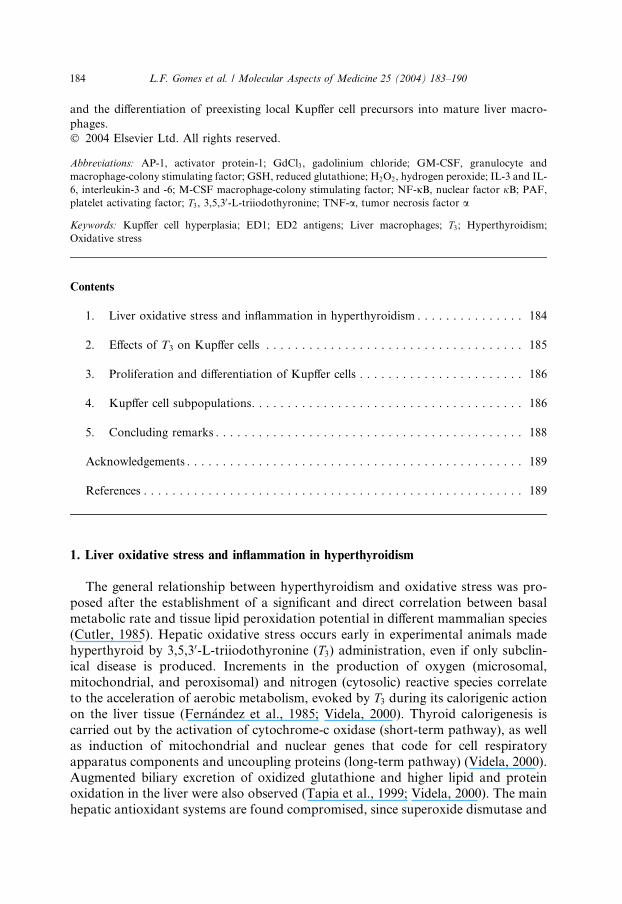
capacity (Tapia et al., 1997). In vivo colloidal carbon uptake is also enhanced in the
Fig. 1. Representative photomicrography of colloidal carbon uptake by the liver of rats treated with:
(A) saline, (B) T3, (C) GdCl3, (D) T3 and GdCl3. Formalin fixed, paraffin embedded liver slices (3 lm)
were counterstained with nuclear neutral red. Magnification: 400·.
186 L.F. Gomes et al. / Molecular Aspects of Medicine 25 (2004) 183–190
liver of T3-treated animals (Fig. 1). All the above effects are abolished by gadolinium
chloride (GdCl3) administration (Tapia et al., 1997). GdCl3, a selective Kupffer cell
inactivator, also significantly reduces GSH consumption, lipid peroxidation, and
nitric oxide production by the liver (Hardonk et al., 1992; Tapia et al., 1997).
The pathophysiology of Kupffer cell hyperplasia is not entirely elucidated. A
direct action of thyroid hormone, such as that verified in other cell types, as in he-patocytes, has been proposed for local Kupffer cell precursors which proliferate in
response to increasing concentrations of T3 (Columbano and Shinosuka, 1996;
Torres et al., 1999). This hypothesis implies a non-inflammatory expansion of the
macrophage population, which can induce oxidative stress as a secondary phe-
nomenon. On the other hand, a pro-inflammatory response in the sinusoidal
microenvironment could trigger monocyte recruitment from peripheral blood and
the production of inflammatory mediators in response to the redox imbalance
produced in the liver.
3. Proliferation and differentiation of Kupffer cells
According to the concept of mononuclear phagocytic system, inflammatory
exudate macrophages and resident macrophages are all derived from peripheralblood monocytes and differentiated into low proliferative capacity cells (Takezawa
et al., 1995). While in the newborn the liver is a central organ for the production and
supplementation of macrophages and their precursors to other tissues, in healthy
adults only 2% of liver cells are under division (Naito et al., 1997). The elevated
proliferative potential of fetal liver macrophages is essential to assure their survival
in the liver and the colonization of other fetal tissues, by means of the blood stream
(Naito et al., 1997). Nevertheless, Kupffer cell proliferation by local cell division can
be observed in adult organisms after injection of macrophage stimulating agents andfollowing partial hepatectomy.
Putative local liver macrophage precursors are proposed to derive from circulating
monocytes, which originate in bone marrow hematopoietic tissue (Crofton et al.,
1978). Among other growth factors, such as interleukins (IL-6, IL-3) and granulocyte
and macrophage-colony stimulating factor (GM-CSF), macrophage-colony stimu-
lating factor (M-CSF) seems the most important stimulator in the development and
differentiation of the restricted macrophage lineage (Naito et al., 1997).
4. Kupffer cell subpopulations
Macrophages constitute a functional and morphologically heterogeneous group
of cells, and the subgroups may be identified by immunohistochemical techniques.
ED1 and ED2 antigens have been particularly useful in the characterization ofKupffer cells subpopulations. ED1 antibody binds to most macrophage populations,
as well as peripheral blood monocytes and bone marrow precursors. Despite the fact
that a few other cell types also express ED1 when activated, it has been used as a
L.F. Gomes et al. / Molecular Aspects of Medicine 25 (2004) 183–190 187
mononuclear phagocytic system member label (Damoiseaux et al., 1994). ED1
antibody recognizes a simple chain of glycoprotein of 90,000–110,000 kDa, pre-
dominantly expressed on the lysosomal membrane and, to a lower extent, on the cell
surface. ED1 expression and glycosylation levels are accentuated by phagocytic
stimulus. Differential expression of this antigen derives from the exchange of
0
200
400
600
I II IIl
-100
0
100
200
300
400
I II III
0
100
200
300
400
I II III
* *
**
*
(A)
(B)
(C)
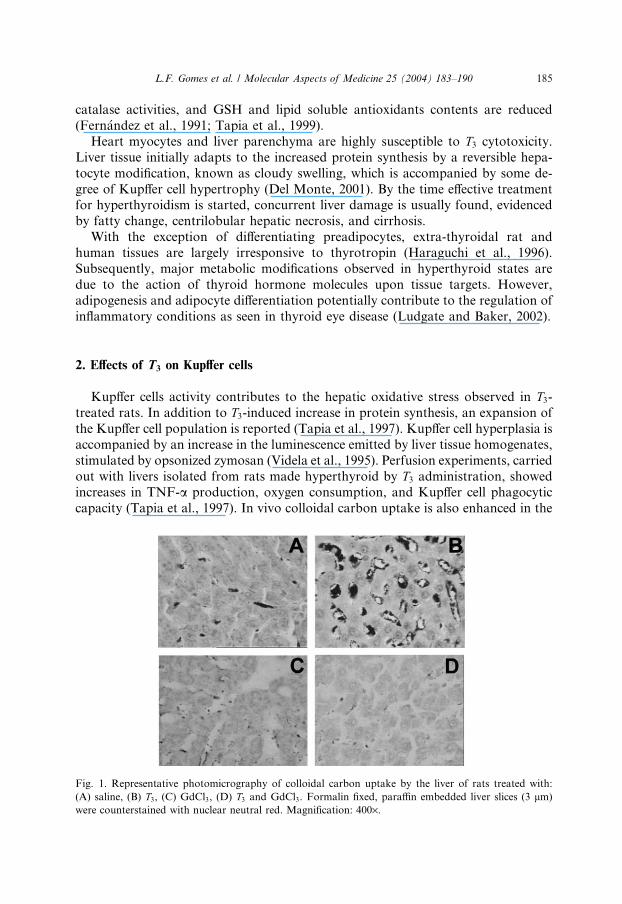
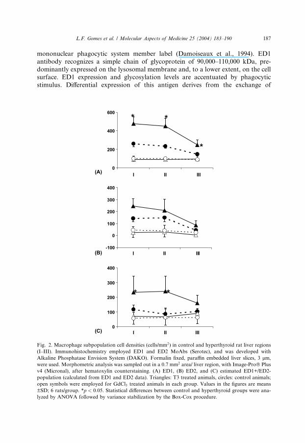
Fig. 2. Macrophage subpopulation cell densities (cells/mm2) in control and hyperthyroid rat liver regions
(I–III). Immunohistochemistry employed ED1 and ED2 MoAbs (Serotec), and was developed with
Alkaline Phosphatase Envision System (DAKO). Formalin fixed, paraffin embedded liver slices, 3 lm,
were used. Morphometric analysis was sampled out in a 0.7 mm2 area/ liver region, with Image-Pro� Plus
v4 (Micronal), after hematoxylin counterstaining. (A) ED1, (B) ED2, and (C) estimated ED1+/ED2-
population (calculated from ED1 and ED2 data). Triangles: T3 treated animals, circles: control animals;
open symbols were employed for GdCl3 treated animals in each group. Values in the figures are means
±SD; 6 rats/group. *p < 0:05. Statistical differences between control and hyperthyroid groups were ana-
lyzed by ANOVA followed by variance stabilization by the Box-Cox procedure.
188 L.F. Gomes et al. / Molecular Aspects of Medicine 25 (2004) 183–190
membrane components between these compartments, since plasma membrane
recycling is an important aspect of phagocytosis (Damoiseaux et al., 1994). ED2
antibody recognizes a membrane antigen of resident macrophages in rats. This
antibody is largely used to identify Kupffer cells (Dijkstra et al., 1985; Yamate et al.,
2001; Ide et al., 2002) and it is specific for resident macrophages.
Two main Kupffer cell subpopulations are distinguishable by the combined use ofED1 and ED2 antibodies. These subpopulations present slightly different morphol-
ogy, but distinct anatomical distribution and functional characteristics (Armbrust
and Ramadori, 1996; Sato et al., 1998; Yamate et al., 1999). One population of small
ED1-positive cells is present around the portal triad and centrilobular veins. This
population is described in the literature as ‘‘small Kupffer cells’’, ‘‘local Kupffer cell
precursors’’, or just taken as the less mature members of the liver macrophage pop-
ulation collectively called ‘‘Kupffer cells’’. Small Kupffer cells are usually not labeled
by ED2 antibody. Another population, constituted by large ED1 and ED2-positivemacrophages, is placed along the sinusoids. These cells are described as mature he-
patic tissue macrophages, often associated to the name ‘‘large Kupffer cells’’ or simply
‘‘Kupffer cells’’ (Armbrust and Ramadori, 1996; Sato et al., 1998; Yamate et al.,
1999). Both ED1 and ED2 labeling increase in the liver of hyperthyroid animals (Fig.
2). However, this increment is more pronounced in the ED1+/ED2- subpopulation
than in ED1+/ED2+ cells, indicating that T3-induced Kupffer cell hyperplasia causes a
shift on liver macrophage population phenotype, leaning towards younger cell types.
The sinusoidal environment itself stimulates local proliferation of macrophageprecursor mononuclear cells and their differentiation into Kupffer cells (Armbrust
and Ramadori, 1996). However, increased Kupffer cell turnover rates modify sinu-
soidal environment and such shift on liver macrophage population in T3-treatedanimals may add to a pro-inflammatory condition. For instance, recently migrated
monocytes are probably activated during their passage through the vascular wall,
producing reactive species (Decker, 1997).
Furthermore, Kupffer cell hyperplasia implies functional effects that may depend
on cell activation. The silent clearance of inflammation promoters, carried outmainly by macrophages, is an anti-inflammatory action (Witmer-Pack et al., 1993).
Regardless great phagocytic capacity, steady state Kupffer cells present small pro-
duction of oxygen and nitrogen reactive species and low expression of class II his-
tocompatibility receptors (MHC) (Bowens et al., 1992; Armbrust and Ramadori,
1996). They can, however, trigger inflammatory response, including antigen pre-
sentation (Naito et al., 1997).
5. Concluding remarks
Kupffer cell activation results in mononuclear cell recruitment from circula-
tion, receptor expression, and the secretion of several molecules, such as enzymes,eicosanoids, PAF, cytokines, complement proteins, oxygen and nitrogen reactive
species, and apolipoprotein E (Winwood and Arthur, 1998; Armbrust and Rama-
dori, 1996). Sinusoidal endothelium and Ito cells respond to factors released by
L.F. Gomes et al. / Molecular Aspects of Medicine 25 (2004) 183–190 189
activated Kupffer cells, thus amplifying the spectrum of released substances during
inflammation. This joint action triggers heat shock protein synthesis and liver
regeneration responses, since some of the produced oxidizing agents, i.e. H2O2, can
activate transcription factors (NF-jB, AP-1) and shift the balance between death
signs and liver cell proliferation (Sen and Packer, 1996). In conclusion, Kupffer cell
hyperplasia secondary to increased T3 levels is a potentially pro-inflammatory event,which involves both the of Kupffer cell precursor population expansion, through
circulating monocyte recruitment, and the differentiation of local Kupffer cell pre-
cursors into mature liver macrophages.
Acknowledgements
The authors would like to thank for the inestimable help of Prof. Dr. VirginiaB.C. Junqueira, from UNIFESP, Brazil. Supported by grants from FAPESP (97/
02335-5), Brazil, and from FONDECYT (1030499), Chile. SL and KASG were
recipients of post-graduate fellowships from CNPq and from FAPESP (01/04379-7).
References
Armbrust, T., Ramadori, G., 1996. Functional characterization of two different Kupffer cell populations
of normal rat liver. J. Hepatol. 25, 518–528.
Bowens, L., De Bleser, P., Vanderkerken, K., Geerts, B., Wisse, E., 1992. Liver cell heterogeneity:
functions of non-parenchymal cells. Enzyme 46, 155–168.
Columbano, A., Shinosuka, H., 1996. Liver regeneration versus direct hyperplasia. FASEB J. 10, 1118–
1128.
Crofton, R.W., Diesselhoff den-Dulk, M.M.C., vanFurth, R., 1978. The origin, kinetics, and character-
istics of the Kupffer cells in the normal steady-state. J. Exp. Med. 148, 1–17.
Cutler, R.G., 1985. Peroxide-producing potential of tissues: inverse correlation with longevity of
mammalian species. Proc. Natl. Acad. Sci. USA 87, 1620–1624.
Damoiseaux, J.G., Dopp, E.A., Calame, W., Chao, D., MacPherson, G.G., Dijkstra, C.D., 1994. Rat
macrophage lysosomal membrane antigen recognized by monoclonal antibody ED1. Immunology 83,
140–147.
Decker, K., 1997. The response of liver macrophages to inflammatory stimulation. Keio J. Med. 47, 1–9.
Del Monte, U., 2001. Thyroid hormones, acute iron overload and the pathogenesis of cloudy swelling.
Redox Rep. 6, 73–75.
Dijkstra, C.D., D€opp, E.A., Joling, P., Kraal, G., 1985. The heterogeneity of mononuclear phagocytes in
lymphoid organs: distinct macrophage subpopulations in the rat recognized by monoclonal antibodies
ED1, ED2, and ED3. Immunology 54, 589–599.
Fern�andez, V., Barrientos, X., Kipreos, K., Valenzuela, A., Videla, L.A., 1985. Superoxide radical
generation, NADPH oxidase activity, and cytochrome P450 content of rat liver microsomal fractions
in an experimental hyperthyroid state: relation to lipid peroxidation. Endocrinology 117, 496–501.
Fern�andez, V., Shimizu, K., Barros, S.B.M., Azzalis, L.A., Pimentel, R., Junqueira, V.B.C., 1991. Effects
of hyperthyroidism in rat liver glutathione metabolism: related enzyme activities, efflux, and turnover.
Endocrinology 129, 85–91.
Haraguchi, K., Shimura, H., Lin, L., Endo, T., Onaya, T., 1996. Differentiation of rat preadipocytes is
accompanied by expression of thyrotropin receptors. Endocrinology 137, 3200–3205.
Hardonk, M.J., Dijkhuis, F.W.J., Hulstaert, C.E., Koudstaal, J., 1992. Heterogeneity of rat liver and
spleen macrophages in gadolinium chloride elimination and repopulation. J. Leukoc. Biol. 52, 296–
302.
190 L.F. Gomes et al. / Molecular Aspects of Medicine 25 (2004) 183–190
Ide, M., Yamate, J., Machida, Y., Nakanish, M., Kuwamura, M., Kotani, T., Sawamoto, O., 2002.
Emergence of different macrophage populations in hepatic fibrosis following thioacetamide-induced
acute hepatocyte injury in rats. J. Comp. Path. 128, 41–51.
Ludgate, M., Baker, G., 2002. Unlocking the immunological mechanisms of orbital inflammation in
thyroid eye disease. Clin. Exp. Immunol., 127,193–198.
Naito, M., Hazegawa, G., Takahashi, K., 1997. Development, differentiation, and maturation of Kupffer
cells. Microsc. Res. Tech. 39, 350–364.
Sato, T., Yamamoto, H., Sasaki, C., Wake, K., 1998. Maturation of rat dendritic cells during intrahepatic
translocation evaluated using monoclonal antibodies and electron microscopy. Cell Tissue Res. 294,
503–514.
Sen, C.K., Packer, L., 1996. Antioxidant and redox regulation of gene transcription. FASEB J. 10, 702–
709.
Takezawa, R., Watanabe, Y., Akaike, T., 1995. Direct evidence of Macrophage differentiation from bone
marrow cells in the liver: a possible origin of Kupffer cells. J. Biochem. 118, 1175–1183.
Tapia, G., Conejo, P., Fern�andez, V., Videla, L.A., 1999. Protein oxidation in thyroid hormone-induced
liver oxidative stress: relation to lipid peroxidation. Toxicol. Lett. 106, 209–214.
Tapia, G., Pepper, I., Smok, G., Videla, L.A., 1997. Kupffer cell function in thyroid hormone-induced
liver oxidative stress. Free Radic. Res. 26, 267–279.
Torres, S., D�ıaz, B., Cabrera, J., D�ıaz-Chico, J., D�ıaz-Chico, B., L�opez-Guerra, A., 1999. Thyroid
hormone regulation of rat hepatocyte proliferation and polyploidization. Am. J. Physiol. 276, G155–
G163.
Videla, L.A., 2000. Energy metabolism, thyroid calorigenesis, and oxidative stress: functional and
cytotoxic consequences. Red. Rep. 5, 265–275.
Videla, L.A., Smok, G., Troncoso, P., Simon, K.A., Junqueira, V.B.C., Fern�andez, V., 1995. Influence of
hyperthyroidism on lindane-induced hepatotoxicity in the rat. Biochem. Pharmacol. 50, 1557–1565.
Winwood, P.J., Arthur, M.J.P., 1998. Kupffer cells and endothelial cells. In: Strain, A., Diehl, A.M. (Eds.),
Liver Growth and Repair. Chapman & Hall, London, pp. 482–511.
Witmer-Pack, M., Crowley, M.T., Inaba, K., Steinman, R.M., 1993. Macrophages, but not dendritic cells,
accumulate colloidal carbon following administration in situ. J. Cell Sci. 105, 965–970.
Yamate, J., Kumagai, D., Tsujino, K., Nakatsuji, S., Kuwamura, M., Kotani, T., Sakuma, S., LaMarre,
J., 1999. Macrophage populations and apoptotic cells in the liver before spontaneous hepatitis in Long-
Evans cinnamon (LEC) rats. J. Comp. Path. 120, 333–346.
Yamate, J., Maeda, M., Tsukamoto, Y., Benn, S.J., Laithwaite, J.E., Allan, A., Kannan, Y., Ide, M.,
Kuwamura, M., Kotani, T., Sakuma, S., LaMarre, J., 2001. Macrophage-like cell line (HS-P) from a
rat histiocytic sarcoma. J. Comp. Path. 124, 183–191.