Embed Size (px)
Citation preview
WORLD JOURNAL OF SURGICAL ONCOLOGY
Franchi et al. World Journal of Surgical Oncology 2013, 11:192http://www.wjso.com/content/11/1/192
CASE REPORT Open Access
Two-stage hepatectomy after autologousCD133+ stem cells administration: a case reportEloisa Franchi, Maria C Canepa, Andrea Peloso, Letizia Barbieri, Laura Briani, Gabor Panyor, Paolo Dionigiand Marcello Maestri*
Abstract
Liver resection is the mainstay of treatment for patients with primary and metastatic liver tumors. However, a largemajority of patients present for initial medical evaluation with primary and metastatic liver tumors when theircancer is unresectable. Several trials have been undertaken to identify alternative treatments and complementarytherapies. In the near future, the field of liver surgery will aim to increase the number of patients that can benefitfrom resection, since radical removal of the tumor currently provides the sole chance of cure. This paper reports thecase of a patient with an advanced colonic cancer in the era of stem cell therapyIn 2011, a 57 years old whiteCaucasian man with a previous history of non-Hodgkin lymphoma (NHL) was diagnosed with colon cancer andbilobar liver metastases. Following neoadjuvant therapy, the patient was enrolled in a protocol of stem celladministration for liver regeneration. Surgery was initially performed on the primary cancer and left liver lobe. Anextended right lobectomy to S1 was then performed after a portal vein embolization (PVE) and stem cellstimulation of the remaining liver. The postoperative course was uneventful and the patient was free of diseaseafter 12 months. Extreme liver resection can provide a safer option and a chance of cure to otherwise unresectablepatients when liver regeneration is boosted by PVE and stem cell administration.
Keywords: Liver surgery, Portal vein embolization, Stem cells, Liver regeneration, Colon cancer, Liver metastases
BackgroundThe treatment of choice for both primary and metastaticliver tumors is radical resection [1]. However, up to 45%of patients present for initial medical evaluation whenthe parenchymal diffusion of cancer requires more surgi-cal resection than is possible. An accepted evaluation ofprospective resection is that 30% of the liver mustremain in order for its function to be unaffected, or asafer 40% if there is an underlying disease (for example,chronic hepatitis, diabetes) or previous chemotherapytreatment [2].Portal vein embolization (PVE) has been proposed as a
tool to stimulate liver regeneration when prospectivesurgical resection is over the limit [3]. This techniquerelies on the portal injection of several agents into thecancer liver lobes. Commonly, patients present with alarge tumor in the right liver lobe and a left lobe that istoo small for radical resection. Following PVE, the
* Correspondence: [email protected] IRCCS Policlinico San Matteo, and University of Pavia, VialeCamillo Golgi 19, Pavia 27100PV, Italy
© 2013 Franchi et al.; licensee BioMed CentralCommons Attribution License (http://creativecreproduction in any medium, provided the or
contralateral segments experience a degree of hypertrophyin the range of 15 to 25%, depending on liver status.When a patient has a tumor that is technically resec-
table but the remaining liver is small, PVE can stimulategrowth, which can be observed until the volume iswithin the necessary limit. The process is continuousand the remaining tissue will continue to grow. Studieshave reported that the liver can take 150 days to developa volume large enough to allow surgical resection [4].However, during this time the disease can continue itsprogression, and patients could die while waiting. Fur-thermore, PVE does not prevent the tumor growthinside the occluded portal lobe, and there are concernsregarding its potential when diffusion of cancer is fasterthan expected [5]. Therefore, a procedure is needed togain volume as swiftly as possible, while minimizingwaiting list time to avoid any advantage for the cancer.Several recent studies have suggested that stem cells
play a key role in the field of tissue regeneration [6]. Theliver reacts to any lesion by several naturals paths. Thus,several populations of cells cooperate through different
Ltd. This is an Open Access article distributed under the terms of the Creativeommons.org/licenses/by/2.0), which permits unrestricted use, distribution, andiginal work is properly cited.

Franchi et al. World Journal of Surgical Oncology 2013, 11:192 Page 2 of 7http://www.wjso.com/content/11/1/192
pathways, and their role depends on the nature of thestimulus and the extent of damage. A normally functio-ning liver will undergo a slow turnover of functionalmass based on the proliferation of hepatocytes. Adulthepatocytes proliferate after a normal liver resectionwhen the residual parenchyma can tolerate the damage,while supplying the needs of the whole body. When theloss of tissue is greater, the liver seems to have a pool ofsleeping cells that have been found mainly in rodents,called ‘oval cells’ [7]. Such cells are relatively undifferen-tiated and several studies have demonstrated that theycan play a role when the normal hepatocytes are notable to repair by normal mechanisms. These emergencycells can possibly differentiate toward the hepatocyte orbiliary cell lines. When the liver suffers an extreme in-sult, both acute and chronic, stem cells can be mobilizedfrom the bone marrow to participate in the repair [8].These cells are CD34+ and their number grows steadilyafter extended liver resections [9], while they do notappear after normal abdominal surgery (that is, gastro-intestinal or pancreatic operations). Among this popu-lation, CD133+ cells are a subset with special liverengraftment potential [10]. The CD133+ phenotype istypical of some cell lines with multipotent differenti-ation capacity. Recently, CD133+ cells have been
Figure 1 Timeline of the patient’s course of disease. After the onset ofcancer with synchronous metastases was initially considered unresectable.
proposed to augment liver regeneration after PVE inselected patients [11,12].This paper presents a case of two-stage hepatectomy
of synchronous liver metastases from colon cancer.
Case presentationA 57 -year-old white Caucasian man was diagnosed withnon-Hodgkin lymphoma (NHL) in 1991 (Figure 1). TheNHL was treated by a standard protocol of chemo-therapy and radiotherapy. The treatment was successfuland achieved a complete response. Thereafter, the pa-tient reported good health and normal quality of life. InMarch 2011, a routine follow-up examination revealed asigmoid cancer and bilobar multiple liver metastases. InVittorio Emanuele hospital, Catania, Italy, the oncologyteam decided to commence a FOLFOX combinationchemotherapy regimen. This treatment of six cycles hadsome success and re-evaluation deemed the disease asstable.The patient was then referred to the liver unit at
Fondazione IRCCS Policlinico San Matteo, UniversityHospital, Pavia, Italy. An additional computed tomo-graphy (CT) scan was undertaken to define the extent ofthe disease (Figures 2 and 3). The multidisciplinary liverteam decided the patient should undergo a two-stage
NHL, the patient had a prolonged period of good health. The colonNHL, non-Hodgkin lymphoma.
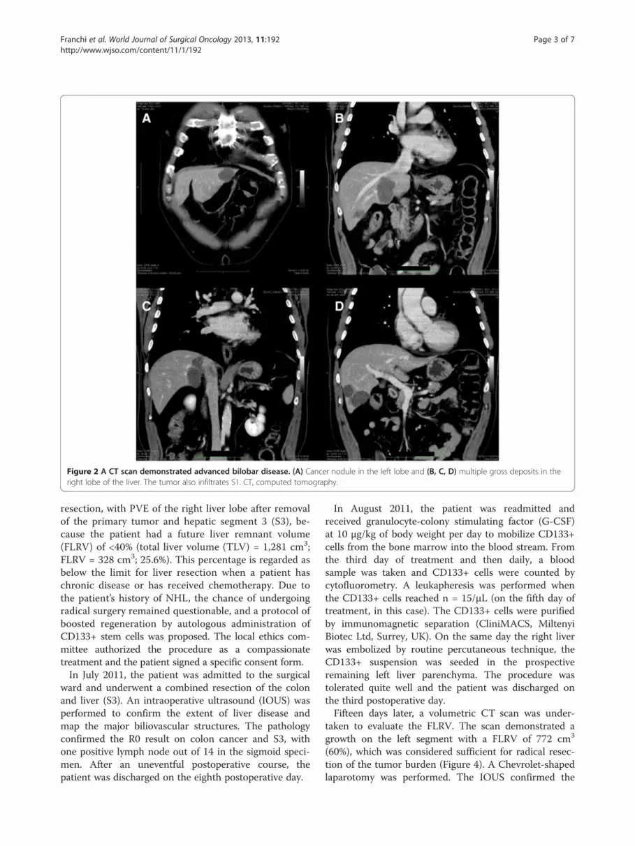
Figure 2 A CT scan demonstrated advanced bilobar disease. (A) Cancer nodule in the left lobe and (B, C, D) multiple gross deposits in theright lobe of the liver. The tumor also infiltrates S1. CT, computed tomography.
Franchi et al. World Journal of Surgical Oncology 2013, 11:192 Page 3 of 7http://www.wjso.com/content/11/1/192
resection, with PVE of the right liver lobe after removalof the primary tumor and hepatic segment 3 (S3), be-cause the patient had a future liver remnant volume(FLRV) of <40% (total liver volume (TLV) = 1,281 cm3;FLRV = 328 cm3; 25.6%). This percentage is regarded asbelow the limit for liver resection when a patient haschronic disease or has received chemotherapy. Due tothe patient’s history of NHL, the chance of undergoingradical surgery remained questionable, and a protocol ofboosted regeneration by autologous administration ofCD133+ stem cells was proposed. The local ethics com-mittee authorized the procedure as a compassionatetreatment and the patient signed a specific consent form.In July 2011, the patient was admitted to the surgical
ward and underwent a combined resection of the colonand liver (S3). An intraoperative ultrasound (IOUS) wasperformed to confirm the extent of liver disease andmap the major biliovascular structures. The pathologyconfirmed the R0 result on colon cancer and S3, withone positive lymph node out of 14 in the sigmoid speci-men. After an uneventful postoperative course, thepatient was discharged on the eighth postoperative day.
In August 2011, the patient was readmitted andreceived granulocyte-colony stimulating factor (G-CSF)at 10 μg/kg of body weight per day to mobilize CD133+cells from the bone marrow into the blood stream. Fromthe third day of treatment and then daily, a bloodsample was taken and CD133+ cells were counted bycytofluorometry. A leukapheresis was performed whenthe CD133+ cells reached n = 15/μL (on the fifth day oftreatment, in this case). The CD133+ cells were purifiedby immunomagnetic separation (CliniMACS, MiltenyiBiotec Ltd, Surrey, UK). On the same day the right liverwas embolized by routine percutaneous technique, theCD133+ suspension was seeded in the prospectiveremaining left liver parenchyma. The procedure wastolerated quite well and the patient was discharged onthe third postoperative day.Fifteen days later, a volumetric CT scan was under-
taken to evaluate the FLRV. The scan demonstrated agrowth on the left segment with a FLRV of 772 cm3
(60%), which was considered sufficient for radical resec-tion of the tumor burden (Figure 4). A Chevrolet-shapedlaparotomy was performed. The IOUS confirmed the

A B
C D
Volume: cm3 772
Figure 4 CT scan, resected liver and specimen. (A, B) After the portal vein embolization (PVE) and administration of autologous CD133+ cells,the future liver remnant volume (FLRV) reached 772 cm3 (15 days after the procedure). The (C) resected liver and (D) specimen are shown. CT,computed tomography; FLRV, future liver remnant volume; PVE, portal vein embolization.
A B
C D
Volume: cm3 328
Figure 3 Volumetric preoperative study. (A, B, C) The study demonstrated a future liver remnant volume (D: FLRV) below the minimum of40% of total liver volume (TLV). This percentage is generally accepted as the lower limit to allow an extended liver resection with a reasonableprobability of success when there is an underlying disease, such as chronic hepatitis, cirrhosis or previous chemotherapy. FLRV, future liverremnant volume; TLV, total liver volume.
Franchi et al. World Journal of Surgical Oncology 2013, 11:192 Page 4 of 7http://www.wjso.com/content/11/1/192

Franchi et al. World Journal of Surgical Oncology 2013, 11:192 Page 5 of 7http://www.wjso.com/content/11/1/192
tumor in the right lobe with involvement of S1 andpart of S4. The liver ligaments were cut and a cavadissection up to the hepatic vein was performed. Thehilum was dissected, and the right portal branch, righthepatic artery and bile duct were ligated and sec-tioned. The right hepatic vein was isolated free at itscaval origin and cut between vascular clamps. Thestumps were closed with running polypropylene su-tures. The parenchyma was transected by cavitron ultra-sonic surgical aspirator (CUSA; Valleylab, Boulder, CO,USA). Small vessels were sealed by harmonic scalpel,while those greater than 3 mm were closed by silkligatures or titanium clips. The resection was extendedto the very lateral portion of S4 and to S1 (Figure 5).The postoperative course was unremarkable and the
patient was discharged on the eighth postoperativeday. The pathology report demonstrated R0 surgicaloutcome. Later, the oncology team prescribed acourse of chemotherapy (Figure 1). On December2012, the patient was alive and well, and free fromdisease at follow-up CT scans.
Figure 5 A CT scan was obtained after surgery. The outcome was unreby the hemostatic clips left in situ. CT, computed tomography.
ConclusionsOnce regarded a risk [13,14], liver surgery is today rou-tine practice for the treatment of several primary andmetastatic tumors. However, when the numbers of pa-tients that are deemed eligible for surgery are assessed, avast majority do not qualify because of anatomy, thenumber of lesions and amount of FLRV.PVE is recognized as a tool to stimulate liver regene-
ration before a liver resection is performed [15]. How-ever, the amount and speed of regeneration can varywith each case, and a number of patients have died whilewaiting to gain a remaining liver large enough to allow aresection. Recently, stem cells have offered a choice oftreatment for several diseases and a possible aid even inthe field of liver surgery, with preliminary clinical studiesdemonstrating a favorable effect when administered afterliver damage [16].We started a clinical study to offer stem cell adminis-
tration to selected patients. To qualify, the patient musthave primary or metastatic liver malignancy, a FLRVbelow the cutoff for safe resection (<30% or <40% if the
markable with normal liver function. The resected area is highlighted

Franchi et al. World Journal of Surgical Oncology 2013, 11:192 Page 6 of 7http://www.wjso.com/content/11/1/192
patient has an underlying liver condition) and a contra-indication to the routine PVE. The patient reported inthis case had a previous history of NHL, which wastreated by chemotherapy. Thus, the patient’s ability toregenerate after an extended resection was questionableand the case was deemed marginal.Patients with hematological cancers and metastatic on-
set of new oncological disease are frequently consideredunsuitable for further aggressive surgical treatments.Even with this single case report, the result was appea-ling and we have begun a local trial on autologousCD133+ stem cells. It must be recognized that there arestill concerns about the use of adult stem cells in clinicalpractice. For example, a patient with cancer can havecancer stem cells in circulation. Thus, we are extensivelytesting any possible risk of cancer spreading, and to datethere has been no evidence that CD133+ autologousadministration can negatively affect the biology of thedisease. However, this is only a case report and the safetyof such procedures must be carefully validated in con-trolled trials.While liver regeneration is likely to be boosted by
local deposition of specific cells, the question arisesabout whether regeneration can be any different fromthe naïve parenchyma. Concerning the hepatic func-tional reserve, there have been no evident changes afterthe administration of CD133+ stem cells. These areprobably effective in increasing the amount of hepaticparenchyma, but the quality of the regenerated tissueremains the same. Again, this issue requires specificstudies which we hope to perform at leading centers.The long-term outcome of this case has not yet been de-fined and the patient is still undergoing follow-up, but itis important to add that the autologous portal adminis-tration of CD133+ cells did not cause any immediate orlate adverse effect. Similarly, other reports do not high-light special complications [11].This report suggests that some cell populations can
stimulate hepatic regeneration and their use should betaken into consideration for patients who need extremeliver surgery. We believe that a prospective approach toregenerative medicine in this field will allow an im-proved indication to resection, while offering a chanceof cure to otherwise unresectable patients. Further stu-dies are required to ensure safety and effectiveness ofthe therapeutic tool against the risk of liver failure afterextended resections.
ConsentWritten informed consent was obtained from the patientfor publication of this case report and any accompanyingimages. A copy of the written consent is available forreview by the Editor-in-Chief of this journal.
AbbreviationsCT: Computed tomography; CUSA: Cavitron ultrasonic surgical aspirator;FLRV: Future liver remnant volume; G-CSF: Granulocyte colony-stimulatingfactor; IOUS: Intraoperative ultrasound; NHL: Non- hodgkin lymphoma;PVE: Portal vein embolization; TLV: Total liver volume.
Competing interestsThe authors declare that they have no competing interests.
Authors’ contributionsEF and MCC drafted the paper. They were part of the surgical team. AP andLB were in charge of the case and the postoperative care. They were part ofthe surgical team. LB and GP were responsible to collect the data and toprepare the manuscript. PD was is in charge of the follow-up, prepared thepictures and gave a substantial contribution to the final form of the draft.MM is the first investigator in a clinical trial of stem cells at FondazioneIRCCS Policlinico San Matteo. He performed the operation, designed thestem cell protocol and the postoperative program of therapy. All authorsread and approved the final manuscript.
Received: 22 December 2012 Accepted: 5 August 2013Published: 13 August 2013
References1. Bengtsson G, Carlsson G, Hafstrom L, Jönsson PE: Natural history of
patients with untreated liver metastases from colorectal cancer.Am J Surg 1981, 141:586–589.
2. Ferrero A, Viganò L, Polastri R, Muratore A, Eminefendic H, Regge D,Capussotti L: Postoperative liver dysfunction and future remnant liver:where is the limit? Results of a prospective study. World J Surg 2007,31:1643–1651.
3. de Baere T, Roche A, Elias D, Lasser P, Lagrange C, Bousson V: Preoperativeportal vein embolization for extension of hepatectomy indications.Hepatology 1996, 24:1386–1391.
4. Broering DC, Hillert C, Krupski G, Fischer L, Mueller L, Achilles EG,Schulte am Esch J, Rogiers X: Portal vein embolization vs. portal veinligation for induction of hypertrophy of the future liver remnant.J Gastrointest Surg 2002, 6:905–913.
5. Madoff DC, Hicks ME, Abdalla EK, Morris JS, Vauthey JN: Portal veinembolization with polyvinyl alcohol particles and coils in preparation formajor liver resection for hepatobiliary malignancy: safety andeffectiveness-study in 26 patients. Radiology 2003, 227:251–260.
6. Theise ND: Liver stem cells: the fall and rise of tissue biology. Hepatology2003, 38:804–806.
7. Kountouras J, Boura P, Lygidakis NJ: Liver regeneration after hepatectomy.Hepato-gastroenterology 2001, 48:556–562.
8. Petersen BE, Bowen WC, Patrene KD, Mars WM, Sullivan AK, Murase N,Boggs SS, Greenberger JS, Goff JP: Bone marrow as a potential source ofhepatic oval cells. Science 1999, 284:1168–1170.
9. Fujii H, Hirose T, Oe S, Yasuchika K, Azuma H, Fujikawa T, Nagao M,Yamaoka Y: Contribution of bone marrow cells to liver regeneration afterpartial hepatectomy in mice. J Hepatol 2002, 36:653–659.
10. Handgretinger R, Gordon PR, Leimig T, Chen X, Buhring HJ, Niethammer D,Kuci S: Biology and plasticity of CD133+ hematopoietic stem cells.Ann N Y Acad Sci 2003, 996:141–151.
11. Fürst G, Schulte am Esch J, Poll LW, Hosch SB, Fritz LB, Klein M,Godehardt E, Krieg A, Wecker B, Stoldt V, Stockschläder M, Eisenberger CF,Mödder U, Knoefel WT: Portal vein embolization and autologousCD133+ bone marrow stem cells for liver regeneration: initialexperience. Radiology 2007, 243:171–179.
12. am Esch JS 2nd, Knoefel WT, Klein M, Ghodsizad A, Fuerst G, Poll LW,Piechaczek C, Burchardt ER, Feifel N, Stoldt V, Stockschläder M, Stoecklein N,Tustas RY, Eisenberger CF, Peiper M, Häussinger D, Hosch SB: Portalapplication of autologous CD133+ bone marrow cells to the liver: anovel concept to support hepatic regeneration. Stem Cells 2005,23:463–470.
13. Bozzetti F, Gennari L, Regalia E, Bignami P, Montalto F, Mazzaferro V, Doci R:Morbidity and mortality after surgical resection of liver tumors. Analysisof 229 cases. Hepato-gastroenterology 1992, 39:237–241.
14. Iwatsuki S, Starzl TE: Personal experience with 411 hepatic resections.Ann Surg 1988, 208:421–434.

Franchi et al. World Journal of Surgical Oncology 2013, 11:192 Page 7 of 7http://www.wjso.com/content/11/1/192
15. Hemming AW, Reed AI, Howard RJ, Fujita S, Hochwald SN, Caridi JG,Hawkins IF, Vauthey JN: Preoperative portal vein embolization forextended hepatectomy. Ann Surg 2003, 237:686–691.
16. am Esch JS, Schmelzle M, Fürst G, Robson SC, Krieg A, Duhme C, Tustas RY,Alexander A, Klein HM, Topp SA, Bode JG, Häussinger D, Eisenberger CF,Knoefel WT: Infusion of CD133+ bone marrow-derived stem cells afterselective portal vein embolization enhances functional hepatic reservesafter extended right hepatectomy: a retrospective single-center study.Ann Surg 2012, 255:79–85.
doi:10.1186/1477-7819-11-192Cite this article as: Franchi et al.: Two-stage hepatectomy afterautologous CD133+ stem cells administration: a case report. WorldJournal of Surgical Oncology 2013 11:192.
Submit your next manuscript to BioMed Centraland take full advantage of:
• Convenient online submission
• Thorough peer review
• No space constraints or color figure charges
• Immediate publication on acceptance
• Inclusion in PubMed, CAS, Scopus and Google Scholar
• Research which is freely available for redistribution
Submit your manuscript at www.biomedcentral.com/submit


