Embed Size (px)
Citation preview

Optical biopsy and tissue phantom selection: a novelapproach combining single-photon timing and spatial-mode
selection
Luca Nardoa, Maria Bondanib and Alessandra Andreonia
aDipartimento di Fisica e Matematica, Universita degli Studi dell’Insubria and C.N.I.S.M.,via Valleggio, 11, 22100 Como, Italy;
bNational Laboratory for Ultrafast and Ultraintense Optical Science (U.L.T.R.A.S.),C.N.R.-I.N.F.M., via Valleggio, 11, 22100 Como, Italy
ABSTRACT
A snake photon detection based imaging technique developed by our group is explained in details and its per-formances compared with those obtained by other experimentalists. The technique is based on simultaneousapplication of time and spatial-mode selection. We also show that in these very particular working conditionscommonly used plastic tissue phantoms display non tissue-like scattering properties.
Keywords: Single-photon detectors, Light propagation in tissues, Photon migration, Time-resolved imaging,Tissue phantoms.
1. INTRODUCTION
Early diagnosis is often essential to assure efficient treatment of cancer. The presently applied diagnostic tech-niques, be they based on nuclear magnetic resonance or X-ray tomography, even if assuring millimeter spatialresolution, fail to offer the tools for large scale routine clinical exams on healthy patients. In fact, both classes oftechniques are extremely invasive, and patients cannot submit themselves to diagnostic exams without incurringinto the risk of contracting side-effect pathologies. At variance with the methods of above, optical imaging uti-lizes non-ionizing radiation. Thus, patients can be repeatedly exposed to screening without any harm. Moreover,a distinction among different soft tissues (optical biopsy) is in principle possible, due to substantial differencesin their way of scattering and absorbing light. On the other hand, at difference with X-rays, light is eitherefficiently absorbed or severely scattered by biological samples. While scattering decreases monotonically atincreasing wavelengths, tissue absorption is sufficiently low as to allow application of optical imaging only inthe near infrared radiation (NIR) wavelength interval between 700 nm and 1100 nm, the so called “therapeuticwindow”.1 In the therapeutic window, NIR is still highly scattered by biological tissues, so that, on the average,a photon is expected to be diffused several times before emerging from an even very thin slice of tissue. Over thepast decades several theoretical models were developed, capable of reproducing and explaining at least qualita-tively the main features of NIR interactions with biological tissues.2–4 It is now assessed that biological tissuesbehave with respect to NIR as highly scattering polydispersed media. The transport of NIR through a highlyscattering medium can be described in terms of the isotropic diffusion approximation model.2–4 The isotropicdiffusion model is able to yield both the collimated transmittance value, that is the fraction of incident lightpassing through the medium without being scattered, and the temporal distribution of the photons transmittedthrough a scattering medium, the so-called photon time of flight (TOF) distribution, and can be applied if thethickness of the medium is much higher than the mean free path of photons inside the medium. In this limit, thevalue of the collimated transmittance is given by T = exp(−μL), being L the thickness of the medium and μ theextinction coefficient. For pure isotropic diffusion, μ is simply the sum of the absorption coefficient μa and the
Further author information: (Send correspondence to L.N.)L.N.: E-mail: [email protected],M.B.: E-mail: [email protected],A.A.: E-mail: [email protected]
Advanced Photon Counting Techniques III, edited by Mark A. Itzler, Joe C. Campbell,Proc. of SPIE Vol. 7320, 732009 · © 2009 SPIE · CCC code: 0277-786X/09/$18 · doi: 10.1117/12.818514
Proc. of SPIE Vol. 7320 732009-1

scattering coefficient μs, which is defined as the inverse of the scattering mean free path. Photons constitutingthe collimated transmittance signal have strictly not been scattered: they are usually referred to as ”ballisticphotons”. Anisotropy of the scattering can be accounted for in the frame of the isotropic model by introduc-ing a third parameter, the anisotropy factor, g, and substituting, in the transmittance equation, the scatteringcoefficient μs with the reduced scattering coefficient (also called transport coefficient) μ′
s = μs(1 − g). In thecase of forward peaked scattering, the effect of anisotropy is to increase the collimated transmittance signal, andthe anisotropy coefficient takes values spanning from 0 (completely isotropic diffusion) to 1 (no diffusion). Inthis case the collimated transmittance signal is superimposed to a number of snake photons that have not beenappreciably deflected, but that could in principle have experienced some forward scattering events. As to thefunctional shape of the TOF distributions, an ultrashort laser pulse impinging on a slice of tissue is temporallyspread into a coherent component and a diffused component, due to multiple scattering caused by random fluc-tuations in the refractive index. The coherent component is made up of the ballistic photons, while the diffusedcomponent consists of multiply scattered photons. As the ballistic photons are neither deflected nor delayed,the coherent peak of the TOF distribution is the earliest arriving. Moreover, it is very sharp, ideally conservingthe same temporal profile as that of the impinging pulse. The diffused peak is asymmetrical-bell shaped. Asmultiply scattered photons travel on paths that are much longer than the straight path followed by the ballisticphotons, and very diverse one to the other depending on the number of scattering events experienced by eachparticular diffused photons, the diffused peak is delayed and much broader than the ballistic peak. The diffusedpeak rise time and temporal delay with respect to the ballistic peak are primarily determined by the μ′
s value,while the shape of the tail of the diffused TOF component primarily depends on μa. The snake photons haveundergone only forward scattering events and have traveled along paths only slightly deviating from the straightpath followed by ballistic photons. Thus, they are minimally delayed and make up the earliest part of the diffusecomponent.From the experimental point of view, a wide body of data is now available regarding the values assumed by ofμa, μs and g in many biological tissues.5–8 Although there is a wide variation in the literature data referred tosimilar tissues, all these data can be summarized for our purposes in the following observations: μs of biologicaltissues is very high, typically spanning in the range from 10 cm−1 to 100 cm−1; in the range of wavelengthspanning from 600 nm to 1100 nm the absorption contribution to the extinction coefficient can be neglected asμa < 0.01μs; scattering by biological tissues is highly forward, with g > 0.9.Due to the high variability and easy deterioration of biological samples, leading to scarcely reproducible exper-imental results, many studies have also been devoted to find media appropriate for in vitro experiments anddisplaying absorption and scattering characteristics similar to those of tissues (tissue phantoms). Many phantommaterials have been designed over the last two decades, including:- water suspensions of either Intralipid (with or without ink)8–10 or micro-spheres of latex, polystyrene orquartz;10–12
- solid tissue phantoms made of: Agar, Intralipid and ink;13 Delrin;14–16 Nylon,15,16 Teflon;16 clear plastic orresin materials with scattering and absorbing bodies embedded.17
Techniques of optical imaging through highly turbid and diffusing media based on the analysis of the multiplyscattered, dominant part of the emerging light18 are endowed with spatial resolution worse than that pursuedby X-ray tomography. Conversely, it is generally accepted that a map of the variations in the optical propertiesof the medium virtually endowed with diffraction-limited spatial resolution could be obtained by applying animaging system capable to select only the ballistic photons, and measure their relative intensity as a functionof the incident light position.19 Unfortunately, in the case of tissues, as the scattering is strongly forward, thecollimated transmittance is superimposed to a number of snake photons. As the snake photons follow a trajec-tory very close to that of the ballistic ones, they still carry information on the spatial distribution of the opticalproperties of the imaged specimen, but the ultimate achievable spatial resolution is reduced by an unpredictableamount. The spatial resolution of a real tissue optical imaging device substantially depends on its efficacy inselecting only minimally scattered photons.Between the end of the ’80s and the first half of the ’90s the attempts to perform optical imaging throughthick scattering media have multiplied.20,21 Almost all these attempts relied on one of the following four majorsnake-photon selection modes: spatial filtering12 (selection of the collimated transmittance signal by detectingonly the light emerging from the sample within a surface corresponding to the incident beam cross sectional
Proc. of SPIE Vol. 7320 732009-2
Channel
�t=1/(113 MHz)
PIN
SPAD
ND
sampleL
F
OBJ
BSlaser
pulses
start
MCA
TACstop
CF
Fsync
ND
Delay
adjustable
distance
BS
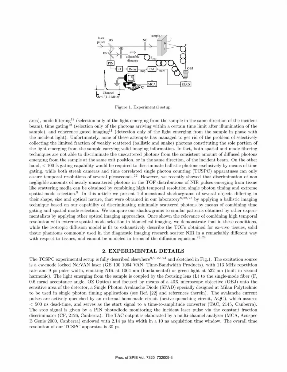
Figure 1. Experimental setup.
area), mode filtering12 (selection only of the light emerging from the sample in the same direction of the incidentbeam), time gating12 (selection only of the photons arriving within a certain time limit after illumination of thesample), and coherence gated imaging11 (detection only of the light emerging from the sample in phase withthe incident light). Unfortunately, none of these attempts has managed to get rid of the problem of selectivelycollecting the limited fraction of weakly scattered (ballistic and snake) photons constituting the sole portion ofthe light emerging from the sample carrying valid imaging information. In fact, both spatial and mode filteringtechniques are not able to discriminate the unscattered photons from the consistent amount of diffused photonsemerging from the sample at the same exit position, or in the same direction, of the incident beam. On the otherhand, < 100 fs gating capability would be required to discriminate ballistic photons exclusively by means of timegating, while both streak cameras and time correlated single photon counting (TCSPC) apparatuses can onlyassure temporal resolutions of several picoseconds.22 However, we recently showed that discrimination of nonnegligible amounts of nearly unscattered photons in the TOF distributions of NIR pulses emerging from tissuelike scattering media can be obtained by combining high temporal resolution single photon timing and extremespatial-mode selection.9 In this article we present 1-dimensional shadowgrams of several objects differing intheir shape, size and optical nature, that were obtained in our laboratory8,22,23 by applying a ballistic imagingtechnique based on our capability of discriminating minimally scattered photons by means of combining timegating and spatial mode selection. We compare our shadowgrams to similar patterns obtained by other experi-mentalists by applying other optical imaging approaches. Once shown the relevance of combining high temporalresolution with extreme spatial mode selection in biomedical imaging, we demonstrate that in these conditions,while the isotropic diffusion model is fit to exhaustively describe the TOFs obtained for ex-vivo tissues, solidtissue phantoms commonly used in the diagnostic imaging research scatter NIR in a remarkably different waywith respect to tissues, and cannot be modeled in terms of the diffusion equation.23,24
2. EXPERIMENTAL DETAILS
The TCSPC experimental setup is fully described elsewhere8,9, 22–24 and sketched in Fig.1. The excitation sourceis a cw-mode locked Nd:VAN laser (GE 100 1064 VAN, Time-Bandwidth Products), with 113 MHz repetitionrate and 9 ps pulse width, emitting NIR at 1064 nm (fundamental) or green light at 532 nm (built in secondharmonic). The light emerging from the sample is coupled by the focusing lens (L) to the single-mode fiber (F,0.6 mrad acceptance angle, OZ Optics) and focused by means of a 40X microscope objective (OBJ) onto thesensitive area of the detector, a Single Photon Avalanche Diode (SPAD) specially designed at Milan Polytechnicto be used in single photon timing applications (see Ref. [22] and references therein). The avalanche currentpulses are actively quenched by an external homemade circuit (active quenching circuit, AQC), which assures< 500 ns dead-time, and serves as the start signal to a time-to-amplitude converter (TAC, 2145, Canberra).The stop signal is given by a PIN photodiode monitoring the incident laser pulse via the constant fractiondiscriminator (CF, 2126, Canberra). The TAC output is elaborated by a multi-channel analyzer (MCA, AcuspecB Genie 2000, Canberra) endowed with 2.14 ps bin width in a 10 ns acquisition time window. The overall timeresolution of our TCSPC apparatus is 30 ps.
Proc. of SPIE Vol. 7320 732009-3
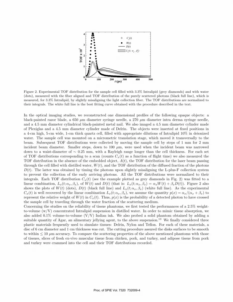
Figure 2. Experimental TOF distribution for the sample cell filled with 3.3% Intralipid (grey diamonds) and with water(dots), measured with the fiber aligned and TOF distribution of the purely scattered photons (black full line), which ismeasured, for 3.3% Intralipid, by slightly misaligning the light collection fiber. The TOF distributions are normalized totheir integrals. The white full line is the best fitting curve obtained with the procedure described in the text.
In the optical imaging studies, we reconstructed one dimensional profiles of the following opaque objects: ablack-painted razor blade, a 650 μm diameter syringe needle, a 270 μm diameter intra derma syringe needle,and a 4.5 mm diameter cylindrical black-painted metal nail. We also imaged a 4.5 mm diameter cylinder madeof Plexiglas and a 4.5 mm diameter cylinder made of Delrin. The objects were inserted at fixed positions ina 4-cm high, 5-cm wide, 1-cm thick quartz cell, filled with appropriate dilutions of Intralipid 10% in deionizedwater. The sample cell was mounted on a micrometric translation stage, which moved it transversally to thebeam. Subsequent TOF distributions were collected by moving the sample cell by steps of 1 mm for 2 mmincident beam diameter. Smaller steps, down to 100 μm, were used when the incident beam was narroweddown to a waist-diameter of ∼ 0.25 mm, with a Rayleigh range longer than the cell thickness. For each setof TOF distributions corresponding to a scan (counts Cx(t) as a function of flight time) we also measured theTOF distribution in the absence of the embedded object, A(t), the TOF distribution for the laser beam passingthrough the cell filled with distilled water, W (t), and the TOF distribution of the diffused fraction of the photonsD(t). The latter was obtained by timing the photons upon slightly misaligning the L-plus-F collection systemto prevent the collection of the early arriving photons. All the TOF distributions were normalized to theirintegrals. Each TOF distribution Cx(t) (see the example plotted as grey diamonds in Fig. 2) was fitted to alinear combination, Lx(t;αx, βx), of W (t) and D(t) (that is: Lx(t; αx, βx) = αxW (t) + βxD(t)). Figure 2 alsoshows the plots of W (t) (dots), D(t) (black full line) and Lx(t; αx, βx) (white full line). As the experimentalCx(t) is well recovered by the linear combination Lx(t; αx, βx), we assume the quantity p(x) = αx/(αx + βx) torepresent the relative weight of W (t) in Cx(t). Thus, p(x) is the probability of a detected photon to have crossedthe sample cell by traveling through the water fraction of the scattering medium.Concerning the studies on the reliability of tissue phantoms, we first tested the performances of a 2.5% weight-to-volume (w/V) concentrated Intralipid suspension in distilled water. In order to mimic tissue absorption, wealso added 0.1% volume-to-volume (V/V) Indian ink. We also probed a solid phantom obtained by adding asuitable quantity of Agar, an alimentary jellying agent, to the above suspension.13 We finally considered threeplastic materials frequently used to simulate tissues: Delrin, Nylon and Teflon. For each of these materials, adisc of 6 cm diameter and 1 cm thickness was cut. The cutting procedure assured the disks surfaces to be smoothto within ≤ 10 μm accuracy. To compare the scattering properties of the above mentioned phantoms with thoseof tissues, slices of fresh ex-vivo muscular tissue from chicken, pork, and turkey, and adipose tissue from porkand turkey were crammed into the cell and their TOF distributions recorded.
Proc. of SPIE Vol. 7320 732009-4

Figure 3. Photon probabilities of traveling through the medium without being scattered, p(x), as calculated from TOFmeasurements on the sample cell containing (a) the razor blade, (b) the ∼ 650 μm syringe needle, and (c) the ∼ 270 μmintra derma needle. The arrows mark the background p-values.
3. RESULTS AND DISCUSSION
3.1 Optical imaging of objects through highly turbid Intralipid suspensions
We present the experiments we made to test the spatial resolution of our apparatus in the detection of opaqueobjects. For these experiments we used an incident beam of ∼ 0.25 mm FWHM diameter. The spatial resolutionof an imaging system is usually determined in the literature by evaluating its capability of localizing a sharp edgeopaque object:8 we used the black painted metal razor blade embedded in 3.3% w/V concentrated intralipid.The blade edge was located approximately 6 mm to the right of the scan starting position. We performed a8 mm course scan, with variable step size (1 mm far from the blade edge, 100 μm in its proximity) and obtainedthe p(x) plotted in Fig. 3 (a) as a function of the micrometric translation stage readings (x). The presenceof the opaque obstacle gives vanishing p(x) for x ≥ 6 mm, whereas p(x) assumes a value of 0.6 already atx = 5.6 mm. The arrow in the figure marks the value (background value, p(x) = 0.22) that we determine fromthe TOF distribution, A(t), measured upon removing the blade. The profile of the blade has been reconstructedwith spatial resolution ∼ 100 μm and signal-to-noise ratio (S/N) ∼ 26.8 We also performed scans to reveal thepresence and assess the position of two sub-millimeter syringe needles, embedded in Intralipid suspension atw/V concentration mimicking tissue layers of several centimeters. We first positioned in the cell, filled with 3.6%concentrated Intralipid, the syringe needle of ∼ 650 μm diameter with its center being displaced by approximately2.5 mm right with respect to the scan initial position. We performed a 5 mm course scan, with variable stepsize, and obtained the p(x) values plotted in Fig. 3 (b). For the used Intralipid concentration, in the absenceof the needle, p(x) = 0.14 (see arrow in Fig. 3 (b)). By effect of the presence of the opaque obstacle, p(x) = 0for 2.2 mm≤ x ≤ 2.6 mm. The syringe needle profile is perfectly symmetric, with its center at x = 2.45 mm,that is exactly at the position the needle was placed. Immediately outside the needle, namely at x = 2.0 mmand x = 2.9 mm, p(x) displays two maxima. At both these positions p(x) assumes the value p(x) ∼ 0.25, thatis much higher than the value measured in the absence of the obstacle. At ≤ x ≤ 2.0 mm and ≥ x ≤ 2.9 mm,the p(x) values slightly decrease. The full width of the profile at half maximum (p(x) = 0.125) is approximately0.7 mm, almost equal to the needle transversal dimension. The edges of the 650 μm syringe are localized with∼ 220 μm spatial resolution. The S/N is ∼ 20. We finally performed the same measurements by substituting the650 μm diameter syringe needle with the needle of 270 μm diameter, immersed at the same position in Intralipidat the same concentration (data reported in Fig. 3 (c)). Once again, the presence of the opaque obstacle wasclearly evidenced by p(x) vanishing for 2.4 mm≤ x ≤ 2.5 mm . The p(x) values steeply increase at the edges of
Proc. of SPIE Vol. 7320 732009-5

Figure 4. Photon probabilities of travelling through the medium without being scattered, p(x), as calculated from TOFmeasurements on the sample cell containing: (a) the 4.5 mm diameter cylinder of Delrin, (b) the 4.5 mm diameter cylinderof Plexiglas, (c) the 4.5 mm diameter black painted nail. The objects are centred at x ∼ 5 mm.
this interval, again assuming a maximum value of ≈ 0.25 for x = 2.0 mm and x = 2.7 mm. For x ≤ 2.0 mmand x ≥ 2.7 mm, p(x) gradually decreases. The syringe needle profile is roughly symmetric. The center of theobscured zone is at x ≥ 2.45 mm, that is exactly at the position the needle was placed. The full width at halfmaximum of the scan pattern is approximately 0.3 mm, very similar to the needle transversal dimension. Theedges of the 270 μm syringe are localized with ∼ 180 μm spatial resolution. We can detect the obstacle withS/N∼ 20. In the literature we did not find any paper reporting on imaging of sub-millimeter opaque objectsimmersed in a medium as diffusing as ours: the work most similar to ours that we could find is one article by LeTolguenec et al.1 reporting a two dimensional map of a sub-centimeter opaque obstacle, that was obtained asthe intensity map of the phase conjugate of the optical field emerging from the sample. The Authors achievedS/N∼ 2.We also performed coarse-grained scans with the aim of demonstrating our capability to reveal partially ortotally translucent discontinuities in a tissue-like scattering medium. This set of scans was performed with fixed1 mm step size. The optics reducing the excitation beam diameter were removed and the collimated, 2 mmwide laser beam exiting the cavity was directly made to impinge on the samples. As the obstacles we used thetransparent cylinder made of Plexiglas and the partially translucent cylinder made of Delrin, whose reducedscattering coefficient value is only ∼ 1.1 times greater than that of the surrounding medium, a 2.5% Intralipidsuspension.8,24 The cylinders were located at x ∼ 5 mm. The p(x) plots obtained for the cylinders made ofDelrin and Plexiglas are shown in Fig. 4 (a)) and Fig. 4 (b)), respectively. The presence of the Delrin obstacleproduces p(x) values down to about 0.05 in the interval 3 mm≤ x ≤ 7 mm, while in the outer region we observea roughly constant p(x) ≈ 0.35 both next to the Delrin cylinder and relatively far from it, which is lower thanthe value measured in the absence of the obstacle, p(x) ∼ 0.61. Conversely, the Plexiglas cylinder is evidenced bya transmittance maximum located exactly at the center of the rod (x ∼ 5 mm), with p(x) = 0.78, greater thanthat determined for the surrounding medium. When the illuminating beam is moved away from the center of thecylinder, we have a sudden drop of p, that reaches values well below 0.61. This can be attributed to deviationsof the light from the incident direction due to the difference between the refraction indexes of Plexiglas (1.49)and Intralipid (1.33). Both the Delrin and the Plexiglas cylinder position are localized and their transversedimensions estimated with millimeter resolution. The Delrin cylinder is detected with S/N∼ 17. Note thatobjects whose scattering properties differ so slightly as ours from those of the scattering medium in which theyare embedded were reported to be at the edge of detectability in a very accurate study of temporal point spread
Proc. of SPIE Vol. 7320 732009-6

Figure 5. (a) TOF distributions at 532 nm for the 1 cm slabs of Delrin (empty circles), Nylon (full dots) and Teflon (solidline). (b) Corresponding TOF distributions at 1064 nm.
functions measured by a streak camera in a gate of 300 ps time duration.25 In agreement with other authors,12
we think that our much better assessment is, once again, due to the concomitance of high time resolution andsmall collection angle. In the case of the totally transparent Plexiglas cylinder we attained a S/N∼ 18. Forthe sake of comparison, we also performed a scan of an opaque object (the black painted nail) having the sameshape and dimensions of the translucent cylinders, placed at the same position and embedded in a 3% w/Vconcentrated Intralipid suspension (Fig. 4 (c)). The three scans in Fig. 4 differ by two qualitative features. Wefirst note that, at the central positions beyond the objects, the p(x) probability vanishes for the opaque object,whereas it displays a non-zero minimum for the diffusing obstacle and a maximum for the transparent one.Secondly, in the region immediately outside the nail, p(x) is much higher than the background value. In notablecontrast with this, immediately outside the Delrin obstacle the p(x) value is much lower than the background,and almost recovers the value of the surrounding medium as soon as the beam avoids hitting the transparentPlexiglas cylinder. The above described distinctive features may be relevant to the purpose of recognizing theoptical nature of objects embedded in a diffusing medium by barely analyzing the image.
3.2 Characterization of the tissue phantomsIn order to choose a suitable tissue phantom for our transillumination imaging experiments, we probed: the 1-cmthick slabs made of Delrin, Teflon and Nylon; the Intralipid/ink suspension; the Intralipid/ink solid phandomobtained by adding Agar. For each of these phantom materials we either extrapolated from the literature dataor derived by means of suitable experiments the μs, g and μ′
s values at 532 nm and at 1064 nm (for a detaileddiscussion on the scattering parameters derivation see23,24). At 532 nm we obtained μs = 317 cm−1, g = 0.88and μ′
s = 39 cm−1, for Delrin; μs ≥ 370 cm−1, g ≥ 0.79 and μ′s = 80 cm−1, for Teflon; μs = 253 cm−1, g = 0.89
and μ′s = 28 cm−1, for Nylon; μs = 160 cm−1, g = 0.49 and μ′
s = 80 cm−1, for the Intralipid suspension. At1064 nm we obtained μs = 284 cm−1, g = 0.91 and μ′
s = 25 cm−1, for Delrin; μs = 216 cm−1, g = 0.89 andμ′
s = 24.5 cm−1, for Teflon; μs = 159 cm−1, g = 0.91 and μ′s = 15 cm−1, for Nylon; μs = 45 cm−1, g = 0.49
and μ′s = 23 cm−1, for the Intralipid suspension. The solid phantom obtained by adding Agar to the Intralipid
suspension was reported to have a μ′s value 30% smaller than that of the suspension.13 For all the considered
phantoms, absorption turned out to be negligible as compared to scattering both at 532 nm and 1064 nm,23,24
similarly to what observed for biological tissues. The criteria for choosing a phantom critically depend on thekind of measurement to be performed. In the literature a tissue phantom is generally considered to be fit to mimica slice of biological tissue in a TOF experiment whenever the product μ′
sL of the transport scattering coefficienttimes the thickness is equal for the phantom and the tissue. We will now show that this is not a reliable criterionin the case that mode selection is coupled to accurate timing, that is in the most desirable conditions to performa transillumination imaging experiment. In Fig. 5 we show the TOF distributions of pulses at 532 nm (a) and1064 nm (b) that we obtained by using either the Delrin (empty circles), the Nylon (full dots), or the Teflon(solid line) slab as the target. The TOF distributions display almost periodical ripples that cannot be describedby means of the time dependent diffusion equation.4 In Fig. 6 we show (empty circles) the TOF distributions
Proc. of SPIE Vol. 7320 732009-7
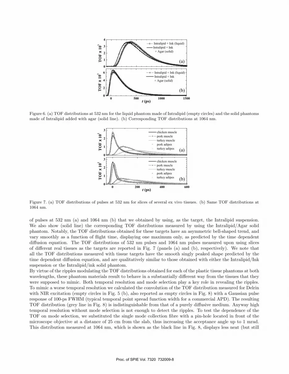
Figure 6. (a) TOF distributions at 532 nm for the liquid phantom made of Intralipid (empty circles) and the solid phantomsmade of Intralipid added with agar (solid line). (b) Corresponding TOF distributions at 1064 nm.
Figure 7. (a) TOF distributions of pulses at 532 nm for slices of several ex vivo tissues. (b) Same TOF distributions at1064 nm.
of pulses at 532 nm (a) and 1064 nm (b) that we obtained by using, as the target, the Intralipid suspension.We also show (solid line) the corresponding TOF distributions measured by using the Intralipid/Agar solidphantom. Notably, the TOF distributions obtained for these targets have an asymmetric bell-shaped trend, andvary smoothly as a function of flight time, displaying one maximum only, as predicted by the time dependentdiffusion equation. The TOF distributions of 532 nm pulses and 1064 nm pulses measured upon using slicesof different real tissues as the targets are reported in Fig. 7 (panels (a) and (b), respectively). We note thatall the TOF distributions measured with tissue targets have the smooth singly peaked shape predicted by thetime dependent diffusion equation, and are qualitatively similar to those obtained with either the Intralipid/Inksuspension or the Intralipid/ink solid phantom.By virtue of the ripples modulating the TOF distributions obtained for each of the plastic tissue phantoms at bothwavelengths, these phantom materials result to behave in a substantially different way from the tissues that theywere supposed to mimic. Both temporal resolution and mode selection play a key role in revealing the ripples.To mimic a worse temporal resolution we calculated the convolution of the TOF distribution measured for Delrinwith NIR excitation (empty circles in Fig. 5 (b), also reported as empty circles in Fig. 8) with a Gaussian pulseresponse of 100-ps FWHM (typical temporal point spread function width for a commercial APD). The resultingTOF distribution (grey line in Fig. 8) is indistinguishable from that of a purely diffusive medium. Anyway hightemporal resolution without mode selection is not enough to detect the ripples. To test the dependence of theTOF on mode selection, we substituted the single mode collection fibre with a pin-hole located in front of themicroscope objective at a distance of 25 cm from the slab, thus increasing the acceptance angle up to 1 mrad.This distribution measured at 1064 nm, which is shown as the black line in Fig. 8, displays less neat (but still
Proc. of SPIE Vol. 7320 732009-8

Figure 8. Delrin TOF distributions at 1064 nm, collected with the fibre (empty circles), with the pin hole (black line) andwithout spatial filtering (full dots), together with the convolution of the data collected with the fibre with a Gaussianpulse response of 100 ps full width at half maximum (grey line).
evident) peaks. Finally, we also acquired a TOF distribution with no spatial filtering between the slab and themicroscope objective, which exhibits no peaks, as shown by the grey full dot plot in Fig. 8. The same experimentsperformed with Delrin at 532 nm and with the Nylon and Teflon slabs gave similar results.
4. CONCLUSIONS
The results of Subsection 3.1 demonstrate that early arriving photons can be directly revealed in the TOF dis-tributions of photons emerging from samples with absorption and scattering parameters comparable to thoseof several-centimeter thick slices of human tissue by means of experimental techniques coupling extreme modeselection and short time response. This capability is relevant in medical diagnostics, as it constitutes the pre-requisite for implementation of any snake photon selection based tissue imaging technique. We presented 1Dprofiles of several opaque and translucent objects, embedded in Intralipid suspensions with tissue-like opticalproperties. Our results suggest that we might in principle detect discontinuities of sizes as small as ∼ 200 μm in1-to-5 cm thick human tissues, and assess their optical and consequently their physiological nature. Thus, theyattest that extremely relevant progresses towards noninvasive diagnosis of severe diseases may be pursued byapplying optical biopsy techniques based on TCSPC with high temporal resolution SPAD’s, and encourage toafford the complex engineering work necessary to make the concrete application of the proposed imaging methodin medical diagnostics feasible in the future.In Subection 3.2 we demonstrate that periodically spaced ripples modulate high temporal resolution TOF distri-butions of green and infrared photons emerging from Delrin, Nylon and Teflon phantoms, acquired by recurringto extreme mode selection, that is in the best experimental situation in order to detect early arriving photons.This would advise not to use tissue phantoms made with Delrin, Teflon and Nylon. Indeed, the ripples are a cluethat light interacts with Delrin, Nylon or Teflon in a remarkably different way as compared to real tissues. On theother hand more realistic solid tissue phantoms can be obtained by adding Agar to Intralipid/ink suspensions.
REFERENCES1. G. Le Tolguenec, F. Devaux, and E. Lantz, “Two-dimensional time-resolved direct imaging through thick
biological tissues: a new step toward noninvasive medical imaging,” Opt. Lett. 24, pp. 1047–1049, 1999.2. M. S. Patterson, B. Chance, and B. C. Wilson, “Time resolved reflectance and transmittance for the non-
invasive measurement of tissue optical properties,” Appl. Opt. 28, pp. 2331–2336, 1989.3. J. M. Kaltenbach and M. Kaschke, “Frequency- and time-domain modeling of light transport in random
media,” in Medical Optical Tomography: Functional Imaging and Monitoring,” Vol. IS11 of SPIE InstituteSeries, G. Mueller, B. Chance, R. R. Alfano, S. Arridge, J. Beuthan, E. Gratton, M. Kaschke, B. Masters,S. Svanberg, and P. van der Zee, eds. (SPIE, Bellingham, Wash., 1993), pp. 65–85.
Proc. of SPIE Vol. 7320 732009-9

4. B. B. Das, F. Liu, and R. R. Alfano, “Time-resolved fluorescence and photon migration studies in biomedicaland model random media,” Rep. Prog. Phys. 60, pp. 227–292, 1997.
5. T. L. Troy and S. N. Thennadil, “Optical properties of human skin in the near infrared wavelength rangeof 1000 to 2200 nm,” J. Biomed. Opt. 6, pp. 167–176, 2001.
6. C. R. Simpson, M. Kohl, M. Essenpreis, and M. Cope, “Near-infrared optical properties of ex-vivo humanskin and subcutaneous tissues measured using the Monte Carlo inversion technique,” Phys. Med. Biol. 43,pp. 2465–2478, 1998.
7. A. Pifferi, J. Swartling, E. Chikoidze, A. Torricelli, P. Taroni, A. Bassi, S. Andersson-Engels, and R.Cubeddu, “Spectroscopic time-resolved diffuse reflectance and transmittance measurements of the femalebreast at different interfiber distances,” J. Biomed. Opt. 9, pp. 1143–1151, 2004.
8. A. Andreoni, L. Nardo, A. Brega, and M. Bondani, “Optical profiles with 180 resolution of objects hiddenin scattering media,” J. Appl. Phys. 101, 024921, 2007.
9. A. Andreoni, M. Bondani, A. Brega, F. Paleari, A. S. Spinelli, and G. Zambra, “Detection of nondelayedphotons in the forward-scattering of picosecond pulses,” Appl. Phys. Lett. 84, pp. 2457–2459, 2004.
10. I. Delfino, M. Lepore, and P. L. Indovina, “Experimental tests of different solutions to the diffusion equationfor optical characterization of scattering media by time-resolved transmittance,” Appl. Opt. 38, pp. 4228–4236, 1999.
11. M. R. Hee, J. A. Izatt, J. M. Jacobson, and J. G. Fujimoto, “Femtosecond transillumination optical coherencetomography,” Opt. Lett. 18, pp. 950–952, 1993.
12. K. M. Yoo and R. R. Alfano, “Time-resolved coherent and incoherent components of forward light scatteringin random media,” Opt. Lett. 15, pp. 320–322, 1990.
13. R. Cubeddu, A. Pifferi, P. Taroni, A. Torricelli, and G. Valentini, “A solid tissue phantom for photonmigration studies,” Phys. Med. Biol. 42, pp. 1971–1979, 1997.
14. M. Sundberg, T. Lindbergh, and T. Strmberg, “Monte Carlo simulations of backscattered light intensityfrom convex and concave surfaces with an optical fiber array sensor,” Proc. SPIE 6084, pp. 608404–608414,2006.
15. W. Cong, K. Durairaj, L. V. Wang, and G. Wang, “A Born-type approximation method for bioluminescencetomography,” Med. Phys. 33, pp. 679–686, 2006.
16. G. Harding, M. Newton, and J. Kosanetzky, “Energy-dispersive x-ray diffraction tomography,” Phys. Med.Biol. 35, pp. 33–41, 1990.
17. M. Firbank and D. T. Delpy, “A design for a stable and reproducible phantom for use in near infra-redimaging and spectroscopy,” Phys. Med. Biol. 38, pp. 847–854, 1993.
18. J. A. Moon, R. Mahon, M. D. Duncan, and J. Reintjes, “Resolution limits for imaging through turbid mediawith diffuse light,” Opt. Lett. 18, pp. 1591–1593, 1993.
19. D. A. Benaron and D. K. Stevenson, “Optical time-of-flight and absorbance imaging of biologic media,”Science 259, pp. 1463–1466, 1993.
20. C. Dunsby and P. M. W. French, “Techniques for depth-resolved imaging through turbid media includingcoherence-gated imaging,” J. Phys. D: Appl. Phys. 36, pp. 207–227, 2003.
21. J. C. Hebden, S. R. Arridge, and D. T. Delpy, “Optical imaging in medicine: I. Experimental techniques,”Phys. Med. Biol. 42, pp. 825–840, 1997.
22. A. Andreoni, L. Nardo, G. Zambra, and M. Bondani, “Time-correlated single-photon counting based methodfor submillimeter transillumination imaging of objects embedded in tissue-phantoms,” J. Mod. Opt. 56,pp. 413–421, 2008.
23. L. Nardo and M. Bondani, “Optical imaging of tissues” in “Research advances in Photochemistry andphotobiology” Global Research Network (eds.), Kerala, India (2009). In press.
24. L. Nardo, A. Brega, M. Bondani, and A. Andreoni, “Non-tissue-like features in the time-of-flight distribu-tions of plastic tissue phantoms,” Appl. Opt. 47, pp. 2477–2485, 2008.
25. S. T. Flock, S. L. Jacques, B. C. Wilson, W. M. Star, and M. J. C. van Gemert, “Optical properties ofIntralipid: a phantom medium for light propagation studies,” Laser Surg. Med. 12, pp. 510–519, 1992.
Proc. of SPIE Vol. 7320 732009-10


