Embed Size (px)
Citation preview
UNIT 7
Urinary System
Pathological Conditions
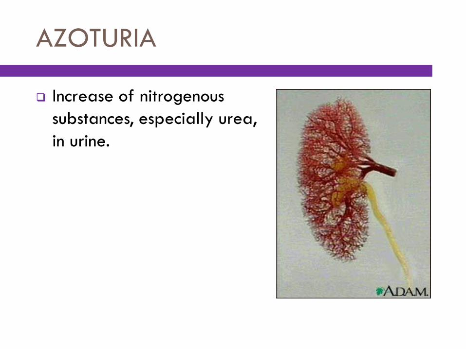
AZOTURIA
Increase of nitrogenous
substances, especially urea,
in urine.
DIURESIS
Increased formation and secretion of urine.
DYSURIA
Painful or difficult urination, symptomatic of cystitis
and other urinary tract conditions.
END-STAGE RENAL DISEASE (ESRD)
Kidney disease that has advanced to the point that
the kidneys can no longer adequately filter the
blood and, ultimately, requires dialysis or renal
transplantation for survival; also called chronic renal
failure.
Common diseases leading to ESRD include malignant
hypertension, infections, diabetes mellitus, and
glomerulonephritis. Diabetes is the most common cause
of kidney transplantation.
ENURESIS
Involuntary discharge of
urine after the age at
which bladder control
should be established;
also called bed-wetting at
night or nocturnal enuresis.
In children, voluntary
control of urination is
usually present by age 5.
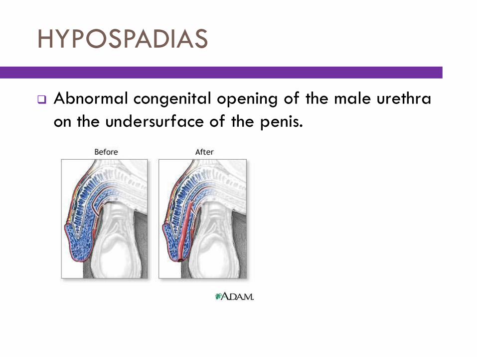
HYPOSPADIAS
Abnormal congenital opening of the male urethra
on the undersurface of the penis.
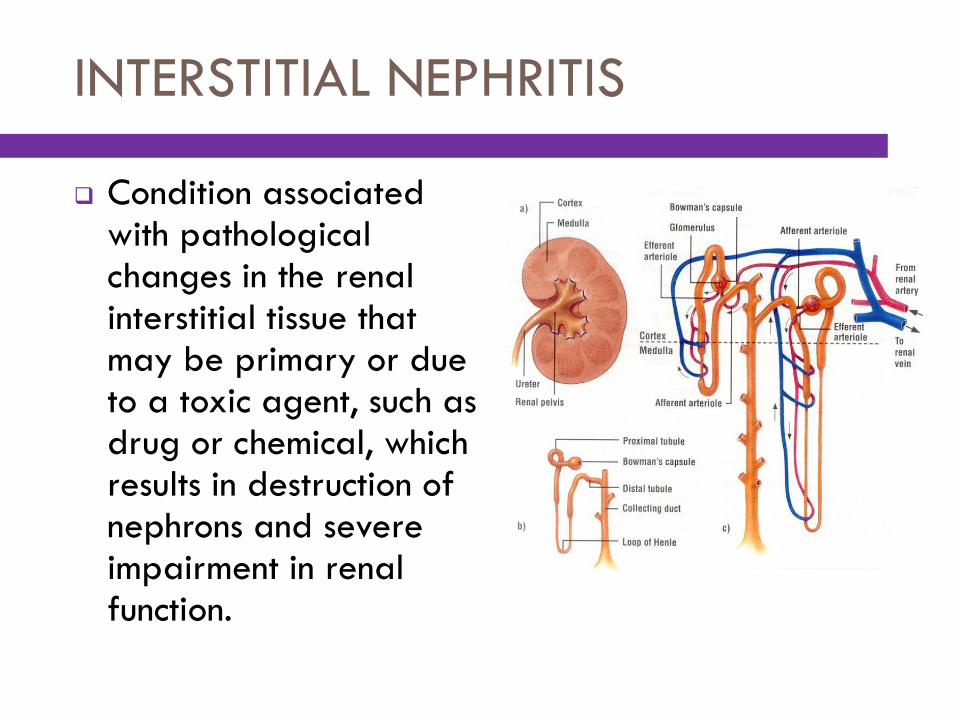
INTERSTITIAL NEPHRITIS
Condition associated with pathological changes in the renal interstitial tissue that may be primary or due to a toxic agent, such as drug or chemical, which results in destruction of nephrons and severe impairment in renal function.
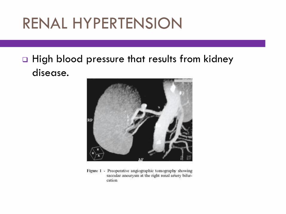
RENAL HYPERTENSION
High blood pressure that results from kidney
disease.
UREMIA
Elevated level of urea and other nitrogenous waste
products in the blood, as occurs in renal failure; also
called azotemia.
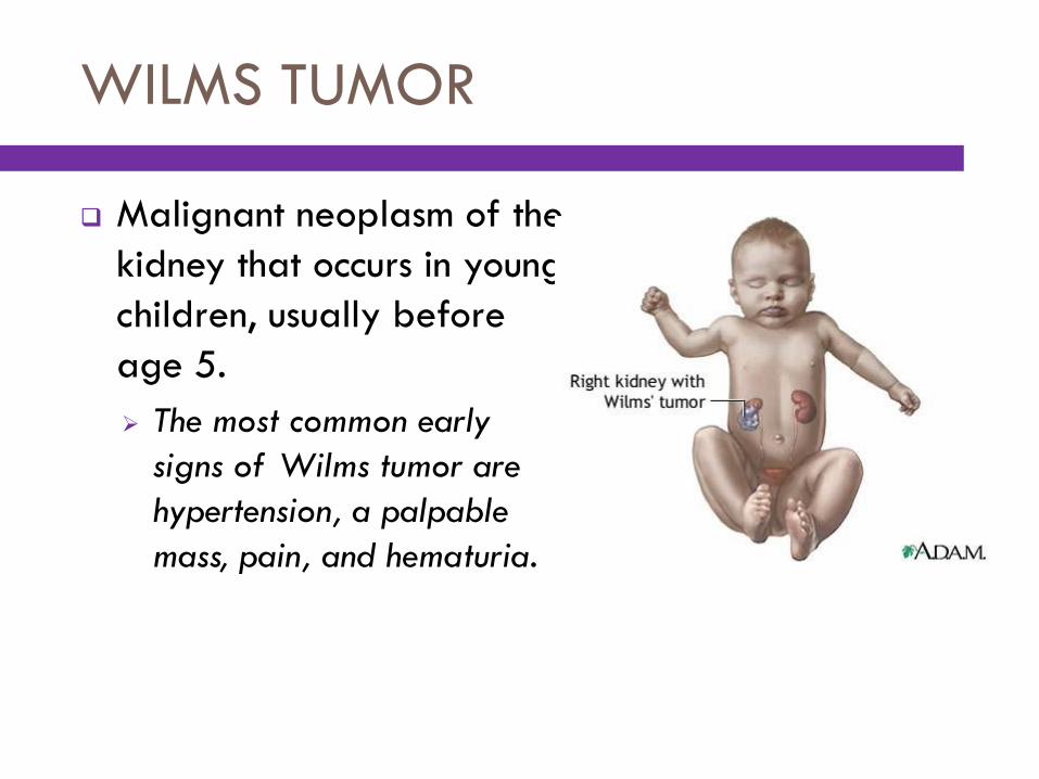
WILMS TUMOR
Malignant neoplasm of the
kidney that occurs in young
children, usually before
age 5.
The most common early
signs of Wilms tumor are
hypertension, a palpable
mass, pain, and hematuria.
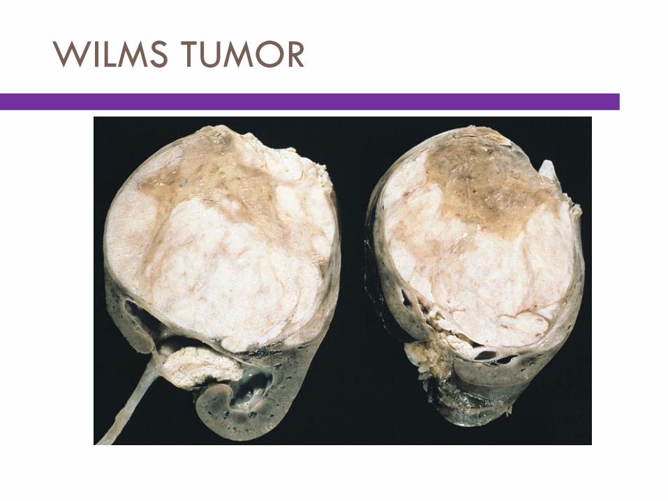
WILMS TUMOR

Diagnostic Procedures
BLOOD UREA NITROGEN (BUN)
Laboratory test that measures the amount of urea
(nitrogenous waste product) in the blood and
demonstrates the kidneys’ ability to filter urea from
the blood for excretion in urine.
An increase in BUN level may indicate impaired kidney
function.
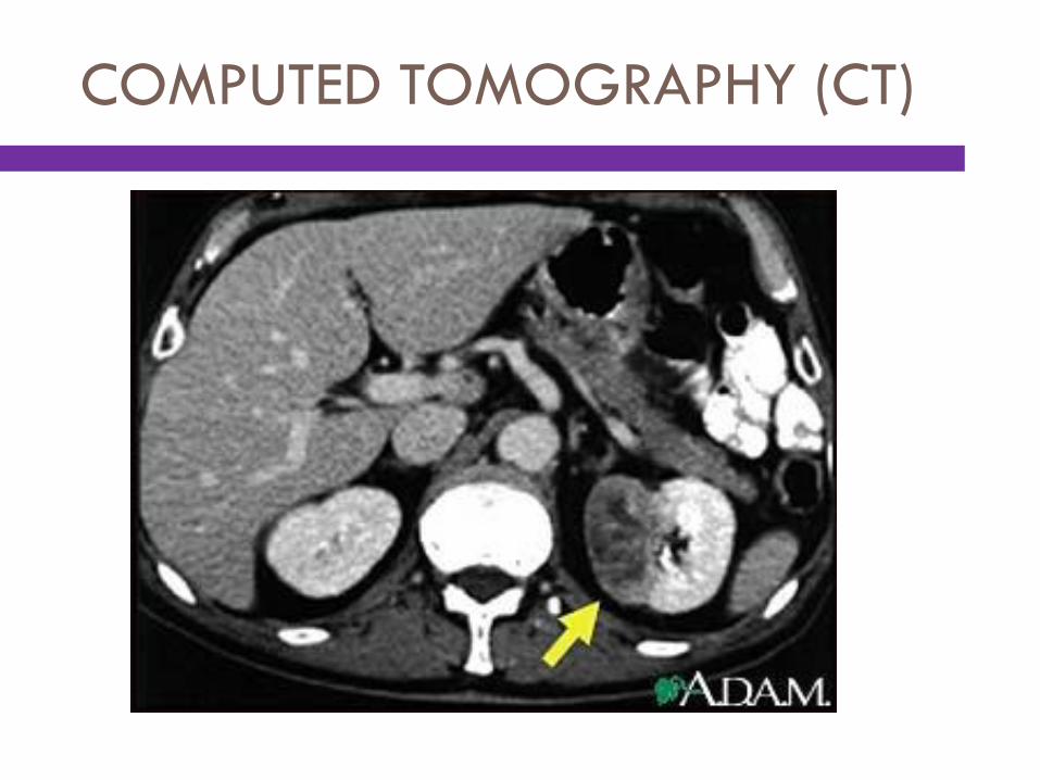
COMPUTED TOMOGRAPHY (CT)
Radiographic technique that uses a narrow beam of
x-rays that rotates in a full arc around the patient
to acquire multiple views of the body that a
computer interprets to produce cross-sectional
images of that body part.
CT scanning is used to diagnose kidney, ureter, and
bladder tumors, cysts; inflammation; abscesses;
perforation; bleeding; and obstructions. It may be
administered with or without a contrast medium.
COMPUTED TOMOGRAPHY (CT)
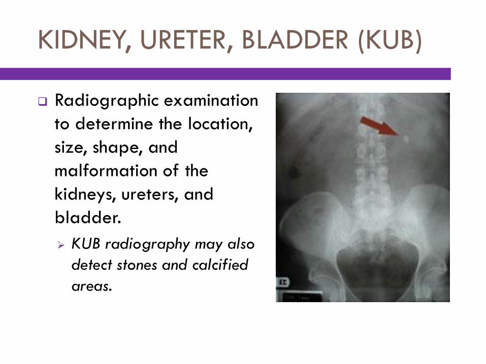
KIDNEY, URETER, BLADDER (KUB)
Radiographic examination
to determine the location,
size, shape, and
malformation of the
kidneys, ureters, and
bladder.
KUB radiography may also
detect stones and calcified
areas.
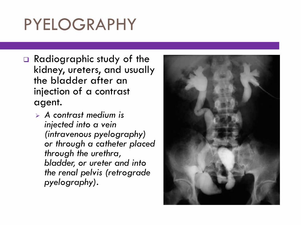
PYELOGRAPHY
Radiographic study of the kidney, ureters, and usually the bladder after an injection of a contrast agent.
A contrast medium is injected into a vein (intravenous pyelography) or through a catheter placed through the urethra, bladder, or ureter and into the renal pelvis (retrograde pyelography).
INTRAVENOUS PYELOGRAPHY (IVP)
Radiographic imaging in which a contrast medium is
injected intravenously and serial x-ray films are
taken to provide visualization of the entire urinary
tract; also called intravenous urography (IVU) or
excretory urography (EU).
In IVP, the x-ray image produced is known as a
pyelogram or urogram.
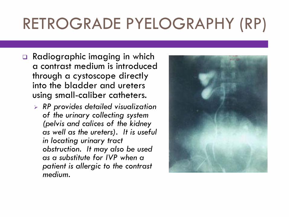
RETROGRADE PYELOGRAPHY (RP)
Radiographic imaging in which a contrast medium is introduced through a cystoscope directly into the bladder and ureters using small-caliber catheters.
RP provides detailed visualization of the urinary collecting system (pelvis and calices of the kidney as well as the ureters). It is useful in locating urinary tract obstruction. It may also be used as a substitute for IVP when a patient is allergic to the contrast medium.
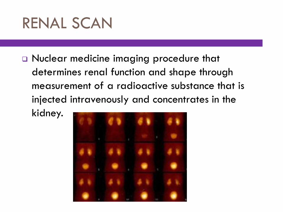
RENAL SCAN
Nuclear medicine imaging procedure that
determines renal function and shape through
measurement of a radioactive substance that is
injected intravenously and concentrates in the
kidney.
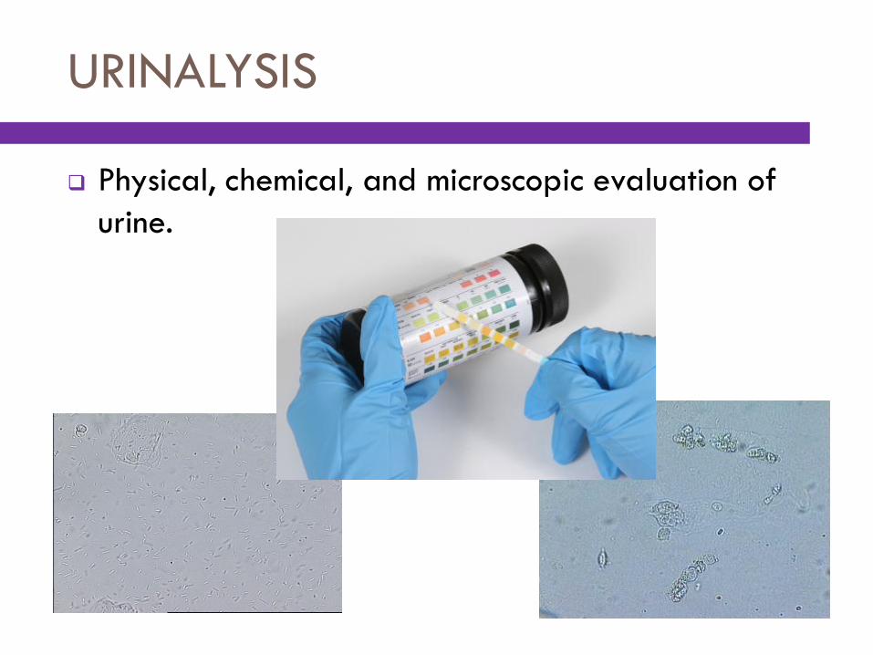
URINALYSIS
Physical, chemical, and microscopic evaluation of
urine.
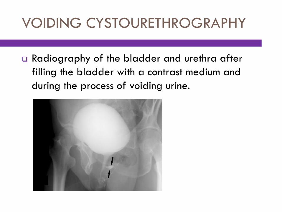
VOIDING CYSTOURETHROGRAPHY
Radiography of the bladder and urethra after
filling the bladder with a contrast medium and
during the process of voiding urine.

Medical and Surgical Procedures
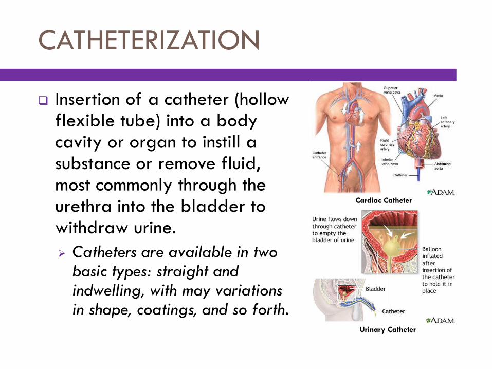
Cardiac Catheter
CATHETERIZATION
Insertion of a catheter (hollow flexible tube) into a body cavity or organ to instill a substance or remove fluid, most commonly through the urethra into the bladder to withdraw urine.
Catheters are available in two basic types: straight and indwelling, with may variations in shape, coatings, and so forth.
Urinary Catheter
DIALYSIS
Mechanical filtering process used to cleanse blood
of high concentrations of metabolic waste products,
draw off excess fluids, and regulate body chemistry
when kidneys fail to function properly.
Two primary methods are used to dialyze the blood:
hemodialysis and peritoneal dialysis.
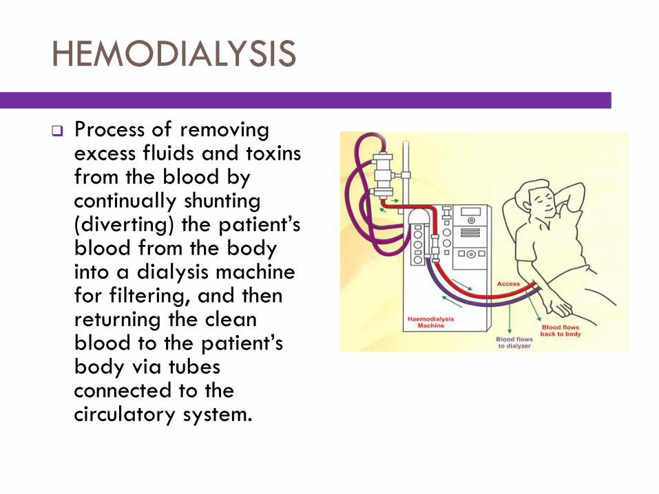
HEMODIALYSIS
Process of removing excess fluids and toxins from the blood by continually shunting (diverting) the patient’s blood from the body into a dialysis machine for filtering, and then returning the clean blood to the patient’s body via tubes connected to the circulatory system.
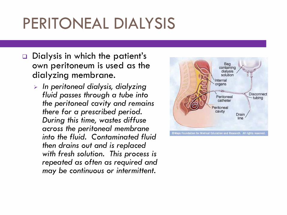
PERITONEAL DIALYSIS
Dialysis in which the patient’s own peritoneum is used as the dialyzing membrane.
In peritoneal dialysis, dialyzing fluid passes through a tube into the peritoneal cavity and remains there for a prescribed period. During this time, wastes diffuse across the peritoneal membrane into the fluid. Contaminated fluid then drains out and is replaced with fresh solution. This process is repeated as often as required and may be continuous or intermittent.
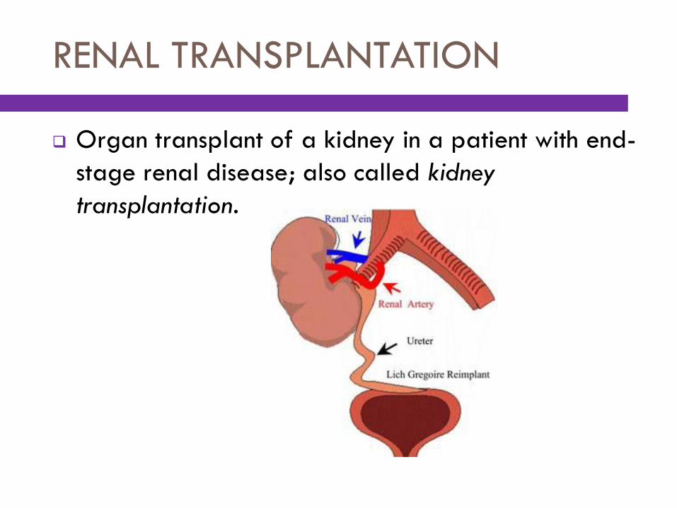
RENAL TRANSPLANTATION
Organ transplant of a kidney in a patient with end-
stage renal disease; also called kidney
transplantation.