Embed Size (px)
Citation preview
THE COUNCIL
President : Dr. Chok Wan Chan
Vice President : Dr. Catherine CC Lam
Honorary Secretary : Dr. Stephenie KY Liu
Honorary Treasurer : Dr. Theresa YL Wong
Council Members : Dr. Mario WK Chak
Dr. Florence MY Lee
Dr. Tim KT Liu
Dr. Kwing Wan Tsui
Dr. Sam CM Yeung
Dr. Josephine SC Chong (Co-opt)
Dr. Jasper CP Chow (Co-opt)
Dr. Wing Cheong Lee (Co-opt)
THE EdITOrIaL BOard Of BraINCHILd
Editor-in-chief : Dr. Chok Wan Chan
Editorial Board : Dr. Catherine CC Lam
Dr. Florence MY Lee
Dr. Wing Cheong Lee
Address : Room 3509, Bank of America Tower, 12 Harcourt Road, Central, Hong KongTel : (852) 2895 5211Fax : (852) 2577 1989 (Society Hon. Secretary Dr. K Y Liu)Email : [email protected] : www.hkcndp.org
Printer : Printhouse Production Center Limited Flat A, 15/F, Gee Luen Hing Industrial Building, 2 Yip Fat Street, Wong Chuk Hang, Hong Kong
Copyright©2016. All rights reserved
The published materials represent the opinions of the authors and not necessarily that of the editors or the Society. The appearance of advertisement is not a warranty, endorsement or approval of the products. The Society disclaims responsibility for any injury to persons or property resulting from any ideas or products referred to in the articles or advertisement.
Our appreciation of thanks to Cheung Man Chun and Ng Sum Lui (students of Hong Kong Christian Service Kwun Tong Nursery School) for the cover drawing named “Magic Forest”.
Dr. Josephine SC Chong
Dr. Eric KC Yau
Dr. Jasper CP Chow
The Hong Kong Society of Child Neurology and Developmental Paediatricswww.hkcndp.orgJuly 2016 Volume 17 No.1
Special Issue on Paediatric Neurophysiology Update by the British Columbia Children’s Hospital Team CONTENTS
1
4
19
27
39
49
53
pageMessage from the PresidentDr. Chok Wan CHAN
Care of Children with Epilepsy at BC Children’s HospitalAnita Datta MD, FRCP(C), CSCN Diplomate (EEG)
BC Children’s Hospital Brain Mapping Program Bruce H. Bjornson, MD, FRCPC
diagnostic Neurophysiology Services Donna Gregory Wood, R.E.T., R.EPT., R.T.(EMG)
Neurodevelopmental Outcomes after Preterm Birth - an OverviewTing JY, MBBS, MPH, MRCPCH, FRCPC, FHKAM (Paediatrics)Synnes AR, MDCM, MHSc, FRCPC,
Psychology Services of the Neurology Multi-disciplinary Team Sare Akdag, Ph.D., R.Psych. Audrey Ho, Ph.D., R.Psych. Sarina Kot, Ph.D., R.Psych. Elizabeth Stanford, Ph.D., R.Psych.
The EEG Technologist Training ProgrammeLisa Langill, R.E.T., R.T.(EMG), R. EP T., CNIM Jacqueline Crawford, R.E.T., R. EP T., CNIM
1
n
2016
Volume 17 No.1 July 2016
The Hong Kong Society of Child Neurology and Developmental PaediatricsEDITOR’S NOTES for the July 2016 Issue
Paediatric Neurophysiology Update by the British Columbia Children’s Hospital TeamDr. Chok Wan CHAN
The current issue of Brainchild is devoted to “Paediatric Neurophysiology Update” capably edited by Professor Peter K. H. Wong B. Eng MD FRCP(C), Professor for Division of Paediatric Neurology, Department of Paediatrics, University of British Columbia, Vancouver, Canada and Director of Department of Diagnostic Neurophysiology, Children's Hospital, Vancouver, B.C., Canada. Through his expert team members, we are able to appreciate the modern team approach to neurophysiology and the multidisciplinary, interdisciplinary and transdisciplinary collaboration in the neurosciences. Thanks to members of the team and we take great pride to present this comprehensive update to all professionals in neurosciences consisting of doctors, nurses, allied health professionals, social workers, psychologists, special teachers and educated patients for their comments and positive feedbacks. We are grateful to Professor Wong for his great leadership and to all the authors for their effort. We appreciate your contributions to the child health community!
Year 2016 is the 22nd anniversary for the Hong Kong Society of Child Neurology and Developmental Paediatrics (HNCNDP). We are pleased to witness our achievements and on looking back we can summarize our activities in the past two decades into several major channels:
1. Regular Professional activities: we have bimonthly meetings on each of the following:
a. Child Neurology Conference b. Neuro-developmental Conference c. RegularBimonthlyScientificMeetings2. AnnualScientificMeeting:eachyearweinviteaworldexpertononeaspectofeither
Developmental Behavioural Paediatrics (DBP) or Child Neurology (CN) from Untied States of America, Canada, United Kingdom, Europe, Australia and other countries to deliver a series of state-of-art aspects of the subject and through participation of localprofessionalswesuccessfullyhostedasignificantnumberofsymposia,seminar,workshops and fora on the various aspects of the subject. The climax is the Keynote Lecture and Dinner each year on the themed topic which remains one of the most popularscientificactivitiesattractinghundredsofprofessionalseachyear.Sofarwehavehostedmorethan20suchannualscientificmeetings.ReaderscanrefertotheSociety website for more details.
3. Publications - the Society launched regularly the following publications consistently, professionally and academically:
2
n
2016
a. The Brainchild – this publication is devoted to special issues on the various aspects of the DBP and CN and is distributed to healthcare professionals and professionals dedicated to neurosciences free of charge. They are available in most of the University Libraries and the public libraries for health educations.
b. The Society Newsletter – this is meant to circulate to professionals on recent advances, meeting announcements and social activities.
4. Professional Accreditations : the Society professionally assists the Hong Kong College of Paediatricians in accrediting subspecialists in CN and DBP via the establishment of two Subspecialty Boards on Child Neurology and on Developmental Behavioural Paediatrics for:
a. Accreditation of First Fellows in CN and DBP b. Establishment of structured training programme for CN and DBP c. Conducting CME and CPD in service training for CN and DBP subspecialist and
for general paediatricians and other child healthcare professionals d. Promotion of research and publications of local studies5. Workshops : special workshops were held regularly for members as below a. Epilepsy b. Botox c. Dyslexia/ASD/ADHD d. EEG/Neurophysiology e. Augmentative and Alternative Communications (AAC) f. Others6. Advocacy: the Society alerted professionals of the existence of Dyslexia in the
Chinese Language and initiated the Position Statement for Dyslexia and for AttentionDeficit/HyperacidityDisorders(ADHD)whichremainscornerstonesformanagement of these disabilities. We have also advocated for the Autistic Spectrum Disorders (ASD). We work hard to provide early identification, assessment and intervention for individuals with these disorders (Dyslexia, ADHD, ASD) in the form of remediation, accommodation and compensation and urge the Hong Kong SAR Government to prioritize resources and support at home, in schools and across the community through the Equal Opportunity Ordinance and to provide special provisions at both the school and in the public examinations.
7. Parent Groups: The society works closely with parent groups for children with special needs for health education, professional guidance, advocacy, self-help and government subsidies and provisions.
8. Research: Dyslexia, ADHD, ASD, Epilepsy, AAC and others with good results and outstanding research publications.
9. Interdisciplinary collaboration with other healthcare professionals in Hong Kong via joint meetings, research projects, patient activities and others.
10. Collaboration with Professionals outside Hong Kong: Hong Kong remains one of the leading professionals in CN and DBP within the Chinese Speaking Communities and amongstpioneersfortheAsia-PacificRegion.
3
n
2016
Volume 17 No.1 July 2016
Dr. CHAN Chok WanEditor-in-Chief, The BrainchildPresident, The HK Society of Child Neurology & Developmental Paediatrics8th July 2016.
In essence what have we achieved a lot over the past two decades and these can be summarized in the following missions accomplished:
1. Promote standard of Child Health and General Paediatrics in Hong Kong2. DefinethesubspecialtiesofCNandDBP3. Coordinate these subspecialties with general Paediatrics4. Assure standard and quality of services for CN and DBP in Hong Kong.5. Advocateforthewelfareandbenefitofchildrenwithspecialneedsinthecommunity.6. Collaborate with other medical specialties, nurses and allied health professionals
within the neurosciences.7. Enhance rehabilitation of children with special needs.8. Promote transdisciplinary team approach for management of children with CN and
DBP.9. Assist to set up CN and DPB services within the upcoming Children’s Hospital for
Hong Kong in the domains of services, training and research10. Put the name of CN and DBP onto the map of Chinese Speaking Communities,
the Asia-Pacific Region and the global world such as the International Pediatric Association (IPA) and the World Health Organization (WHO).
We are proud of our achievements and results. To all these, we would like to thank our world experts, Course Directors who conducted our Annual Scientific Meetings each year, speakers at our professional clinical conferences, fellow members of the healthcare professionals, our patient group for their support and participations. Above all, I would like to thank all the Council Members and our devoted members for their support and guidance. At the time of our 22nd Anniversary, we would like to show our heartfelt appreciation to all the pioneers (Professor C Elaine Filed, Dr. Lui Wai-ying, Dr. Flora Baber, Professor JH Hutchison and others), senior members of the Society for their contributions and to our incoming Council Members for advancing the touch of our Society forwards. We are confidentthatwithoursolidarityforCNandDBPaswellasourever-unfailingenthusiasm,the future of HKCNDP should be promising and the skyline of achievements unlimited!
I wish you all reading pleasure and best of health!
4
n
2016
Care of Children with Epilepsy at BC Children’s HospitalAnita Datta, MD, FRCP(C), CSCN Diplomate (EEG)
Clinical Assistant ProfessorEpilepsy Service and Division of NeurologyDepartment of Pediatrics, UBC and BC Children’s HospitalVancouver, Canada
IntroductionBC Children’s Hospital is the only paediatric tertiary care center in the province
of British Columbia (BC), Canada, serving a population of 4.7 million people. It is the major treatment, teaching and research facility for children in the province. The Division of Neurology and the Comprehensive Epilepsy Program provide specialised services for diagnosing, investigating and managing epilepsy which are not available elsewhere in the province. This includes care by pediatric epileptologists and other subspecialists, neurophysiology services, and specialised neuro-imaging. In children, epilepsy is one of the most common neurological condition. This article will describe the epilepsy program at BC Children’s Hospital and how epilepsy care is provided by a large team from the neonatal period to young adults via various outpatient and inpatient services.
BackgroundWhat is a Seizure/Epilepsy?
Epilepsy is one of the most common neurological condition. It peaks in children under five years of age, especially the first year of life, and the elderly. Seizures are caused by excessive electrical discharges in a group of neurons. These discharges can arise from different parts of the brain, leading to various clinical manifestations, from brief lapses of attention, autonomic or psychiatric symptoms to severe and prolonged convulsions. Seizures can also vary in frequency, from less than 1 per year to many per day.
Havingaseizuredoesnotnecessarilymeanachildhasepilepsy.Epilepsywasdefinedas a disorder of the brain characterized by an enduring predisposition to generate epileptic seizures. The International League Against Epilepsy (ILAE) defines it as having at least twounprovokedorreflexseizuresoccurring>24hoursapart,oneunprovokedseizureanda probability of further seizures similar to the general recurrence risk (at least 60%) after two, unprovoked seizures occurring over the next 10 years, or a diagnosis of an epilepsy syndrome. Epilepsy is considered to be resolved for individuals who had an age-dependent epilepsy syndrome but are now past the applicable age or those who have remained seizure-free for the last 10 years, with no seizure medications for the last 5 years1.
What is Treatment Resistant Epilepsy?Approximately 30 % of people affected by epilepsy are refractory to treatment.
5
n
2016
Volume 17 No.1 July 2016
This is often accompanied by learning disabilities, behavioural problems, memory loss, psychiatric disorders and/or other adaptive problems. It is proposed that drug resistant or medically refractory epilepsy are defined as failure after adequate trials of two tolerated and appropriately chosen and used anti-epileptic drug (AED) schedules (whether as monotherapies or in combination) to achieve sustained seizure freedom2.
Etiology and ManagementEpilepsyhasnumerouscauses,includingstructural,metabolic,geneticandinflammatory.
In some situations, the cause is unknown, although with advancements in neuro-imaging and genetic testing, this group is becoming smaller. The Epilepsy program at BC Children’s Hospital aims to provide the most up-to-date tools to diagnose and manage patient. Management of seizure and epilepsy includes medication, surgical evaluation, ketogenic diet, vagal nerve stimulation, and providing adequate psychosocial support.
Consequences of Epilepsy short and long-termEarly diagnosis and control of seizures is crucial, as there can be many negative
consequences from epilepsy. These include the effects of seizures on brain development, quality of life in the child and family, and psychosocial and mental health issues.
Uncontrolled frequent seizures are associated with an increased risk of death. Sudden unexplained death in epilepsy (SUDEP) rates are quoted to be the highest in those with treatment resistant epilepsy compared with community prevalence samples3. Seizures can have a negative effect on brain development and learning - especially in children. Good seizure control, even after years of treatment resistance, can have a beneficial impact on cognition4.
With temporal lobe epilepsy, cross-sectional studies had shown that memory was worse in patients with a longer duration and earlier age at onset of epilepsy5. In one longitudinal study, epilepsy surgery abolished or reversed the decline in memory function6. In another study, 25-40% of treatment resistant patients showed decline on tests of confrontation and naming compared to friend or relative controls7.
It also known that frequent seizures can lead to “pseudo-regression” where the seizures and medications impact sleep, energy, attention, mood, learning and interaction with the environment. This is thought to be reversible with better seizure control.
Incidence and Prevalence/Service Demand Worldwide statistics
Approximately 50 million people worldwide have epilepsy, making it one of the most common neurologic condition. The incidence of epilepsy in children ranges from 41-187/100,000. Higher incidence is reported from underdeveloped countries. The incidence ishighestinthefirstyearoflifeanddeclinestoadultlevelsbytheendofthefirstdecade.
6
n
2016
The prevalence of epilepsy is higher than the incidence and ranges from 3.2-5.5/1,000 in developed countries and 3.6-44/1,000 in underdeveloped countries. Prevalence also seems highest in rural areas. This is likely due to the increased risk of endemic conditions such as malaria or neurocysticercosis, higher incidence of motor vehicle accidents, birth-related injuries, and variations in medical infrastructure, availability of preventative health programmes, and accessible care8.
Canadian and BC statistics0.6% of Canadians have epilepsy. Each year, an average of 15-500 people is diagnosed
with epilepsy in Canada. Of this number, 44% are diagnosed before the age of 5, 55% before the age of 10, 75-85% before age 18, and 1% of children will have recurrent seizures before age 14. 1.3% are over the age of 60. This means that about 60% of new patients are young children and senior citizens9.
In British Columbia, 40,000 individuals have epilepsy. Approximately 30% have treatment resistant epilepsy. Approximately, 3,000-5,000 patients have treatment resistant epilepsy requiring close follow-ups and continuing care. BC Children’s hospital has six pediatric epileptologists. In addition, there is 1 neurophysiologist and 7 neurologists who also see patients with seizures. In the province, there is one pediatric neurologist in private practice. The remainder work at the hospital. The Division of Neurology receives over 2,000 new referrals each year and 40-50% of these are in regards to seizures or epilepsy.
Goals of the Program Mission of Neurology Program:
To improve the neurological health of children and youth in BC through compassionate leading edge care, education and research, and to implement advances in paediatric neuroscience, particularly those that significantly improve patient outcomes.
The Epilepsy program at BC Children’s Hospital is committed to providing excellent clinical management by:
• Applying advanced andup todatediagnostic and therapeutic techniques andapproaches to all patients with seizures and epilepsy
• Providingholisticassessmentsandcareforchildrenandfamilies• Collaborationwithotherprofessionalstoensureaseamlessservicethatispatientand
family centered• Providingeducationabouttheimpactofepilepsyandpracticalmanagement• Participatinginresearchtoadvancethefieldandimprovepatientcare• Minimizingthepsychosocialimpactofepilepsyforthechildthrougheducationand
empowerment• Devisingindividualhealthcareplansthataddresstheneedsofthechildinallareasof
their life, and therefore expanding their world and vision
7
n
2016
Volume 17 No.1 July 2016
Multi-disciplinary and Inter-disciplinary team approachCaregivers and services involved in care
The team of physicians, nurses, healthcare professionals and support staff participate in the evaluation and treatment of our epilepsy patients who come here from across BC and sometimes other parts of Canada.
This team consists of paediatric epileptologists, neurophysiologists, paediatric neurologists, paediatric neurosurgeons, adult epileptologists, neuro-radiologists, nuclear medicine physicians, nurse specialists, pharmacologists, physiotherapists, occupational and speech therapists, dieticians, psychologists and psychiatrists, educational counsellors and social workers, and an array of scientists and technologists, who all work together to offer individualized care to children.
Program Overview Ambulatory care/Epilepsy Clinics
A large volume of patients are seen in the outpatient setting, where the vast majority of seizure management takes place. Each epileptologist have several clinics per week. There are over 20 clinics by epileptologists each week. These consist of predominantly epilepsy patients, many whom are treatment resistant. General pediatric neurologists also see patients with epilepsy. However, if the patients have failed two anti-seizure medications or are considered treatment resistant, it is encouraged that the patients are referred to an epileptologist. Sometimes when there is a delay for pre-surgical evaluations, patients with epilepsy may suffer from numerous co-morbidities related to uncontrolled seizures. This system allows for prompt pre-surgical evaluations, access to ketogenic diet, vagal nerve stimulation, or optimizing medical management.
In addition to patients with epilepsy, patients with paroxysmal episodes, first time seizures, and non-epileptic events, will be seen in clinic as well. There are some specialized clinics, such as epilepsy surgery clinic, where the epileptologist and paediatric neurosurgeon meet with families together to discuss a plan for surgery or for post-surgical follow-ups. Some of the epileptologists are also involved in the Tuberous Sclerosis Clinic, where a large percentage of the children have epilepsy. Since tuberous sclerosis is a multi-systemic condition, other specialists can join the clinic or see the patients the same day. These clinics also provide an opportunity for clinical research. For example, the tuberous sclerosis clinic is currently involved in a clinical trial using a novel drug, which is an mTor inhibitor, for patients with treatment resistant epilepsy.
The care of patients in clinic involves a team of health professionals. Neurology nurses provide patient education in clinics. They are also the contact person for the patients if any concerns or issues arise. For ketogenic diet evaluations and follow-ups, the ketogenic dietician and nurses meet with the patients in clinic. The epilepsy surgery nurse is involved with teaching and support around surgery, as well as adjusting the vagal nerve stimulator.
8
n
2016
Trainees, including medical students, pediatric and adult neurology residents, general pediatric residents and epilepsy fellows, are present. One day per week, a neuropsychologist isavailabletomeetwithpatientswhomaybeexperiencingcognitivedifficultiesandwhoare not necessarily surgical candidates. Many clinical trials are also done in the clinic setting, such as drug trials, assessments of quality of life and mental health, etc. Research assistants are frequently present at clinics assisting in clinical research.
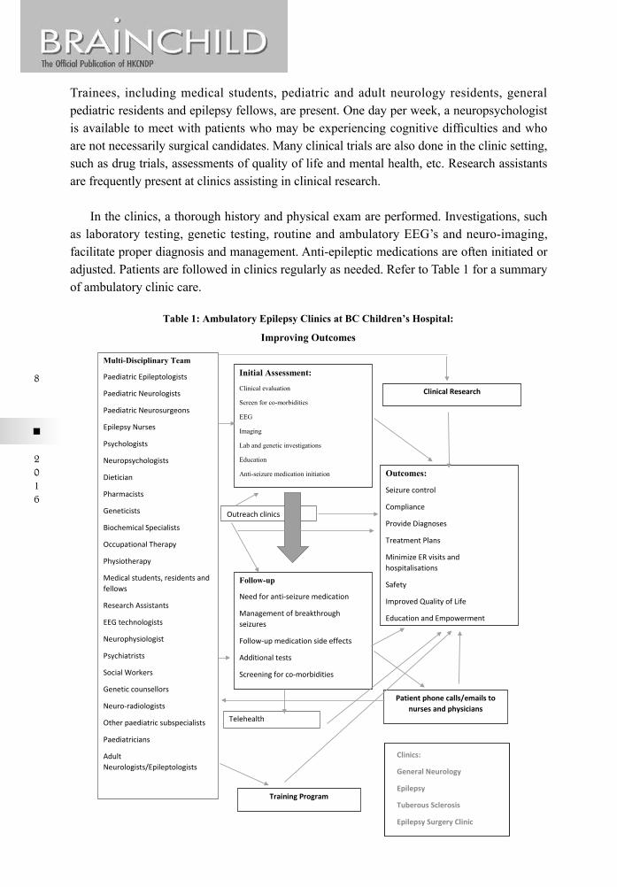
In the clinics, a thorough history and physical exam are performed. Investigations, such as laboratory testing, genetic testing, routine and ambulatory EEG’s and neuro-imaging, facilitate proper diagnosis and management. Anti-epileptic medications are often initiated or adjusted. Patients are followed in clinics regularly as needed. Refer to Table 1 for a summary of ambulatory clinic care.
Initial Assessment:
Clinical evaluation
Screen for co-morbidities
EEG
Imaging
Lab and genetic investigations
Education
Anti-seizure medication initiation
Follow-up
Need for anti-seizure medication
Management of breakthrough seizures
Follow-up medication side effects
Additional tests
Screening for co-morbidities
Multi-Disciplinary Team
Paediatric Epileptologists
Paediatric Neurologists
Paediatric Neurosurgeons
Epilepsy Nurses
Psychologists
Neuropsychologists
Dietician
Pharmacists
Geneticists
Biochemical Specialists
Occupational Therapy
Physiotherapy
Medical students, residents and fellows
Research Assistants
EEG technologists
Neurophysiologist
Psychiatrists
Social Workers
Genetic counsellors
Neuro-radiologists
Other paediatric subspecialists
Paediatricians
Adult Neurologists/Epileptologists
Outreach clinics
Telehealth
Outcomes:
Seizure control
Compliance
Provide Diagnoses
Treatment Plans
Minimize ER visits and hospitalisations
Safety
Improved Quality of Life
Education and Empowerment
Patient phone calls/emails to nurses and physicians
Clinics:
General Neurology
Epilepsy
Tuberous Sclerosis
Epilepsy Surgery Clinic
Table 1: Ambulatory Epilepsy Clinics at BC Children’s Hospital:
Improving Outcomes
Clinical Research
Training Program
9
n
2016
Volume 17 No.1 July 2016
Inpatient servicesNeurologists on call do consultations on the inpatient wards, neonatal and paediatric
intensive care, and emergency room. Patients with seizures or epilepsy that require hospital stay are usually admitted under the Neurology service. Complex epilepsy patients on the inpatient service are usually followed or co-managed by an epileptologist.
Video EEG Monitoring (Non-Invasive)BC Children’s Hospital has a two bed EEG monitoring unit. At least two patients are
electively admitted every week. It is used to either confirm the diagnosis of epilepsy, or confirmthefirstpartofaseriesofinvestigationsforthepossibleroleofepilepsysurgeryfor the treatment of epilepsy. Approximately 100 children are admitted per year to this unit.PatientsareusuallyadmittedforuptofivedaystotheEpilepsyMonitoringUnitwithplacement of scalp EEG electrodes. The goal of video EEG monitoring is to record the patient’s typical seizures. For this reason, the anti-epileptic medications are usually tapered downandfrequentlydiscontinuedwithinthefirstfewdaysofthepatient’sstayinhospital.The seizures are captured with video and EEG recordings and upon the completion of the evaluation the patient is reloaded on their anti-epileptic medications. The monitoring allows not only to diagnose a seizure problem accurately, but also to design the best possible treatment plan.
Investigative Tools at BC Children’s HospitalOutpatient EEG
The EEG is often used to investigate a patient's history of seizures. The “interictal EEG” describes abnormalities seen in patients between seizures. It is well known that even when patients appear well and have no outward manifestation of seizures, the brain waves can show abnormalities suggestive of a seizure tendency or risk. The “ictal EEG” describes theEEGchangesthatareseenduringaseizure.ThisfindingisnotcommonlyseenduringtheroutineoutpatientEEGrecordings.Thecaptureofaseizureconfirmsthediagnosisofepilepsyandhelpsinidentifyingthespecificepilepsysyndromeordisorderthepatienthas.Prolonged EEG recordings lasting several hours are often done as well. This is especially useful to characterise frequent paroxysmal events in patients.
Ambulatory EEG Ambulatory electroencephalography monitoring is a technology that allows prolonged
EEG recording in the home setting. Its ability to record continuously for 24-48 hours increases the chance of recording an ictal event or capturing interictal epileptiform discharges. This is a less expensive alternative to inpatient monitoring. It is also clinically useful, as some patients are more likely to have a paroxysmal episode in the home setting. However, medications are usually not weaned during this test for safety reasons.
10
n
2016
Laboratory testsFurther investigations, including blood work is useful to aid in diagnosis, as well
as monitoring side effects of anti-seizure medications. Common investigations include hematology profile, electrolyte panel, liver and renal function tests, drug levels, tests for inborn errors of metabolisms, autoimmune, endocrine, and vasculitic conditions. Genetic tests include karyotype and chromosomal microarray. In some cases, a lumbar puncture is neededforfurthermetabolic,inflammatoryorautoimmunework-up.
Genetic testingIn a population based study in Minnesota, approximately half of children had an
unknown etiology for their epilepsy, and of the remainder, 22% were genetic and 28% were structural/metabolic10. The number of patients where etiology for seizures is unknown is diminishing with advances in genetic testing. In addition to karyotype or chromosomal microarray, if no etiology has been determined for seizures, further genetic studies can be done. Advances in genomic technologies, including targeted next generation sequencing and wholeexomesequencing(WES),enablesidentificationofpathogenicvariantsin10–78%of patients with unexplained epilepsy11.Theclinicalimpactissignificantincludingearlierdiagnosisandspecifictreatmentinterventions.
Between December 2014 to June 2015 WES was performed in 43 patients under a study protocol.Adefinitediagnosiswasmadein13patients.Obtainingadiagnosishasnumerousbenefits,includingpreventingexcessinvestigations,tailoringmanagementoptions,screeningpatients for other co-morbidities, and providing closure to families. All patients receiving testing obtain pre and post-test genetic counselling.
ImagingMagnetic Resonance Imaging (MRI) Scan
MRI scans have become a crucial component in the evaluation of patients with epilepsy and are particularly helpful in the evaluation for possible epilepsy surgery. Advances in technology have also helped determine the cause of seizures in patients who were called “idiopathic” in the past. The MRI shows the gross appearance of the brain and therefore can give anatomic and pathological information of the brain. The use of the MRI scan in patients with epilepsy has been aided by FLAIR techniques and High Resolution or 3T MRI imaging. High Resolution MRI with thin slices of the brain facilitates in identifying subtle lesions, such as cortical dysplasias. It is usually used in patients with focal epilepsy, in whom no lesion has been detected on the standard 1.5T MRI.
Positron Emission Tomography (PET scan)Positron Emission Tomography (PET scan) is a neuro-imaging test of how the brain
functions. PET scans can be used in the evaluation for epilepsy surgery. FDG (fluoro-deoxy-glucose) PET is the most frequently used PET scan in epilepsy patients and provides information on how various regions of the brain utilize glucose. Areas of the brain that do not
11
n
2016
Volume 17 No.1 July 2016
metabolizeglucosemayreflectthedysfunctionalregionofthebraingivingrisetoseizures.This test can be helpful in providing additional information for accurately locating epilepsy inaspecificregionofthebrain.ThisisoccasionallyusedinepilepsysurgeryevaluationsatBC Children’s Hospital.
Ictal Single Photon Emission Computed Tomography (SPECT) ScanSingle Photon Emission Computed Tomography (SPECT scan) is another functional
neuro-imaging test that helps in the localization of epilepsy to one region of the brain. SPECTscanmeasurestherelativebloodflowinvariousregionsofthebrainataspecificmoment in time, i.e. when a particular pharmacologic tracer is injected. When the tracer is injectedsoonaftertheonsetofaseizure,informationofbloodflowduringaseizurecanbeobtained.Areasthat"lightup"duringthistestmayreflecttheareasofthebraininwhichtheseizure begins. SPECT scans are usually done when the patient is admitted for video EEG monitoring; however interictal SPECT scans can be done as an outpatient.
Magnetic Resonance Spectroscopy (MRS)Magnetic resonance spectroscopy uses techniques from MRI imaging to give quantitative
chemical information of the brain. The information is usually displayed graphically, showing where there is too much or too little of certain chemicals in specific regions of the brain. Recently, a patient at BC Children’s hospital had received treatment for resistant seizures. MRSledtothediagnosisofcreatinedeficiency,andtreatmentcouldbeinitiated.
fMRIFunctionalMRI(fMRI)isbasedonincreasedcerebralbloodflowduringbrainactivation
using blood oxygenation level dependent (BOLD) contrast. Blood flow increase exceeds the increase in local cerebral oxygen and this leads to localized increase in the ratio of oxyhemoglobintodeoxyhemoglobin.Itcanlocalizebrainfunctionandfunctionaldeficit.AtBC Children’s hospital, this is frequently used for pre-surgical evaluations and has replaced the WADA test for language lateralization, as it is less invasive.
Epilepsy Surgery Program Various epilepsy surgery procedures can be used to cure or reduce seizure frequency.
Most procedures are either designed to resect or disconnect the area of the brain where seizures originate or spread. Epilepsy surgery is a procedure that could either remove or isolate the area of the brain where seizures originate. Epilepsy surgery can significantly improve seizure control in carefully selected individuals. The percentage of seizure freedom canbeashighas70-80%,andalargepercentageofpatientscanhaveasignificantreductionin seizure frequency or fewer disabling seizures following surgery. However, referral for evaluation often is delayed and occurs years later after numerous medications have been tried. Unfortunately, it often takes 20 years for a patient to be referred for evaluation for epilepsy surgery.
12
n
2016
Worldwide, epilepsy surgery is underutilized. Less than 1% of patients with treatment resistant epilepsy are referred for a surgical evaluation. Lack of knowledge by physicians of the benefits of surgery and appropriate surgical candidates, fear of complications, and the thought that people may outgrow the epilepsy at a later time are some of the reasons. Another reason could be due to physicians’ perception of epilepsy surgery as a “last resort” procedure.
In British Columbia, 40,000 individuals have epilepsy. Approximately 30% have treatment resistant epilepsy. Therefore, 3,000-5,000 patients in our province could benefit from epilepsy surgery. The goals of our epilepsy program are to decrease seizure frequency or to render patients seizure free sooner with better success than medical therapy. In children, the aim is also to prevent the negative effects of seizure on brain development, and to improve quality of life in the child and family. Better and earlier seizure control should reduce seizure associated morbidities.
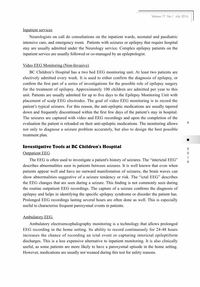
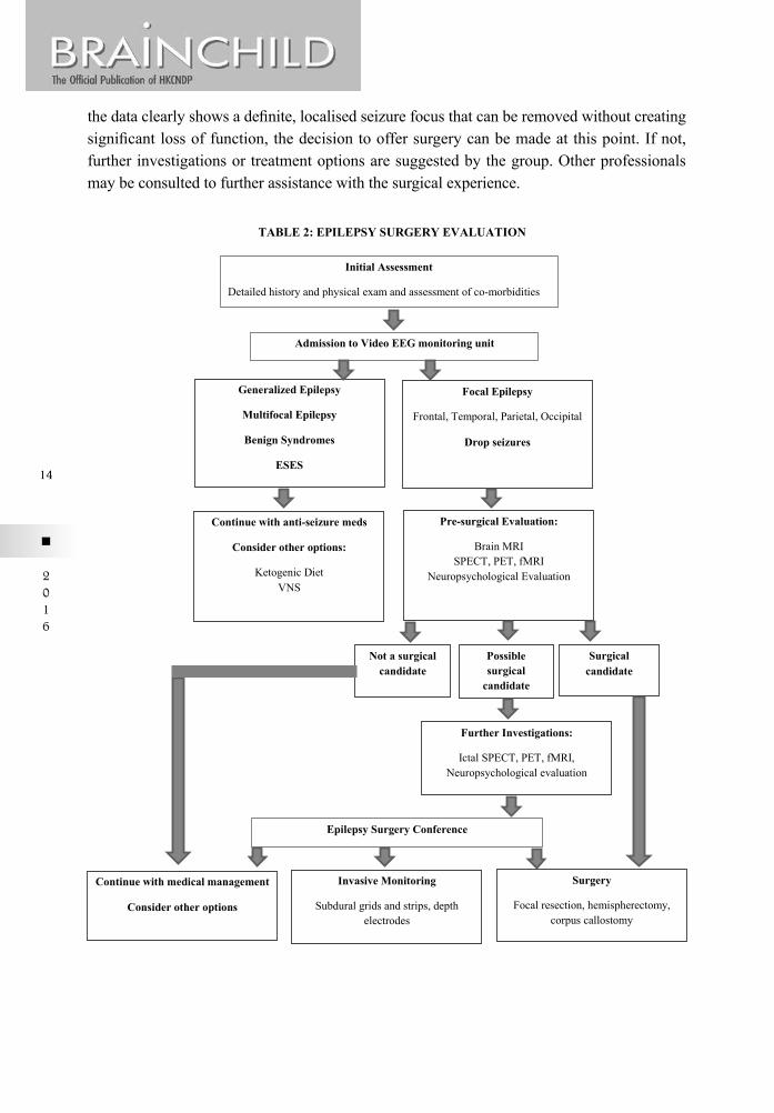
When medications are unable to control seizures, a careful evaluation is done to determine the best alternative for treatment. The goal of the pre-surgical evaluation is to determine the epileptogenic zone: the part of the brain that is integral for the generation of seizures, and to determine if this can be safely resected. This work-up involves a detailed patient history, description of seizure semiology, video-EEG monitoring to localize seizures, neuro-imaging to identify the type, location, and extent of structural abnormalities, and nuclear medicine tests, such as SPECT and PET scans for seizure onset localisation. Assessments of cognitive functioning, including neuropsychological evaluations and fMRI can be used to assess cognitive co-morbidities associated with the epilepsy and predict the cognitive outcome of patients after surgery. The work-up and investigations are tailored to each patient. In some patients, the seizure focus cannot be fully identified and further invasive monitoring is necessarily. On average 18-20 children have epilepsy surgery each year in the program. With an increasing number of new epileptologists, the number may change. Please refer to Table 2 for a summary of the pre-surgical evaluation at BC Children’s Hospital.
Invasive MonitoringInvasive monitoring is used in pre-surgical evaluations at BC Children’s Hospital if the
epileptogenic zone cannot be localised by surface EEG and other investigations. Invasive EEG recordings are made with surgically implanted electrodes on the surface or within the depth of the brain. Subdural EEG electrodes are those electrodes which are placed over the surface of the brain. Depth EEG electrodes are those electrodes which are placed within the parenchyma of the brain. These recordings are done to better localise the region of the brain fromwhichtheepilepsyisarising.Theplacementoftheseelectrodesisoftenconfirmedwithco-registration on an MRI scan image.
13
n
2016
Volume 17 No.1 July 2016
Functional Localisation TechniquesFunctional localisation techniques are performed to predict or prevent functional
deficitwithepilepsysurgery.Thesearedoneseparatelyduringtheoperationorpriortotheoperation by the epileptologists and EEG technologists.
Cortical stimulation entails delivering a short duration of electrical current to the brain via the invasive electrodes. This is done to determine what function that region of the brain subserves. Patients are often asked to perform a task during cortical stimulation, such as speaking, to determine if stimulation impairs the performance of that task. Functions that can be tested using this technique include language, vision, movement, and sensation.
Cortical somatosensory evoked potentials are used to locate an important structure in the brain, called the central sulcus, which helps to delineate motor from sensory function.
Neuropsychological EvaluationResearch has shown that many people with epilepsy have cognitive difficulties as a
resultoftheirseizures.Thetypeofcognitivedifficultiespeopleexperiencedependsonthearea of the brain the seizures are coming from. For example, those with frontal lobe epilepsy canhavedifficultieswithexecutivefunctionandattention.Patientswithlefttemporallobeepilepsy may have difficulties with short term memory and verbal memory. Many people with epilepsy also experience depression and anxiety that can affect their thinking skills. A neuropsychological evaluation is conducted by a neuropsychologist as a formal assessment of cognitive abilities (e.g. memory, concentration, and problem solving), mood and personality. Pre-surgical patients have priority for testing. Patients are periodically followed post-operatively as well.
Vagal Nerve Stimulator (VNS)At BC Children’s Hospital, VNS is used in those with treatment resistant epilepsy,
who are not epilepsy surgery candidates or who cannot tolerate the ketogenic diet. It is a palliative therapeutic modality with a response rate of 50% reduction in seizure frequency in one third to one half of patients. It delivers electrical stimulation to the left vagus nerve. The patients are followed in clinic and the epileptologist or epilepsy surgery nurse adjust the settings periodically for optimal response. From 2006, 57 VNS’s have been implanted, with approximately 3-5 devices being implanted per year. There are currently approximately 30 patients being followed with VNS in the program. The remainder have transitioned to adult care, passed away or have had the device stopped.
Epilepsy surgery conference:After the necessary tests are completed, cases are presented at Epilepsy Surgery
Conference. This conference occurs for 2 hours every second week. Here, a multidisciplinary group — including epileptologists, a neurophysiologist, neurosurgeons, neuropsychologists, neuroradiologists, nurses, and trainees, gather to review and discuss all the data collected. If
14
n
2016
thedataclearlyshowsadefinite,localisedseizurefocusthatcanberemovedwithoutcreatingsignificantlossoffunction,thedecisiontooffersurgerycanbemadeatthispoint.Ifnot,further investigations or treatment options are suggested by the group. Other professionals may be consulted to further assistance with the surgical experience.
Admission to Video EEG monitoring unit
Focal Epilepsy
Frontal, Temporal, Parietal, Occipital
Drop seizures
Continue with anti-seizure meds
Consider other options:
Ketogenic Diet VNS
Pre-surgical Evaluation:
Brain MRI SPECT, PET, fMRI
Neuropsychological Evaluation
Generalized Epilepsy
Multifocal Epilepsy
Benign Syndromes
ESES
Invasive Monitoring
Subdural grids and strips, depth electrodes
Continue with medical management
Consider other options
Surgery
Focal resection, hemispherectomy, corpus callostomy
Initial Assessment
Detailed history and physical exam and assessment of co-morbidities
Possible surgical
candidate
Not a surgical candidate
Surgical candidate
Further Investigations:
Ictal SPECT, PET, fMRI, Neuropsychological evaluation
Epilepsy Surgery Conference
TABLE 2: EPILEPSY SURGERY EVALUATION
15
n
2016
Volume 17 No.1 July 2016
Ketogenic Diet ProgramThe Ketogenic Diet (KD) is a strictly calculated, medically prescribed therapeutic diet
thathasprovenefficacyfortreatmentofintractablechildhoodepilepsy.TheclassicKD(alsoknown as the long-chain triglyceride diet) is a high fat, low carbohydrate dietary therapy.
Although double-blind control trials are lacking, there is sufficient evidence to suggest that theclassicKDifeffective inreducingseizurefrequency(>90%reduction)in approximately 30% of patient with treatment resistant epilepsy. If effective and tolerated, children remain on the KD for 2 to 3 years, at which point it is often weaned and discontinued.
The ketogenic diet team at BC Children’s hospital consists of 1 dietician, 2 nurses, and 3 epileptologists. If another neurologist or epileptologist considers starting a patient on KD, they will refer the patient to this team. In the program, the diet is introduced in the outpatient setting over four days for medically stable patients over one year of age. Rarely, very young patients or complex patients initiate the diet as an inpatient on the wards or ICU. Comparisons of inpatient and outpatient treatments using a KD have been done. It has been shownthatthebenefitsofoutpatienttreatmentincludeimprovedacceptabilityandabilitytomaintain and comply with the diet. It also avoids the expense, inconvenience and potential low blood sugar associated with starvation during inpatient initiation12. The initiation of the diet varies based on whether the patient is on oral diet versus formula diet and whether or not they are older or younger than one year of age. In some situations, the ratio is progressed (with full calories) but in some cases the calories are progressed (at goal ratio). The program currently has 40 patients on the diet. On average, 1 – 2 patients are initiated on the diet per month. Recently, the patients on the diet are asked to complete forms on quality of life and psychosocial functioning to assess the impact of the diet on other aspects of their lives, besides seizure frequency.
Telehealth and Outreach ClinicsTelehealth
The epilepsy program is based primarily at BC Children’s Hospital. Access to epilepsy clinicscanbedifficultforallchildrenintheprovince.Geographically,BritishColumbiaisa large province and many patients live at a great distance from the hospital or in remote areas. There is also disparity with access to transportation and socio-economic status. The epileptologists use telehealth for some follow-up appointments. Assessment and care planning is done over a videoconferencing system rather than traveling to access the same services. All telehealth services are provided over a secure network so that privacy is ensured. More than 40 BC communities have accessed to the services.
Outreach ClinicsSome of the neurologists and epileptologists also go to different areas of the province to
see patients who live in distant or remote areas from BC Children’s Hospital. These include 5 different sites in the province. From 2014-2015, there were 718 outreach clinic visits, of which approximately 40-50% of these were seizure or epilepsy patients.
16
n
2016
Quality of LifeIn addition to seizures, patients with epilepsy have a higher risk of emotional,
behavioural, social, cognitive, and academic and family problems when compared to healthy peers and children with other chronic health conditions. These can affect a child’s quality of life, including physical, social and mental well-being, and functioning in daily life. When treating patients with epilepsy, it is important to monitor the degree to which epilepsy affects their life. Assessments of quality of life can document the patient’s status, including how their status is before and after treatment changes.
Recently, the epilepsy group starting using the Pediatric Quality of Life Inventory to patients for monitoring this. These questionnaires are filled out by the patient or parent, depending on the child’s age. Patients who are given this questionnaire include those who are newly diagnosed with seizures, patients in the epilepsy surgery program, and those who areontheketogenicdiet.Inaddition,theADHD-IVRatingScale,StrengthsandDifficultiesQuestionnaire, and Parental Stress Scale are administered. If there are any concerns, referrals can be promptly made to psychology or psychiatry.
Transition Planning to Adult CareSignificant improvement in medical management over the last decades has resulted
in more children surviving to adulthood. For adolescents with epilepsy there is very little information on the best way to transition. At BC Children’s Hospital, patients are seen up to 18 years of age. Usually, planning for transition begins 1-2 years earlier. A detailed referral summarizing the patient’s history and investigations is sent to the adult physician. Patients with treatment resistant epilepsy, complex patients, those who require epilepsy surgery work-up or have had vagal nerve stimulation, are referred to adult epileptologists. The remainder are referred to adult neurologists in the province.
At BC Children’s Hospital, a group of physicians including 5 neurologists (1 adult and 4 pediatric), adolescent medicine specialists, family practitioners, psychiatrists, and 7 allied health professionals, created long term care plans to ensure access and attachment to adult healthcare providers and to identify strategies for providing support during the transition period including training modules, tools, and resources and system improvements. Different aspects of epilepsy have been covered, such as patients with treatment resistant epilepsy, epilepsy surgery patients, women in epilepsy, etc. The transition development is still in progress. However, the process has so far resulted in a collaborative approach between paediatric and adult healthcare providers. It has helped to create plans to better organise and optimize the transition process and has provided a forum to identify and remedy gaps in the transitional care process.
Patient EducationThe epilepsy program aims to provide seizure management and safety information to
patients and families, as well as to minimize the psychosocial impact of epilepsy for the child through education and empowerment.
17
n
2016
Volume 17 No.1 July 2016
In addition to the physicians, the epilepsy nurses play an important role in patient education. They help educate the child, family, respite services, etc. about the full impact of epilepsy. Epilepsy nurses also help with practical management, including emergency medication, seizure first aid, and Sudden Unexplained Death in Epilepsy (SUDEP). They build and sustain long term relationships with the child and family and ensure they understand the information provided by the physician. They also provide practical advice regarding medication administration. They are an accessible point of contact for the child and family and address any issues arising. Handouts and brochures are available to families on various topics, including descriptions of epilepsy, epilepsy safety, and anti-seizure medications.
The BC Epilepsy Society is a provincially incorporated non-profit charitable organisation, dedicated to serving people in British Columbia with epilepsy and their families. The organization provides support and education for patients and families in various ways, such as providing a resource center, lecture series for families, and educational events. They also provide hospital outreach, where staff members from BC Epilepsy Society meet with families attending the Neurology Clinic at BC Children's Hospital to provide epilepsy resources and assistance with community services. Resources are also provided to other hospitals and health clinics across the province.
Training Program An important part of the epilepsy program at BC Children’s Hospital is training medical
students, neurology residents, and clinical neurophysiology and epilepsy fellows. The high volume clinical practice and varied areas of expertise of the faculty provides a rich learning environment. Trainees get exposure to a wide variety of cases in multiple clinical settings, including outpatient clinics, OR, inpatient monitoring unit, and ICU. Various academic rounds are in place, such as Video EEG session, Epilepsy Surgery conference, Epilepsy Genetics Rounds, and Epilepsy/neurophysiology lectures.
Trainees have the opportunity of attending epilepsy clinics, including epilepsy surgery clinic and tuberous sclerosis clinic. Neurology residents do a mandatory epilepsy and neurophysiology rotation in their residency.
Epilepsy and Neurophysiology fellows receive 1-2 year positions, during which they acquire extensive experience in electro-diagnostic testing, including electroencephalograms, long term video EEG monitoring, evoked potentials, and intraoperative monitoring. They become competent with clinical management, including epilepsy surgery evaluations, and following patients on the ketogenic diet. They are expected to be involved in research as well. The program has graduated several alumni who now are clinicians and scientists in many centers in the world.
ResearchResearch into the causes and treatment of epilepsy is very important part of the epilepsy
program. The BC Children’s Epilepsy Program provides an environment where basic sciencestudiesthatadvancethescientificknowledgeofepilepsycanbetranslatedtoclinicalstudiesthatbenefitpatientsandtheirfamilies.Alloftheepileptologistsandalargenumber
18
n
2016
of students, fellows, and other staff are involved in various prospective and retrospective studies, including basic science and clinical research. At the present time, there are 11 ongoing prospective trials in the epilepsy program. Some of them involve ketogenic diet, quality of life, tuberous sclerosis complex, telehealth, epilepsy surgery, epilepsy genomics, fMRI, electrical status epilepticus in slow wave sleep, etc. Numerous retrospective studies are also ongoing. This is facilitated by a clinical epilepsy database and an extensive EEG database.
ConclusionThe high-volume clinical practice at BC Children’s Hospital provides care to paediatric
patients from all over the province of British Columbia, as well as occasionally other provinces in Canada. A coordinated, multidisciplinary team contributes a comprehensive range of skills and knowledge of paediatric epilepsy. Excellence in diagnostics, medical and surgical clinical treatment programs, and research have yielded important contributions. There are still areas that are work in progress, such as improving the transition process to adult care, having more specialised clinics, such as neurogenetic or ketogenic diet clinic, and being able to obtain funding for specialized genetic testing or imaging modalities for patients, outside of research protocols.
References1. Fisher RS, Acevedo C, Arzimanoglou A, Bogacz A, Cross JH, Elger CE, Engel J Jr, Forsgren L, French JA,
Glynn M, Hesdorffer DC, Lee BI, Mathern GW, Moshé SL, Perucca E, Scheffer IE, Tomson T, Watanabe M, WiebeS.(2014).ILAEofficialreport:apracticalclinicaldefinitionofepilepsy.Epilepsia,55(4):475-82.
2. Kwan P, Arzimanoglou A, Berg AT, Brodie MJ, Allen Hauser W, Mathern G, Moshé SL, Perucca E, Wiebe S,FrenchJ.(2010).Definitionofdrugresistantepilepsy:consensusproposalbytheadhocTaskForceoftheILAE Commission on Therapeutic Strategies. Epilepsia, 51(6):1069-77.
3. Hitiris N, Mohanraj R, Norrie J, Brodie MJ.(2007). Mortality in epilepsy. Epilepsy Behav, 10(3):363-76. 4. Thompson PJ1, Duncan JS. (2005). Cognitive decline in severe intractable epilepsy. Epilepsia, 46(11):1780-7.5. Jokeit H, Ebner. (1999). A Long term effects of refractory temporal lobe epilepsy on cognitive abilities: a
cross sectional study.. J Neurol Neurosurg Psychiatry, 67(1):44-50.6. Helmstaedter C, Kurthen M, Lux S, Reuber M, Elger CE. (2003). Chronic epilepsy and cognition: a
longitudinal study in temporal lobe epilepsy. Ann Neurol, 54(4):425-32.7. Hermann BP1, Seidenberg M, Dow C, Jones J, Rutecki P, Bhattacharya A, Bell B. (2006). Cognitive
prognosis in chronic temporal lobe epilepsy. Ann Neurol, 60(1):80-7. 8. CamfieldP,CamfieldC.(2015).Incidence,prevalenceandaetiologyofseizuresandepilepsyinchildren.
Epileptic Disord. 2015, 17(2):117-23. 9. NationalPopulationHealthSurvey,1998-1999(Cycle3):HouseholdComponent.Generalfile.[machine
readabledatafile].Ottawa,ON.StatisticsCanada.2001-01-19.10. WirrellEC,GrossardtBR,Wong-KisielLC,NickelsKC.(2011).Incidenceandclassificationofnew-onset
epilepsy and epilepsy syndromes in children in Olmsted County, Minnesota from 1980 to 2004: a population-based study. Epilepsy Res, 95(1-2):110-8.
11. Scheffer IE. (2014). Epilepsy genetics revolutionizes clinical practice. Neuropediatrics, 45(2):70-4. 12. VaisleibII,BuchhalterJR,ZupancML.(2004).Ketogenicdiet:outpatientinitiation,withoutfluid,orcaloric
restrictions. Pediatr Neurol, 31(3):198-202.
19
n
2016
Volume 17 No.1 July 2016
BC Children’s Hospital Brain Mapping ProgramBruce H. Bjornson, MD, FRCPCClinical Assistant Professor, Division of Neurology, BCCHAssociate Clinician Scientist, Child & Family Research InstituteMedical Director, Brain Mapping & Neurotechnology LaboratoryScientific Director, Child & Family Research Imaging Facility
BC Children’s Hospital (BCCH), the primary paediatric teaching hospital of the Faculty of Medicine of the University of British Columbia in Vancouver, provides pediatric neurological and neurosurgical care services to the province of British Columbia, and is a major Canadian academic paediatric neurosciences centre, contributing tertiary and quaternary care services to children with a wide range of neurological disorders. Children with neurological disorders often require several different consultations and diagnostic tests to reach a precise diagnosis and to plan an optimal treatment strategy to achieve the best outcome, often requiring a team approach to care. Comprehensive multidisciplinary team evaluation is especially relevant to children with epilepsy, brain tumors and strokes, who may need to undergo medical and neurosurgical intervention as part of their care. The BCCH Brain Mapping program was created to enhance neurological evaluation and surgical planning by teams of paediatric neurologists and neurosurgeons.
The BCCH Brain Mapping program provides individualised surgical planning guidance by combining structural neuroimaging, to locate brain pathology to be targeted by neurosurgeons, with functional neuroimaging, to highlight areas responsible for important brain functions, which are in close proximity to surgical targets, in order to minimize damage to those areas. Brain functions of particular importance include motor function (especially handfinemotorfunction),vision,hearing,speechandlanguage.
In developing the BCCH Brain Mapping program, our initial key goals were: (1) to reduce reliance on previously available invasive methods of mapping of brain function (unilateral carotid artery injection of amobarbital and awake brain surgery with direct cortical electrical stimulation); (2) to avoid exposure to ionizing radiation (CT, PET); (3) to design a functional neuroimaging laboratory environment that was child-friendly; (4) to design tasksadaptabletoavarietyoflevelsofdifficultyandtomeasurebehavioralperformance,cerebral neurophysiology and brain anatomy simultaneously; and (5) to visualize and present multimodality, multidimensional brain data in a format that would help guide pediatric neurological and neurosurgical evaluation and care.
To achieve these goals, over the past twenty years, we have created infrastructure to specificallydevelopandsupportpaediatricbrainmappingtechnologyandmethodologywithBC Children’s Hospital, and we have progressively translated advances in technology into advances in neurological care of children.
20
n
2016
A. Motivation, Challenges & OpportunitiesWhat motivated us to develop a paediatric brain mapping program at BCCH? The main
motivation was the opportunity to enhance paediatric neurological care by applying advances in MRI and computing technology to enable fusion of three-dimensional images of changes in brain physiology (correlated with behavioural events), with three-dimensional images of brain anatomy, to achieve interactive in vivo spatiotemporal mapping of brain function. This presents many associated challenges. Most fundamental of these is the fact that the brain is an incredibly complex organ consisting of nearly 100 billion (1011) neurons, with approximately one million billion (1015) connections among them. Connections form during early fetal life under the influence of genetic and environmental factors, with ongoing development and refinement occurring throughout infancy, childhood and adulthood. All the diversity and complexity of human perception, cognition, thought, emotions, memories and behaviour result from interactions among neurons participating in multiple, dynamic, distributed brain networks. These, in turn, are organised according by location and connections in the brain (neuroanatomy and connectivity), and governed by constraints of neurophysiology (e.g., metabolism, electrical transmission, neurotransmitters and receptors).
In the past, in order to determine which areas of the brain are most responsible for critical functions,neurologicaldeficitsobservedinlifewerecorrelatedwithresultsofexaminationof the brain after death (clinical-pathological correlation). Subsequently, it became possible to observe the effects of temporary disruption of brain activity on living persons by direct electrical stimulation of the brain during awake brain surgery, a method pioneered by neurosurgeonDr.WilderPenfieldinMontreal.Anothermethodoftemporaryperturbationof brain function, described by Dr. Juhn Wada, involved selective unilateral injection of the anesthetic agent, amobarbital, into either the left or the right internal carotid artery during awake cerebral angiography, and then observing for deficits of language or memory, to determine whether one hemisphere appeared to be “dominant” for language or memory. We were motivated by the opportunity to provide similar information without reliance on such invasive methods.
As more powerful superconducting magnets became available for MRI, higher-resolution structural images became possible, with better delineation of tissue boundaries making possible more accurate quantitative measurement of grey and white matter volumes in areas of the brain, during development (brain image segmentation, parcellation and morphometric analysis).
Meanwhile, MRI studies have demonstrated that deoxyhemoglobin (paramagnetic, or weakly magnetic, due to unbound iron in heme) and oxyhemoglobin (diamagnetic, with minimal magnetic effect) have different effects upon the MRI signal in various areas of the brain, and that these effects vary, moment by moment, with transient changes in neuronal activation. Thus, areas of the brain, that are receiving incoming neuronal stimulation (“activation”), experience a temporary increase in the locally measured MRI signal due to
21
n
2016
Volume 17 No.1 July 2016
transient local increases in blood oxygen content. This phenomenon is termed the “Blood Oxygen Level Dependent (BOLD)” effect, and is the basis of “functional MRI”.
Other MRI studies explored the diffusion of water within various types of tissue (diffusion weighted imaging), leading to the observation that diffusion of water was favoured in the direction of axons within white matter tracts of the brain. This led to the development of MRI techniques for “diffusion tensor imaging (DTI)” to demonstrate white matter pathways (“tractography”).
High-resolution anatomical (structural) MRI can now be combined with studies of structural (white matter tractography) connectivity and BOLD functional MRI connectivity, acquired either during specific tasks or at rest (intrinsically autocorrelated functional networks), and such studies can be performed non-invasively on infants, children and adults.
B. Creating a Paediatric Brain Mapping TeamThe initial concept of a Paediatric Brain Mapping program at BCCH was the result
of a careful long-term planning by a small “Brain Mapping Steering Group” (Drs. B. Bjornson and P. Wong , paediatric neurologists; Dr. D. Cochrane, paediatric neurosurgeon; and Dr. D. Giaschi, vision scientist), subsequently enhanced through collaboration with others (particularly Dr. A. Mackay, MRI Physicist; and Dr. S. Miller, neonatal neurologist). Progress has depended upon the talents and innovations contributed by many individuals, including undergraduate and graduate students in several disciplines (psychology, physics, engineering, computer sciences, medicine and neurosciences). Our current Brain Mapping & Neurotechnology Laboratory team consists of a Brain Mapping Technology Development Manager, with expertise in experimental design and neuroimaging; a Research Imaging Technology Integration Manager, responsible for integrating EEG, MEG and other physiological data with functional MRI data; and a Brain Mapping Neuroinformatics Engineer, with expertise in software development and computing. We currently collaborate closely with several MRI Scientists who optimize MRI pulse sequence software and MRI hardware performance to enhance MRI data quality and yield, and with Neurologists, Neurophysiologists, Neuroscientists and Computer Scientists with additional expertise in digital EEG, experimental design, image processing and statistical analysis.
C. Customizing Brain Mapping Task Protocols for ChildrenWe have developed customized paediatric experimental protocols and software, which
allow us to select functional MRI protocols suitable for children of multiple different levels of ability. We have nearly 100 different versions and levels that can be used to map visual, auditory, language and visuospatial functions in children.
22
n
2016
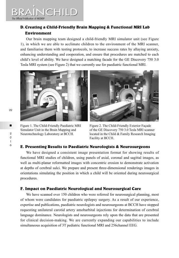
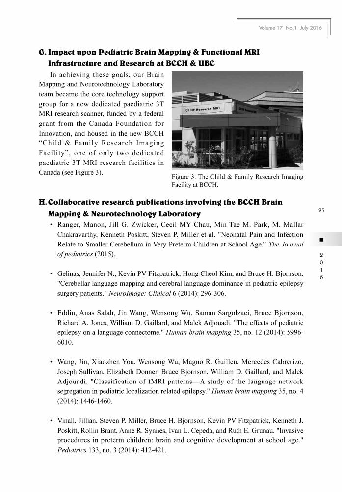
D. Creating a Child-Friendly Brain Mapping & Functional MRI Lab EnvironmentOur brain mapping team designed a child-friendly MRI simulator unit (see Figure
1), in which we are able to acclimate children to the environment of the MRI scanner, and familiarise them with testing protocols, to increase success rates by allaying anxiety, enhancing understanding and cooperation, and ensure that procedures are matched to each child’s level of ability. We have designed a matching facade for the GE Discovery 750 3.0 Tesla MRI system (see Figure 2) that we currently use for paediatric functional MRI.
Figure 1. The Child-Friendly Paediatric MRI Simulator Unit in the Brain Mapping and Neurotechnology Laboratory at BCCH.
Figure 2. The Child-Friendly Exterior Façade of the GE Discovery 750 3.0 Tesla MRI scanner located in the Child & Family Research Imaging Facility at BCCH.
E. Presenting Results to Paediatric Neurologists & NeurosurgeonsWe have designed a consistent image presentation format for showing results of
functional MRI studies of children, using panels of axial, coronal and sagittal images, as well as multi-planar reformatted images with concentric erosion to demonstrate activation at depths of cerebral sulci. We prepare and present three-dimensional renderings images in orientations simulating the position in which a child will be oriented during neurosurgical procedures.
F. Impact on Paediatric Neurological and Neurosurgical CareWe have scanned over 150 children who were referred for neurosurgical planning, most
of whom were candidates for paediatric epilepsy surgery. As a result of our experience, expertise and publications, paediatric neurologists and neurosurgeons at BCCH have stopped requesting unilateral carotid artery amobarbital injections for determination of cerebral language dominance. Neurologists and neurosurgeons rely upon the data that are presented for clinical decision-making. We are currently expanding our capabilities to include simultaneous acquisition of 3T pediatric functional MRI and 256channel EEG.
23
n
2016
Volume 17 No.1 July 2016
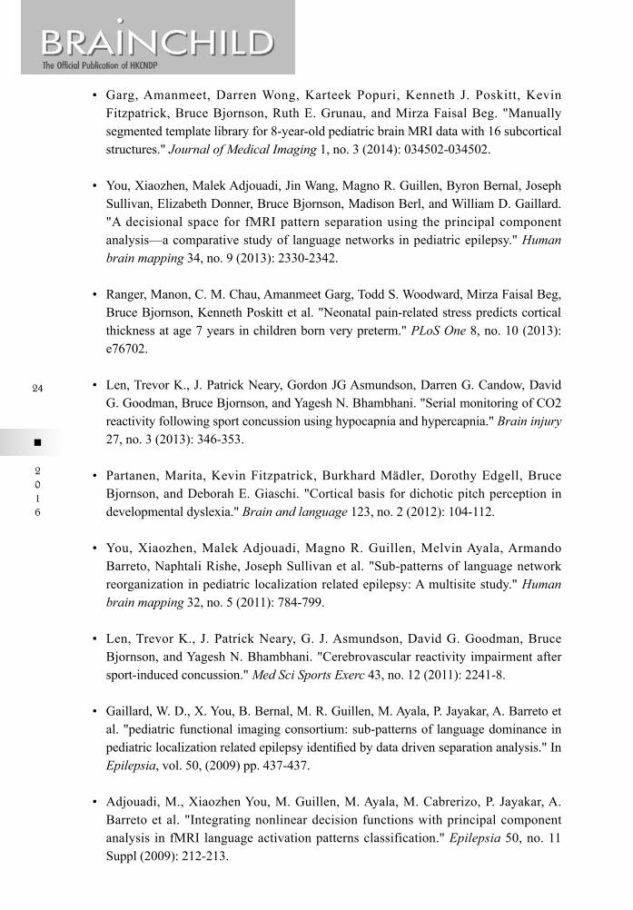
G. Impact upon Pediatric Brain Mapping & Functional MRI Infrastructure and Research at BCCH & UBCIn achieving these goals, our Brain
Mapping and Neurotechnology Laboratory team became the core technology support group for a new dedicated paediatric 3T MRI research scanner, funded by a federal grant from the Canada Foundation for Innovation, and housed in the new BCCH “Child & Family Research Imaging Facility”, one of only two dedicated paediatric 3T MRI research facilities in Canada (see Figure 3).
H. Collaborative research publications involving the BCCH Brain Mapping & Neurotechnology Laboratory
• Ranger,Manon, JillG.Zwicker,CecilMYChau,MinTaeM.Park,M.MallarChakravarthy, Kenneth Poskitt, Steven P. Miller et al. "Neonatal Pain and Infection Relate to Smaller Cerebellum in Very Preterm Children at School Age." The Journal of pediatrics (2015).
• Gelinas,JenniferN.,KevinPVFitzpatrick,HongCheolKim,andBruceH.Bjornson."Cerebellar language mapping and cerebral language dominance in pediatric epilepsy surgery patients." NeuroImage: Clinical 6 (2014): 296-306.
• Eddin,AnasSalah,JinWang,WensongWu,SamanSargolzaei,BruceBjornson,Richard A. Jones, William D. Gaillard, and Malek Adjouadi. "The effects of pediatric epilepsy on a language connectome." Human brain mapping 35, no. 12 (2014): 5996-6010.
• Wang,Jin,XiaozhenYou,WensongWu,MagnoR.Guillen,MercedesCabrerizo,Joseph Sullivan, Elizabeth Donner, Bruce Bjornson, William D. Gaillard, and Malek Adjouadi. "Classification of fMRI patterns—A study of the language network segregation in pediatric localization related epilepsy." Human brain mapping 35, no. 4 (2014): 1446-1460.
• Vinall,Jillian,StevenP.Miller,BruceH.Bjornson,KevinPVFitzpatrick,KennethJ.Poskitt, Rollin Brant, Anne R. Synnes, Ivan L. Cepeda, and Ruth E. Grunau. "Invasive procedures in preterm children: brain and cognitive development at school age." Pediatrics 133, no. 3 (2014): 412-421.
Figure 3. The Child & Family Research Imaging Facility at BCCH.
24
n
2016
• Garg,Amanmeet,DarrenWong,KarteekPopuri,Kenneth J. Poskitt,KevinFitzpatrick, Bruce Bjornson, Ruth E. Grunau, and Mirza Faisal Beg. "Manually segmented template library for 8-year-old pediatric brain MRI data with 16 subcortical structures." Journal of Medical Imaging 1, no. 3 (2014): 034502-034502.
• You,Xiaozhen,MalekAdjouadi,JinWang,MagnoR.Guillen,ByronBernal,JosephSullivan, Elizabeth Donner, Bruce Bjornson, Madison Berl, and William D. Gaillard. "A decisional space for fMRI pattern separation using the principal component analysis—a comparative study of language networks in pediatric epilepsy." Human brain mapping 34, no. 9 (2013): 2330-2342.
• Ranger,Manon,C.M.Chau,AmanmeetGarg,ToddS.Woodward,MirzaFaisalBeg,Bruce Bjornson, Kenneth Poskitt et al. "Neonatal pain-related stress predicts cortical thickness at age 7 years in children born very preterm." PLoS One 8, no. 10 (2013): e76702.
• Len,TrevorK.,J.PatrickNeary,GordonJGAsmundson,DarrenG.Candow,DavidG. Goodman, Bruce Bjornson, and Yagesh N. Bhambhani. "Serial monitoring of CO2 reactivity following sport concussion using hypocapnia and hypercapnia." Brain injury 27, no. 3 (2013): 346-353.
• Partanen,Marita,KevinFitzpatrick,BurkhardMädler,DorothyEdgell,BruceBjornson, and Deborah E. Giaschi. "Cortical basis for dichotic pitch perception in developmental dyslexia." Brain and language 123, no. 2 (2012): 104-112.
• You,Xiaozhen,MalekAdjouadi,MagnoR.Guillen,MelvinAyala,ArmandoBarreto, Naphtali Rishe, Joseph Sullivan et al. "Sub-patterns of language network reorganization in pediatric localization related epilepsy: A multisite study." Human brain mapping 32, no. 5 (2011): 784-799.
• Len,TrevorK., J.PatrickNeary,G. J.Asmundson,DavidG.Goodman,BruceBjornson, and Yagesh N. Bhambhani. "Cerebrovascular reactivity impairment after sport-induced concussion." Med Sci Sports Exerc 43, no. 12 (2011): 2241-8.
• Gaillard,W.D.,X.You,B.Bernal,M.R.Guillen,M.Ayala,P.Jayakar,A.Barretoetal. "pediatric functional imaging consortium: sub-patterns of language dominance in pediatriclocalizationrelatedepilepsyidentifiedbydatadrivenseparationanalysis."InEpilepsia, vol. 50, (2009) pp. 437-437.
• Adjouadi,M.,XiaozhenYou,M.Guillen,M.Ayala,M.Cabrerizo,P.Jayakar,A.Barreto et al. "Integrating nonlinear decision functions with principal component analysis in fMRI language activation patterns classification." Epilepsia 50, no. 11 Suppl (2009): 212-213.
25
n
2016
Volume 17 No.1 July 2016
• Lanyon,LindaJ.,DeborahGiaschi,SimonAuYoung,KevinFitzpatrick,LuDiao,Bruce H. Bjornson, and Jason JS Barton. "Combined functional MRI and diffusion tensor imaging analysis of visual motion pathways." Journal of Neuro-Ophthalmology 29, no. 2 (2009): 96-103.
• Giaschi,Deborah,AmyZwicker,SimonAuYoung,andBruceBjornson."Theroleof cortical area V5/MT+ in speed-tuned directional anisotropies in global motion perception." Vision research 47, no. 7 (2007): 887-898.
• Giaschi,Deborah,AmyZwicker,SimonAuYoung,BoscoLee,andBruceBjornson."The role of area V5/MT+ in the centripetal bias in global motion perception." Journal of Vision 7, no. 9 (2007): 751-751.
• Giaschi,Deborah,VeronicaEdwards,SimonAuYoung, andBruceBjornson."Asymmetrical cortical activation by global motion in children with dyslexia." Journal of Vision 5, no. 8 (2005): 848-848.
• Edwards,VeronicaT.,DeborahE.Giaschi,RobertF.Dougherty,DorothyEdgell,Bruce H. Bjornson, Christopher Lyons, and Robert M. Douglas. "Psychophysical indexes of temporal processing abnormalities in children with developmental dyslexia." Developmental neuropsychology 25, no. 3 (2004): 321-354.
• Giaschi,Deborah,JamesE.Jan,BruceBjornson,SimonAuYoung,MatthewTata,Christopher J. Lyons, William V. Good, and Peter KH Wong. "Conscious visual abilities in a patient with early bilateral occipital damage." Developmental Medicine & Child Neurology 45, no. 11 (2003): 772-781.
• Orchard,Jeff,ChenGreif,GeneH.Golub,BruceBjornson,andM.StellaAtkins."Simultaneous registration and activation detection for fMRI." Medical Imaging, IEEE Transactions on 22, no. 11 (2003): 1427-1435.
• deVeber,Gabrielle,MaureenAndrew,ColeenAdams,BruceBjornson,FrancesBooth,DavidJ.Buckley,CarolS.Camfieldetal."Cerebralsinovenousthrombosisinchildren." New England Journal of Medicine 345, no. 6 (2001): 417-423.
• Biornson,Bruce,DeborahGiaschi,DanielSlick,MaryConnolly,andSimonAuYoung. "Hemispheric dissociation of verb generation from verb reading: functional MRI of an adolescent with early left MCA infarction." NeuroImage 13, no. 6 (2001): S508.
• Bjornson,Bruce,DeborahGiaschi,DouglasCochrane,SimonAuYoung,DanielRootman, Catherine Boden, Mary Connolly et al. "Non-invasive mapping of
26
n
2016
sensorimotor cortex in a child with a cavernous angioma: fMRI and high-resolution EEG compared with surgical mapping." Neuroimage 9 (1999): S696-S696.
• Dougherty,RobertF.,MaxS.Cynader,BruceH.Bjornson,DorothyEdgell,andDeborah E. Giaschi. "Dichotic pitch: a new stimulus distinguishes normal and dyslexic auditory function." Neuroreport 9, no. 13 (1998): 3001-3005.
• Dougherty,R.F.,S.M.W.AuYoung,D.E.Giaschi,B.H.Bjornson,andP.K.H.Wong. "Comparison of visual activation measured by fMRI and high resolution EEG." Neuroimage 7, no. 4II (1998): S309.
• Dougherty,R.F.,D.E.Giaschi,B.H.Bjornson,S.L.Chamut,D.Edgell,andC.J. Lyons. "In search of the best psychophysical indicator of an M-stream deficit in developmental dyslexia." In Investigative Ophthalmology & Visual Science, vol. 38, no. 4 (1997), pp. 3037-3037.
27
n
2016
Volume 17 No.1 July 2016
Diagnostic Neurophysiology ServicesDonna Gregory Wood, R.E.T., R.EPT., R.T.(EMG)
Dept. of Diagnostic NeurophysiologyBC Children’s Hospital
Clinical ServicesWe provide neurodiagnostic testing for all paediatric and maternity patients at
Children’s and Women’s Health Centre of BC (190 beds), and we also accept referrals from paediatricians and general practitioners in the community. We are the only dedicated paediatric and maternity hospital in the province of British Columbia (population 3.5 million). Because of our uniqueness, we see many interesting and difficult patients. This means that our technologists require a high level of expertise and need to stay on the cutting edge of techniques and technology.
The department is under the direction of Dr. P.K.H. Wong and includes five electroencephalographers, one electromyographer, two Epilepsy Fellows, 13 technologists, one biomed data coordinator and four clerical support personnel.
Our service is unique in that all modalities of neurophysiological diagnostic testing are provided in one department. We do this by performing 26 different procedures on all levels of the nervous system. For example, procedures include EEG, cEEG, EMG, Evoked Potentials of all modalities, and Intensive and Ambulatory EEG Monitoring. Our Epilepsy surgery work-up includes electrocorticography, motor strip and speech localisation, and indwelling subdural recordings. We also offer intraoperative monitoring for Orthopaedic and Neurosurgical cases. This includes somatosensory and motor evoked potentials, cranial nerve monitoring, deep white matter stimulation and free-running and triggered EMG during lipoma removal and selective posterior rhizotomies.
This document gives an overview of how each service is provided.
EEGChildren are sleep deprived to obtain a natural sleep recording where possible. All EEGs
are performed with simultaneous video, and parents accompany their children during the test. For safety and accuracy of electrode application, patients who have a tendency to move a lot are secured into a papoose board. A Velcro strap is secured gently across the stomach and a low tumble form roll may be placed under the neck. Parents often lie on the bed beside their child. Once the head is measured, reusable gold electrodes are applied with Ten.20 conductive paste and the hair is overlapped to hold each electrode in place. For young and uncooperativechildrenahatismadefromSurgifixtocontaintheelectrodewires.Thismoreeasily allows the parents to hold their infant or toddler child during testing.
28
n
2016
HyperventilationHyperventilation (HV) is performed for 3 minutes on an anterior to posterior bipolar
montage with 1-minute pre- and post-HV baselines recorded. Younger children are given windmill toys to blow into. The room lights are turned up to optimise the video recording during this time.
Children presenting with questionable staring spells are asked to hyperventilate for 4 minutes.IfthefirstperiodofHVshowsspike-and-wavedischarges,orifthepatientdoesnot fall asleep during the EEG, a second 4-minute HV is done. Children who have absence seizures in the first HV or who have treated absence seizures are asked to perform two periods of HV for 4 minutes each. Optimally, the second HV is performed with the patient sitting up in order to demonstrate any loss of body tone. Two 4 minute periods of HV are repeated when these children return for follow-up EEGs after treatment. Often, breakthrough absence seizures will be seen only during the second HV in patients on treatment.
Photic StimulationFlash stimulation is performed on all patients over 3 months of age recording on a bipolar
montage with anterior-posterior and posterior halo derivations. Photoparoxysmal responses are most prominent on arousal from sleep. When possible, photic stimulation is done after the patient is aroused from the sleep recording.
Our protocol calls for the strobe light (0.72 joules) to be positioned 30 cm in front of the eyes. Room lights are set to minimum. Stimulation begins at 16 Hz on eye closure followed by stimulation at 1, 3, 6 and 9 Hz for a duration of 5 seconds each with an inter-stimulus interval of 5 seconds. Patients are instructed to keep their eyes closed until 9 Hz after which theyareinstructedtoclosetheireyeswhenthelightbeginstoflashandopenthemwhentheflashingstops.Frequencyincrementsof3Hzareused,upto21Hz.Forchildrenyoungerthan 2 years, stimulation is done at lower frequencies (0.5-10 Hz) while awake and with eyes open, if possible.
When a photoparoxysmal response is evoked, the strobe light is stopped as soon as possible to determine if the discharge outlasts the stimulus. Any clinical accompaniments are noted. For generalized discharges, response testing of the patient is done during stimulation.
Capturing Events and Response TestingWhen the EEG referral questions whether or not a particular behaviour is a seizure,
the recording is often prolonged to capture the event. EMG electrodes are applied if limb movements are involved, the room lights are turned up and the video camera image may be customized to maximize recording of the event.
Response testing can take many forms depending on the age and cooperation of the patient. Practice trials are done to form a baseline of the response time. The technologist will
29
n
2016
Volume 17 No.1 July 2016
instruct patients to repeat words or numbers said to them. If patients do not repeat words given to them during the discharge, they are asked to recall those words afterward. Stimuli should be presented as soon as possible after onset of the EEG discharge. If no impairment in consciousness is demonstrated, patient is asked to perform a motor activity before or during stimulation (clapping or pointing to the door). If no impairment is noted, the technologist asks quick math questions during stimulation (for example, what is 2+2?) or to state the opposite of a word given (for example the opposite of day is night). Often, patients who are able to recall words or do motor activities during a discharge are not able to perform these higher functions.
Epilepsy Monitoring Unit and Ambulatory EEG MonitoringThe demand for 1-5 days in-patient or overnight out-patient EEG recordings has been
increasing. We have two dedicated epilepsy monitoring beds on the ward (with a planned increase to 6 beds in the near future) and two ambulatory EEG systems. We apply disposable disc electrodes with Collodion glue for these overnight recordings. To reduce the risk of accidental strangulation, the wires are slipped through narrow Surgifix gauze and the headbox is placed in a pouch with a belt that goes around the patient’s waist. The backpack must be worn by the patient at all times during the monitoring. The parents are given instructions on response testing and rules to ensure optimal recording conditions (keeping the child in the camera range, keeping blinds closed, not chewing gum, etc.) Parents must stay with their child for the duration of the monitoring to identify events, press the event button, describe the events and perform response testing. This requires detailed instruction to parents.
24-hour Continuous EEG Monitoring (cEEG)Continuous EEG monitoring is currently performed for comatose patients having
seizures or for patients in status epilepticus. There is a great demand to expand this service to be the standard of care for all patients at risk for seizures in the Intensive Care Unit (ICU), including patients who are post-cardiac surgery and those with meningitis, encephalitis or decreased levels of consciousness.
For routine portable EEGs in the ICU, a full set of disposable disc electrodes is applied with Ten.20 cream and the hair is overlapped to hold each electrode in place. Continuous EEG recordings are often a continuation of a routine recording during which seizures are captured. To further secure the electrodes for continuous recording, Grass EC2 cream is placedontopofeachelectrodefollowedbyagauzesquare.ASurgifixhatisplacedoverthehead and covers the electrode wires connected to the headbox. The headbox is secured to the head of the bed. An event button is made available for staff to press for clinical seizures. The video angle is set very wide to include staff movement around the bed. The bedside computer is plugged into a network outlet and EEG reader stations in the department are enabled for remote viewing.
30
n
2016
These recordings are interpreted a minimum of twice a day and reported by EEG Fellows under the supervision of staff electroencephalographers. Written communication of the results occurs daily with more frequent verbal communication of results to attending physicians as dictated by the clinical scenario.
Sedated ProceduresChloral hydrate sedation is used for EEG testing on severely uncooperative or autistic
patients and for children having nerve conduction, EMG, BAER and SEP testing. These patients are sleep deprived and kept nil by mouth for 2-6 hours before sedation (depending on whether they are out-patients or in-patients). The standard dose of chloral hydrate is 40-50 mg/kg, given one hour before the test.
Nerve Conduction and Electromyography (NC/EMG)Neuromuscular testing is most often requested for inflammatory demyelinating
neuropathies, focal mononeuropathies, investigation of infants with abnormal tone and, rarely, myotonic dystrophies.
Chloral hydrate sedation is administered by a registered nurse one hour before the testing. The nurse explains the test and shows the parent and patient pictures of the procedure. Two technologists are assigned for each paediatric NC study to distract the patient and obtain accurate results as quickly as possible. One technologist runs the equipment and the other performs the study. Parents are given the option to come in with their child or to stay in the waiting area during the testing.
Our standard protocol for NC testing on the upper limb is to perform sensory and motor recordings (with F wave) on the median and ulnar nerves. On the lower limb, the following recordings are done: sural sensory and tibial and peroneal motor (with F wave). Additional nerves are tested as clinically indicated. Interpretation of results is primarily based on conduction values and the amplitude of the responses. Distances for stimulation and recording are not always a standard length due to small limb size in young patients. Rarely, repetitive nerve studies (facial nerve) are performed, but these are technically challenging in children.
A nerve conduction study typically takes 20 minutes. The electromyographer then reviews the results and if indicated, proceeds to do needle EMG studies. Attempts are made to test distal and proximal lower limb muscles (tibialis anterior, medial gastrocnemius) and one upper limb muscle (biceps). The area is often sprayed with a topical anaesthetic and a disposable needle electrode is inserted into the belly of the muscle by the electromyographer. Activity upon insertion, rest and flexion of the muscle is recorded. Additional muscles are tested as clinically indicated. For investigation of diffuse processes, testing is usually performed on the right side and the left side is left untouched for biopsy purposes.
31
n
2016
Volume 17 No.1 July 2016
Evoked PotentialsVisual (VEP), brainstem (BAER), and somatosensory evoked potentials (SSEP) are
performed on out-patients and in-patients. Referrals are most often for biochemical or demyelinating diseases and neurosurgical tumours.
VEPOccipital electrodes are placed according to the Queen’s Square measuring system and
are referred to a mid-frontal electrode. For testing of optic nerve function in children who are abletofollowinstructionstofixateonthescreen,apatternstimulusisused.Visualacuityisnotedortested.Thepatientispositioned100cmfromthescreenandisaskedtofixateonaspot marked in the centre of the screen. A checker board pattern of 32x32 squares is used. For those with poor visual acuity, a larger pattern check is used. Monocular stimulation is performed. Typically 200 samples are averaged. A minimum of two trials are performed with each eye, and the latency and voltage of the P100 response are recorded. Normal values are a mid-occipital latency of <115 msec with an inter-eye variation of <8 msec.
For very young children or those who are unable to cooperate for pattern stimulation, goggle flash stimulation is used. The goggles are worn by the patient and monocular stimulation is performed to test for optic nerve dysfunction. Bilateral stimulation is performed for suspected cortical visual impairment or other diffuse disease processes. Typically 100 samples are averaged and two trials are done.
BAERThis testing is done during natural or sedated sleep when possible. Ear electrodes are
referred to a vertex electrode. Tiptrode stimulating electrodes are placed gently into the ear canal after visually ensuring that there is no wax in the ear canal. Unilateral alternating 70 dB HL clicks are delivered at frequency of 10.1 Hz to each ear (with 40 dB masking to alternate ear). One thousand responses are averaged. A minimum of two trials is performed on each ear. The peak and inter-peak latency of waves I-V are recorded. If a poor response is obtained rarefraction clicks maybe tried to enhance wave I, or condensation clicks to enhance wave V.
SSEPChildren receive chloral hydrate sedation for somatosensory EPs. Upper limb stimulation
is performed via the median nerve at the wrist. Peripheral responses are recorded at Erb’s point, subcortical responses at C7S (N13 P14) and cortical responses from the central and midline scalp areas (N20/P22). Lower limb stimulation is performed via the posterior tibial nerve at the ankle. Peripheral responses are recorded at the popliteal fossa, followed by spine responses at L1 and T6 levels (N14), subcortical responses at the neck (N20) and cortical responses from the midline and central areas (P37/N45). Typically 500 responses are averaged. A minimum of two trials are performed and the latency and amplitude of the peak waveforms are measured.
32
n
2016
Intraoperative MonitoringWe have a very active intraoperative monitoring (IOM) program providing daily
services to orthopedic and neurosurgical departments. Monitoring is performed on scoliosis patients and neurosurgical posterior fossa tumour, lipoma and spinal cord tumour patients. Intraoperative monitoring has increased from being 20% to 35% of our total workload. Other departments are requesting intra-operative monitoring services but we are unable to provide services at this time. Recruitment to add a full time IOM Specialist with PhD credentials to our team is ongoing.
ScoliosisEachscoliosiscaseismonitoredbytwotechnologists(atleastoneofwhomiscertified
in Neurophysiological Intraoperative Monitoring) working closely with three surgeons who are very knowledgeable about monitoring. Motor and sensory pathways are monitored. All electrodes are applied in the Operating Room once the child is anaesthetised. For motor stimulation, corkscrew electrodes are inserted into the skull over the pre-Rolandic region (1 cm forward to C1, C3, C2 & C4). Corkscrew electrodes are also used to record the post-Rolandic sensory, midline and subcortical potentials (C3’, Cz’, C4’, Fz, Pz, Oz and high cervical spine). Needle recording electrodes are inserted into the abductor pollicus brevis, brachioradialis, abductor hallucis and tibialis anterior muscles to record motor evoked potentials. Surface sticky electrodes are used for ulnar and posterior tibial somatosensory stimulating and peripheral recording.
Once the electrodes are applied and the patient is positioned prone, the technologists plug in the electrodes and stimulators, check the impedances and obtain pre-drape baseline trials. Stimulus parameters for SSEPs start at 15mA for the ulnar nerve and 40mA for the tibial nerve. Transcranial motor stimulation begins with single pulses at 150v, ISI 1.1, train of 5 and duration 50 usec. The stimulus voltage is increased at small intervals until reproducible responses are seen from all muscles or a maximum of 800v is reached. If responses are variable, the voltage is reduced and double train stimulation is performed. Variations in the ISI, increasing the pulse duration and varying the order of stimulation (R-L, L-R-L) are also done to optimize responses both when getting baselines or during the case.
When traction is used for scoliosis, the baseline is set before traction is applied and trials are taken for a minimum of 20 minutes before the surgeon starts cautery to open the surgical site. Limited trials can be done during cautery. Our protocol requires stimulation for transcranial motor electrical potentials (MEP) every 5 to 7minutes, and technologists will ask for permission to interrupt cautery to get a trial when possible.
Our alarm criteria for MEPs is a drop in amplitude to <50% of the baseline. For SEPs, it is a drop in amplitude to <50% of the baseline plus a latency increase of 2 msec. Our orthopaedic surgeons will not operate on the spine without monitoring. Confidence in our monitoring has enabled them to be more aggressive with corrections and has allowed
33
n
2016
Volume 17 No.1 July 2016
correction of more severe curves. During 35-50% of our cases, alarms have been called and interventions have been performed (reduction or release of traction, adjustment to anesthesia, increase in median arterial pressure (MAP)). Decreased amplitude and loss of responses associated with low MAP has encouraged anesthesiologists to keep the MAP above 55. Early detection of other genuine response losses has resulted in the neuro-protective protocol being started immediately when indicated.
Neurosurgical An IOM Specialist attends most neurosurgical cases and performs the monitoring with
relief provided by technologists. The parameters monitored are customised to the clinical case and the level of the lesion. Set-up of these patients is similar to that previously described for SSEP and MEP (unless contraindicated). MEPs are not done in cases with cranial abnormalities(largeboneflapormetalplateintheskull).
Brainstem Monitoring involves SSEP, MEPs, brainstem auditory evoked responses and free-run EMG from any or all of the following muscles: frontalis, masseter, obicularis oculi, orbicularis oris, masseter, crycothyroid, tongue or trapezius. Triggered EMG is done using either a Kartush bipolar or a Prass monopolar stimulator when requested by the surgeon.
Lipoma monitoring involves MEPs and free-run EMG recorded bilaterally from the anal sphincter, medial hamstring, tibialis anterior and gastrocnemius muscles. Tonic discharges are noted and communicated to the surgeon. Also triggered EMG is done using the bipolar or monopolar stimulator as requested by the surgeon to identify nerve tissue.
For cases with cervical instability median nerve SSEPs are added and occasionally an extra upper limb muscle (median). Baseline traces are obtained on the stretcher with the patient supine and again after positioning on the bed when prone.
Epilepsy SurgeryAn electroencephalographer attends every electrocorticography. We record from Ad-
Tech single use grid and strip electrodes (most commonly 4x5 and 2x5’s in combination). Technologists insert corkscrew recording electrodes for a system reference, ground and reference electrode. Duplicate electrodes are also inserted, in case the original electrode becomes dislodged or faulty during recording.
Once the grid electrodes are placed by the neurosurgeon the technologist plugs them into the 128 channel headbox and the recording is started on a referential montage to a reference on the opposite hemisphere to the surgical site. Photos are taken with an iPad of the patient’s position and of the exposed brain prior to and after grid placement, and following the resection. The photo of the patient’s position is helpful for orientation of subsequent pictures with grid positions. The photos of the grid on a screen are helpful for discussion in the OR of the contacts involved in epileptic activity. Post resection recordings are occasionally done.
34
n
2016
In some cases the electrodes are implanted and the patient returns to the ward for a week ofrecording.Duringthistimeseizuresarelocalized,thesensorystripisidentifiedwithSEPsand the motor strip localised with motor stimulation. The patient returns to the operating room for resection of the predetermined area.
Operational DetailsAlltestingisdonebytechnologistswhoarequalifiedinmultiplemodalities.Allofour
technologistsarecertifiedinthreeorfourmodalities(EEG,EP,EMG,andIOM),allowingusto provide all diagnostic neurophysiology services in one department. This is advantageous in that all procedures require similar technical expertise and equipment, and gives maximum flexibilityinprovidingservices.Technologistsalsohavegreaterjobsatisfactionbecauseofthe variety and stimulation of their work.
We offer services from 0630-1630 hours on week days and 24/7 on-call coverage for evenings and weekends. We accept routine referrals from all medical practitioners. Requests for specialised diagnostic procedures (epilepsy unit monitoring, ambulatory monitoring, continuous EEG and intraoperative monitoring) require referral from designated neurologists or surgeons.
Technologists and clerical staff work an extended 10-hour day, four days per week. The long hours of service allow staff to be available to match the hours of surgical cases and reduce the frequency of overtime work and call-backs. Call-backs after hours are performed for Neurology or Neurosurgical referrals including infantile spasms, status epilepticus or decreased level of consciousness of unknown etiology.
On a daily basis, we seek ways to enhance the care we provide while continuing to operate efficiently. Technologists take a full history for each patient, and interpretation of EEGsbyphysiciansisfirstperformedwithoutknowledgeofthepatient’shistory.Thehistoryis taken into account for the clinical impression part of the report only. Such information is saved digitally in our database, and provides useful information for the time of the study, especially for retrospective research studies.
AclassificationsystemforEEGfindingswasdevelopedin-houseandhasbeenusedforthe past 25 years. There is a regular formal review of reports for data accuracy, and weekly departmental meeting are held to identify problems and issues and to discuss solutions. Therehavealsobeenformalreviewsofprocessestoimproveefficiencyanddepartmentalretreatstoevaluateandchangeaspectsofworkflowandfunction.Incombination,theEEGclassificationandhistoryinformationavailableforeachEEGtestallowsusefulresearchlikefollow-up studies. Currently some 80,000 patients over 25 years are available for database inquiry,thuspotentiallyprovidinglargecohortsforstudieslikeEEGfindings(e.g.autism,with over 2,000 patients).
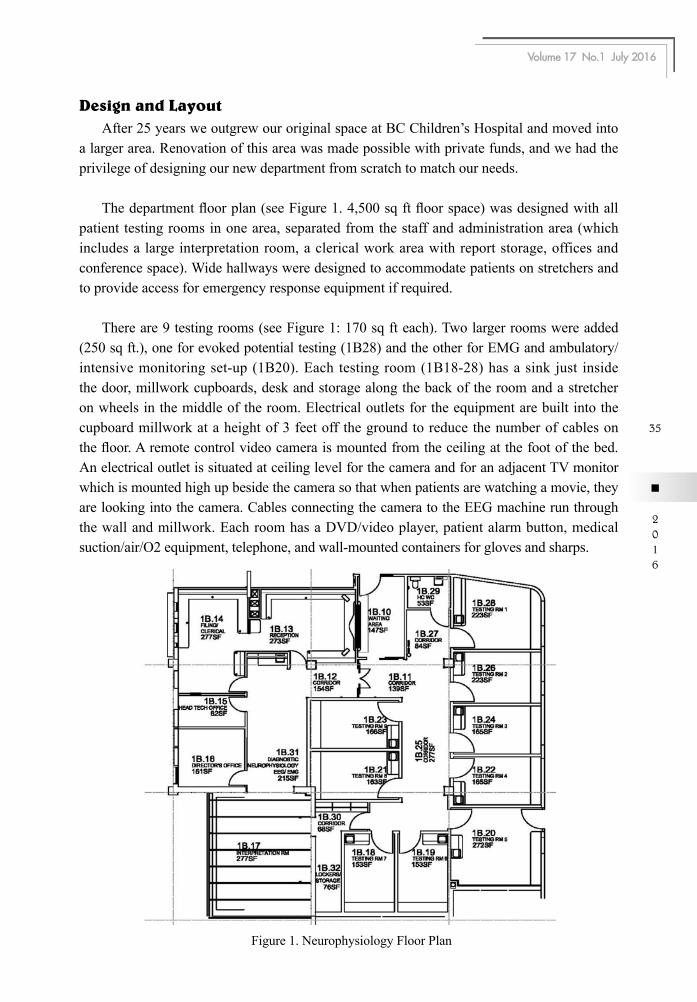
35
n
2016
Volume 17 No.1 July 2016
Design and LayoutAfter 25 years we outgrew our original space at BC Children’s Hospital and moved into
a larger area. Renovation of this area was made possible with private funds, and we had the privilege of designing our new department from scratch to match our needs.
Thedepartmentfloorplan(seeFigure1.4,500sqftfloorspace)wasdesignedwithallpatient testing rooms in one area, separated from the staff and administration area (which includes a large interpretation room, a clerical work area with report storage, offices and conference space). Wide hallways were designed to accommodate patients on stretchers and to provide access for emergency response equipment if required.
There are 9 testing rooms (see Figure 1: 170 sq ft each). Two larger rooms were added (250 sq ft.), one for evoked potential testing (1B28) and the other for EMG and ambulatory/intensive monitoring set-up (1B20). Each testing room (1B18-28) has a sink just inside the door, millwork cupboards, desk and storage along the back of the room and a stretcher on wheels in the middle of the room. Electrical outlets for the equipment are built into the cupboard millwork at a height of 3 feet off the ground to reduce the number of cables on thefloor.Aremotecontrolvideocameraismountedfromtheceilingatthefootofthebed.An electrical outlet is situated at ceiling level for the camera and for an adjacent TV monitor which is mounted high up beside the camera so that when patients are watching a movie, they are looking into the camera. Cables connecting the camera to the EEG machine run through the wall and millwork. Each room has a DVD/video player, patient alarm button, medical suction/air/O2 equipment, telephone, and wall-mounted containers for gloves and sharps.
Figure 1. Neurophysiology Floor Plan
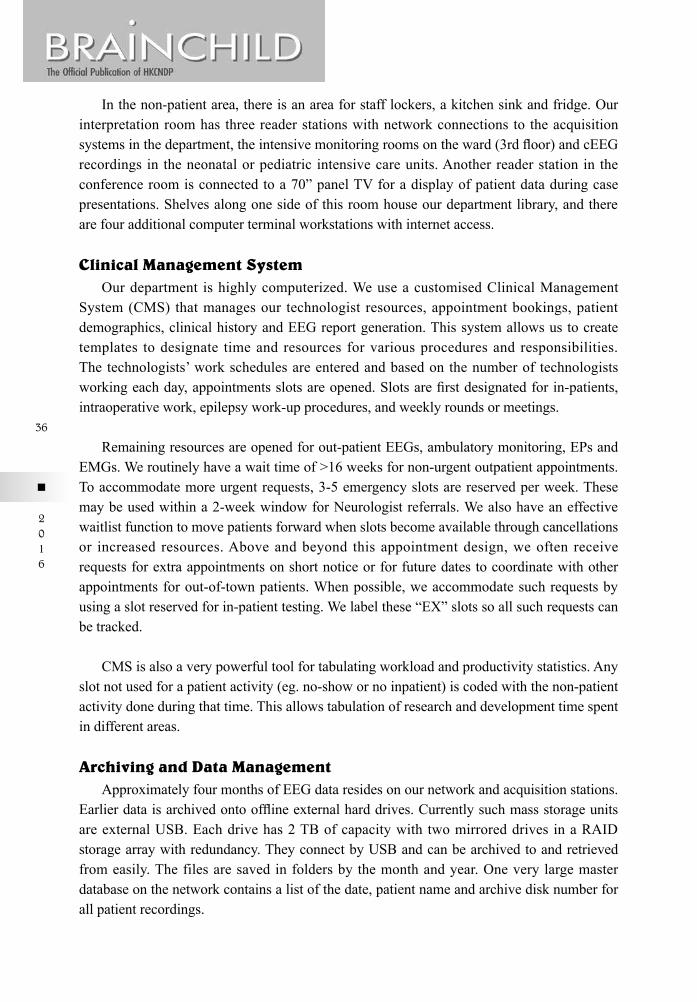
36
n
2016
In the non-patient area, there is an area for staff lockers, a kitchen sink and fridge. Our interpretation room has three reader stations with network connections to the acquisition systemsinthedepartment,theintensivemonitoringroomsontheward(3rdfloor)andcEEGrecordings in the neonatal or pediatric intensive care units. Another reader station in the conference room is connected to a 70” panel TV for a display of patient data during case presentations. Shelves along one side of this room house our department library, and there are four additional computer terminal workstations with internet access.
Clinical Management SystemOur department is highly computerized. We use a customised Clinical Management
System (CMS) that manages our technologist resources, appointment bookings, patient demographics, clinical history and EEG report generation. This system allows us to create templates to designate time and resources for various procedures and responsibilities. The technologists’ work schedules are entered and based on the number of technologists workingeachday,appointmentsslotsareopened.Slotsarefirstdesignatedforin-patients,intraoperative work, epilepsy work-up procedures, and weekly rounds or meetings.
Remaining resources are opened for out-patient EEGs, ambulatory monitoring, EPs and EMGs.Weroutinelyhaveawaittimeof>16weeksfornon-urgentoutpatientappointments.To accommodate more urgent requests, 3-5 emergency slots are reserved per week. These may be used within a 2-week window for Neurologist referrals. We also have an effective waitlist function to move patients forward when slots become available through cancellations or increased resources. Above and beyond this appointment design, we often receive requests for extra appointments on short notice or for future dates to coordinate with other appointments for out-of-town patients. When possible, we accommodate such requests by usingaslotreservedforin-patienttesting.Welabelthese“EX”slotssoallsuchrequestscanbe tracked.
CMS is also a very powerful tool for tabulating workload and productivity statistics. Any slot not used for a patient activity (eg. no-show or no inpatient) is coded with the non-patient activity done during that time. This allows tabulation of research and development time spent in different areas.
Archiving and Data ManagementApproximately four months of EEG data resides on our network and acquisition stations.
Earlierdataisarchivedontoofflineexternalharddrives.Currentlysuchmassstorageunitsare external USB. Each drive has 2 TB of capacity with two mirrored drives in a RAID storage array with redundancy. They connect by USB and can be archived to and retrieved from easily. The files are saved in folders by the month and year. One very large master database on the network contains a list of the date, patient name and archive disk number for all patient recordings.
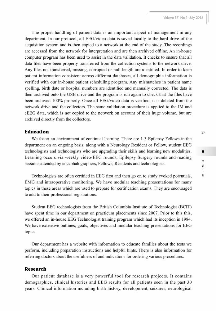
37
n
2016
Volume 17 No.1 July 2016
The proper handling of patient data is an important aspect of management in any department. In our protocol, all EEG/video data is saved locally to the hard drive of the acquisition system and is then copied to a network at the end of the study. The recordings areaccessedfromthenetworkforinterpretationandarethenarchivedoffline.Anin-housecomputer program has been used to assist in the data validation. It checks to ensure that all datafileshavebeenproperlytransferredfromthecollectionsystemstothenetworkdrive.Anyfilesnottransferred,missing,corruptedornull-lengthareidentified.Inordertokeeppatient information consistent across different databases, all demographic information is verified with our in-house patient scheduling program. Any mismatches in patient name spelling,birthdateorhospitalnumbersareidentifiedandmanuallycorrected.ThedataisthenarchivedontotheUSBdriveandtheprogramisrunagaintocheckthatthefileshavebeen archived 100% properly. Once all EEG/video data is verified, it is deleted from the network drive and the collectors. The same validation procedure is applied to the IM and cEEG data, which is not copied to the network on account of their huge volume, but are archived directly from the collectors.
EducationWe foster an environment of continual learning. There are 1-3 Epilepsy Fellows in the
department on an ongoing basis, along with a Neurology Resident or Fellow, student EEG technologists and technologists who are upgrading their skills and learning new modalities. Learning occurs via weekly video-EEG rounds, Epilepsy Surgery rounds and reading sessions attended by encephalographers, Fellows, Residents and technologists.
TechnologistsareoftencertifiedinEEGfirstandthengoontostudyevokedpotentials,EMG and intraoperative monitoring. We have modular teaching presentations for many topicsintheseareaswhichareusedtoprepareforcertificationexams.Theyareencouragedto add to their professional registrations.
Student EEG technologists from the British Columbia Institute of Technologist (BCIT) have spent time in our department on practicum placements since 2007. Prior to this this, we offered an in-house EEG Technologist training program which had its inception in 1984. We have extensive outlines, goals, objectives and modular teaching presentations for EEG topics.
Our department has a website with information to educate families about the tests we perform, including preparation instructions and helpful hints. There is also information for referring doctors about the usefulness of and indications for ordering various procedures.
ResearchOur patient database is a very powerful tool for research projects. It contains
demographics, clinical histories and EEG results for all patients seen in the past 30 years. Clinical information including birth history, development, seizures, neurological
38
n
2016
examination, diagnosis, and radiology results are recorded. The EEG report is summarized viaacodedclassificationsystem.Retrievalofthisinformationiseasyandisavailableforstudies and research papers.
Areas of interest over the years have been Benign Rolandic Epilepsy, centro-temporal discharges and the prognostic value of neonatal EEG. Our database has enabled comparison studies involving hundreds of patients with Rolandic dipole vs. non-dipole discharges and neonatal EEGs with normal vs. abnormal background.
ConclusionThe ability to carry out 26 electrophysiological procedures within one department is quite
a feat, and one that we are most proud of. It requires dedicated, compassionate and skilled staffswhoarequalifiedandknowledgeableinallmodalities.Eachmemberismotivatedtowork hard to obtain high quality results that will assist physicians in offering the best care for each patient.
The essential factors necessary to run a smooth Neurophysiology program are retaining qualified staff (in an era of a world-wide shortage), having time and motivation to keep skills current, and keeping up-to-date with changes in technology. The common challenges that arise are long wait times for non-urgent tests, competition for allocation of resources to programswithconflictingdemands,anddifficultygettingsupportforcontinuingeducation.We will continue to work hard together with other departments to meet the needs of our patients and their families, as well as to keep up with the advances in Neurophysiology.
39
n
2016
Volume 17 No.1 July 2016
Neurodevelopmental Outcomes after Preterm BirthTing JY, MBBS, MPH, MRCPCH, FRCPC, FHKAM (Paediatrics)
Neonatologist, BC Women's Hospital and Health Centre;Clinical Assistant Professor, Department of Pediatrics, University of British Columbia
Synnes AR, MDCM, MHSc, FRCPC
Neonatologist & Director, Neonatal Follow-Up Program, BC Women's Hospital and Health Centre;Clinical Professor, Department of Pediatrics, University of British Columbia
IntroductionAdvances in perinatal medicine and neonatal intensive care have revolutionised outcomes
for neonates in the past thirty years1-7. The widespread use of evidence-based treatments, such as antenatal corticosteroids and surfactant therapy, and other improvements in delivery room resuscitation and nutritional care have resulted in dramatically improved survival, especially among infants born at the threshold of viability1, 3, 4, 6, 7. There have been concerns about whether these new survivors face an increased risk of neurodevelopmental disabilities, includingimpairedcognitiveskills,motordeficits,sensoryimpairment,andbehavioural&psychological problems3, 5, 8, 9. There is increasing evidence of sustained adverse outcomes intoschoolageandadolescence,notonlyforextremely-low-birth-weight(ELBW,≤1000grams) infants but also for infants born at late preterm5, 8, 10. A substantial proportion of “apparently neurologically normal” preterm infants exhibits minor and moderate neuromotor dysfunctions that are not generally detected before school age11. The overall adverse short- andlong-termhealthoutcomesofthesebabiescanimposeaconsiderableburdenonfinitehealthcare resources12.
Neurodevelopmental Outcomes After Preterm Birth – An OverviewPreterm birth is an emerging public health problem worldwide. Its prevalence in Canada
increased from 6.3% of live births in 1981–1983 to 7.7% in 201013, 14; this is lower than the prevalence in the United States, where preterm birth affected 1 of every 9 infants15. The factors cited in connection with this increased prevalence include increased rates of obstetric intervention (i.e. medically indicated labour induction and caesarean delivery) and increases in maternal age and multiple births13. Preterm birth is the leading cause of long-term neurological disabilities in children in the United States15, and it accounts for 45% of children with cerebral palsy16.
Although survival rates for preterm infants have increased in recent decades, motor disabilities (including cerebral palsy) persist, and impairments in cognitive, language, social, and executive functions have not decreased17. The preterm brain develops rapidly during the third trimester, when the volume of the whole brain doubles and the volume of grey matter increases approximately fourfold18. This rapidly growing immature brain is exposed to an extremely complicated and stressful environment outside of the womb19. Preterm
40
n
2016
infants are at high risk for brain injury, which is considered to be a complex amalgam of primary destructive disease and secondary maturational and trophic disturbances20. Their clinical manifestations on follow-up can include cerebral palsy, developmental coordination disorder, visual impairment, hearing impairment, cognitive impairment and neurobehavioural disorders21.
Among the most immature survivors (ELBW or extremely low gestational age neonates, ELGAN), a high prevalence of neurodevelopmental disability has been consistently reported in different cohorts. In the EPICure study from the United Kingdom & Ireland, 308 surviving patients born in 1995 who were less than 25 weeks of gestation were assessed; 30% had developmentaldelay,definedasscoreslessthan2standarddeviations(S.D.)belowthemeanon the Bayley Mental Developmental Index (MDI) and Bayley Psychomotor Developmental Index (PDI), at 30 months of age22. Ten percent of the assessed children had severe neuromotor disability, 7% were blind or perceived light only, and 3% had hearing loss that was uncorrectable or required aids22.
In a large scale study from the United States National Institute of Child Health and Human Development (NICHD) that included 5,250 ELBW infants who were born between 1998and2001,<1%of live-born infantsweighin≤500gsurvivedwithout impairment(definedasaBayleyscore≥85,anormalneurologicexamination,andnormalvision,hearing, and walking) at 18 months; this value increased to almost 24% for infants weighing 901 to 1,000 g at birth23. Female gender, singleton birth, higher birth weight, an absence of neonatal morbidities, private health insurance, and white race increase the likelihood of unimpaired status in this cohort23.
A more recent population-based prospective cohort of consecutive extremely preterm infants born before 27 weeks of gestation was conducted between 2004 and 2007 in Sweden, where universal access to antenatal health care and general health insurance is available to all citizens. In that study, the children in the preterm group were compared with control participants in terms of cognition, language, and motor functions at a median age of 30.5 months using the Bayley-III scales24.Overall,16%hadmoderatedisability(definedasscoresbetween -2 and -3 S.D. from the mean of any of the Bayley-III scales, able to walk with aids, hearing impairment corrected with a hearing aid, and registered at low vision centres without blindness), and 11% had severe disability (Bayley-III composite cognitive, language or motor score < mean – 3 S.D., unable to walk even with an aid, or bilateral blindness or deafness).
In the EPIPAGE study, which included follow-up of 2,901 livebirths between 22 and 32 completed weeks of gestation from nine regions in France in the post-surfactant era, the children underwent a medical examination and a cognitive assessment with the Kaufman assessment battery for children (K-ABC), and their scores on the mental processing composite (MPC) scale were recorded at 5 years of age25. Cerebral palsy was diagnosed
41
n
2016
Volume 17 No.1 July 2016
in 159 (9%) of the children born very preterm; 503 (32%) had an MPC score less than 85, and 182 (12%) had an MPC score less than 7025. The likelihood of having cognitive and neuromotor impairments at 5 years of age increased with decreasing gestational age, and many of the children born very preterm required a high level of specialised care.
A meta-analysis that included 9,653 children with a mean birth weight of 1,060 g and a mean gestational age of 28.2 weeks in 41 studies demonstrated clear evidence of substantial motor impairments in very preterm or very-low-birth-weight (VLBW) children from infancy to 15 years of age26.
Verypretermand/orVLBWchildrenalsohavemoderate-to-severedeficitsinacademicachievement, attention problems, and internalizing behavioural problems and poor executive functions, and continue to lag behind their term-born peers during their transition to young adulthood27. In the follow-up of the EPICURE cohort, 30% attended special schools, and approximately 60% of those attending mainstream schools had special education needs at 11 years of age28. Upon follow-up, these extremely preterm children were more than three times morelikelythantheirclassmatestohaveapsychiatricdisorder,includingattention-deficit/hyperactivity disorder (ADHD), emotional disorders, and autism spectrum disorders29.
Fine and gross motor coordination deficits are not uncommon among survivors born preterm. Developmental coordination disorder, defined as impaired motor performance sufficienttoproducefunctionalimpairmentthatcannotbeotherwiseexplainedbythechild’sage, cognitive ability, or neurologic or psychiatric diagnosis, is found in 31% to 34% of VLBW and 50% of ELBW infants30.
There are many challenges in interpreting neurodevelopmental outcome studies because of their heterogeneous nature, which includes differences in survival and neurodevelopmental impairment rates by geographic region and neonatal network related to multiple factors, including population characteristics, perinatal/neonatal management, follow-up protocols, assessments,andevendefinitions30. The Bayley Scales of Infant Development is not without limitations, despite being the most widely used measure to assess neurodevelopment in very pretermandVLBWinfantsinthefirstthreeyearsoflife.In2006,thenewlyrevisedversion(Bayley-III) was introduced in an attempt to isolate cognitive skills from language skills31. However, the Bayley-III cognitive scores were significantly higher than Bayley II MDI scores for term and preterm toddlers, both combined and separately, which implied a need to exercise caution when using test scores from different versions of a test as the primary outcome measure in research studies31. Moreover, a recent meta-analysis showed a limited predictive value of Bayley Scales of Infant Development scores for the later development of preterm infants, with the MDI explaining 37% of later cognitive functioning and the Bayley motor scale explaining 12% of later motor functioning32. Further research on developmental assessmenttoolswithsufficientsensitivityandspecificitytopredictlatercognitiveandmotordevelopment and guide future interventions is needed32.
42
n
2016
Prediction Of Neurodevelopmental Outcomes – Imaging TechniquesCranial ultrasound (CUS) has been routinely used as the primary imaging tool for
evaluating brain injury in premature neonates since its introduction in 1980s27, 33. It is non-invasive, inexpensive and portable, and it allows the accurate detection of intraventricular haemorrhage (IVH), cystic periventricular white matter injury and hydrocephalus34. In the 2013 Canadian Neonatal Network report, 9.7% of infants with a GA<33 weeks exhibited neuroimaging abnormalities, with 15%, 7% and 3% presenting ventricular enlargement, intraparenchymal lesions and periventricular leukomalacia (PVL), respectively35. Among VLBW infants, 48% developed varying degrees of IVH, ranging from germinal matrix haemorrhage to grade 435. In the past, small haemorrhages (grade 1 or 2 IVH) were believed tonotincreasetheriskofdisabilitysignificantlybeyondthebackgroundriskassociatedwithgestational age and birth weight27. However, current evidence suggests that even a small IVH increases the risk of disability in terms of cerebral palsy or neurodevelopmental impairment, even in infants with only grade 1-2 IVH36, 37.
CUS has a limited ability to accurately diagnose non-cystic white matter injury and other brain parenchymal injuries38. For example, diffuse PVL has been regarded as an important cause of cognitive deficits and chronic neurodevelopmental impairment, but it is difficult to recognise with CUS39, 40. Even serial CUS only has a sensitivity of 26% for identifying white matter abnormalities in premature infants41. Advanced neuroimaging methods, such as magnetic resonance imaging (MRI), magnetic resonance spectroscopy, and diffusion-weightedMRI,haveidentifiedpatternsofdamageafterinsulttotheimmaturebrain42. The braininjurypatternsidentifiedandtheregionalchangesobservedonMRIarepredictiveofparticular neurodevelopmental syndromes later in childhood40, 43.
White matter abnormalities, including parenchymal lesions and punctate hyperintensities, are typically readily identifiable with conventional imaging sequences, including T1- and T2-weighted images, and their developmental effects have been studied extensively34. Parenchymal lesions on term-equivalent MRI, defined as parenchymal haemorrhage, PVL, infarction, and white matter loss, have been reported to have 100% sensitivity and 79% specificityforpredictingcerebralpalsyat18monthsofage44. In another study conducted by Kanagawa Children's Medical Centre in Japan, all 37 patients with cerebral palsy who were evaluated at 3 to 5 years of age had periventricular lesions with T1 hyperintensity or cysts in the corona radiata above the posterior limb of the internal capsule on coronal sections, and there was a tendency for the presence of widespread lesions in this region to be correlated with the severity of the motor handicap45. In a cohort of infants born at <34 weeks gestational age, an abnormal neurodevelopmental outcome at 18 months adjusted age was associated with increasing severity of white matter injury, ventriculomegaly, and IVH on MRI and moderate/severeabnormalitiesonthefirstandsecondMRIstudies,performedatthemedianagesof32and 37 weeks, respectively46.UsingtheGriffithsMentalDevelopmentScalesforassessment,Dyet reported that adverse outcomes at 18 months’ corrected age were associated with major destructive lesions and diffuse excessive high signal intensity within the white matter47. In
43
n
2016
Volume 17 No.1 July 2016
contrast, cerebellar haemorrhages observed on MRI studies did not demonstrate a consistent relationship with adverse neurodevelopmental outcomes across different studies48, 49.
Standardised scoring systems for the results of conventional imaging sequences have been developed to evaluate and quantify regional and/or global injury and provide a comprehensive assessment of cerebral alterations that correlates with neurological outcomes34. Significant white matter abnormality quantified by the standard scoring systems not only correlates with adverse early neurodevelopment outcomes40, 46 but is related to motor and cognitive impairment in childhood50, 51. Newer imaging techniques such as volumetry, surface-based morphometry, diffusion tensor imaging and magnetic resonance spectroscopic imaging provide novel insights into aberrant cerebral development in high-risk preterm infants34, 52. Recent advances in magnetoencephalography analysis presents an excellent opportunity to map neural processes before and after birth in infants at high risk for disabilities, which may carry prognostic values in their neurodevelopmental outcomes53-55.
The Neonatal Follow-Up Programme, Vancouver The Neonatal Follow-Up Program at the British Columbia Children’s & Women’s
Hospital was started in 1983 by Dr. Michael Whitfield to collect audit information about the survival of different categories of high risk infants in the province. The goals of the programme are to evaluate the short and long-term results of perinatal/neonatal intensive care in the Provincial Tertiary Perinatal Unit via sequential clinical and neuro-developmental assessment of surviving infants during infancy and early childhood; to identify impairments early in this high-risk population to promote early intervention that can minimise the severity of perinatally acquired handicaps; to provide educational experience with developmental assessment and the long-term effects of perinatal/neonatal intensive care for learners in perinatal/neonatal care training programmes; and to promote and conduct research to advance knowledge of the long-term effects of select aspects of perinatal and neonatal management.
The programme is multidisciplinary and provides comprehensive, age-appropriate, sequential neurodevelopmental assessments at 4 months, 8 months, 18 months, 3 years, and4½yearscorrectedage[Table1].Theclinicstaff includesnurses,psychologists,physiotherapists, occupational therapists, audiologists, speech-language pathologists, and paediatricians/neonatologists,inadditiontoadatamanagerandofficestaff.
Patients are recruited for follow-up based on perinatal/neonatal risk factors. The programme focuses on children at highest risk of disability or for whom there is little knowledgeaboutpotentialoutcomes.Currentrecruitmentcriteriaincludebirthweight≤800g;gestationalage≤25weeks(forgestationalages26-28weeks,oneassessmentisprovidedat18months[CanadianNeonatalFollow-UpNetwork]); infantswith intraparenchymal/intracranial haemorrhage, cystic PVL, severe retinopathy of prematurity (grade 4 or treated with laser/Avastin), discharge on home oxygen, congenital diaphragmatic hernia and patients participating in funded studies.
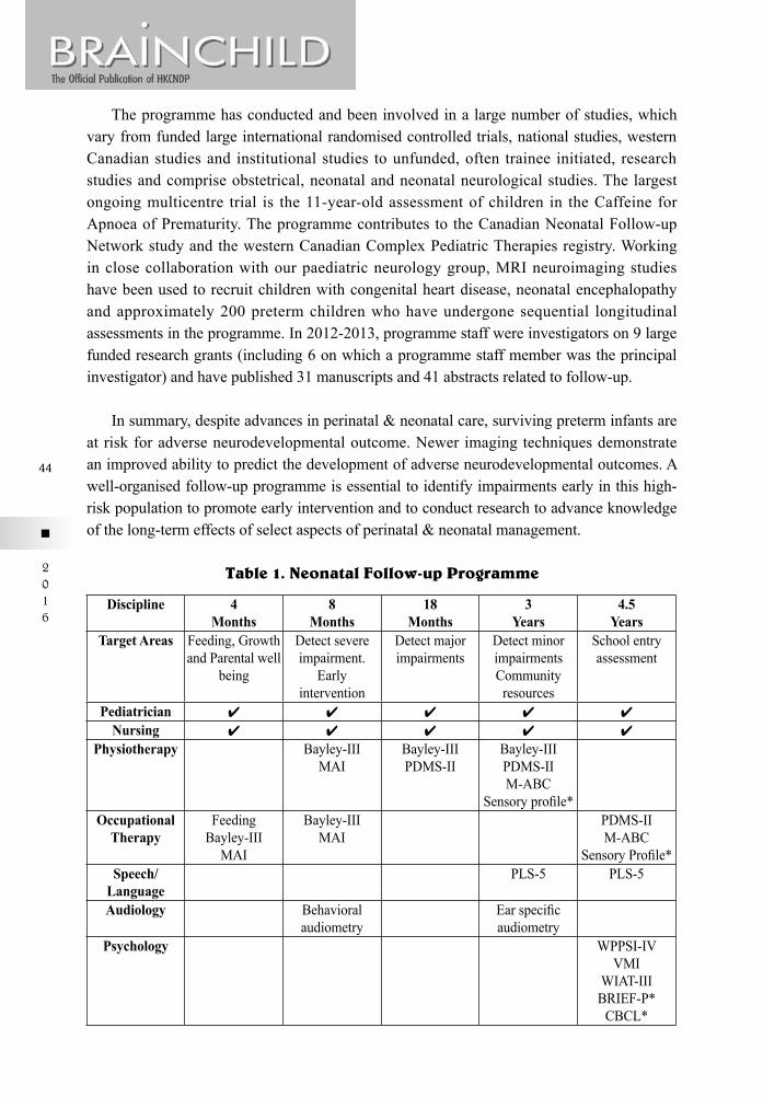
44
n
2016
The programme has conducted and been involved in a large number of studies, which vary from funded large international randomised controlled trials, national studies, western Canadian studies and institutional studies to unfunded, often trainee initiated, research studies and comprise obstetrical, neonatal and neonatal neurological studies. The largest ongoing multicentre trial is the 11-year-old assessment of children in the Caffeine for Apnoea of Prematurity. The programme contributes to the Canadian Neonatal Follow-up Network study and the western Canadian Complex Pediatric Therapies registry. Working in close collaboration with our paediatric neurology group, MRI neuroimaging studies have been used to recruit children with congenital heart disease, neonatal encephalopathy and approximately 200 preterm children who have undergone sequential longitudinal assessments in the programme. In 2012-2013, programme staff were investigators on 9 large funded research grants (including 6 on which a programme staff member was the principal investigator) and have published 31 manuscripts and 41 abstracts related to follow-up.
In summary, despite advances in perinatal & neonatal care, surviving preterm infants are at risk for adverse neurodevelopmental outcome. Newer imaging techniques demonstrate an improved ability to predict the development of adverse neurodevelopmental outcomes. A well-organised follow-up programme is essential to identify impairments early in this high-risk population to promote early intervention and to conduct research to advance knowledge of the long-term effects of select aspects of perinatal & neonatal management.
Discipline 4Months
8Months
18Months
3Years
4.5Years
Target Areas Feeding, Growth and Parental well
being
Detect severe impairment.
Early intervention
Detect major impairments
Detect minor impairmentsCommunity
resources
School entry assessment
Pediatrician 4 4 4 4 4
Nursing 4 4 4 4 4
Physiotherapy Bayley-IIIMAI
Bayley-IIIPDMS-II
Bayley-IIIPDMS-IIM-ABC
Sensoryprofile*Occupational
TherapyFeeding
Bayley-IIIMAI
Bayley-IIIMAI
PDMS-II M-ABC
SensoryProfile*Speech/
LanguagePLS-5 PLS-5
Audiology Behavioral audiometry
Earspecificaudiometry
Psychology WPPSI-IVVMI
WIAT-IIIBRIEF-P*CBCL*
Table 1. Neonatal Follow-up Programme
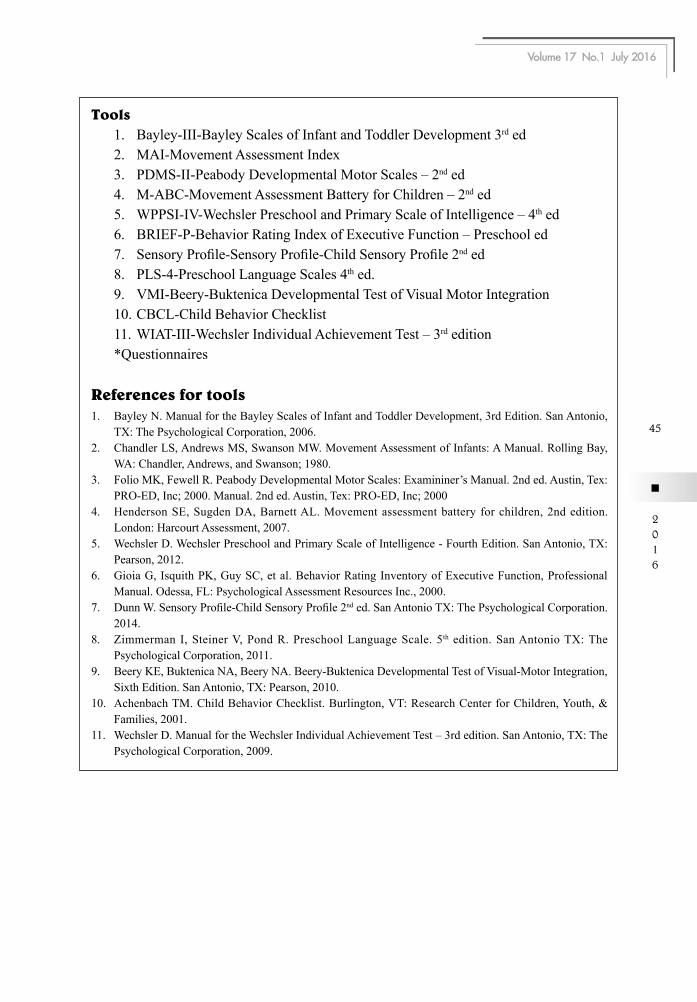
45
n
2016
Volume 17 No.1 July 2016
Tools1. Bayley-III-Bayley Scales of Infant and Toddler Development 3rd ed2. MAI-Movement Assessment Index3. PDMS-II-Peabody Developmental Motor Scales – 2nd ed4. M-ABC-Movement Assessment Battery for Children – 2nd ed5. WPPSI-IV-Wechsler Preschool and Primary Scale of Intelligence – 4th ed6. BRIEF-P-Behavior Rating Index of Executive Function – Preschool ed7. SensoryProfile-SensoryProfile-ChildSensoryProfile2nd ed8. PLS-4-Preschool Language Scales 4th ed.9. VMI-Beery-Buktenica Developmental Test of Visual Motor Integration10. CBCL-Child Behavior Checklist11. WIAT-III-Wechsler Individual Achievement Test – 3rd edition*Questionnaires
References for tools1. Bayley N. Manual for the Bayley Scales of Infant and Toddler Development, 3rd Edition. San Antonio,
TX:ThePsychologicalCorporation,2006.2. Chandler LS, Andrews MS, Swanson MW. Movement Assessment of Infants: A Manual. Rolling Bay,
WA: Chandler, Andrews, and Swanson; 1980.3. Folio MK, Fewell R. Peabody Developmental Motor Scales: Examininer’s Manual. 2nd ed. Austin, Tex:
PRO-ED, Inc; 2000. Manual. 2nd ed. Austin, Tex: PRO-ED, Inc; 20004. Henderson SE, Sugden DA, Barnett AL. Movement assessment battery for children, 2nd edition.
London: Harcourt Assessment, 2007.5. WechslerD.WechslerPreschoolandPrimaryScaleofIntelligence-FourthEdition.SanAntonio,TX:
Pearson, 2012.6. Gioia G, Isquith PK, Guy SC, et al. Behavior Rating Inventory of Executive Function, Professional
Manual. Odessa, FL: Psychological Assessment Resources Inc., 2000.7. DunnW.SensoryProfile-ChildSensoryProfile2nded.SanAntonioTX:ThePsychologicalCorporation.
2014.8. Zimmerman I, Steiner V, Pond R. Preschool Language Scale. 5th edition.SanAntonioTX:The
Psychological Corporation, 2011.9. Beery KE, Buktenica NA, Beery NA. Beery-Buktenica Developmental Test of Visual-Motor Integration,
SixthEdition.SanAntonio,TX:Pearson,2010.10. Achenbach TM. Child Behavior Checklist. Burlington, VT: Research Center for Children, Youth, &
Families, 2001.11. WechslerD.ManualfortheWechslerIndividualAchievementTest–3rdedition.SanAntonio,TX:The
Psychological Corporation, 2009.
46
n
2016
References1. Fanaroff AA, Hack M, Walsh MC. The NICHD neonatal research network: changes in practice and outcomes
duringthefirst15years.SeminPerinatol.2003;27(4):281-7.2. Fanaroff AA, Stoll BJ, Wright LL, Carlo WA, Ehrenkranz RA, Stark AR, et al. Trends in neonatal morbidity
and mortality for very low birthweight infants. Am J Obstet Gynecol. 2007;196 (2):147 e1-8.3. Hintz SR, Kendrick DE, Bohr BR et al. Changes in Neurodevelopmental Outcomes at 18 to 22 Months'
Corrected Age Among Infants of Less Than 25 Weeks' Gestational Age Born in 1993-1999. Pediatrics. 2005;115 (6):1645-51.
4. Lemons JA, Bauer CR, Oh W, Korones SB, Papile LA, Stoll BJ, et al. Very low birth weight outcomes of the National Institute of Child health and human development neonatal research network, January 1995 through December 1996. NICHD Neonatal Research Network. Pediatrics. 2001;107 (1):E1.
5. Wilson-Costello D, Friedman H, Minich N, Siner B, Taylor G, Schluchter M, et al. Improved neurodevelopmental outcomes for extremely low birth weight infants in 2000-2002. Pediatrics. 2007;119 (1):37-45.
6. Ting JY, Wong KY, Goh WHS. Changes in Mortality, Morbidity and Early Neurodevelopmental Outcomes among Extremely-low-birth-weight (ELBW) Infants Born in the Early and Recent Post-Surfactant Era. HK J Paediatr. 2007 (12):78-85.
7. Costeloe KL, Hennessy EM, Haider S, Stacey F, Marlow N, Draper ES. Short term outcomes after extreme preterm birth in England: comparison of two birth cohorts in 1995 and 2006 (the EPICure studies). BMJ. 2012;345:e7976.
8. Vohr BR, Wright LL, Poole WK, McDonald SA. Neurodevelopmental outcomes of extremely low birth weight infants <32 weeks' gestation between 1993 and 1998. Pediatrics. 2005;116 (3):635-43.
9. Laptook AR, O'Shea TM, Shankaran S, Bhaskar B. Adverse neurodevelopmental outcomes among extremely low birth weight infants with a normal head ultrasound: prevalence and antecedents. Pediatrics. 2005;115 (3):673-80.
10. Hack M, Wilson-Costello D, Friedman H et al. Neurodevelopment and Predictors of Outcomes of Children With Birth Weights of Less Than 1000g. Arch Pediatr Adolesc Med. 2000;154:725-31.
11. Fallang B, Oien I, Hellem E, Saugstad OD, Hadders-Algra M. Quality of reaching and postural control in young preterm infants is related to neuromotor outcome at 6 years. Pediatr Res. 2005;58 (2):347-53.
12. Petrou S, Henderson J, Bracewell M, Hockley C, Wolke D, Marlow N, et al. Pushing the boundaries of viability: the economic impact of extreme preterm birth. Early Hum Dev. 2006;82 (2):77-84.
13. Public Health Agency of Canada. Canadian Perinatal Health Report 2008.14. Public Health Agency of Canada. Canadian Perinatal Health Report 2013.15. CentersForDiseaseControlAndPrevention,UnitedStatesofAmerica.ReproductiveHealth[9thAug.,
2015].Availablefrom:http://www.cdc.gov/reproductivehealth/maternalinfanthealth/pretermbirth.htm.16. Allen MC. Neurodevelopmental outcomes of preterm infants. Curr Opin Neurol. 2008;21 (2):123-8.17. Duerden EG, Taylor MJ, Miller SP. Brain development in infants born preterm: looking beyond injury.
Semin Pediatr Neurol. 2013;20 (2):65-74.18. HuppiPS,WarfieldS,KikinisR,BarnesPD,ZientaraGP,JoleszFA,etal.Quantitativemagneticresonance
imaging of brain development in premature and mature newborns. Ann Neurol. 1998;43 (2):224-35.19. Berger I. Mind the gap: the unique neurodevelopment of extremely low birth weight infants. Isr Med Assoc J.
2008;10 (10):718-21.20. Volpe JJ. Brain injury in premature infants: a complex amalgam of destructive and developmental
disturbances. Lancet Neurol. 2009;8 (1):110-24.21. Arpino C, Compagnone E, Montanaro ML, Cacciatore D, De Luca A, Cerulli A, et al. Preterm birth and
neurodevelopmental outcome: a review. Childs Nerv Syst. 2010;26 (9):1139-49.22. Wood NS, Marlow N, Costeloe K, Gibson AT, Wilkinson AR. Neurologic and developmental disability after
extremely preterm birth. EPICure Study Group. N Engl J Med. 2000;343 (6):378-84.23. Gargus RA, Vohr BR, Tyson JE, High P, Higgins RD, Wrage LA, et al. Unimpaired outcomes for extremely
low birth weight infants at 18 to 22 months. Pediatrics. 2009;124 (1):112-21.24. Serenius F, Kallen K, Blennow M, Ewald U, Fellman V, Holmstrom G, et al. Neurodevelopmental outcome
in extremely preterm infants at 2.5 years after active perinatal care in Sweden. JAMA. 2013;309 (17):1810-20.
47
n
2016
Volume 17 No.1 July 2016
25. Larroque B, Ancel PY, Marret S, Marchand L, Andre M, Arnaud C, et al. Neurodevelopmental disabilities and special care of 5-year-old children born before 33 weeks of gestation (the EPIPAGE study): a longitudinal cohort study. Lancet. 2008;371 (9615):813-20.
26. de Kieviet JF, Piek JP, Aarnoudse-Moens CS, Oosterlaan J. Motor development in very preterm and very low-birth-weight children from birth to adolescence: a meta-analysis. JAMA. 2009;302 (20):2235-42.
27. Whitelaw A. Core Concepts: Intraventricular Hemorrhage. NeoReviews. 2011;12 (2):c94-100.28. Johnson S, Hennessy E, Smith R, Trikic R, Wolke D, Marlow N. Academic attainment and special
educational needs in extremely preterm children at 11 years of age: the EPICure study. Arch Dis Child Fetal Neonatal Ed. 2009;94 (4):F283-9.
29. Johnson S, Hollis C, Kochhar P, Hennessy E, Wolke D, Marlow N. Psychiatric disorders in extremely preterm children: longitudinal finding at age 11 years in the EPICure study. J Am Acad Child Adolesc Psychiatry. 2010;49 (5):453-63 e1.
30. Vohr BR. Neurodevelopmental outcomes of extremely preterm infants. Clin Perinatol.41 (1):241-55.31. Lowe JR, Erickson SJ, Schrader R, Duncan AF. Comparison of the Bayley II Mental Developmental Index
and the Bayley III Cognitive Scale: are we measuring the same thing? Acta Paediatr. 2012;101 (2):e55-8.32. Luttikhuizen dos Santos ES, de Kieviet JF, Konigs M, van Elburg RM, Oosterlaan J. Predictive value of the
Bayley scales of infant development on development of very preterm/very low birth weight children: a meta-analysis. Early Hum Dev. 2013;89 (7):487-96.
33. Sheth RD. Trends in incidence and severity of intraventricular hemorrhage. J Child Neurol. 1998;13 (6):261-4.34. Ortinau CM, Inder TE, Smyser CD. Predictive Value of Neonatal Magnetic Resonance Imaging in Preterm
Infants. NeoReviews. 2013;14 (10):c490-9.35. Shah P, Yoon EW, Chan P, Members of the Annual Report Review Committee. The Canadian Neonatal
Network™ Annual Report. The Canadian Neonatal Network™. 2013.36. Patra K, Wilson-Costello D, Taylor HG, Mercuri-Minich N, Hack M. Grades I-II intraventricular hemorrhage
in extremely low birth weight infants: effects on neurodevelopment. J Pediatr. 2006;149 (2):169-73.37. Sherlock RL, Anderson PJ, Doyle LW, Victorian Infant Collaborative Study G. Neurodevelopmental sequelae
of intraventricular haemorrhage at 8 years of age in a regional cohort of ELBW/very preterm infants. Early Hum Dev. 2005;81 (11):909-16.
38. Miller SP, Cozzio CC, Goldstein RB, Ferriero DM, Partridge JC, Vigneron DB, et al. Comparing the diagnosis of white matter injury in premature newborns with serial MR imaging and transfontanel ultrasonographyfindings.AJNRAmJNeuroradiol.2003;24(8):1661-9.
39. MaaloufEF,DugganPJ,CounsellSJ,RutherfordMA,CowanF,AzzopardiD,etal.Comparisonoffindingson cranial ultrasound and magnetic resonance imaging in preterm infants. Pediatrics. 2001;107 (4):719-27.
40. Woodward LJ, Anderson PJ, Austin NC, Howard K, Inder TE. Neonatal MRI to predict neurodevelopmental outcomes in preterm infants. N Engl J Med. 2006;355 (7):685-94.
41. Inder TE, Anderson NJ, Spencer C, Wells S, Volpe JJ. White matter injury in the premature infant: a comparisonbetweenserialcranialsonographicandMRfindingsatterm.AJNRAmJNeuroradiol.2003;24(5):805-9.
42. Ferriero DM. Neonatal brain injury. N Engl J Med. 2004;351 (19):1985-95.43. Barnett A, Mercuri E, Rutherford M, Haataja L, Frisone MF, Henderson S, et al. Neurological and
perceptual-motor outcome at 5 - 6 years of age in children with neonatal encephalopathy: relationship with neonatal brain MRI. Neuropediatrics. 2002;33 (5):242-8.
44. Valkama AM, Paakko EL, Vainionpaa LK, Lanning FP, Ilkko EA, Koivisto ME. Magnetic resonance imaging at term and neuromotor outcome in preterm infants. Acta Paediatr. 2000;89 (3):348-55.
45. Nanba Y, Matsui K, Aida N, Sato Y, Toyoshima K, Kawataki M, et al. Magnetic resonance imaging regional T1 abnormalities at term accurately predict motor outcome in preterm infants. Pediatrics. 2007;120 (1):e10-9.
46. Miller SP, Ferriero DM, Leonard C, Piecuch R, Glidden DV, Partridge JC, et al. Early brain injury in premature newborns detected with magnetic resonance imaging is associated with adverse early neurodevelopmental outcome. J Pediatr. 2005;147 (5):609-16.
47. Dyet LE, Kennea N, Counsell SJ, Maalouf EF, Ajayi-Obe M, Duggan PJ, et al. Natural history of brain lesions in extremely preterm infants studied with serial magnetic resonance imaging from birth and neurodevelopmental assessment. Pediatrics. 2006;118 (2):536-48.
48
n
2016
48. Zayek MM, Benjamin JT, Maertens P, Trimm RF, Lal CV, Eyal FG. Cerebellar hemorrhage: a major morbidity in extremely preterm infants. J Perinatol. 2012;32 (9):699-704.
49. Tam EW, Rosenbluth G, Rogers EE, Ferriero DM, Glidden D, Goldstein RB, et al. Cerebellar hemorrhage on magnetic resonance imaging in preterm newborns associated with abnormal neurologic outcome. J Pediatr. 2011;158 (2):245-50.
50. Spittle AJ, Cheong J, Doyle LW, Roberts G, Lee KJ, Lim J, et al. Neonatal white matter abnormality predicts childhood motor impairment in very preterm children. Dev Med Child Neurol. 2011;53 (11):1000-6.
51. Iwata S, Nakamura T, Hizume E, Kihara H, Takashima S, Matsuishi T, et al. Qualitative brain MRI at term and cognitive outcomes at 9 years after very preterm birth. Pediatrics. 2012;129 (5):e1138-47.
52. Chau V, Synnes A, Grunau RE, Poskitt KJ, Brant R, Miller SP. Abnormal brain maturation in preterm neonates associated with adverse developmental outcomes. Neurology. 2013;81 (24):2082-9.
53. Frye RE, Malmberg B, McLean J, 3rd, Swank P, Smith K, Papanicolaou A, et al. Increased left prefrontal activation during an auditory language task in adolescents born preterm at high risk. Brain Res. 2010;1336:89-97.
54. Doesburg SM, Ribary U, Herdman AT, Miller SP, Poskitt KJ, Moiseev A, et al. Altered long-range alpha-band synchronization during visual short-term memory retention in children born very preterm. Neuroimage. 2011;54 (3):2330-9.
55. Lowery CL, Eswaran H, Murphy P, Preissl H. Fetal magnetoencephalography. Semin Fetal Neonatal Med. 2006;11 (6):430-6.
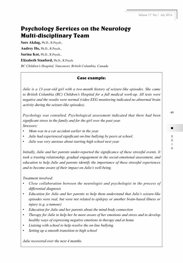
49
n
2016
Volume 17 No.1 July 2016
Psychology Services on the Neurology Multi-disciplinary TeamSare Akdag, Ph.D., R.Psych.,
Audrey Ho, Ph.D., R.Psych.,
Sarina Kot, Ph.D., R.Psych.,
Elizabeth Stanford, Ph.D., R.Psych.
BC Children’s Hospital, Vancouver, British Columbia, Canada
Case example:
Julie is a 13-year-old girl with a two-month history of seizure-like episodes. She came to British Columbia (BC) Children’s Hospital for a full medical work-up. All tests were negative and the results were normal (video EEG monitoring indicated no abnormal brain activity during the seizure-like episodes).
Psychology was consulted. Psychological assessment indicated that there had been significant stress in the family and for the girl over the past year. Stressors:• Mom was in a car accident earlier in the year.• Julie had experienced significant on-line bullying by peers at school. • Julie was very anxious about starting high school next year.
Initially, Julie and her parents under-reported the significance of these stressful events. It took a trusting relationship, gradual engagement in the social-emotional assessment, and education to help Julie and parents identify the importance of these stressful experiences and to become aware of their impact on Julie’s well-being.
Treatment involved:• Close collaboration between the neurologist and psychologist in the process of
differential diagnosis• Education for Julie and her parents to help them understand that Julie’s seizure-like
episodes were real, but were not related to epilepsy or another brain-based illness or injury (e.g. a tumour)
• Education for Julie and her parents about the mind-body connection • Therapy for Julie to help her be more aware of her emotions and stress and to develop
healthy ways of expressing negative emotions in therapy and at home• Liaising with school to help resolve the on-line bullying• Setting up a smooth transition to high school
Julie recovered over the next 4 months.

50
n
2016
The Role of Clinical PsychologyIn the multi-disciplinary Neurology service at BC Children’s Hospital, psychologists are
active members of the team, collaborating with the other health professionals to help with differential diagnosis (as in the case example above), improving coping and compliance with a medical condition and its treatment, and providing support around the social-emotional and behavioural sequelae of neurological conditions. The treatment offered by the psychologist includes education for the child and the family on mind-body connection, common challenges children with neurological conditions may face in every day life, and how to utilise effective coping strategies to manage the stress associated with the illness. The identification of stressors plays an important role in promoting stress management. When the child and parents begin to appreciate the role of stress in symptom management, they are more likely to accept and utilise stress management tools such as relaxation training.
For more information on stress management, please go to: http://www.bcepilepsy.com/files/Information_Sheets/Stress_Management.pdf
Parents play a key role in promoting the social-emotional functioning of their children with neurological problems. The psychologist: • Workswithparentstoeffectivelycopewiththediagnosis • Provides parent education onmanaging symptoms and promoting normal
development (e.g. independence, social relationships, learning) • Educatesparentsonparenting theirchildrenwithneurologicalconditions (e.g.
epilepsy) during developmental transitions (e.g. being not too restrictive when the child grows from childhood to adolescence)
The psychologist also provides support to connect the child to community resources. In addition to connecting the family with counselling in the community, a psychologist may also liaise with school or recreational/social professionals to support the child’s smooth transition back to community-based activities. The goal is to ensure the optimal functioning of the child in the community. Parent and child education on symptom management and planning for potential challenges will empower the child and family to effectively cope and live with the neurological condition. It feels less frightening for the child and family when they know what they can do to cope when pain is high or when the child is having symptoms.
Different psychological approaches are used to assess and treat children with medical issues. A social-emotional assessment is typically used to identify stressors and explore coping styles. Play therapy or cognitive behavioural therapy (CBT) is used to treat the child’s social-emotional challenges and promote coping. Stressors in the family system are addressed with parents or with the whole family. If a child needs hospitalisation, the psychologist works with Child Life specialists to promote the child’s coping with procedures and surgeries during the in-patient stay. The psychologist also works with the school to support the child’s functioning and learning. In the unfortunate situation of a child death, bereavement services are provided.
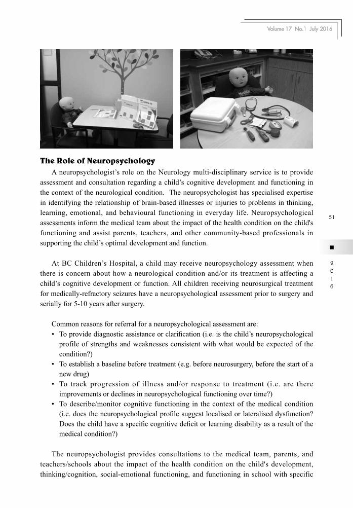
51
n
2016
Volume 17 No.1 July 2016
The Role of NeuropsychologyA neuropsychologist’s role on the Neurology multi-disciplinary service is to provide
assessment and consultation regarding a child’s cognitive development and functioning in the context of the neurological condition. The neuropsychologist has specialised expertise in identifying the relationship of brain-based illnesses or injuries to problems in thinking, learning, emotional, and behavioural functioning in everyday life. Neuropsychological assessments inform the medical team about the impact of the health condition on the child's functioning and assist parents, teachers, and other community-based professionals in supporting the child’s optimal development and function.
At BC Children’s Hospital, a child may receive neuropsychology assessment when there is concern about how a neurological condition and/or its treatment is affecting a child’s cognitive development or function. All children receiving neurosurgical treatment for medically-refractory seizures have a neuropsychological assessment prior to surgery and serially for 5-10 years after surgery.
Common reasons for referral for a neuropsychological assessment are: • Toprovidediagnosticassistanceorclarification(i.e.isthechild’sneuropsychological
profile of strengths and weaknesses consistent with what would be expected of the condition?)
• Toestablishabaselinebeforetreatment(e.g.beforeneurosurgery,beforethestartofanew drug)
• To track progression of illness and/or response to treatment (i.e. are thereimprovements or declines in neuropsychological functioning over time?)
• Todescribe/monitorcognitivefunctioningin thecontextof themedicalcondition(i.e.doestheneuropsychologicalprofilesuggestlocalisedorlateraliseddysfunction?Doesthechildhaveaspecificcognitivedeficitorlearningdisabilityasaresultofthemedical condition?)
The neuropsychologist provides consultations to the medical team, parents, and teachers/schools about the impact of the health condition on the child's development, thinking/cognition, social-emotional functioning, and functioning in school with specific
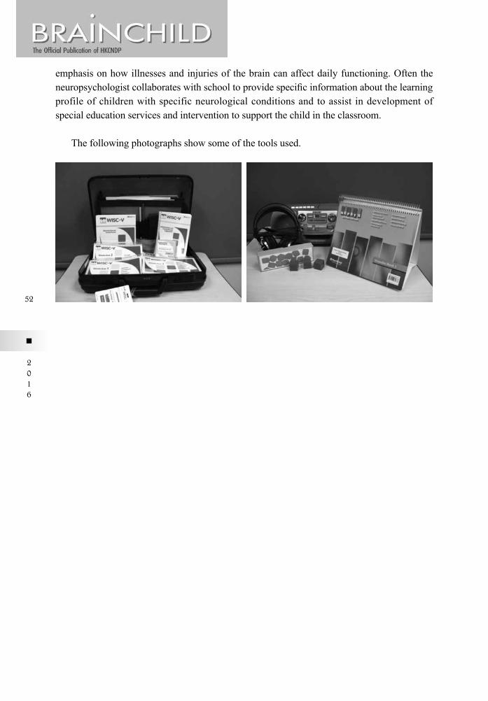
52
n
2016
emphasis on how illnesses and injuries of the brain can affect daily functioning. Often the neuropsychologistcollaborateswithschooltoprovidespecificinformationaboutthelearningprofile of children with specific neurological conditions and to assist in development of special education services and intervention to support the child in the classroom.
The following photographs show some of the tools used.
53
n
2016
Volume 17 No.1 July 2016
The EEG Technologist training programmeLisa Langill, R.E.T., R.T.(EMG), R. EP T., CNIM
Jacqueline Crawford, R.E.T., R. EP T., CNIM
Department of Diagnostic Neurophysiology, BC Children’s Hospital, Vancouver, British Columbia, Canada
British Columbia Children’s Hospital’s (BCCH) first opened its doors in 1982 in the current site. As the only tertiary care centre for paediatrics in the province, the hospital sees children with a wide variety of presenting complaints. At its inception, the Department of Diagnostic Neurophysiology had 5 full-time technologists performing EEGs (electroencephalography), one of whom also performed EMGs (electromyograhy). Currently the department has 11 full-time and 1 part-time multidisciplinary technologists who are certified in EEG, EMG, EPS(evoked potentials) and intraoperative neuromonitoring. Six of those technologists are graduates of the Children’s Hospital training programme. Each member of our department is dedicated and motivated to obtain high quality results that will assist physicians in offering the best care for the patient.
History Of Recruitment For Our Eeg Training ProgrammeIn 1984 the department decided to establish its own 2-year in-house EEG training
programme with the intention of training technologists to take on full-time positions in the department. In this way, students could be hand-picked and trained to meet the specific demands of the department.
Students were initially recruited through the hospital’s volunteer programme newsletter. Applicants included high-school students who were volunteering to get experience in a paediatric medical environment prior to graduating and going into the medical field, and older applicants who had an interest in paediatrics and were looking for a career change. Requirements for application were simple: graduation from high school with a C+/B average. After the first applicants successfully completed the programme and were hired, the recruitment process was expanded to include local high schools. Flyers advertising the programme were circulated around the local high schools at graduation time and when possible, a technologist would present an overview of the programme on career day.
At this time we also expanded our training to include students that had graduated from the 2 year full-time electroneurophysiology diploma programme at the British Columbia Institute of Technology (BCIT). Graduates of this programme require more patient/clinical experience in order to sit their Canadian Board of Registered EEG Technologists (CBRET) finalexamination.Forthosestudentsweofferedamodifiedone-yearprogrammecoveringthe more advanced syllabus that students in the BCCH programme would receive in the second year of their training.
54
n
2016
In more recent years our recruitment and requirements for applicants has changed significantly. Applicants must have a B or greater graduating average from high school with a strong emphasis on science courses. Applicants must also have recently graduated from a degree or diploma programme with a 75% or greater average. Preference was given to graduates in the field of sciences who had work experience (not necessarily related to themedicalfieldorpaediatrics).Recruitmentissolelybasedinthelocaluniversities.Thedisadvantages of this recruitment philosophy are that applicants are older and in most cases living independently from their family. Many have incurred student loans and other debts. Unlike applicants who were straight out of high school and still living with family, these applicantsoftencannotfinanciallyaffordtobeinatwo-yeartrainingprogrammewithonlya minimum wage pay scale. In addition, many have a long-term goal of entering medical school and see our programme as a “stepping stone” towards that goal. These applicants do notfitwithourphilosophyoftrainingstudentswhowishtobeemployedinourdepartmentand who see diagnostic neurophysiology as a long-term professional commitment.
We currently also offer our department as a practicum site for BCIT students who are looking for a department where they can obtain the minimum number of paediatric patients thatarerequiredinordertosittheCBRETfinalexamination.Thesestudentsareherestrictlyto perform EEGs under the guidance of a qualified technologist. If there are not enough suitable patients in a day, and time permits, they may have the chance to review their records with a technologist. Employment in our department is not contingent with this service.
The ProgrammeThe two year programme covers practical training in the set-up and running of an EEG,
the interpretation of EEG data as well as in-depth study of electronics, neuroanatomy and clinical disease processes including, but not limited to, neurological and epileptic disorders. Allpracticaltrainingisdoneunderthedirectsupervisionofaqualifiedtechnologist.Alltestsandlectures[seeTable1]arepreparedandgivenbyaqualifiedtechnologist.
Lectures 1 – 3 Months Lectures 4 – 6 Months10-20 measuring system electronics running a record digital EEG theory patient safety electrodes normal waking and sleep full calibration EEG convention and montages function of lobes artifacts specificseizuretypesandepileptiformwaveformsactivation procedures classificationofepilepsyandbasicepileptic
syndromesnormal variants abnormal non-epileptiform waveformsbasicseizuretypes&non-specificepileptiformwaveforms EEGclassificationsystem
Table 1
55
n
2016
Volume 17 No.1 July 2016
The First YearThefirstyearoftheprogrammeisdividedintofour3-monthblocks.Duringeachofthe
block a student is paired with a senior technologist “teacher”. At the beginning of each block the student receives a set of practical goals that they need to meet. At the end of each block written test(s) is /are given to assess the student’s understanding and competency. Each day of the block is comprised of two EEGs (1.5 hours total time for each) during which practical applications and history taking are the focus. Each EEG is followed by a 1.5 hour session during which EEG interpretation is the main focus. During this time they review the patient’s history,determinewhatEEGfindingswouldbeexpectedinthatclinicalscenarioandwhattechnical factors should be taken into consideration (i.e. activation procedures, additional electrodes, etc.). Lectures and tests are given once a week with tests encompassing the entire syllabus covered up to that time. Towards the end of the first year more multiple choices questions are introduced in preparation for the CBRET written examination.
1-3 months:The objectives during this time are head measurement and electrode application, basic
EEGconceptsandtheidentificationandinterpretationofbasicEEGwaveforms.Lecturesduring this block focus on the International 10-20 system, basic recording strategies/techniquesandpatientsafety,activationprocedures,basicwaveformidentification(normaland abnormal waking and sleep), identification and troubleshooting of both physiological
Lectures 7 - 9 Months Lectures 10 - 12 Monthspharmacology electrical safety brainstem and cranial nerves myoclonus neurological exam generators of EEGpatient history infectious diseases and infection controlbasic anatomy of cerebellum, basal ganglia, pituitary gland, internal capsule and diencephalon
anoxia and coma
skull and meninges ECSventricular system, CSF and hydrocephalus degenerative diseases sleep staging and disorders dementias cerebral circulation metabolic disorders vascular diseases / disorders trauma and head injuries headaches and migraines respiratory systems, apnea and breathholding spells tumours
Lectures 13-24 Monthsneonatal EEG development of the brain and spinal cordspinal cord anatomyauditory and visual systemsneuroimagingepileptic syndromes and status epilepticuschromosomal and congenital anomaliespsychiatryautonomic and limbic systemsperipheral nerve disorders
56
n
2016
and non-physiological artifacts, basic seizure types, basic digital instrumentation, montages and EEG convention.
At the end of this block, the student is expected to be able to measure and apply a set of electrodes using the 10-20 system in one hour or less with fewer than 5 errors (an error is defined as any inter-electrode measurement that is incorrect by more than 5 mm). The student should also be able to correctly identify all the normal waking and sleep waveforms, identifyallthesleepstatesandhaveagoodunderstandingoftheidentificationandcorrectionof artifacts and technical errors (i.e. incorrect inter-electrode distances).
4-6 months:The objectives of this block emphasize hands-on learning including more efficient
electrode application, running an EEG with the assistance of the teacher and practicing waveform identification while running an EEG. Lecture material contains topics on more advanced electronics, more advanced digital instrumentation, electrode properties, neurophysiology,specificseizuretypes/classification,epilepticwaveforms,classificationofepilepsy and basic epileptic syndromes.
At the end of this block the student is expected to measure and apply a full-set of 10-20 electrodes on a co-operative patient in less than one hour with no more than 3 errors. In addition, the student should be able to independently run a normal EEG recording or one with only very minor, non-complicated abnormalities.
7-9 months:This block focuses on the student’s ability to run EEGs on older, co-operative patients
independently. At this time the skills necessary to perform EEGs on younger children (including measuring and applying electrodes on patients while they are lying down and moving) are introduced. Lectures during this block have a stronger emphasis on clinical theory. Topics include taking a detailed clinical history, neuroanatomy, basic neurological disorders and their effects on the EEG, the neurological examination, sleep staging and disorders.
At the end of this block the student is expected to be proficient in taking a detailed clinical history and measuring and applying a full set of 10-20 electrodes in less than 45 minutes on a co-operative older patient with no more than 3 errors. They should also be able to independently record the EEG and remove the electrodes. The objective is to accomplish all of this in 90 minutes or less.
10-12 months:The objectives for the final three months incorporate portable EEG recordings and
dealing with more critically ill patients. Lectures cover a wide range of subjects including electrical safety, infection control, complex neurological disorders and their EEG presentation, respiratory systems and abnormalities arising from dysfunction. The guidelines for electrocerebral silence (ECS) recordings and the recognition of artifacts in those situations is covered in detail.
57
n
2016
Volume 17 No.1 July 2016
Attheendofthisblockthestudentisexpectedtobeproficientintakingahistoryonanytypeofpatientandshouldbeproficientinmeasuringandapplyingelectrodesinlessthan30 minutes with no more than one error. Additionally, they should be able to routinely take a complete history, measure and apply the electrodes and run the recording (including all of the appropriate activation procedures and the appropriate wait for sleep should it not be obtainable) in 90 minutes or less on any patient.
The Second YearThis year focuses on preparation for the CBRET practical examination. CBRET requires
that students must have trained for a minimum of 24 months under the direct supervision of a qualified technologist. Candidates are required to perform and document 500 EEGs in patients from a wide age spectrum. In a training programme such as ours, based in a paediatric facility, 400 of those EEGs can be performed on children (age 12 years or less), 100 must be recorded on adults (21 years or older) with 20 of those being greater than 60 years of age.
Duringthesecondyearthestudentcontinuestoworkunderthesupervisionofaqualifiedtechnologist, however they perform EEGs independently. Prior to completing a patient and removing the electrodes the record is reviewed by a senior technologist. If all of the technical requirements have been met and the appropriate states captured the student may then remove electrodes and clean up the patient. The student performs 3 EEGs a day with time set aside at the end of the day to review the records with a technologist. During this review time emphasis is placed on oral examination techniques, correlation of the presenting history with theEEGfindingsandrecognitionanddefinitionofbothnormalandabnormalfindings.
Lecture topics during the second year are more advanced and include neonatal EEG, development of the brain and spinal cord and in-depth neuroanatomy. Peripheral nerve disorders, neuroimaging, psychiatry, status epilepticus and epileptic syndromes are also covered. Evoked potential basics are introduced in the last 3 months of training.
Attheendofthe2-yearprogramme,thecandidatewillbeindependentandconfidentinthe role of neurophysiology technologist.
ConclusionTherearemanychallengestoruna2-yearintensivetrainingprogram.Thefirstistogain
financialapprovalfromthehospitaltohireastudent.Anotherfinancialconsiderationisthatthe full-time employee capacity is reduced due to one senior technologist being dedicated to thetrainingprogramme.Althoughtherearedifficultiesinrecruitingastudent,onceyouhavefoundanidealcandidateandtheyhavecompletedthecourseyouhavethebenefitofafullytrained, compassionate and knowledgeable neurophysiology technologist, ready to take an activeroleinthefieldofneurophysiology.
58
n
2016


