Embed Size (px)
Citation preview

Kusmic et al. Journal of Translational Medicine 2014, 12:89http://www.translational-medicine.com/content/12/1/89
RESEARCH Open Access
Up-regulation of heme oxygenase-1 after infarctinitiation reduces mortality, infarct size and leftventricular remodeling: experimental evidenceand proof of conceptClaudia Kusmic1*, Cristina Barsanti2, Marco Matteucci2, Nicoletta Vesentini1, Gualtiero Pelosi1, Nader G Abraham3
and Antonio L’Abbate1,2
Abstract
Background: Up-regulation of HO-1 by genetic manipulation or pharmacological pre-treatment has been reportedto provide benefits in several animal models of myocardial infarction (MI). However, its efficacy following MIinitiation (as in clinical reality) remains to be tested. Therefore, this study investigated whether HO-1over-expression, by cobalt protoporphyrin (CoPP) administered after LAD ligation, is still able to improvefunctional and structural changes in left ventricle (LV) in a rat model of 4-week MI.
Methods: A total of 144 adult male Wistar rats were subjected to either left anterior coronary artery ligation orsham-operation. The effect of CoPP treatment (5 mg/kg i.p. at the end of the surgical session and, then, once aweek for 4 weeks) was evaluated on the basis of survival, electro- and echocardiography, plasma levels of B-typenatriuretic peptide (BNP), endothelin-1 and prostaglandin E2, coronary microvascular reactivity, MI size, LV wallthickness and vascularity. Besides, the expression of HO-1 and connexin-43 in different LV territories was assessedby western blot analysis and immunohistochemistry, respectively.
Results: CoPP induced an increased expression of HO-1 protein with >16 h delay. CoPP treatment significantlyreduced mortality, MI size, BNP concentration, ECG alterations, LV dysfunction, microvascular constriction, capillaryrarefaction and restored connexin-43 expression as compared to untreated MI. These functional and structuralchanges were paralleled by increased HO-1 expression in all LV territories. HO activity inhibition bytin-mesoporphyrin abolished the differences between CoPP-treated and untreated MI animals.
Conclusions: This is the first report demonstrating the putative role of pharmacological induction of HO-1 followingcoronary occlusion to benefit infarcted and remote territories, leading to better cardiac function in a 4-week MIoutcome.
Keywords: Myocardial infarction, Coronary microvascular reactivity, Left ventricular vascularity, Ventricularremodeling, Connexin-43, Cobalt protoporphyrin IX, Tin mesoporphyrin
* Correspondence: [email protected] Institute of Clinical Physiology, Via G Moruzzi 1, 56124 Pisa, ItalyFull list of author information is available at the end of the article
© 2014 Kusmic et al.; licensee BioMed Central Ltd. This is an Open Access article distributed under the terms of the CreativeCommons Attribution License (http://creativecommons.org/licenses/by/2.0), which permits unrestricted use, distribution, andreproduction in any medium, provided the original work is properly credited.

Kusmic et al. Journal of Translational Medicine 2014, 12:89 Page 2 of 13http://www.translational-medicine.com/content/12/1/89
BackgroundMyocardial infarction (MI) is a life-threatening dynamicprocess initiating with coronary occlusion and frequentlyprogressing towards chronic heart failure in survivors. Inrecent years much research has been devoted to revascu-larization of ischemic territory by both pharmacologicaland mechanical interventions in the very acute phase, inorder to minimize ischemic necrosis and infarct size andreduce early mortality. However, beyond the initial phase,the final outcome of MI is conditioned by a long series ofbiological processes, which on one hand modulates lossand regeneration of myocardial tissue in the infarcted ter-ritory, and on the other remodels the remote, viable myo-cardium with a relevant progressive loss of function andfailure. Noticeably, the unfavorable outcome of MI isprimarily related to the activation of the neuro-humoralsystem but only partially to infarct size. The well-documented beneficial effects of long-term treatment withbeta-blockers, diuretics, ace-inhibitors and angiotensin re-ceptor blockers support this view [1-3]. Moreover, amongbiological factors intervening in structural and functionalmodifications that characterize the infarcted heart, inflam-mation and oxidation play a prominent role during the en-tire time course of MI, from early necrosis to heart failure[4-7]. In this study we have focused on the potential use ofpharmacological intervention too late to save irreversiblyinjured ischemic myocardium, but timed to limit add-itional myocardial loss due to inflammatory, oxidative andapoptotic processes [8,9], accelerate the repair process andreduce ventricular remodeling.The heme oxygenase-1 (HO-1) gene has cytoprotective
properties mediated by its anti-oxidative, anti-inflammatoryand anti-apoptotic effects. In animal models of MI, HO-1over-expression in either transgenic [10-12] or transfectedrodents [13-15] or even pre-treatment with cobalt proto-porphyrin IX (CoPP) [16], a powerful and widely usedinducer of HO-1 expression [17,18], reduced infarct sizeas well as ventricular remodeling, enhanced endothelialfunction, promoted neoangiogenesis and restored cardiacmetabolism. Thus, it is well established that HO-1 over-expression, when present at the time of coronary occlusion,is able to minimize myocardial damage and improve out-come through a series of cytoprotective activities and rep-arative processes involving formation of new vascularstructures and myocytes [16]. Unfortunately, the above ex-perimental conditions are far from the clinical reality,which allows treatment only after infarct initiation. To thebest of our knowledge the only documentation of the bene-ficial effect of post-MI HO-1 over-expression comes fromLin and coworkers [19] who showed that invasive injectionof recombinant AAV bearing HO-1 gene into the border-zone early after induction of MI in mice, promoted neovas-cularization in the ischemic region and significantly limitedleft ventricular (LV) fibrosis and dysfunction at 4 weeks.
Thus, in view of a potential novel pharmacological chal-lenge, the aim of our study was to assess whether adminis-tration of a pharmacological HO-1 activation, whenchances to save ischemic myocardium are greatly reducedor even nil, has any effect on MI outcome. This is a ‘proofof concept’ experimental controlled trial (treated vs un-treated vs sham MI). We hypothesized that when ischemicdamage was already irreversible, HO-1 over-expressionwould be able, by its well-documented anti-oxidant, anti-inflammatory, anti-apoptotic and angiogenetic effects, topositively modulate those post-ischemic phenomena that(beyond the initial ischemic damage) significantly contrib-ute to final infarct size, left ventricular dilation and remod-eling, and progression towards heart failure. In order toinduce HO-1 over-expression we administered CoPP atthe end of the surgical procedure of LAD ligation in therat, following the preliminary documentation that CoPPup-regulates HO-1 protein expression with a delay longerthan 16 h.
MethodsEthics statementAnimals used in this investigation conformed to the re-commendations in the Guide for the Care and Use ofLaboratory Animals published by the US National Insti-tutes of Health (NIH Publication No. 85–23, revised 1996)and the protocol was approved by the Animal Care Com-mittee of the Italian Ministry of Health (Endorsementn.135/2008-B). All surgery was performed under anesthesia,and all efforts were made to minimize suffering.
AnimalsMale Wistar rats were either bred in our local animalhusbandry facility or purchased from Harlan Italy s.r.l(Udine, Italy). Animals were housed in an environmentwith controlled 12 h/12 h light/dark cycle, temperature(21 ± 0.5°C) and relative humidity (55% ± 2%) and fedwith 4 RF 18 standard rat diet for long-term mainten-ance (Mucedola, Milano, Italy). Water was available adlibitum.
Cobalt protoporphyrin administrationCobalt protoporphyrin (CoPP, Frontier Scientific Inc.,Logan, UT, USA), a well-known inducer of HO-1 ex-pression [17,18] was administered via intra-peritonealinjection. We prepared a 5 mg/ml CoPP stock solutionin TRIS buffer (pH 8.0); handling of CoPP solution wasperformed in the dark due to its sensitivity to light. Theinjected dose was 5 mg/kg body weight.In order to preliminarily assess the time course of HO-1
protein synthesis after a single injection of CoPP (5 mg/kg),we monitored cardiac expression of HO-1 protein byWestern blot analysis in 10 control rats sacrificed at dif-ferent times (0, 8, 16, 24 and 48 h) following CoPP

Kusmic et al. Journal of Translational Medicine 2014, 12:89 Page 3 of 13http://www.translational-medicine.com/content/12/1/89
administration (n = 2 for each time point) (see details inAdditional file 1: S1). The expression of HO-1 proteinhad increased by 16 h and continued to increasethrough 48 h (see Additional file 1: Figure S1A). Inaddition, to exclude that the rise of HO activity in theheart forerun the increase in HO-1 abundance (16 h),we also measured the HO activity in myocardial tissue(Additional file 1: Figure S1B) at 0 and 8 hours follow-ing CoPP administration (n = 3 for each time point). Noincrease in HO activity was observed at 8 h.
Tin mesoporphyrin administrationTin mesoporphyrin IX (SnMP, Rockfeller University,New York, NY, USA), an inducer of HO protein synthe-sis [20], is considered a potent inhibitor of the activity ofboth preformed and newly synthesized enzyme [21,22].SnMP was administered via intra-peritoneal injection ata dose of 8 mg/kg body weight.
Induction of myocardial infarctionA total of 144 male Wistar rats 10–12 weeks old andweighing 310 ± 3 g were used in the study. After thoracot-omy, MI was induced by ligation of the left anterior de-scending coronary artery (LAD) (see details in Additionalfile 1: S2). Immediately after chest closure rats received anintra-peritoneal injection of either vehicle alone (TRISbuffer, MI group, n = 63) or cobalt protoporphyrin alone(CoPP-treated MI group, n = 40) or CoPP in associationwith SnMP (CoPP + SnMP group, n = 8). Due to the > 16h delay of CoPP induced HO-1 up-regulation, the adoptedscheme of treatment mimicked the clinical condition of apharmacological intervention well after initiation of MI.Thereafter, in order to sustain HO-1 over-expression, add-itional doses of CoPP or vehicle were administered i.p.once a week over a 4-week study period. Sham-operatedrats underwent all surgical procedures except LADligation and received the same intra-peritoneal treatmentwith vehicle (Sham group, n = 30). In the CoPP + SnMPgroup, SnMP was administered i.p. every second day [23].The number and times of spontaneous deaths during
the 4 weeks were carefully recorded. Out of 144 animals,three died during surgery due to irreversible ventricularfibrillation and were not allocated to any group. Thus theremaining 141 are included in the study. At 4 weeks, atotal of 117 rats survived and entered the morpho-functional study. Allocation of animals to different groupsand procedures is detailed in the Additional file 1: S3 andAdditional file 1: Table S1.
Electrocardiographic studyECG was recorded at 2 kHz sampling rate and heartrate was calculated using the Power Lab monitoring sys-tem (ML135 PowerLab/8SP) equipped with ML135Dual Bio Amp and MLA0112 ECG Lead Switch Box
(ADI Instruments Ltd., Oxford, UK). ECG recordings werecontinuously acquired before (basal condition) and duringsurgery up to 60 min. Arrhythmias were classified accord-ing to the Lambeth Conventions [24] and their severitywas scored (range 0–5) [25] as detailed in the Additionalfile 1: S4. Moreover, an extensive ECG analysis was carriedout at 4 weeks after infarction as previously described[26]. The ECG parameters studied were: heart rate, frontalQRS axis (ÂQRS) using D1 and AVF leads, and QRS ampli-tude index (IQRS), i.e., the sum of positive or negative peakdeflections of the QRS complexes in D1, D2 and D3, andQRS duration (TQRS). ECG at 4 weeks was compared tothe basal one.
Echocardiographic studyEchocardiographic studies were performed 4 weeks afterinfarction with a portable ultrasound system (MyLab 25,Esaote SpA, Genova, Italy) equipped with a high fre-quency linear transducer (LA523, 12.5 MHz). Underintra-peritoneal anesthesia, as previously described, im-ages were obtained from the left parasternal view. Ashort-axis 2-dimensional view of the left ventricle (LV)was taken at the level of papillary muscles to obtain M-mode recording. Anterior (infarcted) and posterior (viable)end-diastolic and end-systolic wall thicknesses, systolicwall thickening, and LV internal dimensions were mea-sured following the American Society of Echocardiographyguidelines. Parameters were the mean of measurements ofthree consecutive cardiac cycles. The global LV systolicfunction was expressed as fractional shortening (FS%).
Plasma determination of BNP, ET-1, and PGE2Plasma samples were assayed to determine the circulatinglevels of B-type natriuretic peptide (BNP), endothelin-1[27,28] and prostaglandin E2 [29], as detailed in theAdditional file 1: S5.
Ex vivo assessment of microvascular reactivityMicrovascular coronary resistance (CR) was evaluated4 weeks after surgery in the isolated beating heart inLangendorff configuration. This procedure has been pre-viously described in detail [30].Briefly, two side-arms in the perfusion line, located close
to the heart inlet, allowed switching between two reser-voirs set at normal (70 mmHg) and low (30 mmHg) pres-sure. Coronary flow was continuously measured with aflowmeter (model T106, Transonic System Inc, Ithaca,NY, USA) and by measurement of effluent volume with acalibrated pipette. Coronary resistance was calculated asinput pressure divided by coronary flow per gram of myo-cardial tissue (mmHg*g*min/ml). As in infarcted heartsthe injection of Evans blue dye into the aortic root did notcolor the infarcted tissue at macroscopic morphometry (inthe Additional file 1: Figure S2), the weight of perfused

Kusmic et al. Journal of Translational Medicine 2014, 12:89 Page 4 of 13http://www.translational-medicine.com/content/12/1/89
myocardium was calculated as the total ventricle weightminus the necrotic myocardium.After an initial 15-min period of stabilization, not in-
cluded in the analysis, CR was measured in two alterna-tive protocols, (i) at the onset and at the end of 65 minof 70 mmHg perfusion pressure (ii) at 20 min of lowperfusion pressure (30 mmHg) and early after reperfu-sion at 70 mmHg (peak hyperemia).
Tissue harvesting and macroscopic morphometryFour weeks after surgery, under deep anesthesia, heartswere arrested in diastole by a lethal KCl injection, and leftventricles were weighted and cut in transversal and paral-lel slices about 2 mm thick. To enhance the contrast be-tween viable and infarcted myocardium, fresh slices wereincubated with triphenyltetrazolium chloride 1% solutionat 37° for 10 min. Slivers were photographed with a digitalcamera and the images processed by dedicated image soft-ware (MIAO, Myocardial Infarcted Area Outline), whichmeasured the value of the infarcted area in radiantsencompassing the pale area and expressed it as a percent-age of the entire left ventricle (360°). The thickness of thecentral infarcted area and of its opposite wall were mea-sured in each animal (see details in Additional file 1: S6).Next, in each slice, cardiac tissue was divided into four
distinct areas, which were frozen and analyzed separ-ately: a) right ventricle wall; b) left ventricle posteriorwall, opposite to LAD territory (remote zone); c) borderregion to LAD area (border zone); d) central zone of theinfarcted area.In addition, the abdomen was incised and four samples
from the right and median lobes of the liver were ex-cised and flash-frozen to determine tissue HO activity.
HO activity measurement in the liverTo define the efficacy of CoPP in inducing HO-1 expres-sion, HO activity was determined in rat liver micro-somes of each MI group by measuring bilirubin aspreviously described [31]. Details of the procedure arereported in the Additional file 1: S7.
HO-1 expressionHeart tissue from different cardiac regions was separatelyhomogenized and blotted on PVDF membrane as detailedin the Additional file 1: S8. The membranes were probedwith rabbit anti-HO-1. Rabbit anti β-actin or rabbit antiGAPDH antibodies were used to probe the reference pro-teins. After incubation with HRP-conjugate secondaryantibody enhanced chemiluminescence detection was per-formed and digital images were acquired for densitometryanalysis of the bands by using “open source” programImage J (National Institute of Health, Bethesda MD, USA).Protein-specific bands were normalized to the proteinloading staining β-actin or GAPDH.
ApoptosisPossible effect of CoPP on cardiac apoptosis was exploredin an especially dedicated additional set of animals (n = 18,6 sham, 6 untreated MI and 6 CoPP-treated MI rats). Ratswere sacrificed at 16 and 24 h, times proved to correspondto the onset-time of HO-1 over-expression (n = 3 for eachtime in each group). Myocardial tissue homogenates frominfarcted, border and remote regions were processed toassay caspase 3 activity (Caspase 3 Colorimetric Assay Kit,AbCam, Cambridge, UK) and to measure BCL-2 levels(custom made rat BCL-W ELISA kit, RayBiotech, Inc,Georgia, USA).
Immunohistochemical analysis (connexin 43 andvascularity)Hearts were arrested in diastole at 4 weeks, ventricles wereweighted and fixed in 10% buffered formalin (see details inthe Additional file 1: S9). Cardiomyocyte junctions wereidentified by immunoperoxidase staining, using a poly-clonal rabbit anti-connexin 43 (Cx43) primary antibody(AbCam, Cambridge, UK); arterioles and capillaries werelabeled with anti-smooth muscle actin (α-SMA) primaryantibody (SantaCruz, Dallas, USA) or anti-PECAM 1(CD31) primary antibody (SantaCruz, Dallas, USA) re-spectively. Sections were counter-stained with hematoxylinand examined using light microscopy (Olympus BX43) at10× to 40× original magnification and digitized by a videosystem (Olympus DP20 camera) interfaced to a computerwith dedicated software (CellSens Dimension, Olympus)for image acquisition and morphometric and/or color ana-lysis. Cx43 was quantified as the percent of myocardial areaoccupied by positive staining (calculated by averaging thevalues from ten fields at 10× magnification) for each LVarea. The density (number/mm2) of arterioles (10–100 μmin diameter) was counted by averaging values from eightfields randomly chosen for each LV area at 20× magnifica-tion. The same was done for capillaries density (<10 μm indiameter), except magnification 40×.
Statistical analysisAll experimental values are expressed as the mean ± stand-ard error of the mean (SEM). On the basis of theKolmogorov-Smirnov test (p > 0.05), the visual inspectionof their histograms, and the normal Q-Q plots we assumedthat our data were approximately normally distributed.Individual means of different groups were compared
by one-way or two-way analysis of variance (ANOVA)followed by post hoc Newman-Keuls tests. To take un-even variances into account, for comparisons betweengroups of unequal numbers, Welch’s correction for theStudent’s t-test was used. Correlation analysis was per-formed using Spearman’s test. Paired or unpaired t-testand chi-square test were used as appropriate. A value ofp < 0.05 was considered statistically significant.

Kusmic et al. Journal of Translational Medicine 2014, 12:89 Page 5 of 13http://www.translational-medicine.com/content/12/1/89
All analyses were done with the statistical computerprogram StatView 5.0 (Abacus Concepts, Berkeley, CA,U.S.A.).
ResultsMortalityAmong the 141 animals who survived the surgical pro-cedure, overall mortality at 4 weeks was 3% in the shamgroup, 25% in the vehicle MI group, 12% in the CoPP-MI group and 25% in the CoPP + SnPP-MI group (in theAdditional file 1: Table S1), with vehicle-MI significantlydifferent from CoPP-treated MI rats (χ2 = 3.89, p =0.048) but not different from CoPP + SnMP MI group(χ2 = 0.167, p = 0.6831). A comparison of vehicle vs CoPPMI mortality at 24 h (early) and at 72 h (intermediatemortality) showed that cumulative mortality was similar inthe two groups at 24 h (6 vs 5%) but had diverged at 72 h(19 vs 7% respectively, χ2 = 6.67, p = 0.0098) supportingthe assumption of a ‘late’ biological effect of our pharma-cological intervention.
ElectrocardiographyIn MI groups severe ventricular arrhythmias occurred 5to 20 min after coronary occlusion. The average severityscore was no different in vehicle when compared toCoPP and CoPP + SnMP MI groups (3.35 ± 0.15, 3.35 ±0.11, and 3.33 ± 0.10 respectively) suggesting the absenceof an apparent anti-arrhythmogenic effect of CoPP inthe first 20 min of MI. At 4 weeks, only sporadic ar-rhythmias were observed, never exceeding a score of 0in all groups.QRS morphology, 4 weeks after surgery, showed a dif-
ference between sham and vehicle MI animals with arightward shift (> 90°) of the frontal QRS axis, reductionin QRS amplitude index, and prolongation of QRS inter-val (Table 1). CoPP treatment significantly decreased therightward shift with 53% of cases of ÂQRS axis < 90°, pre-served QRS amplitude index, partially restored QRS dur-ation. Concurrent SnMP administration cancelled outthe CoPP effect and abolished ECG morphological dif-ferences relative to the vehicle MI group.
EchocardiographyThe main echocardiographic parameters are shown inTable 2. LV geometry changed in vehicle MI as the result
Table 1 In vivo heart functional parameters of rats by ECG at
Sham group MI group
n = 20 n = 35
ÂQRS (cases >90°) 59° ± 2 (0/20) 128° ± 2**# (35/3
IQRS (mV) 4.1 ± 0.2 2.6 ± 0.1**#
TQRS (ms) 14 ± 0.6 19 ± 0.5**#
Values are mean ± SEM; n, number of animals tested; ÂQRS, frontal QRS axis; IQRS, QR#p < 0.001 vs. CoPP-treated infarct.
of a progressive LV remodeling over the 4-week studyperiod post coronary artery ligation. Both LV end-diastolic and end-systolic diameters, as well as HR in-creased in untreated MI compared to sham-operatedanimals. Moreover, anterior (infarcted) LV wall thicknesswas reduced in untreated MI hearts, while the posterior(viable) wall thickness was higher than sham rats andCoPP-MI. In vehicle MI the percentage of systolic thick-ening (an index of contractility) of the anterior and theposterior walls as well as LV fractional shortening wasreduced compared to sham animals. CoPP treatmentsignificantly reduced both end-diastolic and end-systolicdiameters of infarcted hearts, blunted both the increasein heart rate and the thinning of the anterior wall andimproved systolic thickening of both anterior and poster-ior walls as well as the fractional shortening compared tountreated MI. Concurrent SnMP administration reversedthe CoPP effect, abolishing both the LV geometry andfunctional changes relative to vehicle MI group (Table 2).
Plasma levels of BNP, ET-1 and PGE2Plasma BNP, ET-1 and PGE2 concentrations before treat-ment (basal value) were 18 ± 5 pg/mL, 19 ± 0.5 pg/mL,and 8.1 ± 0.5 ng/mL, respectively. Changes in plasmaconcentrations of the three markers 4 weeks after sur-gery in sham-operated as well as in vehicle- or CoPP-treated infarcted rats are shown in Additional file 1:Table S2. Briefly, MI group exhibited an increase in bothBNP (p = 0.06) and ET-1 (p < 0.01) plasma concentra-tion levels. CoPP treatment dampened the post-infarctrise of both parameters to levels akin to those of sham-operated animals. Moreover CoPP caused a significantincrease in PGE2 circulating levels in comparison toboth sham and MI animals.
Effect of CoPP treatment on microvascular coronaryresistance in isolated heartsEx vivo measurements of CR are shown in Table 3. Heartsfrom sham-operated rats perfused at constant pressure ex-hibited only a small increase in CR detectable over the 65-min study period (p < 0.05). In infarcted animals, coronaryresistance values were significantly higher than those inthe sham group (p < 0.001 vs sham-operated). The bolusadministration of papaverine (50 μg), abolished the in-crease in resistance in infarcted rats, proving its active
4 weeks
CoPP-MI group CoPP + SnMP -MI group
n = 25 n = 6
5) 94° ± 5** (12/25) 125° ± 7**# (6/6)
3.6 ± 0.2 2.4 ± 0.2**#
15 ± 0.5 18 ± 1.6**#
S amplitude index; TQRS, QRS duration; **p < 0.001 vs sham-operated;

Table 2 In vivo heart functional parameters of rats by echocardiography at 4 weeks
Heartfunction
Sham group MI group CoPP-MI group CoPP + SnMP MI group
n = 20 n = 35 n = 25 n = 6
LVEDd (mm) 5.5 ± 0.2 7.4 ± 0.4**# 6.3 ± 0.3* 8.5 ± 0.4**#
LVESd (mm) 2.0 ± 0.2 5.0 ± 0.3**# 2.9 ± 0.1 6.0 ± 0.4**#
EDAW (mm) 1.6 ± 0.1 1.1 ± 0.1**# 1.5 ± 0.1 1.1 ± 0.1**#
EDPW (mm) 1.6 ± 0.1 1.9 ± 0.1**# 1.6 ± 0.1 1.9 ± 0.7**#
SAWT% 71 ± 3 35 ± 2**# 60 ± 3* 38 ± 5**#
SPWT% 70 ± 2 41 ± 2**# 69 ± 2 52 ± 4**#
FS% 65 ± 3 34 ± 2**# 54 ± 2* 29 ± 3**#
HR 456 ± 7 495 ± 5**# 443 ± 6 470 ± 9#
Values are mean ± SEM; n, number of animals tested; LVEDd, left ventricular end-diastole diameter; LVESd, left ventricular end-systole diameter; EDAW,end-diastole anterior wall thickness; EDPW, end-diastole posterior (remote) wall thickness; SAWT%, percent systolic anterior wall thickening; SPWT%, percentsystolic posterior (remote) wall thickening; FS%. percent fractional shortening. HR, heart rate. *p < 0.05 and **p < 0.001 vs sham-operated; #p < 0.001 vsCoPP-treated infarct.
Kusmic et al. Journal of Translational Medicine 2014, 12:89 Page 6 of 13http://www.translational-medicine.com/content/12/1/89
nature and ruling out the intervention of extra-vascularcompressive forces.CoPP treatment blunted the increased coronary resist-
ance observed in infarcted rats (p < 0.01 vs untreated MI)(Table 3). Treated animals did not differ from sham-operated ones. In sham-operated hearts, hypotensioncaused an increase in coronary resistance (paradoxicalvasoconstriction, p < 0.01 vs baseline). The return to nor-mal perfusion pressure caused a fall in resistance in the firstminute (hyperemic response) below baseline (p < 0.001 vsbaseline).As shown in Table 3, in untreated MI hearts,
hypotension induced a marked rise in resistance (p <0.01 vs sham and CoPP-MI). The return to normal per-fusion pressure caused a fall in resistance to a valuehigher than baseline (p < 0.05 vs baseline). The values ofCR were higher than those of sham-operated animals(p < 0.001 vs sham-operated rats).CoPP-treated MI rats showed basal CR values similar
to those of the sham group and lower than those of un-treated infarct group (p < 0.01 vs untreated infarct). Dur-ing transient low-pressure ischemia, CoPP treatment in
Table 3 Values of coronary resistance in isolated heart inLangendorff configuration
Sham group MI group CoPP-MI group
Perfusion at constant pressure (70 mmHg)
Baseline 8.8 ± 0.5 13.4 ± 0.5** 9.2 ± 0.5#
65 min 12.5 ± 1.3 32.7 ± 3.3** 17.8 ± 1.8#
Perfusion at low pressure (30 mmHg) and reperfusion
Baseline 9.3 ± 0.6 13.6 ± 0.7** 8.8 ± 0.6#
At 20 min low pressure 15.0 ± 1.6 26.1 ± 2.0* 13.2 ± 1.5#
Reperfusion 8.5 ± 0.5 19.3 ± 0.9** 11.5 ± 0.8
Values are mean ± SEM. *p < 0.01 and **p < 0.001 vs sham-operated;#p < 0.01 vs MI group.
MI rats largely abolished the vasoconstrictive responseto hypotension. At the peak, CR was not significantlydifferent from that of the sham group. Reperfusion pro-duced a drop in CR to a level that was no different fromthat of the sham-operated group.
Macroscopic morphometry and infarct sizeVentricles of MI group had significantly (p < 0.001) higherweights than those of sham-operated animals (Additionalfile 1: Table S3). CoPP treatment decreased (p < 0.001) thepost-infarct gain in ventricle weight.Figure 1A shows MI size evaluated 4 weeks after MI
or sham operation. Sham group showed an insignificantarea of LV damage (< 2%), mainly due to tissue traumaduring sham operation. Four weeks after MI, infarct sizewas 35.9 ± 1.6% of LV (p < 0.001 vs sham-operated).Thickness of the core infarcted wall was about 43% ofthe one in the sham group, whereas opposite wall thick-ness was about 119%, a value significantly (p < 0.05)higher than in sham animals (Additional file 1: TableS3). CoPP treatment significantly reduced MI size to23.2 ± 1.3% of LV (p < 0.002 vs untreated MI). Moreover,it reduced thinning of the core infarcted region (66% ofsham group) and completely prevented the increase inthickness of the opposite region (101% of sham group).SnMP resulted in MI size of 33.6 ± 1.5% of LV after
4 weeks (p < 0.001 vs CoPP-MI); thickness of the coreinfarcted wall was 39% while the opposite wall was 120%compared to the sham group.
Liver HO activityCoPP increased HO activity in the liver of infarcted rats2.1-fold as compared to untreated infarcted rats (24.0 ±5.0 vs 11.6 ± 1.5 pmol bilirubin * min-1 * mg-1, p < 0.05).SnMP-treated animals exhibited minimal HO activity
(0.1 ± 0.08 pmol bilirubin * min-1 * mg-1, p < 0.001 vsboth vehicle and CoPP treated-MI groups).

Figure 1 Histograms of infarct size and HO-1 expression in hearts at 4 weeks after LAD ligation. From left to right: sham operated (n = 6),vehicle-treated MI (n = 16), CoPP-treated MI (n = 14) and CoPP + SnMP-treated MI rats (n = 6). A. Representative freshly-cut transverse sections usedfor determination of infarct size (upper) and quantitative results on infarct size (lower) expressed as mean ± SE percentage of left ventricle. RV:right ventricle; LVP: left ventricular posterior free wall; LVA: left ventricular anterior free wall. B. Representative western blots (upper) andquantitative results from densitometric analysis of HO-1 and β-actin expression (lower) of the corresponding regions shown in the areas outlinedby the black boxes and labeled with letters in the representative freshly-cut transverse section: a) right ventricle wall; b) left ventricle posteriorwall, opposite to LAD territory; c) border region to LAD area and d) central zone of the infarcted area. Histograms are expressed as ratio betweenHO-1 and the comparative protein β-actin.
Kusmic et al. Journal of Translational Medicine 2014, 12:89 Page 7 of 13http://www.translational-medicine.com/content/12/1/89
HO-1 expression in the heartIn sham-operated hearts HO-1 protein levels in differentmyocardial regions were low (Figure 1B). Conversely, inthe untreated MI group 4 weeks after LAD occlusion,HO-1 expression levels were significantly increased inthe central zone of the infarcted area. CoPP treatmentresulted in an increase of HO-1 protein levels in all re-gions compared with both sham and untreated MIgroups (p < 0.001 vs the other groups). Nonetheless inthe CoPP-treated MI group there was a heterogeneousexpression of HO-1 protein in the different myocardialregions with the highest levels in the core of the in-farcted area (p < 0.05 vs border zone and p < 0.001 vsboth LV remote zone and RV). Likewise, HO-1 levels inthe border region were about twofold higher than inboth remote area and right ventricle (p < 0.05). The
relative values of HO-1 expression level in each regionof CoPP-treated MI group were: IZ > BZ > RZ = RV.The addition of SnMP induced a regional pattern and
levels of HO-1 expression in each cardiac region akin tothose of CoPP alone.
Connexin-43Immunohistochemistry showed a discrete pattern ofconnexin-43 staining in LV sections of sham-operatedrats, predominantly localized to the myocyte-myocytejunction corresponding to intercalated discs. As com-pared to sham hearts, quantitative analysis of connexin-43 staining showed a significantly decreased Cx43 inuntreated MI rats both in the remote region (57% ± 8%of sham, p < 0.001 vs sham group) (Figure 2) and in theborder zone (51% ± 10% of sham, p < 0.001 vs sham

Figure 2 Distribution of connexins 43 in the remote region at 4 weeks after LAD ligation. Upper panels: representative immunostainingfor Cx43 from sham-operated (a), vehicle treated MI (b), CoPP-treated MI (c), and CoPP + SnMP-treated MI groups (d) at low magnification(10×, calibration bar: 40 μm) and at higher magnification (400×, insets). Lower panel: histograms of the proportion of the total cell area occupiedby Cx43 immunoreactive signal in the remote region. Data are expressed as means ± SE. *p < 0.001 compared with both sham-operated andCoPP-treated MI groups; #p < 0.02 compared with sham-operated group.
Kusmic et al. Journal of Translational Medicine 2014, 12:89 Page 8 of 13http://www.translational-medicine.com/content/12/1/89
group) (data not shown). CoPP treatment resulted in asignificant conservation of Cx43 expression in the re-mote region (84% ± 8% of sham, p < 0.001 vs MI vehiclegroup) and also in the border areas (75% ± 16% of sham,p < 0.02 vs MI vehicle group).In the SnMP treated group, Cx43 staining was signifi-
cantly lower in both the remote zone (66% of sham, p <
0.02 vs sham) and in the border zone (63% of sham, p <0.05 vs sham), similar to untreated MI hearts.
Arteriolar and capillary densityFigure 3 shows the results of morphometric analysis inuntreated and in CoPP treated animals. In untreated MI,as compared to the corresponding zones in sham hearts,
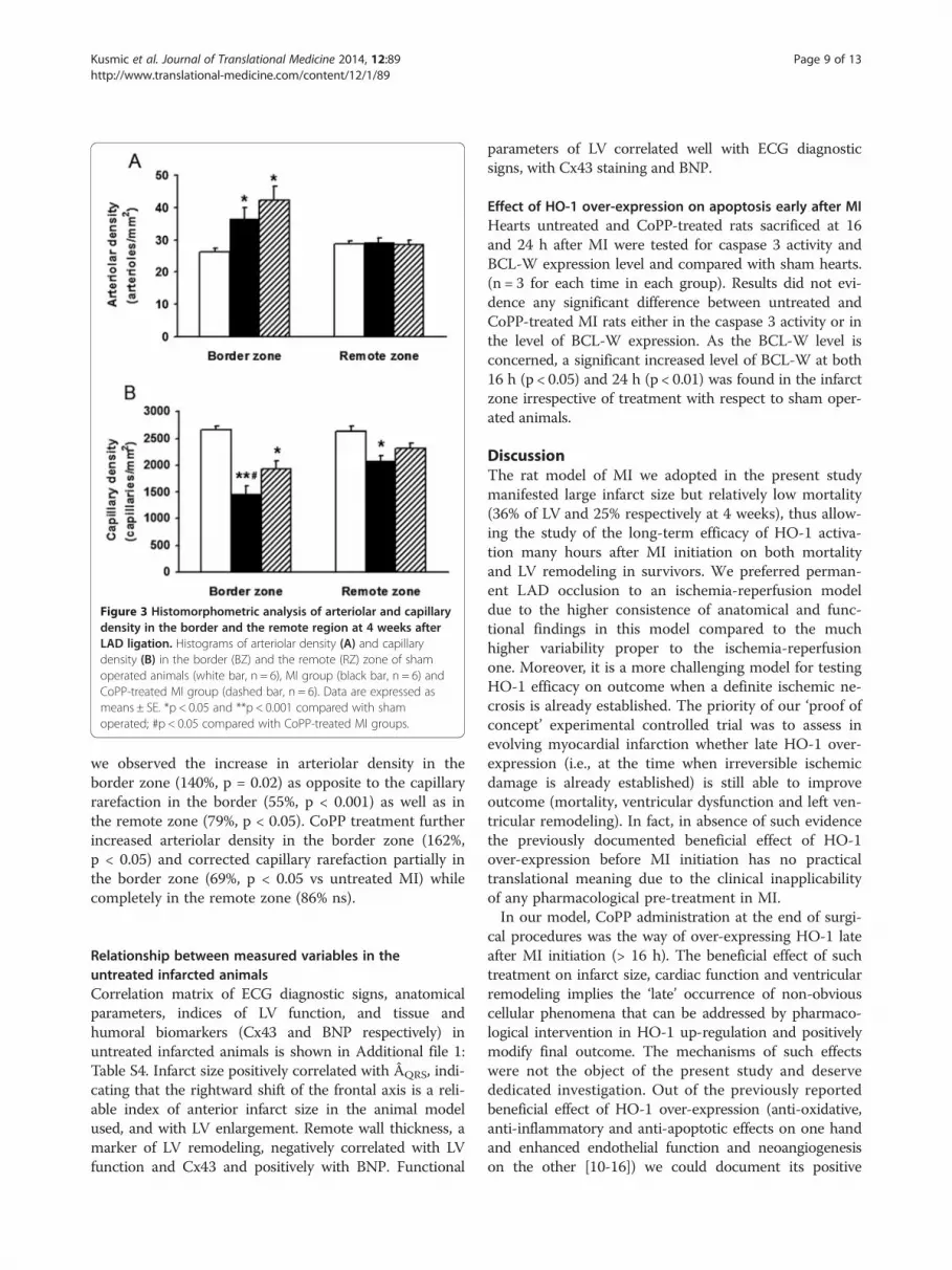
Figure 3 Histomorphometric analysis of arteriolar and capillarydensity in the border and the remote region at 4 weeks afterLAD ligation. Histograms of arteriolar density (A) and capillarydensity (B) in the border (BZ) and the remote (RZ) zone of shamoperated animals (white bar, n = 6), MI group (black bar, n = 6) andCoPP-treated MI group (dashed bar, n = 6). Data are expressed asmeans ± SE. *p < 0.05 and **p < 0.001 compared with shamoperated; #p < 0.05 compared with CoPP-treated MI groups.
Kusmic et al. Journal of Translational Medicine 2014, 12:89 Page 9 of 13http://www.translational-medicine.com/content/12/1/89
we observed the increase in arteriolar density in theborder zone (140%, p = 0.02) as opposite to the capillaryrarefaction in the border (55%, p < 0.001) as well as inthe remote zone (79%, p < 0.05). CoPP treatment furtherincreased arteriolar density in the border zone (162%,p < 0.05) and corrected capillary rarefaction partially inthe border zone (69%, p < 0.05 vs untreated MI) whilecompletely in the remote zone (86% ns).
Relationship between measured variables in theuntreated infarcted animalsCorrelation matrix of ECG diagnostic signs, anatomicalparameters, indices of LV function, and tissue andhumoral biomarkers (Cx43 and BNP respectively) inuntreated infarcted animals is shown in Additional file 1:Table S4. Infarct size positively correlated with ÂQRS, indi-cating that the rightward shift of the frontal axis is a reli-able index of anterior infarct size in the animal modelused, and with LV enlargement. Remote wall thickness, amarker of LV remodeling, negatively correlated with LVfunction and Cx43 and positively with BNP. Functional
parameters of LV correlated well with ECG diagnosticsigns, with Cx43 staining and BNP.
Effect of HO-1 over-expression on apoptosis early after MIHearts untreated and CoPP-treated rats sacrificed at 16and 24 h after MI were tested for caspase 3 activity andBCL-W expression level and compared with sham hearts.(n = 3 for each time in each group). Results did not evi-dence any significant difference between untreated andCoPP-treated MI rats either in the caspase 3 activity or inthe level of BCL-W expression. As the BCL-W level isconcerned, a significant increased level of BCL-W at both16 h (p < 0.05) and 24 h (p < 0.01) was found in the infarctzone irrespective of treatment with respect to sham oper-ated animals.
DiscussionThe rat model of MI we adopted in the present studymanifested large infarct size but relatively low mortality(36% of LV and 25% respectively at 4 weeks), thus allow-ing the study of the long-term efficacy of HO-1 activa-tion many hours after MI initiation on both mortalityand LV remodeling in survivors. We preferred perman-ent LAD occlusion to an ischemia-reperfusion modeldue to the higher consistence of anatomical and func-tional findings in this model compared to the muchhigher variability proper to the ischemia-reperfusionone. Moreover, it is a more challenging model for testingHO-1 efficacy on outcome when a definite ischemic ne-crosis is already established. The priority of our ‘proof ofconcept’ experimental controlled trial was to assess inevolving myocardial infarction whether late HO-1 over-expression (i.e., at the time when irreversible ischemicdamage is already established) is still able to improveoutcome (mortality, ventricular dysfunction and left ven-tricular remodeling). In fact, in absence of such evidencethe previously documented beneficial effect of HO-1over-expression before MI initiation has no practicaltranslational meaning due to the clinical inapplicabilityof any pharmacological pre-treatment in MI.In our model, CoPP administration at the end of surgi-
cal procedures was the way of over-expressing HO-1 lateafter MI initiation (> 16 h). The beneficial effect of suchtreatment on infarct size, cardiac function and ventricularremodeling implies the ‘late’ occurrence of non-obviouscellular phenomena that can be addressed by pharmaco-logical intervention in HO-1 up-regulation and positivelymodify final outcome. The mechanisms of such effectswere not the object of the present study and deservededicated investigation. Out of the previously reportedbeneficial effect of HO-1 over-expression (anti-oxidative,anti-inflammatory and anti-apoptotic effects on one handand enhanced endothelial function and neoangiogenesison the other [10-16]) we could document its positive

Kusmic et al. Journal of Translational Medicine 2014, 12:89 Page 10 of 13http://www.translational-medicine.com/content/12/1/89
effect on border zone arteriolar density and capillarity inboth border and remote zones. Conversely, we were un-able to document any effect of HO-1 activation on early(16 and 24 h) cardiac apoptosis. As the time course ofapoptosis following acute MI is still unknown, different re-sults at different times cannot be ruled out.
Untreated infarctLAD ligation produced a LV antero-lateral transmuralscar, which averaged 35.9 ± 1.6% of the LV 4-weeks afterocclusion, confirming previous data [16,32-34].
ECG, LV function and Connexin43In untreated MI animals the frontal QRS axis (ÂQRS)shifted to the right, QRS amplitude decreased, whileQRS duration increased when compared to sham ani-mals. ÂQRS deviation embodies an imbalance in the car-diac electrical field, due to the loss of an electricallyactive area, towards a direction divergent from infarctlocation [26,35,36]. In contrast, the decrease in QRSamplitude reflects the reduction in viable myocardiumthat contributes to electrical potential generation as wellas to ventricular function [26]. Finally, the increase inQRS duration reflects a delay in electrical propagationthrough the viable myocardium being generally attributedto the derangement of the intra-ventricular Purkinjeconduction system and thought to be responsible forasynchrony of contraction and thus loss of ventricularsystolic function [37]. However, in the interpretation ofECG changes it should be considered that gap junctions(clusters of channels constructed from connexins, mainlyCx43) are a major determinant of the electrical propaga-tion and electro-mechanical coupling [38,39].In our study Cx43 staining positively correlated with
QRS amplitude and negatively with QRS duration. Previ-ous studies have shown decreased QRS amplitude ingenetically restricted Cx43 mice [40,41] and Bacharovaet al. have attributed to Cx43 reduction the discrepancybetween QRS voltage and LV mass in spontaneouslyhypertensive rats [42]. Finally, at least two studies haveshown a significant prolongation of QRS complex in het-erozygous deficient Cx43 mice [38,43].Regarding myocardial infarction, decreased Cx43 ex-
pression and disorganization of the gap junctions in theborder zone [44] or in the peri-infarct zone [45] or inboth [15,46] have been reported. In contrast, informa-tion on Cx43 expression and organization in the remoteLV region is very scanty. In line with our findings, de-creased Cx43 staining in the myocardium far from anyscar has been reported in ischemic patients [47] as wellas in the remote region of infarcted mouse heart [48].Unfortunately previous studies did not focus on the rela-tionship of Cx43 with ECG and LV structural and
functional changes in MI but rather on the putative ar-rhythmogenic role of Cx43 [46,49,50].
Microvascular reactivityCompared to sham-operated hearts, untreated infarctedhearts had a higher basal resistance that progressively in-creased during prolonged perfusion, and higher vasocon-strictive response to hypotension. The vasoconstrictivenature of this increase in resistance was confirmed by itsreversal by papaverine. The finding of increased basalmicrovascular resistance is in agreement with the docu-mented hypo-perfusion of the remote viable region inpatients with chronic myocardial infarction [51,52]. In pre-vious ex-vivo experiments, we found a similar increase incoronary resistance in control hearts following L-NAME[30] suggesting impaired endothelial function and reducedNO bioavailability in the surviving myocardium. However,the mechanism(s) underlying the further increase in resist-ance during hypotension (paradoxical vasoconstriction) inthe remote zone of infarcted heart remains to be eluci-dated, especially in relation to the observed significant in-crease of circulating ET-1 in MI as opposed to PGE2.
Effects of post-occlusion CoPP treatmentThe preliminary test on the time course of HO-1 over-expression induced by CoPP i.p. injection in a group ofnormal rats indicated that HO-1 over-expression becameapparent 16 h after CoPP administration, and further in-creased at 24 and at 48 h (Additional file 1: Figure S1).These results augment previous data from Lakkisto et al.showing a 1.5-twofold increase in HO-1 protein at 24 hfollowing a single i.p. injection of CoPP at the same dosewe used, which lasted for 1 week [16]. Continuing in-crease of HO-1 expression at 48 h confirms a previousreport suggesting a long-lasting effect of CoPP on HO-1over-expression [17]. In MI animals we administeredCoPP after coronary occlusion, and then once a weekfor 4 weeks. This treatment schedule pursued the idea ofinducing HO-1 expression late after LAD occlusion andsustaining its activation for the 4-week study period. Apowerful and prolonged increase in HO-1 expressionwas evident at 4 weeks in all cardiac regions with thehighest level in the infarcted area. Compared to un-treated MI, CoPP significantly decreased the rate ofspontaneous death beyond 24 h, inferring a beneficial ef-fect of treatment at delayed times only. Compared to un-treated MI, CoPP reduced ECG alterations at 4 weeks,according to the decreased size and transmurality of theinfarct, as well as to the preserved Cx43 architecture inthe remote zone. CoPP treatment improved left ven-tricular function as assessed in vivo by reduced LV vol-umes, increased LV fractional shortening and increasedsystolic thickening of the viable wall. The above were as-sociated in vitro with reduced infarct size, no clear
Kusmic et al. Journal of Translational Medicine 2014, 12:89 Page 11 of 13http://www.translational-medicine.com/content/12/1/89
hypertrophy of the remote myocardium, and preserva-tion of the Cx43 staining and its spatial organization inthe remote region. SnMP cancelled the beneficial effectsof CoPP, abolishing any differences relative to the un-treated MI group. This finding focuses on HO activity asvery primarily responsible for the improved conditionsin the heart after LAD occlusion. CoPP also preventedthe increase in heart rate observed in untreated MI. Thisfinding was likely related to the protective effect of CoPPon ventricular function and its limitation of heart failure.The reduction of infarct size by CoPP post-occlusion ad-
ministration is of considerable clinical significance. AsHO-1 up-regulation conceivably began well beyond thecompletion of the ischemic necrosis, a process not suscep-tible to reversibility, one should consider that the infarctsize at 4 weeks in untreated animals was the integratedresult of ischemic plus non-ischemic myocardial loss tak-ing place late after ischemic necrosis [9]. Moreover, oneshould consider additional processes moving in the oppos-ite direction, i.e., repairing and regenerative processes lim-iting final infarct size. Lakkisto and colleagues found thata single injection of CoPP 24 h before LAD ligation wasable to increase cell proliferation and tissue repair and de-crease the apoptotic loss of cardiomyocytes in the borderarea during the first few days after MI [16,53]. This verydynamic scenario of loss and replacement of myocardialtissue makes it reasonable to conceive that the increase inHO activity, although delayed, was able to prevent ‘non-ischemic’ tissue loss, and/or to exalt regeneration of newmuscular tissue or both.Although this study does not allow discrimination be-
tween these mechanisms, the higher expression of HO-1in the infarct area and in the border zone relatively tothe other territories, still evident at 4 weeks after the ini-tial event, strongly suggests its role in the healingprocess of MI. The occurrence of reparative processessustained by the formation of new vascular structuresseems to be supported by the finding of increased dens-ity of both arterioles and capillaries in the present studyfollowing CoPP.Moreover, CoPP prevented the increase in CR ob-
served ex vivo in the isolated heart of untreated MIhearts. The effects of CoPP seen here may be partly dueto the end-products of heme degradation, bilirubin andcarbon monoxide, a powerful vasodilator. In addition,CoPP administration abolished the progressive increaseof resistance during prolonged perfusion, in agreementwith our previous observations obtained in animalmodels with critical shortage in both NO productionand bioavailability [54-56].
ConclusionPrevious studies have shown the beneficial effect of HO-1over-expression in animal models of myocardial infarction
and have widely explored the numerous underlying mo-lecular mechanisms. However, despite their pathophysio-logical importance, these studies have no clinical impactsince HO-1 over-expression was induced either by geneticmanipulation or pharmacological pre-treatment, two con-ditions that are far from the clinical scenario that con-ceives of treatment only during evolving acute infarction.In the present study, we provided experimental evidencethat HO-1 over-expression begun late after LAD ligation,and continuing afterward in the healing and chronicphase, is still able to reduce mortality, infarct size, left ven-tricular dysfunction and remodeling. This emphasizes thedynamic nature of the event ‘infarction’ that cannot beconfined to the post-occlusion myocardial ischemic dam-age but progresses ahead in a continuum of biological pro-cesses involving the whole heart.Our findings support the putative role of pharmaco-
logical induction of HO-1 in the clinical setting, wheremedical therapy is always initiated after the onset of in-farction, in order to obtain benefits in both infarcted andremote territories, leading to better cardiac function andauspiciously to better medium- to long-term outcome. Inthis perspective, our results support research on novelpharmacological inducers of HO-1 over-expression inhumans.
Additional file
Additional file 1: Supplemental material with tables and figures onmethodological details and specific results. The file contains thefollowing issues: S1. Time course of HO-1 expression and HO activityfollowing CoPP administration. S2. Myocardial infarction. S3. Allocationof animals to different groups and procedures. S4. Arrhythmia severityscoring rank. S5. Plasma determination of BNP, ET-1, and PGE2. S6.Macroscopic morphometry. S7. HO activity measurement in the liver.S8. HO-1 expression (western blot). S9. Immunohistochemistry (connexin43 and vascularity). S10. Correlation matrix of variables in untreatedMI group.
Competing interestsThe authors declare that they have no competing interests.
Authors’ contributionsCK conceived, designed and performed in vivo ed ex vivo experiments,analyzed the data and performed statistical analyses, drafted the manuscript.CB performed sample collection, enzyme activity assays, western blot anddata analyses. MM performed immunohistochemistry and image analyses.NV provided a major contribution in data analyses and in the design of themanuscript coordination and its writing. GP provided a major contribution inimage analyses and helped the manuscript coordination. NGA participated inthe design and coordination of all studies and helped to draft themanuscript. AL conceived, designed and coordinated all studies and draftedthe manuscript. All authors read and approved the final manuscript.
AcknowledgmentsWe thank Mrs. Cecilia Ciampi for her helpful assistance in animal care andMr. Enrico Fantini who kindly provided the MIAO software.This work was supported by the Consiglio Nazionale delle Ricerche, Italy(grant CNR-DG.RSTL.035.007-035), Scuola Superiore Sant’Anna, Italy(grant PNAZ.M6010AL), and Monte dei Paschi Foundation, Italy (grantM18MPS09AL).

Kusmic et al. Journal of Translational Medicine 2014, 12:89 Page 12 of 13http://www.translational-medicine.com/content/12/1/89
Author details1CNR Institute of Clinical Physiology, Via G Moruzzi 1, 56124 Pisa, Italy.2Institute of Life Sciences, Scuola Superiore Sant’Anna, Pisa, Italy. 3MarshallUniversity School of Medicine, Huntington, WV, USA.
Received: 24 May 2013 Accepted: 27 March 2014Published: 5 April 2014
References1. Vaughan DE, Pfeffer MA: Angiotensin converting enzyme inhibitors and
cardiovascular remodelling. Cardiovasc Res 1994, 28:159–165.2. Solomon SD, Skali H, Anavekar NS, Bourgoun M, Barvik S, Ghali JK, Warnica
JW, Khrakovskaya M, Arnold JM, Schwartz Y, Velazquez EJ, Califf RM,McMurray JV, Pfeffer MA: Changes in ventricular size and function inpatients treated with valsartan, captopril, or both after myocardialinfarction. Circulation 2005, 111:3411–3419.
3. Koitabashi N, Kass DA: Reverse remodeling in heart failure mechanisms andtherapeutic opportunities. Nat Rev Cardiol 2011, doi:10.1038/nrcardio.2011.172.
4. Cleutjens JPMW, Blankesteijn MW, Daemen MJAP, Smits JFM: The infarctedmyocardium: simply dead tissue, or a lively target for therapeuticinterventions. Cardiovasc Res 1999, 44:232–241.
5. Nian M, Lee P, Khaper N, Liu P: Inflammatory cytokines andpostmyocardial infarction remodeling. Circ Res 2004, 94:1543–1553.
6. Frangogiannis NG: The immune system and cardiac repair. Pharmacol Res2008, 58:88–111.
7. Sun Y: Myocardial repair/remodelling following infarction: roles of localfactors. Cardiovasc Res 2009, 81:482–490.
8. Pelosi G, Matteucci M, Kusmic C, Vesentini N, Piccolomini F, Viglione F,Trivella MG, L’Abbate A: Time course of TUNEL nuclear positivity ofcardiomyocytes in a chronic rat model of coronary occlusion with andwithout reperfusion. Cardiovasc Res 2010, 87:S76.
9. Whelan RS, Kaplinskiy V, Kitsis RN: Cell death in the pathogenesis of heartdisease: mechanisms and significance. Ann Rev Physiol 2010, 72:19–44.
10. Yet SF, Tian R, Layne MD, Wang ZY, Maemura K, Solovyeva M, Ith B, MeloLG, Zhang L, Ingwall JS, Dzau VJ, Lee ME, Perrella MA: Cardiac-specificexpression of heme oxygenase-1 protects against ischemia andreperfusion injury in transgenic mice. Circ Res 2001, 89:168–173.
11. Wang G, Hamid T, Keith RJ, Zhou G, Partridge CR, Xiang X, Kingery JR, LewisRK, Li Q, Rokosh DG, Ford R, Spinale FG, Riggs DW, Srivastava S, BhatnagarA, Bolli R, Prabhu SD: Cardioprotective and antiapoptotic effects of hemeoxygenase-1 in the failing heart. Circulation 2010, 121:1912–1925.
12. Juhasz B, Varga B, Czompa A, Bak I, Lekli I, Gesztelyi R, Zsuga J, Kemeny-Beke A, Antal M, Szendrei L, Tosaki A: Postischemic cardiac recovery inheme oxygenase-1 transgenic ischemic/reperfused mouse myocardium.J Cell Mol Med 2011, 15:1973–1982.
13. Melo LG, Agrawal R, Zhang L, Rezvani M, Mangi AA, Ehsan A, Griese DP,Dell’Acqua G, Mann MJ, Oyama J, Yet SF, Layne MD, Perrella MA, Dzau VJ:Gene therapy strategy for long-term myocardial protection usingadenoassociated virus-mediated delivery of heme oxygenase gene.Circulation 2002, 105:602–607.
14. Liu X, Pachori AS, Ward CA, Davis JP, Gnecchi M, Kong D, Zhang L, MurduckJ, Yet SF, Perrella MA, Pratt RE, Dzau VJ, Melo LG: Heme oxygenase-1(HO-1) inhibits postmyocardial infarct remodeling and restoresventricular function. FASEB J 2006, 20:207–216.
15. Liu X, Simpson JA, Brunt KR, Ward CA, Hall SR, Kinobe RT, Barrette V, Tse MY,Pang SC, Pachori AS, Dzau VJ, Ogunyankin KO, Melo LG: Preemptive hemeoxygenase-1 gene delivery reveals reduced mortality and preservationof left ventricular function 1 yr after acute myocardial infarction.Am J Physiol Heart Circ Physiol 2007, 293:H48–H49.
16. Lakkisto P, Kytö V, Forsten H, Siren JM, Segersvärd H, Voipio-Pulkki LM, Tikkanen I:Heme oxygenase-1 and carbon monoxide promote neovascularization aftermyocardial infarction by modulating the expression of HIF-1alpha,SDF-1alpha and VEGF-B. Eur J Pharmacol 2010, 635:156–164.
17. Kappas A, Drummond GS: Control of heme metabolism with syntheticmetalloporphyrins. J Clin Invest 1986, 77:335–339.
18. Smith A, Alam J, Escriba PV, Morgan WT: Regulation of heme oxygenaseand metallothionein gene expression by the heme analogs, cobalt-, andtin-protoporphyrin. J Biol Chem 1993, 268:7365–7371.
19. Lin HH, Chen YH, Chang PF, Lee YT, Yet SF, Chau LY: Heme oxygenase-1promotes neovascularization in ischemic heart by coinduction of VEGFand SDF-1. J Mol Cell Cardiol 2008, 45:44–55.
20. Sardana MK, Kappas A: Dual control mechanism for heme oxygenase: Tin(IV)-protoporphyrin potently inhibits enzyme activity while markedlyincreasing content of enzyme protein in liver. Proc Natl Acad Sci U S A1987, 84:2464–2468.
21. Delaney JK, Mauzerall D, Drummond GS, Kappas A: Photophysicalproperties of Sn-porphyrins: potential clinical implications. Pediatrics1988, 81:498–504.
22. DeSandre GH, Wong RJ, Morioka I, Contag CH, Stevenson DK: Theeffectiveness of oral tin mesoporphyrin prophylaxis in reducing bilirubinproduction after an oral heme load in a transgenic mouse model.Biol Neonat 2005, 89:139–146.
23. Kim DH, Burgess AP, Li M, Tsenovoy PL, Addabbo F, McClung JA, puri N,Abraham NG: Heme oxygenase-mediated increases in adiponectindecrease fat content and inflammatory cytokines tumor necrosisfactor-alpha and interleukin-6 in Zucker rats and reduce adipogenesis inhuman mesenchymal stem cells. J Pharmacol Exp Ther 2008, 325:833–840.
24. Walker MJ, Curtis MJ, Hearse DJ, Campbell RW, Janse MJ, Yellon DM, CobbeSM, Coker SJ, Harness JB, Harron DW, Higgins AJ, Julian DG, Lab MJ,Manning AS, Northover BJ, Parratt JR, Riemersma RA, Riva E, Russell DC,Sheridan DJ, Winslow E, Woodward B: The Lambeth Conventions:guidelines for the study of arrhythmias in ischemia, infarction, andreperfusion. Cardiovasc Res 1988, 22:447–455.
25. Demiryurek AT, Yildiz G, Esiyok S, Altug S: Protective effects of poly (ADP-ribose) synthase inhibitors on digoxin-induced cardiotoxicity inguinea-pig isolated hearts. Pharmacol Res 2002, 45:189–194.
26. Miranda A, Costa-E-Sousa RH, Werneck-De-Castro JPS, Mattos EC, OlivaresEL, Ribeiro VP, Silva MG, Goldenberg R, Campos-de-Carbalho AC: Timecourse of echocardiographic and electrocardiographic parameters inmyocardial infarct in rats. Ann Braz Acad Sci 2007, 70:639–648.
27. Pousset F, Isnard R, Lechat P, Kalotka H, Carayon A, Maistre G, Escolano S,Thomas D, Komajda M: Prognostic value of plasma endothelin-1 inpatients with chronic heart failure. Eur Heart J 1997, 18:254–258.
28. Moe GW, Rouleau JL, Nguyen QT, Cernacek P, Stewart DJ: Role of endothelinsin congestive heart failure. Can J Physiol Pharmacol 2003, 81:588–597.
29. Degousee N, Fazel S, Angoulvant D, Stefanski E, Pawelzik SC, Korotkova M,Arab S, liu P, Lindsay TF, Zhuo S, Butany J, Li RK, Audoly L, Schmidt R,Angioni C, Geisslinger G, Jakobsson PJ, Rubin BB: Microsomalprostaglandin E2 synthase-1 deletion leads to adverse left ventricularremodeling after myocardial infarction. Circulation 2008, 117:1701–1710.
30. Kusmic C, Lazzerini G, Coceani F, Barsacchi R, L'Abbate A, Sambuceti G:Paradoxical coronary microcirculatory constriction during ischemia: asynergic function for nitric oxide and endothelin. Am J Physiol Heart CircPhysiol 2006, 291:H1814–H1821.
31. Motterlini R, Foresti R, Intaglietta M, Winslow RM: NO-mediated activationof heme oxygenase: Endogenous cytoprotection against oxidative stressto endothelium. Am J Physiol Heart Circ Physiol 1996, 270:H107–H114.
32. Pfeffer MA, Pfeffer JM, Fishbein MC, Fletcher PJ, Spadaro J, Kloner RA,Braunwald E: Myocardial infarct size and ventricular function in rats.Circ Res 1979, 44:503–512.
33. Chen L, Chen CX, Gan XT, Beier N, Scholz W, Karmazyn M: Inhibition andreversal of myocardial infarction-induced hypertrophy and heart failureby NHE-1 inhibition. Am J Physiol Heart Circ Physiol 2004, 286:H381–H387.
34. Hou Y, Huang C, Cai X, Zhao J, Guo W: Improvements in the establishmentof a rat myocardial infarction model. J Int Med Res 2011, 39:1284–1292.
35. De Serro-Azul LG, Moffa PJ, Mignone CA, De Carvalho Filho ET, Pileggi F,Tranchesi J: Vectorcardiographic diagnosis of left anterior divisional blockin myocardial infarct. Rev Paul Med 1974, 83:267–273.
36. Santos PEB, Masuda MO: The electrocardiogram of rats with an oldextensive myocardial infarction. Braz J Med Biol Res 1991, 24:1173–1177.
37. Spragg DD, Akar FG, Helm RH, Tunin RS, Tomaselli GF, Kass DA: Abnormalconduction and repolarization in late-activated myocardium ofdyssynchronously contracting hearts. Cardiovasc Res 2005, 67:77–86.
38. Thomas SA, Schuessler RB, Berul CI, Beardslee MA, Beyer EC, MendelsohnME, Saffitz JE: Disparate effects of deficient expression of connexin43 onatrial and ventricular conduction evidence for chamber-specificmolecular determinants of conduction. Circulation 1998, 97:686–691.
39. Jalife J, Morley GE, Vaidya D: Connexin and impulse propagation in themouse heart. J Cariovasc Electrophysiol 1999, 10:1649–1663.
40. Danik SB, Liu F, Zhang J, Suk HJ, Morley GE, Fishman GI, Gutstein DE:Modulation of cardiac gap junction expression and arrhythmicsusceptibility. Circ Res 2004, 95:1035–1041.

Kusmic et al. Journal of Translational Medicine 2014, 12:89 Page 13 of 13http://www.translational-medicine.com/content/12/1/89
41. Morley GE, Danik SB, Bernstein B, Sun Y, Rosner G, Gutstein DE, Fishman GI:Reduced intercellular coupling leads to paradoxical propagation acrossthe Purkinje-ventricular junction and aberrant myocardial activation.Proc Natl Am Soc 2005, 102:4126–4129.
42. Bacharova L, Plandorova J, Klimas J, Krenek P, Kyselovic J: Discrepancybetween increased left ventricular mass and “normal” QRS voltage isassociated with decreased connexin 43 expression in early stage of leftventricular hypertrophy in spontaneously hypertensive rats.J Electrocardiol 2008, 41:730–734.
43. Guerrero PA, Schuessler RB, Davis LM, Beyer EC, Johnson CM, Yamada KA,Saffitz JE: Slow ventricular conduction in mice heterozygous for aconnexin43 null mutation. J Clin Invest 1997, 99:1991–1998.
44. Yuan MJ, Huang H, Tang YH, Wu G, Gu YW, Chen YJ, Huang CX: Effects ofghrelin on Cx43 regulation and electrical remodeling after myocardialinfarction in rats. Peptides 2011, 32:2357–2361.
45. Wen H, Jliang H, He B, Chen J, Zhao D: Carvedilol ameliorates thedecreases in connexin 43 and ventricular fibrillation threshold in ratswith myocardial infarction. Tohoku J Exp Med 2009, 218:121–127.
46. Peters NS, Coromilas J, Severs NJ, Wit AL: Disturbed connexin43 gapjunction distribution correlates with the location of reentrant circuits inthe epicardial border zone of healing canine infarcts that causeventricular tachycardia. Circulation 1997, 95:988–996.
47. Peters NS, Green CR, Poole-Wilson PA, Severs NJ: Reduced content ofconnexin43 gap junctions in ventricular myocardium from hypertrophiedand ischemic human hearts. Circulation 1993, 88:864–875.
48. Lindsey ML, Escobar GP, Mukherjee R, Goshorn DK, Bruce JA, Mains IM,Hendrick JK, Hewett KW, Gourdie RG, Matrisian LM, Spinale FG: Matrixmetalloproteinase-7 affects connexin-43 levels, electrical conduction,and survival after myocardial infarction. Circulation 2006, 113:2919–2928.
49. Lerner DL, Yamada KA, Schuessler RB, Saffitz JE: Accelerated onset andincreased incidence of ventricular arrhythmias induced by ischemia inCx43-deficient mice. Circulation 2000, 101:547–552.
50. Poelzing S, Rosenbaum DS: Altered connexin43 expression producesarrhythmia substrate in heart failure. Am J Physiol Heart Circ Physiol 2004,287:H1762–H1770.
51. Heras M, Sanz G, Roig E, Perez-Villa F, Recasens L, Serra A, Betriu A: Endothelialdysfunction of the non-infarct related, angiographically normal, coronaryartery in patients with an acute myocardial infarction. Eur Heart J 1996,17:715–720.
52. Uren NG, Crake T, Lefroy DC, de Silva R, Davies GJ, Maseri A: Reducedcoronary vasodilator function in infarcted and normal myocardium aftermyocardial infarction. N Engl J Med 1994, 331:222–227.
53. Lakkisto P, Siren JM, Kyto V, Forsten H, Laine M, Pulkki K, Tikkanen I: Hemeoxygenase-1 induction protects the heart and modulates cellular andextracellular remodelling after myocardial infarction in rats. Exp Biol Med2011, 326:1437–1448.
54. L'Abbate A, Neglia D, Vecoli C, Novelli M, Ottaviano V, Baldi S, Barsacchi R,Paolicchi A, Masiello P, Drummond GS, McClung JA, Abraham NG:Beneficial effect of heme oxygenase-1 expression on myocardialischemia-reperfusion involves an increase in adiponectin in mildlydiabetic rats. Am J Physiol Heart Circ Physiol 2007, 293:H3532–H3541.
55. Kusmic C, L'Abbate A, Sambuceti G, Drummond G, Barsanti C, Matteucci M,Cao J, Piccolomini F, Cheng J, Abraham NG: Myocardial perfusion inchronic diabetic mice by the up-regulation of pLKB1 and AMPKsignaling. J Cell Biochem 2010, 109:1033–1044.
56. Schildknecht S, Ullrich V: Peroxynitrite as regulator of vascular prostanoidsynthesis. Arch Biochem Biophys 2009, 484:183–189.
doi:10.1186/1479-5876-12-89Cite this article as: Kusmic et al.: Up-regulation of heme oxygenase-1after infarct initiation reduces mortality, infarct size and left ventricularremodeling: experimental evidence and proof of concept. Journal ofTranslational Medicine 2014 12:89.
Submit your next manuscript to BioMed Centraland take full advantage of:
• Convenient online submission
• Thorough peer review
• No space constraints or color figure charges
• Immediate publication on acceptance
• Inclusion in PubMed, CAS, Scopus and Google Scholar
• Research which is freely available for redistribution
Submit your manuscript at www.biomedcentral.com/submit


