Embed Size (px)
Citation preview

TH
EJ
OU
RN
AL
OF
CE
LL
BIO
LO
GY
JCB: ARTICLE
The Rockefeller University Press $30.00J. Cell Biol. Vol. 182 No. 4 777–790www.jcb.org/cgi/doi/10.1083/jcb.200712050 JCB 777
Correspondence to Christopher J. Marshall: [email protected]
Abbreviations used in this paper: ERK, extracellular signal – regulated kinase; GEF, guanine nucleotide exchange factor; GPI, glycosylphosphatidylinositol; HEK, human embryonic kidney; MEK, MAPK/ERK kinase; SD, substrate do-main; uPAR, urokinase-type plasminogen activator receptor.
The online version of this article contains supplemental material.
Introduction The urokinase-type plasminogen activator receptor (uPAR) is
overexpressed in many human cancers, its expression often cor-
relating with poor prognosis ( Memarzadeh et al., 2002 ; Kaneko
et al., 2003 ; El-Kott et al., 2004 ; Salajegheh et al., 2005 ; Meng
et al., 2006 ; for review see Bene et al., 2004 ). It is expressed
as a glycosylphosphatidylinositol (GPI)-anchored plasma mem-
brane protein and in a soluble form that is secreted or shed from
the cell surface ( Pedersen et al., 1993 ; Pyke et al., 1993 ; Blasi
and Carmeliet, 2002 ). Through binding to its ligands, the prote-
ase uPA and the extracellular matrix glycoprotein vitronectin,
uPAR may be involved in several processes related to tumor
progression, including growth factor signaling ( Liu et al., 2002 ;
Chaurasia et al., 2006 ; Jo et al., 2006 ), release of sequestered
growth factors from the ECM ( Saksela and Rifkin, 1990 ; Sato
et al., 1990 ; Ribatti et al., 1999 ), and reemergence from tumor
cell dormancy (for review see Aguirre-Ghiso, 2007 ). Importantly,
expression of uPAR is associated with the acquisition of a motile,
invasive tumor cell phenotype, a process thought to be crucial
for cancer metastasis ( Vial et al., 2003 ; Lester et al., 2007 ; Madsen
et al., 2007 ).
GPI-anchored uPAR localizes to the leading edge of migrat-
ing cells, and complexes of uPA – uPAR are thought to promote
cell motility by activating the plasminogen system to degrade
ECM ( Blasi and Carmeliet, 2002 ; Dano et al., 2005 ). In addi-
tion to its roles in the regulation of pericellular proteolysis, a
large body of evidence has identifi ed uPAR as a signaling recep-
tor that activates intracellular pathways. Activation of the Rho
family small GTPase Rac has emerged as an important event in
the promotion of motility and invasion by uPAR ( Kjoller and
Hall, 2001 ; Vial et al., 2003 ). Ectopic uPAR expression results
in Rac-dependent lamellipodial protrusion and cell motility
( Kjoller and Hall, 2001 ; Jo et al., 2003 ), and inhibiting endog-
enous uPAR expression inactivates Rac and strongly inhibits
lamellipodial protrusion and cell motility ( Ma et al., 2002 ; Vial
et al., 2003 ). Rac activation by uPAR can occur in the absence
of uPA, but depends on binding to vitronectin ( Kjoller and Hall,
2001 ; Ma et al., 2002 ; Madsen et al., 2007 ). However, uPA
binding may contribute to signaling by increasing the affi nity of
uPAR for vitronectin ( Sidenius et al., 2002 ; Madsen et al., 2007 ).
Because the vitronectin-binding site is located on the opposite
side of the molecule from the uPA-binding cleft, multimeric
complexes containing all three molecules may form ( Llinas et al.,
2005 ; Madsen et al., 2007 ).
Being GPI anchored and lacking transmembrane and cyto-
plasmic domains, uPAR relies on transmembrane coreceptors for
intracellular signaling. Potential coreceptors for uPAR include
G protein – coupled receptors ( Resnati et al., 2002 ), tetraspanins
( Bass et al., 2005 ), low density lipoprotein receptor-related pro-
tein ( Czekay et al., 2001 ), and Endo180/UPARAP ( Behrendt
et al., 2000 ). In particular, several studies suggest that integrins
The urokinase-type plasminogen activator receptor
(uPAR) drives tumor cell membrane protrusion and
motility through activation of Rac; however, the
pathway leading from uPAR to Rac activation has not
been described. In this study we identify DOCK180 as
the guanine nucleotide exchange factor acting down-
stream of uPAR. We show that uPAR cooperates with in-
tegrin complexes containing � 3 integrin to drive formation
of the p130Cas – CrkII signaling complex and activation
of Rac, resulting in a Rac-driven elongated-mesenchymal
morphology, cell motility, and invasion. Our fi ndings
identify a signaling pathway underlying the morphologi-
cal changes and increased cell motility associated with
uPAR expression.
uPAR promotes formation of the p130Cas – Crk complex to activate Rac through DOCK180
Harvey W. Smith , Pierfrancesco Marra , and Christopher J. Marshall
Cancer Research UK Centre for Cell and Molecular Biology, Institute of Cancer Research, London SW3 6JB, England, UK
© 2008 Smith et al. This article is distributed under the terms of an Attribution–Noncommercial–Share Alike–No Mirror Sites license for the fi rst six months after the publica-tion date (see http://www.jcb.org/misc/terms.shtml). After six months it is available under a Creative Commons License (Attribution–Noncommercial–Share Alike 3.0 Unported license, as described at http://creativecommons.org/licenses/by-nc-sa/3.0/).

JCB • VOLUME 182 • NUMBER 4 • 2008 778
fl attening, loss of ruffl es and lamellipodia, more pronounced
cortical actin staining, and occasional stress fi bers ( Fig. 1 A
and Fig. S1 A, available at http://www.jcb.org/cgi/content/full/
jcb.200712050/DC1).
To show that uPAR signals through DOCK180 to activate
Rac, we used ectopic expression in human embryonic kidney
(HEK) 293T cells that lack endogenous uPAR. Transfection with
a uPAR expression construct activates Rac approximately twofold
( Fig. 1, B and C ). Strikingly, this stimulation was lost when uPAR
was expressed in HEK 293T cells in which DOCK180 expression
had been abrogated with either of two different siRNAs ( Fig. 1,
B and C ). Signifi cantly, in empty vector controls, DOCK180 silenc-
ing had no effect on Rac activity, showing basal Rac activity in
HEK 293T cells does not require DOCK180. Therefore, in this
system DOCK180 is required for uPAR-driven Rac activation
rather than basal levels of Rac activity.
To examine whether DOCK180 is required for Rac activation
in tumor cell lines expressing uPAR, we used BE, MDA-MB-231
breast carcinoma cells, and SNB19 glioblastoma cells in which
are involved in uPAR signaling. Expression of uPAR results in
integrin-associated signaling events such as phosphorylation of
FAK and Src family kinases ( Aguirre Ghiso, 2002 ; Zhang et al.,
2003 ; Wei et al., 2007 ). uPAR – integrin interactions have been
shown by coimmunoprecipitation of uPAR with leukocyte inte-
grin Mac-1 ( Simon et al., 1996 ), fi bronectin receptors � 3 � 1 and
� 5 � 1 ( Wei et al., 2001 ; Wei et al., 2005 ), and vitronectin recep-
tors � v � 3 and � v � 5 ( Carriero et al., 1999 ; Degryse et al., 2005 ).
The formation of these uPAR – integrin interactions may depend
both on integrin subunit expression and composition of the
ECM ( Xue et al., 1997 ). Association of uPAR with integrins has
been proposed to alter integrin conformation ( Wei et al., 2005 ).
However, the existence of direct uPAR – integrin binding re-
mains controversial, as a recent study has shown that the puta-
tive integrin-binding residues in uPAR are dispensable ( Madsen
et al., 2007 ). These authors proposed that uPAR interacts in-
directly with integrins by increasing cell matrix adhesion through
uPAR – vitronectin binding, therefore facilitating integrin bind-
ing to ligands.
Of particular interest in the context of cell motility is how
uPAR signals to Rac activation. Cycling of small GTPases be-
tween active GTP-bound and inactive GDP-bound forms is regu-
lated by guanine nucleotide exchange factors (GEFs), which
catalyze the exchange of GDP for GTP ( Bos et.al. 2007 ), and
GTPase activating proteins (GAPs), which stimulate the intrinsic
GTPase activity (for review see Jaffe and Hall, 2005 ). Because
many studies link uPAR to integrin signaling, we used a candi-
date approach to identify integrin-associated GEFs that might
be required for Rac activation in uPAR-expressing cell lines.
These studies identifi ed DOCK180 as the GEF involved in uPAR-
mediated Rac activation. We then investigated how uPAR infl u-
ences signaling to DOCK180.
Results DOCK180 is required for uPAR-driven Rac activation and invasion Because uPAR may signal together with integrins, we examined
the role of GEFs that have been linked to integrin signaling to
identify GEFs that may function downstream of uPAR. A litera-
ture search identifi ed � -PIX, � -PIX, DOCK180, Sos1, Tiam1,
Tiam2, Vav1, Vav2, and Vav3 as potential Rac GEFs down-
stream of integrins ( Kiyokawa et al., 1998 ; Moores et al., 2000 ;
Marignani and Carpenter, 2001 ; Matsuo et al., 2003 ; Rosenberger
et al., 2003 ; Arthur et al., 2004 ; Gakidis et al., 2004 ; Faccio
et al., 2005 ; Hamelers et al., 2005 ). We used RNAi to silence
expression of these GEFs (apart from � -PIX, Vav1, and Vav3 for
which no expression was detected) in the colon carcinoma cell
line BE. BE cells endogenously express uPAR and exhibit a bi-
polar mesenchymal morphology with abundant membrane ruffl ing
and lamellipodia shown by phalloidin staining to be F-actin rich
( Fig. 1 A ). This characteristic morphology, together with extensive
random migration, is abrogated by silencing uPAR or Rac ( Vial
et al., 2003 ). We used this easily scorable phenotype to search
for GEFs whose silencing mimicked the effects of silencing
uPAR. DOCK180 was the only GEF for which silenc ing resulted
in similar effects to abrogating uPAR expression, resulting in
Figure 1. DOCK180 is required for uPAR-driven membrane ruffl ing and Rac activation. (A) BE colon carcinoma cells transfected with indicated siRNAs were plated on vitronectin-coated coverslips for 12 h, fi xed, and stained with Texas red – conjugated phalloidin. Bar, 50 μ m. (B and C) HEK 293T cells were transfected with siRNAs targeting DOCK180 or non-targeting control (NT), and 48 h later were transfected with uPAR expression vector or empty vector control. After 24 h, Rac-GTP pull-down assays were performed. (B) Representative immunoblots to show pull-down assay, uPAR expression, and DOCK180 silencing. (C) Rac activation was quantitated and analyzed as described in Materials and methods (mean + SEM; n = 3). *, P < 0.05; **, P < 0.01; unpaired Student ’ s t test.

779 U PAR SIGNALS TO RAC ACTIVATION VIA DOCK180 • Smith et al.
( Vuori et al., 1996 ), and as uPAR has been linked to c-Src activity
( Zhang et al., 2003 ), we investigated whether uPAR-dependent
Rac activation required c-Src activity. Ectopic expression of uPAR
in HEK 293T cells led to increased c-Src phosphorylation on
the Y416 activation site and treatment with the Src inhibitors
PP1 or PP2 or the structurally unrelated SU6656 blocked Rac
activation, whereas PP3, the inactive stereoisomer of PP2, had
no effect (Fig. S3 A, available at http://www.jcb.org/cgi/content/
full/jcb.200712050/DC1).
To examine whether endogenously expressed uPAR sig-
nals through p130Cas and Crk, we silenced uPAR expression
in the tumor cell lines. Silencing uPAR with each of three dif-
ferent siRNA oligonucleotides reduced p130Cas SD tyrosine
phosphorylation by up to 40% in each cell line ( Fig. 3 C ). Simi-
lar results were observed in SNB19 and MDA-MB-231 cells
(unpublished data). In all three tumor cell lines, the formation of
the p130Cas – Crk complex was also strongly inhibited by silencing
uPAR ( Fig. 3 D ).
endogenous uPAR signaling is required for cell motility or inva-
sion ( Mohan et al., 1999 ; Sturge et al., 2002 ; Vial et al., 2003 ).
We silenced DOCK180 expression in these cell lines using a panel
of siRNA oligonucleotides, including an ON-TARGET SMART
pool that incorporates technology designed to reduce “ off-target ”
effects. Each siRNA treatment abrogating DOCK180 expression
signifi cantly reduced Rac activation ( Fig. 2 A ). The degree of in-
hibition of Rac activation resulting from silencing DOCK180 was
very similar to that from silencing uPAR ( � 50 – 60%; Fig. 2 A ).
As well as reducing Rac activation, siRNA treatments against ei-
ther DOCK180 or uPAR elicited similar morphological changes
in the three cell lines with reduced membrane ruffl ing and lamelli-
podial protrusion (Fig. S1 B and not depicted), demonstrating that
the effects are a true consequence of silencing these genes rather
than a nonspecifi c or off-target effect.
Because uPAR-driven Rac activation has been shown to
promote invasion ( Vial et al., 2003 ), BE and MDA-MB-231
cells were assayed for invasion of a three-dimensional collagen
matrix in response to a chemotactic gradient of serum. Fig. 2 B
shows that in both cell lines silencing DOCK180 or uPAR in-
hibited invasion to a comparable degree (40 – 50%). Confi rming
that loss of Rac reduced cell motility, time-lapse phase-contrast
microscopy revealed a severe defect in random cell motility
when DOCK180 or uPAR was silenced (Videos 1 – 3 [BE], avail-
able at http://www.jcb.org/cgi/content/full/jcb.200712050/DC1;
and not depicted for MDA-MB-231 and SNB19).
These studies show that in an ectopic uPAR expression sys-
tem and in three different tumor cell lines expressing endogenous
uPAR, silencing DOCK180 results in reduced Rac activation.
This suggests that uPAR signals through DOCK180 for uPAR-
driven Rac activation and membrane protrusion, resulting in cell
motility and invasion.
uPAR drives tyrosine phosphorylation of p130Cas and formation of the Cas – Crk complex Because uPAR signals through DOCK180 to activate Rac, the
roles of known upstream regulators of DOCK180 were exam-
ined to characterize the pathway linking uPAR and DOCK180.
The regulation of DOCK180 by integrin signaling involves protein –
protein interactions where the N-terminal SH3 domain of the
adaptor protein Crk binds to a proline-rich region in DOCK180
( Matsuda et al., 1996 ) and the SH2 domain of Crk binds to phos-
photyrosine residues in the substrate domain (SD) of the adap-
tor p130Cas ( Sakai et al., 1994 ). The p130Cas – Crk – DOCK180
module associates with integrins via binding of p130Cas to FAK
( Polte and Hanks, 1995 ). To investigate whether uPAR infl u-
ences p130Cas SD tyrosine phosphorylation and recruitment of
Crk, we fi rst examined the effects of ectopic uPAR expression
in HEK 293T cells. Expression of uPAR results in an � 50%
increase in tyrosine phosphorylation of the p130Cas SD ( Fig. 3 A ).
Increased tyrosine phosphorylation of p130Cas was associ-
ated with a dramatic induction of the p130Cas – Crk complex,
as determined by coimmunoprecipitation of Crk and p130Cas
( Fig. 3 B ). These results show that ectopic expression of uPAR
drives formation of the p130Cas – Crk complex. As the p130Cas
SD has been shown to be phosphorylated by Src family kinases
Figure 2. DOCK180 is required for Rac activation and invasion in uPAR-expressing tumor cell lines. (A) BE (closed bars), MDA-MB-231 (open bars), and SNB19 (shaded bars) cells were transfected with siRNAs. NT, non-targeting control; NT-OT, ON-TARGET nontargeting control. Rac activ ity was quantitated at 72 h (mean + SEM; n ≥ 3). *, P < 0.05; **, P < 0.01; unpaired Student ’ s t test. (inset) Representative immunoblots from one Rac pull down in BE cells. Irrelevant lanes were removed (represented by verti-cal black lines). (B) BE (closed bars) and MDA-MB-231 (open bars) cells transfected with siRNAs were assayed for collagen-I invasion (mean + SEM; n ≥ 3). *, P < 0.05; **, P < 0.01; unpaired Student ’ s t test. Immunoblots showing knockdown are in Fig. S2 A, available at http://www.jcb.org/cgi/content/full/jcb.200712050/DC1.

JCB • VOLUME 182 • NUMBER 4 • 2008 780
either adaptor using three different siRNA oligonucleotides, in-
cluding ON-TARGET SMART pools, in the endogenous uPAR-
expressing tumor cell lines BE and SNB19 resulted in � 50 – 60%
inhibition of Rac activity ( Fig. 5 A ).
These results argue that uPAR activates Rac through driving
the formation of p130Cas – Crk complexes that could potentially
recruit DOCK180 through association with Crk. To confi rm that
signaling through uPAR can recruit DOCK180 to complexes
containing p130Cas, we immunoprecipitated DOCK180 and
To confi rm that p130Cas and Crk are required for uPAR
signaling to Rac activation, we used the ectopic uPAR expres-
sion system. Silencing p130Cas or Crk abrogated the 2 – 2.5-fold
stimulation of Rac-GTP loading on uPAR expression in HEK
293T cells ( Fig. 4, A and B ). As with DOCK180 silencing
( Fig. 1, B and C ), silencing p130Cas or Crk did not affect basal
Rac-GTP loading, demonstrating the specifi c role of p130Cas
and Crk in uPAR signaling to Rac. Consistent with the fi ndings
from the ectopic expression studies, silencing the expression of
Figure 3. uPAR expression drives p130Cas SD phosphorylation and formation of the p130Cas – CrkII complex. (A) p130Cas SD tyrosine phosphorylation in uPAR- or vector-transfected (Vec) HEK 293T cells. Left, repre-sentative immunoblots; right, quantitation of SD phosphorylation (mean + SEM; n = 3). (B) p130Cas – Crk complex formation in uPAR- or empty vector – transfected HEK 293T cells. Left, representative immunoblots; right, immunoblot showing expression of p130Cas, uPAR, and GAPDH. (C) p130Cas SD tyrosine phosphory-lation in siRNA-transfected cells. Top left, rep-resentative immunoblots (BE); top right, uPAR immunoblotting; bottom, quantitation. Closed bars, BE; open bars, MDA-MB-231; shaded bars, SNB19 cells (mean + SEM; n = 4). *, P < 0.05; **, P < 0.01; unpaired Student ’ s t test. (D) Analysis of p130Cas – Crk complex formation. Left, representative immunoblots (BE); right, quantitation. Closed bars, BE cells; open bars, MDA-MB-231 cells; shaded bars, SNB19 cells (mean + SEM; n ≥ 4). *, P < 0.05; **, P < 0.01; unpaired Student ’ s t test.

781 U PAR SIGNALS TO RAC ACTIVATION VIA DOCK180 • Smith et al.
sion ( Fig. 2 B ) to when either uPAR or DOCK180 is silenced
( Fig. 2, A and B ).
Requirement for � 3 integrin in uPAR-driven Rac activation Our data show that uPAR expression drives tyrosine phosphory-
lation of the p130Cas SD, promoting the formation of the
p130Cas – Crk complex that recruits DOCK180. This leads to
Rac activation and acquisition of a motile, invasive phenotype in
tumor cell lines. Because the p130Cas – Crk – DOCK180 pathway is
known to be activated by integrin-mediated adhesion ( Kiyokawa
et al., 1998 ), we investigated which integrins are involved.
blotted for p130Cas. Fig. 5 B shows that in BE cells, p130Cas
and DOCK180 coimmunoprecipitate but the amount of this
complex is reduced when uPAR is silenced. This indicates that
DOCK180 and the adaptor proteins p130Cas and Crk are in the
same pathway downstream of uPAR rather than in separate path-
ways ( Tosello-Trampont et al., 2007 ).
To demonstrate that p130Cas – Crk complex signaling to Rac
contributes to tumor cell invasion, we studied whether abrogat-
ing expression of p130Cas or Crk affects BE cell invasion of a
three-dimensional collagen matrix. Fig. 5 C shows that silencing
p130Cas or Crk inhibited invasion by � 40 – 50%. Silencing p130Cas
or Crk has similar effects on Rac activation ( Fig. 1 C ) or inva-
Figure 4. p130Cas and Crk are required for uPAR-stimulated Rac acti-vation in HEK 293T cells. HEK 293T cells were transfected with siRNAs. 48 h after siRNA transfection cells were transfected with uPAR expres-sion vector or empty vector control (Vec). 24 h later (72 h after siRNA transfection), Rac pull-down assays were performed. (A) Representative immunoblots from one experiment. (B) Quantitation of Rac activation (mean + SEM; n = 3). *, P < 0.05; **, P < 0.01; unpaired Student ’ s t test. (C) Representative immunoblots of silencing p130Cas and CrkII at 72 h after transfection.
Figure 5. p130Cas and Crk are required for Rac activation and invasion in uPAR-expressing tumor cell lines. (A) BE cells (closed bars) and SNB19 cells (shaded bars) were transfected with siRNAs, and Rac activation was quantitated as described in Fig. 2 (mean + SEM; n ≥ 5). **, P < 0.01; unpaired Student ’ s t test. (B) BE cells were transfected with siRNAs, and the association of p130Cas with DOCK180 was determined by immuno-precipitation of DOCK180 and immunoblotting for p130Cas. Immunoblots are representative of three independent experiments. (C) BE cells were transfected with siRNAs. At 60 h after transfection, cells were assayed for collagen-I invasion (mean + SEM; n ≥ 4). *, P < 0.05; **, P < 0.01; unpaired Student ’ s t test. Immunoblots showing knockdown are in Fig. S2 B, avail-able at http://www.jcb.org/cgi/content/full/jcb.200712050/DC1.
JCB • VOLUME 182 • NUMBER 4 • 2008 782
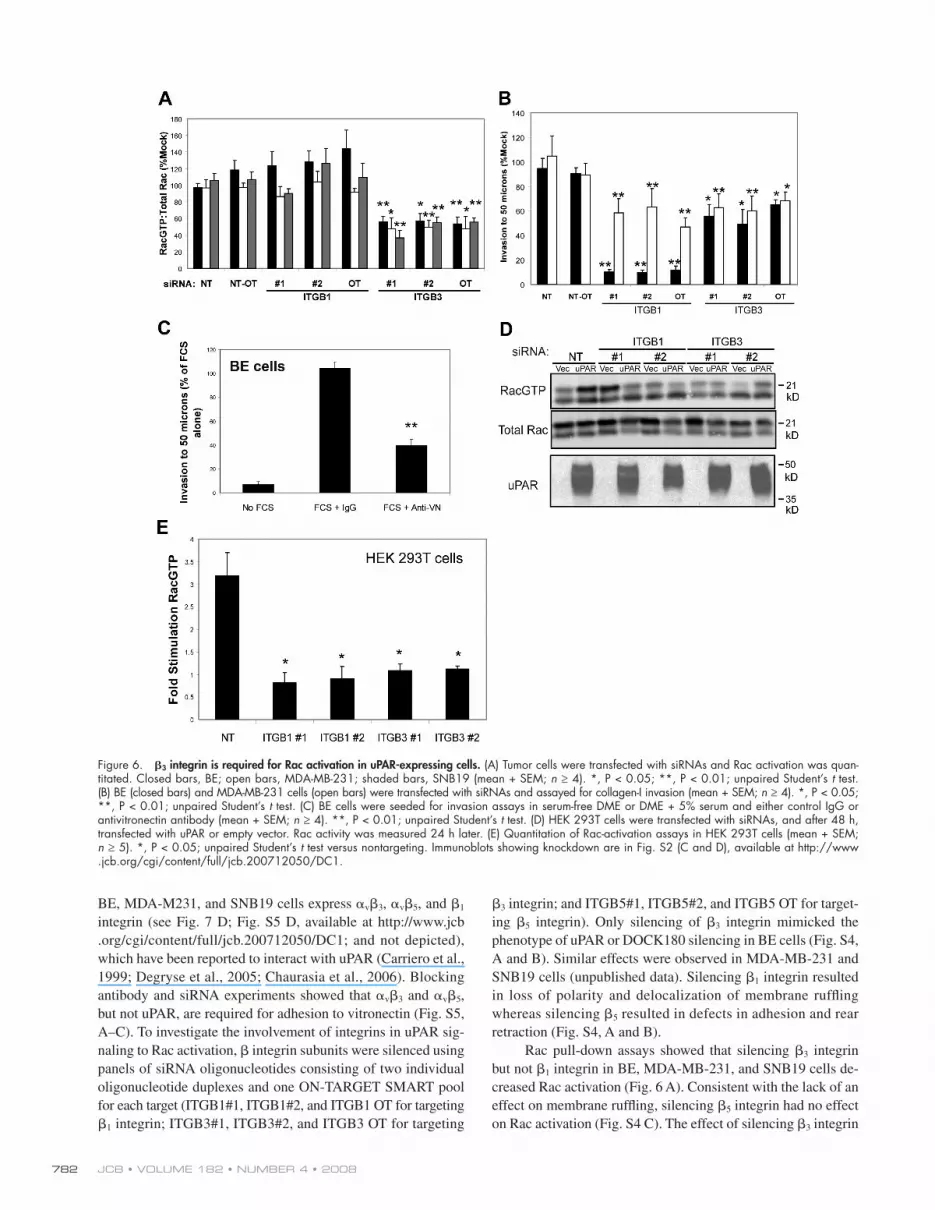
� 3 integrin; and ITGB5#1, ITGB5#2, and ITGB5 OT for target-
ing � 5 integrin). Only silencing of � 3 integrin mimicked the
phenotype of uPAR or DOCK180 silencing in BE cells (Fig. S4,
A and B). Similar effects were observed in MDA-MB-231 and
SNB19 cells (unpublished data). Silencing � 1 integrin resulted
in loss of polarity and delocalization of membrane ruffl ing
whereas silencing � 5 resulted in defects in adhesion and rear
retraction (Fig. S4, A and B).
Rac pull-down assays showed that silencing � 3 integrin
but not � 1 integrin in BE, MDA-MB-231, and SNB19 cells de-
creased Rac activation ( Fig. 6 A ). Consistent with the lack of an
effect on membrane ruffl ing, silencing � 5 integrin had no effect
on Rac activation (Fig. S4 C). The effect of silencing � 3 integrin
BE, MDA-M231, and SNB19 cells express � v � 3 , � v � 5 , and � 1
integrin (see Fig. 7 D ; Fig. S5 D, available at http://www.jcb
.org/cgi/content/full/jcb.200712050/DC1; and not depicted),
which have been reported to interact with uPAR ( Carriero et al.,
1999 ; Degryse et al., 2005 ; Chaurasia et al., 2006 ). Blocking
antibody and siRNA experiments showed that � v � 3 and � v � 5 ,
but not uPAR, are required for adhesion to vitronectin (Fig. S5,
A – C). To investigate the involvement of integrins in uPAR sig-
naling to Rac activation, � integrin subunits were silenced using
panels of siRNA oligonucleotides consisting of two individual
oligonucleotide duplexes and one ON-TARGET SMART pool
for each target (ITGB1#1, ITGB1#2, and ITGB1 OT for targeting
� 1 integrin; ITGB3#1, ITGB3#2, and ITGB3 OT for targeting
Figure 6. � 3 integrin is required for Rac activation in uPAR-expressing cells. (A) Tumor cells were transfected with siRNAs and Rac activation was quan-titated. Closed bars, BE; open bars, MDA-MB-231; shaded bars, SNB19 (mean + SEM; n ≥ 4). *, P < 0.05; **, P < 0.01; unpaired Student ’ s t test. (B) BE (closed bars) and MDA-MB-231 cells (open bars) were transfected with siRNAs and assayed for collagen-I invasion (mean + SEM; n ≥ 4). *, P < 0.05; **, P < 0.01; unpaired Student ’ s t test. (C) BE cells were seeded for invasion assays in serum-free DME or DME + 5% serum and either control IgG or antivitronectin antibody (mean + SEM; n ≥ 4). **, P < 0.01; unpaired Student ’ s t test. (D) HEK 293T cells were transfected with siRNAs, and after 48 h, transfected with uPAR or empty vector. Rac activity was measured 24 h later. (E) Quantitation of Rac-activation assays in HEK 293T cells (mean + SEM; n ≥ 5). *, P < 0.05; unpaired Student ’ s t test versus nontargeting. Immunoblots showing knockdown are in Fig. S2 (C and D), available at http://www.jcb.org/cgi/content/full/jcb.200712050/DC1.

783 U PAR SIGNALS TO RAC ACTIVATION VIA DOCK180 • Smith et al.
immunoblotting showed that uPAR expression, � 1 integrin knock-
down, or inhibition of ERK activation did not affect total cell
levels of � 3 integrin ( Fig. 7 A ).
In contrast to HEK 293T cells, uPAR or � 1 integrin silenc-
ing in BE colon carcinoma cells did not affect the surface local-
ization of � v � 3 integrin ( Fig. 7 D ); however, surface expression
of � v � 3 integrin in BE, MDA-MB231, and SNB19 was partially
dependent on ERK activation ( Fig. 7 D and Fig. S5 D). In none
of the tumor cell lines was � 1 integrin or uPAR required for ERK
activation ( Fig. 7 E ). BE and MDA-MB231 harbor activating
mutations in KRAS and BRAF or KRAS alone ( Vial et al., 2003 )
that presumably uncouple ERK activation from a requirement
for � 1 integrin and uPAR. These results therefore argue that
uPAR signals through � v � 3 for Rac activation, but that uPAR
signaling through � 1 integrins can provide an ERK signal for
surface localization of � v � 3 .
� 3 integrin is required for p130Cas SD tyrosine phosphorylation and formation of the p130Cas – CrkII complex Because � 3 integrin – silenced cells had defects in morphology,
Rac activation, and invasion similar to those observed in cells
where components of the uPAR – DOCK180 pathway had been
silenced, we examined the roles of � integrin subunits in signal-
ing through the p130Cas – CrkII adaptor complex. Silencing � 3
integrin in BE, MDA-MB231, and SNB19 tumor cells strongly
reduced tyrosine phosphorylation of the p130Cas SD ( Fig. 8 A ).
Conversely, silencing � 1 integrin did not affect p130Cas SD
phosphorylation. In keeping with the p130Cas SD tyrosine
phosphorylation data, coimmunoprecipitation of p130Cas with
CrkII was also inhibited by silencing � 3 integrin, whereas silenc-
ing of � 1 integrin had no effect ( Fig. 8 B ). These data show that
expression of uPAR promotes signaling through � 3 integrin
to drive tyrosine phosphorylation of the p130Cas SD and for-
mation of the p130Cas – CrkII adaptor complex. Consistent with
the requirement for Src in Rac activation driven by uPAR, silenc-
ing � 3 integrin blocked Src activation driven by uPAR (Fig. S3,
B and D) but did not affect FAK Y397 phosphorylation (Fig. S3 C).
Silencing � 3 integrin also abrogated the stimulation of p130Cas
SD tyrosine phosphorylation by ectopically expressed uPAR in
HEK 293T cells ( Fig. 8 C ). However, as observed for uPAR-
driven Rac activation, silencing � 1 integrin in HEK 293T cells
also abrogated uPAR-driven p130Cas SD tyrosine phosphory-
lation. This is consistent with the role of � 1 integrin in promot-
ing cell surface expression of � 3 integrin by cooperating with
uPAR to activate ERK, a function of � 1 integrin that is not re-
quired in the tumor cells where ERK activity does not require
� 1 integrin ( Fig. 7 E ).
Discussion In this study we have identifi ed a mechanism of Rac activation
by uPAR. We show for the fi rst time that in both ectopic and endog-
enous systems uPAR expression results in activation of Rac via
the GEF DOCK180. DOCK180 has been shown to have a role
in cell motility ( Klemke et al., 1998 ) and developmental pro-
cesses such as myoblast fusion, dorsal closure, and phagocytosis
is very similar in magnitude to that observed when uPAR,
DOCK180, Crk, or p130Cas is silenced. Consistent with the fact
that � v � 3 is a major vitronectin receptor ( Cheresh and Spiro,
1987 ), we found that silencing uPAR or � 3 integrin only affected
Rac activity in BE cells plated on vitronectin and not on collagen
or fi bronectin, which are major � 1 integrin ligands (Fig. S5 E).
Similarly, ectopic expression of uPAR in HEK 293T cells led to
Rac activation if the cells were plated in serum-free medium on
vitronectin or in serum as a source of vitronectin, but there was
no Rac activation if the cells were plated in serum-free medium
on fi bronectin or collagen-1 (Fig. S5 F).
The contributions of signaling through � 1 and � 3 integrin
subunits to invasion were examined by testing � 1 - and � 3 -
silenced BE and MDA-MB-231 cells for invasion of a three-
dimensional collagen-1 matrix ( Fig. 6 B ). In both BE and
MDA-MB-231 cells, silencing � 3 integrin inhibited invasion by
� 40%. Invasion in these assays was dependent on vitronectin
present in serum as no invasion took place in the absence of se-
rum, and the addition of a vitronectin-blocking antibody ( Zanetti
et al., 1994 ) blocked serum-dependent invasion ( Fig. 6 C ). This
was consistent with the observed effects on invasion of silencing
uPAR, DOCK180, Crk, and p130Cas in these cells. Although it
did not affect Rac activation, silencing � 1 integrin decreased in-
vasion by � 90% in BE cells and 40 – 50% in MDA-MB-231 cells.
This is not unexpected because all collagen-binding integrins
contain the � 1 subunit, and adhesion to the substratum is essen-
tial for the elongated/mesenchymal mode of migration ( Pollard
and Borisy, 2003 ).
To investigate whether � 3 integrin was required for uPAR-
driven Rac activation in HEK 293T cells, integrin subunits were
silenced with siRNA; Fig. 6 (D and E) shows that silencing � 3
in HEK 293T cells blocked Rac activation. However, unlike
the tumor cell lines, � 1 silencing did reduce Rac activation in
uPAR-transfected HEK 293T cells. Signifi cantly, fl ow cytom-
etry showed that � v � 3 was not expressed at the surface of control
or empty vector – transfected HEK 293T cells but was expressed
at the surface of uPAR-transfected cells ( Fig. 7 A ). Previous
work has shown that sustained extracellular signal – regulated
kinase (ERK) activation leads to surface expression of � 3 inte-
grin ( Woods et al., 2001 ), and as uPAR-mediated ERK activation
has been shown to be � 1 dependent ( Aguirre Ghiso et al., 1999 ),
we examined whether � 1 integrin expression and ERK activa-
tion were required for surface expression of � 3 . Fig. 7 A shows
that silencing � 1 integrin or treatment with the MAPK/ERK
kinase (MEK) inhibitors PD184352 or UO126 (unpublished
data) blocked surface expression of � v � 3 in uPAR-transfected
HEK 293T cells. Silencing � 1 integrin but not � 3 integrin blocked
uPAR-dependent ERK activation, showing that ERK activation
by uPAR requires � 1 but not � 3 integrin ( Fig. 7 B ). Consistent
with the observations that � 1 integrin signals to ERK activation
and surface expression of � v � 3 , inhibition of ERK activation with
MEK inhibitors PD184352 or UO126 blocked Rac activation
in uPAR-transfected HEK 293T cells ( Fig. 7 C ). These results
show that signaling via uPAR and � 1 integrins to ERK activation
can provide the surface localization of � v � 3 required for uPAR-
dependent Rac activation. Although � 1 integrin – dependent ERK
activation was required for surface expression of � 3 integrin,

JCB • VOLUME 182 • NUMBER 4 • 2008 784
Figure 7. Cooperation between uPAR signaling with � 1 integrin to ERK-MAPK activation and � 3 integrin – dependent Rac activation. (A) Cell surface ex-pression of � v � 3 in uPAR- or vector-transfected HEK 293T cells. Top three left panels, siRNA-transfected cells; top right panel, 2 μ M PD184352 or vehicle (DMSO) treatment. Red, IgG control; blue, � v � 3 vector transfected; green, uPAR – � v � 3 transfected. (bottom) � 3 integrin immunoblot of siRNA-transfected or PD184352-treated HEK 293T cells. (B) HEK 293T cells were transfected as in A and ERK1/2 activation was measured (mean + SEM; n = 6). *, P < 0.05; unpaired Student ’ s t test. (C) Rac activation in HEK 293T cells transfected with uPAR or vector and treated with MEK inhibitors or vehicle (DMSO)

785 U PAR SIGNALS TO RAC ACTIVATION VIA DOCK180 • Smith et al.
complex assembly by uPAR may regulate a variety of other sig-
naling pathways.
The identifi cation of DOCK180, an integrin-associated GEF,
in uPAR-Rac signaling is in keeping with a large body of evi-
dence implicating integrins as the signaling partners of uPAR.
A relatively large array of integrin heterodimers have been
shown to interact with uPAR, but whether any of these are
specifi cally required for uPAR to activate Rac in tumor cells
had not been previously investigated. In the tumor cell lines
we examined, � 3 but not � 1 integrin was required for uPAR –
DOCK180 signaling to Rac activation. In HEK 293T cells, � 1
as well as � 3 integrin are required for uPAR-stimulated Rac
activation, but in these cells the role of � 1 integrin appears to
be to provide the ERK activation ( Aguirre-Ghiso et al., 1999 )
necessary for surface expression of � v � 3 . Thus, in some cells
uPAR – � 1 integrin signaling to ERK-dependent surface expression
of � v � 3 cooperates with uPAR – � v � 3 signaling for Rac activa-
tion, whereas in other cells uPAR – � 3 integrin drives Rac acti-
vation but ERK activation does not seem to require uPAR or � 1
integrin signaling.
Several papers have emphasized the importance of vitronec-
tin in membrane protrusion and cell motility induced by uPAR ex-
pression, and both integrin signaling and direct binding of uPAR to
vitronectin were recently shown to be required for stimulation of
membrane ruffl ing and lamellipodial protrusion by ectopic uPAR
expression in HEK 293T cells ( Kjoller and Hall, 2001 ; Madsen
et al., 2007 ). We have shown that uPAR and � 3 integrin are re-
quired for Rac activation in tumor cells cultured on vitronectin or
in the presence of serum, which is an abundant source of vitronectin
with concentrations in the range of 200 to 400 μ g/ml ( Schvartz
et al., 1999 ). In the collagen-1 – based three-dimensional invasion
assay we have used, a function-blocking antivitronectin antibody
inhibits serum-stimulated invasion, demonstrating that invasion is
dependent on vitronectin. Consistent with its role in uPAR-driven
Rac activation in the presence of vitronectin, silencing � 3 integrin
expression also inhibits serum-stimulated invasion.
These data suggest that uPAR and � 3 integrin engage vitro-
nectin to promote Rac activity and tumor cell invasion. The nature
of uPAR – integrin interactions is controversial. Although many
studies have shown uPAR – integrin coimmunoprecipitation, this
does not prove the existence of direct binding. Immunoprecipita-
tion under gentle conditions may result in the detection of many
proteins associated with detergent-resistant lipid rafts, including
uPAR and integrins. The study of Madsen et al. (2007) has cast
doubt on the role of specifi c uPAR residues in mediating bind-
ing to integrins, although it does not rule out direct interactions
involving multiple residues over a large binding surface. Our data
are consistent with a model where both uPAR and � 3 integrin
coordinately engage vitronectin. This could affect signaling in
several ways, for example, by facilitation of integrin – ligand in-
teraction, effects on integrin clustering, or modifi cation of integrin
of apoptotic cells ( Nolan et al., 1998 ; Wu and Horvitz, 1998 ; Moore
et al., 2007 ). Our data show that uPAR signaling to DOCK180
results in the induction of tumor cell motility and invasion.
In several systems, ELMO has been linked to DOCK180 function
possibly through acting as a cofactor for GEF activity ( Gumienny
et al., 2001 ; Brugnera et al., 2002 ). Whether it is involved in
uPAR-driven Rac activation will be an interesting topic for
future investigation.
Having identifi ed DOCK180 as a Rac GEF regulated by
uPAR, we examined how DOCK180 is activated downstream
of uPAR. Previous work in other systems shows that integrin
signaling recruits DOCK180 to the plasma membrane via the for-
mation of a p130Cas – Crk – DOCK180 complex. Key to the forma-
tion of this complex is tyrosine phosphorylation of the p130Cas
SD that recruits Crk – DOCK180 complexes via the SH2 domain of
Crk. We show that uPAR, expressed endogenously by tumor cells
or ectopically in HEK 293T cells, drives the tyrosine phosphory-
lation of the p130Cas SD and formation of the p130Cas – Crk
complex. For Rac activation by uPAR, uPA does not seem to be
essential ( Kjoller and Hall 2001 ) and is not expressed by HEK
293T cells ( Wei et al., 1994 ). However, uPA – uPAR interactions
may play an important role in other systems or in tumor cell in-
vasion in vivo, whether by enhancing uPAR binding to vitro-
nectin or through mechanisms such as focused ECM proteolysis
at the leading edge or enhancing local availability of growth
factors. Consistent with uPAR signaling through the p130Cas –
Crk complex, we fi nd that p130Cas and Crk are required for
Rac activation by uPAR and for the invasion of uPAR-expressing
tumor cells.
p130Cas is a multifunctional adaptor protein required for
embryonic development and oncogenic signal transduction in
tumor cells ( Auvinen et al., 1995 ; Nievers et al., 1997 ; Honda
et al., 1998 ; Kirsch et al., 2002 ; Cabodi et al., 2006 ). It is also
an important regulator of cell migration, and in particular its
association with Crk constitutes a molecular switch vital for cell
motility by recruiting DOCK180 to integrin-containing adhe-
sion complexes ( Klemke et al., 1998 ). These complexes also
serve a mechanosensory function allowing the cell to sense the
physical properties, such as rigidity, of the ECM (for review see
Bershadsky et al., 2006 ). Interestingly, in vitro data suggests that
p130Cas can function as a transducer of mechanical signals,
with the SD adopting an extended conformation permissive for
phosphorylation in response to increased physical force ( Sawada
et al., 2006 ). This could promote Rac-driven migration in response
to physical cues in the extracellular environment. As we have
shown that uPAR stimulates the tyrosine phosphorylation of the
p130Cas SD, the role of uPAR in integrin-mediated mechano-
transduction is an interesting subject for future investigation.
In addition, it is well known that p130Cas and Crk can interact
with other partners besides DOCK180. Therefore, promotion
of p130Cas SD tyrosine phosphorylation and p130Cas – CrkII
for 24 h (mean + SEM; n = 6). *, P < 0.05; unpaired Student ’ s t test. (D) Surface expression of � v � 3 on BE cells. Left and middle, siRNA transfections; right, MEK inhibition. Data are representative of three independent experiments. (E) ERK1/2 activation in cells transfected with siRNAs. Left, representative immunoblot (BE), total ERK1/2 (red), phospho-ERK1/2 (green); right, quantitation (mean + SEM; n = 4). Closed bars, BE cells; open bars, MDA-MB-231; shaded bars, SNB19.

JCB • VOLUME 182 • NUMBER 4 • 2008 786
which is a major regulator of polarized migration ( Etienne-
Manneville, 2004 ). Also by inhibiting internalization of lipid
rafts, which contain both uPAR and binding sites for activated
Rac ( del Pozo et al., 2004 ), and by regulating interactions with
RhoGDI ( Del Pozo et al., 2002 ), � 1 integrin may affect spatial
control of Rac-driven protrusion and motility.
Determination of the molecular mechanisms underlying
uPAR signaling, such as the Rac activation pathway described
here, is essential to provide insight into the well-established
role of uPAR in tumor cell invasion. Understanding these path-
ways will provide new therapeutic targets for the prevention of
human tumor metastasis.
conformation by lateral uPAR – integrin interactions. Interestingly,
silencing uPAR expression or blocking uPAR function using an
antibody that recognizes the vitronectin-binding site had no effect
on the adhesion of tumor cells to vitronectin (Fig. S5, A – C). Sig-
nifi cantly, while this paper was in preparation, Wei et al. (2008)
have demonstrated that uPAR expression leads to the activation
of � 3 integrins in the murine kidney ( Wei et al., 2008 ).
Interestingly, our data show that � 1 integrin silencing causes
a severe cell motility phenotype in uPAR-expressing tumor cell
lines, without affecting Rac activation but causing a delocaliza-
tion of membrane ruffl ing and lamellipodia. � 1 integrin signaling
may affect tumor cell polarity, for example, by regulating Cdc42,
Figure 8. � 3 integrin is required for p130Cas SD tyrosine phosphorylation and formation of the p130Cas – CrkII complex. (A) p130Cas SD tyrosine phosphorylation in siRNA-transfected cells. Left, representative immunoblot (BE); right, quantitation (mean + SEM; n = 5). *, P < 0.05; **, P < 0.01; unpaired Student ’ s t test. Closed bars, BE; open bars, MDA-MB-231; shaded bars, SNB19. Irrelevant lanes were removed (represented by vertical black lines). (B) Analysis of p130Cas – Crk complexes. Left, representative immunoblots (BE); right, quantitation (mean + SEM; n = 4). *, P < 0.05; unpaired Stu-dent ’ s t test. Closed bars, BE; open bars, MDA-MB-231; shaded bars, SNB19. (C) Analysis of p130Cas SD tyrosine phosphorylation in HEK 293T cells transfected with siRNAs and uPAR or empty vector was performed as described in Fig. 3 (mean + SEM; n = 4). *, P < 0.05; unpaired Student ’ s t test. Immunoblots showing knockdown are in Fig. S2 E, available at http://www.jcb.org/cgi/content/full/jcb.200712050/DC1.

787 U PAR SIGNALS TO RAC ACTIVATION VIA DOCK180 • Smith et al.
24 h at a rate of one frame per site per 4 min. Movies were exported from Simple PCI software as uncompressed AVI fi les with a frame rate of 15 frames per second. Premiere (v6.0; Adobe) was used to compress movie fi les using the MPEG codec, which were then converted to MOV (Quick-time) format using iMovie HD with a frame rate of 15 frames per second (dimensions: 640 × 512).
Confocal sections were obtained with a laser-scanning confocal im-aging system (MRC 1024; Bio-Rad Laboratories) mounted on an upright fl uorescence microscope (E600; Nikon) with PLan Apo 60 × oil immersion objective (NA 1.4) at 21 ° C, and using LaserSharp acquisition software (Bio-Rad Laboratories). Images were exported as PIC fi les and processed for brightness and contrast using Photoshop, supplemented with PIC fi le recognition plug-in (Bio-Rad Laboratories).
Analysis of Rac1 activation A GST fusion of the CRIB domain of PAK1 was used to pull down the ac-tivated form of Rac ( Benard et al., 1999 ). The PAK1-CRIB domain GST fusion protein was bound to glutathione-Sepharose beads (GE Health-care). 5 × 10 5 � 10 6 cells in a 10-cm dish were washed in Rac wash buf-fer (50 mM Tris-HCl, 10 mM MgCl 2 , 1 mM DTT [Sigma-Aldrich], and EDTA-free complete protease inhibitors [Roche]) and lysed on ice for 3 min in ice-cold Rac lysis buffer (50 mM Tris-HCl, 10% glycerol, 1% NP-40, 5 mM MgCl 2 , 100 mM NaCl, and EDTA-free complete protease in-hibitors). Lysates were cleared by centrifugation at 13,200 rpm for 5 min in a centrifuge (5810R; Eppendorff) at 4 ° C and an aliquot was kept for determination of total Rac levels by Western blotting. The remainder of the lysate was incubated with 30- μ l PAK-CRIB – Sepharose beads for 45 min on a rotating wheel at 4 ° C. Beads were collected by brief centrifuga-tion and washed three times in 500 μ l of ice-cold Rac lysis buffer and re-suspended in 20 μ l of LDS sample buffer (Invitrogen), and electrophoresis (NOVEX NuPAGE Midi gel system; Invitrogen) was performed with the total volume of each Rac pull down and equivalent volumes of total cell lysate for determination of total Rac. Fluorescent immunoblotting of Rac in pull downs and total lysate used anti-Rac clone 23A8 (Fitzgerald) and the Odyssey (Li-COR Biosciences). Data from the Odyssey were exported into Excel (Microsoft) and the ratio of signals for Rac GTP/Total Rac was calculated for each sample. For tumor cell lines, data are normalized to values from mock-transfected cells. Statistical comparisons for each siRNA were made against the nontargeting control and ON-TARGET SMART Pools were compared with ON-TARGET nontargeting controls. For HEK 293T cells, data are shown as fold stimulation of Rac activation, obtained by dividing the Rac activation in uPAR-transfected cells by that of the vector control for each condition.
Invasion assay Cells were suspended in 2.3 mg/ml of serum-free liquid bovine collagen at 10 5 cells/ml. 100- μ l aliquots were dispensed into black 96-well View-Plates (PerkinElmer) coated with bovine serum albumin. Plates were cen-trifuged at 300 g and incubated in a 37 ° C/10% CO 2 tissue culture incubator. Once collagen had polymerized, FCS was added on top of the collagen to a fi nal concentration of 5%. For vitronectin-blocking stud-ies, 20 μ g/ml antivitronectin antibody or isotype-matched IgG control were preincubated with FCS for 30 min at room temperature. After 24-h incubation at 37 ° C in 10% CO 2 , cells were fi xed and stained for 2 h in 4% formaldehyde solution (Sigma-Aldrich) containing 5 μ g/ml Hoechst 33258 nuclear stain (Invitrogen). Confocal z slices were collected from each well at 50 μ m to count invaded cells, and at the bottom (3 μ m) to count total cells using a high content microscope (INCELL3000; GE Healthcare) with a 40 × PlanFluor ELWD objective (0.6 NA; Nikon). Nuclear staining in each slice was quantified automatically with INCELL3000 Object Intensity module to determine the percentage of in-vaded cells. Samples were run in quadruplicate and averaged. Data analysis was performed using Excel. Invasion index was calculated at number of cells at 50 μ m per total number of cells. Data are presented as a percentage of the invasion index of mock-transfected cells. Statisti-cal comparisons for siRNAs were made against the nontargeting control, and ON-TARGET SMART Pools were compared with ON-TARGET nontar-geting controls.
Immunoprecipitation and immunoblotting Cells were grown in 10- or 15-cm plates and lysed in 1% NP-40 buffer (1% NP-40, 150 mM NaCl, 50 mM Tris-HCl, pH 7.4, 25 mM sodium � -glycerophosphate, 1 mM sodium vanadate, 5 mM NaF, and com-plete protease inhibitors). A minimum of 1 mg of total cellular protein was incubated at 4 ° C overnight or for 3 h on a rotating wheel with antibody
Materials and methods Antibodies and reagents The following antibodies were used: anti-Rac1 (clone 23A8; Fitzgerald), anti-uPAR (R & D Systems), anti-GAPDH (Novus Biologicals), anti-DOCK180, anti – � 3 integrin, anti – � 1 integrin (clone P5D2; Santa Cruz Biotechnology, Inc.), anti – � 1 integrin (clone JB1a), anti – � 3 integrin rabbit polyclonal, anti- � v � 3 (LM609), anti- � v � 5 (P1F6), antivitronectin (clone BV2; Milli-pore), anti-p130Cas, anti-Crk, anti-FAK (BD Biosciences), anti – � -tubulin, anti-total, phosphoERK (Sigma-Aldrich), anti-p130Cas phosphotyrosine 410 (Cell Signaling Technology), anti – c-Src (clone GD11; Millipore), anti-Src phosphoY416 (Invitrogen), anti-FAK phosphoY397 (Affi nity Bio-Reagents), and mouse IgG isotype controls (R & D systems). Vitronectin and fi bronectin (purifi ed from human serum) were purchased from Sigma-Aldrich. Bovine Type I collagen solution was purchased from Invitrogen. PD184352 was obtained from C. Springer (Institute of Cancer Research, Sutton, England, UK) and U0126 was obtained from Sigma-Aldrich. PP1, PP2, PP3, and SU6656 were purchased from EMD). Texas red – labeled phalloidin was purchased from Invitrogen. pRcCMV-uPAR was a provided by A. Hall (Memorial Sloan-Kettering Cancer Center, New York, NY). HRP-coupled secondary antibodies were from purchased Sigma-Aldrich and fl uorophore-coupled secondary antibodies were purchased from Li-COR Biosciences.
Cell culture BE colon carcinoma cells were obtained from the Institute of Cancer Re-search Tissue Resource Laboratory; HEK 293T and MDA-MB-231 breast carcinoma cells from the American Tissue Type Culture Collection; and SNB19 glioblastoma cells from the Deutsche Sammlung von Mikroorganis-men und Zellkulturen. All cells were maintained in DME, supplemented with 10% FCS purchased from PAA Laboratories, 100 μ g/ml streptomycin, and 60 μ g/ml penicillin. Cells were maintained at 37 ° C and 10% CO 2 . siRNA transfections in tumor cell lines were performed using InterferIN (Polyplus) according to the manufacturer ’ s instructions. The fi nal concentra-tion of siRNA in the transfection was 2 nM. For siRNA transfection of HEK 293T cells, HiPerfect (QIAGEN) was used with 50-nM fi nal concentration of siRNA according to the manufacturer ’ s instructions. Sequences of siRNA oligonucleotides were as follows: DOCK180 #1, CUGACUCAGAAC-GUGGACC; DOCK180 #2, UAAAUGAGCAGCUGUACAA; DOCK180-OT (Thermo Fisher Scientifi c); uPAR #1, GAAGAGACUUUCCUCAUUG; uPAR #2, GGUGACGCCUUCAGCAUGA; uPAR #3, GGUGAAGAAGGGC-GUCCAA; Crk #1, AAUAGGAGAUCAAGAGUUU; Crk #2, GGACAGC-GAAGGCAAGAGA; Crk-OT (Thermo Fisher Scientifi c); p130Cas #1, GGUCGACAGUGGUGUGUAU; p130Cas #2, AGAAGGAGCUGCUG-GAAAA; p130Cas-OT (Thermo Fisher Scientifi c); ITGB1 #1 (targeting � 1 integrin), GAACAGAUCUGAUGAAUGA; ITGB1 #2 (targeting � 1 integrin), CAAGAGAGCUGAAGACUAU; ITGB1-OT (targeting � 1 integrin; Thermo Fisher Scientifi c); ITGB3 #1 (targeting � 3 integrin), CUCUCCUGAUGUAG-CACUUAA; ITGB3 #2 (targeting � 3 integrin), CACGUGUGGCCU-GUUCUUCUA; ITGB3-OT (targeting � 3 integrin; Thermo Fisher Scientifi c); ITGB5 #1 (targeting � 5 integrin), GAACAACGGUGGAGAUUUU; ITGB5 #2 (targeting � 5 integrin), GGAGGGAGUUUGCAAAGUU; ITGB5-OT (targeting � 5 integrin; Thermo Fisher Scientifi c). Controls used were All-Stars nontargeting control (QIAGEN), ON-TARGET nontargeting control SMART Pool (Thermo Fisher Scientifi c), and nontargeting control SMART Pool (Thermo Fisher Scientifi c).
Transfection of HEK 293T cells with plasmid DNA (6 μ g DNA per 10-cm cell culture dish) was performed using GeneJuice (EMD) according to the manufacturer ’ s instructions. Plasmid DNA was prepared using the HiSpeed Plasmid Maxi kit (QIAGEN).
Microscopy Static phase-contrast images were obtained from a microscope camera workstation (Digital Site DS-4; Nikon) attached to an inverted phase-contrast microscope (TS100; Nikon) using a LWD 20 × objective (NA 0.4; Nikon) at 21 ° C. Images were processed for contrast and brightness using Photoshop v7.0 (Adobe).
Multi-site time-lapse video microscopy was performed in a humidi-fi ed, CO 2 -equilibrated chamber at 37 ° C using an inverted phase-contrast microscope (TE2000; Nikon) in conjunction with digital cameras (either Orca-ER or C9100EM-CCD; Hamamatsu Photonics), and equipped with motorized stage, focus, and shutter systems (Prior Scientifi c Instruments, Ltd.), all controlled by Simple PCI AIC acquisition software (v6.5; Compix Imaging Systems). Cells were imaged using PlanFluor 10 × (0.3 NA; Nikon) or PlanFluor ELWD 20 × (0.45 NA; Nikon) objectives for at least

JCB • VOLUME 182 • NUMBER 4 • 2008 788
References Aguirre Ghiso , J.A. 2002 . Inhibition of FAK signaling activated by urokinase
receptor induces dormancy in human carcinoma cells in vivo. Oncogene . 21 : 2513 – 2524 .
Aguirre-Ghiso , J.A. 2007 . Models, mechanisms and clinical evidence for cancer dormancy. Nat. Rev. Cancer . 7 : 834 – 846 .
Aguirre Ghiso , J.A. , K. Kovalski , and L. Ossowski . 1999 . Tumor dormancy in-duced by downregulation of urokinase receptor in human carcinoma in-volves integrin and MAPK signaling. J. Cell Biol. 147 : 89 – 104 .
Arthur , W.T. , L.A. Quilliam , and J.A. Cooper . 2004 . Rap1 promotes cell spread-ing by localizing Rac guanine nucleotide exchange factors. J. Cell Biol. 167 : 111 – 122 .
Auvinen , M. , A. Paasinen-Sohns , H. Hirai , L.C. Andersson , and E. Holtta . 1995 . Ornithine decarboxylase- and ras-induced cell transformations: reversal by protein tyrosine kinase inhibitors and role of pp130CAS. Mol. Cell. Biol. 15 : 6513 – 6525 .
Bass , R. , F. Werner , E. Odintsova , T. Sugiura , F. Berditchevski , and V. Ellis . 2005 . Regulation of urokinase receptor proteolytic function by the tet-raspanin CD82. J. Biol. Chem. 280 : 14811 – 14818 .
Behrendt , N. , O.N. Jensen , L.H. Engelholm , E. Mortz , M. Mann , and K. Dano . 2000 . A urokinase receptor-associated protein with specifi c collagen binding properties. J. Biol. Chem. 275 : 1993 – 2002 .
Benard , V. , B.P. Bohl , and G.M. Bokoch . 1999 . Characterization of rac and cdc42 activation in chemoattractant-stimulated human neutrophils using a novel assay for active GTPases. J. Biol. Chem. 274 : 13198 – 13204 .
Bene , M.C. , G. Castoldi , W. Knapp , G.M. Rigolin , L. Escribano , P. Lemez , W.D. Ludwig , E. Matutes , A. Orfao , F. Lanza , and M. van ’ t Veer . 2004 . CD87 (urokinase-type plasminogen activator receptor), function and pathology in hematological disorders: a review. Leukemia . 18 : 394 – 400 .
Bershadsky , A. , M. Kozlov , and B. Geiger . 2006 . Adhesion-mediated mechano-sensitivity: a time to experiment, and a time to theorize. Curr. Opin. Cell Biol. 18 : 472 – 481 .
Blasi , F. , and P. Carmeliet . 2002 . uPAR: a versatile signalling orchestrator. Nat. Rev. Mol. Cell Biol. 3 : 932 – 943 .
Bos , J.L. , H. Rehmann , and A. Wittinghofer . 2007 . GEFs and GAPs: critical ele-ments in the control of small G proteins. Cell . 129 : 865 – 877 .
Brugnera , E. , L. Haney , C. Grimsley , M. Lu , S.F. Walk , A.C. Tosello-Trampont , I.G. Macara , H. Madhani , G.R. Fink , and K.S. Ravichandran . 2002 . Unconventional Rac-GEF activity is mediated through the Dock180-ELMO complex. Nat. Cell Biol. 4 : 574 – 582 .
Cabodi , S. , A. Tinnirello , P. Di Stefano , B. Bisaro , E. Ambrosino , I. Castellano , A. Sapino , R. Arisio , F. Cavallo , G. Forni , et al . 2006 . p130Cas as a new reg-ulator of mammary epithelial cell proliferation, survival, and HER2-neu oncogene-dependent breast tumorigenesis. Cancer Res. 66 : 4672 – 4680 .
Carriero , M.V. , S. Del Vecchio , M. Capozzoli , P. Franco , L. Fontana , A. Zannetti , G. Botti , G. D ’ Aiuto , M. Salvatore , and M.P. Stoppelli . 1999 . Urokinase recep-tor interacts with alpha(v)beta5 vitronectin receptor, promoting urokinase-dependent cell migration in breast cancer. Cancer Res. 59 : 5307 – 5314 .
Chaurasia , P. , J.A. Aguirre-Ghiso , O.D. Liang , H. Gardsvoll , M. Ploug , and L. Ossowski . 2006 . A region in urokinase plasminogen receptor domain III controlling a functional association with alpha5beta1 integrin and tumor growth. J. Biol. Chem. 281 : 14852 – 14863 .
Cheresh , D.A. , and R.C. Spiro . 1987 . Biosynthetic and functional properties of an Arg-Gly-Asp-directed receptor involved in human melanoma cell attachment to vitronectin, fi brinogen, and von Willebrand factor. J. Biol. Chem. 262 : 17703 – 17711 .
Cunningham , O. , A. Andolfo , M.L. Santovito , L. Iuzzolino , F. Blasi , and N. Sidenius . 2003 . Dimerization controls the lipid raft partitioning of uPAR/CD87 and regulates its biological functions. EMBO J. 22 : 5994 – 6003 .
Czekay , R.-P. , T.A. Kuemmel , R.A. Orlando , and M.G. Farquhar . 2001 . Direct binding of occupied urokinase receptor (uPAR) to LDL receptor-related protein is required for endocytosis of uPAR and regulation of cell surface urokinase activity. Mol. Biol. Cell . 12 : 1467 – 1479 .
Dano , K. , N. Behrendt , G. Hoyer-Hansen , M. Johnsen , L.R. Lund , M. Ploug , and J. Romer . 2005 . Plasminogen activation and cancer. Thromb. Haemost. 93 : 676 – 681 .
Degryse , B. , M. Resnati , R.P. Czekay , D.J. Loskutoff , and F. Blasi . 2005 . Domain 2 of the urokinase receptor contains an integrin-interacting epitope with intrinsic signaling activity: generation of a new integrin inhibitor. J. Biol. Chem. 280 : 24792 – 24803 .
Del Pozo , M.A. , W.B. Kiosses , N.B. Alderson , N. Meller , K.M. Hahn , and M.A. Schwartz . 2002 . Integrins regulate GTP-Rac localized effector inter-actions through dissociation of Rho-GDI. Nat. Cell Biol. 4 : 232 – 239 .
del Pozo , M.A. , N.B. Alderson , W.B. Kiosses , H.H. Chiang , R.G. Anderson , and M.A. Schwartz . 2004 . Integrins regulate Rac targeting by internalization of membrane domains. Science . 303 : 839 – 842 .
and complexes, and were then precipitated with 25 μ l of protein G – Agarose (Thermo Fisher Scientifi c) for 20 min on a rotating wheel at 4 ° C. Beads were washed at least three times in 500 μ l of lysis buffer and resuspended in NuPAGE LDS sample buffer before SDS-PAGE. Fluorescent immuno-blotting was conducted using the Odyssey infrared scanner according to the manufacturer ’ s protocols (Li-COR Biosciences). Fluorescence data from the Odyssey were exported into Excel. For phosphorylation analysis, sig-nal from phosphospecifi c antibodies was divided by that of antibodies recognizing total protein (e.g., PY410/Total p130Cas). For p130Cas – Crk complexes, p130Cas signal was divided by Crk signal. For siRNA experiments in tumor cells, data were normalized to values from mock-transfected cells. For HEK 293T cells, data are presented as fold stimula-tion of phosphorylation, obtained by dividing the phospho/total ratio in uPAR-transfected cells by the vector control. In all cases, statistical compar-isons for each siRNA were made against the nontargeting control, and ON-TARGET SMART Pools were compared with ON-TARGET nontar-geting controls.
Quantitative PCR Total cellular RNA was isolated from cultured cells using Trizol (Invitro-gen) or the RNeasy Mini kit (QIAGEN). Real-time RT-PCR amplifi cations were performed using the Brilliant II SYBR Green QRT-PCR Master Mix kit (Stratagene). A standard curve was constructed using a range of 0.01 to 10 ng RNA from BE cells for each set of primers used. Relative quantitation was performed using the � � C t method. All primers used were Quantitect SYBR green primer assays (QIAGEN). Reactions were performed in triplicate in 50- μ l volumes containing 25 μ l of 2 × Brilliant II mastermix, 5 μ l of 10 × Quantitect SYBR green primer assay, 1 μ l of RT-RNase-block enzyme mixture (Stratagene), and the appropriate amount of RNA with remaining volume made up with nuclease-free water (Ambion). PCR was performed in a Fast Real-Time PCR cycler (7900HT; Applied Biosystems). Data were analyzed using SDS software (Applied Biosystems).
Flow cytometry Detached cells (5 × 10 6 ) were stained on ice for 45 min using 10 μ g/ml LM609, 10 μ g/ml P1F6, or 1 μ g/ml P5D2 to detect � v � 3 , � v � 5 , and � 1 , re-spectively. Alexa fl uor 488 – conjugated goat anti – mouse F(ab) 2 fragment used for detection (at 1:250) was obtained from Invitrogen. Cells were analyzed on an LSR II fl ow cytometer (BD Biosciences).
Adhesion assays Adhesion assays were performed according to the method of Cunningham et al. (2003) . Cells were detached by short incubation with trypsin, counted, and washed in serum-free medium. 3 × 10 4 cells were allowed to adhere to 96-well plates (Thermo Fisher Scientifi c) precoated with 10 μ g/ml fi bronectin, 2 μ g/ml vitronectin, or 10 μ g/ml Type-I collagen for 30 min at 37 ° C. For blocking antibody studies, cells were preincubated with anti-bodies or control IgG (at 10 μ g/ml) for 30 min before addition to the plate. Plates were washed three times in medium containing 0.2% bovine serum albumin (Sigma-Aldrich), fi xed in formol saline, and stained with crystal violet. Staining was quantifi ed by measuring absorbance at 540 nm using a SpectraMax M5 (Invitrogen) plate reader.
Online supplemental material Fig. S1 shows a morphological screen of integrin-associated Rac GEFs. Fig. S2 shows siRNA-mediated knockdown of uPAR, DOCK180, p130Cas, Crk, and integrin subunits. Fig. S3 shows that uPAR-driven Rac activity in 293T cells requires � 3 integrin – dependent Src activation. Fig. S4 shows that silencing of � integrin subunits in BE colon carcinoma cells affects cell morphology. Fig. S5 shows that uPAR-driven Rac activation is vitronectin dependent but adhesion to vitronectin requires � v � 3 or � v � 5, but not uPAR. Video 1 shows control BE colon carcinoma cells. Video 2 shows BE colon carcinoma cells transfected with siRNA-targeting uPAR. Video 3 shows BE colon carcinoma cells transfected with siRNA-targeting DOCK180. Online supplemental material is available at http://www.jcb.org/cgi/content/full/jcb.200712050/DC1.
We thank C. Isacke for helpful comments. This work was supported by Cancer Research UK and a Federation of
European Biochemical Societies long-term fellowship to P. Marra. C.J. Marshall is a Gibb life fellow of Cancer Research UK.
Submitted: 11 December 2007 Accepted: 28 July 2008

789 U PAR SIGNALS TO RAC ACTIVATION VIA DOCK180 • Smith et al.
Meng , S. , D. Tripathy , S. Shete , R. Ashfaq , H. Saboorian , B. Haley , E. Frenkel , D. Euhus , M. Leitch , C. Osborne , et al . 2006 . uPAR and HER-2 gene status in individual breast cancer cells from blood and tissues. Proc. Natl. Acad. Sci. USA . 103 : 17361 – 17365 .
Mohan , P.M. , S.K. Chintala , S. Mohanam , C.L. Gladson , E.S. Kim , Z.L. Gokaslan , S.S. Lakka , J.A. Roth , B. Fang , R. Sawaya , et al . 1999 . Adenovirus-mediated delivery of antisense gene to urokinase-type plas-minogen activator receptor suppresses glioma invasion and tumor growth. Cancer Res. 59 : 3369 – 3373 .
Moore , C.A. , C.A. Parkin , Y. Bidet , and P.W. Ingham . 2007 . A role for the Myoblast city homologues Dock1 and Dock5 and the adaptor pro-teins Crk and Crk-like in zebrafish myoblast fusion. Development . 134 : 3145 – 3153 .
Moores , S.L. , L.M. Selfors , J. Fredericks , T. Breit , K. Fujikawa , F.W. Alt , J.S. Brugge , and W. Swat . 2000 . Vav family proteins couple to diverse cell surface receptors. Mol. Cell. Biol. 20 : 6364 – 6373 .
Nievers , M.G. , R.B. Birge , H. Greulich , A.J. Verkleij , H. Hanafusa , and P.M. van Bergen en Henegouwen . 1997 . v-Crk-induced cell transforma-tion: changes in focal adhesion composition and signaling. J. Cell Sci. 110 : 389 – 399 .
Nolan , K.M. , K. Barrett , Y. Lu , K.Q. Hu , S. Vincent , and J. Settleman . 1998 . Myoblast city, the Drosophila homolog of DOCK180/CED-5, is required in a Rac signaling pathway utilized for multiple developmental processes. Genes Dev. 12 : 3337 – 3342 .
Pedersen , N. , M. Schmitt , E. Ronne , M.I. Nicoletti , G. Hoyer-Hansen , M. Conese , R. Giavazzi , K. Dano , W. Kuhn , F. Janicke , et al . 1993 . A ligand-free, soluble urokinase receptor is present in the ascitic fl uid from patients with ovarian cancer. J. Clin. Invest. 92 : 2160 – 2167 .
Pollard , T.D. , and G.G. Borisy . 2003 . Cellular motility driven by assembly and disassembly of actin fi laments. Cell . 112 : 453 – 465 .
Polte , T.R. , and S.K. Hanks . 1995 . Interaction between focal adhesion kinase and Crk-associated tyrosine kinase substrate p130Cas. Proc. Natl. Acad. Sci. USA . 92 : 10678 – 10682 .
Pyke , C. , J. Eriksen , H. Solberg , B.S. Nielsen , P. Kristensen , L.R. Lund , and K. Dano . 1993 . An alternatively spliced variant of mRNA for the human receptor for urokinase plasminogen activator. FEBS Lett. 326 : 69 – 74 .
Resnati , M. , I. Pallavicini , J.M. Wang , J. Oppenheim , C.N. Serhan , M. Romano , and F. Blasi . 2002 . The fi brinolytic receptor for urokinase activates the G protein-coupled chemotactic receptor FPRL1/LXA4R. Proc. Natl. Acad. Sci. USA . 99 : 1359 – 1364 .
Ribatti , D. , D. Leali , A. Vacca , R. Giuliani , A. Gualandris , L. Roncali , M.L. Nolli , and M. Presta . 1999 . In vivo angiogenic activity of urokinase: role of endogenous fi broblast growth factor-2. J. Cell Sci. 112 : 4213 – 4221 .
Rosenberger , G. , I. Jantke , A. Gal , and K. Kutsche . 2003 . Interaction of aPIX (ARHGEF6) with b-parvin (PARVB) suggests an involvement of aPIX in integrin-mediated signaling. Hum. Mol. Genet. 12 : 155 – 167 .
Sakai , R. , A. Iwamatsu , N. Hirano , S. Ogawa , T. Tanaka , H. Mano , Y. Yazaki , and H. Hirai . 1994 . A novel signaling molecule, p130, forms stable com-plexes in vivo with v-Crk and v-Src in a tyrosine phosphorylation-dependent manner. EMBO J. 13 : 3748 – 3756 .
Saksela , O. , and D.B. Rifkin . 1990 . Release of basic fi broblast growth factor-heparan sulfate complexes from endothelial cells by plasminogen activator-mediated proteolytic activity. J. Cell Biol. 110 : 767 – 775 .
Salajegheh , M. , A. Rudnicki , and T.W. Smith . 2005 . Expression of urokinase-type plasminogen activator receptor (uPAR) in primary central nervous system neoplasms. Appl. Immunohistochem. Mol. Morphol. 13 : 184 – 189 .
Sato , Y. , R. Tsuboi , R. Lyons , H. Moses , and D.B. Rifkin . 1990 . Characterization of the activation of latent TGF-beta by co-cultures of endothelial cells and pericytes or smooth muscle cells: a self-regulating system. J. Cell Biol. 111 : 757 – 763 .
Sawada , Y. , M. Tamada , B.J. Dubin-Thaler , O. Cherniavskaya , R. Sakai , S. Tanaka , and M.P. Sheetz . 2006 . Force sensing by mechanical extension of the Src family kinase substrate p130Cas. Cell . 127 : 1015 – 1026 .
Schvartz , I. , D. Seger , and S. Shaltiel . 1999 . Vitronectin. Int. J. Biochem. Cell Biol. 31 : 539 – 544 .
Sidenius , N. , A. Andolfo , R. Fesce , and F. Blasi . 2002 . Urokinase regulates vitro-nectin binding by controlling urokinase receptor oligomerization. J. Biol. Chem. 277 : 27982 – 27990 .
Simon , D.I. , N.K. Rao , H. Xu , Y. Wei , O. Majdic , E. Ronne , L. Kobzik , and H.A. Chapman . 1996 . Mac-1 (CD11b/CD18) and the urokinase receptor (CD87) form a functional unit on monocytic cells. Blood . 88 : 3185 – 3194 .
Sturge , J. , J. Hamelin , and G.E. Jones . 2002 . N-WASP activation by a beta1-integrin-dependent mechanism supports PI3K-independent chemo-taxis stimulated by urokinase-type plasminogen activator. J. Cell Sci. 115 : 699 – 711 .
Tosello-Trampont , A.C. , J.M. Kinchen , E. Brugnera , L.B. Haney , M.O. Hengartner , and K.S. Ravichandran . 2007 . Identifi cation of two signaling
El-Kott , A.F. , A.M. Khalil , and M. El-Kenawy Ael . 2004 . Immunohistochemical expressions of uPA and its receptor uPAR and their prognostic signifi cant in urinary bladder carcinoma. Int. Urol. Nephrol. 36 : 417 – 423 .
Etienne-Manneville , S. 2004 . Cdc42 — the centre of polarity. J. Cell Sci. 117 : 1291 – 1300 .
Faccio , R. , S.L. Teitelbaum , K. Fujikawa , J. Chappel , A. Zallone , V.L. Tybulewicz , F.P. Ross , and W. Swat . 2005 . Vav3 regulates osteoclast function and bone mass. Nat. Med. 11 : 284 – 290 .
Gakidis , M.A. , X. Cullere , T. Olson , J.L. Wilsbacher , B. Zhang , S.L. Moores , K. Ley , W. Swat , T. Mayadas , and J.S. Brugge . 2004 . Vav GEFs are re-quired for � 2 integrin-dependent functions of neutrophils. J. Cell Biol. 166 : 273 – 282 .
Gumienny , T.L. , E. Brugnera , A.C. Tosello-Trampont , J.M. Kinchen , L.B. Haney , K. Nishiwaki , S.F. Walk , M.E. Nemergut , I.G. Macara , R. Francis , et al . 2001 . CED-12/ELMO, a novel member of the CrkII/Dock180/Rac pathway, is required for phagocytosis and cell migration. Cell . 107 : 27 – 41 .
Hamelers , I.H. , C. Olivo , A.E. Mertens , D.M. Pegtel , R.A. van der Kammen , A. Sonnenberg , and J.G. Collard . 2005 . The Rac activator Tiam1 is required for � 3 � 1-mediated laminin-5 deposition, cell spreading, and cell migration. J. Cell Biol. 171 : 871 – 881 .
Honda , H. , H. Oda , T. Nakamoto , Z. Honda , R. Sakai , T. Suzuki , T. Saito , K. Nakamura , K. Nakao , T. Ishikawa , et al . 1998 . Cardiovascular anomaly, impaired actin bundling and resistance to Src-induced transformation in mice lacking p130Cas. Nat. Genet. 19 : 361 – 365 .
Jaffe , A.B. , and A. Hall . 2005 . Rho GTPases: biochemistry and biology. Annu. Rev. Cell Dev. Biol. 21 : 247 – 269 .
Jo , M. , K.S. Thomas , D.M. O ’ Donnell , and S.L. Gonias . 2003 . Epidermal growth factor receptor-dependent and -independent cell-signaling pathways orig-inating from the urokinase receptor. J. Biol. Chem. 278 : 1642 – 1646 .
Jo , M. , K.S. Thomas , S. Takimoto , A. Gaultier , E.H. Hsieh , R.D. Lester , and S.L. Gonias . 2006 . Urokinase receptor primes cells to proliferate in response to epidermal growth factor. Oncogene . 26 : 2585 – 2594 .
Kaneko , T. , H. Konno , M. Baba , T. Tanaka , and S. Nakamura . 2003 . Urokinase-type plasminogen activator expression correlates with tumor angiogen-esis and poor outcome in gastric cancer. Cancer Sci. 94 : 43 – 49 .
Kirsch , K. , M. Kensinger , H. Hanafusa , and A. August . 2002 . A p130Cas ty-rosine phosphorylated substrate domain decoy disrupts v-crk signaling. BMC Cell Biol. 3 : 18 .
Kiyokawa , E. , Y. Hashimoto , T. Kurata , H. Sugimura , and M. Matsuda . 1998 . Evidence that DOCK180 up-regulates signals from the CrkII-p130(Cas) complex. J. Biol. Chem. 273 : 24479 – 24484 .
Kjoller , L. , and A. Hall . 2001 . Rac mediates cytoskeletal rearrangements and increased cell motility induced by urokinase-type plasminogen activator receptor binding to vitronectin. J. Cell Biol. 152 : 1145 – 1157 .
Klemke , R.L. , J. Leng , R. Molander , P.C. Brooks , K. Vuori , and D.A. Cheresh . 1998 . CAS/Crk coupling serves as a “ molecular switch ” for induction of cell migration. J. Cell Biol. 140 : 961 – 972 .
Lester , R.D. , M. Jo , V. Montel , S. Takimoto , and S.L. Gonias . 2007 . uPAR in-duces epithelial – mesenchymal transition in hypoxic breast cancer cells. J. Cell Biol. 178 : 425 – 436 .
Liu , D. , J. Aguirre Ghiso , Y. Estrada , and L. Ossowski . 2002 . EGFR is a trans-ducer of the urokinase receptor initiated signal that is required for in vivo growth of a human carcinoma. Cancer Cell . 1 : 445 – 457 .
Llinas , P. , M.H. Le Du , H. Gardsvoll , K. Dano , M. Ploug , B. Gilquin , E.A. Stura , and A. Menez . 2005 . Crystal structure of the human urokinase plasminogen activator receptor bound to an antagonist peptide. EMBO J. 24 : 1655 – 1663 .
Ma , Z. , K.S. Thomas , D.J. Webb , R. Moravec , A.M. Salicioni , W.M. Mars , and S.L. Gonias . 2002 . Regulation of Rac1 activation by the low density lipo-protein receptor – related protein. J. Cell Biol. 159 : 1061 – 1070 .
Madsen , C.D. , G.M. Ferraris , A. Andolfo , O. Cunningham , and N. Sidenius . 2007 . uPAR-induced cell adhesion and migration: vitronectin provides the key. J. Cell Biol. 177 : 927 – 939 .
Marignani , P.A. , and C.L. Carpenter . 2001 . Vav2 is required for cell spreading. J. Cell Biol. 154 : 177 – 186 .
Matsuda , M. , S. Ota , R. Tanimura , H. Nakamura , K. Matuoka , T. Takenawa , K. Nagashima , and T. Kurata . 1996 . Interaction between the amino-terminal SH3 domain of CRK and its natural target proteins. J. Biol. Chem. 271 : 14468 – 14472 .
Matsuo , N. , M. Terao , Y.-i. Nabeshima , and M. Hoshino . 2003 . Roles of STEF/Tiam1, guanine nucleotide exchange factors for Rac1, in regulation of growth cone morphology. Mol. Cell. Neurosci. 24 : 69 – 81 .
Memarzadeh , S. , K.R. Kozak , L. Chang , S. Natarajan , P. Shintaku , S.T. Reddy , and R. Farias-Eisner . 2002 . Urokinase plasminogen activator receptor: prognostic biomarker for endometrial cancer. Proc. Natl. Acad. Sci. USA . 99 : 10647 – 10652 .

JCB • VOLUME 182 • NUMBER 4 • 2008 790
submodules within the CrkII/ELMO/Dock180 pathway regulating engulf-ment of apoptotic cells. Cell Death Differ. 14 : 963 – 972 .
Vial , E. , E. Sahai , and C.J. Marshall . 2003 . ERK-MAPK signaling coordinately regulates activity of Rac1 and RhoA for tumor cell motility. Cancer Cell . 4 : 67 – 79 .
Vuori , K. , H. Hirai , S. Aizawa , and E. Ruoslahti . 1996 . Introduction of p130cas signaling complex formation upon integrin-mediated cell adhesion: a role for Src family kinases. Mol. Cell. Biol. 16 : 2606 – 2613 .
Wei , C. , C.C. Moller , M.M. Altintas , J. Li , K. Schwarz , S. Zacchigna , L. Xie , A. Henger , H. Schmid , M.P. Rastaldi , et al . 2008 . Modifi cation of kidney barrier function by the urokinase receptor. Nat. Med. 14 : 55 – 63 .
Wei , Y. , D.A. Waltz , N. Rao , R.J. Drummond , S. Rosenberg , and H.A. Chapman . 1994 . Identifi cation of the urokinase receptor as an adhesion receptor for vitronectin. J. Biol. Chem. 269 : 32380 – 32388 .
Wei , Y. , J.A. Eble , Z. Wang , J.A. Kreidberg , and H.A. Chapman . 2001 . Urokinase receptors promote beta1 integrin function through interactions with inte-grin alpha3beta1. Mol. Biol. Cell . 12 : 2975 – 2986 .
Wei , Y. , R.P. Czekay , L. Robillard , M.C. Kugler , F. Zhang , K.K. Kim , J.P. Xiong , M.J. Humphries , and H.A. Chapman . 2005 . Regulation of � 5 � 1 integrin conformation and function by urokinase receptor binding. J. Cell Biol. 168 : 501 – 511 .
Wei , Y. , C.H. Tang , Y. Kim , L. Robillard , F. Zhang , M.C. Kugler , and H.A. Chapman . 2007 . Urokinase receptors are required for alpha 5 beta 1 integrin-mediated signaling in tumor cells. J. Biol. Chem. 282 : 3929 – 3939 .
Woods , D. , H. Cherwinski , E. Venetsanakos , A. Bhat , S. Gysin , M. Humbert , P.F. Bray , V.L. Saylor , and M. McMahon . 2001 . Induction of beta3-integrin gene expression by sustained activation of the Ras-regulated Raf-MEK-extracellular signal-regulated kinase signaling pathway. Mol. Cell. Biol. 21 : 3192 – 3205 .
Wu , Y.C. , and H.R. Horvitz . 1998 . C. elegans phagocytosis and cell-migration protein CED-5 is similar to human DOCK180. Nature . 392 : 501 – 504 .
Xue , W. , I. Mizukami , R.F. Todd III , and H.R. Petty . 1997 . Urokinase-type plasminogen activator receptors associate with beta1 and beta3 integrins of fi brosarcoma cells: dependence on extracellular matrix components. Cancer Res. 57 : 1682 – 1689 .
Zanetti , A. , G. Conforti , S. Hess , I. Martin-Padura , E. Ghibaudi , K.T. Preissner , and E. Dejana . 1994 . Clustering of vitronectin and RGD peptides on microspheres leads to engagement of integrins on the luminal aspect of endothelial cell membrane. Blood . 84 : 1116 – 1123 .
Zhang , F. , C.C. Tom , M.C. Kugler , T.T. Ching , J.A. Kreidberg , Y. Wei , and H.A. Chapman . 2003 . Distinct ligand binding sites in integrin � 3 � 1 regulate matrix adhesion and cell – cell contact. J. Cell Biol. 163 : 177 – 188 .


