Embed Size (px)
Citation preview
cells
Review
Message in a Bottle: Upgrading Cardiac Repairinto Rejuvenation
Carolina Balbi 1,† , Ambra Costa 2,†, Lucio Barile 3,4,*,‡ and Sveva Bollini 2,*,‡
1 Laboratory of Cellular and Molecular Cardiology, Cardiocentro Ticino Foundation, 6900 Lugano,Switzerland; [email protected]
2 Regenerative Medicine Laboratory, Dept. of Experimental Medicine (DIMES), University of Genova,16132 Genova, Italy; [email protected]
3 Laboratory for Cardiovascular Theranostics, Cardiocentro Ticino Foundation, 6900 Lugano, Switzerland4 Faculty of Biomedical Sciences, Università della Svizzera Italiana, 6900 Lugano, Switzerland* Correspondence: [email protected] (L.B.); [email protected] (S.B.)† Joint contribution.‡ Joint contribution.
Received: 8 January 2020; Accepted: 12 March 2020; Published: 15 March 2020�����������������
Abstract: Ischaemic cardiac disease is associated with a loss of cardiomyocytes and an intrinsiclack of myocardial renewal. Recent work has shown that the heart retains limited cardiomyocyteproliferation, which remains inefficient when facing pathological conditions. While broadly activein the neonatal mammalian heart, this mechanism becomes quiescent soon after birth, suggestingloss of regenerative potential with maturation into adulthood. A key question is whether thistemporary regenerative window can be enhanced via appropriate stimulation and further extended.Recently the search for novel therapeutic approaches for heart disease has centred on stem cell biology.The “paracrine effect” has been proposed as a promising strategy to boost endogenous reparativeand regenerative mechanisms from within the cardiac tissue by exploiting the modulatory potentialof soluble stem cell-secreted factors. As such, growing interest has been specifically addressedtowards stem/progenitor cell-secreted extracellular vesicles (EVs), which can be easily isolated in vitrofrom cell-conditioned medium. This review will provide a comprehensive overview of the currentparadigm on cardiac repair and regeneration, with a specific focus on the role and mechanism(s) ofparacrine action of EVs from cardiac stromal progenitors as compared to exogenous stem cells in orderto discuss the optimal choice for future therapy. In addition, the challenges to overcoming translationalEV biology from bench to bedside for future cardiac regenerative medicine will be discussed.
Keywords: paracrine effect; extracellular vesicles; exosomes; cardiac repair; angiogenesis; myocardialrenewal; regeneration
1. Introduction: Cardioprotection Versus Regeneration: Where do We Stand?
1.1. Cardioprotective Mechanisms: Current Limits and Future Perspective
Contrary to lower vertebrates, the adult mammalian heart cannot withstand prolonged injury, suchas severe ischaemia, being endowed with a defective repair programme as the one and only emergencyand life-saving mechanism. Indeed, evidence dating back to 1977 has shown a time-dependencyspread of the infarction wavefront during ongoing ischaemia [1]. It is well known now that irreversibleischaemic myocardial cell injury develops in an increasing number of compromised cells as the durationof coronary occlusion is prolonged up to 2 to 3 h [1]. In response to ischaemia, the intrinsic cardiacrepair mechanism within the adult heart represents an impaired wound healing response, leadingover time to cardiomyocyte (CM) hypertrophy with the spreading of fibrosis, which can result in the
Cells 2020, 9, 724; doi:10.3390/cells9030724 www.mdpi.com/journal/cells
Cells 2020, 9, 724 2 of 28
detrimental remodelling of the cardiac chambers and heart failure. Currently, the best cure for cardiacdysfunction and heart failure is represented by heart transplantation, which is heavily influenced bydonor supply and compatibility.
Several cardioprotective strategies have consistently been utilised with the aim of protectingthe heart during periods of ischaemia or hypoxia followed by reperfusion. Hypothermia (32–35 ◦C)activates survival signalling mechanisms that involves either the extracellular signal-regulated kinases(ERK1/2) and/or the Akt/phosphoinositide 3-kinase/mammalian target of rapamycin pathways [2]leading to a reduction of myocardial infarct size in rabbits [3], sheep [4], pigs [5] and rats [6].Conditioning phenomena [7] are also known to exert cardioprotective effects. This process includesthree types of manipulations: (1) ischaemic preconditioning (IPC), with cycles of occlusion (5 min)and reperfusion (5 min) before sustained coronary occlusion [8]; (2) ischaemic postconditioning (POC)for which cycles of occlusion and reperfusion are applied at the onset of reperfusion after acutemyocardial infarction (AMI) [9]; and finally the remote ischaemic preconditioning (RIPC) by which theconditioning is performed remotely by inflating a blood pressure cuff placed on the upper limb [10].Several potential signalling and molecular pathways have been proposed as intracellular mechanismsfor cardioprotection in conditioning phenomena. Heusch et al. have recently formulated the hypothesisthat most of these processes are affecting mitochondrial function and converge on this organelle asthe final effector [8]. Mitochondrial damage negatively affects CMs function as it leads to disruptionof oxidative phosphorylation, Ca(2+) dyshomeostasis and increased oxidative stress. The processis initiated when the high-conductance channel located at the contact sites between the inner andouter mitochondrial membranes, the so-called Mitochondrial Permeability Transition Pore (MPTP),open for longer terms and dissipates the inner mitochondrial membrane potential. The activationof PI3K, phosphoinositide-dependent kinase, Akt and ERK (RISK Pathway) [11,12] by IPC [13] andPOC [14] targets the glycogen synthase kinase 3β (GSK 3β) and inhibit the opening of MPTP [7].NO is an essential molecule in IPC, POC and RIC, besides the well-known mechanism implying theincrease cGMP formation and activation of PKG [15]. NO targets mitochondrial proteins through thenitrosination or nitrosylation process [16,17]. Nitrosation of mitochondrial respiratory chain complexI [18] and mitochondrial connexin 43 (mtCx43) [19] are crucial in the cardioprotective effects mediated byIPC and RIPC, respectively. IPC induces also translocation of mtCx43 into the mitochondria through aheat shock protein-dependent transport mechanism [7,20], which determines bioenergetics stabilisationof mitochondria by increasing potassium uptake [19,20]. The ATP-dependent potassium channel (KATP)at inner mitochondrial membrane [21,22] is involved in the protective influence exerted by IPC [23],POC [24] and RIPC [25]. Although mechanisms are not completely clear, the mitochondrial KATP
channel upon its activation (following mitochondrial translocation of PKC) releases reactive oxygenspecies (ROS) which, in turn, activates the PKCε in a positive feedback loop [7]. There is additionalevidence showing anaesthetic agents such as opioids enhance RIPC-induced cardioprotection ininfarcted patients [26] which is in agreement with observations that the beneficial effect of RIPC isinhibited by the opioid receptor blocker naloxone [27], although mechanisms that govern this effecthave not been completely elucidated.
Pharmacological intervention can reduce morbidity and mortality associated with myocardialinfarction [28]. Current international guidelines support the cardioprotective benefits of beta-blockersin perioperative myocardial infarction as they contribute to reducing the force of contraction andheart rate, thus leading to decreased myocardial oxygen demand [29,30]. Beta-blockers also suppressmonocyte activation and inflammatory cytokine response [31]. The latter mechanism has been alsopostulated as the basis of cardioprotective effects of beta-blockers when orally administered in rats inchronic settings, 12 weeks after large myocardial infarction, where attenuation of TNF-α and IL-1βexpression was observed [32].
Recently, cell therapy has been studied with the goal of direct “cell replacement” of theinjured myocardium with newly formed functional cardiomyocytes, especially via transplantationof pre-committed or undifferentiated stem cells. There was initial excitement over the adult cell
Cells 2020, 9, 724 3 of 28
transplantation approach for its relative ease of use and good safety profile, but the initial mechanistichypothesis has been lately questioned. To date, cell therapy has been effective in reducing cardiomyocyteapoptosis and tissue fibrosis, while promoting angiogenesis and immunomodulation of inflammation,likely via modulatory stimulation of the local microenvironment [33,34]. This is leading to a paradigmshift, where the trophic molecules secreted by the transplanted cells are considered more criticalthan the differentiation potential of the cells. Indeed, modulation of paracrine cardiac endogenousmechanisms that exploit stem/progenitor cell secretory capacity has become an appealing strategyfor future cardiovascular medicine. In this scenario, cell-secreted factors could be envisioned toenhance and optimise defective cardiac repair programme, by means of supporting the survival ofresident cardiomyocytes, activating therapeutic angiogenesis and modulation of inflammation, inorder counteract excessive fibrosis.
1.2. Gap between Experimental Animal Research and Clinical Outcome Studies
Cardioprotective approaches often remain confined at preclinical levels. Experimental settingsin small animals mainly aim at unveiling new specific molecular mechanisms by pointing out singleaspects. Complex systems, including risk factors, comorbidities and co-medications, are intentionallyavoided [35,36]. Although large animals are more suitable for setting up studies that take confoundersinto consideration, as they most resemble human physiological and pathophysiological processes [37],robust data from large preclinical models of myocardial ischaemia/reperfusion are still missing [36].Ethical concerns, complex study designs including long time follow-up, proper housing and breedingconditions, that inevitably increase costs are some of the aspects that limit the use of large animals inpreclinical research [37]. Even when cardioprotective agents have been evaluated in large animalsusing rigorous experimental conditions, based on principles of clinical trials (investigator blinding,randomisation, exclusion criteria) [38,39], the scientific community has faced substantial failurein translation.
The most daring challenge for newly studied approaches is to gain cardioprotective effectsover reperfusion. Early reperfusion per se may be sufficient to salvage myocardium at risk [36].Adjunct cardioprotection that rescues reperfused myocardium can be achieved in a very limited timewindow [36,40]. In patients after acute myocardial infarction (MI), intracoronary or systemic delivery ofthe cardioprotective agent, soon after the percutaneous coronary angioplasty, is a desirable option, as ithas been implemented with systemic thrombolysis [41]. This approach has never been comprehensivelytested to evaluate the cardioprotective effects of stem/progenitor cells within the heart regenerationscenario [40]. Indeed, studies in small animals have investigated cells in a setting of permanent coronaryligation [42], and several others have targeted chronically scarred myocardium [43–45]. Clinical trialsbased on the injection of cells in autologous settings suffer from intrinsic delay as time-consumingtissue harvesting and cell processing are required first. As for the conditioning protocols, they havebeen tested in humans, with some limitations. For example, IPC can be used in elective settings such aspercutaneous transluminal coronary angioplasty (PTCA), and it can be applied before reperfusion topatients undergoing a coronary artery bypass graft (CABG). POC can only be applied at the immediateonset of reperfusion. RIPC would be the most clinically relevant methodology as it is applied beforeand during evolving infarction; however, two recently published studies showed no benefit of RIPCon clinical outcomes following cardiac surgery [46,47].
In this context, cell-secreted factors and/or vesicles might represent an allogeneic and off-the-shelfdonor-derived product to be tested in the acute reperfusion phase. However, prior to retracing theroad of canonical stem cell therapy and receiving similar dispiriting results, multi-centred rigorouspreclinical tests in large animals are absolutely needed in close analogy to clinical trials [36].
1.3. Cardiac Regeneration by Rejuvenation: Challenging the Postnatal Memory Loss
In the last few years, there have been high expectations that endogenous stromal cardiacmesenchymal cells, originally defined as cardiac progenitor cells (CPCs), were endowed with
Cells 2020, 9, 724 4 of 28
cardiomyogenic and cardiovascular commitment potential. Indeed, CPCs have been long considered asan appealing source for cardiac regeneration via their in situ reactivation, expansion and differentiationor, alternatively, by means of trans-differentiation of either autologous or allogeneic ones transplantedinto the injured myocardium [48–55] Multiple independent investigators have reported severalheterogeneous CPC populations according to specific isolation protocols and marker expressionpatterns [54–60]. Notably, CPCs and, in particular, epicardium-derived progenitor cells, namelyEPDCs, have been suggested to play a pivotal role in modulating the underlying myocardium anddirectly supporting to coronary vascular smooth muscle cells, cardiac fibroblasts and possibly a smallproportion of ventricular cardiomyocytes. While broadly active during embryonic development,EPDCs become almost completely quiescent soon after birth, being unresponsive to cardiac injuryand with significant loss of developmental memory [50,61–63]. Therefore, insights from endogenousmechanisms driving EPDC activation and instructing their role in organ formation during cardiogenesiscould be exploited to reinstate their embryonic potential in adulthood, thus enhancing their therapeuticrelevance. This has been the case for murine WT1-positive EPDCs primed with the cardio-activepeptide thymosin β4, as this signalling cue restored their cardiomyogenic and cardiovascular potentialfollowing myocardial infarct, albeit with quite limited efficiency [64].
More recently, a growing body of evidence has questioned the true cardiomyogenic potential ofadult CPCs within the diseased myocardium, with much scepticism and concern on the contributionof non-cardiomyocyte cells to post-injury de novo cardiomyogenesis. Indeed, several studies based onaccurate CPC genetic lineage tracing have debated their trans-differentiation capacity into functionaladult cardiomyocytes [65–69]. Nevertheless, a much less controversial paradigm suggests that, whiletheir cardiovascular differentiation potential may not be therapeutically relevant, their endogenousreactivation might ameliorate and preserve cardiac function during disease progression or followinginjury. This may be mainly achieved via paracrine modulatory effects on the neighbouring residentcardiac cells, as confirmed both via injection of the CPC-conditioned medium containing all thecell-secreted soluble factors, or by their in situ stimulation [70]. In light of such evidence, endogenousCPCs may still represent an appealing cell source for cardiac repair and regeneration by means of adhoc reactivation along with the recapitulation of their pro-active secretory paracrine potential.
A major challenge for cardiac regeneration is the loss of functional muscle tissue due to celldeath or premature senescence of mature contractile cardiomyocytes during disease or severe injury(i.e., myocardial infarction) [71]. While pharmacological treatment and interventional cardiology cansignificantly contribute to preserving compromised cardiomyocytes after acute and limited-in-timeischaemia, there is still a worldwide unmet clinical urge for myocardial renewal, with structural andfunctional bona fide reconstitution of the lost cardiomyocytes. Indeed, the injured mammalian heart canonly cope by means of a meagre repair programme, rather than via true regeneration, as comprehensivelyreviewed in Vujic et al. [72]. Given that stem/progenitor cell cardiomyogenic trans-differentiationhas been shown to be not that therapeutically relevant, an alternative strategy is to trigger residentcardiomyocyte proliferation. The foetal myocardial tissue harbours remarkable renewal activity viacardiomyocyte hyperplasia. Nevertheless, this potential severely declines over time to almost completeunresponsiveness, with very modest turn-over maintenance of pre-existing cardiomyocytes duringadulthood (about 1% per year in the mature human heart) [73–75]. Despite clearly insufficient to offsetthe loss of billions of cardiomyocytes following myocardial infarct, a key question is whether this rateof proliferation can be therapeutically enhanced. Notably, it has been shown that in the neonatal mouseheart, full regeneration following injury can be underpinned by the active proliferation of existingmononuclear cardiomyocytes. Unfortunately, this mechanism is transient, being lost after the firstweek of birth (P7), with the transition from complete regeneration to scarring/fibrosis [76].
Hence, the adult cardiac tissue experiences a sort of “memory loss” affecting specificembryonic/early postnatal mechanisms that drive regenerative effects to the detriment of defectiverepair. This regenerative capacity needs to be “rejuvenated” by specific stimuli, in order torestore the regenerative potential in the damaged adult heart, by addressing CPC restoration and
Cells 2020, 9, 724 5 of 28
resident cardiomyocyte bona fide proliferation. Indeed, accumulating evidence from state-of-the-artpreclinical models in rodents and lower vertebrates, like the zebrafish, suggest that signals activatingcardiomyocyte proliferation elicit global activation within the myocardium, pointing at paracrinemechanisms as master drivers of endogenous tissue reactivation [76,77]. The relevance of suchintercellular communication has been further confirmed by recent studies emphasising the instructingrole of macrophages in driving angiogenesis during murine neonatal heart regeneration [78] and ofregulatory T cells in promoting foetal and maternal cardiomyocyte proliferation during pregnancy andafter myocardial infarction [79].
1.4. Moving Forward Towards Paracrine Therapy
Considering all this evidence, paracrine stimulation of cardiac tissue might represent an idealapproach to “reboot” the endogenous potential of the adult heart, so as to recapitulate the neonatalreparative and regenerative profile. To further pursue such paracrine dogma for future cardiovasculartherapy, the search is now on to identify the ideal stem cell source with the optimal cardio-activesecretome (the whole of soluble paracrine molecules secreted by the cell), in order to turn backthe myocardial rejuvenation clock and activate the right paracrine instructions within the injuredmyocardium. In this perspective, the secretome of different stem and progenitor cells have demonstratedthe enhancement of cardiac repair mechanisms by providing cardioprotection, increased angiogenesisand modulation of scarring, while counteracting the worsening of cardiac function. These cells havealso been described to target endogenous CPCs to stimulate resident pre-existing cardiomyocyte tore-enter the cell cycle [80–86]. In this scenario, interest has been growing towards a strategic role forprogenitor cell-secreted extracellular vesicles (EVs) as immunologically inert vehicles for regenerativeactivity [87,88]. EVs are a heterogeneous population of nano/micro-scaled lipid vesicles with potentparacrine potential, including exosomes (Exo) and microvesicles (MV), and are increasingly being usedin preclinical research for cancer and cardiac disease [89–91].
In this review, we will provide a comprehensive overview on the paracrine potential of differentstem cells, with specific attention on the key role of their secreted EVs, in enhancing cardiac repair upto regeneration. In particular, we will compare the biological relevance of endogenous CPCs versusother stem/progenitor cells to discuss the optimal candidate source to be exploited for future therapy.
2. EV Biology in the Paracrine Era
2.1. Extracellular Vesicles: One Name, Many Faces
The field of EV biology is becoming extremely appealing in both preclinical and clinical research,as shown by the increasing number of relevant publications. Indeed, as EVs are critical modulators inphysiological and pathological conditions, their impact on tissue function and role within inter-cellularparacrine communication may be pivotal, thus representing an interesting and novel therapeutic and/ordiagnostic tool [92]. EVs are enriched with different bioactive proteins, metabolites, biolipids andgenetic information, and harbour signalling molecules that target the behaviour of recipient cells [93–95].Thus, they have recently emerged as critical paracrine conveyors of cell-to-cell information transfer innumerous biological systems. EVs can be isolated from in vitro cell-conditioned medium, as well asfrom plasma and several bodily fluids (e.g., urine, cerebrospinal fluid, saliva, and breast milk) [91].EVs are mainly identified according to size which range from 35 nm to 1000 nm: very small (<200 nm)exosomes (Exo), medium-sized (200–500nm) MVs and larger sized apoptotic bodies (>500 nm) [96].Within the Exo compartment, EVs can be further sorted into three subcomponents defined as large Exo(90–120nm, Exo-L), small Exo (60–80nm, Exo-S) and non-membranous nanoparticles called exomeres(about 35 nm) [94,97] that show distinct N-glycosylation, protein, lipid and genetic profiles.
Exos have a lipid bilayer membrane that envelops proteins and nucleic acid cargo, and originatewithin the endocytic pathway by originating from late endosomes. Endosomes that escape fusionwith lysosome for degradation undergo a secondary membrane invagination leading to a bud of

Cells 2020, 9, 724 6 of 28
intraluminal vesicles forming the multi-vesicular body (MVB). MVB fuses its membrane with thecell membrane and releases Exo in the extracellular space. MVs are released by shedding from theplasma membrane by losing contact with the cytoskeleton. Apoptotic bodies resulting from thefractionation of the cellular content of cells undergoing programmed cell death. The specific drivingmechanism for EV release is not yet completely understood [98]. It is markedly induced by variousstimuli. For example, Ca2+ induces phospholipid redistribution and MV release in human erythrocytemembranes [99], while lipopolysaccharide (LPS) triggers MV release in monocytes [100]. In the vastmajority of studies devoted to EVs, however, MVs and Exo were not discerned. This uncertainty reflectsa lack of knowledge of the molecular mechanisms that drive the sorting of molecules into distinct EVpopulations. Despite the attention that has been dedicated to the analysis of the regenerative and/orpathological role of Exo among heterogeneous EVs, a consensus on the ideal strategy to isolate themas a pure preparation with optimal yield has not been reached yet. Therefore, in this review, we willmore generally address heterogeneous Exo preparations as EVs when discussing the relevance ofExo-enriched populations.
Proteins transported by EVs include tetraspanins involved in MVB biogenesis, heat shock proteinsand flotillins. Another important EV cargo comprises nucleic acid such as DNA (genomic andmitochondrial (mtDNA)) [100] and RNA, (including messenger RNA (mRNA), long noncoding RNA(lncRNA) and microRNA (miRNA)) [101,102]. Several of these molecules are common amongall EVs (ExoCarta, http://www.exocarta.org) and are used as specific EV markers. Others arespecific of the EV-producing cells and reflect their pathophysiological state. In addition, EVsreleased into the circulation and other bodily fluids display different RNA and protein contentsin healthy subjects compared to patients with different diseases, which can be measured as potentialbiomarkers [103–105]. Notably, accumulating evidence points towards a key role of secreted EVsin mediating cell-communication processes, as well as in acting as indirect paracrine mediators ofdelivered cells within host tissue following cell therapy [91]. Thus, efforts to harness new “theranostic”applications have focussed on EVs as they combine accurate diagnostics with therapeutic effects [106].
2.2. Isolating Protocols and Their Impact on EV Biology
Effective methods for the isolation and characterisation of EVs remain challenging. To date,there is no consensus on standardised methods to separate the different vesicle sub-fractions in areproducible way. As a result, isolation, characterisation and functional analysis of EVs have become amajor focus in this research field [107,108].
The different methods for vesicle isolation take advantage of physical membrane propertiesof EVs. Ultracentrifugation, one of the most popular techniques used to isolate EVs from culturedcell-conditioned medium, is based on differential density [109–111]. Size exclusion chromatography(SEC) ultrafiltration separate EVs based on size [111,112]. Other methods are based on EV solubilitywhen mixed to different substances, such as sodium acetate [113] or polyethylene glycol [114,115].Immuno-capture beads have also been used for vesicle purification by targeting Exo surfacemarkers [110]. Each isolation technique has its advantages and limits, which may impact EVbiology and functional validation.
EV isolation via serial ultracentrifugation steps at increasing speeds (i.e., 10,000× g and 100,000× g)is the most commonly used technique, also for Exo enrichment [109]. Isolation by differentialultracentrifugation is also called the “pelleting method” or just the “ultracentrifugation method” [116]and it aims at separating medium-sized EV (which precipitate under 10.000× g acceleration) fromnano-sized ones (which sediment at higher speed, 100,000× g). Ultracentrifuge can be performedwith swinging or fixed-angle rotors. In order to pellet particles in a consistent and reproducibleway, under different centrifugation conditions, the type of rotor should be set carefully, since rotortype and centrifugation time influences the yield and purity of extracellular vesicles [117]. At theend of the different ultracentrifugation steps, the EV pellet, which should be enriched with Exo,can be re-suspended in an appropriate solution, such as phosphate saline buffer (PBS), and stored
Cells 2020, 9, 724 7 of 28
at −80 ◦C or used immediately for further analyses. Variations of ultracentrifugation also exist,such as density gradient ultracentrifugation. A gradient can be created with sucrose or iodixanol.This latter improved the separation of EVs from other particles, such as apoptotic bodies, at alldensities; hence, it may offer better preservation the vesicle size during their passage through thegradient [118]. In this method, samples are loaded on the top or on the bottom of a gradient in thecentrifuge tube and upon applying centrifugal force, particles, including EVs, settle as individual zonesthrough the density gradient. The separated vesicles can then be conveniently recovered by simplefraction collection. For example, EVs concentrate within a density gradient range of 1.10 and 1.21 g/mLgradient density [119]. After recovery from density gradient separation, the obtained EV fractionsrequire further ultracentrifugation, according to the canonical pelleting method. Density gradientultracentrifugation, as opposed to the canonical one alone, provides the cleanest EV samples that aresuitable for detailed analyses, including omics technologies (from proteomics to RNA sequencing(RNAseq)). Classical ultracentrifugation may result in more contamination of proteins that cansediment along with EVs. Nevertheless, the more pelleting steps that are required, the higher the riskto compromise EV integrity for further investigation [120].
Stirred ultrafiltration is a simple and fast way to isolate EVs based on their size [121,122].The pressure generated by the externally supplied nitrogen causes the sample to be passed throughthe ultrafiltration membrane resulting in EVs isolation. However, since the force applied may result inthe deformation of vesicles, this could impact downstream analysis [123]. On the contrary, SEC is agentler method allowing recovery of pure fractions. Samples are loaded on top of a sepharose solutionand molecules smaller than the isolation range can be slowed down, as they enter into the pores ofthe stationary phase while larger particles, which are eluted from the column earlier [124]. SEC maybe limited by the fact that EVs are recovered in a large collecting volume, thus further pelletingultracentrifugation may be required to increase EV yield. EV isolation based on precipitation protocolsis commonly available from commercial kits. This technique is definitely less time-consuming thanserial ultracentrifugation or SEC, more user-friendly and does not involve specific laboratory equipment.While it is usually recommended for processing biological fluids, this method may be significantlyaffected by cross-contamination as a result of the precipitation technique itself. Immune-captureprocedures have also been recently developed as addressing exosomal specific surface markers.Beads coated with specific antibodies are incubated with the biological samples and then pelleted inorder to remove the unbound particles. Different types of beads are now available, such as magneticbeads [125], which allow simple removal of the unbound fraction, while increasing the probability ofobtaining a cleaner EV sample. While being user-friendly and fast-acting, this method may be limitedthe following need of physical separation of captured EVs from the beads, thus affecting in vivo orin vitro analyses.
2.3. Unveiling the EV Cargo
As EVs represent very appealing theranostic tools, extensive effort has been made in characterisingtheir biological content, especially under different conditions influencing their release from the parentalcell, or when considering distinct secreting cells. EV protein cargo is influenced by their biogenesispathway, thus protein involved in MVB formation are commonly found in Exo, such as Alix and TSG101,which can be then referred to bona fide EV/exosomal markers [126], along with tetraspanin CD63, CD81and CD9 surface antigens (Figure 1) [96]. Results of proteomic analyses from several independentgroups are included in comprehensive online databases such as EXOCarta (http://www.exocarta.org);vesciclepedia (http://www.microvesicles.org) and EVpedia (http://evpedia.info). EV protein contentwas at first investigated by gel electrophoresis separation and mass spectrometry [127–130]. Using theseapproaches, only 10–30 proteins per study were discovered in addition to the classical EV markers.In the last 10 years, protein separation and mass spectrometry technology have dramatically improvedand the number of proteins found in EV samples has drastically increased [131]. Since EVs are only asmall fraction of the entire cell secretome, contaminating soluble proteins may affect proteomic analyses.
Cells 2020, 9, 724 8 of 28
Contamination can occur from the original biofluids, as well as from the parental cell-conditionedmedium. Providing that the number and type of the analysed protein are largely dependent onthe type of isolation method used, technical standardisation, especially for sensible analyses suchas proteomics, is urgently needed (extensively reviewed by Witwer et al. 2013) [132]. Anothercurrent major limitation for proteomic characterisation of vesicle content is represented by the lack ofknowledge on the sorting mechanisms of specific cytosolic constituent into EV as messenger cargo.Nevertheless, different studies confirmed the presence of heat shock family proteins on EV surface [133].HSP70, found on plasma EVs, can bind TLR4 on cardiomyocyte surfaces leading to activation ofcardioprotective HSP27 [134]. Vicenio et al. identified the HSP70/TLR4 axis as a critical component ofEV-mediated cardioprotection [134]. Moreover, HSP20 overexpressed on EVs derived from primarycardiomyocytes were promoted their proliferation, and migration and tube formation in endothelialcells [135]. Cardiac contractile dysfunction, observed in a streptozotocin-induced diabetes preclinicalmodel, was significantly attenuated when transgenic mice overexpressed exosomal HSP20, comparedto wild-type ones [135].
Genetic information is an important constituent of EV cargo, as delivered to the recipient cell viahorizontal transfer. EVs are enriched with several RNAs that not always reflect the originating cellRNA profile [95,136–138], suggesting putative selectively incorporation into EVs. Yet, independentstudies failed to demonstrate whether identified extracellular RNAs could be undeniably associatedwith EVs or rather with RNA–protein complexes which may have been co-isolated with EVs. Hence,RNAse treatment of EV pellet may be recommended [139]. Furthermore, since RNAseq on singleEV may be technically challenging, the analyses performed so far can provide information on theaverage of the copy number obtained from a large number of heterogeneous EVs. This is particularlyrelevant in EVs obtained from biofluids, as these are released from different donor tissues and cellsthat can be contaminated by different lipoproteins associated with miRNAs [140]. As described earlier,different isolation methods can lead to extensive variation in the RNA yield. Lasser et al., in 2017, finelycharacterised extracellular RNA of pelleted EVs via classical ultracentrifugation and density gradientloading [141]. From the 10 fractions obtained, the detected RNA was divided into high density (HD)and low density (LD). Both fractions contained mRNA and miRNA, but mRNAs in the LD fractioncorrelated closely with the cellular mRNA, whereas the HD mRNA did not. Several other studies haveextensively classified all the types of RNA present in isolated EVs [104,142].
Another relevant aspect concerning EV cargo is related to their putative mitochondrial content,including mtDNA. Mitochondria are vital for cell energy production by means of oxidativephosphorylation (OXPHOS). OXPHOS complexes on the invaginations of the mitochondrial innermembrane contain mitochondrial genome-encoded subunits [143–146]. It is known that compromisedmitochondria can be eliminated by the autophagy/lysosome system in many cell types, includingcardiomyocytes, in order to provide tissue protection [147,148]. Indeed, a highly selective type ofautophagy, named mitophagy, can be activated following damage that targets the respiratory chain,alterations in membrane permeability and Ca2+ homeostasis, as well as mtDNA mutation [149].Therefore, transfer of mitochondrial content via EVs may be specifically relevant as a paracrinepro-survival strategy within inter-cellular communication. Notably, EVs isolated from humanmesenchymal stromal cells (MSCs) have been shown to carry mitochondrial functional respiratorycomplexes I, IV and V, suggesting their role in providing ATP synthesis restoration in compromisedcells [150]. Moreover, MVs (i.e., >100nm in size) from human bone marrow MSC-conditioned mediumwere found enriched with mitochondria-like structures during mitophagy; likewise, human bonemarrow MSC-EVs have been proven to enhance macrophage bioenergetics [151].
Cells 2020, 9, 724 9 of 28Cells 2020, 9, x 9 of 28
9
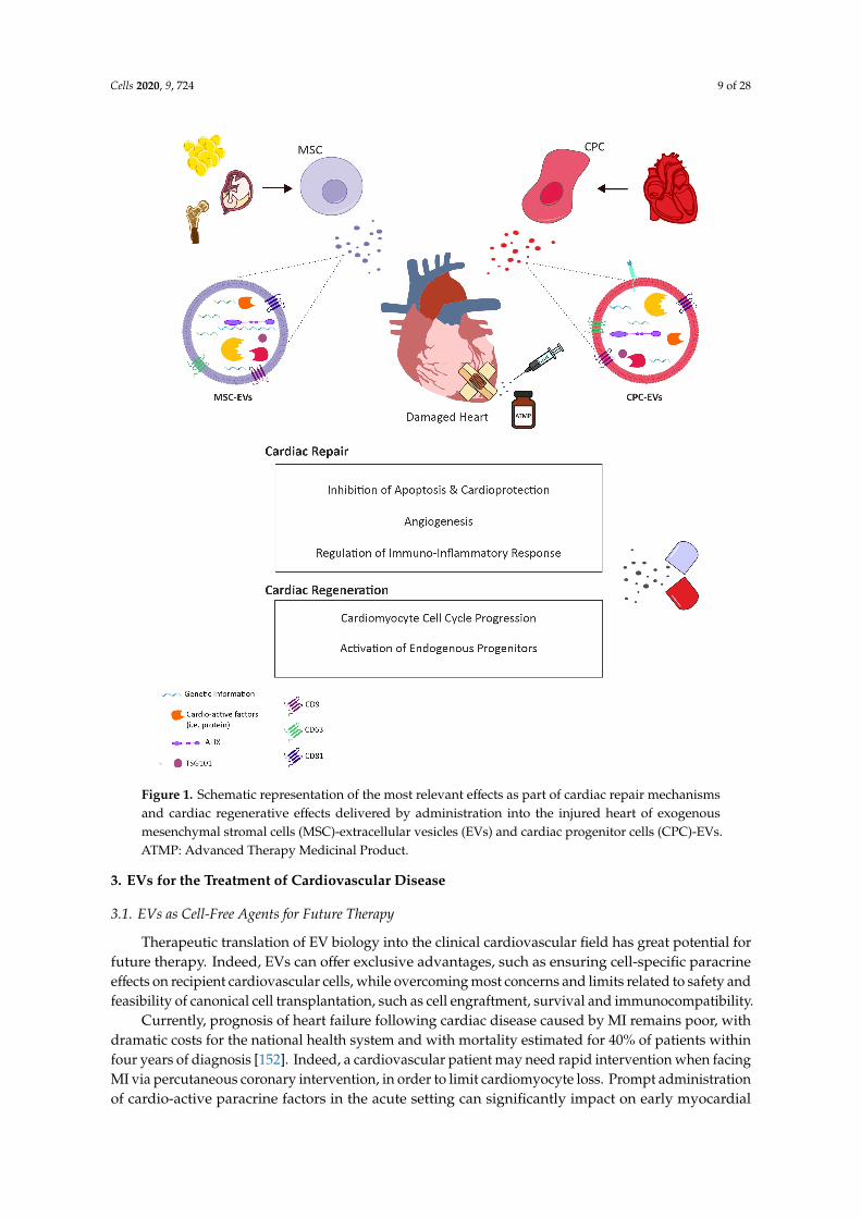
Figure 1. Schematic representation of the most relevant effects as part of cardiac repair mechanisms and cardiac regenerative effects delivered by administration into the injured heart of exogenous mesenchymal stromal cells (MSC)-extracellular vesicles (EVs) and cardiac progenitor cells (CPC)-EVs. ATMP: Advanced Therapy Medicinal Product.
3. EVs for the Treatment of Cardiovascular Disease
3.1. EVs as Cell-Free Agents for Future Therapy
Therapeutic translation of EV biology into the clinical cardiovascular field has great potential for future therapy. Indeed, EVs can offer exclusive advantages, such as ensuring cell-specific paracrine effects on recipient cardiovascular cells, while overcoming most concerns and limits related to safety and feasibility of canonical cell transplantation, such as cell engraftment, survival and immunocompatibility.
Currently, prognosis of heart failure following cardiac disease caused by MI remains poor, with dramatic costs for the national health system and with mortality estimated for 40% of patients within
Figure 1. Schematic representation of the most relevant effects as part of cardiac repair mechanismsand cardiac regenerative effects delivered by administration into the injured heart of exogenousmesenchymal stromal cells (MSC)-extracellular vesicles (EVs) and cardiac progenitor cells (CPC)-EVs.ATMP: Advanced Therapy Medicinal Product.
3. EVs for the Treatment of Cardiovascular Disease
3.1. EVs as Cell-Free Agents for Future Therapy
Therapeutic translation of EV biology into the clinical cardiovascular field has great potential forfuture therapy. Indeed, EVs can offer exclusive advantages, such as ensuring cell-specific paracrineeffects on recipient cardiovascular cells, while overcoming most concerns and limits related to safety andfeasibility of canonical cell transplantation, such as cell engraftment, survival and immunocompatibility.
Currently, prognosis of heart failure following cardiac disease caused by MI remains poor, withdramatic costs for the national health system and with mortality estimated for 40% of patients withinfour years of diagnosis [152]. Indeed, a cardiovascular patient may need rapid intervention when facingMI via percutaneous coronary intervention, in order to limit cardiomyocyte loss. Prompt administrationof cardio-active paracrine factors in the acute setting can significantly impact on early myocardial
Cells 2020, 9, 724 10 of 28
injury response, by preserving more viable tissue and sustaining local angiogenesis, thus limitingexacerbation of inflammation into chronic activation of myofibroblasts and spreading of fibrosis andscarring. This may result in effective cardioprotection while opposing detrimental remodelling,thus enhancing defective cardiac repair and inhibiting or at least delaying the onset of heartfailure. As well, patients affected by non-ischaemic cardiomyopathy and/or cardiac dysfunctiondue to oncological treatment-derived cardiotoxicity may benefit from paracrine therapy to counteractpremature senescence of resident cardiomyocytes and myocardial mitochondria impairment fromincreased oxidative stress.
Likewise, congenital heart defects, including very severe conditions (i.e., hypoplastic left heartsyndrome) or those with relatively limited structural alterations (i.e., septal defects), may often result inpaediatric heart failure. For defects requiring early treatment, the standard therapy is elective surgerywithin the first weeks of life using prosthetic implants to provide structural reconstruction [153].However, such implants do not grow with the patient and additional surgeries are necessary with age.Thus, it would be of great advantage to support structural and functional myocardial reconstitution inthese patients by stimulating resident cardiomyocyte renewal potential via paracrine stimulation.
In this scenario, EVs may represent a promising “off-the-shelf” next-generation advanced medicinaltherapy product, which may be produced via a scale-up system from in vitro cultured cells to bepromptly available as a pharmaceutical formulation for simple administration to the cardiovascularpatient, when needed. Hence, the big question in preclinical research is focussing on identifying themost suitable stem/progenitor cell population to isolate EVs endowed with the most cardio-activeparacrine potential. Here, we will review two main sources, endogenous CPCs versus exogenousstem/progenitor cells and discuss their capacity to impact cardiac intrinsic reparative and rejuvenatingmechanisms via EV modulatory activity, as illustrated by the schematic in Figure 1.
3.2. A Role for CPC-Derived EVs
EVs released by primary cardiac stromal cells, obtained from atrial appendage and grown asmonolayer [89] or by cardiosphere-derived cells (CDCs) [154], demonstrated reproduction of thecardioprotective and angiogenic effects of their parent cells both in vitro and in vivo. The benefits ofCPC secretome in protecting cardiomyocytes from apoptosis have been attributed to their EV fractionacting through mechanisms involving both the transfer of nucleic acids and protein-protein interactions.For example, CPC-EVs are enriched in anti-apoptotic miRNA210, which acts by downregulatingspecific target genes in recipient cells such as ephrin A3 and PTP1b [89]. Likewise, miRNA132 inCPC-EVs was shown to exert a pro-angiogenic effect on endothelial cells by targeting RasGAP-p120 [89].The surface protein defined as pregnancy-associated plasma protein-A (PAPP-A) was found to beassociated within CPC-EV as master regulator responsible for the release of active IGF factor leadingto activation of pro-survival pathways involving phosphorylation of ERK1/2 and Akt in recipientcells [155]. In a preclinical rat model of acute myocardial infarction (MI), CPC-EVs injected into theinfarct border zone or systemically injected via tail vein increased viable mass and vessel density,resulted in a diminished scar and improved global heart function [154,156]. Ibrahim et al. showedthat CDC-EVs were enriched in miRNA146a-5p. This miRNA was responsible for the increasedviability of cardiomyocytes exposed to oxidative stress when treated with CDC-EVs as compared tocontrols. miRNA146a-5p downregulates specific genes in the target cells, including Traf6 and Irak1, twosignalling mediators of the TLR-NFKB axis. Moreover, miR-146a knock-out mice showed larger infarctscompared to wild-type mice; injection of a miR-146a mimic at the time of MI rescued the level of ejectionfraction similarly to wild-type animals, suggesting a mechanism of action for the cardioprotective effectmediated by CDC-EVs [90]. A similar effect has also been reported in a preclinical model of cancerdrug-induced cardiotoxicity [157]. Indeed, human CPC-EVs were able to significantly decrease reactiveoxygen species (ROS) production in cardiomyocytes following Doxorubicin/trastuzumab treatmentin vitro. CPC-EVs prevented Doxorubicin-derived induction of Traf6, Smad4, Irak1, Nox4 and Mpogene expression; this effect was abolished when miRNA146a-5p was knocked down via loading of
Cells 2020, 9, 724 11 of 28
a specific siRNA into the EV-producing cells. Moreover, in vivo intravenous administration of EVssignificantly preserved cardiac function during Doxorubicin/trastuzumab treatment [157]. Additionalstudies have also identified Y RNA fragment inside CDC-EVs [158]. Such specific RNA was found tobe actively transferred from EVs to macrophages and was able to induce macrophage polarisationinto an M2 resolving phenotype. Y RNA provided cardioprotection by enhancing phagocytosis andscavenging debris from dying cells, thus attenuating damage from post-myocardial infarction [158].CDC-EVs have been also tested in a porcine preclinical model, as delivered by intracoronary (i.c.) oropen-chest intramyocardial (i.m.) routes 30 min after reperfusion, with the first being shown to beineffective over the latter, in which EVs concurred to decrease infarct size and preserve left ventricularejection fraction [159].
Priming of secreting CPCs via specific stimuli has also been suggested to improve EV biologicalactivity; EVs produced by CPCs cultured for 12 h under hypoxia conditions were able to increasetube formation in endothelial cells and decrease pro-fibrotic gene expression in TGF-α stimulatedfibroblast in vitro [160]. Microarray analysis identified eleven miRNAs upregulated in hypoxicpreconditioned CPC-EVs compared to naive ones. Administration of hypoxic EVs into a preclinicalmodel of ischaemia reperfusion injury (I/R) resulted in an improved cardiac functionality outcomecompared to normoxic EVs [160]. Likewise, the potency of neonatal, infant or child human CPC-EVsunder normoxic and hypoxic conditions has been also investigated. All three different populationof EVs derived from CPCs exposed to hypoxia were more effective as compared to their normoxiccounterpart, in terms of increasing new vessels formation and decreasing of fibrosis in a preclinical ratmodel of myocardial ischaemia/reperfusion injury (I/R) [161] With regards to their beneficial effects oncardiac function, neonatal CPC-EVs showed similar results in hypoxic or normoxic conditions [161].While the cardioprotective effect of CPC-EVs has been broadly reported, very little is currently knownabout their putative effect on cardiomyocyte renewal. Positive effects of EVs on in vitro cultured ratneonatal cardiomyocytes have been described [90], together with encouraging data showing Ki67+
cardiomyocytes in an MI mouse model injected with CPC-EVs; yet the mechanism of action underlyingsuch effect still needs clarification [162,163].
3.3. Contribution of Exogenous Stem/Progenitor Cell-EVs
CPCs have lately gained increasing attention as an appealing source for cardiac repair as beingtissue-specific resident progenitors with significant paracrine activity, thus intrinsically likely toproduce a more effective cardio-active secretome compared to other (somatic) sources. Despite theincreasing number of studies supporting their EV cardiovascular modulatory potential, it is still amatter of debate whether they can represent the most suitable cell reservoir for future paracrine therapy.Indeed, isolation feasibility and self-renewal potential are key aspects of the ideal stem/progenitorsecreting cell for clinical translation. In this perspective, as compared to other non-cardiac specificstem/progenitor cell sources, CPCs may be limited in their isolation from cardiac specimen via invasivesurgical procedures or from cadaver donor supply and their limited proliferative potential. On theother hand, exogenous stem cells may offer an easily accessible and exploitable resource.
In such scenarios, several populations have been investigated in the last years. Embryonic stemcell (ESC)-EVs have been described as positively modulating the cardiac healing process in a preclinicalacute MI murine model. Notably, murine ESC-EVs not only supported local neovascularization,promoted cardiomyocyte survival and reduced scarring, but also enhanced CPC reactivation and theircontribution to cardiac repair putatively via miR-294 delivery [164]; likewise, rodent ESC-EVs havebeen tested as an anti-inflammatory treatment in a preclinical mouse model of Doxorubicin-derivedcardiomyopathy as quenching inflammasome protein expression leading to cardiomyocyte pyroptosis,as well as promoting the skewing of macrophages from pro-inflammatory M1 into pro-resolvingM2 phenotype [165]. Since the discovery of induced pluripotent stem (iPS) cells in 2006 [166], theyhave been increasingly been tested as therapeutic agents for cardiovascular disease since overcomingmajor ESC concerns in terms of availability and ethical issues. More recently, on top of their plasticity,
Cells 2020, 9, 724 12 of 28
the paracrine potential of their secreted EVs has also been evaluated; indeed, mouse iPS cell-EVshave shown to exert cardioprotective effects on target cardiomyocyte both in vitro and in vivo byexosomal transfer of miR-21 and miR-210 as regulated by Nanog and HIF-1α [167]. Another strategythat has been lately pursued focusses on iPS cells as an exploitable “biopharmaceutical” source of(cardiovascular) progenitor cells and cardiomyocytes [168]. Indeed, multipotent CPCs have beengenerated from human iPS cells via stimulation of the small antioxidant molecule ISX-9; extracellularvesicles secreted by ISX-9-induced CPCs (EV-CPCISX-9) counteracted cardiac fibrosis and amelioratedlocal angiogenesis in mice undergoing MI putatively via antifibrotic miR-373 delivery [169]. Similarly,iPS cell-derived cardiomyocytes (iCMs) have been broadly described as an alternative option for celltherapy approach [170]; since rodent cardiomyocytes have shown to secrete EVs biologically activeon cardiovascular cells, iCM-EVs may offer an abundant paracrine source as well. Notably, a tissueengineering approach based on the application of an engineered hydrogel patch for the controlledrelease of iCM-EVs in a rat preclinical model of MI reduced infarct size while supporting cardiacfunction [171]. Despite such appealing results, EVs from iPS-derived cells may require extensivemanipulation and can be cost-effective.
Therefore, several somatic stem and tissue progenitor cells have been broadly investigated as afeasible alternative as EV biosource. Several preclinical and clinical studies have described beneficialmodulatory effects of human bone marrow-derived CD34+ haematopoietic stem cells for cardiovasculardisease, such as limiting angina frequency [172–174]. The therapeutic pro-angiogenic paracrine activityof human CD34+ stem cells in counteracting ischaemic injury has been shown to be recapitulated by theirsecreted EVs [175,176]. Given the relevance of therapeutic angiogenesis for de novo vessel formationfollowing ischaemic injury, much interest has also been dedicated to the characterisation of endothelialprogenitor cell (EPC)-EVs. Indeed, stimulation of cardiomyocytes undergoing Angiotensin II-inducedhypertrophy and apoptosis with EPC-derived microvesicles resulted in improved survival as mediatedby RNA-driven modulation of PI3K/Akt/eNOS pathway [177]. More recently, EPC-EVs have beenadministered to a rat model of MI within a shear-thinning hydrogel for their precise delivery and steadyrelease, thus sustaining cardiac function and enhancing vessel density [178]. Notably, interleukin-10deficiency within EPC-EVs has been recently shown to significantly affect their therapeutic influencefor myocardial repair as altering their protein cargo and upregulating integrin-linked kinase-mediatedactivation of NF-κB in recipient cells; thus, modulation of the inflammatory priming of secreting EPCcan impact on their EV profile and cardiovascular paracrine potential [179].
Haematopoietic stem and endothelial progenitor cells may represent promising cell sources;yet, their isolation yield might be limited by the need for specific stimulation. Hence, specificattention has been lately alternatively focussed on mesenchymal stromal cells (MSCs) obtained fromeither bone marrow (BM) or adipose tissue (AD) as easily reachable cell references for EV isolation.In particular, MSC-EVs have been shown to exert comparable, or even superior, therapeutic activitiesover parental MSCs in terms of quenching pro-inflammatory processes, oxidative damage and spreadof fibrosis within the injured tissue [180]. With respect to ischaemic cardiovascular disease, MSC-EVtreatment resulted in reduced myocardial apoptosis, and consequently decreased infarct size, withimproved functional recovery and formation of new vessels, as extensively reviewed in [181]. Indeed,intra-myocardial delivery of EVs obtained from BM-MSCs has been reported to significantly modulatethe cardiac microenvironment after acute myocardial infarction (MI), by stimulating neovascularisationand restraining the inflammation response [182]. Another relevant source of somatic MSCs isrepresented by the adipose tissue, since cells can be easily harvested by minimally invasive surgicaltechniques during lipoaspirate procedures. EVs obtained from AD-MSCs have been demonstratedprotection of the myocardium against I/R injury via the Wnt/b-catenin signalling pathway [183]. As amatter of fact, the remarkably strong immunosuppressive effect of both BM- and AD-MSC-EVs onT cell activation and macrophage polarisation has been documented as one of their major beneficialeffects [184–186]. Increasing evidence has indicated that the EV microRNA (miRNA) content as oneof their most likely paracrine mechanism(s) of action. Several miRNAs have been associated with
Cells 2020, 9, 724 13 of 28
MSC-EV cardioprotective, pro-survival and angiogenic effects, including, for example, miR-21 [187];miR-125b [188]; miR-320d [189]; miR-95–3p [190]; miR-210 [191]; miR-29 and miR-24 [180]. Additionally,EVs have been shown to biochemically restore ATP and NADH levels and redox state within theinjured tissue [192].
Given the remarkable cardio-active effects of EVs, different methods have been lately exploredto boost their therapeutic exploitation by ad hoc preconditioning or engineering of parental MSCs.Indeed, somatic MSCs have been stimulated either under hypoxic and/or pro-inflammatory conditions,resulting in the implementation of their secreted EVs to provide tissue repair, along with modulatoryand pro-angiogenic effects [188,193–195]. Pharmacological priming by atorvastatin, a well-establishedlipid-lowering drug that can enhance stromal progenitor cell cardioprotective effects, has also beenreported [196]. Another well-accepted approach is to increase EV function via genetic overexpressionof specific miRNAs within parental MSC as vesicle cargo loading strategy; as result, a significant foldenrichment of those regulatory miRNAs putatively involved in EV paracrine ability has been observed,thus enhancing their phenotypic effects on responder cells [189,190,197].
Adult somatic or embryonic stem cells may be limited by low yield, invasive sampling,controversial self-renewal and ethical issues. Stromal progenitors isolated from foetal and perinatalextra-embryonic tissues can offer an ideal alternative as a leftover sample from prenatal screeningprocedures during gestation or as waste material after birth. Indeed, foetal and perinatal MSCs areimmature progenitors with high self-renewal potential, have significant immunomodulatory propertiesand distinct pro-angiogenic, cytoprotective and anti-inflammatory paracrine profile. Foetal andperinatal sources include amniotic fluid and term umbilical cord and placenta, respectively, from whichMSC can be easily isolated without any ethical concern [198]. EVs from umbilical cord-derived MSCs(UC-MSC-EVs) have been recently broadly scrutinised in terms of their beneficial effects in differentpreclinical animal models of myocardial injury. Human UC-MSC-EVs overexpressing the pro-survivalAkt kinase was shown to modulate local angiogenesis in a preclinical rat model of MI, via PDGF-Das pro-angiogenic mediator [199]. Likewise, EVs from UC-MSCs overexpressing the tissue matrixmetalloproteinase inhibitor 2 (TIMP2) or the cardioprotective stromal-derived factor 1 alpha (SFD1a)have been shown to limit detrimental ventricular remodelling via the pro-survival Akt/Sfrp2 pathwayand to inhibit apoptosis and autophagy of myocardial cells while sustaining local angiogenesis inpreclinical rodent models of MI [200,201]. MSCs derived from human term placenta, also referredto as amniotic mesenchymal stromal cells (AMSCs), are well-known for their (immuno)modulatoryproperties [202,203]; EVs released by human term placenta-MSCs have been recently demonstratedexertion of relevant therapeutic effects as supporting new vessel development in vitro and in vivo [204],with nitric oxide (NO)-releasing polymer stimulation as a functional trigger of exosomal enrichmentof pro-angiogenic VEGF and miR-126 [205]. Notably, placenta-MSCs and their derived EVs havebeen shown to counteract skewing of myoblasts to fibrogenic phenotype and collagen IV expressionin the cardiac tissue of preclinical models of Duchenne musclar dystrophy, via targeted delivery ofmiR-29c [206].
Notably broadly multipotent MSCs with remarkable proliferative capacity along withpro-angiogenic, anti-inflammatory and cardioprotective modulatory potential have been describedas isolated from human amniotic fluid, namely c-KIT+ human amniotic fluid-derived stem cells(hAFSCs) [207–213]. hAFSCs can be easily isolated from leftover samples of amniotic fluid obtainedfrom prenatal screening amniocentesis (i.e., foetal hAFSCs) or from clinical waste during eligiblecaesarean delivery without any ethical concern (i.e., perinatal hAFSCs). In particular, hAFSC-EVshave been reported to enhance cell survival in preclinical models of kidney, lung and skeletal andcardiac muscle injury, possibly via the horizontal transfer of their miRNA cargo, including cardio-activemiR-199a-3p and miR-210 [83,214–216]. In mice undergoing MI, single intramyocardial administrationof hAFSC-EVs obtained following cell preconditioning under hypoxia resulted in prolonged beneficialeffects ameliorating cardiac repair mechanisms and inhibiting worsening of myocardial function inthe long term [83]; hence, being hAFSCs immature and “developmentally young” progenitors, they
Cells 2020, 9, 724 14 of 28
may possess a powerful paracrine potential for myocardial “rejuvenation” strategies and represent anappealing cell source for future therapy.
Notably, fewer studies have specifically addressed the MSC-EV regenerative potential to locallyreactivate in vivo endogenous CPCs, along with stimulation of resident cardiomyocyte renewal. HumanmiR-590–3p has been previously reported to impact myocardial reconstitution by downregulating genesinhibiting cardiomyocyte proliferation; indeed, based on their miR-590–3p enrichment, EVs obtainedfrom MSC genetically engineered to express a cardiac troponin-targeting peptide showed to targetcardiomyocyte cell cycle re-entry and sustained cardiac function in a preclinical MI model [217];similarly, miR-199a-loaded BM-MSC-EVs have demonstrated support of rodent cardiomyocytecell cycle progression in vitro via dose-dependent manner by means of Crim1 and Caspase-9downregulation [197]). Likewise, hAFSC-EVs were able to stimulate endogenous regenerativemechanisms from within the injured murine myocardium by reactivating epicardial WT1+ CPCsparacrine activity and triggering resident cardiomyocyte cell cycle progression up to DNA duplicationphase, both at early and longer time points after MI [83]. Since structural and functional cardiomyocytereconstitution is the sine qua non condition for true cardiac regeneration, further investigationsare required to carefully define the EV potential in sustaining bona fide cardiomyocyte duplicationby rigorous evaluation of complete cytokinesis over defective karyokinesis with DNA duplicationresulting in binucleation/polyploidy.
3.4. Looking for the Right Address: Improving EV Cardiac Tropism
A critical aspect to be considered in clinical translation of EVs for future cardiovascular medicineis represented by their quite limited homing potential when delivered systemically.
Intra-myocardial administration of EVs soon after injury (i.e., myocardial infarction) has beenshown to provide beneficial paracrine effects addressing both cardiac repair and regenerativemechanisms, resulting in long-term effects after a single acute injection [89,90,155,218]. Nevertheless,paracrine effects are well known to act promptly and locally, therefore follow-up administrationwould be the ideal treatment regime, especially to modulate cardiac detrimental pathological cardiacremodelling over time. Several ideas have been put forward to overcome the low EV retention rateafter transplantation in vivo by suggesting, for example, functional vesicle encapsulation in optimisedhydrogel formulation to ensure topical sustained release over time [219]. It is important to bearin mind that not all cardiovascular patients may be eligible for acute in situ treatment requiringeither percutaneous coronary intervention/angioplasty or collateral cardiac surgery; therefore, there isan increasing need for optimizing EV cardiac-specific homing for future cell-free paracrine therapy.Several studies have been lately reported suggesting alternative strategies to functionalise EVs for moreaccurate targeting of the damaged heart [220,221]. These include: lentiviral vector-based engineeringof secreting cells to upregulate the expression of cardiomyocyte-specific binding peptides fused tothe murine transmembrane protein Lamp2b, in order to enrich the targeting epitope on the exosomalsurface [222]; overexpression of exosomal CXCR4 to push their bioavailability towards the ischaemicheart [156]; membrane anchoring systems to directly dock tissue-specific antibodies or homing antigenson the EV surface [223,224].
Notably, a very elegant work has recently translated CAR-T cell technology beyond oncologyinto the cardiovascular field, to specifically address and treat cardiac fibrosis via the engineering ofCAR receptor against a cardiac endogenous protein activating resident fibroblasts [225]. Likewise,CAR-EVs, as derived from CAR-T cells, have also been reported as maintaining target specificity witha much lower risk of cytotoxic cytokine release syndrome commonly caused by adoptive CAR-T celltherapy [226]. This may shed new light on the development of novel strategies to enhance EV cardiactropism as putative future advanced medicinal paracrine therapy products to counteract cardiovasculardisease and heart failure.
Cells 2020, 9, 724 15 of 28
4. Conclusions: Challenges to be Overcome
The analytical study of EVs is a very active area of research. Functional readouts of biomedicalapplications of EV obtained by either CPCs or exogenous stromal cells of non-cardiac origin haverevealed that both types may harbour therapeutic relevance for cardiovascular disease, questioningthe rationale that CPCs could have superior cardio-active effects. Yet, some additional aspects arein need of further clarification in order to achieve a general consensus. Indeed, in order to developan EV-based therapeutic approach, a comprehensive characterisation of the tissue/cellular source ofEVs is imperative. Detailed methods to obtain human cells from different tissues origin are wellreported; however, donor-to-donor variability remains a prominent challenge. BM-MSCs from agedmice have demonstrated reduced wound healing, angiogenesis, proliferation and anti-apoptoticcapabilities [227]; similarly, human adipose tissue MSCs, derived from patients at different ages,showed remarkably reduced in vitro differentiation potential [227]. Therefore, age and degree ofdevelopmental/maturation commitment can affect stem and progenitor cell reparative and regenerativepotential; how this specifically impacts the therapeutic capacity of derived EVs remains to be specificallydefined. Currently, there is little knowledge of how co-purified non-EV associated molecules affect theactivity of the samples. Furthermore, it is reasonable to think that only a portion of EVs mediate theexpected therapeutic effect while others could act in an antagonistic manner. Indeed, due to a currentlack of practical technologies to analyse EVs at the single vesicle level, the heterogeneity of EV fractionscannot be comprehensively addressed, even if they are harvested from apparently homogeneous cellsources. Proper in vitro and in vivo tests are required to predict the intended therapeutic potential,along with safety and potency, of the EV fractions. Moreover, one of the most relevant therapeuticadvantage of EVs, compared to the parental cells, is their potential to “escape” to the immune system.The absence of costimulatory molecule MHC-II gives EVs the potential to escape recognition by CD4lymphocytes [90,216,228], although this aspect needs more detailed investigation.
A key point to be considered is the current lack of standardised procedure available for EVisolation, as well as for their storage. For instance, there is evidence that independent EVs preparationshows different immunomodulatory capability [229]. The International Society of Extracellular Vesicles(ISEV), in 2017, in a position paper [88], addressed some aspects of safety and regulatory requirementsthat must be considered for clinical application. EV-based therapeutics are now subjected to regulatoryframeworks concerning biological medicinal products; however, there is a consensus that specialguidelines for EV-based therapeutics may be needed. Moreover, the clinical translation of EVsrequires standardisation of administration and dosing. Nowadays, for liposome-encapsulated forms ofdoxorubicin [230], methods such as TRPS are currently used to verify particle size and characterisationand are accepted by FDA and EMA, yet for EVs, regulatory guidance is lacking.
Eventually, the development of a good manufacturing practice (GMP)-grade method for thelarge-scale preparation of EVs as an advanced therapy medicinal product (ATMP, Figure 1) is alsomandatory to implement their bench-to-bedside application. The first evidence of this has been recentlyperformed for CPC-EVs. Indeed, human CPCs cultured in xeno-free conditions showed production ofEVs with similar features as in research-grade conditions [228]. The GMP method guaranteed highexosome yield (>1013 particles) by isolating through a closed system of ultrafiltration, and consistentremoval (≥97%) of contaminating proteins. Thus, such standardised production method for large-scalemanufacturing of CPC-EVs may offer a good demonstration of the clinical translation of EV biologyfor human therapeutic applications for the treatment of acute myocardial infarction syndrome [228].
Funding: L.B. was funded by the Swiss National Science Foundation (IZCOZ0_182948/1); S.B. was funded bythe “Curiosity Driven” Starting Grant from University of Genova, Genova, Italy. This article contributes to theCOST Action CA17116 “International Network for Translating Research on Perinatal Derivatives into TherapeuticApproaches (SPRINT)” supported by COST (European Cooperation in Science and Technology), (S.B.).
Conflicts of Interest: The authors declare no potential conflicts of interests, neither any specific disclosure tobe reported.
Cells 2020, 9, 724 16 of 28
References
1. Reimer, K.A.; Lowe, J.E.; Rasmussen, M.M.; Jennings, R.B. The wavefront phenomenon of ischemic cell death.1. Myocardial infarct size vs duration of coronary occlusion in dogs. Circulation 1977, 56, 786–794. [CrossRef][PubMed]
2. Tissier, R.; Ghaleh, B.; Cohen, M.V.; Downey, J.M.; Berdeaux, A. Myocardial protection with mild hypothermia.Cardiovasc. Res. 2012, 94, 217–225. [CrossRef] [PubMed]
3. Hale, S.L.; Kloner, R.A. Myocardial temperature reduction attenuates necrosis after prolonged ischemia inrabbits. Cardiovasc. Res. 1998, 40, 502–507. [CrossRef]
4. Hamamoto, H.; Leshnower, B.G.; Parish, L.M.; Sakamoto, H.; Kanemoto, S.; Hinmon, R.; Miyamoto, S.;Gorman, J.H., 3rd; Gorman, R.C. Regional heterogeneity of myocardial reperfusion injury: Effect of mildhypothermia. Ann. Thorac. Surg. 2009, 87, 164–171. [CrossRef] [PubMed]
5. Dae, M.W.; Gao, D.W.; Sessler, D.I.; Chair, K.; Stillson, C.A. Effect of endovascular cooling on myocardialtemperature, infarct size, and cardiac output in human-sized pigs. Am. J. Physiol. Heart Circ. Physiol. 2002,282, H1584–H1591. [CrossRef] [PubMed]
6. van den Doel, M.A.; Gho, B.C.; Duval, S.Y.; Schoemaker, R.G.; Duncker, D.J.; Verdouw, P.D. Hypothermiaextends the cardioprotection by ischaemic preconditioning to coronary artery occlusions of longer duration.Cardiovasc. Res. 1998, 37, 76–81. [CrossRef]
7. Heusch, G. Molecular basis of cardioprotection: Signal transduction in ischemic pre-, post-, and remoteconditioning. Circ. Res. 2015, 116, 674–699. [CrossRef]
8. Murry, C.E.; Jennings, R.B.; Reimer, K.A. Preconditioning with ischemia: A delay of lethal cell injury inischemic myocardium. Circulation 1986, 74, 1124–1136. [CrossRef]
9. Zhao, Z.Q.; Corvera, J.S.; Halkos, M.E.; Kerendi, F.; Wang, N.P.; Guyton, R.A.; Vinten-Johansen, J.Inhibition of myocardial injury by ischemic postconditioning during reperfusion: Comparison with ischemicpreconditioning. Am. J. Physiol. Heart Circ. Physiol. 2003, 285, H579–H588. [CrossRef]
10. Przyklenk, K.; Bauer, B.; Ovize, M.; Kloner, R.A.; Whittaker, P. Regional ischemic ’preconditioning’ protectsremote virgin myocardium from subsequent sustained coronary occlusion. Circulation 1993, 87, 893–899.[CrossRef]
11. Schulman, D.; Latchman, D.S.; Yellon, D.M. Urocortin protects the heart from reperfusion injury viaupregulation of p42/p44 MAPK signaling pathway. Am. J. Physiol. Heart Circ. Physiol. 2002, 283,H1481–H1488. [CrossRef] [PubMed]
12. Hausenloy, D.J.; Tsang, A.; Yellon, D.M. The reperfusion injury salvage kinase pathway: A common targetfor both ischemic preconditioning and postconditioning. Trends Cardiovasc. Med. 2005, 15, 69–75. [CrossRef][PubMed]
13. Juhaszova, M.; Zorov, D.B.; Kim, S.H.; Pepe, S.; Fu, Q.; Fishbein, K.W.; Ziman, B.D.; Wang, S.; Ytrehus, K.;Antos, C.L.; et al. Glycogen synthase kinase-3beta mediates convergence of protection signaling to inhibitthe mitochondrial permeability transition pore. J. Clin. Investig. 2004, 113, 1535–1549. [CrossRef] [PubMed]
14. Tsang, A.; Hausenloy, D.J.; Mocanu, M.M.; Yellon, D.M. Postconditioning: A form of “modified reperfusion”protects the myocardium by activating the phosphatidylinositol 3-kinase-Akt pathway. Circ. Res. 2004, 95,230–232. [CrossRef] [PubMed]
15. Cohen, M.V.; Yang, X.M.; Liu, Y.; Solenkova, N.V.; Downey, J.M. Cardioprotective PKG-independent NOsignaling at reperfusion. Am. J. Physiol. Heart Circ. Physiol. 2010, 299, H2028–H2036. [CrossRef] [PubMed]
16. Sun, J.; Aponte, A.M.; Kohr, M.J.; Tong, G.; Steenbergen, C.; Murphy, E. Essential role of nitric oxide in acuteischemic preconditioning: S-nitros(yl)ation versus sGC/cGMP/PKG signaling? Free Radic. Biol. Med. 2013, 54,105–112. [CrossRef]
17. Kohr, M.J.; Sun, J.; Aponte, A.; Wang, G.; Gucek, M.; Murphy, E.; Steenbergen, C. Simultaneous measurementof protein oxidation and S-nitrosylation during preconditioning and ischemia/reperfusion injury withresin-assisted capture. Circ. Res. 2011, 108, 418–426. [CrossRef]
18. Rassaf, T.; Totzeck, M.; Hendgen-Cotta, U.B.; Shiva, S.; Heusch, G.; Kelm, M. Circulating nitrite contributesto cardioprotection by remote ischemic preconditioning. Circ. Res. 2014, 114, 1601–1610. [CrossRef]
19. Soetkamp, D.; Nguyen, T.T.; Menazza, S.; Hirschhauser, C.; Hendgen-Cotta, U.B.; Rassaf, T.; Schluter, K.D.;Boengler, K.; Murphy, E.; Schulz, R. S-nitrosation of mitochondrial connexin 43 regulates mitochondrialfunction. Basic Re.s Cardiol. 2014, 109, 433. [CrossRef]
Cells 2020, 9, 724 17 of 28
20. Boengler, K.; Dodoni, G.; Rodriguez-Sinovas, A.; Cabestrero, A.; Ruiz-Meana, M.; Gres, P.; Konietzka, I.;Lopez-Iglesias, C.; Garcia-Dorado, D.; Di Lisa, F.; et al. Connexin 43 in cardiomyocyte mitochondria and itsincrease by ischemic preconditioning. Cardiovasc. Res. 2005, 67, 234–244. [CrossRef]
21. Ardehali, H.; O’Rourke, B. Mitochondrial K(ATP) channels in cell survival and death. J. Mol. Cell. Cardiol.2005, 39, 7–16. [CrossRef] [PubMed]
22. Dorofeyeva, L.V. Obtaining of measles virus haemagglutinin from strain L-16 grown in primary cell cultures.Acta Virol. 1975, 19, 497. [PubMed]
23. Patel, H.H.; Gross, E.R.; Peart, J.N.; Hsu, A.K.; Gross, G.J. Sarcolemmal KATP channel triggers delayedischemic preconditioning in rats. Am. J. Physiol. Heart Circ. Physiol. 2005, 288, H445–H447. [CrossRef][PubMed]
24. Penna, C.; Mancardi, D.; Rastaldo, R.; Losano, G.; Pagliaro, P. Intermittent activation of bradykinin B2receptors and mitochondrial KATP channels trigger cardiac postconditioning through redox signaling.Cardiovasc. Res. 2007, 75, 168–177. [CrossRef]
25. Kristiansen, S.B.; Henning, O.; Kharbanda, R.K.; Nielsen-Kudsk, J.E.; Schmidt, M.R.; Redington, A.N.;Nielsen, T.T.; Botker, H.E. Remote preconditioning reduces ischemic injury in the explanted heart by a KATPchannel-dependent mechanism. Am. J. Physiol. Heart Circ. Physiol. 2005, 288, H1252–H1256. [CrossRef]
26. Rentoukas, I.; Giannopoulos, G.; Kaoukis, A.; Kossyvakis, C.; Raisakis, K.; Driva, M.; Panagopoulou, V.;Tsarouchas, K.; Vavetsi, S.; Pyrgakis, V.; et al. Cardioprotective role of remote ischemic periconditioning inprimary percutaneous coronary intervention: Enhancement by opioid action. JACC Cardiovasc. Interv. 2010,3, 49–55. [CrossRef]
27. Shimizu, M.; Tropak, M.; Diaz, R.J.; Suto, F.; Surendra, H.; Kuzmin, E.; Li, J.; Gross, G.; Wilson, G.J.;Callahan, J.; et al. Transient limb ischaemia remotely preconditions through a humoral mechanism actingdirectly on the myocardium: Evidence suggesting cross-species protection. Clin. Sci. 2009, 117, 191–200.[CrossRef]
28. Leung, M.K.; Irwin, M.G. Perioperative cardioprotection. F1000Prime Rep. 2013, 5, 7. [CrossRef]29. Gragnano, F.; Cattano, D.; Calabro, P. Perioperative care of cardiac patient’s candidate for non-cardiac
surgery: A critical appraisal of emergent evidence and international guidelines. Intern. Emerg. Med. 2018, 13,1185–1190. [CrossRef]
30. Lionetti, V.; Barile, L. Perioperative cardioprotection: Back to bedside. Minerva Anestesiol. 2019. [CrossRef]31. Mushtaq, M.; Cohn, S.L. Perioperative beta-blockers in noncardiac surgery: The evidence continues to evolve.
Clevel. Clin. J. Med. 2014, 81, 501–512. [CrossRef]32. Prabhu, S.D.; Chandrasekar, B.; Murray, D.R.; Freeman, G.L. beta-adrenergic blockade in developing heart
failure: Effects on myocardial inflammatory cytokines, nitric oxide, and remodeling. Circulation 2000, 101,2103–2109. [CrossRef] [PubMed]
33. Gnecchi, M.; Danieli, P.; Malpasso, G.; Ciuffreda, M.C. Paracrine mechanisms of mesenchymal stem cells intissue repair. Methods Mol. Biol. 2016, 1416, 123–146. [CrossRef] [PubMed]
34. Chimenti, I.; Smith, R.R.; Li, T.S.; Gerstenblith, G.; Messina, E.; Giacomello, A.; Marban, E. Relative roles ofdirect regeneration versus paracrine effects of human cardiosphere-derived cells transplanted into infarctedmice. Circ. Res. 2010, 106, 971–980. [CrossRef] [PubMed]
35. Rossello, X.; Yellon, D.M. Cardioprotection: The disconnect between bench and bedside. Circulation 2016,134, 574–575. [CrossRef]
36. Heusch, G. Critical Issues for the Translation of Cardioprotection. Circ. Res. 2017, 120, 1477–1486. [CrossRef]37. Lukacs, E.; Magyari, B.; Toth, L.; Petrasi, Z.; Repa, I.; Koller, A.; Horvath, I. Overview of large animal
myocardial infarction models (review). Acta Physiol. Hung. 2012, 99, 365–381. [CrossRef]38. Jones, S.P.; Tang, X.L.; Guo, Y.; Steenbergen, C.; Lefer, D.J.; Kukreja, R.C.; Kong, M.; Li, Q.; Bhushan, S.;
Zhu, X.; et al. The NHLBI-sponsored consortium for preclinicAl assESsment of cARdioprotective therapies(CAESAR): A new paradigm for rigorous, accurate, and reproducible evaluation of putative infarct-sparinginterventions in mice, rabbits, and pigs. Circ. Res. 2015, 116, 572–586. [CrossRef]
39. Lefer, D.J.S.; Steenbergen, C.; Kukreja, R.; Guo, Y.; Tang, X.L.; Li, Q.; Ockaili, R.; Salloum, F.; Kong, M.;Polhemus, D.; et al. Sodium Nitrite Fails to Limit Myocardial Infarct Size: Results from the CAESARCardioprotection Consortium. FASEB 2014, 28, LB645.
40. Lefer, D.J.; Marban, E. Is Cardioprotection Dead? Circulation 2017, 136, 98–109. [CrossRef]
Cells 2020, 9, 724 18 of 28
41. Serebruany, V.L.; Atar, D. Assessment of bleeding events in clinical trials–proposal of a new classification.Am. J. Cardiol. 2007, 99, 288–290. [CrossRef] [PubMed]
42. Segers, V.F.; Lee, R.T. Stem-cell therapy for cardiac disease. Nature 2008, 451, 937–942. [CrossRef] [PubMed]43. Bolli, R.; Tang, X.L.; Sanganalmath, S.K.; Rimoldi, O.; Mosna, F.; Abdel-Latif, A.; Jneid, H.; Rota, M.; Leri, A.;
Kajstura, J. Intracoronary delivery of autologous cardiac stem cells improves cardiac function in a porcinemodel of chronic ischemic cardiomyopathy. Circulation 2013, 128, 122–131. [CrossRef]
44. Johnston, P.V.; Sasano, T.; Mills, K.; Evers, R.; Lee, S.T.; Smith, R.R.; Lardo, A.C.; Lai, S.;Steenbergen, C.; Gerstenblith, G.; et al. Engraftment, differentiation, and functional benefits of autologouscardiosphere-derived cells in porcine ischemic cardiomyopathy. Circulation 2009, 120, 1075–1083, 1077 pfollowing 1083. [CrossRef] [PubMed]
45. Lee, S.T.; White, A.J.; Matsushita, S.; Malliaras, K.; Steenbergen, C.; Zhang, Y.; Li, T.S.; Terrovitis, J.; Yee, K.;Simsir, S.; et al. Intramyocardial injection of autologous cardiospheres or cardiosphere-derived cells preservesfunction and minimizes adverse ventricular remodeling in pigs with heart failure post-myocardial infarction.J. Am. Coll. Cardiol. 2011, 57, 455–465. [CrossRef] [PubMed]
46. Hausenloy, D.J.; Candilio, L.; Evans, R.; Ariti, C.; Jenkins, D.P.; Kolvekar, S.; Knight, R.; Kunst, G.; Laing, C.;Nicholas, J.; et al. Remote ischemic preconditioning and outcomes of cardiac surgery. N. Engl. J. Med. 2015,373, 1408–1417. [CrossRef] [PubMed]
47. Meybohm, P.; Bein, B.; Brosteanu, O.; Cremer, J.; Gruenewald, M.; Stoppe, C.; Coburn, M.; Schaelte, G.;Boning, A.; Niemann, B.; et al. A multicenter trial of remote ischemic preconditioning for heart surgery. N.Engl. J. Med. 2015, 373, 1397–1407. [CrossRef]
48. Gittenberger-de Groot, A.C.; Winter, E.M.; Poelmann, R.E. Epicardium-derived cells (EPDCs) in development,cardiac disease and repair of ischemia. J. Cell. Mol. Med. 2010, 14, 1056–1060. [CrossRef]
49. Shrivastava, S.; Srivastava, D.; Olson, E.N.; DiMaio, J.M.; Bock-Marquette, I. Thymosin beta4 and cardiacrepair. Ann. N. Y. Acad. Sci. 2010, 1194, 87–96. [CrossRef]
50. Smart, N.; Risebro, C.A.; Melville, A.A.; Moses, K.; Schwartz, R.J.; Chien, K.R.; Riley, P.R. Thymosinbeta4 induces adult epicardial progenitor mobilization and neovascularization. Nature 2007, 445, 177–182.[CrossRef]
51. Reich, H.; Tseliou, E.; de Couto, G.; Angert, D.; Valle, J.; Kubota, Y.; Luthringer, D.; Mirocha, J.; Sun, B.;Smith, R.R.; et al. Repeated transplantation of allogeneic cardiosphere-derived cells boosts therapeuticbenefits without immune sensitization in a rat model of myocardial infarction. J. Heart Lung. Transplant.2016, 35, 1348–1357. [CrossRef] [PubMed]
52. Gyongyosi, M.; Wojakowski, W.; Lemarchand, P.; Lunde, K.; Tendera, M.; Bartunek, J.; Marban, E.; Assmus, B.;Henry, T.D.; Traverse, J.H.; et al. Meta-analysis of cell-based CaRdiac stUdiEs (ACCRUE) in patients withacute myocardial infarction based on individual patient data. Circ. Res. 2015, 116, 1346–1360. [CrossRef][PubMed]
53. Aminzadeh, M.A.; Tseliou, E.; Sun, B.; Cheng, K.; Malliaras, K.; Makkar, R.R.; Marban, E. Therapeutic efficacyof cardiosphere-derived cells in a transgenic mouse model of non-ischaemic dilated cardiomyopathy. Eur.Heart J. 2015, 36, 751–762. [CrossRef] [PubMed]
54. Smith, R.R.; Barile, L.; Cho, H.C.; Leppo, M.K.; Hare, J.M.; Messina, E.; Giacomello, A.; Abraham, M.R.;Marban, E. Regenerative potential of cardiosphere-derived cells expanded from percutaneous endomyocardialbiopsy specimens. Circulation 2007, 115, 896–908. [CrossRef]
55. Smits, A.M.; van Laake, L.W.; den Ouden, K.; Schreurs, C.; Szuhai, K.; van Echteld, C.J.; Mummery, C.L.;Doevendans, P.A.; Goumans, M.J. Human cardiomyocyte progenitor cell transplantation preserves long-termfunction of the infarcted mouse myocardium. Cardiovasc. Res. 2009, 83, 527–535. [CrossRef]
56. Bollini, S.; Smart, N.; Riley, P.R. Resident cardiac progenitor cells: At the heart of regeneration. J. Mol. Cell.Cardiol. 2011, 50, 296–303. [CrossRef]
57. Scalise, M.; Marino, F.; Cianflone, E.; Mancuso, T.; Marotta, P.; Aquila, I.; Torella, M.; Nadal-Ginard, B.;Torella, D. Heterogeneity of Adult Cardiac Stem Cells. Adv. Exp. Med. Biol. 2019, 1169, 141–178. [CrossRef]
58. Beltrami, A.P.; Madeddu, P. Pericytes and cardiac stem cells: Common features and peculiarities. Pharmacol.Res. 2018, 127, 101–109. [CrossRef]
59. van Vliet, P.; Roccio, M.; Smits, A.M.; van Oorschot, A.A.; Metz, C.H.; van Veen, T.A.; Sluijter, J.P.;Doevendans, P.A.; Goumans, M.J. Progenitor cells isolated from the human heart: A potential cell source forregenerative therapy. Neth. Heart J. 2008, 16, 163–169. [CrossRef]
Cells 2020, 9, 724 19 of 28
60. Smits, A.M.; Dronkers, E.; Goumans, M.J. The epicardium as a source of multipotent adult cardiac progenitorcells: Their origin, role and fate. Pharmacol. Res. 2018, 127, 129–140. [CrossRef]
61. Simoes, F.C.; Riley, P.R. The ontogeny, activation and function of the epicardium during heart developmentand regeneration. Development 2018, 145. [CrossRef] [PubMed]
62. Zhou, B.; Pu, W.T. More than a cover: Epicardium as a novel source of cardiac progenitor cells. Regen. Med.2008, 3, 633–635. [CrossRef] [PubMed]
63. Zhou, B.; Ma, Q.; Rajagopal, S.; Wu, S.M.; Domian, I.; Rivera-Feliciano, J.; Jiang, D.; von Gise, A.; Ikeda, S.;Chien, K.R.; et al. Epicardial progenitors contribute to the cardiomyocyte lineage in the developing heart.Nature 2008, 454, 109–113. [CrossRef] [PubMed]
64. Smart, N.; Bollini, S.; Dube, K.N.; Vieira, J.M.; Zhou, B.; Davidson, S.; Yellon, D.; Riegler, J.; Price, A.N.;Lythgoe, M.F.; et al. De novo cardiomyocytes from within the activated adult heart after injury. Nature 2011,474, 640–644. [CrossRef]
65. Vagnozzi, R.J.; Sargent, M.A.; Lin, S.J.; Palpant, N.J.; Murry, C.E.; Molkentin, J.D. Genetic Lineage Tracing ofSca-1(+) Cells Reveals Endothelial but Not Myogenic Contribution to the Murine Heart. Circulation 2018,138, 2931–2939. [CrossRef]
66. Zhang, L.; Sultana, N.; Yan, J.; Yang, F.; Chen, F.; Chepurko, E.; Yang, F.C.; Du, Q.; Zangi, L.; Xu, M.; et al.Cardiac Sca-1(+) Cells Are Not Intrinsic Stem Cells for Myocardial Development, Renewal, and Repair.Circulation 2018, 138, 2919–2930. [CrossRef]
67. Li, Y.; He, L.; Huang, X.; Bhaloo, S.I.; Zhao, H.; Zhang, S.; Pu, W.; Tian, X.; Li, Y.; Liu, Q.; et al. Genetic lineagetracing of nonmyocyte population by dual recombinases. Circulation 2018, 138, 793–805. [CrossRef]
68. Vicinanza, C.; Aquila, I.; Cianflone, E.; Scalise, M.; Marino, F.; Mancuso, T.; Fumagalli, F.; Giovannone, E.D.;Cristiano, F.; Iaccino, E.; et al. Kit(cre) knock-in mice fail to fate-map cardiac stem cells. Nature 2018, 555,E1–E5. [CrossRef]
69. Chen, Q.; Yang, M.; Wu, H.; Zhou, J.; Wang, W.; Zhang, H.; Zhao, L.; Zhu, J.; Zhou, B.; Xu, Q.; et al. Geneticlineage tracing analysis of c-kit(+) stem/progenitor cells revealed a contribution to vascular injury-inducedneointimal lesions. J. Mol. Cell. Cardiol. 2018, 121, 277–286. [CrossRef]
70. Tang, X.L.; Li, Q.; Rokosh, G.; Sanganalmath, S.K.; Chen, N.; Ou, Q.; Stowers, H.; Hunt, G.; Bolli, R.Long-term outcome of administration of c-kit(POS) cardiac progenitor cells after acute myocardial infarction:Transplanted cells do not become cardiomyocytes, but structural and functional improvement andproliferation of endogenous cells persist for at least one year. Circ. Res. 2016, 118, 1091–1105. [CrossRef]
71. Whelan, R.S.; Kaplinskiy, V.; Kitsis, R.N. Cell death in the pathogenesis of heart disease: Mechanisms andsignificance. Annu. Rev. Physiol. 2010, 72, 19–44. [CrossRef] [PubMed]
72. Vujic, A.; Natarajan, N.; Lee, R.T. Molecular mechanisms of heart regeneration. Semin. Cell Dev. Biol. 2019,100, 20–28. [CrossRef] [PubMed]
73. Bergmann, O.; Bhardwaj, R.D.; Bernard, S.; Zdunek, S.; Barnabe-Heider, F.; Walsh, S.; Zupicich, J.; Alkass, K.;Buchholz, B.A.; Druid, H.; et al. Evidence for cardiomyocyte renewal in humans. Science 2009, 324, 98–102.[CrossRef]
74. Senyo, S.E.; Steinhauser, M.L.; Pizzimenti, C.L.; Yang, V.K.; Cai, L.; Wang, M.; Wu, T.D.; Guerquin-Kern, J.L.;Lechene, C.P.; Lee, R.T. Mammalian heart renewal by pre-existing cardiomyocytes. Nature 2013, 493, 433–436.[CrossRef]
75. Eschenhagen, T.; Bolli, R.; Braun, T.; Field, L.J.; Fleischmann, B.K.; Frisen, J.; Giacca, M.; Hare, J.M.; Houser, S.;Lee, R.T.; et al. Cardiomyocyte Regeneration: A Consensus Statement. Circulation 2017, 136, 680–686.[CrossRef] [PubMed]
76. Porrello, E.R.; Mahmoud, A.I.; Simpson, E.; Hill, J.A.; Richardson, J.A.; Olson, E.N.; Sadek, H.A. Transientregenerative potential of the neonatal mouse heart. Science 2011, 331, 1078–1080. [CrossRef] [PubMed]
77. Lepilina, A.; Coon, A.N.; Kikuchi, K.; Holdway, J.E.; Roberts, R.W.; Burns, C.G.; Poss, K.D. A dynamicepicardial injury response supports progenitor cell activity during zebrafish heart regeneration. Cell 2006,127, 607–619. [CrossRef] [PubMed]
78. Aurora, A.B.; Porrello, E.R.; Tan, W.; Mahmoud, A.I.; Hill, J.A.; Bassel-Duby, R.; Sadek, H.A.; Olson, E.N.Macrophages are required for neonatal heart regeneration. J. Clin. Investig. 2014, 124, 1382–1392. [CrossRef][PubMed]
Cells 2020, 9, 724 20 of 28
79. Zacchigna, S.; Martinelli, V.; Moimas, S.; Colliva, A.; Anzini, M.; Nordio, A.; Costa, A.; Rehman, M.; Vodret, S.;Pierro, C.; et al. Paracrine effect of regulatory T cells promotes cardiomyocyte proliferation during pregnancyand after myocardial infarction. Nat. Commun. 2018, 9, 2432. [CrossRef]
80. Malliaras, K.; Zhang, Y.; Seinfeld, J.; Galang, G.; Tseliou, E.; Cheng, K.; Sun, B.; Aminzadeh, M.; Marban, E.Cardiomyocyte proliferation and progenitor cell recruitment underlie therapeutic regeneration aftermyocardial infarction in the adult mouse heart. EMBO Mol. Med. 2013, 5, 191–209. [CrossRef]
81. Malliaras, K.; Ibrahim, A.; Tseliou, E.; Liu, W.; Sun, B.; Middleton, R.C.; Seinfeld, J.; Wang, L.; Sharifi, B.G.;Marban, E. Stimulation of endogenous cardioblasts by exogenous cell therapy after myocardial infarction.EMBO Mol. Med. 2014, 6, 760–777. [CrossRef] [PubMed]
82. Santos Nascimento, D.; Mosqueira, D.; Sousa, L.M.; Teixeira, M.; Filipe, M.; Resende, T.P.; Araujo, A.F.;Valente, M.; Almeida, J.; Martins, J.P.; et al. Human umbilical cord tissue-derived mesenchymal stromalcells attenuate remodeling after myocardial infarction by proangiogenic, antiapoptotic, and endogenouscell-activation mechanisms. Stem Cell Res. Ther. 2014, 5, 5. [CrossRef] [PubMed]
83. Balbi, C.; Lodder, K.; Costa, A.; Moimas, S.; Moccia, F.; van Herwaarden, T.; Rosti, V.; Campagnoli, F.;Palmeri, A.; De Biasio, P.; et al. Reactivating endogenous mechanisms of cardiac regeneration via paracrineboosting using the human amniotic fluid stem cell secretome. Int. J. Cardiol. 2019, 287, 87–95. [CrossRef][PubMed]
84. Moghaddam, A.S.; Afshari, J.T.; Esmaeili, S.A.; Saburi, E.; Joneidi, Z.; Momtazi-Borojeni, A.A. CardioprotectivemicroRNAs: Lessons from stem cell-derived exosomal microRNAs to treat cardiovascular disease.Atherosclerosis 2019, 285, 1–9. [CrossRef]
85. Peng, K.Y.; Liu, Y.H.; Li, Y.W.; Yen, B.L.; Yen, M.L. Extracellular matrix protein laminin enhances mesenchymalstem cell (MSC) paracrine function through alphavbeta3/CD61 integrin to reduce cardiomyocyte apoptosis.J. Cell. Mol. Med. 2017, 21, 1572–1583. [CrossRef]
86. Jackson, R.; Tilokee, E.L.; Latham, N.; Mount, S.; Rafatian, G.; Strydhorst, J.; Ye, B.; Boodhwani, M.; Chan, V.;Ruel, M.; et al. Paracrine Engineering of Human Cardiac Stem Cells With Insulin-Like Growth Factor 1Enhances Myocardial Repair. J. Am. Heart Assoc. 2015, 4, e002104. [CrossRef]
87. Lasser, C.; Jang, S.C.; Lotvall, J. Subpopulations of extracellular vesicles and their therapeutic potential. Mol.Aspects Med. 2018, 60, 1–14. [CrossRef]
88. Lener, T.; Gimona, M.; Aigner, L.; Borger, V.; Buzas, E.; Camussi, G.; Chaput, N.; Chatterjee, D.; Court, F.A.;Del Portillo, H.A.; et al. Applying extracellular vesicles based therapeutics in clinical trials - an ISEV positionpaper. J. Extracell. Vesicles 2015, 4, 30087. [CrossRef] [PubMed]
89. Barile, L.; Lionetti, V.; Cervio, E.; Matteucci, M.; Gherghiceanu, M.; Popescu, L.M.; Torre, T.; Siclari, F.;Moccetti, T.; Vassalli, G. Extracellular vesicles from human cardiac progenitor cells inhibit cardiomyocyteapoptosis and improve cardiac function after myocardial infarction. Cardiovasc. Res. 2014, 103, 530–541.[CrossRef] [PubMed]
90. Ibrahim, A.G.; Cheng, K.; Marban, E. Exosomes as critical agents of cardiac regeneration triggered by celltherapy. Stem Cell Rep. 2014, 2, 606–619. [CrossRef] [PubMed]
91. Barile, L.; Vassalli, G. Exosomes: Therapy delivery tools and biomarkers of diseases. Pharmacol. Ther. 2017,174, 63–78. [CrossRef]
92. Lotvall, J.; Hill, A.F.; Hochberg, F.; Buzas, E.I.; Di Vizio, D.; Gardiner, C.; Gho, Y.S.; Kurochkin, I.V.;Mathivanan, S.; Quesenberry, P.; et al. Minimal experimental requirements for definition of extracellularvesicles and their functions: A position statement from the International Society for Extracellular Vesicles. J.Extracell. Vesicles 2014, 3, 26913. [CrossRef] [PubMed]
93. Tetta, C.; Ghigo, E.; Silengo, L.; Deregibus, M.C.; Camussi, G. Extracellular vesicles as an emerging mechanismof cell-to-cell communication. Endocrine 2013, 44, 11–19. [CrossRef] [PubMed]
94. Zhang, H.; Freitas, D.; Kim, H.S.; Fabijanic, K.; Li, Z.; Chen, H.; Mark, M.T.; Molina, H.; Martin, A.B.;Bojmar, L.; et al. Identification of distinct nanoparticles and subsets of extracellular vesicles by asymmetricflow field-flow fractionation. Nat. Cell Biol. 2018, 20, 332–343. [CrossRef] [PubMed]
95. Ratajczak, J.; Wysoczynski, M.; Hayek, F.; Janowska-Wieczorek, A.; Ratajczak, M.Z. Membrane-derivedmicrovesicles: Important and underappreciated mediators of cell-to-cell communication. Leukemia 2006, 20,1487–1495. [CrossRef] [PubMed]
Cells 2020, 9, 724 21 of 28
96. Thery, C.; Witwer, K.W.; Aikawa, E.; Alcaraz, M.J.; Anderson, J.D.; Andriantsitohaina, R.; Antoniou, A.;Arab, T.; Archer, F.; Atkin-Smith, G.K.; et al. Minimal information for studies of extracellular vesicles 2018(MISEV2018): A position statement of the international society for extracellular vesicles and update of theMISEV2014 guidelines. J. Extracell. Vesicles 2018, 7, 1535750. [CrossRef]
97. Zhang, H.; Lyden, D. Asymmetric-flow field-flow fractionation technology for exomere and small extracellularvesicle separation and characterization. Nat. Protoc. 2019, 14, 1027–1053. [CrossRef]
98. Kalluri, R.; LeBleu, V.S. The biology, function, and biomedical applications of exosomes. Science 2020, 367.[CrossRef]
99. Bucki, R.; Bachelot-Loza, C.; Zachowski, A.; Giraud, F.; Sulpice, J.C. Calcium induces phospholipidredistribution and microvesicle release in human erythrocyte membranes by independent pathways.Biochemistry 1998, 37, 15383–15391. [CrossRef]
100. Koifman, N.; Biran, I.; Aharon, A.; Brenner, B.; Talmon, Y. A direct-imaging cryo-EM study of sheddingextracellular vesicles from leukemic monocytes. J. Struct. Biol. 2017, 198, 177–185. [CrossRef]
101. Keller, S.; Sanderson, M.P.; Stoeck, A.; Altevogt, P. Exosomes: From biogenesis and secretion to biologicalfunction. Immunol. Lett. 2006, 107, 102–108. [CrossRef] [PubMed]
102. Mathivanan, S.; Ji, H.; Simpson, R.J. Exosomes: Extracellular organelles important in intercellularcommunication. J. Proteom. 2010, 73, 1907–1920. [CrossRef] [PubMed]
103. Lasser, C.; Eldh, M.; Lotvall, J. Isolation and characterization of RNA-containing exosomes. J. Vis. Exp. 2012,e3037. [CrossRef] [PubMed]
104. Huang, X.; Yuan, T.; Tschannen, M.; Sun, Z.; Jacob, H.; Du, M.; Liang, M.; Dittmar, R.L.; Liu, Y.; Liang, M.;et al. Characterization of human plasma-derived exosomal RNAs by deep sequencing. BMC Genom. 2013,14, 319. [CrossRef] [PubMed]
105. Thery, C.; Zitvogel, L.; Amigorena, S. Exosomes: Composition, biogenesis and function. Nat. Rev. Immunol.2002, 2, 569–579. [CrossRef]
106. Corrado, C.; Raimondo, S.; Chiesi, A.; Ciccia, F.; De Leo, G.; Alessandro, R. Exosomes as intercellularsignaling organelles involved in health and disease: Basic science and clinical applications. Int. J. Mol. Sci.2013, 14, 5338–5366. [CrossRef]
107. Sharma, A.; Khatun, Z.; Shiras, A. Tumor exosomes: Cellular postmen of cancer diagnosis and personalizedtherapy. Nanomedicine 2016, 11, 421–437. [CrossRef]
108. Khatun, Z.; Bhat, A.; Sharma, S.; Sharma, A. Elucidating diversity of exosomes: Biophysical and molecularcharacterization methods. Nanomedicine 2016, 11, 2359–2377. [CrossRef]
109. Thery, C.; Amigorena, S.; Raposo, G.; Clayton, A. Isolation and characterization of exosomes from cell culturesupernatants and biological fluids. Curr. Protoc. Cell Biol. 2006, 30, 3.22.1–3.22.29. [CrossRef]
110. Tauro, B.J.; Greening, D.W.; Mathias, R.A.; Ji, H.; Mathivanan, S.; Scott, A.M.; Simpson, R.J. Comparison ofultracentrifugation, density gradient separation, and immunoaffinity capture methods for isolating humancolon cancer cell line LIM1863-derived exosomes. Methods 2012, 56, 293–304. [CrossRef]
111. Taylor, D.D.; Shah, S. Methods of isolating extracellular vesicles impact down-stream analyses of theircargoes. Methods 2015, 87, 3–10. [CrossRef] [PubMed]
112. Koh, Y.Q.; Almughlliq, F.B.; Vaswani, K.; Peiris, H.N.; Mitchell, M.D. Exosome enrichment byultracentrifugation and size exclusion chromatography. Front. Biosci. 2018, 23, 865–874. [CrossRef]
113. Brownlee, Z.; Lynn, K.D.; Thorpe, P.E.; Schroit, A.J. A novel “salting-out” procedure for the isolation oftumor-derived exosomes. J. Immunol. Methods 2014, 407, 120–126. [CrossRef] [PubMed]
114. Colombet, J.; Robin, A.; Lavie, L.; Bettarel, Y.; Cauchie, H.M.; Sime-Ngando, T. Virioplankton ‘pegylation’:Use of PEG (polyethylene glycol) to concentrate and purify viruses in pelagic ecosystems. J. Microbiol.Methods 2007, 71, 212–219. [CrossRef] [PubMed]
115. Gamez-Valero, A.; Monguio-Tortajada, M.; Carreras-Planella, L.; Franquesa, M.; Beyer, K.; Borras, F.E.Size-Exclusion Chromatography-based isolation minimally alters Extracellular Vesicles’ characteristicscompared to precipitating agents. Sci. Rep. 2016, 6, 33641. [CrossRef] [PubMed]
116. Yamashita, T.; Takahashi, Y.; Nishikawa, M.; Takakura, Y. Effect of exosome isolation methods onphysicochemical properties of exosomes and clearance of exosomes from the blood circulation. Eur.J. Pharm. Biopharm. 2016, 98, 1–8. [CrossRef]
117. Cvjetkovic, A.; Lotvall, J.; Lasser, C. The influence of rotor type and centrifugation time on the yield andpurity of extracellular vesicles. J. Extracell. Vesicles 2014, 3. [CrossRef]
Cells 2020, 9, 724 22 of 28
118. Cantin, R.; Diou, J.; Belanger, D.; Tremblay, A.M.; Gilbert, C. Discrimination between exosomes and HIV-1:Purification of both vesicles from cell-free supernatants. J. Immunol. Methods 2008, 338, 21–30. [CrossRef]
119. Miranda, K.C.; Bond, D.T.; Levin, J.Z.; Adiconis, X.; Sivachenko, A.; Russ, C.; Brown, D.; Nusbaum, C.;Russo, L.M. Massively parallel sequencing of human urinary exosome/microvesicle RNA reveals apredominance of non-coding RNA. PLoS ONE 2014, 9, e96094. [CrossRef]
120. Konoshenko, M.Y.; Lekchnov, E.A.; Vlassov, A.V.; Laktionov, P.P. Isolation of Extracellular Vesicles: GeneralMethodologies and Latest Trends. BioMed Res. Int. 2018, 2018, 8545347. [CrossRef]
121. Li, J.; He, X.; Deng, Y.; Yang, C. An Update on Isolation Methods for Proteomic Studies of ExtracellularVesicles in Biofluids. Molecules 2019, 24, 3516. [CrossRef] [PubMed]
122. Zeringer, E.; Barta, T.; Li, M.; Vlassov, A.V. Strategies for isolation of exosomes. Cold Spring Harb. Protoc.2015, 2015, 319–323. [CrossRef] [PubMed]
123. Batrakova, E.V.; Kim, M.S. Using exosomes, naturally-equipped nanocarriers, for drug delivery. J. Control.Release 2015, 219, 396–405. [CrossRef] [PubMed]
124. Lobb, R.; Moller, A. Size Exclusion chromatography: A simple and reliable method for exosome purification.Methods Mol. Biol. 2017, 1660, 105–110. [CrossRef]
125. Zarovni, N.; Corrado, A.; Guazzi, P.; Zocco, D.; Lari, E.; Radano, G.; Muhhina, J.; Fondelli, C.; Gavrilova, J.;Chiesi, A. Integrated isolation and quantitative analysis of exosome shuttled proteins and nucleic acids usingimmunocapture approaches. Methods 2015, 87, 46–58. [CrossRef] [PubMed]
126. van Niel, G.; Wubbolts, R.; Ten Broeke, T.; Buschow, S.I.; Ossendorp, F.A.; Melief, C.J.; Raposo, G.; vanBalkom, B.W.; Stoorvogel, W. Dendritic cells regulate exposure of MHC class II at their plasma membrane byoligoubiquitination. Immunity 2006, 25, 885–894. [CrossRef]
127. Thery, C.; Regnault, A.; Garin, J.; Wolfers, J.; Zitvogel, L.; Ricciardi-Castagnoli, P.; Raposo, G.; Amigorena, S.Molecular characterization of dendritic cell-derived exosomes. Selective accumulation of the heat shockprotein hsc73. J. Cell Biol. 1999, 147, 599–610. [CrossRef]
128. Thery, C.; Boussac, M.; Veron, P.; Ricciardi-Castagnoli, P.; Raposo, G.; Garin, J.; Amigorena, S. Proteomicanalysis of dendritic cell-derived exosomes: A secreted subcellular compartment distinct from apoptoticvesicles. J. Immunol. 2001, 166, 7309–7318. [CrossRef]
129. van Niel, G.; Raposo, G.; Candalh, C.; Boussac, M.; Hershberg, R.; Cerf-Bensussan, N.; Heyman, M. Intestinalepithelial cells secrete exosome-like vesicles. Gastroenterology 2001, 121, 337–349. [CrossRef]
130. Skokos, D.; Botros, H.G.; Demeure, C.; Morin, J.; Peronet, R.; Birkenmeier, G.; Boudaly, S.; Mecheri, S. Mastcell-derived exosomes induce phenotypic and functional maturation of dendritic cells and elicit specificimmune responses in vivo. J. Immunol. 2003, 170, 3037–3045. [CrossRef]
131. Choi, D.S.; Kim, D.K.; Kim, Y.K.; Gho, Y.S. Proteomics, transcriptomics and lipidomics of exosomes andectosomes. Proteomics 2013, 13, 1554–1571. [CrossRef] [PubMed]
132. Witwer, K.W.; Soekmadji, C.; Hill, A.F.; Wauben, M.H.; Buzas, E.I.; Di Vizio, D.; Falcon-Perez, J.M.; Gardiner, C.;Hochberg, F.; Kurochkin, I.V.; et al. Updating the MISEV minimal requirements for extracellular vesiclestudies: Building bridges to reproducibility. J. Extracell. Vesicles 2017, 6, 1396823. [CrossRef] [PubMed]
133. Reddy, V.S.; Madala, S.K.; Trinath, J.; Reddy, G.B. Extracellular small heat shock proteins: Exosomal biogenesisand function. Cell Stress Chaperones 2018, 23, 441–454. [CrossRef] [PubMed]
134. Vicencio, J.M.; Yellon, D.M.; Sivaraman, V.; Das, D.; Boi-Doku, C.; Arjun, S.; Zheng, Y.; Riquelme, J.A.;Kearney, J.; Sharma, V.; et al. Plasma exosomes protect the myocardium from ischemia-reperfusion injury. J.Am. Coll. Cardiol. 2015, 65, 1525–1536. [CrossRef]
135. Wang, X.; Huang, W.; Liu, G.; Cai, W.; Millard, R.W.; Wang, Y.; Chang, J.; Peng, T.; Fan, G.C. Cardiomyocytesmediate anti-angiogenesis in type 2 diabetic rats through the exosomal transfer of miR-320 into endothelialcells. J. Mol. Cell. Cardiol. 2014, 74, 139–150. [CrossRef]
136. Valadi, H.; Ekstrom, K.; Bossios, A.; Sjostrand, M.; Lee, J.J.; Lotvall, J.O. Exosome-mediated transfer ofmRNAs and microRNAs is a novel mechanism of genetic exchange between cells. Nat. Cell Biol. 2007, 9,654–659. [CrossRef]
137. Skog, J.; Wurdinger, T.; van Rijn, S.; Meijer, D.H.; Gainche, L.; Sena-Esteves, M.; Curry, W.T., Jr.; Carter, B.S.;Krichevsky, A.M.; Breakefield, X.O. Glioblastoma microvesicles transport RNA and proteins that promotetumour growth and provide diagnostic biomarkers. Nat. Cell Biol. 2008, 10, 1470–1476. [CrossRef]
Cells 2020, 9, 724 23 of 28
138. Nolte-’t Hoen, E.N.; Buermans, H.P.; Waasdorp, M.; Stoorvogel, W.; Wauben, M.H.; t Hoen, P.A. Deepsequencing of RNA from immune cell-derived vesicles uncovers the selective incorporation of smallnon-coding RNA biotypes with potential regulatory functions. Nucleic Acids Res. 2012, 40, 9272–9285.[CrossRef]
139. Eldh, M.; Lotvall, J.; Malmhall, C.; Ekstrom, K. Importance of RNA isolation methods for analysis of exosomalRNA: Evaluation of different methods. Mol. Immunol. 2012, 50, 278–286. [CrossRef]
140. Vickers, K.C.; Palmisano, B.T.; Shoucri, B.M.; Shamburek, R.D.; Remaley, A.T. MicroRNAs are transportedin plasma and delivered to recipient cells by high-density lipoproteins. Nat. Cell Biol. 2011, 13, 423–433.[CrossRef]
141. Lasser, C.; Shelke, G.V.; Yeri, A.; Kim, D.K.; Crescitelli, R.; Raimondo, S.; Sjostrand, M.; Gho, Y.S.; Van KeurenJensen, K.; Lotvall, J. Two distinct extracellular RNA signatures released by a single cell type identified bymicroarray and next-generation sequencing. RNA Biol. 2017, 14, 58–72. [CrossRef] [PubMed]
142. van Balkom, B.W.; Eisele, A.S.; Pegtel, D.M.; Bervoets, S.; Verhaar, M.C. Quantitative and qualitative analysisof small RNAs in human endothelial cells and exosomes provides insights into localized RNA processing,degradation and sorting. J. Extracell. Vesicles 2015, 4, 26760. [CrossRef] [PubMed]
143. Formosa, L.E.; Ryan, M.T. Mitochondrial OXPHOS complex assembly lines. Nat. Cell Biol. 2018, 20, 511–513.[CrossRef] [PubMed]
144. Kehrein, K.; Bonnefoy, N.; Ott, M. Mitochondrial protein synthesis: Efficiency and accuracy. Antioxid. RedoxSignal. 2013, 19, 1928–1939. [CrossRef]
145. Stoldt, S.; Wenzel, D.; Kehrein, K.; Riedel, D.; Ott, M.; Jakobs, S. Spatial orchestration of mitochondrialtranslation and OXPHOS complex assembly. Nat. Cell Biol. 2018, 20, 528–534. [CrossRef]
146. Cogliati, S.; Enriquez, J.A.; Scorrano, L. Mitochondrial cristae: Where beauty meets functionality. TrendsBiochem. Sci. 2016, 41, 261–273. [CrossRef]
147. Nakai, A.; Yamaguchi, O.; Takeda, T.; Higuchi, Y.; Hikoso, S.; Taniike, M.; Omiya, S.; Mizote, I.; Matsumura, Y.;Asahi, M.; et al. The role of autophagy in cardiomyocytes in the basal state and in response to hemodynamicstress. Nat. Med. 2007, 13, 619–624. [CrossRef]
148. Oka, T.; Hikoso, S.; Yamaguchi, O.; Taneike, M.; Takeda, T.; Tamai, T.; Oyabu, J.; Murakawa, T.; Nakayama, H.;Nishida, K.; et al. Mitochondrial DNA that escapes from autophagy causes inflammation and heart failure.Nature 2012, 485, 251–255. [CrossRef]
149. Guo, C.; Sun, L.; Chen, X.; Zhang, D. Oxidative stress, mitochondrial damage and neurodegenerative diseases.Neural Regen. Res. 2013, 8, 2003–2014. [CrossRef]
150. Panfoli, I.; Ravera, S.; Podesta, M.; Cossu, C.; Santucci, L.; Bartolucci, M.; Bruschi, M.; Calzia, D.; Sabatini, F.;Bruschettini, M.; et al. Exosomes from human mesenchymal stem cells conduct aerobic metabolism in termand preterm newborn infants. FASEB J. 2016, 30, 1416–1424. [CrossRef]
151. Phinney, D.G.; Di Giuseppe, M.; Njah, J.; Sala, E.; Shiva, S.; St Croix, C.M.; Stolz, D.B.; Watkins, S.C.; Di, Y.P.;Leikauf, G.D.; et al. Mesenchymal stem cells use extracellular vesicles to outsource mitophagy and shuttlemicroRNAs. Nat. Commun. 2015, 6, 8472. [CrossRef] [PubMed]
152. Roger, V.L. Epidemiology of heart failure. Circ. Res. 2013, 113, 646–659. [CrossRef] [PubMed]153. Ohye, R.G.; Schranz, D.; D’Udekem, Y. Current Therapy for Hypoplastic Left Heart Syndrome and Related
Single Ventricle Lesions. Circulation 2016, 134, 1265–1279. [CrossRef] [PubMed]154. Ibrahim, A.; Marban, E. Exosomes: Fundamental biology and roles in cardiovascular physiology. Annu. Rev.
Physiol. 2016, 78, 67–83. [CrossRef]155. Barile, L.; Cervio, E.; Lionetti, V.; Milano, G.; Ciullo, A.; Biemmi, V.; Bolis, S.; Altomare, C.; Matteucci, M.;
Di Silvestre, D.; et al. Cardioprotection by cardiac progenitor cell-secreted exosomes: Role ofpregnancy-associated plasma protein-A. Cardiovasc. Res. 2018, 114, 992–1005. [CrossRef]
156. Ciullo, A.; Biemmi, V.; Milano, G.; Bolis, S.; Cervio, E.; Fertig, E.T.; Gherghiceanu, M.; Moccetti, T.; Camici, G.G.;Vassalli, G.; et al. Exosomal Expression of CXCR4 Targets Cardioprotective Vesicles to Myocardial Infarctionand Improves Outcome after Systemic Administration. Int. J. Mol. Sci. 2019, 20, 468. [CrossRef]
157. Milano, G.; Biemmi, V.; Lazzarini, E.; Balbi, C.; Ciullo, A.; Bolis, S.; Ameri, P.; Di Silvestre, D.; Mauri, P.;Barile, L.; et al. Intravenous administration of cardiac progenitor cell-derived exosomes protects againstdoxorubicin/trastuzumab-induced cardiac toxicity. Cardiovasc. Res. 2019, 116, 383–392. [CrossRef]
Cells 2020, 9, 724 24 of 28
158. Cambier, L.; de Couto, G.; Ibrahim, A.; Echavez, A.K.; Valle, J.; Liu, W.; Kreke, M.; Smith, R.R.; Marban, L.;Marban, E. Y RNA fragment in extracellular vesicles confers cardioprotection via modulation of IL-10expression and secretion. EMBO Mol. Med. 2017, 9, 337–352. [CrossRef]
159. Gallet, R.; Dawkins, J.; Valle, J.; Simsolo, E.; de Couto, G.; Middleton, R.; Tseliou, E.; Luthringer, D.; Kreke, M.;Smith, R.R.; et al. Exosomes secreted by cardiosphere-derived cells reduce scarring, attenuate adverseremodelling, and improve function in acute and chronic porcine myocardial infarction. Eur. Heart J. 2017, 38,201–211. [CrossRef]
160. Gray, W.D.; French, K.M.; Ghosh-Choudhary, S.; Maxwell, J.T.; Brown, M.E.; Platt, M.O.; Searles, C.D.;Davis, M.E. Identification of therapeutic covariant microRNA clusters in hypoxia-treated cardiac progenitorcell exosomes using systems biology. Circ. Res. 2015, 116, 255–263. [CrossRef]
161. Agarwal, U.; George, A.; Bhutani, S.; Ghosh-Choudhary, S.; Maxwell, J.T.; Brown, M.E.; Mehta, Y.; Platt, M.O.;Liang, Y.; Sahoo, S.; et al. Experimental, systems, and computational approaches to understanding themicroRNA-mediated reparative potential of cardiac progenitor cell-derived exosomes from pediatric patients.Circ. Res. 2017, 120, 701–712. [CrossRef] [PubMed]
162. Maring, J.A.; Lodder, K.; Mol, E.; Verhage, V.; Wiesmeijer, K.C.; Dingenouts, C.K.E.; Moerkamp, A.T.;Deddens, J.C.; Vader, P.; Smits, A.M.; et al. Cardiac progenitor cell-derived extracellular vesicles reduceinfarct size and associate with increased cardiovascular cell proliferation. J. Cardiovasc. Transl. Res. 2019, 12,5–17. [CrossRef] [PubMed]
163. Ju, C.; Shen, Y.; Ma, G.; Liu, Y.; Cai, J.; Kim, I.M.; Weintraub, N.L.; Liu, N.; Tang, Y. Transplantation of cardiacmesenchymal stem cell-derived exosomes promotes repair in ischemic myocardium. J. Cardiovasc. Transl.Res. 2018, 11, 420–428. [CrossRef] [PubMed]
164. Khan, M.; Nickoloff, E.; Abramova, T.; Johnson, J.; Verma, S.K.; Krishnamurthy, P.; Mackie, A.R.; Vaughan, E.;Garikipati, V.N.; Benedict, C.; et al. Embryonic stem cell-derived exosomes promote endogenous repairmechanisms and enhance cardiac function following myocardial infarction. Circ. Res. 2015, 117, 52–64.[CrossRef] [PubMed]
165. Singla, D.K.; Johnson, T.A.; Tavakoli Dargani, Z. Exosome treatment enhances anti-inflammatory M2macrophages and reduces inflammation-induced pyroptosis in doxorubicin-induced cardiomyopathy. Cells2019, 8, 1224. [CrossRef]
166. Takahashi, K.; Yamanaka, S. Induction of pluripotent stem cells from mouse embryonic and adult fibroblastcultures by defined factors. Cell 2006, 126, 663–676. [CrossRef]
167. Wang, Y.; Zhang, L.; Li, Y.; Chen, L.; Wang, X.; Guo, W.; Zhang, X.; Qin, G.; He, S.H.; Zimmerman, A.; et al.Exosomes/microvesicles from induced pluripotent stem cells deliver cardioprotective miRNAs and preventcardiomyocyte apoptosis in the ischemic myocardium. Int. J. Cardiol. 2015, 192, 61–69. [CrossRef]
168. Nishiga, M.; Guo, H.; Wu, J.C. Induced pluripotent stem cells as a biopharmaceutical factory for extracellularvesicles. Eur. Heart J. 2018, 39, 1848–1850. [CrossRef]
169. Xuan, W.; Wang, L.; Xu, M.; Weintraub, N.L.; Ashraf, M. miRNAs in extracellular vesicles from iPS-derivedcardiac progenitor cells effectively reduce fibrosis and promote angiogenesis in infarcted heart. Stem Cells Int.2019, 2019, 3726392. [CrossRef]
170. Laflamme, M.A.; Chen, K.Y.; Naumova, A.V.; Muskheli, V.; Fugate, J.A.; Dupras, S.K.; Reinecke, H.; Xu, C.;Hassanipour, M.; Police, S.; et al. Cardiomyocytes derived from human embryonic stem cells in pro-survivalfactors enhance function of infarcted rat hearts. Nat. Biotechnol. 2007, 25, 1015–1024. [CrossRef]
171. Liu, B.; Lee, B.W.; Nakanishi, K.; Villasante, A.; Williamson, R.; Metz, J.; Kim, J.; Kanai, M.; Bi, L.; Brown, K.;et al. Cardiac recovery via extended cell-free delivery of extracellular vesicles secreted by cardiomyocytesderived from induced pluripotent stem cells. Nat. Biomed. Eng. 2018, 2, 293–303. [CrossRef] [PubMed]
172. Losordo, D.W.; Schatz, R.A.; White, C.J.; Udelson, J.E.; Veereshwarayya, V.; Durgin, M.; Poh, K.K.;Weinstein, R.; Kearney, M.; Chaudhry, M.; et al. Intramyocardial transplantation of autologous CD34+ stemcells for intractable angina: A phase I/IIa double-blind, randomized controlled trial. Circulation 2007, 115,3165–3172. [CrossRef] [PubMed]
173. Velagapudi, P.; Turagam, M.; Kolte, D.; Khera, S.; Hyder, O.; Gordon, P.; Aronow, H.D.; Leopold, J.; Abbott, J.D.Intramyocardial autologous CD34+ cell therapy for refractory angina: A meta-analysis of randomizedcontrolled trials. Cardiovasc. Revasc. Med. 2019, 20, 215–219. [CrossRef]
Cells 2020, 9, 724 25 of 28
174. Losordo, D.W.; Henry, T.D.; Davidson, C.; Sup Lee, J.; Costa, M.A.; Bass, T.; Mendelsohn, F.; Fortuin, F.D.;Pepine, C.J.; Traverse, J.H.; et al. Intramyocardial, autologous CD34+ cell therapy for refractory angina. Circ.Res. 2011, 109, 428–436. [CrossRef] [PubMed]
175. Sahoo, S.; Klychko, E.; Thorne, T.; Misener, S.; Schultz, K.M.; Millay, M.; Ito, A.; Liu, T.; Kamide, C.;Agrawal, H.; et al. Exosomes from human CD34(+) stem cells mediate their proangiogenic paracrine activity.Circ. Res. 2011, 109, 724–728. [CrossRef] [PubMed]
176. Mathiyalagan, P.; Liang, Y.; Kim, D.; Misener, S.; Thorne, T.; Kamide, C.E.; Klyachko, E.; Losordo, D.W.;Hajjar, R.J.; Sahoo, S. Angiogenic mechanisms of human CD34(+) stem cell exosomes in the repair of ischemichindlimb. Circ. Res. 2017, 120, 1466–1476. [CrossRef] [PubMed]
177. Gu, S.; Zhang, W.; Chen, J.; Ma, R.; Xiao, X.; Ma, X.; Yao, Z.; Chen, Y. EPC-derived microvesicles protectcardiomyocytes from Ang II-induced hypertrophy and apoptosis. PLoS ONE 2014, 9, e85396. [CrossRef][PubMed]
178. Chen, C.W.; Wang, L.L.; Zaman, S.; Gordon, J.; Arisi, M.F.; Venkataraman, C.M.; Chung, J.J.; Hung, G.;Gaffey, A.C.; Spruce, L.A.; et al. Sustained release of endothelial progenitor cell-derived extracellular vesiclesfrom shear-thinning hydrogels improves angiogenesis and promotes function after myocardial infarction.Cardiovasc. Res. 2018, 114, 1029–1040. [CrossRef]
179. Yue, Y.; Wang, C.; Benedict, C.; Huang, G.; Truongcao, M.; Roy, R.; Cimini, M.; Garikipati, V.N.S.; Cheng, Z.;Koch, W.J.; et al. Interleukin-10 deficiency alters endothelial progenitor cell-derived exosome Reparativeeffect on myocardial repair via integrin-linked kinase enrichment. Circ. Res. 2020, 126, 315–329. [CrossRef]
180. Shao, L.; Zhang, Y.; Lan, B.; Wang, J.; Zhang, Z.; Zhang, L.; Xiao, P.; Meng, Q.; Geng, Y.J.; Yu, X.Y.; et al.MiRNA-sequence indicates that mesenchymal stem cells and exosomes have similar mechanism to enhanceCardiac repair. BioMed Res. Int. 2017, 2017, 4150705. [CrossRef]
181. Borger, V.; Bremer, M.; Ferrer-Tur, R.; Gockeln, L.; Stambouli, O.; Becic, A.; Giebel, B. Mesenchymalstem/stromal cell-derived extracellular vesicles and their potential as novel immunomodulatory therapeuticagents. Int. J. Mol. Sci. 2017, 18, 1450. [CrossRef] [PubMed]
182. Teng, X.; Chen, L.; Chen, W.; Yang, J.; Yang, Z.; Shen, Z. Mesenchymal stem cell-derived exosomes improvethe microenvironment of infarcted myocardium contributing to angiogenesis and anti-inflammation. Cell.Physiol. Biochem. 2015, 37, 2415–2424. [CrossRef] [PubMed]
183. Cui, X.; He, Z.; Liang, Z.; Chen, Z.; Wang, H.; Zhang, J. Exosomes from adipose-derived mesenchymalstem cells protect the myocardium against ischemia/reperfusion injury through Wnt/beta-catenin signalingpathway. J. Cardiovasc. Pharmacol. 2017, 70, 225–231. [CrossRef] [PubMed]
184. van den Akker, F.; Vrijsen, K.R.; Deddens, J.C.; Buikema, J.W.; Mokry, M.; van Laake, L.W.; Doevendans, P.A.;Sluijter, J.P.G. Suppression of T cells by mesenchymal and cardiac progenitor cells is partly mediated viaextracellular vesicles. Heliyon 2018, 4, e00642. [CrossRef]
185. Zhao, J.; Li, X.; Hu, J.; Chen, F.; Qiao, S.; Sun, X.; Gao, L.; Xie, J.; Xu, B. Mesenchymal stromal cell-derivedexosomes attenuate myocardial ischemia-reperfusion injury through miR-182-regulated macrophagepolarization. Cardiovasc. Res. 2019, 115, 1205–1216. [CrossRef]
186. Deng, S.; Zhou, X.; Ge, Z.; Song, Y.; Wang, H.; Liu, X.; Zhang, D. Exosomes from adipose-derived mesenchymalstem cells ameliorate cardiac damage after myocardial infarction by activating S1P/SK1/S1PR1 signaling andpromoting macrophage M2 polarization. Int. J. Biochem. Cell Biol. 2019, 114, 105564. [CrossRef]
187. Cai, H.; Wu, F.Y.; Wang, Q.L.; Xu, P.; Mou, F.F.; Shao, S.J.; Luo, Z.R.; Zhu, J.; Xuan, S.S.; Lu, R.; et al.Self-assembling peptide modified with QHREDGS as a novel delivery system for mesenchymal stem celltransplantation after myocardial infarction. FASEB J. 2019, 33, 8306–8320. [CrossRef]
188. Zhu, L.P.; Tian, T.; Wang, J.Y.; He, J.N.; Chen, T.; Pan, M.; Xu, L.; Zhang, H.X.; Qiu, X.T.; Li, C.C.;et al. Hypoxia-elicited mesenchymal stem cell-derived exosomes facilitates cardiac repair throughmiR-125b-mediated prevention of cell death in myocardial infarction. Theranostics 2018, 8, 6163–6177.[CrossRef]
189. Liu, L.; Zhang, H.; Mao, H.; Li, X.; Hu, Y. Exosomal miR-320d derived from adipose tissue-derived MSCsinhibits apoptosis in cardiomyocytes with atrial fibrillation (AF). Artif. Cells Nanomed. Biotechnol. 2019, 47,3976–3984. [CrossRef]
190. Liu, J.; Jiang, M.; Deng, S.; Lu, J.; Huang, H.; Zhang, Y.; Gong, P.; Shen, X.; Ruan, H.; Jin, M.; et al.miR-93–5p-containing exosomes treatment attenuates acute myocardial infarction-induced myocardialdamage. Mol. Ther. Nucleic Acids 2018, 11, 103–115. [CrossRef]
Cells 2020, 9, 724 26 of 28
191. Zhu, J.; Lu, K.; Zhang, N.; Zhao, Y.; Ma, Q.; Shen, J.; Lin, Y.; Xiang, P.; Tang, Y.; Hu, X.; et al. Myocardialreparative functions of exosomes from mesenchymal stem cells are enhanced by hypoxia treatment of thecells via transferring microRNA-210 in an nSMase2-dependent way. Artif. Cells Nanomed. Biotechnol. 2018,46, 1659–1670. [CrossRef] [PubMed]
192. Arslan, F.; Lai, R.C.; Smeets, M.B.; Akeroyd, L.; Choo, A.; Aguor, E.N.; Timmers, L.; van Rijen, H.V.;Doevendans, P.A.; Pasterkamp, G.; et al. Mesenchymal stem cell-derived exosomes increase ATP levels,decrease oxidative stress and activate PI3K/Akt pathway to enhance myocardial viability and prevent adverseremodeling after myocardial ischemia/reperfusion injury. Stem Cell Res. 2013, 10, 301–312. [CrossRef][PubMed]
193. Sung, D.K.; Chang, Y.S.; Sung, S.I.; Ahn, S.Y.; Park, W.S. Thrombin preconditioning of extracellular vesiclesderived from mesenchymal stem cells accelerates cutaneous wound healing by boosting their biogenesis andenriching cargo content. J. Clin. Med. 2019, 8, 533. [CrossRef]
194. Bang, O.Y.; Chung, J.W.; Lee, M.J.; Kim, S.J.; Cho, Y.H.; Kim, G.M.; Chung, C.S.; Lee, K.H.; Ahn, M.J.;Moon, G.J. cancer cell-derived extracellular vesicles are associated with coagulopathy causing ischemic strokevia tissue factor-independent way: The OASIS-CANCER study. PLoS ONE 2016, 11, e0159170. [CrossRef][PubMed]
195. Xue, C.; Shen, Y.; Li, X.; Li, B.; Zhao, S.; Gu, J.; Chen, Y.; Ma, B.; Wei, J.; Han, Q.; et al. Exosomes derived fromhypoxia-treated human adipose mesenchymal stem cells enhance angiogenesis through the PKA signalingpathway. Stem Cells Dev. 2018, 27, 456–465. [CrossRef] [PubMed]
196. Huang, P.; Wang, L.; Li, Q.; Tian, X.; Xu, J.; Xu, J.; Xiong, Y.; Chen, G.; Qian, H.; Jin, C.; et al. Atorvastatinenhances the therapeutic efficacy of mesenchymal stem cells derived exosomes in acute myocardial infarctionvia up-regulating long non-coding RNA H19. Cardiovasc. Res. 2019, 116, 353–367. [CrossRef] [PubMed]
197. Ferguson, S.W.; Wang, J.; Lee, C.J.; Liu, M.; Neelamegham, S.; Canty, J.M.; Nguyen, J. The microRNAregulatory landscape of MSC-derived exosomes: A systems view. Sci. Rep. 2018, 8, 1419. [CrossRef]
198. Bollini, S.; Silini, A.R.; Banerjee, A.; Wolbank, S.; Balbi, C.; Parolini, O. Cardiac restoration stemming from theplacenta tree: Insights from fetal and perinatal cell biology. Front. Physiol. 2018, 9, 385. [CrossRef]
199. Ma, J.; Zhao, Y.; Sun, L.; Sun, X.; Zhao, X.; Sun, X.; Qian, H.; Xu, W.; Zhu, W. Exosomes derived fromAkt-modified human umbilical cord mesenchymal stem cells improve cardiac regeneration and promoteangiogenesis via activating platelet-derived growth factor D. Stem Cells Transl. Med. 2017, 6, 51–59. [CrossRef]
200. Ni, J.; Liu, X.; Yin, Y.; Zhang, P.; Xu, Y.W.; Liu, Z. Exosomes derived from TIMP2-modified human umbilicalcord mesenchymal stem cells enhance the repair effect in rat model with myocardial infarction possibly bythe Akt/Sfrp2 pathway. Oxid. Med. Cell. Longev. 2019, 2019, 1958941. [CrossRef]
201. Gong, X.H.; Liu, H.; Wang, S.J.; Liang, S.W.; Wang, G.G. Exosomes derived from SDF1-overexpressingmesenchymal stem cells inhibit ischemic myocardial cell apoptosis and promote cardiac endothelialmicrovascular regeneration in mice with myocardial infarction. J. Cell Physiol. 2019, 234, 13878–13893.[CrossRef] [PubMed]
202. Silini, A.R.; Magatti, M.; Cargnoni, A.; Parolini, O. Is immune modulation the mechanism underlying thebeneficial effects of amniotic cells and their derivatives in regenerative medicine? Cell Transplant. 2017, 26,531–539. [CrossRef] [PubMed]
203. Magatti, M.; Vertua, E.; De Munari, S.; Caro, M.; Caruso, M.; Silini, A.; Delgado, M.; Parolini, O. Humanamnion favours tissue repair by inducing the M1-to-M2 switch and enhancing M2 macrophage features. J.Tissue Eng. Regen. Med. 2017, 11, 2895–2911. [CrossRef] [PubMed]
204. Komaki, M.; Numata, Y.; Morioka, C.; Honda, I.; Tooi, M.; Yokoyama, N.; Ayame, H.; Iwasaki, K.; Taki, A.;Oshima, N.; et al. Exosomes of human placenta-derived mesenchymal stem cells stimulate angiogenesis.Stem Cell Res. Ther. 2017, 8, 219. [CrossRef]
205. Du, W.; Zhang, K.; Zhang, S.; Wang, R.; Nie, Y.; Tao, H.; Han, Z.; Liang, L.; Wang, D.; Liu, J.; et al. Enhancedproangiogenic potential of mesenchymal stem cell-derived exosomes stimulated by a nitric oxide releasingpolymer. Biomaterials 2017, 133, 70–81. [CrossRef]
206. Bier, A.; Berenstein, P.; Kronfeld, N.; Morgoulis, D.; Ziv-Av, A.; Goldstein, H.; Kazimirsky, G.; Cazacu, S.;Meir, R.; Popovtzer, R.; et al. Placenta-derived mesenchymal stromal cells and their exosomes exerttherapeutic effects in Duchenne muscular dystrophy. Biomaterials 2018, 174, 67–78. [CrossRef]
Cells 2020, 9, 724 27 of 28
207. Lazzarini, E.; Balbi, C.; Altieri, P.; Pfeffer, U.; Gambini, E.; Canepa, M.; Varesio, L.; Bosco, M.C.; Coviello, D.;Pompilio, G.; et al. The human amniotic fluid stem cell secretome effectively counteracts doxorubicin-inducedcardiotoxicity. Sci. Rep. 2016, 6, 29994. [CrossRef]
208. Bollini, S.; Cheung, K.K.; Riegler, J.; Dong, X.; Smart, N.; Ghionzoli, M.; Loukogeorgakis, S.P.; Maghsoudlou, P.;Dube, K.N.; Riley, P.R.; et al. Amniotic fluid stem cells are cardioprotective following acute myocardialinfarction. Stem Cells Dev. 2011, 20, 1985–1994. [CrossRef]
209. Schiavo, A.A.; Franzin, C.; Albiero, M.; Piccoli, M.; Spiro, G.; Bertin, E.; Urbani, L.; Visentin, S.; Cosmi, E.;Fadini, G.P.; et al. Endothelial properties of third-trimester amniotic fluid stem cells cultured in hypoxia.Stem Cell Res. Ther. 2015, 6, 209. [CrossRef]
210. Loukogeorgakis, S.P.; De Coppi, P. Concise Review: Amniotic Fluid Stem Cells: The Known, the Unknown,and Potential Regenerative Medicine Applications. Stem Cells 2017, 35, 1663–1673. [CrossRef]
211. Di Trapani, M.; Bassi, G.; Fontana, E.; Giacomello, L.; Pozzobon, M.; Guillot, P.V.; De Coppi, P.; Krampera, M.Immune regulatory properties of CD117(pos) amniotic fluid stem cells vary according to gestational age.Stem Cells Dev 2015, 24, 132–143. [CrossRef] [PubMed]
212. Piccoli, M.; Franzin, C.; Bertin, E.; Urbani, L.; Blaauw, B.; Repele, A.; Taschin, E.; Cenedese, A.; Zanon, G.F.;Andre-Schmutz, I.; et al. Amniotic fluid stem cells restore the muscle cell niche in a HSA-Cre, Smn(F7/F7)mouse model. Stem Cells 2012, 30, 1675–1684. [CrossRef] [PubMed]
213. De Coppi, P.; Bartsch, G., Jr.; Siddiqui, M.M.; Xu, T.; Santos, C.C.; Perin, L.; Mostoslavsky, G.; Serre, A.C.;Snyder, E.Y.; Yoo, J.J.; et al. Isolation of amniotic stem cell lines with potential for therapy. Nat. Biotechnol.2007, 25, 100–106. [CrossRef] [PubMed]
214. Sedrakyan, S.; Villani, V.; Da Sacco, S.; Tripuraneni, N.; Porta, S.; Achena, A.; Lavarreda-Pearce, M.;Petrosyan, A.; Soloyan, H.; Filippo, R.E.; et al. Amniotic fluid stem cell-derived vesicles protect fromVEGF-induced endothelial damage. Sci. Rep. 2017, 7, 16875. [CrossRef]
215. Antounians, L.; Tzanetakis, A.; Pellerito, O.; Catania, V.D.; Sulistyo, A.; Montalva, L.; McVey, M.J.; Zani, A.The regenerative potential of amniotic fluid stem cell extracellular vesicles: Lessons learned by comparingdifferent isolation techniques. Sci. Rep. 2019, 9, 1837. [CrossRef]
216. Balbi, C.; Piccoli, M.; Barile, L.; Papait, A.; Armirotti, A.; Principi, E.; Reverberi, D.; Pascucci, L.; Becherini, P.;Varesio, L.; et al. First characterization of human amniotic fluid stem cell extracellular vesicles as a powerfulparacrine tool endowed with regenerative potential. Stem Cells Transl. Med. 2017, 6, 1340–1355. [CrossRef]
217. Wang, Y.; Ding, N.; Guan, G.; Liu, G.; Huo, D.; Li, Y.; Wei, K.; Yang, J.; Cheng, P.; Zhu, C. Rapid delivery ofHsa-miR-590–3p using targeted exosomes to treat acute myocardial infarction through regulation of the cellcycle. J. Biomed. Nanotechnol. 2018, 14, 968–977. [CrossRef]
218. Pianezzi, E.; Altomare, C.; Bolis, S.; Balbi, C.; Torre, T.; Rinaldi, A.; Camici, G.G.; Barile, L.; Vassalli, G.Role of somatic cell sources in the maturation degree of human induced pluripotent stem cell-derivedcardiomyocytes. Biochim. Biophys. Acta Mol. Cell. Res 2019, 118538. [CrossRef]
219. Han, L.; Li, Y.; Tchao, J.; Kaplan, A.D.; Lin, B.; Li, Y.; Mich-Basso, J.; Lis, A.; Hassan, N.; London, B.; et al. Studyfamilial hypertrophic cardiomyopathy using patient-specific induced pluripotent stem cells. Cardiovasc. Res.2014, 104, 258–269. [CrossRef]
220. Kim, H.; Yun, N.; Mun, D.; Kang, J.Y.; Lee, S.H.; Park, H.; Park, H.; Joung, B. Cardiac-specific delivery bycardiac tissue-targeting peptide-expressing exosomes. Biochem. Biophys. Res. Commun. 2018, 499, 803–808.[CrossRef]
221. Vandergriff, A.; Huang, K.; Shen, D.; Hu, S.; Hensley, M.T.; Caranasos, T.G.; Qian, L.; Cheng, K. Targetingregenerative exosomes to myocardial infarction using cardiac homing peptide. Theranostics 2018, 8, 1869–1878.[CrossRef]
222. Mentkowski, K.I.; Lang, J.K. Exosomes engineered to express a cardiomyocyte binding peptide demonstrateimproved cardiac retention in vivo. Sci. Rep. 2019, 9, 10041. [CrossRef] [PubMed]
223. Antes, T.J.; Middleton, R.C.; Luther, K.M.; Ijichi, T.; Peck, K.A.; Liu, W.J.; Valle, J.; Echavez, A.K.;Marban, E. Targeting extracellular vesicles to injured tissue using membrane cloaking and surface display. J.Nanobiotechnol. 2018, 16, 61. [CrossRef] [PubMed]
224. Villa, F.; Quarto, R.; Tasso, R. Extracellular vesicles as natural, safe and efficient drug delivery systems.Pharmaceutics 2019, 11, 557. [CrossRef] [PubMed]
Cells 2020, 9, 724 28 of 28
225. Aghajanian, H.; Kimura, T.; Rurik, J.G.; Hancock, A.S.; Leibowitz, M.S.; Li, L.; Scholler, J.; Monslow, J.; Lo, A.;Han, W.; et al. Targeting cardiac fibrosis with engineered T cells. Nature 2019, 573, 430–433. [CrossRef][PubMed]
226. Fu, W.; Lei, C.; Liu, S.; Cui, Y.; Wang, C.; Qian, K.; Li, T.; Shen, Y.; Fan, X.; Lin, F.; et al. CAR exosomes derivedfrom effector CAR-T cells have potent antitumour effects and low toxicity. Nat. Commun. 2019, 10, 4355.[CrossRef]
227. Choudhery, M.S.; Badowski, M.; Muise, A.; Pierce, J.; Harris, D.T. Donor age negatively impacts adiposetissue-derived mesenchymal stem cell expansion and differentiation. J. Transl. Med. 2014, 12, 8. [CrossRef]
228. Andriolo, G.; Provasi, E.; Lo Cicero, V.; Brambilla, A.; Soncin, S.; Torre, T.; Milano, G.; Biemmi, V.; Vassalli, G.;Turchetto, L.; et al. Exosomes from human cardiac progenitor cells for therapeutic applications: Developmentof a GMP-grade manufacturing method. Front. Physiol. 2018, 9. [CrossRef]
229. Kordelas, L.; Rebmann, V.; Ludwig, A.K.; Radtke, S.; Ruesing, J.; Doeppner, T.R.; Epple, M.; Horn, P.A.;Beelen, D.W.; Giebel, B. MSC-derived exosomes: A novel tool to treat therapy-refractory graft-versus-hostdisease. Leukemia 2014, 28, 970–973. [CrossRef]
230. Schindler, C.; Collinson, A.; Matthews, C.; Pointon, A.; Jenkinson, L.; Minter, R.R.; Vaughan, T.J.; Tigue, N.J.Exosomal delivery of doxorubicin enables rapid cell entry and enhanced in vitro potency. PLoS ONE 2019,14, e0214545. [CrossRef]
© 2020 by the authors. Licensee MDPI, Basel, Switzerland. This article is an open accessarticle distributed under the terms and conditions of the Creative Commons Attribution(CC BY) license (http://creativecommons.org/licenses/by/4.0/).


