Embed Size (px)
Citation preview

MILITARY MEDICINE, 174, 9:904, 2009
904 MILITARY MEDICINE, Vol. 174, September 2009
INTRODUCTION
Military Threat of Arthropod-Borne Diseases Arthropod-borne diseases have historically posed a signifi cant threat to deployed military forces. 1–11 The pathogens causing these diseases are transmitted by a variety of biting arthro-pods, to include mosquitoes, ticks, chiggers, sand fl ies, lice, fl eas, and biting midges. Arthropod-borne diseases consid-ered a signifi cant threat to military forces include malaria, 8 dengue, 6,11,12 leishmaniasis, 4,7,9,10 scrub typhus, 1,2,5 epidemic and endemic typhus, 1,5 Crimean-Congo hemorrhagic fever (CCHF) virus, Rift Valley fever (RVF) virus, Sindbis virus, sandfl y fever viruses (SFV), Venezuelan equine encephalitis
(VEE) virus, tick-borne encephalitis (TBE) viruses, West Nile (WN) virus and Japanese encephalitis (JE) virus. A variety of additional arthropod-borne diseases could potentially impact military operations. A summary of some of the most signifi -cant arthropod-borne threat agents, 3 to include pathogen, pri-mary reservoir(s), and vectors, is presented in Table I .
Although vaccines and/or prophylactic drugs are the pre-ferred method of protecting deployed military personnel from infectious diseases, 13,14 these protective measures are not avail-able for many arthropod-borne diseases ( Table I ). Currently, FDA-licensed vaccines for widespread use are available for yellow fever virus, JE virus, and plague, while limited-use vaccines (normally restricted to individuals at high risk of infection) are available for VEE, RVF, CCHF, TBE, and east-ern and western equine encephalitis (EEE and WEE) viruses. Prophylactic drugs are widely used for the prevention of malaria and less frequently for protection from scrub typhus and various rickettsial diseases, but provide no protection against the majority of arthropod-borne diseases ( Table I ).
Requirement for Vector Diagnostics In the absence of a vaccine or prophylactic drug, the most effective means of protecting deployed military personnel from arthropod-borne diseases is to prevent infected arthro-pods from biting them. Prevention of bites from infected arthropods can be achieved through effective use of personal protective measures (PPMs) or by reducing vector popula-tions. Effective PPMs include application of DEET-containing insect repellents to exposed skin, wearing a permethrin-treated uniform, and sleeping under an insecticide-treated bed net, 7,15–19 while vector populations may be reduced by judi-ciously using insecticides or eliminating vector habitat. 3,20,21 A key tenet of military vector control operations is that the goal is not to merely decrease vector populations but to actually
Use of Vector Diagnostics During Military Deployments: Recent Experience in Iraq and Afghanistan
COL Russell E. Coleman , MS USA * ; Lisa P. Hochberg, MS * ; Col John L. Putnam , USAF BSC † ; Katherine I. Swanson , PhD * ; LTC John S. Lee , MS USAR ‡ ;
James C. McAvin, MS † ; Adeline S. Chan, PhD * ; LTC Monica L. O’Guinn , MS USA ‡ ; LTC Jeffry R. Ryan , MS USA (Ret.) * ; COL Robert A. Wirtz , MS USAR (Ret.) § ;
John K. Moulton , PhD ¶ ; Kirti Dave , PhD || ; Michael K. Faulde, PhD **
ABSTRACT Vector-borne diseases such as malaria, dengue, and leishmaniasis are a threat to military forces deployed outside of the United States. The availability of specifi c information on the vector-borne disease threat (e.g., presence or absence of a specifi c disease agent, temporal and geographic distribution of competent vectors, and vector infection rates) allows for effective implementation of appropriate measures to protect our deployed military forces. Vector diag-nostics can provide critical, real-time information crucial to establishing effective vector prevention/control programs. In this article we provide an overview of current vector diagnostic capabilities, evaluate the use of vector diagnostics in Operation Enduring Freedom and Operation Iraqi Freedom, and discuss the concept of operations under which vector diagnostics are employed.
* Department of Entomology, Walter Reed Army Institute of Research, Silver Spring, MD.
† Epidemiological Surveillance Division, Air Force Institute of Opera-tional Health, San Antonio, TX.
‡ Virology Division, U.S. Army Medical Research Institute of Infectious Diseases, Fort Detrick, MD.
§ Division of Parasitic Diseases, National Center for Zoonotic, Vector-borne and Enteric Diseases, Centers for Disease Control and Prevention, Atlanta, GA.
|| VecTOR Test Systems, Inc., Thousand Oaks, CA. ¶ Department of Entomology and Plant Pathology, University of
Tennessee, Knoxville, TN. ** Department of Medical Zoology, Central Institute of the Bundeswehr
Medical Service, Koblenz, Germany. Disclaimer: Material has been reviewed by the Walter Reed Army Institute
of Research and the U.S. Army Medical Research and Material Command. There is no objection to its presentation and/or publication. The opinions or assertions contained herein are the private views of the author, and are not to be construed as offi cial, or as refl ecting true views of the Department of the Army or the Department of Defense. One of the coauthors (Dr. Kirti Dave) has an interest in a commercial company that produces and sells some of the hand-held assays described in this article.
This manuscript was received for review in January 2009. The revisedmanuscript was accepted for publication in June 2009.
Dow
nloaded from https://academ
ic.oup.com/m
ilmed/article/174/9/904/4336505 by guest on 22 O
ctober 2022

Use of Vector Diagnostics During Military Deployments: Recent Experience in Iraq and Afghanistan
MILITARY MEDICINE, Vol. 174, September 2009 905
reduce transmission of the pathogen to deployed military per-sonnel. To effectively reduce transmission it is critical that insecticides or other prevention/control measures be applied in areas where the risk is greatest. The risk of arthropod-borne disease transmission is best expressed by the “entomologi-cal inoculation rate” (EIR), which is the number of infec-tious bites received per person per day in a given area. Key factors affecting the EIR are (i) the number of bites received per person per day and (ii) the proportion of arthropods that are infected and capable of transmitting the pathogen. 22,23 Although it is extremely diffi cult to determine the proportion of arthropods capable of transmitting a particular pathogen, methods of determining whether an arthropod is infected have improved signifi cantly and can greatly facilitate the estima-tion of the EIR.
Many arthropod-borne diseases are extremely focal. The distribution of an arthropod-borne disease in a given envi-ronment is a refl ection of many factors, to include proximity of the pathogen, reservoir and vector in time and space, and the presence of environmental conditions that facilitate rapid development of the pathogen in the vector and allow the vec-tor to survive long enough to transmit the pathogen. 24–30 The mere presence of a potential vector does not in itself indicate that there is a risk of disease. The term “anophelism without
malaria” refers to the fact that Anopheles mosquitoes capa-ble of transmitting malaria are found in many areas in which malaria transmission is rare or does not occur. 31 For example, malaria-competent Anopheles vectors are found in most of North America and Europe yet locally transmitted malaria is essentially nonexistent in these regions.
A number of recent studies suggest that effective pre-vention and control of vector-borne diseases requires a tar-geted approach in which maximum resources are committed to areas where the risk of acquiring the particular disease is highest. 28,32–34 For example, Smith et al. 32 found that 20% of people received 80% of all malaria infections in Africa and suggested that targeted control would provide a dispropor-tionate impact and signifi cant benefi ts, while Vanwambeke et al. 35 felt that dengue control programs in Thailand needed to take into account the temporal and geographic focality of dengue to be truly effective.
Clearly, many factors affect the distribution of vector-borne diseases and the associated risk posed to deployed mil-itary forces. Likewise, many different methods can be used to assess potential risk. Presence or absence of a key vector, abundance of the vector, and presence of pathogen-specifi c antibodies in animal reservoirs or in people living in the area of operations can all provide useful information to help assess
TABLE I. Twenty-Four Arthropod-Borne Diseases That Pose a Signifi cant Threat to Deployed Military Forces 3
Vector Assays
Disease 1° Pathogen(s) 1° Vector(s) 1° Reservoir(s) Vaccine DrugHand-Held
AssayReal-Time PCR Assay
Malaria Plasmodium falciparum/vivax Anopheles spp. Humans No Yes Yes YesDengue Dengue virus (serotypes 1–4) Aedes aegypti/albopictus Humans No No No b YesRift Valley fever Rift Valley fever virus Mosquitoes Vertebrates Yes a No No b YesChikungunya Chikungunya virus Aedes aegypti/albopictus Humans Yes a No No Yes c CCHF CCHF virus Hyalomma spp. Small mammals Yes a No No Yes c
Sand fl y fever Sand fl y fever viruses Phlebotomus spp. Humans No No No b Yes c
Onyong-nyong Onyong-nyong virus Anopheles funestus Unknown No No No NoSindbis Sindbis virus Mosquitoes Birds No No No Yes c Scrub typhus Orientia tsutsugamushi Leptotromidium spp. Rodents No Yes No b YesVisceral leishmaniasis Leishmania donovan i/ infantum Phlebotomine sand fl ies Humans/canids No No No b YesEpidemic typhus Rickettsia prowazeki Pediculus humanus Humans/squirrels No Yes No Yes c Tick-borne encephalitis TBE viruses Ixodes spp. Ticks Yes a No No Yes c
Japanese encephalitis JE virus Mosquitoes Pigs Yes No No b YesMurine typhus Rickettsia typhi Xenopsylla cheopis Rodents No Yes No YesPlague Yersinia pestis Various fl eas Rodents/squirrels Yes Yes No YesVEE VEE virus Culex spp. Rodents Yes a No No YesOropouche Oropouche virus Biting midges Sloths No No No Yes c Cutaneous leishmaniasis L. major/tropica/braziliensis Phlebotomine sand fl ies Rodents/humans No No No b YesTularemia Francisella tularensis Ticks/deer fl ies Small mammals Yes a No No Yes c
African trypanosomiasis Trypanosoma brucei gambiense Glossina morsitans Humans No No No Yes c West Nile fever West Nile virus Mosquitoes Birds No No Yes YesAfrican trypanosomiasis Trypanosoma brucei
rhodesiense Glossina morsitans Wild game/cattle No No No Yes c
Lyme disease Borrellia burgdorferi Ixodes scapularis/ricinus Rodents/birds No No No YesMayaro fever Mayaro virus Haemogogous spp. Primates No No No No
Sixty percent ( n = 24) of the top 40 infectious disease threats are primarily transmitted by arthropods, while 4 additional diseases (bacterial and protozoal diarrhea-causing agents, cholera, and Q fever) are occasionally transmitted by arthropods. Only 11 (27.5%) of the top 40 threat agents have no arthropod involvement. a Vaccines not approved for general use in the United States. b Hand-held diagnostic assay for vector assessment currently under development. c Real-time PCR assays for target agent exist; however, these assays may not have been fully validated with arthropod samples.
Dow
nloaded from https://academ
ic.oup.com/m
ilmed/article/174/9/904/4336505 by guest on 22 O
ctober 2022

Use of Vector Diagnostics During Military Deployments: Recent Experience in Iraq and Afghanistan
906 MILITARY MEDICINE, Vol. 174, September 2009
risk and facilitate the development of targeted control pro-grams. With the emergence of fi eld-deployable diagnostic assays, early detection of the pathogen in vector populations has also emerged as an effective method of rapidly assess-ing risk. 36,37 Ideally, detection of a vector-borne disease threat would occur before the occurrence of cases in deployed mili-tary personnel, 37 thereby allowing for the early implementa-tion of vector control measures and minimizing the impact of the disease on military operations.
Methods of Conducting Vector Diagnostics Vector diagnostics is the detection of disease-causing patho-gens within the arthropod vector. One of the earliest methods of determining if an arthropod was infected was to visually examine appropriate organs (e.g., the midgut or salivary glands of Anopheles mosquitoes for malaria parasites) under a microscope. 38 This method is normally not appropriate for use during military operations as it is time consuming and requires a high level of training. The development of immunological methods in general, and the enzyme-linked immunosorbent assay (ELISA) in particular, revolutionized the fi eld of vec-tor diagnostics. 39–42 The circumsporozoite protein ELISA for the detection of human malaria parasites in mosquitoes was fi rst developed at the Walter Reed Army Institute of Research (WRAIR) and rapidly became the standard method for assess-ing mosquito infection rates. 40,41 ELISA assays are capable of testing large numbers of arthropods very rapidly; however, they are not routinely used during military operations because of the amount of equipment required, complexity, and the requirement for a cold chain.
The need for vector surveillance during military deploy-ments led to the development of a series of hand-held immu-nochromatographic assays. These assays are simple, can be used anywhere, and do not require a cold chain, thereby overcoming most of the challenges associated with the use of ELISA assays. The malaria VecTest assay was developed through a collaborative effort between Navix, Inc. (subse-quently Medical Analysis Systems, Inc.) and the WRAIR. The assay detects Plasmodium falciparum and P. vivax cir-cumsporozoite protein in anopheline mosquitoes and is sold as a kit containing 20 assays, with each assay capable of test-ing up to 20 mosquitoes in 15 minutes ( Fig. 1 ). Sensitivity ranges from 91 to 100% and specifi city from 94 to 99.7%, on the basis of fi eld trials conducted in Africa, Asia, and South America. 43–47 The malaria VecTest kit requires no refrigera-tion or freezing and is stable up to 24 months at temperatures up to 32°C and for shorter periods at temperatures up to 50°C. The malaria VecTest kit has been assigned a national stock number (NSN: 6550-01-551-5327) and is currently available from VecTOR Test Systems, Inc., Thousand Oaks, CA.
Subsequent to the development of the malaria VecTest kit, the Centers for Disease Control and Prevention (CDC), the WRAIR, and Medical Analysis Systems, Inc. developed a series of fi ve VecTest assay kits for the detection of arthro-pod-borne viruses. Each kit contains 50 assays, with each
assay capable of testing up to 50 mosquitoes. Available kits include (i) a WN virus assay (NSN: 6550-01-533-3943), (ii) a Saint Louis encephalitis (SLE) virus assay, (iii) a combined WN/SLE virus assay, (iv) a combined WN/SLE/EEE virus assay (NSN: 6550-01-533-1564), and (v) a combined WN/SLE/WEE virus assay (NSN: 6550-01-533-1572). Although these assays were originally developed for the detection of viruses in mosquitoes, 47,48 the WN virus assay has also been used to detect WN virus in birds. 49–51 Efforts currently focus on the development of hand-held immunochromatographic assays to detect Leishmania parasites and dengue, JE, RVF, SFV, and Ross River viruses.
Although hand-held immunochromatographic assays are ideal for fi eld use, specifi city and sensitivity of the assays can be lower than desired. Confi rmatory assays, although not absolutely essential, are therefore desirable. Polymerase chain reaction (PCR)-based methods have revolutionized the fi eld of vector diagnostics and can serve as stand-alone screening assays or as confi rmatory assays. Initially developed in the 1980s, PCR was fi rst used to detect dengue virus and malaria parasites in mosquitoes in the early 1990s. 52,53 Although tradi-tional PCR equipment has become cheaper, lighter, and easier to use, requirement for gels, multistep procedures, and risk of contamination still preclude routine use under fi eld condi-tions. However, the development of fl uorogenic or real-time PCR assays has overcome many of the limitations of tradi-tional PCR and offers great potential for use during military deployments. Recently, real-time PCR assays were used dur-ing Operation Iraqi Freedom to detect Leishmania parasites in sand fl ies, rodents, and human patients. 37,54,55
The Joint Biological Agent Identifi cation and Diagnostic System (JBAIDS) is the U.S. military’s fi eld-deployable plat-form for real-time PCR assays. JBAIDS is a military-specifi c version of the Idaho Technology R.A.P.I.D. (Ruggedized Advanced Pathogen Identifi cation Device). The JBAIDS inte-grates Idaho Technology’s LightCycler real-time PCR tech-nology into a portable, impact-resistant package ideal for fi eld use. Distinctive software allows simple “push-button”
FIGURE 1. The Malaria VecTEST Assay.
Dow
nloaded from https://academ
ic.oup.com/m
ilmed/article/174/9/904/4336505 by guest on 22 O
ctober 2022

Use of Vector Diagnostics During Military Deployments: Recent Experience in Iraq and Afghanistan
MILITARY MEDICINE, Vol. 174, September 2009 907
use of the JBAIDS by fi eld personnel with minimal training. The JBAIDS is currently used by fi eld-deployable forces such as Army Area Medical Laboratories, Army Combat Support Hospitals, and Navy Forward Deployed Preventive Medicine Units. Although the JBAIDS is primarily being developed for the detection of biological threat agents in clinical samples, 56 it can be used for vector assessment as well. A limited num-ber of assays for the detection of arthropod-borne pathogens are currently available on the JBAIDS platform, to include Yersinia pestis (plague), Francisella tularensis (tularemia), and Rickettsia prowazeki (epidemic typhus); however, to date none of these assays has been validated for use in the detec-tion of pathogens within the arthropod vector.
Rationale for This Study Until recently, vector diagnostics had not been routinely used during military deployments. However, the development of hand-held assays and real-time PCR assays that can be used in a fi eld environment resulted in the employment of both sys-tems during Operation Iraqi Freedom (OIF) and Operation Enduring Freedom (OEF) by U.S. and German military forces. The goal of this article is to assess the use of these assays dur-ing OIF and OEF, to identify issues related to their use, and to make recommendations for future use.
MATERIALS AND METHODS
Collection of Mosquitoes and Sand fl ies Mosquitoes and sand fl ies were collected as part of a systematic effort by U.S. and German preventive medicine (PVNTMED) assets to assess vector populations in areas where mili-tary forces were located throughout Iraq and Afghanistan. Standardized guideline on collection procedures were provide to each PVNTMED unit that participated in this study. This effort was initiated in 2003 and has continued to date (2009). Insect collections were primarily made using unbaited CDC miniature light traps; however, German PVNTMED person-nel also collected Anopheles mosquitoes from inside of tents, buildings, and latrines using a commercially available hand-held mouth aspirator with a hepa fi lter. Although a variety of individuals from a number of different units were responsible for insect collections, the following procedures were gener-ally used. Light traps were normally placed shortly before sunset and were retrieved soon after dawn. Traps were nor-mally placed within 1 meter of the ground. Fine mesh collect-ing cups suitable for the collection of sand fl ies, mosquitoes, and other small insects were used with the light traps. Upon return to the fi eld laboratory, collection cups were placed in a freezer to kill the collected insects. Mosquitoes collected using aspirators were placed in 1-pint screened cartons and returned to the fi eld laboratory where they were killed by freezing at −20°C or using ethanol. Within 1–2 hours the col-lection cups were removed from the freezer, contents placed in a Petri dish, and mosquitoes and sand fl ies separated from the remaining insects using a dissecting microscope.
Collections in Iraq and Afghanistan began in 2003 and 2004, respectively, and have continued since then. At the time that this article was prepared, U.S. Army data from 2003–2005 and German data from 2006 were available and were used for all analyses. Sand fl ies collected in 2003 and 2004 were identifi ed to subfamily (Phlebotominae), while those collected in 2005 were identifi ed to genus ( Phlebotomus and Sergentomyia ) and those in 2006 to species. Sand fl ies were stored frozen at −70°C or in 80–100% ethanol until tested. Mosquitoes were identifi ed to genus, 57 with all anopheline mosquitoes subsequently identifi ed to species using the key of Glick et al. 58 Female anopheline mosquitoes were either tested immediately with the malaria VecTest assay or were stored frozen at −70°C for testing at a later date.
Testing Anopheline Mosquitoes Using the Malaria VecTest Assay Procedures described in the insert for the malaria VecTest kit were used. All supplies required for running the assay are provided with the kit ( Fig. 1 ). In brief, from 1-10 anopheline mosquitoes were placed into a conical grinding tube and 13 drops of grinding solution dispensed into each tube. A grind-ing pestle was placed in each tube and rotated vigorously for approximately 1 minute or until the mosquitoes were thor-oughly homogenized. A test strip was labeled and placed into the grinding tube containing the mosquito suspension. After 15 minutes the test strip was removed and results read imme-diately per the insert.
Testing Sand Flies Using Real-Time Polymerase Chain Reaction Assays
DNA Extraction
U.S. units used the Qiagen QIAamp DNA Mini kit to extract DNA from pools of sand fl ies per procedures described in the product insert. German units homogenized sand fl ies mechan-ically using a Roche Diagnostics MagNA Lyser device and extracted DNA using a Roche Diagnostics High Pure PCR Template Preparation kit. Tubes containing extracted DNA were labeled and stored at 4°C or −20°C if PCR was to be performed within 3 days or at −70°C if PCR was to be per-formed more than 3 days later.
Real-Time PCR Assays
Sand fl ies were initially tested using a Leishmania -genus real-time (fl uorogenic) PCR assay modifi ed from an assay devel-oped by Wortmann et al. 59 for testing of clinical samples. The assay was modifi ed so that each reaction contained 1 puReTaq Ready-to-Go PCR bead, 6mM MgCl
2 , 800 nM of each primer
(LEIS L1, LEIS U1), 120 nM of probe (LEIS P1), and 2.0 µl of template DNA. The assay was established and validated at the WRAIR in 2001. Assay validation consisted of an evaluation of the limit of detection of the assay as well as sensitivity and specifi city of the assay using cultured L. major , L. donovani ,
Dow
nloaded from https://academ
ic.oup.com/m
ilmed/article/174/9/904/4336505 by guest on 22 O
ctober 2022

Use of Vector Diagnostics During Military Deployments: Recent Experience in Iraq and Afghanistan
908 MILITARY MEDICINE, Vol. 174, September 2009
L. infantum , and L. tropica amastigotes, uninfected sand fl ies and sand fl ies infected with L. major . 60 The assay was used at the WRAIR for approximately 2 years before deploying the assay to OIF in 2003. A more detailed description of the assay is contained in Coleman et al. 55
Samples with a mean cycle threshold (Ct) value of 40 were considered negative, while samples with a mean Ct value <40 were considered potentially positive and were retested at least one additional time. Samples testing positive the second time were considered “presumptive positives,” while samples test-ing negative the second time were tested a third time. Samples testing positive on this third test were considered “presumptive positives,” while samples testing negative the third time were considered indeterminate (i.e., could not determine if they were true positives or true negatives). An algorithm for the determination of infectivity status is presented in Figure 2 .
Four separate laboratories conducted real-time PCR test-ing, to include the 520th Theater Army Medical Laboratory (TAML), an Air Force biological assessment team (BAT), the WRAIR, and a German military laboratory. The TAML and the BAT were located at Tallil Air Base (TAB), Iraq, while the WRAIR and the German military laboratory were located in Silver Spring, Maryland, and Koblenz, Germany, respec-tively. Although the same Leishmania -genus real-time PCR
assay described above was used by each of the four laborato-ries, the assay was run on different PCR platforms, to include a Smartcycler (Cepheid, Inc.) used by WRAIR, a Lightcycler (Roche, Inc.) used by the TAML and German military lab-oratory, and a R.A.P.I.D. (Idaho Technology, Inc.) used by the BAT. Although the limited availability of infected sand fl ies prevented us from fully validating each assay on each platform, whenever possible all positive samples and approxi-mately 10% of negative samples were retested at the WRAIR using the Smartcyler. Although a detailed evaluation of the performance of the different assays at the different sites is beyond the scope of this article, our analysis indicated that the performance of each assay on the different platforms was comparable. 61
Sequencing of Real-Time PCR Positive Samples Procedures used for the sequencing of Leishmania para-sites consisted of (i) a conventional nested PCR reaction, (ii) sequencing of a fragment of the glucose-6-phosphate-isomerase (GPI) gene, and (iii) phylogenetic analysis. Samples determined to be L. donovani -complex parasites by GPI sequencing were analyzed further to determine whether they were L. infantum or L. donovani . An approximately 600-bp region of the “Hyper” gene was used to differentiate between L. infantum and L. donovani —this region possesses 4 substi-tution differences and one point deletion/insertion event that differentiate between L . donovani and L . infantum/chagasi (J.K. Molton , unpublished data).
Standard PCR
A 360-bp fragment of the GPI gene was selected as a target. A fi rst round PCR primer set and a second round nested PCR primer set were selected based on the only Leishmania GPI sequence (accession no. X78206 ) contained in GenBank at the time of the primer design. Each 25-µL reaction contained one puReTaq Ready-to-Go PCR bead, 10 pmol of each primer, and 2 µL of template DNA, either Leishmania DNA as puri-fi ed above or 2 µL of the fi rst round PCR reaction. Cycling conditions included an initial denaturation at 94°C for 2 min-utes followed by 35 cycles of denaturation at 94°C for 30 sec-onds, annealing at 50°C for 30 seconds, extension at 72°C for 45 seconds, and a fi nal extension at 72°C for 7 minutes. Reactions were performed using an MJ-Research PTC-100 Thermal Cycler. Appropriate negative (water) and positive controls were included in all reaction sets. Positive samples were verifi ed on a 1.5% agarose gel containing ethidium bro-mide by visualization of a band of the expected size using a transilluminator.
DNA Sequencing
In brief, procedures for sequencing were as follows. The PCR amplifi cation product remaining after gel electrophoresis was purifi ed using the QIAquick PCR purifi cation kit accord-ing to the manufacturer’s instructions. Automated sequenc-ing was performed using an ABI 3100 genetic analyzer and a
FIGURE 2. Algorithm used to detect and identify Leishmania parasites in sand fl ies collected in Iraq and Afghanistan.
Dow
nloaded from https://academ
ic.oup.com/m
ilmed/article/174/9/904/4336505 by guest on 22 O
ctober 2022

Use of Vector Diagnostics During Military Deployments: Recent Experience in Iraq and Afghanistan
MILITARY MEDICINE, Vol. 174, September 2009 909
Big-Dye v1.1 or v3.1 sequencing kit according to the manu-facturer’s instructions. Primer, excess nucleotides, and buffer were removed from the Big-Dye sequencing reaction by elut-ing the material from a Sephadex G-50 column equilibrated with water. Sequencing of the approximately 600-bp region of the “Hyper” gene was conducted using a BaseStation-100 Automated DNA Sequencer with accompanying BCS and Cartographer software.
Phylogenetic Analysis
Sequences were aligned using the MegAlign program and sequence ends were trimmed to a uniform length. Phylogenetic analyses of aligned sequences were performed using the ClustalW method 62 with a gap penalty of 15 and a gap length of 6.66. The phylogenetic tree generated by MegAlign is a rooted tree with the number of substitution events indicated at the bottom of the tree. Bootstrap replication was used to eval-uate the strength of the clustering analysis. Unknown sample sequences were compared to sequences determined for known culture isolates and to other sequences present in GenBank.
RESULTS
Evaluation of Anopheline Mosquitoes for Plasmodium Parasites
Iraq
United States PVNTMED units collected a total of 430 anopheline mosquitoes in Iraq. Forty-nine pools containing
191 female An. pulcherrimus were tested using the malaria VecTest assay. None of the pools were positive for either P. falciparum or P. vivax ( Table II ).
Afghanistan
German PVNTMED personnel collected a total of 1,595 anopheline mosquitoes in Afghanistan, to include 1,030 An. pulcherrimus , 348 An. hyrcanus , and 217 An. superpictus ( Table II ). A total of 321 pools containing 1,423 female anophelines were tested, of which 7 were positive for P. falci-parum , 3 for P. vivax polymorph 210, and 32 for P. vivax poly-morph 247. One pool of An. pulcherrimus was positive for P. vivax polymorphs 210 and 247. Assuming only one mos-quito in each pool was infected, the minimum infection rate was 3.2% for An. pulcherrimus , 2.8% for An. hyrcanus , and 2.0% for An. superpictus ( Table II ).
Evaluation of Phlebotomine Sand Flies for Leishmania Parasites
Iraq
United States PVNTMED units collected 148,096 sand fl ies in Iraq ( Table III ). A total of 6,633 pools containing 57,696 sand fl ies were tested using the Leishmania -genus real-time PCR assay. Seven hundred twenty-seven pools initially tested positive for Leishmania parasites; however, after retesting only 577 were considered true positives, with 150 pools con-sidered indeterminate. Assuming that only one sand fl y in
TABLE II. Evaluation of Anopheles Mosquitoes Collected During OIF/OEF for Malaria Parasites Using the Malaria VecTest Assay
Country Anopheles
SpeciesNo.
CollectedNo.
TestedNo. of Pools
No. Positive
Infection Rate (%)
Plasmodium Species a
Iraq An. pulcherrimus 430 191 49 0 0.0 N/AAfghanistan An. pulcherrimus 1,030 942 207 30 3.2 3 Pf, 24 PV-247, b 3 PV-210 b
An. hyrcanus 348 281 70 8 2.8 8 Pv-247 An. superpictus 217 200 44 4 2.0 4 Pf
a Pf, Plasmodium falciparum ; Pv, Plasmodium vivax . b One pool of An. pulcherrimus was positive for both PV-210 and PV-247.
TABLE III. Evaluation of Phlebotomine Sand Flies Collected in Iraq and Afghanistan for Leishmania Parasites Using a Leishmania Genus Real-Time PCR Assay
Country Year a,b No. of Sand Flies
Collected No Tested No. of Pools No. PositiveInfection Rate (%) c
Iraq 2003 77,766 30,921 2,816 356 1.152004 46,493 17,713 2,268 208 1.172005 23,837 9,062 1,549 13 0.14Total 148,096 57,696 6,633 577 1.00
Afghanistan 2004 1,537 1,014 178 13 1.282005 11,519 1,540 320 22 1.432006 8,442 540 43 8 1.48Total 21,498 3,094 541 43 1.39
a 2006 collections from Afghanistan were made by German Bundeswehr PVNTMED personnel; all other collections were made by U.S. PVNTMED units. b Prior to testing, sand fl ies collected in 2003 and 2004 were sorted to subfamily (Phlebotominae), sand fl ies collected in 2005 were sorted to genus ( Phlebotomus and Sergentomyia ), and sand fl ies collected in 2006 were sorted to species. c Infection rate, number of positive pools divided by the number of sand fl ies tested (assumes only one sand fl y in a pool is infected).
Dow
nloaded from https://academ
ic.oup.com/m
ilmed/article/174/9/904/4336505 by guest on 22 O
ctober 2022

Use of Vector Diagnostics During Military Deployments: Recent Experience in Iraq and Afghanistan
910 MILITARY MEDICINE, Vol. 174, September 2009
each pool was infected, the minimum fi eld infection rate was 1.00% ( Table III ). Overall infection rates were almost identi-cal for 2003 (1.15%) and 2004 (1.17%); however, infection rates in 2005 (0.14%) were signifi cantly lower (Pearson’s c 2 test, p < 0.05) ( Table III ). A summary of results for a variety of separate areas in Iraq is presented in Table IV . Over 55% of the sand fl ies were collected from the vicinity of TAB; how-ever, large (>1,000) numbers were collected from 9 other sites in Iraq. Infection rates ranged from a low of 0% in several areas to a high of 5.6% in Ashraf.
Afghanistan
United States and German PVNTMED units collected a total of 21,498 sand fl ies in Afghanistan, with 541 pools containing 3,094 sand fl ies tested for the presence of Leishmania para-sites using the Leishmania -genus real-time PCR assay ( Table III ). Sixty pools initially tested positive for Leishmania para-sites; however, after extensive retesting only 43 pools were
determined to be true positives, with 17 considered indeter-minate ( Table III ). The minimum fi eld infection rate was 1.39%. Eight PCR-positive pools collected by the German Bundeswehr were positive when subsequently tested using a L. major -specifi c assay. A summary of results for a variety of areas in Afghanistan is presented in Table IV . Large (>1,000) numbers of sand fl ies were only collected from Kandahar and Mazar-e Sharif. Infection rates ranged from a low of 0% in several areas to a high of 1.7% in Kandahar.
Sequencing of Leishmania We sequenced a 360-bp region of the GPI gene from 731 pools of sand fl ies collected in Iraq and Afghanistan by U.S. PVNTMED units ( Table V ) and 8 samples collected in Afghanistan by the German Bundeswehr. DNA from each of these pools had been previously extracted and assessed using the Leishmania- genus real-time PCR assay. These 739 pools included 570 of the 620 PCR-positive samples, 158 of the 167
TABLE IV. Summary of Sand Fly Collections Made in a Variety of Locations in Iraq (2003–2005) and Afghanistan (2004–2006), to Include Real-Time PCR and Sequencing Results
Sand Fly Collections a Real-Time PCR Assay Results b Sequencing Results c
Location No. Collected No. of Traps X/Trap No. of Pools No. Flies Tested No. Positive Infection Rate Ld Lt Lm Ltar Neg Total
IraqTallil Air Base 82,054 1,836 44.7 2,525 26,851 456 1.70 18 3 2 d 262 238 523Baghdad 13,523 955 14.2 629 5,471 10 0.18 1 2 16 19Balad 10,010 1,331 7.5 637 4,754 14 0.29 5 19 24Tikrit 9,516 1,986 4.8 541 3,094 18 0.58 13 9 22Diwaniyah 7,993 Unknown N/A 325 3,262 7 0.21 1 2 18 21Babylon 5,861 2,082 2.8 562 3,913 8 0.20 1 1 11 13Muqdadiyah 4,889 Unknown N/A 231 2,164 7 0.32 5 3 8Taji 4,488 301 14.9 336 2,538 15 0.59 1 3 17 21Baquaba 3,029 63 48.1 195 1,583 3 0.19 1 6 7Al-Asad 2,491 513 4.9 256 1,445 3 0.21 1 6 7Mosul 975 161 6.1 116 635 3 0.47 2 6 8Ashraf 685 60 11.4 68 482 27 5.60 12 2 14Kirkuk 558 42 13.3 51 291 0 0.00 0Habbaniyah 532 15 35.5 29 254 2 0.79 1 1 2Al Kut 417 Unknown N/A 20 298 2 0.67 1 2 3Ramadi 272 Unknown N/A 24 240 1 0.42 1 1Tal Afar 198 598 0.3 34 69 0 0.00 0Tuz 184 3 61.3 10 86 0 0.00 0Bayji 174 50 3.5 13 102 1 0.98 1 1 1Other Sites 247 53 4.7 31 164 0 0.00 _ _ 0Total 148,096 Unknown N/A 6,633 57,696 577 1.00 20 4 3 313 354 694
AfghanistanKandahar 12,112 316 38.3 335 1,888 32 1.69 25 9 34Mazar-e Sharif 8,442 157 57.8 43 540 8 1.48 8 8Bagram 263 244 1.1 74 204 0 0.00 0Salerno 235 50 4.7 46 151 2 1.32 1 1 2Jalalabad 230 4 57.5 17 153 1 0.65 1 1Kabul 191 100 1.9 23 140 0 0.00 0Other Sites 25 2 12.5 3 18 0 0.00 _ _ _ 0Total 21,498 873 24.6 541 3,094 43 1.39 0 0 8 27 10 45
Ld, L. donovani -complex; Lt, L. tropica , Lm, L. major ; Ltar, L. tarentolae ; Neg, samples that did not yield a sequence determined to be Leishmania . a All col-lections were made by U.S. military PVNTMED unites except for collections from Mazar-e Sharif, Afghanistan, which were made by German Bundeswehr PVNTMED units. b Includes only confi rmed PCR-positive samples. c A total of 739 samples were sequenced, to include 731 sequenced by the WRAIR, and 8 by the German Bundeswehr (these 739 samples include 570 that were PCR-positive, 158 that were PCR-indeterminate, and 11 that were PCR-negative). d These two samples are similar to both L. major and L. tropica ( L. major / tropica “like”).
Dow
nloaded from https://academ
ic.oup.com/m
ilmed/article/174/9/904/4336505 by guest on 22 O
ctober 2022
Use of Vector Diagnostics During Military Deployments: Recent Experience in Iraq and Afghanistan
MILITARY MEDICINE, Vol. 174, September 2009 911
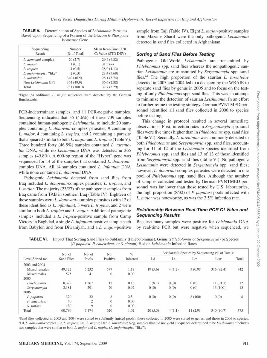
PCR-indeterminate samples, and 11 PCR-negative samples. Sequencing indicated that 35 (4.6%) of these 739 samples contained human-pathogenic Leishmania , to include 20 sam-ples containing L. donovani- complex parasites, 9 containing L. major , 4 containing L. tropica , and 2 containing a parasite that appeared similar to both L. major and L. tropica ( Table V ). Three hundred forty (46.5%) samples contained L. tarento-lae DNA, while no Leishmania DNA was detected in 365 samples (49.8%). A 600-bp region of the “Hyper” gene was sequenced for 14 of the samples that contained L. donovani -complex DNA. All 14 samples contained L. infantum DNA while none contained L. donovani DNA.
Pathogenic Leishmania detected from sand fl ies from Iraq included L. donovani -complex parasites, L. tropica , and L. major . The majority (23/27) of the pathogenic samples from Iraq came from TAB in southern Iraq ( Table IV ). Eighteen of these samples were L. donovani -complex parasites (with 12 of these identifi ed as L. infantum ), 3 were L. tropica , and 2 were similar to both L. tropica and L. major . Additional pathogenic samples included a L. tropica -positive sample from Camp Victory in Baghdad, a single L. infantum -positive sample each from Babylon and from Diwaniyah, and a L. major -positive
sample from Taji ( Table IV ). Eight L. major -positive samples from Mazar-e Sharif were the only pathogenic Leishmania detected in sand fl ies collected in Afghanistan.
Sorting of Sand Flies Before Testing Pathogenic Old-World Leishmania are transmitted by Phlebotomus spp. sand fl ies whereas the nonpathogenic sau-rian Leishmania are transmitted by Sergentomyia spp. sand fl ies. 63 The high proportion of the saurian L. tarentolae detected in 2003 and 2004 led to a decision by the WRAIR to separate sand fl ies by genus in 2005 and to focus on the test-ing of only Phlebotomus spp. sand fl ies. This was an attempt to minimize the detection of saurian Leishmania . In an effort to further refi ne the testing strategy, German PVNTMED per-sonnel identifi ed all sand fl ies collected in 2006 to species before testing.
This change in protocol resulted in several immediate observations. First, infection rates in Sergentomyia spp. sand fl ies were fi ve times higher than in Phlebotomus spp. sand fl ies ( Table VI ). Secondly, L. tarentolae was commonly detected in both Phlebotomus and Sergentomyia spp. sand fl ies, account-ing for 11 of 12 of the Leishmania species identifi ed from Phlebotomus spp. sand fl ies and 13 of 13 of those identifi ed from Sergentomyia spp. sand fl ies ( Table VI ). No pathogenic Leishmania were detected in Sergentomyia spp. sand fl ies; however, L. donovani -complex parasites were detected in one pool of Phlebotomus spp. sand fl ies. Although the number of samples collected and tested by German PVNTMED per-sonnel was far lower than those tested by U.S. laboratories, the high proportion (8/32) of P. papatasi pools infected with L. major was noteworthy, as was the 2.5% infection rate.
Relationship Between Real-Time PCR Ct Value and Sequencing Results Because many samples were positive for Leishmania DNA by real-time PCR but were negative when sequenced, we
TABLE V. Determination of Species of Leishmania Parasites Based Upon Sequencing of a Portion of the Glucose 6-Phosphate
Isomerase Gene
Sequencing Result
Number (% of Total)
Mean Real-Time PCR Ct Value (STD DEV)
L. donovani complex 20 (2.7) 29.4 (4.82) L. major a 1 (0.1) 31.3 (−) L. tropica 4 (0.5) 38.0 (1.13) L. major/tropica “like” 2 (0.3) 28.4 (3.68) L. tarentolae 340 (46.5) 28.1 (3.74)Non- Leishmania GPI 364 (49.9) 36.6 (2.88)Total 731 (100.0) 32.7 (5.29)
a Eight (8) additional L. major sequences were detected by the German Bundeswehr.
TABLE VI. Impact That Sorting Sand Flies to Subfamily (Phlebotominae), Genus ( Phlebotomus or Sergentomyia ) or Species ( P. papatasi , P. caucasicus , or S. sintoni ) Had on Leishmania Infection Rates
No. ofSand Flies
No. ofPools
No.Positive
%Infected
Leishmania Species by Sequencing (% of Total) b
Level Sorted to a Ld Lt Lm Ltar Total
2003 and 2004Mixed females 49,122 5,232 577 1.17 19 (5.6) 4 (1.2) 3 (0.9) c 316 (92.4) 342Mixed males 575 41 0 0.00
2005 Phlebotomus 8,372 1,567 15 0.18 1 (8.3) 0 (0) 0 (0) 11 (91.7) 12 Sergentomyia 2,181 291 20 0.92 0 (0) 0 (0) 0 (0) 13 (100) 13
2006 P. papatasi 320 32 8 2.5 0 (0) 0 (0) 8 (100) 0 (0) 8 P. caucasicus 40 2 0 0.00 S. sintoni 180 9 0 0.00
Total 60,790 7,174 620 1.02 20 (5.3) 4 (1.1) 11 (2.9) 340 (90.7) 375
a Sand fl ies collected in 2003 and 2004 were sorted to subfamily (mixed pools), those collected in 2005 were sorted to genus, and those in 2006 to species. b Ld, L. donovani -complex; Lt, L. tropica ; Lm, L. major ; Ltar, L. tarentolae ; Neg, samples that did not yield a sequence determined to be Leishmania . c Includes two samples that were similar to both L. major and L. tropica ( L. major / tropica “like”).
Dow
nloaded from https://academ
ic.oup.com/m
ilmed/article/174/9/904/4336505 by guest on 22 O
ctober 2022

Use of Vector Diagnostics During Military Deployments: Recent Experience in Iraq and Afghanistan
912 MILITARY MEDICINE, Vol. 174, September 2009
compared real-time PCR Ct values with sequencing results to determine whether there were any relationships between strength of the PCR reaction and the proportion of samples determined to be Leishmania by sequencing ( Table VII ). As the mean Ct values decreased (i.e., reaction became stronger), the proportion of samples that tested positive for Leishmania parasite GPI sequences increased while the proportion of other GPI sequences decreased. For example, 99% of the samples with a mean Ct value <26 matched known Leishmania spp. sequences, whereas only 3% of the samples with a mean Ct value between 38 and 39.99 matched any known Leishmania spp. sequence ( Table VII ).
DISCUSSION
Evaluation of the Malaria VecTest Assay In spite of an intensive surveillance effort, only 430 anopheline mosquitoes were collected in Iraq and all 191 that were tested using the malaria VecTest assay were negative. In contrast, 1,595 anopheline mosquitoes were collected in Afghanistan, with a total of 35 pools infected with Plasmodium vivax and 7 with P. falciparum . Although we were not able to deter-mine whether any of the infected mosquitoes were capable of transmitting malaria, An. pulcherrimus , An. hrycanus , and An. superpictus are all known vectors of malaria in Southwest Asia. 64,65 These data suggest that malaria rates should be much higher in service members deployed to Afghanistan compared to those deployed to Iraq, and that the majority of cases should be the result of infection with P. vivax . Unfortunately, the long incubation period of temperate-strain P. vivax , 66 combined with the mobility of U.S. military personnel makes it diffi -cult to determine exactly where vivax malaria infections were acquired. 67 Although 60 soldiers who deployed to Iraq since 2003 have been diagnosed with malaria, only 7 individuals in Iraq during the transmission season had no other documented
exposure, suggesting that most of these soldiers acquired their infections elsewhere. 67 In contrast, signifi cant numbers of military personnel appear to have been infected while in Afghanistan. 10 Cimera and Brundage 67 reported that 74 mili-tary members with malaria had served in Afghanistan during the transmission season, with 41 (55%) having no other docu-mented exposure risk, while Kotwal et al. 68 reported 38 active duty soldiers from a 725-man Ranger Task Force contracted malaria while operating in eastern Afghanistan in 2002.
Our data suggest that hand-held assays are a remarkably powerful tool with which to assess the threat of vector-borne diseases to deployed military forces. These assays can be used anywhere and can provide real-time feedback. These assays will be most valuable when used to assess the vector-borne disease threat immediately before or soon after moving mili-tary forces into a given area—the goal should be the detec-tion of pathogens before the onset of disease in our deployed military forces. Early detection of a pathogen will allow for the implementation of pathogen/vector-specifi c protective measures that can minimize casualties to our military forces. For example, use of the malaria VecTest assay in Afghanistan clearly demonstrated that infected anopheline mosquitoes were present—these positive assay results were extremely useful in obtaining command support for mandatory use of PPM. In contrast, all of the mosquitoes tested with this assay at TAB, Iraq were negative, suggesting that malaria was not present or was exceedingly rare at this site. Information on the scarcity of anopheline mosquitoes in most areas in Iraq combined with the negative malaria VecTest assay results helped medical authorities implement a policy in which man-datory malaria prophylaxis was discontinued. We believe that hand-held vector assays are a valuable force multiplier and should play a key role in the Deployment Environmental Surveillance Program directed by the U.S. Army Center for Health Promotion and Preventive Medicine (USACHPPM).
TABLE VII. Relationship Between Threshold Cycle (Ct) Values of a Real-Time Leishmania Genus PCR Assay and the Proportion of Samples Determined to Be Positive Upon Retesting With the Same PCR Assay and by Sequencing of a Portion of the Glucose-6-
Phosphate-Isomerase Gene
Real-Time PCR Assay Results Sequencing Results
InitialCt Value a
No. ofSamples
No. (%)Negative b
No. (%)Positive b
No. (%)Indeterminate b
No.Sequenced
No. (%) Leishmania
No. (%)Other
40 6,352 6,352 (100) 0 (0) 0 (0) 11 0 (0) 11 (100)38–39.99 147 0 (0) 7 (5) 140 (95) 131 4 (3) 127 (97)36–37.99 113 0 (0) 88 (78) 25 (22) 110 8 (7) 102 (93)34–35.99 83 0 (0) 82 (99) 1 (1) 73 20 (27) 53 (73)32–33.99 72 0 (0) 71 (99) 1 (1) 68 32 (47) 36 (53)30–31.99 89 0 (0) 89 (100) 0 (0) 86 61 (71) 25 (29)28–29.99 78 0 (0) 78 (100) 0 (0) 71 63 (89) 8 (11)26–27.99 71 0 (0) 71 (100) 0 (0) 62 61 (98) 1 (2)
<26 126 0 (0) 126 (100) 0 (0) 119 118 (99) 1 (1)Total 7,131 6,352 (89) 612 (9) 167 (2) 731 367 (50) 364 (50)
a The Ct value is the number of cycles at which the sample was considered positive. The lower the value the stronger the reaction. A Ct value of 40 is considered negative. b The determination as to whether the sample was considered positive, negative, or indeterminate was based on retesting of the sample as outlined in Figure 2 .
Dow
nloaded from https://academ
ic.oup.com/m
ilmed/article/174/9/904/4336505 by guest on 22 O
ctober 2022

Use of Vector Diagnostics During Military Deployments: Recent Experience in Iraq and Afghanistan
MILITARY MEDICINE, Vol. 174, September 2009 913
Hand-held vector assays should also be integrated into PVNTMED units of all services.
The malaria VectorTest (trademark of VecTOR Test Systems, Inc.) assay and the various other VecTest (trade-mark of products from Medical Analysis System, Inc., now a part of ThermoFisher) assays for the detection of EEE, WEE, SLE, and WN viruses are currently the only hand-held assays available for vector surveillance during military deployments. A variety of additional VectorTest assays, to include assays for the detection of Leishmania parasites and dengue, RVF, JE, SFV, and Ross River viruses, are currently being devel-oped and could potentially be fi elded within the next several years. The ultimate goal is the fi elding of hand-held vector assays for all of the top threat agents identifi ed in Table I .
Evaluation of Real-Time PCR Assays for Leishmania Surveillance In contrast to hand-held vector assays that can be used almost anywhere, 45 real-time PCR assays normally require a power supply and a cold chain. In spite of these limitations, real-time PCR assays can be used under a variety of fi eld conditions, to include tents and other portable structures. Although not as rapid as the hand-held assays, under optimal conditions DNA can be extracted and assay results obtained within several hours. We found that the Leishmania -genus real-time PCR assay was a useful tool that allowed us to screen hundreds of sand fl ies each day for Leishmania parasites. However, as with all other tools, the limitations of the assay must be under-stood when interpreting results.
The high infection rates in sand fl ies collected in the vicin-ity of TAB and several other areas in Iraq suggested that ser-vice members stationed in these areas were at high risk of becoming infected with Leishmania ( Table IV ). It was only once sequencing of the 360-bp region of the GPI gene had been completed that we were able to determine that the actual threat was much lower than originally suggested, since the nonpathogenic L. tarentolae accounted for the majority of the PCR-positive samples. Before our deployment to Iraq in 2003, there was little information available on the abundance of nonpathogenic Leishmania in sand fl ies in the Middle East. Although we realized that nonpathogenic species such as L. tarentolae , L. turanica , and L. gerbilli were potentially present in Iraq, there was no published data that suggested nonpathogenic Leishmania were present or that they would account for over 90% of the PCR-positive samples from sand fl ies. Unfortunately, the Leishmania- genus real-time PCR assay is not capable of differentiating pathogenic from non-pathogenic species.
Clearly, appropriate targets must be developed for assays that will be used in vector surveillance. Although a Leishmania- genus real-time PCR assay is acceptable for use with human samples, as any Leishmania detected in a human sample is intuitively pathogenic, our data demonstrate that a Leishmania- genus assay cannot be used independently to assess the medical threat posed by sand fl ies. Sequencing of
appropriate targets (e.g., the GPI and “Hyper” genes as done in this study) can be used to identify the Leishmania spe-cies; however, sequencing is currently not possible in a fi eld environment and can take several weeks. Assays that can dif-ferentiate pathogenic from nonpathogenic Leishmania are clearly needed, as are species-specifi c assays. We have devel-oped two assays specifi c for L. donovani -complex parasites and one for L. major and are currently developing assays for L. tropica and L. tarentolae . We have also developed an assay that is specifi c for pathogenic Leishmania parasites of the Old World, to include L. tropica , L. major , L. aethiopica , and L. donovani -complex parasites. This assay does not detect nonpathogenic species. Once fully validated, these assays will allow for a more rapid assessment of the medical threat posed by sand fl ies in the Middle East.
In addition to requiring additional species-specifi c assays, it is critical that the performance of each assay be well estab-lished before use in an operational setting. Procedures used to validate initial test results should be established, as should a rubric for identifying the causative agent. At the onset of OIF, we had completed initial validation of the Leishmania- genus assay using laboratory-infected sand fl ies and had calculated the limit of detection for L. major . We had also developed assays specifi c for L. major , L. tropica , and L. donovani -complex parasites; however, we had not yet fully validated these assays. Although we knew that these species-specifi c assays were approximately 10 times less sensitive than the Leishmania- genus assay, we had not determined the limit of detection for each assay nor had we evaluated cross-reactivity of the assays. Initially, all samples that tested positive with the Leishmania- genus assay were subsequently tested using the L. major , L. tropica , and L. donovani -complex assays. Over 90% of the samples testing positive using the Leishmania- genus assay were negative with the species-specifi c assays. Because of our incomplete understanding of the performance of each assay, we were not able to determine whether the spe-cies-specifi c assays were negative because (i) they were less sensitive than the Leishmania- genus assay, (ii) the Leishmania- genus assay was yielding false positive results, or (iii) the Leishmania- genus assay was detecting a species of parasite not recognized by the species-specifi c assays. The development of a testing algorithm ( Fig. 2 ) that included multiple retesting using real-time PCR and sequencing of a portion of the GPI gene was an attempt to overcome these limitations.
When using real-time PCR assays it is also important to establish realistic cut-off values that can be used to deter-mine whether samples are positive, negative, or indetermi-nate. When establishing the Leishmania- genus PCR assay at WRAIR, our laboratory data suggested that all Ct values < 40 should be considered positive. However, when our fi eld data were carefully analyzed ( Table VII ) to include the use of sequencing in our testing paradigm, it became clear that it was necessary to establish an indeterminate range (i.e., not possible to determine if the result was a true positive or a true negative). Retesting of samples that initially tested positive
Dow
nloaded from https://academ
ic.oup.com/m
ilmed/article/174/9/904/4336505 by guest on 22 O
ctober 2022

Use of Vector Diagnostics During Military Deployments: Recent Experience in Iraq and Afghanistan
914 MILITARY MEDICINE, Vol. 174, September 2009
with the real-time PCR assay suggested that an appropriate cut-off might be at a Ct of 38, as only 5% of samples with a Ct between 38 and 39.99 were positive upon retesting, whereas 78% of samples with a Ct between 36 and 37.99 tested posi-tive upon retesting ( Table VII ). Although sequencing proved to be a useful tool for confi rming the identify of parasites, it was diffi cult to determine if a negative sequencing result meant that the original PCR assay result was a false positive or whether the large number of PCR-positive samples that were negative by sequencing refl ected limitations of our sequenc-ing procedures. Our results clearly indicate that confi rmatory assays are a key component of any testing program. Ideally, any confi rmatory assay should have a limit of detection simi-lar to or below that of the screening assay and should target a separate genomic region of the target pathogen.
In addition to refi ning our test procedures as a means of conclusively identifying Leishmania parasites in sand fl ies, we also attempted to determine whether sorting sand fl ies to genus and/or species could eliminate the nonpathogenic Leishmania . Since Sergentomyia spp. sand fl ies are believed to be the vectors of Sauroleishmania (e.g., L. tarentolae ), 63 we hoped that by focusing testing on Phlebotomus spp. sand fl ies we would reduce or eliminate positive samples resulting from detection of nonpathogenic species of Leishmania . Although Leishmania parasites were detected in Sergentomyia spp. sand fl ies much more frequently than in the Phlebotomus spp. sand fl ies, L. tarentolae nevertheless accounted for 92% (11/12) of the Leishmania samples detected in Phlebotomus spp. sand fl ies. We were not able to determine whether the Phlebotomus spp. sand fl ies were capable of transmitting L. tarentolae ; however, these data clearly demonstrated that sorting of sand fl ies to genus will not eliminate the detection of nonpatho-genic Leishmania .
To date, only three military personnel deployed to Iraq since 2003 have developed visceral leishmaniasis. However, our data demonstrate that parasites that cause visceral leish-maniasis posed a threat to military personnel in Iraq in 2003 and 2004. The fact that more symptomatic cases have not yet occurred in deployed military personnel is not unexpected as visceral leishmaniasis caused by L. infantum has historically been considered a disease of young children who are mal-nourished and/or immunocompromised. 69 Among residents in the Mediterranean basin, symptomatic visceral leishmaniasis in adults is almost exclusively a result of concomitant infec-tion with L. infantum and human immunodefi ciency virus. 69,70 The fact that deployed military personnel are presumably healthy, well-fed, immune-competent adults suggests that the risk of developing symptomatic visceral leishmaniasis result-ing from infection with L. infantum is low; however, there may be potential long-term concerns involving the disease. Leishmania parasites can persist in the body for life, even fol-lowing successful treatment of symptomatic individuals, 71 and asymptomatic carriers can become symptomatic follow-ing suppression of their immune system. 70 Although the exact number of military personnel actually exposed to parasites
that cause visceral leishmaniasis may never be determined, as there are currently no FDA-licensed tests that can be used to assess exposure, our data clearly demonstrate that sand fl ies infected with L. infantum were present in areas where U.S. military personnel were stationed.
Concept of Operations for Vector Diagnostics Vector diagnostics provides deployed military forces with a powerful tool to assess the threat from vector-borne diseases. Vector diagnostics is not a stand-alone process, but rather is part of an integrated prevention effort consisting of (i) vector surveillance, (ii) vector identifi cation, (iii) vector diagnostics, (iv) individual protective measures, and (v) collective pro-tective measures. When this integrated prevention process is effectively implemented it can result in a rapid assessment of the vector-borne disease threat and assist in the establishment of disease prevention programs, such as the “Leishmaniasis Control Program” established at TAB, Iraq in 2003. 37 The development of diagnostic procedures for identifying patho-gens in arthropods has lagged far behind the other four steps in this process. However, technological advances since the early 1990s have led to the creation of diagnostic tools that allow trained personnel to identify pathogens in arthropods in a rapid and effi cient manner. The following is our pro-posed concept of operations (CONOPS) for the use of vector diagnostics:
Assays Used
We propose a two-tiered system for use by deployed mili-tary forces, with more extensive capabilities available in fi xed medical facilities such as the WRAIR or USACHPPM. In this two-tiered system, hand-held immunochromatographic assays are used as screening tools and real-time PCR assays are used as confi rmatory tools. The hand-held assays will ideally target a broad range of pathogens (e.g., genus-level assays), while each real-time PCR assay would ideally target a single patho-gen (e.g., species-level assays). For example, a Leishmania- genus hand-held screening assay could be complemented by real-time PCR assays specifi c for L. major , L. tropica , and L. donovani -complex parasites. Confi rmatory assays should consist of two real-time PCR assays that recognize two sepa-rate targets on different genes for each pathogen of interest. Currently, very few hand-held or real-time PCR assays are available for vector diagnostics ( Table I ). The development and validation of additional assays and employment prac-tices should be a priority of the Military Infectious Disease Research Program administered by the U.S. Army Medical Research and Material Command.
Fixed facilities such as the WRAIR and the U.S. Army Medical Research Institute of Infectious Diseases (USAMRIID) possess a variety of more sophisticated diag-nostic capabilities that would not normally be available to deployed forces, to include procedures such as sequencing of genomic material and culturing and subsequent identifi ca-tion of pathogenic agents. These fi xed facilities are a valuable
Dow
nloaded from https://academ
ic.oup.com/m
ilmed/article/174/9/904/4336505 by guest on 22 O
ctober 2022

Use of Vector Diagnostics During Military Deployments: Recent Experience in Iraq and Afghanistan
MILITARY MEDICINE, Vol. 174, September 2009 915
resource that can facilitate the identifi cation of vector-borne pathogens.
The Armed Forces Pest Management Board ( www.afpmb.org ) maintains a database that lists a variety of vector assays that have been developed by various military laboratories and the CDC. The intent of this spreadsheet is to provide potential users with a list of assays that may be used to support their individual requirements. Inclusion of a specifi c assay in this spreadsheet does not imply that the assay has been endorsed by the AFPMB except where noted.
Assay Validation
If possible, all assays used during military deployments should be fully validated for all target organisms. Standard performance criteria should be calculated for each assay, to include limit-of-detection, sensitivity, specifi city, and posi-tive- and negative-predictive value. Ideally, performance should be evaluated using infected arthropods. Unfortunately, the challenges associated with obtaining infected arthropods will frequently preclude comprehensive evaluation of assay performance. In these instances, it is critical that the limita-tions of the assay be fully understood and that these limita-tions be considered when interpreting assay results.
Units Using Vector Diagnostics
Hand-held assays are primarily used by units actually conduct-ing vector surveillance operations, such as the PVNTMED Section of a Brigade Combat Team or an Army PVNTMED Detachment. These units will normally not be equipped with real-time PCR assays and will therefore need to ship specimens to a medical laboratory for confi rmatory testing. Real-time PCR assays are primarily used by units with sophisticated lab-oratory capabilities beyond those found in a Brigade Combat Team or Army PVNTMED Detachment. Units with real-time PCR capabilities include Army Area Medical Laboratories, Navy Forward Deployed PVNTMED Units, and Air Force Biological Assessment Teams, among others. Although these sophisticated laboratories may on occasion conduct vector surveillance operations, their primary mission is testing envi-ronmental samples to assess the medical threat to deployed forces. Therefore, in most instances samples will be shipped to these laboratories by the units actually conducting vector surveillance operations.
Collection of Arthropods
A variety of procedures may be used to collect arthropods that will subsequently be tested for the presence of pathogens. The collection procedure should not interfere with the diagnostic assay to be performed. Although the majority of collection procedures will not interfere with diagnostic procedures, some procedures (e.g., sticky traps) could potentially interfere and should not be used unless proven to be compatible with diag-nostic procedures. Standardized sampling procedures should be used whenever possible, with guidance on procedures pro-vided before the onset of sampling. The type of trap used,
height of trap, period of trap placement, and numbers and loca-tions of traps should all be standardized, as should the use of attractants such as dry ice or compressed carbon dioxide. Data collection begins at this step and the collectors must enter and maintain the data in electronic or written form so that this infor-mation can be forwarded with and linked to the samples. Basic data should include location, date/time, collector’s name, col-lection method, and habitat information. Other parameters can be added to the data set as needed by the study design.
Killing, Preserving, and Storing Arthropods
The procedure used to kill, preserve, and store collected arthro-pods should not interfere with the diagnostic assay that will subsequently be performed. Acceptable methods of killing arthropods include freezing or immersion in ethanol. While a variety of additional methods (e.g., heat or use of chemi-cals such as ethyl acetate or potassium cyanide) are frequently used to kill collected arthropods, further studies are needed to ensure that these procedures are compatible with each diag-nostic procedure to be used. Each diagnostic assay should pro-vide detailed information on how specimens should be stored before testing. In general, specimens should be tested immedi-ately or stored frozen at −20°C or colder (−70°C is preferable if samples will be stored for months or years before testing) or in 90–100% ethanol. A variety of additional storage methods may be compatible with test procedures; however, these stor-age procedures should not be used unless empirical data dem-onstrate that they will not adversely affect the performance of the diagnostic procedure. Unnecessary freeze–thaw cycles should be minimized as this can affect assay results.
Pooling Specimens
In an unconstrained environment, each arthropod collected would be tested individually; however, because of cost and time this is rarely feasible. Instead, groups of “like” speci-mens are normally combined into pools of 5–50 individuals and the pools tested. Whenever possible, arthropods in a sin-gle pool should come from a single collection made at a par-ticular site and at a particular time. For example, arthropods in a given pool would come from a single light trap collec-tion made on a single night. In many cases, the specimens are placed into pools during the sorting process to facilitate future specimen testing.
Labeling Samples
Each sample (e.g., vial containing whole arthropods, homog-enized arthropods, or DNA/RNA extracts) should receive a unique identifi er that allows that particular sample to be linked to the collection data. Identifi ers should be written directly on vials using a permanent, water/ethanol resistant marker or using preprinted labels intended for use with cryovials.
Homogenization/Extraction Procedures
A variety of procedures may be used for the homogenization of samples and/or the extraction of DNA or RNA. Arthropods
Dow
nloaded from https://academ
ic.oup.com/m
ilmed/article/174/9/904/4336505 by guest on 22 O
ctober 2022
Use of Vector Diagnostics During Military Deployments: Recent Experience in Iraq and Afghanistan
916 MILITARY MEDICINE, Vol. 174, September 2009
tested using hand-held VecTest assays are homogenized in a grinding buffer using a small mortar and pestle provided with the kit, with the dipstick placed directly into the homogenate. Samples to be tested using real-time PCR assays are homog-enized and then DNA or RNA are extracted using a variety of different protocols. Safety issues related to homogeniza-tion procedures are discussed further in the biological safety section.
Assay Protocols
Each assay used for vector diagnostics should include a detailed protocol specifying procedures to be used and limita-tions of the assay. A point-of-contact (telephone number and e-mail address) should be provided so that questions regard-ing the assay can be rapidly addressed. Whenever possible, a given surveillance program should be directed by a central coordinating agency (e.g., USACHPPM) so that protocols can be implemented consistently and data from different sites compared to one another.
Use of Multiple Test Procedures
Although most samples will be tested using only a single pro-cedure, in some instances additional diagnostic testing may be required. Reasons for conducting additional testing include confi rmation of the identity of a positive sample or testing for additional pathogens. In all instances where multiple test pro-cedures may be used, a protocol should be established that will allow for subsequent testing of samples. For example, samples that are stored in ethanol cannot subsequently be used to cul-ture viable pathogens, while the grinding buffer used in cer-tain assays may be incompatible with other test procedures.
Interpretation of Results
All test results, whether positive or negative, should be recorded. Negative test results have limited value, particu-larly when a small number of samples have been evaluated. Negative results do not indicate that the specifi c pathogen is not present, but rather that the pathogen was not detected in the sample population. Although a comprehensive surveil-lance program would ideally collect periodic samples from a variety of sites over the course of a transmission season, in many instances samples are collected from a limited number of sites over a short period of time. In these instances negative test results may be the result of surveillance that misses key sites or surveillance conducted at a time when infected arthro-pods were not present. Although the value of negative test results increase as the sample size increases, it is diffi cult to estimate a minimum number of negative samples required to assure a high degree of confi dence that a particular pathogen has a low or nonexistent threat because many factors affect the effi ciency of pathogen transmission.
Positive samples are much more valuable than negative samples, as they indicate that a particular pathogen was pres-ent at a certain location on a certain date. Positive samples can be used to calculate minimum fi eld infection rates that
can provide a rough estimate of the magnitude of the threat. However, as described in this article, care must be used when interpreting positive assay results, as the organism detected may not be pathogenic to humans, the vector may not be infectious, or the vector may not feed on humans.
Reporting of Test Results
Standardized forms should be used for recording and report-ing diagnostic assay results. These forms will ideally be pre-pared by a single organization such as the USACHPPM and reviewed and approved by the AFPMB. Copies of all data and reports should be provided to the organization that collected the samples and to a single organization that will archive the data. Summaries of data should be provided to all organiza-tions with an inherent interest in vector diagnostic test results, to include the National Center for Medical Intelligence (for-merly the Armed Forces Medical Intelligence Center), the AFPMB, the USACHPPM, and through the medical chain-of-command to the Combatant Commands Surgeon’s offi ce.
Biological Safety Considerations
The use of vector diagnostics poses a potential risk to person-nel conducting the testing. The primary risk is exposure to aerosolized pathogens during homogenization of the arthro-pods. Although to our knowledge no disease transmission has ever occurred during processing of fi eld-collected arthropods for vector diagnostics, the CDC publication “Biosafety in microbiological and biomedical laboratories” has documented 151 instances in which a laboratory worker was infected with an arthropod-borne virus—the majority of these infections are believed to have resulted from aerosolization of the virus. 72 Although no specifi c guidelines for the handling of fi eld-collected arthropods are available, the CDC recommends that Biosafety Level 2 practices be used for processing fi eld-collected mosquito pools for West Nile virus. 73 In the absence of specifi c Department of Defense (DoD) or Department of Health and Human Services (HHS) guidelines on the handling of fi eld-collected arthropods, whenever possible arthropods should be homogenized in a sealed tube containing a grind-ing medium or diluent that will inactivate arboviruses and other easily aerosolized pathogens. 74 Unfortunately, the use of diluents that inactivate pathogens may preclude the use of these samples with some diagnostic assays.
Transport of Specimens
The U.S. Code of Federal Regulations (CFR) Title 49 is the document that governs the transportation of hazardous mate-rials, to include infectious substances. Detailed information on the shipment of diagnostic specimens can be found in the USACHPPM fact sheet “Packaging hospital samples and specimens for transport” ( http://chppm-www.apgea.army.mil/hmwp/Factsheets/Transport Summary.htm). Arthropods, to include homogenized samples and/or DNA/RNA extracts, are considered general diagnostic specimens unless a spe-cifi c pathogen has been identifi ed or is suspected. Regulations
Dow
nloaded from https://academ
ic.oup.com/m
ilmed/article/174/9/904/4336505 by guest on 22 O
ctober 2022

Use of Vector Diagnostics During Military Deployments: Recent Experience in Iraq and Afghanistan
MILITARY MEDICINE, Vol. 174, September 2009 917
pertaining to the air shipment of general diagnostic specimens include 9 CFR (Animal and Plant Health Inspection Service), 21 CFR (Food and Drug Administration), and 42 CFR (Centers for Disease Control and Prevention). To ensure that proper protection is in place to contain any undetected pathogenic micro-organisms, general diagnostic specimens should be packaged in accordance with International Air Transportation Association (IATA) Dangerous Goods regulations. In gen-eral, procedures for packing general diagnostic specimens for air shipment include (i) sample placed in a primary watertight receptacle with a leakproof seal, (ii) watertight receptacle wrapped in absorbent material, (iii) wrapped container placed in a secondary watertight receptacle, (iv) the entire package is placed in a strong outer packaging approved by the U.S. Department of Transportation (DOT) for transport of the haz-ardous material, (v) itemized list describing contents placed in the unsealed shipping box, and (vi) designated certifying offi cial approves package for transport. If needed, ice, dry ice, or prefrozen packs should be placed between the secondary watertight receptacle and the outer packaging. Packages con-taining dry ice must permit the release of carbon dioxide.
Diagnostic samples in which an infectious agent has been detected are no longer considered general diagnostic samples but rather are considered infectious substances. Infectious substances are defi ned as “a viable micro-organism, or its toxin, that causes or may cause disease in humans or animals, and includes those agents listed in 42 CFR 72.3 of the regula-tions of the Department of Health and Human Services or any other agent that causes or may cause severe, disabling or fatal disease.” Very stringent regulations pertain to the shipment of infectious substances, with requirements described in detail in 49 CFR Part 173.196. The shipment of infectious substances requires coordinated action by the shipper, the transporter, and the receiver to ensure safe transport and arrival on time. Based on the defi nition of infectious substances as a “viable micro-organism,” diagnostic samples containing a pathogen inactivated using appropriate grinding media or diluent are considered general diagnostic samples and not infectious sub-stances. As such, the less stringent procedures pertaining to the shipment of general diagnostic samples apply. Depending on the type of samples (e.g., dead arthropods, general diag-nostic sample, or infectious substances), appropriate CDC or U.S. Department of Agriculture (USDA) permits may be required to ship samples to the United States.
Biological Select Agents and Toxins
Certain microbial pathogens and toxins that potentially pose a severe threat to human, animal, or plant health are referred to as biological select agents and toxins (BSATs). The U.S. Department of Health and Human Services and the USDA have regulatory authority over BSAT that can affect human and animal/plant health, respectively. A current list of USDA and HHS regulated BSATs can be found at http://www.aphis.usda.gov/programs/ag_selectagent/ag_bioterr_toxinslist.html and http://www.selectagents.gov/, respec-
tively. Laws regulating BSATs include Section 511 of the Antiterrorism and Effective Death Penalty Act of 1996, the Uniting and Strengthening America by Providing Appropriate Tools Required to Intercept and Obstruct Terrorism Act of 2001 ( USA PATRIOT Act), and the Public Health Security and Bioterrorism Preparedness and Response Act of 2002 (Bioterrorism Act). On March 18, 2005, HHS and USDA published the fi nal Select Agent Regulations (42 CFR Part 73, 7 CFR Part 331, 9 CFR Part 121) in the Federal Register . Subsequent to the publication of these regulations, the DoD and each of the military services have published specifi c instructions pertaining to BSATs, to include DoD Instructions 5210.88 (Safeguarding Biological Select Agents and Toxins) and 5210.89 (Minimum Security Standards for Safeguarding Biological Select Agents and Toxins), Army Regulations 50-1 (Biological Surety), and 190-17 (Biological Select Agent and Toxins Security Program), and Air Force Policy Directive 10-39 (Safeguarding Biological Select Agents and Toxins).
A number of BSATs are transmitted by arthropods, to include African swine fever, bluetongue, CCHF, EEE, JE, RVF, TBE, and VEE viruses, as well as Rickettsia prowazekii , R. rickettsii , Francisella tularensis , and Yersinia pestis . Each of these pathogens could potentially be detected in arthropods during military deployments. Because of the severe threat posed by BSATs, the handling, transport, security, destruc-tion, and reporting of these agents are highly regulated. While the various regulations provide clear guidance on procedures used within the United States, there is little guidance on the detection and identifi cation of BSATs in vectors during mili-tary deployments. Accepted practice during military deploy-ments is to implement procedures that best meet the intent of relevant U.S. BSAT laws and regulations.
A key issue during military deployments is to determine whether a diagnostic sample should be considered a BSAT, as determination that a sample is a BSAT triggers a variety of specifi c actions/responses. In general, diagnostic samples are only considered to contain BSATs once a viable patho-gen has been identifi ed using a confi rmatory assay (42 CFR 73). As described previously, a true confi rmatory assay should consist of two separate, validated tests that detect different targets on different genes. There are currently no fi eld-deploy-able vector assays that meet this requirement; therefore, vec-tor samples are normally considered “presumptive positives” and would not be considered BSATs. However, the future development and fi elding of true confi rmatory assays could potentially result in vector samples being classifi ed as BSAT. An additional issue is the fact that procedures used to pre-pare samples for both screening and confi rmatory diagnostic assays frequently inactivate any pathogens that are present, so that even though a confi rmatory assay identifi ed a partic-ular pathogen, that sample would not be considered BSAT as no viable pathogen is present. However, laboratory per-sonnel should use caution when making a determination that a positive sample is not a BSAT, as it is extremely diffi cult in a fi eld setting to determine whether a viable pathogen is
Dow
nloaded from https://academ
ic.oup.com/m
ilmed/article/174/9/904/4336505 by guest on 22 O
ctober 2022

Use of Vector Diagnostics During Military Deployments: Recent Experience in Iraq and Afghanistan
918 MILITARY MEDICINE, Vol. 174, September 2009
present. Additionally, any portion of the diagnostic sample that did not undergo nucleic acid extraction or other steril-izing procedures that would have inactivated the infectious virus may still contain viable BSATs (if identifi ed by a confi r-matory assay) and should be destroyed within 7 days as speci-fi ed in 42 CFR 73. To further complicate matters, nucleic acid from positive-stranded RNA viruses can be used to produce infectious virus—this material would be considered a BSAT if detected using a confi rmatory assay.
Clearly, personnel conducting diagnostic testing for BSATs should understand the rules and regulations pertain-ing to BSATs. Criteria for determining whether a sample con-tains a BSAT should be established, as should procedures for securing, transporting, and destroying these samples per Army, DoD, and U.S. laws and regulations. Personnel con-ducting diagnostic testing must also understand that samples that do not meet the strict defi nition of a BSAT may still pose a considerable threat to anyone exposed to these samples. For example, a sample that tests positive for RVF virus using a hand-held screening assay is not considered a BSAT; how-ever, a potentially lethal virus may still be present in the sam-ple if the grinding diluent does not completely inactivate the pathogen.
SUMMARY Vector diagnostics are a powerful tool that can greatly facil-itate the assessment of the threat posed by arthropod-borne diseases. In the absence of appropriate vaccines or prophy-lactic drugs, appropriately used vector diagnostics can pro-vide an early warning system that can be used to mitigate the threat of vector-borne diseases. The goal of the diagnos-tics effort described in this article was to provide the vari-ous PVNTMED units stationed in Iraq and Afghanistan with information on the vector-borne disease threat in their respec-tive areas of operation. These PVNTMED units were carry-ing out vector control operations on a routine basis, and we believed that the rapid evaluation of mosquitoes and sand fl ies for the presence of Plasmodium and Leishmania parasites, respectively, and the dissemination of results would allow the PVNTMED units to more effectively develop control pro-grams that focused on areas that were at greatest risk for dis-ease transmission. During OIF, implementation of a vector diagnostics program during the weeks following the invasion of Iraq resulted in the determination that leishmaniasis posed a signifi cant threat to coalition military forces. 37 This deter-mination was made before the detection of any human cases and resulted in the aggressive implementation of prevention and control measures. Unfortunately, the diagnostic assays that were used did not adequately differentiate pathogenic from nonpathogenic Leishmania , resulting in an overestimate of the risk posed to deployed military forces. Nevertheless, we believe that the sand fl y surveillance program provided valuable information that raised the awareness of the leish-maniasis threat to deployed military forces in both Iraq and Afghanistan. Currently, the USACHPPM continues to test
sand fl ies collected in Iraq and Afghanistan for Leishmania parasites, and the WRAIR and other organization such as the Air Force Institute for Operational Health continue to develop improved diagnostic assays for vector surveillance.
ACKNOWLEDGMENTS Funding for this project was provided by the Military Infectious Diseases Research Program, the Armed Forces Medical Intelligence Center, and the Deployed War-Fighter Protection Program. Many thanks to LTC Bill Sames for his review of this manuscript, and to LTC (Ret.) Richard Lofts, COL David Craft, and Dr. Michael Turell for their guidance on BSATs. This study would not have been possible without the support of the many entomologists and environmental science offi cers conducting vector surveillance throughout Iraq and Afghanistan. In addition, the superb support of our laboratory tech-nicians running the PCR assays is greatly appreciated—thanks to Wayne and Lara Gilmore, Monzia Moodie, Cathy Westbrook, and SGT Ronald Hadley.
REFERENCES 1. Bavaro MF , Kelly DJ , Dasch GA , et al : History of U.S. military contri-
butions to the study of rickettsial diseases . Mil Med 2005 ; 170 (4, Suppl) : 49 – 60 .
2. Corwin A , Soderquist R , Suwanabun N , et al : Scrub typhus and military operations in Indochina . Clin Infect Dis 1999 ; 29 : 940 – 1 .
3. Burnette WN , Hoke CH , Scovill J , et al : Infectious diseases investment decision evaluation algorithm: a quantitative algorithm for prioritiza-tion of naturally occurring infectious disease threats to the U.S. military . Mil Med 2008 ; 173 (2) : 174 – 81 .
4. Elston DM , Miller SD : Leishmaniasis acquired in the Iraqi Theater of Operations: lessons learned . Cutis 2004 ; 74 : 253 – 5 .
5. Kelly DJ , Richards AL , Temenak J , Strickman D , Dasch GA : The past and present threat of rickettsial diseases to military medicine and interna-tional public health . Clin Infect Dis 2002 ; 34 (Suppl 4) : S145 – 69 .
6. Kitchener S , Leggat PA , Brennan L , McCall B : Importation of dengue by soldiers returning from East Timor to north Queensland, Australia . J Travel Med 2002 ; 9 : 180 – 3 .
7. Martin S , Gambel J , Jackson J , et al : Leishmaniasis in the United States military . Mil Med 1998 ; 163 : 801 – 7 .
8. Ockenhouse CF , Magill A , Smith D , Milhous W : History of U.S. mili-tary contributions to the study of malaria . Mil Med 2005 ; 170 (4, Suppl) : 12 – 6 .
9. Sanders JW , Putnam SD , Frankart C , et al : Impact of illness and non-combat injury during Operations Iraqi Freedom and Enduring Freedom (Afghanistan) . Am J Trop Med Hyg 2005 ; 73 : 713 – 9 .
10. Zapor MJ , Moran KA : Infectious diseases during wartime . Curr Opin Infect Dis 2005 ; 18 : 395 – 9 .
11. Endy TP , Thomas SJ , Lawler JV : History of U.S. military contributions to the study of viral hemorrhagic fevers . Mil Med 2005 ; 170 (4, Suppl) : 77 – 91 .
12. Innis BL , Eckels KH : Progress in development of a live-attenuated, tetravalent dengue virus vaccine by the United States Army Medical Research and Materiel Command . Am J Trop Med Hyg 2003 ; 69 (6, Suppl) : 1 – 4 .
13. Culpepper RC , Pratt WD : Advances in medical biological defense tech-nology . Clin Lab Med 2001 ; 21 : 679 – 89 .
14. Takafuji ET , Russell PK : Military immunizations. Past, present, and future prospects . Infect Dis Clin North Am 1990 ; 4 : 143 – 58 .
15. Touze JE , Paule P , Baudon D , Boutin JP : Malaria prophylaxis in the French armed forces: evolution of concepts . Med Trop (Mars) 2001 ; 61 : 79 – 82 .
16. Robert LL : Malaria prevention and control in the United States military . Med Trop (Mars) 2001 ; 61 : 67 – 76 .
17. Gambel JM , Brundage JF , Burge RJ , et al : Survey of U.S. Army sol-diers’ knowledge, attitudes, and practices regarding personal protection
Dow
nloaded from https://academ
ic.oup.com/m
ilmed/article/174/9/904/4336505 by guest on 22 O
ctober 2022

Use of Vector Diagnostics During Military Deployments: Recent Experience in Iraq and Afghanistan
MILITARY MEDICINE, Vol. 174, September 2009 919
measures to prevent arthropod-related diseases and nuisance bites. Mil Med 1998 ; 163 : 695 – 701 .
18. Young GD , Evans S : Safety and effi cacy of DEET and permethrin in the prevention of arthropod attack . Mil Med 1998 ; 163 : 324 – 30 .
19. Mehr ZA , Rutledge LC , Echano NM , Gupta RK : U.S. Army soldiers’ perceptions of arthropod pests and their effects on military missions . Mil Med 1997 ; 162 : 804 – 7 .
20. Croft AM , Baker D , von Bertele MJ : An evidence-based vector control strategy for military deployments: the British Army experience . Med Trop (Mars) 2001 ; 61 : 91 – 8 .
21. Horosko S 3rd, Robert LL : U.S. Army vector control (preventive med-icine) operations during Operation Restore Hope, Somalia . Mil Med 1996 ; 161 : 577 – 81 .
22. Onori E , Grab B : Indicators for the forecasting of malaria epidemics . Bull World Health Organ 1980 ; 58 : 91 – 8 .
23. Smith DL , McKenzie FE : Statics and dynamics of malaria infection in Anopheles mosquitoes . Malar J 2004 ; 3 : 13 .
24. Keesing F , Holt RD , Ostfeld RS : Effects of species diversity on disease risk . Ecol Lett 2006 ; 9 : 485 – 98 .
25. Ogurtsov AA , Riazantseva GA , Levkov PA , et al : Effi ciency of control of Ixodes ticks, vectors of natural and focal infections n the south of the Tyumen Region . Med Parazitol (Mosk) 2005 ; 3 : 15 – 9 (In Russian).
26. Botto C , Escalona E , Vivas-Martinez S , et al : Geographical patterns of onchocerciasis in southern Venezuela: relationships between environ-ment and infection prevalence . Parassitologia 2005 ; 47 : 145 – 50 .
27. Schulze TL , Jordan RA : Infl uence of meso- and microscale habitat struc-ture on focal distribution of sympatric Ixodes scapularis and Amblyomma americanum (Acari: Ixodidae) . J Med Entomol 2005 ; 42 : 285 – 94 .
28. Carter R , Mendis KN , Roberts D : Spatial targeting of interventions against malaria . Bull World Health Organ 2000 ; 78 : 1401 – 11 .
29. Randolph SE , Green RM , Peacey MF , Rogers DJ : Seasonal synchrony: the key to tick-borne encephalitis foci identifi ed by satellite data . Parasitology 2000 ; 121 (Pt 1) : 15 – 23 .
30. Claborn D : Abundance of three mosquito vectors in Okinawa with rel-evance to disease risk . Mil Med 1995 ; 160 : 172 – 4 .
31. Kuhn KG , Campbell-Lendrum DH , Davies CR : A continental risk map for malaria mosquito (Diptera: Culicidae) vectors in Europe . J Med Entomol 2002 ; 39 : 621 – 30 .
32. Smith DL , Dushoff J , Snow RW , Hay SI : The entomological inoculation rate and Plasmodium falciparum infection in African children . Nature 2005 ; 438 : 492 – 5 .
33. Tachiiri K , Klinkenberg B , Mak S , Kazmi J : Predicting outbreaks: a spa-tial risk assessment of West Nile virus in British Columbia . Int J Health Geogr 2006 ; 5 : 21 .
34. Werneck GL , Costa CH , Walker AM , et al : The urban spread of vis-ceral leishmaniasis: clues from spatial analysis . Epidemiology 2002 ; 13 : 364 – 7 .
35. Vanwambeke SO , Van Benthem BH , Khantikul N , et al : Multi-level analyses of spatial and temporal determinants for dengue infection . Int J Health Geogr 2006 ; 5 : 5 .
36. Gu W , Novak RJ : Short report: detection probability of arbovirus infec-tion in mosquito populations . Am J Trop Med Hyg 2004 ; 71 : 636 – 8 .
37. Coleman RE , Burkett DA , Putnam JL , et al : Impact of phlebotom-ine sand fl ies on U.S. military operations at Tallil Air Base, Iraq: 1. Background, military situation, and development of a “Leishmaniasis Control Program.” J Med Entomol 2006 ; 43 : 647 – 62 .
38. Beier JC : Malaria parasite development in mosquitoes . Annu Rev Entomol 1998 ; 43 : 519 – 43 .
39. Beier JC : Vector incrimination and entomological inoculation rates . Methods Mol Med 2002 ; 72 : 3 – 11 .
40. Wirtz RA , Burkot TR , Andre RG , et al : Identifi cation of Plasmodium vivax sporozoites in mosquitoes using an enzyme-linked immunosorbent assay . Am J Trop Med Hyg 1985 ; 34 : 1048 – 54 .
41. Wirtz RA , Duncan JF , Njelesani EK , et al : ELISA method for detecting Plasmodium falciparum circumsporozoite antibody . Bull World Health Organ 1989 ; 67 : 535 – 42 .
42. Philip Samuel P , Tyagi BK : Diagnostic methods for detection and isola-tion of dengue viruses from vector mosquitoes . Indian J Med Res 2006 ; 123 : 615 – 28 .
43. Sattabongkot J , Kiattibut C , Kumpitak C , et al : Evaluation of the VecTest malaria antigen panel assay for the detection of Plasmodium falciparum and P. vivax circumsporozoite protein in anopheline mosquitoes in Thailand . J Med Entomol 2004 ; 41 : 209 – 14 .
44. Appawu MA , Bosompem KM , Dadzie S , et al : Detection of malaria sporozoites by standard ELISA and VecTest dipstick assay in fi eld-col-lected anopheline mosquitoes from a malaria endemic site in Ghana . Trop Med Int Health 2003 ; 8 : 1012 – 7 .
45. Ryan JR , Dave K , Collins KM , et al : Extensive multiple test centre eval-uation of the VecTest malaria antigen panel assay . Med Vet Entomol 2002 ; 16 : 321 – 7 .
46. Ryan JR , Dave K , Emmerich E , et al : Dipsticks for rapid detection of plasmodium in vectoring anopheles mosquitoes . Med Vet Entomol 2001 ; 15 : 225 – 30 .
47. Nasci RS , Gottfried KL , Burkhalter KL , et al : Sensitivity of the VecTest antigen assay for eastern equine encephalitis and western equine enceph-alitis viruses . J Am Mosq Control Assoc 2003 ; 19 : 440 – 4 .
48. Ryan J , Dave K , Emmerich E , et al : Wicking assays for the rapid detec-tion of West Nile and St. Louis encephalitis viral antigens in mosquitoes (Diptera: Culicidae) . J Med Entomol 2003 ; 40 : 95 – 9 .
49. Panella NA , Burkhalter KL , Langevin SA , et al : Rapid West Nile virus antigen detection . Emerg Infect Dis 2005 ; 11 : 1633 – 5 .
50. Nemeth NM , Beckett S , Edwards E , et al : Avian mortality surveillance for West Nile virus in Colorado . Am J Trop Med Hyg 2007 ; 76 : 431 – 7 .
51. Stone WB , Okoniewski JC , Therrien JE , et al : VecTest as diagnostic and surveillance tool for West Nile virus in dead birds . Emerg Infect Dis 2004 ; 10 : 2175 – 81 .
52. Schriefer ME , Sacci JB Jr , Wirtz RA , et al : Detection of polymerase chain reaction-amplifi ed malarial DNA in infected blood and individual mosquitoes . Exp Parasitol 1991 ; 73 : 311 – 6 .
53. Tardieux I , Poupel O : Use of DNA amplifi cation for rapid detection of dengue viruses in midgut cells of individual mosquitoes . Res Virol 1990 ; 141 : 455 – 7 .
54. Weina PJ , Neafi e RC , Wortmann G , et al : Old world leishmaniasis: an emerging infection among deployed US military and civilian workers . Clin Infect Dis 2004 ; 39 : 1674 – 80 .
55. Coleman RE , Hochberg L , Swanson K , et al : Impact of phlebotomine sand fl ies on U.S. military operations at Tallil Air Base, Iraq: 4. Detection and identifi cation of Leishmania parasites in sand fl ies . J Med Entomol 2009 ; 46 : 649 – 63 .
56. Christensen DR , Hartman LJ , Loveless BM , et al : Detection of biological threat agents by real-time PCR: comparison of assay performance on the R.A.P.I.D., the LightCycler, and the Smart Cycler platforms . Clin Chem 2006 ; 52 : 141 – 5 .
57. Harbach R : Pictorial keys to the genera of mosquitoes, subgenera of Culex and the species of Culex (Culex) occurring in Southwestern Asia and Egypt, with a note on the subgeneric placement of Culex deserticola (Diptera: Culicidae) . Mosq Syst 1985 ; 17 : 83 – 107 .
58. Glick J : Illustrated key to the female Anopheles of Southwestern Asia and Egypt (Diptera: Culicidae) . Mosq Syst 1992 ; 24 : 125 – 53 .
59. Wortmann G , Sweeney C , Houng HS , et al : Rapid diagnosis of leishma-niasis by fl uorogenic polymerase chain reaction . Am J Trop Med Hyg 2001 ; 65 : 583 – 7 .
60. Hochberg LP , Moodie M , Liebig C , et al : The validation and use of a real-time PCR assay to detect Leishmania in phlebotomine sand fl y vectors in South West Asia . 2005 , Proceedings of the 5 th International Symposium on Phlebotomine sand fl ies , Tunis, Tunisia .
61. Hochberg LP , Gilmore L , Coleman RE : A comparison of two real time PCR platforms for the detection of Leishmania in sand fl y vectors from Iraq . 2004 . Proceedings of the Annual meeting of the American Society of Tropical Medicine and Hygiene , Miami, FL .
62. Higgins DG , Thompson JD , Gibson TJ : Using CLUSTAL for multiple sequence alignments . Methods Enzymol 1996 ; 266 : 383 – 402 .
Dow
nloaded from https://academ
ic.oup.com/m
ilmed/article/174/9/904/4336505 by guest on 22 O
ctober 2022

Use of Vector Diagnostics During Military Deployments: Recent Experience in Iraq and Afghanistan
920 MILITARY MEDICINE, Vol. 174, September 2009
63. Lane RP : Sandfl ies (Phlebotominae) . In: Medical Insects and Arachnids , pp 78 – 119 . Edited by Lane RP , Crosskey RW . London , Chapman & Hall , 1993 .
64. Severini C , Menegon M , Di Luca M , et al : Risk of Plasmodium vivax malaria reintroduction in Uzbekistan: genetic characterization of para-sites and status of potential malaria vectors in the Surkhandarya region . Trans R Soc Trop Med Hyg 2004 ; 98 : 585 – 92 .
65. Rowland M , Mohammed N , Rehman H , et al : Anopheline vectors and malaria transmission in eastern Afghanistan . Trans R Soc Trop Med Hyg 2002 ; 96 : 620 – 6 .
66. Schwartz E , Parise M , Kozarsky P , Cetron M : Delayed onset of malaria—implications for chemoprophylaxis in travelers . N Engl J Med 2003 ; 349 : 1510 – 6 .
67. Ciminera P , Brundage J : Malaria in U.S. military forces: a description of deployment exposures from 2003 through 2005 . Am J Trop Med Hyg 2007 ; 76 : 275 – 9 .
68. Kotwal RS , Wenzel RB , Sterling RA , et al : An outbreak of malaria in US Army Rangers returning from Afghanistan . JAMA 2005 ; 293 : 212 – 6 .
69. Fenech FF : Leishmaniasis in Malta and the Mediterranean basin . Ann Trop Med Parasitol 1997 ; 91 : 747 – 53 .
70. Dedet JP , Pratlong F : Leishmania, Trypanosoma and monoxenous try-panosomatids as emerging opportunistic agents . J Eukaryot Microbiol 2000 ; 47 : 37 – 9 .
71. Bogdan C , Röllinghoff M : The immune response to Leishmania : mechanisms of parasite control and evasion . Int J Parasitol 1998 ; 28 : 121 – 34 .
72. Richmond JY , McKinney RW : Biosafety in microbiological and biomed-ical laboratories , Ed 4 , Washington, D.C. , U.S. Department of Health and Human Services, Centers for Disease Control and Prevention and National Institutes of Health , 1999 .
73. Centers for Disease Control and Prevention : Epidemic/epizootic West Nile virus in the United States: guidelines for surveillance, prevention, and control . Atlanta, GA , Centers for Disease Control and Prevention , 2003 .
74. Blow JA , Dohm DJ , Negley DL : Virus inactivation by nucleic acid extraction reagents . J Virol Methods 2004 ; 119 : 195 – 8 .
Dow
nloaded from https://academ
ic.oup.com/m
ilmed/article/174/9/904/4336505 by guest on 22 O
ctober 2022


