Embed Size (px)
Citation preview

Abbassa International Journal for Aquaculture, ISSN 1687-7683, Special Issue for Global Fisheries & Aquaculture Research Conference, Cairo International Convention Center, 24 – 26 October 2009, pp 407-428 _____________________________________________________________________________________________
407
USING MAJORANA HORTENSIS AGAINST CONTAMINATION OF MONO-SEX NILE TILAPIA OREOCHROMIS NILOTICUS DIET BY
LEAD OXIDE
KHALIL, F. F., F. H. FARRAG AND A.I. MEHRIM
Animal Production Dept., Faculty of Agriculture, Mansoura University, Al-Mansoura,
Egypt.
__________________________________________________________________________
Abstract Factorial design experiment (3X3) was conducted to study the effect of dietary supplementation by marjoram Majorana hortensis (as medicinal herb) with levels (0, 1 and 2%) as an attempt to decreasing the toxic effects of dietary lead oxide levels (0, 100 and 1000 ppm) on growth performance, carcass composition, its residues in fish muscles, and blood hematological and biochemical parameters of mono-sex Nile tilapia Oreochromis niloticus after 16 weeks. The obtained results showed that fish fed on contaminated diet with lead oxide led to significantly (P ≤ 0.05) decreased means values of growth performance (final weight, weight gain, average daily gain, specific growth rate and feed intake), carcass composition (dry matter, crude protein and ash). Blood hematological (hemoglobin, red blood cells, packed cell volume, blood platelets, white blood cells and the percentage of lymphocytes), as well as blood biochemical parameters (total protein, albumin, globulin, albumin/globulin ratio and total cholesterol) were decreased in compared with control group. Also, survival rate and blood indices (MCV, MCH and MCHC) were decreased insignificantly (P ≥ 0.05). But, feed conversion ratio impaired significantly compared with control group. On the other side, ether extract, residues of total lead in fish muscles, the percentage of monocytes, neutrophils, eosinophils, aspartate aminotransferase, alanine aminotransferase and uric acid concentrations were increased significantly (P ≤ 0.05) compared with the control group. These drastic effects were increased by increasing level of dietary lead oxide. Moreover, dietary supplementation of medicine herb (marjoram) eliminated the toxic effects of lead oxide on fish especially with level 1%. So, it could be concluded that the useful addition of 1% marjoram to reduce the toxic effects of lead oxide in O. niloticus diets.
Keywords: Nile tilapia – Lead oxide - Marjoram – Residues – Blood constituents.
INTRODUCTION The contamination of food and feedstuff with heavy metals represents many problems
for human and animal's health, thereby continuously attracting worldwide attention (Satarug
et al., 2003). Hence the accumulation of heavy metals in the environment was of increasing
concern due to the food safety issues and potential health risks (McLaughlin et al., 1999).

Majorana hortensis AGAINST CONTAMINATION OF MONO-SEX NILE TILAPIA Oreochromis niloticus DIET BY LEAD OXID
408
Lead (Pb) is one of the ubiquitous environmental pollutants, particularly widespread
in industrial areas. Animals are exposed to lead from numerous sources as well as from the
general environment. The main sources of contamination of feed by lead are soil, industrial
pollution, agricultural technology and feed processing. Ingested lead has resulted in
poisoning, poor performance and death in animals (McDowell, 1992 and Gurer and Ercal,
2000). Such accumulated lead is toxic in most of its chemical forms, whether it is inhaled or
ingested in water or feed. The extent to which orally administered lead is absorbed in small
amount. However, due to its slow rate of elimination, harmful levels of lead can accumulate
in tissues after prolonged exposure to low quantities (Demichele, 1984 and Ercal et al.,
2001). Lead produces acute and chronic poisoning and induces a broad range of
physiological, biochemical and behavioral dysfunctions in animals. Indeed, many enzymes,
membranes and biochemical processes have been shown to be affected by lead. Among the
well-established effects of lead is anemia due to inhibition of heme synthesizing enzymes. It
is also known to reduce erythrocyte membrane stability, while the formation of intranuclear
inclusion bodies is one of the earliest manifestations of exposure to lead. Lead affects the
metabolism of other minerals and has an affinity for bone, by replacing calcium. The highest
concentrations of lead are usually found in bone, kidney and liver (Humphreys, 1991 and
Heaney, 2000).
Herbs are still found in 40% of prescription drugs (Smith and Winder, 1996 and
Davidson, 1999). Herbs are most often defined as any part of a plant that is used in the diet
for its aromatic properties (Smith and Winder, 1996 and Davidson, 1999). Also, herbs have
been identified as sources of various phytochemicals, which possess important antioxidant
activity (Velioglu et al., 1998 and KahkÖnen et al., 1999). Marjoram M. hortensis L. is a
hardy perennial and herbaceous plant which grows wild in its natural areas; Egypt and
eastern Mediterranean countries (Furia and Bellanca, 1971). It contains phenolic terpenoids
(thymol, carvacrol), flavonoids (diosmetin, luteolin, apigenin), tannins, hydroquinone,
phenolic glycosides (arbutin, methyl arbutin, vitexin, orientin, thymonin), triacontan,
sitosterol, acids (oleanolic acid) and cis-sabinene hydrate showing strong antioxidant activity
(Assaf et al., 1987; Novak et al., 2000; Roth, 2001 and Matsuura et al., 2003). The Food and
Drug Administration regards M. hortensis to be generally safe. Extracts of M. hortensis
decreased response to acetylcholine, histamine, serotonine and nicotine (Kelly, 2004). In folk
medicine, marjoram is used for cramps, depression, dizziness, gastrointestinal disorders,

KHALIL, F. F. et al.
409
migraine, nervous headaches, and paroxysmal coughs and as a diuretic (Van Den Broucke
and Lemli, 1980). The antioxidant and antitumour activities of marjoram have recently been
determined by (Campanella et al., 2003 and Dorman et al., 2004). Therefore, the present
study was designed to use the medicinal herb marjoram Majorana hortensis at level of 1 and
2% to alleviate the toxicity of dietary lead oxide at low (100 ppm) and high levels (1000
ppm) on growth performance, carcass composition, its residues in muscles and blood
hematological and biochemical parameters of mono-sex Nile tilapia Oreochromis niloticus
for 16 weeks.
MATERIAL AND METHODS The present study was conducted during the summer season 2008 in Fish laboratory
research, Animal production department, Faculty of Agriculture, Mansoura University, Al-
Dakahlia Governorate, Egypt. Fish were stocked in rearing tank for two weeks as adaptation
period on the wet lab. conditions and feeding on basal experimental diet. A total number of
135 apparently-healthy fish at average initial body weights (5.0-6.0 g) were distributed
randomly. Then fish were stocked at rates of 5 fish/glass aquarium (90 x 40 x 50 cm). Each
aquarium was supplied with 108 l dechlorinated tap water and an air stone connected with
electric compressor. The replacement of the aquaria water was done partially every day to re-
new the tap water and to remove the wastes. Light was controlled by a timer to provide a 14h
light: 10h dark as a daily photoperiod.
The basal diet as chemical composition (89.19% dry matter, 27.24% crude protein,
6.42% ether extract, 55.4% carbohydrates, 10.91% ash), but gross energy 439.94 Kcal/100g
DM and Protein/Energy ratio 61.91 mg CP/Kcal GE, which was calculated according
(Macdonald et al., 1973). It was formulated from the commercial ingredients (fish meal 12%,
soybean meal 31%, yellow corn 20%, wheat bran 25%, Corn oil 5%, Vit. and mineral
mixture 2% and molasses 5%). The dietary ingredients, lead oxide and leaves of marjoram
M. hortensis were bought from the local market. Feed ingredients were grinded, lead oxide
and marjoram M. hortensis were mixed manually at
different levels by warm water and molasses. Diets were pressed by manufacturing
machine (pellets size 1mm). During the experimental period (16 weeks), the fish were fed
the experimental diets at a rate of 4% of the live body weight daily, six days a week.
Experimental diets were introduced by hand twice daily, at 8 a.m. and 2 p.m. All fish were

Majorana hortensis AGAINST CONTAMINATION OF MONO-SEX NILE TILAPIA Oreochromis niloticus DIET BY LEAD OXID
410
divided at 9 treatments (each three aquaria were refereed as a treatment). Design of the
experimental treatments showed in Table (1). Table (1): The experimental design and treatments
Treat. Details T1 0.00 ppm lead oxide + 0 % marjoram T2 0.00 ppm lead oxide + 1 % marjoram T3 0.00 ppm lead oxide + 2 % marjoram T4 100 ppm lead oxide + 0 % marjoram T5 100 ppm lead oxide + 1 % marjoram T6 100 ppm lead oxide + 2 % marjoram T7 1000 ppm lead oxide + 0 % marjoram T8 1000 ppm lead oxide + 1 % marjoram T9 1000 ppm lead oxide + 2 % marjoram
At the end of the experiment, the remained fish and its muscles were sampled from
each treatment and kept frozen for chemical analysis. The chemical analyses of the basal diet
and whole fish body were carried out according to the AOAC (2000). However, the residues
of total lead content was determined by digesting the fish muscles using a mixture of sulfuric
and perchloric acids according to Jackson (1973), which determined by using the atomic
absorption spectrophotometer – Model PERKIN ELMER 2380. Body weight of individual
fish was measured biweekly to point feed quantity and to calculate growth performance
according to Abdelhamid (2000) in form of: average weight gain (g/fish) AWG = average
final weight (g) – average initial weight (g), average daily gain, (g/fish/day) ADG = AWG
(g)/experimental period (days), specific growth rate (SGR, %/day) = [ln final weight – ln
initial weight] x 100/Experimental period (d), feed conversion ratio (FCR) = feed intake
(g)/live weight gain (g) and survival rate (SR%) = number of the alive fish at the end/
number of the fish at the beginning of the experiment x 100.
At the end of the experiment, blood samples were collected from the fish caudal
peduncle of the different groups. Adequate amounts of whole blood in small plastic vials
containing heparin were used for the determination of hemoglobin (Hb) by using commercial
kits (Diamond Diagnostic, Egypt) and the hematocrit (PCV %) was measured according to
Stoskopf (1993). Also, total erythrocytes (RBCs), platelets and total leukocytes (WBCs)
were counted according to Dacie and Lewis (1995) on an Ao Bright – Line Haemocytometer
model (Neubauer improved, Precicolor HBG, Germany). Other blood samples were collected
and transferred for centrifugation at 3500 rpm for 15 min to obtain blood plasma for
determination of total protein (TP) (Gornall et al., 1949); albumin (Al) (Weichsebum, 1946);
KHALIL, F. F. et al.
411
globulin (Gl) by difference (Doumas and Biggs, 1972); uric acid (Schultz, 1984); aspartate
aminotransferase (AST), alanine aminotransferase (ALT) (Reitman and Frankel, 1957) and
total cholesterol (Ellefson and Caraway, 1976) were assayed following commercial test kits
using a spectrophotometer (model 5010, Germany).
The data collected were statistically analyzed by using SAS (1997), with factorial
design (3 X 3) and evaluated by using the following model:
Yijk = μ + Li + Mj + LMij + eijk
Where, Yijk is the data of growth performance, carcass composition, residues in
muscles and blood hematological and biochemical parameters of mono-sex Nile tilapia, μ is
the overall mean, Li is the fixed effect of the dietary lead oxide levels, Mj is the fixed effect
of the dietary supplementation of marjoram levels, LMij is the interaction effect between
dietary lead oxide levels and dietary supplementation of marjoram levels and eijk is the
random error. The differences between means were statistically compared for the
significance (P ≤ 0.05) using Duncan (1955) multiple range test.
RESULTS 1- Growth performance
Dietary contaminated with lead oxide led to significantly (P ≤ 0.05) decreased overall
means of final weight, weight gain, average daily gain, specific growth rate, feed conversion
ratio and feed intake when compared with control group, but survival rate was insignificantly
(P ≥0.05) decreased. These drastic effects were increased significantly at the high level of
lead oxide (1000 ppm). On the other side, dietary supplementation with 1% marjoram M.
Hortensis regardless lead oxide led to insignificantly (P ≥ 0.05) changed of final weight,
weight gain, average daily gain, specific growth rate and survival rate while, feed intake not
affected but feed conversion ratio improved significantly compared with the control group
(Table 2).
The interaction between dietary lead oxide levels with marjoram supplementation
levels did not show any significant (P ≥ 0.05) differences in specific growth rate, feed intake
and feed conversion ratio. However, the effect of dietary supplementation of 1% marjoram
M. hortensis led to significantly (P ≤ 0.05) increased of final weight, weight gain and
average daily gain but, survival rate not affected. While, addition of 1% marjoram to
contaminated diet with low level (100 ppm) from lead oxide not realized any improvement in

Majorana hortensis AGAINST CONTAMINATION OF MONO-SEX NILE TILAPIA Oreochromis niloticus DIET BY LEAD OXID
412
above growth performance parameters, but in case of high level of dietary lead oxide (1000
ppm) addition of 1% marjoram led to insignificantly improvement in all growth performance
parameters compared with the control group.
Table 2. Effect of dietary lead oxide, marjoram and their interaction on growth performance of mono-sex O. niloticus (means ±SE)
Treat.
Initial weight
(g)
Final weight
(g)
Total weight gain (g) ADG SGR
SR FI FCR Lead oxide levels, ppm (L)
0 5.58
±0.03 46.3 a ±0.80
40.7 a ±0.81
339.6 a ±6.71
1.76 a ±0.02
93.33 ±4.21
67.2 a ±0.44
1.65 c ±0.03
100 5.57
±0.05 40.5 b ±0.95
34.9 b ±0.93
291.1b ±7.69
1.65 b ±0.02
93.33 ±6.66
62.7 b ±0.72
1.80 b ±0.03
1000 5.66
±0.04 30.8 c ±0.93
25.2 c ±0.93
209.6 c ±7.70
1.41 c ±0.03
86.67 ±6.66
56.9 c ±1.20
2.28 a ±0.07
Marjoram levels, % (M)
0 5.64
±0.04 38.8
±2.49 33.2
±2.48 276.3 ±20.7
1.59 ±0.06
80.00 ±7.30
62.7 ±1.98
1.96 a ±0.12
1 5.61
±0.04 40.2
±2.47 34.6
±2.48 288.5 ±20.7
1.63 ±0.05
93.33 ±4.21
61.6 ±1.69
1.82 b ±0.08
2 5.56
±0.04 38.6
±2.30 33.1
±2.32 275.6 ±19.3
1.60 ±0.05
100.00 ±0.00
62.5 ±1.38
1.95 ab ±0.10
Interaction (L*M)
0*0 5.58
±0.03 43.6 c ±0.20
38.0 c ±0.23
316.9 c ±1.96
1.71 ±0.01
90.00 ±9.99
67.5 ±0.75
1.77 ±0.03
0*1 5.60
±0.09 48.8 a ±0.85
43.2 a ±0.95
360.0 a ±7.76
1.81 ±0.03
90.00 ±9.99
66.8 ±1.01
1.55 ±0.02
0*2 5.57
±0.01 46.6 b ±0.35
41.0 b ±0.32
342.0 b ±2.77
1.77 ±0.01
100.00 ±0.00
67.1 ±0.78
1.64 ±0.00
100*0 5.69
±0.06 43.6 a ±0.00
37.9 a ±0.06
315.6 a ±0.52
1.70 ±0.01
80.00 ±19.99
64.6 ±0.29
1.71 ±0.00
100*1 5.57
±0.06 39.6 b ±1.11
34.1 b ±1.20
283.9 b ±9.94
1.64 ±0.03
100.00 ±0.00
61.6 ±0.69
1.81 ±0.04
100*2 5.45
±0.05 38.3 b ±1.43
32.9 b ±1.42
273.9 b ±11.7
1.62 ±0.02
100.00 ±0.00
62.0 ±1.76
1.89 ±0.04
1000*0 5.66
±0.10 29.2
±2.31 23.6
±2.28 196.5 ±19.0
1.36 ±0.06
70.00 ±9.99
56.0 ±3.23
2.41 ±0.19
1000*1 5.66
±0.02 32.3
±1.53 26.6
±1.56 221.6 ±12.8
1.45 ±0.04
90.00 ±9.99
56.3 ±2.22
2.12 ±0.04
1000*2 5.67
±0.06 31.0
±0.58 25.3
±0.64 210.8 ±5.28
1.41 ±0.03
100.00 ±0.00
58.4 ±2.86
2.31 ±0.06
a-c: Means in the same column having differ small letters are significantly differ (P ≤ 0.05). ADG = Average daily gain (mg/fish/day) SGR = Specific growth rate (%/d) SR = Survival rate (%) FI = Feed intake (g/fish) FCR = Feed conversion ratio

KHALIL, F. F. et al.
413
2- Carcass composition of fish and residues of lead in muscles
A. Carcass composition of fish
Results in Table (3) showed that O. niloticus fed contaminated diet with lead oxide led
to significantly (P ≤ 0.05) decreased of dry matter, ash and crude protein but ether extract
was significantly increased compared with the control group. However, dietary
supplementation with 1% marjoram led to significantly (P ≤ 0.05) increased of dry matter
and ether extract, while ash and crude protein not affected compared with the control group.
The supplementation of 1% marjoram to the untreated diet with lead oxide (0 ppm)
led to significantly (P ≤ 0.05) increased of dry matter and ether extract but, ash significantly
decreased. While, crude protein was not affected compared with the control group. On the
other side, addition of 1% marjoram to contaminated diet with low level (100 ppm) from
lead oxide led to significantly (P ≤ 0.05) decreased of dry matter and crude protein however,
ash had significantly increased and ether extract not affected compared with the control
group. While, addition of 1% marjoram to contaminated diet with high level (1000 ppm)
from lead oxide led to significantly (P ≤ 0.05) increased of dry matter, ether extract and
crude protein while, ash was significantly (P ≤ 0.05) decreased compared with the control
group.
B. Residues of lead in fish muscles
The residues of lead in fish muscles significantly (P ≤ 0.05) increased of O. niloticus
fed on contaminated diet by lead oxide compared with the control group. Moreover,
bioaccumulation of lead in fish muscles significantly increased by increasing level of dietary
lead oxide (1000 ppm). While, addition of 2% marjoram result in significantly decreased of
bioaccumulation of lead in fish muscles compared with the control group (Table 3).
Addition of 2% or 1% of marjoram to contaminated diet with low level (100 ppm) or
high level (1000 ppm) of lead oxide respectively, led to significantly (P ≤ 0.05) decreased the
residues of lead in fish muscles compared with the control group.

Majorana hortensis AGAINST CONTAMINATION OF MONO-SEX NILE TILAPIA Oreochromis niloticus DIET BY LEAD OXID
414
Table 3. Effect of dietary lead oxide, marjoram and their interaction on carcass
composition and lead residues of mono-sex O. niloticus (means ±SE)
Treat. Dry matter% Ash % Ether extract
% Crude protein % Residues of lead
(ppm)
Lead oxide levels, ppm (L) 0 26.2±0.51a 13.6±0.50a 24.9±0.53b 61.5±0.38b 0.32±0.01c
100 23.1±0.59c 11.5±0.92b 23.5±0.26c 65.0±0.87a 0.41±0.02b
1000 25.2±0.35b 14.2±0.66c 25.9±0.97a 59.9±0.82c 0.46±0.03a
Marjoram levels, % (M)
0 25.0±0.18ab 13.7±0.84 24.9±0.77b 61.4±1.24b 0.43±0.04a
1 25.2±1.07a 13.0±0.53 25.9±0.63a 61.1±0.46b 0.40±0.02a
2 24.4±0.41b 12.7±0.98 23.5±0.57c 63.8±0.96a 0.36±0.02b
Interaction (L*M) 0*0 24.6±0.27c 15.3±0.55a 22.9±0.14c 61.9±0.41 0.29±0.01b
0*1 28.1±0.27a 13.1±0.17b 26.4±0.34a 60.4±0.16 0.33±0.01ab
0*2 26.0±0.05b 12.5±0.79b 25.3±0.09b 62.1±0.83 0.36±0.02a
100*0 24.8±0.38a 10.6±1.05b 24.0±0.47 65.4±0.74a 0.46±0.01a
100*1 21.1±0.76b 14.3±1.22a 23.5±0.18 62.2±1.09b 0.45±0.00a
100*2 23.3±0.13a 9.70±1.25ab 23.0±0.54 67.3±0.72a 0.32±0.02b
1000*0 25.4±0.03b 15.2±0.29a 27.9±0.12a 57.0±0.16b 0.55±0.01a
1000*1 26.4±0.01a 11.7±0.33b 27.6±0.33a 60.7±0.66a 0.43±0.01b
1000*2 24.0±0.09c 15.8±0.15a 22.2±0.93b 62.0±0.83a 0.41±0.04b
a-c: Means in the same column having differ small letters are significantly differ (P ≤ 0.05).
3- Blood hematological and biochemical parameters The overall mean of blood hematological parameters (hemoglobin, red blood cells, packed cell volume and blood platelets) were significantly (P ≤ 0.05) decreased of mono-sex Nile tilapia exposed to dietary lead oxide with low level 100 ppm (Table 4) while, this decreasing not significantly (P ≥ 0.05) in blood indices (MCV, MCH and MCHC) compared with the control group. These drastic effects significantly increased by increasing level of lead oxide (1000 ppm) in diet. On the other side, dietary supplemented with 1% marjoram led to significantly increased of all previous blood parameters but not significant only in MCH compared with the control group (Table 4).
The interaction between dietary lead oxide levels and supplementation levels did not show any significant (P ≥ 0.05) differences in all blood indices. While, dietary supplemented by 1% marjoram without lead oxide (0 ppm) causes insignificant (P ≥ 0.05) increased of all

KHALIL, F. F. et al.
415
hematological parameters compared with control group. Since, addition of 1% marjoram to contaminated diet with low (100 ppm) or high (1000 ppm) levels of lead oxide led to significantly (P ≤ 0.05) increased of hemoglobin, red blood cells, packed cell volume and blood platelets (Table 4).
Table (4): Effect of dietary lead oxide, marjoram and their interaction on blood hematological
parameters of mono-sex O. niloticus (means ±SE)
Treat. Hb
(g / dl) RBCs
(X106/mm3) PCV (%) MCV (µ3)
MCH (pg)
MCHC (%)
Platelets (X103/mm3)
Lead oxide levels, ppm (L)
0 5.17 a ±0.17
1.60 a ±0.04
15.8 a ±0.23
99.5 ±2.89
32.4 ±0.99
32.7 ±0.79
532 a ±4.41
100 4.72 b ±0.26
1.37 b ±0.08
14.3 b ±0.36
106.4 ±4.33
34.7 ±1.10
33.0 ±1.16
488 b ±13.1
1000 4.18 c ±0.32
1.29 b ±0.06
13.3 c ±0.32
103.0 ±2.58
31.9 ±1.42
31.2 ±1.71
463 c ±17.7
Marjoram levels, % (M)
0 4.03 c ±0.28
1.27 c ±0.09
13.7 c ±0.57
107.9 a ±2.86
31.4 ±0.64
29.3 c ±0.97
453 c ±19.3
1 5.45 a ±0.17
1.60 a ±0.05
15.3 a ±0.34
96.1 b ±2.90
34.5 ±1.68
35.7 a ±0.80
525 a ±6.24
2 4.58 b ±0.15
1.39 b ±0.04
14.4 b ±0.30
104.9 a ±3.27
33.2 ±0.95
31.9 b ±0.94
505 b ±7.07
Interaction (L*M)
0*0 5.05
±0.20 1.60
±0.06 15.9
±0.20 99.3
±2.31 31.6
±0.15 31.8
±0.87 525
±2.89
0*1 5.55
±0.38 1.70
±0.06 16.2
±0.52 96.0
±6.32 33.0
±3.35 34.1
±1.21 540
±11.5
0*2 4.90
±0.17 1.50
±0.06 15.4
±0.38 103.1 ±6.47
32.7 ±0.12
32.1 ±1.91
530 ±5.77
100*0 3.85 c ±0.14
1.15 b ±0.03
13.1 c ±0.20
113.8 ±4.62
33.4 ±0.40
29.6 ±1.56
440 c ±5.77
100*1 5.55 a ±0.26
1.65 a ±0.09
15.4 a ±0.32
94.1 ±6.86
34.2 ±3.38
36.1 ±0.92
525 a ±8.66
100*2 4.75 b ±0.14
1.30 b ±0.06
14.4 b ±0.20
111.3 ±6.50
36.6 ±0.52
33.2 ±1.47
500 b ±5.77
1000*0 3.20 c ±0.17
1.05 b ±0.03
12.1 c ±0.23
110.6 ±3.70
29.1 ±0.06
26.4 ±0.92
395 c ±8.66
1000*1 5.25 a ±0.32
1.45 a ±0.03
14.2 a ±0.17
98.2 ±3.15
36.4 ±2.92
36.9 ±1.79
510 a ±5.77
1000*2 4.10 b ±0.17
1.37 a ±0.03
13.5 b ±0.17
100.3 ±3.43
30.3 ±0.64
30.4 ±1.67
485 b ±5.77
a-c: Means in the same column having differ letters are significantly differ (P ≤ 0.05). Hb= Hemoglobin RBCs= Red blood cells (Erythrocytes) PCV= Packed cell volume MCV= Mean corpuscular volume MCH= Mean corpuscular hemoglobin MCHC= Mean corpuscular hemoglobin concentration Platelets= Blood platelets (Thrombocytes)

Majorana hortensis AGAINST CONTAMINATION OF MONO-SEX NILE TILAPIA Oreochromis niloticus DIET BY LEAD OXID
416
The leukogram showed leukopenia, lymphopenia, monocytosis, neutrophilia and esinophilia in dietary contaminated lead oxide group (100 ppm) when compared with control group (Table 5). These drastic effects appeared with increasing the level of lead oxide (1000 ppm). On the other hand, the present results showed that dietary inclusion of 1% marjoram M. hortensis significantly (P ≤ 0.05) increased the white blood cells count and the percentage of lymphocytes but, the percentage of monocytes, neutrophils and eosinophils were decreased significantly compared with the control group.The interaction between dietary lead oxide levels and supplementation levels did not show any significant (P ≥ 0.05) differences in the percentage of monocytes, neutrophils and eosinophils. Mono-sex O. niloticus treated group with 1% marjoram M. hortensis reveled lymphocytosis and insignificantly increased in total leukocytes count when compared with the control group. On the other side, leukocytosis and lymphocytosis were observed in 1% marjoram treated diet with low (100 ppm) or high (1000 ppm) levels of lead oxide when compared with the control group (Table 5).
Table 5. Effect of dietary lead oxide, marjoram and their interaction on total leukocytes and
differential count of mono-sex O. niloticus (means ±SE)
Treat. WBCs
(X103/ mm3) Lymphocytes
(%) Monocytes
(%) Neutrophils
(%) Eosinophils
(%)
Lead oxide levels, ppm (L)
0 853±4.41a 93.6±0.60a 2.44±0.24b 3.11±0.31c 1.22±0.15b
100 810±12.4b 89.4±0.53b 3.11±0.20a 5.78±0.36b 2.00±0.00a
1000 775±18.3c 88.4±0.60c 3.22±0.22a 6.78±0.36a 1.89±0.11a
Marjoram levels, % (M)
0 773±21.6c 89.2±0.98b 3.44±0.24a 5.89±0.59a 1.67±0.17ab
1 842±7.36a 92.4±0.84a 2.33±0.24c 4.11±0.56b 1.56±0.18b
2 823±7.82b 89.8±0.66b 3.00±0.00b 5.67±0.60a 1.89±0.11a
Interaction (L*M)
0*0 850±11.5 93.0±0.58b 2.67±0.33 3.67±0.33 1.00±0.00
0*1 860±5.77 95.7±0.33a 1.67±0.33 2.00±0.00 1.00±0.00
0*2 850±5.77 92.0±0.58b 3.00±0.00 3.67±0.33 1.67±0.33
100*0 765±8.66b 87.7±0.33b 3.67±0.33 7.00±0.00 2.00±0.00
100*1 845±8.66a 91.0±0.58a 2.67±0.33 4.67±0.33 2.00±0.00
100*2 820±5.77a 89.7±0.33a 3.00±0.00 5.67±0.33 2.00±0.00
1000*0 705±8.66b 87.0±0.58b 4.00±0.00 7.00±0.58 2.00±0.00
1000*1 820±11.5a 90.7±0.33a 2.67±0.33 5.67±0.33 1.67±0.33
1000*2 800±5.77a 87.7±0.33b 3.00±0.00 7.67±0.33 2.00±0.00 a-c: Means in the same column having differ small letters are significantly differ (P ≤ 0.05). WBCs = White blood cells (Leukocytes).

KHALIL, F. F. et al.
417
The obtained results showed that mono-sex O. niloticus fed on contaminated diet with low level of lead oxide indicated to significantly (P ≤ 0.05) decreased in total plasma proteins, albumin, globulin and albumin/globulin ratio), these drastic effects on plasma proteins were increased significantly by increasing the level of lead oxide (1000 ppm) compared with the control group. In spite of, the addition of 1% marjoram M. hortensis led to significantly (P ≤ 0.05) increased in total protein, albumin and globulin but albumin/globulin ratio was increased insignificantly (P ≥ 0.05) compared with the control (Table 6).The effects of addition lead oxide and marjoram levels to the experimental diets did not show any significant (P ≥ 0.05) differences in albumin and albumin/globulin ratio. However, the interaction between dietary addition of 1% marjoram and the untreated diet with lead oxide (0 ppm) led to significantly increased in total protein. Meanwhile, supplementation of 1% marjoram to contaminated diet with low (100 ppm) or high (1000 ppm) levels from lead oxide led to significantly increased of plasma total protein and globulin compared with the control group (Table 6). Table 6. Effect of dietary lead oxide, marjoram and their interaction on plasma proteins of
mono-sex O. niloticus (means ±SE)
Treat. TP (g/dl) AL (g/dl) GL (g/dl) AL/GL*
Lead oxide levels, ppm (L)
0 7.62±0.04a 3.63±0.05a 3.99±0.04b 0.91±0.02a
100 7.10±0.13b 2.76±0.05b 4.35±0.11a 0.64±0.02b
1000 5.99±0.10c 2.29±0.07c 3.70±0.05c 0.62±0.01b
Marjoram levels, % (M)
0 6.62±0.28c 2.76±0.23c 3.86±0.08b 0.71±0.05
1 7.18±0.21a 3.06±0.18a 4.12±0.10a 0.74±0.05
2 6.91±0.25b 2.86±0.19b 4.05±0.15a 0.71±0.05
Interaction (L*M)
0*0 7.62±0.02ab 3.60±0.11 4.03±0.10 0.90±0.05
0*1 7.75±0.07a 3.74±0.06 4.01±0.01 0.93±0.01
0*2 7.49±0.03b 3.56±0.03 3.93±0.07 0.91±0.03
100*0 6.58±0.03b 2.63±0.06 3.95±0.10b 0.67±0.03
100*1 7.42±0.07a 2.92±0.02 4.50±0.09a 0.65±0.02
100*2 7.32±0.05a 2.72±0.05 4.60±0.10a 0.59±0.02
1000*0 5.67±0.03c 2.06±0.03 3.61±0.07b 0.57±0.02
1000*1 6.37±0.03a 2.52±0.04 3.85±0.01a 0.65±0.01
1000*2 5.93±0.04b 2.30±0.02 3.63±0.06b 0.63±0.02 a-c: Means in the same column having differ small letters are significantly differ (P ≤ 0.05).
TP = Total protein AL = Albumin GL = Globulin *AL/GL ratio = Albumin / Globulin.
Majorana hortensis AGAINST CONTAMINATION OF MONO-SEX NILE TILAPIA Oreochromis niloticus DIET BY LEAD OXID
418
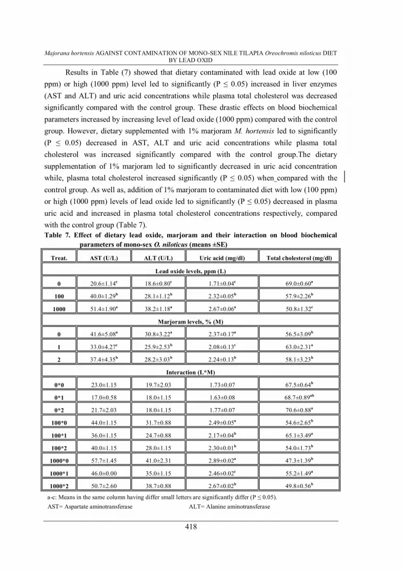
Results in Table (7) showed that dietary contaminated with lead oxide at low (100 ppm) or high (1000 ppm) level led to significantly (P ≤ 0.05) increased in liver enzymes (AST and ALT) and uric acid concentrations while plasma total cholesterol was decreased significantly compared with the control group. These drastic effects on blood biochemical parameters increased by increasing level of lead oxide (1000 ppm) compared with the control group. However, dietary supplemented with 1% marjoram M. hortensis led to significantly (P ≤ 0.05) decreased in AST, ALT and uric acid concentrations while plasma total cholesterol was increased significantly compared with the control group.The dietary supplementation of 1% marjoram led to significantly decreased in uric acid concentration while, plasma total cholesterol increased significantly (P ≤ 0.05) when compared with the control group. As well as, addition of 1% marjoram to contaminated diet with low (100 ppm) or high (1000 ppm) levels of lead oxide led to significantly (P ≤ 0.05) decreased in plasma uric acid and increased in plasma total cholesterol concentrations respectively, compared with the control group (Table 7). Table 7. Effect of dietary lead oxide, marjoram and their interaction on blood biochemical
parameters of mono-sex O. niloticus (means ±SE)
Treat. AST (U/L) ALT (U/L) Uric acid (mg/dl) Total cholesterol (mg/dl)
Lead oxide levels, ppm (L)
0 20.6±1.14c 18.6±0.80c 1.71±0.04c 69.0±0.60a
100 40.0±1.29b 28.1±1.12b 2.32±0.05b 57.9±2.26b
1000 51.4±1.90a 38.2±1.18a 2.67±0.06a 50.8±1.32c
Marjoram levels, % (M)
0 41.6±5.08a 30.8±3.22a 2.37±0.17a 56.5±3.09b
1 33.0±4.27c 25.9±2.53b 2.08±0.13c 63.0±2.31a
2 37.4±4.35b 28.2±3.03b 2.24±0.13b 58.1±3.23b
Interaction (L*M)
0*0 23.0±1.15 19.7±2.03 1.73±0.07 67.5±0.64b
0*1 17.0±0.58 18.0±1.15 1.63±0.08 68.7±0.89ab
0*2 21.7±2.03 18.0±1.15 1.77±0.07 70.6±0.88a
100*0 44.0±1.15 31.7±0.88 2.49±0.05a 54.6±2.65b
100*1 36.0±1.15 24.7±0.88 2.17±0.04b 65.1±3.49a
100*2 40.0±1.15 28.0±1.15 2.30±0.01b 54.0±1.73b
1000*0 57.7±1.45 41.0±2.31 2.89±0.02a 47.3±1.39b
1000*1 46.0±0.00 35.0±1.15 2.46±0.02c 55.2±1.49a
1000*2 50.7±2.60 38.7±0.88 2.67±0.02b 49.8±0.56b
a-c: Means in the same column having differ small letters are significantly differ (P ≤ 0.05). AST= Aspartate aminotransferase ALT= Alanine aminotransferase

KHALIL, F. F. et al.
419
DISCUSSION The drastic effects of dietary contaminated by lead oxide on the growth performance
parameters of mono-sex Nile tilapia in the present study are agreement of those recorded by
(Hussein and Mekkawy, 2001 and Ayyat et al., 2003). Yet, Swailehl and Ezzughayyar
(2001) who reported that growth inhibition of land snail Helix engaddensis caused by Pb-
polluted diet might be reversible but in a slow rate, which could be linked to elimination of
the metal. Moreover, Ayyat et al. (2003) reported that live body weight and daily body gain
of Nile tilapia decreased significantly (P < 0.001) with increasing dietary lead level (200
ppm). The same authors added that daily feed intake decreased significantly with increasing
dietary lead level (200 ppm). Also, increasing dietary lead level impaired the feed conversion
ratio during the whole experimental period (4 months). This reduction in body weight gain,
feed intake and feed conversion ratio may be due to nutritional disturbance and also related
to appetite depression induced by lead exposure. Whereas, lead may depress appetite through
its action on sites influencing gastrointestinal tract e.g. brain centers involved in the
regulation of food consumption (Zaki El-Dean et al., 1996). On the other side, the positive
effects of marjoram M. hortensis on growth performance parameters and also significantly
interaction between dietary supplementation of 1% marjoram and the untreated diet with lead
oxide may be due to its content of effective components e.g. volatile oil and nice fragrant
that make it to be a good attractant for human (Abo Zied, 1989) and for Nile tilapia fish
(Abou-Zeid, 1998). Also, El-Dakar et al. (2004 a) confirmed that herbs and medicinal plants
including marjoram are environment friendly feedstuff in fish diets because their ability to
reduce release organic matter into the aquatic environment.
Contaminated diet by lead oxide in the present study result in hazard effects of fish
carcass composition and its accumulation in fish muscles may be related to the reduction of
fish growth performance, feed intake and feed conversion ratio (Table 2). The same drastic
effects of lead oxide in fish carcass composition were recorded by Ayyat et al. (2003) whom
reported that body moisture and ether extract contents were significantly (P < 0.001)
decreased, while protein content was significantly (P < 0.05) increased by increasing dietary
lead level, body ash did not affected by dietary lead level, ether extract contents were
significantly (P < 0.001) increased. In addition, they added that body lead residues increased
significantly (P < 0.001) by increasing lead level (200 ppm) in fish diets. Yet, Nounou et al.
(1997) reported that the residual amounts of the heavy metals (lead, mercury and arsenic)

Majorana hortensis AGAINST CONTAMINATION OF MONO-SEX NILE TILAPIA Oreochromis niloticus DIET BY LEAD OXID
420
were mostly concentrated in liver. Kidneys were considered the second organ, and finally,
muscle tissues contain the least amount. Moreover, Ashraf et al. (2006) found that the
concentrations of lead in muscle of three species (grey mullet, eel and common sole) were
considerably lower that the permissible limits. Whereas, uptake and bioaccumulation
mechanisms of heavy metals in fish are depend on their chemical speciation which governs
their bioavailability. In addition, life cycle aspects of species such as age and sexual
maturation, and feeding and reproductive behavior must be considered to investigate some
subtle effects of contaminants (Oliveira Ribeiro et al., 1999). Yet, from the healthy point of
view, Karadede et al. (2004) confirmed that the accumulation of heavy metals in fish and
other aquatic lives is of great importance to man as fish is consumed by a large section of the
population. On the other side, the positive effects and significantly interaction between the
dietary addition of marjoram and untreated or treated diets with lead oxide may be due to its
based on the premise that medicine plants contain natural substances that can promote health
and alleviate illness (Eisenberg et al., 1993). Moreover, El-Ashmawy et al. (2005) confirmed
that marjoram as a medicinal herb there is a general agreement using natural feed additives to
improved performance, health and immunity.
Blood parameters are considered pathophysiological indicators of the whole body and
therefore are important in diagnosing the structural and functional status of fish exposed to
toxicants (Adhikari et al., 2004). In addition, hematological studies provide quite frequently
and routinely accepted procedures in diagnosis of mammal research diseases (Ranzani-Paiva
et al., 2000). Results, in the present study showed that the toxic effects of dietary lead oxide
on blood hematological parameters (hemoglobin, red blood cells, packed cell volume, blood
indices and blood platelets), white blood cells count and its differentiation (the percentage of
lymphocytes, monocytes, neutrophils and eosinophils) of mono-sex O. niloticus as well as,
the obtained results confirmed that these drastic effects were increased by increasing level of
dietary lead oxide (1000 ppm) compared with fish fed free or low diets from lead oxide.
These findings were in agreement with those reported by Hussein and Mekkawy (2001) who
stated that the toxic effects of lead in Tilapia zillii treated fish led to significant (P < 0.05)
decreases in hematological components (Hb and PCV%), also the same trend observed on
the other fish species, in Clarias. lazera Shabana (1983); in Barbus conchonius Tewari et al.
(1987) and in European eel Amin (1992), which reflected the role of lead in producing
severe haemolytic anemia with increasing blood clotting time by inhibiting hemoglobin

KHALIL, F. F. et al.
421
synthesis and shortening the span life of circulating erythrocytes as postulated in mammals
by Hernberg (1976). Yet, the most results referred to the decrease in Hb concentration, RBCs
count and PCV% which is responsible to the case of hypochromic anemia induced. As well
as, this exhibited anemia and the associated decrease in Hb, RBCs count and PCV% values
particularly in high dose-treated fish of lead may be due to the high percentage of inhibition
of erythrocytic enzyme amino levulinic acid dehydratase (ALA-D) (>86%) (Heath, 1995).
However, dietary supplementation by 1% marjoram alleviated these toxic effects of dietary
lead oxide on blood hematological components and led to significantly interaction between
the dietary addition of marjoram and treated diets with lead oxide may be due to the natural
feed additives as the medicinal herbs such as marjoram leaves are contained aromatic
substances and essential oils. Marjoram oil is useful for several ailments impacting on the
immune system. Antioxidant and antibacterial activities of the isolated essential oils, the oils
of marjoram plants were strongly characterized by р-cymene (16.80%), γ-terpinene
(16.80%), thymol (8.40%), and carvacrol (1.10%), all essential oils possessed antioxidants
activity (Hazzit et al., 2006).
The blood compartment is often used as a marker for recent lead exposure, so results
revealed that fish fed contaminated diet by lead oxide led to significantly hypoproteinemia,
hypoalbuminemia, hypoglobulinemia, decreased A/G ratio and hypochlosterolemia, while,
other plasma constituents were significantly elevated (AST, ALT and uric acid
concentrations) compared with the control group. These drastic effects were increased by
increasing level of dietary lead oxide as well as, may be due to liver dysfunction which plays
an important role in protein synthesis. These findings are according with those reported by
Hussein and Mekkawy (2001). Also, Ayyat et al. (2003) found that serum total protein and
albumin were significantly (P < 0.001) decreased, while urea-N, creatinine, AST and ALT
significantly (P < 0.001) increased with increasing dietary lead level (200 ppm). In contrary,
Salmeron et al. (1990) described non-significant changes in serum total protein of lead
poisoned fish. Also, Santos and Hall (1990) didn't find any changes in the total cholesterol
level in lead exposed fish as compared with the controls. In this topic, Holcombe et al.
(1976) explained the toxic role of lead on liver concerning the cholesterol synthesis,
estrification and excretion. The observed hypoproteinaemia could be explained by the
demonstrated toxic effects of lead on the hepatic parenchyma (Holcombe et al., 1976 and
Hussein and Mekkawy, 2001). Recently, Newairy and Abdou (2009) reported that lead

Majorana hortensis AGAINST CONTAMINATION OF MONO-SEX NILE TILAPIA Oreochromis niloticus DIET BY LEAD OXID
422
acetate capable of causing marked oxidative damage and hypercholesterolemia in addition to
inhibiting the activities of antioxidant enzymes.
On the other side, the benefit effects of dietary addition of 1% marjoram to alleviated
these toxic effects of dietary lead oxide on plasma constituents and led to significantly
interaction between the dietary additions of marjoram to untreated or treated diets with lead
oxide. These positive effects may be due to the role of marjoram as a potent antioxidant,
which significantly reduced number of gaps, ring chromosome and stickiness, and its plays
also an important role in ameliorating liver and kidney functions and genotoxicity (El-
Ashmawy et al., 2005). As well as, this beneficent effects of marjoram may be due to
bioactive food components of marjoram medicinal plant were eugenol, limonene, ursolic
acid, 1,8-cineole, α-pinene, α-terpinene, carvacrol, farnesol, geraniol, p-cymene, rosmarinic
acid, sterols, thymol and apigenin (Kaefer and Milner, 2008). In this topic, El-Ashmawy et
al. (2005) also confirmed that the aqueous and alcoholic extracts of M. hortensis and its
volatile oil exhibited a hepato-renal protection against lead induced hepato-renal toxicity.
The alcoholic extract of M. hortensis was more potent than its volatile oil or the aqueous
extract in the protection against lead-induced genotoxicity. Moreover, El-Ashmawy et al.
(2007) found that marjoram oil has antioxidative properties and may be useful herbal
remedies, especially for controlling oxidative damages.
CONCLUSION
Based on the obtained results, it could be confirmed the drastic effects of dietary
contaminated by lead oxide on mono-sex O. niloticus fish causes significantly reduction
effects of growth parameters, and impaired blood hematological and biochemical parameters.
Therefore, it could be concluded that the useful and safety effects of dietary supplementation
of 1% marjoram to alleviated the toxic effects of dietary lead oxide on mono-sex O. niloticus
fish and reduce the human health hazard.
REFERENCES 1. Abdelhamid, A. M. 2000. Scientific Fundamentals of Fish Production and
Husbandry. 2nd. Ed., Mansoura Faculty of Agriculture. (ISBN: 977-5526-04-1.(
2. Abo Zied, E. N. 1989. Aromatic plants and its pharmaceutical and agricultural
products. Academic press, Egypt, 473pp.

KHALIL, F. F. et al.
423
3. Abou-Zeid, R. M. 1998. Evaluation of some medicinal plants as a feed additive in
diet of Nile tilapia Oreochromis niloticus M.Sc. Thesis, Faculty of Agriculture, El-
Fayoum, Cairo University, Egypt.
4. Adhikari, S., B. Sarkar, A. Chatterjee, C.T. Mahapatra, and S. Ayyappan, 2004.
Effects of cypermethrin and carbofuran on certain hematological parameters and
prediction of their recovery in a freshwater teleost; Labeo rohita (Hamilton).
Ecotoxicol. Environ. Saf., 58: 220–226.
5. Amin, E. M. 1992. Haematological changes and light microscopic study of
peripheral blood cells in European eel exposed to lead concentrations. Rapp.
Comm. Int. Mer. Medit., 33: 165.
6. AOAC. 2000. Association of Official Analytical Chemists of official methods of
analysis, 17th Ed. Washington, DC.
7. Ashraf, W., Z. Seddigi, A. Abulkibash and M. Khali., 2006. Levels of selected
metals in canned fish consumed in Kingdom of Saudi Arabia. Environ. Monit.
Assess., 117: 271–279.
8. Assaf, M. H., A. A. Ali, M. A. Makboul, J. P. Beck and R. Anton, 1987.
Preliminary study of phenolic glycerosides from Origanum majorana; quantitative
estimation of arbutin; cytotoxic activity of hydroquinone. Planta Medica, 53: 343–
345.
9. Ayyat, M. S., S. M. Sharaf, F. S. Abbas and H. I. El-Marakby, 2003. Reduction of
dietary lead toxicity in Nile tilapia (Oreochromis niloticus). Egyptian J. Nutrition
and Feeds (Special Issue), 6: 419-431.
10. Campanella, L., A. Bonanni, G. Favero and M. Tamassetti. 2003. Determination of
antioxidant properties of aromatic herbs, olives and fresh fruit using an enzymatic
sensor. Anal. Bioanal. Chem., 375: 1011–1016.
11. Dacie, J. V. and S. M. Lewis, 1995. Practical Haematology. 8th ed. Edinburgh,
Scotland: Churchill Livingstone.
12. Davidson, A. 1999. The Oxford Companion to Food. Oxford University Press,
Oxford, UK.
13. Demichele, S. J. 1984. Nutrition of lead. Comp. Biochem. Phys. Part A: Physiology,
78, 401-408.
14. Dorman, H. J., O. Bachmayer, M. Kosar and R. Hiltunen, 2004. Antioxidant
properties of aqueous extracts from selected Lamiaceae species grown in Turkey. J.
Agric. Food Chem., 52: 762–770.

Majorana hortensis AGAINST CONTAMINATION OF MONO-SEX NILE TILAPIA Oreochromis niloticus DIET BY LEAD OXID
424
15. Doumas, B. T. and H. G. Biggs. 1972. Determination of serum albumin. In:
Standard Method of Clinical Chemistry.Vol.7 Edited by G.R. Cooper, New York
Academic press.
16. Duncan, D.B., 1955. Multiple range and multiple F-test. Biometrics, 11:1-42.
17. Eisenberg, D. M., R. C. Kessler, C. Foster, F. E. Norlock, D. R. Calkins and T. L.
Delbanco. 1993. Unconventional medicine in the United States. Preference, costs
and patterns of use. J. Med., 328:246-252.
18. El-Ashmawy, I. M., A. Saleh and O. M. Salama. 2007. Effects of marjoram volatile
oil and grape seed extract on ethanol toxicity in male rats. Basic Clin Pharmacol
Toxicol., 101: 320-327.
19. El-Ashmawy, I. M., A. F. El-Nahas and O. M. Salama. 2005. Protective Effect of
Volatile Oil, Alcoholic and Aqueous Extracts of Origanum majorana on Lead
Acetate Toxicity in Mice. Basic & Clinical Pharmacology & Toxicology, 97: 238–
243.
20. El-Dakar, A. Y., G. D. Hassanien, S. S. Gad and S. E. Sakr, 2004. Medicinal and
aromatic plants as feeding attractants for fish: 1- Effect of dried marjoram leaves on
performance of hybrid tilapia, Oreochromis niloticus X Oreochromis aureus,
fingerlings. J. Egypt Acad. Soc. Environ. Develop., (B- Aquaculture), 5: 67-83.
21. Ellefson, R. D. and W. T. Caraway. 1976. Fundamentals of clinical chemistry. Ed
Tietz NW., p 506.
22. Ercal, N., H. Gurer-Orhan and N. Aykin-Burns, 2001. Toxic metals and oxidative
stress. Part I: Mechanisms involved in metal-induced oxidative damage. Curr. Top.
Med. Chem., 1: 529-539.
23. Furia, T. E. and N. Bellanca. 1971. Fenaroli's handbook of flavor ingredients, The
Chemical Rubber Co., Cleveland, OH. Internet site:
(http://www.rawtheartoflivingblog.com/triedtastedserved/dried-marjoram-
leaves.html.(
24. Gornall, A. G., G. J. Bardawill and M. M. Parid, 1949. Method of determination
protein in serum blood .J. Biol. Chem., 177: 751.
25. Gurer, H. and N. Ercal, 2000. Can antioxidants be beneficial in the treatment of lead
poisoning? Free Radical Bio. Med., 29: 927-945 .
26. Hazzit, M., A. Baaliouamer, M. L. Faleiro and M. G. Miguel. 2006. Composition of
the essential oils of Thymus and Origanum species from Algeria and their
antioxidant and antimicrobial activities. J. Agric. Food Chem., 54: 6314-6321.

KHALIL, F. F. et al.
425
27. Heaney, R. P. 2000. Lead in calcium supplements. J. Am. Med. Assoc., 284: 1432-
1433.
28. Heath, A. G. 1995. Water Pollution and Fish Physiology, 2nd Ed., Lewis
Publishers: New York, USA. (ISBN: 0-87371-632-9.(
29. Hernberg, S. 1976. Biochemical, subclinical and clinical responses to lead and their
relation to different exposure levels as indicated by concentration of lead in blood.
In: Effects and Dose-Response Relationship of Toxic Metals, Norberg, G.F. Ed.
Elsevier: Amsterdam.
30. Holcombe, G. W. M., D. A. Benoit, E. N. Leonard and L. M. Mckim. 1976. Long-
term effects of lead exposure on three generations of brook trout Salvelinus
fontinalis. J. Fish Res. Board. Can., 33: 1731-1741.
31. Humphreys, D J., 1991. Effects of exposure to excessive quantities of lead on
animals. Br. Vet. J., 147: 18-30.
32. Hussein, S. Y. and I. A. A. Mekkawy. 2001. The effects of lead-exposure and lead-
clay interaction on the growth performance, biochemical and physiological
characteristics and histopathology of Tilapia zillii. Bull. Fac. Sci., Assiut Univ., 30:
65-97.
33. Jackson, M. L. 1973. Soil Chemical Analysis. Prentic Hall. TAC. Inc., N.J., U.S.A.
34. Kaefer, C. M. and J. A. Milner. 2008. REVIEWS: CURRENT TOPICS, The role of
herbs and spices in cancer prevention. Journal of Nutritional Biochemistry, 19: 347–
361.
35. KahakÖnen, M. P., A. I. Hopia, H. J. Vuorela, J. Rauha, K. Pihlaja, T. S. Kujala and
M. Heinonen, 1999. Antioxidant activity of plant extracts containing phenolic
compounds. J. Agric. Food Chem., 47: 3954–3962.
36. Karadede, H., S. A. Oymak and E. Ünlu, 2004. Heavy metals in mullet, Liza abu,
and catfish, Silurus triostegus, from the Ataturk Dam Lake (Euphrates), Turkey.
Environ. Int., 30: 183–188.
37. Kelly, W. J. 2004. Herbal medicine handbook. LippincottWilliamsWilkins A
Wolters, Kluwer Co., pp. 289–290.
38. Macdonald, P., R. A. Edwards and J. F. D. Greenhalgh. 1973. Animal Nutrition,
2nd Ed., Longman, London.
39. Matsuura H., H. Chiji, C. Asakawa, M. Amano, T. Yoshihara and J. Mizutani. 2003.
DPPH radical scavengers from dried leaves of oregano (Origanum vulgare). Biosci.
Biotechnol. Biochem., 67: 2311-2316.

Majorana hortensis AGAINST CONTAMINATION OF MONO-SEX NILE TILAPIA Oreochromis niloticus DIET BY LEAD OXID
426
40. McDowell, L. S. 1992. Minerals in Animal and Human Nutrition. Academic Press,
Inc, California, pp. 361-364.
41. McLaughlin, M. J., D. Parker and J. M. Clarke, 1999. Metals and micronutrients
food safety issues. Field Crops Res., 60: 143–63.
42. Newairy, Al. A. and H. M. Abdou. 2009. Protective role of flax lignans against lead
acetate induced oxidative damage and hyperlipidemia in rats. Food and Chemical
Toxicology, 47: 813–818.
43. Nounou, A. H., M. M. Soliman, E. H. Rizkalla and M. N. Assad. 1997.
Hematological and biochemical changes in Clarias lazera as a result of long term
exposure to a triad combination of lead, mercury and arsenic. Egypt. J. Agric. Res.,
75: 247-271.
44. Novak, J., B. Christina, J. Langbehn, F. Pank, M. Skoula, Y. Gotsiou, C. M. Franz.
2000. Ratios of cis-and trans-sabinene hydrate in Origanum majorana L. and
Origanum midrophyllum (Bentham) vogel. Biochem. Syst. Ecol., 28: 697–704.
45. Oliveira Ribeiro, C. A., C. Rouleau, E. Pelletier, H. Tjalve and C. Audet. 1999.
Distribution kinetics of dietary methylmercury in the arctic charr (Salvelinus
alpinus). Environ. Sci. Technol., 33: 902–907.
46. Ranzani-Paiva, M. J. T., A. T. S. Souza, G. C. Pavanelli, R. M. Takemoto and A.C.
Eiras. 2000. Hematological evaluation in commercial fish species from the
floodplain of upper Parana River, Brazil. Acta Sci., 22: 507–513.
47. Reitman, S. and S. Frankel, 1957. Transaminase in serum. Am. J. Clin. Path., 28:
56-63.
48. Roth, L.S. 2001. Mosby’s handbook of herbs and natural supplements. Mosby, A.
Harcort Health Sciences Co., pp. 561–563.
49. Salmeron, F. P., C. M. E. Melendez and T. L. Martinez. 1990. Hepatotoxic and
nephertoxic effect of lead on the Tilapia Sarotherodon aureus. J. Anales de la
Escuela Nacional de ciencias Biologicas Mexico, 33: 147.
50. Santos, M. A. and A. Hall. 1990. Influence of inorganic lead on the biochemical
blood composition of the Eel Anguilla anguilla (L.). J. Ecotoxic. Envir. Saf., 20: 7.
51. SAS, 1997. SAS/STAT Guide for personal computer. SAS Inst. Cary, N. C. (ISBN:
3-540-65014-8.(
52. Satarug, S., J. R. Baker, S. Urbenjapol, M. Haswell-Elkins, P. E. B. Reilly and D. J.
Williams et al.. 2003. A global perspective on cadmium pollution and toxicity in
non-occupationally exposed population. Toxicol Lett., 137:65–83.

KHALIL, F. F. et al.
427
53. Schultz, A., 1984. Uric acid. Clin. Chem, The C.V. Mosby Co. St. Louis. Toronto.
Princeton, 1261-1266.
54. Shabana, M. B., 1983. Induced pathological and biochemical stress of acute lead
poisoning in Egyptian catfish Clarias lazera. Bull. Fac. Sci. Alex. Univ., 23: 1-14.
55. Smith, R .J. and M. L. Winder, 1996. Medicinal garden. In: The National Herb
Garden Guidebook (Ober, R., ed.) pp. 61–71. The Herb Society of America,
Springfield, VA.
56. Stoskopf, M. K. 1993. Fish Medicine. W.B. Saunders Company Harcour Brace
Lovanovish, Inc.
57. Swailehl, K. M. and A. Ezzughayyar. 2001. Dose-dependent effects of dietary Pb
and Zn on feeding and growth rates of the land snail Helix engaddensis.
Ecotoxicology and Environmental Safety, 50: 9-14.
58. Tewari, H., T. Gill and J. Pant, 1987. Impact of chronic lead poisoning on the
hematological and biochemical profiles of Fish, Barbus conchonius. Bull. Environ.
Contam. Toxicol., 38: 748.
59. Van Den Broucke, C. O. and J. A. Lemli. 1980. Antispasmodic activity of
Origanum compactum. Planta Med., 38: 317–331.
60. Velioglu, Y. S., G. Mazza, L. Gao and B. D. Oomah. 1998. Antioxidant activity and
total phenolics in selected fruits, vegetables, and grain products. J. Agric. Food
Chem., 46: 4113–4117.
61. Weichsebum, T. E. 1946. Method for determination of albumin in serum blood.
Amer. J. Clin. Pathol., 16-40. 62. Zaki El-Dean, M., N. M. K. El-Toukhy, Y. A. F. Hammouda and G. A. Hassan.
1996. The effect of lead on the performance of male rabbits and some physiological
parameters. Egypt. J. Anim. Prod., 33:43-55.

Majorana hortensis AGAINST CONTAMINATION OF MONO-SEX NILE TILAPIA Oreochromis niloticus DIET BY LEAD OXID
428
بأكسید أسماك البلطى النیلى وحید الجنس تلوث علیقةاستخدام البردقوش ضد
الرصاص
، فایق حسنى فراج ، أحمد إسماعیل محرم فتحى فتوح خلیل مصر –المنصورة –جامعة المنصورة –كلیة الزراعة - قسم إنتاج الحیوان
بمستویات ) كعشب طبى(إضافة البردقوش تأثیر لدراسة) 3X3( عاملیھتم تصمیم تجربة
1000، 100صفر، (كمحاولة لتقلیل التأثیرات السامة ألكسید الرصاص بمستویات ) %2 ،1 صفر،(
على كفاءة النمو، التحلیل الكیماوى لجسم األسماك، متبقیات الرصاص فى عضالت ) جزء فى الملیون
حیث . أسبوع 16بعد و البیوكیمیائیة لدم أسماك البلطى النیلى وحید الجنس الدمویةاألسماك، القیاسات
لملوثة بأكسید الرصاص أدت إلى إلى أن األسماك المغذاه على العالئق ا تشیر النتائج المتحصل علیھا
الوزن النھائى، الزیادة الوزنیة، معدل الزیادة (أنخفاض معنوى لقیم متوسطات قیاسات النمو لألسماك
المادة الجافة، (، التحلیل الكیماوى لجسم األسماك )، استھالك الغذاءمعدل النمو النوعى الیومیة،
حجم الھیموجلوبین، عدد خالیا الدم الحمراء، (تولوجیة ، قیاسات الدم الھیما)البروتین الخام، الرماد
الصفائح الدمویة، عدد خالیا الدم البیضاء، النسبة المئویة لخالیا الدم ، كرات الدم الحمراء المضغوطة
البروتین الكلى، األلبیومین، الجلوبیولین، النسبة (، قیاسات الدم البیوكیمیائیة )البیضاء اللیمفاویة
بینما حدث أنخفاض غیر معنوى لكل من . مقارنة بالمجموعة الضابطة )ترول الكلى، الكلسبینھما
متوسط وزن ،MCV متوسط حجم الخلیة( لكرات الدم الحمراء معدل اإلعاشة، دالئل الدم
أما كفاءة تحویل . )MCHCالھیموجلوبین فى الخلیة تركیزمتوسط و MCH الھیموجلوبین فى الخلیة
و من ناحیة أخرى أوضحت النتائج أیضًا . معنویًا مقارنة بالمجموعة الضابطةساءت لألسماك الغذاء
أن المستخلص األثیرى، متبقیات الرصاص فى عضالت األسماك، النسبة المئویة لخالیا الدم البیضاء
األالنینین أمینوترانسفریز واألسبرتیت األحادیة،المتعادلة،الحامضیة، تركیزات إنزیمات
، حیث زادت ھذه ، حامض الیوریك زادت معنویًا مقارنة بالمجموعة الضابطةأمینوترانسفریز
عالوة على ذلك أظھرت النتائج أن .التأثیرات السیئة مع زیادة مستوى أكسید الرصاص فى العلیقة
إضافة العشب الطبى البردقوش للعالئق خففت من التأثیرات السامة ألكسید الرصاص على األسماك
لتقلیل التأثیرات % 1لذا توصى الدراسة باإلضافة المفیدة للبردقوش بمستوى . %1خاصة بمستوى
.أسماك البلطى النیلى لیقةالسامة ألكسید الرصاص فى ع
.مكونات الدم –المتبقیات –البردقوش –أكسید الرصاص – البلطى النیلى أسماك: الكلمات الدالة


