Embed Size (px)
Citation preview

ARTICLEdoi:10.1038/nature13976
b-catenin mediates stress resiliencethrough Dicer1/microRNA regulationCaroline Dias1*, Jian Feng1*, Haosheng Sun1, Ning yi Shao1, Michelle S. Mazei-Robison1{, Diane Damez-Werno1, Kimberly Scobie1,Rosemary Bagot1, Benoit LaBonte1, Efrain Ribeiro1, XiaoChuan Liu1, Pamela Kennedy1{, Vincent Vialou1{, Deveroux Ferguson1{,Catherine Pena1, Erin S. Calipari1, Ja Wook Koo1, Ezekiell Mouzon1, Subroto Ghose2, Carol Tamminga2, Rachael Neve3, Li Shen1
& Eric J. Nestler1
b-catenin is a multi-functional protein that has an important role in the mature central nervous system; its dysfunctionhas been implicated in several neuropsychiatric disorders, including depression. Here we show that in mice b-cateninmediates pro-resilient and anxiolytic effects in the nucleus accumbens, a key brain reward region, an effect mediated byD2-type medium spiny neurons. Using genome-wide b-catenin enrichment mapping, we identify Dicer1—important insmall RNA (for example, microRNA) biogenesis—as a b-catenin target gene that mediates resilience. Small RNA profilingafter excising b-catenin from nucleus accumbens in the context of chronic stress reveals b-catenin-dependent microRNAregulation associated with resilience. Together, these findings establish b-catenin as a critical regulator in the develop-ment of behavioural resilience, activating a network that includes Dicer1 and downstream microRNAs. We thus present afoundation for the development of novel therapeutic targets to promote stress resilience.
Despite decades of research, the molecular pathophysiology of depres-sion remains elusive. One molecular player implicated in neuropsy-chiatric illnesses, including depression, is b-catenin1–5. In addition tohaving a structural role at synapses, b-catenin mediates the transcrip-tional output of canonical Wnt signalling6–8. This multi-functionality hasmade it difficult to untangle the mechanism through which b-cateninmight contribute to pathological states. We recently demonstrated theinvolvement of upstream Wnt signalling in the nucleus accumbens (NAc)in mouse depression models, with impaired signalling mediating sus-ceptibility to social stress and increased signalling mediating resili-ence9. We thus began by studying the behavioural role of b-cateninin this brain region.
b-catenin mediates resilience and anxiolytic responsesWe overexpressed b-catenin in a herpes simplex virus (HSV) vector inNAc (Fig. 1a; Extended Data Fig. 1a), which increases b-catenin solelyin the nuclear compartment, as measured by subcellular fractionationand immunohistochemistry (IHC), whereas global N-cadherin/b-catenincomplexes were unaffected (Extended Data Fig. 1b, c). This suggests thatHSV-b-catenin selectively activates the transcriptional function of theprotein, without having direct effects on N-cadherin at synapses, con-sistent with earlier work in cultured cells10.
We next overexpressed b-catenin in NAc during accelerated socialdefeat stress (ASD)11,12. We found that, while HSV-GFP injected controlanimals developed social avoidance, an indicator of depression-like beha-viour, overexpression of b-catenin prevented this phenotype (Fig. 1b).Furthermore, in baseline behavioural assays, b-catenin mediated anantidepressant-like response in the forced swim test (FST) (Fig. 1c),and anxiolytic effects in the elevated plus maze (EPM) (Fig. 1d). We sawno changes in sucrose preference or cocaine conditioned place pref-erence (data not shown), suggesting thatb-catenin does not cause hedonic
changes. To confirm the pro-resilient effect of b-catenin, we used astabilized b-catenin mutant (S33Y)13, and found identical results forwild-type b-catenin in the ASD and FST (Supplementary Notes), withno change in sucrose preference (data not shown). Finally, cell-type-specific overexpression of b-catenin in D2- but not D1-type mediumspiny neurons (MSNs) in NAc (Fig. 1e, Extended Data Fig. 2a) induceda pro-resilient phenotype.
We also investigated the consequences of blocking b-catenin sig-nalling in NAc with two approaches: excising b-catenin from NAc ofconditional floxed mice (Extended Data Fig. 2b) and overexpressing abehaviourally validated dominant negative b-catenin mutant (ExtendedData Fig. 2c)14. Both manipulations promoted susceptibility to stress inmice subjected to a sub-threshold defeat procedure (Fig 1f, g). Excisingb-catenin from NAc caused no change in social interaction or locomo-tion in control animals, demonstrating a specific association with stress(Extended Data Fig. 3a–c). These results establish a critical role forb-catenin signalling in NAc in behavioural resilience.
To explore the endogenous activity of b-catenin in depression, weexamined its transcriptional activity in post-mortem NAc of depressedhumans. Axin2, a universal readout of activated canonical b-cateninsignalling, was robustly suppressed in NAc of depressed humans (Fig. 2a,Supplementary Table 1, Extended Data Fig. 4a). In contrast, total N-cadherin andb-catenin messenger RNA levels were unchanged, point-ing specifically to b-catenin nuclear function alterations in depression.There was also suppression of Tcf3 and Tcf4 (T cell transcription factors3 and 4) levels in depressed patients (Fig. 2a); these are two of severaltranscription factors through whichb-catenin acts. Together, these datademonstrate downregulation of the transcriptional output ofb-cateninin NAc in human depression.
We next investigated Axin2 mRNA levels in mouse NAc 48 h afterchronic social defeat stress (CSDS). We found no difference between
*These authors contributed equally to this work.
1Fishberg Department of Neuroscience and Friedman Brain Institute, Icahn School of Medicine at Mount Sinai, New York, New York 10029, USA. 2Department of Psychiatry, University of TexasSouthwestern, Dallas, Texas 75390, USA. 3Department of Brain and Cognitive Sciences, Massachusetts Institute of Technology, Cambridge, Massachusetts 02139, USA. {Present addresses: Department ofPhysiology, Michigan State University, East Lansing, Michigan 48824, USA (M.S.M.-R.); Department of Psychology, UCLA College of Life Sciences, Los Angeles, California 90095, USA (P.K.); Institut Nationalde la Sante et de la Recherche Medicale (INSERM) U1130; CNRS UMR8246; UPMC UM18, Neuroscience Paris Seine, 75005 Paris, France (V.V.); Department of Basic Medical Sciences, The University ofArizona College of Medicine-Phoenix, Arizona 85004, USA (D.F.).
0 0 M O N T H 2 0 1 4 | V O L 0 0 0 | N A T U R E | 1
Macmillan Publishers Limited. All rights reserved©2014

susceptible and resilient animals (Fig. 2b). However, resilient animalsdisplayed increased Tcf3 and Tcf4, indicating that resilience may beassociated with upregulation ofb-catenin signalling (Fig. 2b). To probethis, we examined the levels of phospho-Ser 675b-catenin, a form withenhanced transcriptional activity, as well as total b-catenin at this timepoint. We found upregulation in resilient versus susceptible animals ofphospho-Ser 675b-catenin but not totalb-catenin (Extended Data Fig. 4b).At 10 days after CSDS, we found elevated levels of Axin2 in resilientmice only (P , 0.05, Supplementary Notes).
Cell-type-specific action of b-catenin in resilienceGiven the small magnitude of change observed above, we questionedwhether the cell-type-specific behavioural effects in Fig. 1e correspondedto differential regulation ofb-catenin signalling in D2 versus D1 MSNs.Using fluorescence-assisted cell sorting-isolated NAc neurons fromD2-GFP mice (whereby the D2 neurons are labelled with green fluor-escent protein, GFP), we found robust induction of Axin2 expressionin D21 neurons of resilient mice, and significantly reduced Axin2 levelsin susceptible versus resilient mice, 48 h post CSDS, effects not seenin D22 cells (Fig. 2c). Furthermore, Axin2 IHC with D1- or D2-GFPtransgenic mice subjected to CSDS revealed downregulation ofb-catenin
transcriptional activity in D2 versus D1 MSNs in susceptible mice (Fig. 2d).In sum, upregulation ofb-catenin signalling occurs in D2 MSNs in resil-ient mice, with downregulation seen in susceptible animals.
Because glutamatergic neurotransmission regulatesb-catenin tran-scriptional activity and stress susceptibility15,16, we tested whether medialprefrontal cortex (PFC) or hippocampus, two important glutamatergicinputs to NAc, control b-catenin signalling in NAc. Using previouslyvalidated constructs and stimulation protocols17,18, we found that opto-genetic stimulation of glutamatergic PFC terminals robustly suppressedb-catenin activity in NAc as indicated by decreased Axin2, Tcf3, andTcf4, whereas stimulation of hippocampus terminals had no effect(Fig. 2e, f). Repeated burst firing of dopamine afferents from the ventraltegmental area (VTA) also had no effect (Extended Data Fig. 5). Thus,PFC to NAc stimulation specifically elicited a molecular ‘signature’ ofsusceptibility, indicating that activation of this circuit could mediatethe maladaptive suppression of b-catenin activity in NAc.
Genome-wide mapping of b-catenin after social defeatWe next conducted b-catenin chromatin immunoprecipitation fol-lowed by deep sequencing (ChIP-seq) on NAc of control, susceptible,and resilient mice after CSDS. We first validated our b-catenin ChIPprotocol by examining an LEF/TCF consensus sequence in the promoterof a known b-catenin target gene, CaMKIV (also known as Camk4). Wefound enrichment ofb-catenin at the LEF/TCF site, but not a distant site,in NAc of resilient mice only (Fig. 3a). Through ChIP-seq19,20 we thenexamined global b-catenin enrichment after CSDS, and found majordifferences in peak numbers (Fig. 3b, Supplementary Data 1). Controland resilient conditions were associated with 10–15-fold higher absolutepeak numbers compared to susceptible conditions, suggesting profoundglobal alterations in b-catenin activity, consistent with our biochemicaldata (Fig. 2). Enrichment ofb-catenin in resilient animals (Fig. 3b) onlyoccurred at transcriptionally active sites, as indicated by high basal bind-ing of two transcriptional activation marks H3K4me3 and H4K16ac(Fig. 3c, Extended Data Fig. 6). However, we did not observe global changesin these two histone marks after CSDS (Extended Data Figs 7, 8), sug-gesting that b-catenin may be recruited to active, open regions of chro-matin through the presence of other, direct DNA-binding transcriptionfactors.
Using Ingenuity pathway analysis, we demonstrated a predicted b-catenin network to be upregulated in NAc of resilient versus susceptiblemice (Extended Data Fig. 9), a prediction specific tob-catenin. Concom-itantly, there were nearly twice as many increases as decreases in b-catenin binding in resilient versus control mice at promoter regions. Incontrast, susceptible versus control animals displayed equivalent numbersof upregulated and downregulated b-catenin binding events (Fig. 3d).These results support our hypothesis that resilience is associated withgenome-wide enrichment of b-catenin. Examining the distribution ofb-catenin peaks across the genome (Fig. 3e) revealed similar results: redis-tribution of b-catenin binding towards promoters and gene bodies inresilience, and redistribution away from promoters/gene bodies andtowards gene deserts in susceptibility.
To validate theb-catenin ChIP-seq data, we conducted quantitativeChIP (qChIP) on independent biological samples at genes that showedsignificant peaks in resilience or upregulation in resilient versus sus-ceptible animals, thus confirming significant b-catenin enrichment atseveral promoters (Fig. 3f). As further validation, we examined mRNAlevels of genes found in our ChIP-seq list that coincided either with in silicolists of predicted or knownb-catenin targets21,22 (Supplementary Table 2)or with the H3K4me3 and H4K16ac ChIP-seq data sets (SupplementaryData 2). We found robust upregulation of many of these genes in NAcof resilient mice (Fig. 3g).
Regulation of Dicer1 and microRNA by b-cateninOne gene validated by qChIP and quantitative PCR (qPCR) was Dicer1,a critical component of microRNA (miRNA) biogenesis23. Thus, select-ive enrichment ofb-catenin binding at Dicer1 in resilient mice (Fig. 4a),
0
20
40
60
80
100
0.0
0.2
0.4
0.6
0.8
1.0
No
targ
et
No
targ
et
Cre
–
Cre
–
Cre
+
Cre
+
D1 line D2 line
*
*
HSV-GFPHSV-β-catenin
Seco
nd
s in
inte
ractio
n z
one
Targ
et
Targ
et
a b
e
0
20
40
60
80
0
50
100
150
200
250
0
50
100
150
Tim
e im
mo
bile
(s)
Seco
nd
s
Open arms Closed armsFST
**
*
HSV-
β-ca
t
HSV-
GFP
HSV-
GFP
HSV-
GFP
HSV-
β-ca
t
HSV-
β-ca
t
c d
NAc
0
20
40
60
80
100
0
20
40
60
80
100HSV-GFP HSV-GFP
HSV-DN-β-cateninHSV-Cre
Targ
et
Targ
et
targ
etNo
No
targ
et
f g
***
So
cia
l
inte
ractio
n r
atio
Seco
nd
s in
inte
ractio
n z
one
Seco
nd
s in
inte
ractio
n z
one
Targ
et
Targ
et
targ
etNo
No
targ
et
Figure 1 | b-catenin in NAc mediates pro-resilient, antidepressant, andanxiolytic responses. a, IHC illustrating viral transgene expression mediatedby HSV-b-catenin with coronal cartoon of NAc highlighted. b, Pro-resilienteffect of HSV-b-catenin on social interaction after ASD (*P , 0.05, two wayANOVA, n 5 8 GFP, n 5 10 b-catenin). c, Antidepressant-like effect ofb-catenin in the forced swim test (*P , 0.05, two-tailed t-test, n 5 6 GFP, n 5 7b-catenin). d, Anxiolytic-like effect of b-catenin in the elevated plus maze(closed arms: *P , 0.01, open arms: *P , 0.01, two-tailed t-test, n 5 6 GFP,n 5 7 b-catenin). e, Cell-type-specific overexpression of b-catenin in ASD(D2 Cre– versus Cre1: *P , 0.05, two-tailed t-test, n 5 13 D2 Cre2, n 5 8 D2Cre1). f, Effect of knocking down b-catenin in a sub-threshold defeatprocedure (**P , 0.01, two-way ANOVA, effect of virus only when targetpresent, n 5 6 GFP, n 5 5 Cre). g, Effect of dominant negative b-catenin insub-threshold defeat (*P , 0.05, two-way ANOVA, interaction effect, n 5 5GFP, n 5 4 dominant negative). Data presented as mean and s.e.m. and arerepresentative of at least two experiments. See Methods and SupplementaryTable 9 for detailed statistics.
RESEARCH ARTICLE
2 | N A T U R E | V O L 0 0 0 | 0 0 M O N T H 2 0 1 4
Macmillan Publishers Limited. All rights reserved©2014

and subsequent validation of this effect (Fig. 3f, g), indicated that Dicer1represents a robust target ofb-catenin in NAc. To study the behaviouraleffects of Dicer1, we knocked it down locally in NAc (Extended DataFig. 10), and conducted sub-threshold defeat. Control animals injectedwith HSV-GFP displayed normal social interaction; however, animalswith Dicer1 knockdown demonstrated social avoidance (Fig. 4b), whichmimicked the effects of blocking b-catenin signalling (Fig. 1). Impor-tantly, we can rule out confounding effects of long-term Dicer1 loss on
neuronal viability24, because our experimental paradigm was limitedto two weeks.
To assess whether the behavioural effect of Dicer1 was related tob-catenin signalling, we first expressed HSV-Cre or HSV-GFP in NAcof floxed Dicer1 mice and found no difference in social interaction underbaseline, non-stressed conditions (Fig. 4c). We then injected all micewith HSV-b-catenin in NAc and subjected them to ASD. b-cateninoverexpression blocked the development of social avoidance in mice
0.0
0.5
1.0
1.5
2.0
0.0
0.5
1.0
1.5
0
2
4
6
8
0.0
0.5
1.0
1.5
020406080
100
Control
Depressed
ControlSusceptibleResilient
** **
*
*
Axin2 N-cad β-cat Tcf3 Tcf4 Axin2 Tcf3 Tcf4Lef1 Tcf1
D2+
Con
trol
Con
trol
Susce
ptible
Susce
ptible
Res
ilient
Res
ilient
D2
D1
GFP Axin2 Merge
Chronic (10 d) stimulation of PFC
or hippocampal glutamatergic,
NAc-specific projecting afferents
9 weeks 1 week
AAV-ChR or
mCherry
surgery
Implant
optic
fibre
SI
test
Collect
tissue
0.0
0.5
1.0
1.5
Axin2 Tcf3 Tcf4
* ****
PFCHippocampus
a b
c d
e
f
****
*
Fo
ld c
ontr
ol m
RN
A
Fo
ld c
ontr
ol m
RN
A
D2–
Fo
ld c
ontr
ol
Axin
2 m
RN
A
GF
P+ c
ells
that
are
Axin
2+ (%
)
D1
susc
eptib
leD2
susc
eptib
le
Fo
ld c
ontr
ol m
RN
A
Figure 2 | Regulation of b-catenin signalling inhuman depression and mouse CSDS. a, mRNAfrom human NAc (Axin2: **P , 0.01; Tcf3:*P , 0.05; Tcf4: *P , 0.05, two-tailed t-test, n 5 6control, n 5 10 depressed). b, mRNA from mousecontrol, susceptible, and resilient NAc 48 h postCSDS (Tcf3: *P , 0.05; Tcf4: *P , 0.05, one-wayANOVA, n 5 16 control, n 5 12 susceptible, n 5 9resilient). c, Axin2 is upregulated in D21 MSNsonly in resilience (Axin2 D21:**P , 0.01, controlversus resilient P , 0.05, *P , 0.01 susceptibleversus resilient, n 5 4 control, n 5 5 susceptible,n 5 3 resilient; Axin2 D22: not significant,P . 0.05, n 5 3 control, n 5 5 susceptible, n 5 3resilient, one-way ANOVA). d, Percentage of cellspositive for Axin2 plus GFP in D1- or D2-GFPsusceptible mice after CSDS (**P , 0.01, two-tailed t-test, n 5 3 per group). e, Optogeneticstimulation protocol. f, mRNA expression in NAcafter repeated stimulation from PFC orhippocampus in ChR2 versus mCherry (Axin2:*P , 0.05, n 5 6 mCherry, n 5 5 ChR; Tcf3:**P , 0.01, n 5 6 mCherry; n 5 4 ChR; Tcf4:**P , 0.01, n 5 6 mCherry, n 5 4 ChR, two-tailedt-test). Human data are from one experiment, allother data are representative of at least twoexperiments. All data presented as mean and s.e.m.
H3K4me3 H4K16ac β-cat C β-cat S β-cat R
←5k -TSS- 5k→Chromosome 1
Gene bodyProximal promoter1 kb promoter3 kb promoterGene desertOther intergenicPericentromere
0.0
0.5
1.0
1.5
2.0
0
1
2
3
4
5
Cre
b
FosB
Gad
d45a
mGluR2
Dicer
1
Pick1
Htr2
a
PPP1r9b
Syt11
Dicer
1Hlf
Clust
erin
GFR
A4
JMJD
2bRai1
Cac
na1a
*
**
**
*
*
*
Fo
ld c
ontr
ol
Fo
ld c
ontr
ol m
RN
A
Fo
ld c
ontr
ol
Read
co
unt
per
mill
ion
map
ped
read
s
TSS TES5′ → 3′
Susceptible
(880)
Resilient
(563)
Susceptible
(819)
Resilient
(1,106)
123 139
0
1
2
3
4
LEFF TFBS Control(2 kb away)
a b c
d
e
f g
Control
Susceptible
Resilient C: 11,963
R: 14,694
S: 1,099 *
0.06
0.08
0.10
0.12
Resilient
Susceptible
Control
Figure 3 | b-catenin ChIP-seq in NAc 48 h postCSDS. a, qChIP validation of b-catenin ChIP(*P , 0.05, one-way ANOVA, post-hoc testcontrol versus resilient and susceptible versusresilient at LEFF transcription factor binding site(TFBS) of a CaMKIV gene, n 5 4 per group).b, Plot of b-catenin binding across genic regions.TSS, transcription start site; TES, transcription endsite. Individual peak numbers per conditionindicated in inset. c, Heat map showing b-cateninbinding 5 kb up- and downstream of TSSs onchromosome 1 in control (C), susceptible (S), andresilient (R) NAc; binding profiles of H3K4me3and H4K16ac under basal conditions are alsoshown. d, Number of increased (up arrow) versusdecreased (down arrow) b-catenin binding sitesat promoters in resilient versus control orsusceptible versus control conditions. e, Genome-wide distribution of b-catenin binding. f, qChIPvalidation of ChIP-seq (Gadd45a: *P , 0.05,one-way ANOVA; Dicer1: *P , 0.05, one-wayANOVA, n 5 4 control, susceptible, n 5 3resilient). g, mRNA validation of b-cateninChIP-seq (Dicer1: *P , 0.01, one-way ANOVA,n 5 13 control, n 5 11 susceptible, n 5 7 resilient).Data presented as mean and s.e.m. and arerepresentative of at least two experiments. Colour-coding in f and g as in a.
ARTICLE RESEARCH
0 0 M O N T H 2 0 1 4 | V O L 0 0 0 | N A T U R E | 3
Macmillan Publishers Limited. All rights reserved©2014

expressing normal Dicer1 levels, but not in mice with NAc Dicer1 knock-down (Fig. 4c). This indicates that at least part of the pro-resilient effectof b-catenin is mediated through Dicer1.
Finally, these data prompted us to examine the global miRNA pro-file in NAc in response to CSDS and study its dependence onb-catenin.We injected an adeno-associated virus (AAV) vector expressing GFPor Cre in NAc of floxed b-catenin mice, subjected them to CSDS orcontrol conditions, and performed small RNA sequencing (Supplemen-tary Table 3). We first compared each group—GFP susceptible (GFP-sus), GFP resilient (GFP-res), Cre control (Cre-con), and Cre susceptible(Cre -sus)—to the ‘GFP-con’ condition. We could not study the Cre re-silient condition, because virtually no mice are resilient uponb-cateninknockout from NAc. We found downregulation of numerous miRNAs,including many that were upregulated in resilience, whenb-catenin wasknocked out from control animals (Cre-con, Fig. 4d, SupplementaryTable 4). Interestingly, a smaller subset of miRNAs was upregulated
following b-catenin knockout, which may represent miRNAs that areregulated by repressive factors under b-catenin control. We identified66 miRNAs that were significantly downregulated in NAc afterb-catenindeletion (Cre-con, Fig. 4e). We also identified downregulated miRNAs(n 5 79) in the Cre-sus condition, many of which were decreased inCre-con, further substantiating our hypothesis that pro-adaptive miRNAresponses are lost in the absence of b-catenin, enhancing susceptibilityto stress (Fig. 4e). miRNAs that overlapped between any two groups(up in GFP-res, but down in Cre-con or Cre-sus), presumably representthe most biologically important b-catenin- and stress-regulated miRNAs(Fig. 4e, Supplementary Table 5). This subset controls several meaningfulgene categories (Fig. 4f), including Wnt and glutamatergic signalling.Finally, to identify potential miRNA targets, we overlapped predictedtargets of these b-catenin-regulated miRNAs (Supplementary Table 5)with mRNA-seq data from NAc after CSDS. We thus found several inter-esting, novel genes to be significantly repressed in resilience (Fig. 4g).
We also examined other small RNAs for regulation by CSDS. Piwi-interacting RNAs (piRNAs), small RNAs widely studied in germ linecells, were detected recently in brain and found to play a functionalrole in spine morphology and synaptic plasticity25,26. 163 piRNAs weredetectable in our data set with read counts in at least one condition,supporting the notion of piRNA expression in brain (SupplementaryTable 6). Although the majority of them were expressed at low levels,approximately 20 piRNAs appear to be regulated by CSDS (Supplemen-tary Table 7). Examining additional small RNA categories that mightbe regulated by Dicer1 revealed several differentially expressed candidates(Supplementary Table 8), laying the groundwork for future investigation.
DiscussionThe present study demonstrates that b-catenin in D2 MSNs activatesa network in NAc that mediates behavioural resilience, whereas def-icits in this pathway contribute to depression-related pathology. PFCinputs to NAc appear to be particularly important in controlling thisb-catenin regulation. D2 MSNs, which comprise the indirect or ‘no-go’ pathway27–30, may be more important for mediating flexible beha-vioural choices in aversive contexts compared to reward-motivatedbehaviour31–33. We thus posit that enhanced b-catenin signalling inNAc D2 MSNs of resilient mice permits increased behavioural flexibil-ity, which allows them, despite having the same experience as suscept-ible mice, to overcome generalizing avoidance of all mice, a processindependent of hedonic responses. This has parallels in humans: resi-lient individuals are more successful at managing stress and recoveringfrom it34.
Our b-catenin ChIP-seq approach provides a valuable resource formining the molecular targets that drive resilience. One validated tar-get is Dicer1, which establishes a novel connection between b-cateninsignalling and miRNAs in brain. Among the regulated miRNAs arethose that feedback and regulate b-catenin signalling35. The cell type-specific role of b-catenin, and the inherent complexity of stress sus-ceptibility versus resilience, which involves many additional regulatorysteps beyond Dicer1, presumably explains the relatively small numberof b-catenin-dependent miRNAs observed in this study. miRNAs pro-vide a crucial layer of post-transcriptional gene regulation in neuraldevelopment, plasticity, and in an increasing number of brain disor-ders36–38. The present study, by identifying specific miRNAs associatedwith stress susceptibility or resilience, offers a template for future stud-ies to induce resilience in inherently more susceptible individuals.
Online Content Methods, along with any additional Extended Data display itemsandSourceData, are available in the online version of the paper; references uniqueto these sections appear only in the online paper.
Received 12 August 2013; accepted 20 October 2014.
Published online 12 November 2014.
1. Madsen, T. M., Newton, S. S., Eaton, M. E., Russell, D. S. & Duman, R. S. Chronicelectroconvulsive seizure up-regulates b-catenin expression in rat hippocampus:role in adult neurogenesis. Biol. Psychiatry 54, 1006–1014 (2003).
TSS 250 500–500 –250
HSV-GFP
or
HSV-Cre
SI HSV-β-cat
all mice
ASD
SI
post-defeat
Pre-defeat Post-defeat
*
**
Fo
ld e
nrich
men
to
ver
inp
ut
aControlSusceptibleResilient 1.5
0
50
100
150
Seco
nd
s in
in
tera
ctio
n z
on
e
HSV-GFP
HSV-Cre
Targ
et
Targ
et
targ
et
targ
etNo
Tim
e in
SI
zo
ne w
ith
targ
et
No
HSV-GFP
HSV-Cre
–1 0 +1
12
473
6 6
57
GFP-res
increase
Cre-con
decrease
Cre-sus
decrease
10
GFP-sus GFP-res Cre-con Cre-sus
b
c
d e
fg
0
20
40
60
80
100
Cancer transcriptional misregulation
Glutamatergic synapse
Endocytosis
Wnt signalling pathway
Long-term potentiation
Axon guidance
Ubiquitin-mediated proteolysis
MelanogenesisPI3K-Akt signalling pathway
Regulation of actin cytoskeleton
MAPK signalling pathway
Putative β-catenin
regulated miRNAGene name
miR103, miR205, miR214
miR32, miR103, miR205,
miR214,miR302b, miR375,
miR483
miR103, miR205, miR214,
miR224, miR302b
miR205, miR214, miR216b,
miR302b, miR375
mir32, miR103, miR214,
miR216b,miR224, miR302b,
miR375
miR 32, miR302b
miR32, miR224
Arc
Btg2
Calcr
Gpr101
Htr4
Nos2
Npas4GO enrichment P value
0 4×10⁻⁸ 10–7 1.3×10⁻⁷
Figure 4 | Dicer1 bridges b-catenin and miRNA regulation in CSDS.a, b-catenin ChIP-seq enrichment around the Dicer1 TSS. b, Effect of NAcDicer1 knockdown (HSV-Cre) in sub-threshold defeat with HSV-GFP ascontrol (**P , 0.01, effect of virus, two-way ANOVA, n 5 7 Cre, n 5 8 GFP).c, Left, schematic of floxed Dicer1 deletion followed by b-catenin rescue; right,social interaction (SI) before and after ASD with HSV-b-catenin (*P , 0.05,interaction effect, matching two-way ANOVA, n 5 7 per group). d, Heat mapof CSDS-regulated miRNA expression changes with (Cre) or without (GFP)b-catenin knockdown. Log2-fold changes of all altered miRNAs among allgroups are shown. e, Venn diagram showing increased miRNAs in GFP-resilient mice (GFP-res) overlap with decreased miRNAs in b-cateninknockout in non-stressed (Cre-con) or susceptible (Cre-sus) animals. f, Top 11most enriched gene ontology terms of target genes of overlapping miRNAs inpanel e. g, Predicted targets of b-catenin-dependent miRNAs displaydownregulation by mRNA-seq in resilient mice after CSDS. Data presented asmean and s.e.m. and are representative of at least two experiments.
RESEARCH ARTICLE
4 | N A T U R E | V O L 0 0 0 | 0 0 M O N T H 2 0 1 4
Macmillan Publishers Limited. All rights reserved©2014

2. Beaulieu, J.-M. et al. Lithium antagonizes dopamine-dependent behaviorsmediated by an AKT/glycogen synthase kinase 3 signaling cascade. Proc. NatlAcad. Sci. USA 101, 5099–5104 (2004).
3. Gould, T. D. et al. Beta-catenin overexpression in the mouse brain phenocopieslithium-sensitive behaviors. Neuropsychopharmacology 32, 2173–2183 (2007).
4. Li, X. & Jope, R. S. Is glycogen synthase kinase-3 a central modulator in moodregulation? Neuropsychopharmacology 35, 2143–2154 (2010).
5. Brennand, K. J. et al. Modelling schizophrenia using human induced pluripotentstem cells. Nature 473, 221–225 (2011).
6. Behrens, J., von Kries, J., Kuhl, M. & Bruhn, L. Functional interaction of b-cateninwith the transcription factor LEF-1. Nature 382, 638–642 (1996).
7. Molenaar, M. et al. XTcf-3 transcription factor mediates b-catenin-induced axisformation in Xenopus embryos. Cell 86, 391–399 (1996).
8. van de Wetering, M. et al.Armadillo coactivates transcription driven by the productof the Drosophila segment polarity gene dTCF. Cell 88, 789–799 (1997).
9. Wilkinson,M.B.et al. Anovel role of theWNT-dishevelled-GSK3b signalingcascadein the mouse nucleus accumbens in a social defeat model of depression.J. Neurosci. 31, 9084–9092 (2011).
10. Sadot, E. et al. Regulation of S33/S37 phosphorylated b-catenin in normal andtransformed cells. J. Cell Sci. 115, 2771–2780 (2002).
11. Berton, O. et al. Essential role of BDNF in the mesolimbic dopamine pathway insocial defeat stress. Science 311, 864–868 (2006).
12. Krishnan, V. et al. Molecular adaptations underlying susceptibility and resistanceto social defeat in brain reward regions. Cell 131, 391–404 (2007).
13. Kolligs, F. T., Hu, G.,Dang,C.V.&Fearon, E. R.Neoplastic transformation ofRK3Ebymutant b-catenin requires deregulation of Tcf/Lef transcription but not activationof c-myc expression. Mol. Cell. Biol. 19, 5696–5706 (1999).
14. Wang, Z. et al.b-cateninpromotes survival of renal epithelial cells by inhibitingBax.J. Am. Soc. Nephrol. 20, 1919–1928 (2009).
15. Rada, P.et al. Glutamate release in the nucleus accumbens is involved inbehavioraldepression during the Porsolt swim test. Neuroscience 119, 557–565 (2003).
16. Abe, K. & Takeichi, M. NMDA-receptor activation induces calpain-mediatedb-catenin cleavages for triggering gene expression. Neuron 53, 387–397 (2007).
17. Tye,K.M.et al. Amygdalacircuitrymediating reversible andbidirectional control ofanxiety. Nature 471, 358–362 (2011).
18. Britt, J. P. et al. Synaptic and behavioral profile of multiple glutamatergic inputs tothe nucleus accumbens. Neuron 76, 790–803 (2012).
19. Feng, J. et al. Chronic cocaine-regulated epigenomic changes in mouse nucleusaccumbens. Genome Biol. 15, R65 (2014).
20. Shen, L. et al. diffReps: detecting differential chromatin modification sites fromChIP-seq data with biological replicates. PLoS ONE 8, e65598 (2013).
21. Hodar, C. et al. Genome-wide identification of new Wnt/b-catenin target genes inthe human genome using CART method. BMC Genomics 11, 348 (2010).
22. Wexler, E. M. et al. Genome-wide analysis of a Wnt1-regulated transcriptionalnetwork implicates neurodegenerative pathways. Sci. Signal. 4, ra65 (2011).
23. Bernstein, E., Caudy, A. A., Hammond, S. M. & Hannon, G. J. Role for a bidentateribonuclease in the initiation step of RNA interference. Nature 409, 363–366(2001).
24. Cuellar, T. L. et al. Dicer loss in striatal neurons produces behavioral andneuroanatomical phenotypes in the absence of neurodegeneration. Proc. NatlAcad. Sci. USA 105, 5614–5619 (2008).
25. Lee, E. J. et al. Identification of piRNAs in the central nervous system. RNA 17,1090–1099 (2011).
26. Rajasethupathy, P. et al. A role for neuronal piRNAs in the epigenetic control ofmemory-related synaptic plasticity. Cell 149, 693–707 (2012).
27. Graybiel, A. M. The basal ganglia. Curr. Biol. 10, R509–R511 (2000).28. Gerfen, C. R. The neostriatal mosaic: multiple levels of compartmental
organization in the basal ganglia. Annu. Rev. Neurosci. 15, 285–320 (1992).29. Kravitz, A. V., Tye, L. D. & Kreitzer, A. C. Distinct roles for direct and indirect pathway
striatal neurons in reinforcement. Nature Neurosci. 15, 816–818 (2012).30. Lobo, M.K.& Nestler, E. J. The striatal balancing act indrugaddiction:distinct roles
of direct and indirect pathway medium spiny neurons. Front. Neuroanat. 5, 41(2011).
31. Hikida, T., Kimura, K., Wada, N., Funabiki, K. & Nakanishi, S. Distinct roles ofsynaptic transmission in direct and indirect striatal pathways to reward andaversive behavior. Neuron 66, 896–907 (2010).
32. Darvas, M. & Palmiter, R. Contributions of striatal dopamine signaling to themodulation of cognitive flexibility. Biol. Psychiatry 69, 704–707 (2011).
33. Yawata, S., Yamaguchi, T., Danjo, T., Hikida, T. & Nakanishi, S. Pathway-specificcontrol of reward learning and its flexibility via selective dopamine receptors in thenucleus accumbens. Proc. Natl Acad. Sci. USA 109, 12764–12769 (2012).
34. Southwick, S. M. & Charney, D. S. The science of resilience: implications for theprevention and treatment of depression. Science 338, 79–82 (2012).
35. Veronese, A. et al. Mutated b-catenin evades a microRNA-dependent regulatoryloop. Proc. Natl Acad. Sci. USA 108, 4840–4845 (2011).
36. Kosik, K. S. The neuronal microRNA system. Nature Rev. Neurosci. 7, 911–920(2006).
37. Im, H.-I. & Kenny, P. J. MicroRNAs in neuronal function and dysfunction. TrendsNeurosci. 35, 325–334 (2012).
38. Issler, O. et al. MicroRNA 135 is essential for chronic stress resiliency,antidepressant efficacy, and intact serotonergic activity. Neuron 83, 344–360(2014).
Supplementary Information is available in the online version of the paper.
Acknowledgements We thank O. Jabado andM.Mahajan for support andS. Borkan forproviding b-catenin constructs. This work was supported by grants from the NationalInstitute of Mental Health and the Hope for Depression Research Foundation (HDRF).
Author Contributions C.D. and J.F. conceived the project, designed research,conducted experiments, interpreted the results, and wrote the manuscript; H.S.,M.S.M.-R., D.D.-W., K.S., R.B., B.L., E.R., P.K., V.V., D.F., C.P., E.C., J.K. and E.M. conductedexperiments; S.G., C.T. provided reagents and tools; R.N. conducted experiments andprovided reagents; N.S., X.L. performed bioinformatic analysis; L.S. performed andsupervised bioinformatic analysis; E.J.N. conceived the project, designed andsupervised research, interpreted the results, and wrote the manuscript. All authorsdiscussed the results and commented on the manuscript.
Author Information All sequencing data have been deposited into the Gene ExpressionOmnibus with accession numbers GSE61294 and GSE61295. Reprints andpermissions information is available at www.nature.com/reprints. The authors declareno competing financial interests. Readers are welcome to comment on the onlineversion of the paper. Correspondence and requests for materials should be addressedto E.J.N. ([email protected]) or L.S. ([email protected]).
ARTICLE RESEARCH
0 0 M O N T H 2 0 1 4 | V O L 0 0 0 | N A T U R E | 5
Macmillan Publishers Limited. All rights reserved©2014

METHODSAnimals. For all experiments, 7–9-week-old male mice were used. Unless other-wise noted for transgenic lines, c57bl/6 mice from Jackson Laboratories wereused. All mice were housed on a 12-h light/dark cycle with ad libitum access tofood and water. CD1 retired breeder mice were obtained from Charles River Labo-ratories. The following transgenic mouse lines were used. From Jackson Laboratories:b-catenin conditional floxed mice (stock no. 004152) and Dicer1 conditional floxedmice (stock no. 006001). Additionally, D1-Cre, D2-Cre, D1-GFP, and D2-GFP malemice that were backcrossed to a c57bl/6 background were used for experimentsas described in the text. For the D1-Cre/D2-Cre cell-type specific overexpressionexperiments, wild-type littermates were used as controls. The Mount Sinai Insti-tutional Animal Care and Use Committee approved all animal protocols used inthis study. For all experiments, extensive laboratory experience was used to estim-ate required sample sizes. Animals were randomly assigned to experimental groupsand whenever possible, experimenters were blinded to the group. (For example, inbehavioural experiments by assigning numbers to animals and in IHC experimentsby hiding group designation until after quantification and analysis.)Viral-mediated gene transfer. Stereotactic surgery was performed on mice underketamine/xylazine anaesthesia. Vectors were infused bilaterally into NAc at a rateof 0.1ml min21 with the following coordinates: 11.6 mm anterior-posterior (A/P),11.5 mm medial-lateral (M/L), 6 4.4 mm dorsal-ventral (D/V) from bregma. Atotal of 0.5ml per side was infused except for the HSV-LS virus, in which case 0.7mlwas infused total. All vectors used were cloned into p1005 HSV or LS1L HSV. Mouseb-catenin constructs were provided by S. Borkan (Boston University). Wild-typeand dominant negative constructs were used, with the dominant negative constructcontaining amino and carboxy-terminal truncations. Because this is a complicatedmutant, we behaviourally validated it by demonstrating a failure to rescueb-cateninloss of function impairments in social interaction (Extended Data Fig. 2c). Humanb-catenin S33Y construct (Addgene Plasmid no. 19286) was originally from E. Fearon(Michigan). This mutant contains an S33Y mutation that prevents phosphorylationat Ser 33 by GSK3b, thus preventing b-catenin degradation. For cell-type-specificoverexpression, an HSV carrying b-catenin in a lox-stop cassette was used (Sup-plementary Fig. 2a) in conjunction with D1- and D2-Cre transgenic mouse lines.Viral-Cre was used for local knockdown of b-catenin or Dicer1 in conditionalfloxed mice.Behaviour. 10-day chronic social defeat stress (CSDS), an accelerated 4-day defeatprocedure (ASD), and a sub-threshold defeat procedure have been described prev-iously and represent an ethologically validated model of depression9,11,12 We usedASD over 4 days (4 days of defeat, twice a day) to coincide with periods of maximalHSV-mediated transgene expression in some experiments as described, which inducesthe same degree of behavioural deficits in normal mice as our standard 10-dayCSDS procedure. For all defeats, social interaction was measured either 24 h or1 week following the last defeat. For all tissue analysis, including ChIP, mice werekilled 48 h after the last defeat (24 h after social interaction) of a 10 day CSDS para-digm unless otherwise specified. Elevated plus maze and forced swim tests wereperformed as described previously12.Post-mortem human tissue. Human post-mortem NAc complementary DNAwas generated and analysed as before9. Briefly, brain tissue was obtained from theDallas Brain Collection, where tissue is collected from the Dallas Medical Examiner’sOffice and UT Southwestern’s Tissue Transplant Program following consent ofnext-of-kin. Tissue was analysed and matched for age, post-mortem interval, RNAintegrity number (RIN), and pH (see Supplementary Table 1) and this same tissueset was used in previously published work39. Samples were subjected to a standarddissection before snap freezing in 240 uC isopentane and storage at 280 uC;further dissection of NAc was performed on frozen tissue. The UT SouthwesternInstitutional Review Board reviewed and approved the collection of this tissue forresearch use. We should note that there was no difference in expression of Axin2between medicated and unmedicated depressed patients, although all patientswere clinically depressed at their time of death (Supplementary Fig. 4a). We thuscombined the medicated and unmedicated groups into one overall depressed groupas presented in Fig. 2.RNA isolation and qPCR. RNA was extracted and purified using a protocolcombining TRIzol/chloroform extraction with the Qiagen RNeasy Micro kit, witha motorized mini-pestle vibrator to homogenize the tissue. After extraction, purityand concentration were measured on a NanoDrop spectrophotometer. RNA wasthen reverse transcribed into cDNA with the iScript DNA synthesis kit (Bio-Rad).GAPDH was used to normalize quantification. Primers were designed to flankexon/intron boundaries and were created using the open-source software Primer3.Real-time qPCR analysis was performed with the DDCt method to obtain relativefold-change of expression as compared to control samples40. BLAST and dissoci-ation curve analysis was also performed to ensure specificity of primer design.Western blotting. NAc was dissected bilaterally using 14 gauge steel circular punches.The tissue was then sonicated in radioimmunoprecipitation assay (RIPA) buffer
with a desktop sonicator (10 mM Tris, pH 7.4, 150 mM NaCl, 1 mM EDTA, 0.1%SDS, 1% Triton X-100, 1% sodium deoxycholate, with protease and phosphataseinhibitors) and centrifuged. The supernatant was collected and the protein con-centration was quantified using the Lowry method. Laemelli sample buffer wasadded to the protein lysate and equal amounts of protein were loaded onto precastSDS–PAGE gels with molecular weight ladders. Samples were transferred to acti-vated PVDF membranes, blocked, and incubated in primary antibody overnight.Blots were washed, and then incubated with Licor secondary fluorescent antibod-ies. After further washing, the blots were scanned and images analysed with ImageJsoftware. The following antibodies were used: phospho-Ser 675 b-catenin (CellSignaling no. 4176; Ser 675 is phosphorylated by PKA), total b-catenin (Cell Sig-naling no. 9562), GAPDH, b-tubulin, and total H3. All antibodies are commer-cially available and have been validated for use in the laboratory. Pre-incubatingthe tissue with calf intestinal phosphatase and demonstrating a decrease in signalwas performed to validate the phospho-Ser 675 b-catenin antibody.Optogenetics. For glutamatergic nerve terminal stimulation, mice were injectedunilaterally with AAV-CAMKIIa-ChR2-mCherry or AAV-CAMKIIa-mCherrywith the coordinates of: (23.6 A/P, 13.05 M/L, 24.85 D/V) for ventral hippocam-pus and (11.9 A/P, 10.5 M/L, 23.0 D/V) for PFC unilaterally. After 9 weeks ofrecovery to allow for expression in terminals, a second stereotactic surgery wasperformed to implant an optic fibre targeting the NAc shell with coordinates of(11.4 A/P, 11.5 M/L, 24 D/V), again unilaterally, ipsilateral to virus expression.After allowing one week for recovery, the mice underwent 10 days of daily 5-minstimulation sessions outside of their home cage as described41,42. Stimulation para-meters were either 20 Hz, 30 pulses per burst, with 10 s between bursts (hippocam-pus); or 30 Hz, 90 pulses per burst, 10 s between bursts (PFC) to roughly balancethe relative intensity of NAc innervation from these two afferent regions. UnilateralNAc tissue was then dissected 48 h later for biochemical experiments. Constructsand stimulation parameters have been previously validated17,18. AAV-ChR2 wasused to stimulate VTA cell bodies with a phasic protocol (20 Hz, 5 spikes per burst,10 s between bursts) given susceptible mice exhibit increased firing rate and burst-ing events following defeat12,43.Co-immunoprecipitation (Co-IP). A co-IP kit (Roche) was used as follows. 4punches of NAc were lysed in 300ml of the provided lysis buffer. 10% total lysatewas reserved and the rest was centrifuged and the supernatant transferred to a cleanmicrocentrifuge tube. It was pre-cleared by incubation with protein G-agarose for3 h on a rotator at 4 uC. The beads were centrifuged, and the supernatant was trans-ferred to fresh tubes, where they were incubated with 5ml ofb-catenin antibody (CellSignaling no. 9581) for one hour before 50ml of a homogeneous protein G-agarosesuspension was added and then incubated overnight at 4 uC on a rotator. The com-plexes were centrifuged and the supernatant was removed, the beads were washedtwice with lysis buffer 1, twice with buffer 2, and once with buffer 3. Protein samplebuffer was added and the samples boiled for 3 min. Complexes were then analysedas described under western blotting.Nuclear/cytoplasmic fractionation. NAc punches were homogenized with aglass Dounce tissue grinder and loose pestle in Buffer A (1 M Tris-HCl, 1 M sucrose,1 M DTT, protease and phosphatase inhibitors). 10% of the lysate was reserved toassay total protein levels, and the rest was centrifuged at 1,450 r.c.f. in an Eppendorf5417c centrifuge for 10 min. The supernatants were then removed, centrifuged at5,970 r.c.f. in the same centrifuge for 7 min, and the resulting supernatants werestocked as the cytoplasmic fraction. Buffer B (1 M Tris-HCl pH 7.5, 0.1 M EDTA,0.1 M EGTA, 1 M sucrose, 1 M DTT, 10% NP-40, protease and phosphatase inhi-bitors) was added to the pellets from the first centrifugation and the samples werekept on ice for 10 min before centrifuging again at 1,450 r.c.f. in the same centrifugefor 10 min. The supernatants were discarded and Buffer C (1 M Tris-HCl, 37.5%glycerol, 5 M NaCl, 0.1 M EDTA, 0.1 M EGTA, 1 M DTT, 10% NP-40, proteaseand phosphatase inhibitors) was used to re-suspend the nuclear fraction. The frac-tions were then processed for western blotting as above or further separated intochromatin and non-chromatin nuclear fractions. Tubulin and total H3 were usedas loading controls and to verify appropriate cytoplasmic and nuclear enrichment.Immunohistochemistry (IHC). Mice were anaesthetized with chloral hydratefollowed by trans-cardial perfusion of 10 ml of filtered PBS, followed by 25 ml offiltered 4% paraformaldehyde (PFA) in PBS, pH 7.4. Brains were dissected outand post-fixed overnight in PFA. They were then rinsed in PBS and placed in 30%sucrose in PBS. For the IHC in Fig. 2, once the brains sank, coronal 35-mm sectionsthrough the NAc were taken on a freezing microtome and kept in PBS with 0.01%sodium-azide. The slices were washed 33 in PBS for 10 min and then blocked for3–4 h (3% normal goat serum, 0.3% TritPBS) in net wells. They were incubated inprimary antibody overnight diluted in block (rabbit Anti-Axin2, Abcam; MouseAnti-GFP, Life Technologies) at 4 degrees. The slices were then washed 33 in PBS,followed by a 1-h incubation in secondary antibody (Alexa-Fluor Anti-Rabbit &Anti-Mouse 680 & 800 diluted 1:1,000 in PBS). The slices were washed 43 in PBSand then mounted on charged slides and allowed to dry overnight. They were
RESEARCH ARTICLE
Macmillan Publishers Limited. All rights reserved©2014

dehydrated, coverslipped with Depex mounting medium, and sealed with clearnail polish. Z-stacks were taken on a Zeiss LSM 710 confocal microscope at 364magnification. Settings were kept identical for all images taken. The specificity ofthe Axin2 antibody was validated by competing the antibody with the immunizingprotein. Average values of 3–5 images per mouse were used. For quantificationpurposes, the percent of Axin21GFP1 cells was counted per image, with Axin21
being defined as .20% above background levels.For the IHC in Supplementary Fig. 1, coronal sections (50 mm thick) were made
with a vibratome; sections were collected into antifreeze solution consisting ofethylene glycol, glycerol and PBS. Free-floating sections were blocked using 3%BSA in 0.1% PBST for 1 h. The sections were stained for 48 h at room temperaturewith primary antibody, and overnight with secondary antibody. The sections weremounted with Prolong Antifade reagent with DAPI (Life Technologies). Z-stackedimages were acquired with a Zeiss LSM780 multi photon confocal system andprocessed using ImageJ. The number of GFP1 cells containing b-catenin stainingwas quantified by requiring the presence of b-catenin in the nucleus. To quantifyb-catenin protein expression, we used the rabbit-conjugated primary antibody fortotalb-catenin (9562; Cell Signaling). We also amplified GFP staining using a chicken-conjugated primary antibody for GFP (Aves Laboratory). Stains were visualizedusing Chicken-Cy2 and Rabbit-Cy3 secondary antibodies (Jackson Immunolabs).FACS. D21 and D22 cells from the NAc of D2-GFP mice were isolated using afluorescence-assisted cell sorting (FACS) protocol. Briefly, 48 h after our standardCSDS protocol, bilateral 12 gauge punches were taken from the NAc and digestedwith an enzyme cocktail for 30 min at 37 uC before being triturated to obtain ahomogeneous cellular preparation. Cells were then processed through a gradient,washed, and labelled with DAPI (viability marker) before being processed throughan Influx sorter (BD Bioscience). D21MSNs were sorted based on the size, internalcomplexity, and intensity of fluorescence with D2 cells emitting in the green chan-nel (GFP). RNA was isolated using the Direct-zol RNA miniprep (Zymo Research)kit and cDNA was synthesized using the Iscript kit (Biorad). We confirmed theenrichment of D2 MSN-enriched genes in D21 cells and D1 MSN-enriched genesin D22 cells.Quantitative chromatin immunoprecipitation (qChIP). Four 14 gauge NAcpunches from each mouse were placed in 1% formaldehyde in 13 PBS to fix theDNA with the associated proteins. After 12 min on the rotator, 2 M glycine wasadded to stop the fixation for 5 min. The punches were then placed on ice andrinsed 53 with ice-cold PBS. Tissue from 5 animals were pooled at this point andhomogenized in SDS lysis buffer (10% SDS, 1 M Tris-HCl, 0.5 M EDTA) with adesktop sonicator. ChIP dilution buffer (10% Triton X-100, 5 M NaCl, 1 M Tris-HCL pH 8.1, 0.5 M EDTA, 10% SDS and protease inhibitors) was added and thechromatin underwent high power sonication with the Bioruptor for 30 cycles of30 s on/30 s off on high power. Conjugated magnetic beads were used to IP b-catenin with the ChIP-validated b-catenin antibody (Cell Signaling no. 4176)overnight in block solution (0.5% BSA in 13 PBS). The IP reaction was collectedwith a magnetic rack, washed, and both the input chromatin and the immuno-precipitated DNA were reverse cross-linked at 65u overnight. The DNA was thenpurified with RNase, proteinase K and the Qiagen PCR purification kit. The Qubitwas used to quantify both the input and immunoprecipitated DNA and RT–PCRwas used to quantify differential binding on the genomic DNA.ChIP-seq. ChIP was performed for b-catenin as above. At the PCR purificationstep, however, 3 replicates were pooled onto one spin column so that each rep-licate became the pooled sample of 15 mice or 60 14 gauge NAc punches (4 NAcpunches per animal), totalling ,100 mg wet weight tissue per library, followingestablished protocols for brain. Animals were pooled so that social interactiontimes of different replicates across a group were approximately equal. ChIP-seqlibraries were then prepared with the Illumina ChIP-seq kit as per their protocol.2 replicates per condition were used for b-catenin while 3 replicates were used forhistone mark experiments. Histone mark ChIP-seq was performed similarly exceptno further pooling was performed at the PCR purification step. Additionally, frag-ments from ,200–400 bp were size-selected for sequencing for b-catenin to com-pensate for the decreased yield of DNA. Libraries were validated on the Bioanalyzerfor appropriate size selection and amplification before being sent to the Mount SinaiGenomics Core for sequencing. Homer44 was used to identify peaks in individualconditions and NGS plot was used to create genome-wide overviews of binding atgene bodies45. We used hierarchical clustering based on the H3K4me3 data set togenerate the heat map in Fig. 3c. To further validate our b-catenin data sets, we
found that, in all 3 treatment conditions, IPA pathway analysis independentlyidentified b-catenin as an upstream regulator due to the enrichment of knownb-catenin target genes.Small RNA-seq and analysis. Small RNA (,200 bp) was isolated and enrichedwith Qiagen RNeasy mini kit (catalogue no. 74104) following instructions. Thesmall RNA was then used for library preparation following Epicentre Scriptminersmall RNA library kit (catalogue no. SMSP10908) with optimization. In brief, a 39
adaptor tag was ligated to the small RNA, then a 59 adaptor oligonucleotide wasattached following removal of excess 39 adaptor oligonucleotide with degradase.The Di-tagged RNA was purified with Zymo RNA Clean & Concentrator Kits (cata-logue no. R1015) and followed with reverse transcription into cDNA using thecDNA Synthesis Primer and MMLV Reverse Transcriptase. After removing RNAtemplate by addition of RNase, the di-tagged cDNA was amplified and individuallybarcoded with nine PCR cycles using indices and PCR primers provided in the kit.The library was purified with Zymo DNA Clean & Concentrator kit (catalogue no.D4003) and size selected with Pippin (Sage Science). The library concentration wasconfirmed on Agilent Bioanalyzer before sequencing. Multiplexed libraries werethen pooled and sequenced on an Illumina Hiseq sequencer. In total, 4–12 libraries/condition were included in this study. Raw sequencing reads were processed bycutadapt (https://code.google.com/p/cutadapt/) to remove adaptor sequence at 39
end, and sequences shorter than 16 nucleotides after this were discarded. FastQC(http://www.bioinformatics.babraham.ac.uk/projects/fastqc/) was applied to inspectthe sequencing quality. We ensured our small RNA sequencing was of good qualityas the majority of reads aligned to mature miRNAs (Supplementary Table 3).miRanalyzer was used to align the short reads to genomic annotations and quantifythe expression of the non-coding RNAs46. All miRNA annotations were down-loaded from miRBase (v. 20)47. piRNA annotations were merged from piRNABankand NCBI48,49. tRNA and mRNA (RefSeq) annotations were downloaded from UCSCgenome browser. The general ncRNA annotations were obtained from RFam (http://rfam.xfam.org/)50. The pipeline was organized by Ruffus (https://code.google.com/p/ruffus/), and the code is accessible from GitHub (https://github.com/shenlab-sinai/miRNA_pipeline_for_miRanalyzer)51. The differential expression detection was appliedby DESeq2 (http://www.bioconductor.org/packages/release/bioc/html/DESeq2.html, http://biorxiv.org/content/early/2014/02/19/002832) with cut-offs of foldchange 1.3 and P value , 0.05.Statistics. One- or two-way analysis of variance (ANOVA) followed by Tukey’smultiple comparison test, or two-way student’s t-test were used for statisticalanalyses. All experiments represent at least 2–3 biological replicates unless other-wise indicated.
39. Robison, A. J. et al. Fluoxetine epigenetically alters the CaMKIIa promoter innucleus accumbens to regulate DFosB binding and antidepressant effects.Neuropsychopharmacology 39, 1178–1186 (2014).
40. Schmittgen, T. D. & Livak, K. J. Analyzing real-time PCR data by the comparative CTmethod. Nature Protocols 3, 1101–1108 (2008).
41. Lobo, M. K. et al. Cell type-specific loss of BDNF signaling mimics optogeneticcontrol of cocaine reward. Science 330, 385–390 (2010).
42. Koo, J. W. et al. BDNF is a negative modulator of morphine action. Science 338,124–128 (2012).
43. Chaudhury, D. et al. Rapid regulation of depression-related behaviours by controlof midbrain dopamine neurons. Nature 493, 532–536 (2013).
44. Heinz, S. et al. Simple combinations of lineage-determining transcription factorsprime cis-regulatory elements required for macrophage and B cell identities. Mol.Cell 38, 576–589 (2010).
45. Shen, L., Shao, N., Liu, X. & Nestler, E. ngs.plot: quick mining and visualization ofnext-generation sequencing data by integrating genomic databases. BMCGenomics 15, 284 (2014).
46. Hackenberg, M., Rodrıguez-Ezpeleta, N. & Aransay, A. M. MiRanalyzer: An updateon the detection and analysis of microRNAs in high-throughput sequencingexperiments. Nucleic Acids Res. 39 (Suppl. 2), W132–W138 (2011).
47. Griffiths-Jones, S. miRBase: the microRNA sequence database. Methods Mol. Biol.342, 129–138 (2006).
48. Sai Lakshmi, S. & Agrawal, S. piRNABank: a web resource on classified andclustered Piwi-interacting RNAs. Nucleic Acids Res. 36 (Suppl. 1), W173–W177(2008).
49. Kozomara, A. & Griffiths-Jones, S. MiRBase: annotating high confidencemicroRNAs using deep sequencing data. Nucleic Acids Res. 42, D68–D73 (2014).
50. Burge, S. W. et al. Rfam 11.0: 10 years of RNA families. Nucleic Acids Res. 41,D226–D232 (2013).
51. Goodstadt, L. Ruffus: A lightweight python library for computational pipelines.Bioinformatics 26, 2778–2779 (2010).
ARTICLE RESEARCH
Macmillan Publishers Limited. All rights reserved©2014

Extended Data Figure 1 | Validation of HSV-b-catenin. a, b-catenin mRNAlevels following HSV-b-catenin versus HSV-GFP injection into NAc(*P , 0.05, two-tailed t-test, n 5 3 per group). b, Top panel, subcellularfractionation of NAc lysates from HSV-GFP or HSV-b-catenin injected mice.Middle panel, representative western blots of data shown in panel a. CYT,cytosolic fraction; NUC, nuclear fraction (2chromatin); CHR, chromatin
fraction. Bottom panel, IHC of nuclear b-catenin 5 days post-injection withHSV-b-catenin versus HSV-GFP (***P , 0.001, two-tailed t-test, n 5 3 pergroup). c, b-catenin IP on virus-injected NAc. IP results are representative of 5replications. All other data shown are representative of at least two experiments.Data are presented as mean and s.e.m.
RESEARCH ARTICLE
Macmillan Publishers Limited. All rights reserved©2014

Extended Data Figure 2 | Other b-catenin manipulations. a, Schematic ofCre-dependent HSV-lox-stop (LS1L)-b-catenin cassette. b, Validation ofb-catenin knockdown in the NAc of floxed b-catenin mice (***P , 0.001, two-tailed t-test, n 5 4 GFP, n 5 5 Cre). c, Failure of dominant negativeb-catenin to
rescue social interaction as compared to GFP after previous excision ofb-catenin from NAc in floxed b-catenin mice undergoing defeat (P . 0.05,two-tailed t-test, n 5 7 per group). Data are presented as mean and s.e.m. Alldata shown are representative of at least two experiments.
ARTICLE RESEARCH
Macmillan Publishers Limited. All rights reserved©2014
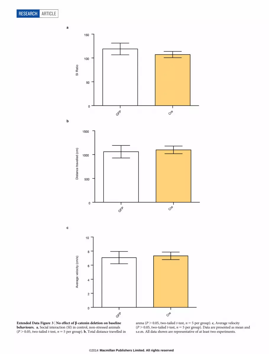
Extended Data Figure 3 | No effect of b-catenin deletion on baselinebehaviours. a, Social interaction (SI) in control, non-stressed animals(P . 0.05, two-tailed t-test, n 5 5 per group). b, Total distance travelled in
arena (P . 0.05, two-tailed t-test, n 5 5 per group). c, Average velocity(P . 0.05, two-tailed t-test, n 5 5 per group). Data are presented as mean ands.e.m. All data shown are representative of at least two experiments.
RESEARCH ARTICLE
Macmillan Publishers Limited. All rights reserved©2014

Extended Data Figure 4 | Regulation of b-catenin signalling in humandepression and after CSDS in mice. a, Axin2 expression is suppressed in bothmedicated and unmedicated depressed patients, both groups of which wereclinically depressed at their time of death (P , 0.01 one-way ANOVA, post-hoctest P . 0.05 between depressed unmedicated and medicated groups, *P , 0.01for either depressed group versus control, n 5 6 control, n 5 5 unmedicated
depressed, medicated depressed). b, Phospho-Ser 675 b-catenin and totalb-catenin levels from mouse control, susceptible, and resilient NAc 48 h postCSDS (phospho-Ser 675: *P , 0.05, one-way ANOVA, post-hoc testsusceptible versus resilient, n 5 5 for control, susceptible, n 5 8 for resilient).Data are presented as mean and s.e.m. Human data are from one experiment.All other data shown are representative of two experiments.
ARTICLE RESEARCH
Macmillan Publishers Limited. All rights reserved©2014

Extended Data Figure 5 | Repeated optogenetic burst stimulation of VTAcell bodies has no effect on canonical b-catenin signalling in NAc.Experiment was performed as in Fig. 2 with the exception of the optic fibre,
which was placed above VTA for cell body stimulation (P . 0.05, two-tailedt-test, n 5 8 per group). Data are presented as mean and s.e.m. Data are fromone experiment.
RESEARCH ARTICLE
Macmillan Publishers Limited. All rights reserved©2014

Extended Data Figure 6 | Genome-wide enrichment of H3K4me3 andH4K16ac binding in NAc at TSSs. NGS plot was used to visualize bindingpatterns.
ARTICLE RESEARCH
Macmillan Publishers Limited. All rights reserved©2014

Extended Data Figure 7 | Genome-wide pattern of H3K4me3 binding to genic regions in NAc under control, susceptible (defeat), and resilient mice. Notethe lack of difference across the three conditions. Data are from one experiment.
RESEARCH ARTICLE
Macmillan Publishers Limited. All rights reserved©2014

Extended Data Figure 8 | Genome-wide pattern of H4K16ac binding to genic regions in NAc under control, susceptible (defeat), and resilient mice. Notethe lack of difference across the three conditions. Shading represents standard error. Data are from one experiment.
ARTICLE RESEARCH
Macmillan Publishers Limited. All rights reserved©2014

Extended Data Figure 9 | Ingenuity pathway analysis (IPA) identifies anetwork of genes that show upregulated b-catenin binding at promoterregions in the NAc of resilient versus susceptible mice. Nodes representdifferentially regulated genes, with green meaning up in resilient versussusceptible and red meaning down in resilient versus susceptible. The bluearrows indicate that the direction of regulation is consistent with IPAprediction of an upregulated b-catenin network in resilience; for example, ablue arrow means that a target gene that would be expected to be upregulated by
b-catenin is in fact upregulated in this list. In contrast, yellow arrows indicatethat the regulation observed is inconsistent with expectations, while greyarrows indicate lack of pre-existing data to formulate expectations of b-cateninaction. Left panel shows mostly expected regulation of the b-catenin network(that is, upregulation) in resilience; right panel shows non-specific changesoccurring in a randomly chosen signal transducer and activator oftranscription-4 (STAT4) network.
RESEARCH ARTICLE
Macmillan Publishers Limited. All rights reserved©2014

Extended Data Figure 10 | Validation of local Dicer1 knockdown. Notesignificant knockdown of Dicer expression in NAc after intra-NAc delivery ofviral-Cre to floxed Dicer mice (*P , 0.05, two-tailed t-test, n 5 7 GFP, n 5 6Cre). Data are presented as mean and s.e.m. and are representative of twoexperiments.
ARTICLE RESEARCH
Macmillan Publishers Limited. All rights reserved©2014


