Embed Size (px)
Citation preview
C
VD
TJa
b
c
a
A
R
R
1
A
K
S
S
D
V
e
0h
ARTICLE IN PRESSOMM-3371; No. of Pages 10
c o m p u t e r m e t h o d s a n d p r o g r a m s i n b i o m e d i c i n e x x x ( 2 0 1 2 ) xxx–xxx
jo ur n al hom ep age : www.int l .e lsev ierhea l th .com/ journa ls /cmpb
alidating a postural evaluation method developed using aigital Image-based Postural Assessment (DIPA) software
ássia Silveira Furlanettoa,1, Cláudia Tarragô Candotti b,2, Tatiana Comerlatoc,3,efferson Fagundes Lossb,∗
Rua Bento Alves, 1501 Ap 204 – São Leopoldo – RS – BrazilRua Fernando Osório, 1887 – Porto Alegre – RS – BrazilRua Neri Silveira, 80 – Erechim – RS – Brazil
r t i c l e i n f o
rticle history:
eceived 8 April 2011
eceived in revised form
2 December 2011
ccepted 28 March 2012
eywords:
pinal postural evaluation
coliosis
igital photographs
alidating
a b s t r a c t
Objective: To investigate (1) the accuracy of the palpatory method to identify anatomical
points by comparison with the X-ray exams, (2) the validity of classifying spinal posture in
the frontal plane using Digital Image-Based Postural Assessment (DIPA) software by com-
parison with the X-ray exams and (3) the intra and inter-evaluator reproducibility of the
DIPA software.
Materials and methods: The postural assessment and X-ray examination of the spine, both in
the frontal plane and standing position, were performed consecutively in 24 subjects. The
postural assessment protocol consisted of: (1) palpation and the use of reflective markers
containing lead to mark the spinous processes (SP) of the C7, T2, T4, T6, T8, T10, T12, L2, L4
and S2 vertebrae and; (2) acquisition of photographic records. First, the X-ray examinations
were used to check the correlation between the palpated and marked SP and the true location
of the SP of the vertebra in question, by assessing the distance between them. The spinal
posture was classified based on the calculation of the scoliosis arrows in the DIPA (DIPA-
SA). The X-ray examinations provided the scoliosis arrows (X-SA), the Cobb angles and the
classification of spinal posture based on the Cobb angle. The results from the DIPA protocol
were compared to those from the X-ray examination-based protocol. The statistical tests
used were: (1) Kruskal–Wallis – differences in terms of the numerical distance between the
markers and the anatomical landmarks, (2) Pearson’s Correlation – DIPA-SA and Cobb angles,
(3) Pearson’s Correlation – X-SA and DIPA-SA; (4) Bland and Altman’s graphic representation
– X-SA and DIPA-SA, (5) Spearman’s Correlation – classification of spinal posture obtained
using the X-ray and DIPA protocols, (6) the intraclass correlation test (ICC) for the relationship
between the DIPA-SA made by each evaluator (inter-evaluator), and (7) independent t-test
from the two evaluation days (intra-evaluator), ̨ = 0.05.
to compare the dataPlease cite this article in press as: T.S. Furlanetto, et al., Validating a postural evaluation method developed using a Digital Image-based PosturalAssessment (DIPA) software, Comput. Methods Programs Biomed. (2012), http://dx.doi.org/10.1016/j.cmpb.2012.03.012
Results: There were no significant differences between the location of the anatomical points
located using palpation and identified with reflective markers and the respective location
of the SP as identified using X-ray exams (�2 = 9.366, p = 0.404). Significant correlations were
© 2012 Elsevier Ireland Ltd. All rights reserved.
∗ Corresponding author. Tel.: +55 51 3308 5822; fax: +55 51 33085843.E-mail addresses: [email protected] (T.S. Furlanetto), [email protected] (C.T. Candotti), [email protected] (T. Com-
rlato), [email protected] (J.F. Loss).1 Tel.: +55 51 30376521.2 Tel.: +55 51 3308 5822.3 Tel.: + 55 54 3321 3796.169-2607/$ – see front matter © 2012 Elsevier Ireland Ltd. All rights reserved.ttp://dx.doi.org/10.1016/j.cmpb.2012.03.012

ARTICLE IN PRESSCOMM-3371; No. of Pages 10
2 c o m p u t e r m e t h o d s a n d p r o g r a m s i n b i o m e d i c i n e x x x ( 2 0 1 2 ) xxx–xxx
found between the DIPA-SA and the Cobb angles in the dorsal (r = 0.75, p < 0.001) and lumbar
(r = 0.76, p = 0.007) regions; between the DIPA-SA and the X-SA in the dorsal (r = 0.79, p < 0.001)
and lumbar (r = 0.92, p < 0.001) regions and; between the classifications of posture obtained
with the DIPA and X-ray protocols (r = 0.804, p < 0.001). Bland and Altman’s representation
showed agreement between DIPA-SA and X-SA for both curvatures. Significant correlations
were found for the intra-evaluator test in the thoracic (r = 0.99, p < 0.001) and lumbar (r = 0.98,
p < 0.001) regions; for the inter-evaluator test in the thoracic (r = 0.99, p < 0.001) and lumbar
(r = 0.88, p < 0.001) regions. The results suggest that the DIPA protocol constitutes a valid
simple, practical and low-cost non-invasive tool for the evaluation of the spine in the frontal
plane which can be used to obtain reproducible measurements (inter and intra-evaluators).
examination, carried out by a responsible professional; and (3)
1. Introduction
The clinical evaluation of frontal plane postural alterations,such as scoliosis, has been based on the calculation of Cobb’sangle of curvature from X-ray exams, which in order to followthe evolution of the patients need to be carried out periodically[1–4]. The main problem with this clinical practice is that thepatient is repeatedly exposed to radiation. This is particularlyharmful in the case of adolescents given the increased riskof leukemia and breast and thyroid cancer [5–7]. In order toavoid any possible negative effect that maybe caused by mul-tiple radiographic examinations during growth there shouldbe a minimum of six months between such examinations[8,9]. However, in orthopedic clinical practice it can be seenthat these examinations are frequently requested at shorterintervals.
Therefore, non-invasive techniques of postural evaluationare an alternative to X-ray examinations [9] when followingthe evolution of physiotherapy for spinal deviations. Sev-eral non-radiographical and non-invasive postural evaluationtechniques have been proposed in order to evaluate andquantify postural alterations during the course of therapeu-tic treatment. Among them are the scoliometer [10], Moirétopography [11,12], arcometer [9,13,14], flexicurve [15–19], pan-tograph [20], kyphometer [21], inclinometer [22], as well as,some computer-based techniques, such as photogrammetryand postural evaluation softwares [23–28].
The process of assessing digital-image based postural eval-uation softwares can be divided into two different steps: (1)using palpation to identify and mark the relevant anatomicalpoints, and (2) using the information regarding those pointsas input data in an algorithm to obtain appropriate results.Although the palpation procedure and its level of accuracywhen used to identify anatomical points has been describedin the literature [29,30], it is considered indispensible that theprocedure used to identify the inputs to be used in any soft-ware should first be tested for its accuracy, and that the resultsobtained using the software are tested for their intra and inter-reproducibility.
The postural evaluation software programs found in theliterature are partially validated, that is, they have only beentested for inter and intra-evaluator reproducibility [25,28].Moreover, the results provided by these software programs
Please cite this article in press as: T.S. Furlanetto, et al., Validating a posturAssessment (DIPA) software, Comput. Methods Programs Biomed. (2012),
are limited to angles, distances and pre-established lengths,which certainly aid visual postural evaluation. However,as far as we know, there are no reports in the literature of
© 2012 Elsevier Ireland Ltd. All rights reserved.
any software that provides an effective classification of thesubject’s spinal posture.
Accordingly, the aims of the present study are to investi-gate (1) the accuracy of the identification of the anatomicalpoints using the palpatory method by comparing them withX-ray exams, (2) the validity of classifying spinal posture inthe frontal plane using Digital Image-Based Postural Assess-ment (DIPA) software by comparing the results with thoseobtained using X-ray exams and (3) the intra and inter-evaluator reproducibility of the DIPA software. The advantageof DIPA software lies not only in the identification of any devia-tion in the alignment of the spinous processes, but also in theapplication of this information to objectively classify spinalposture in frontal plane. It is believed that information of thisnature maybe useful for demonstrating the reliability of treat-ment results, as well as providing quantitative analysis of thepostural alterations in the spine.
2. Methodology
2.1. Sample
The sample consisted of 24 patients (16 women and 8 men)from a radiology clinic. The average age, body mass andheight were 31.9 years (±12.3); 58.4 kg (±9.4) and 1.66 m (±0.07)respectively. The inclusion criteria were: clinical diagnosis ofscoliosis and a medical prescription for X-ray examination.The exclusion criteria were: the presence of a sixth lumbarvertebra, diseases or disorders that impede orthostasis andcontraindications to X-ray examination. All the subjects vol-untarily agreed to participate in the study and signed a freeinformed consent form. The study was in accordance withHelsinki Declaration and approved by the ethics committee ofthe institution were the study was conducted and registeredunder the number 2006660.
2.2. Data acquisition procedures
Each subject, while wearing underclothes, was submitted tothree data acquisition procedures in sequence: (1) palpationand marking of the anatomical reference points; (2) X-ray
al evaluation method developed using a Digital Image-based Posturalhttp://dx.doi.org/10.1016/j.cmpb.2012.03.012
frontal plane photography. The palpation, marking proceduresand photography were carried out by the same evaluator, whowas previously trained in these procedures.
ARTICLE IN PRESSCOMM-3371; No. of Pages 10
c o m p u t e r m e t h o d s a n d p r o g r a m s i n b
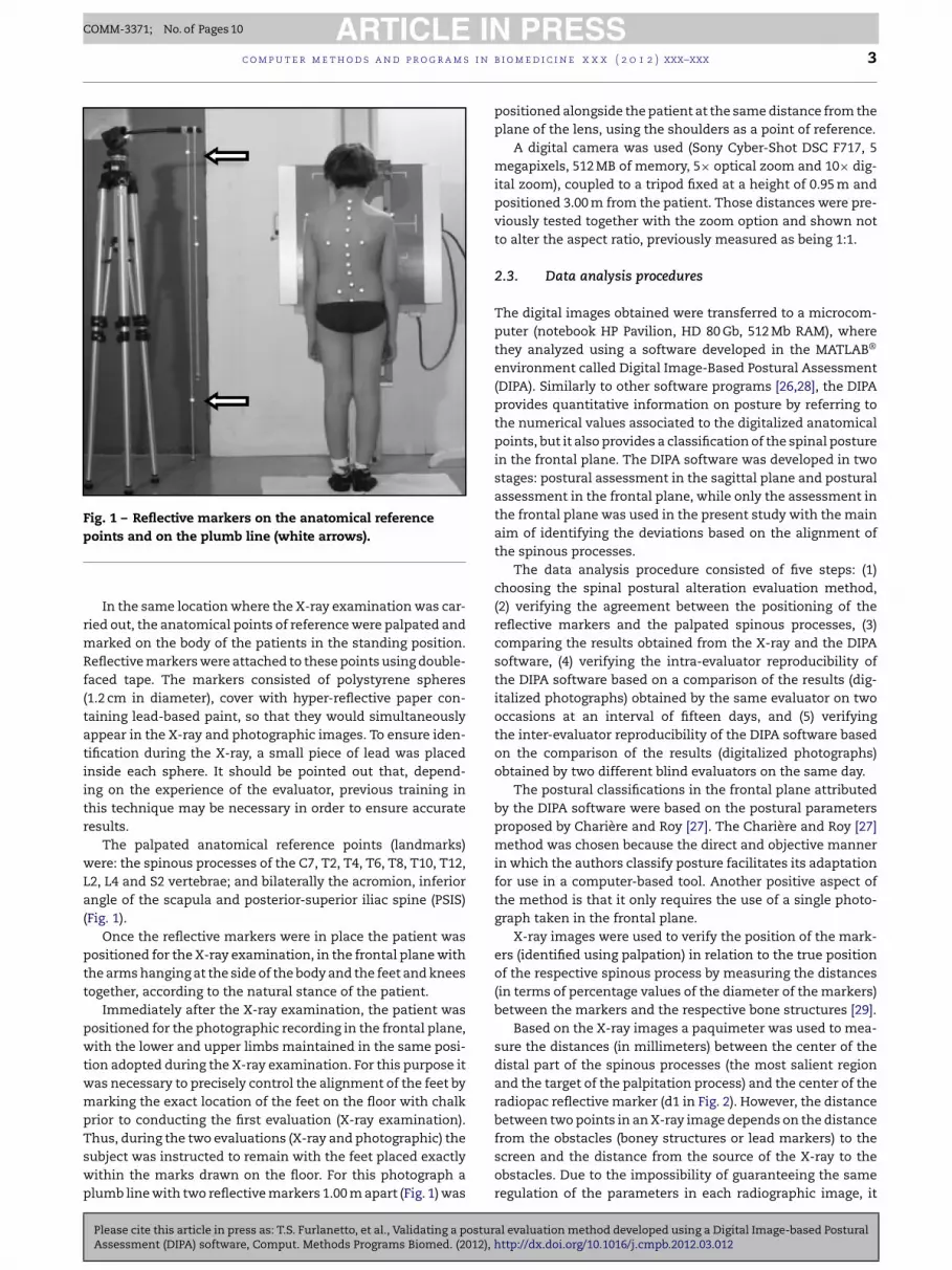
Fig. 1 – Reflective markers on the anatomical referencepoints and on the plumb line (white arrows).
rmRf(tatiitr
wLa(
ptt
pwtwmpTswp
from the obstacles (boney structures or lead markers) to the
In the same location where the X-ray examination was car-ied out, the anatomical points of reference were palpated and
arked on the body of the patients in the standing position.eflective markers were attached to these points using double-aced tape. The markers consisted of polystyrene spheres1.2 cm in diameter), cover with hyper-reflective paper con-aining lead-based paint, so that they would simultaneouslyppear in the X-ray and photographic images. To ensure iden-ification during the X-ray, a small piece of lead was placednside each sphere. It should be pointed out that, depend-ng on the experience of the evaluator, previous training inhis technique may be necessary in order to ensure accurateesults.
The palpated anatomical reference points (landmarks)ere: the spinous processes of the C7, T2, T4, T6, T8, T10, T12,
2, L4 and S2 vertebrae; and bilaterally the acromion, inferiorngle of the scapula and posterior-superior iliac spine (PSIS)Fig. 1).
Once the reflective markers were in place the patient wasositioned for the X-ray examination, in the frontal plane withhe arms hanging at the side of the body and the feet and kneesogether, according to the natural stance of the patient.
Immediately after the X-ray examination, the patient wasositioned for the photographic recording in the frontal plane,ith the lower and upper limbs maintained in the same posi-
ion adopted during the X-ray examination. For this purpose itas necessary to precisely control the alignment of the feet byarking the exact location of the feet on the floor with chalk
rior to conducting the first evaluation (X-ray examination).hus, during the two evaluations (X-ray and photographic) the
Please cite this article in press as: T.S. Furlanetto, et al., Validating a posturAssessment (DIPA) software, Comput. Methods Programs Biomed. (2012), h
ubject was instructed to remain with the feet placed exactlyithin the marks drawn on the floor. For this photograph alumb line with two reflective markers 1.00 m apart (Fig. 1) was
i o m e d i c i n e x x x ( 2 0 1 2 ) xxx–xxx 3
positioned alongside the patient at the same distance from theplane of the lens, using the shoulders as a point of reference.
A digital camera was used (Sony Cyber-Shot DSC F717, 5megapixels, 512 MB of memory, 5× optical zoom and 10× dig-ital zoom), coupled to a tripod fixed at a height of 0.95 m andpositioned 3.00 m from the patient. Those distances were pre-viously tested together with the zoom option and shown notto alter the aspect ratio, previously measured as being 1:1.
2.3. Data analysis procedures
The digital images obtained were transferred to a microcom-puter (notebook HP Pavilion, HD 80 Gb, 512 Mb RAM), wherethey analyzed using a software developed in the MATLAB®
environment called Digital Image-Based Postural Assessment(DIPA). Similarly to other software programs [26,28], the DIPAprovides quantitative information on posture by referring tothe numerical values associated to the digitalized anatomicalpoints, but it also provides a classification of the spinal posturein the frontal plane. The DIPA software was developed in twostages: postural assessment in the sagittal plane and posturalassessment in the frontal plane, while only the assessment inthe frontal plane was used in the present study with the mainaim of identifying the deviations based on the alignment ofthe spinous processes.
The data analysis procedure consisted of five steps: (1)choosing the spinal postural alteration evaluation method,(2) verifying the agreement between the positioning of thereflective markers and the palpated spinous processes, (3)comparing the results obtained from the X-ray and the DIPAsoftware, (4) verifying the intra-evaluator reproducibility ofthe DIPA software based on a comparison of the results (dig-italized photographs) obtained by the same evaluator on twooccasions at an interval of fifteen days, and (5) verifyingthe inter-evaluator reproducibility of the DIPA software basedon the comparison of the results (digitalized photographs)obtained by two different blind evaluators on the same day.
The postural classifications in the frontal plane attributedby the DIPA software were based on the postural parametersproposed by Charière and Roy [27]. The Charière and Roy [27]method was chosen because the direct and objective mannerin which the authors classify posture facilitates its adaptationfor use in a computer-based tool. Another positive aspect ofthe method is that it only requires the use of a single photo-graph taken in the frontal plane.
X-ray images were used to verify the position of the mark-ers (identified using palpation) in relation to the true positionof the respective spinous process by measuring the distances(in terms of percentage values of the diameter of the markers)between the markers and the respective bone structures [29].
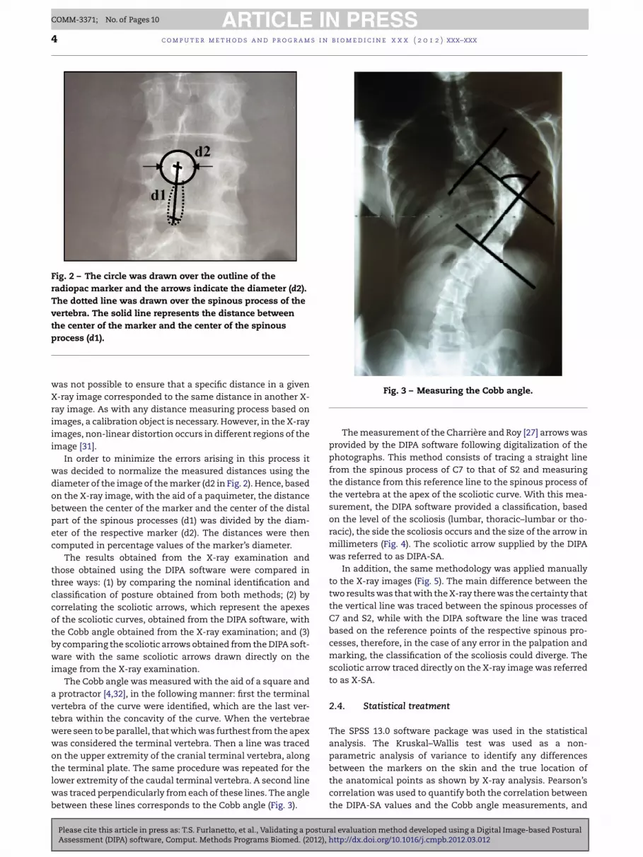
Based on the X-ray images a paquimeter was used to mea-sure the distances (in millimeters) between the center of thedistal part of the spinous processes (the most salient regionand the target of the palpitation process) and the center of theradiopac reflective marker (d1 in Fig. 2). However, the distancebetween two points in an X-ray image depends on the distance
al evaluation method developed using a Digital Image-based Posturalttp://dx.doi.org/10.1016/j.cmpb.2012.03.012
screen and the distance from the source of the X-ray to theobstacles. Due to the impossibility of guaranteeing the sameregulation of the parameters in each radiographic image, it
ARTICLE IN PRESSCOMM-3371; No. of Pages 10
4 c o m p u t e r m e t h o d s a n d p r o g r a m s i n b i o m e d i c i n e x x x ( 2 0 1 2 ) xxx–xxx
Fig. 2 – The circle was drawn over the outline of theradiopac marker and the arrows indicate the diameter (d2).The dotted line was drawn over the spinous process of thevertebra. The solid line represents the distance betweenthe center of the marker and the center of the spinousprocess (d1).
was not possible to ensure that a specific distance in a givenX-ray image corresponded to the same distance in another X-ray image. As with any distance measuring process based onimages, a calibration object is necessary. However, in the X-rayimages, non-linear distortion occurs in different regions of theimage [31].
In order to minimize the errors arising in this process itwas decided to normalize the measured distances using thediameter of the image of the marker (d2 in Fig. 2). Hence, basedon the X-ray image, with the aid of a paquimeter, the distancebetween the center of the marker and the center of the distalpart of the spinous processes (d1) was divided by the diam-eter of the respective marker (d2). The distances were thencomputed in percentage values of the marker’s diameter.
The results obtained from the X-ray examination andthose obtained using the DIPA software were compared inthree ways: (1) by comparing the nominal identification andclassification of posture obtained from both methods; (2) bycorrelating the scoliotic arrows, which represent the apexesof the scoliotic curves, obtained from the DIPA software, withthe Cobb angle obtained from the X-ray examination; and (3)by comparing the scoliotic arrows obtained from the DIPA soft-ware with the same scoliotic arrows drawn directly on theimage from the X-ray examination.
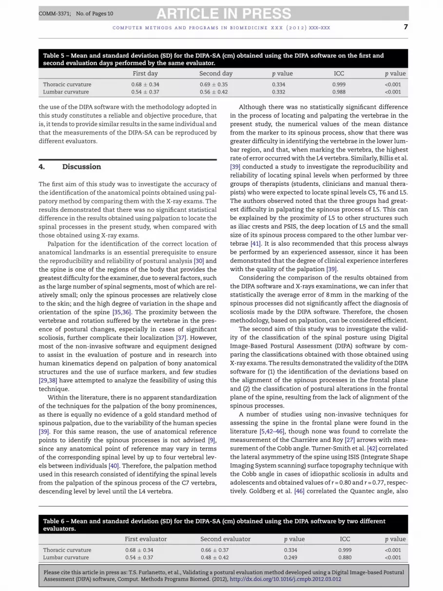
The Cobb angle was measured with the aid of a square anda protractor [4,32], in the following manner: first the terminalvertebra of the curve were identified, which are the last ver-tebra within the concavity of the curve. When the vertebraewere seen to be parallel, that which was furthest from the apexwas considered the terminal vertebra. Then a line was tracedon the upper extremity of the cranial terminal vertebra, alongthe terminal plate. The same procedure was repeated for the
Please cite this article in press as: T.S. Furlanetto, et al., Validating a posturAssessment (DIPA) software, Comput. Methods Programs Biomed. (2012),
lower extremity of the caudal terminal vertebra. A second linewas traced perpendicularly from each of these lines. The anglebetween these lines corresponds to the Cobb angle (Fig. 3).
Fig. 3 – Measuring the Cobb angle.
The measurement of the Charrière and Roy [27] arrows wasprovided by the DIPA software following digitalization of thephotographs. This method consists of tracing a straight linefrom the spinous process of C7 to that of S2 and measuringthe distance from this reference line to the spinous process ofthe vertebra at the apex of the scoliotic curve. With this mea-surement, the DIPA software provided a classification, basedon the level of the scoliosis (lumbar, thoracic–lumbar or tho-racic), the side the scoliosis occurs and the size of the arrow inmillimeters (Fig. 4). The scoliotic arrow supplied by the DIPAwas referred to as DIPA-SA.
In addition, the same methodology was applied manuallyto the X-ray images (Fig. 5). The main difference between thetwo results was that with the X-ray there was the certainty thatthe vertical line was traced between the spinous processes ofC7 and S2, while with the DIPA software the line was tracedbased on the reference points of the respective spinous pro-cesses, therefore, in the case of any error in the palpation andmarking, the classification of the scoliosis could diverge. Thescoliotic arrow traced directly on the X-ray image was referredto as X-SA.
2.4. Statistical treatment
The SPSS 13.0 software package was used in the statisticalanalysis. The Kruskal–Wallis test was used as a non-parametric analysis of variance to identify any differencesbetween the markers on the skin and the true location of
al evaluation method developed using a Digital Image-based Posturalhttp://dx.doi.org/10.1016/j.cmpb.2012.03.012
the anatomical points as shown by X-ray analysis. Pearson’scorrelation was used to quantify both the correlation betweenthe DIPA-SA values and the Cobb angle measurements, and

ARTICLE IN PRESSCOMM-3371; No. of Pages 10
c o m p u t e r m e t h o d s a n d p r o g r a m s i n b i o m e d i c i n e x x x ( 2 0 1 2 ) xxx–xxx 5
Fig. 4 – Measuring the DIPA-SA and the report supply by DIPA software. The circle indicates the DIPA-SA corresponding tot
tgbwctIrt
FX
he vertebral level at the apex of the scoliosis.
he correlation between the DIPA-SA and X-SA. Bland-Altmanraphic analysis [33] was used to analyze the agreementetween the DIPA-SA and X-SA. Spearman’s correlation testas used to quantify the correlation between the nominal
lassification of the data from the X-ray exams and fromhe DIPA software. The intraclass correlation test (ICC) type
[34] and independent t-test were performed to assess the
Please cite this article in press as: T.S. Furlanetto, et al., Validating a posturAssessment (DIPA) software, Comput. Methods Programs Biomed. (2012), h
elationship between the DIPA-SA assessments made byhe two evaluators (inter-evaluator) and made by the same
ig. 5 – Measuring the X-SA, measured directly on the-ray image.
evaluator on two different days (intra-evaluator), respectively.The significance level adopted in all tests was p < 0.05.
3. Results
The Kruskal Wallis test showed there were no significant dif-ferences between the reflective markers placed with the aid ofpalpation and the true location of the respective spinous pro-cesses (�2 = 9.366; p = 0.404). Table 1 shows the mean values ofthe distances between the markers and the spinous processnormalized by the marker diameter.
When comparing the L4 vertebra (largest mean distance)with the S2 vertebra (smallest mean distance), the differencebetween them was not significant. Consequently, any othercomparison between the means will not be significant. Fur-thermore, when the mean value of the distances (0.71) ismultiplied by the marker diameter (12 mm) the mean errorvalue was approximately 8 mm in absolute terms.
The mean values and standard deviations of the DIPA-SAand X-SA and Cobb angles for thoracic and lumbar curvaturesare shown in Table 2. It can be seen that, in absolute values,both the arrow supplied by the DIPA software and the X-raymeasurements, show similar values for both curvatures.
The DIPA-SA results were correlated with those of the Cobbangles from the thoracic and lumbar regions, separately, andthe results of the Pearson test are shown in Table 3. The resultsshow that there is a strong and significant correlation betweenthe quantitative results supplied by both methods.
The DIPA-SA results were correlated with those of the X-SA from the thoracic and lumbar regions, separately, and theresults of the Pearson test are shown in Table 4. The resultsshow that there is a strong and significant correlation betweenthe quantitative results indicated by the arrows obtained fromthe X-ray examination and from the DIPA software.
Fig. 6 shows the plot of the difference against the means
al evaluation method developed using a Digital Image-based Posturalttp://dx.doi.org/10.1016/j.cmpb.2012.03.012
of the DIPA-SA and X-SA, the standard deviation of the dif-ferences and the limits of agreement for the thoracic (Fig. 6a)and lumbar (Fig. 6b) curvatures. The mean difference betweenDIPA-SA and X-SA was 0.00 cm for the thoracic curvature

ARTICLE IN PRESSCOMM-3371; No. of Pages 10
6 c o m p u t e r m e t h o d s a n d p r o g r a m s i n b i o m e d i c i n e x x x ( 2 0 1 2 ) xxx–xxx
Table 1 – Mean values of the distances between the markers and the spinous process normalized by the marker diameter.
Vertebra C7 T2 T4 T6 T8 T10 T12 L2 L4 S2
Mean distance 0.59 0.57 0.73 0.80 0.76 0.70 0.72 0.77 1.01 0.50
Table 2 – Mean and standard deviation (SD) of the DIPA-SA (cm), X-SA (cm) and Cobb angles (◦) values of thoracic andlumbar curvatures.
DIPA-SA thoracic X-SA thoracic Cobb thoracic DIPA-SA lumbar X-SA lumbar Cobb lumbar
Mean 0.68 0.68 13.88 0.54 0.64 9.82SD 0.34 0.49 8.97 0.37 0.35 8.62
Fig. 6 – Graph showing the levels of agreement of the differences between DIPA-SA and X-SA in relation to the mean([DIPA-SA + X-SA]/2). (a) In the thoracic spine, the mean of the difference (d) = 0.0 cm, standard deviation of the difference (SDd) = 0.55 cm and the limits of agreement are +1,10; −1.10 cm. (b) In the lumbar spine, d = −0.08 cm, o SD d = 0.17 cm and the
limits of agreement are +0.26; −0.42 cm.and 0.08 cm for the lumbar curvature. These results indicatethe absence of any difference between DIPA-SA and X-SA forthoracic curvature and the presence of a negative system-atic difference for the lumbar curvature, so that, the valuesof lumbar arrows obtained from the DIPA software are, onaverage, 0.08 cm smaller than those obtained from the X-ray exam. The random distribution of the points in bothgraphs (Fig. 6a and b) indicates the absence of any tendencythroughout the range of the measurements obtained using thetwo assessment procedures. Hence, these analyses demon-strated the agreement between the DIPA-SA and X-SA for bothcurvatures.
The results of the reports obtained from the radiographicexamination and classification of the posture provided by theDIPA software were coded for possible statistical analysis. TheSpearman test showed a strong and significant correlation
Please cite this article in press as: T.S. Furlanetto, et al., Validating a posturAssessment (DIPA) software, Comput. Methods Programs Biomed. (2012),
(r = 0.804, p < 0.001) between the results for the classification ofposture provided by the X-ray examination and DIPA, demon-strating that, besides identifying the lateral deviations of the
Table 3 – Pearson’s coefficient correlation (r) and p valuesof the numerical variables supplied by DIPA software(DIPA-SA) and by X-ray examination (Cobb angle).
DIPA-SA × Cobb Correlation (r) P value
Thoracic curvature 0.752 <0.001Lumbar curvature 0.760 0.007
spine found in scoliosis, the DIPA software is able to provide avalid classificatory result for the pathology.
Table 5 shows a comparison of the results obtained by thesame evaluator on two occasions at an interval of fifteen days.The results of the independent t-test demonstrate the simi-larity between the two evaluation days, showing that there isno significant difference in the DIPA-SA. When the values ofthe DIPA-SA were correlated, the results of the ICC showedthere to be a strong and significant correlation between thetwo evaluation days. These results suggest that the use of theDIPA software with the methodology adopted in this studyconstitutes a reliable procedure, by which the results arereproducible on different evaluation days by the same eval-uator.
Table 6 shows the results of the inter-evaluator compar-ison. It can be seen that there is no significant difference
al evaluation method developed using a Digital Image-based Posturalhttp://dx.doi.org/10.1016/j.cmpb.2012.03.012
between the DIPA-SA obtained by the two evaluators, and thatthere is a strong and significant correlation between the DIPA-SA obtained by the two evaluators. These results suggest that
Table 4 – Pearson’s coefficient correlation (r) and p valuesof DIPA-SA and X-SA.
DIPA-SA × X-AS Correlation (r) p value
Thoracic curvature 0.790 <0.001Lumbar curvature 0.918 <0.001
ARTICLE IN PRESSCOMM-3371; No. of Pages 10
c o m p u t e r m e t h o d s a n d p r o g r a m s i n b i o m e d i c i n e x x x ( 2 0 1 2 ) xxx–xxx 7
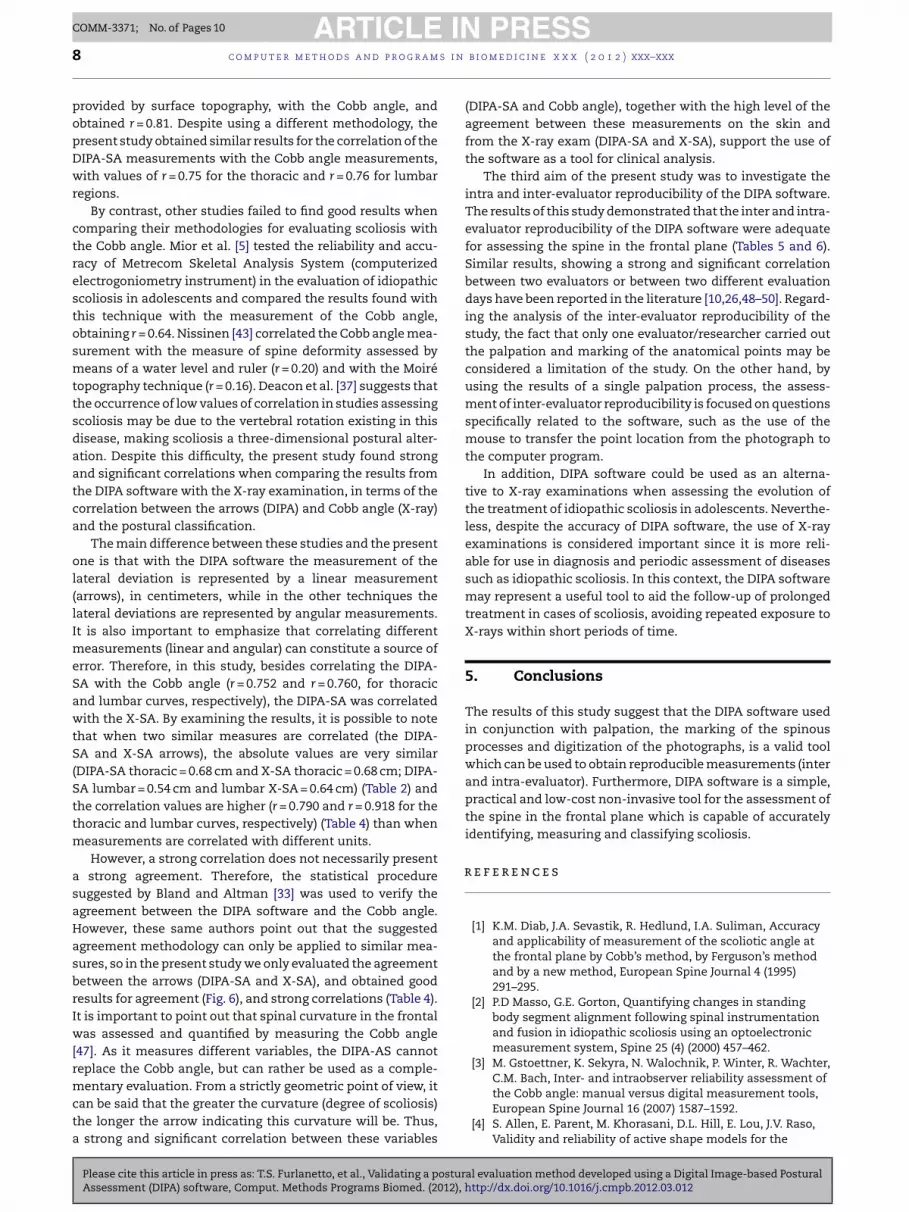
Table 5 – Mean and standard deviation (SD) for the DIPA-SA (cm) obtained using the DIPA software on the first andsecond evaluation days performed by the same evaluator.
First day Second day p value ICC p value
0.35
0.42
ttitd
4
Ttprdst
attgaatovesmths[t
oas[psoeufd
Thoracic curvature 0.68 ± 0.34 0.69 ±
Lumbar curvature 0.54 ± 0.37 0.56 ±
he use of the DIPA software with the methodology adopted inhis study constitutes a reliable and objective procedure, thats, it tends to provide similar results in the same individual andhat the measurements of the DIPA-SA can be reproduced byifferent evaluators.
. Discussion
he first aim of this study was to investigate the accuracy ofhe identification of the anatomical points obtained using pal-atory method by comparing them with the X-ray exams. Theesults demonstrated that there was no significant statisticalifference in the results obtained using palpation to locate thepinal processes in the present study, when compared withhose obtained using X-ray exams.
Palpation for the identification of the correct location ofnatomical landmarks is an essential prerequisite to ensurehe reproducibility and reliability of postural analysis [30] andhe spine is one of the regions of the body that provides thereatest difficulty for the examiner, due to several factors, suchs the large number of spinal segments, most of which are rel-tively small; only the spinous processes are relatively closeo the skin; and the high degree of variation in the shape andrientation of the spine [35,36]. The proximity between theertebrae and rotation suffered by the vertebrae in the pres-nce of postural changes, especially in cases of significantcoliosis, further complicate their localization [37]. However,ost of the non-invasive software and equipment designed
o assist in the evaluation of posture and in research intouman kinematics depend on palpation of bony anatomicaltructures and the use of surface markers, and few studies29,38] have attempted to analyze the feasibility of using thisechnique.
Within the literature, there is no apparent standardizationf the techniques for the palpation of the bony prominences,s there is equally no evidence of a gold standard method ofpinous palpation, due to the variability of the human species39]. For this same reason, the use of anatomical referenceoints to identify the spinous processes is not advised [9],ince any anatomical point of reference may vary in termsf the corresponding spinal level by up to four vertebral lev-
Please cite this article in press as: T.S. Furlanetto, et al., Validating a posturAssessment (DIPA) software, Comput. Methods Programs Biomed. (2012), h
ls between individuals [40]. Therefore, the palpation methodsed in this research consisted of identifying the spinal levelsrom the palpation of the spinous process of the C7 vertebra,escending level by level until the L4 vertebra.
Table 6 – Mean and standard deviation (SD) for the DIPA-SA (cmevaluators.
First evaluator Second eva
Thoracic curvature 0.68 ± 0.34 0.66 ± 0.37
Lumbar curvature 0.54 ± 0.37 0.48 ± 0.42
0.334 0.999 <0.0010.332 0.988 <0.001
Although there was no statistically significant differencein the process of locating and palpating the vertebrae in thepresent study, the numerical values of the mean distancefrom the marker to its spinous process, show that there wasgreater difficulty in identifying the vertebrae in the lower lum-bar region, and that, when marking the vertebra, the highestrate of error occurred with the L4 vertebra. Similarly, Billis et al.[39] conducted a study to investigate the reproducibility andreliability of locating spinal levels when performed by threegroups of therapists (students, clinicians and manual thera-pists) who were expected to locate spinal levels C5, T6 and L5.The authors observed noted that the three groups had great-est difficulty in palpating the spinous process of L5. This canbe explained by the proximity of L5 to other structures suchas iliac crests and PSIS, the deep location of L5 and the smallsize of its spinous process compared to the other lumbar ver-tebrae [41]. It is also recommended that this process alwaysbe performed by an experienced assessor, since it has beendemonstrated that the degree of clinical experience interfereswith the quality of the palpation [39].
Considering the comparison of the results obtained fromthe DIPA software and X-rays examinations, we can infer thatstatistically the average error of 8 mm in the marking of thespinous processes did not significantly affect the diagnosis ofscoliosis made by the DIPA software. Therefore, the chosenmethodology, based on palpation, can be considered efficient.
The second aim of this study was to investigate the valid-ity of the classification of the spinal posture using DigitalImage-Based Postural Assessment (DIPA) software by com-paring the classifications obtained with those obtained usingX-ray exams. The results demonstrated the validity of the DIPAsoftware for (1) the identification of the deviations based onthe alignment of the spinous processes in the frontal planeand (2) the classification of postural alterations in the frontalplane of the spine, resulting from the lack of alignment of thespinous processes.
A number of studies using non-invasive techniques forassessing the spine in the frontal plane were found in theliterature [5,42–46], though none was found to correlate themeasurement of the Charrière and Roy [27] arrows with mea-surement of the Cobb angle. Turner-Smith et al. [42] correlatedthe lateral asymmetry of the spine using ISIS (Integrate Shape
al evaluation method developed using a Digital Image-based Posturalttp://dx.doi.org/10.1016/j.cmpb.2012.03.012
Imaging System scanning) surface topography technique withthe Cobb angle in cases of idiopathic scoliosis in adults andadolescents and obtained values of r = 0.80 and r = 0.77, respec-tively. Goldberg et al. [46] correlated the Quantec angle, also
) obtained using the DIPA software by two different
luator p value ICC p value
0.334 0.999 <0.0010.249 0.880 <0.001
ARTICLE IN PRESSCOMM-3371; No. of Pages 10
s i n
r
8 c o m p u t e r m e t h o d s a n d p r o g r a m
provided by surface topography, with the Cobb angle, andobtained r = 0.81. Despite using a different methodology, thepresent study obtained similar results for the correlation of theDIPA-SA measurements with the Cobb angle measurements,with values of r = 0.75 for the thoracic and r = 0.76 for lumbarregions.
By contrast, other studies failed to find good results whencomparing their methodologies for evaluating scoliosis withthe Cobb angle. Mior et al. [5] tested the reliability and accu-racy of Metrecom Skeletal Analysis System (computerizedelectrogoniometry instrument) in the evaluation of idiopathicscoliosis in adolescents and compared the results found withthis technique with the measurement of the Cobb angle,obtaining r = 0.64. Nissinen [43] correlated the Cobb angle mea-surement with the measure of spine deformity assessed bymeans of a water level and ruler (r = 0.20) and with the Moirétopography technique (r = 0.16). Deacon et al. [37] suggests thatthe occurrence of low values of correlation in studies assessingscoliosis may be due to the vertebral rotation existing in thisdisease, making scoliosis a three-dimensional postural alter-ation. Despite this difficulty, the present study found strongand significant correlations when comparing the results fromthe DIPA software with the X-ray examination, in terms of thecorrelation between the arrows (DIPA) and Cobb angle (X-ray)and the postural classification.
The main difference between these studies and the presentone is that with the DIPA software the measurement of thelateral deviation is represented by a linear measurement(arrows), in centimeters, while in the other techniques thelateral deviations are represented by angular measurements.It is also important to emphasize that correlating differentmeasurements (linear and angular) can constitute a source oferror. Therefore, in this study, besides correlating the DIPA-SA with the Cobb angle (r = 0.752 and r = 0.760, for thoracicand lumbar curves, respectively), the DIPA-SA was correlatedwith the X-SA. By examining the results, it is possible to notethat when two similar measures are correlated (the DIPA-SA and X-SA arrows), the absolute values are very similar(DIPA-SA thoracic = 0.68 cm and X-SA thoracic = 0.68 cm; DIPA-SA lumbar = 0.54 cm and lumbar X-SA = 0.64 cm) (Table 2) andthe correlation values are higher (r = 0.790 and r = 0.918 for thethoracic and lumbar curves, respectively) (Table 4) than whenmeasurements are correlated with different units.
However, a strong correlation does not necessarily presenta strong agreement. Therefore, the statistical proceduresuggested by Bland and Altman [33] was used to verify theagreement between the DIPA software and the Cobb angle.However, these same authors point out that the suggestedagreement methodology can only be applied to similar mea-sures, so in the present study we only evaluated the agreementbetween the arrows (DIPA-SA and X-SA), and obtained goodresults for agreement (Fig. 6), and strong correlations (Table 4).It is important to point out that spinal curvature in the frontalwas assessed and quantified by measuring the Cobb angle[47]. As it measures different variables, the DIPA-AS cannotreplace the Cobb angle, but can rather be used as a comple-
Please cite this article in press as: T.S. Furlanetto, et al., Validating a posturAssessment (DIPA) software, Comput. Methods Programs Biomed. (2012),
mentary evaluation. From a strictly geometric point of view, itcan be said that the greater the curvature (degree of scoliosis)the longer the arrow indicating this curvature will be. Thus,a strong and significant correlation between these variables
b i o m e d i c i n e x x x ( 2 0 1 2 ) xxx–xxx
(DIPA-SA and Cobb angle), together with the high level of theagreement between these measurements on the skin andfrom the X-ray exam (DIPA-SA and X-SA), support the use ofthe software as a tool for clinical analysis.
The third aim of the present study was to investigate theintra and inter-evaluator reproducibility of the DIPA software.The results of this study demonstrated that the inter and intra-evaluator reproducibility of the DIPA software were adequatefor assessing the spine in the frontal plane (Tables 5 and 6).Similar results, showing a strong and significant correlationbetween two evaluators or between two different evaluationdays have been reported in the literature [10,26,48–50]. Regard-ing the analysis of the inter-evaluator reproducibility of thestudy, the fact that only one evaluator/researcher carried outthe palpation and marking of the anatomical points may beconsidered a limitation of the study. On the other hand, byusing the results of a single palpation process, the assess-ment of inter-evaluator reproducibility is focused on questionsspecifically related to the software, such as the use of themouse to transfer the point location from the photograph tothe computer program.
In addition, DIPA software could be used as an alterna-tive to X-ray examinations when assessing the evolution ofthe treatment of idiopathic scoliosis in adolescents. Neverthe-less, despite the accuracy of DIPA software, the use of X-rayexaminations is considered important since it is more reli-able for use in diagnosis and periodic assessment of diseasessuch as idiopathic scoliosis. In this context, the DIPA softwaremay represent a useful tool to aid the follow-up of prolongedtreatment in cases of scoliosis, avoiding repeated exposure toX-rays within short periods of time.
5. Conclusions
The results of this study suggest that the DIPA software usedin conjunction with palpation, the marking of the spinousprocesses and digitization of the photographs, is a valid toolwhich can be used to obtain reproducible measurements (interand intra-evaluator). Furthermore, DIPA software is a simple,practical and low-cost non-invasive tool for the assessment ofthe spine in the frontal plane which is capable of accuratelyidentifying, measuring and classifying scoliosis.
e f e r e n c e s
[1] K.M. Diab, J.A. Sevastik, R. Hedlund, I.A. Suliman, Accuracyand applicability of measurement of the scoliotic angle atthe frontal plane by Cobb’s method, by Ferguson’s methodand by a new method, European Spine Journal 4 (1995)291–295.
[2] P.D Masso, G.E. Gorton, Quantifying changes in standingbody segment alignment following spinal instrumentationand fusion in idiopathic scoliosis using an optoelectronicmeasurement system, Spine 25 (4) (2000) 457–462.
[3] M. Gstoettner, K. Sekyra, N. Walochnik, P. Winter, R. Wachter,C.M. Bach, Inter- and intraobserver reliability assessment of
al evaluation method developed using a Digital Image-based Posturalhttp://dx.doi.org/10.1016/j.cmpb.2012.03.012
the Cobb angle: manual versus digital measurement tools,European Spine Journal 16 (2007) 1587–1592.
[4] S. Allen, E. Parent, M. Khorasani, D.L. Hill, E. Lou, J.V. Raso,Validity and reliability of active shape models for the

ARTICLE IN PRESSCOMM-3371; No. of Pages 10
i n b
c o m p u t e r m e t h o d s a n d p r o g r a m sestimation of Cobb angle in patients with adolescentidiopathic scoliosis, Journal of Digital Imaging 21 (2) (2008)208–218.
[5] S.A. Mior, D. Kopansky-Giles, E. Crowther, J. Wright, Acomparison of radiographic and electrogoniometric anglesin adolescent idiopathic scoliosis, Spine 21 (13) (1996)1549–1555.
[6] C. Bone, G. Hsieh, The risk of carcinogenesis fromradiographs to pediatric orthopaedic patients, Journal ofPediatric Orthopaedics 20 (2) (2000) 251–254.
[7] M.M. Doody, J.E. Lonstein, M. Stovall, D.G. Hacker, N.Luckyanov, C.E. Land, Breast cancer mortality afterdiagnostic radiography, Spine 25 (16) (2000) 2052–2063.
[8] A.E. Oestreich, L.W. Young, T.Y. Poussaint, Scoliosis circa2000: radiologic imaging perspective. I. Diagnosis andpretreatment evaluation, Skeletal Radiologic 27 (1998)591–605.
[9] F. D’osualdo, S. Schierano, C. Cisotti, The evaluation of thespine through the surface: the role of surface measurementsin the evaluation and treatment of spine diseases in Youngpatients, Europa Medicophysica 38 (3) (2002) 147–152.
[10] P. Côté, B. Kreitz, J.D. Cassidy, A.K. Dzus, J. Martel, A study ofdiagnostic accuracy and reliability of the scoliometer andAdam’s forward bend test, Spine 23 (7) (1998) 796–802.
[11] J.S. Daruwalla, P. Balasubramaniam, Moiré topography inscoliosis: its accuracy in detecting the site and size of thecurve, The Journal of Bone and Joint Surgery 67-B (2) (1985)211–213.
[12] I.A.F. Stokes, M.S. Moreland, Concordance of back surfaceasymmetry and spine shape in idiopathic scoliosis, Spine 14(1) (1989) 73–78.
[13] F.O. Chaise, C.T. Candotti, M. La Torre, et al., Validation,repeatability and reproducibility of a noninvasiveinstrument for measuring thoracic and lumbar curvature ofthe spine in the sagittal plane, Brazilian Journal of PhysicalTherapy (2011), ahead of print, EpubNov 03.
[14] F. D’osualdo, S. Schierano, M. Iannis, Validation of clinicalmeasurement of kyphosis with a simple instrument, thearcometer, Spine 22 (1997) 408–422.
[15] S.R. Simpson, Evaluation of a flexible ruler technique for ameasuring lumbar lordosis in the clinical assessment of lowback pain, Journal of the Society of Occupational Medicine39 (1989) 25–29.
[16] W.B. Cutler, E. Friedmann, E. Genovese-Stone, Prevalence ofkyphosis in a healthy sample of pre- and postmenopausalwomen, American Journal of Physical Medicine &Rehabilitation 72 (4) (1993) 219–225.
[17] M.P. Caine, A.K. McConnell, D. Taylor, Assessment of spinalcurvature: an evaluation of the flexicurve and associatedmeans of analysis, International Journal of RehabilitationResearch 19 (1996) 271–278.
[18] M.R. Hinman, Comparison of thoracic kyphosis and posturalstiffness in younger and older women, The Spine Journal 4(2004) 413–417.
[19] R. Rajabi, F. Seidi, F. Mohamadi, Which method is accuratewhen using the flexible ruler to measure the lumbarcurvature angle? Deep point or midpoint of arch? WorldApplied Sciences Journal 4 (6) (2008) 849–852.
[20] S. Willner, Spinal pantograph: a non-invasive technique fordescribing kyphosis and lordosis in the thoraco-lumbarspine, Acta Orthopaedica Scandinavica 52 (1981) 525–529.
[21] G. Öhlén, E. Spangfort, C. Tingvall, Measurement of spinalsagittal configuration and mobility with Debrunner’skyphometer, Spine (1989) 580–583.
[22] G. Mellin, Measurement of thoracolumbar posture and
Please cite this article in press as: T.S. Furlanetto, et al., Validating a posturAssessment (DIPA) software, Comput. Methods Programs Biomed. (2012), h
mobility with a Myrin inclinometer, Spine 11 (1986) 759–762.[23] K.P. Singer, T.J. Jones, P.D. Breidahl, A comparison of
radiographic and computer-assisted measurements of
i o m e d i c i n e x x x ( 2 0 1 2 ) xxx–xxx 9
thoracic and thoracolumbar sagittal curvature, SkeletalRadiology 19 (1990) 21–26.
[24] M.A. Leroux, K. Zabjek, G. Simard, J. Badeaux, C. Coillard,C.H. Rivard, A noninvasive anthropometric technique formeasuring kyphosis and lordosis, Spine 25 (13) (2000)1689–1694.
[25] D.E. Harrison, T.J. Janik, R. Cailliet, et al., Validation of acomputer analysis to determine 3-D rotations andtranslations of the rib cage in upright posture from three 2-Ddigital images, European Spine Journal 16 (2007) 213–218.
[26] M.C. Normand, M. Descarreaux, D.D. Harrison, et al., Threedimensional evaluation of posture in standing with thePosturePrint: an intra- and inter-examiner reliability study,Chiropractic & Ostheopaty 15 (15) (2007).
[27] L. Charrière, J. Roy, Kinésithérapie des déviations latéralesdu rachis, 2nd edition, Toray-Masson, Paris, 1983.
[28] E.A.G. Ferreira, M. Duarte, E.P. Maldonado, T.N. Burke, A.P.Marques, Postural assessment software (PAS/SAPO):validation and reliability, Clinics 65 (7) (2010) 675–681.
[29] J.R. Engsberg, L.G. Lenke, K.H. Bridwell, M.L. Uhrich, C.M.Trout, Relationships between spinal landmarks and skinsurface markers, Journal of Applied Biomechanics 24 (2008)94–97.
[30] M.T. Haneline, M. Young, A review of intraexaminer andinterexaminer reliability of static spinal palpation: aliterature synthesis, Journal of Manipulative andPhysiological Therapeutics 32 (5) (2009) 379–386.
[31] V. Baltzopoulos, A videofluoroscopy method for opticaldistortion correction and measurement of knee-jointkinematics, Clinical Biomechanics 10 (2) (1995) 85–92.
[32] A.M. Briggs, T.V. Wrigley, E.A. Tully, P.E. Adams, A.M. Greig,K.L. Bennell, Radiographic measures of thoracic kyphosis inosteoporosis: Cobb and vertebral centroid angles, SkeletalRadiology 36 (2007) 761–767.
[33] J.M. Bland, D.G. Altman, Statistical methods for assessingagreement between two methods of clinical measurement,Lancet (1986) 307–310.
[34] D.E. Krebs, Declare your ICC type, Physical Therapy 66 (1986)1431.
[35] A. Lundberg, On the use of bone and skin markers inkinematics research, Human Movement Science 15 (1996)411–422.
[36] J.C. Harlick, S. Milosavljevic, P.D. Milburn, Palpationidentification of spinous processes in the lumbar spine,Manual Therapy 12 (2007) 56–62.
[37] P. Deacon, B.M. Flood, R.A. Dickson, Idiopathic scoliosis inthree dimensions, The Journal of Bone and Joint Surgery66-B (4) (1984) 509–512.
[38] C. Fortin, D.E. Feldman, F. Cheriet, H. Labelle, Validity of aquantitative clinical measurement tool of trunk posture inidiopathic scoliosis, Spine 35 (19) (2010) E988–E994.
[39] E.V. Billis, N.E. Foster, C.C. Wright, Reproducibility andrepeatability: errors of three groups of physiotherapists inlocating spinal levels by palpation, Manual Therapy 8 (4)(2003) 223–232.
[40] R. Cooperstein, M.T. Haneline, Spinous process palpationusing the scapular tip as a landmark vs a radiographiccriterion standard, Journal of Chiropractic Medicine 6 (2007)87–93.
[41] V.A. Fann, The prevalence of postural asymmetry in peoplewith and without chronic low back pain, Archives ofPhysical Medicine and Rehabilitation 83 (12) (2002)1736–1738.
[42] A.R. Turner-Smith, J.D. Harris, G.R. Houghton, R.J. Jefferson,A method for analysis of back shape in scoliosis,
al evaluation method developed using a Digital Image-based Posturalttp://dx.doi.org/10.1016/j.cmpb.2012.03.012
Biomechanics 21 (6) (1988) 497–509.[43] M. Nissinen, Trunk asymmetry and scoliosis, Acta Paediatric
Scandinavica 78 (1989) 747–753.

ARTICLE IN PRESSCOMM-3371; No. of Pages 10
s i n
10 c o m p u t e r m e t h o d s a n d p r o g r a m[44] T.N. Theologis, R.J. Jefferson, A.H.R.W. Simpson, A.R.Turner-Smith, J.C.T. Fairbank, Quantifying the cosmeticdefect of adolescent idiopathic scoliosis, Spine 18 (7) (1993)909–912.
[45] T.N. Theologis, J.C.T. Fairbank, A.R. Turner-Smith, T.Pantazopoulos, Early detection of progression in adolescentidiopathic scoliosis by measurement of changes in backshape with the integrated shape imaging system scanner,Spine 22 (11) (1997) 1223–1227.
[46] C.J. Goldberg, M. Kaliszer, D.P. Moore, E.E. Forgarty, F.E.
Please cite this article in press as: T.S. Furlanetto, et al., Validating a posturAssessment (DIPA) software, Comput. Methods Programs Biomed. (2012),
Dowling, Surface topography, Cobb angles and cosmeticchange in scoliosis, Spine 26 (4) (2001) E55–E63.
[47] R.T. Morrissy, G.S. Goldsmith, E.C. Hall, D. Kehl, G.H. Cowie,Measurement of the Cobb angle on radiographs of patients
b i o m e d i c i n e x x x ( 2 0 1 2 ) xxx–xxx
who have scoliosis. Evaluation of intrinsic error, Journal ofBone and Joint Surgery 72 (1990) 320–327.
[48] F. Lovell, J. Rothstein, W. Personius, Reliability of clinicalmeasurements of lumbar lordosis taken with a flexible rule,Physical Therapy 69 (2) (1989) 96–102.
[49] P. Korovessis, G. Petsinis, Z. Papazisis, A. Baillousis,Prediction of thoracic kyphosis using the De Brunnerkyphometer, Journal of Spinal Disorders 14 (1) (2001) 67–72.
[50] A. Mannion, K. Knecht, J.E. Balaban, D. Grob, A newskin-surface device for measuring the curvature and global
al evaluation method developed using a Digital Image-based Posturalhttp://dx.doi.org/10.1016/j.cmpb.2012.03.012
and segmental ranges of motion of the spine: reliability ofmeasurements and comparison with data reviewed fromthe literature, European Spine Journal 13 (2004)122–136.