Embed Size (px)
Citation preview
HAL Id: tel-03682822https://tel.archives-ouvertes.fr/tel-03682822
Submitted on 31 May 2022
HAL is a multi-disciplinary open accessarchive for the deposit and dissemination of sci-entific research documents, whether they are pub-lished or not. The documents may come fromteaching and research institutions in France orabroad, or from public or private research centers.
L’archive ouverte pluridisciplinaire HAL, estdestinée au dépôt et à la diffusion de documentsscientifiques de niveau recherche, publiés ou non,émanant des établissements d’enseignement et derecherche français ou étrangers, des laboratoirespublics ou privés.
Valorization of Lebanese seaweed extracts andevaluation of their potential antibiofilm activity
Maya Rima
To cite this version:Maya Rima. Valorization of Lebanese seaweed extracts and evaluation of their potential antibiofilmactivity. Agricultural sciences. Université Paul Sabatier - Toulouse III; Université Libanaise, 2021.English. �NNT : 2021TOU30264�. �tel-03682822�
THÈSEEn vue de l’obtention du
DOCTORAT DE L’UNIVERSITÉ DE TOULOUSE
Délivré par l'Université Toulouse 3 - Paul Sabatier
Cotutelle internationale: Université libanaise
Présentée et soutenue par
Maya RIMA
Le 8 décembre 2021
Valorisation des extraits dalgues Libanaises et évaluation de leuractivité antibiofilm potentielle
Ecole doctorale : SEVAB - Sciences Ecologiques, Vétérinaires, Agronomiques etBioingenieries
Spécialité : Ingénieries microbienne et enzymatique
Unité de recherche :LGC - Laboratoire de Génie Chimique
Thèse dirigée parChristine ROQUES et Asma CHBANI
JuryM. Joseph SAAB, Rapporteur
Mme Fatima EL GARAH, ExaminatriceMme Marion GIRARDOT, Examinatrice
Mme Christine ROQUES, Directrice de thèseMme Asma CHBANI, Co-directrice de thèse
M. Sébastien VILAIN, Président
'

THESE de doctorat en Cotutelle
Pour obtenir le grade de Docteur délivré par
L’Université de Toulouse III – Paul Sabatier (Ecole Doctorale SEVAB)
Spécialité : Ingénieries microbienne et enzymatique
ET
L’Université Libanaise (Ecole Doctorale des Sciences et Technologie)
Spécialité : Biotechnologie
Présentée et soutenue par
Mme Maya RIMA Le 8 décembre 2021 à Toulouse - France
Valorization of Lebanese seaweed extracts and evaluation of their potential antibiofilm activity
Membres du jury :
M. Sébastien VILAIN, ENSTBB Bordeaux, Rapporteur – Président du jury
M. Joseph SAAB, Université Saint-Esprit de Kaslik, Rapporteur
Mme Marion GIRARDOT, Université de Poitiers, Examinatrice
Mme Fatima EL GARAH, UPS Toulouse III, Co-encadrante de thèse
Mme Asma CHBANI, Université Libanaise, Directrice de thèse
Mme Christine ROQUES, UPS Toulouse III, Directrice de thèse

Acknowledgements
Here we are, we’ve arrived! Three years that have really changed and marked my life with their ups and downs are coming to an end. This extremely enriching experience from which I have learned so much would not have been possible without all people who have contributed in some way in this thesis work. It is therefore with these lines that I express my grateful thanks to all of them.
I would like, first of all, to express my sincere gratitude to my supervisors, Pr. Asma CHBANI, Pr. Christine ROQUES, and Dr. Fatima EL GARAH. Many thanks for your support, your encouragement, and your precious guidance during these three years.
Pr. Asma CHBANI, I sincerely thank you for your continuous support, your precious help and your confidence since I was your student in Master 2 till the end of this thesis. I would certainly like to express my gratitude to you for giving me the particular opportunity to do my thesis in collaboration with LGC and on a very interesting project.
Pr. Christine ROQUES, from whom I learned a lot about both knowledge and scientific rigor, I sincerely thank you for your continuous guidance and support throughout these three years. It is really a great honor to work under your supervision. Many thanks for all the time you dedicated from the beginning of the project to the redaction and the defense day, for all the scientific discussions we had, and for your constructive advices leading always to the best solutions. Indeed, I cannot forget your kindness and your good humor ensuring the workflow in a wonderful environment.
Dr. Fatima EL GARAH, I cannot find the words to express all my gratitude to you for your endless care, kindness, and support “on so many levels” since the first day I arrived in Toulouse until now… Thank you for always being ready to answer all my questions, to give help, and for always findings the ways to ensure the progress of work in the most favorable conditions. I also thank you for having provided me with all the supervision qualities and for your valuable advices and remarks throughout these three years as well as during the writing of this manuscript and the preparation of the final defense. I thank you wholeheartedly for all your human qualities and for always believing in my abilities.
I would like to express my respectful thanks to the members of jury committee who have honored me by accepting to evaluate and judge this work. Pr. Sébastien VILAIN, Pr. Joseph SAAB, and Dr. Marion GIRARDOT, I sincerely thank you for your suggestions and your judicious remarks. I greatly appreciate your interest in my thesis work.
My sincere thanks also goes to Dr. Raphaël LAMI for following this work as a member of my thesis progress committee and especially for welcoming me in his laboratory (Laboratory of Microbial Biodiversity and Biotechnology USR 3579-LBBM – Banyuls-sur-mer) for a formation on QSI assay. Thank you for all his team especially for Carole and Emilie for their help and great kindness.
Acknowledgments
I wish to acknowledge, and thank as well, Dr. Jalloul BOUAJILA, member of my thesis progress committee, for all discussions we had about the preparation of natural extracts.
I would like to express my thanks to Laure LATAPIE for the analysis of the chemical composition of extracts (GC/MS and LC/MS) and for all the discussions we had in this regard. Many thanks also to Anais VANDENBOSSCHE and Brigitte DUSTOU for their help.
I also thank Pr. Geneviève BAZIARD for training me on the use of the Digidrop contact angle meter.
I extend my warmest thanks to LGC team: Dr. Barbora LAJOIE, Dr. Salomé EL HAGE, and Mr. Laurent AMIELET for their kind welcome, their helpfulness, and for the pleasant work environment they provided. I also thank my colleagues that I really had the chance to meet them and spend good moments with them: Charlotte – always with a cheerful spirit and enjoyable talks, Nabil, Marianne, Simon, Sophie, Ibrahima, and of course “my desk neighbor” Jeanne who is always ready to help and to listen to me even when I was just complaining. Thank you Jeanne for your great kindness and for all the good times we have spent. I will really miss our long conversations in front of the PSM!
I also would like to thank all personnel of FONDERPHAR: Cathy, Jocelyne, Sandra, Sylvie, Celine, and Élisabeth.
I infinitely thank my dear friends in Lebanon “many to be listed” who despite the distance they were always present to give me support and encouragement. I hope to see you in the best conditions! Thank you to my dear friend Nour for her continuous support, her valuable advices and for always giving me back my confidence. I also would thank Duaa for accompanying me step by step when I arrived in Toulouse and for helping me in several ways.
Great thanks to my friend Rachad for his support during the hardest moments. Thank you for being always ready to listen to my “stories” and I hope to see you soon, it has been a long time!
A special thanks to my dear friend Iman. Thank you for all the hours of phone conversations we had (and it is never over) especially during the unforgettable lockdown. Thank you for being virtually with me 20h/24h repeating always the same discussions and with the same reactions!
Last but certainly not least, I express my heartfelt thanks and gratitude to my dear parents, to whom I dedicate this thesis. No amount of words will be enough to tell how grateful I am to you. Everything I am today and everything I may become tomorrow is thanks to your sacrifices. Thank you for instilling in me a passion for knowledge and an endless determination to succeed despite all the circumstances. I also would like to thank my sisters Aya and Marwa and my brother Imad. Thank you for your support and your continuous encouragement…”until we meet again”…
Maya RIMA

SCIENTIFIC PRODUCTION
Oral communications
Maya RIMA, Asma CHBANI, Christine ROQUES, Fatima EL GARAH. Seaweeds: a promising source of antibiofilm agent against pathogenic bacteria. Plant Based Summit International conference and business meetings September 2021 – Reims, France.
Maya RIMA, Asma CHBANI, Christine ROQUES, Fatima EL GARAH. Seaweeds: a promising source of antibiofilm agent against Staphylococcus aureus. International Congress of the French Society of Cosmetology November 2021 – Paris, France.
Poster communications Maya RIMA, Asma CHBANI, Christine ROQUES, Fatima EL GARAH. Comparative study of the insecticidal activity of the pigments and extracts of high green plants (Spinacia oleracea) and a chlorophytae alga (Ulva lactuca) against the fruit fly Drosophila melanogaster. 2nd International Symposium on Materials, Electrochemistry and Environment CIMEE’18 October 2018 – Tripoli, Lebanon.
Maya RIMA, Asma CHBANI, Christine ROQUES, Fatima EL GARAH. Evaluation of the insecticidal activity of a green alga against Drosophila melanogaster fruit fly. 2nd Seaweed for Health International Conference August 2020 – Spain (Virtual).
Maya RIMA, Asma CHBANI, Christine ROQUES, Fatima EL GARAH. Seaweeds: a promising source of antibiofilm agent against pathogenic bacteria. 2nd Seaweed for Health International Conference August 2020 – Spain (Virtual).
Scientific articles
Article I – Published:
Comparative study of the insecticidal activity of a high green plant (Spinacia oleracea) and a chlorophytae algae (Ulva lactuca) extracts against Drosophila melanogaster fruit fly. Ann. Pharm. Fr., 2021, 79(1), 36-43.
Rima, M., Chbani, A., Roques, C., & El Garah, F.
Doi: 10.1016/j.pharma.2020.08.005.
Scientific production
Article II – Published:
Seaweed extracts: A promising source of antibiofilm agents with distinct mechanisms of action against Pseudomonas aeruginosa. Mar. Drugs., 2022, 20(2), 92.
Rima, M., Trognon, J., Latapie, L., Chbani, A., Roques, C., & El Garah, F.
Doi : //doi.org/10.3390/md20020092
Article III – To be submitted:
Seaweed extracts as an effective gateway in the search for novel antibiofilm agents against Staphylococcus aureus.
Rima, M., Chbani, A., Roques, C., & El Garah, F.
Price
National Prize for Young Researchers – Promega France - 2021
Rima, M. Silencing bacterial chats with seaweed extracts: Pharmaceutical prospects.
https://france.promega.com/c/jeunes-chercheurs-nomines-2021/#project6
ABSTRACT
he massive and often uncontrolled use of antibiotics has led to the development of multi-resistant bacterial strains (MDRs) capable of causing infectious diseases that
are difficult or even untreatable. In addition, the organization of bacteria into biofilms corresponds to adaptive resistance and is involved in almost 80% of chronic infections. By definition, a biofilm is an aggregation of microorganisms attached to a biotic or abiotic surface and enclosed in an extracellular polymeric matrix (EPS). This sessile lifestyle provides a protective barrier against antimicrobial agents. In this regard, much attention has been paid to the search for anti-biofilm agents able to regulate or even inhibit biofilm formation without interfering with bacterial growth. Natural products represent a valuable source of new molecules, including possible drug candidates. Marine organisms, in particular macroalgae, constitute a reservoir of bioactive compounds with a broad spectrum of biological activities, including insecticidal, antimicrobial and anti-biofilm activities, via different mechanisms. For example, the halogenated furanone isolated from the red alga Delisea pulchra is the first inhibitor molecule of Quorum Sensing, an intercellular communication system playing a major role in the formation of bacterial biofilms. In this context, the objective of this study is to explore the potential of extracts derived from three Lebanese algae: the green alga Ulva lactuca, the brown alga Stypocaulon scoparium and the red alga Pterocladiella capillacea, in terms of anti-biofilm activity against Pseudomonas aeruginosa and Staphylococcus aureus, two opportunistic pathogens responsible for serious infections, particularly in immunocompromised subjects and cystic fibrosis patients. To do that, various complementary approaches (crystal violet staining method, colony-forming unit counts method, epifluorescence microscopic analysis, synergistic activity with conventional antibiotics…) were adopted. Interestingly, results showed the ability of various extracts to present a significant anti-biofilm activity against these two critical bacteria by exhibiting different mechanisms of action. At the same time, the analysis of the chemical composition of extracts was carried out in an attempt to identify compound(s) which could be responsible for their demonstrated activity. On the other hand, in order to evaluate the potentiality of the green alga Ulva lactuca to present an alternative to toxic phytosanitary products, its possible insecticidal activity was studied against the Drosophila melanogaster fruit fly (insect pest and best model for studying the insecticidal activity) by complementary in vivo tests. Results showed an interesting insecticidal activity of its acetonic extract as well as of its purified green pigments. This study provides new insight into the exploration of seaweed as a valuable source of bioactive compounds that can be valorized in the agricultural area as well as in the industrial/pharmaceutical field. Keywords: Seaweed, Anti-biofilm activity, P. aeruginosa, S. aureus, insecticidal activity.
T
RÉSUMÉ
’utilisation massive et souvent incontrôlée des antibiotiques a conduit au développement de souches bactériennes multi‐ résistantes (MDR) capables de causer
des maladies infectieuses difficiles et même impossibles à traiter. Par ailleurs, l’organisation des bactéries en biofilm correspond à une résistance adaptative et est impliquée dans presque 80% des infections chroniques. Par définition, un biofilm est une agrégation des microorganismes attachée à une surface biotique ou abiotique et enfermée dans une matrice polymérique extracellulaire (EPS). Ce mode de vie sessile assure une barrière de protection contre les agents antimicrobiens. À cet égard, une grande attention a été accordée à la recherche d’agents anti‐biofilms dont le rôle est de réguler, voire d’inhiber, la formation de biofilm sans interférer avec la croissance bactérienne. Les produits naturels représentent une source précieuse de nouvelles molécules dont des candidats médicaments. Les organismes marins, en particulier les macroalgues, constituent un réservoir de composés bioactifs ayant un large spectre d’activités biologiques, y compris des activités insecticide, antimicrobienne et antibiofilm, via différents mécanismes. Par exemple, la furanone halogénée isolée de l’algue rouge Delisea pulchra est la première molécule inhibitrice du système de Quorum Sensing, un système de communication intercellulaire jouant un rôle majeur dans la formation des biofilms bactériens. Dans ce contexte, l’objectif de cette étude est d'explorer le potentiel des extraits issus de trois algues Libanaises : l’algue verte Ulva lactuca, l’algue brune Stypocaulon scoparium et l’algue rouge Pterocladiella capillacea, en termes d'activité antibiofilm, contre Pseudomonas aeruginosa et Staphylococcus aureus, deux agents pathogènes opportunistes responsables d’infections graves, notamment chez les sujets immunodéprimés et les patients atteints de mucoviscidose. Pour ce faire, plusieurs approches complémentaires (méthode de marquage au crystal violet, méthode de dénombrement des unités-formant colonies, analyse microscopique à épifluorescence, activité synergique avec des antibiotiques conventionnels…) ont été adoptées. Les résultats ont montré que plusieurs extraits ont une activité antibiofilm intéressante contre ces deux bactéries critiques, avec des mécanismes d’action différents. Parallèlement, l’analyse de la composition chimique des extraits a été menée afin d’identifier le(s) composé(s) qui pourraient être à l’origine de leur activité démontrée. D’autre part, afin d’évaluer la potentialité de l’algue verte Ulva lactuca à présenter une alternative aux produits phytosanitaires toxiques, son activité insecticide a été étudiée contre la mouche de fruit Drosophila melanogaster (insecte ravageur et le meilleure modèle d’étude de l’activité insecticide) par différents tests complémentaires. Les résultats ont montré que l’extrait acétonique ainsi que les pigments verts purifiés présentent la meilleure activité insecticide.
L
Résumé
Cette étude fournit un nouvel aperçu de l’exploration des algues comme étant une source précieuse de composés bioactifs pouvant être valorisés dans le domaine agricole ainsi que dans le secteur industriel/pharmaceutique.
Mots clés : Macroalgues, activité antibiofilm, P. aeruginosa, S. aureus, activité insecticide.
LIST OF ABBREVIATIONS
AHLs N-acyl homoserine lactones
AI Autoinducers
AIP Autoinducer peptide
ATP Adenosine triphosphate
BAC benzalkonium chloride
Bap Biofilm associated protein
BB Biofilm broth
CF Cystic fibrosis
CFTR Cystic fibrosis transmembrane conductance
CFU Colony forming unit
CH Cyclohexane
Chl a Chlorophyll a
Chl b Chlorophyll b
CLSM Confocal laser scanning microscopy
ConA Concanavalin A
CV Crystal violet
DCM Dichloromethane
DW Dry weight
EA Ethyl acetate
eDNA Extracellular DNA
EP Eradication percentage
EPS Extracellular polymeric matrix
FC Fluorescence control
FDA Food and drug administration
GC-MS Gas chromatography–mass spectrometry
GFP Green fluorescent protein
IDSA Infectious Diseases Society of America
IP Inhibition percentage
LB Lysogeny Broth
LC-MS Liquid chromatography–mass spectrometry
MBB Modified Biofilm Broth
MDR Multidrug resistant
MeOH Methanol
List of abbreviations
MHB Mueller Hinton Broth
MIC Minimum inhibitory concertation
MRSA Methicillin resistant Staphylococcus aureus
NO Nitric oxide
OD Optical density
P.c Pterocladiella capillacea red alga
P’ Polarity index
PI Propidium iodide
PIA Polysaccharide intercellular adhesin
PIPs Plant-incorporated protectants
POPs Persistent organic pollutants
PQS Pseudomonas quinolone signal
qPCR Quantitative polymerase chain reaction
QS Quorum Sensing
QSI Quorum Sensing inhibitors
ROS Reactive oxygen species
RT-qPCR Reverse transcription quantitative polymerase chain reaction
S.s Stypocaulon scoparium brown alga
SC Sterility control
SDW Sterile distilled water
SEM Scanning electron microscopy
TA Toxin-antitoxin system
TSA Trypticase soy agar
U.l Ulva lactuca green alga
UNEP United Nations Environment Programme
US. EPA United States Environmental Protection Agency
WHO World Health Organization
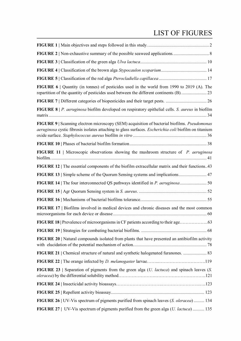
LIST OF FIGURES FIGURE 1 | Main objectives and steps followed in this study. .................................................... 2
FIGURE 2 | Non-exhaustive summary of the possible seaweed applications. .............................. 8
FIGURE 3 | Classification of the green alga Ulva lactuca......................................................... 10
FIGURE 4 | Classification of the brown alga Stypocaulon scoparium ....................................... 14
FIGURE 5 | Classification of the red alga Pterocladiella capillacea ......................................... 17
FIGURE 6 | Quantity (in tonnes) of pesticides used in the world from 1990 to 2019 (A). The repartition of the quantity of pesticides used between the different continents (B). ..................... 23
FIGURE 7 | Different categories of biopesticides and their target pests. ................................... 26
FIGURE 8 | P. aeruginosa biofilm developed on respiratory epithelial cells. S. aureus in biofilm matrix ....................................................................................................................................... 34
FIGURE 9 | Scanning electron microscopy (SEM) acquisition of bacterial biofilms. Pseudomonas aeruginosa cystic fibrosis isolates attaching to glass surfaces. Escherichia coli biofilm on titanium oxide surface. Staphylococcus aureus biofilm in vitro ............................................................... 36
FIGURE 10 | Phases of bacterial biofilm formation .................................................................. 38
FIGURE 11 | Microscopic observations showing the mushroom structure of P. aeruginosa biofilm. ..................................................................................................................................... 41
FIGURE 12 | The essential components of the biofilm extracellular matrix and their functions..43
FIGURE 13 | Simple scheme of the Quorum Sensing systems and implications ........................ 47
FIGURE 14 | The four interconnected QS pathways identified in P. aeruginosa ....................... 50
FIGURE 15 | Agr Quorum Sensing system in S. aureus. .......................................................... 52
FIGURE 16 | Mechanisms of bacterial biofilms tolerance......................................................... 55
FIGURE 17 | Biofilms involved in medical devices and chronic diseases and the most common microorganisms for each device or disease ................................................................................ 60
FIGURE 18 | Prevalence of microorganisms in CF patients according to their age……………….63
FIGURE 19 | Strategies for combating bacterial biofilms. ........................................................ 68
FIGURE 20 | Natural compounds isolated from plants that have presented an antibiofilm activity with elucidation of the potential mechanism of action............................................................... 78
FIGURE 21 | Chemical structure of natural and synthetic halogenated furanones. .................... 83
FIGURE 22 | The orange infected by D. melanogaster larvae……...………………………….119
FIGURE 23 | Separation of pigments from the green alga (U. lactuca) and spinach leaves (S. oleracea) by the differential solubility method…………………….…………………………….121
FIGURE 24 | Insecticidal activity bioassays………………………….……………………..….123
FIGURE 25 | Repellent activity bioassay................................................................................ 123
FIGURE 26 | UV-Vis spectrum of pigments purified from spinach leaves (S. oleracea) ......... 134
FIGURE 27 | UV-Vis spectrum of pigments purified from the green alga (U. lactuca) .......... 135
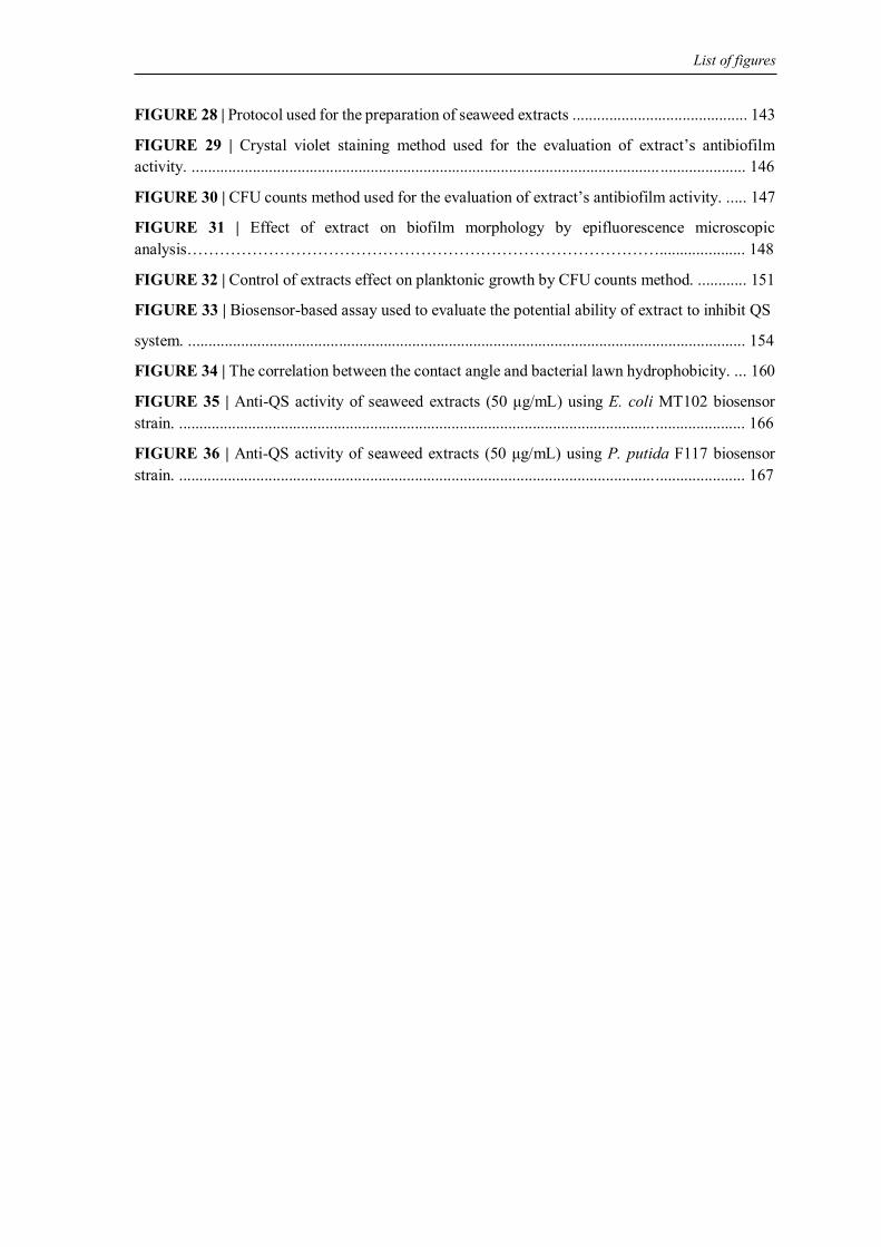
List of figures
FIGURE 28 | Protocol used for the preparation of seaweed extracts ........................................... 143
FIGURE 29 | Crystal violet staining method used for the evaluation of extract’s antibiofilm activity. ................................................................................................................... ..................... 146
FIGURE 30 | CFU counts method used for the evaluation of extract’s antibiofilm activity. ..... 147
FIGURE 31 | Effect of extract on biofilm morphology by epifluorescence microscopic analysis……………………………………………………………………………..................... 148
FIGURE 32 | Control of extracts effect on planktonic growth by CFU counts method. ............ 151
FIGURE 33 | Biosensor-based assay used to evaluate the potential ability of extract to inhibit QS
system. ......................................................................................................................................... 154
FIGURE 34 | The correlation between the contact angle and bacterial lawn hydrophobicity. ... 160
FIGURE 35 | Anti-QS activity of seaweed extracts (50 μg/mL) using E. coli MT102 biosensor strain. ........................................................................................................................................... 166
FIGURE 36 | Anti-QS activity of seaweed extracts (50 μg/mL) using P. putida F117 biosensor strain. ........................................................................................................................................... 167
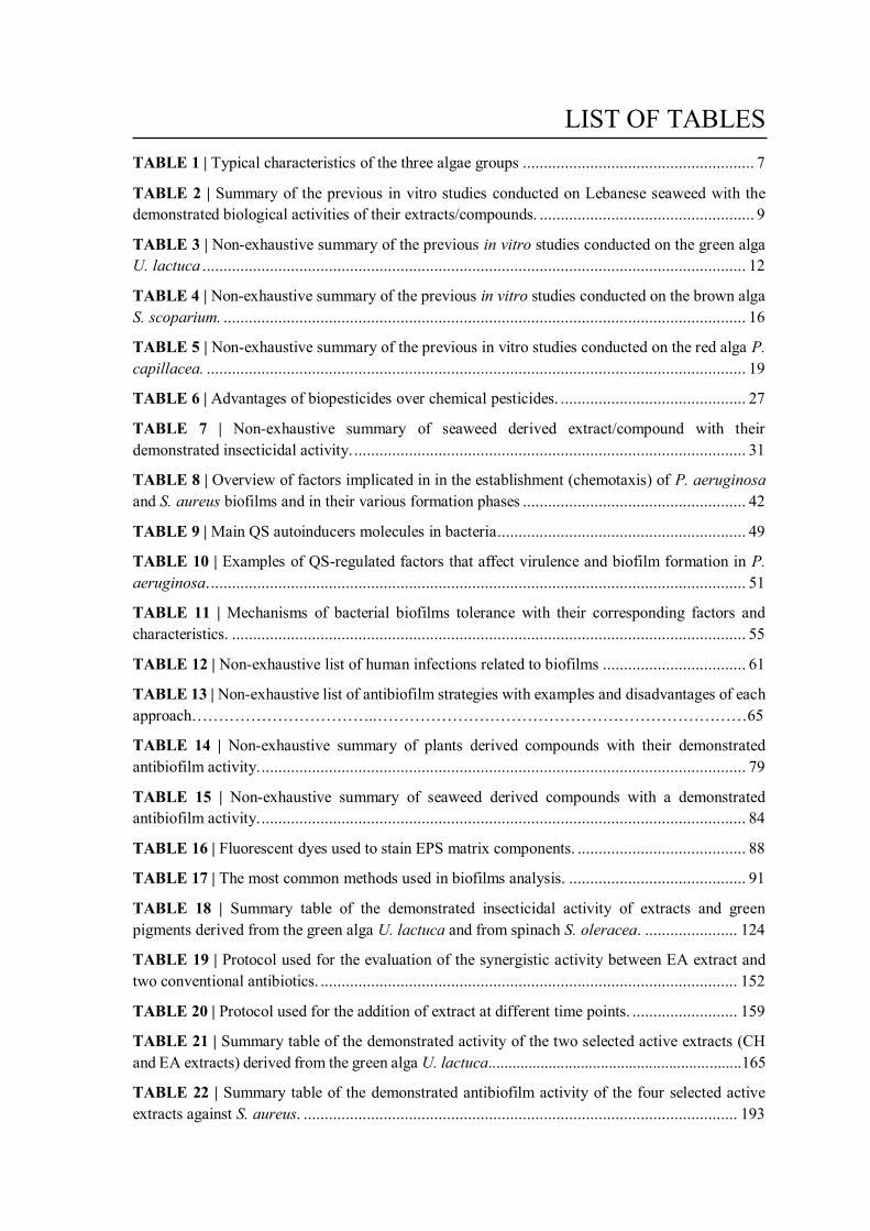
LIST OF TABLES TABLE 1 | Typical characteristics of the three algae groups ....................................................... 7
TABLE 2 | Summary of the previous in vitro studies conducted on Lebanese seaweed with the demonstrated biological activities of their extracts/compounds. ................................................... 9
TABLE 3 | Non-exhaustive summary of the previous in vitro studies conducted on the green alga U. lactuca ................................................................................................................................. 12
TABLE 4 | Non-exhaustive summary of the previous in vitro studies conducted on the brown alga S. scoparium. ............................................................................................................................ 16
TABLE 5 | Non-exhaustive summary of the previous in vitro studies conducted on the red alga P. capillacea. ................................................................................................................................ 19
TABLE 6 | Advantages of biopesticides over chemical pesticides. ............................................ 27
TABLE 7 | Non-exhaustive summary of seaweed derived extract/compound with their demonstrated insecticidal activity. ............................................................................................. 31
TABLE 8 | Overview of factors implicated in in the establishment (chemotaxis) of P. aeruginosa and S. aureus biofilms and in their various formation phases ..................................................... 42
TABLE 9 | Main QS autoinducers molecules in bacteria ........................................................... 49
TABLE 10 | Examples of QS-regulated factors that affect virulence and biofilm formation in P. aeruginosa. ............................................................................................................................... 51
TABLE 11 | Mechanisms of bacterial biofilms tolerance with their corresponding factors and characteristics. .......................................................................................................................... 55
TABLE 12 | Non-exhaustive list of human infections related to biofilms .................................. 61
TABLE 13 | Non-exhaustive list of antibiofilm strategies with examples and disadvantages of each approach……………………………..……………………………………………………………65
TABLE 14 | Non-exhaustive summary of plants derived compounds with their demonstrated antibiofilm activity. ................................................................................................................... 79
TABLE 15 | Non-exhaustive summary of seaweed derived compounds with a demonstrated antibiofilm activity. ................................................................................................................... 84
TABLE 16 | Fluorescent dyes used to stain EPS matrix components. ........................................ 88
TABLE 17 | The most common methods used in biofilms analysis. .......................................... 91
TABLE 18 | Summary table of the demonstrated insecticidal activity of extracts and green pigments derived from the green alga U. lactuca and from spinach S. oleracea. ...................... 124
TABLE 19 | Protocol used for the evaluation of the synergistic activity between EA extract and two conventional antibiotics. ................................................................................................... 152
TABLE 20 | Protocol used for the addition of extract at different time points. ......................... 159
TABLE 21 | Summary table of the demonstrated activity of the two selected active extracts (CH and EA extracts) derived from the green alga U. lactuca...............................................................165
TABLE 22 | Summary table of the demonstrated antibiofilm activity of the four selected active extracts against S. aureus. ....................................................................................................... 193
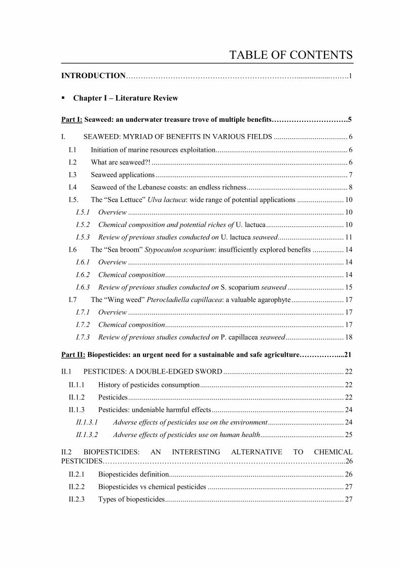
TABLE OF CONTENTS INTRODUCTION……………………………………………………………..................….….1
Chapter I – Literature Review
Part I: Seaweed: an underwater treasure trove of multiple benefits………………………….5
I. SEAWEED: MYRIAD OF BENEFITS IN VARIOUS FIELDS ...................................... 6
I.1 Initiation of marine resources exploitation.................................................................... 6
I.2 What are seaweed?! ..................................................................................................... 6
I.3 Seaweed applications ................................................................................................... 7
I.4 Seaweed of the Lebanese coasts: an endless richness.................................................... 8
I.5. The “Sea Lettuce” Ulva lactuca: wide range of potential applications ........................ 10
I.5.1 Overview ............................................................................................................... 10
I.5.2 Chemical composition and potential riches of U. lactuca ........................................ 10
I.5.3 Review of previous studies conducted on U. lactuca seaweed .................................. 11
I.6 The “Sea broom” Stypocaulon scoparium: insufficiently explored benefits ................ 14
I.6.1 Overview ............................................................................................................... 14
I.6.2 Chemical composition ............................................................................................ 14
I.6.3 Review of previous studies conducted on S. scoparium seaweed ............................. 15
I.7 The “Wing weed” Pterocladiella capillacea: a valuable agarophyte ........................... 17
I.7.1 Overview ............................................................................................................... 17
I.7.2 Chemical composition ............................................................................................ 17
I.7.3 Review of previous studies conducted on P. capillacea seaweed .............................. 18
Part II: Biopesticides: an urgent need for a sustainable and safe agriculture……………....21
II.1 PESTICIDES: A DOUBLE-EDGED SWORD .............................................................. 22
II.1.1 History of pesticides consumption .......................................................................... 22
II.1.2 Pesticides ............................................................................................................... 22
II.1.3 Pesticides: undeniable harmful effects .................................................................... 24
II.1.3.1 Adverse effects of pesticides use on the environment ....................................... 24
II.1.3.2 Adverse effects of pesticides use on human health ........................................... 25
II.2 BIOPESTICIDES: AN INTERESTING ALTERNATIVE TO CHEMICAL PESTICIDES……………………………………………………………………………………...26
II.2.1 Biopesticides definition.......................................................................................... 26
II.2.2 Biopesticides vs chemical pesticides ...................................................................... 27
II.2.3 Types of biopesticides ............................................................................................ 27
Table of contents
II.2.3.1 Microbial biopesticides .................................................................................. 28
II.2.3.2 Biochemical biopesticides .............................................................................. 28
II.2.3.3 Plant-incorporated protectants (PIPs) ............................................................ 29
II.2.4 Marine world: a valuable and promising source of biopesticides ............................. 29
II.2.4.1 Seaweed as a potential source of biopesticides ............................................... 29
Part III: Biofilms: a microbial assemblage of scientific significance………………….……..33
III.1 BACTERIAL BIOFILMS ............................................................................................. 35
III.1.1 History of biofilm discovery .................................................................................. 35
III.1.2 Biofilm definition .................................................................................................. 35
III.1.3 Biofilms: Bad or good?! ......................................................................................... 37
III.1.4 Biofilm life-cycle: from adhesion to dispersion ...................................................... 38
III.1.4.1 Reversible attachment .................................................................................... 38
III.1.4.2 Irreversible attachment .................................................................................. 39
III.1.4.3 Proliferation and matrix production ............................................................... 40
III.1.4.4 Maturation phase ........................................................................................... 41
III.1.4.5 Dispersion phase ............................................................................................ 41
III.1.5 The EPS matrix: a major biofilm component with essential functions ..................... 42
III.1.5.1 Matrix exopolysaccharides ............................................................................. 44
III.1.5.2 Matrix extracellular proteins .......................................................................... 45
III.1.5.3 Extracellular DNA ......................................................................................... 45
III.1.6 Quorum sensing: microbial chatter orchestrating cells’ behavior ............................ 46
III.1.6.1 Definition and discovery................................................................................. 46
III.1.6.2 Quorum sensing circuit .................................................................................. 46
III.1.6.3 QS autoinducers in bacteria ........................................................................... 47
III.1.6.4 Connection between QS and biofilm formation ............................................... 48
III.1.6.5 Quorum sensing network in P. aeruginosa ...................................................... 50
III.1.6.6 Quorum sensing network in S. aureus ............................................................. 52
III.1.7 Biofilms: a resilient strength .................................................................................. 54
III.1.7.1 Diffusion barrier ............................................................................................ 56
III.1.7.2 Reduction in growth rate ................................................................................ 57
III.1.7.3 Modification of genes expression: example of efflux pumps ............................ 57
III.1.7.4 Persister cells................................................................................................. 58
III.1.7.5 Mutagenesis and horizontal gene transfer ...................................................... 59
III.1.8 Biofilm-related diseases ......................................................................................... 60
III.1.8.1 Medical device-related biofilm and associated diseases .................................. 60
III.1.8.2 Other biofilm-related diseases: example of cystic fibrosis ............................... 62
Table of contents
III.2 CHALLENGE OF TREATING BIOFILM-ASSOCIATED INFECTIONS .................... 64
III.2.1 How to handle with biofilms? Current therapeutic approaches and strategies .......... 64
III.2.1.1 Prevention of biofilm formation – Disruption of the initial phases .................. 68
III.2.1.2 Weakening of the biofilm by disarming bacteria ............................................. 69
III.2.1.3 Dispersion of biofilms – Restauration of bacterial sensibility .......................... 72
III.2.1.4 Killing of the biofilm – Combination strategies ............................................... 74
III.2.2 Natural medicine: breakthrough in the search for antibiofilm agents ....................... 75
III.2.2.1 Plant derived compounds with antibiofilm activity .......................................... 76
III.2.2.2 Marine environment: a valuable source of antibiofilm molecules .................... 82
III.3 EXPERIMENTAL BIOFILM ASSAYS USED FOR BIOFILM STUDIES ................... 86
III.3.1 Counting method – CFU counts assay .................................................................... 86
III.3.2 Staining methods ................................................................................................... 86
III.3.2.1 Crystal violet assay (quantitative test) ............................................................ 86
III.3.2.2 DMMB assay (quantitative test) ..................................................................... 87
III.3.3 Microscopic observations ....................................................................................... 87
III.3.3.1 Fluorescent assay – focus on the most popular live/dead mixture.................... 88
III.3.4 Metabolic methods................................................................................................. 89
III.3.4.1 Resazurin assay.............................................................................................. 89
III.3.4.2 The XTT assay ................................................................................................ 89
III.3.4.3 The ATP assay ............................................................................................... 90
III.3.5 Molecular biology methods .................................................................................... 90
III.3.5.1 Quantitative polymerase chain reaction qPCR ............................................... 90
MAIN OBJECTIVES……………………………………...……………..….…………………..93
REFERENCES………..……………………………………………………………………..……94
Chapter II – Green Seaweed: potential alternative to chemical insecticide
I. MATERIALS & METHODS .......................................................................................... 118 I.1. MATERIALS .......................................................................................................... 118
I.1.1 Organic solvents .................................................................................................. 118 I.1.2 Chemical compounds ........................................................................................... 118 I.1.3 Algal material ...................................................................................................... 118 I.1.4 Plant material ....................................................................................................... 118 I.1.5 Biological material ............................................................................................... 119
I.2 METHODS ............................................................................................................. 120 I.2.1 Preparation of crude extracts ................................................................................ 120 I.2.2 Quantification of pigments content in acetonic and ethanolic extracts ................... 120
Table of contents
I.2.3 Separation of chlorophyll and carotenoid pigments by the differential solubility method………………………………………………………………………………………120 I.2.4 Insecticidal activity bioassays .............................................................................. 122
II. ARTICLE SUMMARY – FOCUS ON THE MAIN RESULTS ................................... 124 PUBLICATION…………………………………………………….……...……………126 III. SUPPLEMENTARY UNPUBLISHED DATA ............................................................ 134
III.1 Absorption spectra of purified pigments .................................................................. .134 REFERENCES………………..………...……………………………………………………….136
Chapter III – Seaweed extracts: a promising source of antibiofilm agents against pathogenic bacteria
Part I: Materials & Methods………………..……………………...……..……………………138
I. MATERIALS & METHODS ...................................................................................... 139
I.1 MATERIALS .......................................................................................................... 139
I.1.1 Laboratory materials and devices ......................................................................... 139
I.1.2 Organic solvents .................................................................................................. 139
I.1.3 Chemical products ............................................................................................... 140
I.1.4 Algal materials..................................................................................................... 140
I.1.5 Bacterial strains and culture media ....................................................................... 141
I.2 METHODS ............................................................................................................. 143
I.2.1 Preparation of algal extracts ................................................................................. 143
I.2.2 Assessment of the potential antibiofilm activity of seaweed extracts against the pathogenic bacteria P. aeruginosa ................................................................................... 145
I.2.3 Assessment of the potential antibiofilm activity of seaweed extracts against the pathogenic bacteria S. aureus .......................................................................................... 157
REFERENCES…………………………………………………………………………………..162
Part II: Evaluation of the antibiofilm activity of seaweed extracts against P. aeruginosa……………………………………………………………….……………….…..163
II.1 ARTICLE SUMMARY – FOCUS ON THE MAIN RESULTS ................................... 164
II.2 ADDITIONNAL EXPERIMENTS .............................................................................. 166
II.2.1 Screening of extracts for their ability to inhibit AHL-based QS system – Biosensor-based assay ......................................................................................................................... 166
PUBLICATION……………………………….…………………...……......…………..……168
Part III: Evaluation of the antibiofilm activity of seaweed extracts against S. aureus.................................................................................................................................…..191
III.1 ARTICLE SUMMARY – FOCUS ON THE MAIN RESULTS ................................... 192
III.2 ARTICLE TO BE SUBMITTED………..…………….............……………………….…194
Table of contents
GENERAL CONCLUSION………………………………………………………….…….….221
REFERENCES………………………………………………………………………….……….229
ANNEXES………………………………………………………………………………..……..232

1
INTRODUCTION he exploration of natural products, broadly defined as chemical compounds
synthetized by living organisms, has received a tremendous interest from the
scientific community in the last decencies and the focus on their wide proprieties is
consistently increasing. Indeed, the high structural and functional diversity as well as the
uniqueness of natural products are the result of an evolution over millions of years. These
natural chemicals are usually produced by living organisms as a natural means of
countering external threats (stressful environmental conditions, competition, infections…)
which explains their huge bioactivity (Sorokina & Steinbeck, 2020). Besides their
prominent role in both traditional and modern pharmacology, various studies have
highlighted the usefulness of natural products in food and cosmetic industries as well as in
agriculture, especially in the area of biopesticides (Newman & Cragg, 2016; Sparks et al.,
2019).
Among the exploited living organisms, those residing in the marine environment are
considered as the most recent source explored for bioactive natural products compared to
terrestrial plants and nonmarine organisms (Jimenez, 2018). In fact, the marine world
which accounts for approximately 70% of the Earth’s surface, is the habitat of a huge
diversity of species (algae, sponges, mollusks, bacteria, fungi…) (Blunt et al., 2018).
Interestingly, in order to survive the harsh marine conditions, marine organisms synthetize
a wide variety of unique natural products with high incidence of bioactivity. However, the
marine world remains under-exploited (less than 5% of its diversity has been explored) and
there is still much to know about this underwater treasure in an attempt to valorize these
fantastic creatures in different fields (Jimenez, 2018).
Among marine organisms, seaweed, the primary producers that occupy the base of the
marine food chain, are well-known for their ability to synthesize several bioactive
substances with a broad spectrum of demonstrated biological activity (antioxidant, anti-
inflammatory, antimicrobial, anticancer, anti-aging…). In fact, regarding their sessile
nature, algae have a strong tendency to produce bioactive metabolites and to evolve defense
mechanisms in order to withstand both biotic (fungal, bacterial infections…) and abiotic
(salinity, temperature, pollutants…) threats faced in the marine environment (Leandro et
al., 2019).
T
Introduction
2
In light of their valuable properties and their usefulness as pharmaceuticals, nutraceuticals,
cosmeceuticals, as well as in feeding and agriculture, the cultivation of seaweed together
with their value in the market are continuously rising (Market Analysis Report, 2020).
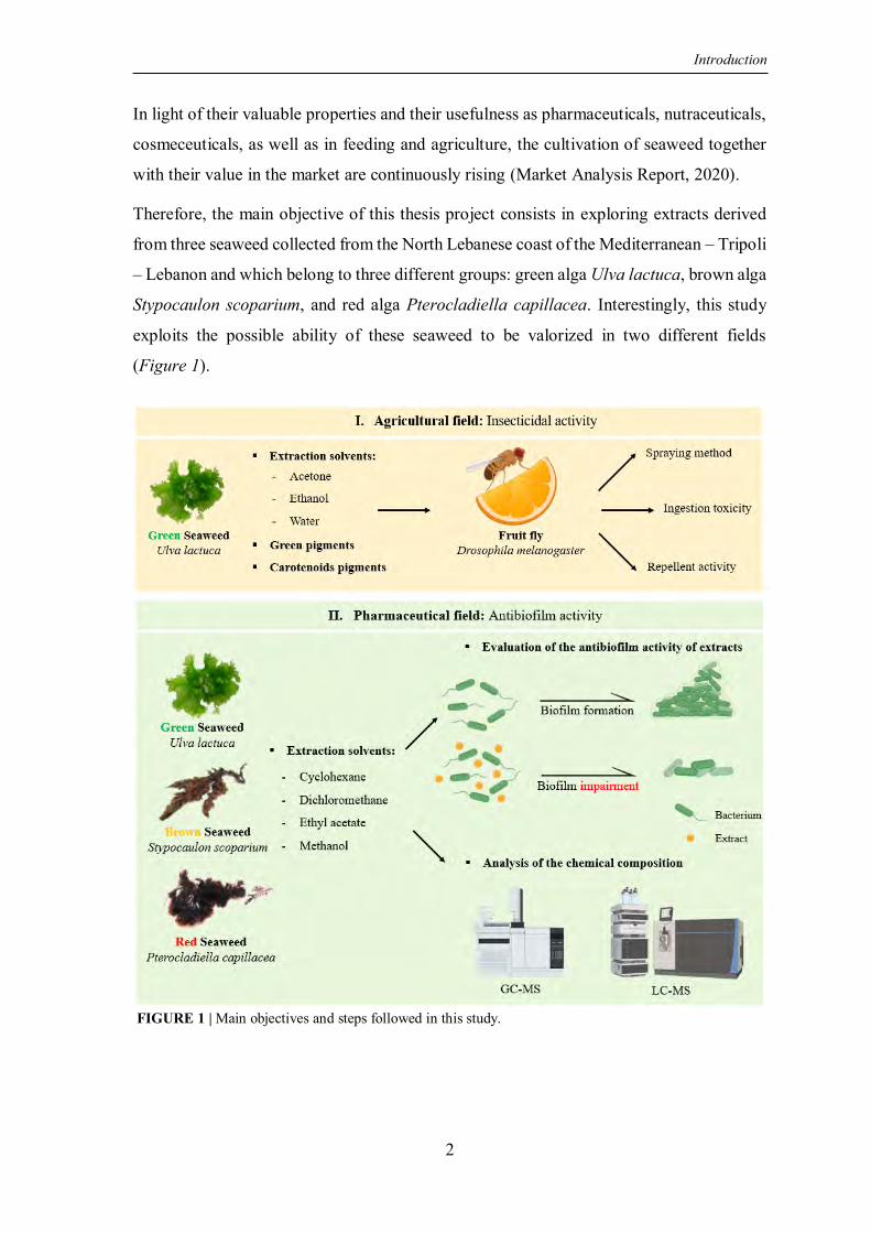
Therefore, the main objective of this thesis project consists in exploring extracts derived
from three seaweed collected from the North Lebanese coast of the Mediterranean – Tripoli
– Lebanon and which belong to three different groups: green alga Ulva lactuca, brown alga
Stypocaulon scoparium, and red alga Pterocladiella capillacea. Interestingly, this study
exploits the possible ability of these seaweed to be valorized in two different fields
(Figure 1).
FIGURE 1 | Main objectives and steps followed in this study.
Introduction
3
Agricultural field: First, the potential capacity of extracts derived from the green alga
U. lactuca to present a natural, eco-friendly, cost-effective, and potentially less-toxic
alternative to conventional agrochemicals was assessed. In fact, the massive use of
synthetic phytosanitary products undoubtedly leads to adverse effects on both public
health and environment, hence the urgent need to look for new strategies (Gyawali,
2018). For this purpose, the insecticidal activity of U. lactuca extracts was evaluated
against the fruit fly Drosophila melanogaster, pest insect and the best model for
studying the insecticidal activity at the laboratory scale. This part of the project was
carried out in the Applied Biotechnology Laboratory (LBA3B-ER032) – AZM Center
for Research in Biotechnology and its Applications – Tripoli – Lebanon.
Pharmaceutical field: On the other hand, various extracts derived from the three
seaweed (U. lactuca, S. scoparium, and P. capillacea) were explored in terms of their
potential antibiofilm activity against two critical bacteria known for their high ability
to produce biofilms: The Gram-negative Pseudomonas aeruginosa and the Gram-
positive Staphylococcus aureus. Indeed, biofilms known as “City of Microbes” and
defined as an aggregation of microorganisms adhered to each other and to any kind of
biotic and abiotic surfaces, embedded in an extracellular polymeric matrix “House of
Biofilm Cells”, provide a strong armor for these bacteria (Flemming et al., 2016). Due
to the increased resilience of this bacterial association and its ability to survive harsh
environmental conditions and to tolerate high concentration of antimicrobial agents as
well as to escape from the host immune response, a great effort is devoted to the search
for new approaches in an attempt to prevent and/or treat biofilm-associated infections
(Uruen et al., 2020). This approach also concerns the biopesticides concept by using
anti-biofilm properties to combat plant infections. In this context, seaweed present a
strong promises given their ability to control their bacterial colonization despite the
abundance of bacteria in seawater, hence the conduct of this study (Shannon & Abu-
Ghannam, 2016). This second part of the project was conducted in the “Laboratoire de
Génie Chimique” (LGC-UMR5503) – Toulouse – France.
It is important to note that the choice of the three seaweed species examined in this study
is based on their wide spectrum of demonstrated biological activity such as antimicrobial,
antioxidant, antidiabetic, anti-inflammatory, and cytotoxic activity (Guner et al., 2019;
Salim et al., 2020; Ismail et al., 2020).
Introduction
4
However, to the best of our knowledge, no previous study has assessed the insecticidal
activity against the fruit fly D. melanogaster as well as the antibiofilm activity against
P. aeruginosa and S. aureus of some extracts derived from these algae.
The present manuscript is composed of three chapters arranged as follows:
I. The first chapter is dedicated to a literature review outlining the background of this
study and divided into three distinct parts. In the first section (Part I), the benefits of
algae as well as their possible applications in different fields with emphasis on the three
seaweed examined in this study (green alga U. lactuca, brown alga S. scoparium, and
red alga P. capillacea) are presented. On the other hand, the harmful effects of synthetic
agrochemicals on environment and public health as well as the importance of
biopesticides in the search for novel alternatives with focus on those derived from
marine organisms, especially from seaweed, are reported in the second part (Part II).
Then, in the third part (Part III), an overview on biofilms, their various resilience
mechanisms as well as the different therapeutic approaches developed in order to
control biofilms formation are outlined. The promising role of natural medicine in the
search for novel and effective antibiofilm agents is also highlighted.
II. The evaluation of the insecticidal activity of extracts derived from the green alga
U. lactuca against the fruit fly D. melanogaster which resulted in a published article
(Rima et al., 2021) is presented in the second chapter of this manuscript.
III. The third chapter which is devoted to the evaluation of the potential antibiofilm activity
of various extracts derived from the three seaweed (green alga U. lactuca, brown alga
S. scoparium, and red alga P. capillacea) is divided into three parts. The first section
(Part I) groups all materials and methods used in this chapter. Then, in the second part
(Part II), results obtained upon the evaluation of the antibiofilm activity of extracts
against P. aeruginosa and which resulted in a submitted article, are presented. The
promising antibiofilm activity of extracts against S. aureus and which also resulted in
an article (to be submitted), are described in the third part (Part III) of this chapter.
At the end of the manuscript, a general conclusion with some perspectives are outlined.
5
PREVIEW
Although most people do not imagine it, seaweed extracts are part of the composition of
many products that we use or consume daily such as toothpaste, deodorizer, ice cream as
well as bottled chocolate drinks. Interestingly, the possible applications of algae are not
restricted to a particular field, but various studies have documented their amazing
properties to use in pharmaceutical, cosmetical, nutraceutical and even in agricultural
sectors. Among their broad spectrum of demonstrated biological activities, a wide variety
of compounds derived from seaweed have exhibited interesting antimicrobial activities.
Thus, seaweed offer a natural resource of unique bioactive products to maintain and
preserve.
In this first part of chapter I, overview of algae as well as their potential benefits in different
fields are introduced with a focus on pharmaceutics. As the seaweeds evaluated in this
study are collected from a Lebanese coast, the actual exploitation of Lebanese algae and
their demonstrated biological activities are reviewed.
On the other hand, the green Ulva lactuca, the brown Stypocaulon scoparium and the red
Pterocladiella capillacea algae involved in this study are presented along with a summary
of the previous studies showing potential biological activities especially regarding different
types of extracts. The region of sample collection is also indicated given the high impact
of environmental conditions related to the location of harvesting on the chemical
composition of algae and thus their activity.
I Part I: Seaweed: an underwater treasure trove of multiple benefits “Focus on the three algae explored in this study”
CHAPTER

Chapter I – Bibliography Part I – Seaweed: an underwater treasure trove of multiple benefits
6
I. SEAWEED: MYRIAD OF BENEFITS IN VARIOUS FIELDS
I.1 Initiation of marine resources exploitation
The initiation of marine world exploitation as a valuable source of natural products with
high pharmaceutical relevance was first launched in 1967 during a conference named
“Drugs from the Sea” held in Rhode Island, USA. Since then, the search for primary and
secondary metabolites derived from marine organisms has received worldwide attention in
view of new drug discovery (Nogueira & Teixeira, 2016). After extensive efforts of many
researchers from around the world who were dedicated to the isolation and identification
of novel marine natural products as well as to the evaluation of their potential bioactivity,
approximately 28,500 bioactive products derived from marine organisms were
characterized by the end of 2016 (Jimenez, 2018; Blunt et al., 2018).
Interestingly, in 2004, the Food and Drug administration (FDA) authorized the first drug
directly derived from a marine organism particularly from a cone snail and that is used for
the treatment of chronic pain. At present, there are six therapeutic structures based on
natural marine products that have been approved by the FDA (Jimenez, 2018).
In this context, the marine world, which hosts a huge species diversity producing a variety
of bioactive metabolites, ensures a promising gateway in the search for novel cost-effective
and highly efficient drugs. Among prokaryotic as well as eukaryotic creatures, seaweed,
the primary producers occupying the base of the marine food chain, are known as a valuable
reservoir of bioactive products already used for different purposes ranging from food
applications to medicine (Leandro et al., 2019).
I.2 What are seaweed?!
Seaweed also named “macroalgae” are macroscopic, multicellular, autotrophic, ubiquitous
organisms that can be found in any wet environment as well as in fresh and salt-water. In
seawater, they often inhabit shallow coastal areas by growing on rocks, pebbles, shells and
even on aquatic plants. Based on the color of their thallus provided by their distinctive
pigments, macroalgae are taxonomically classified into three large groups: Chlorophycea
(green algae), Phaeophycea (brown algae) and Rhodophycea (red algae) (Leandro et al.,
2019; Nakhate & van der Meer, 2021). The typical characteristics as well as the pigments
associated with each group are summarized in the table below (Table 1).
Chapter I – Bibliography Part I – Seaweed: an underwater treasure trove of multiple benefits
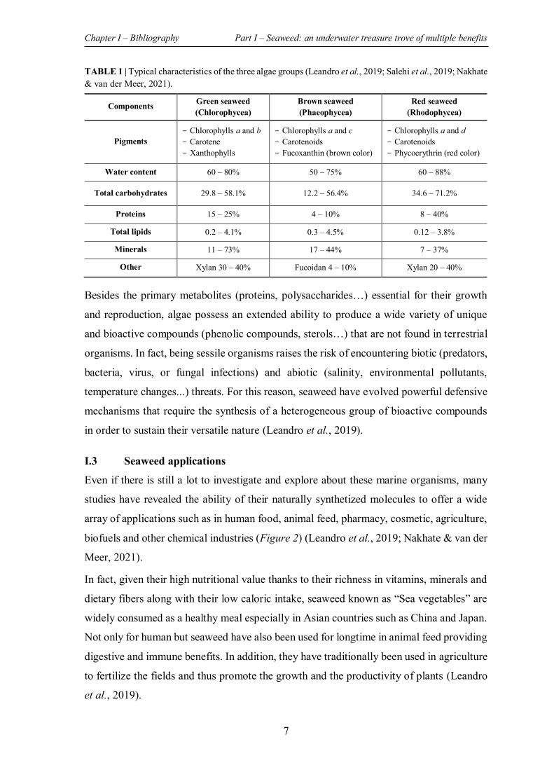
7
TABLE 1 | Typical characteristics of the three algae groups (Leandro et al., 2019; Salehi et al., 2019; Nakhate & van der Meer, 2021).
Components Green seaweed (Chlorophycea)
Brown seaweed (Phaeophycea)
Red seaweed (Rhodophycea)
Pigments - Chlorophylls a and b - Carotene - Xanthophylls
- Chlorophylls a and c - Carotenoids - Fucoxanthin (brown color)
- Chlorophylls a and d - Carotenoids - Phycoerythrin (red color)
Water content 60 – 80% 50 – 75% 60 – 88%
Total carbohydrates 29.8 – 58.1% 12.2 – 56.4% 34.6 – 71.2%
Proteins 15 – 25% 4 – 10% 8 – 40%
Total lipids 0.2 – 4.1% 0.3 – 4.5% 0.12 – 3.8%
Minerals 11 – 73% 17 – 44% 7 – 37%
Other Xylan 30 – 40% Fucoidan 4 – 10% Xylan 20 – 40%
Besides the primary metabolites (proteins, polysaccharides…) essential for their growth
and reproduction, algae possess an extended ability to produce a wide variety of unique
and bioactive compounds (phenolic compounds, sterols…) that are not found in terrestrial
organisms. In fact, being sessile organisms raises the risk of encountering biotic (predators,
bacteria, virus, or fungal infections) and abiotic (salinity, environmental pollutants,
temperature changes...) threats. For this reason, seaweed have evolved powerful defensive
mechanisms that require the synthesis of a heterogeneous group of bioactive compounds
in order to sustain their versatile nature (Leandro et al., 2019).
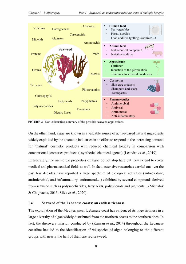
I.3 Seaweed applications
Even if there is still a lot to investigate and explore about these marine organisms, many
studies have revealed the ability of their naturally synthetized molecules to offer a wide
array of applications such as in human food, animal feed, pharmacy, cosmetic, agriculture,
biofuels and other chemical industries (Figure 2) (Leandro et al., 2019; Nakhate & van der
Meer, 2021).
In fact, given their high nutritional value thanks to their richness in vitamins, minerals and
dietary fibers along with their low caloric intake, seaweed known as “Sea vegetables” are
widely consumed as a healthy meal especially in Asian countries such as China and Japan.
Not only for human but seaweed have also been used for longtime in animal feed providing
digestive and immune benefits. In addition, they have traditionally been used in agriculture
to fertilize the fields and thus promote the growth and the productivity of plants (Leandro
et al., 2019).
Chapter I – Bibliography Part I – Seaweed: an underwater treasure trove of multiple benefits
8
On the other hand, algae are known as a valuable source of active-based natural ingredients
widely exploited by the cosmetic industries in an effort to respond to the increasing demand
for “natural” cosmetic products with reduced chemical toxicity in comparison with
conventional cosmetics products (“synthetic” chemical agents) (Leandro et al., 2019).
Interestingly, the incredible properties of algae do not stop here but they extend to cover
medical and pharmaceutical fields as well. In fact, extensive researches carried out over the
past few decades have reported a large spectrum of biological activities (anti-oxidant,
antimicrobial, anti-inflammatory, antitumoral…) exhibited by several compounds derived
from seaweed such as polysaccharides, fatty acids, polyphenols and pigments…(Michalak
& Chojnacka, 2015; Silva et al., 2020).
I.4 Seaweed of the Lebanese coasts: an endless richness
The exploitation of the Mediterranean Lebanese coast has evidenced its huge richness in a
large diversity of algae widely distributed from the northern coasts to the southern ones. In
fact, the discovery mission conducted by (Kanaan et al., 2014) throughout the Lebanese
coastline has led to the identification of 94 species of algae belonging to the different
groups with nearly the half of them are red seaweed.
Human food - Sea vegetables - Pasta / noodles - Food additive (gelling, stabilizer…)
Animal feed - Nutraceutical compound - Nutritive additive
Agriculture - Fertilizer - Induction of the germination - Tolerance to stressful conditions
Cosmetics - Skin care products - Shampoos and soaps - Toothpastes
Pharmaceutics - Antimicrobial - Antiviral - Antitumoral - Anti-inflammatory
Vitamins
Proteins
Minerals Alginates
Carrageenans
Carotenoids
Chlorophylls
Terpenes
Polysaccharides
Dietary fibres Fucoidans
Phlorotannins
Fatty acids
Ulvans
Polyphenols
Sterols
Agar
Alkaloids
Amino acids
Seaweed
FIGURE 2 | Non-exhaustive summary of the possible seaweed applications.
Chapter I – Bibliography Part I – Seaweed: an underwater treasure trove of multiple benefits
9
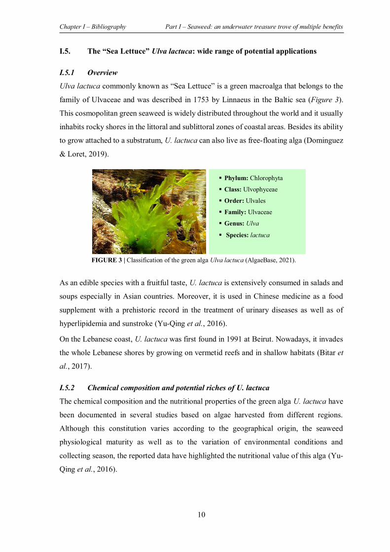
Despite their diversity, studies that focus on the investigation of Lebanese seaweed in view
of their potential capacity to be valorized in different fields remain limited. Nevertheless,
some studies have evaluated the potential biological activities (in-vitro) of extracts and/or
compounds (especially polysaccharides) derived from some Lebanese algae (Table 2).
However, there is a lot to be achieved in an attempt to advance towards a real application
of these seaweed products in the pharmaceutical field.
TABLE 2 | Summary of the previous in vitro studies conducted on Lebanese seaweed with the demonstrated biological activities of their extracts/compounds.
Seaweed species
Evaluated fraction Demonstrated biological activities Reference
Laurencia obtusa
(Red alga) Protein fraction
- Antioxidant activity - Antiproliferative activity (Human colorectal cancer
cells HCT 116) (Al Monla et
al., 2021)
Padina pavonica
(Brown alga)
Organic extracts: - Petroleum ether - Chloroform - Methanol
- Antibacterial activity against E.coli, P. aeruginosa, K. pneumonia, P. vulgaris and E. faecalis
(Chbani et al., 2011)
Colpomenia sinuosa
(Brown alga)
Organic extracts: - DCM : MeOH - MeOH
- Antiproliferative activity (Human colorectal cancer HCT 116 and breast cancer cells MCF-7)
- Antioxidant activity - Anti-inflammatory activity - Antibacterial activity (P. aeruginosa, S. aureus, B.
subtilis, P. vulgaris and E. faecalis)
(Al Monla, Dassouki,
Kouzayha, et al., 2020; Al
Monla, Dassouki, Gali-Muhtasib, et al.,
2020)
Dictyopteris polypodioides (Brown alga)
Polysaccharides: - Fucoidan - Laminaran - Mannuronan
- Antioxidant activity - Anticoagulant activity - Antiproliferative activity (Human melanoma cells
RPMI-7951)
(Sokolova et al., 2011; Karaki et
al., 2013)
Corallina (Red alga)
Polysaccharides: - Sulfated
galactans - Carrageenan
- Anticoagulant activity - Antibacterial activity (S. epidermidis and E. faecalis)
(Sebaaly et al., 2014)
Stypopodium schimperi
(Brown alga)
Polysaccharides: - Fucoidan - Sodium
alginate
- Antioxidant activity - Antiproliferative activity (Human colorectal cancer
cells HCT 116) (Haddad et al.,
2017)
Pterocladia (Red alga)
Polysaccharides: - Sulfated
galactans - Carrageenan
- Antioxidant activity - Anticoagulant activity
(Sebaaly et al., 2012)
Chapter I – Bibliography Part I – Seaweed: an underwater treasure trove of multiple benefits
10
I.5. The “Sea Lettuce” Ulva lactuca: wide range of potential applications
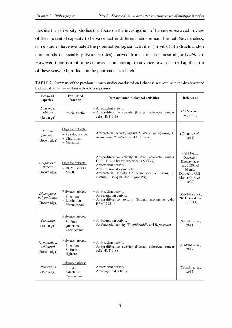
I.5.1 Overview Ulva lactuca commonly known as “Sea Lettuce” is a green macroalga that belongs to the
family of Ulvaceae and was described in 1753 by Linnaeus in the Baltic sea (Figure 3).
This cosmopolitan green seaweed is widely distributed throughout the world and it usually
inhabits rocky shores in the littoral and sublittoral zones of coastal areas. Besides its ability
to grow attached to a substratum, U. lactuca can also live as free-floating alga (Dominguez
& Loret, 2019).
As an edible species with a fruitful taste, U. lactuca is extensively consumed in salads and
soups especially in Asian countries. Moreover, it is used in Chinese medicine as a food
supplement with a prehistoric record in the treatment of urinary diseases as well as of
hyperlipidemia and sunstroke (Yu-Qing et al., 2016).
On the Lebanese coast, U. lactuca was first found in 1991 at Beirut. Nowadays, it invades
the whole Lebanese shores by growing on vermetid reefs and in shallow habitats (Bitar et
al., 2017).
I.5.2 Chemical composition and potential riches of U. lactuca
The chemical composition and the nutritional properties of the green alga U. lactuca have
been documented in several studies based on algae harvested from different regions.
Although this constitution varies according to the geographical origin, the seaweed
physiological maturity as well as to the variation of environmental conditions and
collecting season, the reported data have highlighted the nutritional value of this alga (Yu-
Qing et al., 2016).
Phylum: Chlorophyta
Class: Ulvophyceae
Order: Ulvales
Family: Ulvaceae
Genus: Ulva
Species: lactuca
FIGURE 3 | Classification of the green alga Ulva lactuca (AlgaeBase, 2021).
Chapter I – Bibliography Part I – Seaweed: an underwater treasure trove of multiple benefits
11
In fact, the richness of U. lactuca green alga in essential minerals (magnesium, iron,
calcium, potassium…), in vitamins (B1, B2, B12, C…), in good unsaturated fatty acids, in
dietary fiber, as well as in proteins, make it an excellent food with high nutritional value
along with a low-fat intake (Yu-Qing et al., 2016; Dominguez & Loret, 2019).
On the other hand, algae belonging to the Ulva genus including U. lactuca are mostly
exploited for their high content in ulvan, sulfated heteropolysaccharide, that accounts to
almost 30% of Ulva dry weight. Owning to its antiviral, antitumor, anticoagulant,
antioxidant and even antidepressant proprieties demonstrated in various studies, this
polysaccharide is increasingly requested for pharmaceutical and food purposes (Kidgell et
al., 2019). Despite its widely documented biological activities (in vitro and in vivo) as well
as its demonstrated ability to be used in pharmaceutical formulations (polymers,
excipients…) and in bone tissue engineering (ulvan-based hydrogels…), further clinical
studies are required prior to its real application (Cindana Mo’o et al., 2020).
In addition, U. lactuca contains phenolic compounds as well as chlorophylls and
carotenoids pigments that can serve as free-radical scavengers (Dominguez & Loret, 2019).
Not only for human, but U. lactuca can also be valued in animal feed, in agriculture as well
as in biofuels production (Dominguez & Loret, 2019).
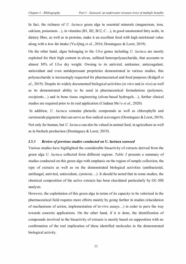
I.5.3 Review of previous studies conducted on U. lactuca seaweed
Various studies have highlighted the considerable bioactivity of extracts derived from the
green alga U. lactuca collected from different regions. Table 3 presents a summary of
studies conducted on this green alga with emphasis on the region of sample collection, the
type of extracts as well as on the demonstrated biological activities (antibacterial,
antifungal, antiviral, antioxidant, cytotoxic…). It should be noted that in some studies, the
chemical composition of the active extracts has been elucidated particularly by GC-MS
analysis.
However, the exploitation of this green alga in terms of its capacity to be valorized in the
pharmaceutical field requires more efforts mainly by going further in studies (elucidation
of mechanisms of action, implementation of in-vivo assays…) in order to pave the way
towards concrete applications. On the other hand, if it is done, the identification of
compounds involved in the bioactivity of extracts is mostly based on supposition with no
confirmation of the real implication of these identified molecules in the demonstrated
biological activity.
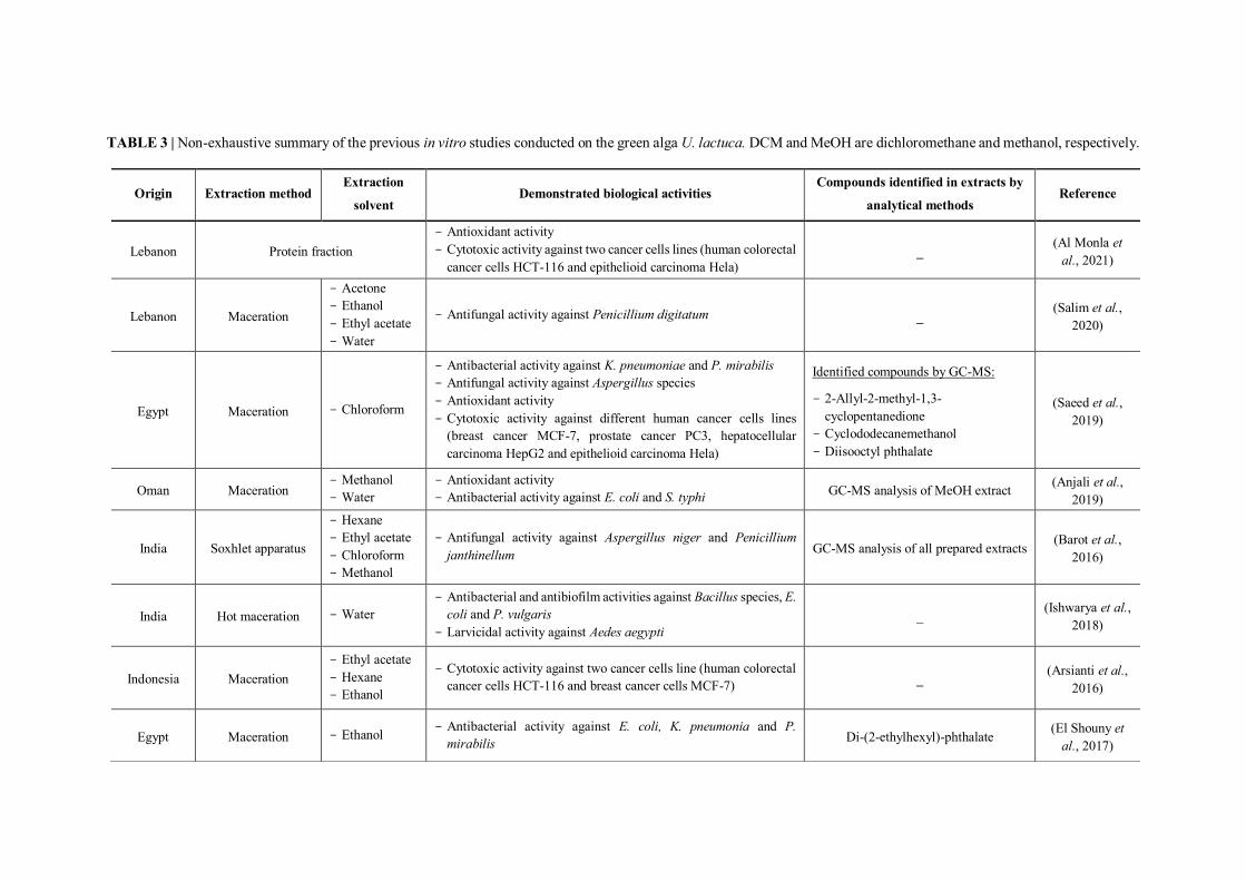
TABLE 3 | Non-exhaustive summary of the previous in vitro studies conducted on the green alga U. lactuca. DCM and MeOH are dichloromethane and methanol, respectively.
Origin Extraction method Extraction
solvent Demonstrated biological activities
Compounds identified in extracts by
analytical methods Reference
Lebanon Protein fraction - Antioxidant activity - Cytotoxic activity against two cancer cells lines (human colorectal
cancer cells HCT-116 and epithelioid carcinoma Hela) _
(Al Monla et al., 2021)
Lebanon Maceration
- Acetone - Ethanol - Ethyl acetate - Water
- Antifungal activity against Penicillium digitatum _ (Salim et al.,
2020)
Egypt Maceration - Chloroform
- Antibacterial activity against K. pneumoniae and P. mirabilis - Antifungal activity against Aspergillus species - Antioxidant activity - Cytotoxic activity against different human cancer cells lines
(breast cancer MCF-7, prostate cancer PC3, hepatocellular carcinoma HepG2 and epithelioid carcinoma Hela)
Identified compounds by GC-MS:
- 2-Allyl-2-methyl-1,3-cyclopentanedione
- Cyclododecanemethanol - Diisooctyl phthalate
(Saeed et al., 2019)
Oman Maceration - Methanol - Water
- Antioxidant activity - Antibacterial activity against E. coli and S. typhi GC-MS analysis of MeOH extract (Anjali et al.,
2019)
India Soxhlet apparatus
- Hexane - Ethyl acetate - Chloroform - Methanol
- Antifungal activity against Aspergillus niger and Penicillium janthinellum GC-MS analysis of all prepared extracts (Barot et al.,
2016)
India Hot maceration - Water - Antibacterial and antibiofilm activities against Bacillus species, E.
coli and P. vulgaris - Larvicidal activity against Aedes aegypti
_ (Ishwarya et al., 2018)
Indonesia Maceration - Ethyl acetate - Hexane - Ethanol
- Cytotoxic activity against two cancer cells line (human colorectal cancer cells HCT-116 and breast cancer cells MCF-7) _ (Arsianti et al.,
2016)
Egypt Maceration - Ethanol - Antibacterial activity against E. coli, K. pneumonia and P. mirabilis Di-(2-ethylhexyl)-phthalate (El Shouny et
al., 2017)
13
India Maceration - Methanol - Antibiofilm activity against Vibrio species, P. aeruginosa, S. aureus and other pathogenic bacteria _ (Yuvaraj &
Arul, 2014)
Morocco Maceration - DCM :
MeOH - Larvicidal activity against Artemia salina _ (Oumaskour et al., 2017)
Egypt Soxhlet apparatus
- Petroleum ether
- Chloroform - Acetone - Ethanol - Methanol
- Insecticidal activity against Culex pipiens and Spodoptera littoralis
- Antifungal activity against Aspergillus niger, Penicillium digitatum, and Rhizoctonia solani
Identified compounds by GC-MS analysis of MeOH extract:
- 1,2-Benzenedicarboxylic acid, bis (2-ethylhexyl) ester
- Palmitic acid - Benzene, 1,2,4-trimethyl - 8-Octadecanoic acid methyl ester - Benzene, 1-ethyl 2-methyl
(Abbassy et al., 2014)
India Soxhlet apparatus
- Hexane - Chloroform - Ethyl acetate - Acetone - Methanol
- Antifungal activity against Candida species. _ (Raj et al., 2017)
India Maceration - Methanol - Antioxidant activity - Antibacterial activity against P. aeruginosa, S. aureus, E. coli… - Antifungal activity against Aspergillus species and C. albicans
GC-MS analysis (Alagan et al., 2017)
India Maceration - Ethanol - Water - Antidiabetic activity _ (Reka et al.,
2017)
Saudi Arabia Maceration - Methanol - Antibacterial activity against P. aeruginosa and S. aureus _ (Al-Zahrani et
al., 2017)
South Africa
Sulfated polysaccharides - Antioxidant activity - Cholinesterase inhibitory activity _
(Olasehinde et al., 2019)
Egypt Maceration - Ethyl acetate - Methanol
- Antifungal activity against Fusarium species, Trichoderma hamatum, Aspergillus flavipes, and Candida albicans GC-MS analysis (Shobier et al.,
2016)
Taiwan Sulfated polysaccharides - Antiviral activity against Japanese Encephalitis Virus _ (Chiu et al., 2012)
TABLE 3 | Continued.
Chapter I – Bibliography Part I – Seaweed: an underwater treasure trove of multiple benefits
14
I.6 The “Sea broom” Stypocaulon scoparium: insufficiently explored benefits
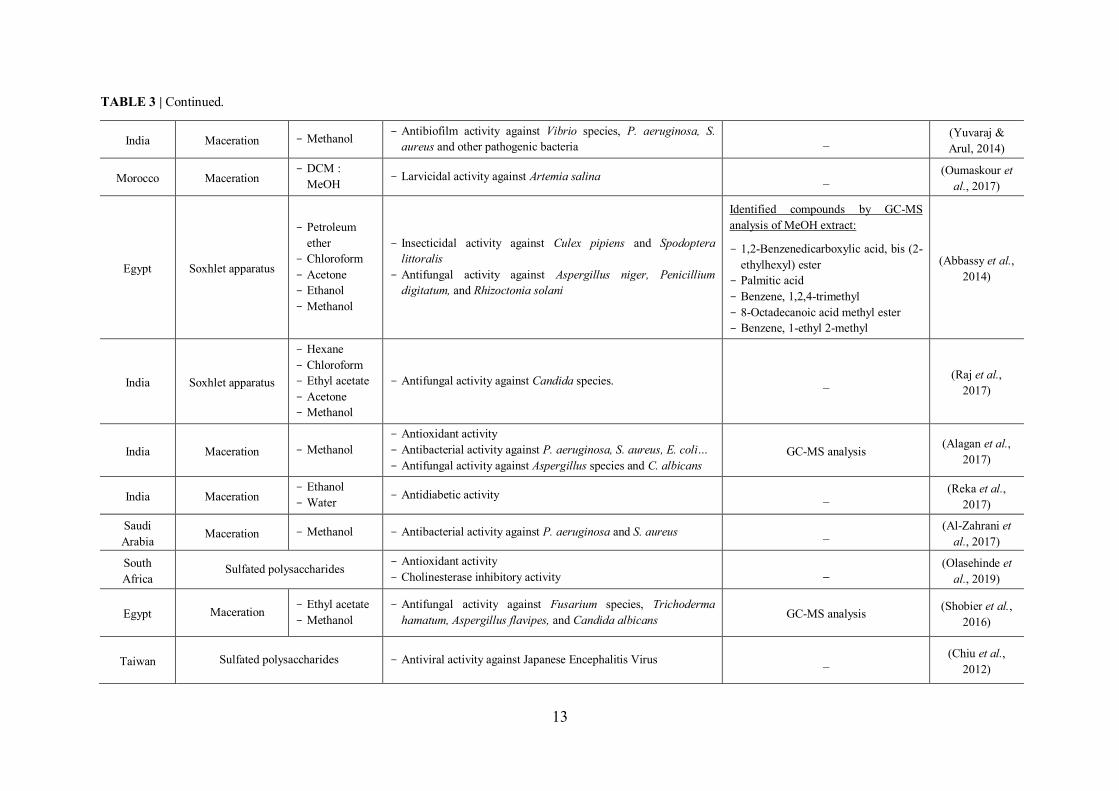
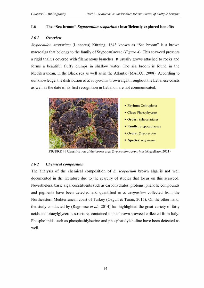
I.6.1 Overview
Stypocaulon scoparium (Linnaeus) Kützing, 1843 known as “Sea broom” is a brown
macroalga that belongs to the family of Stypocaulaceae (Figure 4). This seaweed presents
a rigid thallus covered with filamentous branches. It usually grows attached to rocks and
forms a beautiful fluffy clumps in shallow water. The sea broom is found in the
Mediterranean, in the Black sea as well as in the Atlantic (MACOI, 2008). According to
our knowledge, the distribution of S. scoparium brown alga throughout the Lebanese coasts
as well as the date of its first recognition in Lebanon are not communicated.
I.6.2 Chemical composition
The analysis of the chemical composition of S. scoparium brown alga is not well
documented in the literature due to the scarcity of studies that focus on this seaweed.
Nevertheless, basic algal constituents such as carbohydrates, proteins, phenolic compounds
and pigments have been detected and quantified in S. scoparium collected from the
Northeastern Mediterranean coast of Turkey (Ozgun & Turan, 2015). On the other hand,
the study conducted by (Ragonese et al., 2014) has highlighted the great variety of fatty
acids and triacylglycerols structures contained in this brown seaweed collected from Italy.
Phospholipids such as phosphatidylserine and phosphatidylcholine have been detected as
well.
Phylum: Ochrophyta
Class: Phaeophyceae
Order: Sphacelariales
Family: Stypocaulaceae
Genus: Stypocaulon
Species: scoparium
FIGURE 4 | Classification of the brown alga Stypocaulon scoparium (AlgaeBase, 2021).
Chapter I – Bibliography Part I – Seaweed: an underwater treasure trove of multiple benefits
15
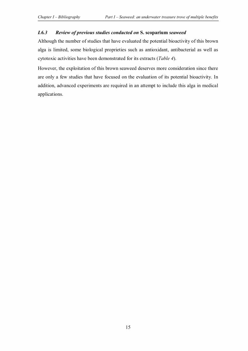
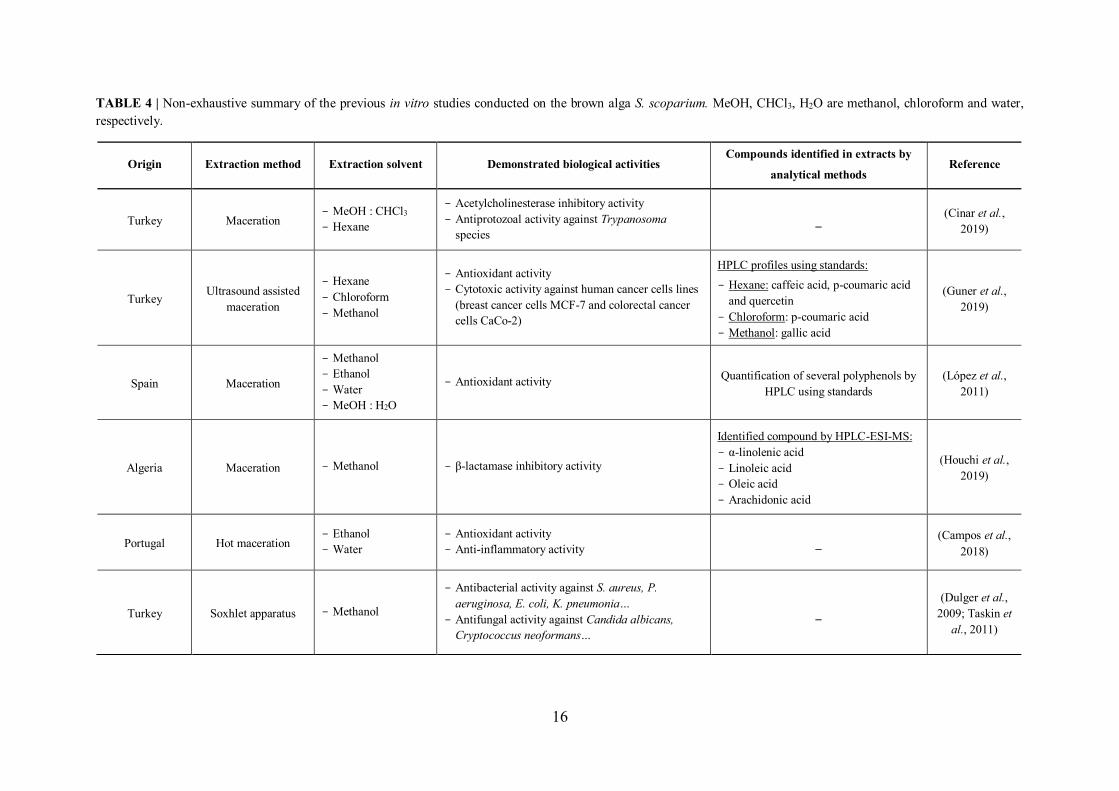
I.6.3 Review of previous studies conducted on S. scoparium seaweed
Although the number of studies that have evaluated the potential bioactivity of this brown
alga is limited, some biological proprieties such as antioxidant, antibacterial as well as
cytotoxic activities have been demonstrated for its extracts (Table 4).
However, the exploitation of this brown seaweed deserves more consideration since there
are only a few studies that have focused on the evaluation of its potential bioactivity. In
addition, advanced experiments are required in an attempt to include this alga in medical
applications.
16
TABLE 4 | Non-exhaustive summary of the previous in vitro studies conducted on the brown alga S. scoparium. MeOH, CHCl3, H2O are methanol, chloroform and water, respectively.
Origin Extraction method Extraction solvent Demonstrated biological activities Compounds identified in extracts by
analytical methods Reference
Turkey Maceration - MeOH : CHCl3 - Hexane
- Acetylcholinesterase inhibitory activity - Antiprotozoal activity against Trypanosoma
species _ (Cinar et al.,
2019)
Turkey Ultrasound assisted maceration
- Hexane - Chloroform - Methanol
- Antioxidant activity - Cytotoxic activity against human cancer cells lines
(breast cancer cells MCF-7 and colorectal cancer cells CaCo-2)
HPLC profiles using standards: - Hexane: caffeic acid, p-coumaric acid
and quercetin - Chloroform: p-coumaric acid - Methanol: gallic acid
(Guner et al., 2019)
Spain Maceration
- Methanol - Ethanol - Water - MeOH : H2O
- Antioxidant activity Quantification of several polyphenols by HPLC using standards
(López et al., 2011)
Algeria Maceration - Methanol - β-lactamase inhibitory activity
Identified compound by HPLC-ESI-MS: - α-linolenic acid - Linoleic acid - Oleic acid - Arachidonic acid
(Houchi et al., 2019)
Portugal Hot maceration - Ethanol - Water
- Antioxidant activity - Anti-inflammatory activity _
(Campos et al., 2018)
Turkey Soxhlet apparatus - Methanol
- Antibacterial activity against S. aureus, P. aeruginosa, E. coli, K. pneumonia…
- Antifungal activity against Candida albicans, Cryptococcus neoformans…
_ (Dulger et al.,
2009; Taskin et al., 2011)
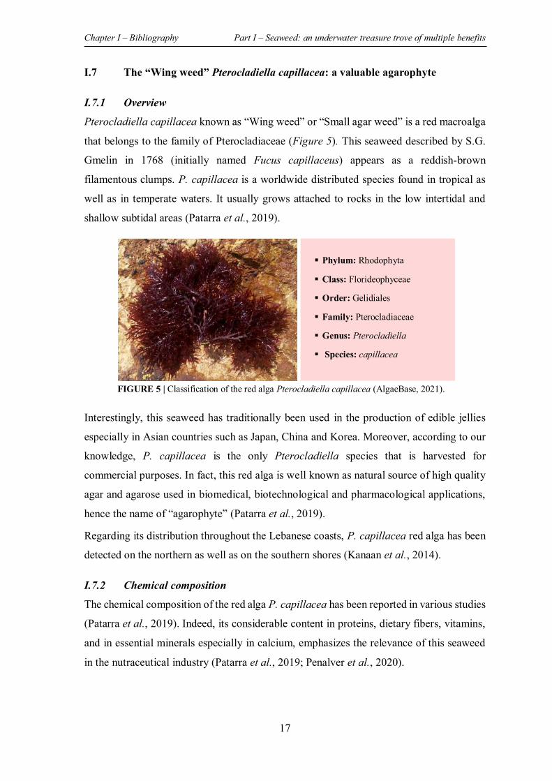
Chapter I – Bibliography Part I – Seaweed: an underwater treasure trove of multiple benefits
17
I.7 The “Wing weed” Pterocladiella capillacea: a valuable agarophyte
I.7.1 Overview
Pterocladiella capillacea known as “Wing weed” or “Small agar weed” is a red macroalga
that belongs to the family of Pterocladiaceae (Figure 5). This seaweed described by S.G.
Gmelin in 1768 (initially named Fucus capillaceus) appears as a reddish-brown
filamentous clumps. P. capillacea is a worldwide distributed species found in tropical as
well as in temperate waters. It usually grows attached to rocks in the low intertidal and
shallow subtidal areas (Patarra et al., 2019).
Interestingly, this seaweed has traditionally been used in the production of edible jellies
especially in Asian countries such as Japan, China and Korea. Moreover, according to our
knowledge, P. capillacea is the only Pterocladiella species that is harvested for
commercial purposes. In fact, this red alga is well known as natural source of high quality
agar and agarose used in biomedical, biotechnological and pharmacological applications,
hence the name of “agarophyte” (Patarra et al., 2019).
Regarding its distribution throughout the Lebanese coasts, P. capillacea red alga has been
detected on the northern as well as on the southern shores (Kanaan et al., 2014).
I.7.2 Chemical composition
The chemical composition of the red alga P. capillacea has been reported in various studies
(Patarra et al., 2019). Indeed, its considerable content in proteins, dietary fibers, vitamins,
and in essential minerals especially in calcium, emphasizes the relevance of this seaweed
in the nutraceutical industry (Patarra et al., 2019; Penalver et al., 2020).
Phylum: Rhodophyta
Class: Florideophyceae
Order: Gelidiales
Family: Pterocladiaceae
Genus: Pterocladiella
Species: capillacea
FIGURE 5 | Classification of the red alga Pterocladiella capillacea (AlgaeBase, 2021).
Chapter I – Bibliography Part I – Seaweed: an underwater treasure trove of multiple benefits
18
In addition, polyunsaturated fatty acids, chlorophylls and carotenoids pigments, as well as
phenolic compounds have also been detected and quantified in this alga (Mohy El-Din &
El-Ahwany, 2018).
As mentioned above, the red alga P. capillacea is mostly exploited for its agar content
which is characterized by a high gelling strength along with low gelling temperatures. In
fact, analyzes performed on algae samples collected from different regions have revealed
that the quality as well as the quantity of the extracted agar which varies between 5 and
34% are site and harvest season specific (Patarra et al., 2019).
I.7.3 Review of previous studies conducted on P. capillacea seaweed Some studies have investigated the potential biological activities (antioxidant, anti-
inflammatory, antimicrobial, …) of P. capillacea red alga. As shown in the table below
(Table 5) most studies have focused on the evaluation of polysaccharide fractions or polar
extracts of which the chemical composition has sometimes been elucidated. As for the two
algae presented below, it is essential to go further in the experiments.
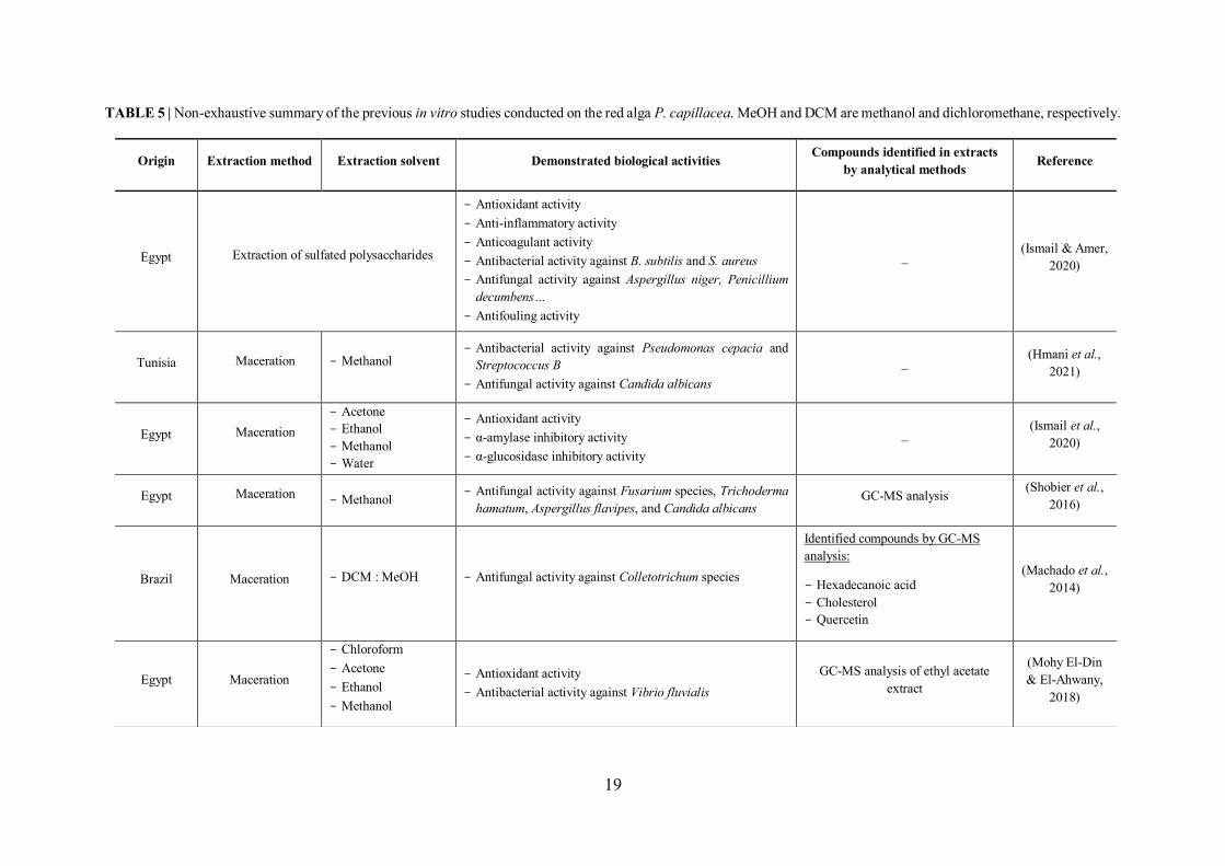
19
TABLE 5 | Non-exhaustive summary of the previous in vitro studies conducted on the red alga P. capillacea. MeOH and DCM are methanol and dichloromethane, respectively.
Origin Extraction method Extraction solvent Demonstrated biological activities Compounds identified in extracts by analytical methods Reference
Egypt Extraction of sulfated polysaccharides
- Antioxidant activity - Anti-inflammatory activity - Anticoagulant activity - Antibacterial activity against B. subtilis and S. aureus - Antifungal activity against Aspergillus niger, Penicillium
decumbens… - Antifouling activity
_ (Ismail & Amer, 2020)
Tunisia Maceration - Methanol - Antibacterial activity against Pseudomonas cepacia and
Streptococcus B - Antifungal activity against Candida albicans
_ (Hmani et al., 2021)
Egypt Maceration - Acetone - Ethanol - Methanol - Water
- Antioxidant activity - α-amylase inhibitory activity - α-glucosidase inhibitory activity
_ (Ismail et al., 2020)
Egypt Maceration - Methanol - Antifungal activity against Fusarium species, Trichoderma
hamatum, Aspergillus flavipes, and Candida albicans GC-MS analysis (Shobier et al.,
2016)
Brazil Maceration - DCM : MeOH - Antifungal activity against Colletotrichum species
Identified compounds by GC-MS analysis:
- Hexadecanoic acid - Cholesterol - Quercetin
(Machado et al., 2014)
Egypt Maceration
- Chloroform - Acetone - Ethanol - Methanol
- Antioxidant activity - Antibacterial activity against Vibrio fluvialis
GC-MS analysis of ethyl acetate extract
(Mohy El-Din & El-Ahwany,
2018)
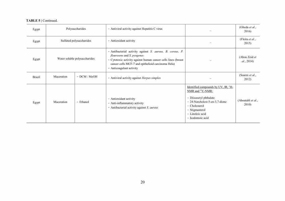
20
Egypt Polysaccharides - Antiviral activity against Hepatitis C virus _ (Gheda et al., 2016)
Egypt Sulfated polysaccharides - Antioxidant activity _ (Fleita et al., 2015)
Egypt Water soluble polysaccharides
- Antibacterial activity against S. aureus, B. cereus, P. flouresens and S. pyogenes
- Cytotoxic activity against human cancer cells lines (breast cancer cells MCF-7 and epithelioid carcinoma Hela)
- Anticoagulant activity
_ (Abou Zeid et al., 2014)
Brazil Maceration - DCM : MeOH - Antiviral activity against Herpes simplex _ (Soares et al.,
2012)
Egypt Maceration - Ethanol - Antioxidant activity - Anti-inflammatory activity - Antibacterial activity against S. aureus
Identified compounds by UV, IR, 1H-NMR and 13C-NMR:
- Diisooctyl phthalate - 24-Norcholest-5-en-3,7-dione - Cholesterol - Stigmasterol - Linoleic acid - Isodomoic acid
(Aboutabl et al., 2010)
TABLE 5 | Continued.
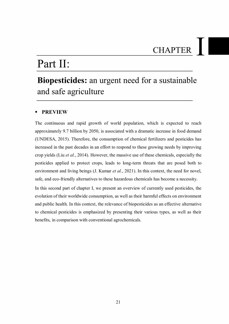
21
PREVIEW
The continuous and rapid growth of world population, which is expected to reach
approximately 9.7 billion by 2050, is associated with a dramatic increase in food demand
(UNDESA, 2015). Therefore, the consumption of chemical fertilizers and pesticides has
increased in the past decades in an effort to respond to these growing needs by improving
crop yields (Liu et al., 2014). However, the massive use of these chemicals, especially the
pesticides applied to protect crops, leads to long-term threats that are posed both to
environment and living beings (J. Kumar et al., 2021). In this context, the need for novel,
safe, and eco-friendly alternatives to these hazardous chemicals has become a necessity.
In this second part of chapter I, we present an overview of currently used pesticides, the
evolution of their worldwide consumption, as well as their harmful effects on environment
and public health. In this context, the relevance of biopesticides as an effective alternative
to chemical pesticides is emphasized by presenting their various types, as well as their
benefits, in comparison with conventional agrochemicals.
I Part II: Biopesticides: an urgent need for a sustainable and safe agriculture
CHAPTER
Chapter I – Bibliography Part II – Biopesticides: an urgent need for sustainable and safe agriculture
22
II. PESTICIDES: A DOUBLE-EDGED SWORD
II.1.1 History of pesticides consumption
For many thousands of years, the protection of farmed crops against all kinds of pests and
diseases has posed a main concern to ancient peoples who have focused on the use of easily
obtained and available remedies. The first records of the use of sulphur-containing
compounds to combat insects and mites date back over 4500 years. At that time, the
phytosanitary products used were mostly of natural origin such as the pyrethrum, derived
from the dried flowers of Chrysanthemum cinerariaefolium, used for over 2000 years
(Unsworth, 2010).
In 1939, the discovery of the insecticidal effect of dichloro-diphenyl-trichloroethane
(DDT) by the Swiss chemist Paul Hermann Müller, who was awarded a Nobel Prize for
this finding, has presented a remarkable shift in this field. In 1940 and during World War
II (1939 - 1945), the use of synthetic chemical pesticides such as DDT, aldrin, parathion…,
has peaked in order to boost food production (Gyawali, 2018).
For some decades, the application of pesticides in agriculture was considered beneficial
and no interest was shown for the eventual risks of these agrochemicals on public health
and environment. However, the book “Silent Spring”, published by the American marine
biologist Rachel Carson in 1962, in which she outlined the harmful effects of the
indiscriminate use of synthetic pesticides, has reversed the international policy leading to
the banning of DDT use in 1972 in the United States, and later in other countries (Carson,
1962 ; Gyawali, 2018). This paved the way for the research of novel safer and eco-friendly
products for agriculture and for other uses including domestic one.
II.1.2 Pesticides
A pesticide is defined as a substance, or mixture of compounds, employed to prevent,
eradicate and/or repel pests. This term comprises insecticides (insect pest control),
herbicides (parasitic plants control), fungicides (harmful microbes control) and any other
substance used to control pests (Gyawali, 2018). According to the Food and Agricultural
Organization of the United Nations (FAO), the worldwide use of pesticides is continuously
increasing over the years, especially in Asian countries (Figure 6) (FAO, 2021).
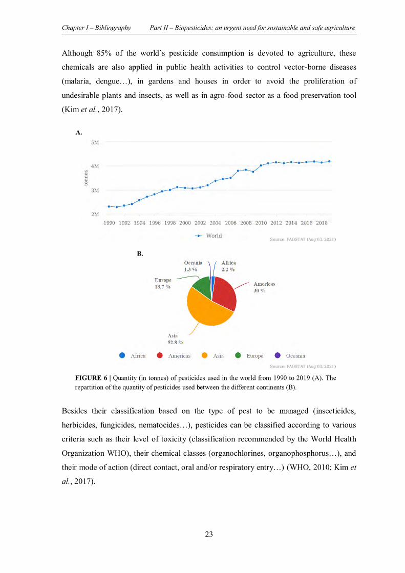
Chapter I – Bibliography Part II – Biopesticides: an urgent need for sustainable and safe agriculture
23
Although 85% of the world’s pesticide consumption is devoted to agriculture, these
chemicals are also applied in public health activities to control vector-borne diseases
(malaria, dengue…), in gardens and houses in order to avoid the proliferation of
undesirable plants and insects, as well as in agro-food sector as a food preservation tool
(Kim et al., 2017).
Besides their classification based on the type of pest to be managed (insecticides,
herbicides, fungicides, nematocides…), pesticides can be classified according to various
criteria such as their level of toxicity (classification recommended by the World Health
Organization WHO), their chemical classes (organochlorines, organophosphorus…), and
their mode of action (direct contact, oral and/or respiratory entry…) (WHO, 2010; Kim et
al., 2017).
A.
B.
FIGURE 6 | Quantity (in tonnes) of pesticides used in the world from 1990 to 2019 (A). The repartition of the quantity of pesticides used between the different continents (B).
Chapter I – Bibliography Part II – Biopesticides: an urgent need for sustainable and safe agriculture
24
II.1.3 Pesticides: undeniable harmful effects
Despite the beneficial outcomes of the use of pesticides in terms of productivity, agriculture
commodity, control of vector-borne diseases…, their excessive and unconscious
application has led to serious repercussions on the environment and consequently on public
health (Gyawali, 2018). Among pesticides, insecticides are known to be the most intensely
toxic which has prompted the banning of many of these agrochemicals, especially those
belonging to organochlorines class (Rani et al., 2021).
II.1.3.1 Adverse effects of pesticides use on the environment
While pesticides are designed to target a particular group of pests, harmful effects on non-
targeted fauna and flora including birds, fish, and beneficial insects, as well as on the
various environmental media (soil, water, and air) are known to occur (Rani et al., 2021).
In fact, it has been estimated that only 5% of the applied pesticides reach the targeted pests,
while more than 95% of these used agrochemicals are able to end up in non-targeted
organisms and to disperse and accumulate in the environment, which significantly affects
the biodiversity (Farcas et al., 2013).
The accumulation of some pesticides in the environment, such as aldrin and chlordane
which belong to the organochlorines class, is due to their content in persistent organic
pollutants (POPs). These compounds resist degradation and therefore persist for many
years in soils and sediments and can bioconcentrate by up to 70 000-fold relative to their
initial concentration (Kim et al., 2017).
Soil contamination by pesticide residues is associated with a considerable decrease in
beneficial soil microorganisms leading to a decline in its quality, fertility and productivity
(Rani et al., 2021). On the other hand, the high ability of volatile pesticides (around 80 -
90% of the used pesticides) to spread rapidly in the atmosphere leads to the disruption of
the whole ecosystem (Gyawali, 2018). In addition, the hazard of the excessive use of
pesticides also occurs in the aquatic environment contaminated through runoff, drift and
draining. Indeed, the contamination of surface waters by these harmful agrochemicals
affects aquatic species at different trophic levels, mainly by decreasing the concentrations
of dissolved oxygen (Rani et al., 2021). Moreover, groundwater poisoning by pesticides is
known to be a worldwide issue once contaminated, it can require many years for
contamination to dissipate or to be cleaned up by expensive and complex techniques
(Gyawali, 2018).
Chapter I – Bibliography Part II – Biopesticides: an urgent need for sustainable and safe agriculture
25
II.1.3.2 Adverse effects of pesticides use on human health
Due to their long-term persistence in the environment as well as to their high ability to
accumulate in the food chain, the continuous application of conventional chemical
pesticides poses a serious threat to public health (Rani et al., 2021). Unfortunately,
according to the report of the United Nations Environment Programme (UNEP),
approximately 385 million cases of accidental pesticides-related intoxication occur every
year, with almost 11,000 deaths (UNEP, 2021).
Exposure to pesticides can happen via various routes such as (Kim et al., 2017):
(1) Inhalation of volatile components of pesticides,
(2) Penetration of pesticides into the organism through ingestion, known as the most
severe way of pesticides poisoning,
(3) Dermal absorption of chemical pesticides, the most common and effective route
of exposure especially for pesticide applicators.
Exposure to pesticides can lead to both acute and chronic illnesses, whose severity depends
not only on the toxicity of agrochemicals, but also on the intake dose, the route of exposure,
the duration of exposure as well as on the age since children and elderly people are more
sensitive than others (Kim et al., 2017). In fact, several studies have highlighted the relation
between pesticides exposure and various health disorders such as cancer, diabetes,
respiratory (asthma, bronchitis…) and neurological (Parkinson, Alzheimer…) issues, as
well as reproductive syndromes (Rani et al., 2021). In this context, Shah et al., have
demonstrated the link between pesticide exposure and the increased risk of Epithelial
Ovarian Cancer by showing the ability of some organochlorine pesticides such as
β-hexachlorocyclohexane and Dieldrin to stimulate the production of cellular ROS
(reactive oxygen species), to induce an inflammatory response, as well as to damage the
DNA of human ovary surface epithelial cells (Shah et al., 2020). In addition, pesticides can
create critical problems for pregnant women due to their ability to cross the placenta
resulting in structural and functional defects in the foetus (Woodruff et al., 2008).
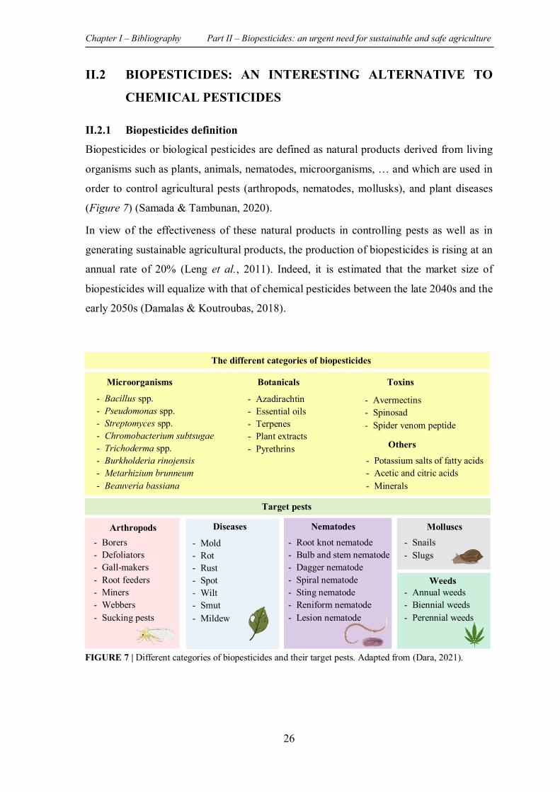
Chapter I – Bibliography Part II – Biopesticides: an urgent need for sustainable and safe agriculture
26
II.2 BIOPESTICIDES: AN INTERESTING ALTERNATIVE TO
CHEMICAL PESTICIDES
II.2.1 Biopesticides definition
Biopesticides or biological pesticides are defined as natural products derived from living
organisms such as plants, animals, nematodes, microorganisms, … and which are used in
order to control agricultural pests (arthropods, nematodes, mollusks), and plant diseases
(Figure 7) (Samada & Tambunan, 2020).
In view of the effectiveness of these natural products in controlling pests as well as in
generating sustainable agricultural products, the production of biopesticides is rising at an
annual rate of 20% (Leng et al., 2011). Indeed, it is estimated that the market size of
biopesticides will equalize with that of chemical pesticides between the late 2040s and the
early 2050s (Damalas & Koutroubas, 2018).
Microorganisms
- Bacillus spp. - Pseudomonas spp. - Streptomyces spp. - Chromobacterium subtsugae - Trichoderma spp. - Burkholderia rinojensis - Metarhizium brunneum - Beauveria bassiana
Botanicals
- Azadirachtin - Essential oils - Terpenes - Plant extracts - Pyrethrins
Toxins
- Avermectins - Spinosad - Spider venom peptide
Others
- Potassium salts of fatty acids - Acetic and citric acids - Minerals
The different categories of biopesticides
Target pests
Arthropods - Borers - Defoliators - Gall-makers - Root feeders - Miners - Webbers - Sucking pests
Diseases
- Mold - Rot - Rust - Spot - Wilt - Smut - Mildew
Nematodes
- Root knot nematode - Bulb and stem nematode - Dagger nematode - Spiral nematode - Sting nematode - Reniform nematode - Lesion nematode
Molluscs - Snails - Slugs
Weeds - Annual weeds - Biennial weeds - Perennial weeds
FIGURE 7 | Different categories of biopesticides and their target pests. Adapted from (Dara, 2021).
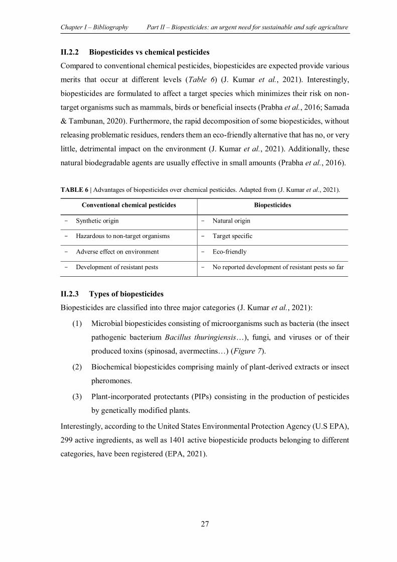
Chapter I – Bibliography Part II – Biopesticides: an urgent need for sustainable and safe agriculture
27
II.2.2 Biopesticides vs chemical pesticides
Compared to conventional chemical pesticides, biopesticides are expected provide various
merits that occur at different levels (Table 6) (J. Kumar et al., 2021). Interestingly,
biopesticides are formulated to affect a target species which minimizes their risk on non-
target organisms such as mammals, birds or beneficial insects (Prabha et al., 2016; Samada
& Tambunan, 2020). Furthermore, the rapid decomposition of some biopesticides, without
releasing problematic residues, renders them an eco-friendly alternative that has no, or very
little, detrimental impact on the environment (J. Kumar et al., 2021). Additionally, these
natural biodegradable agents are usually effective in small amounts (Prabha et al., 2016).
TABLE 6 | Advantages of biopesticides over chemical pesticides. Adapted from (J. Kumar et al., 2021).
II.2.3 Types of biopesticides
Biopesticides are classified into three major categories (J. Kumar et al., 2021):
(1) Microbial biopesticides consisting of microorganisms such as bacteria (the insect
pathogenic bacterium Bacillus thuringiensis…), fungi, and viruses or of their
produced toxins (spinosad, avermectins…) (Figure 7).
(2) Biochemical biopesticides comprising mainly of plant-derived extracts or insect
pheromones.
(3) Plant-incorporated protectants (PIPs) consisting in the production of pesticides
by genetically modified plants.
Interestingly, according to the United States Environmental Protection Agency (U.S EPA),
299 active ingredients, as well as 1401 active biopesticide products belonging to different
categories, have been registered (EPA, 2021).
Conventional chemical pesticides Biopesticides
- Synthetic origin - Natural origin
- Hazardous to non-target organisms - Target specific
- Adverse effect on environment - Eco-friendly
- Development of resistant pests - No reported development of resistant pests so far
Chapter I – Bibliography Part II – Biopesticides: an urgent need for sustainable and safe agriculture
28
II.2.3.1 Microbial biopesticides
The search for biopesticides of microbial origin, in which microorganisms act as active
ingredients used to control plant diseases and pests, has attracted an extensive attention
over the last decades.
Different kinds of pests can be controlled by microbial biopesticides, although each active
ingredient is relatively specific to a particular species (Kesho, 2020; Thakura et al., 2020).
Owing to their high efficiency, specificity, as well as to their environmental friendliness,
microbial biopesticides market accounts for 90% of total biopesticides (Kesho, 2020).
The most widely known microorganism in the development of microbial biopesticides, and
the one that paved the way for novel discoveries is the insect pathogenic bacterium Bacillus
thuringiensis. The insecticidal activity of this bacterium is expressed by its ability to
produce toxins (crystal proteins), which once ingested by the target insect, induces its death
through lysis of its gut cells. Interestingly, B. thuringiensis-based biopesticide is
characterized by its host-specificity with a limited chances to affect non-target organisms
(P. Kumar et al., 2021).
II.2.3.2 Biochemical biopesticides
Biochemical pesticides include chemical compounds recovered from living organisms such
as those extracted from plants, as well as pheromones produced by insects (Thakura et al.,
2020).
Plants are naturally able to produce a wide variety of secondary metabolites (flavonoids,
phenols, alkaloids, terpenes…) in order to protect themselves against pests and microbial
attacks (Prabha et al., 2016). Therefore, in the last few years, plant-based extracts have
occupied a prominent place in the search for alternatives to conventional chemical
pesticides (J. Kumar et al., 2021). In this context, about 2400 plant species have been
reported for their wide range of action against pests. These exhibit various mechanisms of
action: repellents, antifeedants, ovicidal, larvicidal effects,… (Thakura et al., 2020; J.
Kumar et al., 2021). Azadirachta indica, commonly called neem and with azadirachtin as
the most important constituent, is a well-known example of plant possessing considerable
insecticidal proprieties (Chaudhary et al., 2017). Indeed, nearly 195 insect species, even
those that have developed a resistance to conventional pesticides, have shown a sensibility
towards neem-based products. Interestingly, beneficial insects such as pollinator insects
are not harmed by the application of neem-based biopesticides (Thakura et al., 2020).
Chapter I – Bibliography Part II – Biopesticides: an urgent need for sustainable and safe agriculture
29
On the other hand, pheromones, chemical compounds produced and dispersed by insects
as a chemical signal that induce a sexual response, provide a promising strategy for
controlling insect pests (Rizvi et al., 2021). These chemical signals are not considered as
true “insecticides” since they do not kill pests, but their mode of action is based on the
perturbation of insect’s behavior (attract and capture insects, mating disruption, mass
trapping…) by acting on their olfactory system (J. Kumar et al., 2021).
The application of insect pheromones in pests control is recognized as a new ecological
concepts (respectful approach to the environment, as well as to the public health) with
recent applications in mosquito control (Wooding et al., 2020; Rizvi et al., 2021).
II.2.3.3 Plant-incorporated protectants (PIPs)
PIPs are a category of biopesticides that are expressed and produced by genetically
modified plants through the incorporation of an exogenous genetic material. Thus, the
genetically modified crops are able to protect themselves from harmful pests by releasing
toxic compounds. The integration of transgenes, encoding for toxic crystal proteins, from
the insect pathogenic bacterium Bacillus thuringiensis into the modified plants is well-
known as the first generation insecticidal PIPs (Parker & Sander, 2017).
II.2.4 Marine world: a valuable and promising source of biopesticides
The marine world, characterized by unique environmental conditions harbors a wide
variety of organisms that are considered as a valuable source of unique and bioactive
natural products (Hamed et al., 2015). Besides the great interest given to marine natural
products in the search for novel drugs, various studies have revealed their expanded
capacity to present alternatives to conventional chemical pesticides (Jimenez, 2018; Song
et al., 2021).
II.2.4.1 Seaweed as a potential source of biopesticides
Although most researches focused on the possible pharmaceutical applications of
seaweeds, several studies have highlighted the ability of these marine organisms to be
valorized in different ways in the agricultural field. Indeed, in view of their richness in
mineral substances, vitamins, amino acids, as well as in plant growth regulators (cytokinin,
auxin, gibberellins…), various seaweed derived extracts have exhibited a high capacity to
stimulate plant growth and productivity (Hamed et al., 2018). Moreover, an improvement
in soil chemical and physical proprieties has also been attributed to algae which can be
used as biofertilizers as well as soil stabilizers (Nabti et al., 2016).
Chapter I – Bibliography Part II – Biopesticides: an urgent need for sustainable and safe agriculture
30
On the other hand, biocidal properties such as microbicidal, virucidal, nematocidal, and
insecticidal activities of seaweed derived crude extracts and/or purified compounds have
been documented in several studies paving the way for their potential application in the
control of plant pathogens and pests (Hamed et al., 2018). In this context, Esserti et al.
(2016) have demonstrated the ability of pulverized aqueous extracts derived from the two
brown algae, Fucus spiralis and Cystoseira myriophylloides, to reduce the damage caused
on tomato plants by two phytopathogenes: Agrobacterium tumefaciens bacterium and
Verticillium dahlia fungus (Esserti et al., 2016). In addition, Baloch et al. (2013) showed
that mixing the soil with the powder of Spatoglossum variabile (brown alga), Stokeyia
indica (brown alga), or of Melanothamnus afaqhusainii (red alga) significantly reduced the
infection of watermelon and eggplant roots by the root knot nematode Meloidogyne
incognita (Baloch et al., 2013).
II.2.4.1.1 Seaweed derived extracts/compounds with insecticidal activity
The promising role of seaweed as a valuable source of bioactive molecules with interesting
insecticidal activity has been evidenced in various studies (Yu et al., 2014; Hamed et al.,
2018). However, for the moment, there are no commercial algae-based products used in
the control of phytopathogenic insects (Machado et al., 2019). This can be explained in
part by the fact that most studies are limited to laboratory scale in vitro screenings, without
proceeding to field trials (Hamed et al., 2018; Machado et al., 2019). Therefore, advanced
investigations in the research of seaweed-based insecticidal agents are still needed in order
to benefit from these marine organisms in the formulation of eco-friendly alternatives to
conventional insecticides. Furthermore, the target specificity of these potential
bioinsecticides must be assessed so as to avoid any possible adverse effects on beneficial
and non-target organisms including humans (Yu et al., 2014).
A non-exhaustive summary of extracts and compounds derived from seaweed that
exhibited an insecticidal activity (through various modes of application) against
phytopathogenic insects as well as disease vectors, are listed in the table below (Table 7)
Interestingly, a synergistic insecticidal activity between seaweed extracts and synthetic
insecticides was also documented in an early study (Thangam & Kathiresan, 1991).
Although these potential synergistic effects between seaweed-derived products and
conventional synthetic insecticides are not sufficiently exploited, their combination will
undoubtedly lead to a reduction in insecticides consumption and consequently in their
adverse effects.
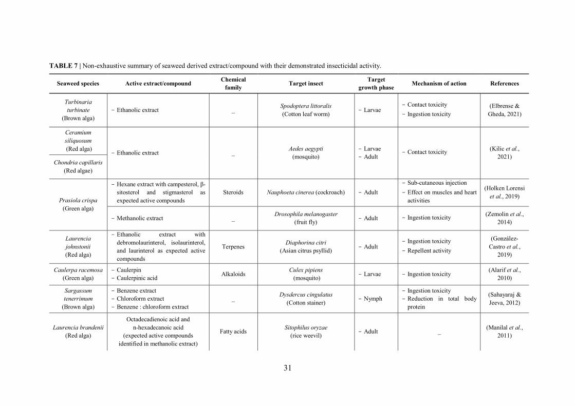
31
TABLE 7 | Non-exhaustive summary of seaweed derived extract/compound with their demonstrated insecticidal activity.
Seaweed species Active extract/compound Chemical family Target insect Target
growth phase Mechanism of action References
Turbinaria turbinate
(Brown alga) - Ethanolic extract _ Spodoptera littoralis
(Cotton leaf worm) - Larvae - Contact toxicity - Ingestion toxicity
(Elbrense & Gheda, 2021)
Ceramium siliquosum (Red alga)
- Ethanolic extract _ Aedes aegypti (mosquito)
- Larvae - Adult
- Contact toxicity (Kilic et al., 2021)
Chondria capillaris (Red algae)
Prasiola crispa (Green alga)
- Hexane extract with campesterol, β-sitosterol and stigmasterol as expected active compounds
Steroids Nauphoeta cinerea (cockroach) - Adult - Sub-cutaneous injection - Effect on muscles and heart
activities
(Holken Lorensi et al., 2019)
- Methanolic extract _ Drosophila melanogaster (fruit fly) - Adult - Ingestion toxicity (Zemolin et al.,
2014)
Laurencia johnstonii (Red alga)
- Ethanolic extract with debromolaurinterol, isolaurinterol, and laurinterol as expected active compounds
Terpenes Diaphorina citri (Asian citrus psyllid) - Adult
- Ingestion toxicity - Repellent activity
(González-Castro et al.,
2019)
Caulerpa racemosa (Green alga)
- Caulerpin - Caulerpinic acid Alkaloids Culex pipiens
(mosquito) - Larvae - Ingestion toxicity (Alarif et al., 2010)
Sargassum tenerrimum
(Brown alga)
- Benzene extract - Chloroform extract - Benzene : chloroform extract
_ Dysdercus cingulatus (Cotton stainer) - Nymph
- Ingestion toxicity - Reduction in total body
protein
(Sahayaraj & Jeeva, 2012)
Laurencia brandenii (Red alga)
Octadecadienoic acid and n-hexadecanoic acid
(expected active compounds identified in methanolic extract)
Fatty acids Sitophilus oryzae (rice weevil) - Adult _ (Manilal et al.,
2011)
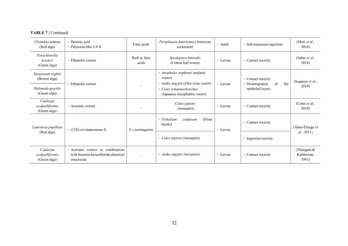
32
Chondria armata (Red alga)
- Domoic acid - Palytoxin-like CA II Fatty acids Periplaneta Americana (American
cockroach) - Adult - Sub-cutaneous injection (Mori et al., 2016)
Parachlorella kessleri
(Green alga) - Ethanolic extract Rich in fatty
acids Spodoptera littoralis (Cotton leaf-worm) - Larvae - Contact toxicity (Saber et al.,
2018)
Sargassum wightii (Brown alga)
- Ethanolic extract _
- Anopheles stephensi (malaria vector)
- Aedes aegypti (Zika virus vector) - Culex tritaeniorhynchus
(Japanese encephalitis vector)
- Larvae - Contact toxicity - Disintegration of the
epithelial layers
(Suganya et al., 2019) Halimeda gracilis
(Green alga)
Caulerpa scalpelliformis (Green alga)
- Acetonic extract _ Culex pipiens (mosquito) - Larvae - Contact toxicity (Cetin et al.,
2010)
Laurencia papillosa (Red alga) - (12E)-cis-maneonene-E C15 acetongenin
- Tribolium confusum (Flour beetle)
- Larvae
- Contact toxicity (Abou-Elnaga et
al., 2011) - Culex pipiens (mosquito) - Ingestion toxicity
Caulerpa scalpelliformis (Green alga)
- Acetonic extract in combination with benzene hexachloride chemical insecticide
_ - Aedes aegypti (mosquito) - Larvae - Contact toxicity (Thangam & Kathiresan,
1991)
TABLE 7 | Continued.
33
PREVIEW
It is obvious that the microbial community known as “Biofilm” is an ubiquitous and
complex structure widely distributed on all kinds of imaginable biotic and abiotic surfaces:
metal, plastic, natural materials (rocks…), medical devices, living tissues, kitchen counters,
contact lenses…Although some microbial communities can be beneficial in particular
fields, some biofilms, especially those formed by pathogenic microorganisms, present a
veritable public health issue, hence the necessity to look for strategies for overcoming them.
In this third part of chapter I, an overview of biofilm, particularly bacterial biofilms,
including its development stages and its major components is presented with emphasis on
two well-known biofilm former opportunistic/pathogenic bacteria: Pseudomonas
aeruginosa and Staphylococcus aureus. The Quorum Sensing (QS) cell-to-cell
communication system, which is strongly implicated in biofilm formation and
maintenance, is also described. Mechanisms responsible for biofilm resilience are then
detailed along with the main biofilms-associated diseases.
On the other hand, the therapeutic approaches, as well as the place occupied by natural
medicine in the search for effective antibiofilm agents are discussed. At the end of this part,
experimental techniques frequently adopted in the evaluation of the antibiofilm activity are
discussed.
P. aeruginosa, bacterium of interest, is a Gram-negative bacillus, aerobic, motile and non-
spore forming rods, isolated for the first time in 1882 from green pus. This bacterium
belonging to the order of Pseudomonadales and to the family of Pseudomonadaceae is
I Part III: Biofilms: a microbial assemblage of scientific significance “Focus on two highly critical pathogenic bacteria”
CHAPTER
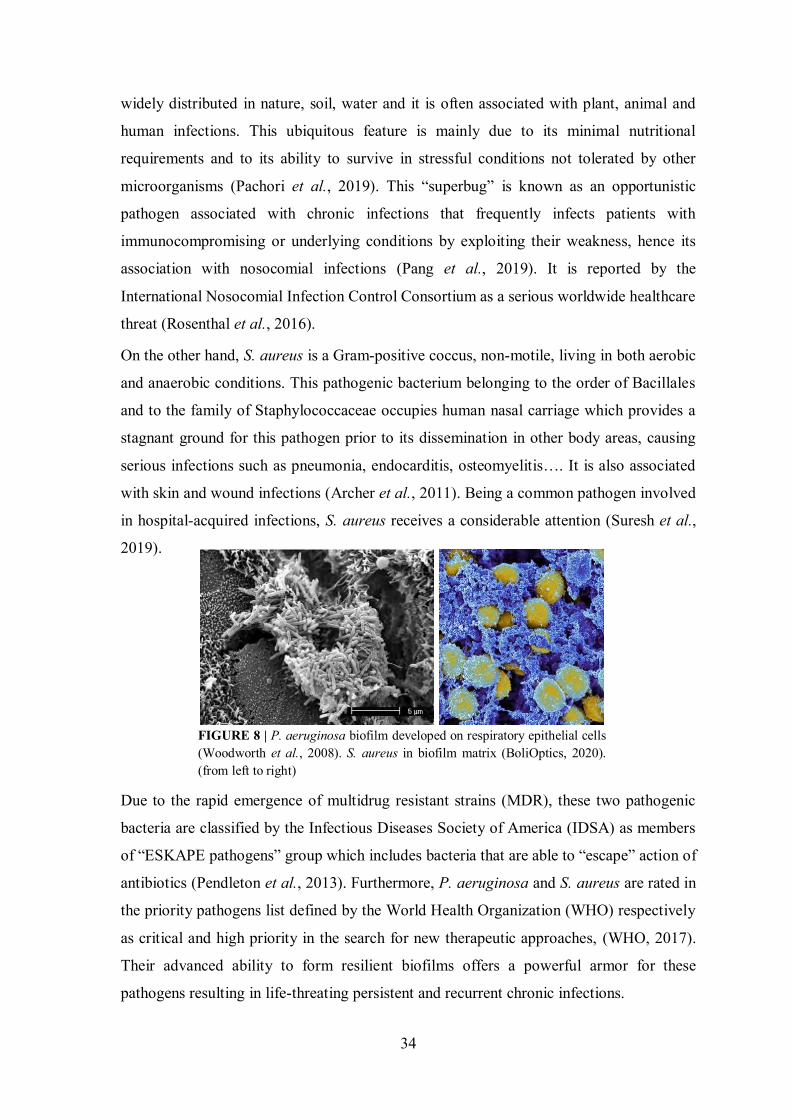
34
widely distributed in nature, soil, water and it is often associated with plant, animal and
human infections. This ubiquitous feature is mainly due to its minimal nutritional
requirements and to its ability to survive in stressful conditions not tolerated by other
microorganisms (Pachori et al., 2019). This “superbug” is known as an opportunistic
pathogen associated with chronic infections that frequently infects patients with
immunocompromising or underlying conditions by exploiting their weakness, hence its
association with nosocomial infections (Pang et al., 2019). It is reported by the
International Nosocomial Infection Control Consortium as a serious worldwide healthcare
threat (Rosenthal et al., 2016).
On the other hand, S. aureus is a Gram-positive coccus, non-motile, living in both aerobic
and anaerobic conditions. This pathogenic bacterium belonging to the order of Bacillales
and to the family of Staphylococcaceae occupies human nasal carriage which provides a
stagnant ground for this pathogen prior to its dissemination in other body areas, causing
serious infections such as pneumonia, endocarditis, osteomyelitis…. It is also associated
with skin and wound infections (Archer et al., 2011). Being a common pathogen involved
in hospital-acquired infections, S. aureus receives a considerable attention (Suresh et al.,
2019).
Due to the rapid emergence of multidrug resistant strains (MDR), these two pathogenic
bacteria are classified by the Infectious Diseases Society of America (IDSA) as members
of “ESKAPE pathogens” group which includes bacteria that are able to “escape” action of
antibiotics (Pendleton et al., 2013). Furthermore, P. aeruginosa and S. aureus are rated in
the priority pathogens list defined by the World Health Organization (WHO) respectively
as critical and high priority in the search for new therapeutic approaches, (WHO, 2017).
Their advanced ability to form resilient biofilms offers a powerful armor for these
pathogens resulting in life-threating persistent and recurrent chronic infections.
FIGURE 8 | P. aeruginosa biofilm developed on respiratory epithelial cells (Woodworth et al., 2008). S. aureus in biofilm matrix (BoliOptics, 2020). (from left to right)
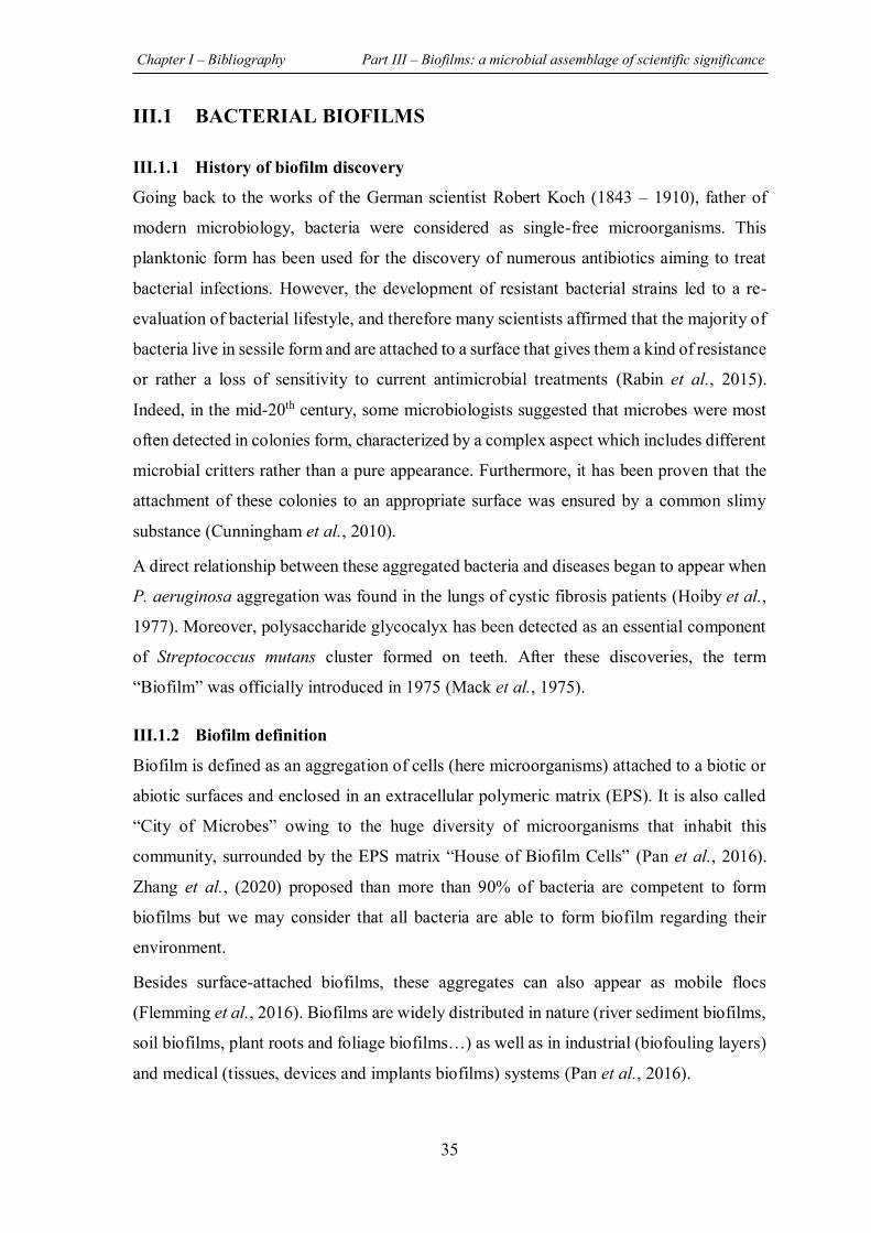
Chapter I – Bibliography Part III – Biofilms: a microbial assemblage of scientific significance
35
III.1 BACTERIAL BIOFILMS
III.1.1 History of biofilm discovery
Going back to the works of the German scientist Robert Koch (1843 – 1910), father of
modern microbiology, bacteria were considered as single-free microorganisms. This
planktonic form has been used for the discovery of numerous antibiotics aiming to treat
bacterial infections. However, the development of resistant bacterial strains led to a re-
evaluation of bacterial lifestyle, and therefore many scientists affirmed that the majority of
bacteria live in sessile form and are attached to a surface that gives them a kind of resistance
or rather a loss of sensitivity to current antimicrobial treatments (Rabin et al., 2015).
Indeed, in the mid-20th century, some microbiologists suggested that microbes were most
often detected in colonies form, characterized by a complex aspect which includes different
microbial critters rather than a pure appearance. Furthermore, it has been proven that the
attachment of these colonies to an appropriate surface was ensured by a common slimy
substance (Cunningham et al., 2010).
A direct relationship between these aggregated bacteria and diseases began to appear when
P. aeruginosa aggregation was found in the lungs of cystic fibrosis patients (Hoiby et al.,
1977). Moreover, polysaccharide glycocalyx has been detected as an essential component
of Streptococcus mutans cluster formed on teeth. After these discoveries, the term
“Biofilm” was officially introduced in 1975 (Mack et al., 1975).
III.1.2 Biofilm definition
Biofilm is defined as an aggregation of cells (here microorganisms) attached to a biotic or
abiotic surfaces and enclosed in an extracellular polymeric matrix (EPS). It is also called
“City of Microbes” owing to the huge diversity of microorganisms that inhabit this
community, surrounded by the EPS matrix “House of Biofilm Cells” (Pan et al., 2016).
Zhang et al., (2020) proposed than more than 90% of bacteria are competent to form
biofilms but we may consider that all bacteria are able to form biofilm regarding their
environment.
Besides surface-attached biofilms, these aggregates can also appear as mobile flocs
(Flemming et al., 2016). Biofilms are widely distributed in nature (river sediment biofilms,
soil biofilms, plant roots and foliage biofilms…) as well as in industrial (biofouling layers)
and medical (tissues, devices and implants biofilms) systems (Pan et al., 2016).
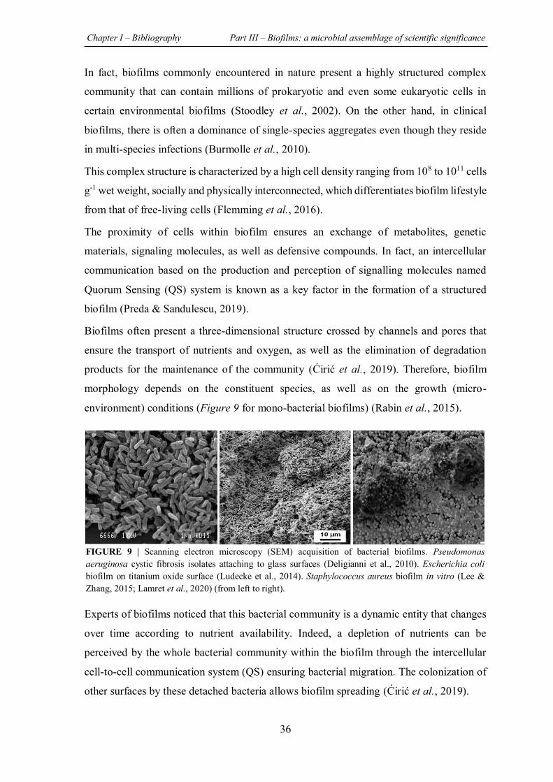
Chapter I – Bibliography Part III – Biofilms: a microbial assemblage of scientific significance
36
In fact, biofilms commonly encountered in nature present a highly structured complex
community that can contain millions of prokaryotic and even some eukaryotic cells in
certain environmental biofilms (Stoodley et al., 2002). On the other hand, in clinical
biofilms, there is often a dominance of single-species aggregates even though they reside
in multi-species infections (Burmolle et al., 2010).
This complex structure is characterized by a high cell density ranging from 108 to 1011 cells
g-1 wet weight, socially and physically interconnected, which differentiates biofilm lifestyle
from that of free-living cells (Flemming et al., 2016).
The proximity of cells within biofilm ensures an exchange of metabolites, genetic
materials, signaling molecules, as well as defensive compounds. In fact, an intercellular
communication based on the production and perception of signalling molecules named
Quorum Sensing (QS) system is known as a key factor in the formation of a structured
biofilm (Preda & Sandulescu, 2019).
Biofilms often present a three-dimensional structure crossed by channels and pores that
ensure the transport of nutrients and oxygen, as well as the elimination of degradation
products for the maintenance of the community (Ćirić et al., 2019). Therefore, biofilm
morphology depends on the constituent species, as well as on the growth (micro-
environment) conditions (Figure 9 for mono-bacterial biofilms) (Rabin et al., 2015).
Experts of biofilms noticed that this bacterial community is a dynamic entity that changes
over time according to nutrient availability. Indeed, a depletion of nutrients can be
perceived by the whole bacterial community within the biofilm through the intercellular
cell-to-cell communication system (QS) ensuring bacterial migration. The colonization of
other surfaces by these detached bacteria allows biofilm spreading (Ćirić et al., 2019).
FIGURE 9 | Scanning electron microscopy (SEM) acquisition of bacterial biofilms. Pseudomonas aeruginosa cystic fibrosis isolates attaching to glass surfaces (Deligianni et al., 2010). Escherichia coli biofilm on titanium oxide surface (Ludecke et al., 2014). Staphylococcus aureus biofilm in vitro (Lee & Zhang, 2015; Lamret et al., 2020) (from left to right).
Chapter I – Bibliography Part III – Biofilms: a microbial assemblage of scientific significance
37
III.1.3 Biofilms: Bad or good?!
Biofilms have been known for long time as a detrimental structure responsible for
significant problems in clinical, as well as in industrial fields. Indeed, according to the
National Institutes of Health (NIH), bacterial biofilm is implicated in almost 65% of
microbial diseases and in more than 80% of chronic infections (Olivares et al., 2020). In
addition to human infections (blood-stream, urinary tract infections…), biofilms are able
to colonize all higher organisms such as plants and animals, leading to persistent invasions.
The harm of biofilms is also presented by their ability to contaminate medical devices and
implants, water systems as well as to spoil food (Hall-Stoodley et al., 2004; Flemming et
al., 2016; Galie et al., 2018). Besides its harmful impact on public health, biofilm formation
leads to serious economic losses by damaging industrial equipment and contaminating
various products. For all these reasons, biofilms are often considered as a socioeconomic
issue costing billions of dollars annually (Gunn et al., 2016).
Although most studies to date have focused on detrimental biofilms, different researches
have highlighted the beneficial side of biofilm formation as their role in human, animal and
vegetal ecosystems regarding healthy status (Louis et al., 2007; Deng et al., 2020) as well
as the possibility of their applications in a wide array of fields, such as food (fermented and
probiotic food…), agricultural (biofilm-based biofertilizers…), medical (probiotic and
bacteriosin produced biofilms…) and environmental (energy, bioremediation,
biogeochemical cycle…) fields (Turhan et al., 2019).
After all, biofilms are considered as a double-edged sword that can present both beneficial
and harmful effects in the same field. It all depends on the colonized surface and the species
of microorganisms, pathogenic or not, living in these communities (Turhan et al., 2019).
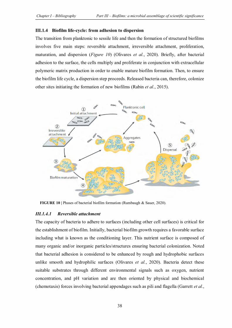
Chapter I – Bibliography Part III – Biofilms: a microbial assemblage of scientific significance
38
III.1.4 Biofilm life-cycle: from adhesion to dispersion
The transition from planktonic to sessile life and then the formation of structured biofilms
involves five main steps: reversible attachment, irreversible attachment, proliferation,
maturation, and dispersion (Figure 10) (Olivares et al., 2020). Briefly, after bacterial
adhesion to the surface, the cells multiply and proliferate in conjunction with extracellular
polymeric matrix production in order to enable mature biofilm formation. Then, to ensure
the biofilm life cycle, a dispersion step proceeds. Released bacteria can, therefore, colonize
other sites initiating the formation of new biofilms (Rabin et al., 2015).
III.1.4.1 Reversible attachment
The capacity of bacteria to adhere to surfaces (including other cell surfaces) is critical for
the establishment of biofilm. Initially, bacterial biofilm growth requires a favorable surface
including what is known as the conditioning layer. This nutrient surface is composed of
many organic and/or inorganic particles/structures ensuring bacterial colonization. Noted
that bacterial adhesion is considered to be enhanced by rough and hydrophobic surfaces
unlike smooth and hydrophilic surfaces (Olivares et al., 2020). Bacteria detect these
suitable substrates through different environmental signals such as oxygen, nutrient
concentration, and pH variation and are then oriented by physical and biochemical
(chemotaxis) forces involving bacterial appendages such as pili and flagella (Garrett et al.,
FIGURE 10 | Phases of bacterial biofilm formation (Rumbaugh & Sauer, 2020).
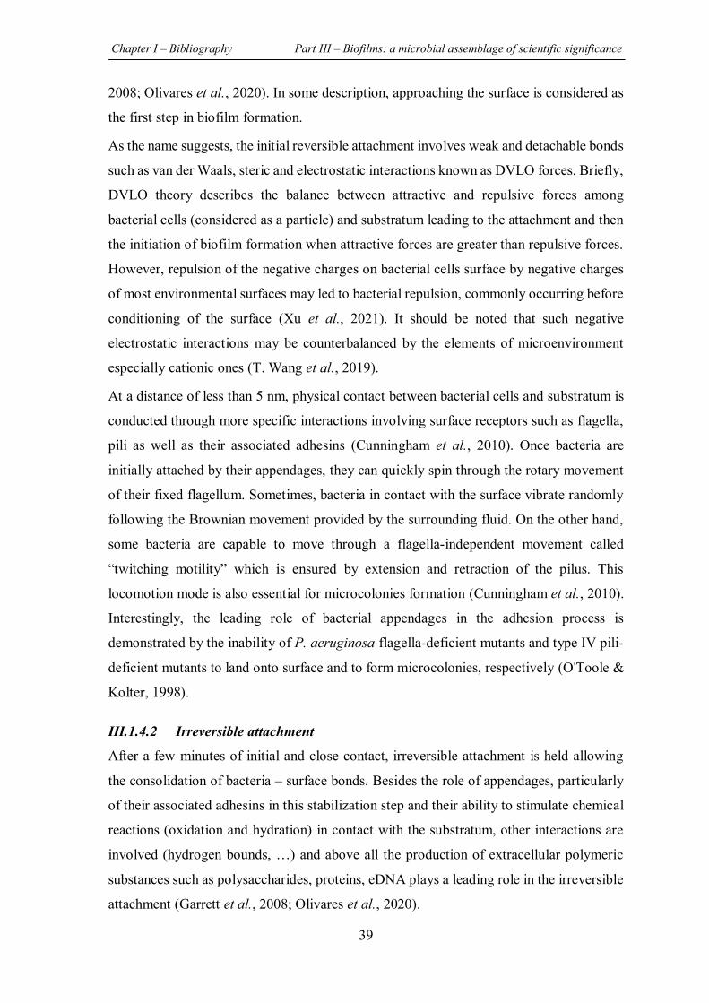
Chapter I – Bibliography Part III – Biofilms: a microbial assemblage of scientific significance
39
2008; Olivares et al., 2020). In some description, approaching the surface is considered as
the first step in biofilm formation.
As the name suggests, the initial reversible attachment involves weak and detachable bonds
such as van der Waals, steric and electrostatic interactions known as DVLO forces. Briefly,
DVLO theory describes the balance between attractive and repulsive forces among
bacterial cells (considered as a particle) and substratum leading to the attachment and then
the initiation of biofilm formation when attractive forces are greater than repulsive forces.
However, repulsion of the negative charges on bacterial cells surface by negative charges
of most environmental surfaces may led to bacterial repulsion, commonly occurring before
conditioning of the surface (Xu et al., 2021). It should be noted that such negative
electrostatic interactions may be counterbalanced by the elements of microenvironment
especially cationic ones (T. Wang et al., 2019).
At a distance of less than 5 nm, physical contact between bacterial cells and substratum is
conducted through more specific interactions involving surface receptors such as flagella,
pili as well as their associated adhesins (Cunningham et al., 2010). Once bacteria are
initially attached by their appendages, they can quickly spin through the rotary movement
of their fixed flagellum. Sometimes, bacteria in contact with the surface vibrate randomly
following the Brownian movement provided by the surrounding fluid. On the other hand,
some bacteria are capable to move through a flagella-independent movement called
“twitching motility” which is ensured by extension and retraction of the pilus. This
locomotion mode is also essential for microcolonies formation (Cunningham et al., 2010).
Interestingly, the leading role of bacterial appendages in the adhesion process is
demonstrated by the inability of P. aeruginosa flagella-deficient mutants and type IV pili-
deficient mutants to land onto surface and to form microcolonies, respectively (O'Toole &
Kolter, 1998).
III.1.4.2 Irreversible attachment
After a few minutes of initial and close contact, irreversible attachment is held allowing
the consolidation of bacteria – surface bonds. Besides the role of appendages, particularly
of their associated adhesins in this stabilization step and their ability to stimulate chemical
reactions (oxidation and hydration) in contact with the substratum, other interactions are
involved (hydrogen bounds, …) and above all the production of extracellular polymeric
substances such as polysaccharides, proteins, eDNA plays a leading role in the irreversible
attachment (Garrett et al., 2008; Olivares et al., 2020).
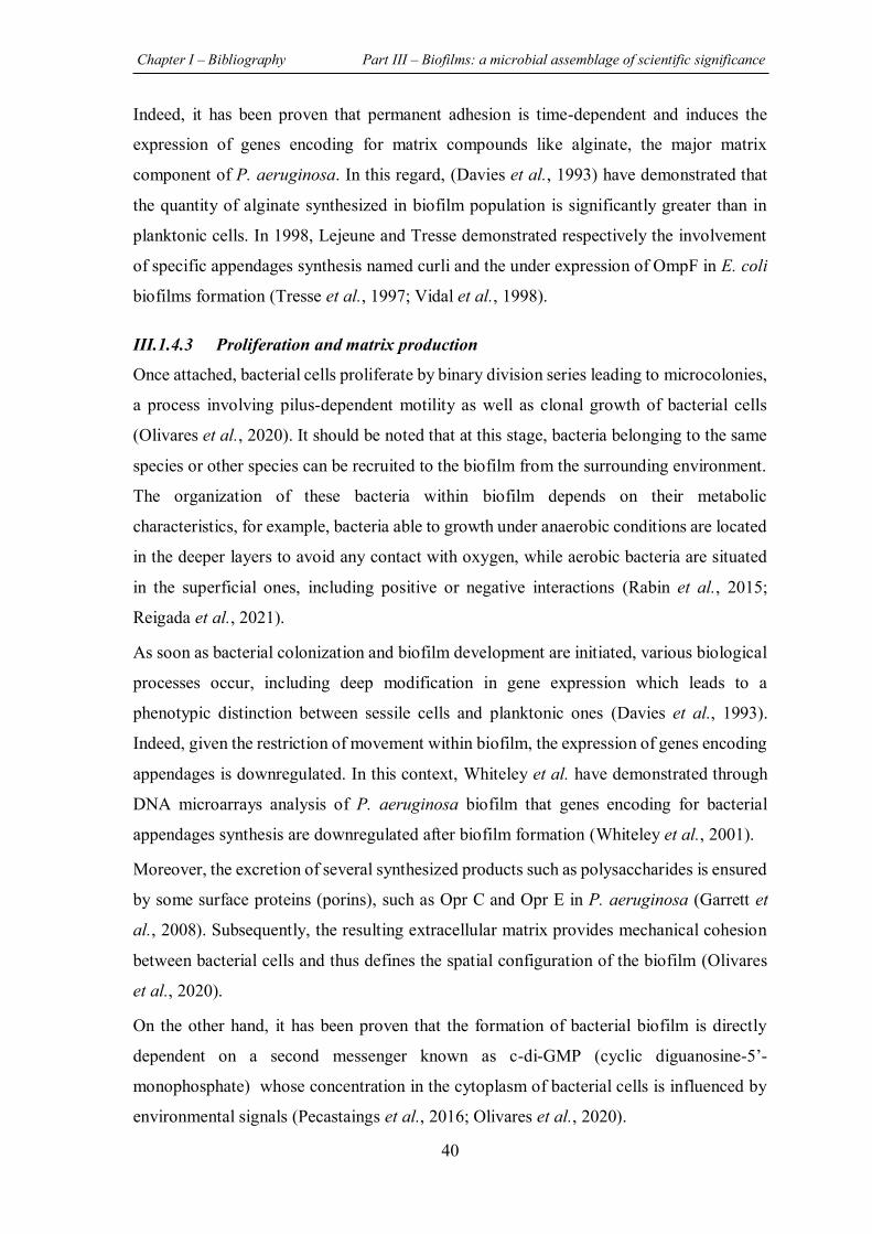
Chapter I – Bibliography Part III – Biofilms: a microbial assemblage of scientific significance
40
Indeed, it has been proven that permanent adhesion is time-dependent and induces the
expression of genes encoding for matrix compounds like alginate, the major matrix
component of P. aeruginosa. In this regard, (Davies et al., 1993) have demonstrated that
the quantity of alginate synthesized in biofilm population is significantly greater than in
planktonic cells. In 1998, Lejeune and Tresse demonstrated respectively the involvement
of specific appendages synthesis named curli and the under expression of OmpF in E. coli
biofilms formation (Tresse et al., 1997; Vidal et al., 1998).
III.1.4.3 Proliferation and matrix production
Once attached, bacterial cells proliferate by binary division series leading to microcolonies,
a process involving pilus-dependent motility as well as clonal growth of bacterial cells
(Olivares et al., 2020). It should be noted that at this stage, bacteria belonging to the same
species or other species can be recruited to the biofilm from the surrounding environment.
The organization of these bacteria within biofilm depends on their metabolic
characteristics, for example, bacteria able to growth under anaerobic conditions are located
in the deeper layers to avoid any contact with oxygen, while aerobic bacteria are situated
in the superficial ones, including positive or negative interactions (Rabin et al., 2015;
Reigada et al., 2021).
As soon as bacterial colonization and biofilm development are initiated, various biological
processes occur, including deep modification in gene expression which leads to a
phenotypic distinction between sessile cells and planktonic ones (Davies et al., 1993).
Indeed, given the restriction of movement within biofilm, the expression of genes encoding
appendages is downregulated. In this context, Whiteley et al. have demonstrated through
DNA microarrays analysis of P. aeruginosa biofilm that genes encoding for bacterial
appendages synthesis are downregulated after biofilm formation (Whiteley et al., 2001).
Moreover, the excretion of several synthesized products such as polysaccharides is ensured
by some surface proteins (porins), such as Opr C and Opr E in P. aeruginosa (Garrett et
al., 2008). Subsequently, the resulting extracellular matrix provides mechanical cohesion
between bacterial cells and thus defines the spatial configuration of the biofilm (Olivares
et al., 2020).
On the other hand, it has been proven that the formation of bacterial biofilm is directly
dependent on a second messenger known as c-di-GMP (cyclic diguanosine-5’-
monophosphate) whose concentration in the cytoplasm of bacterial cells is influenced by
environmental signals (Pecastaings et al., 2016; Olivares et al., 2020).
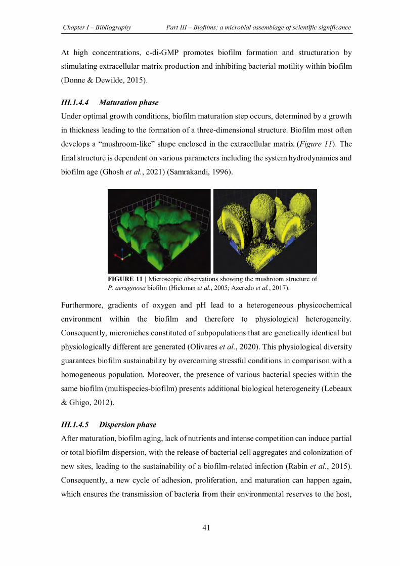
Chapter I – Bibliography Part III – Biofilms: a microbial assemblage of scientific significance
41
At high concentrations, c-di-GMP promotes biofilm formation and structuration by
stimulating extracellular matrix production and inhibiting bacterial motility within biofilm
(Donne & Dewilde, 2015).
III.1.4.4 Maturation phase
Under optimal growth conditions, biofilm maturation step occurs, determined by a growth
in thickness leading to the formation of a three-dimensional structure. Biofilm most often
develops a “mushroom-like” shape enclosed in the extracellular matrix (Figure 11). The
final structure is dependent on various parameters including the system hydrodynamics and
biofilm age (Ghosh et al., 2021) (Samrakandi, 1996).
Furthermore, gradients of oxygen and pH lead to a heterogeneous physicochemical
environment within the biofilm and therefore to physiological heterogeneity.
Consequently, microniches constituted of subpopulations that are genetically identical but
physiologically different are generated (Olivares et al., 2020). This physiological diversity
guarantees biofilm sustainability by overcoming stressful conditions in comparison with a
homogeneous population. Moreover, the presence of various bacterial species within the
same biofilm (multispecies-biofilm) presents additional biological heterogeneity (Lebeaux
& Ghigo, 2012).
III.1.4.5 Dispersion phase
After maturation, biofilm aging, lack of nutrients and intense competition can induce partial
or total biofilm dispersion, with the release of bacterial cell aggregates and colonization of
new sites, leading to the sustainability of a biofilm-related infection (Rabin et al., 2015).
Consequently, a new cycle of adhesion, proliferation, and maturation can happen again,
which ensures the transmission of bacteria from their environmental reserves to the host,
FIGURE 11 | Microscopic observations showing the mushroom structure of P. aeruginosa biofilm (Hickman et al., 2005; Azeredo et al., 2017).
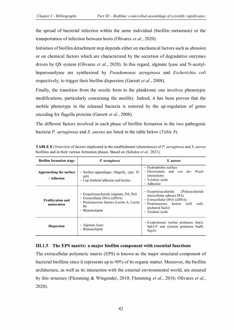
Chapter I – Bibliography Part III – Biofilms: a microbial assemblage of scientific significance
42
the spread of bacterial infection within the same individual (biofilm metastasis) or the
transportation of infection between hosts (Olivares et al., 2020).
Initiation of biofilm detachment step depends either on mechanical factors such as abrasion
or on chemical factors which are characterized by the secretion of degradative enzymes
driven by QS system (Olivares et al., 2020). In this regard, alginate lyase and N-acetyl-
heparosanlyase are synthesized by Pseudomonas aeruginosa and Escherichia coli
respectively, to trigger their biofilm dispersion (Garrett et al., 2008).
Finally, the transition from the sessile form to the planktonic one involves phenotypic
modifications, particularly concerning the motility. Indeed, it has been proven that the
mobile phenotype in the released bacteria is restored by the up-regulation of genes
encoding for flagella proteins (Garrett et al., 2008).
The different factors involved in each phase of biofilm formation in the two pathogenic
bacteria P. aeruginosa and S. aureus are listed in the table below (Table 8).
TABLE 8 | Overview of factors implicated in the establishment (chemotaxis) of P. aeruginosa and S. aureus biofilms and in their various formation phases. Based on (Schulze et al., 2021).
III.1.5 The EPS matrix: a major biofilm component with essential functions
The extracellular polymeric matrix (EPS) is known as the major structural component of
bacterial biofilms since it represents up to 90% of its organic matter. Moreover, the biofilm
architecture, as well as its interaction with the external environmental world, are ensured
by this structure (Flemming & Wingender, 2010; Flemming et al., 2016; Olivares et al.,
2020).
Biofilm formation stage P. aeruginosa S. aureus
Approaching the surface
/ Adhesion
- Surface-appendages (flagella, type IV pili)
- Cup fimbrial adhesins and lectins
- Hydrophobic-surface - Electrostatic and van der Waals
interactions - Teichoic acids - Adhesins
Proliferation and maturation
- Exopolysaccharide (alginate, Psl, Pel) - Extracellular DNA (eDNA) - Proteinaceous factors (Lectin A, Lectin
B) - Rhamnolipids
- Exopolysaccharide (Polysaccharide intercellular adhesin PIA)
- Extracellular DNA (eDNA) - Proteinaceous factors (cell wall-
anchored SasG) - Teichoic acids
Dispersion - Alginate lyase - Rhamnolipids
- Exoproteases (serine proteases SspA, SpIA-F and cysteine proteases SspB, ScpA)
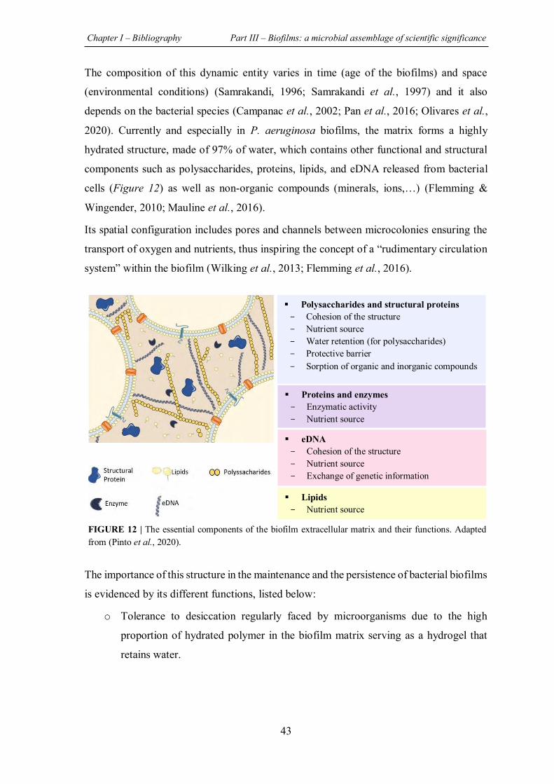
Chapter I – Bibliography Part III – Biofilms: a microbial assemblage of scientific significance
43
The composition of this dynamic entity varies in time (age of the biofilms) and space
(environmental conditions) (Samrakandi, 1996; Samrakandi et al., 1997) and it also
depends on the bacterial species (Campanac et al., 2002; Pan et al., 2016; Olivares et al.,
2020). Currently and especially in P. aeruginosa biofilms, the matrix forms a highly
hydrated structure, made of 97% of water, which contains other functional and structural
components such as polysaccharides, proteins, lipids, and eDNA released from bacterial
cells (Figure 12) as well as non-organic compounds (minerals, ions,…) (Flemming &
Wingender, 2010; Mauline et al., 2016).
Its spatial configuration includes pores and channels between microcolonies ensuring the
transport of oxygen and nutrients, thus inspiring the concept of a “rudimentary circulation
system” within the biofilm (Wilking et al., 2013; Flemming et al., 2016).
The importance of this structure in the maintenance and the persistence of bacterial biofilms
is evidenced by its different functions, listed below:
o Tolerance to desiccation regularly faced by microorganisms due to the high
proportion of hydrated polymer in the biofilm matrix serving as a hydrogel that
retains water.
Polysaccharides and structural proteins - Cohesion of the structure - Nutrient source - Water retention (for polysaccharides) - Protective barrier - Sorption of organic and inorganic compounds
Proteins and enzymes - Enzymatic activity - Nutrient source
eDNA - Cohesion of the structure - Nutrient source - Exchange of genetic information
Lipids - Nutrient source
FIGURE 12 | The essential components of the biofilm extracellular matrix and their functions. Adapted from (Pinto et al., 2020).
Chapter I – Bibliography Part III – Biofilms: a microbial assemblage of scientific significance
44
o Capture and digestion of organic and inorganic nutrient resources from the
substratum or from the outer aqueous environment through the sorption properties
of the sponge-like EPS biofilm matrix.
o Improvement of the intercellular signaling interaction which is strongly affected
by the properties of biofilm matrix (Flemming et al., 2016).
o Induction of the transition from reversible to irreversible adhesion of bacterial
cells prior to the formation of a cohesive biofilm (Pan et al., 2016).
o Diffusion barrier against antimicrobial agents: their activity can be inhibited by
enzymatic degradation, by chelation or by neutralization/consumption by matrix
components (Pinto et al., 2020).
The following parts are dedicated to the main EPS components.
III.1.5.1 Matrix exopolysaccharides
Exopolysaccharides are high molecular weight polymers secreted by bacteria in the
surrounding environment and composed of sugar residues. Glucose, galactose, mannose
are the most abundant carbohydrates found in biofilm matrix, followed by galacturonic
acid, arabinose, N-acetyl-glucosamine, fucose, xylose and rhamnose. These polymers have
a considerable role in adhesion and act as scaffolds for other matrix components (Rabin et
al., 2015).
Interestingly, despite the fact that these polysaccharides are not specific to biofilms, their
production is boosted under the stressful conditions associated with biofilm formation
(Rabin et al., 2015).
The composition and structure of these exopolysaccharides depend on the bacterial species
present in the biofilm. Indeed, concerning Staphylococcus biofilms, the polysaccharide
intercellular adhesin (PIA), also known as poly N-acetyl glucosamine, was characterized.
This linear polymer is formed of β-1,6-linked glucosamine residues (Mack et al., 1996).
On the other hand, in P. aeruginosa biofilm, three types of exopolysaccharides were well
described (Moradali et al., 2017):
1. Alginate (O-acetylated (1-4) linked D-mannuronic acid and variable proportions of
L-guluronic acid). Its proportion varies regarding the mucoid characteristics of the
strains (Marty et al., 1998)
2. Psl polysaccharides (repeating pentasaccharide including D-mannose, D-glucose
and L-rhamnose residues)
Chapter I – Bibliography Part III – Biofilms: a microbial assemblage of scientific significance
45
3. Pel polysaccharide (Partially acetylated (1-4) glycosidic linkages of N-
acetylgalactosamine and N-acetylglucosamine)
III.1.5.2 Matrix extracellular proteins
Extracellular proteins are another major component of the matrix providing functional and
structural benefits (Gunn et al., 2016). Some proteins tend to bind to cell surfaces and to
the exopolysaccharides, thus assisting in biofilm formation and stability (Rabin et al.,
2015).
The involvement of matrix extracellular proteins in biofilm formation and biofilm-related
infections was demonstrated in different studies. Dueholm et al. proved that the
overexpression of Fap amyloids, an insoluble fibrous protein, in P. aeruginosa enhances
cell aggregation and therefore biofilm formation (Dueholm et al., 2013). On the other hand,
Cucarella et al. demonstrated the important role of a Bap protein (biofilm associated
protein) in the initial adhesion of S. aureus, as well as in the formation of its biofilm
(Cucarella et al., 2001). The expression of this protein was also implicated in S. aureus
pathogenicity and in the infection persistence in a murine catheter-induced infection model.
Degradation enzymes were also detected in biofilm matrix. In fact, during starvation, these
enzymes are able to provide the carbon and energy resources required for biofilm cells by
the degradation of EPS matrix components, such as polysaccharides, other proteins, nucleic
acids, and lipids. In addition, enzymatic functions are involved in the detachment and
dispersion processes thus ensuring a new biofilm lifestyle (Rabin et al., 2015).
III.1.5.3 Extracellular DNA
The extracellular DNA (eDNA) found in the biofilm matrix is not only derived from lysed
cells, but can also be actively secreted by living bacteria through membranous vesicles
fusion (Olivares et al., 2020). As a result of their interaction with substrate receptors, eDNA
plays an essential role in biofilm establishment by enhancing cells aggregation and
adhesion to surfaces (Rabin et al., 2015).
Furthermore, Whitchurch et al. hypothesized the early involvement of eDNA in the
formation of P. aeruginosa biofilm since the addition of the DNase I enzyme prevented the
formation of biofilms and was able to dissolve preformed ones (Whitchurch et al., 2002).
Chapter I – Bibliography Part III – Biofilms: a microbial assemblage of scientific significance
46
III.1.6 Quorum sensing: microbial chatter orchestrating cells’ behavior
III.1.6.1 Definition and discovery
Quorum sensing is an intercellular communication system that, depending on bacterial
population density, is able to coordinate bacterial behavior via chemical signals regulating
genes expression accordingly. The QS system involves the production, exchange and
detection of signaling molecules, called autoinducers (AIs), which are constitutively
synthetized and passively or actively excreted into the surrounding environment (Paul et
al., 2018). When a given number of cells (or cell contacts) is reached, along with an external
accumulation of the signal molecules, bacteria leave their singular planktonic character and
a shift in gene expression is detected leading to consider that the bacterial population may
act collectively, as a group.
Interestingly, the QS communication system is not restricted to the one considered species
but can occur cross species (Bachtiar & Bachtiar, 2020) and even kingdom barriers (Li et
al., 2019). Indeed, some signaling molecules are able to act on different bacterial species,
as well as to affect the transcriptional programs of eukaryotic epithelial cells and host
immune cells (Antonioli et al., 2018; Song et al., 2019; Medina-Rodriguez et al., 2020)
QS system was discovered in 1970s in studying marine environment through an
exceptional symbiotic association between the halophilic bacterium Vibrio fischeri and the
Hawaiian bobtail squid Euprymna scolopes. Briefly, the high bacterial density of V. fischeri
that colonizes the outer surface of the squid during the night was correlated with light
production, providing a kind of squid camouflage against predators. Diffusible signal
molecules and genetic cluster implicated in this phenomenon were then described in 1980s
(Lami, 2019).
III.1.6.2 Quorum sensing circuit
Most of QS communication systems involve hierarchical auto-induction loops that consist
of the synthesis, recognition and response to signal molecules (Figure 13). The enzymatic
process of signal molecules production is generally catalyzed by a synthetase protein
encoding by the gene I (inducer). While the bacterial density increased, along the AIs
external concentration reach a critical threshold, a cell internalization (free diffusion or
transport, Table 9) and a formation of complex between the signal molecule and the
corresponding regulatory protein encoding by the gene R (receptor) occur.
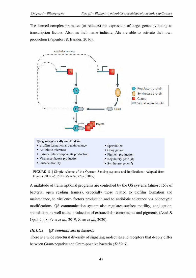
Chapter I – Bibliography Part III – Biofilms: a microbial assemblage of scientific significance
47
The formed complex promotes (or reduces) the expression of target genes by acting as
transcription factors. Also, as their name indicate, AIs are able to activate their own
production (Papenfort & Bassler, 2016).
A multitude of transcriptional programs are controlled by the QS systems (almost 15% of
bacterial open reading frames), especially those related to biofilm formation and
maintenance, to virulence factors production and to antibiotic tolerance via phenotypic
modifications. QS communication system also regulates surface motility, conjugation,
sporulation, as well as the production of extracellular components and pigments (Asad &
Opal, 2008; Pena et al., 2019; Zhao et al., 2020).
III.1.6.3 QS autoinducers in bacteria
There is a wide structural diversity of signaling molecules and receptors that deeply differ
between Gram-negative and Gram-positive bacteria (Table 9).
QS genes generally involved in: Biofilm formation and maintenance Antibiotic tolerance Extracellular components production Virulence factors production Surface motility
Sporulation Conjugation Pigment production Regulatory gene (R) Synthetase gene (I)
FIGURE 13 | Simple scheme of the Quorum Sensing systems and implications. Adapted from (Bjarnsholt et al., 2013; Moradali et al., 2017).
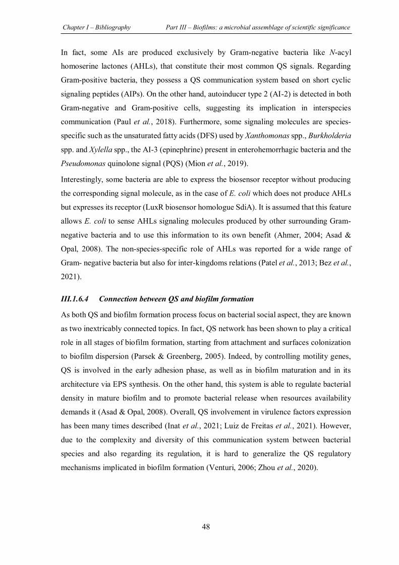
Chapter I – Bibliography Part III – Biofilms: a microbial assemblage of scientific significance
48
In fact, some AIs are produced exclusively by Gram-negative bacteria like N-acyl
homoserine lactones (AHLs), that constitute their most common QS signals. Regarding
Gram-positive bacteria, they possess a QS communication system based on short cyclic
signaling peptides (AIPs). On the other hand, autoinducer type 2 (AI-2) is detected in both
Gram-negative and Gram-positive cells, suggesting its implication in interspecies
communication (Paul et al., 2018). Furthermore, some signaling molecules are species-
specific such as the unsaturated fatty acids (DFS) used by Xanthomonas spp., Burkholderia
spp. and Xylella spp., the AI-3 (epinephrine) present in enterohemorrhagic bacteria and the
Pseudomonas quinolone signal (PQS) (Mion et al., 2019).
Interestingly, some bacteria are able to express the biosensor receptor without producing
the corresponding signal molecule, as in the case of E. coli which does not produce AHLs
but expresses its receptor (LuxR biosensor homologue SdiA). It is assumed that this feature
allows E. coli to sense AHLs signaling molecules produced by other surrounding Gram-
negative bacteria and to use this information to its own benefit (Ahmer, 2004; Asad &
Opal, 2008). The non-species-specific role of AHLs was reported for a wide range of
Gram- negative bacteria but also for inter-kingdoms relations (Patel et al., 2013; Bez et al.,
2021).
III.1.6.4 Connection between QS and biofilm formation
As both QS and biofilm formation process focus on bacterial social aspect, they are known
as two inextricably connected topics. In fact, QS network has been shown to play a critical
role in all stages of biofilm formation, starting from attachment and surfaces colonization
to biofilm dispersion (Parsek & Greenberg, 2005). Indeed, by controlling motility genes,
QS is involved in the early adhesion phase, as well as in biofilm maturation and in its
architecture via EPS synthesis. On the other hand, this system is able to regulate bacterial
density in mature biofilm and to promote bacterial release when resources availability
demands it (Asad & Opal, 2008). Overall, QS involvement in virulence factors expression
has been many times described (Inat et al., 2021; Luiz de Freitas et al., 2021). However,
due to the complexity and diversity of this communication system between bacterial
species and also regarding its regulation, it is hard to generalize the QS regulatory
mechanisms implicated in biofilm formation (Venturi, 2006; Zhou et al., 2020).
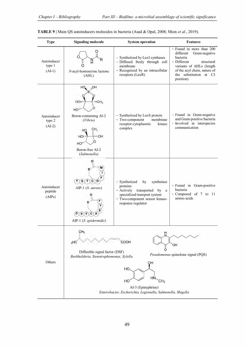
Chapter I – Bibliography Part III – Biofilms: a microbial assemblage of scientific significance
49
TABLE 9 | Main QS autoinducers molecules in bacteria (Asad & Opal, 2008; Mion et al., 2019).
Type Signaling molecule System operation Features
Autoinducer type 1 (AI-1) N-acyl-homoserine lactone
(AHL)
- Synthetized by LuxI synthases - Diffused freely through cell
membrane - Recognized by an intracellular
receptors (LuxR)
- Found in more than 200 different Gram-negative bacteria
- Different structural variants of AHLs (length of the acyl chain, nature of the substitution at C3 position)
Autoinducer type 2 (AI-2)
Boron-containing AI-2 (Vibrio)
Boron-free AI-2 (Salmonella)
- Synthetized by LuxS protein - Two-component membrane
receptor-cytoplasmic kinase complex
- Found in Gram-negative and Gram-positive bacteria
- Involved in interspecies communication
Autoinducer peptide (AIPs)
AIP-1 (S. aureus)
AIP-1 (S. epidermidis)
- Synthetized by synthetase proteins
- Actively transported by a specialized transport system
- Two-component sensor kinase-response regulator
- Found in Gram-positive bacteria
- Composed of 7 to 11 amino acids
Others
Diffusible signal factor (DSF) Burkholderia, Stenotrophomonas, Xylella
AI-3 (Epinephrine) Enterobacter, Escherichia, Legionella, Salmonella, Shigella
Pseudomonas quinolone signal (PQS)
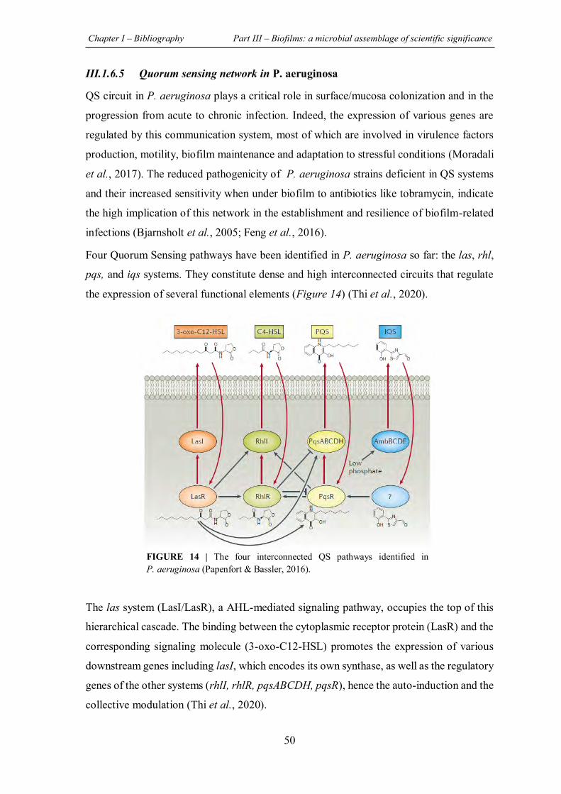
Chapter I – Bibliography Part III – Biofilms: a microbial assemblage of scientific significance
50
III.1.6.5 Quorum sensing network in P. aeruginosa
QS circuit in P. aeruginosa plays a critical role in surface/mucosa colonization and in the
progression from acute to chronic infection. Indeed, the expression of various genes are
regulated by this communication system, most of which are involved in virulence factors
production, motility, biofilm maintenance and adaptation to stressful conditions (Moradali
et al., 2017). The reduced pathogenicity of P. aeruginosa strains deficient in QS systems
and their increased sensitivity when under biofilm to antibiotics like tobramycin, indicate
the high implication of this network in the establishment and resilience of biofilm-related
infections (Bjarnsholt et al., 2005; Feng et al., 2016).
Four Quorum Sensing pathways have been identified in P. aeruginosa so far: the las, rhl,
pqs, and iqs systems. They constitute dense and high interconnected circuits that regulate
the expression of several functional elements (Figure 14) (Thi et al., 2020).
The las system (LasI/LasR), a AHL-mediated signaling pathway, occupies the top of this
hierarchical cascade. The binding between the cytoplasmic receptor protein (LasR) and the
corresponding signaling molecule (3-oxo-C12-HSL) promotes the expression of various
downstream genes including lasI, which encodes its own synthase, as well as the regulatory
genes of the other systems (rhlI, rhlR, pqsABCDH, pqsR), hence the auto-induction and the
collective modulation (Thi et al., 2020).
FIGURE 14 | The four interconnected QS pathways identified in P. aeruginosa (Papenfort & Bassler, 2016).
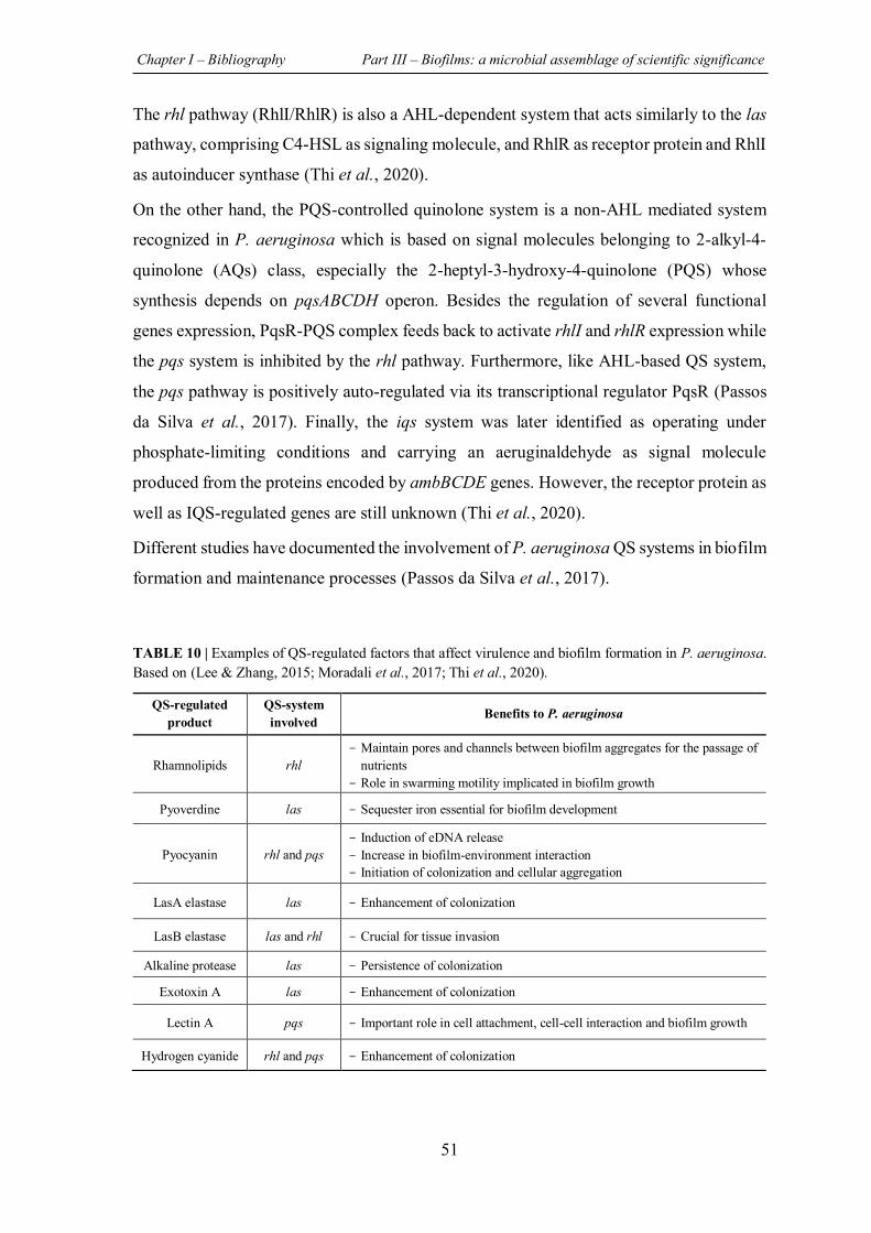
Chapter I – Bibliography Part III – Biofilms: a microbial assemblage of scientific significance
51
The rhl pathway (RhlI/RhlR) is also a AHL-dependent system that acts similarly to the las
pathway, comprising C4-HSL as signaling molecule, and RhlR as receptor protein and RhlI
as autoinducer synthase (Thi et al., 2020).
On the other hand, the PQS-controlled quinolone system is a non-AHL mediated system
recognized in P. aeruginosa which is based on signal molecules belonging to 2-alkyl-4-
quinolone (AQs) class, especially the 2-heptyl-3-hydroxy-4-quinolone (PQS) whose
synthesis depends on pqsABCDH operon. Besides the regulation of several functional
genes expression, PqsR-PQS complex feeds back to activate rhlI and rhlR expression while
the pqs system is inhibited by the rhl pathway. Furthermore, like AHL-based QS system,
the pqs pathway is positively auto-regulated via its transcriptional regulator PqsR (Passos
da Silva et al., 2017). Finally, the iqs system was later identified as operating under
phosphate-limiting conditions and carrying an aeruginaldehyde as signal molecule
produced from the proteins encoded by ambBCDE genes. However, the receptor protein as
well as IQS-regulated genes are still unknown (Thi et al., 2020).
Different studies have documented the involvement of P. aeruginosa QS systems in biofilm
formation and maintenance processes (Passos da Silva et al., 2017).
TABLE 10 | Examples of QS-regulated factors that affect virulence and biofilm formation in P. aeruginosa. Based on (Lee & Zhang, 2015; Moradali et al., 2017; Thi et al., 2020).
QS-regulated product
QS-system involved Benefits to P. aeruginosa
Rhamnolipids rhl - Maintain pores and channels between biofilm aggregates for the passage of
nutrients - Role in swarming motility implicated in biofilm growth
Pyoverdine las - Sequester iron essential for biofilm development
Pyocyanin rhl and pqs - Induction of eDNA release - Increase in biofilm-environment interaction - Initiation of colonization and cellular aggregation
LasA elastase las - Enhancement of colonization
LasB elastase las and rhl - Crucial for tissue invasion
Alkaline protease las - Persistence of colonization
Exotoxin A las - Enhancement of colonization
Lectin A pqs - Important role in cell attachment, cell-cell interaction and biofilm growth
Hydrogen cyanide rhl and pqs - Enhancement of colonization
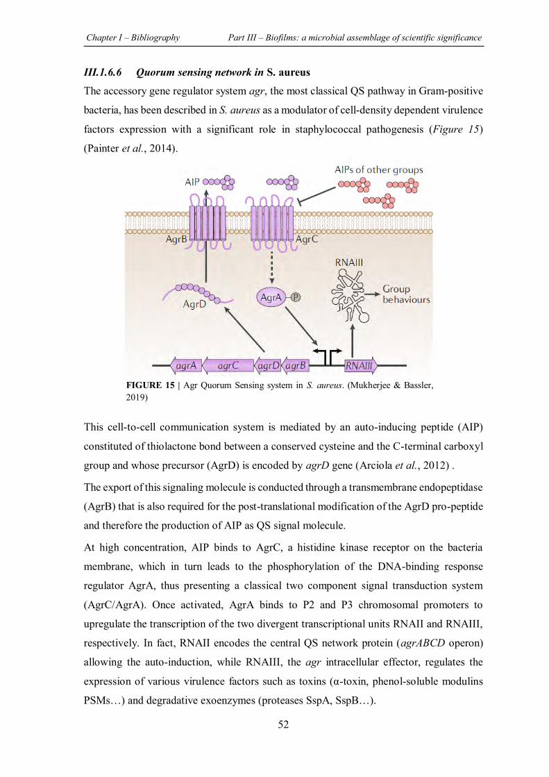
Chapter I – Bibliography Part III – Biofilms: a microbial assemblage of scientific significance
52
III.1.6.6 Quorum sensing network in S. aureus
The accessory gene regulator system agr, the most classical QS pathway in Gram-positive
bacteria, has been described in S. aureus as a modulator of cell-density dependent virulence
factors expression with a significant role in staphylococcal pathogenesis (Figure 15)
(Painter et al., 2014).
This cell-to-cell communication system is mediated by an auto-inducing peptide (AIP)
constituted of thiolactone bond between a conserved cysteine and the C-terminal carboxyl
group and whose precursor (AgrD) is encoded by agrD gene (Arciola et al., 2012) .
The export of this signaling molecule is conducted through a transmembrane endopeptidase
(AgrB) that is also required for the post-translational modification of the AgrD pro-peptide
and therefore the production of AIP as QS signal molecule.
At high concentration, AIP binds to AgrC, a histidine kinase receptor on the bacteria
membrane, which in turn leads to the phosphorylation of the DNA-binding response
regulator AgrA, thus presenting a classical two component signal transduction system
(AgrC/AgrA). Once activated, AgrA binds to P2 and P3 chromosomal promoters to
upregulate the transcription of the two divergent transcriptional units RNAII and RNAIII,
respectively. In fact, RNAII encodes the central QS network protein (agrABCD operon)
allowing the auto-induction, while RNAIII, the agr intracellular effector, regulates the
expression of various virulence factors such as toxins (α-toxin, phenol-soluble modulins
PSMs…) and degradative exoenzymes (proteases SspA, SspB…).
FIGURE 15 | Agr Quorum Sensing system in S. aureus. (Mukherjee & Bassler, 2019)
Chapter I – Bibliography Part III – Biofilms: a microbial assemblage of scientific significance
53
Interestingly, four Agr allelic variants that contribute to the production of different AIP
(differ in amino acids residues) have been identified in S. aureus. Although AIP acts as QS
activator in cells that produce it, it is able to inhibit QS system in other bacterial strains that
produce different AIPs.
Regarding the role of this network in S. aureus biofilms, it has been shown that biofilm
formation is negatively regulated by agr system given the significant increase in biofilm
development in S. aureus agr mutants (Vuong et al., 2000). However, the agr system is
strongly involved in biofilm dispersion due to the positive regulation of several
extracellular proteases expression conducted by AgrA response regulator (Boles &
Horswill, 2008; Arciola et al., 2012).
Chapter I – Bibliography Part III – Biofilms: a microbial assemblage of scientific significance
54
III.1.7 Biofilms: a resilient strength
Understanding the factors responsible for biofilms strength is crucial, especially in the
search for treatments against those which are pathogenic and infectious.
Biofilm strength is the result of its ability to overcome harsh environmental conditions,
tolerate high concentrations of antimicrobial agents, and escape from the host immune
response in the case of clinical infection, which explains the fact that biofilm is the
predominant and survival state of bacteria (Rabin et al., 2015). Moreover, since it is tightly
attached to a surface or tissues, biofilm can withstand the eradication factors such as water
flow, blood-stream and shear forces.
Additionally, it has been shown that biofilm cells are about 100 to 1000 times more
resistant than their planktonic form (Alasri et al., 1992; Campanac et al., 2002; Davies,
2003). In this context, Nickel et al., (1985) have initially demonstrated that P. aeruginosa
in floating form is significantly more sensitive to tobramycin antibiotic treatment than its
biofilm state. Indeed, a significant proportion of adherent cells within the biofilm was found
to be resistant to tobramycin treatment at a very high concentration (1.0 mg/mL).
Furthermore, Luppens et al. (2002) have proved that S. aureus biofilms are respectively 50
and 600 times more resistant to benzalkonium chloride (BAC) and to the oxidizing agent
sodium hypochlorite than suspension cells. In addition, we previously demonstrated the
specific loss of sensitivity when bacteria are under biofilm regarding current disinfectants
(Samrakandi et al., 1994; Campanac et al., 2002).
However, it has been proven that the sensitivity of bacteria to antimicrobial agents is
restored after resuspension of biofilm cells, which shows the fact that this resilience
characteristic is reversible, phenotypic, and non-heritable (Lebeaux & Ghigo, 2012).
Therefore, we talk about biofilm tolerance rather than resistance. Indeed, the resistance is
defined as the ability of microorganisms to multiply in presence of antimicrobial agents
due to different heritable or genetically acquired mechanisms such as the alteration of
antibiotics target, modification of bacterial outer cell walls permeability, or destruction of
the antimicrobial agents. By contrast, tolerant bacteria can survive in high concentrations
of antimicrobial agents but with interrupted growth like sessile bacteria within the biofilm
(Olivares et al., 2020).
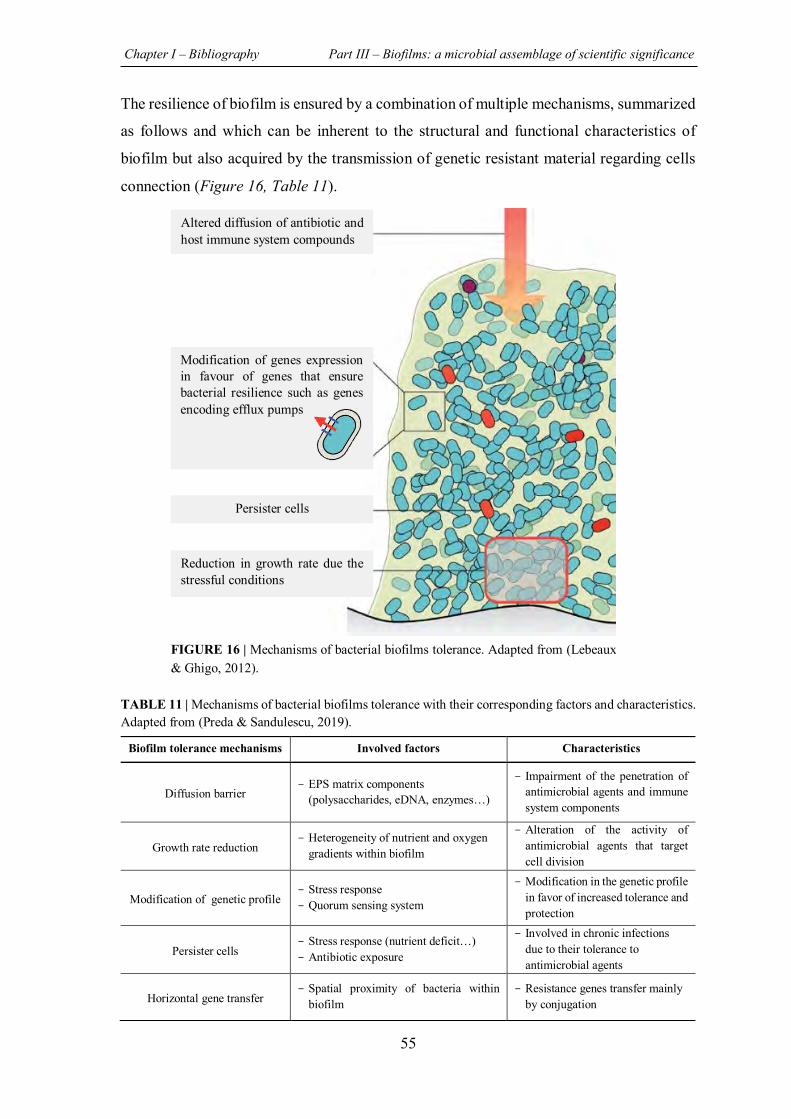
Chapter I – Bibliography Part III – Biofilms: a microbial assemblage of scientific significance
55
The resilience of biofilm is ensured by a combination of multiple mechanisms, summarized
as follows and which can be inherent to the structural and functional characteristics of
biofilm but also acquired by the transmission of genetic resistant material regarding cells
connection (Figure 16, Table 11).
TABLE 11 | Mechanisms of bacterial biofilms tolerance with their corresponding factors and characteristics. Adapted from (Preda & Sandulescu, 2019).
Biofilm tolerance mechanisms Involved factors Characteristics
Diffusion barrier - EPS matrix components
(polysaccharides, eDNA, enzymes…)
- Impairment of the penetration of antimicrobial agents and immune system components
Growth rate reduction - Heterogeneity of nutrient and oxygen
gradients within biofilm
- Alteration of the activity of antimicrobial agents that target cell division
Modification of genetic profile - Stress response - Quorum sensing system
- Modification in the genetic profile in favor of increased tolerance and protection
Persister cells - Stress response (nutrient deficit…) - Antibiotic exposure
- Involved in chronic infections due to their tolerance to antimicrobial agents
Horizontal gene transfer - Spatial proximity of bacteria within
biofilm - Resistance genes transfer mainly
by conjugation
Altered diffusion of antibiotic and host immune system compounds
Reduction in growth rate due the stressful conditions
Persister cells
Modification of genes expression in favour of genes that ensure bacterial resilience such as genes encoding efflux pumps
FIGURE 16 | Mechanisms of bacterial biofilms tolerance. Adapted from (Lebeaux & Ghigo, 2012).
Chapter I – Bibliography Part III – Biofilms: a microbial assemblage of scientific significance
56
III.1.7.1 Diffusion barrier
The complex architecture of biofilm mainly composed of extracellular polymeric matrix
(EPS) presents an “innate” tolerance which provides mechanical barrier limiting
antimicrobial agent penetration within the biofilm and therefore their effects on
microorganisms (Olivares et al., 2020).
The high viscosity and hydrophilicity of the extracellular polymeric matrix, as well as the
electrostatic charge of some of these components (polysaccharides, eDNA…), ensure the
trapping of different kinds of antibiotics which then prevent them from reaching their
effective concentrations in the deep layers of biofilm to the cells (Rabin et al., 2015;
Olivares et al., 2020). Consequently, due to this delayed invasion, bacteria located in the
deeper layers can expand physiological adaptation (expression of porins,…) in comparison
with surface cells which are more sensitive toward antibiotic treatment (Olivares et al.,
2020).
The involvement of EPS matrix in P. aeruginosa biofilm resistance towards antibiotics has
been highlighted previously. In fact, Hentzer et al., have demonstrated that P. aeruginosa
biofilm developed by a strain that overproduces alginate, an essential component of P.
aeruginosa matrix, exhibit an increased resistance to tobramycin in comparison with
biofilm formed by wild-type strain (Hentzer et al., 2001).
As for antimicrobial agents, the extracellular matrix forms a protective barrier against the
penetration of host immune system compounds (cytokines, …) in a manner that even
immunocompetent individuals are unable to eradicate a biofilm infection (Olivares et al.,
2020). Indeed, the phagocytic cells of the innate immune system such as macrophages and
neutrophils are generally activated by direct contact with bacterial surface and then they
accumulate around the biofilm. In view of their difficult penetration through the
extracellular matrix, they are slowed down which makes them more sensitive to the
inactivation by bacterial enzymes. Consequently, the increased lysis of neutrophils leads to
the release of harmful compounds and therefore to consecutive tissue damages (Watters et
al., 2016). Regarding the role of EPS matrix, tolerance to many disinfectants with oxidant
proprieties is linked to the presence of reducing components like some proteins
(Samrakandi et al., 1997; Bridier et al., 2011).
Chapter I – Bibliography Part III – Biofilms: a microbial assemblage of scientific significance
57
Nevertheless, this reduced diffusion cannot be considered as the radical tolerance
mechanism of biofilm given the good penetration of some antibiotics (fluoroquinolone,
ampicillin…) (Lebeaux & Ghigo, 2012). Thus, this protective barrier is strain and antibiotic
dependent (Olivares et al., 2020).
III.1.7.2 Reduction in growth rate
Gradients of oxygen and nutrients in mature biofilms lead to the formation of hypoxic, and
stressful zones where bacteria are less metabolically active (Olivares et al., 2020).
Subsequently, some microorganisms, particularly those located in the deeper layers, tend
to get back to the stationary phase by slowing down their growth and division rates (Rabin
et al., 2015).
Since the mode of action of the majority of antimicrobial agent targets dividing cells such
as replication, transcription, and translation processes, the reduced growth rate of sessile
cells is in part responsible for biofilm tolerance (Hoiby et al., 2010).
In this context, it has been demonstrated that the bacteriolytic activity of β-lactams,
antibiotics that act on dividing cells and inhibit the synthesis of bacterial cell wall, is
diminished when they are used on E. coli biofilms (Rabin et al., 2015). In addition, Bauer
et al., have reported the failure of some antibiotics such as fusidic acid (inhibitor of proteins
synthesis) and moxifloxacin (inhibitor of DNA replication) to completely destroy S. aureus
biofilms (Bauer et al., 2013).
However, decreasing growth rate cannot explain biofilm tolerance to antibiotics that are
considered bactericidal and effective also on stationary bacteria such as quinolones
(Lebeaux & Ghigo, 2012).
III.1.7.3 Modification of genes expression: example of efflux pumps
The microarray analysis conducted by Wagner et al., in order to investigate QS-regulated
genes in P. aeruginosa highlighted the involvement of this key system in global gene
expression by positive or negative regulation (Wagner et al., 2003). In fact, some genes
such as those implicated in the adaptation and protection as well as those encoding secreted
factors (toxins, enzymes, alginates…) are upregulated by QS system. In addition, results
demonstrated the involvement of this communication system in the positive regulation of
three efflux pumps expression.
Chapter I – Bibliography Part III – Biofilms: a microbial assemblage of scientific significance
58
Indeed, these membrane transporters can pump out all kinds of intracellular toxins or
xenobiotics, including antibiotics and therefore prevent them from reaching their targets
(Rabin et al., 2015).
Interestingly, it has been demonstrated that some efflux pumps genes are overexpressed in
biofilm cells, which may contribute to their implication in biofilm tolerance (Rabin et al.,
2015). Indeed, Zhang & Mah (2008) have identified a novel efflux system in P. aeruginosa
whose expression in sessile cells is higher than in planktonic cells. This efflux system
enhances P. aeruginosa biofilm tolerance to tobramycin, gentamycin and ciprofloxacin
given that the complete deletion of genes encoding this pump leads to the formation of
sensitive biofilm (Zhang & Mah, 2008).
III.1.7.4 Persister cells
Another important mechanism which contributes to biofilm tolerance is the formation of a
small bacterial subpopulation characterized by a growth rate of zero or extremely weak,
called “persister cells” or dormant cells (Rabin et al., 2015). Indeed, these metabolically
inactive cells are not genetically resistant to antimicrobial agents but they display a
transient phenotype giving them a tolerance to high concentrations of these agents,
especially those targeting cell growth and division processes (Rabin et al., 2015; Pang et
al., 2019). Furthermore, the downregulation of the expression of genes involved in motility
and energy production has been shown in this type of cells (Olivares et al., 2020).
Transition to the dormant state is induced by environmental stimuli and stresses, such as
nutritional deficiency or antibiotic exposure (Harms et al., 2016).
Moreover, it has been indicated that the QS communication system is implicated in this
phenotypic switch. In fact, Moker et al. have demonstrated that the QS-regulatory signaling
molecule 3-oxo-C12-HSL (N-(3-oxododecanoyl)-homoserine lactone) significantly
increased the number of P. aeruginosa persister cells within the biofilm (Moker et al.,
2010).
Besides, it has been elucidated that the toxin-antitoxin (TA) system plays a leading role in
the formation of persister cells of P. aeruginosa. Indeed, this system consists of a protein
toxin that is able to block essential cellular mechanisms and of its antitoxin which can be a
protein or a small non-coding-RNA whose function is to neutralize toxicity of the
corresponding toxin (Pang et al., 2019).
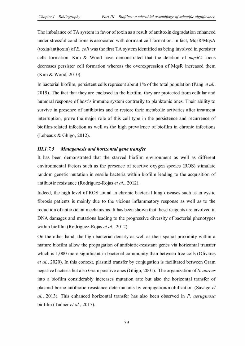
Chapter I – Bibliography Part III – Biofilms: a microbial assemblage of scientific significance
59
The imbalance of TA system in favor of toxin as a result of antitoxin degradation enhanced
under stressful conditions is associated with dormant cell formation. In fact, MqsR/MqsA
(toxin/antitoxin) of E. coli was the first TA system identified as being involved in persister
cells formation. Kim & Wood have demonstrated that the deletion of mqsRA locus
decreases persister cell formation whereas the overexpression of MqsR increased them
(Kim & Wood, 2010).
In bacterial biofilm, persistent cells represent about 1% of the total population (Pang et al.,
2019). The fact that they are enclosed in the biofilm, they are protected from cellular and
humoral response of host’s immune system contrarily to planktonic ones. Their ability to
survive in presence of antibiotics and to restore their metabolic activities after treatment
interruption, prove the major role of this cell type in the persistence and recurrence of
biofilm-related infection as well as the high prevalence of biofilm in chronic infections
(Lebeaux & Ghigo, 2012).
III.1.7.5 Mutagenesis and horizontal gene transfer
It has been demonstrated that the starved biofilm environment as well as different
environmental factors such as the presence of reactive oxygen species (ROS) stimulate
random genetic mutation in sessile bacteria within biofilm leading to the acquisition of
antibiotic resistance (Rodriguez-Rojas et al., 2012).
Indeed, the high level of ROS found in chronic bacterial lung diseases such as in cystic
fibrosis patients is mainly due to the vicious inflammatory response as well as to the
reduction of antioxidant mechanisms. It has been shown that these reagents are involved in
DNA damages and mutations leading to the progressive diversity of bacterial phenotypes
within biofilm (Rodriguez-Rojas et al., 2012).
On the other hand, the high bacterial density as well as their spatial proximity within a
mature biofilm allow the propagation of antibiotic-resistant genes via horizontal transfer
which is 1,000 more significant in bacterial community than between free cells (Olivares
et al., 2020). In this context, plasmid transfer by conjugation is facilitated between Gram
negative bacteria but also Gram positive ones (Ghigo, 2001). The organization of S. aureus
into a biofilm considerably increases mutation rate but also the horizontal transfer of
plasmid-borne antibiotic resistance determinants by conjugation/mobilization (Savage et
al., 2013). This enhanced horizontal transfer has also been observed in P. aeruginosa
biofilm (Tanner et al., 2017).
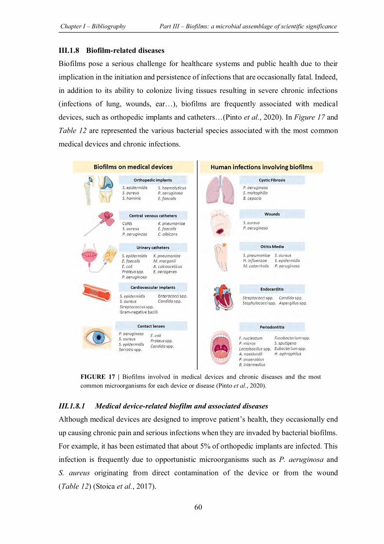
Chapter I – Bibliography Part III – Biofilms: a microbial assemblage of scientific significance
60
III.1.8 Biofilm-related diseases
Biofilms pose a serious challenge for healthcare systems and public health due to their
implication in the initiation and persistence of infections that are occasionally fatal. Indeed,
in addition to its ability to colonize living tissues resulting in severe chronic infections
(infections of lung, wounds, ear…), biofilms are frequently associated with medical
devices, such as orthopedic implants and catheters…(Pinto et al., 2020). In Figure 17 and
Table 12 are represented the various bacterial species associated with the most common
medical devices and chronic infections.
III.1.8.1 Medical device-related biofilm and associated diseases
Although medical devices are designed to improve patient’s health, they occasionally end
up causing chronic pain and serious infections when they are invaded by bacterial biofilms.
For example, it has been estimated that about 5% of orthopedic implants are infected. This
infection is frequently due to opportunistic microorganisms such as P. aeruginosa and
S. aureus originating from direct contamination of the device or from the wound
(Table 12) (Stoica et al., 2017).
FIGURE 17 | Biofilms involved in medical devices and chronic diseases and the most common microorganisms for each device or disease (Pinto et al., 2020).
Chapter I – Bibliography Part III – Biofilms: a microbial assemblage of scientific significance
61
TABLE 12 | Non-exhaustive list of human infections related to biofilms (Fux et al., 2009).
Infection or disease Common bacterial species involved
Dental caries Acidogenic Gram-positive cocci (Streptococcus sp.)
Periodontitis Gram-negative anaerobic oral bacteria
Otitis media Nontypeable Haemophilus influenzae
Chronic tonsillitis Various species
Cystic fibrosis pneumonia Pseudomonas aeruginosa, Burkholderia cepacia
Endocarditis Viridans group streptococci, staphylococci
Necrotizing fasciitis Group A streptococci
Musculoskeletal infections Gram-positive streptococci
Osteomyelitis Various species
Biliary tract infection Enteric bacteria
Infectious kidney stones Gram-negative rods
Bacterial prostatitis Escherichia coli and other Gram-negative bacteria
Infections associated with foreign body material
Contact lens P. aeruginosa, Gram-positive cocci
Sutures Staphylococci
Ventilation-associated pneumonia Gram-negative rods
Mechanical heart valves Staphylococci
Vascular grafts Gram-positive cocci
Arteriovenous shunts Staphylococci
Endovascular catheter infections Staphylococci
Peritoneal dialysis (CAPD) peritonitis Various species
Urinary catheter infections E. coli, Gram-negative rods
IUDs Actinomyces israelii and others
Penile prostheses Staphylococci
Orthopedic prosthesis Staphylococci
Unfortunately, treatment of these infections is extremely difficult and usually requires the
removal of the device which is not always convenient for the patient (Pinto et al., 2020).
Mechanical cardiac valve can also be colonized by pathogenic biofilms. Despite the fact
that these infections are less frequent, they are considered a worry owing to the high
mortality rate that can attain 30% of implanted patients (Pinto et al., 2020). In this case, the
danger consists in the blockage of the artificial cardiac valve, as well as the diffusion of the
infection via bloodstream as a result of the detachment of biofilm fragments (Rabin et al.,
2015).
Chapter I – Bibliography Part III – Biofilms: a microbial assemblage of scientific significance
62
III.1.8.2 Other biofilm-related diseases: example of cystic fibrosis
Cystic fibrosis (CF) is an autosomal recessive inherited genetic disorder associated with
the secretion of a viscous layer of mucus on the respiratory epithelium. This disease is
caused by one of more than 1,500 possible mutations in a membrane-bound chloride
channel named cystic fibrosis transmembrane conductance regulator (CFTR), whose
dysfunction leads to a warm, humid, stressful, and nutrient-rich environment suitable for
bacterial colonization (Folkesson et al., 2012).
Due to the high viscosity and dehydration of the mucus layer of CF patients, mucociliary
clearance, a normal process in which the cilia of the epithelial cells in the upper respiratory
tract remove particles and microbes trapped in the fluid mucus, is impaired , preventing the
elimination of trapped microorganisms (Folkesson et al., 2012). Unfortunately, CF patients
also suffer from a dysregulation of the innate immune system leading to the evolution from
colonization to lethal chronic infections (Doring & Gulbins, 2009).
Various bacterial species are associated with respiratory tract infection in CF patients with
an age-dependent prevalence such as (Lipuma, 2010):
o Staphylococcus aureus
o Pseudomonas aeruginosa
o Haemophilus influenzae
o Stenotrophomonas maltophilia
o Achromobacter xylosoxidans
o Burkholderia cepacia complex
o Burkholderia gladioli
o Ralstonia species, Cupriavidus species, and Pandoraea species.
According to the Cystic Fibrosis Foundation report, P. aeruginosa and S. aureus are
considered as the most commonly detected bacterial pathogens in CF patients (CFF, 2019).
While the highest prevalence of S. aureus infection occurs in younger patients,
P. aeruginosa colonization is most prominent in older adolescents and adults’ patients
(Figure 18).
The longevity of these infections is attributed to the formation of bacterial biofilms which
are not only resilient to antibiotic treatments, but also serve as a reservoir for disease
recurrence (Rabin et al., 2015).
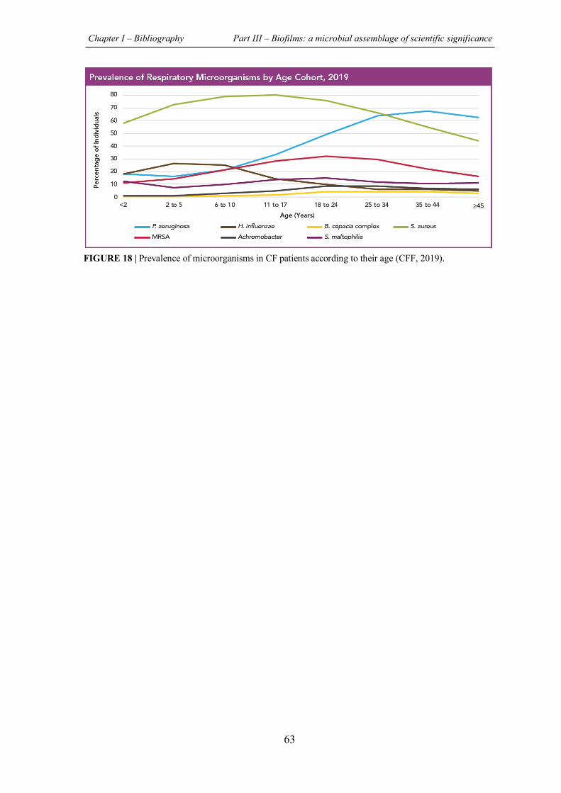
Chapter I – Bibliography Part III – Biofilms: a microbial assemblage of scientific significance
63
FIGURE 18 | Prevalence of microorganisms in CF patients according to their age (CFF, 2019).
Chapter I – Bibliography Part III – Biofilms: a microbial assemblage of scientific significance
64
III.2 CHALLENGE OF TREATING BIOFILM-ASSOCIATED
INFECTIONS
III.2.1 How to handle with biofilms? Current therapeutic approaches and strategies
In view of the complexity of biofilm-associated infections, as well as the inherent resilience
of this bacterial community, the search for effective treatments represents a veritable
challenge. In fact, for accessible contaminated surfaces such as catheters, the conventional
treatment consists in replacing the colonized material. However, for some implants, such
as joint prostheses, this solution is considered the last option and antibiotherapy is initially
set up. Moreover, the cost of these surgeries remains exorbitant (Ong et al., 2018).
On the other hand, concerning biofilm-related infections of host tissue, the current therapies
are still based on the use of conventional antimicrobial agents for extended periods with
high concentrations and combinations. Unfortunately, these therapies are sometimes
insufficient due to the failure in targeting more than one component of the heterogeneous
biofilm microenvironment (Pinto et al., 2020). In addition, such treatments often result in
incomplete bacterial killing, allowing unaffected bacteria to ensure infection recurrence
upon the withdrawal of the drug (Ong et al., 2018). Moreover, besides the toxicity issues
arising from the administration of high doses of antibiotics, too low concentrations may
not only fail to destroy the biofilm, but also promotes biofilm formation/maintaining
(Bjarnsholt et al., 2013).
Over all, the new knowledge on bacterial behavior regarding colonization and infection
underline the mismatch with current antibiotic targets.
Therefore, a great interest has been dedicated to the search for novel, multi-targeted or
combinatorial therapies that can prevent or even eradicate biofilms during infections, hence
the fundamental importance of understanding biofilm formation mechanisms.
In the following, four principle treatment approaches are emphasized, with (1) preventive,
(2) weakening, (3) disruptive or (4) killing effect (Figure 19). It should be noted that the
combination of these strategies will be most successful. Some examples with the
corresponding advantages and disadvantages of each strategy combatting biofilm,
especially regarding P. aeruginosa and S. aureus, are summarized in Table 13.
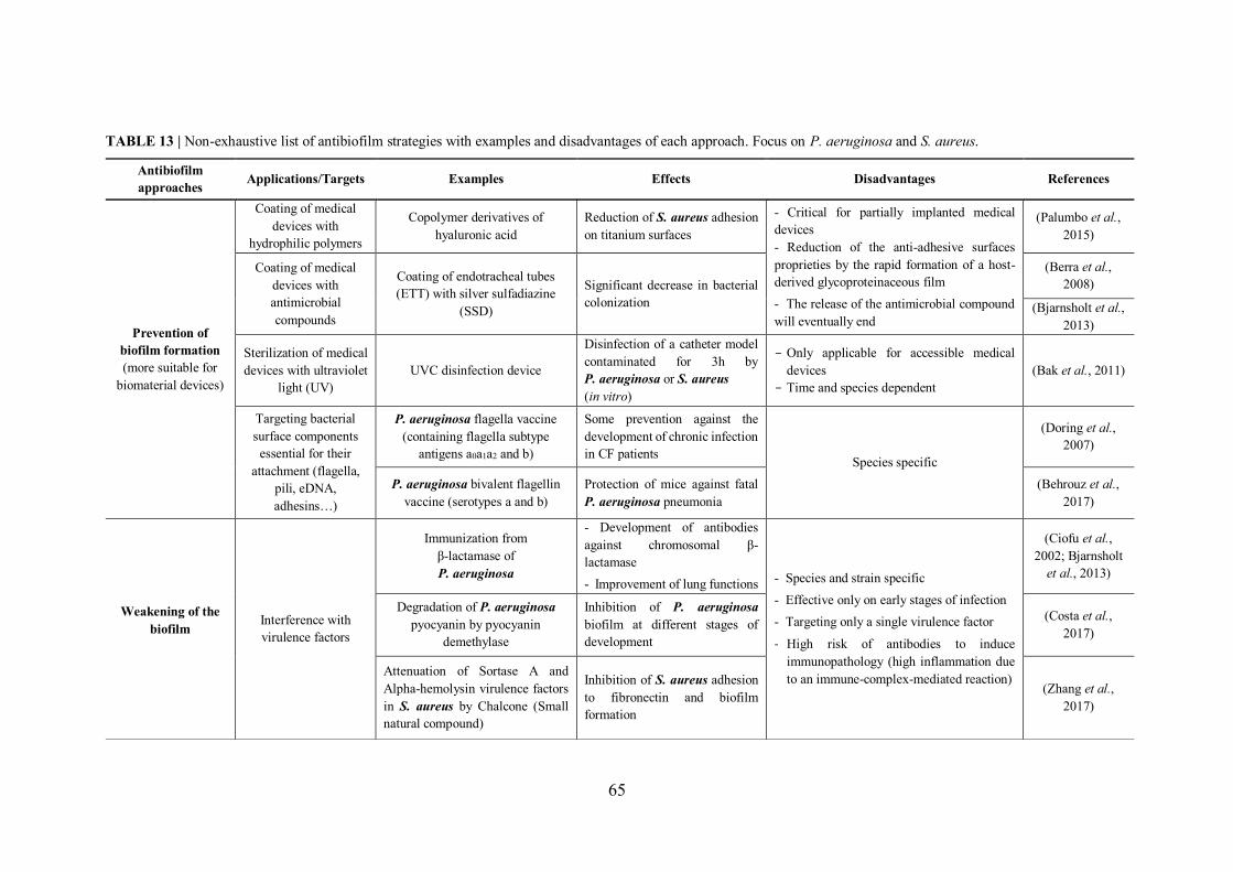
65
TABLE 13 | Non-exhaustive list of antibiofilm strategies with examples and disadvantages of each approach. Focus on P. aeruginosa and S. aureus. Antibiofilm approaches Applications/Targets Examples Effects Disadvantages References
Prevention of biofilm formation (more suitable for
biomaterial devices)
Coating of medical devices with
hydrophilic polymers
Copolymer derivatives of hyaluronic acid
Reduction of S. aureus adhesion on titanium surfaces
- Critical for partially implanted medical devices - Reduction of the anti-adhesive surfaces proprieties by the rapid formation of a host-derived glycoproteinaceous film - The release of the antimicrobial compound will eventually end
(Palumbo et al., 2015)
Coating of medical devices with antimicrobial compounds
Coating of endotracheal tubes (ETT) with silver sulfadiazine
(SSD)
Significant decrease in bacterial colonization
(Berra et al., 2008)
(Bjarnsholt et al., 2013)
Sterilization of medical devices with ultraviolet
light (UV) UVC disinfection device
Disinfection of a catheter model contaminated for 3h by P. aeruginosa or S. aureus (in vitro)
- Only applicable for accessible medical devices
- Time and species dependent (Bak et al., 2011)
Targeting bacterial surface components essential for their
attachment (flagella, pili, eDNA, adhesins…)
P. aeruginosa flagella vaccine (containing flagella subtype
antigens a0a1a2 and b)
Some prevention against the development of chronic infection in CF patients Species specific
(Doring et al., 2007)
P. aeruginosa bivalent flagellin vaccine (serotypes a and b)
Protection of mice against fatal P. aeruginosa pneumonia
(Behrouz et al., 2017)
Weakening of the biofilm
Interference with virulence factors
Immunization from β-lactamase of P. aeruginosa
- Development of antibodies against chromosomal β-lactamase - Improvement of lung functions - Species and strain specific
- Effective only on early stages of infection - Targeting only a single virulence factor
- High risk of antibodies to induce immunopathology (high inflammation due to an immune-complex-mediated reaction)
(Ciofu et al., 2002; Bjarnsholt
et al., 2013)
Degradation of P. aeruginosa pyocyanin by pyocyanin
demethylase
Inhibition of P. aeruginosa biofilm at different stages of development
(Costa et al., 2017)
Attenuation of Sortase A and Alpha-hemolysin virulence factors in S. aureus by Chalcone (Small natural compound)
Inhibition of S. aureus adhesion to fibronectin and biofilm formation
(Zhang et al., 2017)
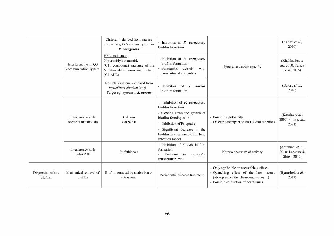
66
Interference with QS communication system
Chitosan – derived from marine crab – Target rhl and las system in
P. aeruginosa
- Inhibition in P. aeruginosa biofilm formation
Species and strain specific
(Rubini et al., 2019)
HSL-analogues: N-pyrimidylbutanamide (C11 compound) analogue of the N-butanoyl-L-homoserine lactone (C4-AHL)
- Inhibition of P. aeruginosa biofilm formation
- Synergistic activity with conventional antibiotics
(Khalilzadeh et al., 2010; Furiga
et al., 2016)
Norlichexanthone – derived from Penicillium algidum fungi -
Target agr system in S. aureus
- Inhibition of S. aureus biofilm formation
(Baldry et al., 2016)
Interference with bacterial metabolism
Gallium Ga(NO3)3
- Inhibition of P. aeruginosa biofilm formation - Slowing down the growth of biofilm-forming cells - Inhibition of Fe uptake - Significant decrease in the biofilm in a chronic biofilm lung infection model
- Possible cytotoxicity - Deleterious impact on host’s vital functions
(Kaneko et al., 2007; Firoz et al.,
2021)
Interference with c-di-GMP Sulfathiazole
- Inhibition of E. coli biofilm formation - Decrease in c-di-GMP intracellular level
Narrow spectrum of activity (Antoniani et al., 2010; Lebeaux &
Ghigo, 2012)
Dispersion of the biofilm
Mechanical removal of biofilm
Biofilm removal by sonication or ultrasound Periodontal diseases treatment
- Only applicable on accessible surfaces - Quenching effect of the host tissues
(absorption of the ultrasound waves…) - Possible destruction of host tissues
(Bjarnsholt et al., 2013)
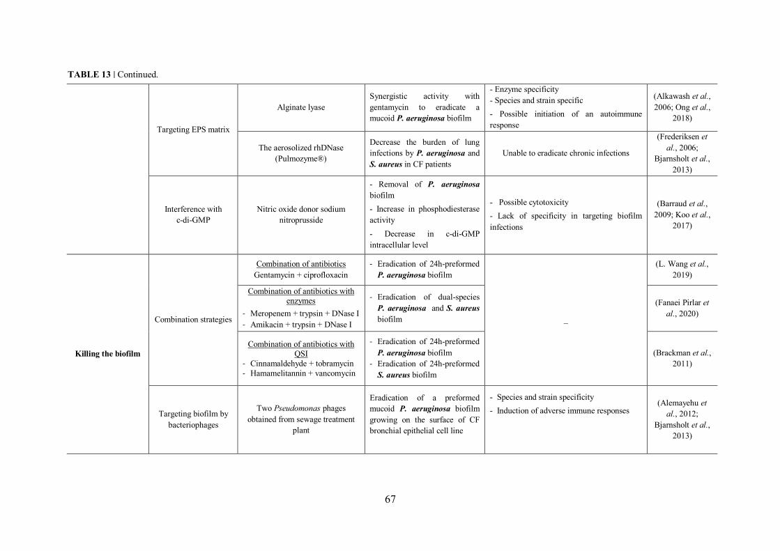
67
Targeting EPS matrix
Alginate lyase Synergistic activity with gentamycin to eradicate a mucoid P. aeruginosa biofilm
- Enzyme specificity - Species and strain specific - Possible initiation of an autoimmune response
(Alkawash et al., 2006; Ong et al.,
2018)
The aerosolized rhDNase (Pulmozyme®)
Decrease the burden of lung infections by P. aeruginosa and S. aureus in CF patients
Unable to eradicate chronic infections
(Frederiksen et al., 2006;
Bjarnsholt et al., 2013)
Interference with c-di-GMP
Nitric oxide donor sodium nitroprusside
- Removal of P. aeruginosa biofilm - Increase in phosphodiesterase activity - Decrease in c-di-GMP intracellular level
- Possible cytotoxicity - Lack of specificity in targeting biofilm infections
(Barraud et al., 2009; Koo et al.,
2017)
Killing the biofilm
Combination strategies
Combination of antibiotics Gentamycin + ciprofloxacin
- Eradication of 24h-preformed P. aeruginosa biofilm
_
(L. Wang et al., 2019)
Combination of antibiotics with enzymes
- Meropenem + trypsin + DNase I - Amikacin + trypsin + DNase I
- Eradication of dual-species P. aeruginosa and S. aureus biofilm
(Fanaei Pirlar et al., 2020)
Combination of antibiotics with QSI
- Cinnamaldehyde + tobramycin - Hamamelitannin + vancomycin
- Eradication of 24h-preformed P. aeruginosa biofilm
- Eradication of 24h-preformed S. aureus biofilm
(Brackman et al., 2011)
Targeting biofilm by bacteriophages
Two Pseudomonas phages obtained from sewage treatment
plant
Eradication of a preformed mucoid P. aeruginosa biofilm growing on the surface of CF bronchial epithelial cell line
- Species and strain specificity - Induction of adverse immune responses
(Alemayehu et al., 2012;
Bjarnsholt et al., 2013)
TABLE 13 | Continued.
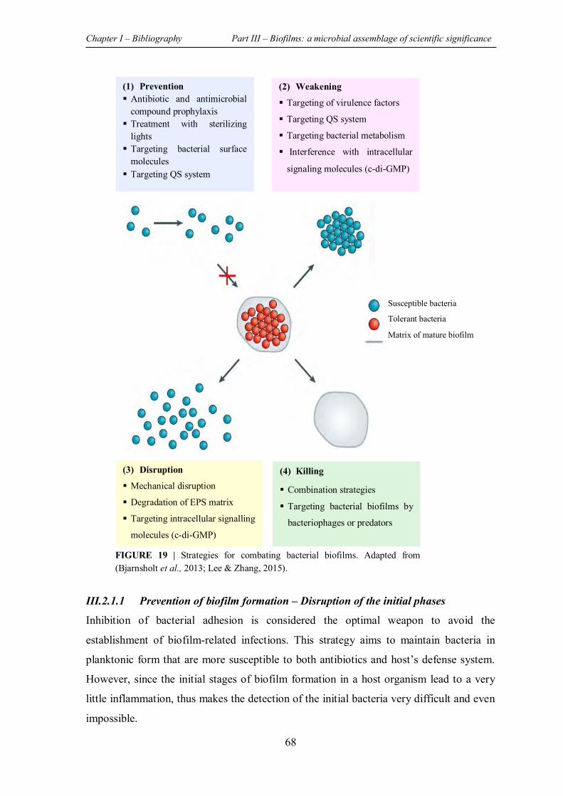
Chapter I – Bibliography Part III – Biofilms: a microbial assemblage of scientific significance
68
III.2.1.1 Prevention of biofilm formation – Disruption of the initial phases
Inhibition of bacterial adhesion is considered the optimal weapon to avoid the
establishment of biofilm-related infections. This strategy aims to maintain bacteria in
planktonic form that are more susceptible to both antibiotics and host’s defense system.
However, since the initial stages of biofilm formation in a host organism lead to a very
little inflammation, thus makes the detection of the initial bacteria very difficult and even
impossible.
(1) Prevention Antibiotic and antimicrobial
compound prophylaxis Treatment with sterilizing
lights Targeting bacterial surface
molecules Targeting QS system
(2) Weakening
Targeting of virulence factors
Targeting QS system
Targeting bacterial metabolism
Interference with intracellular
signaling molecules (c-di-GMP)
(3) Disruption
Mechanical disruption
Degradation of EPS matrix
Targeting intracellular signalling
molecules (c-di-GMP)
(4) Killing
Combination strategies
Targeting bacterial biofilms by
bacteriophages or predators
Susceptible bacteria
Tolerant bacteria
Matrix of mature biofilm
FIGURE 19 | Strategies for combating bacterial biofilms. Adapted from (Bjarnsholt et al., 2013; Lee & Zhang, 2015).
Chapter I – Bibliography Part III – Biofilms: a microbial assemblage of scientific significance
69
Indeed, the inflammation is only detectable after the establishment of the insensitive
biofilm. For this reason, this strategy has to be considered in prophylactic setting, such as
the prevention of implants and catheters contamination (Bjarnsholt et al., 2013).
Antibioprophylaxis may also be considered a way to control planktonic bacteria diffusion
via bloodstream (i.e. before oral surgery for patients with cardiac risk) or at the surgical
site (i.e. bone surgery) then preventing their adhesion.
On the one hand, for medical devices, the preventive global strategy towards bacterial
adhesion consists in selecting less adherent materials and/or modifying the
physicochemical properties of the surfaces by grafting/coating them with hydrophilic
polymers, which hinders their interaction with hydrophobic bacterial surfaces (Ong et al.,
2018). On the other hand, covering biomaterials devices with antimicrobial agents or with
antibiotics has shown effectiveness in delaying rather than totally preventing biofilm
formation (Cho et al., 2001). The major limitation of this approach which consists in
coating medical devices to avoid biofilm formation lies in the rapid conditioning of these
implants with host-derived glycoproteinaceous film (fibronectin, fibrinogen…) thus
preventing the release of antibiotics and/or reducing the effectiveness of the anti-adhesion
surfaces (Gristina, 1987). In addition, concerning partially implanted medical devices, the
risk of being colonized is increased due to their exterior contact. It should be noted that no
surface can be considered as “non-colonizable” (Costerton et al., 1978).
Other preventive approaches, considered more species specific, have also been investigated
such as targeting the components of bacterial surfaces that are involved in their initial
attachment (flagella, pili, eDNA, polysaccharides…) by blocking the production pathways
of these constituents or by employing specific neutralizing antibodies such as adhesin-
binding antibody (Bjarnsholt et al., 2013; Kisiela et al., 2015).
III.2.1.2 Weakening of the biofilm by disarming bacteria
In situations where the preventive approach is not feasible nor efficient, another strategy
consists in weakening and disarming bacteria within the biofilm by targeting the virulence
factors and their biofilm-forming features can be applied. However, this approach is
restricted by its specificity towards bacterial species and strains. Moreover, its effectiveness
is only demonstrated on immature or developing biofilms, but not on mature ones and it
never leads to the total eradication of the biofilm (Bjarnsholt et al., 2013).
Chapter I – Bibliography Part III – Biofilms: a microbial assemblage of scientific significance
70
Several studies have focused on the search for compounds that weaken bacteria by acting
on different targets. In the following, approaches that involve the interference with
virulence factors, Quorum Sensing (QS) communication system, iron metabolism and
sRNAs are discussed.
III.2.1.2.1 Interference with the expression/activity of virulence factors
In view of the essential involvement of virulence factors during the colonization, invasion
and persistence of bacteria under biofilm in a susceptible host, anti-virulence therapy has
emerged as a promising antimicrobial approach expected to be superior to conventional
antibiotics (Totsika, 2017; Fleitas Martinez et al., 2019) or at last to increase the
antibiotherapy efficiency. The relevance of this strategy resides in the complexity of
developing bacterial resistance towards the anti-virulence agents (Totsika, 2017).
However, this approach targets a single virulence factor which induces many disadvantages
(Cegelski et al., 2008):
o Virulence factors are species and sometimes strain specific
o Bacteria express various virulence factors and targeting only one generally induces
a transient and low reduction in biofilm
o The transient effect observed refers also to bacterial adaptation with modification
in expression or role of each virulence factor
III.2.1.2.2 Interference with QS communication system
As mentioned before, the intercellular communication system Quorum Sensing has a key
role in biofilm formation, as well as in the regulation of multiple virulence factors
expression. Therefore, a great interest has been attributed to the search for QS interference
ways so as to disarm bacteria from their biofilm-forming proprieties and from their
virulence (Remy et al., 2018).
The interference with QS system can occur on different levels: on signal generation point,
on signal molecules and on signal reception level. Indeed, QS inhibitor (QSI) molecules
can target the signal generator by inhibiting signal molecules synthesis. Moreover, the
complete degradation or inactivation of signal molecules by specific enzymes such as
AHL-acylases and AHL-lactonases is another anti-QS mechanism of action. This strategy
is known as Quorum Quenching. On the other hand, QS system can be blocked by
antagonistic molecules that are able to interfere with signaling molecules for their binding
to the corresponding receptor proteins (Husain et al., 2019).
Chapter I – Bibliography Part III – Biofilms: a microbial assemblage of scientific significance
71
A wide variety of either naturally sourced (derived from plants, fungi, animals…) or
chemically synthetized QSI molecules has been identified (Kalia, 2013).
Interestingly, the fact that QSI compounds have no direct effect on the bacterial life
processes renders the emergence of bacterial resistance towards such drugs a minimized
(to be under survey) phenomenon with effectiveness on antibiotic resistant bacteria
(Hentzer & Givskov, 2003). In addition, this type of molecules has been found to enhance
the susceptibility of bacteria to conventional antibiotics, as well as to the host’s immune
defense (Christensen et al., 2012; Bjarnsholt et al., 2013; Furiga et al., 2016).
The disadvantages of this approach may be that (Krzyzek, 2019):
o QS systems and effectors are species specific. The inter-species and inter-kingdoms
networks have been underlined in some papers (Lowery et al., 2008; Wu & Luo,
2021) but the research on multi-species biofilm behavior regarding QS and QSI is
just beginning.
o Bacteria present different QS systems interconnected and their adaptation
capability has to be under survey when using QSI.
At last, the possible toxicity of QSI including impact on human/ animal ecosystems must
be taken into consideration.
III.2.1.2.3 Interference with bacterial metabolism
Targeting bacterial metabolism is considered a classical target combatting pathogens but
with renewed interest in the search for novel strategies to combat biofilm-related infections.
For example, given the critical role of iron in biofilm formation in various pathogenic
bacteria, antibiofilm approach that target bacterial iron metabolism has been explored
(Banin et al., 2005; Lin et al., 2012).
The involvement of iron in the establishment of P. aeruginosa infection in cystic fibrosis
patients has been earlier demonstrated. In fact, the mutation of CF airway cells (ΔF508-
CFTR) enhances biofilm formation which is correlated with the increased availability of
iron ensured by these mutated cells (Moreau-Marquis et al., 2008).
Therefore, different attempts have been focused on the search for agents that can block
bacterial iron-dependent pathways required for cell growth and biofilm formation. Indeed,
the approach that consists in replacing iron by similar but metabolically inactive metals
such as gallium has shown an interesting efficiency in reducing P. aeruginosa biofilm
biomass in a chronic lung infection model (Kaneko et al., 2007).
Chapter I – Bibliography Part III – Biofilms: a microbial assemblage of scientific significance
72
Also, the application of iron chelators in complement with metals (siderephore-gallium
complex) or even with antibiotics (deferoxamine-tobramycin) have shown a significant
antibiofilm activity against this opportunistic pathogen in vitro (Banin et al., 2008; Moreau-
Marquis et al., 2009).
The major drawbacks of this approach are certainly the deleterious impact on host’s vital
functions as well as its potential cytotoxicity (Firoz et al., 2021).
III.2.1.2.4 Interference with the second messenger c-di-GMP
c-di-GMP (cyclic diguanosine-5’-monophosphate) is a small diffusible molecule acting as
a central second messenger which, depending on its intracellular concentration, controls
the transition between the free bacterial life (planktonic) and the sessile one (biofilm)
(Lebeaux & Ghigo, 2012). Generally, the high intracellular concentration of c-di-GMP
induces biofilm formation while the decrease of its cytoplasmic level leads to biofilm
dispersion. Synthesis and degradation of this signaling molecule are ensured by two
enzymes with opposite activities and which are diguanylyl cyclase and phosphodiesterase,
respectively (Bjarnsholt et al., 2013). In this context, the application of c-di-GMP inhibitors
can present a novel biofilm weakening strategy that aims to disrupt the genes network
regulated by this signal molecule. Antoniani et al. have demonstrated the ability of
sulfathiazole to inhibit E. coli biofilm formation by interfering with c-di-GMP metabolism
(Antoniani et al., 2010).
III.2.1.3 Dispersion of biofilms – Restauration of bacterial sensibility
As mentioned above, the tolerance to antimicrobial treatments of bacteria residing in a
biofilm is a reversible phenotype that is reverted after their release. Therefore, a new
approach that consists in inducing the disruption of this bacterial aggregation can reverse
this physical tolerance, thus releasing planktonic cells that are more sensitive to
conventional treatments. It should be noted that all biofilm dispersal approaches must be
combined with antibiotic treatment in order to eliminate the released planktonic cells and
avoid their spreading in the body (Bjarnsholt et al., 2013).
Mechanical or surgical removal of the biofilm presents the most effective approach for
treating biofilm-related infections but unfortunately this method is obviously only possible
on accessible surfaces. The main example is the mechanical treatment applied for
prevention and treatment of periodontal diseases (Apatzidou & Kinane, 2010).
Chapter I – Bibliography Part III – Biofilms: a microbial assemblage of scientific significance
73
Since the extracellular matrix has a definite role in biofilm stabilization and maintenance,
targeting this structure by inhibiting its production or disrupting it can weaken the biofilm
by rendering it susceptible to antibiotics and host defense system (Bjarnsholt et al., 2013).
In this context, Alkawash et al. demonstrated the ability of an alginate lyase combined with
gentamycin to eradicate P. aeruginosa biofilms formed by a mucoid strain isolated from
the lung of a CF patient (Alkawash et al., 2006). The improvement of gentamycin activity
in the presence of alginate lyase enzyme is probably due to the restoration of gentamycin
bactericidal activity on susceptible cells released from the biofilm after degradation of the
matrix alginate. However, this effect is restricted to the mucoid trait since the non-mucoid
strain was not affected by alginate lyase treatment.
In addition, Frederiksen et al., have demonstrated the efficacy of the aerosolized rhDNase
(Pulmozyme®) in reducing the burden of lung infections in CF patients (Frederiksen et al.,
2006).
Regarding the various molecules implicated in EPS, the main part of these approaches may
be limited by:
o Species and strain specific composition (as an example: alginate for Pseudomonas
sp. biofilms).
o Adaptation of bacteria to microenvironment even regarding EPS composition
(Samrakandi et al., 1997).
On the other hand, since the dispersion of a mature bacterial biofilm is a normal process
induced as a response to a starved condition, some studies have focused on identifying
bacterial components responsible for this spreading. D. G. Davies & Marques have
identified an unsaturated fatty acid cis-2-decenoic acid, a small messenger produced by P.
aeruginosa and which is involved in biofilm dispersion (Davies & Marques, 2009). The
addition of this compound was able to enhance the dispersion of biofilms formed by a range
of Gram-positive (S. aureus, B. subtilis) and Gram-negative (K. pneumoniae, E. coli)
bacteria. In addition, Allegrone et al., have demonstrated the ability of natural
(rhamnolipide produced by P. aeruginosa) and synthetic (Tween® 80 and TritonTM X-100)
surfactants to significantly enhance S. aureus biofilm dispersion (Allegrone et al., 2021).
Another strategy that aims to promote biofilm dispersion is the interference with the
intracellular secondary messenger c-di-GMP. While the increase in the intracellular level
of this molecule promotes biofilm formation, its reduction leads to biofilm dispersal (Koo
et al., 2017).
Chapter I – Bibliography Part III – Biofilms: a microbial assemblage of scientific significance
74
One well-characterized compound that is able to regulate c-di-GMP level leading to the
removal of P. aeruginosa preformed biofilm is nitric oxide (NO). Indeed, upon the addition
of NO donor sodium nitroprusside, an increase in phosphodiesterase activity (enzyme that
govern c-di-GMP degradation) coupled to a decrease in the amount of intracellular c-di-
GMP has been demonstrated (Barraud et al., 2009).
This short review of anti-biofilm strategies underlines the difficulty to reach a rapid and
deep efficiency with only one approach thus leading to evaluate the interest of
combinations.
III.2.1.4 Killing of the biofilm – Combination strategies
It is clear that the combination of several strategies is the most effective approach that can
potentially treat a biofilm-related infection. In fact, owning to the heterogeneous nature of
this microbial community, it is essential to find a treatment that can target cells in their
different metabolic states and environmental conditions, which is provided by the
combinatory strategy (Grassi et al., 2017; Belfield et al., 2017). This approach relies on:
o Combination of two conventional antibiotics in an attempt to enhance their activity
o Combination of antibiotic with anti-matrix compounds such as matrix-degrading
enzymes in order to facilitate the access of this antimicrobial agent to cells by
impairing the protective barrier
o Combination of antibiotic with Quorum sensing inhibitors or anti-virulence
compounds in an attempt to enhance biofilm eradication and mitigate the severity
of infection
On the other hand, an innovative approach involving the application of bacteriophages has
also been elucidated. Bacteriophages are viruses that replicate by infecting bacteria (lytic
replication cycle) at the site of infection. In this context, Alemayehu et al. have
demonstrated the ability of two phages to eradicate a preformed mucoid P. aeruginosa
biofilm developed on a CF bronchial epithelial cell line (Alemayehu et al., 2012). However,
the specificity, as well as the risk of inducing adverse immune responses make the large
application of bacteriophages limited.
Chapter I – Bibliography Part III – Biofilms: a microbial assemblage of scientific significance
75
III.2.2 Natural medicine: breakthrough in the search for antibiofilm agents
The administration of antibiotics is known as the conventional treatment for bacterial
infections (Zhang et al., 2020). However, in biofilm-associated infections, their
effectiveness is reduced due the increased tolerance provided by this bacterial community
towards these antimicrobial agents. Therefore, a great interest has been given to the search
for novel treatments (Mishra et al., 2020). In this context, natural medicine, which has been
used since ancient times in healing and treatment of diseases, presents strong promises
given the remarkable antibiofilm activity demonstrated for several natural products (single
compounds and/or mixtures of natural products) (Yuan et al., 2016; Zhang et al., 2020;
Mishra et al., 2020; Furner-Pardoe et al., 2020).
Interestingly, owning to their wide chemical diversity, natural products identified as anti-
biofilm agents exhibit their action via various mechanisms. Some compounds showed a
significant effect on the initial biofilm formation by inhibiting the early cell attachment.
Other products were found to act on the extracellular matrix by reducing or interrupting its
production, thus blocking biofilm development. Other natural molecules that attenuate
biofilm formation by interfering with QS system were also described. Consequently, a
reduction in QS-regulated virulence factors was demonstrated. On the other hand, the
notable synergy demonstrated between some natural products and conventional antibiotics
suggest their ability to restore the efficacy of these antimicrobial agents against bacterial
cells under biofilm (Mishra et al., 2020; Zhang et al., 2020).
The advantage of natural medicine lies in the fact that, in general, natural products are
expected to present fewer side effects than their synthetic counterparts (Mishra et al.,
2020). Nevertheless, the potential harmful effect of natural products should not be
neglected and the realization of clinical trials is mandatory (Ćirić et al., 2019). Furthermore,
the unique chemical features and the structural complexity of some natural products, not
easily obtained through chemical synthesis, allow a wide range of mechanisms compared
to conventional antibacterial agents (Silva et al., 2016).
Unfortunately, despite huge efforts and the encouraging preclinical in vitro and in vivo
results, no natural antibiofilm product has been approved by the U.S Food and Drug
Administration (FDA) so far (Lu et al., 2019; Mishra et al., 2020).
Chapter I – Bibliography Part III – Biofilms: a microbial assemblage of scientific significance
76
Besides the widely explored medicinal plants, various studies have highlighted the ability
of compounds isolated from marine organisms (seaweeds, sponges…), as well as from
microorganisms (fungi and bacteria) to present a valuable input in the search for new
antibiofilm agents (Melander et al., 2020).
Indeed, these organisms possess sophisticated defense mechanisms that involve the natural
synthesis of secondary metabolites in order to overcome any kind of undesirable attacks
(Paul et al., 2018).
III.2.2.1 Plant derived compounds with antibiofilm activity
Medicinal plants have always played an important role in traditional medicine by exhibiting
various benefits such as their ability to inhibit microorganisms’ growth. In fact, their
prominence was accentuated in the late 1990s, especially in the industrial countries, when
the efficacy of antibiotics started to decrease as a result of their excessive and uncontrolled
use. Interestingly, an endless number of extracts and compounds derived from plants have
been documented for their broad spectrum of antimicrobial activity (Simoes et al., 2009).
In the search for new antibiofilm agents, plant derived compounds have also occupied a
preponderant place given their demonstrated capacity to inhibit biofilm formation and/or
to eradicate a preformed one by showing different mechanisms of action (Guzzo et al.,
2020). A summary of some plant derived products that have exhibited antibiofilm activity,
particularly against P. aeruginosa and S. aureus biofilms, and whose potential mechanism
of action has been elucidated is presented below, with the identified chemical structures
(Figure 20) (Table 14).
Baicalin (7-glucuronic acid, 5,6-dihydroxyflavone) isolated from Scutellaria baicalensis
roots, is considered as one of the most medicinally active natural compounds given its
broad spectrum of demonstrated biological activities. Interestingly, this natural flavonoid
has shown significant antibiofilm activity against a range of bacterial species with a clear
mode of action that relies mainly on the interference with their various QS systems (Ozma
et al., 2021). In this context, Luo et al., have proven the ability of baicalin to inhibit P.
aeruginosa biofilm formation by interfering with QS communication system, resulting in
a downregulation of several QS-regulatory genes (rhlI, rhlR, lasI, lasR, pqsR and pqsA)
expression (Luo et al., 2017). Reduction in QS-regulated virulence factors such as LasA
protease and LasB elastase has also been observed.
Chapter I – Bibliography Part III – Biofilms: a microbial assemblage of scientific significance
77
Moreover, it has been demonstrated that baicalin is able to suppress the expression of AI-
2 QS signal molecule synthesis genes (luxS and pfS) in E. coli leading to a significant
inhibition of biofilm formation (Peng et al., 2019). This antibiofilm activity was associated
with a downregulation of fimA (type I pili) and csgA/csgB (curli pili A) genes expression.
On the other hand, the relevant effect of this molecule was not restricted to Gram-negative
bacteria but a significant antibiofilm activity has also been detected for its aglycone
(baicalein, 5,6,7-trihydroxyflavone) against the Gram-positive S. aureus by suppressing the
expression of the QS-regulatory gene agrA (Chen et al., 2016).
Moreover, given the prominent involvement of the EPS matrix in bacterial biofilm
formation, it has presented the target of various plants derived compounds. In this context,
Packiavathy et al. have demonstrated the capacity of curcumin extracted from Curcuma
longa rhizomes to impair EPS production in P. aeruginosa, especially that of its major
component, alginate (Packiavathy et al., 2014). Similarly, the antibiofilm activity of
cassipourol and β-sitosterol isolated from Platostoma rotundifolium against P. aeruginosa
has been attributed to a reduction in EPS matrix production, paired with a downregulation
of pelA gene expression, which is involved in the production of the extracellular cationic
polysaccharide Pel (Rasamiravaka et al., 2017).
This mechanism of action has also been observed in S. aureus where celasterol and emodin,
two natural quinones, have been found to inhibit biofilm formation by restricting the
production of extracellular proteins and carbohydrates (Woo et al., 2017; Xiang et al.,
2017). Interestingly, Xiang et al. (2017) have shown that the antibiofilm activity of emodin
is expressed during the adhesion and the proliferation phases of biofilm formation, which
is correlated with its capacity to reduce the production of the polysaccharide intercellular
adhesin (PIA), implicated in early stage of S. aureus biofilm formation (Arciola et al.,
2015) (Xiang et al., 2017).
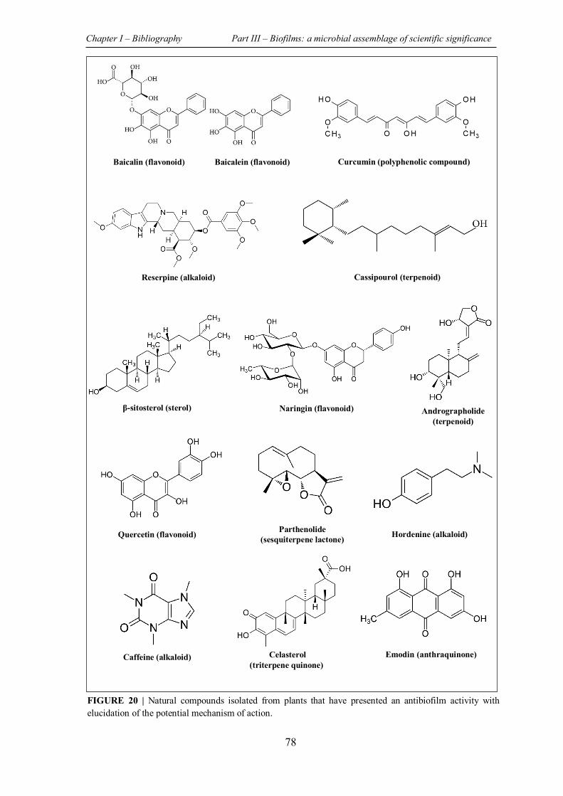
Chapter I – Bibliography Part III – Biofilms: a microbial assemblage of scientific significance
78
Baicalin (flavonoid) Baicalein (flavonoid) Curcumin (polyphenolic compound)
Reserpine (alkaloid)
β-sitosterol (sterol) Naringin (flavonoid) Andrographolide (terpenoid)
Quercetin (flavonoid) Parthenolide (sesquiterpene lactone) Hordenine (alkaloid)
Caffeine (alkaloid) Celasterol (triterpene quinone)
Emodin (anthraquinone)
Cassipourol (terpenoid)
FIGURE 20 | Natural compounds isolated from plants that have presented an antibiofilm activity with elucidation of the potential mechanism of action.
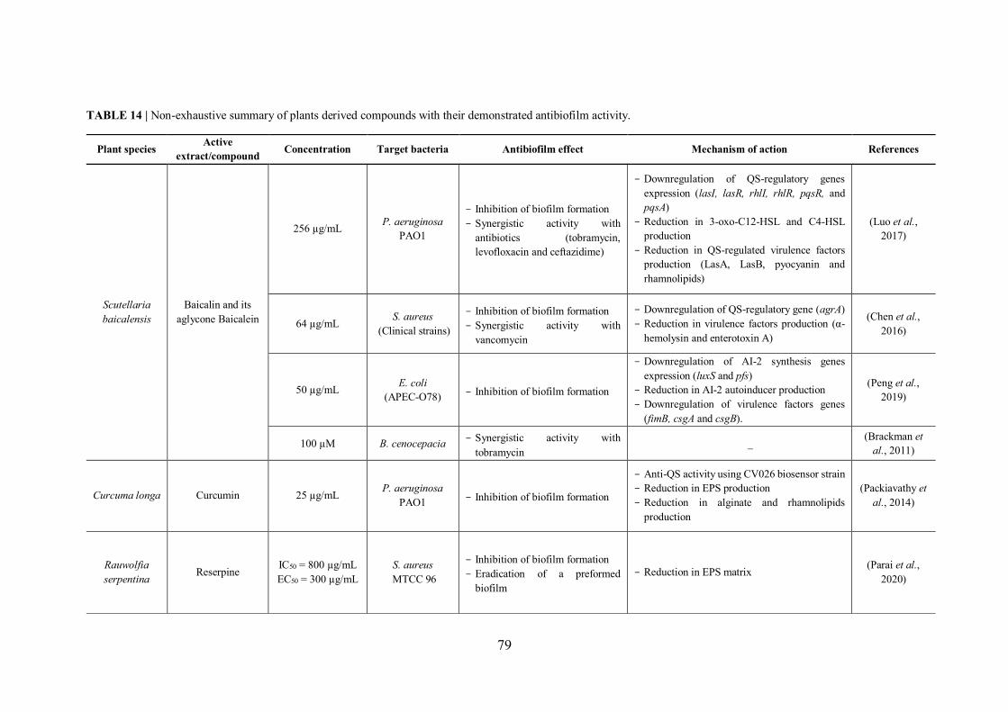
79
TABLE 14 | Non-exhaustive summary of plants derived compounds with their demonstrated antibiofilm activity.
Plant species Active extract/compound Concentration Target bacteria Antibiofilm effect Mechanism of action References
Scutellaria baicalensis
Baicalin and its aglycone Baicalein
256 µg/mL P. aeruginosa PAO1
- Inhibition of biofilm formation - Synergistic activity with
antibiotics (tobramycin, levofloxacin and ceftazidime)
- Downregulation of QS-regulatory genes expression (lasI, lasR, rhlI, rhlR, pqsR, and pqsA)
- Reduction in 3-oxo-C12-HSL and C4-HSL production
- Reduction in QS-regulated virulence factors production (LasA, LasB, pyocyanin and rhamnolipids)
(Luo et al., 2017)
64 µg/mL S. aureus (Clinical strains)
- Inhibition of biofilm formation - Synergistic activity with
vancomycin
- Downregulation of QS-regulatory gene (agrA) - Reduction in virulence factors production (α-
hemolysin and enterotoxin A)
(Chen et al., 2016)
50 µg/mL E. coli (APEC-O78) - Inhibition of biofilm formation
- Downregulation of AI-2 synthesis genes expression (luxS and pfs)
- Reduction in AI-2 autoinducer production - Downregulation of virulence factors genes
(fimB, csgA and csgB).
(Peng et al., 2019)
100 µM B. cenocepacia - Synergistic activity with tobramycin
_ (Brackman et al., 2011)
Curcuma longa Curcumin 25 µg/mL P. aeruginosa PAO1 - Inhibition of biofilm formation
- Anti-QS activity using CV026 biosensor strain - Reduction in EPS production - Reduction in alginate and rhamnolipids
production
(Packiavathy et al., 2014)
Rauwolfia serpentina Reserpine IC50 = 800 µg/mL
EC50 = 300 µg/mL S. aureus
MTCC 96
- Inhibition of biofilm formation - Eradication of a preformed
biofilm - Reduction in EPS matrix (Parai et al.,
2020)
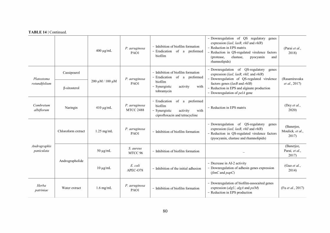
80
400 µg/mL P. aeruginosa PAO1
- Inhibition of biofilm formation - Eradication of a preformed
biofilm
- Downregulation of QS regulatory genes expression (lasI, lasR, rhlI and rhlR)
- Reduction in EPS matrix - Reduction in QS-regulated virulence factors
(protease, elastase, pyocyanin and rhamnolipids)
(Parai et al., 2018)
Platostoma rotundifolium
Cassipourol
200 µM / 100 µM P. aeruginosa PAO1
- Inhibition of biofilm formation - Eradication of a preformed
biofilm - Synergistic activity with
tobramycin
- Downregulation of QS-regulatory genes expression (lasI, lasR, rhlI, and rhlR)
- Downregulation of QS-regulated virulence factors genes (lasB and rhlB)
- Reduction in EPS and alginate production - Downregulation of pelA gene
(Rasamiravaka et al., 2017)
β-sitosterol
Combretum albiflorum Naringin 410 µg/mL P. aeruginosa
MTCC 2488
- Eradication of a preformed biofilm
- Synergistic activity with ciprofloxacin and tetracycline
- Reduction in EPS matrix (Dey et al., 2020)
Andrographis paniculata
Chloroform extract 1.25 mg/mL P. aeruginosa PAO1 - Inhibition of biofilm formation
- Downregulation of QS-regulatory genes expression (lasI, lasR, rhlI and rhlR)
- Reduction in QS-regulated virulence factors (pyocyanin, elastase and rhamnolipids)
(Banerjee, Moulick, et al.,
2017)
Andrographolide
50 µg/mL S. aureus MTCC 96 - Inhibition of biofilm formation _
(Banerjee, Parai, et al.,
2017)
10 µg/mL E. coli APEC-O78 - Inhibition of the initial adhesion
- Decrease in AI-2 activity - Downregulation of adhesin genes expression
(fimC and papC)
(Guo et al., 2014)
Herba patriniae Water extract 1.6 mg/mL P. aeruginosa
PAO1 - Inhibition of biofilm formation - Downregulation of biofilm-assocaited genes
expression (algU, algA and pslM) - Reduction in EPS production
(Fu et al., 2017)
TABLE 14 | Continued.
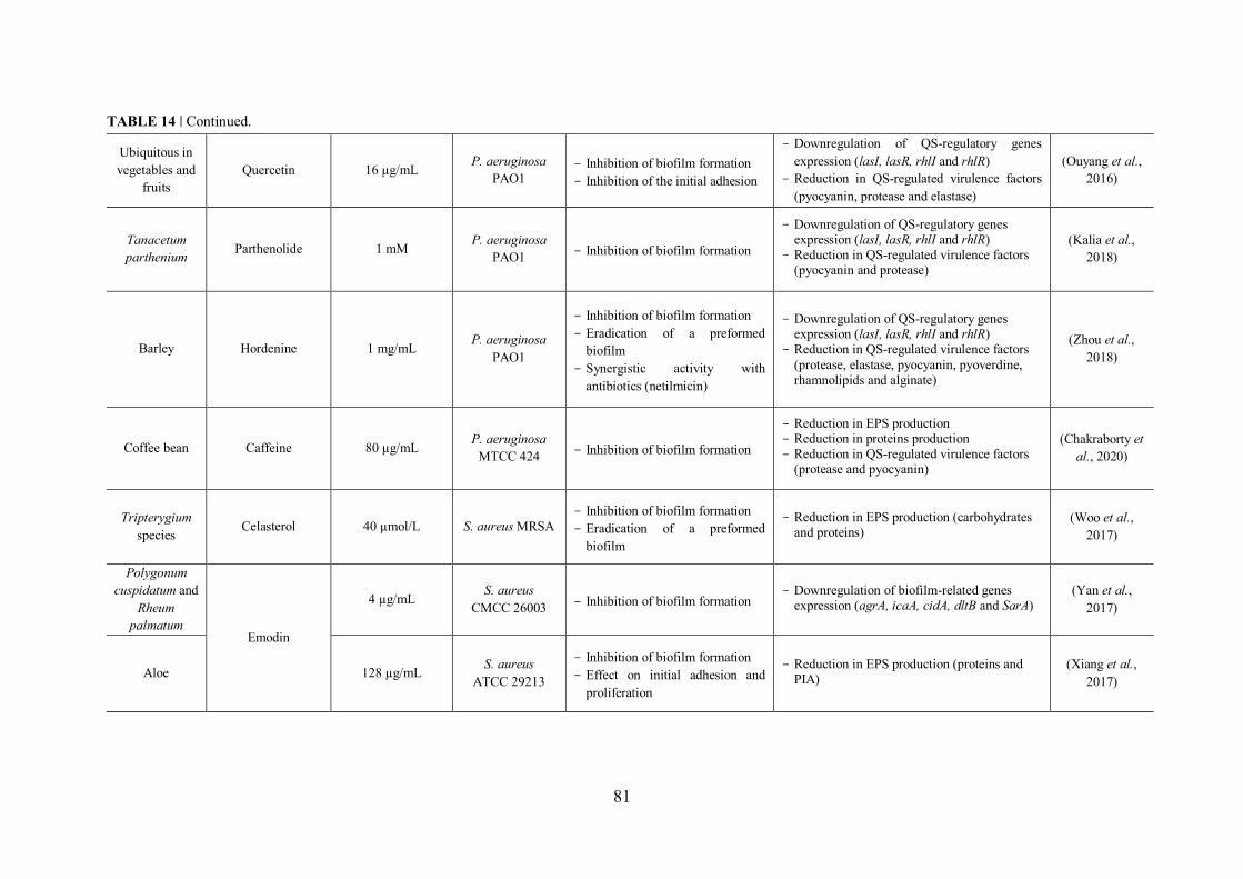
81
Ubiquitous in vegetables and
fruits Quercetin 16 µg/mL P. aeruginosa
PAO1 - Inhibition of biofilm formation - Inhibition of the initial adhesion
- Downregulation of QS-regulatory genes expression (lasI, lasR, rhlI and rhlR)
- Reduction in QS-regulated virulence factors (pyocyanin, protease and elastase)
(Ouyang et al., 2016)
Tanacetum parthenium Parthenolide 1 mM P. aeruginosa
PAO1 - Inhibition of biofilm formation
- Downregulation of QS-regulatory genes expression (lasI, lasR, rhlI and rhlR)
- Reduction in QS-regulated virulence factors (pyocyanin and protease)
(Kalia et al., 2018)
Barley Hordenine 1 mg/mL P. aeruginosa PAO1
- Inhibition of biofilm formation - Eradication of a preformed
biofilm - Synergistic activity with
antibiotics (netilmicin)
- Downregulation of QS-regulatory genes expression (lasI, lasR, rhlI and rhlR)
- Reduction in QS-regulated virulence factors (protease, elastase, pyocyanin, pyoverdine, rhamnolipids and alginate)
(Zhou et al., 2018)
Coffee bean Caffeine 80 µg/mL P. aeruginosa MTCC 424 - Inhibition of biofilm formation
- Reduction in EPS production - Reduction in proteins production - Reduction in QS-regulated virulence factors
(protease and pyocyanin)
(Chakraborty et al., 2020)
Tripterygium species Celasterol 40 µmol/L S. aureus MRSA
- Inhibition of biofilm formation - Eradication of a preformed
biofilm
- Reduction in EPS production (carbohydrates and proteins)
(Woo et al., 2017)
Polygonum cuspidatum and
Rheum palmatum
Emodin
4 µg/mL S. aureus CMCC 26003 - Inhibition of biofilm formation
- Downregulation of biofilm-related genes expression (agrA, icaA, cidA, dltB and SarA)
(Yan et al., 2017)
Aloe 128 µg/mL S. aureus ATCC 29213
- Inhibition of biofilm formation - Effect on initial adhesion and
proliferation
- Reduction in EPS production (proteins and PIA)
(Xiang et al., 2017)
TABLE 14 | Continued.
Chapter I – Bibliography Part III – Biofilms: a microbial assemblage of scientific significance
82
III.2.2.2 Marine environment: a valuable source of antibiofilm molecules
The stressful conditions faced by organisms living in marine environment, especially those
that are sessile, have led to the evolution of chemical defence systems in order to evade
predation and prevent biofouling (Torres et al., 2019). By definition, biofouling represents
the undesirable development of micro (bacteria, protists…) and macroorganisms
(invertebrates, algae…) on biotic or abiotic surfaces.
The uniqueness, as well as the huge structural and functional diversity of marine natural
products, and therefore their wide spectrum of demonstrated biological activities, are surely
related to the unique marine life conditions (Jimenez, 2018).
III.2.2.2.1 Seaweed derived compounds with antibiofilm activity
The ability of sessile seaweed to remain intact for long periods of time despite the high risk
of biofouling encountered in the marine environment suggests their strong capacity to
synthetize antifouling compounds. In this context, different studies have demonstrated the
antifouling activity of extracts and/or of compounds, especially those derived from brown
and red algae (Dahms & Dobretsov, 2017). However, despite the significant antibiofilm
activity exhibited by some seaweed derived compounds against pathogenic bacteria, these
marine organisms remain underexplored in novel antibiofilm agent’s discovery scenario
(Table 15).
Interestingly, the first natural molecule identified for its QS inhibitory activity and
consequently biofilm formation inhibitory is the halogenated furanone isolated from the
red alga Delisea pulchra. In fact, over 20 halogenated furanone compounds, differing in
the structure and the substitution in their side chain, as well as in the number and nature of
halogens, have been identified in this red alga, native to south eastern coast of Australia
(de Nys et al., 1993).
Initially, Manefield et al. have demonstrated the ability of a natural halogenated furanone
to inhibit the AHL-based QS system by displacing AHL signal molecule from its LuxR
receptor, which is explained by the structural similarity between this natural product and
the AHLs signal molecules (Manefield et al., 1999). Afterwards, the natural furanone has
provided a source of inspiration to chemists who have focused on the synthesis of a wide
variety of furanone analogues in order to elucidate the structure-activity relationships for
future use in drug development (Lyons et al., 2020).
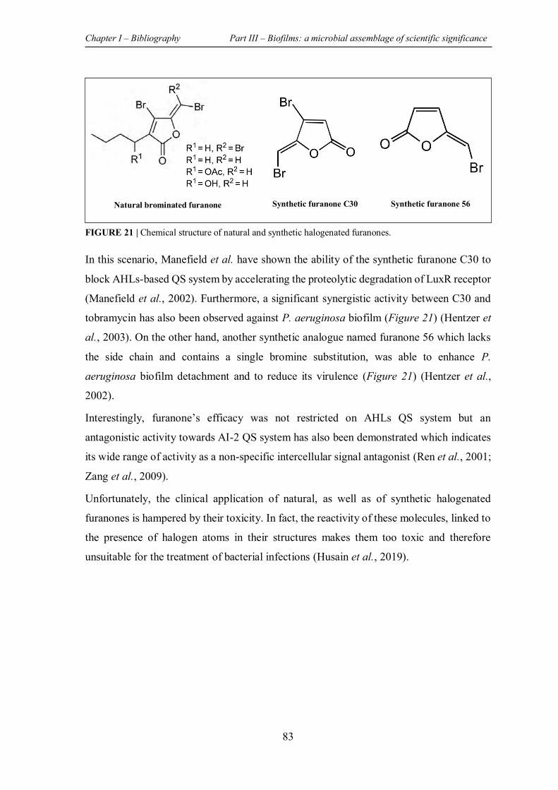
Chapter I – Bibliography Part III – Biofilms: a microbial assemblage of scientific significance
83
In this scenario, Manefield et al. have shown the ability of the synthetic furanone C30 to
block AHLs-based QS system by accelerating the proteolytic degradation of LuxR receptor
(Manefield et al., 2002). Furthermore, a significant synergistic activity between C30 and
tobramycin has also been observed against P. aeruginosa biofilm (Figure 21) (Hentzer et
al., 2003). On the other hand, another synthetic analogue named furanone 56 which lacks
the side chain and contains a single bromine substitution, was able to enhance P.
aeruginosa biofilm detachment and to reduce its virulence (Figure 21) (Hentzer et al.,
2002).
Interestingly, furanone’s efficacy was not restricted on AHLs QS system but an
antagonistic activity towards AI-2 QS system has also been demonstrated which indicates
its wide range of activity as a non-specific intercellular signal antagonist (Ren et al., 2001;
Zang et al., 2009).
Unfortunately, the clinical application of natural, as well as of synthetic halogenated
furanones is hampered by their toxicity. In fact, the reactivity of these molecules, linked to
the presence of halogen atoms in their structures makes them too toxic and therefore
unsuitable for the treatment of bacterial infections (Husain et al., 2019).
Natural brominated furanone Synthetic furanone C30 Synthetic furanone 56
FIGURE 21 | Chemical structure of natural and synthetic halogenated furanones.
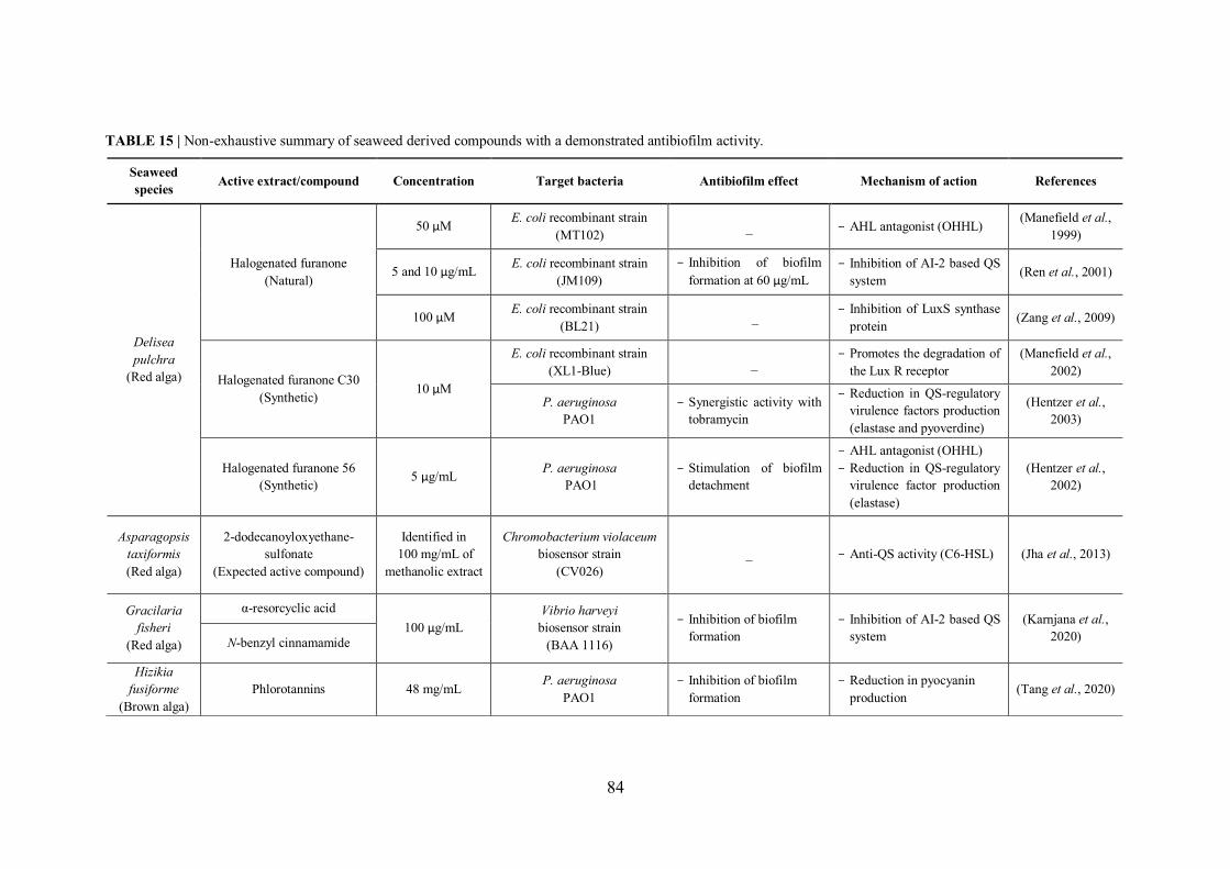
84
TABLE 15 | Non-exhaustive summary of seaweed derived compounds with a demonstrated antibiofilm activity.
Seaweed species Active extract/compound Concentration Target bacteria Antibiofilm effect Mechanism of action References
Delisea pulchra
(Red alga)
Halogenated furanone (Natural)
50 µM E. coli recombinant strain (MT102) _ - AHL antagonist (OHHL) (Manefield et al.,
1999)
5 and 10 µg/mL E. coli recombinant strain (JM109)
- Inhibition of biofilm formation at 60 µg/mL
- Inhibition of AI-2 based QS system (Ren et al., 2001)
100 µM E. coli recombinant strain (BL21) _ - Inhibition of LuxS synthase
protein (Zang et al., 2009)
Halogenated furanone C30 (Synthetic) 10 µM
E. coli recombinant strain (XL1-Blue) _
- Promotes the degradation of the Lux R receptor
(Manefield et al., 2002)
P. aeruginosa PAO1
- Synergistic activity with tobramycin
- Reduction in QS-regulatory virulence factors production (elastase and pyoverdine)
(Hentzer et al., 2003)
Halogenated furanone 56 (Synthetic) 5 µg/mL P. aeruginosa
PAO1 - Stimulation of biofilm
detachment
- AHL antagonist (OHHL) - Reduction in QS-regulatory
virulence factor production (elastase)
(Hentzer et al., 2002)
Asparagopsis taxiformis (Red alga)
2-dodecanoyloxyethane-sulfonate
(Expected active compound)
Identified in 100 mg/mL of
methanolic extract
Chromobacterium violaceum biosensor strain
(CV026) _ - Anti-QS activity (C6-HSL) (Jha et al., 2013)
Gracilaria fisheri
(Red alga)
α-resorcyclic acid 100 µg/mL
Vibrio harveyi biosensor strain
(BAA 1116)
- Inhibition of biofilm formation
- Inhibition of AI-2 based QS system
(Karnjana et al., 2020) N-benzyl cinnamamide
Hizikia fusiforme
(Brown alga) Phlorotannins 48 mg/mL P. aeruginosa
PAO1 - Inhibition of biofilm
formation - Reduction in pyocyanin
production (Tang et al., 2020)
Chapter I – Bibliography Part III – Biofilms: a microbial assemblage of scientific significance
85
III.2.2.2.2 Other antibiofilm compounds derived from marine macro- and
microorganisms
In addition to algae, the saline environment harbours a myriad of living organisms whose
significant ability to produce unique bioactive molecules notably antibiofilm ones, have
been demonstrated in various studies. The exploitation of marine microorganisms has
received a great attention in this field in view of their huge diversity, their ability to produce
active metabolites as well as the possibility of resource regeneration (Wang et al., 2017;
Goel et al., 2021). In this context, marine sponges, which account for around 30% of
bioactive marine molecules, present a valuable source of antibiofilm compounds, some of
which are effective against pathogenic bacteria such as P. aeruginosa and S. aureus (Stowe
et al., 2011).
Chapter I – Bibliography Part III – Biofilms: a microbial assemblage of scientific significance
86
III.3 EXPERIMENTAL BIOFILM ASSAYS USED FOR BIOFILM
STUDIES
Due to their rapid increase and their severity, biofilm-mediated infections are considered
as an important concern that requires the search for novel and effective antibiofilm agents.
However, the inerrant complexity and heterogeneity of biofilms make their exhaustive
analysis by integrating all its components very challenging. In fact, different qualitative
and quantitative methods have been applied, each of which targets a particular biofilm
feature (matrix, living cells, both living and dead cells…). Therefore, the combination of
various experimental approaches such as biochemical, genetic and physical ones is
considered the most suitable to achieve a global biofilm analysis (Pantanella et al., 2013).
In the following, the most commonly methods used for the quantification and/or the
visualization of biofilms are outlines, with their principles, their advantages, as well as their
limitations (Table 17).
III.3.1 Counting method – CFU counts assay
One of the commonly used methods to quantify biofilm cells is the colony forming units
(CFU) counts assay. Indeed, this method relies on the detachment of adhered cells from
surface by scraping or sonication followed by their spreading on agar-plate culture prior to
counting them. It should be noted that in comparison with two other assays (CV and
resazurin tests), Allkja et al., have highlighted the high responsiveness of this method in
antimicrobial efficacy test (Allkja et al., 2021).
However, one of the drawbacks of this method is the incomplete detachment of adhered
cells (soft scraping) and/or the presence of aggregates, which can lead to an
underestimation of biofilm cells number. Moreover, by CFU counts method, only the
culturable cells are considered (no data on dormant and viable but non-culturable cells
VBNC) (Pantanella et al., 2013; Azeredo et al., 2017).
III.3.2 Staining methods
III.3.2.1 Crystal violet assay (quantitative test)
Currently, one of the most widely used methods for bacterial biofilms quantification is the
crystal violet (CV) staining method. This assay consists in labelling the biofilm biomass
with CV.
Chapter I – Bibliography Part III – Biofilms: a microbial assemblage of scientific significance
87
The basic dye binds indifferently to the matrix polysaccharides, as well as to negatively
charged bacteria. The stain fixed by biofilm biomass is extracted by an organic solvent,
such as ethanol, methanol or acetic acid, prior to measuring the absorbance which is
proportional to the total biomass (Pantanella et al., 2013).
The main advantage of this method resides in the fact that it allows a high throughput
screening which is crucial, especially in the evaluation of the antibiofilm activity of large
libraries of compounds. However, since this assay quantifies the entire biofilm biomass
including living cells, dead cells, and the EPS matrix, its combination with another
approach that allows detachment and quantification of adhered cells such as the CFU count
method is required (Pantanella et al., 2013). Moreover, the sensitivity of this assay towards
experimental conditions of biofilm growth, such as environmental factors and inoculum
preparation method, reduces its reproducibility which makes the comparison of data
between studies not feasible (Allkja et al., 2020).
In addition, variation between bacterial species has also been recorded for this method
making the standardization of a well-defined protocol unachievable (Stiefel et al., 2016).
III.3.2.2 DMMB assay (quantitative test)
This colorimetric method is mostly used to quantify S. aureus biofilms. Briefly, the cationic
dye DMMB (1,9-dimethyl methylene blue) is able to bind to the intercellular
polysaccharide adhesin (PIA), a major component of S. aureus biofilm matrix (Pantanella
et al., 2013). The amount of fixed DMMB dye is determined by spectrophotometric
measurement (OD) after addition of a decomplexation solution (Tote et al., 2008).
The DMMB assay presents an economical, rapid and simple method. However, as the CV
staining assay, it is unable to assess the number of living bacterial cells within the biofilm
(Pantanella et al., 2013).
The major limitations of biofilm quantification methods that are based on OD measurement
lie in the detection limit, which is often high, as well as in the possible background signal.
Moreover, when evaluating the antibiofilm activity of a product, its potential absorbance
and/or its interaction with the dye can distort results (Stiefel et al., 2016).
III.3.3 Microscopic observations
In order to decipher biofilm spatial structure and its associated functions, several imaging
modalities have been used such as light microscopy, Scanning Electron Microscopy
(SEM), and Confocal Laser Scanning Microscopy (CLSM).
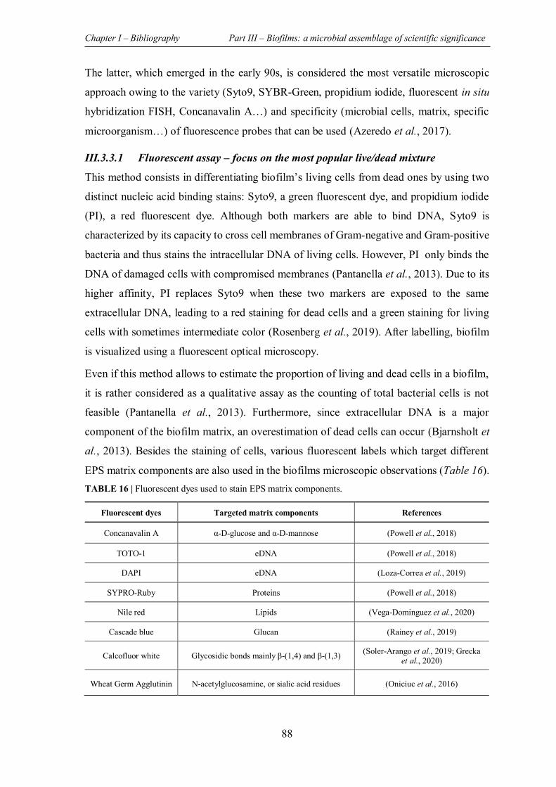
Chapter I – Bibliography Part III – Biofilms: a microbial assemblage of scientific significance
88
The latter, which emerged in the early 90s, is considered the most versatile microscopic
approach owing to the variety (Syto9, SYBR-Green, propidium iodide, fluorescent in situ
hybridization FISH, Concanavalin A…) and specificity (microbial cells, matrix, specific
microorganism…) of fluorescence probes that can be used (Azeredo et al., 2017).
III.3.3.1 Fluorescent assay – focus on the most popular live/dead mixture
This method consists in differentiating biofilm’s living cells from dead ones by using two
distinct nucleic acid binding stains: Syto9, a green fluorescent dye, and propidium iodide
(PI), a red fluorescent dye. Although both markers are able to bind DNA, Syto9 is
characterized by its capacity to cross cell membranes of Gram-negative and Gram-positive
bacteria and thus stains the intracellular DNA of living cells. However, PI only binds the
DNA of damaged cells with compromised membranes (Pantanella et al., 2013). Due to its
higher affinity, PI replaces Syto9 when these two markers are exposed to the same
extracellular DNA, leading to a red staining for dead cells and a green staining for living
cells with sometimes intermediate color (Rosenberg et al., 2019). After labelling, biofilm
is visualized using a fluorescent optical microscopy.
Even if this method allows to estimate the proportion of living and dead cells in a biofilm,
it is rather considered as a qualitative assay as the counting of total bacterial cells is not
feasible (Pantanella et al., 2013). Furthermore, since extracellular DNA is a major
component of the biofilm matrix, an overestimation of dead cells can occur (Bjarnsholt et
al., 2013). Besides the staining of cells, various fluorescent labels which target different
EPS matrix components are also used in the biofilms microscopic observations (Table 16). TABLE 16 | Fluorescent dyes used to stain EPS matrix components.
Fluorescent dyes Targeted matrix components References
Concanavalin A α-D-glucose and α-D-mannose (Powell et al., 2018)
TOTO-1 eDNA (Powell et al., 2018)
DAPI eDNA (Loza-Correa et al., 2019)
SYPRO-Ruby Proteins (Powell et al., 2018)
Nile red Lipids (Vega-Dominguez et al., 2020)
Cascade blue Glucan (Rainey et al., 2019)
Calcofluor white Glycosidic bonds mainly β-(1,4) and β-(1,3) (Soler-Arango et al., 2019; Grecka et al., 2020)
Wheat Germ Agglutinin N-acetylglucosamine, or sialic acid residues (Oniciuc et al., 2016)
Chapter I – Bibliography Part III – Biofilms: a microbial assemblage of scientific significance
89
III.3.4 Metabolic methods
Biofilm quantification based on cells metabolic activity presents another strategy adopted
in the study of microbial biofilms. The principle of this approach consists in the conversion
through cellular metabolic activity of a specific substrate into a detectable product (by
measurement of OD (XTT assay), fluorescence (resazurin assay), or of luminosity (ATP
assay)) (Azeredo et al., 2017).
III.3.4.1 Resazurin assay
Resazurin (7-hydroxy-3H-phenoxazin-3-one-10-oxide) is a non-fluorescent blue stain used
to quantify living cells within biofilms, based on their metabolic activity. Indeed, living
cells are able to reduce resazurin and irreversibly convert it into resorufin, a pink-
fluorescent dye. The fluorescence measurement of the formed resorufin reflects the number
of metabolically active living cells present within the biofilm (Pantanella et al., 2013).
One of the major drawbacks of this method is its high sensitivity to metabolic rate of
bacterial cells, which renders it dependent on their growth phase, as well as on biofilm age
and thickness. Furthermore, the standardization of the experimental conditions is difficult
as the time of resazurin reduction is species and strain specific (Van den Driessche et al.,
2014). In fact, Allkja et al., have demonstrated the suitability of this method for S. aureus
biofilm quantification. However, this assay has shown a poor responsiveness in treatment
efficacy experiments which can be explained by an unknown interaction between resazurin
and the tested compound (Allkja et al., 2021).
III.3.4.2 The XTT assay
As for the resazurin test, the XTT (2,3-bis (2-methoxy-4-nitro-5-sulfophenyl)-5-
[(phenylamino)carbonyl]-2H-tetrazolium hydroxide salt) method relies on the ability of the
respiratory metabolism of living cells to reduce the tetrazolium salt and to convert it to a
water-soluble formazan. The absorbance measurement of the supernatant permits to
determine the number of living cells within biofilm (Xu et al., 2016).
Like other metabolic tests, heterogeneity and complexity of biofilm structure, resulting in
a metabolic gradient, diminish the accuracy of XTT assay. On the other hand, the potential
retention of XTT reduction or formazan release limits the relevance of this assay in mature
biofilm studies (Pantanella et al., 2013).
Chapter I – Bibliography Part III – Biofilms: a microbial assemblage of scientific significance
90
III.3.4.3 The ATP assay
This method relies on the quantification of viable bacteria by their mitochondrial adenosine
triphosphate (ATP) which is directly proportional to their number. In fact, this assay is
based on the reaction between luciferin and ATP, catalyzed by luciferase, leading to the
formation of oxyluciferin with emission of a detectable luminescent signal (Herten et al.,
2017).
ATP assay is considered as a rapid, accurate, and sensitive method that can be used to
quantify various bacterial species. However, the required materials (e.g. luciferase and
luciferin) are relatively expensive, mainly for the analysis of numerous samples (Stiefel et
al., 2016; Herten et al., 2017).
III.3.5 Molecular biology methods
III.3.5.1 Quantitative polymerase chain reaction qPCR
The application of qPCR allows quantification of bacteria contained in a biofilm through
detection and quantification of specific gene sequences related to a bacterial species.
So, the major advantage of this technique in biofilm analysis is the ability to quantify
different species within one sample (Azeredo et al., 2017). However, this expensive
method is not informative about the proportion of living cells in a biofilm since it does not
differentiate between DNA of living and dead cells (Pantanella et al., 2013).
On the other hand, RT-qPCR (Reverse Transcription quantitative PCR) method which is
based on detection and quantification of mRNA with short half-life has been used in
biofilms studies. In this context, Magalhaes et al., have applied RNA-based qPCR in an
attempt to investigate the interactions occurring in a dual-species biofilm composed by P.
aeruginosa and S. aureus (Magalhaes et al., 2019).
For both of these gene-based assays, the choice of the primer sequences is a crucial step in
order to prevent the amplification of gene sequences not relevant for the objectives of the
study (Pantanella et al., 2013). In addition, the fact that these molecular biology methods
are not performed directly on biofilm but on the resuspended adhered cells (potential
modification of genes expression) present a drawback to these approaches.
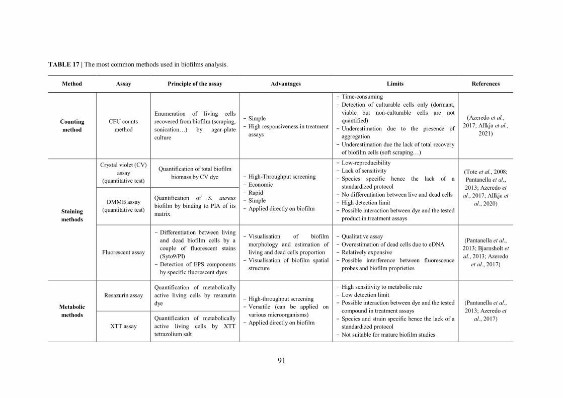
91
TABLE 17 | The most common methods used in biofilms analysis.
Method Assay Principle of the assay Advantages Limits References
Counting method
CFU counts method
Enumeration of living cells recovered from biofilm (scraping, sonication…) by agar-plate culture
- Simple - High responsiveness in treatment
assays
- Time-consuming - Detection of culturable cells only (dormant,
viable but non-culturable cells are not quantified)
- Underestimation due to the presence of aggregation
- Underestimation due the lack of total recovery of biofilm cells (soft scraping…)
(Azeredo et al., 2017; Allkja et al.,
2021)
Staining methods
Crystal violet (CV) assay
(quantitative test)
Quantification of total biofilm biomass by CV dye - High-Throughput screening
- Economic - Rapid - Simple - Applied directly on biofilm
- Low-reproducibility - Lack of sensitivity - Species specific hence the lack of a
standardized protocol - No differentiation between live and dead cells - High detection limit - Possible interaction between dye and the tested
product in treatment assays
(Tote et al., 2008; Pantanella et al., 2013; Azeredo et
al., 2017; Allkja et al., 2020)
DMMB assay
(quantitative test)
Quantification of S. aureus biofilm by binding to PIA of its matrix
Fluorescent assay
- Differentiation between living and dead biofilm cells by a couple of fluorescent stains (Syto9/PI)
- Detection of EPS components by specific fluorescent dyes
- Visualisation of biofilm morphology and estimation of living and dead cells proportion
- Visualisation of biofilm spatial structure
- Qualitative assay - Overestimation of dead cells due to eDNA - Relatively expensive - Possible interference between fluorescence
probes and biofilm proprieties
(Pantanella et al., 2013; Bjarnsholt et al., 2013; Azeredo
et al., 2017)
Metabolic methods
Resazurin assay Quantification of metabolically active living cells by resazurin dye
- High-throughput screening - Versatile (can be applied on
various microorganisms) - Applied directly on biofilm
- High sensitivity to metabolic rate - Low detection limit - Possible interaction between dye and the tested
compound in treatment assays - Species and strain specific hence the lack of a
standardized protocol - Not suitable for mature biofilm studies
(Pantanella et al., 2013; Azeredo et
al., 2017) XTT assay
Quantification of metabolically active living cells by XTT tetrazolium salt
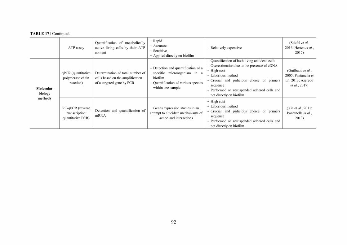
92
ATP assay Quantification of metabolically active living cells by their ATP content
- Rapid - Accurate - Sensitive - Applied directly on biofilm
- Relatively expensive (Stiefel et al.,
2016; Herten et al., 2017)
Molecular biology
methods
qPCR (quantitative polymerase chain
reaction)
Determination of total number of cells based on the amplification of a targeted gene by PCR
- Detection and quantification of a specific microorganism in a biofilm
- Quantification of various species within one sample
- Quantification of both living and dead cells - Overestimation due to the presence of eDNA - High-cost - Laborious method - Crucial and judicious choice of primers
sequence - Performed on resuspended adhered cells and
not directly on biofilm
(Guilbaud et al., 2005; Pantanella et al., 2013; Azeredo
et al., 2017)
RT-qPCR (reverse transcription
quantitative PCR)
Detection and quantification of mRNA
Genes expression studies in an attempt to elucidate mechanisms of
action and interactions
- High cost - Laborious method - Crucial and judicious choice of primers
sequence - Performed on resuspended adhered cells and
not directly on biofilm
(Xie et al., 2011; Pantanella et al.,
2013)
TABLE 17 | Continued.
93
MAIN OBJECTIVES
The previous literature review highlights the richness of seaweed in bioactive molecules
that can be valorized in different fields. Indeed, although the exploration of algae for their
potential insecticidal and mostly for their antibiofilm activity remains limited, some
extracts and/or compounds derived from seaweed have been documented for their
effectiveness in these two areas. In this context, we focus in this study on the exploration
of three algae collected from Lebanon in terms of their potential insecticidal and antibiofilm
activity against the fruit fly D. melanogaster and two pathogenic bacteria (P. aeruginosa
and S. aureus), respectively.
Regarding the evaluation of the insecticidal activity, the effect of the green alga
U. lactuca on the fruit fly D. melanogaster was assessed using different methods, each
of which presents a specific mode of exposure.
Concerning the evaluation of the antibiofilm activity, the potential ability of extracts
derived from three algae (green U. lactuca, brown S. scoparium, and red P. capillacea)
to impair both P. aeruginosa and S. aureus biofilm was assessed. Different methods
have been used in an attempt to decipher the antibiofilm mechanism of action exhibited
by the active extracts.
94
REFERENCES Abbassy, M., Marzouk, M., Rabea, E., & Abd-Elnabi, A. (2014). Insecticidal and Fungicidal
Activity of Ulva lactuca Linnaeus (Chlorophyta) Extracts and their Fractions. Annu. res. rev. biol, 4(13), 2252-2262. doi:10.9734/arrb/2014/9511
Abou-Elnaga, Z. S., Alarif, W. M., & Al-lihaibi, S. S. (2011). New Larvicidal Acetogenin from the Red Alga Laurencia papillosa. CLEAN - Soil, Air, Water, 39(8), 787-794. doi:10.1002/clen.201000597
Abou Zeid, A. H., Aboutabl, E. A., Sleem, A. A., & El-Rafie, H. M. (2014). Water soluble polysaccharides extracted from Pterocladia capillacea and Dictyopteris membranacea and their biological activities. Carbohydr Polym, 113, 62-66. doi:10.1016/j.carbpol.2014.06.004
Aboutabl, E. A., Abou Zeid, A. H., Sleem, A. A., & El Rafi, H. M. (2010). Secondary metabolites and certain bioactivities of Pterocladiella capillacea (S. Gmelin) bornet and Dictyopteris menbranacea (Stackhouse) Batters Med Aromat Plant Sci Biotechnol, 4 (1), 41-48.
Ahmer, B. M. (2004). Cell-to-cell signalling in Escherichia coli and Salmonella enterica. Mol Microbiol, 52(4), 933-945. doi:10.1111/j.1365-2958.2004.04054.x
Al-Zahrani, A., Al-Judaibi, E., Omar, H., & Al-Judaibi, A. (2017). Effects of Biochemical and Molecular Inhibitors of Plant Extracts on Pathogenic Bacteria. J Biosci Med, 05(05), 44-55. doi:10.4236/jbm.2017.55005
Al Monla, R., Dassouki, Z., Gali-Muhtasib, H., & Mawlawi, H. (2020). Chemical analysis and biological potentials of extracts from Colpomenia sinuosa. Phcog Res, 12:272-277. doi:10.4103/pr.pr_91_19
Al Monla, R., Dassouki, Z., Kouzayha, A., Salma, Y., Gali-Muhtasib, H., & Mawlawi, H. (2020). The Cytotoxic and Apoptotic Effects of the Brown Algae Colpomenia sinuosa are Mediated by the Generation of Reactive Oxygen Species. Molecules, 25(8). doi:10.3390/molecules25081993
Al Monla, R., Salma, Y., Kouzayha, A., Gali-Muhtasib, H., Dassouki, Z., & Mawlawi, H. (2021). Antioxidative, cytotoxic, and anti-metastatic potentials of Laurencia obtusa and Ulva lactuca seaweeds. Asian Pac. J. Trop. Biomed, 11(7). doi:10.4103/2221-1691.317242
Alagan, V., Valsala, R., & Rajesh, K. (2017). Bioactive Chemical Constituent Analysis, in vitro Antioxidant and Antimicrobial Activity of Whole Plant Methanol Extracts of Ulva lactuca Linn. Br. J. Pharm. Res, 15(1), 1-14. doi:10.9734/bjpr/2017/31818
Alarif, W. M., Abou-Elnaga, Z. S., Ayyad, S.-E. N., & Al-lihaibi, S. S. (2010). Insecticidal Metabolites from the Green Alga Caulerpa racemosa. CLEAN - Soil, Air, Water, 38(5-6), 548-557. doi:10.1002/clen.201000033
Alasri, A., Roques, C., Michel, G., Cabassud, C., & Aptel, P. (1992). Bactericidal properties of peracetic acid and hydrogen peroxide, alone and in combination, and chlorine and formaldehyde against bacterial water strains. Can J Microbiol, 38(7), 635-642. doi:10.1139/m92-104
Alemayehu, D., Casey, P. G., McAuliffe, O., Guinane, C. M., Martin, J. G., Shanahan, F., et al. (2012). Bacteriophages phiMR299-2 and phiNH-4 can eliminate Pseudomonas aeruginosa in the murine lung and on cystic fibrosis lung airway cells. mBio, 3(2), e00029-00012. doi:10.1128/mBio.00029-12
AlgaeBase. (2021). World-wide electronic publication, National University of Ireland, Galway. Retrieved from http://www.algaebase.orgsearched on 28 June 2021
References
95
Alkawash, M. A., Soothill, J. S., & Schiller, N. L. (2006). Alginate lyase enhances antibiotic killing of mucoid Pseudomonas aeruginosa in biofilms. APMIS, 114(2), 131-138. doi:10.1111/j.1600-0463.2006.apm_356.x
Allegrone, G., Ceresa, C., Rinaldi, M., & Fracchia, L. (2021). Diverse Effects of Natural and Synthetic Surfactants on the Inhibition of Staphylococcus aureus Biofilm. Pharmaceutics, 13(8). doi:10.3390/pharmaceutics13081172
Allkja, J., Bjarnsholt, T., Coenye, T., Cos, P., Fallarero, A., Harrison, J. J., et al. (2020). Minimum information guideline for spectrophotometric and fluorometric methods to assess biofilm formation in microplates. Biofilm, 2, 100010. doi:10.1016/j.bioflm.2019.100010
Allkja, J., van Charante, F., Aizawa, J., Reigada, I., Guarch-Perez, C., Vazquez-Rodriguez, J. A., et al. (2021). Interlaboratory study for the evaluation of three microtiter plate-based biofilm quantification methods. Scientific Reports, 11(1), 13779. doi:10.1038/s41598-021-93115-w
Anjali, K. P., Sangeetha, B. M., Devi, G., Raghunathan, R., & Dutta, S. (2019). Bioprospecting of seaweeds (Ulva lactuca and Stoechospermum marginatum): The compound characterization and functional applications in medicine-a comparative study. J Photochem Photobiol B, 200, 111622. doi:10.1016/j.jphotobiol.2019.111622
Antoniani, D., Bocci, P., Maciag, A., Raffaelli, N., & Landini, P. (2010). Monitoring of diguanylate cyclase activity and of cyclic-di-GMP biosynthesis by whole-cell assays suitable for high-throughput screening of biofilm inhibitors. Appl Microbiol Biotechnol, 85(4), 1095-1104. doi:10.1007/s00253-009-2199-x
Antonioli, L., Blandizzi, C., Pacher, P., Guilliams, M., & Hasko, G. (2018). Quorum sensing in the immune system. Nat Rev Immunol, 18(9), 537-538. doi:10.1038/s41577-018-0040-4
Apatzidou, D. A., & Kinane, D. F. (2010). Nonsurgical mechanical treatment strategies for periodontal disease. Dent Clin North Am, 54(1), 1-12. doi:10.1016/j.cden.2009.08.006
Archer, N. K., Mazaitis, M. J., Costerton, J. W., Leid, J. G., Powers, M. E., & Shirtliff, M. E. (2011). Staphylococcus aureus biofilms: properties, regulation, and roles in human disease. Virulence, 2(5), 445-459. doi:10.4161/viru.2.5.17724
Arciola, C. R., Campoccia, D., Ravaioli, S., & Montanaro, L. (2015). Polysaccharide intercellular adhesin in biofilm: structural and regulatory aspects. Front Cell Infect Microbiol, 5, 7. doi:10.3389/fcimb.2015.00007
Arciola, C. R., Campoccia, D., Speziale, P., Montanaro, L., & Costerton, J. W. (2012). Biofilm formation in Staphylococcus implant infections. A review of molecular mechanisms and implications for biofilm-resistant materials. Biomaterials, 33(26), 5967-5982. doi:10.1016/j.biomaterials.2012.05.031
Arsianti, A. A., Fadilah, F., Suid, K., Yazid, F., Wibisono, L. K., Azizah, N. N., et al. (2016). Phytochemical composition and anticancer activity of seaweeds Ulva lactuca and Eucheuma cottonii against breast MCF-7 and colon HCT-116 cells Asian Journal of Pharmaceutical and Clinical Research, 9(6), 115. doi:10.22159/ajpcr.2016.v9i6.13798
Asad, S., & Opal, S. M. (2008). Bench-to-bedside review: Quorum sensing and the role of cell-to-cell communication during invasive bacterial infection. Crit Care, 12(6), 236. doi:10.1186/cc7101
Azeredo, J., Azevedo, N. F., Briandet, R., Cerca, N., Coenye, T., Costa, A. R., et al. (2017). Critical review on biofilm methods. Crit Rev Microbiol, 43(3), 313-351. doi:10.1080/1040841X.2016.1208146
References
96
Bachtiar, E. W., & Bachtiar, B. M. (2020). Effect of cell-free spent media prepared from Aggregatibacter actinomycetemcomitans on the growth of Candida albicans and Streptococcus mutans in co-species biofilms. Eur J Oral Sci, 128(5), 395-404. doi:10.1111/eos.12725
Bak, J., Begovic, T., Bjarnsholt, T., & Nielsen, A. (2011). A UVC device for intra-luminal disinfection of catheters: in vitro tests on soft polymer tubes contaminated with Pseudomonas aeruginosa, Staphylococcus aureus, Escherichia coli and Candida albicans. Photochem Photobiol, 87(5), 1123-1128. doi:10.1111/j.1751-1097.2011.00962.x
Baldry, M., Nielsen, A., Bojer, M. S., Zhao, Y., Friberg, C., Ifrah, D., et al. (2016). Norlichexanthone Reduces Virulence Gene Expression and Biofilm Formation in Staphylococcus aureus. PLoS One, 11(12), e0168305. doi:10.1371/journal.pone.0168305
Baloch, G., Tariq, S., Ehteshamul-Haque, S., Athar, M., Sultana, V., & Ara, J. (2013). Management of root diseases of eggplant and watermelon with the application of asafoetida and seaweeds. J. Appl. Bot. Food Qual, 86, 138 - 142. doi:10.5073/JABFQ.2013.086.019
Banerjee, M., Moulick, S., Bhattacharya, K. K., Parai, D., Chattopadhyay, S., & Mukherjee, S. K. (2017). Attenuation of Pseudomonas aeruginosa quorum sensing, virulence and biofilm formation by extracts of Andrographis paniculata. Microb Pathog, 113, 85-93. doi:10.1016/j.micpath.2017.10.023
Banerjee, M., Parai, D., Chattopadhyay, S., & Mukherjee, S. K. (2017). Andrographolide: antibacterial activity against common bacteria of human health concern and possible mechanism of action. Folia Microbiol (Praha), 62(3), 237-244. doi:10.1007/s12223-017-0496-9
Banin, E., Lozinski, A., Brady, K. M., Berenshtein, E., Butterfield, P. W., Moshe, M., et al. (2008). The potential of desferrioxamine-gallium as an anti-Pseudomonas therapeutic agent. Proc Natl Acad Sci U S A, 105(43), 16761-16766. doi:10.1073/pnas.0808608105
Banin, E., Vasil, M. L., & Greenberg, E. P. (2005). Iron and Pseudomonas aeruginosa biofilm formation. Proc Natl Acad Sci U S A, 102(31), 11076-11081. doi:10.1073/pnas.0504266102
Barot, M., Kumar Ji, N., & Kumar, R. N. (2016). Bioactive compounds and antifungal activity of three different seaweed species Ulva lactuca, Sargassum tenerrimum and Laurencia obtusa collected from Okha coast, Western India. J. Coast. Life Med, 4(4), 284-289. doi:10.12980/jclm.4.2016J5-185
Barraud, N., Schleheck, D., Klebensberger, J., Webb, J. S., Hassett, D. J., Rice, S. A., et al. (2009). Nitric oxide signaling in Pseudomonas aeruginosa biofilms mediates phosphodiesterase activity, decreased cyclic di-GMP levels, and enhanced dispersal. J Bacteriol, 191(23), 7333-7342. doi:10.1128/JB.00975-09
Bauer, J., Siala, W., Tulkens, P. M., & Van Bambeke, F. (2013). A combined pharmacodynamic quantitative and qualitative model reveals the potent activity of daptomycin and delafloxacin against Staphylococcus aureus biofilms. Antimicrob Agents Chemother, 57(6), 2726-2737. doi:10.1128/AAC.00181-13
Behrouz, B., Hashemi, F. B., Fatemi, M. J., Naghavi, S., Irajian, G., Halabian, R., et al. (2017). Immunization with Bivalent Flagellin Protects Mice against Fatal Pseudomonas aeruginosa Pneumonia. J Immunol Res, 2017, 5689709. doi:10.1155/2017/5689709
Belfield, K., Bayston, R., Hajduk, N., Levell, G., Birchall, J. P., & Daniel, M. (2017). Evaluation of combinations of putative anti-biofilm agents and antibiotics to eradicate biofilms of Staphylococcus aureus and Pseudomonas aeruginosa. J Antimicrob Chemother, 72(9), 2531-2538. doi:10.1093/jac/dkx192
References
97
Berra, L., Kolobow, T., Laquerriere, P., Pitts, B., Bramati, S., Pohlmann, J., et al. (2008). Internally coated endotracheal tubes with silver sulfadiazine in polyurethane to prevent bacterial colonization: a clinical trial. Intensive Care Med, 34(6), 1030-1037. doi:10.1007/s00134-008-1100-1
Bez, C., Covaceuzach, S., Bertani, I., Choudhary, K. S., & Venturi, V. (2021). LuxR Solos from Environmental Fluorescent Pseudomonads. mSphere, 6(2). doi:10.1128/mSphere.01322-20
Bitar, G., Ramos-EsplÁ, A. A., OcaÑA, O., Sghaier, Y. R., Forcada, A., Valle, C., et al. (2017). Introduced marine macroflora of Lebanon and its distribution on the Levantine coast. Mediterr. Mar. Sci, 18(1). doi:10.12681/mms.1993
Bjarnsholt, T., Ciofu, O., Molin, S., Givskov, M., & Hoiby, N. (2013). Applying insights from biofilm biology to drug development - can a new approach be developed? Nat Rev Drug Discov, 12(10), 791-808. doi:10.1038/nrd4000
Bjarnsholt, T., Jensen, P. O., Burmolle, M., Hentzer, M., Haagensen, J. A. J., Hougen, H. P., et al. (2005). Pseudomonas aeruginosa tolerance to tobramycin, hydrogen peroxide and polymorphonuclear leukocytes is quorum-sensing dependent. Microbiology (Reading), 151(Pt 2), 373-383. doi:10.1099/mic.0.27463-0
Blunt, J. W., Carroll, A. R., Copp, B. R., Davis, R. A., Keyzers, R. A., & Prinsep, M. R. (2018). Marine natural products. Nat Prod Rep, 35(1), 8-53. doi:10.1039/c7np00052a
Boles, B. R., & Horswill, A. R. (2008). Agr-mediated dispersal of Staphylococcus aureus biofilms. PLoS Pathog, 4(4), e1000052. doi:10.1371/journal.ppat.1000052
Brackman, G., Cos, P., Maes, L., Nelis, H. J., & Coenye, T. (2011). Quorum sensing inhibitors increase the susceptibility of bacterial biofilms to antibiotics in vitro and in vivo. Antimicrob Agents Chemother, 55(6), 2655-2661. doi:10.1128/AAC.00045-11
Bridier, A., Briandet, R., Thomas, V., & Dubois-Brissonnet, F. (2011). Resistance of bacterial biofilms to disinfectants: a review. Biofouling, 27(9), 1017-1032. doi:10.1080/08927014.2011.626899
Burmolle, M., Thomsen, T. R., Fazli, M., Dige, I., Christensen, L., Homoe, P., et al. (2010). Biofilms in chronic infections - a matter of opportunity - monospecies biofilms in multispecies infections. FEMS Microbiol. Immunol, 59(3), 324-336. doi:10.1111/j.1574-695X.2010.00714.x
Campanac, C., Pineau, L., Payard, A., Baziard-Mouysset, G., & Roques, C. (2002). Interactions between Biocide Cationic Agents and Bacterial Biofilms. Antimicrob. Agents Chemother, 46(5), 1469-1474. doi:10.1128/AAC.46.5.1469-1474.2002
Campos, A. M., Matos, J., Afonso, C., Gomes, R., Bandarra, N. M., & Cardoso, C. (2018). Azorean macroalgae (Petalonia binghamiae, Halopteris scoparia and Osmundea pinnatifida) bioprospection: a study of fatty acid profiles and bioactivity. Int. J. Food Sci. Technol, 54(3), 880-890. doi:10.1111/ijfs.14010
Carson, R. (1962 ). Silent Spring (H. Mifflin Ed.). Boston - United States of America
Cegelski, L., Marshall, G. R., Eldridge, G. R., & Hultgren, S. J. (2008). The biology and future prospects of antivirulence therapies. Nat. Rev. Microbiol, 6(1), 17-27. doi:10.1038/nrmicro1818
Cetin, H., Gokoglu, M., & Oz, E. (2010). Larvicidal activity of the extract of seaweed, Caulerpa scalpelliformis, against Culex pipiens. J Am Mosq Control Assoc, 26(4), 433-435. doi:10.2987/10-6046.1
CFF. (2019). Cystic Fibrosis Foundation - Patient Registry Annual Data Report.
References
98
Chakraborty, P., Dastidar, D. G., Paul, P., Dutta, S., Basu, D., Sharma, S. R., et al. (2020). Inhibition of biofilm formation of Pseudomonas aeruginosa by caffeine: a potential approach for sustainable management of biofilm. Arch Microbiol, 202(3), 623-635. doi:10.1007/s00203-019-01775-0
Chaudhary, S., Kanwar, R. K., Sehgal, A., Cahill, D. M., Barrow, C. J., Sehgal, R., et al. (2017). Progress on Azadirachta indica Based Biopesticides in Replacing Synthetic Toxic Pesticides. Front Plant Sci, 8, 610. doi:10.3389/fpls.2017.00610
Chbani, A., Mawlawi, H., & Etahiri, S. (2011). Activité antibactérienne des extraits d’une algue brune Padina pavonica de la côte méditerranéenne au Liban. Phytothérapie, 9(5), 283-286. doi:10.1007/s10298-011-0634-5
Chen, Y., Liu, T., Wang, K., Hou, C., Cai, S., Huang, Y., et al. (2016). Baicalein Inhibits Staphylococcus aureus Biofilm Formation and the Quorum Sensing System In Vitro. PLoS One, 11(4), e0153468. doi:10.1371/journal.pone.0153468
Chiu, Y. H., Chan, Y. L., Li, T. L., & Wu, C. J. (2012). Inhibition of Japanese encephalitis virus infection by the sulfated polysaccharide extracts from Ulva lactuca. Mar Biotechnol (NY), 14(4), 468-478. doi:10.1007/s10126-011-9428-x
Cho, Y. H., Lee, S. J., Lee, J. Y., Kim, S. W., Kwon, I. C., Chung, S. Y., et al. (2001). Prophylactic efficacy of a new gentamicin-releasing urethral catheter in short-term catheterized rabbits. BJU Int, 87(1), 104-109. doi:10.1046/j.1464-410x.2001.00978.x
Christensen, L. D., van Gennip, M., Jakobsen, T. H., Alhede, M., Hougen, H. P., Hoiby, N., et al. (2012). Synergistic antibacterial efficacy of early combination treatment with tobramycin and quorum-sensing inhibitors against Pseudomonas aeruginosa in an intraperitoneal foreign-body infection mouse model. J Antimicrob Chemother, 67(5), 1198-1206. doi:10.1093/jac/dks002
Cinar, E., Taskin, E., Tasdemir, D., Ozkale, E., Grienke, U., & Firsova, D. (2019). Acetylcholinesterase, Antiprotozoal and Cytotoxic Activities of some turkish marine algae. Fresenius Environ. Bull, 28 (5), 3991-4000.
Cindana Mo’o, F. R., Wilar, G., Devkota, H. P., & Wathoni, N. (2020). Ulvan, a Polysaccharide from Macroalga Ulva sp.: A Review of Chemistry, Biological Activities and Potential for Food and Biomedical Applications. Appl. Sci, 10(16). doi:10.3390/app10165488
Ciofu, O., Bagge, N., & Hoiby, N. (2002). Antibodies against beta-lactamase can improve ceftazidime treatment of lung infection with beta-lactam-resistant Pseudomonas aeruginosa in a rat model of chronic lung infection. APMIS, 110(12), 881-891. doi:10.1034/j.1600-0463.2002.1101207.x
Ćirić, A. D., Petrović, J. D., Glamočlija, J. M., Smiljković, M. S., Nikolić, M. M., Stojković, D. S., et al. (2019). Natural products as biofilm formation antagonists and regulators of quorum sensing functions: A comprehensive review update and future trends. S. Afr. J. Bot, 120, 65-80. doi:10.1016/j.sajb.2018.09.010
Costa, K. C., Glasser, N. R., Conway, S. J., & Newman, D. K. (2017). Pyocyanin degradation by a tautomerizing demethylase inhibits Pseudomonas aeruginosa biofilms. Science, 355(6321), 170-173. doi:10.1126/science.aag3180
Costerton, J. W., Geesey, G. G., & Cheng, K. J. (1978). How bacteria stick. Sci Am, 238(1), 86-95. doi:10.1038/scientificamerican0178-86
Cucarella, C., Solano, C., Valle, J., Amorena, B., Lasa, I., & Penades, J. R. (2001). Bap, a Staphylococcus aureus surface protein involved in biofilm formation. J Bacteriol, 183(9), 2888-2896. doi:10.1128/JB.183.9.2888-2896.2001
Cunningham, A., Lennox, J., & Rockford, J. (2010). Biofilm: The hypertextbook Retrieved from http://www.hypertextbookshop.com/biofilmbook/v004/r003/
References
99
Dahms, H., & Dobretsov, S. (2017). Antifouling Compounds from Marine Macroalgae. Mar. Drugs, 15(9), 265. doi:10.3390/md15090265
Damalas, C., & Koutroubas, S. (2018). Current Status and Recent Developments in Biopesticide Use. Agriculture, 8(1). doi:10.3390/agriculture8010013
Dara, S. (2021). Biopesticides: Categories and use strategies for IPM and IRM. J. Entomol. Biologic.
Davies, D. (2003). Understanding biofilm resistance to antibacterial agents. Nat Rev Drug Discov, 2(2), 114-122. doi:10.1038/nrd1008
Davies, D., Chakrabarty, A., & Geesey, G. (1993). Exopolysaccharide production in biofilms: substratum activation of alginate gene expression by Pseudomonas aeruginosa. Appl. Environ. Microbiol, 59(4), 1181–1186.
Davies, D. G., & Marques, C. N. (2009). A fatty acid messenger is responsible for inducing dispersion in microbial biofilms. J Bacteriol, 191(5), 1393-1403. doi:10.1128/JB.01214-08
de Nys, R., Wright, A. D., König, G. M., & Sticher, O. (1993). New halogenated furanones from the marine alga Delisea pulchra (cf. fimbriata). Tetrahedron, 49(48), 11213-11220. doi:10.1016/s0040-4020(01)81808-1
Deligianni, E., Pattison, S., Berrar, D., Ternan, N. G., Haylock, R. W., Moore, J. E., et al. (2010). Pseudomonas aeruginosa cystic fibrosis isolates of similar RAPD genotype exhibit diversity in biofilm forming ability in vitro. BMC Microbiol, 10, 38. doi:10.1186/1471-2180-10-38
Deng, Z., Luo, X. M., Liu, J., & Wang, H. (2020). Quorum Sensing, Biofilm, and Intestinal Mucosal Barrier: Involvement the Role of Probiotic. Front Cell Infect Microbiol, 10, 538077. doi:10.3389/fcimb.2020.538077
Dey, P., Parai, D., Banerjee, M., Hossain, S. T., & Mukherjee, S. K. (2020). Naringin sensitizes the antibiofilm effect of ciprofloxacin and tetracycline against Pseudomonas aeruginosa biofilm. Int J Med Microbiol, 310(3), 151410. doi:10.1016/j.ijmm.2020.151410
Dominguez, H., & Loret, E. P. (2019). Ulva lactuca, A Source of Troubles and Potential Riches. Mar. Drugs, 17(6), 357. doi:10.3390/md17060357
Donne, J., & Dewilde, S. (2015). The Challenging World of Biofilm Physiology. Adv Microb Physiol, 67, 235-292. doi:10.1016/bs.ampbs.2015.09.003
Doring, G., & Gulbins, E. (2009). Cystic fibrosis and innate immunity: how chloride channel mutations provoke lung disease. Cell Microbiol, 11(2), 208-216. doi:10.1111/j.1462-5822.2008.01271.x
Doring, G., Meisner, C., Stern, M., & Flagella Vaccine Trial Study, G. (2007). A double-blind randomized placebo-controlled phase III study of a Pseudomonas aeruginosa flagella vaccine in cystic fibrosis patients. Proc Natl Acad Sci U S A, 104(26), 11020-11025. doi:10.1073/pnas.0702403104
Dueholm, M. S., Sondergaard, M. T., Nilsson, M., Christiansen, G., Stensballe, A., Overgaard, M. T., et al. (2013). Expression of Fap amyloids in Pseudomonas aeruginosa, P. fluorescens, and P. putida results in aggregation and increased biofilm formation. Microbiologyopen, 2(3), 365-382. doi:10.1002/mbo3.81
Dulger, B., Hacioglu, N., Erdugan, H., & Aysel, V. (2009). Antimicrobial Activity of Some Brown Algae from Turkey. Asian J. Chem, 21 (5), 4113-4117.
References
100
El Shouny, W. A., M. Gaafar, R., A. Ismail, G., & M. Elzanaty, M. (2017). Antibacterial Activity of Some Seaweed Extracts against Multidrug Resistant Urinary Tract Bacteria and Analysis of their Virulence Genes. Int J Curr Microbiol Appl Sci, 6(11), 2569-2586. doi:10.20546/ijcmas.2017.611.302
Elbrense, h., & Gheda, s. (2021). Evaluation of the insecticidal and antifeedant activities of some seaweed extracts against the Egyptian cotton leaf worm, Spodoptera littoralis, and the lesser grain borer Rhyzopertha dominica. Egypt. J. Exp. Biol. (Zoo.), 17(1). doi:10.5455/egysebz.20201218092110
EPA. (2021). Biopesticide Active Ingredients. Retrieved from https://www.epa.gov/ingredients-used-pesticide-products/biopesticide-active-ingredients On 17 August 2021
Esserti, S., Smaili, A., Rifai, L. A., Koussa, T., Makroum, K., Belfaiza, M., et al. (2016). Protective effect of three brown seaweed extracts against fungal and bacterial diseases of tomato. J. Appl. Phycol., 29(2), 1081-1093. doi:10.1007/s10811-016-0996-z
Fanaei Pirlar, R., Emaneini, M., Beigverdi, R., Banar, M., W, B. v. L., & Jabalameli, F. (2020). Combinatorial effects of antibiotics and enzymes against dual-species Staphylococcus aureus and Pseudomonas aeruginosa biofilms in the wound-like medium. PLoS One, 15(6), e0235093. doi:10.1371/journal.pone.0235093
FAO. (2021). FAOSTAT - Pesticides Use. Food and Agriculture Organization of the United Nations Retrieved from http://www.fao.org/faostat/en/#data/RP/visualizeon 03 August 2021
Farcas, A., Valceanu Matei, A., Florian, C., Badea, M., & Coman, G. (2013). Health Effects Associated with Acute and Chronic Exposure to Pesticides. In Environmental Security Assessment and Management of Obsolete Pesticides in Southeast Europe: NATO Science for Peace and Security Series C: Environmental Security. doi:10.1007/978-94-007-6461-3_8
Feng, L., Xiang, Q., Ai, Q., Wang, Z., Zhang, Y., & Lu, Q. (2016). Effects of Quorum Sensing Systems on Regulatory T Cells in Catheter-Related Pseudomonas aeruginosa Biofilm Infection Rat Models. Mediators Inflamm, 2016, 4012912. doi:10.1155/2016/4012912
Firoz, A., Haris, M., Hussain, K., Raza, M., Verma, D., Bouchama, M., et al. (2021). Can Targeting Iron Help in Combating Chronic Pseudomonas Infection? A Systematic Review. Cureus, 13(3), e13716. doi:10.7759/cureus.13716
Fleita, D., El-Sayed, M., & Rifaat, D. (2015). Evaluation of the antioxidant activity of enzymatically-hydrolyzed sulfated polysaccharides extracted from red algae; Pterocladia capillacea. LWT - Food Sci. Technol., 63(2), 1236-1244. doi:10.1016/j.lwt.2015.04.024
Fleitas Martinez, O., Cardoso, M. H., Ribeiro, S. M., & Franco, O. L. (2019). Recent Advances in Anti-virulence Therapeutic Strategies With a Focus on Dismantling Bacterial Membrane Microdomains, Toxin Neutralization, Quorum-Sensing Interference and Biofilm Inhibition. Front Cell Infect Microbiol, 9, 74. doi:10.3389/fcimb.2019.00074
Flemming, H.-C., & Wingender, J. (2010). The biofilm matrix. Nat. Rev. Microbiol., 8(9), 623-633. doi:10.1038/nrmicro2415
Flemming, H.-C., Wingender, J., Szewzyk, U., Steinberg, P., Rice, S. A., & Kjelleberg, S. (2016). Biofilms: an emergent form of bacterial life. Nat. Rev. Microbiol., 14(9), 563-575. doi:10.1038/nrmicro.2016.94
Folkesson, A., Jelsbak, L., Yang, L., Johansen, H. K., Ciofu, O., Hoiby, N., et al. (2012). Adaptation of Pseudomonas aeruginosa to the cystic fibrosis airway: an evolutionary perspective. Nat. Rev. Microbiol., 10(12), 841-851. doi:10.1038/nrmicro2907
References
101
Frederiksen, B., Pressler, T., Hansen, A., Koch, C., & Hoiby, N. (2006). Effect of aerosolized rhDNase (Pulmozyme) on pulmonary colonization in patients with cystic fibrosis. Acta Paediatr, 95(9), 1070-1074. doi:10.1080/08035250600752466
Fu, B., Wu, Q., Dang, M., Bai, D., Guo, Q., Shen, L., et al. (2017). Inhibition of Pseudomonas aeruginosa Biofilm Formation by Traditional Chinese Medicinal Herb Herba patriniae. Biomed Res Int, 2017, 9584703. doi:10.1155/2017/9584703
Furiga, A., Lajoie, B., El Hage, S., Baziard, G., & Roques, C. (2016). Impairment of Pseudomonas aeruginosa Biofilm Resistance to Antibiotics by Combining the Drugs with a New Quorum-Sensing Inhibitor. Antimicrobial Agents and Chemotherapy, 60(3), 1676-1686. doi:10.1128/AAC.02533-15
Furner-Pardoe, J., Anonye, B. O., Cain, R., Moat, J., Ortori, C. A., Lee, C., et al. (2020). Anti-biofilm efficacy of a medieval treatment for bacterial infection requires the combination of multiple ingredients. Scientific Reports, 10(1), 12687. doi:10.1038/s41598-020-69273-8
Fux, C., Stoodley, P., Shirtliff, M., & Costerton, W. (2009). The Functional Resistance of Bacterial Biofilms. In Antimicrobial Drug Resistance: Humana Press. doi:10.1007/978-1-59745-180-2_11
Galie, S., Garcia-Gutierrez, C., Miguelez, E. M., Villar, C. J., & Lombo, F. (2018). Biofilms in the Food Industry: Health Aspects and Control Methods. Front Microbiol, 9, 898. doi:10.3389/fmicb.2018.00898
Garrett, T. R., Bhakoo, M., & Zhang, Z. (2008). Bacterial adhesion and biofilms on surfaces. Prog. Nat. Sci., 18(9), 1049-1056. doi:10.1016/j.pnsc.2008.04.001
Gheda, S., El-Adawi, H., & El-Deeb, N. (2016). Antiviral Profile of Brown and Red Seaweed Polysaccharides Against Hepatitis C Virus. Iran. J. Pharm. Sci., 15 (3), 483-491.
Ghigo, J. M. (2001). Natural conjugative plasmids induce bacterial biofilm development. Nature, 412(6845), 442-445. doi:10.1038/35086581
Ghosh, U. U., Ali, H., Ghosh, R., & Kumar, A. (2021). Bacterial streamers as colloidal systems: Five grand challenges. J Colloid Interface Sci, 594, 265-278. doi:10.1016/j.jcis.2021.02.102
Goel, N., Fatima, S. W., Kumar, S., Sinha, R., & Khare, S. K. (2021). Antimicrobial resistance in biofilms: Exploring marine actinobacteria as a potential source of antibiotics and biofilm inhibitors. Biotechnol Rep (Amst), 30, e00613. doi:10.1016/j.btre.2021.e00613
González-Castro, A. L., Muñoz-Ochoa, M., Hernández-Carmona, G., & López-Vivas, J. M. (2019). Evaluation of seaweed extracts for the control of the Asian citrus psyllid Diaphorina citri. J. Appl. Phycol., 31(6), 3815-3821. doi:10.1007/s10811-019-01896-5
Grassi, L., Maisetta, G., Esin, S., & Batoni, G. (2017). Combination Strategies to Enhance the Efficacy of Antimicrobial Peptides against Bacterial Biofilms. Front Microbiol, 8, 2409. doi:10.3389/fmicb.2017.02409
Grecka, K., Xiong, Z. R., Chen, H., Pelka, K., Worobo, R. W., & Szweda, P. (2020). Effect of Ethanol Extracts of Propolis (EEPs) against Staphylococcal Biofilm-Microscopic Studies. Pathogens, 9(8). doi:10.3390/pathogens9080646
Gristina, A. G. (1987). Biomaterial-centered infection: microbial adhesion versus tissue integration. Science, 237(4822), 1588-1595. doi:10.1126/science.3629258
Guilbaud, M., de Coppet, P., Bourion, F., Rachman, C., Prevost, H., & Dousset, X. (2005). Quantitative detection of Listeria monocytogenes in biofilms by real-time PCR. Appl Environ Microbiol, 71(4), 2190-2194. doi:10.1128/AEM.71.4.2190-2194.2005
References
102
Guner, A., Nalbantsoy, A., Sukatar, A., & Karabay Yavasoglu, N. U. (2019). Apoptosis-inducing activities of Halopteris scoparia L. Sauvageau (Brown algae) on cancer cells and its biosafety and antioxidant properties. Cytotechnology, 71(3), 687-704. doi:10.1007/s10616-019-00314-5
Gunn, J. S., Bakaletz, L. O., & Wozniak, D. J. (2016). What's on the Outside Matters: The Role of the Extracellular Polymeric Substance of Gram-negative Biofilms in Evading Host Immunity and as a Target for Therapeutic Intervention. J Biol Chem, 291(24), 12538-12546. doi:10.1074/jbc.R115.707547
Guo, X., Zhang, L. Y., Wu, S. C., Xia, F., Fu, Y. X., Wu, Y. L., et al. (2014). Andrographolide interferes quorum sensing to reduce cell damage caused by avian pathogenic Escherichia coli. Vet Microbiol, 174(3-4), 496-503. doi:10.1016/j.vetmic.2014.09.021
Guzzo, F., Scognamiglio, M., Fiorentino, A., Buommino, E., & D’Abrosca, B. (2020). Plant Derived Natural Products against Pseudomonas aeruginosa and Staphylococcus aureus: Antibiofilm Activity and Molecular Mechanisms. Molecules, 25(21), 5024. doi:10.3390/molecules25215024
Gyawali, K. (2018). Pesticide Uses and its Effects on Public Health and Environment. J Health Promot, 6, 28-36. doi:10.3126/jhp.v6i0.21801
Haddad, M., Zein, S., Hazimeh, G., Karaki, R., Krivoruchko, E., Makhour, Y., et al. (2017). Structural Characteristics, Antitumor and Antioxidant Properties of Polysaccharides isolated from the brown algae Stypopodium schimperi growing on the Lebanese coast. ARJMD, 36-43.
Hall-Stoodley, L., Costerton, J. W., & Stoodley, P. (2004). Bacterial biofilms: from the natural environment to infectious diseases. Nat. Rev. Microbiol., 2(2), 95-108. doi:10.1038/nrmicro821
Hamed, I., Özogul, F., Özogul, Y., & Regenstein, J. M. (2015). Marine Bioactive Compounds and Their Health Benefits: A Review. Compr. Rev. Food Sci. Food Saf., 14(4), 446-465. doi:10.1111/1541-4337.12136
Hamed, S., Abd El-Rhman, A., Abdel-Raouf, N., & Ibraheem, I. (2018). Role of marine macroalgae in plant protection & improvement for sustainable agriculture technology. Int. j. basic appl. sci., 7(1), 104-110. doi:10.1016/j.bjbas.2017.08.002
Harms, A., Maisonneuve, E., & Gerdes, K. (2016). Mechanisms of bacterial persistence during stress and antibiotic exposure. Science, 354(6318). doi:10.1126/science.aaf4268
Hentzer, M., & Givskov, M. (2003). Pharmacological inhibition of quorum sensing for the treatment of chronic bacterial infections. J Clin Invest, 112(9), 1300-1307. doi:10.1172/JCI20074
Hentzer, M., Riedel, K., Rasmussen, T. B., Heydorn, A., Andersen, J. B., Parsek, M. R., et al. (2002). Inhibition of quorum sensing in Pseudomonas aeruginosa biofilm bacteria by a halogenated furanone compound. Microbiology (Reading), 148(Pt 1), 87-102. doi:10.1099/00221287-148-1-87
Hentzer, M., Teitzel, G. M., Balzer, G. J., Heydorn, A., Molin, S., Givskov, M., et al. (2001). Alginate overproduction affects Pseudomonas aeruginosa biofilm structure and function. J Bacteriol, 183(18), 5395-5401. doi:10.1128/jb.183.18.5395-5401.2001
Hentzer, M., Wu, H., Andersen, J. B., Riedel, K., Rasmussen, T. B., Bagge, N., et al. (2003). Attenuation of Pseudomonas aeruginosa virulence by quorum sensing inhibitors. EMBO J, 22(15), 3803-3815. doi:10.1093/emboj/cdg366
Herten, M., Bisdas, T., Knaack, D., Becker, K., Osada, N., Torsello, G. B., et al. (2017). Rapid in Vitro Quantification of S. aureus Biofilms on Vascular Graft Surfaces. Front Microbiol, 8, 2333. doi:10.3389/fmicb.2017.02333
References
103
Hickman, J. W., Tifrea, D. F., & Harwood, C. S. (2005). A chemosensory system that regulates biofilm formation through modulation of cyclic diguanylate levels. Proc Natl Acad Sci U S A, 102(40), 14422-14427. doi:10.1073/pnas.0507170102
Hmani, I., Ktari, L., Ismail, A., M’dallel, C., & El Bour, M. (2021). Assessment of the antioxidant and antibacterial properties of red algae (Rhodophyta) from the north coast of Tunisia. EMJE, 6(1). doi:10.1007/s41207-020-00222-7
Hoiby, N., Bjarnsholt, T., Givskov, M., Molin, S., & Ciofu, O. (2010). Antibiotic resistance of bacterial biofilms. Int J Antimicrob Agents, 35(4), 322-332. doi:10.1016/j.ijantimicag.2009.12.011
Hoiby, N., Flensborg, E., Beck, B., Friis, B., Jacobsen, S., & Jacobsen, L. (1977). Pseudomonas aeruginosa infection in cystic fibrosis. Diagnostic and prognostic significance of Pseudomonas aeruginosa precipitins determined by means of crossed immunoelectrophoresis. Scand J Respir Dis, 58(2), 65-79.
Holken Lorensi, G., Soares Oliveira, R., Leal, A. P., Zanatta, A. P., Moreira de Almeida, C. G., Barreto, Y. C., et al. (2019). Entomotoxic Activity of Prasiola crispa (Antarctic Algae) in Nauphoeta cinerea Cockroaches: Identification of Main Steroidal Compounds. Mar Drugs, 17(10). doi:10.3390/md17100573
Houchi, S., Mahdadi, R., Khenchouche, A., Song, J., Zhang, W., Pang, X., et al. (2019). Investigation of common chemical components and inhibitory effect on GES-type beta-lactamase (GES22) in methanolic extracts of Algerian seaweeds. Microb Pathog, 126, 56-62. doi:10.1016/j.micpath.2018.10.034
Husain, F., Al-Shabib, N., Noor, S., Khan, R., Khan, M., Ansari, F., et al. (2019). Chapter 12 - Current Strategy to Target Bacterial Quorum Sensing and Virulence by Phytocompounds. New Look to Phytomedicine - Advancements in Herbal Products as Novel Drug Leads. doi://doi.org/10.1016/B978-0-12-814619-4.00012-4
Inat, G., Siriken, B., Baskan, C., Erol, I., Yildirim, T., & Ciftci, A. (2021). Quorum sensing systems and related virulence factors in Pseudomonas aeruginosa isolated from chicken meat and ground beef. Sci. Rep., 11(1), 15639. doi:10.1038/s41598-021-94906-x
Ishwarya, R., Vaseeharan, B., Kalyani, S., Banumathi, B., Govindarajan, M., Alharbi, N. S., et al. (2018). Facile green synthesis of zinc oxide nanoparticles using Ulva lactuca seaweed extract and evaluation of their photocatalytic, antibiofilm and insecticidal activity. J Photochem Photobiol B, 178, 249-258. doi:10.1016/j.jphotobiol.2017.11.006
Ismail, G., Gheda, S., Abo-Shady, A., & Abdel-Karim, O. (2020). In vitro potential activity of some seaweeds as antioxidants and inhibitors of diabetic enzymes. Food Sci. Technol., 40 (3), 681-691. doi://doi.org/10.1590/fst.15619
Ismail, M., & Amer, M. (2020). Characterization and biological properties of sulfated polysaccharides of Corallina officinalis and Pterocladia capillacea. Acta Botanica Brasilica, 34 (4), 623-632. doi://doi.org/10.1590/0102-33062020abb0121
Jha, B., Kavita, K., Westphal, J., Hartmann, A., & Schmitt-Kopplin, P. (2013). Quorum Sensing Inhibition by Asparagopsis taxiformis, a Marine Macro Alga: Separation of the Compound that Interrupts Bacterial Communication. Mar. Drugs, 11, 253-265.
Jimenez, C. (2018). Marine Natural Products in Medicinal Chemistry. ACS Med Chem Lett, 9(10), 959-961. doi:10.1021/acsmedchemlett.8b00368
Kalia, M., Yadav, V. K., Singh, P. K., Sharma, D., Narvi, S. S., & Agarwal, V. (2018). Exploring the impact of parthenolide as anti-quorum sensing and anti-biofilm agent against Pseudomonas aeruginosa. Life Sci, 199, 96-103. doi:10.1016/j.lfs.2018.03.013
Kalia, V. C. (2013). Quorum sensing inhibitors: an overview. Biotechnol Adv, 31(2), 224-245. doi:10.1016/j.biotechadv.2012.10.004
References
104
Kanaan, H., Belous, O., & Chokr, A. (2014). Diversity Investigation of the Seaweeds Growing on the Lebanese Coast. J. mar. sci., res. dev., 05(01). doi:10.4172/2155-9910.1000156
Kaneko, Y., Thoendel, M., Olakanmi, O., Britigan, B. E., & Singh, P. K. (2007). The transition metal gallium disrupts Pseudomonas aeruginosa iron metabolism and has antimicrobial and antibiofilm activity. J Clin Invest, 117(4), 877-888. doi:10.1172/JCI30783
Karaki, N., Sebaaly, C., Chahine, N., Faour, T., Zinchenko, A., Rachid, S., et al. (2013). The antioxidant and anticoagulant activities of polysaccharides isolated from the brown algae Dictyopteris polypodioides growing on the Lebanese coast. J App Pharm Sci. doi:10.7324/japs.2013.30208
Karnjana, K., Nobsathian, S., Soowannayan, C., Zhao, W., Tang, Y. J., & Wongprasert, K. (2020). Purification and Evaluation of N-benzyl Cinnamamide from Red Seaweed Gracilaria fisheri as an Inhibitor of Vibrio harveyi AI-2 Quorum Sensing. Mar Drugs, 18(2). doi:10.3390/md18020080
Kesho, A. (2020). Microbial Bio-Pesticides and Their Use in Integrated Pest Management. Chem. Biomol. Eng., 5(1). doi:10.11648/j.cbe.20200501.15
Khalilzadeh, P., Lajoie, B., El Hage, S., Furiga, A., Baziard, G., Berge, M., et al. (2010). Growth inhibition of adherent Pseudomonas aeruginosa by an N-butanoyl-L-homoserine lactone analog. Can J Microbiol, 56(4), 317-325. doi:10.1139/w10-013
Kidgell, J. T., Magnusson, M., de Nys, R., & Glasson, C. R. K. (2019). Ulvan: A systematic review of extraction, composition and function. Algal Res., 39. doi:10.1016/j.algal.2019.101422
Kilic, M., Orhan, I. E., Eren, G., Okudan, E. S., Estep, A. S., Bencel, J. J., et al. (2021). Insecticidal activity of forty-seven marine algae species from the Mediterranean, Aegean, and Sea of Marmara in connection with their cholinesterase and tyrosinase inhibitory activity. S. Afr. J. Bot. doi:10.1016/j.sajb.2021.06.038
Kim, K. H., Kabir, E., & Jahan, S. A. (2017). Exposure to pesticides and the associated human health effects. Sci Total Environ, 575, 525-535. doi:10.1016/j.scitotenv.2016.09.009
Kim, Y., & Wood, T. K. (2010). Toxins Hha and CspD and small RNA regulator Hfq are involved in persister cell formation through MqsR in Escherichia coli. Biochem Biophys Res Commun, 391(1), 209-213. doi:10.1016/j.bbrc.2009.11.033
Kisiela, D. I., Avagyan, H., Friend, D., Jalan, A., Gupta, S., Interlandi, G., et al. (2015). Inhibition and Reversal of Microbial Attachment by an Antibody with Parasteric Activity against the FimH Adhesin of Uropathogenic E. coli. PLoS Pathog, 11(5), e1004857. doi:10.1371/journal.ppat.1004857
Koo, H., Allan, R. N., Howlin, R. P., Stoodley, P., & Hall-Stoodley, L. (2017). Targeting microbial biofilms: current and prospective therapeutic strategies. Nat. Rev. Microbiol., 15(12), 740-755. doi:10.1038/nrmicro.2017.99
Krzyzek, P. (2019). Challenges and Limitations of Anti-quorum Sensing Therapies. Front Microbiol, 10, 2473. doi:10.3389/fmicb.2019.02473
Kumar, J., Ramlal, A., Mallick, D., & Mishra, V. (2021). An Overview of Some Biopesticides and Their Importance in Plant Protection for Commercial Acceptance. Plants 10(6). doi:10.3390/plants10061185
Kumar, P., Kamle, M., Borah, R., Mahato, D. K., & Sharma, B. (2021). Bacillus thuringiensis as microbial biopesticide: uses and application for sustainable agriculture. Egypt. J. Biol. Pest Control., 31(1). doi:10.1186/s41938-021-00440-3
Lami, R. (2019). Chapter 3 - Quorum Sensing in Marine Biofilms and Environments. doi://doi.org/10.1016/B978-0-12-814905-8.00003-4
References
105
Lamret, F., Colin, M., Mongaret, C., Gangloff, S. C., & Reffuveille, F. (2020). Antibiotic Tolerance of Staphylococcus aureus Biofilm in Periprosthetic Joint Infections and Antibiofilm Strategies. Antibiotics (Basel), 9(9). doi:10.3390/antibiotics9090547
Leandro, A., Pereira, L., & Goncalves, A. M. M. (2019). Diverse Applications of Marine Macroalgae. Mar Drugs, 18(1). doi:10.3390/md18010017
Lebeaux, D., & Ghigo, J. M. (2012). Management of biofilm-associated infections: what can we expect from recent research on biofilm lifestyles? Med Sci (Paris), 28(8-9), 727-739. doi:10.1051/medsci/2012288015
Lee, J., & Zhang, L. (2015). The hierarchy quorum sensing network in Pseudomonas aeruginosa. Protein Cell 6(1):26–41.
Leng, P., Zhang, Z., Pan, G., & Zhao, M. (2011). Applications and development trends in biopesticides. Afr. j. biotechnol., 10(86). doi:10.5897/ajbx11.009
Li, Q., Ren, Y., & Fu, X. (2019). Inter-kingdom signaling between gut microbiota and their host. Cell Mol Life Sci, 76(12), 2383-2389. doi:10.1007/s00018-019-03076-7
Lin, M. H., Shu, J. C., Huang, H. Y., & Cheng, Y. C. (2012). Involvement of iron in biofilm formation by Staphylococcus aureus. PLoS One, 7(3), e34388. doi:10.1371/journal.pone.0034388
Lipuma, J. J. (2010). The changing microbial epidemiology in cystic fibrosis. Clin Microbiol Rev, 23(2), 299-323. doi:10.1128/CMR.00068-09
Liu, Y., Pan, X., & Li, J. (2014). A 1961–2010 record of fertilizer use, pesticide application and cereal yields: a review. Agron Sustain Dev, 35(1), 83-93. doi:10.1007/s13593-014-0259-9
López, A., Rico, M., Rivero, A., & Suárez de Tangil, M. (2011). The effects of solvents on the phenolic contents and antioxidant activity of Stypocaulon scoparium algae extracts. Food Chem., 125(3), 1104-1109. doi:10.1016/j.foodchem.2010.09.101
Louis, P., Scott, K. P., Duncan, S. H., & Flint, H. J. (2007). Understanding the effects of diet on bacterial metabolism in the large intestine. J Appl Microbiol, 102(5), 1197-1208. doi:10.1111/j.1365-2672.2007.03322.x
Lowery, C. A., Dickerson, T. J., & Janda, K. D. (2008). Interspecies and interkingdom communication mediated by bacterial quorum sensing. Chem Soc Rev, 37(7), 1337-1346. doi:10.1039/b702781h
Loza-Correa, M., Ayala, J. A., Perelman, I., Hubbard, K., Kalab, M., Yi, Q. L., et al. (2019). The peptidoglycan and biofilm matrix of Staphylococcus epidermidis undergo structural changes when exposed to human platelets. PLoS One, 14(1), e0211132. doi:10.1371/journal.pone.0211132
Lu, L., Hu, W., Tian, Z., Yuan, D., Yi, G., Zhou, Y., et al. (2019). Developing natural products as potential anti-biofilm agents. Chin Med, 14, 11. doi:10.1186/s13020-019-0232-2
Ludecke, C., Jandt, K. D., Siegismund, D., Kujau, M. J., Zang, E., Rettenmayr, M., et al. (2014). Reproducible biofilm cultivation of chemostat-grown Escherichia coli and investigation of bacterial adhesion on biomaterials using a non-constant-depth film fermenter. PLoS One, 9(1), e84837. doi:10.1371/journal.pone.0084837
Luiz de Freitas, L., Pereira da Silva, F., Fernandes, K. M., Carneiro, D. G., Licursi de Oliveira, L., Martins, G. F., et al. (2021). The virulence of Salmonella Enteritidis in Galleria mellonella is improved by N-dodecanoyl-homoserine lactone. Microb Pathog, 152, 104730. doi:10.1016/j.micpath.2021.104730
References
106
Luo, J., Dong, B., Wang, K., Cai, S., Liu, T., Cheng, X., et al. (2017). Baicalin inhibits biofilm formation, attenuates the quorum sensing-controlled virulence and enhances Pseudomonas aeruginosa clearance in a mouse peritoneal implant infection model. PLoS One, 12(4), e0176883. doi:10.1371/journal.pone.0176883
Luppens, S. B., Reij, M. W., van der Heijden, R. W., Rombouts, F. M., & Abee, T. (2002). Development of a standard test to assess the resistance of Staphylococcus aureus biofilm cells to disinfectants. Appl Environ Microbiol, 68(9), 4194-4200. doi:10.1128/aem.68.9.4194-4200.2002
Lyons, T., Gahan, C. G., & O'Sullivan, T. P. (2020). Structure-activity relationships of furanones, dihydropyrrolones and thiophenones as potential quorum sensing inhibitors. Future Med Chem, 12(21), 1925-1943. doi:10.4155/fmc-2020-0244
Machado, L., Gasparoto, M., Santos-Filho, N., & Pavarini, R. (2019). Chapter 6 - Seaweeds in the Control of Plant Diseases and Insects. In Seaweeds as Plant Fertilizer, Agricultural Biostimulants and Animal Fodder: CRC Press. doi:10.1201/9780429487156-6
Machado, L. P., Matsumoto, S. T., Jamal, C. M., da Silva, M. B., Centeno Dda, C., Colepicolo Neto, P., et al. (2014). Chemical analysis and toxicity of seaweed extracts with inhibitory activity against tropical fruit anthracnose fungi. J Sci Food Agric, 94(9), 1739-1744. doi:10.1002/jsfa.6483
Mack, D., Fischer, W., Krokotsch, A., Leopold, K., Hartmann, R., Egge, H., et al. (1996). The intercellular adhesin involved in biofilm accumulation of Staphylococcus epidermidis is a linear beta-1,6-linked glucosaminoglycan: purification and structural analysis. J Bacteriol, 178(1), 175-183. doi:10.1128/jb.178.1.175-183.1996
Mack, W. N., Mack, J. P., & Ackerson, A. O. (1975). Microbial film development in a trickling filter. Microb Ecol, 2(3), 215-226. doi:10.1007/BF02010441
MACOI. (2008). Portugueuse seaweeds website Retrieved from http://macoi.ci.uc.pt on 29 June 2021
Magalhaes, A. P., Franca, A., Pereira, M. O., & Cerca, N. (2019). RNA-based qPCR as a tool to quantify and to characterize dual-species biofilms. Sci. Rep. , 9(1), 13639. doi:10.1038/s41598-019-50094-3
Manefield, M., de Nys, R., Naresh, K., Roger, R., Givskov, M., Peter, S., et al. (1999). Evidence that halogenated furanones from Delisea pulchra inhibit acylated homoserine lactone (AHL)-mediated gene expression by displacing the AHL signal from its receptor protein. Microbiology, 145(2), 283-291. doi:10.1099/13500872-145-2-283
Manefield, M., Rasmussen, T. B., Henzter, M., Andersen, J. B., Steinberg, P., Kjelleberg, S., et al. (2002). Halogenated furanones inhibit quorum sensing through accelerated LuxR turnover. Microbiology (Reading), 148(Pt 4), 1119-1127. doi:10.1099/00221287-148-4-1119
Manilal, A., Sujith, S., Sabarathnam, B., Kiran, G., Selvin, J., Shakir, C., et al. (2011). Biological activity of the red alga Laurencia brandenii. Acta Bot Croat 70(1), 81-90. doi:10.2478/v10184-010-0001-x
Market Analysis Report. (2020). Commercial Seaweeds Market Size, Share & Trends Analysis Report By Product (Brown Seaweeds, Red Seaweeds, Green Seaweeds), By Form (Liquid, Powdered, Flakes), By Application, By Region, And Segment Forecasts, 2020 - 2027. Retrieved from https://www.grandviewresearch.com/industry-analysis/commercial-seaweed-market27 September 2021
Marty, N., Pasquier, C., Dournes, J. L., Chemin, K., Chavagnat, F., Guinand, M., et al. (1998). Effects of characterised Pseudomonas aeruginosa exopolysaccharides on adherence to human tracheal cells. J Med Microbiol, 47(2), 129-134. doi:10.1099/00222615-47-2-129
References
107
Mauline, L., Gressier, M., Hammer, P., Ribeiro, S., Caiut, J., Menu, M., et al. (2016). Bifunctional silica nanoparticles for the exploration of Pseudomonas aeruginosa biofilm. IEEE Nanotechnol. Mater. Devices Conf. NMDC, pp. 1-2. doi:10.1109/NMDC.2016.7777158
Medina-Rodriguez, E. M., Madorma, D., O'Connor, G., Mason, B. L., Han, D., Deo, S. K., et al. (2020). Identification of a Signaling Mechanism by Which the Microbiome Regulates Th17 Cell-Mediated Depressive-Like Behaviors in Mice. Am J Psychiatry, 177(10), 974-990. doi:10.1176/appi.ajp.2020.19090960
Melander, R. J., Basak, A. K., & Melander, C. (2020). Natural products as inspiration for the development of bacterial antibiofilm agents. Nat Prod Rep, 37(11), 1454-1477. doi:10.1039/d0np00022a
Michalak, I., & Chojnacka, K. (2015). Algae as production systems of bioactive compounds. Eng Life Sci., 15(2), 160-176. doi:10.1002/elsc.201400191
Mion, S., Remy, B., Plener, L., Chabriere, E., & Daude, D. (2019). Quorum sensing and quorum quenching: how to disrupt bacterial communication to inhibit virulence? Med Sci (Paris), 35(1), 31-38. doi:10.1051/medsci/2018310
Mishra, R., Panda, A. K., De Mandal, S., Shakeel, M., Bisht, S. S., & Khan, J. (2020). Natural Anti-biofilm Agents: Strategies to Control Biofilm-Forming Pathogens. Front Microbiol, 11, 566325. doi:10.3389/fmicb.2020.566325
Mohy El-Din, S. M., & El-Ahwany, A. M. D. (2018). Bioactivity and phytochemical constituents of marine red seaweeds (Jania rubens,Corallina mediterranea and Pterocladia capillacea). J Taibah Univ Sci, 10(4), 471-484. doi:10.1016/j.jtusci.2015.06.004
Moker, N., Dean, C. R., & Tao, J. (2010). Pseudomonas aeruginosa increases formation of multidrug-tolerant persister cells in response to quorum-sensing signaling molecules. J Bacteriol, 192(7), 1946-1955. doi:10.1128/JB.01231-09
Moradali, M. F., Ghods, S., & Rehm, B. H. (2017). Pseudomonas aeruginosa Lifestyle: A Paradigm for Adaptation, Survival, and Persistence. Frontiers in Cellular and Infection Microbiology, 7, 39. doi:10.3389/fcimb.2017.00039
Moreau-Marquis, S., Bomberger, J. M., Anderson, G. G., Swiatecka-Urban, A., Ye, S., O'Toole, G. A., et al. (2008). The DeltaF508-CFTR mutation results in increased biofilm formation by Pseudomonas aeruginosa by increasing iron availability. Am J Physiol Lung Cell Mol Physiol, 295(1), L25-37. doi:10.1152/ajplung.00391.2007
Moreau-Marquis, S., O'Toole, G. A., & Stanton, B. A. (2009). Tobramycin and FDA-approved iron chelators eliminate Pseudomonas aeruginosa biofilms on cystic fibrosis cells. Am J Respir Cell Mol Biol, 41(3), 305-313. doi:10.1165/rcmb.2008-0299OC
Mori, S., Sugahara, K., Maeda, M., Nomoto, K., Iwashita, T., & Yamagaki, T. (2016). Insecticidal activity guided isolation of palytoxin from a red alga, Chondria armata. Tetrahedron Lett., 57(32), 3612-3617. doi:10.1016/j.tetlet.2016.06.108
Mukherjee, S., & Bassler, B. L. (2019). Bacterial quorum sensing in complex and dynamically changing environments. Nat. Rev. Microbiol., 17(6), 371-382. doi:10.1038/s41579-019-0186-5
Nabti, E., Jha, B., & Hartmann, A. (2016). Impact of seaweeds on agricultural crop production as biofertilizer. Int. J. Environ. Sci. Technol., 14(5), 1119-1134. doi:10.1007/s13762-016-1202-1
Nakhate, P., & van der Meer, Y. (2021). A Systematic Review on Seaweed Functionality: A Sustainable Bio-Based Material. Sustainability, 13(11). doi:10.3390/su13116174
Newman, D. J., & Cragg, G. M. (2016). Natural Products as Sources of New Drugs from 1981 to 2014. J Nat Prod, 79(3), 629-661. doi:10.1021/acs.jnatprod.5b01055
References
108
Nickel, J. C., Ruseska, I., Wright, J. B., & Costerton, J. W. (1985). Tobramycin resistance of Pseudomonas aeruginosa cells growing as a biofilm on urinary catheter material. Antimicrob Agents Chemother, 27(4), 619-624. doi:10.1128/aac.27.4.619
Nogueira, C., & Teixeira, V. (2016). Chapter 12 - Seaweeds as Source of New Bioactive Prototypes. Algae - Organisms for Imminent Biotechnology. doi:10.5772/62913
O'Toole, G. A., & Kolter, R. (1998). Flagellar and twitching motility are necessary for Pseudomonas aeruginosa biofilm development. Mol Microbiol, 30(2), 295-304. doi:10.1046/j.1365-2958.1998.01062.x
Olasehinde, T. A., Mabinya, L. V., Olaniran, A. O., & Okoh, A. I. (2019). Chemical characterization of sulfated polysaccharides from Gracilaria gracilis and Ulva lactuca and their radical scavenging, metal chelating, and cholinesterase inhibitory activities. Int. J. Food Prop., 22(1), 100-110. doi:10.1080/10942912.2019.1573831
Olivares, E., Badel-Berchoux, S., Provot, C., Prévost, G., Bernardi, T., & Jehl, F. (2020). Clinical Impact of Antibiotics for the Treatment of Pseudomonas aeruginosa Biofilm Infections. Front. Microbiol. , 10, 2894. doi:10.3389/fmicb.2019.02894
Ong, K. S., Mawang, C. I., Daniel-Jambun, D., Lim, Y. Y., & Lee, S. M. (2018). Current anti-biofilm strategies and potential of antioxidants in biofilm control. Expert Rev Anti Infect Ther, 16(11), 855-864. doi:10.1080/14787210.2018.1535898
Oniciuc, E. A., Cerca, N., & Nicolau, A. I. (2016). Compositional Analysis of Biofilms Formed by Staphylococcus aureus Isolated from Food Sources. Front Microbiol, 7, 390. doi:10.3389/fmicb.2016.00390
Oumaskour, K., Hassou, N., Boujaber, N., Lakhdar, F., Assobhei, O., & Etahiri, S. (2017). Seasonal Fluctuation of Cytotoxic Activity of Moroccan Marine Algae (El Jadida-Morocco). Int. J. Pharm. Pharm. Sci, Vol. 10 (4), 292-301.
Ouyang, J., Sun, F., Feng, W., Sun, Y., Qiu, X., Xiong, L., et al. (2016). Quercetin is an effective inhibitor of quorum sensing, biofilm formation and virulence factors in Pseudomonas aeruginosa. J Appl Microbiol, 120(4), 966-974. doi:10.1111/jam.13073
Ozgun, S., & Turan, F. (2015). Biochemical composition of some brown algae from Iskenderun Bay, the northeastern Mediterranean coast of Turkey. J. Black Sea/Medit. Environ. , 21 (2), 125-134.
Ozma, M. A., Khodadadi, E., Pakdel, F., Kamounah, F. S., Yousefi, M., Yousefi, B., et al. (2021). Baicalin, a natural antimicrobial and anti-biofilm agent. J. Herb. Med., 27. doi:10.1016/j.hermed.2021.100432
Pachori, P., Gothalwal, R., & Gandhi, P. (2019). Emergence of antibiotic resistance Pseudomonas aeruginosa in intensive care unit; a critical review. Genes Dis, 6(2), 109-119. doi:10.1016/j.gendis.2019.04.001
Packiavathy, I. A., Priya, S., Pandian, S. K., & Ravi, A. V. (2014). Inhibition of biofilm development of uropathogens by curcumin - an anti-quorum sensing agent from Curcuma longa. Food Chem, 148, 453-460. doi:10.1016/j.foodchem.2012.08.002
Painter, K. L., Krishna, A., Wigneshweraraj, S., & Edwards, A. M. (2014). What role does the quorum-sensing accessory gene regulator system play during Staphylococcus aureus bacteremia? Trends Microbiol, 22(12), 676-685. doi:10.1016/j.tim.2014.09.002
Palumbo, F. S., Bavuso Volpe, A., Cusimano, M. G., Pitarresi, G., Giammona, G., & Schillaci, D. (2015). A polycarboxylic/amino functionalized hyaluronic acid derivative for the production of pH sensible hydrogels in the prevention of bacterial adhesion on biomedical surfaces. Int J Pharm, 478(1), 70-77. doi:10.1016/j.ijpharm.2014.11.015
References
109
Pan, M., Zhu, L., Chen, L., Qiu, Y., & Wang, J. (2016). Detection Techniques for Extracellular Polymeric Substances in Biofilms: A Review. BioResources, 11(3). doi:10.15376/biores.11.3.8092-8115
Pang, Z., Raudonis, R., Glick, B. R., Lin, T. J., & Cheng, Z. (2019). Antibiotic resistance in Pseudomonas aeruginosa: mechanisms and alternative therapeutic strategies. Biotechnol Adv, 37(1), 177-192. doi:10.1016/j.biotechadv.2018.11.013
Pantanella, F., Valenti, P., & Natalizi, T. (2013). Analytical techniques to study microbial biofilm on abiotic surfaces: pros and cons of the main techniques currently in use. Ann. ig.: med. prev. comunita(1), 31-42. doi:10.7416/AI.2013.1904
Papenfort, K., & Bassler, B. L. (2016). Quorum sensing signal-response systems in Gram-negative bacteria. Nat. Rev. Microbiol., 14(9), 576-588. doi:10.1038/nrmicro.2016.89
Parai, D., Banerjee, M., Dey, P., Chakraborty, A., Islam, E., & Mukherjee, S. K. (2018). Effect of reserpine on Pseudomonas aeruginosa quorum sensing mediated virulence factors and biofilm formation. Biofouling, 34(3), 320-334. doi:10.1080/08927014.2018.1437910
Parai, D., Banerjee, M., Dey, P., & Mukherjee, S. K. (2020). Reserpine attenuates biofilm formation and virulence of Staphylococcus aureus. Microb Pathog, 138, 103790. doi:10.1016/j.micpath.2019.103790
Parker, K. M., & Sander, M. (2017). Environmental Fate of Insecticidal Plant-Incorporated Protectants from Genetically Modified Crops: Knowledge Gaps and Research Opportunities. Environ Sci Technol, 51(21), 12049-12057. doi:10.1021/acs.est.7b03456
Parsek, M. R., & Greenberg, E. P. (2005). Sociomicrobiology: the connections between quorum sensing and biofilms. Trends Microbiol, 13(1), 27-33. doi:10.1016/j.tim.2004.11.007
Passos da Silva, D., Schofield, M. C., Parsek, M. R., & Tseng, B. S. (2017). An Update on the Sociomicrobiology of Quorum Sensing in Gram-Negative Biofilm Development. Pathogens, 6(4). doi:10.3390/pathogens6040051
Patarra, R. F., Iha, C., Pereira, L., & Neto, A. I. (2019). Concise review of the species Pterocladiella capillacea (S.G. Gmelin) Santelices & Hommersand. J. Appl. Phycol., 32(2), 787-808. doi:10.1007/s10811-019-02009-y
Patel, H. K., Suarez-Moreno, Z. R., Degrassi, G., Subramoni, S., Gonzalez, J. F., & Venturi, V. (2013). Bacterial LuxR solos have evolved to respond to different molecules including signals from plants. Front Plant Sci, 4, 447. doi:10.3389/fpls.2013.00447
Paul, D., Gopal, J., Kumar, M., & Manikandan, M. (2018). Nature to the natural rescue: Silencing microbial chats. Chemico-Biological Interactions, 280, 86-98. doi:10.1016/j.cbi.2017.12.018
Pecastaings, S., Allombert, J., Lajoie, B., Doublet, P., Roques, C., & Vianney, A. (2016). New insights into Legionella pneumophila biofilm regulation by c-di-GMP signaling. Biofouling, 32(8), 935-948. doi:10.1080/08927014.2016.1212988
Pena, R. T., Blasco, L., Ambroa, A., Gonzalez-Pedrajo, B., Fernandez-Garcia, L., Lopez, M., et al. (2019). Relationship Between Quorum Sensing and Secretion Systems. Front Microbiol, 10, 1100. doi:10.3389/fmicb.2019.01100
Penalver, R., Lorenzo, J. M., Ros, G., Amarowicz, R., Pateiro, M., & Nieto, G. (2020). Seaweeds as a Functional Ingredient for a Healthy Diet. Mar Drugs, 18(6). doi:10.3390/md18060301
Pendleton, J. N., Gorman, S. P., & Gilmore, B. F. (2013). Clinical relevance of the ESKAPE pathogens. Expert Rev Anti Infect Ther, 11(3), 297-308. doi:10.1586/eri.13.12
Peng, L. Y., Yuan, M., Wu, Z. M., Song, K., Zhang, C. L., An, Q., et al. (2019). Anti-bacterial activity of baicalin against APEC through inhibition of quorum sensing and inflammatory responses. Sci. Rep., 9(1), 4063. doi:10.1038/s41598-019-40684-6
References
110
Pinto, R. M., Soares, F. A., Reis, S., Nunes, C., & Van Dijck, P. (2020). Innovative Strategies Toward the Disassembly of the EPS Matrix in Bacterial Biofilms. Frontiers in Microbiology, 11, 952. doi:10.3389/fmicb.2020.00952
Powell, L. C., Pritchard, M. F., Ferguson, E. L., Powell, K. A., Patel, S. U., Rye, P. D., et al. (2018). Targeted disruption of the extracellular polymeric network of Pseudomonas aeruginosa biofilms by alginate oligosaccharides. npj Biofilms and Microbiomes, 4(1), 13. doi:10.1038/s41522-018-0056-3
Prabha, S., Yadav, A., Kumar, A., Yadav, A., Yadav, H., Kumar, S., et al. (2016). Biopesticides - an alternative and eco-friendly source for the control of pests in agricultural crops. Plant Arch., 16 (2), 902-906.
Preda, V. G., & Sandulescu, O. (2019). Communication is the key: biofilms, quorum sensing, formation and prevention. Discoveries (Craiova), 7(3), e100. doi:10.15190/d.2019.13
Rabin, N., Zheng, Y., Opoku-Temeng, C., Du, Y., Bonsu, E., & Sintim, H. O. (2015). Biofilm formation mechanisms and targets for developing antibiofilm agents. Future Med Chem, 7(4), 493-512. doi:10.4155/fmc.15.6
Ragonese, C., Tedone, L., Beccaria, M., Torre, G., Cichello, F., Cacciola, F., et al. (2014). Characterisation of lipid fraction of marine macroalgae by means of chromatography techniques coupled to mass spectrometry. Food Chem, 145, 932-940. doi:10.1016/j.foodchem.2013.08.130
Rainey, K., Michalek, S. M., Wen, Z. T., & Wu, H. (2019). Glycosyltransferase-Mediated Biofilm Matrix Dynamics and Virulence of Streptococcus mutans. Appl Environ Microbiol, 85(5). doi:10.1128/AEM.02247-18
Raj, A., Chandrasekaran, M., Jegan, S., & Venkatesalu, V. (2017). Phytochemical analysis and antifungal activity of Ulva species from the kanniyakumari gulf of manar, south coast india European j. biomed. pharm. sci., 4 (9).
Rani, L., Thapa, K., Kanojia, N., Sharma, N., Singh, S., Grewal, A. S., et al. (2021). An extensive review on the consequences of chemical pesticides on human health and environment. J. Clean. Prod., 283. doi:10.1016/j.jclepro.2020.124657
Rasamiravaka, T., Ngezahayo, J., Pottier, L., Ribeiro, S. O., Souard, F., Hari, L., et al. (2017). Terpenoids from Platostoma rotundifolium (Briq.) A. J. Paton Alter the Expression of Quorum Sensing-Related Virulence Factors and the Formation of Biofilm in Pseudomonas aeruginosa PAO1. Int J Mol Sci, 18(6). doi:10.3390/ijms18061270
Reigada, I., San-Martin-Galindo, P., Gilbert-Girard, S., Chiaro, J., Cerullo, V., Savijoki, K., et al. (2021). Surfaceome and Exoproteome Dynamics in Dual-Species Pseudomonas aeruginosa and Staphylococcus aureus Biofilms. Front Microbiol, 12, 672975. doi:10.3389/fmicb.2021.672975
Reka, P., A, T. B., & Seethalakshmi, M. (2017). Alpha Amylase and Alpha Glucosidase Inhibition Activity of Selected Edible Seaweeds from South Coast Area of India. J. Pharm. Pharm. Sci., 9(6). doi:10.22159/ijpps.2017v9i6.17684
Remy, B., Mion, S., Plener, L., Elias, M., Chabriere, E., & Daude, D. (2018). Interference in Bacterial Quorum Sensing: A Biopharmaceutical Perspective. Front Pharmacol, 9, 203. doi:10.3389/fphar.2018.00203
Ren, D., Sims, J. J., & Wood, T. K. (2001). Inhibition of biofilm formation and swarming of Escherichia coli by (5Z)-4-bromo-5-(bromomethylene)-3-butyl-2(5H)-furanone. Environ Microbiol, 3(11), 731-736. doi:10.1046/j.1462-2920.2001.00249.x
References
111
Rima, M., Chbani, A., Roques, C., & El Garah, F. (2021). Comparative study of the insecticidal activity of a high green plant (Spinacia oleracea) and a chlorophytae algae (Ulva lactuca) extracts against Drosophila melanogaster fruit fly. Ann Pharm Fr, 79(1), 36-43. doi:10.1016/j.pharma.2020.08.005
Rizvi, S. A. H., George, J., Reddy, G. V. P., Zeng, X., & Guerrero, A. (2021). Latest Developments in Insect Sex Pheromone Research and Its Application in Agricultural Pest Management. Insects, 12(6). doi:10.3390/insects12060484
Rodriguez-Rojas, A., Oliver, A., & Blazquez, J. (2012). Intrinsic and environmental mutagenesis drive diversification and persistence of Pseudomonas aeruginosa in chronic lung infections. J Infect Dis, 205(1), 121-127. doi:10.1093/infdis/jir690
Rosenberg, M., Azevedo, N. F., & Ivask, A. (2019). Propidium iodide staining underestimates viability of adherent bacterial cells. Sci. Rep., 9(1), 6483. doi:10.1038/s41598-019-42906-3
Rosenthal, V. D., Al-Abdely, H. M., El-Kholy, A. A., AlKhawaja, S. A. A., Leblebicioglu, H., Mehta, Y., et al. (2016). International Nosocomial Infection Control Consortium report, data summary of 50 countries for 2010-2015: Device-associated module. Am J Infect Control, 44(12), 1495-1504. doi:10.1016/j.ajic.2016.08.007
Rubini, D., Banu, S. F., Subramani, P., Hari, B. N. V., Gowrishankar, S., Pandian, S. K., et al. (2019). Extracted chitosan disrupts quorum sensing mediated virulence factors in Urinary tract infection causing pathogens. Pathog Dis, 77(1). doi:10.1093/femspd/ftz009
Rumbaugh, K. P., & Sauer, K. (2020). Biofilm dispersion. Nat. Rev. Microbiol., 18(10), 571-586. doi:10.1038/s41579-020-0385-0
Saber, A. A., Hamed, S. M., Abdel-Rahim, E. F. M., & Cantonati, M. (2018). Insecticidal prospects of algal and cyanobacterial extracts against the cotton leafworm Spodoptera littoralis. Vie et Milieu 68 (4), 199-212.
Saeed, A., Abotaleb, S., Gheda, S., Alam, N., & Elmehalawy, A. (2019). In vitro Assessment of Antimicrobial, Antioxidant and Anticancer Activities of Some Marine Macroalgae. Egypt. J. Bot., 0(0), 0-0. doi:10.21608/ejbo.2019.11363.1303
Sahayaraj, K., & Jeeva, Y. M. (2012). Nymphicidal and Ovipositional Efficacy of Seaweed Sargassum tenerrimum (J. Agardh) against Dysdercus cingulatus (Fab.) (Pyrrhocoridae). Chil. J. Agric. Res., 72(1), 152-156. doi:10.4067/s0718-58392012000100024
Salehi, B., Sharifi-Rad, J., Seca, A. M. L., Pinto, D., Michalak, I., Trincone, A., et al. (2019). Current Trends on Seaweeds: Looking at Chemical Composition, Phytopharmacology, and Cosmetic Applications. Molecules, 24(22). doi:10.3390/molecules24224182
Salim, D., Caro, P. d., Merah, O., & Chbani, A. (2020). Control of Post-harvest Citrus Green Mold using Ulva lactuca Extracts as a Source of Active Substances. Int. j. bio-resour. stress manag., 11(3), 287-296. doi:10.23910/1.2020.2107
Samada, L. H., & Tambunan, U. S. F. (2020). Biopesticides as Promising Alternatives to Chemical Pesticides: A Review of Their Current and Future Status. Online J. Biol. Sci., 20(2), 66-76. doi:10.3844/ojbsci.2020.66.76
Samrakandi, M. (1996). Biofilms bactériens : croissance, facteurs de résistance au chlore et état de stress. (Thèse de doctorat en microbiologie ). Université Paul Sabatier - Toulouse
Samrakandi, M., Roques, C., & Michel, G. (1994). Sporocidic activity of sodium hypochlorite and peracetic acid alone or combined against free or fixed spores or on biofilm. Pathol Biol (42(5)), 432-437.
Samrakandi, M., Roques, C., & Michel, G. (1997). Influence of trophic conditions on exopolysaccharide production: bacterial biofilm susceptibility to chlorine and monochloramine. Can J Microbiol, 43(8), 751-758. doi:10.1139/m97-108
References
112
Savage, V. J., Chopra, I., & O'Neill, A. J. (2013). Staphylococcus aureus biofilms promote horizontal transfer of antibiotic resistance. Antimicrob Agents Chemother, 57(4), 1968-1970. doi:10.1128/AAC.02008-12
Schulze, A., Mitterer, F., Pombo, J. P., & Schild, S. (2021). Biofilms by bacterial human pathogens: Clinical relevance - development, composition and regulation - therapeutical strategies. Microb Cell, 8(2), 28-56. doi:10.15698/mic2021.02.741
Sebaaly, C., Karaki, N., Chahine, N., Evidente, A., Yassine, A., Habib, J., et al. (2012). Polysaccharides of the red algae 'Pterocladia' growing on the Lebanese coast: Isolation, structural features with antioxidant and anticoagulant activities. J App Pharm Sci. doi:10.7324/japs.2012.21001
Sebaaly, C., Kassem, S., Grishina, E., Kanaan, H., Sweidan, A., Chmit, M., et al. (2014). Anticoagulant and antibacterial activities of polysaccharides of red algae Corallina collected from Lebanese coast. J App Pharm Sci. doi:10.7324/japs.2014.40406
Shah, H. K., Sharma, T., & Banerjee, B. D. (2020). Organochlorine pesticides induce inflammation, ROS production, and DNA damage in human epithelial ovary cells: An in vitro study. Chemosphere, 246, 125691. doi:10.1016/j.chemosphere.2019.125691
Shannon, E., & Abu-Ghannam, N. (2016). Antibacterial Derivatives of Marine Algae: An Overview of Pharmacological Mechanisms and Applications. Mar. Drugs, 14(4), 81. doi:10.3390/md14040081
Shobier, A. H., Abdel Ghani, S. A., & Barakat, K. M. (2016). GC/MS spectroscopic approach and antifungal potential of bioactive extracts produced by marine macroalgae. Egypt. J. Aquat. Res., 42(3), 289-299. doi:10.1016/j.ejar.2016.07.003
Silva, A., Silva, S. A., Carpena, M., Garcia-Oliveira, P., Gullón, P., Barroso, M. F., et al. (2020). Macroalgae as a Source of Valuable Antimicrobial Compounds: Extraction and Applications. Antibiotics, 9(10), 642. doi:10.3390/antibiotics9100642
Silva, L. N., Zimmer, K. R., Macedo, A. J., & Trentin, D. S. (2016). Plant Natural Products Targeting Bacterial Virulence Factors. Chem Rev, 116(16), 9162-9236. doi:10.1021/acs.chemrev.6b00184
Simoes, M., Bennett, R. N., & Rosa, E. A. (2009). Understanding antimicrobial activities of phytochemicals against multidrug resistant bacteria and biofilms. Nat Prod Rep, 26(6), 746-757. doi:10.1039/b821648g
Soares, A. R., Robaina, M. C. S., Mendes, G. S., Silva, T. S. L., Gestinari, L. M. S., Pamplona, O. S., et al. (2012). Antiviral activity of extracts from Brazilian seaweeds against herpes simplex virus. Rev Bras Farmacogn, 22(4), 714-723. doi:10.1590/s0102-695x2012005000061
Sokolova, R. V., Ermakova, S. P., Awada, S. M., Zvyagintseva, T. N., & Kanaan, H. M. (2011). Composition, structural characteristics, and antitumor properties of polysaccharides from the brown algae Dictyopteris polypodioides and Sargassum sp. Chem. Nat. Compd, 47(3), 329-334. doi:10.1007/s10600-011-9925-1
Soler-Arango, J., Figoli, C., Muraca, G., Bosch, A., & Brelles-Marino, G. (2019). The Pseudomonas aeruginosa biofilm matrix and cells are drastically impacted by gas discharge plasma treatment: A comprehensive model explaining plasma-mediated biofilm eradication. PLoS One, 14(6), e0216817. doi:10.1371/journal.pone.0216817
Song, C., Yang, J., Zhang, M., Ding, G., Jia, C., Qin, J., et al. (2021). Marine Natural Products: The Important Resource of Biological Insecticide. Chem Biodivers, 18(5), e2001020. doi:10.1002/cbdv.202001020
References
113
Song, D., Meng, J., Cheng, J., Fan, Z., Chen, P., Ruan, H., et al. (2019). Pseudomonas aeruginosa quorum-sensing metabolite induces host immune cell death through cell surface lipid domain dissolution. Nat Microbiol, 4(1), 97-111. doi:10.1038/s41564-018-0290-8
Sorokina, M., & Steinbeck, C. (2020). Review on natural products databases: where to find data in 2020. J Cheminform, 12(1), 20. doi:10.1186/s13321-020-00424-9
Sparks, T. C., Wessels, F. J., Lorsbach, B. A., Nugent, B. M., & Watson, G. B. (2019). The new age of insecticide discovery-the crop protection industry and the impact of natural products. Pestic Biochem Physiol, 161, 12-22. doi:10.1016/j.pestbp.2019.09.002
Stiefel, P., Rosenberg, U., Schneider, J., Mauerhofer, S., Maniura-Weber, K., & Ren, Q. (2016). Is biofilm removal properly assessed? Comparison of different quantification methods in a 96-well plate system. Appl Microbiol Biotechnol, 100(9), 4135-4145. doi:10.1007/s00253-016-7396-9
Stoica, P., Chifiriuc, M., Rapa, M., & Lazăr, V. (2017). Overview of biofilm-related problems in medical devices. Biofilms and Implantable Medical Devices, 3-23. doi://doi.org/10.1016/B978-0-08-100382-4.00001-0
Stoodley, P., Sauer, K., Davies, D. G., & Costerton, J. W. (2002). Biofilms as complex differentiated communities. Annu Rev Microbiol, 56, 187-209. doi:10.1146/annurev.micro.56.012302.160705
Stowe, S. D., Richards, J. J., Tucker, A. T., Thompson, R., Melander, C., & Cavanagh, J. (2011). Anti-biofilm compounds derived from marine sponges. Mar Drugs, 9(10), 2010-2035. doi:10.3390/md9102010
Suganya, S., Ishwarya, R., Jayakumar, R., Govindarajan, M., Alharbi, N. S., Kadaikunnan, S., et al. (2019). New insecticides and antimicrobials derived from Sargassum wightii and Halimeda gracillis seaweeds: Toxicity against mosquito vectors and antibiofilm activity against microbial pathogens. S. Afr. J. Bot. , 125, 466-480. doi:10.1016/j.sajb.2019.08.006
Suresh, M. K., Biswas, R., & Biswas, L. (2019). An update on recent developments in the prevention and treatment of Staphylococcus aureus biofilms. Int J Med Microbiol, 309(1), 1-12. doi:10.1016/j.ijmm.2018.11.002
Tang, J., Wang, W., & Chu, W. (2020). Antimicrobial and Anti-Quorum Sensing Activities of Phlorotannins From Seaweed (Hizikia fusiforme). Front Cell Infect Microbiol, 10, 586750. doi:10.3389/fcimb.2020.586750
Tanner, W. D., Atkinson, R. M., Goel, R. K., Toleman, M. A., Benson, L. S., Porucznik, C. A., et al. (2017). Horizontal transfer of the blaNDM-1 gene to Pseudomonas aeruginosa and Acinetobacter baumannii in biofilms. FEMS Microbiol Lett, 364(8). doi:10.1093/femsle/fnx048
Taskin, E., Taskin, E., & Ozturk, M. (2011). Inhibitor activities of some seaweeds from the Aegan coast of Turkey J. Appl. Biol. Sci. , 5 (1), 11-15.
Thakura, N., Kaura, S., Tomara, P., Thakurb, S., & Yadav, A. (2020). Microbial biopesticides: Current status and advancement for sustainable agriculture and environment. New and Future Developments in Microbial Biotechnology and Bioengineering, 243-282. doi://doi.org/10.1016/B978-0-12-820526-6.00016-6
Thangam, T. S., & Kathiresan, K. (1991). Mosquito Larvicidal Activity of Marine Plant Extracts with Synthetic Insecticides. Bot. Mar., 34(6). doi:10.1515/botm.1991.34.6.537
Thi, M. T. T., Wibowo, D., & Rehm, B. H. A. (2020). Pseudomonas aeruginosa Biofilms. Int J Mol Sci, 21(22). doi:10.3390/ijms21228671
Torres, M., Dessaux, Y., & Llamas, I. (2019). Saline Environments as a Source of Potential Quorum Sensing Disruptors to Control Bacterial Infections: A Review. Mar Drugs, 17(3). doi:10.3390/md17030191
References
114
Tote, K., Vanden Berghe, D., Maes, L., & Cos, P. (2008). A new colorimetric microtitre model for the detection of Staphylococcus aureus biofilms. Lett Appl Microbiol, 46(2), 249-254. doi:10.1111/j.1472-765X.2007.02298.x
Totsika, M. (2017). Disarming pathogens: benefits and challenges of antimicrobials that target bacterial virulence instead of growth and viability. Future Med Chem, 9(3), 267-269. doi:10.4155/fmc-2016-0227
Tresse, O., Jouenne, T., & Junter, G. A. (1997). Underexpression of porin protein OmpF in agar-entrapped, sessile-like Escherichia coli. Antonie Van Leeuwenhoek, 72(4), 271-274. doi:10.1023/a:1000478424541
Turhan, E., Erginkaya, Z., Korukluoğlu, M., & Konuray, G. (2019). Beneficial Biofilm Applications in Food and Agricultural Industry. Health and Safety Aspects of Food Processing Technologies, 445-469. doi://doi.org/10.1007/978-3-030-24903-8_15
UNDESA. (2015). United Nations, Department of Economic and Social Affairs, Population Division. World Population Prospects: The 2015 Revision, Key Findings and Advance Tables. Working Paper No. ESA/P/WP.241.
UNEP. (2021). Environmental and Health Impacts of Pesticides and Fertilizers and Ways of Minimizing Them. Retrieved from https://www.unep.org/resources/report/environmental-and-health-impacts-pesticides-and-fertilizers-and-ways-minimizingSeptember 2021
Unsworth, J. (2010). History of pesticide use Retrieved from https://agrochemicals.iupac.org/index.php?option=com_sobi2&sobi2Task=sobi2Details&catid=3&sobi2Id=31Searched on 02 August 2021
Uruen, C., Chopo-Escuin, G., Tommassen, J., Mainar-Jaime, R. C., & Arenas, J. (2020). Biofilms as Promoters of Bacterial Antibiotic Resistance and Tolerance. Antibiotics (Basel), 10(1). doi:10.3390/antibiotics10010003
Van den Driessche, F., Rigole, P., Brackman, G., & Coenye, T. (2014). Optimization of resazurin-based viability staining for quantification of microbial biofilms. J Microbiol Methods, 98, 31-34. doi:10.1016/j.mimet.2013.12.011
Vega-Dominguez, P., Peterson, E., Pan, M., Di Maio, A., Singh, S., Umapathy, S., et al. (2020). Biofilms of the non-tuberculous Mycobacterium chelonae form an extracellular matrix and display distinct expression patterns. Cell Surf, 6, 100043. doi:10.1016/j.tcsw.2020.100043
Venturi, V. (2006). Regulation of quorum sensing in Pseudomonas. FEMS Microbiol Rev, 30(2), 274-291. doi:10.1111/j.1574-6976.2005.00012.x
Vidal, O., Longin, R., Prigent-Combaret, C., Dorel, C., Hooreman, M., & Lejeune, P. (1998). Isolation of an Escherichia coli K-12 mutant strain able to form biofilms on inert surfaces: involvement of a new ompR allele that increases curli expression. J Bacteriol, 180(9), 2442-2449. doi:10.1128/JB.180.9.2442-2449.1998
Vuong, C., Saenz, H. L., Gotz, F., & Otto, M. (2000). Impact of the agr quorum-sensing system on adherence to polystyrene in Staphylococcus aureus. J Infect Dis, 182(6), 1688-1693. doi:10.1086/317606
Wagner, V. E., Bushnell, D., Passador, L., Brooks, A. I., & Iglewski, B. H. (2003). Microarray analysis of Pseudomonas aeruginosa Quorum-sensing regulons: effects of growth phase and environment. J Bacteriol, 185(7), 2080-2095. doi:10.1128/JB.185.7.2080-2095.2003
Wang, K. L., Wu, Z. H., Wang, Y., Wang, C. Y., & Xu, Y. (2017). Mini-Review: Antifouling Natural Products from Marine Microorganisms and Their Synthetic Analogs. Mar Drugs, 15(9). doi:10.3390/md15090266
References
115
Wang, L., Di Luca, M., Tkhilaishvili, T., Trampuz, A., & Gonzalez Moreno, M. (2019). Synergistic Activity of Fosfomycin, Ciprofloxacin, and Gentamicin Against Escherichia coli and Pseudomonas aeruginosa Biofilms. Front Microbiol, 10, 2522. doi:10.3389/fmicb.2019.02522
Wang, T., Flint, S., & Palmer, J. (2019). Magnesium and calcium ions: roles in bacterial cell attachment and biofilm structure maturation. Biofouling, 35(9), 959-974. doi:10.1080/08927014.2019.1674811
Watters, C., Fleming, D., Bishop, D., & Rumbaugh, K. P. (2016). Host Responses to Biofilm. Prog Mol Biol Transl Sci, 142, 193-239. doi:10.1016/bs.pmbts.2016.05.007
Whitchurch, C. B., Tolker-Nielsen, T., Ragas, P. C., & Mattick, J. S. (2002). Extracellular DNA required for bacterial biofilm formation. Science, 295(5559), 1487. doi:10.1126/science.295.5559.1487
Whiteley, M., Bangera, M. G., Bumgarner, R. E., Parsek, M. R., Teitzel, G. M., Lory, S., et al. (2001). Gene expression in Pseudomonas aeruginosa biofilms. Nature, 413(6858), 860-864. doi:10.1038/35101627
WHO. (2010). The WHO recommended classification of pesticides by hazard and guidlines to classification: 2009. World Health Organization. Retrieved from https://apps.who.int/iris/handle/10665/44271On 02 August 2021
WHO. (2017). Global priority list of antibiotic-resistant bacteria to guide research, discovery, and develipment of new antibiotics. Retrieved from https://www.who.int/medicines/publications/global-priority-list-antibiotic-resistant-bacteria/en/
Wilking, J. N., Zaburdaev, V., De Volder, M., Losick, R., Brenner, M. P., & Weitz, D. A. (2013). Liquid transport facilitated by channels in Bacillus subtilis biofilms. Proc Natl Acad Sci U S A, 110(3), 848-852. doi:10.1073/pnas.1216376110
Woo, S. G., Lee, S. Y., Lee, S. M., Lim, K. H., Ha, E. J., & Eom, Y. B. (2017). Activity of novel inhibitors of Staphylococcus aureus biofilms. Folia Microbiol (Praha), 62(2), 157-167. doi:10.1007/s12223-016-0485-4
Wooding, M., Naude, Y., Rohwer, E., & Bouwer, M. (2020). Controlling mosquitoes with semiochemicals: a review. Parasit Vectors, 13(1), 80. doi:10.1186/s13071-020-3960-3
Woodruff, T. J., Carlson, A., Schwartz, J. M., & Giudice, L. C. (2008). Proceedings of the Summit on Environmental Challenges to Reproductive Health and Fertility: executive summary. Fertil Steril, 89(2), 281-300. doi:10.1016/j.fertnstert.2007.10.002
Woodworth, B. A., Tamashiro, E., Bhargave, G., Cohen, N. A., & Palmer, J. N. (2008). An in vitro model of Pseudomonas aeruginosa biofilms on viable airway epithelial cell monolayers. Am J Rhinol, 22(3), 235-238. doi:10.2500/ajr.2008.22.3178
Wu, L., & Luo, Y. (2021). Bacterial Quorum-Sensing Systems and Their Role in Intestinal Bacteria-Host Crosstalk. Front Microbiol, 12, 611413. doi:10.3389/fmicb.2021.611413
Xiang, H., Cao, F., Ming, D., Zheng, Y., Dong, X., Zhong, X., et al. (2017). Aloe-emodin inhibits Staphylococcus aureus biofilms and extracellular protein production at the initial adhesion stage of biofilm development. Appl Microbiol Biotechnol, 101(17), 6671-6681. doi:10.1007/s00253-017-8403-5
Xie, Z., Thompson, A., Kashleva, H., & Dongari-Bagtzoglou, A. (2011). A quantitative real-time RT-PCR assay for mature C. albicans biofilms. BMC Microbiol, 11, 93. doi:10.1186/1471-2180-11-93
Xu, Z., Liang, Y., Lin, S., Chen, D., Li, B., Li, L., et al. (2016). Crystal Violet and XTT Assays on Staphylococcus aureus Biofilm Quantification. Curr Microbiol, 73(4), 474-482. doi:10.1007/s00284-016-1081-1
References
116
Xu, Z., Niu, W. A., Rivera, S. L., Tuominen, M. T., Siegrist, M. S., & Santore, M. M. (2021). Surface Chemistry Guides the Orientations of Adhering E. coli Cells Captured from Flow. Langmuir, 37(25), 7720-7729. doi:10.1021/acs.langmuir.1c00764
Yan, X., Gu, S., Shi, Y., Cui, X., Wen, S., & Ge, J. (2017). The effect of emodin on Staphylococcus aureus strains in planktonic form and biofilm formation in vitro. Arch Microbiol, 199(9), 1267-1275. doi:10.1007/s00203-017-1396-8
Yu-Qing, T., Mahmood, K., Shehzadi, R., & Ashraf, M. (2016). Ulva Lactuca and Its Polysaccharides: Food and Biomedical Aspects. J. biol. agric. healthc. , Vol.6, 140-151.
Yu, K. X., Jantan, I., Ahmad, R., & Wong, C. L. (2014). The major bioactive components of seaweeds and their mosquitocidal potential. Parasitol Res, 113(9), 3121-3141. doi:10.1007/s00436-014-4068-5
Yuan, H., Ma, Q., Ye, L., & Piao, G. (2016). The Traditional Medicine and Modern Medicine from Natural Products. Molecules, 21(5). doi:10.3390/molecules21050559
Yuvaraj, N., & Arul, V. (2014). Preliminary Screening of Anti-Biofilm, Anti-Larval Settlement and Cytotoxic Potential of Seaweeds and Seagrasses Collected from Pondicherry and Rameshwaram Coastal Line, India. WJFMS, 6(2), 169-175. doi:10.5829/idosi.wjfms.2014.06.02.82308
Zang, T., Lee, B. W., Cannon, L. M., Ritter, K. A., Dai, S., Ren, D., et al. (2009). A naturally occurring brominated furanone covalently modifies and inactivates LuxS. Bioorg Med Chem Lett, 19(21), 6200-6204. doi:10.1016/j.bmcl.2009.08.095
Zemolin, A. P., Cruz, L. C., Paula, M. T., Pereira, B. K., Albuquerque, M. P., Victoria, F. C., et al. (2014). Toxicity induced by Prasiola crispa to fruit fly Drosophila melanogaster and cockroach Nauphoeta cinerea: evidence for bioinsecticide action. J Toxicol Environ Health A, 77(1-3), 115-124. doi:10.1080/15287394.2014.866927
Zhang, B., Teng, Z., Li, X., Lu, G., Deng, X., Niu, X., et al. (2017). Chalcone Attenuates Staphylococcus aureus Virulence by Targeting Sortase A and Alpha-Hemolysin. Front Microbiol, 8, 1715. doi:10.3389/fmicb.2017.01715
Zhang, L., Liang, E., Cheng, Y., Mahmood, T., Ge, F., Zhou, K., et al. (2020). Is combined medication with natural medicine a promising therapy for bacterial biofilm infection? Biomed Pharmacother, 128, 110184. doi:10.1016/j.biopha.2020.110184
Zhang, L., & Mah, T. F. (2008). Involvement of a novel efflux system in biofilm-specific resistance to antibiotics. J Bacteriol, 190(13), 4447-4452. doi:10.1128/JB.01655-07
Zhao, X., Yu, Z., & Ding, T. (2020). Quorum-Sensing Regulation of Antimicrobial Resistance in Bacteria. Microorganisms, 8(3). doi:10.3390/microorganisms8030425
Zhou, J. W., Luo, H. Z., Jiang, H., Jian, T. K., Chen, Z. Q., & Jia, A. Q. (2018). Hordenine: A Novel Quorum Sensing Inhibitor and Antibiofilm Agent against Pseudomonas aeruginosa. J Agric Food Chem, 66(7), 1620-1628. doi:10.1021/acs.jafc.7b05035
Zhou, L., Zhang, Y., Ge, Y., Zhu, X., & Pan, J. (2020). Regulatory Mechanisms and Promising Applications of Quorum Sensing-Inhibiting Agents in Control of Bacterial Biofilm Formation. Front Microbiol, 11, 589640. doi:10.3389/fmicb.2020.589640
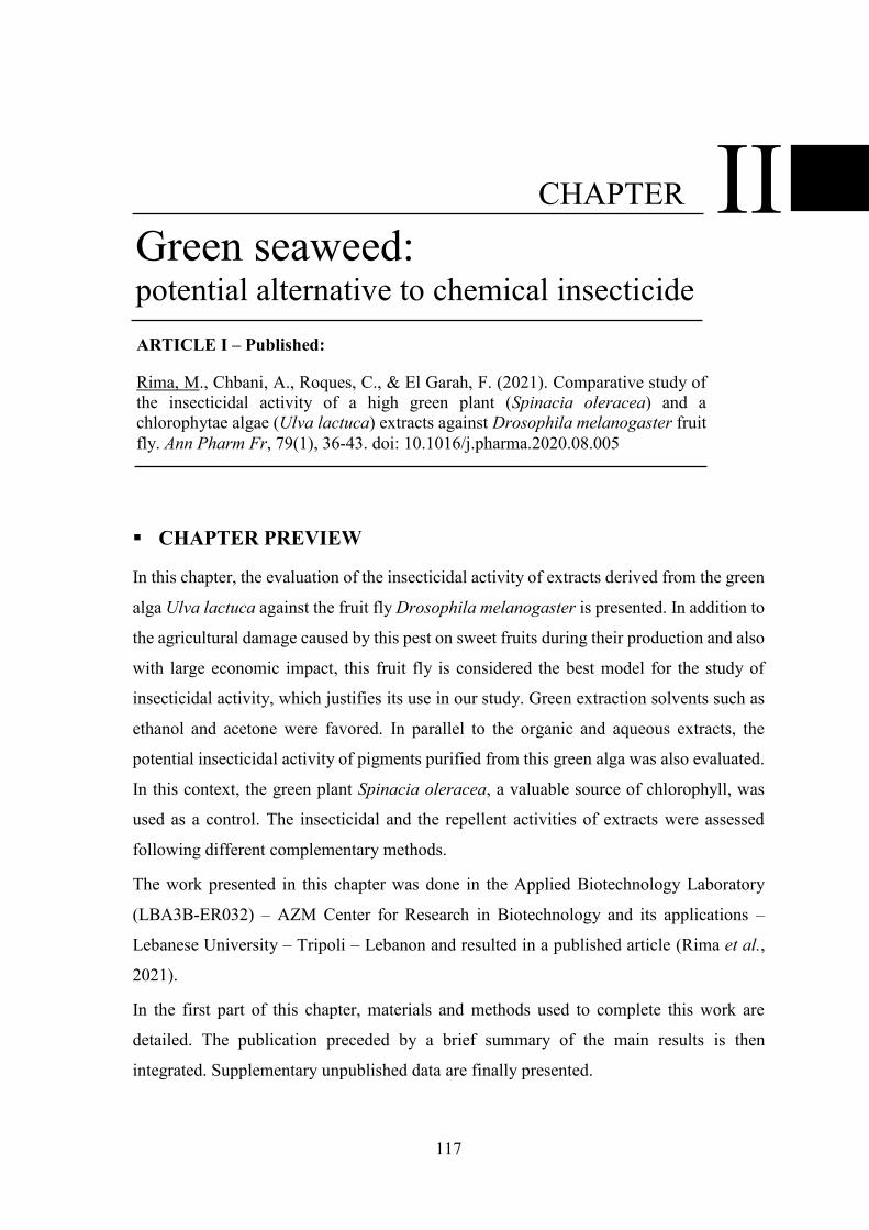
117
CHAPTER PREVIEW
In this chapter, the evaluation of the insecticidal activity of extracts derived from the green
alga Ulva lactuca against the fruit fly Drosophila melanogaster is presented. In addition to
the agricultural damage caused by this pest on sweet fruits during their production and also
with large economic impact, this fruit fly is considered the best model for the study of
insecticidal activity, which justifies its use in our study. Green extraction solvents such as
ethanol and acetone were favored. In parallel to the organic and aqueous extracts, the
potential insecticidal activity of pigments purified from this green alga was also evaluated.
In this context, the green plant Spinacia oleracea, a valuable source of chlorophyll, was
used as a control. The insecticidal and the repellent activities of extracts were assessed
following different complementary methods.
The work presented in this chapter was done in the Applied Biotechnology Laboratory
(LBA3B-ER032) – AZM Center for Research in Biotechnology and its applications –
Lebanese University – Tripoli – Lebanon and resulted in a published article (Rima et al.,
2021).
In the first part of this chapter, materials and methods used to complete this work are
detailed. The publication preceded by a brief summary of the main results is then
integrated. Supplementary unpublished data are finally presented.
Green seaweed: potential alternative to chemical insecticide
II CHAPTER
ARTICLE I – Published:
Rima, M., Chbani, A., Roques, C., & El Garah, F. (2021). Comparative study of the insecticidal activity of a high green plant (Spinacia oleracea) and a chlorophytae algae (Ulva lactuca) extracts against Drosophila melanogaster fruit fly. Ann Pharm Fr, 79(1), 36-43. doi: 10.1016/j.pharma.2020.08.005
Chapter II - Green seaweed: potential alternative to chemical insecticide Materials & Methods
118
I. MATERIALS & METHODS
I.1. MATERIALS
I.1.1 Organic solvents
The organic solvents used are listed below:
o Ethanol 96% (Sigma-Aldrich)
o Acetone > 99.5% (Sigma-Aldrich)
o Methanol 99.8% (Sigma-Aldrich)
o Diethyl ether > 99.0% (Sigma-Aldrich)
o Petroleum ether (Sigma-Aldrich)
I.1.2 Chemical compounds
The chemical compounds used are mentioned below:
o Sodium carbonate (Sigma-Aldrich)
o Sodium sulfate (Sigma-Aldrich)
o Potassium hydroxide (Sigma-Aldrich)
I.1.3 Algal material
The green alga Ulva lactuca used in this study was manually collected from the North
Lebanese coast of the Mediterranean, particularly from El Mina - Tripoli in July 2017. In
order to remove undesirable impurities such as adhered sand particles and epiphytes which
can contaminate the samples, a rigorous washing with the surrounding seawater was
applied to the collected seaweed. Then, alga was immediately transported to the laboratory
of AZM center of research in biotechnology and its applications where a second wash with
distilled water was carried out. Alga samples were air-dried at room temperature in the dark
for several weeks and weighed continuously until complete drying (Al Monla et al., 2020).
Dried algae were then stored in sealed bags at room temperature in the dark until use.
I.1.4 Plant material
Spinach leaves (Spinacia oleracea) used in this study were purchased from a Tripoli –
Lebanon local market in July 2017. The same washing, drying, and storage procedures as
for the green alga were applied to this plant.
Oranges (Citrus sinensis) were used for the insecticide bioassays. They were purchased
from a Tripoli – Lebanon local market, washed with distilled water to remove impurities,
and manipulated on the same day.
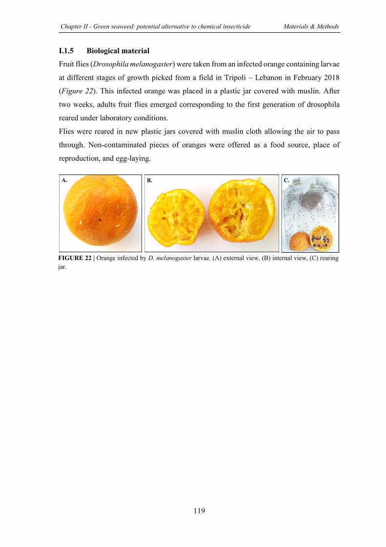
Chapter II - Green seaweed: potential alternative to chemical insecticide Materials & Methods
119
I.1.5 Biological material
Fruit flies (Drosophila melanogaster) were taken from an infected orange containing larvae
at different stages of growth picked from a field in Tripoli – Lebanon in February 2018
(Figure 22). This infected orange was placed in a plastic jar covered with muslin. After
two weeks, adults fruit flies emerged corresponding to the first generation of drosophila
reared under laboratory conditions.
Flies were reared in new plastic jars covered with muslin cloth allowing the air to pass
through. Non-contaminated pieces of oranges were offered as a food source, place of
reproduction, and egg-laying.
B. C. A.
FIGURE 22 | Orange infected by D. melanogaster larvae. (A) external view, (B) internal view, (C) rearing jar.
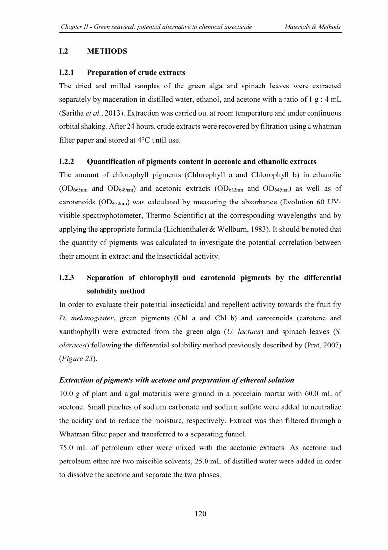
Chapter II - Green seaweed: potential alternative to chemical insecticide Materials & Methods
120
I.2 METHODS
I.2.1 Preparation of crude extracts
The dried and milled samples of the green alga and spinach leaves were extracted
separately by maceration in distilled water, ethanol, and acetone with a ratio of 1 g : 4 mL
(Saritha et al., 2013). Extraction was carried out at room temperature and under continuous
orbital shaking. After 24 hours, crude extracts were recovered by filtration using a whatman
filter paper and stored at 4°C until use.
I.2.2 Quantification of pigments content in acetonic and ethanolic extracts The amount of chlorophyll pigments (Chlorophyll a and Chlorophyll b) in ethanolic
(OD665nm and OD649nm) and acetonic extracts (OD662nm and OD645nm) as well as of
carotenoids (OD470nm) was calculated by measuring the absorbance (Evolution 60 UV-
visible spectrophotometer, Thermo Scientific) at the corresponding wavelengths and by
applying the appropriate formula (Lichtenthaler & Wellburn, 1983). It should be noted that
the quantity of pigments was calculated to investigate the potential correlation between
their amount in extract and the insecticidal activity.
I.2.3 Separation of chlorophyll and carotenoid pigments by the differential
solubility method
In order to evaluate their potential insecticidal and repellent activity towards the fruit fly
D. melanogaster, green pigments (Chl a and Chl b) and carotenoids (carotene and
xanthophyll) were extracted from the green alga (U. lactuca) and spinach leaves (S.
oleracea) following the differential solubility method previously described by (Prat, 2007)
(Figure 23).
Extraction of pigments with acetone and preparation of ethereal solution
10.0 g of plant and algal materials were ground in a porcelain mortar with 60.0 mL of
acetone. Small pinches of sodium carbonate and sodium sulfate were added to neutralize
the acidity and to reduce the moisture, respectively. Extract was then filtered through a
Whatman filter paper and transferred to a separating funnel.
75.0 mL of petroleum ether were mixed with the acetonic extracts. As acetone and
petroleum ether are two miscible solvents, 25.0 mL of distilled water were added in order
to dissolve the acetone and separate the two phases.
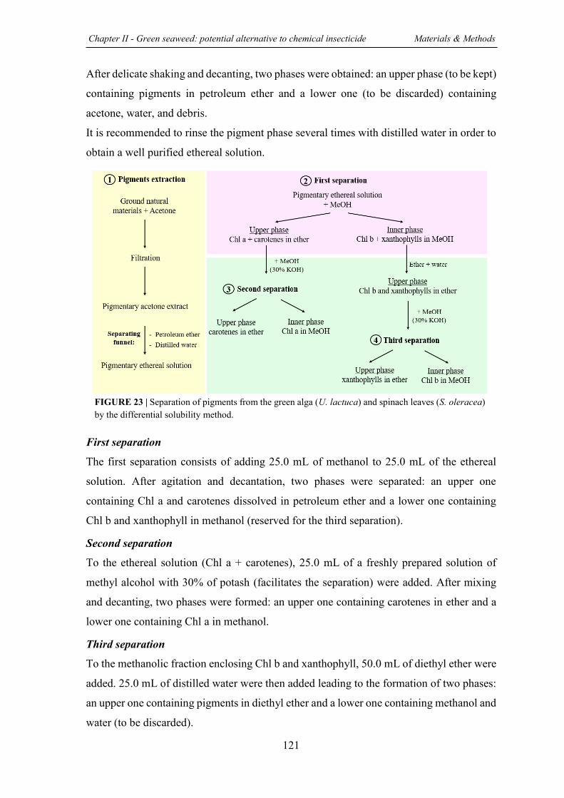
Chapter II - Green seaweed: potential alternative to chemical insecticide Materials & Methods
121
After delicate shaking and decanting, two phases were obtained: an upper phase (to be kept)
containing pigments in petroleum ether and a lower one (to be discarded) containing
acetone, water, and debris.
It is recommended to rinse the pigment phase several times with distilled water in order to
obtain a well purified ethereal solution.
First separation
The first separation consists of adding 25.0 mL of methanol to 25.0 mL of the ethereal
solution. After agitation and decantation, two phases were separated: an upper one
containing Chl a and carotenes dissolved in petroleum ether and a lower one containing
Chl b and xanthophyll in methanol (reserved for the third separation).
Second separation
To the ethereal solution (Chl a + carotenes), 25.0 mL of a freshly prepared solution of
methyl alcohol with 30% of potash (facilitates the separation) were added. After mixing
and decanting, two phases were formed: an upper one containing carotenes in ether and a
lower one containing Chl a in methanol.
Third separation
To the methanolic fraction enclosing Chl b and xanthophyll, 50.0 mL of diethyl ether were
added. 25.0 mL of distilled water were then added leading to the formation of two phases:
an upper one containing pigments in diethyl ether and a lower one containing methanol and
water (to be discarded).
FIGURE 23 | Separation of pigments from the green alga (U. lactuca) and spinach leaves (S. oleracea) by the differential solubility method.

Chapter II - Green seaweed: potential alternative to chemical insecticide Materials & Methods
122
Finally, 25.0 mL of the methyl alcohol (30% of potash) were added to the ether phase
leading to the separation of Chl b and xanthophyll (xanthophyll dissolved in diethyl ether
in the upper phase and Chl b solubilized in methanol in the lower phase).
The absorbance spectrum between 400 and 700 nm of each purified pigment was plotted
to confirm the proper separation (Suppl. III.1).
I.2.4 Insecticidal activity bioassays
The potential insecticidal activity of aqueous, ethanolic, and acetonic extracts derived from
the green alga (U. lactuca) and spinach leaves (S. oleracea) against the fruit fly (D.
melanogaster) was evaluated following various complementary methods. Interestingly,
each test exhibits a specific mode of exposure. Effect of purified pigments (Chl a, Chl b,
carotene, and xanthophyll) was also tested. Extraction solvents were used as negative
control and each test was performed in triplicate. Insecticidal activity of extracts was
determined using Sun-Shepard formula (non-uniform population) (1), which corrects the
efficacy by nullifying solvent effect (Puntener, 1981).
mortality % in treated plot + change % in control plot population
100 + change % in control plot population x 100
o With change % in control plot population =
Population in control after treatment-population in control before treatment
Population in control before treatment x 100
I.2.4.1 Test 1: Spraying oranges
In this first test, natural conditions were imitated on a laboratory scale by using the protocol
previously described by (Chaieb et al., 2010) with some modifications (Figure 24, A).
Oranges were sprayed with 1.0 mL of extract or not and after drying, each one was placed
in an individual plastic jar. Then, 15 + 5 adult flies were distributed into each jar. The
number of dead flies in each jar was recorded after 24, 48, and 72h. It should be noted that
in this assay the tested extract can penetrate into the insect by simple contact and/or by
ingestion.
I.2.4.2 Test 2: Ingestion toxicity
In this second test, the potential ability of extracts to kill flies by ingestion was assessed
following the protocol developed by (Aboussaid et al., 2010) with some modifications
(Figure 24, B).
(1)
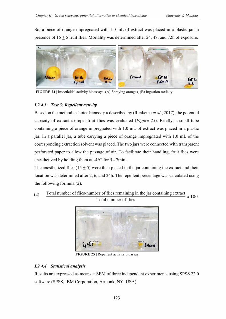
Chapter II - Green seaweed: potential alternative to chemical insecticide Materials & Methods
123
So, a piece of orange impregnated with 1.0 mL of extract was placed in a plastic jar in
presence of 15 + 5 fruit flies. Mortality was determined after 24, 48, and 72h of exposure.
I.2.4.3 Test 3: Repellent activity
Based on the method « choice bioassay » described by (Renkema et al., 2017), the potential
capacity of extract to repel fruit flies was evaluated (Figure 25). Briefly, a small tube
containing a piece of orange impregnated with 1.0 mL of extract was placed in a plastic
jar. In a parallel jar, a tube carrying a piece of orange impregnated with 1.0 mL of the
corresponding extraction solvent was placed. The two jars were connected with transparent
perforated paper to allow the passage of air. To facilitate their handling, fruit flies were
anesthetized by holding them at -4°C for 5 - 7min.
The anesthetized flies (15 + 5) were then placed in the jar containing the extract and their
location was determined after 2, 6, and 24h. The repellent percentage was calculated using
the following formula (2).
Total number of flies-number of flies remaining in the jar containing extract
Total number of flies x 100
I.2.4.4 Statistical analysis
Results are expressed as means + SEM of three independent experiments using SPSS 22.0
software (SPSS, IBM Corporation, Armonk, NY, USA)
A. B.
FIGURE 24 | Insecticidal activity bioassays. (A) Spraying oranges, (B) Ingestion toxicity.
FIGURE 25 | Repellent activity bioassay.
(2)
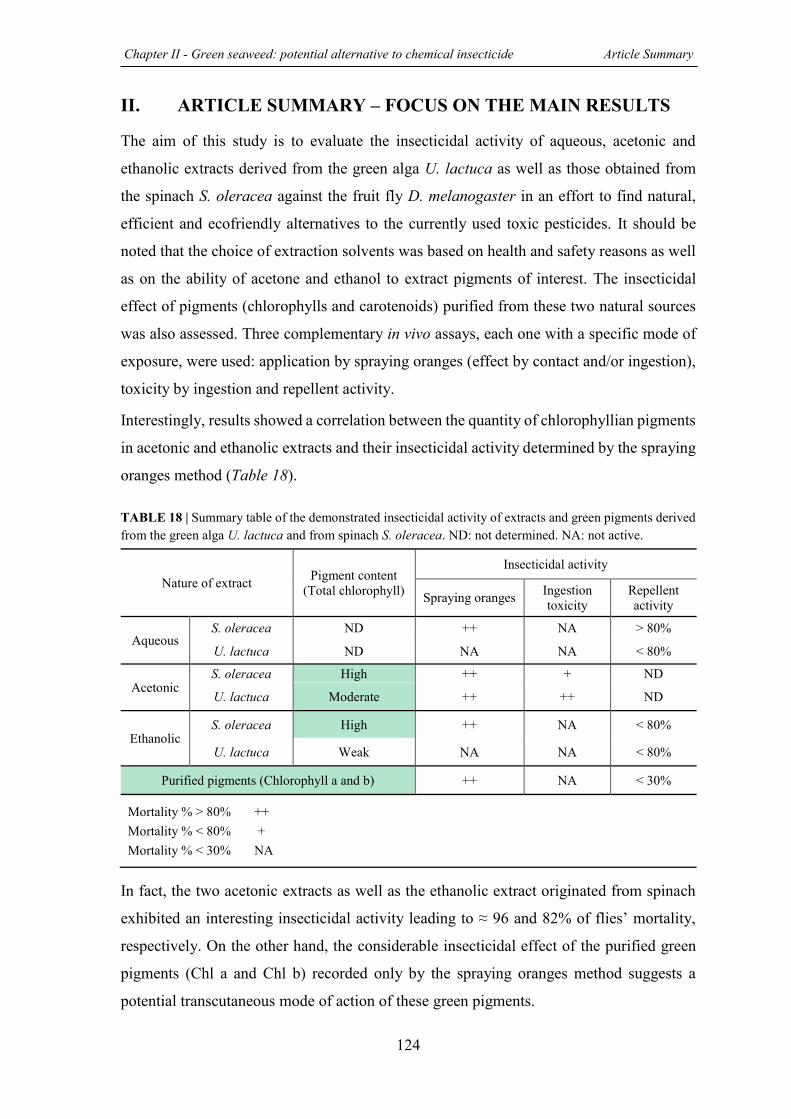
Chapter II - Green seaweed: potential alternative to chemical insecticide Article Summary
124
II. ARTICLE SUMMARY – FOCUS ON THE MAIN RESULTS
The aim of this study is to evaluate the insecticidal activity of aqueous, acetonic and
ethanolic extracts derived from the green alga U. lactuca as well as those obtained from
the spinach S. oleracea against the fruit fly D. melanogaster in an effort to find natural,
efficient and ecofriendly alternatives to the currently used toxic pesticides. It should be
noted that the choice of extraction solvents was based on health and safety reasons as well
as on the ability of acetone and ethanol to extract pigments of interest. The insecticidal
effect of pigments (chlorophylls and carotenoids) purified from these two natural sources
was also assessed. Three complementary in vivo assays, each one with a specific mode of
exposure, were used: application by spraying oranges (effect by contact and/or ingestion),
toxicity by ingestion and repellent activity.
Interestingly, results showed a correlation between the quantity of chlorophyllian pigments
in acetonic and ethanolic extracts and their insecticidal activity determined by the spraying
oranges method (Table 18). TABLE 18 | Summary table of the demonstrated insecticidal activity of extracts and green pigments derived from the green alga U. lactuca and from spinach S. oleracea. ND: not determined. NA: not active.
In fact, the two acetonic extracts as well as the ethanolic extract originated from spinach
exhibited an interesting insecticidal activity leading to ≈ 96 and 82% of flies’ mortality,
respectively. On the other hand, the considerable insecticidal effect of the purified green
pigments (Chl a and Chl b) recorded only by the spraying oranges method suggests a
potential transcutaneous mode of action of these green pigments.
Nature of extract Pigment content (Total chlorophyll)
Insecticidal activity
Spraying oranges Ingestion toxicity
Repellent activity
Aqueous S. oleracea ND ++ NA > 80%
U. lactuca ND NA NA < 80%
Acetonic S. oleracea High ++ + ND
U. lactuca Moderate ++ ++ ND
Ethanolic S. oleracea High ++ NA < 80%
U. lactuca Weak NA NA < 80%
Purified pigments (Chlorophyll a and b) ++ NA < 30%
Mortality % > 80% ++ Mortality % < 80% + Mortality % < 30% NA
Chapter II - Green seaweed: potential alternative to chemical insecticide Article Summary
125
Moreover, the strong insecticidal effect observed for the acetonic extract originated from
the green alga U. lactuca whether sprayed on oranges or mixed with flies’ nutrient source
suggests its richness in other unique bioactive compounds.
The obtained results summarized in the table below highlighted the potential ability of
green pigments as well as of extracts originated from the green alga U. lactuca to be
exploited as an effective natural alternative to synthetic insecticide. These encouraging
findings require further experiments in order to analyze the chemical composition of the
extracts and thus identify novel bioactive molecules.
126
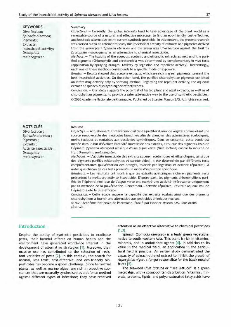
127
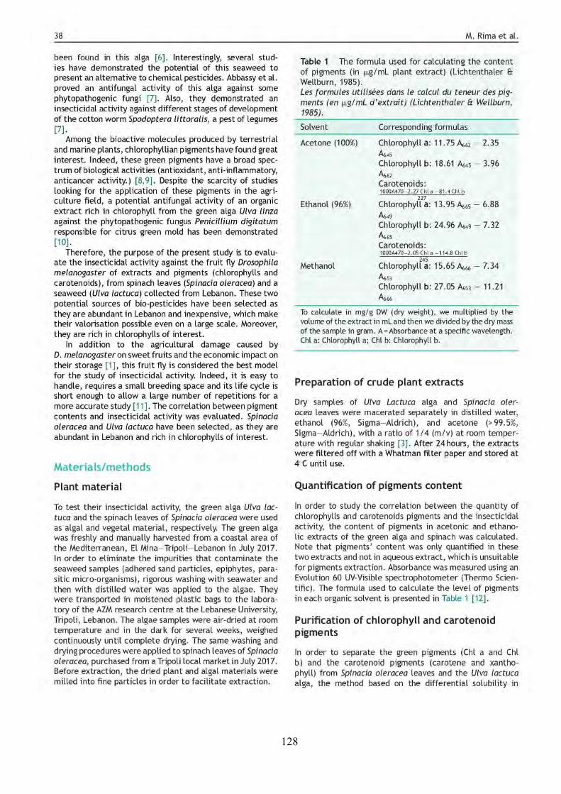
128
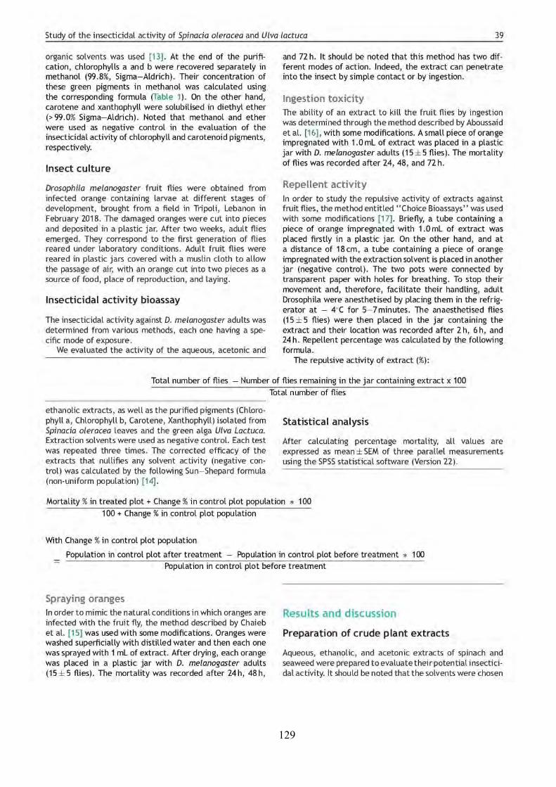
129
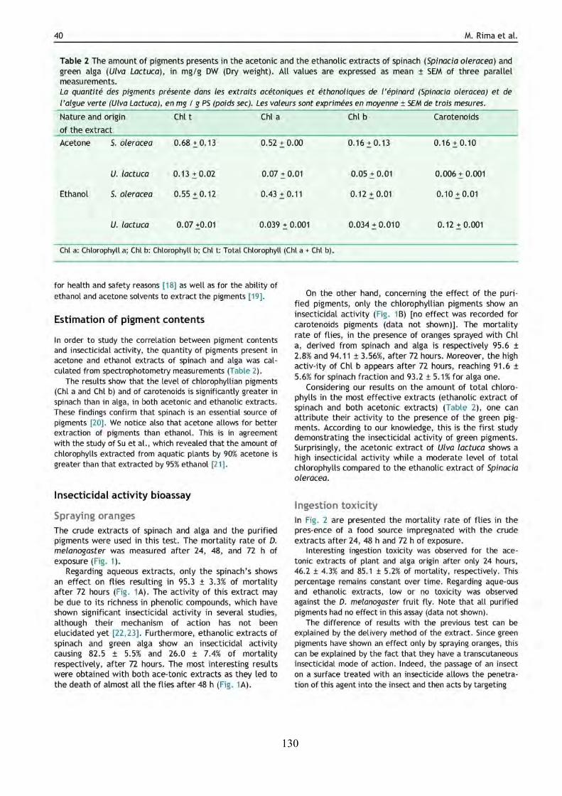
130
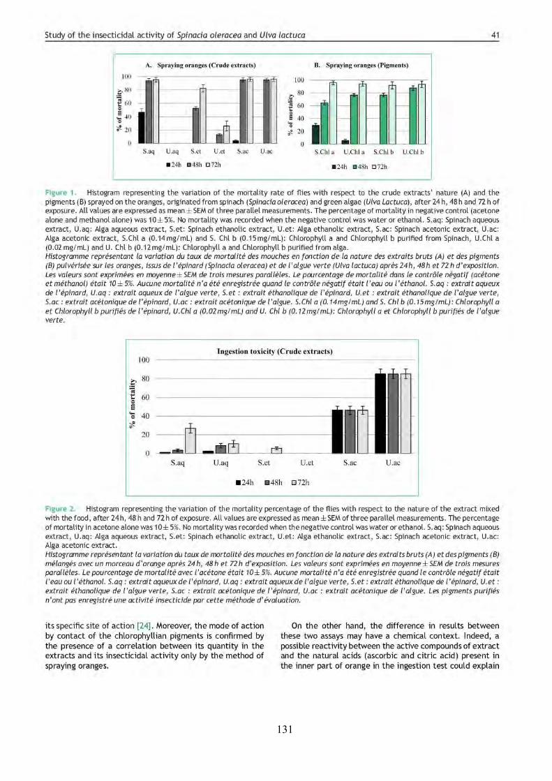
131
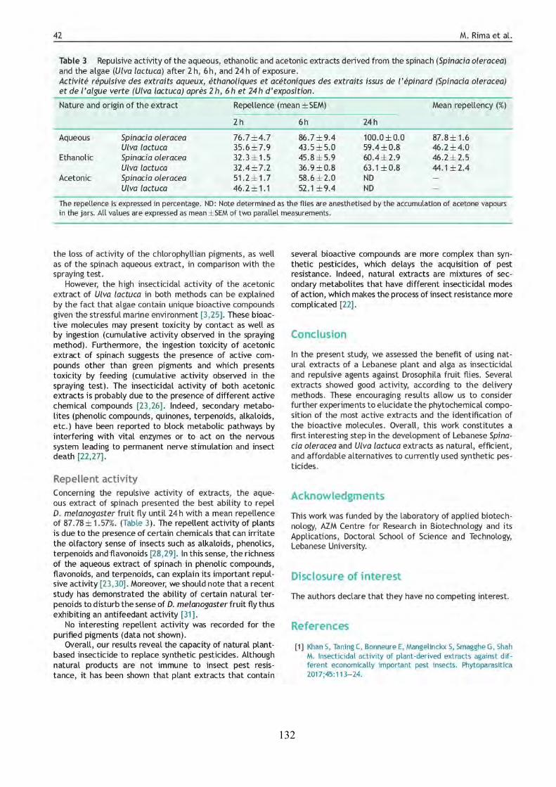
132
133
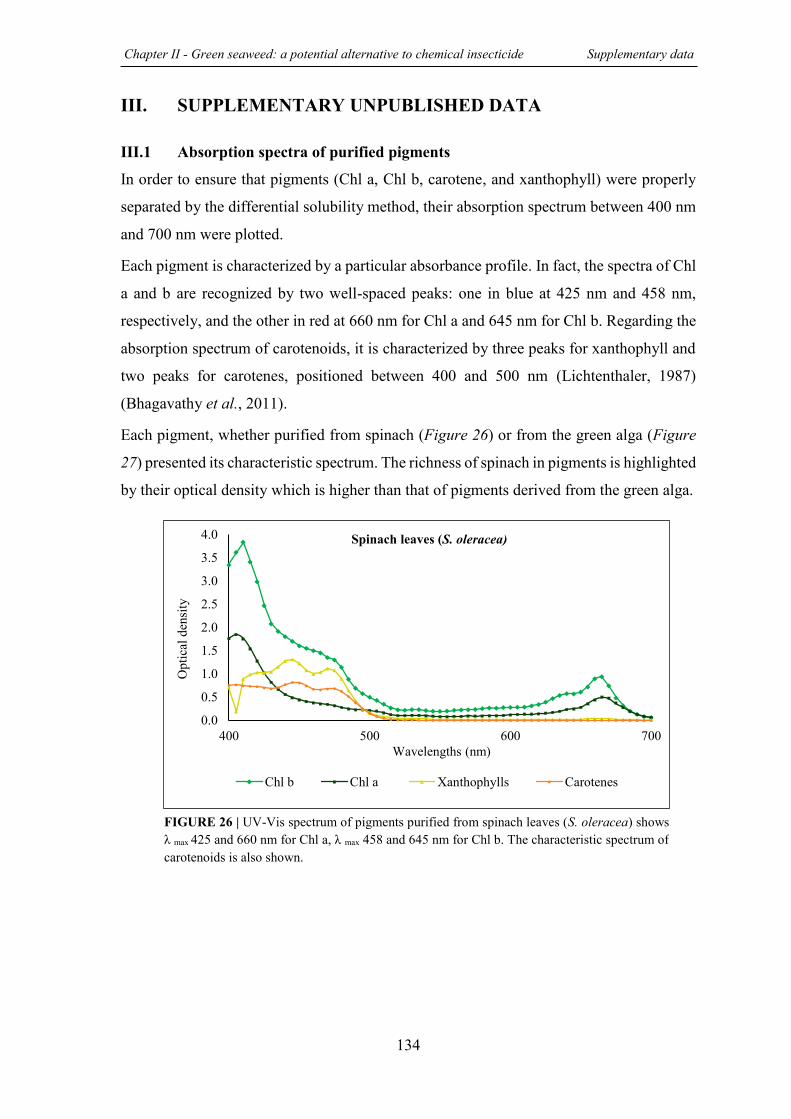
Chapter II - Green seaweed: a potential alternative to chemical insecticide Supplementary data
134
III. SUPPLEMENTARY UNPUBLISHED DATA
III.1 Absorption spectra of purified pigments
In order to ensure that pigments (Chl a, Chl b, carotene, and xanthophyll) were properly
separated by the differential solubility method, their absorption spectrum between 400 nm
and 700 nm were plotted.
Each pigment is characterized by a particular absorbance profile. In fact, the spectra of Chl
a and b are recognized by two well-spaced peaks: one in blue at 425 nm and 458 nm,
respectively, and the other in red at 660 nm for Chl a and 645 nm for Chl b. Regarding the
absorption spectrum of carotenoids, it is characterized by three peaks for xanthophyll and
two peaks for carotenes, positioned between 400 and 500 nm (Lichtenthaler, 1987)
(Bhagavathy et al., 2011).
Each pigment, whether purified from spinach (Figure 26) or from the green alga (Figure
27) presented its characteristic spectrum. The richness of spinach in pigments is highlighted
by their optical density which is higher than that of pigments derived from the green alga.
0.0
0.5
1.0
1.5
2.0
2.5
3.0
3.5
4.0
400 500 600 700
Chl b Chl a Xanthophylls Carotenes
Spinach leaves (S. oleracea)
Opt
ical
dens
ity
Wavelengths (nm)
FIGURE 26 | UV-Vis spectrum of pigments purified from spinach leaves (S. oleracea) shows λ max 425 and 660 nm for Chl a, λ max 458 and 645 nm for Chl b. The characteristic spectrum of carotenoids is also shown.
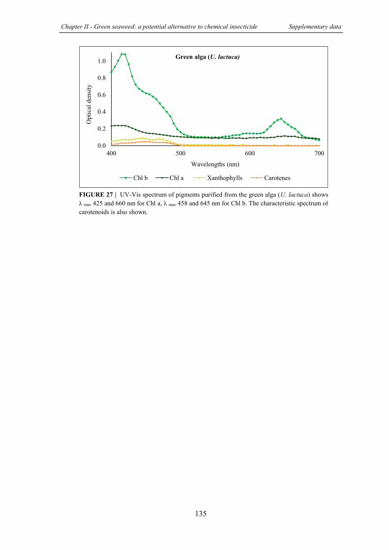
Chapter II - Green seaweed: a potential alternative to chemical insecticide Supplementary data
135
0.0
0.2
0.4
0.6
0.8
1.0
400 500 600 700
Chl b Chl a Xanthophylls Carotenes
Green alga (U. lactuca)
Opt
ical
den
sity
Wavelengths (nm)
FIGURE 27 | UV-Vis spectrum of pigments purified from the green alga (U. lactuca) shows λ max 425 and 660 nm for Chl a, λ max 458 and 645 nm for Chl b. The characteristic spectrum of carotenoids is also shown.
136
REFERENCES Aboussaid, H., El Messoussi, S., & Oufdou, K. (2010). Activité insecticide d’une souche marocaine
de Bacillus thuringiensis sur la mouche méditerranéenne : Ceratitis capitata (Wied.) (Diptera : Tephritidae). Afr. Sci. Rev. Int. Sci. Technol., 5(1). doi:10.4314/afsci.v5i1.61719
Al Monla, R., Dassouki, Z., Kouzayha, A., Salma, Y., Gali-Muhtasib, H., & Mawlawi, H. (2020). The Cytotoxic and Apoptotic Effects of the Brown Algae Colpomenia sinuosa are Mediated by the Generation of Reactive Oxygen Species. Molecules, 25(8). doi:10.3390/molecules25081993
Bhagavathy, S., Sumathi, P., & Jancy Sherene Bell, I. (2011). Green algae Chlorococcum humicola a new source of bioactive compounds with antimicrobial activity. Asian Pac. J. Trop. Biomed., 1(1), S1-S7. doi:10.1016/s2221-1691(11)60111-1
Chaieb, I., Baouandi, M., & Bouhachem, S. (2010). Activité insecticide des extraits d’Ecballium elaterium (Cucurbitacae) sur Aphis fabae (Aphididae). Rev Regions Arides, 3, 1299-1303.
Lichtenthaler, H. K. (1987). Chlorophylls and carotenoids: Pigments of photosynthetic biomembranes. Meth. Enzymol., 148, 350-382. doi://doi.org/10.1016/0076-6879(87)48036-1
Lichtenthaler, H. K., & Wellburn, A. R. (1983). Determinations of total carotenoids and chlorophylls a and b of leaf extracts in different solvents. Biochem. Soc. Trans., 11(5), 591-592. doi:10.1042/bst0110591
Prat, R. (2007). Experminetation en biologie et physiologie végétale (Hermann Ed.).
Puntener, W. (1981). Manual for field trials in plant protection (Ciba-Geigy, Ltd ed.). Basle, Switzerland.
Renkema, J. M., Buitenhuis, R., & Hallett, R. H. (2017). Reduced Drosophila suzukii Infestation in Berries Using Deterrent Compounds and Laminate Polymer Flakes. Insects, 8(4). doi:10.3390/insects8040117
Rima, M., Chbani, A., Roques, C., & El Garah, F. (2021). Comparative study of the insecticidal activity of a high green plant (Spinacia oleracea) and a chlorophytae algae (Ulva lactuca) extracts against Drosophila melanogaster fruit fly. Ann Pharm Fr, 79(1), 36-43. doi:10.1016/j.pharma.2020.08.005
Saritha, K., Aswathi Elizabeth, M., Priyalaxmi, M., & Patterson, J. (2013). Antibacterial activity and biochemical constituents of Seaweed Ulva lactuca. Glob. J. Pharmacol., 3(7), 276-282. doi:10.5829/idosi.gjp.2013.7.3.75156
137
CHAPTER PREVIEW
In this chapter, the evaluation of the potential antibiofilm activity of the different extracts
derived from the three tested algae (U. lactuca, S. scoparium and P. capillacea) against
P. aeruginosa and S. aureus pathogenic bacteria is presented.
In the first part, the materials and methods followed are detailed. Then, the second and the
third part of this chapter are devoted to the presentation and discussion of the results
obtained following the evaluation of the antibiofilm effect against P. aeruginosa and
S. aureus, respectively.
III Seaweed extracts: a promising source of antibiofilm agents against pathogenic bacteria “Particularly against P. aeruginosa & S. aureus”
CHAPTER
138
PREVIEW
After the listing of materials used in this part of the study, the method used for the
preparation of algal extracts is described. Then, the protocols followed in the evaluation of
the antibiofilm activity (biofilm inhibition and reduction, biofilm microscopic analysis,
synergistic activity with conventional antibiotics) of algal extracts against P. aeruginosa
gram-negative bacterium are detailed. In addition, the principle of the biosensor-based
assay carried out in order to assess the potential capacity of extracts to inhibit AHL-based
QS systems is also outlined. It should be noted that the biosensor strains used in this assay
were obtained from the Laboratory of Microbial Biodiversity and Biotechnology
(USR3579 – LBBM) – Banyuls-sur-Mer, France and we would like to thank Dr. Raphaël
LAMI and his team for welcoming me in his laboratory to train me to conduct this test.
Similarly, the methods used in the assessment of the antibiofilm activity of extracts against
S. aureus are presented. On the other hand, the contact angle measurement method which
allows the determination of the hydrophobicity of bacteria previously treated with extracts
is described.
Part I:
Materials & Methods
III CHAPTER
Chapter III – Antibiofilm activity of seaweed extracts Part I – Materials & Methods
139
I. MATERIALS & METHODS
I.1 MATERIALS
I.1.1 Laboratory materials and devices
Microplates used for evaluating the antibacterial and antibiofilm activities of extracts are
96-well microtiter plates (Falcon, TC-treated, polystyrene) and 24-well plates (Falcon, TC-
treated, polystyrene), respectively. Biofilms visualized with epifluorescence microscope
are prepared in 6-well plates (Falcon, TC-treated, polystyrene).
Extracts solutions were sterilized by filtration using a syringe filter (Cellulose Acetate
Syringe Filter, 0.45 μm, GE Healthcare Whatman).
Cellulose acetate membrane filter (0.45 µm, Sigma-Aldrich) was used for preparation of
bacterial layer in the contact angle measurement assay.
List of employed devices is presented below:
o Ultrasonic bath (VWR ultrasonic cleaning bath, 45 KHz)
o Orbital shaker (MaxQ 4000, Thermo Fisher Scientific)
o Microplate spectrophotometer (MultiskanTM GO, Thermo Fisher Scientific)
o Epifluorescence microscope: Zeiss – Axiotech microscope using a 20 X / 0.50
(Zeiss, EC Plan-Neofluar) objective and equipped with an HXP 120 C light
source
o Fluorometer plate reader (BMG Labtech)
o Digidrop contact angle meter (GBX Scientific Instruments, Romans-sur-Isère,
France)
I.1.2 Organic solvents
The solvents used in the algae extraction are listed below:
o Cyclohexane 99.5% (Sigma-Aldrich, France)
o Dichloromethane 100% (VWR, France)
o Ethyl acetate 99.9% (VWR, France)
o Methanol 99.8% (Sigma-Aldrich, France)
On the other hand, ethanol 96% (Sigma-Aldrich, France) was used to extract the crystal
violet fixed by the bacterial biomass in the evaluation of the antibiofilm activity.
Chapter III – Antibiofilm activity of seaweed extracts Part I – Materials & Methods
140
I.1.3 Chemical products
All the chemicals products employed in this study are categorized below:
o Crystal violet (Sigma-Aldrich, France)
o Antibiotics:
- Tobramycin (Sigma-Aldrich, France)
- Colistin (Colistin sodium methanesulfonate, Sigma-Aldrich, France)
- Tetracycline (Sigma-Aldrich, France)
- Gentamycin solution 10 mg/mL (Sigma-Aldrich, France)
o Fluorescent markers:
- Syto9 (5 mM, InvitrogenTM, ThermoFisher Scientific)
- Propidium iodide (1 mg/mL, InvitrogenTM, ThermoFisher Scientific)
- Concanavalin A (Tetramethylrhodamine conjugate, ThermoFisher
Scientific)
- SYPRO Ruby stain (InvitrogenTM, FilmTracerTM, SYPROTM Ruby biofilm
matrix stain)
o Chemical compounds used in biosensor-based assay:
- N-Hexanoyl-L-homoserine lactone (C6-HSL) (Sigma-Aldrich, France)
- N-(3-Oxodecanoyl)-L-homoserine lactone (Oxo-C10-HSL) (Sigma-
Aldrich, France)
- The synthetic compound C11 (analogue of the N-butanoyl-L-homoserine
lactone (C4-AHL) (Khalilzadeh et al., 2010)
- Cinnamaldehyde (Sigma-Aldrich, France)
- BacTiter-GloTM Microbial Cell Viability Assay (PROMEGA, France)
I.1.4 Algal materials
The three species of algae (green alga Ulva lactuca, brown alga Stypocaulon scoparium
and red alga Pterocladiella capillacea) used in this study were manually collected from the
North Lebanese coast of the Mediterranean, particularly from El Mina - Tripoli in
September 2019. In order to remove undesirable impurities such as adhered sand particles
and epiphytes which can contaminate the samples, a rigorous washing with seawater was
applied to the collected algae. Then, they were immediately transported to the laboratory
of AZM center of research in biotechnology and its applications where a second wash with
Chapter III – Antibiofilm activity of seaweed extracts Part I – Materials & Methods
141
distilled water was carried out. Algae samples were air-dried at room temperature in the
dark for several weeks and weighed continuously until complete drying.
The dried samples were ground into fine powder and were then transported in sealed bags
to the Laboratoire de Génie Chimique of Toulouse, France, where the extraction were
carried out.
I.1.5 Bacterial strains and culture media
Bacterial strains involved in this study are Pseudomonas aeruginosa PAO1 (CIP 104116)
and Staphylococcus aureus (CIP 4.83) which are obtained from the collection of Pasteur
Institute (Paris, France) and are stored at -80°C in a protective solution (3.0 g/L of beef
extract powder, 5.0 g/L of Tryptone Pancreatic digest of casein, 150.0 g/L of glycerol, pH
of 6.9 + 0.2 at 20 + 2°C). It should be noted that the bacterial inoculum used in each assay
was taken from a second overnight subculture on Trypticase soy agar (TSA) that was
incubated under aerobic conditions at 37°C. The culture media used are mentioned below:
o Trypticase soy agar (TSA) (BioMérieux, Crapone, France) used for bacterial
subculture and CFU counts.
o Mueller-Hinton broth (MHB) (Oxoid microbiology products, Basingstoke,
UK) used to determine the minimal inhibitory concentration (MIC) of extracts.
o Modified biofilm broth (MBB) used in P. aeruginosa biofilm culture and thus
for the evaluation of the antibiofilm activity of extracts against this bacterium.
The MBB 10X medium is composed of FeSO4, 7H2O (0.005 g/L), Na2HPO4
(12.5 g/L), KH2PO4 (5.0 g/L), (NH4)2SO4 (1.0 g/L), glucose (0.5 g/L) and
MgSO4, 7H2O (0.2 g/L) (Khalilzadeh et al., 2010). All these compounds were
purchased from Sigma-Aldrich, France.
o Biofilm broth (BB) used in S. aureus biofilm culture and thus for the
evaluation of the antibiofilm activity of extracts against this bacterium. The BB
10X medium is composed of FeSO4, 7H2O (0.005 g/L), Na2HPO4 (12.5 g/L),
KH2PO4 (5.0 g/L), (NH4)2 SO4 (1.0 g/L), lactose (0.25 g/L), yeast extract (1.0
g/L), vitamin assay casamino acids (1.0 g/L) and MgSO4, 7H2O (0.2 g/L)
(Campanac et al., 2002). Except for yeast extract (BactoTM, ThermoFisher
scientific) and vitamin assay casamino acids (DifcoTM, ThermoFisher
scientific) all these compounds were purchased from Sigma-Aldrich, France.
Chapter III – Antibiofilm activity of seaweed extracts Part I – Materials & Methods
142
All media were prepared by dissolution of ingredients in distilled water and sterilized just
after preparation (121°C, 15 min).
The biosensor bacterial strains used in the evaluation of the anti-QS activity of extracts are
Escherichia coli MT102 (pJBA132) and Pseudomonas putida F117 (pRK-C12) which
detect short-chain AHLs (<8 carbons in the acyl side chain) and long chain AHLs (>8
carbons in the acyl side chain), respectively. These reporter strains were obtained from the
Laboratory of Microbial Biodiversity and Biotechnology (USR3579 – LBBM) – Banyuls-
sur-Mer, France and were stored at -80°C. The culture media used for these strains are:
o LB broth (Lennox) (Sigma-Aldrich, France) supplemented with agar (Sigma-
Aldrich, France) used for bacterial subculture.
o LB broth (Lennox) (Sigma-Aldrich, France) used for liquid bacterial culture
as well as in the biosensor-based assay
Media are prepared as indicated before. It should be noted that both LB broth and LB agar
are supplemented with the corresponding antibiotic, tetracycline for MT102 (final
concentration 25.0 µg/mL) and gentamycin for F117 (final concentration 20.0 µg/mL) in
order to maintain the selection pressure.
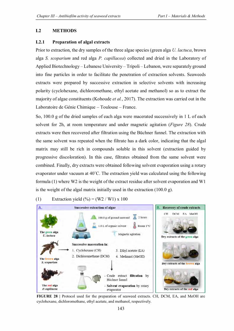
Chapter III – Antibiofilm activity of seaweed extracts Part I – Materials & Methods
143
I.2 METHODS
I.2.1 Preparation of algal extracts
Prior to extraction, the dry samples of the three algae species (green alga U. lactuca, brown
alga S. scoparium and red alga P. capillacea) collected and dried in the Laboratory of
Applied Biotechnology – Lebanese University – Tripoli – Lebanon, were separately ground
into fine particles in order to facilitate the penetration of extraction solvents. Seaweeds
extracts were prepared by successive extraction in selective solvents with increasing
polarity (cyclohexane, dichloromethane, ethyl acetate and methanol) so as to extract the
majority of algae constituents (Kohoude et al., 2017). The extraction was carried out in the
Laboratoire de Génie Chimique – Toulouse – France.
So, 100.0 g of the dried samples of each alga were macerated successively in 1 L of each
solvent for 2h, at room temperature and under magnetic agitation (Figure 28). Crude
extracts were then recovered after filtration using the Büchner funnel. The extraction with
the same solvent was repeated when the filtrate has a dark color, indicating that the algal
matrix may still be rich in compounds soluble in this solvent (extraction guided by
progressive discoloration). In this case, filtrates obtained from the same solvent were
combined. Finally, dry extracts were obtained following solvent evaporation using a rotary
evaporator under vacuum at 40˚C. The extraction yield was calculated using the following
formula (1) where W2 is the weight of the extract residue after solvent evaporation and W1
is the weight of the algal matrix initially used in the extraction (100.0 g).
(1) Extraction yield (%) = (W2 / W1) x 100
FIGURE 28 | Protocol used for the preparation of seaweed extracts. CH, DCM, EA, and MeOH are cyclohexane, dichloromethane, ethyl acetate, and methanol, respectively.
Chapter III – Antibiofilm activity of seaweed extracts Part I – Materials & Methods
144
Stock solutions of extracts were prepared in sterile distilled water (SDW) at a concentration
of 100.0 µg/mL using an ultrasonic bath for almost 6 hours in order to improve their
solubility. They were then sterilized by filtration through a syringe filter (0.45µm) prior to
the evaluation of their antibacterial, antibiofilm and anti-QS activities.
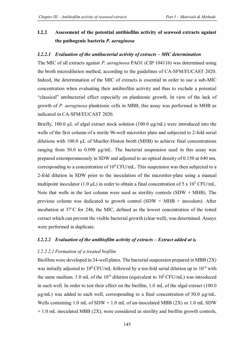
Chapter III – Antibiofilm activity of seaweed extracts Part I – Materials & Methods
145
I.2.2 Assessment of the potential antibiofilm activity of seaweed extracts against
the pathogenic bacteria P. aeruginosa
I.2.2.1 Evaluation of the antibacterial activity of extracts – MIC determination
The MIC of all extracts against P. aeruginosa PAO1 (CIP 104116) was determined using
the broth microdilution method, according to the guidelines of CA-SFM/EUCAST 2020.
Indeed, the determination of the MIC of extracts is essential in order to use a sub-MIC
concentration when evaluating their antibiofilm activity and thus to exclude a potential
“classical” antibacterial effect especially on planktonic growth. In view of the lack of
growth of P. aeruginosa planktonic cells in MBB, this assay was performed in MHB as
indicated in CA-SFM/EUCAST 2020.
Briefly, 100.0 µL of algal extract stock solution (100.0 µg/mL) were introduced into the
wells of the first column of a sterile 96-well microtiter plate and subjected to 2-fold serial
dilutions with 100.0 µL of Mueller-Hinton broth (MHB) to achieve final concentrations
ranging from 50.0 to 0.098 µg/mL. The bacterial suspension used in this assay was
prepared extemporaneously in SDW and adjusted to an optical density of 0.150 at 640 nm,
corresponding to a concentration of 108 CFU/mL. This suspension was then subjected to a
2-fold dilution in SDW prior to the inoculation of the microtiter-plate using a manual
multipoint inoculator (1.0 µL) in order to obtain a final concentration of 5 x 10⁵ CFU/mL.
Note that wells in the last column were used as sterility controls (SDW + MHB). The
previous column was dedicated to growth control (SDW + MHB + inoculum). After
incubation at 37˚C for 24h, the MIC, defined as the lowest concentration of the tested
extract which can prevent the visible bacterial growth (clear well), was determined. Assays
were performed in duplicate.
I.2.2.2 Evaluation of the antibiofilm activity of extracts – Extract added at t0
I.2.2.2.1 Formation of a treated biofilm
Biofilms were developed in 24-well plates. The bacterial suspension prepared in MBB (2X)
was initially adjusted to 108 CFU/mL followed by a ten-fold serial dilution up to 10-6 with
the same medium. 1.0 mL of the 10-6 dilution (equivalent to 102 CFU/mL) was introduced
in each well. In order to test their effect on the biofilm, 1.0 mL of the algal extract (100.0
µg/mL) was added to each well, corresponding to a final concentration of 50.0 µg/mL.
Wells containing 1.0 mL of SDW + 1.0 mL of un-inoculated MBB (2X) or 1.0 mL SDW
+ 1.0 mL inoculated MBB (2X), were considered as sterility and biofilm growth controls,
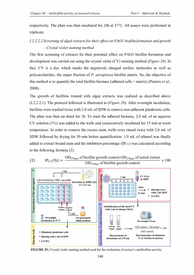
Chapter III – Antibiofilm activity of seaweed extracts Part I – Materials & Methods
146
respectively. The plate was then incubated for 24h at 37˚C. All assays were performed in
triplicate.
I.2.2.2.2 Screening of algal extracts for their effect on PAO1 biofilm formation and growth
– Crystal violet staining method
The first screening of extracts for their potential effect on PAO1 biofilm formation and
development was carried out using the crystal violet (CV) staining method (Figure 29). In
fact, CV is a dye which marks the negatively charged surface molecules as well as
polysaccharides, the major fraction of P. aeruginosa biofilm matrix. So, the objective of
this method is to quantify the total biofilm biomass (adhered cells + matrix) (Peeters et al.,
2008).
The growth of biofilms treated with algae extracts was realized as described above
(I.2.2.2.1). The protocol followed is illustrated in (Figure 29). After overnight incubation,
biofilms were washed twice with 2.0 mL of SDW to remove non-adherent planktonic cells.
The plate was then air-dried for 1h. To stain the adhered biomass, 2.0 mL of an aqueous
CV solution (1%) was added to the wells and consecutively incubated for 15 min at room
temperature. In order to remove the excess stain, wells were rinsed twice with 2.0 mL of
SDW followed by drying for 30 min before quantification. 1.0 mL of ethanol was finally
added to extract bound stain and the inhibition percentage (IPCV) was calculated according
to the following formula (2).
(2) IPCV(%) = OD570nm of biofilm growth control-OD570nm of tested extract
OD570nm of biofilm growth control x 100
FIGURE 29 | Crystal violet staining method used for the evaluation of extract’s antibiofilm activity.
Chapter III – Antibiofilm activity of seaweed extracts Part I – Materials & Methods
147
I.2.2.2.3 Effect of the potentially active extracts on the number of adhered bacteria – CFU
counts method
After screening of extracts for their effect on the total biofilm biomass by CV staining
method, the algal extracts showing an inhibition percentage higher than 50% (IPCV > 50%)
were selected to assess their effect on the number of adhered cells by the CFU counts
method. In this assay, the protocol developed by (Campanac et al., 2002) and (Khalilzadeh
et al., 2010) was followed with some modifications.
Treated biofilms were developed as described above (I.2.2.2.1) and the detailed protocol is
described in (Figure 30).
After 24h of incubation, planktonic cells were discarded by rinsing twice with 2.0 mL of
SDW. Adhered cells were then recovered by scarping for 1 min with a sterile spatula into
1.0 mL of SDW. These recuperated cells were diluted by ten-fold serial dilution (from 10-
1 to 10-6) and 900.0 µL of each dilution was inoculated by inclusion in TSA plates. After
48h of incubation at 37˚C, the numbers of CFU were counted by considering only those
between 15 and 300 CFU. The adhered biomass was then calculated and subjected to
logarithmic transformation by the following formula (3). The logarithmic reduction as well
as the IPCFU with respect to the corresponding untreated control were also calculated using
the formulas below (4)(5).
FIGURE 30 | CFU counts method used for the evaluation of extract’s antibiofilm activity.
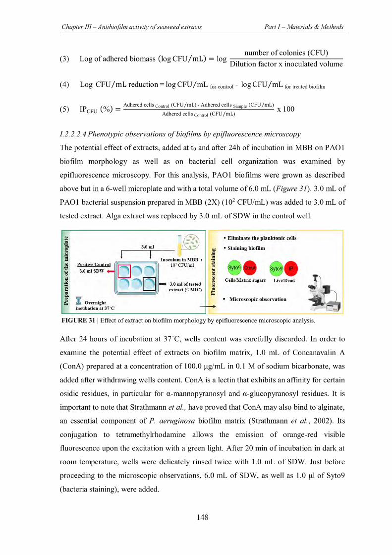
Chapter III – Antibiofilm activity of seaweed extracts Part I – Materials & Methods
148
(3) Log of adhered biomass (log CFU mL⁄ ) = log number of colonies (CFU)
Dilution factor x inoculated volume
(4) Log CFU mL reduction = log CFU mL for control - log CFU mL for treated biofilm ⁄ ⁄⁄
(5) IPCFU (%) =Adhered cells Control (CFU mL) - Adhered cells Sample (CFU mL)⁄⁄
Adhered cells Control (CFU mL)⁄ x 100
I.2.2.2.4 Phenotypic observations of biofilms by epifluorescence microscopy
The potential effect of extracts, added at t0 and after 24h of incubation in MBB on PAO1
biofilm morphology as well as on bacterial cell organization was examined by
epifluorescence microscopy. For this analysis, PAO1 biofilms were grown as described
above but in a 6-well microplate and with a total volume of 6.0 mL (Figure 31). 3.0 mL of
PAO1 bacterial suspension prepared in MBB (2X) (102 CFU/mL) was added to 3.0 mL of
tested extract. Alga extract was replaced by 3.0 mL of SDW in the control well.
After 24 hours of incubation at 37˚C, wells content was carefully discarded. In order to
examine the potential effect of extracts on biofilm matrix, 1.0 mL of Concanavalin A
(ConA) prepared at a concentration of 100.0 μg/mL in 0.1 M of sodium bicarbonate, was
added after withdrawing wells content. ConA is a lectin that exhibits an affinity for certain
osidic residues, in particular for α-mannopyranosyl and α-glucopyranosyl residues. It is
important to note that Strathmann et al., have proved that ConA may also bind to alginate,
an essential component of P. aeruginosa biofilm matrix (Strathmann et al., 2002). Its
conjugation to tetramethylrhodamine allows the emission of orange-red visible
fluorescence upon the excitation with a green light. After 20 min of incubation in dark at
room temperature, wells were delicately rinsed twice with 1.0 mL of SDW. Just before
proceeding to the microscopic observations, 6.0 mL of SDW, as well as 1.0 μl of Syto9
(bacteria staining), were added.
FIGURE 31 | Effect of extract on biofilm morphology by epifluorescence microscopic analysis.
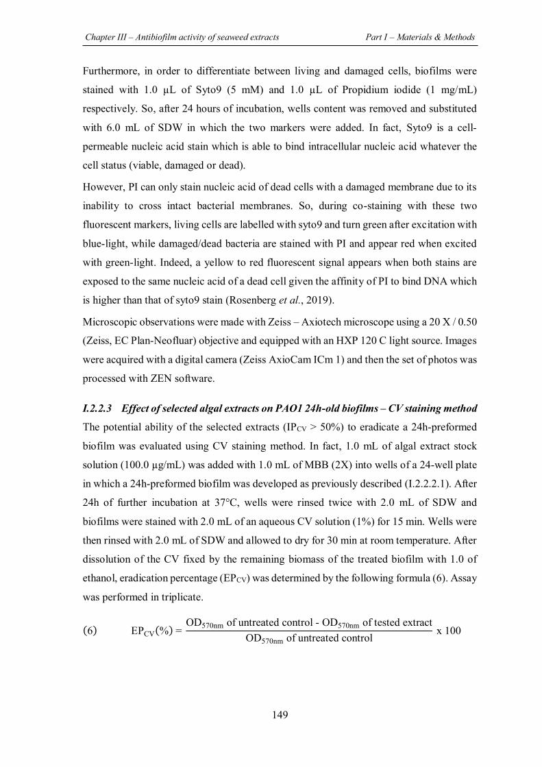
Chapter III – Antibiofilm activity of seaweed extracts Part I – Materials & Methods
149
Furthermore, in order to differentiate between living and damaged cells, biofilms were
stained with 1.0 µL of Syto9 (5 mM) and 1.0 µL of Propidium iodide (1 mg/mL)
respectively. So, after 24 hours of incubation, wells content was removed and substituted
with 6.0 mL of SDW in which the two markers were added. In fact, Syto9 is a cell-
permeable nucleic acid stain which is able to bind intracellular nucleic acid whatever the
cell status (viable, damaged or dead).
However, PI can only stain nucleic acid of dead cells with a damaged membrane due to its
inability to cross intact bacterial membranes. So, during co-staining with these two
fluorescent markers, living cells are labelled with syto9 and turn green after excitation with
blue-light, while damaged/dead bacteria are stained with PI and appear red when excited
with green-light. Indeed, a yellow to red fluorescent signal appears when both stains are
exposed to the same nucleic acid of a dead cell given the affinity of PI to bind DNA which
is higher than that of syto9 stain (Rosenberg et al., 2019).
Microscopic observations were made with Zeiss – Axiotech microscope using a 20 X / 0.50
(Zeiss, EC Plan-Neofluar) objective and equipped with an HXP 120 C light source. Images
were acquired with a digital camera (Zeiss AxioCam ICm 1) and then the set of photos was
processed with ZEN software.
I.2.2.3 Effect of selected algal extracts on PAO1 24h-old biofilms – CV staining method
The potential ability of the selected extracts (IPCV > 50%) to eradicate a 24h-preformed
biofilm was evaluated using CV staining method. In fact, 1.0 mL of algal extract stock
solution (100.0 µg/mL) was added with 1.0 mL of MBB (2X) into wells of a 24-well plate
in which a 24h-preformed biofilm was developed as previously described (I.2.2.2.1). After
24h of further incubation at 37°C, wells were rinsed twice with 2.0 mL of SDW and
biofilms were stained with 2.0 mL of an aqueous CV solution (1%) for 15 min. Wells were
then rinsed with 2.0 mL of SDW and allowed to dry for 30 min at room temperature. After
dissolution of the CV fixed by the remaining biomass of the treated biofilm with 1.0 of
ethanol, eradication percentage (EPCV) was determined by the following formula (6). Assay
was performed in triplicate.
(6) EPCV(%) = OD570nm of untreated control - OD570nm of tested extract
OD570nm of untreated control x 100
Chapter III – Antibiofilm activity of seaweed extracts Part I – Materials & Methods
150
I.2.2.4 Effect of the most active extract on PAO1 24h-old biofilms – CFU counts
method
The most active extract that has exhibited an eradication percentage (EPCV) greater than
80% by CV staining method was also evaluated by the CFU counts method. In this case,
both the planktonic cells released from biofilm as well as the remaining adhered cells were
quantified to evaluate the potential effect of extracts in promoting the dispersion and
detachment of biofilm cells, respectively.
Also here, 24h-old biofilms were treated with 1.0 mL of the active extract (100.0 µg/mL),
which is EA extract originated from the green alga U. lactuca. 1.0 mL of MBB (2X) was
also added to the wells. After 24h of incubation at 37°C and before rinsing the wells twice
with 2.0 mL of SDW, 1.0 mL of the supernatant was withdrawn to quantify biofilm-
released cells. Adhered cells were then recuperated by scarping for 1 min in 1.0 mL of
SDW. Finally, planktonic and adhered cells were submitted to ten-fold serial dilution
followed by inoculation in TSA (900.0 µL) for CFU quantification. The number of CFU
counted after 48h of incubation at 37°C was subjected to logarithmic transformation based
on the following formula (7). Assay was performed in triplicate.
(7) Log CFU mL =⁄ log number of colonies (CFU)
Dilution factor x inoculated volume
I.2.2.5 Control of extracts effect on PAO1 planktonic growth
I.2.2.5.1 Assessment of the impact of CH and EA extracts on planktonic growth – CFU
counts method
In order to exclude the potential bactericidal effect of the two active extracts (CH and EA
extracts derived from the green alga U. lactuca) at the tested concentration (50.0 µg/mL)
as well as to ensure that EA extract has no planktonic-growth promoting effect, their impact
on PAO1 planktonic cells was assessed. The protocol developed by Feuillolay et al., was
used in this assay (Feuillolay et al., 2016).
Briefly, 5.0 mL of PAO1 bacterial suspension (105 or 102 CFU/mL) prepared in MBB (2X)
medium and supplemented with 5.0 mL of sterile distilled water were incubated for 24
hours at 37˚C. Water was replaced by 5.0 mL of extracts (CH or EA extracts) in the sample
tubes. The potential bactericidal activity of extracts was determined on both suspension
(105 and 102 CFU/mL). Tubes were maintained under agitation (100 rpm) in an orbital
shaker (MaxQ 4000, Thermo Fisher Scientific, Waltham, USA).
Chapter III – Antibiofilm activity of seaweed extracts Part I – Materials & Methods
151
The number of planktonic cells was monitored after 24 hours of incubation at 37°C by CFU
counts. Prior to their quantification, samples were homogenized then 1.0 mL was taken and
serially diluted (10-1 to 10-6). 900 μl of each dilution were inoculated by inclusion in TSA
agar plates and overnight incubated at 37˚C for cell quantification. Assays were performed
in duplicate. Results were expressed as ratio (log CFU/mL for sample / log CFU/mL for
untreated control). The protocol followed is illustrated in the figure below (Figure 32).
I.2.2.5.2 Assessment of the impact of culture media and EA extract on PAO1 planktonic
growth kinetics – OD measurement
In order to justify the use of MBB in the antibiofilm assays as a low-nutritive medium that
allow the growth of bacteria in adhered form rather than in planktonic form, a comparison
of PAO1 planktonic growth in MBB versus that in rich medium MHB was carried out.
PAO1 growth kinetics curves in MHB, MBB (2X), as well as in presence of EA extract (to
ensure the absence of planktonic-growth promoting effect) were performed. Briefly, 100.0
µL of tested media (MHB and MBB) was introduced respectively into the wells of a sterile
96-well microtiter plate supplemented with 100.0 µL of SDW. In order to evaluate the
potential effect of EA extract on the PAO1 growth curve, 100.0 µL of EA extract stock
solution (100.0 µg/mL) were added to 100.0 µL of MBB (2X).
The bacterial suspension used in this assay was prepared in SDW and adjusted to an
OD640nm of 0.150 corresponding to a concentration of 108 CFU/mL followed by dilution
(1:10) to achieve a concentration of 107 CFU/mL.
FIGURE 32 | Control of extracts effect on planktonic growth by CFU counts method.
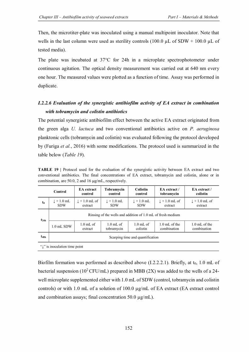
Chapter III – Antibiofilm activity of seaweed extracts Part I – Materials & Methods
152
Then, the microtiter-plate was inoculated using a manual multipoint inoculator. Note that
wells in the last column were used as sterility controls (100.0 µL of SDW + 100.0 µL of
tested media).
The plate was incubated at 37°C for 24h in a microplate spectrophotometer under
continuous agitation. The optical density measurement was carried out at 640 nm every
one hour. The measured values were plotted as a function of time. Assay was performed in
duplicate.
I.2.2.6 Evaluation of the synergistic antibiofilm activity of EA extract in combination
with tobramycin and colistin antibiotics
The potential synergistic antibiofilm effect between the active EA extract originated from
the green alga U. lactuca and two conventional antibiotics active on P. aeruginosa
planktonic cells (tobramycin and colistin) was evaluated following the protocol developed
by (Furiga et al., 2016) with some modifications. The protocol used is summarized in the
table below (Table 19).
TABLE 19 | Protocol used for the evaluation of the synergistic activity between EA extract and two conventional antibiotics. The final concentrations of EA extract, tobramycin and colistin, alone or in combination, are 50.0, 2 and 16 µg/mL, respectively.
Biofilm formation was performed as described above (I.2.2.2.1). Briefly, at t0, 1.0 mL of
bacterial suspension (102 CFU/mL) prepared in MBB (2X) was added to the wells of a 24-
well microplate supplemented either with 1.0 mL of SDW (control, tobramycin and colistin
controls) or with 1.0 mL of a solution of 100.0 µg/mL of EA extract (EA extract control
and combination assays; final concentration 50.0 µg/mL).
Control EA extract control
Tobramycin control
Colistin control
EA extract / tobramycin
EA extract / colistin
t0 ↓ + 1.0 mL SDW
↓ + 1.0 mL of extract
↓ + 1.0 mL SDW
↓ + 1.0 mL SDW
↓ + 1.0 mL of extract
↓ + 1.0 mL of extract
t24h Rinsing of the wells and addition of 1.0 mL of fresh medium
1.0 mL SDW 1.0 mL of extract
1.0 mL of tobramycin
1.0 mL of colistin
1.0 mL of the combination
1.0 mL of the combination
t48h Scarping time and quantification
“↓” is inoculation time point
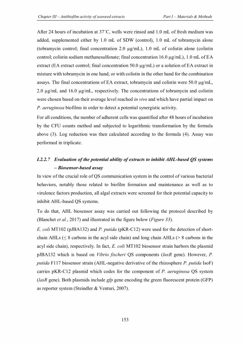
Chapter III – Antibiofilm activity of seaweed extracts Part I – Materials & Methods
153
After 24 hours of incubation at 37˚C, wells were rinsed and 1.0 mL of fresh medium was
added, supplemented either by 1.0 mL of SDW (control), 1.0 mL of tobramycin alone
(tobramycin control; final concentration 2.0 µg/mL), 1.0 mL of colistin alone (colistin
control; colistin sodium methanesulfonate; final concentration 16.0 µg/mL), 1.0 mL of EA
extract (EA extract control; final concentration 50.0 µg/mL) or a solution of EA extract in
mixture with tobramycin in one hand, or with colistin in the other hand for the combination
assays. The final concentrations of EA extract, tobramycin and colistin were 50.0 µg/mL,
2.0 µg/mL and 16.0 µg/mL, respectively. The concentrations of tobramycin and colistin
were chosen based on their average level reached in vivo and which have partial impact on
P. aeruginosa biofilms in order to detect a potential synergistic activity.
For all conditions, the number of adherent cells was quantified after 48 hours of incubation
by the CFU counts method and subjected to logarithmic transformation by the formula
above (3). Log reduction was then calculated according to the formula (4). Assay was
performed in triplicate.
I.2.2.7 Evaluation of the potential ability of extracts to inhibit AHL-based QS systems
– Biosensor-based assay
In view of the crucial role of QS communication system in the control of various bacterial
behaviors, notably those related to biofilm formation and maintenance as well as to
virulence factors production, all algal extracts were screened for their potential capacity to
inhibit AHL-based QS systems.
To do that, AHL biosensor assay was carried out following the protocol described by
(Blanchet et al., 2017) and illustrated in the figure below (Figure 33).
E. coli MT102 (pJBA132) and P. putida (pKR-C12) were used for the detection of short-
chain AHLs (< 8 carbons in the acyl side chain) and long chain AHLs (> 8 carbons in the
acyl side chain), respectively. In fact, E. coli MT102 biosensor strain harbors the plasmid
pJBA132 which is based on Vibrio fischeri QS components (luxR gene). However, P.
putida F117 biosensor strain (AHL-negative derivative of the rhizosphere P. putida IsoF)
carries pKR-C12 plasmid which codes for the component of P. aeruginosa QS system
(lasR gene). Both plasmids include gfp gene encoding the green fluorescent protein (GFP)
as reporter system (Steindler & Venturi, 2007).
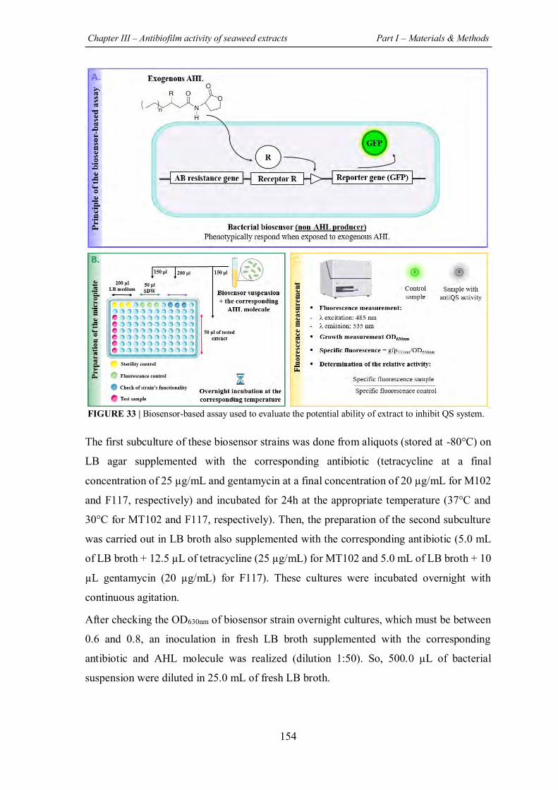
Chapter III – Antibiofilm activity of seaweed extracts Part I – Materials & Methods
154
The first subculture of these biosensor strains was done from aliquots (stored at -80°C) on
LB agar supplemented with the corresponding antibiotic (tetracycline at a final
concentration of 25 µg/mL and gentamycin at a final concentration of 20 µg/mL for M102
and F117, respectively) and incubated for 24h at the appropriate temperature (37°C and
30°C for MT102 and F117, respectively). Then, the preparation of the second subculture
was carried out in LB broth also supplemented with the corresponding antibiotic (5.0 mL
of LB broth + 12.5 µL of tetracycline (25 µg/mL) for MT102 and 5.0 mL of LB broth + 10
µL gentamycin (20 µg/mL) for F117). These cultures were incubated overnight with
continuous agitation.
After checking the OD630nm of biosensor strain overnight cultures, which must be between
0.6 and 0.8, an inoculation in fresh LB broth supplemented with the corresponding
antibiotic and AHL molecule was realized (dilution 1:50). So, 500.0 µL of bacterial
suspension were diluted in 25.0 mL of fresh LB broth.
FIGURE 33 | Biosensor-based assay used to evaluate the potential ability of extract to inhibit QS system.
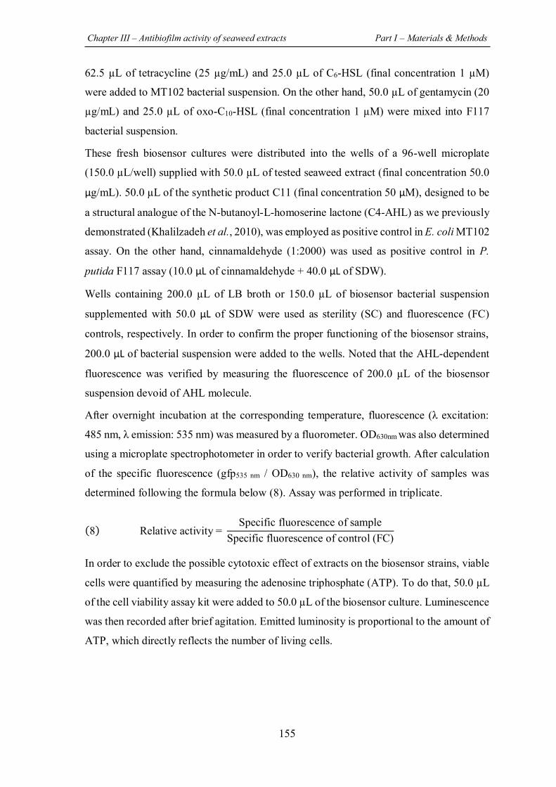
Chapter III – Antibiofilm activity of seaweed extracts Part I – Materials & Methods
155
62.5 µL of tetracycline (25 µg/mL) and 25.0 µL of C6-HSL (final concentration 1 µM)
were added to MT102 bacterial suspension. On the other hand, 50.0 µL of gentamycin (20
µg/mL) and 25.0 µL of oxo-C10-HSL (final concentration 1 µM) were mixed into F117
bacterial suspension.
These fresh biosensor cultures were distributed into the wells of a 96-well microplate
(150.0 µL/well) supplied with 50.0 µL of tested seaweed extract (final concentration 50.0
µg/mL). 50.0 µL of the synthetic product C11 (final concentration 50 µM), designed to be
a structural analogue of the N-butanoyl-L-homoserine lactone (C4-AHL) as we previously
demonstrated (Khalilzadeh et al., 2010), was employed as positive control in E. coli MT102
assay. On the other hand, cinnamaldehyde (1:2000) was used as positive control in P.
putida F117 assay (10.0 µL of cinnamaldehyde + 40.0 µL of SDW).
Wells containing 200.0 µL of LB broth or 150.0 µL of biosensor bacterial suspension
supplemented with 50.0 µL of SDW were used as sterility (SC) and fluorescence (FC)
controls, respectively. In order to confirm the proper functioning of the biosensor strains,
200.0 µL of bacterial suspension were added to the wells. Noted that the AHL-dependent
fluorescence was verified by measuring the fluorescence of 200.0 µL of the biosensor
suspension devoid of AHL molecule.
After overnight incubation at the corresponding temperature, fluorescence (λ excitation:
485 nm, λ emission: 535 nm) was measured by a fluorometer. OD630nm was also determined
using a microplate spectrophotometer in order to verify bacterial growth. After calculation
of the specific fluorescence (gfp535 nm / OD630 nm), the relative activity of samples was
determined following the formula below (8). Assay was performed in triplicate.
(8) Relative activity = Specific fluorescence of sample
Specific fluorescence of control (FC)
In order to exclude the possible cytotoxic effect of extracts on the biosensor strains, viable
cells were quantified by measuring the adenosine triphosphate (ATP). To do that, 50.0 µL
of the cell viability assay kit were added to 50.0 µL of the biosensor culture. Luminescence
was then recorded after brief agitation. Emitted luminosity is proportional to the amount of
ATP, which directly reflects the number of living cells.
Chapter III – Antibiofilm activity of seaweed extracts Part I – Materials & Methods
156
I.2.2.8 Statistical analysis
All values are expressed as mean + SD for three independent experiments. Student t-test
was used to calculate the significance of the differences between the mean effects of the
extract and those for the associated untreated control in the CFU counts method (log
CFU/mL) after checking equality of variances with Levene’s test (P-value < 0.05). Student
t-test was also used to analyze results obtained by CV staining method. The significance of
differences was determined between the mean OD570 nm of CV fixed by the treated biofilm
and that of the related un-treated biofilm. Regarding biosensor-based assay, the
significance of differences was calculated between the mean specific fluorescence in
sample and that of the related control, also using student t-test. Statistically significant
values were defined as a P-value (* < 0.05, ** < 0.01 or *** < 0.001). SPSS 22.0 software
(SPSS, IBM Corporation, Armonk, NY, USA) was used in the statistical analysis.
Chapter III – Antibiofilm activity of seaweed extracts Part I – Materials & Methods
157
I.2.3 Assessment of the potential antibiofilm activity of seaweed extracts against
the pathogenic bacteria S. aureus
I.2.3.1 Evaluation of the antibacterial activity of extracts – MIC determination
Prior to the assessment of their potential effect on biofilm, the evaluation of antibacterial
activity of extracts on S. aureus is essential to ensure the use of a sub-MIC concentration
in antibiofilm activity assays. For this purpose, the minimal inhibitory concentration (MIC)
of all extracts against S. aureus was determined using the broth microdilution method,
according to the guidelines of CA-SFM/EUCAST 2020 and as described above for
P. aeruginosa.
I.2.3.2 Evaluation of the antibiofilm activity of extracts
First, the ability of extracts derived from the three tested algae to inhibit S. aureus biofilm
formation and growth was determined. Extracts that exhibited a significant activity were
selected in order to assess their potential capacity to reduce a 24h-preformed biofilm.
Furthermore, to investigate the biofilm growth phase targeted by the active extracts, a third
experiment was conducted by adding extract to a S. aureus biofilm at various development
stages (t2h, t4h, t6h, and t24h). It should be noted that in all assays, the quantification of
S. aureus biofilm was realized by CFU counts method. In fact, the application of CV
staining method is not suitable here due to the limited quantity of biomass produced by S.
aureus biofilm (below the detection limit) under the culture conditions adopted in this
study. Assays were performed in triplicate.
I.2.3.2.1 Effect of extracts (added at t0) on S. aureus biofilm formation and growth
The influence of extracts on the number of adhered cells was evaluated following the
colony counts method previously described by (Khalilzadeh et al., 2010) with some
modifications.
The bacterial suspension used in this assay was prepared in the low-nutritive medium BB
(2X) and was adjusted to 108 CFU/mL (OD640nm = 0.150) followed by ten-fold serial
dilution up to 10-6 with the same medium. Then, 1.0 mL of the 10-6 dilution (equivalent to
102 CFU/mL) was introduced into the wells of a 24-well plate. 1.0 mL of algal extract
(100.0 µg/mL) was added at t0, corresponding to a final concentration of 50.0 µg/mL. Algal
extract was replaced by 1.0 mL of SDW in biofilm growth control. Wells containing 1.0
mL of SDW + 1.0 mL of un-inoculated BB (2X) medium was considered as negative
control.
Chapter III – Antibiofilm activity of seaweed extracts Part I – Materials & Methods
158
After overnight incubation at 37°C, wells content was discarded followed by rinsing (x 2)
with 2.0 mL of SDW in order to remove unattached planktonic cells. Adhered cells were
then recovered by scarping for 1 min with a sterile spatula into 1.0 mL of SDW followed
by a ten-fold serial dilution (from 10-1 to 10-6). 900.0 µL of each dilution was then
inoculated by inclusion in TSA plates.
After 48h of incubation at 37°C, the numbers of CFU were determined by considering only
plates with 15 to 300 CFU. The adhered biomass was then calculated and subjected to
logarithmic transformation by the following formula (9). The logarithmic reduction with
respect to the corresponding untreated control was calculated using the formula below (10).
(9) Log of adhered biomass (log CFU mL⁄ ) = log number of colonies (CFU)
Dilution factor x inoculated volume
(10) Log CFU mL reduction = log CFU mL for control - log CFU mL for treated biofilm ⁄ ⁄⁄
I.2.3.2.2 Phenotypic observations of biofilms by epifluorescence microscopy
The biofilm formed in presence of the most active extracts (***, P-value < 0.001; **, P-
value < 0.01) was visualized using an epifluorescence microscopy. For this analysis,
S. aureus biofilms were grown as described above (I.2.3.2.1) but in a 6-well microplate and
with a total volume of 6.0 mL (3.0 mL of S. aureus bacterial suspension prepared in BB
2X (102 CFU/mL) + 3.0 mL of tested extract or 3.0 mL of SDW for the untreated control).
After 24 hours of incubation at 37°C and in order to evaluate the potential effect of extracts
on S. aureus biofilm matrix, 1.0 mL of SYPRO Ruby stain was added after discarding wells
content. This stain binds to most classes of proteins including glycoproteins, lipoproteins,
phosphoproteins and fibrillar proteins. After 30 min of incubation in dark at room
temperature, wells were carefully washed twice with 1.0 mL of SDW. 6.0 mL of SDW was
then added supplemented with 1.0 µL of Syto9 stain for cells observation.
Microscopic observations were made with Zeiss – Axiotech microscope using a 20 X / 0.50
(Zeiss, EC Plan-Neofluar) objective and equipped with an HXP 120 C light source. Images
were acquired with a digital camera (Zeiss AxioCam ICm 1) and then the set of photos was
processed with ZEN software.
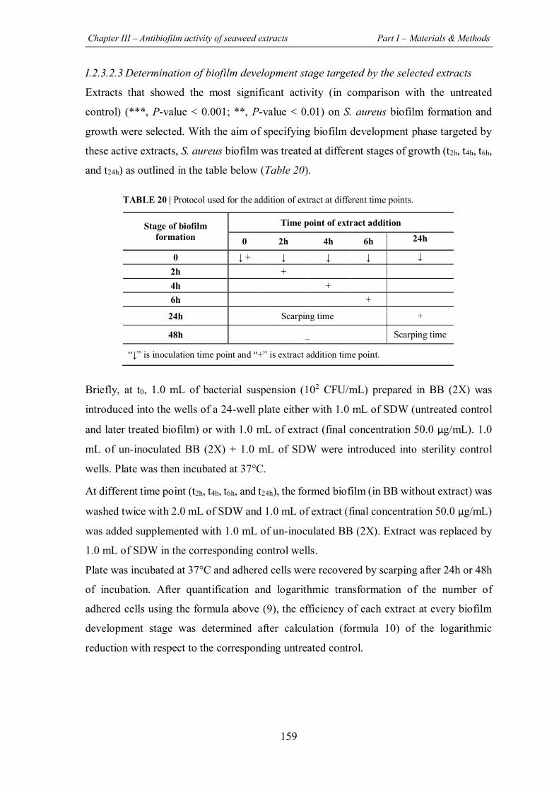
Chapter III – Antibiofilm activity of seaweed extracts Part I – Materials & Methods
159
I.2.3.2.3 Determination of biofilm development stage targeted by the selected extracts
Extracts that showed the most significant activity (in comparison with the untreated
control) (***, P-value < 0.001; **, P-value < 0.01) on S. aureus biofilm formation and
growth were selected. With the aim of specifying biofilm development phase targeted by
these active extracts, S. aureus biofilm was treated at different stages of growth (t2h, t4h, t6h,
and t24h) as outlined in the table below (Table 20).
TABLE 20 | Protocol used for the addition of extract at different time points.
Briefly, at t0, 1.0 mL of bacterial suspension (102 CFU/mL) prepared in BB (2X) was
introduced into the wells of a 24-well plate either with 1.0 mL of SDW (untreated control
and later treated biofilm) or with 1.0 mL of extract (final concentration 50.0 µg/mL). 1.0
mL of un-inoculated BB (2X) + 1.0 mL of SDW were introduced into sterility control
wells. Plate was then incubated at 37°C.
At different time point (t2h, t4h, t6h, and t24h), the formed biofilm (in BB without extract) was
washed twice with 2.0 mL of SDW and 1.0 mL of extract (final concentration 50.0 µg/mL)
was added supplemented with 1.0 mL of un-inoculated BB (2X). Extract was replaced by
1.0 mL of SDW in the corresponding control wells.
Plate was incubated at 37°C and adhered cells were recovered by scarping after 24h or 48h
of incubation. After quantification and logarithmic transformation of the number of
adhered cells using the formula above (9), the efficiency of each extract at every biofilm
development stage was determined after calculation (formula 10) of the logarithmic
reduction with respect to the corresponding untreated control.
Stage of biofilm formation
Time point of extract addition
0 2h 4h 6h 24h
0 ↓ + ↓ ↓ ↓ ↓ 2h +
4h + 6h +
24h Scarping time +
48h _ Scarping time
“↓” is inoculation time point and “+” is extract addition time point.
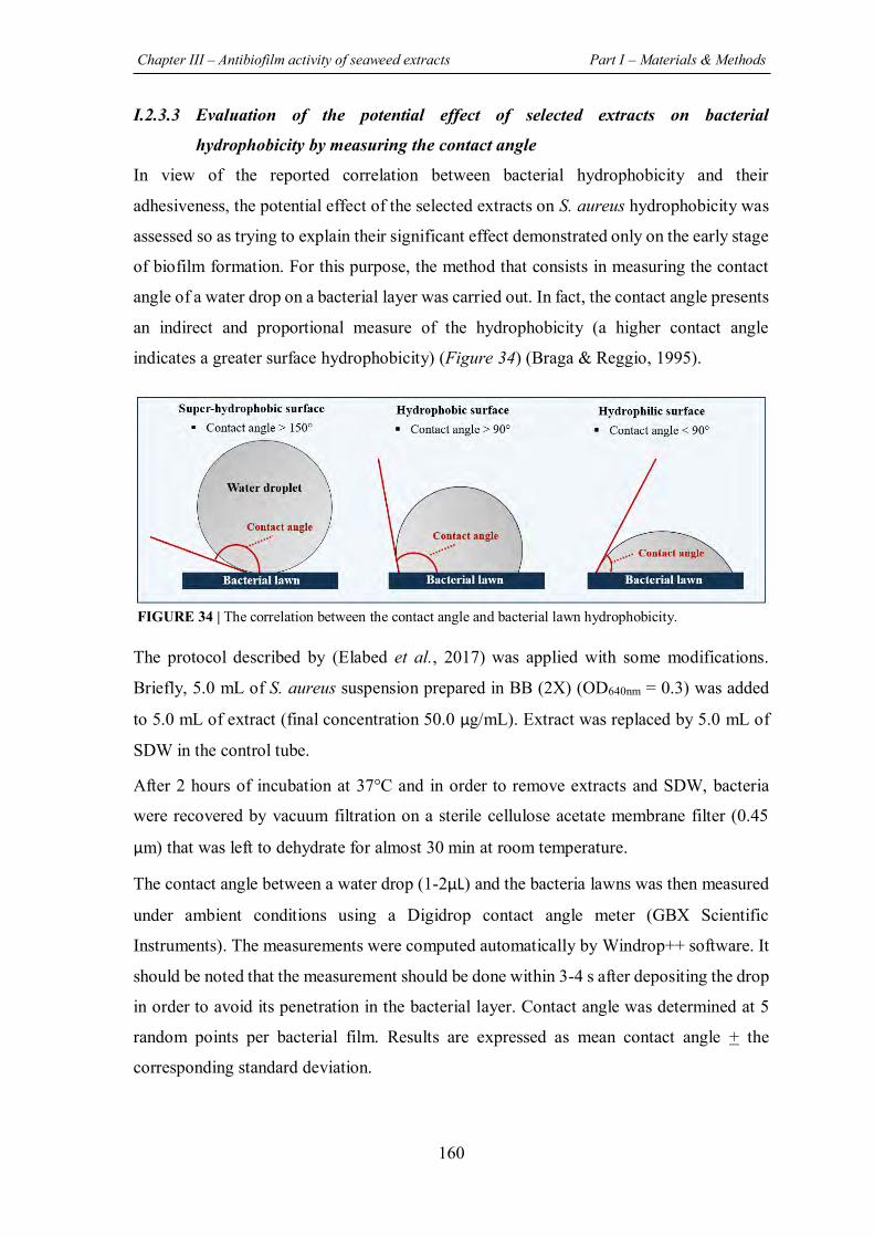
Chapter III – Antibiofilm activity of seaweed extracts Part I – Materials & Methods
160
I.2.3.3 Evaluation of the potential effect of selected extracts on bacterial
hydrophobicity by measuring the contact angle
In view of the reported correlation between bacterial hydrophobicity and their
adhesiveness, the potential effect of the selected extracts on S. aureus hydrophobicity was
assessed so as trying to explain their significant effect demonstrated only on the early stage
of biofilm formation. For this purpose, the method that consists in measuring the contact
angle of a water drop on a bacterial layer was carried out. In fact, the contact angle presents
an indirect and proportional measure of the hydrophobicity (a higher contact angle
indicates a greater surface hydrophobicity) (Figure 34) (Braga & Reggio, 1995).
The protocol described by (Elabed et al., 2017) was applied with some modifications.
Briefly, 5.0 mL of S. aureus suspension prepared in BB (2X) (OD640nm = 0.3) was added
to 5.0 mL of extract (final concentration 50.0 µg/mL). Extract was replaced by 5.0 mL of
SDW in the control tube.
After 2 hours of incubation at 37°C and in order to remove extracts and SDW, bacteria
were recovered by vacuum filtration on a sterile cellulose acetate membrane filter (0.45
µm) that was left to dehydrate for almost 30 min at room temperature.
The contact angle between a water drop (1-2µL) and the bacteria lawns was then measured
under ambient conditions using a Digidrop contact angle meter (GBX Scientific
Instruments). The measurements were computed automatically by Windrop++ software. It
should be noted that the measurement should be done within 3-4 s after depositing the drop
in order to avoid its penetration in the bacterial layer. Contact angle was determined at 5
random points per bacterial film. Results are expressed as mean contact angle + the
corresponding standard deviation.
FIGURE 34 | The correlation between the contact angle and bacterial lawn hydrophobicity.
Chapter III – Antibiofilm activity of seaweed extracts Part I – Materials & Methods
161
I.2.3.4 Statistical analysis
All values are expressed as mean + SD for three independent experiments. The student t-
test was used to calculate the significance of the differences between the mean effects of
the extract and those for the associated untreated control after checking equality of
variances with Levene’s test (P-value < 0.05). Statistically significant values were defined
as a P-value (* < 0.05, ** < 0.01 or *** < 0.001). SPSS 22.0 software (SPSS, IBM
Corporation, Armonk, NY, USA) was used.
162
REFERENCES Blanchet, E., Prado, S., Stien, D., Oliveira da Silva, J., Ferandin, Y., Batailler, N., et al. (2017).
Quorum Sensing and Quorum Quenching in the Mediterranean Seagrass Posidonia oceanica Microbiota. Front. Mar. Sci., 4. doi:10.3389/fmars.2017.00218
Braga, P., & Reggio, S. (1995). Correlation between reduction of surface hydrophobicity of and the decrease in its adhesiveness induced by subinhibitory concentrations of brodimoprim. Pharmacol. Res., 32(5), 315-319. doi:10.1016/s1043-6618(05)80030-1
Campanac, C., Pineau, L., Payard, A., Baziard-Mouysset, G., & Roques, C. (2002). Interactions between Biocide Cationic Agents and Bacterial Biofilms. Antimicrob. Agents Chemother, 46(5), 1469-1474. doi:10.1128/AAC.46.5.1469-1474.2002
Elabed, S., Elabed, A., Sadiki, M., Elfarricha, O., & Ibnsouda, S. (2017). Assessment of the Salvia officinalis and Myrtus communis Aqueous Extracts Effect on Cell Surface Tension Parameters and Hydrophobicity of Staphylococcus aureus CIP54354 and Bacillus subtilis ILP142B. J. Appl. Sci., 17(5), 246-252. doi:10.3923/jas.2017.246.252
Feuillolay, C., Pecastaings, S., Le Gac, C., Fiorini-Puybaret, C., Luc, J., Joulia, P., et al. (2016). A Myrtus communis extract enriched in myrtucummulones and ursolic acid reduces resistance of Propionibacterium acnes biofilms to antibiotics used in acne vulgaris. Phytomedicine, 23(3), 307-315. doi:10.1016/j.phymed.2015.11.016
Furiga, A., Lajoie, B., El Hage, S., Baziard, G., & Roques, C. (2016). Impairment of Pseudomonas aeruginosa Biofilm Resistance to Antibiotics by Combining the Drugs with a New Quorum-Sensing Inhibitor. Antimicrobial Agents and Chemotherapy, 60(3), 1676-1686. doi:10.1128/AAC.02533-15
Khalilzadeh, P., Lajoie, B., El Hage, S., Furiga, A., Baziard, G., Berge, M., et al. (2010). Growth inhibition of adherent Pseudomonas aeruginosa by an N-butanoyl-L-homoserine lactone analog. Can J Microbiol, 56(4), 317-325. doi:10.1139/w10-013
Kohoude, M. J., Gbaguidi, F., Agbani, P., Ayedoun, M.-A., Cazaux, S., & Bouajila, J. (2017). Chemical composition and biological activities of extracts and essential oil of Boswellia dalzielii leaves. Pharm. Biol., 55(1), 33-42. doi:10.1080/13880209.2016.1226356
Peeters, E., Nelis, H. J., & Coenye, T. (2008). Comparison of multiple methods for quantification of microbial biofilms grown in microtiter plates. J. Microbiol. Methods, 72(2), 157-165. doi:10.1016/j.mimet.2007.11.010
Rima, M., Trognon, J., Latapie, L., Chbani, A., Roques, C., & El Garah, F. (2022). Seaweed Extracts: A Promising Source of Antibiofilm Agents with Distinct Mechanisms of Action against Pseudomonas aeruginosa. Mar. Drugs, 20(2). doi:10.3390/md20020092
Rosenberg, M., Azevedo, N. F., & Ivask, A. (2019). Propidium iodide staining underestimates viability of adherent bacterial cells. Sci. Rep., 9(1), 6483. doi:10.1038/s41598-019-42906-3
Steindler, L., & Venturi, V. (2007). Detection of quorum-sensing N-acyl homoserine lactone signal molecules by bacterial biosensors. FEMS Microbiol Lett, 266(1), 1-9. doi:10.1111/j.1574-6968.2006.00501.x
Strathmann, M., Wingender, J., & Flemming, H.-C. (2002). Application of fluorescently labelled lectins for the visualization and biochemical characterization of polysaccharides in biofilms of Pseudomonas aeruginosa. J. Microbiol. Methods, 50(3), 237-248. doi:10.1016/S0167-7012(02)00032-5

163
PREVIEW
In this part, the potential ability of extracts derived from the three Lebanese seaweed (green
alga U. lactuca, brown alga S. scoparium, and red alga P. capillacea) to exhibit an
antibiofilm activity against the pathogenic bacterium P. aeruginosa was investigated. The
potential antibiofilm mechanisms of action of the most active extracts was elaborated. In
addition, the most active extract was tested in combination with two antibiotics which are
generally used to combat P. aeruginosa biofilm infections, in order to detect a possible
synergistic antibiofilm activity.
The work presented in this part was done in the Laboratoire de Génie Chimique (LGC –
UMR5503) – Toulouse – France and resulted in a published article (Rima et al., 2022).
After a brief summary of the article with a highlight on the main results, the publication is
integrated followed by the supplementary materials.
On the other hand, a screening of extracts for their potential capacity to inhibit AHL-based
QS system is presented.
Part II: Evaluation of the antibiofilm activity of seaweed extracts against P. aeruginosa
III CHAPTER
ARTICLE II – Published:
Rima, M., Trognon, J., Latapie, L., Chbani, A., Roques, C., & El Garah, F. (2022) Seaweed extracts: A promising source of antibiofilm agents with distinct mechanisms of action against Pseudomonas aeruginosa. Mar. Drugs, 20(2), 92. doi: //doi.org/10.3390/md20020092
Chapter III – Antibiofilm activity of seaweed extracts Part II – Effect on P. aeruginosa biofilm
164
II.1 ARTICLE SUMMARY – FOCUS ON THE MAIN RESULTS
Screening of extracts derived from the three algae evaluated in this study for their capacity
to inhibit PAO1 biofilm formation and growth (extract added at t0) permitted the selection
of CH and EA extracts obtained from the green alga U. lactuca as the most promising. It
should be noted that the antibiofilm activity was assessed following two complementary
methods: by crystal violet staining, which allows the quantification of total biofilm biomass
(cells + matrix), and by quantification of adhered bacteria (CFU counts method).
Interestingly, a consistency between these two methods was demonstrated for CH extract
(IPCV = 69.4 + 13.6%; IPCFU = 67.2 + 17.2%). However, the significant antibiofilm effect
of EA extract was only observed by CV staining method (IPCV = 84.0 + 9.6%) which
suggests the involvement of two distinct antibiofilm mechanisms of action for these
extracts.
The epifluorescence microscopic analysis of biofilm developed in presence of these two
active extracts supports this hypothesis. In fact, CH extract has led to the formation of an
unstructured biofilm formed by separated bacterial aggregates with an associated matrix.
On the other hand, a dispersed biofilm with a diffused matrix was developed in presence
of EA extract.
The potential effect of EA extract on the production and/or the degradation of PAO1
biofilm matrix was also evidenced by its ability to significantly reduce 24h-preformed
biofilm biomass (EPCV = 85.5 + 7.4%) as well as to promote the release of biofilm cells.
In view of the significant involvement of EPS matrix in biofilm tolerance towards
antimicrobial agents, the possible synergistic antibiofilm activity between EA extract, a
potential matrix disruptor, and two conventional antibiotics (tobramycin and colistin), was
evaluated. Interestingly, EA extract significantly improved the antibiofilm activity of
tobramycin against EA extract-pretreated biofilm.
These encouraging findings summarized in the table below (Table 21) emphasize the
relevance of the green alga U. lactuca as promising source of antibiofilm molecules with
different modes of action and that can be used alone or in combination with antibiotics in
the treatment of biofilm-related infections.
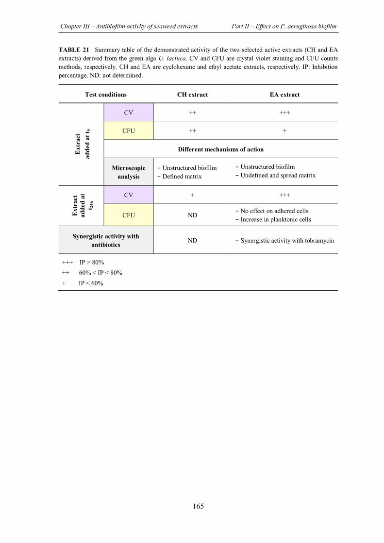
Chapter III – Antibiofilm activity of seaweed extracts Part II – Effect on P. aeruginosa biofilm
165
TABLE 21 | Summary table of the demonstrated activity of the two selected active extracts (CH and EA extracts) derived from the green alga U. lactuca. CV and CFU are crystal violet staining and CFU counts methods, respectively. CH and EA are cyclohexane and ethyl acetate extracts, respectively. IP: Inhibition percentage. ND: not determined.
Test conditions CH extract EA extract
Ext
ract
ad
ded
at t 0
CV ++ +++
CFU ++ +
Different mechanisms of action
Microscopic analysis
- Unstructured biofilm - Defined matrix
- Unstructured biofilm - Undefined and spread matrix
Ext
ract
ad
ded
at
t 24h
CV + +++
CFU ND - No effect on adhered cells - Increase in planktonic cells
Synergistic activity with antibiotics
ND - Synergistic activity with tobramycin
+++ IP > 80% ++ 60% < IP < 80% + IP < 60%
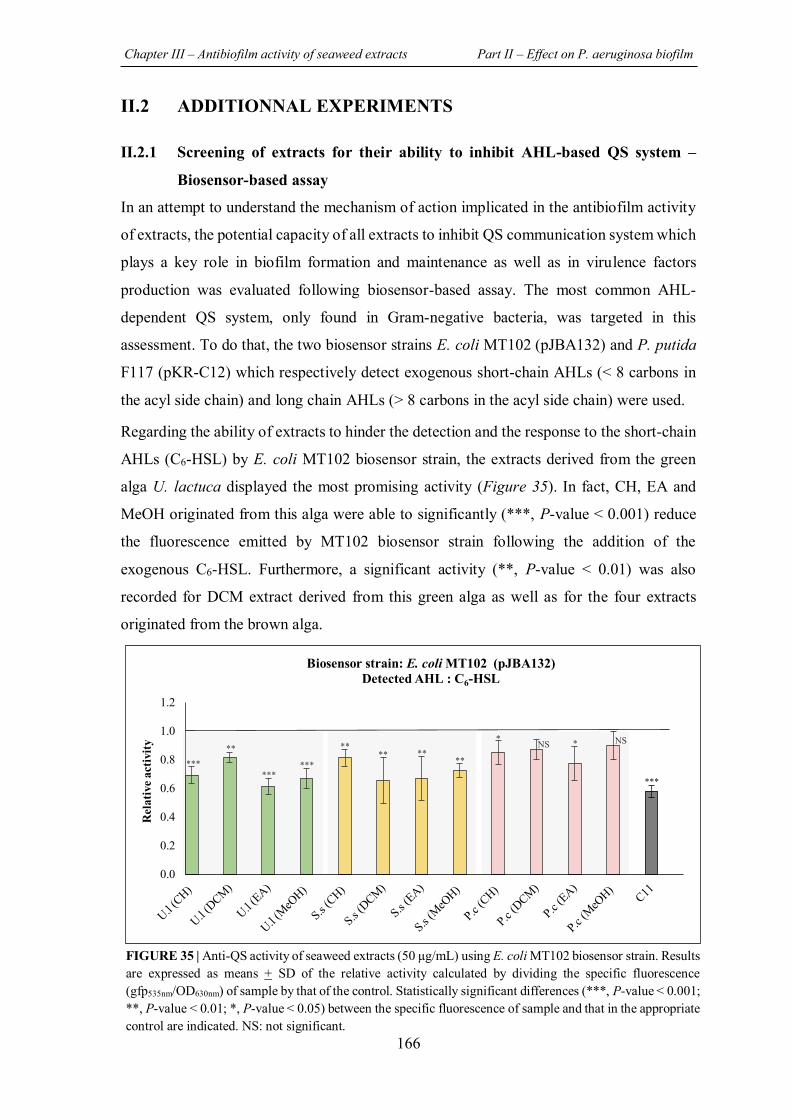
Chapter III – Antibiofilm activity of seaweed extracts Part II – Effect on P. aeruginosa biofilm
166
II.2 ADDITIONNAL EXPERIMENTS
II.2.1 Screening of extracts for their ability to inhibit AHL-based QS system –
Biosensor-based assay
In an attempt to understand the mechanism of action implicated in the antibiofilm activity
of extracts, the potential capacity of all extracts to inhibit QS communication system which
plays a key role in biofilm formation and maintenance as well as in virulence factors
production was evaluated following biosensor-based assay. The most common AHL-
dependent QS system, only found in Gram-negative bacteria, was targeted in this
assessment. To do that, the two biosensor strains E. coli MT102 (pJBA132) and P. putida
F117 (pKR-C12) which respectively detect exogenous short-chain AHLs (< 8 carbons in
the acyl side chain) and long chain AHLs (> 8 carbons in the acyl side chain) were used.
Regarding the ability of extracts to hinder the detection and the response to the short-chain
AHLs (C6-HSL) by E. coli MT102 biosensor strain, the extracts derived from the green
alga U. lactuca displayed the most promising activity (Figure 35). In fact, CH, EA and
MeOH originated from this alga were able to significantly (***, P-value < 0.001) reduce
the fluorescence emitted by MT102 biosensor strain following the addition of the
exogenous C6-HSL. Furthermore, a significant activity (**, P-value < 0.01) was also
recorded for DCM extract derived from this green alga as well as for the four extracts
originated from the brown alga.
0.0
0.2
0.4
0.6
0.8
1.0
1.2
Rel
ativ
e act
ivity
Biosensor strain: E. coli MT102 (pJBA132)Detected AHL : C6-HSL
** ** ** **
* * NS NS
***
*** ***
***
**
FIGURE 35 | Anti-QS activity of seaweed extracts (50 µg/mL) using E. coli MT102 biosensor strain. Results are expressed as means + SD of the relative activity calculated by dividing the specific fluorescence (gfp535nm/OD630nm) of sample by that of the control. Statistically significant differences (***, P-value < 0.001; **, P-value < 0.01; *, P-value < 0.05) between the specific fluorescence of sample and that in the appropriate control are indicated. NS: not significant.
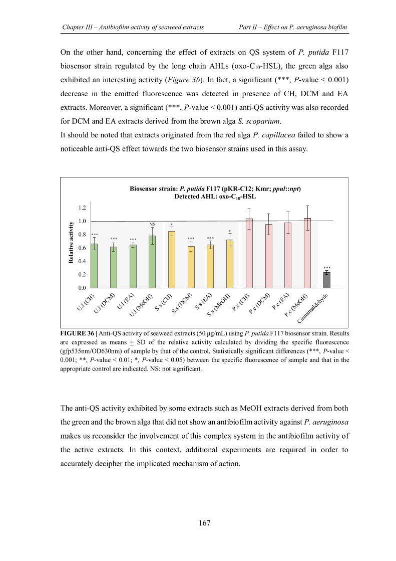
Chapter III – Antibiofilm activity of seaweed extracts Part II – Effect on P. aeruginosa biofilm
167
On the other hand, concerning the effect of extracts on QS system of P. putida F117
biosensor strain regulated by the long chain AHLs (oxo-C10-HSL), the green alga also
exhibited an interesting activity (Figure 36). In fact, a significant (***, P-value < 0.001)
decrease in the emitted fluorescence was detected in presence of CH, DCM and EA
extracts. Moreover, a significant (***, P-value < 0.001) anti-QS activity was also recorded
for DCM and EA extracts derived from the brown alga S. scoparium.
It should be noted that extracts originated from the red alga P. capillacea failed to show a
noticeable anti-QS effect towards the two biosensor strains used in this assay.
The anti-QS activity exhibited by some extracts such as MeOH extracts derived from both
the green and the brown alga that did not show an antibiofilm activity against P. aeruginosa
makes us reconsider the involvement of this complex system in the antibiofilm activity of
the active extracts. In this context, additional experiments are required in order to
accurately decipher the implicated mechanism of action.
0.0
0.2
0.4
0.6
0.8
1.0
1.2
Rel
ativ
e act
ivity
Biosensor strain: P. putida F117 (pKR-C12; Kmr; ppul::npt)Detected AHL: oxo-C10-HSL
*** *** *** NS
*** ***
* *
***
FIGURE 36 | Anti-QS activity of seaweed extracts (50 µg/mL) using P. putida F117 biosensor strain. Results are expressed as means + SD of the relative activity calculated by dividing the specific fluorescence (gfp535nm/OD630nm) of sample by that of the control. Statistically significant differences (***, P-value < 0.001; **, P-value < 0.01; *, P-value < 0.05) between the specific fluorescence of sample and that in the appropriate control are indicated. NS: not significant.
�����������������
Citation: Rima, M.; Trognon, J.;
Latapie, L.; Chbani, A.; Roques, C.;
El Garah, F. Seaweed Extracts: A
Promising Source of Antibiofilm
Agents with Distinct Mechanisms of
Action against Pseudomonas
aeruginosa. Mar. Drugs 2022, 20, 92.
https://doi.org/10.3390/
md20020092
Academic Editor: William
Lindsey White
Received: 21 December 2021
Accepted: 18 January 2022
Published: 21 January 2022
Publisher’s Note: MDPI stays neutral
with regard to jurisdictional claims in
published maps and institutional affil-
iations.
Copyright: © 2022 by the authors.
Licensee MDPI, Basel, Switzerland.
This article is an open access article
distributed under the terms and
conditions of the Creative Commons
Attribution (CC BY) license (https://
creativecommons.org/licenses/by/
4.0/).
marine drugs
Article
Seaweed Extracts: A Promising Source of AntibiofilmAgents with Distinct Mechanisms of Action againstPseudomonas aeruginosaMaya Rima 1,2, Jeanne Trognon 1, Laure Latapie 1, Asma Chbani 2,3, Christine Roques 1,4,* and Fatima El Garah 1,*
1 Laboratoire de Génie Chimique, Université de Toulouse, CNRS, INPT, UPS, 31062 Toulouse, France;[email protected] (M.R.); [email protected] (J.T.); [email protected] (L.L.)
2 Laboratory of Applied Biotechnology, AZM Center for Research in Biotechnology and Its Applications,Doctoral School of Science and Technology, Lebanese University, El Mittein Street, Tripoli 1300, Lebanon;[email protected]
3 Faculty of Public Health III, Lebanese University, Tripoli 1300, Lebanon4 Bacteriology-Hygiene Department, Centre Hospitalier Universitaire, Hôpital Purpan, 31300 Toulouse, France* Correspondence: [email protected] (C.R.); [email protected] (F.E.G.);
Tel.: +33-562-25-68-60 (C.R.); +33-562-25-68-55 (F.E.G.)
Abstract: The organization of bacteria in biofilms is one of the adaptive resistance mechanismsproviding increased protection against conventional treatments. Thus, the search for new antibiofilmagents for medical purposes, especially of natural origin, is currently the object of much attention.The objective of the study presented here was to explore the potential of extracts derived from threeseaweeds: the green Ulva lactuca, the brown Stypocaulon scoparium, and the red Pterocladiella capillacea,in terms of their antibiofilm activity against P. aeruginosa. After preparation of extracts by successivemaceration in various solvents, their antibiofilm activity was evaluated on biofilm formation and onmature biofilms. Their inhibition and eradication abilities were determined using two complementarymethods: crystal violet staining and quantification of adherent bacteria. The effect of active extractson biofilm morphology was also investigated by epifluorescence microscopy. Results revealed apromising antibiofilm activity of two extracts (cyclohexane and ethyl acetate) derived from thegreen alga by exhibiting a distinct mechanism of action, which was supported by microscopicanalyses. The ethyl acetate extract was further explored for its interaction with tobramycin andcolistin. Interestingly, this extract showed a promising synergistic effect with tobramycin. Firstanalyses of the chemical composition of extracts by GC–MS allowed for the identification of severalmolecules. Their implication in the interesting antibiofilm activity is discussed. These findingssuggest the ability of the green alga U. lactuca to offer a promising source of bioactive candidates thatcould have both a preventive and a curative effect in the treatment of biofilms.
Keywords: seaweed extracts; Ulva lactuca; anti-biofilm; Pseudomonas aeruginosa; synergistic activity;biofilm-matrix
1. Introduction
Although the discovery of antibiotics has revolutionized modern medicine and hassaved the lives of millions of patients, their massive use has contributed to a selectionpressure on bacteria leading to the rapid emergence of multidrug-resistant strains (MDR) [1].Unfortunately, the World Health Organization (WHO) has warned of a “post-antibiotic”world in which a supposedly life-saving drug will lose its effectiveness [2].
Pseudomonas aeruginosa, an opportunistic human pathogen often associated withchronic and nosocomial infections, is one of the three bacteria (Acinetobacter baumanniiand Enterobacteriaceae) classified by the WHO as a critical priority in the search for newtherapeutic strategies, due to its phenotypic and genotypic resistance towards most con-ventional antibiotics [3]. This ubiquitous Gram-negative bacterium is characterized by its
Mar. Drugs 2022, 20, 92. https://doi.org/10.3390/md20020092 https://www.mdpi.com/journal/marinedrugs
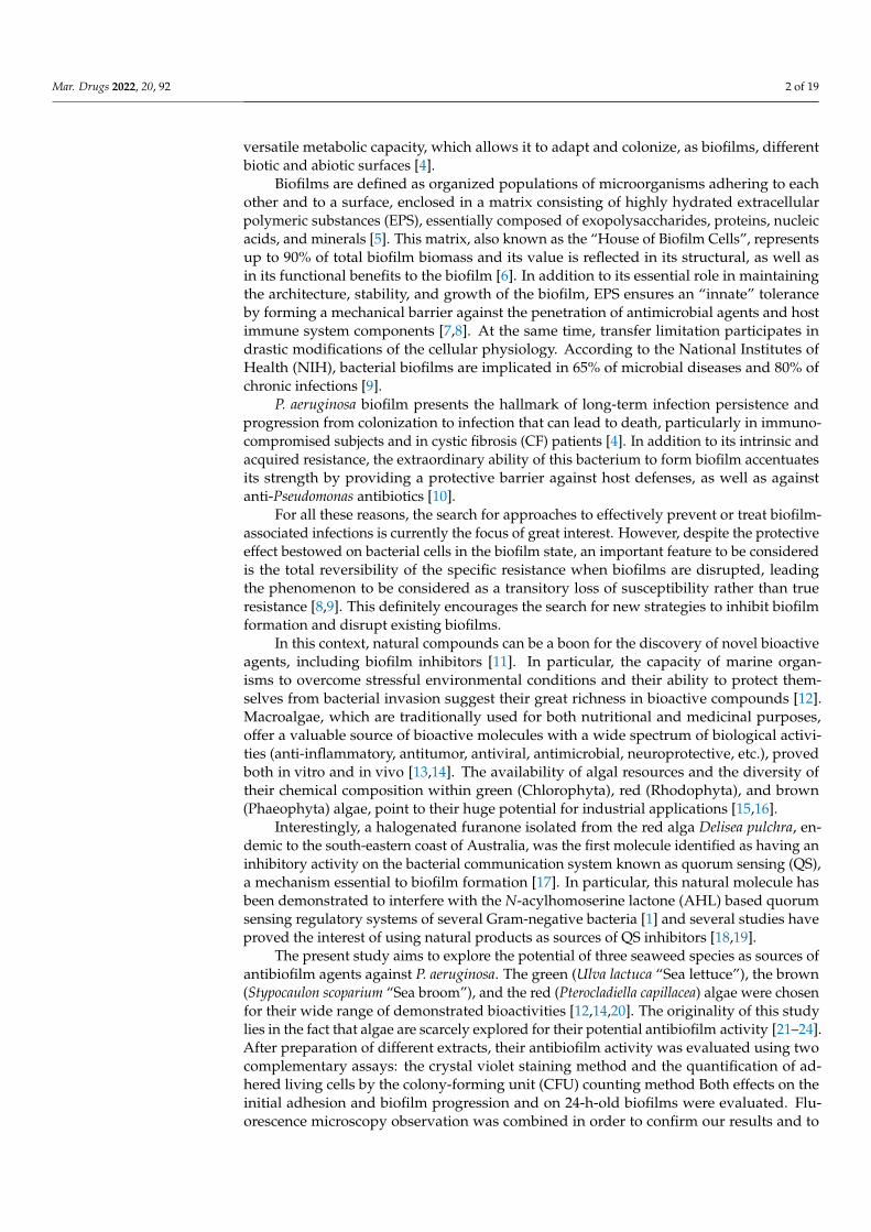
Mar. Drugs 2022, 20, 92 2 of 19
versatile metabolic capacity, which allows it to adapt and colonize, as biofilms, differentbiotic and abiotic surfaces [4].
Biofilms are defined as organized populations of microorganisms adhering to eachother and to a surface, enclosed in a matrix consisting of highly hydrated extracellularpolymeric substances (EPS), essentially composed of exopolysaccharides, proteins, nucleicacids, and minerals [5]. This matrix, also known as the “House of Biofilm Cells”, representsup to 90% of total biofilm biomass and its value is reflected in its structural, as well asin its functional benefits to the biofilm [6]. In addition to its essential role in maintainingthe architecture, stability, and growth of the biofilm, EPS ensures an “innate” toleranceby forming a mechanical barrier against the penetration of antimicrobial agents and hostimmune system components [7,8]. At the same time, transfer limitation participates indrastic modifications of the cellular physiology. According to the National Institutes ofHealth (NIH), bacterial biofilms are implicated in 65% of microbial diseases and 80% ofchronic infections [9].
P. aeruginosa biofilm presents the hallmark of long-term infection persistence andprogression from colonization to infection that can lead to death, particularly in immuno-compromised subjects and in cystic fibrosis (CF) patients [4]. In addition to its intrinsic andacquired resistance, the extraordinary ability of this bacterium to form biofilm accentuatesits strength by providing a protective barrier against host defenses, as well as againstanti-Pseudomonas antibiotics [10].
For all these reasons, the search for approaches to effectively prevent or treat biofilm-associated infections is currently the focus of great interest. However, despite the protectiveeffect bestowed on bacterial cells in the biofilm state, an important feature to be consideredis the total reversibility of the specific resistance when biofilms are disrupted, leadingthe phenomenon to be considered as a transitory loss of susceptibility rather than trueresistance [8,9]. This definitely encourages the search for new strategies to inhibit biofilmformation and disrupt existing biofilms.
In this context, natural compounds can be a boon for the discovery of novel bioactiveagents, including biofilm inhibitors [11]. In particular, the capacity of marine organ-isms to overcome stressful environmental conditions and their ability to protect them-selves from bacterial invasion suggest their great richness in bioactive compounds [12].Macroalgae, which are traditionally used for both nutritional and medicinal purposes,offer a valuable source of bioactive molecules with a wide spectrum of biological activi-ties (anti-inflammatory, antitumor, antiviral, antimicrobial, neuroprotective, etc.), provedboth in vitro and in vivo [13,14]. The availability of algal resources and the diversity oftheir chemical composition within green (Chlorophyta), red (Rhodophyta), and brown(Phaeophyta) algae, point to their huge potential for industrial applications [15,16].
Interestingly, a halogenated furanone isolated from the red alga Delisea pulchra, en-demic to the south-eastern coast of Australia, was the first molecule identified as having aninhibitory activity on the bacterial communication system known as quorum sensing (QS),a mechanism essential to biofilm formation [17]. In particular, this natural molecule hasbeen demonstrated to interfere with the N-acylhomoserine lactone (AHL) based quorumsensing regulatory systems of several Gram-negative bacteria [1] and several studies haveproved the interest of using natural products as sources of QS inhibitors [18,19].
The present study aims to explore the potential of three seaweed species as sources ofantibiofilm agents against P. aeruginosa. The green (Ulva lactuca “Sea lettuce”), the brown(Stypocaulon scoparium “Sea broom”), and the red (Pterocladiella capillacea) algae were chosenfor their wide range of demonstrated bioactivities [12,14,20]. The originality of this studylies in the fact that algae are scarcely explored for their potential antibiofilm activity [21–24].After preparation of different extracts, their antibiofilm activity was evaluated using twocomplementary assays: the crystal violet staining method and the quantification of ad-hered living cells by the colony-forming unit (CFU) counting method Both effects on theinitial adhesion and biofilm progression and on 24-h-old biofilms were evaluated. Flu-orescence microscopy observation was combined in order to confirm our results and to
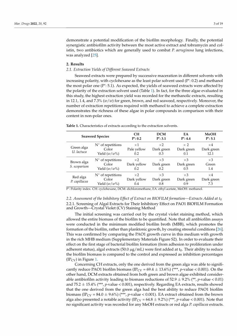
Mar. Drugs 2022, 20, 92 3 of 19
demonstrate a potential modification of the biofilm morphology. Finally, the potentialsynergistic antibiofilm activity between the most active extract and tobramycin and col-istin, two antibiotics which are generally used to combat P. aeruginosa lung infections,was analyzed [25].
2. Results2.1. Extraction Yields of Different Seaweed Extracts
Seaweed extracts were prepared by successive maceration in different solvents withincreasing polarity, with cyclohexane as the least polar solvent used (P’: 0.2) and methanolthe most polar one (P’: 5.1). As expected, the yields of seaweed extracts were affected bythe polarity of the extraction solvent used (Table 1). In fact, for the three algae evaluated inthis study, the highest extraction yield was recorded for the methanolic extracts, resultingin 12.1, 1.4, and 7.3% (w/w) for green, brown, and red seaweed, respectively. Moreover, thenumber of extraction repetitions required with methanol to achieve a complete extractiondemonstrates the richness of these algae in polar compounds in comparison with theircontent in non-polar ones.
Table 1. Characteristics of extracts according to the extraction solvents.
Seaweed Species CHP’: 0.2
DCMP’: 3.1
EAP’: 4.4
MeOHP’: 5.1
Green algaU. lactuca
N◦ of repetitions ×1 ×2 × 2 ×4Color Pale yellow Dark green Dark green Dark green
Yield (w/w%) 0.2 0.3 0.1 12.1
Brown algaS. scoparium
N◦ of repetitions ×2 ×3 ×3 ×3Color Dark yellow Dark green Dark green Green
Yield (w/w%) 0.2 0.2 0.5 1.4
Red algaP. capillacea
N◦ of repetitions ×2 ×3 ×3 ×4Color Dark yellow Dark green Dark green Dark green
Yield (w/w%) 0.4 0.8 0.9 7.3P’: Polarity index. CH: cyclohexane, DCM: dichloromethane, EA: ethyl acetate, MeOH: methanol.
2.2. Assessment of the Inhibitory Effect of Extract on BIOFILM formation—Extracts Added at t02.2.1. Screening of Algal Extracts for Their Inhibitory Effect on PAO1 BIOFILM Formationand Growth—Crystal Violet (CV) Staining Method
The initial screening was carried out by the crystal violet staining method, whichallowed the entire biomass of the biofilm to be quantified. Note that all antibiofilm assayswere conducted in the minimum modified biofilm broth (MBB), which promotes theformation of the biofilm, rather than planktonic growth, by creating stressful conditions [26].This was confirmed by comparing the PAO1 growth curve in this medium with growthin the rich MHB medium (Supplementary Materials Figure S2). In order to evaluate theireffect on the first stage of bacterial biofilm formation (from adhesion to proliferation underadherent status), algal extracts (50.0 µg/mL) were first added at t0. Their ability to reducethe biofilm biomass is compared to the control and expressed as inhibition percentages(IPCV) in Figure 1.
Concerning CH extracts, only the one derived from the green alga was able to signifi-cantly reduce PAO1 biofilm biomass (IPCV = 69.4 ± 13.6%) (***, p-value < 0.001). On theother hand, DCM extracts obtained from both green and brown algae exhibited consider-able antibiofilm activity leading to biomass reductions of 52.9 ± 9.2% (**, p-value < 0.01)and 75.2 ± 15.4% (***, p-value < 0.001), respectively. Regarding EA extracts, results showedthat the one derived from the green alga had the best ability to reduce PAO1 biofilmbiomass (IPCV = 84.0 ± 9.6%) (***, p-value < 0.001). EA extract obtained from the brownalga also presented a notable activity (IPCV = 64.8 ± 9.2%) (***, p-value < 0.001). Note thatno significant activity was recorded for any MeOH extracts or red alga P. capillacea extracts.
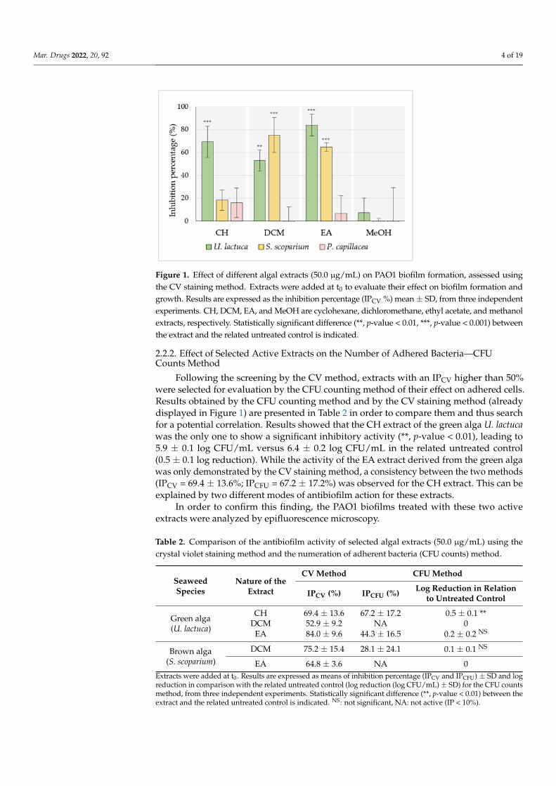
Mar. Drugs 2022, 20, 92 4 of 19Mar. Drugs 2022, 20, x FOR PEER REVIEW 4 of 20
Figure 1. Effect of different algal extracts (50.0 µg/mL) on PAO1 biofilm formation, assessed using
the CV staining method. Extracts were added at t0 to evaluate their effect on biofilm formation and
growth. Results are expressed as the inhibition percentage (IPCV %) mean + SD, from three inde-
pendent experiments. CH, DCM, EA, and MeOH are cyclohexane, dichloromethane, ethyl acetate,
and methanol extracts, respectively. Statistically significant difference (**, p-value < 0.01, ***, p-value
< 0.001) between the extract and the related untreated control is indicated.
Concerning CH extracts, only the one derived from the green alga was able to signif-
icantly reduce PAO1 biofilm biomass (IPCV = 69.4 ± 13.6%) (***, p-value < 0.001). On the
other hand, DCM extracts obtained from both green and brown algae exhibited consider-
able antibiofilm activity leading to biomass reductions of 52.9 ± 9.2% (**, p-value < 0.01)
and 75.2 ± 15.4% (***, p-value < 0.001), respectively. Regarding EA extracts, results showed
that the one derived from the green alga had the best ability to reduce PAO1 biofilm bio-
mass (IPCV = 84.0 ± 9.6%) (***, p-value < 0.001). EA extract obtained from the brown alga
also presented a notable activity (IPCV = 64.8 ± 9.2%) (***, p-value < 0.001). Note that no
significant activity was recorded for any MeOH extracts or red alga P. capillacea extracts.
2.2.2. Effect of Selected Active Extracts on the Number of Adhered Bacteria—CFU
Counts Method
Following the screening by the CV method, extracts with an IPCV higher than 50%
were selected for evaluation by the CFU counting method of their effect on adhered cells.
Results obtained by the CFU counting method and by the CV staining method (already
displayed in Figure 1) are presented in Table 2 in order to compare them and thus search
for a potential correlation. Results showed that the CH extract of the green alga U. lactuca
was the only one to show a significant inhibitory activity (**, p-value < 0.01), leading to 5.9
± 0.1 log CFU/mL versus 6.4 ± 0.2 log CFU/mL in the related untreated control (0.5 ± 0.1
log reduction). While the activity of the EA extract derived from the green alga was only
demonstrated by the CV staining method, a consistency between the two methods (IPCV =
69.4 ± 13.6%; IPCFU = 67.2 ± 17.2%) was observed for the CH extract. This can be explained
by two different modes of antibiofilm action for these extracts.
In order to confirm this finding, the PAO1 biofilms treated with these two active ex-
tracts were analyzed by epifluorescence microscopy.
Figure 1. Effect of different algal extracts (50.0 µg/mL) on PAO1 biofilm formation, assessed usingthe CV staining method. Extracts were added at t0 to evaluate their effect on biofilm formation andgrowth. Results are expressed as the inhibition percentage (IPCV %) mean ± SD, from three independentexperiments. CH, DCM, EA, and MeOH are cyclohexane, dichloromethane, ethyl acetate, and methanolextracts, respectively. Statistically significant difference (**, p-value < 0.01, ***, p-value < 0.001) betweenthe extract and the related untreated control is indicated.
2.2.2. Effect of Selected Active Extracts on the Number of Adhered Bacteria—CFUCounts Method
Following the screening by the CV method, extracts with an IPCV higher than 50%were selected for evaluation by the CFU counting method of their effect on adhered cells.Results obtained by the CFU counting method and by the CV staining method (alreadydisplayed in Figure 1) are presented in Table 2 in order to compare them and thus searchfor a potential correlation. Results showed that the CH extract of the green alga U. lactucawas the only one to show a significant inhibitory activity (**, p-value < 0.01), leading to5.9 ± 0.1 log CFU/mL versus 6.4 ± 0.2 log CFU/mL in the related untreated control(0.5 ± 0.1 log reduction). While the activity of the EA extract derived from the green algawas only demonstrated by the CV staining method, a consistency between the two methods(IPCV = 69.4 ± 13.6%; IPCFU = 67.2 ± 17.2%) was observed for the CH extract. This can beexplained by two different modes of antibiofilm action for these extracts.
In order to confirm this finding, the PAO1 biofilms treated with these two activeextracts were analyzed by epifluorescence microscopy.
Table 2. Comparison of the antibiofilm activity of selected algal extracts (50.0 µg/mL) using thecrystal violet staining method and the numeration of adherent bacteria (CFU counts) method.
SeaweedSpecies
Nature of theExtract
CV Method CFU Method
IPCV (%) IPCFU (%) Log Reduction in Relationto Untreated Control
Green alga(U. lactuca)
CH 69.4 ± 13.6 67.2 ± 17.2 0.5 ± 0.1 **DCM 52.9 ± 9.2 NA 0
EA 84.0 ± 9.6 44.3 ± 16.5 0.2 ± 0.2 NS
Brown alga(S. scoparium)
DCM 75.2 ± 15.4 28.1 ± 24.1 0.1 ± 0.1 NS
EA 64.8 ± 3.6 NA 0Extracts were added at t0. Results are expressed as means of inhibition percentage (IPCV and IPCFU) ± SD and logreduction in comparison with the related untreated control (log reduction (log CFU/mL) ± SD) for the CFU countsmethod, from three independent experiments. Statistically significant difference (**, p-value < 0.01) between theextract and the related untreated control is indicated. NS: not significant, NA: not active (IP < 10%).
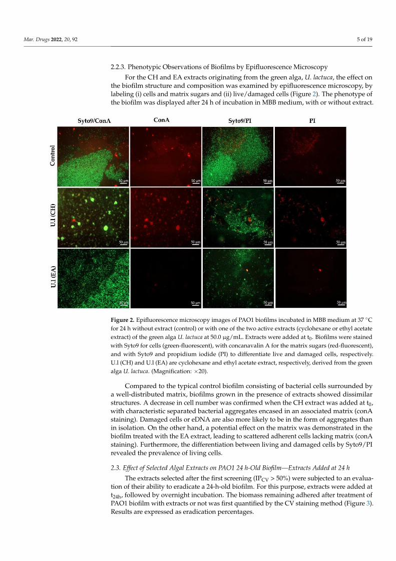
Mar. Drugs 2022, 20, 92 5 of 19
2.2.3. Phenotypic Observations of Biofilms by Epifluorescence Microscopy
For the CH and EA extracts originating from the green alga, U. lactuca, the effect onthe biofilm structure and composition was examined by epifluorescence microscopy, bylabeling (i) cells and matrix sugars and (ii) live/damaged cells (Figure 2). The phenotype ofthe biofilm was displayed after 24 h of incubation in MBB medium, with or without extract.
Mar. Drugs 2022, 20, x FOR PEER REVIEW 6 of 20
Figure 2. Epifluorescence microscopy images of PAO1 biofilms incubated in MBB medium at 37 °C
for 24 h without extract (control) or with one of the two active extracts (cyclohexane or ethyl acetate
extract) of the green alga U. lactuca at 50.0 μg/mL. Extracts were added at t0. Biofilms were stained
with Syto9 for cells (green-fluorescent), with concanavalin A for the matrix sugars (red-fluorescent),
and with Syto9 and propidium iodide (PI) to differentiate live and damaged cells, respectively. U.l
(CH) and U.l (EA) are cyclohexane and ethyl acetate extract, respectively, derived from the green
alga U. lactuca. (Magnification: ×20).
2.3. Effect of Selected Algal Extracts on PAO1 24 h-Old Biofilm—Extracts Added at 24 h
The extracts selected after the first screening (IPCV > 50%) were subjected to an eval-
uation of their ability to eradicate a 24-h-old biofilm. For this purpose, extracts were added
at t24h, followed by overnight incubation. The biomass remaining adhered after treatment
of PAO1 biofilm with extracts or not was first quantified by the CV staining method (Fig-
ure 3). Results are expressed as eradication percentages.
Results revealed that the EA extract of the green seaweed exhibited the best eradica-
tion activity (EPCV = 85.6 ± 7.4%). In addition, CH and DCM extract also obtained from U.
lactuca displayed a moderate activity on PAO1 24 h-old biofilm, leading to 55.5 ± 10.0%
and 56.1 ± 21.0% of eradication, respectively.
On the other hand, the effect of the most active extract (EA extract with EPCV > 80%)
was evaluated using the CFU counting assay, with quantification of both adhered and
detached (planktonic) cells (Figure 4). While no notable effect was observed on adhered
cell counts, a significant increase in the number of detached cells was measured (**, p-
value < 0.01) in the presence of EA extract (8.0 ± 0.3 log CFU/mL) compared to the control
(7.1 ± 0.5 log CFU/mL). In order to exclude a possible growth promoter effect of the EA
extract, its effect on planktonic growth was examined by plotting the growth curve. Re-
sults validated the absence of any significant effect of the EA extract on planktonic growth
(Supplementary Materials Figure S2).
Figure 2. Epifluorescence microscopy images of PAO1 biofilms incubated in MBB medium at 37 ◦Cfor 24 h without extract (control) or with one of the two active extracts (cyclohexane or ethyl acetateextract) of the green alga U. lactuca at 50.0 µg/mL. Extracts were added at t0. Biofilms were stainedwith Syto9 for cells (green-fluorescent), with concanavalin A for the matrix sugars (red-fluorescent),and with Syto9 and propidium iodide (PI) to differentiate live and damaged cells, respectively.U.l (CH) and U.l (EA) are cyclohexane and ethyl acetate extract, respectively, derived from the greenalga U. lactuca. (Magnification: ×20).
Compared to the typical control biofilm consisting of bacterial cells surrounded bya well-distributed matrix, biofilms grown in the presence of extracts showed dissimilarstructures. A decrease in cell number was confirmed when the CH extract was added at t0,with characteristic separated bacterial aggregates encased in an associated matrix (conAstaining). Damaged cells or eDNA are also more likely to be in the form of aggregates thanin isolation. On the other hand, a potential effect on the matrix was demonstrated in thebiofilm treated with the EA extract, leading to scattered adherent cells lacking matrix (conAstaining). Furthermore, the differentiation between living and damaged cells by Syto9/PIrevealed the prevalence of living cells.
2.3. Effect of Selected Algal Extracts on PAO1 24 h-Old Biofilm—Extracts Added at 24 h
The extracts selected after the first screening (IPCV > 50%) were subjected to an evalua-tion of their ability to eradicate a 24-h-old biofilm. For this purpose, extracts were added att24h, followed by overnight incubation. The biomass remaining adhered after treatment ofPAO1 biofilm with extracts or not was first quantified by the CV staining method (Figure 3).Results are expressed as eradication percentages.
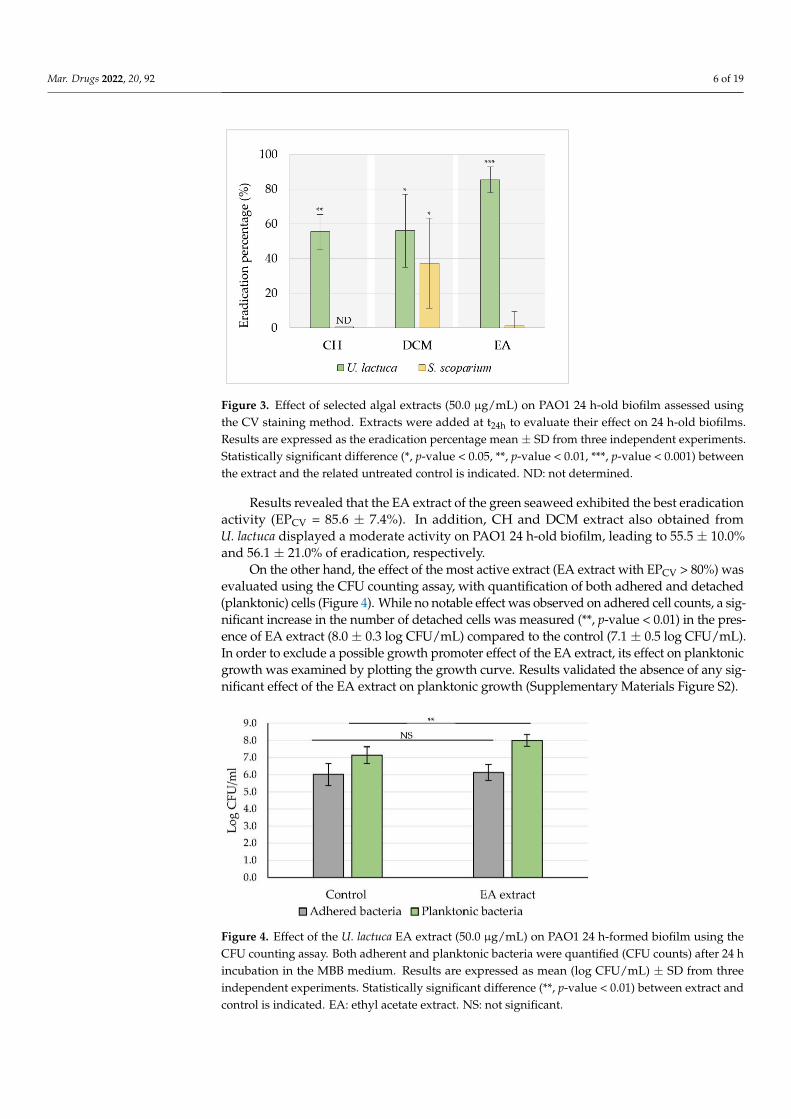
Mar. Drugs 2022, 20, 92 6 of 19Mar. Drugs 2022, 20, x FOR PEER REVIEW 7 of 20
Figure 3. Effect of selected algal extracts (50.0 µg/mL) on PAO1 24 h-old biofilm assessed using the
CV staining method. Extracts were added at t24h to evaluate their effect on 24 h-old biofilms. Results
are expressed as the eradication percentage mean ± SD from three independent experiments. Statis-
tically significant difference (*, p-value < 0.05, **, p-value < 0.01, ***, p-value < 0.001) between the
extract and the related untreated control is indicated. ND: not determined.
Figure 4. Effect of the U. lactuca EA extract (50.0 μg/mL) on PAO1 24 h-formed biofilm using the
CFU counting assay. Both adherent and planktonic bacteria were quantified (CFU counts) after 24
h incubation in the MBB medium. Results are expressed as mean (log CFU/mL) ± SD from three
independent experiments. Statistically significant difference (**, p-value < 0.01) between extract and
control is indicated. EA: ethyl acetate extract. NS: not significant.
2.4. Evaluation of the Synergistic Antibiofilm Activity of EA Extract in Combination with
Tobramycin or Colistin
Since the CV staining method showed EA extract originating from the green alga U.
lactuca to be the most effective in reducing the formation of the biofilm, as well as in erad-
icating previously formed biofilms, the potential synergy of the extract with two conven-
tional antibiotics was evaluated following the CFU counting method. The choice of this
evaluation method was based on the demonstrated responsiveness of CFU counting
method in treatment efficacy testing in comparison with the CV staining method [27]. For
comparison, the antibiofilm activity of tobramycin and colistin alone was evaluated on 24
h-old untreated biofilms, while the effect of the antibiotic/EA extract combination was
determined on 24 h-old biofilms, previously exposed to the EA extract for 24 h (Figure 5).
Results confirmed that EA extract had no significant effect on adherent CFU counts, while
tobramycin and colistin were able to induce a 3- or 2-log significant reduction, respec-
tively. The potential synergy was expressed by comparing the logarithmic reduction in
Figure 3. Effect of selected algal extracts (50.0 µg/mL) on PAO1 24 h-old biofilm assessed usingthe CV staining method. Extracts were added at t24h to evaluate their effect on 24 h-old biofilms.Results are expressed as the eradication percentage mean ± SD from three independent experiments.Statistically significant difference (*, p-value < 0.05, **, p-value < 0.01, ***, p-value < 0.001) betweenthe extract and the related untreated control is indicated. ND: not determined.
Results revealed that the EA extract of the green seaweed exhibited the best eradicationactivity (EPCV = 85.6 ± 7.4%). In addition, CH and DCM extract also obtained fromU. lactuca displayed a moderate activity on PAO1 24 h-old biofilm, leading to 55.5 ± 10.0%and 56.1 ± 21.0% of eradication, respectively.
On the other hand, the effect of the most active extract (EA extract with EPCV > 80%) wasevaluated using the CFU counting assay, with quantification of both adhered and detached(planktonic) cells (Figure 4). While no notable effect was observed on adhered cell counts, a sig-nificant increase in the number of detached cells was measured (**, p-value < 0.01) in the pres-ence of EA extract (8.0 ± 0.3 log CFU/mL) compared to the control (7.1 ± 0.5 log CFU/mL).In order to exclude a possible growth promoter effect of the EA extract, its effect on planktonicgrowth was examined by plotting the growth curve. Results validated the absence of any sig-nificant effect of the EA extract on planktonic growth (Supplementary Materials Figure S2).
Mar. Drugs 2022, 20, x FOR PEER REVIEW 7 of 20
Figure 3. Effect of selected algal extracts (50.0 µg/mL) on PAO1 24 h-old biofilm assessed using the
CV staining method. Extracts were added at t24h to evaluate their effect on 24 h-old biofilms. Results
are expressed as the eradication percentage mean ± SD from three independent experiments. Statis-
tically significant difference (*, p-value < 0.05, **, p-value < 0.01, ***, p-value < 0.001) between the
extract and the related untreated control is indicated. ND: not determined.
Figure 4. Effect of the U. lactuca EA extract (50.0 μg/mL) on PAO1 24 h-formed biofilm using the
CFU counting assay. Both adherent and planktonic bacteria were quantified (CFU counts) after 24
h incubation in the MBB medium. Results are expressed as mean (log CFU/mL) ± SD from three
independent experiments. Statistically significant difference (**, p-value < 0.01) between extract and
control is indicated. EA: ethyl acetate extract. NS: not significant.
2.4. Evaluation of the Synergistic Antibiofilm Activity of EA Extract in Combination with
Tobramycin or Colistin
Since the CV staining method showed EA extract originating from the green alga U.
lactuca to be the most effective in reducing the formation of the biofilm, as well as in erad-
icating previously formed biofilms, the potential synergy of the extract with two conven-
tional antibiotics was evaluated following the CFU counting method. The choice of this
evaluation method was based on the demonstrated responsiveness of CFU counting
method in treatment efficacy testing in comparison with the CV staining method [27]. For
comparison, the antibiofilm activity of tobramycin and colistin alone was evaluated on 24
h-old untreated biofilms, while the effect of the antibiotic/EA extract combination was
determined on 24 h-old biofilms, previously exposed to the EA extract for 24 h (Figure 5).
Results confirmed that EA extract had no significant effect on adherent CFU counts, while
tobramycin and colistin were able to induce a 3- or 2-log significant reduction, respec-
tively. The potential synergy was expressed by comparing the logarithmic reduction in
Figure 4. Effect of the U. lactuca EA extract (50.0 µg/mL) on PAO1 24 h-formed biofilm using theCFU counting assay. Both adherent and planktonic bacteria were quantified (CFU counts) after 24 hincubation in the MBB medium. Results are expressed as mean (log CFU/mL) ± SD from threeindependent experiments. Statistically significant difference (**, p-value < 0.01) between extract andcontrol is indicated. EA: ethyl acetate extract. NS: not significant.
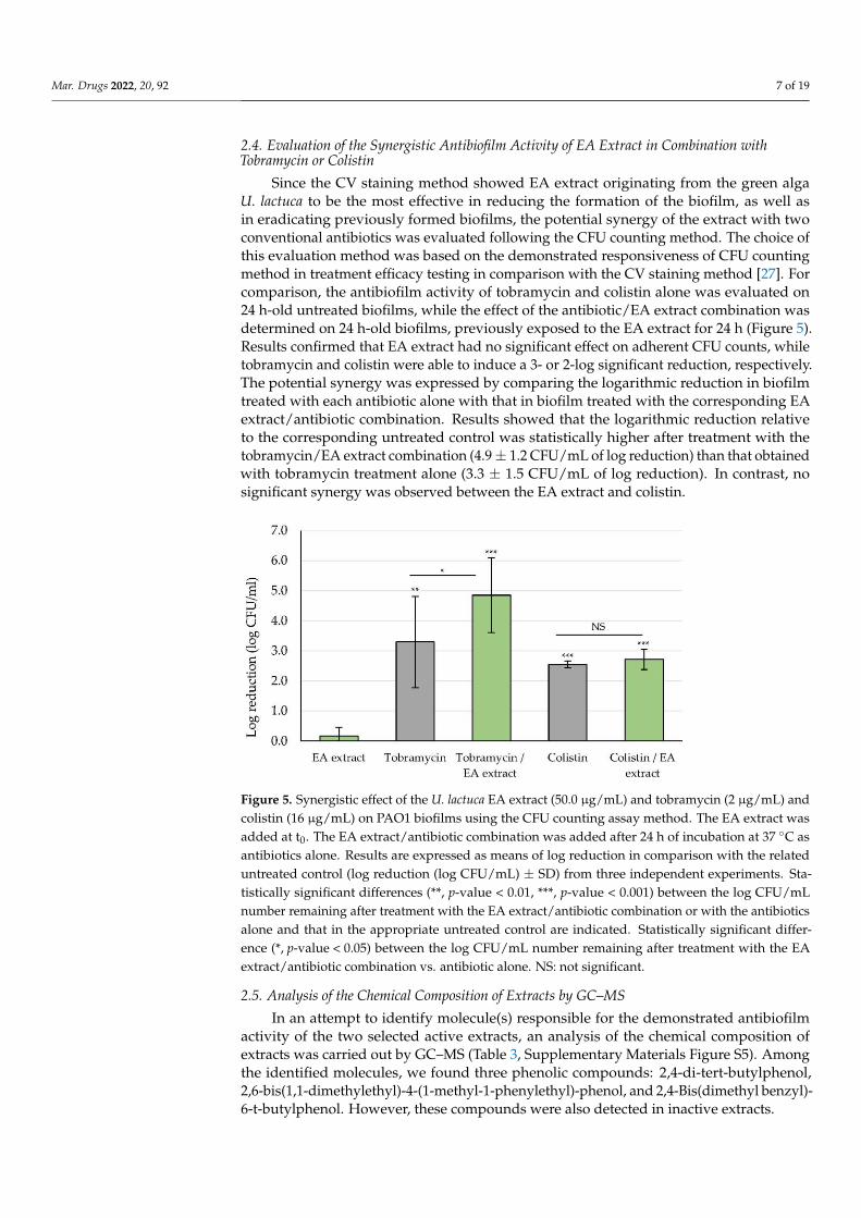
Mar. Drugs 2022, 20, 92 7 of 19
2.4. Evaluation of the Synergistic Antibiofilm Activity of EA Extract in Combination withTobramycin or Colistin
Since the CV staining method showed EA extract originating from the green algaU. lactuca to be the most effective in reducing the formation of the biofilm, as well asin eradicating previously formed biofilms, the potential synergy of the extract with twoconventional antibiotics was evaluated following the CFU counting method. The choice ofthis evaluation method was based on the demonstrated responsiveness of CFU countingmethod in treatment efficacy testing in comparison with the CV staining method [27]. Forcomparison, the antibiofilm activity of tobramycin and colistin alone was evaluated on24 h-old untreated biofilms, while the effect of the antibiotic/EA extract combination wasdetermined on 24 h-old biofilms, previously exposed to the EA extract for 24 h (Figure 5).Results confirmed that EA extract had no significant effect on adherent CFU counts, whiletobramycin and colistin were able to induce a 3- or 2-log significant reduction, respectively.The potential synergy was expressed by comparing the logarithmic reduction in biofilmtreated with each antibiotic alone with that in biofilm treated with the corresponding EAextract/antibiotic combination. Results showed that the logarithmic reduction relativeto the corresponding untreated control was statistically higher after treatment with thetobramycin/EA extract combination (4.9 ± 1.2 CFU/mL of log reduction) than that obtainedwith tobramycin treatment alone (3.3 ± 1.5 CFU/mL of log reduction). In contrast, nosignificant synergy was observed between the EA extract and colistin.
Mar. Drugs 2022, 20, x FOR PEER REVIEW 8 of 20
biofilm treated with each antibiotic alone with that in biofilm treated with the correspond-
ing EA extract/antibiotic combination. Results showed that the logarithmic reduction rel-
ative to the corresponding untreated control was statistically higher after treatment with
the tobramycin/EA extract combination (4.9 ± 1.2 CFU/mL of log reduction) than that ob-
tained with tobramycin treatment alone (3.3 ± 1.5 CFU/mL of log reduction). In contrast,
no significant synergy was observed between the EA extract and colistin.
Figure 5. Synergistic effect of the U. lactuca EA extract (50.0 µg/mL) and tobramycin (2 µg/mL) and
colistin (16 µg/mL) on PAO1 biofilms using the CFU counting assay method. The EA extract was
added at t0. The EA extract/antibiotic combination was added after 24 h of incubation at 37 °C as
antibiotics alone. Results are expressed as means of log reduction in comparison with the related
untreated control (log reduction (log CFU/mL) ± SD) from three independent experiments. Statisti-
cally significant differences (**, p-value < 0.01, ***, p-value < 0.001) between the log CFU/mL number
remaining after treatment with the EA extract/antibiotic combination or with the antibiotics alone
and that in the appropriate untreated control are indicated. Statistically significant difference (*, p-
value < 0.05) between the log CFU/mL number remaining after treatment with the EA extract/anti-
biotic combination vs. antibiotic alone. NS: not significant.
2.5. Analysis of the Chemical Composition of Extracts by GC–MS
In an attempt to identify molecule(s) responsible for the demonstrated antibiofilm
activity of the two selected active extracts, an analysis of the chemical composition of ex-
tracts was carried out by GC–MS (Table 3, Supplementary Materials Figure S5). Among
the identified molecules, we found three phenolic compounds: 2,4-di-tert-butylphenol,
2,6-bis(1,1-dimethylethyl)-4-(1-methyl-1-phenylethyl)-phenol, and 2,4-Bis (dimethyl ben-
zyl)-6-t-butylphenol. However, these compounds were also detected in inactive extracts.
Figure 5. Synergistic effect of the U. lactuca EA extract (50.0 µg/mL) and tobramycin (2 µg/mL) andcolistin (16 µg/mL) on PAO1 biofilms using the CFU counting assay method. The EA extract wasadded at t0. The EA extract/antibiotic combination was added after 24 h of incubation at 37 ◦C asantibiotics alone. Results are expressed as means of log reduction in comparison with the relateduntreated control (log reduction (log CFU/mL) ± SD) from three independent experiments. Sta-tistically significant differences (**, p-value < 0.01, ***, p-value < 0.001) between the log CFU/mLnumber remaining after treatment with the EA extract/antibiotic combination or with the antibioticsalone and that in the appropriate untreated control are indicated. Statistically significant differ-ence (*, p-value < 0.05) between the log CFU/mL number remaining after treatment with the EAextract/antibiotic combination vs. antibiotic alone. NS: not significant.
2.5. Analysis of the Chemical Composition of Extracts by GC–MS
In an attempt to identify molecule(s) responsible for the demonstrated antibiofilmactivity of the two selected active extracts, an analysis of the chemical composition ofextracts was carried out by GC–MS (Table 3, Supplementary Materials Figure S5). Amongthe identified molecules, we found three phenolic compounds: 2,4-di-tert-butylphenol,2,6-bis(1,1-dimethylethyl)-4-(1-methyl-1-phenylethyl)-phenol, and 2,4-Bis(dimethyl benzyl)-6-t-butylphenol. However, these compounds were also detected in inactive extracts.
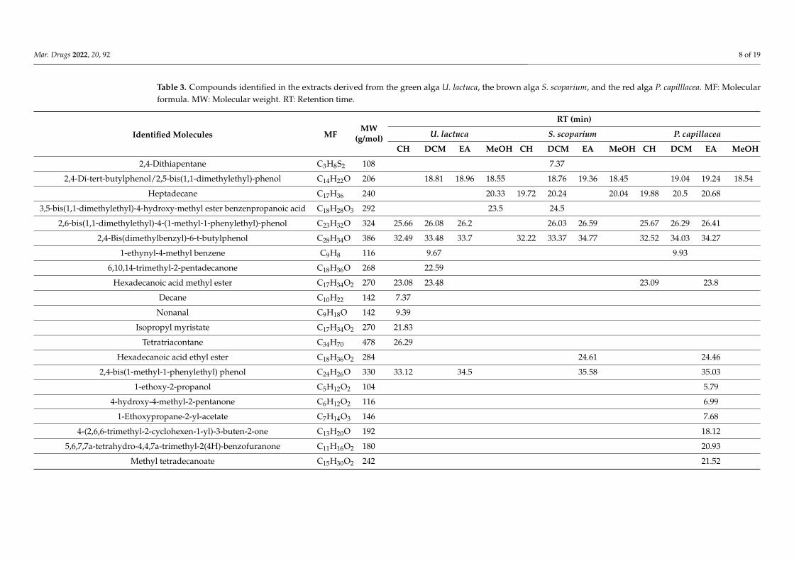
Mar. Drugs 2022, 20, 92 8 of 19
Table 3. Compounds identified in the extracts derived from the green alga U. lactuca, the brown alga S. scoparium, and the red alga P. capilllacea. MF: Molecularformula. MW: Molecular weight. RT: Retention time.
Identified Molecules MFMW
(g/mol)
RT (min)
U. lactuca S. scoparium P. capillacea
CH DCM EA MeOH CH DCM EA MeOH CH DCM EA MeOH
2,4-Dithiapentane C3H8S2 108 7.37
2,4-Di-tert-butylphenol/2,5-bis(1,1-dimethylethyl)-phenol C14H22O 206 18.81 18.96 18.55 18.76 19.36 18.45 19.04 19.24 18.54
Heptadecane C17H36 240 20.33 19.72 20.24 20.04 19.88 20.5 20.68
3,5-bis(1,1-dimethylethyl)-4-hydroxy-methyl ester benzenpropanoic acid C18H28O3 292 23.5 24.5
2,6-bis(1,1-dimethylethyl)-4-(1-methyl-1-phenylethyl)-phenol C23H32O 324 25.66 26.08 26.2 26.03 26.59 25.67 26.29 26.41
2,4-Bis(dimethylbenzyl)-6-t-butylphenol C28H34O 386 32.49 33.48 33.7 32.22 33.37 34.77 32.52 34.03 34.27
1-ethynyl-4-methyl benzene C9H8 116 9.67 9.93
6,10,14-trimethyl-2-pentadecanone C18H36O 268 22.59
Hexadecanoic acid methyl ester C17H34O2 270 23.08 23.48 23.09 23.8
Decane C10H22 142 7.37
Nonanal C9H18O 142 9.39
Isopropyl myristate C17H34O2 270 21.83
Tetratriacontane C34H70 478 26.29
Hexadecanoic acid ethyl ester C18H36O2 284 24.61 24.46
2,4-bis(1-methyl-1-phenylethyl) phenol C24H26O 330 33.12 34.5 35.58 35.03
1-ethoxy-2-propanol C5H12O2 104 5.79
4-hydroxy-4-methyl-2-pentanone C6H12O2 116 6.99
1-Ethoxypropane-2-yl-acetate C7H14O3 146 7.68
4-(2,6,6-trimethyl-2-cyclohexen-1-yl)-3-buten-2-one C13H20O 192 18.12
5,6,7,7a-tetrahydro-4,4,7a-trimethyl-2(4H)-benzofuranone C11H16O2 180 20.93
Methyl tetradecanoate C15H30O2 242 21.52
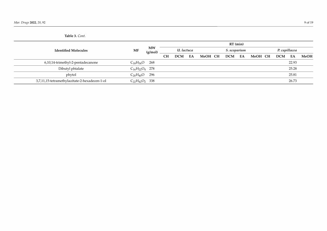
Mar. Drugs 2022, 20, 92 9 of 19
Table 3. Cont.
Identified Molecules MFMW
(g/mol)
RT (min)
U. lactuca S. scoparium P. capillacea
CH DCM EA MeOH CH DCM EA MeOH CH DCM EA MeOH
6,10,14-trimethyl-2-pentadecanone C18H36O 268 22.93
Dibutyl phtalate C16H22O4 278 25.28
phytol C20H40O 296 25.81
3,7,11,15-tetramethylacétate-2-hexadecen-1-ol C22H42O2 338 26.73
Mar. Drugs 2022, 20, 92 10 of 19
3. Discussion
At this critical time when pathogens’ development of multiple resistance pathways hasenabled them to outgrow our ability to effectively control them, we find ourselves facing aserious public health problem, since most conventional antimicrobial agents are no longerfunctional. In this context, the marine world, a habitat of immense biodiversity, offers asource of inspiration in the search for natural alternatives with novel mechanisms to preventand/or treat life-threatening diseases [16]. Despite the richness of seawater in bacteria(≈1 million cells/mL of seawater), such as Pseudomonas species, and the correspondinglyhigh risk of colonization by a bacterial biofilm, many marine organisms, particularly sessileones such as algae, successfully control this bacterial threat, which suggests their innateability to synthesize metabolites to protect themselves [28]. Several studies are emerging,bringing evidence of the significant antimicrobial, antibiofilm, and antifouling activities ofextracts and compounds derived from green, brown, and red macroalgae [12,20].
In the present study, the extracts of three macroalgae were explored for their potentialantibiofilm activity against the “superbug” P. aeruginosa. Great interest has been focused onthe search for synthetic and natural alternatives to conventional antibiotics to overcome thestrong ability of this pathogen to form deleterious biofilms that override antibiotherapy [29].In this context, the present study focuses on the screening of various seaweed extracts(mixtures of compounds) for their possible antibiofilm activity. Different approaches arecombined in an attempt to determine their potential mechanisms of action and select themost promising extracts for further studies.
To explore their potential antibacterial and antibiofilm activities, different extractswere prepared from the three seaweeds examined in this study, using solvents of increasingpolarity. As expected, the extraction yield depends on the polarity of the solvent used.Results showed that the dry matter of the three tested algae was richer in polar compoundsthan in nonpolar ones, since the highest yield of the crude extract was obtained withmethanol (Table 1). These results are not unexpected, given that macroalgae are character-ized by a high carbohydrate content (that can reach approximately 76% of their dry weight)versus a low lipid content [30]. Besides, this is in accordance with recent studies that havedemonstrated the richness of the red alga P. capillacea and the green alga U. lactuca in polarcompounds [31,32].
The first screening of extracts (50.0 µg/mL) for their ability to inhibit the formationand the development of PAO1 biofilms was performed using the CV staining method.Although this method provides a good estimate of the total biofilm biomass by markingEPS, especially the polysaccharides, it is not informative on the viability and the numberof adhered cells. This makes it necessary to combine the CV assay with the more accurateCFU counting method [33]. Furthermore, Allkja et al., 2021 proved that the CFU countingassay is more responsive in treatment experiments than CV staining, due to potentialinteraction between the treatment and the dye [27]. Results obtained by adding extractsat t0 revealed that those derived from the green alga U. lactuca, particularly CH and EAextracts, are the most promising in reducing bacterial adherent biomass, in comparison tothe two other algae tested here (Table 2). It should be noted that the potential bactericidaleffect of these two selected extracts at the tested concentration (50.0 µg/mL) was checked inorder to confirm that the observed effect is definitely related to an antibiofilm activity. Nobactericidal effect (neither on 102 nor on 105 CFU/mL) was recorded for these two extracts(Supplementary Materials Table S3).
By evaluating different types of extract, the value of green alga U. lactuca has beenhighlighted by various studies revealing its richness in bioactive compounds suitable forpharmaceutical (antioxidant, anti-proliferative, etc.) cosmetic, nutritional, and energyapplications [34–36].
To the best of our knowledge, the only publication that has evaluated the antibiofilmactivity of this green alga against P. aeruginosa by the CV method demonstrated the ability ofa MeOH extract, prepared by a single maceration in methanol, to reduce total biomass [37].This difference with our results can be attributed to many parameters, such as the extraction
Mar. Drugs 2022, 20, 92 11 of 19
method, the bacterial strain and the biofilm formation conditions. Whereas rich media arecommonly used for the evaluation of antibiofilm activity, in this study, PAO1 biofilms weregrown in a low-nutritive medium, and using a low inoculum concentration, in order topromote biofilm formation through the growth of adherent cells. Moreover, our extractswere tested at a rather low concentration (50.0 µg/mL) to avoid solubility issues. On theother hand, due to the high variability of protocols and conditions in biofilm experiments(environmental factors, inoculum preparation, etc.) and quantification, especially thosebased on spectrophotometry, such as the CV staining method; data comparison betweenstudies is very complicated [38].
Regarding the CH extract derived from the green alga U. lactuca, results of CV(IPCV = 69.4 ± 13.6%) and CFU (IPCFU = 67.2 ± 17.2%) assays were consistent, whichimplies a significant effect on biofilm biomass formation and growth, as well as on thenumber of adhered cells (Table 2). An alteration in the morphology of the cell aggregatesthat formed was also revealed by microscopic analysis (Figure 2). On the other hand,when tested on a 24 h-old biofilm, the ability of CH extract to reduce biofilm biomasswas moderate, which suggests an effect restricted to the early stages of biofilm formation(Figure 3). Such a mechanism of action has been observed with a synthetic compound,N-(2-pyrimidyl)butanamide (C11 compound), designed to be a structural analogue of theN-butanoyl-L-homoserine lactone (C4-AHL) [26]. AHLs are signal molecules involvedin the quorum sensing (QS) cell-to-cell communication system, a key factor in virulenceand in biofilm formation. This “chat circuitry” requires the production, detection, andresponse to signal molecules leading to the synchronization of bacterial group behavior.In P. aeruginosa, three major QS systems are well described: rhl and las systems based onsignal molecules belonging to acyl-homoserine lactones (C4-HSL and C12-HSL) and the pqssystem regulated by 2-alkyl-4-quinolone (AQs) molecules [39,40]. Interestingly, C11 is ableto prevent P. aeruginosa biofilm formation and proliferation only when added during theinitial stages, with a dose-dependent effect, and with demonstrated antagonistic effect ofthe C4-AHL [26,41].
Concerning the effect of the EA extract, also derived from the green alga U. lactuca,on PAO1 biofilm formation and growth, a significant activity was recorded only withthe CV staining method (IPCV = 84.0 ± 9.6%) (Table 2). Since the main objective of thisassay is to quantify the total biofilm biomass, including EPS, these results can be explainedby a potential action on the production and/or degradation of the biofilm matrix. Thishypothesis is supported by the epifluorescence microscopic analysis, which proved thataddition of the EA extract leads to the formation of a biofilm characterized by an undefinedspread matrix (Figure 2). Interestingly, this extract also showed considerable efficiencyin reducing a 24 h-old biofilm by the CV method (EPCV = 85.5 ± 7.4%) (Figure 3). Thisfinding allowed us to select the EA extract and use the CFU method to explore the effectof the extract on the number of remaining adhered cells, as well as on the number ofplanktonic cells released. Results showed a significant increase in planktonic cells, whileno effect on the adhered cell counts was observed (Figure 4), which can be attributedto a matrix modification that promotes the release of biofilm cells. This mechanism ofaction targeting biofilm structure and morphology has been described for usnic acid,a secondary lichen metabolite [42]. P. aeruginosa biofilm grown on a usnic acid-loadedpolymer formed an altered structure consisting of microcolonies separated by interstitialvoid areas. Furthermore, Powell et al., 2018 have demonstrated the ability of alginateoligosaccharides derived from the brown alga Laminaria hyperborea to decrease P. aeruginosabiofilm biomass by disrupting its EPS network [43]. The function of the EPS is not limited toproviding a protective barrier against exogenous factors, it also ensures nutrition, hydration,and intercellular interaction within the biofilm. In this scenario, and given its major role inthe formation, development, and maintenance of biofilms, the EPS matrix has become apotential target in the search for novel anti-biofilm strategies such as the use of alginate lyase,DNase, or mucolytic agents, which aim to impair the complex structure of biofilms andconsequently eradicate them or reduce their high resistance to antimicrobial treatments [44].
Mar. Drugs 2022, 20, 92 12 of 19
On the other hand, several studies have focused on the search for a therapy thatcombines an antimicrobial agent with an innovative adjuvant, especially one that candisassemble the biofilm matrix. This can be considered as a good therapy that aims tominimize the long-term administration of high doses of antibiotics [44]. The lack of biofilmsensitivity towards antibiotics is a well-known, ubiquitous phenomenon caused by acombination of factors. Generally, a biofilm’s complexity and heterogeneity can hinderthe efficiency of antibiotics by many mechanisms: (1) the restricted penetration ensuredby the EPS matrix components interacting with antibiotics, (2) the physiological toleranceassociated with the formation of a subpopulation within the biofilm, characterized by aslower cell metabolism, leading to the inactivity of antibiotics that target fundamentalcellular processes (replication, protein or cell wall synthesis, etc.), (3) tolerance based onspecific genes whose expression is strictly associated with biofilm formation [45].
Thus, the possible synergistic activity between the active EA extract, which acts bypotentially affecting the PAO1 matrix structure, and tobramycin or colistin antibiotics,commonly used in the treatment of P. aeruginosa infections, was evaluated. Tobramycinis a polycationic aminoglycoside antibiotic with hydrophilic properties. Its antibacterialmechanism of action is based on its ability to bind to ribosomal subunits, resulting insuppression of mRNA translation and subsequently the inhibition of protein synthesis [46].Colistin is a polypeptide antibiotic belonging to the polymyxin family, with amphiphilicand cationic properties. Its binding to LPSs and phospholipids of the outer membraneof Gram-negative bacteria leads to the disruption of the cell membrane with leakage ofintracellular contents and, finally, to cell death [47].
In the present study, EA extract/antibiotic combinations were evaluated on 24 h-oldbiofilms exposed to the EA extract. It should be noted that the tested antibiotic concentrationswere selected in a previous study based on the level reached in the serum (for tobramycin)or sputum (for colistin) 1 h after administration of a single dose, which corresponded to8 µg/mL for tobramycin and 32 µg/mL for colistin [41]. However, since these concentrationsled to a strong biofilm reduction in vitro (data not shown), they were lowered to 2 and16 µg/mL for tobramycin and colistin, respectively, in order to detect a potential synergisticeffect. Results showed a significant increase in the antibiofilm activity of tobramycin againstEA-extract-pretreated biofilm (Figure 5). In contrast, no synergistic activity was recordedwith colistin. This can be explained by the difference in the mechanisms involved in biofilm-associated tolerance to aminoglycoside antibiotics (tobramycin) and antimicrobial peptide(colistin) and/or by a possible denaturing effect of EA extract on colistin.
For aminoglycoside antibiotics, which act at the intracellular level by targeting bacterialprotein synthesis, various studies have highlighted the major role played by negativelycharged EPS matrix in limiting the diffusion of such polycationic compounds through thebiofilm, thus blocking their effects. For instance, alginate, a polyanionic exopolysaccharideand a component of P. aeruginosa biofilm matrix, has been shown to have a crucial functionin protecting the biofilm from polycationic aminoglycosides, such as tobramycin, throughionic interactions [48].
In the present study, the EA extract has been proven to significantly reduce the totalbiomass, potentially by altering the EPS matrix structure and architecture of P. aeruginosabiofilms. Thus, the synergistic effect observed with tobramycin may be explained bythe partial restoration of the susceptibility of PAO1 EA extract-pretreated biofilms. Theabsence of total recovery of biofilm sensitivity to tobramycin may be linked to other factorsrelated to the biofilm state itself, such as the involvement of efflux pumps (e.g., MexAB-OprM) or the modification of cellular targets [49]. Interestingly, a synergistic effect withtobramycin has also been demonstrated with the C4-HSL analogue (C11) mentioned above,and also the halogenated furanone, known as a substance antagonistic to the bacterialQS communication system [41,50], since the efficacy of tobramycin on furanone-treatedP. aeruginosa biofilms is exerted on both the surface cells and those present in the deepestlayers, while the antibiotic had a limited effect on untreated biofilms.
Mar. Drugs 2022, 20, 92 13 of 19
On the other hand, the absence of synergy between the EA extract and colistin can beexplained by its lower retention by the EPS matrix, in comparison with the polycationictobramycin. Furthermore, evidence has been provided that biofilm tolerance to antimicro-bial peptides is correlated with eDNA-mediated activation of pmr/arn operon, encoding theLPS modification enzyme [45].
To progress towards the identification of the bioactive compounds present in the twoselected extracts, an analysis of the chemical composition of all extracts was performedby GC–MS (Table 3). Various molecules have been identified, some of which have alreadybeen described for their biological activity, such as the 2,4-di-tert-butylphenol [51]. Infact, Viszwapriya et al., 2016 have demonstrated the ability of this phenolic compoundto inhibit Streptococcus pyogenes biofilm formation along with a reduction in EPS matrixproduction [52]. Moreover, a synergistic antibiofilm activity of this phenol with gentamycinhas been reported against Serratia marcescens [53]. However, since most of the identifiedcompounds were also detected in the inactive extracts, such as the extracts derived fromthe red alga, their specific implication in the demonstrated antibiofilm activity of the twoactive extracts has to be confirmed by further purification and analyses to identify andquantify the active molecule and/or the effective mixture.
Finally, as the QS communication system is a key factor in bacterial biofilm formation,the two active extracts discovered in this study may potentially act on this complex systemand/or on other factors regulated by QS, such as the production of rhamnolipids. Thisbiosurfactant, controlled by the rhl QS system, is involved in the different stages of biofilmformation, particularly in the mediation of cell dispersion [54]. Thus, the present resultsencourage towards elucidating the potential direct and/or indirect anti-QS activity ofthese extracts.
4. Materials and Methods4.1. Collection of Algal Materials
Seaweed samples belonging to three different groups (green alga Ulva lactuca, brownalga Stypocaulon scoparium, and red alga Pterocladiella capillacea) were manually collectedin the Mediterranean Sea, from the northern Lebanese coast, particularly from El Mina inTripoli in September 2019 (Supplementary Materials Figure S1). After collection, the freshmacroalgae were rinsed with seawater to remove impurities such as particles of adheredsand or epiphytes. The samples were immediately transported to the laboratory of appliedbiotechnology, AZM research center, Lebanese university, Tripoli, Lebanon, where theywere rigorously washed with distilled water. Then, seaweed samples were air-dried in adark place at room temperature (20–27 ◦C) for several weeks and weighed continuouslyuntil they were completely dry. The dried samples were ground into a fine powder in orderto facilitate extractions, and were then transported in sealed bags to the Laboratoire deGénie Chimique of Toulouse, France, where the extractions were carried out.
4.2. Organic Solvents, Chemicals and Antibiotics
The solvents used in this study were cyclohexane 99.5% (Sigma–Aldrich, St. QuentinFallavier, France), dichloromethane 100% (VWR, Rosny-sous-Bois, France), ethyl acetate 99.9%(VWR, Rosny-sous-Bois, France), methanol 99.8% (Sigma–Aldrich, St. Quentin Fallavier,France) and ethanol 96% (Sigma–Aldrich, St. Quentin Fallavier, France). Unless otherwisementioned, all chemicals, including dyes and antibiotics, were purchased from Sigma–Aldrich,St. Quentin Fallavier, France.
4.3. Bacterial Strain and Culture Media
The bacterial strain used in this study was Pseudomonas aeruginosa PAO1 (CIP 104116),purchased from the collection of the Pasteur Institute (Paris, France) and preserved at−80 ◦C. The inoculum used in each experiment came from a second subculture on Trypti-case soy agar (BioMérieux, Crapone, France) that was incubated under aerobic conditionsat 37 ◦C for 24 h. A low-nutritive medium, named minimum biofilm broth (MBB) was used
Mar. Drugs 2022, 20, 92 14 of 19
for the biofilm formation and the evaluation of the antibiofilm activity of extracts, in orderto create stressful conditions and subsequently promote biofilm formation and growth ofadherent cells rather than planktonic growth. The MBB 10X medium is composed of FeSO4,7 H2O (0.005 g/L), Na2HPO4 (12.5 g/L), KH2PO4 (5.0 g/L), (NH4)2SO4 (1.0 g/L), glucose(0.5 g/L) and MgSO4, 7 H2O (0.2 g/L) [26].
4.4. Preparation of Seaweed Extracts
In order to extract a maximum of seaweed constituents, a successive extraction methodusing selective solvents with increasing polarity (cyclohexane, dichloromethane, ethylacetate, and methanol) was adopted [55]. One hundred grams of the dried samples of eachalga were macerated successively in 1 L of each solvent for 2 h under magnetic agitation.Crude extracts were recovered after filtration using a Büchner funnel followed by solventevaporation using a rotary evaporator under vacuum at 40 ◦C. Note that maceration withthe same solvent was repeated until discoloration of the filtrate. In this case, the differentextracts obtained from the same solvent were combined.
The extraction yield was then calculated using the following formula (1), where W2 isthe weight of the extract residue after solvent evaporation and W1 is the weight of the algalmatrix initially used in the extraction (100.0 g).
Extraction yield (%) =
(W2
W1
)× 100 (1)
To evaluate their bioactivity, extract solutions were prepared by dissolving the extractsin sterile distilled water (SDW) at 100.0 µg/mL, using an ultrasonic bath (VWR ultrasoniccleaning bath, 45 kHz) for 1 to 6 h until complete dissolution. Extract solutions were thensterilized by filtration through a syringe filter (Cellulose Acetate Syringe Filter, 0.45 µm,GE Healthcare Whatman).
4.5. Assessment of the Inhibitory Effect of Extract on Biofilm Formation—Extract Added at t04.5.1. Formation of PAO1 Biofilms
Biofilms were developed in 24-well plates (Falcon, TC-treated, polystyrene). Thebacterial suspension prepared in MBB (2X) was initially adjusted to 108 CFU/mL followedby a serial dilution to 10−6 with the same medium. One milliliter of the 10−6 dilution(equivalent to 102 CFU/mL) was introduced into each well. In order to test its effect on thebiofilm, 1.0 mL of the algal extract (100.0 µg/mL) (sub-MIC Supplementary Materials S4)was added to each well, corresponding to a final concentration of 50.0 µg/mL. Wellscontaining 1.0 mL of SDW + 1.0 mL of un-inoculated MBB or 1.0 mL SDW + 1.0 mLinoculated MBB, were considered as sterility and biofilm growth controls, respectively. Theplate was then incubated for 24 h at 37 ◦C. All assays were performed in triplicate.
4.5.2. Screening of Algal Extracts for Their Effect on PAO1 Biofilm Formation andGrowth—Crystal Violet Staining Method
The objective of this method was to quantify the total biomass of the biofilm (adheredcells + matrix) by crystal violet (CV) staining and consequently to evaluate the effect ofthe extract on the formation and proliferation of the biofilm [33]. The protocol adoptedby Genovese et al., 2021 was followed with some modifications [56]. After overnightincubation, biofilms were washed twice with 2.0 mL of SDW to remove non-adherentplanktonic cells. The plate was then air-dried for 1 h. To stain the adhered biomass, 2.0 mLof an aqueous 1% CV solution was added to the wells and consecutively incubated for15 min at room temperature. In order to remove the excess stain, wells were rinsed twicewith 2.0 mL of SDW followed by drying for 30 min before quantification. One milliliter of
Mar. Drugs 2022, 20, 92 15 of 19
ethanol was finally added to extract bound stain and the inhibition percentage (IPCV) wascalculated according to the following Formula (2):
IPCV(%) =OD570 nm of biofilm growth control − OD570 nm of tested extract
OD570 nm of biofilm growth control× 100 (2)
The absence of any interference between the extracts and CV staining was checkedusing blank wells (1.0 mL of extract + 1.0 mL cell-free MBB).
4.5.3. Effect of the Potentially Active Extracts on the Number of Adhered Bacteria—CFUCounts Method
In this assay, the protocol developed by [8,26] was used with some modifications.After 24 h of incubation, wells were rinsed twice with 2.0 mL of SDW, and then the attachedcells were scraped (for 1 min) with a sterile spatula into 1 mL of SDW. The recoveredsuspension was diluted by serial dilution (from 10−1 to 10−6) and 900 µL of each dilutionwas inoculated by inclusion in TSA agar plates. After 48 h of incubation at 37 ◦C, thenumbers of CFU were counted by considering only plates with 15 to 300 CFU. The adheredbiomass was then calculated and subjected to logarithmic transformation by Formula (3).The logarithmic reduction and the IPCFU with respect to the corresponding untreatedcontrol were also calculated using Formulas (4) and (5).
log of adhered biomass (log CFU/mL) = lognumber of colonies (CFU)
Dilution factor × inoculated volume(3)
log CFU/mL reduction = log CFU/mL for control − log CFU/mL for treated biofilm (4)
IPCFU (%) =Adhered cells Control(CFU/mL)− Adhered cells Sample(CFU/mL)
Adhered cells Control(CFU/mL)× 100 (5)
4.5.4. Phenotypic Observations by Epifluorescence Microscopy
The potential effect of extracts, added at t0, on PAO1 formed biofilm morphologyand on bacterial cell organization was examined by epifluorescence microscopy (EM).For this analysis, P. aeruginosa biofilms were grown as described above but in a 6-wellmicroplate (Falcon, TC-treated, polystyrene) and with a total volume of 6.0 mL (3.0 mL ofPAO1 bacterial suspension prepared in MBB 2X (102 CFU/mL) + 3.0 mL of tested extractor 3.0 mL of SDW for the control).
After 24 h of incubation at 37 ◦C, well content was carefully discarded and replaced by6.0 mL of SDW. Live and damaged cells were differentiated by staining with 1.0 µL of Syto9(5 mM, InvitrogenTM, ThermoFisher Scientific, Illkirch, France) and 1.0 µL of propidiumiodide (1 mg/mL, InvitrogenTM, ThermoFisher Scientific), respectively.
Moreover, to examine the potential effect of extracts on the biofilm matrix, 1.0 mLof concanavalin A (ConA, tetramethylrhodamine conjugate, ThermoFisher Scientific) pre-pared at a concentration of 100.0 µg/mL in 0.1 M of sodium bicarbonate, was added to thewell after its contents had been withdrawn. ConA is a lectin that exhibits an affinity for cer-tain osidic residues, in particular for α-mannopyranosyl and α-glucopyranosyl residues. Itis important to note that Strathman et al. [57] have proven that ConA may also bind to algi-nate, a component of P. aeruginosa biofilm matrix. Its conjugation to tetramethylrhodamineleads to the emission of orange-red visible fluorescence upon excitation with a green light.After 20 min of incubation in the dark at room temperature, wells were delicately rinsedtwice with 1.0 mL of SDW. Just before proceeding to the microscopic observations, 6.0 mLof SDW, together with 1.0 µL of Syto9, were added. Microscopic observations were madewith Zeiss—Axiotech microscope using a 20 X/0.50 (Zeiss, EC Plan-Neofluar) objectiveand equipped with an HXP 120 C light source. Images were acquired with a digital camera(Zeiss AxioCam ICm 1) and the set of photos was processed with ZEN software.
Mar. Drugs 2022, 20, 92 16 of 19
4.6. Effect of Selected Algal Extracts on PAO1 24 h-Old Biofilms—Extract Added at t24 h
Extracts for which the CV staining method revealed an effect on the biofilm formation(i.e., IPCV > 50%) were subjected to an experiment to evaluate their potential impact on a24-h-old biofilm. In this assay, 1.0 mL of algal extract solution (100.0 µg/mL) was addedwith 1.0 mL of MBB into wells of a 24-well plate in which a 24-h-old biofilm was developedas previously described. The plate was then re-incubated at 37 ◦C for 24 h. After incubation,wells were rinsed twice with 2.0 mL of SDW before the remaining biomass was quantifiedby the CV staining method.
The eradication percentage was calculated using the following Formula (6):
EPCV(%) =OD570 nm of untreated control − OD570 nm of tested extract
OD570 nm of untreated control× 100 (6)
The extract exhibiting an eradication percentage (EPCV) greater than 80% was alsoevaluated by the CFU counts method. In this case, both the adhered and the detached(planktonic) cells were quantified. To do this, before the wells were rinsed and scraped,1.0 mL of the supernatant was withdrawn and submitted to serial dilution followed byinoculation in TSA agar for CFU quantification of planktonic cells. The adherent cells werequantified as described above. The number of CFU counted after 48 h of incubation at37 ◦C was subjected to logarithmic transformation based on the above Formula (3).
4.7. Evaluation of the Synergistic Antibiofilm Activity of the Active Extract in Combination withTobramycin or Colistin on 24 h-Old Treated Biofilms
The potential synergistic antibiofilm effect of the U. lactuca ethyl acetate (EA) activeextract with tobramycin and colistin was evaluated on 24 h-old biofilms, previously treatedwith the EA extract or not, following the protocol developed by Furiga et al., 2016 withsome modifications. Since the objective here was to detect a potential synergistic effect,the tested concentrations of antibiotics had to be lower than the concentration that wouldbe fully effective in eradicating PAO1 biofilm, hence the choice of 2 and 16 µg/mL fortobramycin and colistin, respectively [41].
First, 1.0 mL of bacterial suspension (102 CFU/mL) prepared in MBB (2X) was addedinto each well of a 24-well microplate, supplemented either with 1.0 mL of SDW (con-trol, tobramycin, and colistin control) or with 1.0 mL of a solution of 100.0 µg/mL ofEA extract (EA extract control and combination assays; final concentration 50.0 µg/mL).After 24 h of incubation at 37 ◦C, the supernatant was removed, and replaced by 1.0 mLof SDW (control) or 1.0 mL of tobramycin alone (tobramycin control; final concentration2.0 µg/mL) or 1.0 mL of colistin alone (colistin control; colistin sodium methanesulfonate;final concentration 16.0 µg/mL) or 1.0 mL of EA extract (EA extract control; final concen-tration 50.0 µg/mL) or a solution of EA extract mixed with either tobramycin or colistin forthe combination assays. The final concentrations of EA extract, tobramycin, and colistinwere 50.0 µg/mL, 2.0 µg/mL and 16.0 µg/mL, respectively. MBB medium was then addedto all wells (1.0 mL/well). For all conditions, the number of adherent cells after 48 h ofincubation was quantified by the CFU counts method, as described above. Log reductionwas then calculated using Formulas (3) and (4).
4.8. Analysis of the Chemical Composition of Extracts by GC–MS
The chemical composition of all extracts was analyzed first by GC–MS; extracts wereprepared at a concentration of 2.5 mg/mL in the corresponding extraction solvent (cyclohex-ane, dichloromethane, ethyl acetate, or methanol). Analyses were performed using GC-MSsystem (TRACETM 1310—ThermoFisher Scientific) equipped with a Rtx-502.2 fused silicacapillary column (30 m in length, 0.25 mm in diameter, 1.4 µm in film thickness). Thecolumn oven temperature was programmed as follows: initial temperature was 50 ◦C (for2 min) then gradually increased to 150 ◦C (for 5 min) at a rate of 20 ◦C/min, and finallyincreased to 290 ◦C at a rate of 10 ◦C/min and maintained for 10 min. Ionization of thesample components was performed in electron impact mode (EI, 70 eV) with 220 ◦C as
Mar. Drugs 2022, 20, 92 17 of 19
ion temperature. The injector and detector temperatures were 250 and 220 ◦C, respectively.Hydrogen was used as carrier gas at a flow rate of 1.0 mL/min. The injection volume of theprepared extract solution (2.5 mg/mL) was 5.0 µL. The total running time of the GC–MSsystem was 36 min. Finally, molecules were identified using Xcalibur software.
4.9. Statistical Analysis
All values were expressed as mean ± SD for three independent experiments. The stu-dent t-test was used to calculate the significance of the differences between the mean effectsof the extract and those for the associated untreated control in the CFU counts method afterchecking equality of variances with Levene’s test (p-value < 0.05). Statistically significantvalues were defined as a p-value (* <0.05, ** <0.01 or *** <0.001). SPSS 22.0 software (SPSS,IBM Corporation, Armonk, NY, USA) was used in the statistical analysis.
5. Conclusions
In the present study, the screening of extracts derived from three algae for theirantibiofilm activity against the pathogenic bacterium P. aeruginosa allowed two U. lactucaextracts (CH and EA extracts) to be selected as the most promising for valorization in thisfield. CH extract appears to impair microcolony growth, resulting in a significant reductionin the number of adherent cells, while an effect on the production and the degradation ofthe biofilm matrix has been suggested as a potential mode of action of EA extract. In lightof these encouraging results, further experiments are envisaged to analyze the chemicalcomposition of the two active extracts and isolate active components as pure molecules.The evaluation of the antibiofilm effect of these extracts on other pathogenic bacteria wouldidentify a broad spectrum of activities. Overall, this study raises the possibility of extractingbioactive compounds from the green alga, U. lactuca, which can potentially be used aloneor in combination with antibiotics in the treatment of biofilm-related infections.
Supplementary Materials: The following are available online at https://www.mdpi.com/article/10.3390/md20020092/s1, Figure S1: Map showing the area where seaweed samples were collected,Figure S2: Planktonic growth kinetics of PAO1 in MHB, MBB and in presence of EA, Table S3: Evalua-tion of the potential bactericidal activity of CH and EA extracts on PAO1. Figure S5: Chromatogramsof extracts derived from the green alga U. lactuca.
Author Contributions: M.R., C.R. and F.E.G. contributed to conception and design of the study. M.R.carried out all the experiments, performed the statistical analysis, and wrote the first draft of themanuscript. J.T. participated to the discussions. L.L. carried out the GC–MSS. analysis. M.R., A.C.,C.R. and F.E.G. wrote sections of the manuscript. All authors have read and agreed to the publishedversion of the manuscript.
Funding: This work was funded by the laboratory Laboratoire de Génie Chimique, Université deToulouse, CNRS, INPT, UPS, Toulouse, France.
Conflicts of Interest: The authors declare they have no conflict of interest.
References1. Paul, D.; Gopal, J.; Kumar, M.; Manikandan, M. Nature to the natural rescue: Silencing microbial chats. Chem.-Biol. Interact.
2018, 280, 86–98. [CrossRef]2. Woolhouse, M.; Farrar, J. Policy: An intergovernmental panel on antimicrobial resistance. Nature 2014, 509, 555–557. [CrossRef]3. WHO. Global Priority List of Antibiotic-Resistant Bacteria to Guide Research, Discovery, and Develipment of New Antibiotics; World
Health Organization: Geneva, Switzerland, 2017.4. Moradali, M.F.; Ghods, S.; Rehm, B.H.A. Pseudomonas aeruginosa Lifestyle: A Paradigm for Adaptation, Survival, and Persistence.
Front. Cell. Infect. Microbiol. 2017, 7, 39. [CrossRef] [PubMed]5. Khan, F.; Pham, D.T.N.; Tabassum, N.; Oloketuyi, S.F.; Kim, Y.M. Treatment strategies targeting persister cell formation in bacterial
pathogens. Crit. Rev. Microbiol. 2020, 46, 665–688. [CrossRef]6. Flemming, H.-C.; Wingender, J.; Szewzyk, U.; Steinberg, P.; Rice, S.A.; Kjelleberg, S. Biofilms: An emergent form of bacterial life.
Nat. Rev. Microbiol. 2016, 14, 563–575. [CrossRef]7. Flemming, H.-C.; Wingender, J. The biofilm matrix. Nat. Rev. Microbiol. 2010, 8, 623–633. [CrossRef]
Mar. Drugs 2022, 20, 92 18 of 19
8. Campanac, C.; Pineau, L.; Payard, A.; Baziard-Mouysset, G.; Roques, C. Interactions between Biocide Cationic Agents andBacterial Biofilms. Antimicrob. Agents Chemother. 2002, 46, 1469–1474. [CrossRef]
9. Olivares, E.; Badel-Berchoux, S.; Provot, C.; Prévost, G.; Bernardi, T.; Jehl, F. Clinical Impact of Antibiotics for the Treatment ofPseudomonas aeruginosa Biofilm Infections. Front. Microbiol. 2020, 10, 2894. [CrossRef]
10. Talebi Bezmin Abadi, A.; Rizvanov, A.A.; Haertlé, T.; Blatt, N.L. World Health Organization Report: Current Crisis of AntibioticResistance. BioNanoScience 2019, 9, 778–788. [CrossRef]
11. Mishra, R.; Panda, A.K.; De Mandal, S.; Shakeel, M.; Bisht, S.S.; Khan, J. Natural Anti-biofilm Agents: Strategies to ControlBiofilm-Forming Pathogens. Front. Microbiol. 2020, 11, 566325. [CrossRef]
12. Dahms, H.; Dobretsov, S. Antifouling Compounds from Marine Macroalgae. Mar. Drugs 2017, 15, 265. [CrossRef] [PubMed]13. Bamunuarachchi, N.I.; Khan, F.; Kim, Y.M. Bactericidal activity of Sargassum aquifolium (Turner) C. Agardh against Gram-positive
and Gram-negative biofilm-forming pathogenic bacteria. Curr. Pharm. Biotechnol. 2021, 22, 1628–1640. [CrossRef] [PubMed]14. Leandro, A.; Pereira, L.; Goncalves, A.M.M. Diverse Applications of Marine Macroalgae. Mar. Drugs 2019, 18, 17. [CrossRef]
[PubMed]15. Rosa, G.P.; Tavares, W.R.; Sousa, P.M.C.; Pagès, A.K.; Seca, A.M.L.; Pinto, D.C.G.A. Seaweed Secondary Metabolites with Beneficial
Health Effects: An Overview of Successes in In Vivo Studies and Clinical Trials. Mar. Drugs 2019, 18, 8. [CrossRef] [PubMed]16. Silva, A.; Silva, S.A.; Carpena, M.; Garcia-Oliveira, P.; Gullón, P.; Barroso, M.F.; Prieto, M.A.; Simal-Gandara, J. Macroalgae as a
Source of Valuable Antimicrobial Compounds: Extraction and Applications. Antibiotics 2020, 9, 642. [CrossRef]17. Manefield, M.; de Nys, R.; Naresh, K.; Roger, R.; Givskov, M.; Peter, S.; Kjelleberg, S. Evidence that halogenated furanones from
Delisea pulchra inhibit acylated homoserine lactone (AHL)-mediated gene expression by displacing the AHL signal from itsreceptor protein. Microbiology 1999, 145, 283–291. [CrossRef]
18. Guzzo, F.; Scognamiglio, M.; Fiorentino, A.; Buommino, E.; D’Abrosca, B. Plant Derived Natural Products against Pseudomonasaeruginosa and Staphylococcus aureus: Antibiofilm Activity and Molecular Mechanisms. Molecules 2020, 25, 5024. [CrossRef]
19. Kim, H.-S.; Lee, S.-H.; Byun, Y.; Park, H.-D. 6-Gingerol reduces Pseudomonas aeruginosa biofilm formation and virulence viaquorum sensing inhibition. Sci. Rep. 2015, 5, 8656. [CrossRef]
20. Bhowmick, S.; Mazumdar, A.; Moulick, A.; Adam, V. Algal metabolites: An inevitable substitute for antibiotics. Biotechnol. Adv.2020, 43, 107571. [CrossRef]
21. Tang, J.; Wang, W.; Chu, W. Antimicrobial and Anti-Quorum Sensing Activities of Phlorotannins From Seaweed (Hizikia fusiforme).Front. Cell Infect. Microbiol. 2020, 10, 586750. [CrossRef]
22. Salem, D.M.S.A.; Ismail, M.M.; Tadros, H.R.Z. Evaluation of the antibiofilm activity of three seaweed species and their biosynthe-sized iron oxide nanoparticles (Fe3O4-NPs). Egypt. J. Aquat. Res. 2020, 46, 333–339. [CrossRef]
23. Cabral, E.M.; Oliveira, M.; Mondala, J.R.M.; Curtin, J.; Tiwari, B.K.; Garcia-Vaquero, M. Antimicrobials from Seaweeds for FoodApplications. Mar. Drugs 2021, 19, 211. [CrossRef]
24. Barreto, M.; Meyer, J.J.M. Isolation and antimicrobial activity of a lanosol derivative from Osmundaria serrata (Rhodophyta) and avisual exploration of its biofilm covering. S. Afr. J. Bot. 2006, 72, 521–528. [CrossRef]
25. Tappenden, P.; Harnan, S.; Uttley, L.; Mildred, M.; Carroll, C.; Cantrell, A. Colistimethate sodium powder and tobramycin powderfor inhalation for the treatment of chronic Pseudomonas aeruginosa lung infection in cystic fibrosis: Systematic review and economicmodel. Health Technol. Assess. 2013, 17, 1–181. [CrossRef] [PubMed]
26. Khalilzadeh, P.; Lajoie, B.; El Hage, S.; Furiga, A.; Baziard, G.; Berge, M.; Roques, C. Growth inhibition of adherent Pseudomonasaeruginosa by an N-butanoyl-L-homoserine lactone analog. Can. J. Microbiol. 2010, 56, 317–325. [CrossRef] [PubMed]
27. Allkja, J.; van Charante, F.; Aizawa, J.; Reigada, I.; Guarch-Perez, C.; Vazquez-Rodriguez, J.A.; Cos, P.; Coenye, T.; Fallarero, A.;Zaat, S.A.J.; et al. Interlaboratory study for the evaluation of three microtiter plate-based biofilm quantification methods. Sci. Rep.2021, 11, 13779. [CrossRef] [PubMed]
28. Shannon, E.; Abu-Ghannam, N. Antibacterial Derivatives of Marine Algae: An Overview of Pharmacological Mechanisms andApplications. Mar. Drugs 2016, 14, 81. [CrossRef]
29. Carette, J.; Nachtergael, A.; Duez, P.; El Jaziri, M.; Rasamiravaka, T. Natural Compounds Inhibiting Pseudomonas aeruginosa BiofilmFormation by Targeting Quorum Sensing Circuitry. In Bacterial Biofilms; Dincer, S., Sümengen Özdenefe, M., Arkut, A., Eds.;IntechOpen: London, UK, 2020.
30. Stiger-Pouvreau, V.; Bourgougnon, N.; Deslandes, E. Carbohydrates From Seaweeds. In Seaweed in Health and Disease Prevention;Elsevier: Amsterdam, The Netherlands, 2016; pp. 223–274.
31. Paiva, L.; Lima, E.; Neto, A.I.; Marcone, M.; Baptista, J. Nutritional and Functional Bioactivity Value of Selected AzoreanMacroalgae: Ulva compressa, Ulva rigida, Gelidium microdon, and Pterocladiella capillacea: Functional metabolites of selected algae.J. Food Sci. 2017, 82, 1757–1764. [CrossRef] [PubMed]
32. Salim, D.; Caro, P.d.; Merah, O.; Chbani, A. Control of Post-harvest Citrus Green Mold using Ulva lactuca Extracts as a Source ofActive Substances. Int. J. Bio-Resour. Stress Manag. 2020, 11, 287–296. [CrossRef]
33. Pantanella, F.; Valenti, P.; Natalizi, T. Analytical techniques to study microbial biofilm on abiotic surfaces: Pros and cons of themain techniques currently in use. Ann. Ig. Med. Prev. Comunita 2013, 25, 31–42. [CrossRef]
34. Hidayati, J.R.; Yudiati, E.; Pringgenies, D.; Oktaviyanti, D.T.; Kusuma, A.P. Comparative Study on Antioxidant Activities, TotalPhenolic Compound and Pigment Contents of Tropical Spirulina platensis, Gracilaria arcuata and Ulva lactuca Extracted in DifferentSolvents Polarity. E3S Web Conf. 2020, 147, 03012. [CrossRef]
Mar. Drugs 2022, 20, 92 19 of 19
35. Dominguez, H.; Loret, E.P. Ulva lactuca, A Source of Troubles and Potential Riches. Mar. Drugs 2019, 17, 357. [CrossRef]36. Arsianti, A.A.; Fadilah, F.; Suid, K.; Yazid, F.; Wibisono, L.K.; Azizah, N.N.; Putrianingsih, R.; Murniasih, T.; Rasyid, A.; Pangestuti,
R. Phytochemical composition and anticancer activity of seaweeds Ulva lactuca and Eucheuma cottonii against breast MCF-7 andcolon HCT-116 cells. Asian J. Pharm. Clin. Res. 2016, 9, 115. [CrossRef]
37. Yuvaraj, N.; Arul, V. Preliminary Screening of Anti-Biofilm, Anti-Larval Settlement and Cytotoxic Potential of Seaweeds andSeagrasses Collected from Pondicherry and Rameshwaram Coastal Line, India. WJFMS 2014, 6, 169–175. [CrossRef]
38. Allkja, J.; Bjarnsholt, T.; Coenye, T.; Cos, P.; Fallarero, A.; Harrison, J.J.; Lopes, S.P.; Oliver, A.; Pereira, M.O.; Ramage, G.; et al.Minimum information guideline for spectrophotometric and fluorometric methods to assess biofilm formation in microplates.Biofilm 2020, 2, 100010. [CrossRef] [PubMed]
39. Lee, J.; Zhang, L. The hierarchy quorum sensing network in Pseudomonas aeruginosa. Protein. Cell 2015, 6, 26–41. [CrossRef]40. Papenfort, K.; Bassler, B.L. Quorum sensing signal-response systems in Gram-negative bacteria. Nat. Rev. Microbiol. 2016, 14, 576–588.
[CrossRef] [PubMed]41. Furiga, A.; Lajoie, B.; El Hage, S.; Baziard, G.; Roques, C. Impairment of Pseudomonas aeruginosa Biofilm Resistance to Antibiotics
by Combining the Drugs with a New Quorum-Sensing Inhibitor. Antimicrob. Agents Chemother. 2016, 60, 1676–1686. [CrossRef]42. Francolini, I.; Norris, P.; Piozzi, A.; Donelli, G.; Stoodley, P. Usnic acid, a natural antimicrobial agent able to inhibit bacterial
biofilm formation on polymer surfaces. Antimicrob. Agents Chemother. 2004, 48, 4360–4365. [CrossRef]43. Powell, L.C.; Pritchard, M.F.; Ferguson, E.L.; Powell, K.A.; Patel, S.U.; Rye, P.D.; Sakellakou, S.-M.; Buurma, N.J.; Brilliant, C.D.;
Copping, J.M.; et al. Targeted disruption of the extracellular polymeric network of Pseudomonas aeruginosa biofilms by alginateoligosaccharides. NPJ Biofilms Microbiomes 2018, 4, 13. [CrossRef]
44. Pinto, R.M.; Soares, F.A.; Reis, S.; Nunes, C.; Van Dijck, P. Innovative Strategies Toward the Disassembly of the EPS Matrix inBacterial Biofilms. Front. Microbiol. 2020, 11, 952. [CrossRef]
45. Ciofu, O.; Tolker-Nielsen, T. Tolerance and Resistance of Pseudomonas aeruginosa Biofilms to Antimicrobial Agents—HowP. aeruginosa Can Escape Antibiotics. Front. Microbiol. 2019, 10, 913. [CrossRef]
46. Kohanski, M.A.; Dwyer, D.J.; Collins, J.J. How antibiotics kill bacteria: From targets to networks. Nat. Rev. Microbiol. 2010, 8, 423–435.[CrossRef] [PubMed]
47. Biswas, S.; Brunel, J.M.; Dubus, J.C.; Reynaud-Gaubert, M.; Rolain, J.M. Colistin: An update on the antibiotic of the 21st century.Expert Rev. Anti. Infect. Ther. 2012, 10, 917–934. [CrossRef]
48. Uruen, C.; Chopo-Escuin, G.; Tommassen, J.; Mainar-Jaime, R.C.; Arenas, J. Biofilms as Promoters of Bacterial Antibiotic Resistanceand Tolerance. Antibiotics 2020, 10, 3. [CrossRef]
49. Soto, S.M. Role of efflux pumps in the antibiotic resistance of bacteria embedded in a biofilm. Virulence 2013, 4, 223–229. [CrossRef]50. Hentzer, M.; Wu, H.; Andersen, J.B.; Riedel, K.; Rasmussen, T.B.; Bagge, N.; Kumar, N.; Schembri, M.A.; Song, Z.; Kristoffersen, P.;
et al. Attenuation of Pseudomonas aeruginosa virulence by quorum sensing inhibitors. EMBO J. 2003, 22, 3803–3815. [CrossRef]51. Zhao, F.; Wang, P.; Lucardi, R.D.; Su, Z.; Li, S. Natural Sources and Bioactivities of 2,4-Di-Tert-Butylphenol and Its Analogs. Toxins
2020, 12, 35. [CrossRef] [PubMed]52. Viszwapriya, D.; Prithika, U.; Deebika, S.; Balamurugan, K.; Pandian, S.K. In vitro and in vivo antibiofilm potential of 2,4-Di-tert-
butylphenol from seaweed surface associated bacterium Bacillus subtilis against group A streptococcus. Microbiol. Res. 2016, 191, 19–31.[CrossRef]
53. Padmavathi, A.R.; Abinaya, B.; Pandian, S.K. Phenol, 2,4-bis(1,1-dimethylethyl) of marine bacterial origin inhibits quorumsensing mediated biofilm formation in the uropathogen Serratia marcescens. Biofouling 2014, 30, 1111–1122. [CrossRef]
54. Nickzad, A.; Deziel, E. The involvement of rhamnolipids in microbial cell adhesion and biofilm development—An approach forcontrol? Lett. Appl. Microbiol. 2014, 58, 447–453. [CrossRef] [PubMed]
55. Kohoude, M.J.; Gbaguidi, F.; Agbani, P.; Ayedoun, M.-A.; Cazaux, S.; Bouajila, J. Chemical composition and biological activities ofextracts and essential oil of Boswellia dalzielii leaves. Pharm. Biol. 2017, 55, 33–42. [CrossRef] [PubMed]
56. Genovese, C.; D’Angeli, F.; Bellia, F.; Distefano, A.; Spampinato, M.; Attanasio, F.; Nicolosi, D.; Di Salvatore, V.; Tempera, G.; LoFurno, D.; et al. In Vitro Antibacterial, Anti-Adhesive and Anti-Biofilm Activities of Krameria lappacea (Dombey) Burdet & B.B.Simpson Root Extract against Methicillin-Resistant Staphylococcus aureus Strains. Antibiotics 2021, 10, 428. [CrossRef] [PubMed]
57. Strathmann, M.; Wingender, J.; Flemming, H.-C. Application of fluorescently labelled lectins for the visualization and biochemicalcharacterization of polysaccharides in biofilms of Pseudomonas aeruginosa. J. Microbiol. Methods 2002, 50, 237–248. [CrossRef]
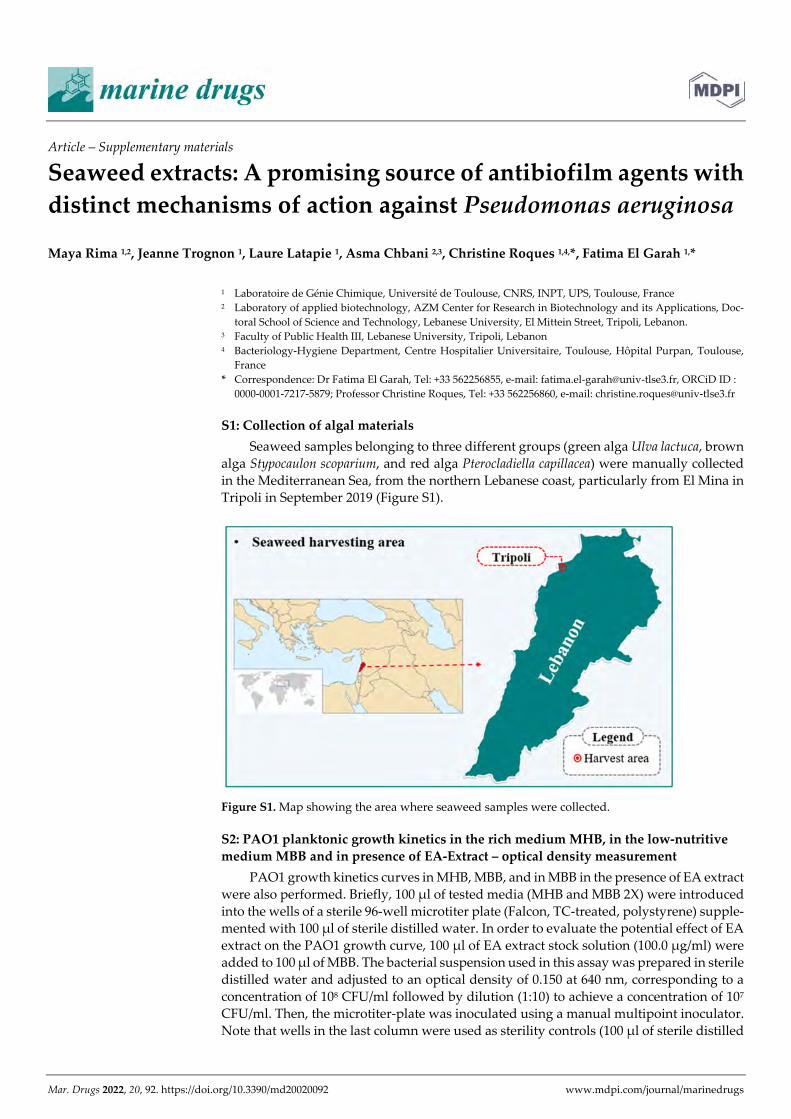
Mar. Drugs 2022, 20, 92. https://doi.org/10.3390/md20020092 www.mdpi.com/journal/marinedrugs
Article – Supplementary materials
Seaweed extracts: A promising source of antibiofilm agents with distinct mechanisms of action against Pseudomonas aeruginosa
Maya Rima 1,2, Jeanne Trognon 1, Laure Latapie 1, Asma Chbani 2,3, Christine Roques 1,4,*, Fatima El Garah 1,*
1 Laboratoire de Génie Chimique, Université de Toulouse, CNRS, INPT, UPS, Toulouse, France 2 Laboratory of applied biotechnology, AZM Center for Research in Biotechnology and its Applications, Doc-
toral School of Science and Technology, Lebanese University, El Mittein Street, Tripoli, Lebanon. 3 Faculty of Public Health III, Lebanese University, Tripoli, Lebanon 4 Bacteriology-Hygiene Department, Centre Hospitalier Universitaire, Toulouse, Hôpital Purpan, Toulouse,
France * Correspondence: Dr Fatima El Garah, Tel: +33 562256855, e-mail: [email protected], ORCiD ID :
0000-0001-7217-5879; Professor Christine Roques, Tel: +33 562256860, e-mail: [email protected]
S1: Collection of algal materials Seaweed samples belonging to three different groups (green alga Ulva lactuca, brown
alga Stypocaulon scoparium, and red alga Pterocladiella capillacea) were manually collected in the Mediterranean Sea, from the northern Lebanese coast, particularly from El Mina in Tripoli in September 2019 (Figure S1).
Figure S1. Map showing the area where seaweed samples were collected.
S2: PAO1 planktonic growth kinetics in the rich medium MHB, in the low-nutritive medium MBB and in presence of EA-Extract – optical density measurement
PAO1 growth kinetics curves in MHB, MBB, and in MBB in the presence of EA extract were also performed. Briefly, 100 µl of tested media (MHB and MBB 2X) were introduced into the wells of a sterile 96-well microtiter plate (Falcon, TC-treated, polystyrene) supple-mented with 100 µl of sterile distilled water. In order to evaluate the potential effect of EA extract on the PAO1 growth curve, 100 µl of EA extract stock solution (100.0 µg/ml) were added to 100 µl of MBB. The bacterial suspension used in this assay was prepared in sterile distilled water and adjusted to an optical density of 0.150 at 640 nm, corresponding to a concentration of 108 CFU/ml followed by dilution (1:10) to achieve a concentration of 107 CFU/ml. Then, the microtiter-plate was inoculated using a manual multipoint inoculator. Note that wells in the last column were used as sterility controls (100 µl of sterile distilled

Mar. Drugs 2022, 20, 92 2 of 4
water + 100 µl of tested media). The plate was incubated at 37°C for 24h in a microplate spectrophotometer (MultiskanTM GO, Thermo Fisher Scientific, Waltham, USA) under continuous agitation. The optical density measurement was carried out at 640 nm every one hour. The measured values were plotted as a function of time. Results are expressed as means + SD (OD640nm) of two independent assays (Figure S2).
Figure S2. Planktonic growth kinetics of PAO1 in MHB, MBB and in presence of EA. Extract (50.0 µg/ml) originated from the green alga U. lactuca. Results are expressed as means + SD of the optical density measured at 640 nm of two independent experiments. EA is ethyl acetate extract. MHB and MBB are Mueller-Hinton broth and modified biofilm broth, respectively.
S3: Checking the potential bactericidal activity of CH and EA extracts derived from the green alga U. lactuca – CFU counts method
In order to exclude the potential bactericidal effect of the two active extracts (CH and EA extracts derived from the green alga U. lactuca) at the tested concentration (50.0 µg/ml), their effect on PAO1 planktonic cells was assessed. The protocol developed by Feuillolay et al., 2016 was used in this assay. Briefly, 5.0 ml of PAO1 bacterial suspension (105 or 102
CFU/ml) prepared in MBB (2X) medium and supplemented with 5.0 ml of sterile distilled water were incubated for 24 hours at 37˚C. Water was replaced by 5.0 ml of extracts (CH or EA extracts) in the sample tubes. The potential bactericidal activity of extracts was de-termined on both suspension (105 and 102 CFU/ml). Tubes were maintained under agita-tion (100 rpm) in an orbital shaker (MaxQ 4000, Thermo Fisher Scientific, Waltham, USA). The number of planktonic cells was monitored after 24 hours of incubation at 37°C by CFU counts. Prior to their quantification, samples were homogenized then 1.0 ml was taken and serially diluted (10-1 to 10-6). 900 µl of each dilution were inoculated by inclusion in TSA agar plates and overnight incubated at 37˚C for cell quantification. Assays were performed in duplicate. Results expressed as ratio (log CFU/ml for sample / log CFU/ml for control) are presented in Table S3.
00.10.20.30.40.50.60.70.8
0 1 2 3 4 5 6 7 8 9 10 11 12 13 14 15 16 17 18 19 20 21 22 23 24
OD
at 6
40 n
m
Incubation time (hours)
Planktonic growth kinetics of PAO1
MHB MBB EA. Extract in MBB
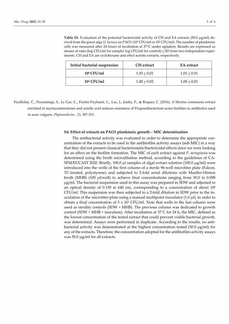
Mar. Drugs 2022, 20, 92 3 of 4
Table S3. Evaluation of the potential bactericidal activity of CH and EA extracts (50.0 µg/ml) de-rived from the green alga U. lactuca on PAO1 (105 CFU/ml or 102 CFU/ml). The number of planktonic cells was measured after 24 hours of incubation at 37°C under agitation. Results are expressed as means of ratio (log CFU/ml for sample/ log CFU/ml for control) + SD from two independent exper-iments. CH and EA are cyclohexane and ethyl acetate extracts, respectively.
Feuillolay, C., Pecastaings, S., Le Gac, C., Fiorini-Puybaret, C., Luc, J., Joulia, P., & Roques, C. (2016). A Myrtus communis extract
enriched in myrtucummulones and ursolic acid reduces resistance of Propionibacterium acnes biofilms to antibiotics used
in acne vulgaris. Phytomedicine , 23, 307-315.
S4: Effect of extracts on PAO1 planktonic growth – MIC determination The antibacterial activity was evaluated in order to determine the appropriate con-
centration of the extracts to be used in the antibiofilm activity assays (sub-MIC) in a way that they did not present classical bacteriostatic/bactericidal effects since we were looking for an effect on the biofilm formation. The MIC of each extract against P. aeruginosa was determined using the broth microdilution method, according to the guidelines of CA-SFM/EUCAST 2020. Briefly, 100.0 µl samples of algal extract solution (100.0 µg/ml) were introduced into the wells of the first column of a sterile 96-well microtiter plate (Falcon, TC-treated, polystyrene) and subjected to 2-fold serial dilutions with Mueller-Hinton broth (MHB) (100 µl/well) to achieve final concentrations ranging from 50.0 to 0.098 µg/ml. The bacterial suspension used in this assay was prepared in SDW and adjusted to an optical density of 0.150 at 640 nm, corresponding to a concentration of about 108 CFU/ml. This suspension was then subjected to a 2-fold dilution in SDW prior to the in-oculation of the microtiter-plate using a manual multipoint inoculator (1.0 µl), in order to obtain a final concentration of 5 x 10⁵ CFU/ml. Note that wells in the last column were used as sterility controls (SDW + MHB). The previous column was dedicated to growth control (SDW + MHB + inoculum). After incubation at 37˚C for 24 h, the MIC, defined as the lowest concentration of the tested extract that could prevent visible bacterial growth, was determined. Assays were performed in duplicate. According to the results, no anti-bacterial activity was demonstrated at the highest concentration tested (50.0 µg/ml) for any of the extracts. Therefore, the concentration adopted for the antibiofilm activity assays was 50.0 µg/ml for all extracts.
Initial bacterial suspension CH extract EA extract
105 CFU/ml 1.03 + 0.01 1.01 + 0.01
102 CFU/ml 1.00 + 0.02 1.00 + 0.01
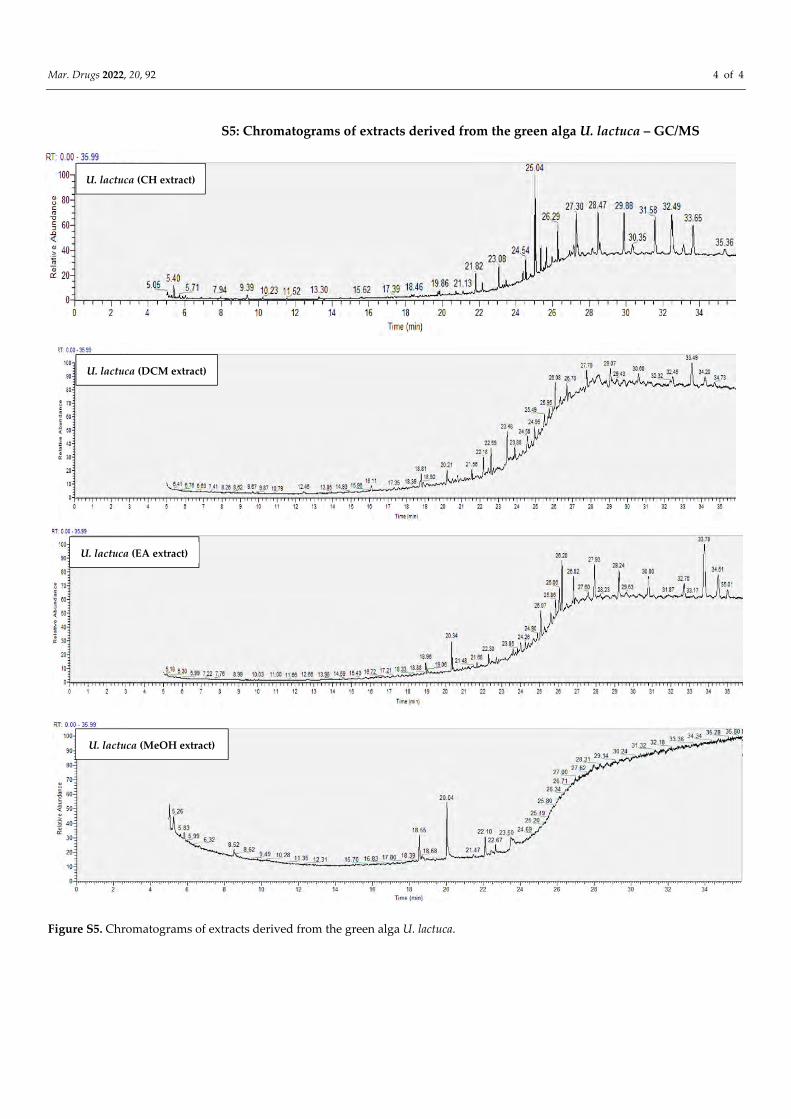
Mar. Drugs 2022, 20, 92 4 of 4
S5: Chromatograms of extracts derived from the green alga U. lactuca – GC/MS
Figure S5. Chromatograms of extracts derived from the green alga U. lactuca.
U. lactuca (CH extract)
U. lactuca (EA extract)
U. lactuca (DCM extract)
U. lactuca (MeOH extract)
191
PREVIEW
In this part, the potential ability of extracts derived from the three Lebanese seaweed (green
alga U. lactuca, brown alga S. scoparium, and red alga P. capillacea) to exhibit an
antibiofilm activity against the bacterium S. aureus was investigated.
The work presented in this part was done in the Laboratoire de Génie Chimique (LGC –
UMR5503) – Toulouse – France and resulted in an article for submission.
After a brief summary of the article with a highlight on the main results, the manuscript is
integrated followed by the supplementary materials.
Part III: Evaluation of the antibiofilm activity of seaweed extracts against S. aureus
III CHAPTER
ARTICLE III – To submit:
Rima, M., Chbani, A., Roques, C., & El Garah, F. Seaweed extracts as an effective gateway in the search for novel antibiofilm agents against Staphylococcus aureus.
Chapter III – Antibiofilm activity of seaweed extracts Part III – Arcticle for submission
192
III.1 ARTICLE SUMMARY – FOCUS ON THE MAIN RESULTS
The potential ability of various extracts derived from the three algae tested in this study
(green alga U. lactuca, brown alga S. scoparium, and red alga P. capillacea) to present a
good candidate in the search for novel antibiofilm agents against S. aureus bacterium was
assessed. First, the potential effect of extracts on biofilm formation and growth (extract
added at t0) was evaluated using CFU counts method. It should be noted that the application
of CV staining method, widely used in the quantification of biofilm biomass, is not suitable
in our case due to the limited quantity of matrix produced by S. aureus (below the detection
limit) in our culture conditions. This first experiment allowed us to select four extracts (CH
and DCM extracts derived from the green alga U. lactuca and CH and EA extracts derived
from the brown alga S. scoparium) showing the most promising antibiofilm activity (***,
P-value < 0.001; **, P-value < 0.01). The microscopic analysis supports their ability to
reduce the number of adhered cells and even some extracts such as (DCM and CH extracts
derived from the green and the brown alga, respectively) have shown an effect on matrix
proteins.
Then, with the aim of specifying biofilm development stage targeted by these selected
active extracts, S. aureus biofilm was treated at different stages of growth (t0, t2h, t4h, t6h,
t24h). Results showed a progressive reduction in the effectiveness of extracts by delaying
their addition which suggests their potential effect on the initial adhesion and proliferation
stages.
In an attempt to decipher the possible mechanism of action exhibited by these extracts,
their potential effect on the hydrophobicity of S. aureus cells was evaluated in view of the
strong involvement of its hydrophobic proprieties in its adhesion. Interestingly, results
showed the ability of the two extracts (CH and DCM extracts) derived from the green alga
U. lactuca to significantly reduce the hydrophobicity of S. aureus which can explain their
demonstrated effect on the early stages of biofilm formation (up to 6h).
These encouraging results summarized in the table below (Table 22) emphasize algae as a
promising source of antibiofilm agents against S. aureus. However, additional experiments
are required in order to go further in the elucidation of the mechanism of action exhibited
by the selected active extracts.
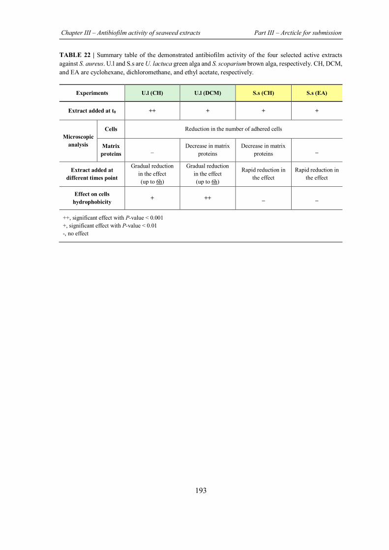
Chapter III – Antibiofilm activity of seaweed extracts Part III – Arcticle for submission
193
TABLE 22 | Summary table of the demonstrated antibiofilm activity of the four selected active extracts against S. aureus. U.l and S.s are U. lactuca green alga and S. scoparium brown alga, respectively. CH, DCM, and EA are cyclohexane, dichloromethane, and ethyl acetate, respectively.
Experiments U.l (CH) U.l (DCM) S.s (CH) S.s (EA)
Extract added at t0 ++ + + +
Microscopic analysis
Cells Reduction in the number of adhered cells
Matrix proteins
_ Decrease in matrix proteins
Decrease in matrix proteins _
Extract added at different times point
Gradual reduction in the effect (up to 6h)
Gradual reduction in the effect (up to 6h)
Rapid reduction in the effect
Rapid reduction in the effect
Effect on cells hydrophobicity + ++ _ _
++, significant effect with P-value < 0.001 +, significant effect with P-value < 0.01 -, no effect
Chapter III – Antibiofilm activity of seaweed extracts Part III – Arcticle for submission
194
III.2 ARTICLE TO BE SUBMITTED
Seaweed extracts as an effective gateway in the search for novel
antibiofilm agents against Staphylococcus aureus
Maya Rima1,2, Asma Chbani2,3, Christine Roques1,4*, Fatima El Garah1*,
1 Laboratoire de Génie Chimique, Université de Toulouse, CNRS, INPT, UPS, Toulouse, France
2 Laboratory of applied biotechnology, AZM Center for Research in Biotechnology and its Applications, Doctoral School of Science and Technology, Lebanese University, El Mittein Street, Tripoli, Lebanon.
3 Faculty of Public Health III, Lebanese University, Tripoli, Lebanon
4 Bacteriology-Hygiene Department, Centre Hospitalier Universitaire, Toulouse, Hôpital Purpan, Toulouse, France Corresponding authors: Dr Fatima El Garah Tel: +33 562256855 e-mail: [email protected] ORCiD ID : 0000-0001-7217-5879 Professor Christine Roques Tel: +33 562256860 e-mail: [email protected]
Chapter III – Antibiofilm activity of seaweed extracts Part III – Arcticle for submission
195
ABSTRACT Background: Currently, the treatment of biofilm-associated infections has become a major challenge in biomedical and clinical fields due to the failure of conventional treatments in controlling this highly complex and tolerant structure. Therefore, the search for novel antibiofilm agents with increased efficacy and few side effects as those provided by natural products, presents an urgent need. Purpose: The aim of this study is to explore extracts derived from three algae (green Ulva lactuca, brown Stypocaulon scoparium, red Pterocladiella capillacea) for their potential antibiofilm activity against Staphylococcus aureus, bacterium responsible for several acute and chronic infections by colonizing tissue and artificial surfaces. Methods: Seaweed extracts were prepared by successive maceration in four solvents with increasing polarity (cyclohexane, dichloromethane, ethyl acetate, and methanol). The ability of the different extracts to inhibit S. aureus biofilm formation was assessed using colony-forming unit (CFU) counts method. Epifluorescence microscopic analysis of biofilm formed in presence of the potentially active extracts was also carried out. Effects of active extracts on growth cycle of biofilm formation (extract added at various times of biofilm development) as well as on S. aureus surface hydrophobicity were evaluated. Results: The obtained results revealed the ability of four extracts (CH and DCM extracts derived from the green alga and CH and EA extracts originated from the brown one) to significantly (***, P-value < 0.001; **, P-value < 0.01) inhibit S. aureus biofilm formation. These findings were supported by microscopic analyses. The gradual reduction in the number of adherent bacteria when the selected extracts were added at various times (t0, t2h, t4h, t6h, and t24h) reveals their potential effect on the initial adhesion and proliferation stages of S. aureus biofilm development. Concerning DCM extract derived from the green alga, its demonstrated ability to significantly (***, P-value < 0.001) reduce S. aureus surface hydrophobicity may account to its effect on the early stages of biofilm formation. Conclusion: These findings present new insight into the exploration of seaweed as a valuable source of antibiofilm agents with preventive effect by inhibiting and/or delaying biofilm formation.
Keywords: Staphylococcus aureus, anti-biofilm, seaweed extracts, hydrophobicity, anti-adhesion.
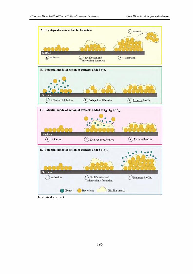
Chapter III – Antibiofilm activity of seaweed extracts Part III – Arcticle for submission
196
Graphical abstract
Chapter III – Antibiofilm activity of seaweed extracts Part III – Arcticle for submission
197
1. INTRODUCTION
Although the huge marine biodiversity is far from being completely explored, previous
studies have evidenced the richness of the marine world in organisms producing a library
of bioactive secondary metabolites that arise from millions of years of natural selection and
evolution (Kiuru et al., 2014; Jimenez, 2018). Seaweed, benthic marine macroalgae widely
distributed on rocky shores as well as at various sea depth, are part of sea’s treasure trove
that have been used for centuries as sea vegetables, fertilizers and medicines (Leandro et
al., 2019). In fact, algae are well known for their richness in unique bioactive compounds
synthetized from the simple resources found in the marine environment as a natural
response and a self-preservation way of facing the stressful environmental conditions
(Shannon & Abu-Ghannam, 2016; Leandro et al., 2019).
In addition to the abiotic challenges (salinity, temperature changes, UV radiation
exposure…) encountered in seawater, algae are also exposed to biotic threats represented
by a considerable risk of being infected by undesirable microorganisms such as bacteria
(Leandro et al., 2019). In this context, different studies have proven the wide spectrum of
antibacterial activity of algal metabolites demonstrated against several Gram-negative and
Gram-positive pathogenic bacteria which provides a promising gateway in the search for
novel drugs (Bhowmick et al., 2020).
It is obvious that the rapid emergence of multidrug resistant bacteria poses a global threat
for human health which calls for intensive efforts in order to overcome the problem of
antibiotic failure. Besides the well-known genetic mechanisms involved in the bacterial
resistance phenomenon as well as the horizontal transfer of antibiotic-resistance genes,
bacteria also exhibit an adaptive strategy that consists in the formation of a strongly
structured cells assembly named “biofilm”, embedded in a self-produced extracellular
matrix and adhered to a biotic or abiotic surfaces. Due to the collective recalcitrance of this
bacterial association towards antibiotics as well as its ability to evade the host immune
defenses, treatment of biofilms related infections is increasingly challenging (Uruen et al.,
2020).
Chapter III – Antibiofilm activity of seaweed extracts Part III – Arcticle for submission
198
The Gram-positive “superbug” Staphylococcus aureus is one of the common pathogenic
bacteria well-known as a biofilm producer. Classified by the Infectious Diseases Society
of America (IDSA) as member of “ESKAPE pathogens” group and defined by the World
Health Organization (WHO) as a high priority in the search for novel therapeutic strategies,
S. aureus receives a considerable attention (Pendleton et al., 2013; WHO, 2017). This
opportunistic bacterium is one of the principle human pathogens that is widely associated
with hospital acquired infections and responsible for several biofilms related infections
worldwide (Tong et al., 2015). Besides its ability to colonize living tissues leading to severe
infections such as osteomyelitis, endocarditis, and respiratory infections, S. aureus readily
forms a resilient biofilm on catheters and implanted medical devices surfaces (Archer et
al., 2011).
Typically, bacterial biofilm formation occurs in three main steps initiated by cell adhesion
to a surface followed by bacterial aggregates proliferation leading to the establishment of
a multi-layered structure of biofilm. Then, to ensure the biofilm life cycle, a dispersion step
proceeds (Rumbaugh & Sauer, 2020).
In S. aureus, the initial attachment to surface is mainly mediated by hydrophobic and
electrostatic interactions followed by the production of the extracellular matrix
(polysaccharides, teichoic acids, extracellular DNA, proteins…) which is highly involved
in mature biofilm resilience by providing a diffusion barrier against antimicrobial agents
(Suresh et al., 2019).
The current treatments of S. aureus biofilm related infections are based on the ablation of
the infected foreign bodies when it’s possible, otherwise, the administration of
conventional antimicrobial agents at high concentration and for an extended period is often
used (Suresh et al., 2019). Thus, the exploration of new approaches to prevent and/or to
treat S. aureus biofilm presents an area of active research. In this context, natural medicine,
which has been used for centuries in healing and treatment of diseases, presents strong
promises given the remarkable antibiofilm activity demonstrated for several natural
products (Mishra et al., 2020; Guzzo et al., 2020) .
Chapter III – Antibiofilm activity of seaweed extracts Part III – Arcticle for submission
199
Therefore, the aim of this study is to evaluate the potential capacity of extracts derived
from three algae (the green alga U. lactuca, the brown alga S. scoparium and the red alga
P. capillacea) collected from the Lebanese coast to control S. aureus biofilm. The potential
antibiofilm effect of the different prepared extracts was first assessed on biofilm formation
and development using colony-forming unit (CFU) counting method. An epifluorescence
microscopic examination of the biofilm formed in the presence of the most active extracts
was also carried out. Then, the biofilm development phase targeted by the selected active
extracts was determined by adding extract at different stages of S. aureus biofilm growth.
Furthermore, the potential influence of the selected extracts on S. aureus surface
hydrophobicity known to be correlated to its adhesiveness was evaluated by contact angle
measurement method.
Chapter III – Antibiofilm activity of seaweed extracts Part III – Arcticle for submission
200
2. MATERIALS AND METHODS
2.1 Collection of algal materials
The three algae chosen to be evaluated in this study are the green alga Ulva lactuca, the
brown alga Stypocaulon scoparium, and the red alga Pterocladiella capillacea. Seaweed
samples were manually collected from the North Lebanese coast of the Mediterranean (El
Mina – Tripoli – Lebanon) in September 2019. Algae were rinsed directly with seawater
followed by a distilled water wash in laboratory to remove associated impurities such as
epiphytes and adhered sand particles. Seaweed samples were then air-dried in dark at room
temperature (20 - 27°C) for several weeks and weighted continuously until complete
drying. Dried algae were powdered in fine-milled form prior to extraction. They were then
transported in sealed bags to Laboratoire de Génie Chimique of Toulouse – France where
the extractions were done.
2.2 Organic solvents and chemicals
Solvents used for preparation of seaweed extracts are cyclohexane 99.5% (Sigma-Aldrich,
France), dichloromethane 100% (VWR, France), ethyl acetate 99.9% (VWR, France), and
methanol 99.8% (Sigma-Aldrich, France). The dyes employed in the microscopic analysis
were purchased from ThermoFisher Scientific – France.
2.3 Bacterial strain and culture media
The bacterial strain used in this study is Staphylococcus aureus (CIP 4.83), purchased from
the collection of Pasteur Institute (Paris, France) and preserved at - 80˚C. Before each
experiment, two successive overnight subcultures were realized on Trypticase soy agar
TSA (BioMérieux, Crapone, France) and incubated under aerobic conditions at 37°C.
Mueller-Hinton broth MHB (Oxoid microbiology products, Basingstoke, UK) was used as
culture medium for the evaluation of the antibacterial activity of extracts (CA-
SFM/EUCAST 2020). On the other hand, the antibiofilm activity assays were conducted
in the previously selected low-nutritive medium named biofilm broth (BB) in order to
create stressful conditions and subsequently promote biofilm formation and adherent cells
growth rather than planktonic growth (Campanac et al., 2002).
Chapter III – Antibiofilm activity of seaweed extracts Part III – Arcticle for submission
201
The BB 10X is composed of FeSO4, 7H2O (0.005 g/L), Na2HPO4 (12.5 g/L), KH2PO4 (5.0
g/L), (NH4)2 SO4 (1.0 g/L), lactose (0.25 g/L), yeast extract (1.0 g/L), vitamin assay
casamino acids (1.0 g/L) and MgSO4, 7H2O (0.2 g/L) (Campanac et al., 2002). Except for
yeast extract (BactoTM, ThermoFisher scientific) and vitamin assay casamino acids
(DifcoTM, ThermoFisher scientific), all these compounds were purchased from Sigma-
Aldrich, France.
2.4 Preparation of seaweed extracts
In order to extract the maximum seaweed’s constituents, a successive extraction method
using selective solvents with increasing polarity (cyclohexane, dichloromethane, ethyl
acetate, and methanol) has been adopted (Kohoude et al., 2017). Thus, 100.0 g of the dried
samples of each alga were macerated successively in 1L of each solvent for 2 hours under
magnetic agitation. Crude extracts were recovered after filtration using the Büchner funnel
followed by solvent evaporation using a rotary evaporator under vacuum at 40˚C. Note that
maceration with the same solvent was repeated until the progressive discoloration of the
filtrate. In this case, the different extracts obtained from the same solvent were combined.
The extraction yield was then calculated using the following formula (1), where W2 is the
weight of the extract residue after solvent evaporation and W1 is the weight of the algal
matrix initially used for the extraction (100.0 g).
(1) Extraction yield (%) = (W2 / W1) x 100
To evaluate their bioactivity, stock solutions were prepared by dissolving the extracts in
sterile distilled water (SDW) at 100.0 µg/mL, using an ultrasonic bath (VWR ultrasonic
cleaning bath, 45 KHz) for almost 6 hours to promote the solubility. Stock solutions were
then sterilized by filtration through a syringe filter (Cellulose Acetate Syringe Filter, 0.45
µm, GE Healthcare Whatman).
2.5 Effect of extracts on S. aureus planktonic growth – MIC determination
Prior to the assessment of their potential effect on biofilm, the evaluation of antibacterial
activity of extracts on S. aureus is essential for ensuring the use of a sub-MIC concentration
in antibiofilm activity assays. For this purpose, the minimal inhibitory concentration (MIC)
of all extracts against S. aureus was determined using the broth microdilution method,
according to the guidelines of CA-SFM/EUCAST 2020.
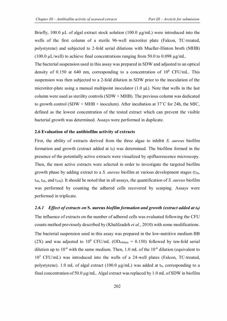
Chapter III – Antibiofilm activity of seaweed extracts Part III – Arcticle for submission
202
Briefly, 100.0 µL of algal extract stock solution (100.0 µg/mL) were introduced into the
wells of the first column of a sterile 96-well microtiter plate (Falcon, TC-treated,
polystyrene) and subjected to 2-fold serial dilutions with Mueller-Hinton broth (MHB)
(100.0 µL/well) to achieve final concentrations ranging from 50.0 to 0.098 µg/mL.
The bacterial suspension used in this assay was prepared in SDW and adjusted to an optical
density of 0.150 at 640 nm, corresponding to a concentration of 108 CFU/mL. This
suspension was then subjected to a 2-fold dilution in SDW prior to the inoculation of the
microtiter-plate using a manual multipoint inoculator (1.0 µL). Note that wells in the last
column were used as sterility controls (SDW + MHB). The previous column was dedicated
to growth control (SDW + MHB + inoculum). After incubation at 37˚C for 24h, the MIC,
defined as the lowest concentration of the tested extract which can prevent the visible
bacterial growth was determined. Assays were performed in duplicate.
2.6 Evaluation of the antibiofilm activity of extracts
First, the ability of extracts derived from the three algae to inhibit S. aureus biofilm
formation and growth (extract added at t0) was determined. The biofilms formed in the
presence of the potentially active extracts were visualized by epifluorescence microscopy.
Then, the most active extracts were selected in order to investigate the targeted biofilm
growth phase by adding extract to a S. aureus biofilm at various development stages (t2h,
t4h, t6h, and t24h). It should be noted that in all assays, the quantification of S. aureus biofilm
was performed by counting the adhered cells recovered by scarping. Assays were
performed in triplicate.
2.6.1 Effect of extracts on S. aureus biofilm formation and growth (extract added at t0)
The influence of extracts on the number of adhered cells was evaluated following the CFU
counts method previously described by (Khalilzadeh et al., 2010) with some modifications.
The bacterial suspension used in this assay was prepared in the low-nutritive medium BB
(2X) and was adjusted to 108 CFU/mL (OD640nm = 0.150) followed by ten-fold serial
dilution up to 10-6 with the same medium. Then, 1.0 mL of the 10-6 dilution (equivalent to
102 CFU/mL) was introduced into the wells of a 24-well plates (Falcon, TC-treated,
polystyrene). 1.0 mL of algal extract (100.0 µg/mL) was added at t0, corresponding to a
final concentration of 50.0 µg/mL. Algal extract was replaced by 1.0 mL of SDW in biofilm
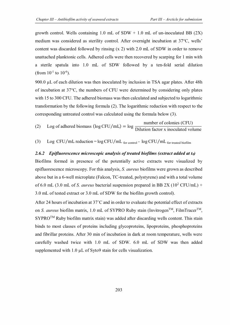
Chapter III – Antibiofilm activity of seaweed extracts Part III – Arcticle for submission
203
growth control. Wells containing 1.0 mL of SDW + 1.0 mL of un-inoculated BB (2X)
medium was considered as sterility control. After overnight incubation at 37°C, wells’
content was discarded followed by rinsing (x 2) with 2.0 mL of SDW in order to remove
unattached planktonic cells. Adhered cells were then recovered by scarping for 1 min with
a sterile spatula into 1.0 mL of SDW followed by a ten-fold serial dilution
(from 10-1 to 10-6).
900.0 µL of each dilution was then inoculated by inclusion in TSA agar plates. After 48h
of incubation at 37°C, the numbers of CFU were determined by considering only plates
with 15 to 300 CFU. The adhered biomass was then calculated and subjected to logarithmic
transformation by the following formula (2). The logarithmic reduction with respect to the
corresponding untreated control was calculated using the formula below (3).
(2) Log of adhered biomass (log CFU mL⁄ ) = log number of colonies (CFU)
Dilution factor x inoculated volume
(3) Log CFU mL reduction = log CFU mL for control - log CFU mL for treated biofilm ⁄ ⁄⁄
2.6.2 Epifluorescence microscopic analysis of treated biofilms (extract added at t0)
Biofilms formed in presence of the potentially active extracts were visualized by
epifluorescence microscopy. For this analysis, S. aureus biofilms were grown as described
above but in a 6-well microplate (Falcon, TC-treated, polystyrene) and with a total volume
of 6.0 mL (3.0 mL of S. aureus bacterial suspension prepared in BB 2X (102 CFU/mL) +
3.0 mL of tested extract or 3.0 mL of SDW for the biofilm growth control).
After 24 hours of incubation at 37˚C and in order to evaluate the potential effect of extracts
on S. aureus biofilm matrix, 1.0 mL of SYPRO Ruby stain (InvitrogenTM, FilmTracerTM,
SYPROTM Ruby biofilm matrix stain) was added after discarding wells content. This stain
binds to most classes of proteins including glycoproteins, lipoproteins, phosphoproteins
and fibrillar proteins. After 30 min of incubation in dark at room temperature, wells were
carefully washed twice with 1.0 mL of SDW. 6.0 mL of SDW was then added
supplemented with 1.0 µL of Syto9 stain for cells visualization.
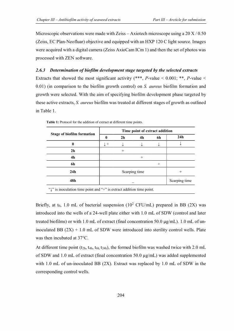
Chapter III – Antibiofilm activity of seaweed extracts Part III – Arcticle for submission
204
Microscopic observations were made with Zeiss – Axiotech microscope using a 20 X / 0.50
(Zeiss, EC Plan-Neofluar) objective and equipped with an HXP 120 C light source. Images
were acquired with a digital camera (Zeiss AxioCam ICm 1) and then the set of photos was
processed with ZEN software.
2.6.3 Determination of biofilm development stage targeted by the selected extracts
Extracts that showed the most significant activity (***, P-value < 0.001; **, P-value <
0.01) (in comparison to the biofilm growth control) on S. aureus biofilm formation and
growth were selected. With the aim of specifying biofilm development phase targeted by
these active extracts, S. aureus biofilm was treated at different stages of growth as outlined
in Table 1. Table 1: Protocol for the addition of extract at different time points.
Briefly, at t0, 1.0 mL of bacterial suspension (102 CFU/mL) prepared in BB (2X) was
introduced into the wells of a 24-well plate either with 1.0 mL of SDW (control and later
treated biofilms) or with 1.0 mL of extract (final concentration 50.0 µg/mL). 1.0 mL of un-
inoculated BB (2X) + 1.0 mL of SDW were introduced into sterility control wells. Plate
was then incubated at 37°C.
At different time point (t2h, t4h, t6h, t24h), the formed biofilm was washed twice with 2.0 mL
of SDW and 1.0 mL of extract (final concentration 50.0 µg/mL) was added supplemented
with 1.0 mL of un-inoculated BB (2X). Extract was replaced by 1.0 mL of SDW in the
corresponding control wells.
Stage of biofilm formation Time point of extract addition
0 2h 4h 6h 24h
0 ↓ + ↓ ↓ ↓ ↓
2h +
4h +
6h +
24h Scarping time +
48h _ Scarping time
“↓” is inoculation time point and “+” is extract addition time point.
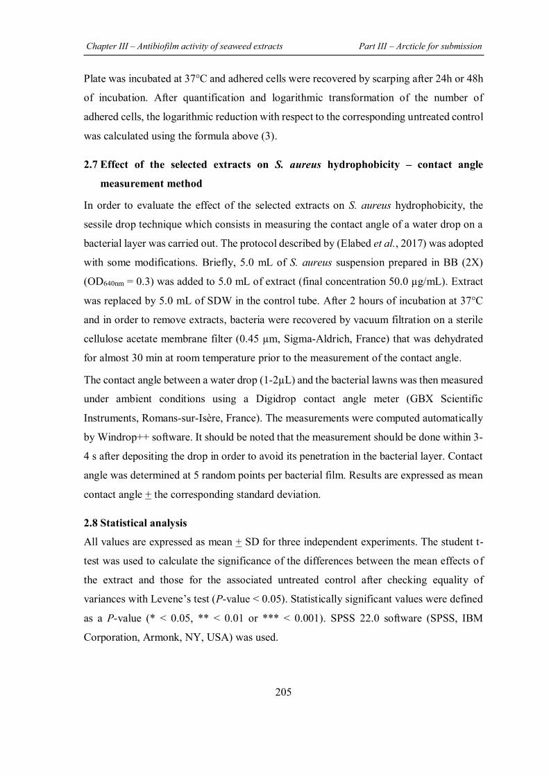
Chapter III – Antibiofilm activity of seaweed extracts Part III – Arcticle for submission
205
Plate was incubated at 37°C and adhered cells were recovered by scarping after 24h or 48h
of incubation. After quantification and logarithmic transformation of the number of
adhered cells, the logarithmic reduction with respect to the corresponding untreated control
was calculated using the formula above (3).
2.7 Effect of the selected extracts on S. aureus hydrophobicity – contact angle
measurement method
In order to evaluate the effect of the selected extracts on S. aureus hydrophobicity, the
sessile drop technique which consists in measuring the contact angle of a water drop on a
bacterial layer was carried out. The protocol described by (Elabed et al., 2017) was adopted
with some modifications. Briefly, 5.0 mL of S. aureus suspension prepared in BB (2X)
(OD640nm = 0.3) was added to 5.0 mL of extract (final concentration 50.0 µg/mL). Extract
was replaced by 5.0 mL of SDW in the control tube. After 2 hours of incubation at 37°C
and in order to remove extracts, bacteria were recovered by vacuum filtration on a sterile
cellulose acetate membrane filter (0.45 µm, Sigma-Aldrich, France) that was dehydrated
for almost 30 min at room temperature prior to the measurement of the contact angle.
The contact angle between a water drop (1-2µL) and the bacterial lawns was then measured
under ambient conditions using a Digidrop contact angle meter (GBX Scientific
Instruments, Romans-sur-Isère, France). The measurements were computed automatically
by Windrop++ software. It should be noted that the measurement should be done within 3-
4 s after depositing the drop in order to avoid its penetration in the bacterial layer. Contact
angle was determined at 5 random points per bacterial film. Results are expressed as mean
contact angle + the corresponding standard deviation.
2.8 Statistical analysis
All values are expressed as mean + SD for three independent experiments. The student t-
test was used to calculate the significance of the differences between the mean effects of
the extract and those for the associated untreated control after checking equality of
variances with Levene’s test (P-value < 0.05). Statistically significant values were defined
as a P-value (* < 0.05, ** < 0.01 or *** < 0.001). SPSS 22.0 software (SPSS, IBM
Corporation, Armonk, NY, USA) was used.
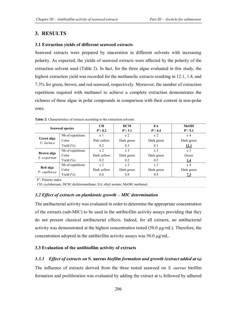
Chapter III – Antibiofilm activity of seaweed extracts Part III – Arcticle for submission
206
3. RESULTS
3.1 Extraction yields of different seaweed extracts
Seaweed extracts were prepared by maceration in different solvents with increasing
polarity. As expected, the yields of seaweed extracts were affected by the polarity of the
extraction solvent used (Table 2). In fact, for the three algae evaluated in this study, the
highest extraction yield was recorded for the methanolic extracts resulting in 12.1, 1.4, and
7.3% for green, brown, and red seaweed, respectively. Moreover, the number of extraction
repetitions required with methanol to achieve a complete extraction demonstrates the
richness of these algae in polar compounds in comparison with their content in non-polar
ones. Table 2: Characteristics of extracts according to the extraction solvents
3.2 Effect of extracts on planktonic growth – MIC determination
The antibacterial activity was evaluated in order to determine the appropriate concentration
of the extracts (sub-MIC) to be used in the antibiofilm activity assays providing that they
do not present classical antibacterial effects. Indeed, for all extracts, no antibacterial
activity was demonstrated at the highest concentration tested (50.0 µg/mL). Therefore, the
concentration adopted in the antibiofilm activity assays was 50.0 µg/mL.
3.3 Evaluation of the antibiofilm activity of extracts
3.3.1 Effect of extracts on S. aureus biofilm formation and growth (extract added at t0)
The influence of extracts derived from the three tested seaweed on S. aureus biofilm
formation and proliferation was evaluated by adding the extract at t0 followed by adhered
Seaweed species CH P’: 0.2
DCM P’: 3.1
EA P’: 4.4
MeOH P’: 5.1
Green alga U. lactuca
Nb of repetitions x 1 x 2 x 2 x 4 Color Pale yellow Dark green Dark green Dark green Yield (%) 0.2 0.3 0.1 12.1
Brown alga S. scoparium
Nb of repetitions x 2 x 3 x 3 x 3 Color Dark yellow Dark green Dark green Green Yield (%) 0.2 0.2 0.5 1.4
Red alga P. capillacea
Nb of repetitions x 2 x 3 x 3 x 4 Color Dark yellow Dark green Dark green Dark green Yield (%) 0.4 0.8 0.9 7.3
P’: Polarity index CH: cyclohexane, DCM: dichloromethane, EA: ethyl acetate, MeOH: methanol.
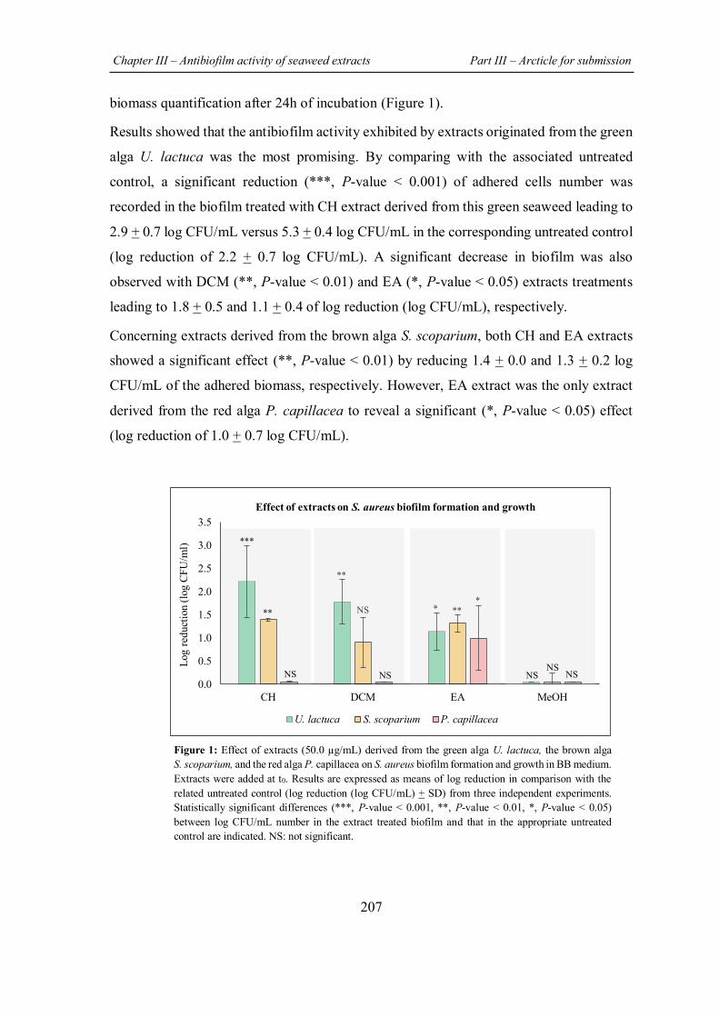
Chapter III – Antibiofilm activity of seaweed extracts Part III – Arcticle for submission
207
biomass quantification after 24h of incubation (Figure 1).
Results showed that the antibiofilm activity exhibited by extracts originated from the green
alga U. lactuca was the most promising. By comparing with the associated untreated
control, a significant reduction (***, P-value < 0.001) of adhered cells number was
recorded in the biofilm treated with CH extract derived from this green seaweed leading to
2.9 + 0.7 log CFU/mL versus 5.3 + 0.4 log CFU/mL in the corresponding untreated control
(log reduction of 2.2 + 0.7 log CFU/mL). A significant decrease in biofilm was also
observed with DCM (**, P-value < 0.01) and EA (*, P-value < 0.05) extracts treatments
leading to 1.8 + 0.5 and 1.1 + 0.4 of log reduction (log CFU/mL), respectively.
Concerning extracts derived from the brown alga S. scoparium, both CH and EA extracts
showed a significant effect (**, P-value < 0.01) by reducing 1.4 + 0.0 and 1.3 + 0.2 log
CFU/mL of the adhered biomass, respectively. However, EA extract was the only extract
derived from the red alga P. capillacea to reveal a significant (*, P-value < 0.05) effect
(log reduction of 1.0 + 0.7 log CFU/mL).
Figure 1: Effect of extracts (50.0 µg/mL) derived from the green alga U. lactuca, the brown alga S. scoparium, and the red alga P. capillacea on S. aureus biofilm formation and growth in BB medium. Extracts were added at t0. Results are expressed as means of log reduction in comparison with the related untreated control (log reduction (log CFU/mL) + SD) from three independent experiments. Statistically significant differences (***, P-value < 0.001, **, P-value < 0.01, *, P-value < 0.05) between log CFU/mL number in the extract treated biofilm and that in the appropriate untreated control are indicated. NS: not significant.
0.0
0.5
1.0
1.5
2.0
2.5
3.0
3.5
CH DCM EA MeOH
Log
redu
ctio
n (lo
g CF
U/m
l)
Effect of extracts on S. aureus biofilm formation and growth
U. lactuca S. scoparium P. capillacea
**
* ** NS ** *
***
NS NS
NS NS NS
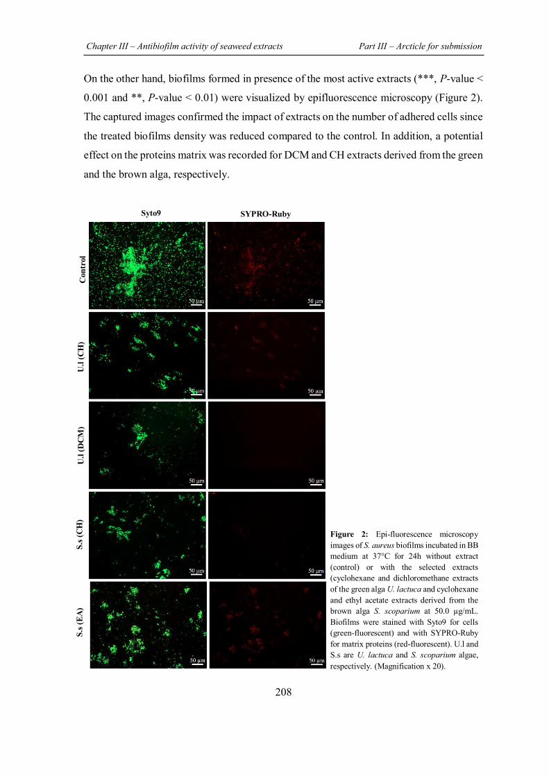
Chapter III – Antibiofilm activity of seaweed extracts Part III – Arcticle for submission
208
On the other hand, biofilms formed in presence of the most active extracts (***, P-value <
0.001 and **, P-value < 0.01) were visualized by epifluorescence microscopy (Figure 2).
The captured images confirmed the impact of extracts on the number of adhered cells since
the treated biofilms density was reduced compared to the control. In addition, a potential
effect on the proteins matrix was recorded for DCM and CH extracts derived from the green
and the brown alga, respectively.
Figure 2: Epi-fluorescence microscopy images of S. aureus biofilms incubated in BB medium at 37°C for 24h without extract (control) or with the selected extracts (cyclohexane and dichloromethane extracts of the green alga U. lactuca and cyclohexane and ethyl acetate extracts derived from the brown alga S. scoparium at 50.0 µg/mL. Biofilms were stained with Syto9 for cells (green-fluorescent) and with SYPRO-Ruby for matrix proteins (red-fluorescent). U.l and S.s are U. lactuca and S. scoparium algae, respectively. (Magnification x 20).
Con
trol
U
.l (C
H)
U.l
(DC
M)
S.s (
CH
) S.
s (EA
)
Syto9 SYPRO-Ruby
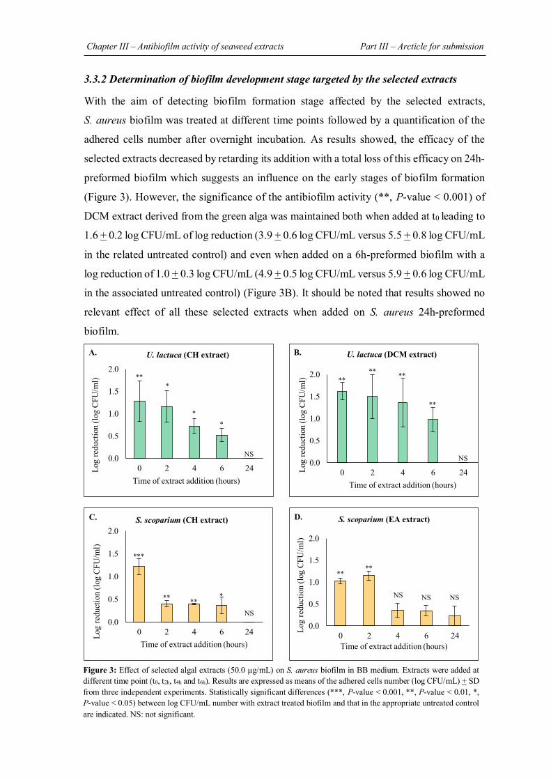
Chapter III – Antibiofilm activity of seaweed extracts Part III – Arcticle for submission
209
3.3.2 Determination of biofilm development stage targeted by the selected extracts
With the aim of detecting biofilm formation stage affected by the selected extracts,
S. aureus biofilm was treated at different time points followed by a quantification of the
adhered cells number after overnight incubation. As results showed, the efficacy of the
selected extracts decreased by retarding its addition with a total loss of this efficacy on 24h-
preformed biofilm which suggests an influence on the early stages of biofilm formation
(Figure 3). However, the significance of the antibiofilm activity (**, P-value < 0.001) of
DCM extract derived from the green alga was maintained both when added at t0 leading to
1.6 + 0.2 log CFU/mL of log reduction (3.9 + 0.6 log CFU/mL versus 5.5 + 0.8 log CFU/mL
in the related untreated control) and even when added on a 6h-preformed biofilm with a
log reduction of 1.0 + 0.3 log CFU/mL (4.9 + 0.5 log CFU/mL versus 5.9 + 0.6 log CFU/mL
in the associated untreated control) (Figure 3B). It should be noted that results showed no
relevant effect of all these selected extracts when added on S. aureus 24h-preformed
biofilm.
Figure 3: Effect of selected algal extracts (50.0 µg/mL) on S. aureus biofilm in BB medium. Extracts were added at different time point (t0, t2h, t4h and t6h). Results are expressed as means of the adhered cells number (log CFU/mL) + SD from three independent experiments. Statistically significant differences (***, P-value < 0.001, **, P-value < 0.01, *, P-value < 0.05) between log CFU/mL number with extract treated biofilm and that in the appropriate untreated control are indicated. NS: not significant.
0.0
0.5
1.0
1.5
2.0
0 2 4 6 24Log
redu
ctio
n (lo
g CF
U/m
l)
Time of extract addition (hours)
** ** **
**
U. lactuca (DCM extract) B.
0.0
0.5
1.0
1.5
2.0
0 2 4 6 24Log
redu
ctio
n (lo
g CF
U/m
l)
Time of extract addition (hours)
***
** ** *
S. scoparium (CH extract) C.
0.0
0.5
1.0
1.5
2.0
0 2 4 6 24Log
redu
ctio
n (lo
g CF
U/m
l)
Time of extract addition (hours)
** **
NS NS NS
S. scoparium (EA extract) D.
NS
0.0
0.5
1.0
1.5
2.0
0 2 4 6 24Log
redu
ctio
n (lo
g CF
U/m
l)
Time of extract addition (hours)
** *
* *
A. U. lactuca (CH extract)
NS NS
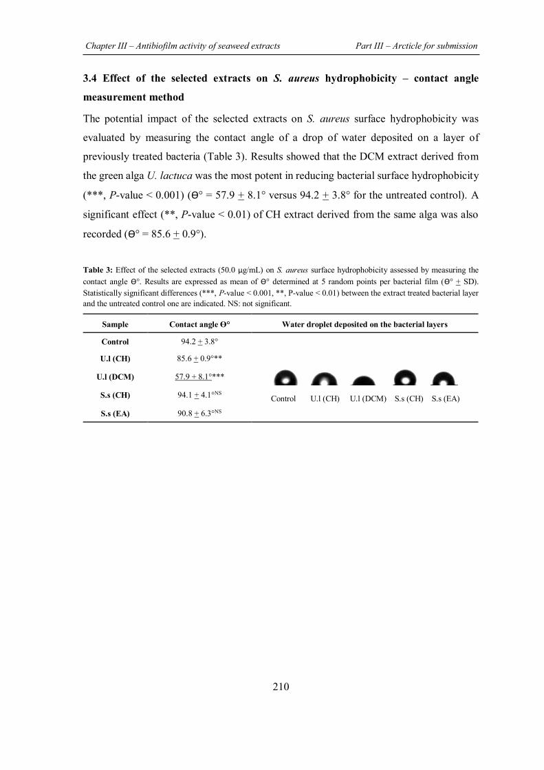
Chapter III – Antibiofilm activity of seaweed extracts Part III – Arcticle for submission
210
3.4 Effect of the selected extracts on S. aureus hydrophobicity – contact angle
measurement method
The potential impact of the selected extracts on S. aureus surface hydrophobicity was
evaluated by measuring the contact angle of a drop of water deposited on a layer of
previously treated bacteria (Table 3). Results showed that the DCM extract derived from
the green alga U. lactuca was the most potent in reducing bacterial surface hydrophobicity
(***, P-value < 0.001) (ϴ° = 57.9 + 8.1° versus 94.2 + 3.8° for the untreated control). A
significant effect (**, P-value < 0.01) of CH extract derived from the same alga was also
recorded (ϴ° = 85.6 + 0.9°).
Table 3: Effect of the selected extracts (50.0 µg/mL) on S. aureus surface hydrophobicity assessed by measuring the contact angle ϴ°. Results are expressed as mean of ϴ° determined at 5 random points per bacterial film (ϴ° + SD). Statistically significant differences (***, P-value < 0.001, **, P-value < 0.01) between the extract treated bacterial layer and the untreated control one are indicated. NS: not significant.
Sample Contact angle ϴ° Water droplet deposited on the bacterial layers
Control 94.2 + 3.8°
Control U.l (CH) U.l (DCM) S.s (CH) S.s (EA)
U.l (CH) 85.6 + 0.9°**
U.l (DCM) 57.9 + 8.1°***
S.s (CH) 94.1 + 4.1°NS
S.s (EA) 90.8 + 6.3°NS
Chapter III – Antibiofilm activity of seaweed extracts Part III – Arcticle for submission
211
4. DISCUSSION
Biofilm formation presents one of the strategies adopted by bacteria in order to overcome
treatment with antimicrobial agents as well as to escape from host immune defenses (Uruen
et al., 2020). Indeed, besides the protection provided by the extracellular matrix against the
penetration of antimicrobial agents, the heterogeneity within the biofilm represented by
nutrient and oxygen gradients, lead to the formation of cells with different metabolic states,
which promotes the resilience of this bacterial community (Campanac et al., 2002; Preda
& Sandulescu, 2019). Therefore, a great interest has been dedicated to the search for novel
antibiofilm agents in an attempt to prevent and/or treat biofilm-related infections
(Bjarnsholt et al., 2013; Koo et al., 2017). In this context, natural products are considered
as a strong promises given their remarkable antibiofilm activity demonstrated against
various pathogenic bacteria by exhibiting different mechanisms of action (Mishra et al.,
2020; Zhang et al., 2020).
Interestingly, various studies have highlighted the ability of compounds isolated from
marine organisms such as seaweed and sponges to present a valuable input in the search
for new antibiofilm agents (Stowe et al., 2011; Dahms & Dobretsov, 2017; Melander et al.,
2020). Indeed, these organisms living in the stressful conditions of the marine environment,
possess sophisticated defense mechanisms that involve the natural synthesis of secondary
metabolites in order to overcome any kind of undesirable attacks (predation, biofouling…)
(Shannon & Abu-Ghannam, 2016).
So, the aim of this study is to explore the potential ability of extracts derived from three
algae (the green U. lactuca, the brown S. scoparium, and the red P. capillacea seaweed) to
control the biofilm formed by S. aureus, a common pathogen involved in hospital-acquired
infections (Suresh et al., 2019). Concerning the green alga U. lactuca, various studies have
highlighted the significant bioactivity (antimicrobial, cytotoxic, antioxidant, insecticidal
activities…) of its extracts (acetonic, methanolic, aqueous…) (Saeed et al., 2019; Anjali et
al., 2019; Rima et al., 2021). On the other hand, although the number of studies that have
evaluated the potential bioactivity of the brown alga S. scoparium is limited, some
biological proprieties such as antioxidant, antibacterial, anti-inflammatory as well as
cytotoxic activities have been demonstrated for its extracts (Campos et al., 2018; Guner et
al., 2019). Regarding the red alga P. capillacea, Ismail et al., and Shobier et al., have
Chapter III – Antibiofilm activity of seaweed extracts Part III – Arcticle for submission
212
respectively demonstrated the ability of its extracts to exhibit antioxidant, antidiabetic, and
antifungal activities (Shobier et al., 2016; Ismail et al., 2020).
After preparation of the different extracts by successive maceration in four solvents
(cyclohexane, dichloromethane, ethyl acetate, and methanol) with increasing polarity, their
antibacterial activity against S. aureus was evaluated. As the objective of this study is to
search for an antibiofilm effect rather than a classical antibacterial activity, this potential
impact of extracts on S. aureus planktonic growth was investigated in order to ensure that
the tested concentration in the antibiofilm assays is sub-MIC and therefore the obtained
results will be restricted to an effect on the biofilm. Results showed that for all extracts, no
visible antibacterial effect was recorded at the highest tested concentration (50.0 µg/mL).
This is in accordance with two previous studies conducted by Pushparaj et al., and De
Alencar et al., which indicated the absence of an inhibitory effect on S. aureus bacterial
growth of extracts derived from the green alga U. lactuca (acetonic, ethyl acetate,
methanolic…extracts) and the red one P. capillacea (hexane and ethanolic extracts),
respectively (Pushparaj et al., 2014; De Alencar et al., 2016). On the other hand, Dulger et
al., demonstrated the capacity of methanolic extract obtained from the brown alga S.
scoparium to inhibit the growth of S. aureus but at much higher concentration (Dulger et
al., 2009). It should be noted that the selection of 50.0 µg/mL as the maximum
concentration tested is intended to avoid solubility issues that may lead to the deposition
of precipitates on the bottom of wells and thereby distorts results and this for both
antibacterial and antibiofilm assays.
The evaluation of the potential ability of extracts to inhibit S. aureus biofilm formation and
growth was first assessed by adding extract at t0. Results showed that CH and DCM extracts
derived from the green alga U. lactuca as well as CH and EA extracts obtained from the
brown alga S. scoparium, are the most promising in exhibiting a significant (***, P-value
< 0.001; **, P-value < 0.01) antibiofilm activity (Figure 1). The epifluorescence
microscopic analysis of S. aureus biofilm formed in presence of these four potential active
extracts support their demonstrated ability to reduce the number of adhered cells associated
for some extracts (DCM and CH extracts derived from the green alga U. lactuca and the
brown alga S. scoparium, respectively) with a decrease in matrix proteins (Figure 2).
Chapter III – Antibiofilm activity of seaweed extracts Part III – Arcticle for submission
213
According to our knowledge, the brown alga S. scoparium has never been explored for
their potential antibiofilm activity. Regarding the green alga U. lactuca, the study
conducted by Yuvaraj & Arul, is the only one to evaluate the antibiofilm activity of this
alga against S. aureus (Yuvaraj & Arul, 2014). In fact, its methanolic extract, prepared by
a single maceration, was able to significantly reduce S. aureus biofilm biomass using the
crystal violet (CV) staining method, commonly used in the quantification of total biofilm
biomass by marking both adherent cells and matrix (Pantanella et al., 2013). It should be
noted that this method of biofilm quantification has been widely used in the exploration of
natural products such as gallic acid and ellagic acid rhamnoside for their antibiofilm
activity against S. aureus (Fontaine et al., 2017; Liu et al., 2017). However, the application
of CV staining method is not suitable in our case due to the limited quantity of matrix
produced by S. aureus (below the detection limit) in our culture conditions, especially in
the low-nutritive medium used (Suppl. S1). Furthermore, Allkja et al., proved that in
comparison with CV staining method, the CFU counting assay followed in our study is
more responsive in treatment experiments, making it the most suitable method to use in
treatment efficacy testing due to the possible interference between the treatment and the
dye (Allkja et al., 2021).
In order to gain insight into their potential mechanism of action, the selected extracts were
added at different times point (t0, t2h, t4h, t6h and t24h) during the development of S. aureus
biofilm. Results showed a gradual biofilm reduction when extracts were added at t0, t2h, t4h,
and t6h (Figure 3). However, regarding the number of remaining cells after extract
treatment, the 24h-old biofilm was completely resistant to the extracts which suggests their
potential effect on the initial adhesion and proliferation stages. In this context, Xiang et al.,
have demonstrated the ability of aloe-emodin, natural product derived from Rheum
officinale plant, to interfere with the early stages of biofilm formation by progressively
reducing S. aureus biofilm biomass (Xiang et al., 2017). In fact, this antibiofilm activity
restricted to the early stages of biofilm formation was explained by a reduction in matrix
components production such as proteins and polysaccharide intercellular adhesin (PIA) that
are involved in S. aureus attachment (Foster et al., 2014; Arciola et al., 2015).
Chapter III – Antibiofilm activity of seaweed extracts Part III – Arcticle for submission
214
Moreover, this mode of action has been previously reported for some antibiotics such as
vancomycin and moxifloxacin whose efficacy has been observed only on S. aureus young
biofilm (6h-old biofilm) and not on mature one (24-hour-old biofilm) (Bauer et al., 2013).
On the other hand, it is recognized that the hydrophobic proprieties of bacterial surfaces
are strongly involved in the adhesion to biotic and abiotic surfaces, especially to medical
devices made of hydrophobic materials such as silicone and stainless steel (Krasowska &
Sigler, 2014). In S. aureus, the attachment to abiotic surfaces is often mediated by ionic
and hydrophobic interactions through surface-anchored proteins such as Bap (biofilm
associated protein) and autolysin, as well as by wall teichoic acid and lipoteichoic acid
(Heilmann, 2011). Indeed, the prevalence of hydrophobic patches compared to hydrophilic
ones on the surface of S. aureus was demonstrated in the study conducted by Forson et al.,
in which the adhesion was favored on the hydrophobic surface (Forson et al., 2020). In
addition, Kouidhi et al., have highlighted a correlation between the surface hydrophobicity
of various S. aureus strains associated with dental caries and their adhesiveness on
polystyrene plates (Kouidhi et al., 2010). In this context, the potential effect of the selected
extracts on S. aureus hydrophobicity was assessed in an attempt to elucidate their potential
mechanism of action. To do that, the sessile drop technique which consists in measuring
the contact angle of a water drop on a bacterial surface was adopted. Basically, the contact
angle presents an indirect and proportional measure of the hydrophobicity as a higher
contact angle indicates a greater surface hydrophobicity (Braga & Reggio, 1995).
The obtained results have revealed the high hydrophobicity of S. aureus cells (ϴ = 94.2 +
3.8°) (Table 3). Interestingly, a significant reduction in the hydrophobicity of S. aureus
cells treated either with CH (**, P-value < 0.01) or DCM (***, P-value < 0.001) extracts
derived from the green alga was shown. Combined with the demonstrated ability of these
two extracts to gradually reduce S. aureus biofilm when added at the early stages of
formation (up to 6h) (Figure 3), their potential mechanism of action may be based on the
inhibition of the initial adhesion (when added at t0) by decreasing surface hydrophobicity
and/or delay biofilm proliferation by altering cells/plate surface interactions. This
mechanism of action has already been described for the brodimoprim, an antibacterial agent
whose ability to reduce the adhesiveness of S. aureus to human epithelial buccal cells has
been correlated with a decrease in bacterial surface hydrophobicity (Braga & Reggio,
Chapter III – Antibiofilm activity of seaweed extracts Part III – Arcticle for submission
215
1995). In addition, Allegrone et al., have demonstrated the capacity of natural rhamnolipids
and TritonTM – X100 (synthetic surfactant) to significantly reduce S. aureus surface
hydrophobicity, as well as to inhibit its adhesion to a surfactant-precoated silicone surface
(Allegrone et al., 2021).
5. CONCLUSION In the present study, the exploration of various extracts derived from three algae for their
potential ability to present an antibiofilm activity against S. aureus permitted the selection
of four extracts (CH and DCM extracts obtained from the green seaweed and CH and EA
extracts derived from the brown one) as the most promising. Their significant antibiofilm
effect was restricted to the early stages of biofilm formation. Regarding the potential
antibiofilm mechanism of action exhibited by CH and DCM extracts originated from the
green alga, a decrease in S. aureus surface hydrophobicity may explain in part their ability
to hinder bacterial adhesion and/or to delay biofilm proliferation. In light of these
encouraging results, further experiments are envisaged in an attempt to decipher the
possible mechanism of action of the selected active extracts, particularly through molecular
analysis. Furthermore, it will be interesting to analyze the chemical composition of the
active extracts in an effort to isolate highly active molecules. Overall, the findings of this
study pave the way for possible future applications of seaweed in the prevention of biofilms
formation.
ACKNOWLEDGMENTS
This work was funded by the laboratory Laboratoire de Génie Chimique, Université de Toulouse, CNRS, INPT, UPS, Toulouse, France.
CONFLICT OF INTERESTS The authors declare they have no conflict of interest.
Chapter III – Antibiofilm activity of seaweed extracts Part III – Arcticle for submission
216
SUPPLEMENTARY MATERIALS
S1: Quantification of S. aureus biofilm by crystal violet staining method
In order to screen extracts derived from the three algae for their ability to inhibit S. aureus
biofilm formation and growth, the crystal violet staining method, commonly used for the
quantification of bacterial biofilms biomass (adhered cells + matrix), was first adopted.
Briefly, after overnight incubation of the treated biofilms, they were washed twice with 2.0
mL of SDW to remove non-adherent planktonic cells. The plate was then air-dried for 1h.
To stain the adhered biomass, 2.0 mL of an aqueous 1% CV solution was added to the wells
and consecutively incubated for 15 min at room temperature. In order to remove the excess
stain, wells were rinsed twice with 2.0 mL of SDW followed by drying for 30 min before
quantification. 1.0 mL of ethanol was finally added to extract bound stain prior to the
absorbance measurement (OD570nm).
Results showed that the evaluation of the antibiofilm activity of extracts against S. aureus
by this method is not feasible in our culture conditions due to the low quantity of biofilm
biomass detected in the control wells (OD570nm = 0.06 + 0.03). For this reason, CFU counts
method was the only assay used in this study.
Chapter III – Antibiofilm activity of seaweed extracts Part III – Arcticle for submission
217
REFERENCES
Allegrone, G., Ceresa, C., Rinaldi, M., & Fracchia, L. (2021). Diverse Effects of Natural and Synthetic Surfactants on the Inhibition of Staphylococcus aureus Biofilm. Pharmaceutics, 13(8). doi:10.3390/pharmaceutics13081172
Allkja, J., van Charante, F., Aizawa, J., Reigada, I., Guarch-Perez, C., Vazquez-Rodriguez, J. A., et al. (2021). Interlaboratory study for the evaluation of three microtiter plate-based biofilm quantification methods. Sci Rep, 11(1), 13779. doi:10.1038/s41598-021-93115-w
Anjali, K. P., Sangeetha, B. M., Devi, G., Raghunathan, R., & Dutta, S. (2019). Bioprospecting of seaweeds (Ulva lactuca and Stoechospermum marginatum): The compound characterization and functional applications in medicine-a comparative study. J Photochem Photobiol B, 200, 111622. doi:10.1016/j.jphotobiol.2019.111622
Archer, N. K., Mazaitis, M. J., Costerton, J. W., Leid, J. G., Powers, M. E., & Shirtliff, M. E. (2011). Staphylococcus aureus biofilms: properties, regulation, and roles in human disease. Virulence, 2(5), 445-459. doi:10.4161/viru.2.5.17724
Arciola, C. R., Campoccia, D., Ravaioli, S., & Montanaro, L. (2015). Polysaccharide intercellular adhesin in biofilm: structural and regulatory aspects. Front Cell Infect Microbiol, 5, 7. doi:10.3389/fcimb.2015.00007
Bauer, J., Siala, W., Tulkens, P. M., & Van Bambeke, F. (2013). A combined pharmacodynamic quantitative and qualitative model reveals the potent activity of daptomycin and delafloxacin against Staphylococcus aureus biofilms. Antimicrob Agents Chemother, 57(6), 2726-2737. doi:10.1128/AAC.00181-13
Bhowmick, S., Mazumdar, A., Moulick, A., & Adam, V. (2020). Algal metabolites: An inevitable substitute for antibiotics. Biotechnol. Adv., 43, 107571. doi:10.1016/j.biotechadv.2020.107571
Bjarnsholt, T., Ciofu, O., Molin, S., Givskov, M., & Hoiby, N. (2013). Applying insights from biofilm biology to drug development - can a new approach be developed? Nat Rev Drug Discov, 12(10), 791-808. doi:10.1038/nrd4000
Braga, P., & Reggio, S. (1995). Correlation between reduction of surface hydrophobicity of and the decrease in its adhesiveness induced by subinhibitory concentrations of brodimoprim. Pharmacol. Res., 32(5), 315-319. doi:10.1016/s1043-6618(05)80030-1
Campanac, C., Pineau, L., Payard, A., Baziard-Mouysset, G., & Roques, C. (2002). Interactions between Biocide Cationic Agents and Bacterial Biofilms. Antimicrob. Agents Chemother, 46(5), 1469-1474. doi:10.1128/AAC.46.5.1469-1474.2002
Campos, A. M., Matos, J., Afonso, C., Gomes, R., Bandarra, N. M., & Cardoso, C. (2018). Azorean macroalgae (Petalonia binghamiae, Halopteris scoparia and Osmundea pinnatifida) bioprospection: a study of fatty acid profiles and bioactivity. Int. J. Food Sci. Technol, 54(3), 880-890. doi:10.1111/ijfs.14010
Dahms, H., & Dobretsov, S. (2017). Antifouling Compounds from Marine Macroalgae. Mar. Drugs, 15(9), 265. doi:10.3390/md15090265
Chapter III – Antibiofilm activity of seaweed extracts Part III – Arcticle for submission
218
De Alencar, D. B., de Carvalho, F. C. T., Reboucas, R. H., Dos Santos, D. R., Dos Santos Pires-Cavalcante, K. M., de Lima, R. L., et al. (2016). Bioactive extracts of red seaweeds Pterocladiella capillacea and Osmundaria obtusiloba (Floridophyceae: Rhodophyta) with antioxidant and bacterial agglutination potential. Asian Pac J Trop Med, 9(4), 372-379. doi:10.1016/j.apjtm.2016.03.015
Dulger, B., Hacioglu, N., Erdugan, H., & Aysel, V. (2009). Antimicrobial Activity of Some Brown Algae from Turkey. Asian J. Chem, 21(5), 4113-4117.
Elabed, S., Elabed, A., Sadiki, M., Elfarricha, O., & Ibnsouda, S. (2017). Assessment of the Salvia officinalis and Myrtus communis Aqueous Extracts Effect on Cell Surface Tension Parameters and Hydrophobicity of Staphylococcus aureus CIP54354 and Bacillus subtilis ILP142B. J. Appl. Sci., 17(5), 246-252. doi:10.3923/jas.2017.246.252
Fontaine, B. M., Nelson, K., Lyles, J. T., Jariwala, P. B., Garcia-Rodriguez, J. M., Quave, C. L., et al. (2017). Identification of Ellagic Acid Rhamnoside as a Bioactive Component of a Complex Botanical Extract with Anti-biofilm Activity. Front Microbiol, 8, 496. doi:10.3389/fmicb.2017.00496
Forson, A. M., van der Mei, H. C., & Sjollema, J. (2020). Impact of solid surface hydrophobicity and micrococcal nuclease production on Staphylococcus aureus Newman biofilms. Sci Rep, 10(1), 12093. doi:10.1038/s41598-020-69084-x
Foster, T. J., Geoghegan, J. A., Ganesh, V. K., & Hook, M. (2014). Adhesion, invasion and evasion: the many functions of the surface proteins of Staphylococcus aureus. Nat Rev Microbiol, 12(1), 49-62. doi:10.1038/nrmicro3161
Guner, A., Nalbantsoy, A., Sukatar, A., & Karabay Yavasoglu, N. U. (2019). Apoptosis-inducing activities of Halopteris scoparia L. Sauvageau (Brown algae) on cancer cells and its biosafety and antioxidant properties. Cytotechnology, 71(3), 687-704. doi:10.1007/s10616-019-00314-5
Guzzo, F., Scognamiglio, M., Fiorentino, A., Buommino, E., & D’Abrosca, B. (2020). Plant Derived Natural Products against Pseudomonas aeruginosa and Staphylococcus aureus: Antibiofilm Activity and Molecular Mechanisms. Molecules, 25(21), 5024. doi:10.3390/molecules25215024
Heilmann, C. (2011). Adhesion mechanisms of staphylococci. Adv Exp Med Biol, 715, 105-123. doi:10.1007/978-94-007-0940-9_7
Ismail, G., Gheda, S., Abo-Shady, A., & Abdel-Karim, O. (2020). In vitro potential activity of some seaweeds as antioxidants and inhibitors of diabetic enzymes. Food Sci. Technol., 40 (3), 681-691. doi://doi.org/10.1590/fst.15619
Jimenez, C. (2018). Marine Natural Products in Medicinal Chemistry. ACS Med Chem Lett, 9(10), 959-961. doi:10.1021/acsmedchemlett.8b00368
Khalilzadeh, P., Lajoie, B., El Hage, S., Furiga, A., Baziard, G., Berge, M., et al. (2010). Growth inhibition of adherent Pseudomonas aeruginosa by an N-butanoyl-L-homoserine lactone analog. Can J Microbiol, 56(4), 317-325. doi:10.1139/w10-013
Kiuru, P., D'Auria, M. V., Muller, C. D., Tammela, P., Vuorela, H., & Yli-Kauhaluoma, J. (2014).
Chapter III – Antibiofilm activity of seaweed extracts Part III – Arcticle for submission
219
Exploring marine resources for bioactive compounds. Planta Med, 80(14), 1234-1246. doi:10.1055/s-0034-1383001
Kohoude, M. J., Gbaguidi, F., Agbani, P., Ayedoun, M.-A., Cazaux, S., & Bouajila, J. (2017). Chemical composition and biological activities of extracts and essential oil of Boswellia dalzielii leaves. Pharm. Biol., 55(1), 33-42. doi:10.1080/13880209.2016.1226356
Koo, H., Allan, R. N., Howlin, R. P., Stoodley, P., & Hall-Stoodley, L. (2017). Targeting microbial biofilms: current and prospective therapeutic strategies. Nat. Rev. Microbiol., 15(12), 740-755. doi:10.1038/nrmicro.2017.99
Kouidhi, B., Zmantar, T., Hentati, H., & Bakhrouf, A. (2010). Cell surface hydrophobicity, biofilm formation, adhesives properties and molecular detection of adhesins genes in Staphylococcus aureus associated to dental caries. Microb Pathog, 49(1-2), 14-22. doi:10.1016/j.micpath.2010.03.007
Krasowska, A., & Sigler, K. (2014). How microorganisms use hydrophobicity and what does this mean for human needs? Front Cell Infect Microbiol, 4, 112. doi:10.3389/fcimb.2014.00112
Leandro, A., Pereira, L., & Goncalves, A. M. M. (2019). Diverse Applications of Marine Macroalgae. Mar Drugs, 18(1). doi:10.3390/md18010017
Liu, M., Wu, X., Li, J., Liu, L., Zhang, R., Shao, D., et al. (2017). The specific anti-biofilm effect of gallic acid on Staphylococcus aureus by regulating the expression of the ica operon. Food Control, 73, 613-618. doi:10.1016/j.foodcont.2016.09.015
Melander, R. J., Basak, A. K., & Melander, C. (2020). Natural products as inspiration for the development of bacterial antibiofilm agents. Nat Prod Rep, 37(11), 1454-1477. doi:10.1039/d0np00022a
Mishra, R., Panda, A. K., De Mandal, S., Shakeel, M., Bisht, S. S., & Khan, J. (2020). Natural Anti-biofilm Agents: Strategies to Control Biofilm-Forming Pathogens. Front Microbiol, 11, 566325. doi:10.3389/fmicb.2020.566325
Pantanella, F., Valenti, P., & Natalizi, T. (2013). Analytical techniques to study microbial biofilm on abiotic surfaces: pros and cons of the main techniques currently in use. Ann. ig.: med. prev. comunita(1), 31-42. doi:10.7416/AI.2013.1904
Pendleton, J. N., Gorman, S. P., & Gilmore, B. F. (2013). Clinical relevance of the ESKAPE pathogens. Expert Rev Anti Infect Ther, 11(3), 297-308. doi:10.1586/eri.13.12
Preda, V. G., & Sandulescu, O. (2019). Communication is the key: biofilms, quorum sensing, formation and prevention. Discoveries (Craiova), 7(3), e100. doi:10.15190/d.2019.13
Pushparaj, A., Raubbin, R., & Balasankar, T. (2014). Antibacterial activity of Kappaphycus alvarezii and Ulva lactuca extracts against human pathogenic bacteria. Int J Curr Microbiol Appl Sci, 3(1), 432-436.
Rima, M., Chbani, A., Roques, C., & El Garah, F. (2021). Comparative study of the insecticidal activity of a high green plant (Spinacia oleracea) and a chlorophytae algae (Ulva lactuca) extracts against Drosophila melanogaster fruit fly. Ann Pharm Fr, 79(1), 36-43. doi:10.1016/j.pharma.2020.08.005
Chapter III – Antibiofilm activity of seaweed extracts Part III – Arcticle for submission
220
Rumbaugh, K. P., & Sauer, K. (2020). Biofilm dispersion. Nat. Rev. Microbiol., 18(10), 571-586. doi:10.1038/s41579-020-0385-0
Saeed, A., Abotaleb, S., Gheda, S., Alam, N., & Elmehalawy, A. (2019). In vitro Assessment of Antimicrobial, Antioxidant and Anticancer Activities of Some Marine Macroalgae. Egypt. J. Bot., 0(0), 0-0. doi:10.21608/ejbo.2019.11363.1303
Shannon, E., & Abu-Ghannam, N. (2016). Antibacterial Derivatives of Marine Algae: An Overview of Pharmacological Mechanisms and Applications. Mar. Drugs, 14(4), 81. doi:10.3390/md14040081
Shobier, A. H., Abdel Ghani, S. A., & Barakat, K. M. (2016). GC/MS spectroscopic approach and antifungal potential of bioactive extracts produced by marine macroalgae. Egypt. J. Aquat. Res., 42(3), 289-299. doi:10.1016/j.ejar.2016.07.003
Stowe, S. D., Richards, J. J., Tucker, A. T., Thompson, R., Melander, C., & Cavanagh, J. (2011). Anti-biofilm compounds derived from marine sponges. Mar Drugs, 9(10), 2010-2035. doi:10.3390/md9102010
Suresh, M. K., Biswas, R., & Biswas, L. (2019). An update on recent developments in the prevention and treatment of Staphylococcus aureus biofilms. Int J Med Microbiol, 309(1), 1-12. doi:10.1016/j.ijmm.2018.11.002
Tong, S. Y., Davis, J. S., Eichenberger, E., Holland, T. L., & Fowler, V. G., Jr. (2015). Staphylococcus aureus infections: epidemiology, pathophysiology, clinical manifestations, and management. Clin Microbiol Rev, 28(3), 603-661. doi:10.1128/CMR.00134-14
Uruen, C., Chopo-Escuin, G., Tommassen, J., Mainar-Jaime, R. C., & Arenas, J. (2020). Biofilms as Promoters of Bacterial Antibiotic Resistance and Tolerance. Antibiotics (Basel), 10(1). doi:10.3390/antibiotics10010003
WHO. (2017). Global priority list of antibiotic-resistant bacteria to guide research, discovery, and develipment of new antibiotics. Retrieved from https://www.who.int/medicines/publications/global-priority-list-antibiotic-resistant-bacteria/en/
Xiang, H., Cao, F., Ming, D., Zheng, Y., Dong, X., Zhong, X., et al. (2017). Aloe-emodin inhibits Staphylococcus aureus biofilms and extracellular protein production at the initial adhesion stage of biofilm development. Appl Microbiol Biotechnol, 101(17), 6671-6681. doi:10.1007/s00253-017-8403-5
Yuvaraj, N., & Arul, V. (2014). Preliminary Screening of Anti-Biofilm, Anti-Larval Settlement and Cytotoxic Potential of Seaweeds and Seagrasses Collected from Pondicherry and Rameshwaram Coastal Line, India. WJFMS, 6(2), 169-175. doi:10.5829/idosi.wjfms.2014.06.02.82308
Zhang, L., Liang, E., Cheng, Y., Mahmood, T., Ge, F., Zhou, K., et al. (2020). Is combined medication with natural medicine a promising therapy for bacterial biofilm infection? Biomed Pharmacother, 128, 110184. doi:10.1016/j.biopha.2020.110184
221
GENERAL CONCLUSION The increased worldwide interest in the exploration and cultivation of seaweed is owed to
their richness in a wide variety of heterogeneous bioactive compounds that can exhibit
multidimensional functionalities (Nakhate & van der Meer, 2021). In fact, algae are
recognized by their high ability to produce different metabolites involving complex
metabolic pathways largely distinct from those of terrestrial organisms. While some of
these compounds are essential for their own growth, several molecules synthetized by
seaweed are implicated in their ability to overcome the extreme marine environmental
conditions (salinity, pollutants, temperature…) as well as to escape the biotic threats
(predators, microbial infections…) faced in seawater (Leandro et al., 2019; Bhowmick et
al., 2020). Interestingly, the benefits of seaweed are not restricted to one field but extend
to cover different areas such as pharmaceutical, cosmetics, food industries, and even the
agricultural field (Leandro et al., 2019).
In this context, the present study investigates the possible valorization of three algae (green
alga U. lactuca, brown alga S. scoparium, and red alga P. capillacea) collected from
Tripoli-Lebanon in two distinct domains: agricultural and pharmaceutical fields.
I. Agricultural field – Evaluation of the insecticidal activity of extracts and
pigments derived from the green alga U. lactuca against the fruit fly D. melanogaster
In order to evaluate the potential ability of the green alga U. lactuca to present a good
alternative to synthetic pesticides, the insecticidal activity of extracts as well as of pigments
(chlorophylls and carotenoids) derived from this seaweed was assessed against the fruit fly
D. melanogaster using different complementary methods (application by spraying oranges,
ingestion toxicity, and repellent activity), each of which providing a specific mode of
exposure (Rima et al., 2021). Interestingly, a significant insecticidal activity of
chlorophylls (purified or present in extracts), potentially based on a transcutaneous
insecticidal mode of action was demonstrated. To the best of our knowledge, this is the first
study that highlight an insecticidal activity of these green pigments. On the other hand, the
efficiency of the acetonic extract derived from the green alga U. lactuca proved by both
spraying oranges and ingestion toxicity assays, reveals the richness of this alga in various
bioactive compounds with several mechanisms of action.
General Conclusion
222
Although for the moment no algae-based biopesticides are commercially available, various
studies have underlined the interesting insecticidal activity exhibited by extracts and/or
compounds derived from seaweed against different phytopathogenic insects (Zemolin et
al., 2014; González-Castro et al., 2019; Elbrense & Gheda, 2021). Regarding the green
alga U. lactuca, some studies have demonstrated its considerable larvicidal and/or
insecticidal activity against certain insects such as Aedes aegypti, Culex pipiens, and
Spodoptera littoralis without elucidating the implicated mechanism of action (Abbassy et
al., 2014; Ishwarya et al., 2018). Indeed, the insecticidal activity of natural products can be
attributed to a possible effect on insect nervous system such as the inhibition of
acetylcholinesterase and/or the blockage of GABA-gated chloride channel resulting in a
lack of neuromuscular coordination and in nervous hyper-excitation, respectively. On the
other hand, a potential impact on vital mitochondrial activity may also occur (Singh Rattan,
2010).
I.1 Perspectives
Overall, the findings obtained in this part of the study emphasize the ability of the green
alga U. lactuca to be a good candidate in the search for natural, eco-friendly, efficient, and
affordable alternatives to currently used synthetic pesticides. In light of the encouraging
results observed, further experiments are to be considered:
- In an attempt to identify the bioactive molecules responsible for the considerable
insecticidal activity of the acetonic extract derived from the green alga U. lactuca, the
analysis of its chemical composition needs to be performed (GC-MS and LC-MS
analysis) in order to define if specific molecules are involved in the activity. This
approach may also involve the elucidation of mechanism of action by enzyme assay
such as the acetylcholinesterase biochemical assay as well as by tests at cellular and
genomic levels (Hematpoor et al., 2017; Ruttanaphan et al., 2020).
- The evaluation of the ovicidal and larvicidal activity of this active extract, its potential
effect on D. melanogaster reproduction as well as its potential insecticidal activity
against other pests will also be of relevance in order to extend the action spectrum of
this alga.
- In the same way, the limit of the extract spectrum has to be defined to ensure its
potential large use without deleterious environmental impact, especially on pollinating
insects.
General Conclusion
223
- It is essential to verify the absence of cytotoxic effect of this extract on human prior to
moving towards applications and trials in the field.
- The risk of selecting resistant flies (or other susceptible insects) has also to be explored.
- It will be also interesting to assess the possible synergistic activity between the active
extract and conventional insecticides in an attempt to reduce their consumption and
therefore their adverse effects.
Regarding biopesticides currently on the market, the development of algal extracts or
molecules originated from alga as insecticides needs a deep evaluation (field trials,
development of a potential resistance, toxicological proprieties, synergistic activity…)
which can be compared to those of Bacillus thuringiensis, the most used and studied
bioinsecticide in the world (Tetreau et al., 2013; ANSE, 2015).
At last, field tests are needed to validate the conditions of use and the real impact on
environment, the wildlife and humans.
Finally, this part of the study conducted in the Applied Biotechnology Laboratory (LBA3B-
ER032) – Lebanese University – Tripoli – Lebanon provides a very interesting
development perspectives, including on the economic level.
II. Pharmaceutical field – Evaluation of the antibiofilm activity of extracts
derived from three algae (green alga U. lactuca, brown alga S. scoparium, and red
alga P. capillacea) against P. aeruginosa and S. aureus
In the second part of this study, various extracts derived from the three tested algae were
evaluated for their potential ability to inhibit biofilm formation and/or to eradicate a
preformed one. Indeed, this exploration is based on the fact that the first molecule identified
as having an inhibitory activity on QS system and therefore on biofilm formation is the
halogenated furanone isolated from the red alga Delisea pulchra (Manefield et al., 1999;
Ren et al., 2001). In addition, seaweeds are recognized for their capacity to synthetize
various metabolites in order to withstand the high risk of being colonized by bacterial
biofilms (Dahms & Dobretsov, 2017). Therefore, in this study we have focused on the
search for novel antibiofilm agents against two critical pathogenic bacteria: The Gram-
negative P. aeruginosa and the Gram-positive S. aureus. It should be noted that biofilms
culture was conducted in low-nutritive media which were MBB (minimum biofilm broth)
and BB (biofilm broth) for P. aeruginosa and S. aureus, respectively, and this in order to
General Conclusion
224
create stressful conditions promoting biofilm formation rather than planktonic growth
(Campanac et al., 2002; Khalilzadeh et al., 2010).
II.1 CH and EA extracts derived from the green alga U. lactuca: two extracts with
promising antibiofilm activity against P. aeruginosa exhibiting two distinct mechanisms
of action
The evaluation of the antibiofilm activity of extracts against P. aeruginosa revealed the
ability of the green alga U. lactuca, especially CH and EA extracts, to offer a promising
source of effective antibiofilm agents. Interestingly, two distinct antibiofilm modes of
action have been observed for these two active extracts. On the one hand, CH extract was
able to reduce the number of adherent cells resulting in an unstructured biofilm formed of
separated bacterial aggregates with associated matrix. On the other hand, both antibiofilm
assays and epifluorescence microscopic analysis revealed a potential effect of EA extract
on the EPS matrix of P. aeruginosa biofilm leading to inhibition of its production and/or
to its degradation. This hypothesis was supported by the detection of a synergistic
antibiofilm activity between EA extract and tobramycin, a polycationic antibiotic which is
highly retained by the EPS matrix, thus limiting its diffusion through the biofilm.
Considering the implication of QS communication system in formation, maintenance, and
resilience of P. aeruginosa biofilm (Moradali et al., 2017) and in an attempt to go further
in deciphering the possible antibiofilm mode of action of the active extracts, the potential
ability of seaweed extracts to interfere with AHL-dependent QS system, the most common
QS system found in Gram-negative bacteria, was assessed by biosensor-based assay.
Regarding the two extracts (CH and EA extracts derived from the green alga U. lactuca)
showing an interesting antibiofilm activity against P. aeruginosa, they were able to
significantly (***, P-value < 0.001) hinder the detection and the response to both the short-
chain AHLs (C6-HSL) and the long-chain AHLs (oxo-C10-HSL). However, the anti-QS
activity exhibited by some extracts such as MeOH extracts derived from both the green and
the brown alga that did not show an antibiofilm activity makes us reconsider the
involvement of this system in the antibiofilm activity of the active extracts. In fact, given
the complexity of QS circuit as well as the myriad of functions controlled by this
communication system (virulence factors production, motility…) (Moradali et al., 2017;
Pena et al., 2019), the demonstration of a direct link between the detected antibiofilm effect
and an anti-QS activity needs further experiments.
General Conclusion
225
II.2 The green alga U. lactuca and the brown alga S. scoparium: promising gateway
in the search for novel antibiofilm agents against S. aureus
The evaluation of the antibiofilm activity of extracts against S. aureus revealed the ability
of four extracts (CH and DCM extracts derived from the green alga U. lactuca and CH and
EA extracts derived from the brown alga S. scoparium) to inhibit the initial adhesion and/or
to delay biofilm proliferation. In fact, regarding CH and DCM extracts derived from the
green alga, their demonstrated antibiofilm activity which was restricted to the early stages
of biofilm development can be attributed in part to a significant reduction (**, P-value <
0.01; ***, P-value < 0.001, respectively) in the hydrophobicity of S. aureus cells, a trait
that is strongly involved in the adhesion (Kouidhi et al., 2010; Krasowska & Sigler, 2014).
In fact, this hydrophobicity reduction may be due to direct interaction of some extract
components with the extra-cellular part of S. aureus but also to modification in the cell wall
composition. Nevertheless, further experiments, particularly through molecular analysis,
are required in an attempt to accurately decipher the targets of these selected active extracts
which can drive their potential use.
Following the evaluation of the antibiofilm activity of extracts against the two pathogenic
bacteria P. aeruginosa and S. aureus, several extracts have exhibited a significant effect by
presenting different mechanisms of action. Interestingly, while some extracts such as CH
and DCM extracts derived from the brown and the green alga, respectively, have exhibited
a significant effect only against S. aureus, the antibiofilm activity of CH extract derived
from the green alga U. lactuca was recorded against both bacteria, which suggests the
richness of these algae in bioactive compounds with a broad spectrum of action. In an
attempt to identify the bioactive molecules responsible for the demonstrated antibiofilm
effect of the active extracts, an analysis of the chemical composition was initiated by GC-
MS first and then by LC-MS (ongoing). For the moment, the identified molecules do not
permit us to predict the active compound(s) since most of them were also detected in the
non-active extracts, such as the extracts derived from the red alga.
General Conclusion
226
II.3 Perspectives
Overall, these findings present a promising gateway in the search for novel and effective
antibiofilm agents and emphasize the suitability of seaweed to be valorized in this field as
preventive and/or curative agent. In light of the encouraging results observed and in order
to go further in this study towards real applications, additional experiments are to be
considered:
- Molecular biology analyses
Concerning EA extract which potentially alters the protective matrix of P. aeruginosa, it
will be interesting to confirm this hypothesis and to evaluate the potential effect of this
extract on the expression of genes that code for P. aeruginosa matrix (alginate operon,
pslA, pelA…) by RT-qPCR. It will be also interesting to examine the potential impact of
the two selected extracts (CH and EA extracts derived from the green alga U. lactuca) on
the expression of P. aeruginosa genes (DNA microarray) including those involved in QS
system (potential variation in genomic expression of QS inducers or of some factors under
the regulation of QS) in an attempt to accurately decipher their mechanism of action.
In addition, the application of this technique will also be of great interest in the exploration
of the genes targeted by the extracts that have exhibited a significant antibiofilm activity
against S. aureus. It should be noted that the mechanism of action exhibited by CH extract
derived from the green alga U. lactuca which showed a significant antibiofilm activity
against both P. aeruginosa and S. aureus cannot be limited to a simple effect on QS system
given the difference of this communication system between Gram-negative and Gram-
positive bacteria.
- Chemical analyses
In order to isolate highly active molecules, it will be necessary to go further in the analysis
of the chemical composition by combining it with a bio-guided fractionation. However, the
isolation and the identification of an active molecule remains a challenging step due to the
wide variety of compounds present in the extract. Moreover, the activity of an extract may
come from a mixture of compounds rather than from a single active molecule.
General Conclusion
227
- Additional experiments
Effect on P. aeruginosa: In order to evaluate the potential implication of QS system in
the mechanism of action exhibited by the two active extracts, it will be interesting to
check as previously done (Khalilzadeh et al., 2010), their possible competition with
natural P. aeruginosa HSL (or other QS inducers as PQS). In addition, it will be also
interesting to evaluate the synergistic antibiofilm activity between the two selected
active extracts and cleaning and/or antimicrobial agents i.e. detergents, disinfectants or
antibiotics in an attempt to improve their efficiency and reduce their consumption.
Effect on S. aureus: To gain insight into the antibiofilm activity of some extracts that
was restricted to the early stages of S. aureus biofilm formation as well as to evaluate
their potential ability to inhibit the initial adhesion and/or to stimulate cells detachment,
it will be interesting to conduct an experiment dedicated to this step in a flow cell by
adding extract at different times point (de la Fuente-Nunez et al., 2012; Soumbo, 2019).
In the same way, other materials could be evaluated as surface for adhesion.
On the other hand, we have currently developed an approach to select the conditions to
obtain multi-species biofilm, especially regarding P. aeruginosa and S. aureus. This
combination is of particular interest for various approaches:
For prevention or treatment of deleterious biofilms formed in industries
In this way, it will be interesting to demonstrate if some extracts (or molecules) express
a ubiquitous antibiofilm activity (Gram-negative and Gram-positive bacteria). This
type of product is considered under BPR (Biocidal Product Regulation) and its
development needs further experiments regarding toxicological and ecotoxicological
assays as the exploration of acquired resistance risk.
For prevention or treatment of deleterious biofilms formed in vivo, during lung
colonization/infection in cystic fibrosis patients
In this way, the antibiofilm activity of extracts can be evaluated using the proposed
dual-species model. This pharmaceutical approach may be dedicated to the
development of active molecule(s) (alone or in combination with current antibiotic
treatments).
General Conclusion
228
In conclusion, this study presents new insight into the exploration of seaweed as a valuable
source of bioactive compounds that can be valorized in the agricultural area as an
alternative to chemical insecticides as well as in the industrial/pharmaceutical field as a
promising source of antibiofilm agents. It should be noted that in view of the implication
of bacterial biofilms (Pseudomonas species) in plant infections, the antibiofilm activity of
these algae can also fall within the scope of biopesticides.
229
REFERENCES Abbassy, M., Marzouk, M., Rabea, E., & Abd-Elnabi, A. (2014). Insecticidal and Fungicidal
Activity of Ulva lactuca Linnaeus (Chlorophyta) Extracts and their Fractions. Annu. res. rev. biol, 4(13), 2252-2262. doi:10.9734/arrb/2014/9511
ANSE. (2015). Avis de l'Agence nationale de sécurité sanitaire de l’alimentation, de l’environnement et du travail relatif à la demande d’autorisation de mise sur le marché du produit biocide VECTOBAC 12AS à base de Bacillus thuringiensis subsp. Israelensis Sérotype H14 souche AM65-52 (Bti-AM65-52), destiné à la lutte contre les moustiques et les mouches par les professionnels de la désinsectisation, de la société SUMITOMO CHEMICAL Agro Europe SAS. Avis n° PB-13-00386. Retrieved from https://www.anses.fr/fr/system/files/biocides/evaluations/VECTOBAC12_PB-13-00386_BAMM_Ans.pdf
Bhowmick, S., Mazumdar, A., Moulick, A., & Adam, V. (2020). Algal metabolites: An inevitable substitute for antibiotics. Biotechnol. Adv., 43, 107571. doi:10.1016/j.biotechadv.2020.107571
Campanac, C., Pineau, L., Payard, A., Baziard-Mouysset, G., & Roques, C. (2002). Interactions between Biocide Cationic Agents and Bacterial Biofilms. Antimicrob. Agents Chemother, 46(5), 1469-1474. doi:10.1128/AAC.46.5.1469-1474.2002
Dahms, H., & Dobretsov, S. (2017). Antifouling Compounds from Marine Macroalgae. Mar. Drugs, 15(9), 265. doi:10.3390/md15090265
de la Fuente-Nunez, C., Korolik, V., Bains, M., Nguyen, U., Breidenstein, E. B., Horsman, S., et al. (2012). Inhibition of bacterial biofilm formation and swarming motility by a small synthetic cationic peptide. Antimicrob Agents Chemother, 56(5), 2696-2704. doi:10.1128/AAC.00064-12
Elbrense, h., & Gheda, s. (2021). Evaluation of the insecticidal and antifeedant activities of some seaweed extracts against the Egyptian cotton leaf worm, Spodoptera littoralis, and the lesser grain borer Rhyzopertha dominica. Egypt. J. Exp. Biol. (Zoo.), 17(1). doi:10.5455/egysebz.20201218092110
González-Castro, A. L., Muñoz-Ochoa, M., Hernández-Carmona, G., & López-Vivas, J. M. (2019). Evaluation of seaweed extracts for the control of the Asian citrus psyllid Diaphorina citri. J. Appl. Phycol., 31(6), 3815-3821. doi:10.1007/s10811-019-01896-5
Hematpoor, A., Liew, S. Y., Azirun, M. S., & Awang, K. (2017). Insecticidal activity and the mechanism of action of three phenylpropanoids isolated from the roots of Piper sarmentosum Roxb. Sci Rep, 7(1), 12576. doi:10.1038/s41598-017-12898-z
Ishwarya, R., Vaseeharan, B., Kalyani, S., Banumathi, B., Govindarajan, M., Alharbi, N. S., et al. (2018). Facile green synthesis of zinc oxide nanoparticles using Ulva lactuca seaweed extract and evaluation of their photocatalytic, antibiofilm and insecticidal activity. J Photochem Photobiol B, 178, 249-258. doi:10.1016/j.jphotobiol.2017.11.006
Khalilzadeh, P., Lajoie, B., El Hage, S., Furiga, A., Baziard, G., Berge, M., et al. (2010). Growth inhibition of adherent Pseudomonas aeruginosa by an N-butanoyl-L-homoserine lactone analog. Can J Microbiol, 56(4), 317-325. doi:10.1139/w10-013
References
230
Kouidhi, B., Zmantar, T., Hentati, H., & Bakhrouf, A. (2010). Cell surface hydrophobicity, biofilm formation, adhesives properties and molecular detection of adhesins genes in Staphylococcus aureus associated to dental caries. Microb Pathog, 49(1-2), 14-22. doi:10.1016/j.micpath.2010.03.007
Krasowska, A., & Sigler, K. (2014). How microorganisms use hydrophobicity and what does this mean for human needs? Front Cell Infect Microbiol, 4, 112. doi:10.3389/fcimb.2014.00112
Leandro, A., Pereira, L., & Goncalves, A. M. M. (2019). Diverse Applications of Marine Macroalgae. Mar Drugs, 18(1). doi:10.3390/md18010017
Manefield, M., de Nys, R., Naresh, K., Roger, R., Givskov, M., Peter, S., et al. (1999). Evidence that halogenated furanones from Delisea pulchra inhibit acylated homoserine lactone (AHL)-mediated gene expression by displacing the AHL signal from its receptor protein. Microbiology, 145(2), 283-291. doi:10.1099/13500872-145-2-283
Moradali, M. F., Ghods, S., & Rehm, B. H. A. (2017). Pseudomonas aeruginosa Lifestyle: A Paradigm for Adaptation, Survival, and Persistence. Front. Cell. Infect. Microbiol., 7. doi:10.3389/fcimb.2017.00039
Nakhate, P., & van der Meer, Y. (2021). A Systematic Review on Seaweed Functionality: A Sustainable Bio-Based Material. Sustainability, 13(11). doi:10.3390/su13116174
Pena, R. T., Blasco, L., Ambroa, A., Gonzalez-Pedrajo, B., Fernandez-Garcia, L., Lopez, M., et al. (2019). Relationship Between Quorum Sensing and Secretion Systems. Front Microbiol, 10, 1100. doi:10.3389/fmicb.2019.01100
Ren, D., Sims, J. J., & Wood, T. K. (2001). Inhibition of biofilm formation and swarming of Escherichia coli by (5Z)-4-bromo-5-(bromomethylene)-3-butyl-2(5H)-furanone. Environ Microbiol, 3(11), 731-736. doi:10.1046/j.1462-2920.2001.00249.x
Rima, M., Chbani, A., Roques, C., & El Garah, F. (2021). Comparative study of the insecticidal activity of a high green plant (Spinacia oleracea) and a chlorophytae algae (Ulva lactuca) extracts against Drosophila melanogaster fruit fly. Ann Pharm Fr, 79(1), 36-43. doi:10.1016/j.pharma.2020.08.005
Ruttanaphan, T., de Sousa, G., Pengsook, A., Pluempanupat, W., Huditz, H. I., Bullangpoti, V., et al. (2020). A Novel Insecticidal Molecule Extracted from Alpinia galanga with Potential to Control the Pest Insect Spodoptera frugiperda. Insects, 11(10). doi:10.3390/insects11100686
Singh Rattan, R. (2010). Mechanism of action of insecticidal secondary metabolites of plant origin. J. Crop Prot. 29 : 913-920.
Soumbo, M. (2019). Adsorption des protéines sur les surfaces de couches minces de silice seules ou additivées de nanoparticules d'argent : impact sur les forces d'adhésion de Candida albicans. (Doctorat ). Université Paul Sabatier - Toulouse III
Tetreau, G., Patil, C. D., Chandor-Proust, A., Salunke, B. K., Patil, S. V., & Despres, L. (2013). Production of the bioinsecticide Bacillus thuringiensis subsp. israelensis with deltamethrin increases toxicity towards mosquito larvae. Lett Appl Microbiol, 57(2), 151-156. doi:10.1111/lam.12089
References
231
Zemolin, A. P., Cruz, L. C., Paula, M. T., Pereira, B. K., Albuquerque, M. P., Victoria, F. C., et al. (2014). Toxicity induced by Prasiola crispa to fruit fly Drosophila melanogaster and cockroach Nauphoeta cinerea: evidence for bioinsecticide action. J Toxicol Environ Health A, 77(1-3), 115-124. doi:10.1080/15287394.2014.866927
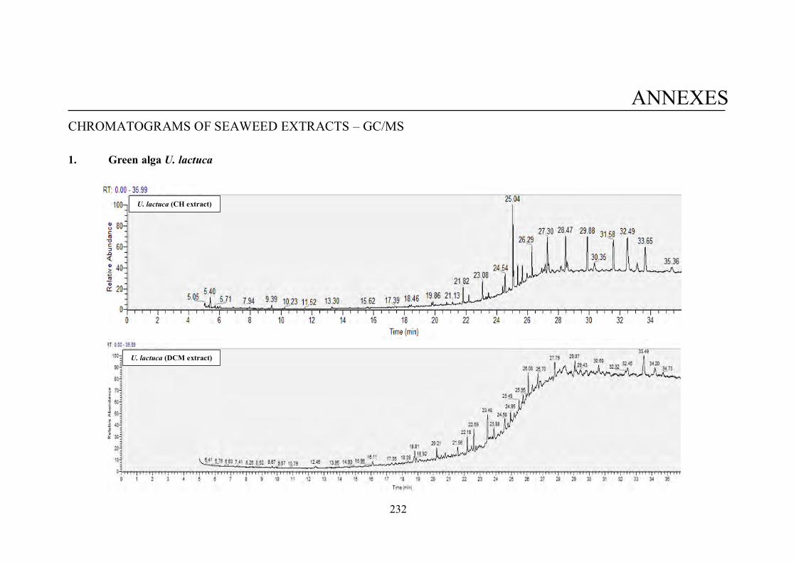
232
ANNEXES CHROMATOGRAMS OF SEAWEED EXTRACTS – GC/MS
1. Green alga U. lactuca
U. lactuca (CH extract)
U. lactuca (DCM extract)
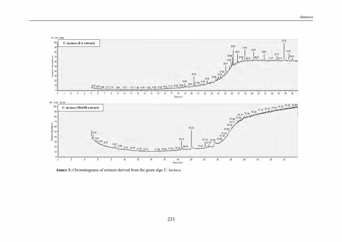
Annexes
233
Annex 1: Chromatograms of extracts derived from the green alga U. lactuca.
U. lactuca (EA extract)
U. lactuca (MeOH extract)
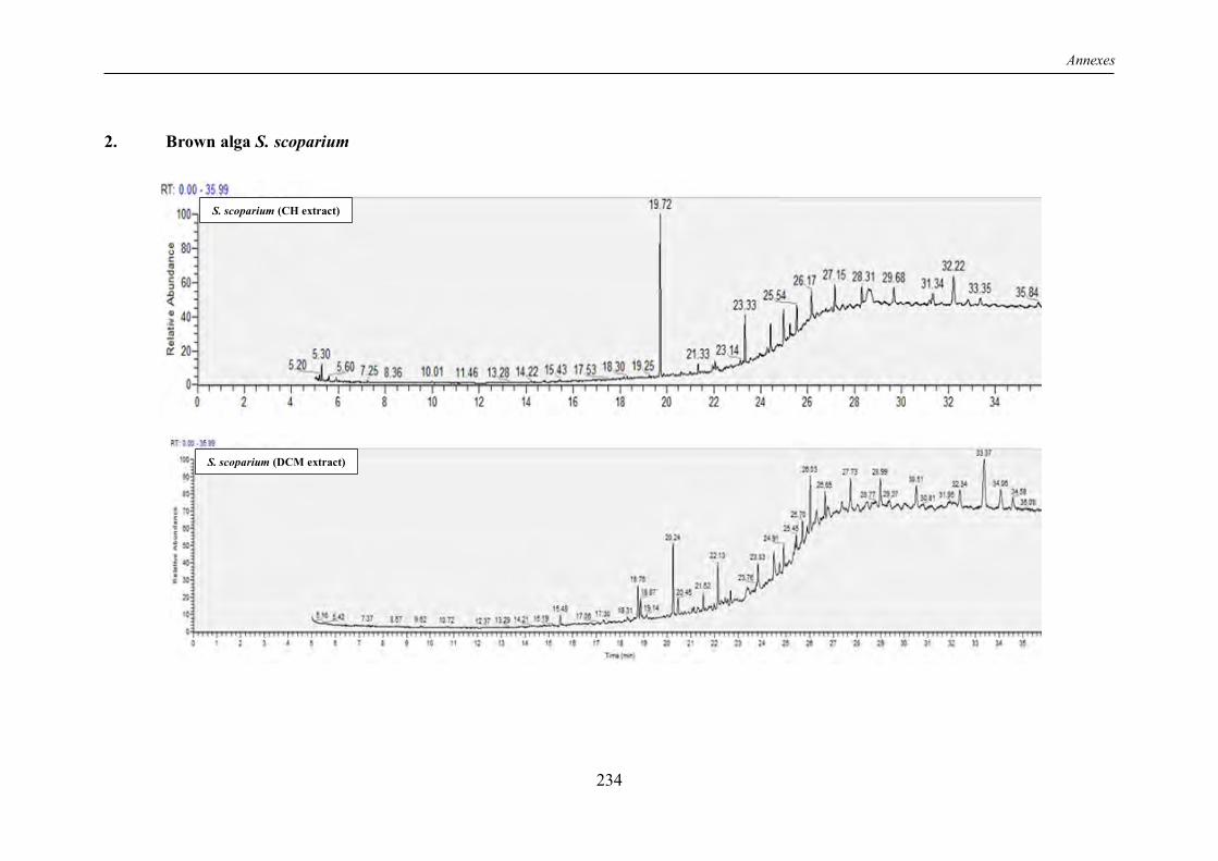
Annexes
234
2. Brown alga S. scoparium
S. scoparium (CH extract)
S. scoparium (DCM extract)
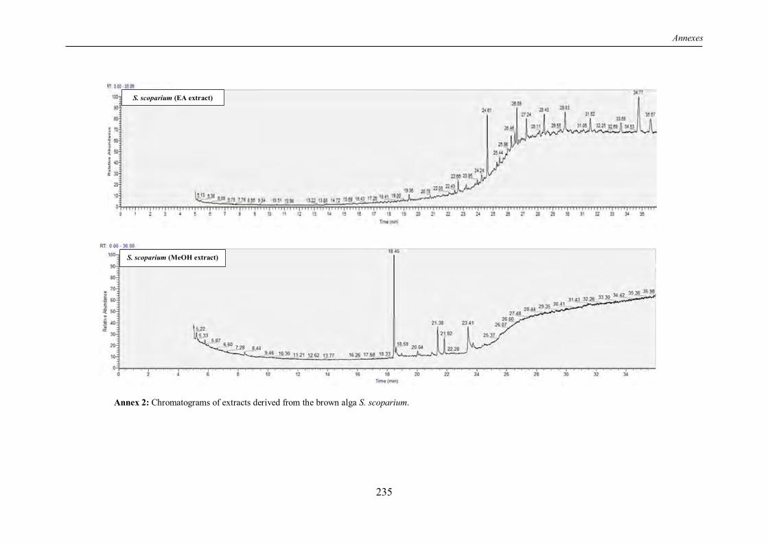
Annexes
235
Annex 2: Chromatograms of extracts derived from the brown alga S. scoparium.
S. scoparium (EA extract)
S. scoparium (MeOH extract)
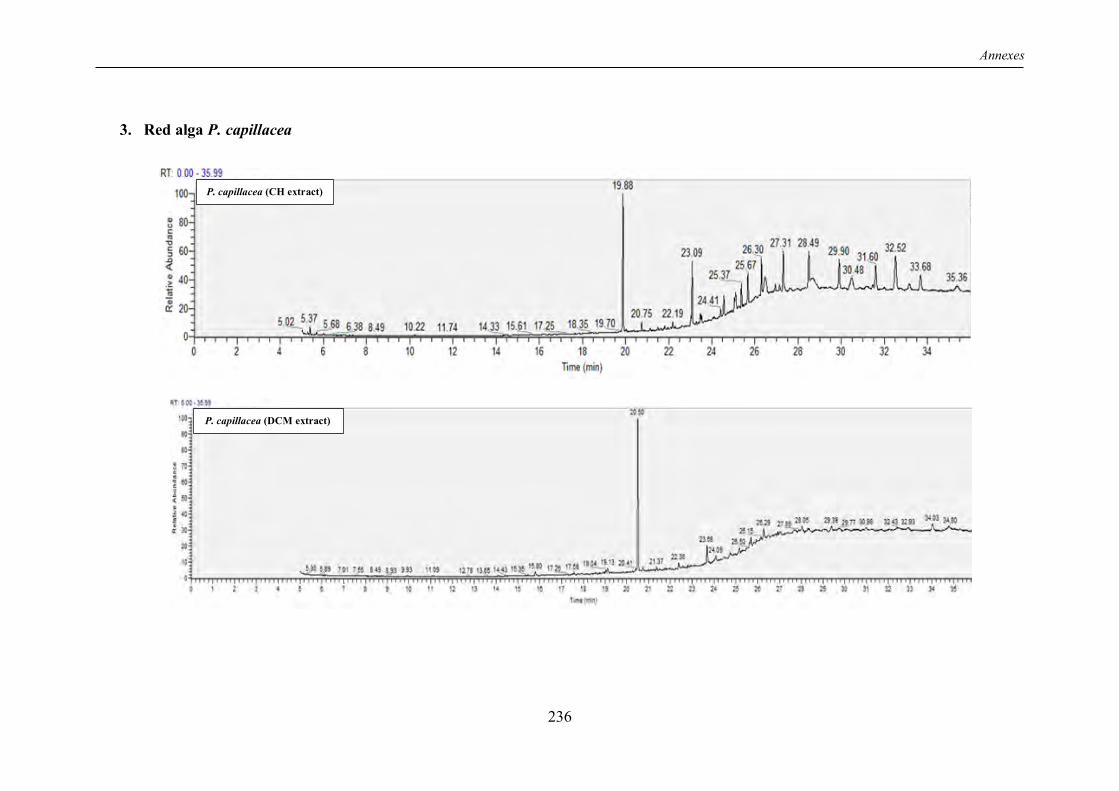
Annexes
236
3. Red alga P. capillacea
P. capillacea (CH extract)
P. capillacea (DCM extract)
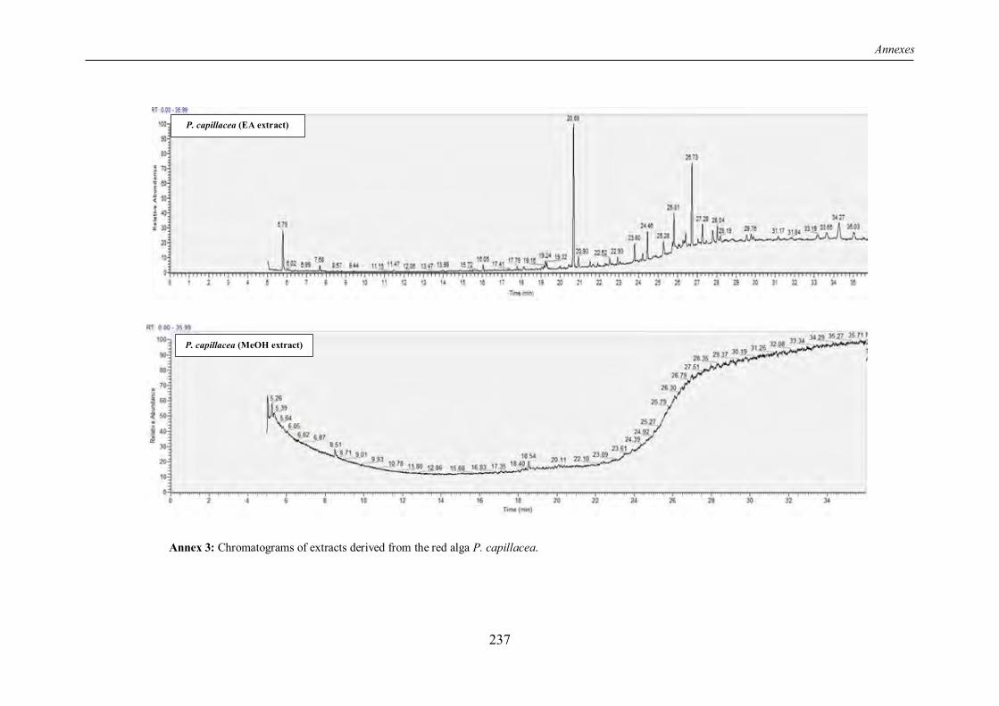
Annexes
237
Annex 3: Chromatograms of extracts derived from the red alga P. capillacea.
P. capillacea (EA extract)
P. capillacea (MeOH extract)

238


