Embed Size (px)
Citation preview

ARTICLE
Received 19 Nov 2013 | Accepted 6 May 2014 | Published 19 Jun 2014
Vanillin formation from ferulic acid in Vanillaplanifolia is catalysed by a single enzymeNethaji J. Gallage1,2,3, Esben H. Hansen4, Rubini Kannangara1,2,3, Carl Erik Olsen1,2,
Mohammed Saddik Motawia1,2,3, Kirsten Jørgensen1,2,3, Inger Holme5, Kim Hebelstrup5,
Michel Grisoni6 & Birger Lindberg Møller1,2,3,7
Vanillin is a popular and valuable flavour compound. It is the key constituent of the natural
vanilla flavour obtained from cured vanilla pods. Here we show that a single hydratase/lyase
type enzyme designated vanillin synthase (VpVAN) catalyses direct conversion of ferulic acid
and its glucoside into vanillin and its glucoside, respectively. The enzyme shows high
sequence similarity to cysteine proteinases and is specific to the substitution pattern at the
aromatic ring and does not metabolize caffeic acid and p-coumaric acid as demonstrated by
coupled transcription/translation assays. VpVAN localizes to the inner part of the vanilla pod
and high transcript levels are found in single cells located a few cell layers from the inner
epidermis. Transient expression of VpVAN in tobacco and stable expression in barley in
combination with the action of endogenous alcohol dehydrogenases and UDP-glucosyl-
transferases result in vanillyl alcohol glucoside formation from endogenous ferulic acid.
A gene encoding an enzyme showing 71% sequence identity to VpVAN was identified
in another vanillin-producing plant species Glechoma hederacea and was also shown to be a
vanillin synthase as demonstrated by transient expression in tobacco.
DOI: 10.1038/ncomms5037 OPEN
1 Plant Biochemistry Laboratory, Department of Plant and Environmental Sciences, Faculty of Science, University of Copenhagen, Thorvaldsensvej 40,Frederiksberg C, DK-1871 Copenhagen, Denmark. 2 VILLUM Research Center ‘Plant Plasticity’, Thorvaldsensvej 40, Frederiksberg C, DK-1871 Copenhagen,Denmark. 3 Center for Synthetic Biology: ‘bioSYNergy’, Thorvaldsensvej 40, Frederiksberg C, DK-1871 Copenhagen, Denmark. 4 Evolva A/S, Lersø Parkalle 42–44, 5th floor, DK-2100 Copenhagen, Denmark. 5 AU Flakkebjerg, Danish Centre for Food and Agriculture, University of Aarhus, Forsøgsvej, DK-4200 Slagelse,Denmark. 6 Centre de Cooperation Internationale en Recherche Agronomique pour le Developement, UMR PVBMT, 97410 Saint Pierre, La Reunion, France.7 Carlsberg Laboratory, Gamle Carlsberg Vej 10, Valby DK-2500, Copenhagen, Denmark. Correspondence and requests for materials should be addressed toB.L.M. (email: [email protected]).
NATURE COMMUNICATIONS | 5:4037 | DOI: 10.1038/ncomms5037 | www.nature.com/naturecommunications 1
& 2014 Macmillan Publishers Limited. All rights reserved.

Vanilla is the world’s most popular flavour principle andused in numerous products. The pods of the climbingorchids, Vanilla planifolia and V. tahitensis are the source
of natural vanilla1, although trace amounts of vanillin can befound in a variety of different plant species scattered in the plantkingdom2. Vanillin (3-methoxy-4-hydroxybenzaldehyde) is themain flavour component of vanilla extract from cured vanillapods1,3. In high concentrations vanillin is toxic to living cells. Inthe pod it is produced and stored as non-toxic vanillin glucoside,which upon tissue damage is hydrolysed to form the activedefense compound, vanillin. Production of vanillin from theorchids is laborious, slow and costly. Five hundred kilograms ofvanilla pods yields only 1 kg of vanillin. Less than 1% of the globalvanillin production originates from the vanilla orchids. Instead,the vast majority is produced chemically from fossil fuels or byacid hydrolysis of lignin4. A biotechnological solution to vanillinproduction via heterologous expression of the native vanillaorchid pathway genes in microorganisms has not been possiblebecause the pathway has remained unknown. Vanillin has beenproduced by microbial bioconversion of substrates structurallyrelated to vanillin5 as well as from glucose6.
Previous studies have shown the conversion of a variety ofcompounds into vanillin glucoside after administration to V.planifolia pods. These studies suggest that vanillin glucoside isderived from phenylalanine, the shikimate pathway intermediatesor monomeric lignin precursors7–12. Vanillin glucoside andp-hydroxybenzaldehyde glucoside, the two most abundantaroma compounds in mature vanilla pods, are structurallysimilar, and a biosynthetic relationship between the formationof these two compounds has been proposed12,13. The necessarychain shortening process of a putative phenylpropanoidprecursor was suggested to proceed by b-oxidation or by acoenzyme A (CoA)-dependent non-b-oxidative pathway11,14–17.p-Hydroxybenzaldehyde was reported to be formed bychain shortening of p-coumaric acid catalysed by p-hydroxy-benzaldehyde synthase (4-HBS)18,19, which was proposed as aprecursor for vanillin glucoside biosynthesis. p-Hydroxy-benzaldehyde would then need to be hydroxylated at its C3carbon by a monooxygenase (C3H), O-methylated at the3-OH position by a O-methyltransferase (OMT), and finallyglucosylated at the 4-OH position by a UDP-glucosyltransferase(UGT).
Other studies have suggested that vanillin is formed fromL-phenylalanine via the monomeric lignin precursors: cinnamicacid, p-coumaric acid, caffeic acid and ferulic acid, involvingphenylalanine ammonia lyase (PAL), hydroxylations, anO-methylation and finally a chain-shortening reaction. Formationof vanillin glucoside would also require the involvement of aUGT, although the point at which the glycosylation would takeplace remains elusive. Enzymes are known that can catalyse mostof these reactions, for example, PAL20, cinnamic acid4-hydroxylase, C4H21, p-coumaric acid 3-hydroxylase, C3H22,but is not clear whether CoA-derivatives are involved or whetherthe C3-hydroxylation step proceeds, for example, throughquinate- and shikimate esters22. Caffeic acid could in principlebe O-methylated23 to form ferulic acid, the substrate for thehypothesized final chain-shortening reaction, and several OMTsare known from V. planifolia. Vanillin UGTs or genes thatencode these enzymes from V. planifolia have not yet beenreported and as stated above it remains to be demonstrated atwhich stage in the pathway glycosylation occurs.
The aim of the current study was to elucidate the vanillinbiosynthesis pathway in V. planifolia. We have carried outbiosynthetic studies with fresh vanilla pods using a number ofdifferent putative radiolabelled precursors. Supported by acombination of transcriptomic and proteomic approaches, we
identified a gene, VpVAN encoding a two-carbon chain-short-ening enzyme converting ferulic acid and its glucoside directlyinto vanillin and its glucoside. VpVAN is produced in cells in theinner part of the vanilla pod. The substrate specificity of VpVANwas determined using a rabbit reticulocyte-coupled in vitrotranscription/translation system and by heterologous expressionof the gene in Nicotiana benthamiana, Hordeum vulgare andSaccharomyces cerevisiae. A gene sequence identical to that ofVpVAN was previously reported to encode an enzyme designated4-HBS catalysing a two-carbon chain-shortening of p-coumaricacid into 4-hydroxybenzaldehyde. We could not verify such afunction in our studies.
ResultsAdministration of putative [14C]- precursors to vanilla pods.To examine whether the p-hydroxybenzaldehyde-based or thelonger lignin precursor-based pathway is the most likely nativevanillin glucoside biosynthetic pathway, [14C]-radiolabelledputative precursors ([14C]-phenylalanine, [14C]-cinnamic acid,[14C]-p-hydroxybenzaldehyde and [14C]-vanillin) were adminis-tered to sliced discs of fresh vanilla pods harvested 6 monthsafter pollination. Vanillin glucoside is located in the inner part ofthe pod, that is, in the papillae and placental tissues, butcompletely absent from the epicarp, outer mesocarp area andseeds24. Accordingly, the experiments with administration ofradiolabelled precursors were carried out separately with innerand outer parts of the pod discs using the outer parts of the podas negative controls. Incubation with [14C]-phenylalanine and[14C]-cinnamic acid resulted in [14C]-vanillin glucosideformation in the tissue representing the inner part of the podwhile administration of [14C]-p-hydroxybenzaldehyde induced[14C]-p-hydroxybenzaldehyde glucoside formation in both theinner and the outer part of the pod (Fig. 1; Supplementary Fig. 1).Incubation with [14C]-p-hydroxybenzaldehyde did not resultin [14C]-vanillin glucoside formation. The radiolabellingstudies confirmed that vanillin glucoside biosynthesis occursonly in the inner part of the pod and demonstrated thatp-hydroxybenzaldehyde is not an intermediate in vanillinbiosynthesis. The incorporation percentages observed varieddepending upon the pod developmental stage, whereas thepattern of radiolabelled compounds observed followingadministration of each of the different labelled precursors atdifferent developmental stages remained similar.
Administration of [14C]-vanillin resulted in formation of[14C]-vanillin glucoside both in the inner and outer part of thepod. Similarly [14C] p-hydroxybenzaldehyde administrationresulted in [14C]-p-hydroxybenzaldehyde glucoside both in theinner and outer part of the pod, demonstrating the presence of aglycosyltransferase capable of glycosylating these precursors.
Identification of candidate genes. To identify genes and enzymesinvolved in vanillin glucoside biosynthesis in V. planifolia, acombination of transcriptomic and proteomic approaches wasundertaken with an initial focus on candidates representing thefive major enzyme families suggested from the literature to play apossible role in vanillin biosynthesis, namely PAL, cytochromeP450s (the monophenol monooxygenases C4H and C3H), OMTs,UGTs and the carbon chain-shortening enzyme, 4-HBS. TheV. planifolia transcriptome was obtained from a 6-month-oldvanilla pod from the island of La Reunion by 454 pyrosequencing.Approximately 40 UGTs, 15 OMTs, a CYP98A3 (C3H) and4-HBS conreads were found in the transcriptome. To furtherassess the likelihood of involvement of each of these genes invanillin biosynthesis, a targeted proteomic approach (proteomicmass finger printing) was carried out in parallel with the broad
ARTICLE NATURE COMMUNICATIONS | DOI: 10.1038/ncomms5037
2 NATURE COMMUNICATIONS | 5:4037 | DOI: 10.1038/ncomms5037 | www.nature.com/naturecommunications
& 2014 Macmillan Publishers Limited. All rights reserved.

transcriptome analysis using the biosynthetically active inner partof the pod as experimental tissue. On the basis of overlayof the pyrosequencing and proteomic data sets, we selectedand cloned 1UGT (VpUGT72U1), 11OMTs, a CYP98A3orthologue (VpCYP98A70) and 4-HBS (Supplementary Table 1;Supplementary Data 1 and 2).
Although in the literature, the vanillin biosynthetic pathwayhas been suggested to be embedded within a metabolic grid, ourinitial in vitro studies with these gene candidates identified a geneencoding an enzyme converting ferulic acid glucoside and ferulicacid directly into vanillin glucoside and vanillin, respectively. Thisrepresents the first committed step in vanillin synthesis anddemonstrates that vanillin formation in V. planifolia is catalysedby a single enzyme using a general substrate from phenylpropa-noid metabolism. We designated the enzyme vanillin synthaseand the gene VpVAN (gene sequence is given in SupplementaryFig. 2a). In a published patent application, the identical genesequence had previously been assigned as encoding an enzymeconverting p-coumaric acid into p-hydroxybenzaldehyde18.Accordingly the gene sequence was initially designated asencoding a p-hydroxybenzaldehyde synthase (4-HBS), anactivity that we did not observe in our studies as reported below.
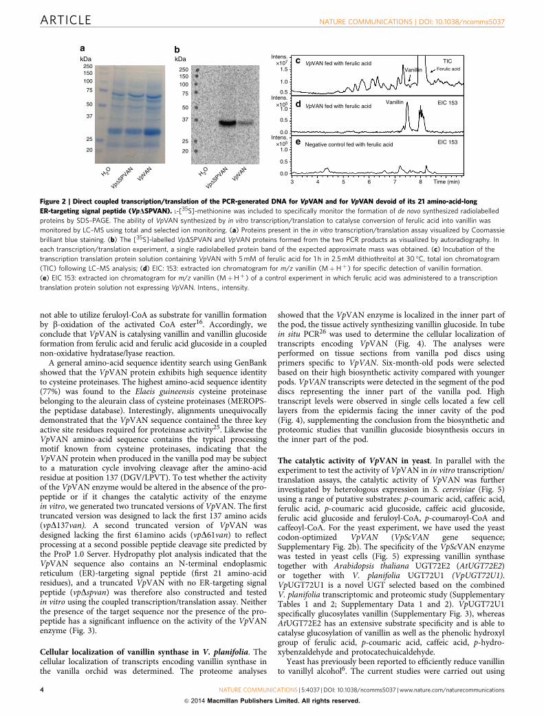
Vanillin synthase catalyses vanillin formation in vitro.The catalytic activity of vanillin synthase using a range ofdifferent putative substrates was monitored by in vitro coupledtranscription/translation assays. Vanillin synthase proteinwas obtained from its PCR-generated DNA in a coupled
transcription/translation assay with the inclusion of L-[35S]-methionine to provide easy monitoring of protein formation bySDS–polyacrylamide gel electrophoresis (PAGE) analysis (Fig. 2).The coupled assay produced a single radiolabelled protein bandmigrating with an apparent molecular mass of 36 kD in closeagreement with the predicted mass of 39.15 kD for VpVAN(Fig. 2), thus enabling us to monitor the activity of the enzyme inan in vitro condition equalling a purified enzyme.
The substrate specificity of the VpVAN enzyme formed wasinvestigated by incubation (1 h and 24 h) with 0.5–5 mM ofp-coumaric acid, caffeic acid, ferulic acid, p-coumaric acidglucoside, caffeic acid glucoside, ferulic acid glucoside,feruloyl-CoA, p-coumaroyl-CoA and caffeoyl-CoA. Liquidchromatography–mass spectrometry (LC–MS) analyses demon-strated that VpVAN catalysed a direct chain shortening of ferulicacid and ferulic acid glucoside to vanillin and vanillin glucoside,respectively (Figs 2 and 3), whereas no activity was found usingp-coumaric acid, caffeic acid and the glucosides of these assubstrates. The conversion of ferulic acid and ferulic acid glucosideinto vanillin and vanillin glucoside proceeded in the absence ofadded adenosine tri-phosphate (ATP) and nicotinamide adeninedinucleotide (NADþ ). These cofactors would have been requiredif the chain shortening had occurred via b-oxidation of anactivated CoA ester16. In a number of experiments, vanillinformation was observed using feruloyl-CoA as a substrate. In suchexperiments, the incubation mixture was found always to containferulic acid present as an impurity in the commercially providedferuloyl-CoA. Since ATP and NADþ were not required ascofactors in these reactions, we conclude that vanillin synthase is
14C Vanillin glucoside
14C Phenylalanine
14C Cinnamic acid
14C p-Hydroxybenzaldehyde
14C Ferulic acid glucoside
14C Vanillin
Origin ofradiolabelledprecursors
* * * *
14C
Van
illin
14C
p-H
ydro
xybe
nzal
dehy
de
14C
Fer
ulic
aci
d gl
ucos
ide
14C
Cin
nam
ic a
cid
14C
Phe
nyla
lani
ne
14C Phenylalanine
Inne
r
Inne
r
Inne
r
Inne
r
Out
er
Out
er
Out
er
Out
er
14C Cinnamicacid
14C p-Hydroxybenzaldehyde
14C Vanillin
Figure 1 | Thin layer chromatography (TLC) analysis of the radiolabelled products formed following administration of different putative 14C-labelled
precursors to tissue slices of fresh vanilla pods. The 14C-labelled products formed following 48 h incubation were extracted into 70% MeOH,
separated by TLC and monitored by autoradiography. The position of the radiolabelled precursors following chromatographic separation is shown on the
right part of the thin layer. (*) indicates formation of 14C-labelled vanillin glucoside from the administered precursor. The presence of ultraviolet-absorbing
components in the vanilla extracts as well as the position of the putative substrates after chromatographic separation was also monitored following
exposure to ultraviolet light (254 nm) (Supplementary Fig. 1).
NATURE COMMUNICATIONS | DOI: 10.1038/ncomms5037 ARTICLE
NATURE COMMUNICATIONS | 5:4037 | DOI: 10.1038/ncomms5037 | www.nature.com/naturecommunications 3
& 2014 Macmillan Publishers Limited. All rights reserved.
not able to utilize feruloyl-CoA as substrate for vanillin formationby b-oxidation of the activated CoA ester16. Accordingly, weconclude that VpVAN is catalysing vanillin and vanillin glucosideformation from ferulic acid and ferulic acid glucoside in a couplednon-oxidative hydratase/lyase reaction.
A general amino-acid sequence identity search using GenBankshowed that the VpVAN protein exhibits high sequence identityto cysteine proteinases. The highest amino-acid sequence identity(77%) was found to the Elaeis guineensis cysteine proteinasebelonging to the aleurain class of cysteine proteinases (MEROPS-the peptidase database). Interestingly, alignments unequivocallydemonstrated that the VpVAN sequence contained the three keyactive site residues required for proteinase activity25. Likewise theVpVAN amino-acid sequence contains the typical processingmotif known from cysteine proteinases, indicating that theVpVAN protein when produced in the vanilla pod may be subjectto a maturation cycle involving cleavage after the amino-acidresidue at position 137 (DGV/LPVT). To test whether the activityof the VpVAN enzyme would be altered in the absence of the pro-peptide or if it changes the catalytic activity of the enzymein vitro, we generated two truncated versions of VpVAN. The firsttruncated version was designed to lack the first 137 amino acids(vpD137van). A second truncated version of VpVAN wasdesigned lacking the first 61amino acids (vpD61van) to reflectprocessing at a second possible peptide cleavage site predicted bythe ProP 1.0 Server. Hydropathy plot analysis indicated that theVpVAN sequence also contains an N-terminal endoplasmicreticulum (ER)-targeting signal peptide (first 21 amino-acidresidues), and a truncated VpVAN with no ER-targeting signalpeptide (vpDspvan) was therefore also constructed and testedin vitro using the coupled transcription/translation assay. Neitherthe presence of the target sequence nor the presence of the pro-peptide has a significant influence on the activity of the VpVANenzyme (Fig. 3).
Cellular localization of vanillin synthase in V. planifolia. Thecellular localization of transcripts encoding vanillin synthase inthe vanilla orchid was determined. The proteome analyses
showed that the VpVAN enzyme is localized in the inner part ofthe pod, the tissue actively synthesizing vanillin glucoside. In tubein situ PCR26 was used to determine the cellular localization oftranscripts encoding VpVAN (Fig. 4). The analyses wereperformed on tissue sections from vanilla pod discs usingprimers specific to VpVAN. Six-month-old pods were selectedbased on their high biosynthetic activity compared with youngerpods. VpVAN transcripts were detected in the segment of the poddiscs representing the inner part of the vanilla pod. Hightranscript levels were observed in single cells located a few celllayers from the epidermis facing the inner cavity of the pod(Fig. 4), supplementing the conclusion from the biosynthetic andproteomic studies that vanillin glucoside biosynthesis occurs inthe inner part of the pod.
The catalytic activity of VpVAN in yeast. In parallel with theexperiment to test the activity of VpVAN in in vitro transcription/translation assays, the catalytic activity of VpVAN was furtherinvestigated by heterologous expression in S. cerevisiae (Fig. 5)using a range of putative substrates: p-coumaric acid, caffeic acid,ferulic acid, p-coumaric acid glucoside, caffeic acid glucoside,ferulic acid glucoside and feruloyl-CoA, p-coumaroyl-CoA andcaffeoyl-CoA. For the yeast experiment, we have used the yeastcodon-optimized VpVAN (VpScVAN gene sequence;Supplementary Fig. 2b). The specificity of the VpScVAN enzymewas tested in yeast cells (Fig. 5) expressing vanillin synthasetogether with Arabidopsis thaliana UGT72E2 (AtUGT72E2)or together with V. planifolia UGT72U1 (VpUGT72U1).VpUGT72U1 is a novel UGT selected based on the combinedV. planifolia transcriptomic and proteomic study (SupplementaryTables 1 and 2; Supplementary Data 1 and 2). VpUGT72U1specifically glucosylates vanillin (Supplementary Fig. 3), whereasAtUGT72E2 has an extensive substrate specificity and is able tocatalyse glucosylation of vanillin as well as the phenolic hydroxylgroup of ferulic acid, p-coumaric acid, caffeic acid, p-hydro-xybenzaldehyde and protocatechuicaldehyde.
Yeast has previously been reported to efficiently reduce vanillinto vanillyl alcohol6. The current studies were carried out using
250kDa kDa
150
50
100
75
37
25
20
H 2O
VpΔSPVAN
VpVAN
H 2O
VpΔSPVAN
VpVAN
250150
50
100
75
37
25
20
Intens.×107
1.5
1.0
0.5Intens.
×105
1.0
0.5
0.0Intens.
×105
1.0
0.5
0.0
VpVAN fed with ferulic acidVanillin
Vanillin
TICFerulic acid
EIC 153
EIC 153
VpVAN fed with ferulic acid
Negative control fed with ferulic acid
3 4 5 6 7 8 Time (min)
Figure 2 | Direct coupled transcription/translation of the PCR-generated DNA for VpVAN and for VpVAN devoid of its 21 amino-acid-long
ER-targeting signal peptide (VpDSPVAN). L-[35S]-methionine was included to specifically monitor the formation of de novo synthesized radiolabelled
proteins by SDS–PAGE. The ability of VpVAN synthesized by in vitro transcription/translation to catalyse conversion of ferulic acid into vanillin was
monitored by LC–MS using total and selected ion monitoring. (a) Proteins present in the in vitro transcription/translation assay visualized by Coomassie
brilliant blue staining. (b) The [35S]-labelled VpDSPVAN and VpVAN proteins formed from the two PCR products as visualized by autoradiography. In
each transcription/translation experiment, a single radiolabelled protein band of the expected approximate mass was obtained. (c) Incubation of the
transcription translation protein solution containing VpVAN with 5 mM of ferulic acid for 1 h in 2.5 mM dithiothreitol at 30 �C, total ion chromatogram
(TIC) following LC–MS analysis; (d) EIC: 153: extracted ion chromatogram for m/z vanillin (MþHþ ) for specific detection of vanillin formation.
(e) EIC 153: extracted ion chromatogram for m/z vanillin (MþHþ ) of a control experiment in which ferulic acid was administered to a transcription
translation protein solution not expressing VpVAN. Intens., intensity.
ARTICLE NATURE COMMUNICATIONS | DOI: 10.1038/ncomms5037
4 NATURE COMMUNICATIONS | 5:4037 | DOI: 10.1038/ncomms5037 | www.nature.com/naturecommunications
& 2014 Macmillan Publishers Limited. All rights reserved.

S. cerevisiae strain Y06460 in which alcohol dehydrogenase 6(ADH6) is disrupted, because use of this strain circumventsreduction of the vanillin formed into vanillyl alcohol6. Genes werealso integrated to simultaneously disrupt EXG1 encoding an
endogenous yeast exoglucosidase 1 (EXG1), which efficientlyhydrolyses vanillin glucoside6. VpVAN was then stably integratedinto the yeast chromosome either alone or together withAtUGT72E2 or together with VpUGT72U1.
When VpScVAN was expressed alone in the yeast strainmodified as described above in the presence of ferulic acid, novanillin glucoside peak was observed in spite of the fact that theyeast endogenous exoglucosidase 1 (EXG1) had been knockedout. This demonstrated that the yeast does not contain a UGTable to glucosylate vanillin. Combined expression of VpScVANand VpUGT72U1 resulted in formation of vanillin glucoside whenyeast was supplied with ferulic acid or ferulic acid glucoside. Inaddition, combined expression of VpScVAN and AtUGT72E2resulted in formation of vanillin glucoside when administratedwith ferulic acid or ferulic acid glucoside. These studiesdemonstrate that the vanillin synthase is able to catalyse carbondouble-bond cleavage of both ferulic acid and ferulic acidglucoside (Fig. 5).
As previously mentioned, VpVAN was predicted to encode aprotein with an ER-targeting signal peptide. Accordingly, anadditional series of biosynthetic studies were carried out withyeast harbouring stably integrated AtUGT72E2 together witheither VpVAN or VpScVAN or truncated VpVAN with no ER-targeting signal peptide (vpDspvan) or truncated VpVAN with nosignal peptide and yeast codon optimized (vpscDspvan). The yeaststrains were incubated with putative substrates for 72 h andmetabolite profiles determined by LC–MS. Formation of vanillinglucoside was observed with ferulic acid as substrate withVpDSpVAN and VpScDSpVAN (Supplementary Fig. 4). ThusVpVAN is catalytically active towards ferulic acid in the presenceas well as absence of the ER-targeting signal peptide. Carbonchain shortening of caffeic acid and p-coumaric acid or glucosidesof these was not observed with any of the modified versions ofVpVAN.
Concomitant with the conversion of ferulic acid and itsglucoside into vanillin and vanillin glucoside, yeasts are able tometabolize ferulic acid into 4-vinylguaiacol. The latter conver-sions are highly significant (Fig. 5). Two enzymes in S. cerevisiaeare known to be responsible for the conversion of ferulic acid to4-vinylguaiacol. These are phenylacrylate decarboxylase (PAD1)and ferulate decarboxylase (FDC1)27–29. Increased levels ofVpVAN-based vanillin production in yeast would thus beenvisioned following disruption or downregulation of the twogenes encoding PAD1 and FDC1.
Molasses are obtained as by-products in the production ofsugar from sugar beets, sugar cane or sorghum, and thesemolasses contain ferulic acid30,31. To examine whether such
0.5×10⁷
Intens.
3 4 5 6 7 8 9
VpVAN fed withferulic acid glucoside
Vp ΔSP VAN fed withferulic acid glucoside
Vp Δ137 VAN fed withferulic acid glucoside
Vp Δ61 VAN fed withferulic acid glucoside
Negative control fed withferulic acid glucoside
Negative control fed withferulic acid glucoside
Vanillinglucoside
Ferulic acidglucoside
Vanillinglucoside
Vanillinglucoside
Vanillinglucoside
EIC 337
EIC 337
EIC 337
EIC 337
EIC 337
EIC 379
0.0
0.5×10⁷
0.0
0.5
×107
0.0
0.5
×107
0.0
0.5×10⁷
0.0
5×10⁸
0
Time (min)
a
b
c
d
e
f
Figure 3 | The ability of enzymes synthesized by in vitro transcription/
translation to catalyse conversion of ferulic acid glucoside into vanillin
glucoside. The experiment shown in (a–d) involved incubation of the
transcription/translation protein solutions with VpVAN, VpDSPVAN,
VpD137VAN and VpD61VAN with 1 mM of ferulic acid glucoside for 1 h in
1 mM dithiothreitol at 30 �C. (a) EIC 337: extracted ion chromatogram m/z
vanillin glucoside (MþNaþ ) obtained following incubation of VpVAN with
ferulic acid glucoside (b) EIC 337: extracted ion chromatogram m/z vanillin
glucoside (MþNaþ ) obtained following incubation of VpDSPVAN with
ferulic acid glucoside (c) EIC 337: extracted ion chromatogram m/z vanillin
glucoside (MþNaþ ) obtained following incubation of VpD137VAN with
ferulic acid glucoside. (d) EIC 337: extracted ion chromatogram m/z vanillin
glucoside (Mþ Naþ ) obtained following incubation of VpD61VAN with
ferulic acid glucoside. (e) EIC 337: extracted ion chromatogram m/z vanillin
glucoside (MþNaþ ) obtained following incubation of a control
transcription translation/ protein solution devoid of any protein of interest
with ferulic acid glucoside. (f) EIC 379: extracted ion chromatogram m/z
ferulic acid glucoside (MþNaþ ) obtained following incubation of a control
transcription translation/ protein solution devoid of any protein of interest
with ferulic acid glucoside. Intens., intensity.
c
a b c
cl
cl
e
c
scl
cl
cl
Figure 4 | Tissue localization of the expression of VpVAN in transverse sections of a 6-month-old vanilla pod determined by in tube in situ PCR.
Transcripts of VpVAN were detected in specific cells (Panels a and b) using FITC-conjugated antibodies recognizing digoxigenin (DIG) incorporated in the
specific PCR products representing the VpVAN transcript. Higher magnification of the selected area in Panel a is shown in Panel b. No transcripts of VpVAN
were detected in the controls in the absence of specific primers for VpVAN (Panel c). The fluorescence detected in Panel c represents unspecific binding of
the FITC-conjugated antibodies recognizing DIG to cell walls and supporting fibre cells surrounding the vascular systems. In all panels the chloroplasts are
visible owing to their auto fluorescence at the used filter settings. The sections were analysed with a Leica FI/RH filter with excitation filters: BP490/15;
560/25 and emission filters: BP525/20; 605/30. e, epidermis; c, cortex; cl, chloroplast; s, supporting fibre tissue; Scale bar, 100mm.
NATURE COMMUNICATIONS | DOI: 10.1038/ncomms5037 ARTICLE
NATURE COMMUNICATIONS | 5:4037 | DOI: 10.1038/ncomms5037 | www.nature.com/naturecommunications 5
& 2014 Macmillan Publishers Limited. All rights reserved.

material could be used for vanillin glucoside production, yeastsexpressing VpVAN and VpScVAN as well as AtUGT72E2 weregrown in molasses-based growth medium using sugar beet as thesource for the molasses. Vanillin glucoside formation wasobserved with both versions of VpVAN, highlighting thepotential of this enzyme for vanillin glucoside production basedon inexpensive waste materials (Fig. 6).
Establishing vanillin synthesis in tobacco and barley. The cat-alytic activity of vanillin synthase in the presence and absence of aputative pro-peptide in vivo was analysed following transientexpression in tobacco and stable expression in barley. The in vivobiological activity of VpVAN (including the ER-targeting signalpeptide) was assessed by transient expression in leaves ofN. benthamiana in the absence of any exogenously added sub-strates. Gene constructs were transferred to Agrobacteriumtumefaciens and co-infiltrated with an A. tumefaciens strainharbouring the p19 gene-silencing suppressor. LC–MS profilingshowed VpVAN-dependent formation of vanillyl alcohol gluco-side (Fig. 7). The vanillyl alcohol glucoside arises by reductionof vanillin by an alcohol dehydrogenase (E.C.1.1.1.1) andsubsequent glucosylation of the primary alcohol group ofvanillyl alcohol, as was previously observed in cell cultures of
N. plumbaginifolia32 and yeast6. Biotechnological production ofvanillin glucoside in plants other than Vanilla sp. by introductionof VpVAN thus requires co-expression of a UGT that effectivelyglucosylates the free vanillin formed into the correspondingglucoside before its reduction into vanillyl alcohol.
Transient expression of vpD137van and vpD61van was alsoincluded in this study to investigate the importance of secondaryprocessing of VpVAN for its in vivo activity. Introduction of eachof these constructs encoding different truncated forms of VpVANwas found to result in vanillyl alcohol glucoside production insimilar amounts as observed with VpVAN (Fig. 8a).
As previously mentioned, the VpVAN sequence showed highsequence identity to proteins belonging to the family cysteineproteinases. We identified a protein belonging to the family ofcysteine proteinases in tobacco in which the amino-acid sequenceidentity to VpVAN was 71% (N. benthamiana cysteine proteinasegene sequence; supplementary Fig. 2c). In order to investigate thepossibility to produce a nascent protein more amenable to propertargeting and processing by the endogenous tobacco machinery, agene construct was made where the VpVAN ER-targetingsignal peptide and pro-peptide protease cleavage site werereplaced with the putative signal peptide and the putative pro-peptide protease cleavage site from the tobacco cysteine protease(vpnbDspD137van gene sequence; Supplementary Fig. 2d).
0
1
2
0
1
2
0
1
2
0
1
2
0
1
3 4 5 6 7 8 9 Time (min) 3 4 5 6 7 8 9 Time (min)
aIntens.
×106
Intens.×106
Intens.×106
Intens.×106
Intens.×108
Intens.×106
Intens.×106
Intens.×106
Intens.×106
Intens.×108
b
c
d
e
VpScVANfed with ferulic acid
VpScVAN::VpUGT72U1fed with ferulic acid
VpScVAN::AtUGT72 E2fed with ferulic acid
Negative controlfed with ferulic acid
Negative controlfed with ferulic acid
EIC 337
EIC 337
EIC 337
EIC 337
EIC 177,195,217,239
Vanillinglucoside
Vanillinglucoside
4-Vinylguaiacolglucoside
4-Vinylguaiacolglucoside
Ferulic acid
0
1
2
0
1
2
0
1
0
1
2
0
1
EIC 337
EIC 337
EIC 337
EIC 337
EIC 379
Negative controlfed with ferulic acid glucoside
VpScVAN::AtUGT72E2fed with ferulic acid glucoside
VpScVAN::VpUGT72U1fed with ferulic acid glucoside
VpScVANfed with ferulic acid glucoside
Negative controlfed with ferulic acid glucoside
Vanillinglucoside
Vanillinglucoside
Vanillinglucoside
4-Vinylguaiacolglucoside
4-Vinylguaiacolglucoside
Ferulic acidglucoside
f
g
h
i
j
Figure 5 | The ability of VpScVAN expressed in an adapted yeast strain to metabolize ferulic acid and ferulic acid glucoside into vanillin and
vanillin glucoside, respectively. (a) Expression of VpScVAN does not result in formation of vanillin glucoside. Extracted ion chromatogram (EIC) 337: m/z
vanillin glucoside (MþNaþ ). (b) Co-expression of VpScVAN and VpUGT72U1 results in formation of low amounts of vanillin glucoside. EIC 337:
m/z vanillin glucoside (MþNaþ ). (c) Co-expression of VpScVAN and AtUGT72E2 results in formation of larger amounts of vanillin glucoside. EIC 337:
m/z vanillin glucoside (MþNaþ ). (d) Negative control demonstrating that administration of ferulic acid to the adapted yeast strain does not result in
vanillin glucoside formation. EIC 337: m/z vanillin glucoside (MþNaþ ). (e) Negative control monitoring the level of ferulic acid substrate administered.
EIC 177, 195, 217, 239: m/z ferulic acid (M�OH¼ 177, MþHþ ¼ 195, MþNaþ ¼ 217 and Mþ 2Naþ ¼ 239). Note that the EIC trace for vanillin
glucoside (MþNaþ ¼ 337) also monitors the presence of 4-vinylguaiacol glucoside (MþNaþ ¼ 335) because this compound is present in such large
amounts that the Mþ 2 mass representing the natural isotope distribution is also recorded. (f) Expression of VpScVAN results in formation of vanillin
glucoside. EIC 337: m/z vanillin glucoside (MþNaþ ). (g) Co-expression of VpScVAN and VpUGT72U1 does not augment vanillin glucoside formation.
EIC 337: m/z vanillin glucoside (MþNaþ ). (h) Co-expression of VpScVAN and AtUGT72E2 does not augment vanillin glucoside formation. EIC 337: m/z
vanillin glucoside (MþNaþ ). (i) Empty vector control demonstrating that administration of ferulic acid glucoside to the adapted yeast strain does
not result in vanillin glucoside formation. EIC 337: m/z vanillin glucoside (MþNaþ ). (j) Empty vector control monitoring the level of ferulic acid glucoside
substrate administered. EIC 379: m/z ferulic acid glucoside (MþNaþ ). Note that the EIC trace for vanillin glucoside (MþNaþ ¼ 337) also monitors
the presence of 4-vinylguaiacol glucoside (MþNaþ ¼ 335) because this compound is present in such large amounts that the Mþ 2 mass representing the
natural isotope distribution is also recorded. Intens., intensity.
ARTICLE NATURE COMMUNICATIONS | DOI: 10.1038/ncomms5037
6 NATURE COMMUNICATIONS | 5:4037 | DOI: 10.1038/ncomms5037 | www.nature.com/naturecommunications
& 2014 Macmillan Publishers Limited. All rights reserved.

The resulting construct vpnbDspD137van was transferred toA. tumefaciens and transiently expressed in tobacco followinginfiltration. LC–MS profiling and extracted ion chromatographyshowed that modification of the VpVAN sequence by insertion ofthe tobacco target sequence and pro-peptide protease cleavage
site resulted in a several fold higher production of vanillyl alcoholglucoside in comparison with the amounts obtained from theVpVAN sequence (Fig. 8e).
Plants belonging to the Poaceae family are known toaccumulate higher amounts of ferulic acid and ferulic acidglucoside compared with other plant families33. It was thereforeof interest to investigate the effects of stable in vivo expression ofVpVAN in barley. In one series of transformations, the VpVANgene sequence including the part encoding the ER-targetingsignal peptide was codon optimized for barley (VpHvVAN genesequence; Supplementary Fig. 2e). In a second series oftransformations, the VpHvVAN gene sequence was additionallymodified to encode a D-hordein signal peptide as a replacementfor the original vanilla ER-targeting signal peptide (vphvDspvan).A constitutive ubiquitin promoter was used to drive theexpression of both genes. Leaf samples from successfullytransformed plants were collected 6–8 weeks after transfer ofplantlets to the greenhouse and metabolic profiling was carriedout by LC–MS. Barley plants transformed with vphvDspvan werefound to accumulate vanillyl alcohol glucoside in significantlyhigher levels than control plants (Supplementary Fig. 5).
Glechoma hederacea contains a vanillin synthase homologue. Astudy of volatile constituents released from G. hederacea (groundivy) belonging to the Lamiaceae family had shown that leaves ofthis plant release traces of vanillin2. RNA was isolated from theleaves. Transcriptome analysis identified an RNA sequenceencoding a protein sequence showing 71% amino-acid sequenceidentity to VpVAN. To investigate whether the ability to producevanillin could be assigned to the expression of this gene, the genewas transiently expressed in tobacco. Analysis of the tobacco leafextracts demonstrated that expression of the gene resulted inaccumulation of vanillyl alcohol glucoside. Thus the gene encodesa protein with similar functional properties as VpVAN.Accordingly the gene was assigned as GhVAN (Fig. 9) (GhVANsequence: Supplementary Fig. 2f).
DiscussionNumerous studies of the formation of vanillin and its glucosidehave been carried out in the vanilla orchid V. planifolia, but noconsensus biosynthetic pathway has emerged and specificenzymes involved in vanillin glucoside biosynthesis have notbeen conclusively demonstrated. In the current study, we haveshown that the de novo biosynthesis of vanillin in the orchid V.planifolia and in G. hederacea (ground ivy), which belongs to theLamiaceae family, is catalysed by a single enzyme, vanillinsynthase that catalyses the two-carbon cleavage of ferulic acid andits glucoside to produce vanillin and vanillin glucoside,respectively (Fig. 10a). This conclusion was supported bybiosynthetic experiments, which demonstrated that administra-tion of the radiolabelled ferulic acid precursors phenylalanine andcinnamic acid to tissue slices of developing V. planifolia podsresulted in the formation of radiolabelled vanillin glucoside.These data combined with proteomic analysis demonstrated thatvanillin biosynthesis takes place only in the inner part of the pod.This result is in accordance with previous observations24. Ourin situ studies indicate that the vanillin synthase transcript andprotein co-occur in the inner part of the pod corroborating thein vivo localization of the vanillin glucoside biosynthetic pathway.
Owing to the presence of high concentrations of 4-hydro-xybenzaldehyde glucoside in mature pods and its structuralsimilarity to vanillin glucoside, 4-hydroxybenzaldehyde or itsglucoside has been proposed to be a precursor in the bio-synthesis of vanillin and vanillin glucoside19. The huge pool of4-hydroxybenzaldehyde glucoside found in mature fresh pods
0
2
0
2
0
2
2 4 6 8 10 Time (min)
Negative control
Vanillinglucoside
a
b
c
Vanillinglucoside
VpScVAN::AtUGT72E2
VpVAN::AtUGT72E2
Intens.
EIC 337
EIC 337
EIC 337
4-Vinylguaiacolglucoside
4-Vinylguaiacolglucoside
×106
Figure 6 | Vanillin glucoside formation in a molasses-based growth
medium following incubation with yeast expressing VpVAN or yeast
codon-optimized VpScVAN as well as AtUGT72E2 stably integrated into
the yeast genome. Both wild-type (panel a, EIC 337: m/z vanillin glucoside
(MþNaþ ) and yeast codon-optimized versions (panel b, EIC 337: m/z
vanillin glucoside (MþNaþ ) of VpVAN were stably integrated into the
yeast chromosome together with AtUGT72E2. Panel c is the wild type yeast
strain (EIC 337: m/z vanillin glucoside (MþNaþ )). The yeast strains were
grown in Delft medium supplemented with 8% molasses and the
production of vanillin glucoside was monitored by LC–MS and extracted ion
chromatography. Intens., intensity.
0.0
0.5
1.0×10⁶
0.0
0.5
1.0×10⁶
0.0
0.5
1.0×10⁶
0
1
2
×108
4 5 6 7 8 Time (min)
EIC 339
EIC 339
EIC 339
EIC 339
Vanillyl alcoholglucoside
Vanillyl alcoholglucoside
N. benthamiana leaf extract
p19 gene silencing suppressorinfiltrated leaf extract
VpVAN + p19 genesilencing suppressorinfiltrated leaf extract
Vanillyl alcoholglucoside standard
Intens.a
b
c
d
Figure 7 | The biological activity of VpVAN assessed by a transient
expression in Nicotiana benthamiana as analysed by LC–MS and
illustrated by extracted ion chromatogram (EIC). (a,b) Vanillyl alcohol
glucoside is neither present in non-transformed N. benthamiana leaf extract
nor in p19 gene-silencing suppressor infiltrated leaf extract (EIC 339: m/z
vanillyl alcohol glucoside (MþNaþ ) (c) VpVAN was transferred to
Agrobacterium tumefaciens and co-infiltrated with an A. tumefaciens strain
harbouring the p19 gene-silencing suppressor in N. benthamiana leaves. Four
days after inoculation, the infiltrated tobacco leaves were harvested and
subjected to metabolite profiling. EIC 339: m/z vanillyl alcohol glucoside
(MþNaþ ). (d) Vanillyl alcohol glucoside standard. EIC 339: m/z vanillyl
alcohol glucoside (MþNaþ ). Intens., intensity.
NATURE COMMUNICATIONS | DOI: 10.1038/ncomms5037 ARTICLE
NATURE COMMUNICATIONS | 5:4037 | DOI: 10.1038/ncomms5037 | www.nature.com/naturecommunications 7
& 2014 Macmillan Publishers Limited. All rights reserved.

could either represent accumulation of an excess of4-hydroxybenzaldehyde glucoside that is not yet converted intovanillin or reflect a separate function of 4-hydroxybenzaldehydeper se. In the current study, we have shown that administration of[14C] 4-hydroxybenzaldehyde to the inner part of the pod doesnot result in radiolabelling of vanillin glucoside under conditionswhere vanillin glucoside is known to be formed. The in plantabiosynthetic routes to 4-hydroxybenzaldehyde and benzoic acidin other plant species also remain partly unresolved17,34. In thedeveloping vanilla pod, the phenylalanine-derived phenyl-propanoids such as p-coumaric acid, ferulic acid and sinapic
acid may be directed towards formation of lignin monomers.The vanillin glucoside concentration increases with the age of thepod after pollination. The vanilla pod achieves its full-grown podsize about 3 months after pollination and is mature when about10 months old. If left on the plant, the pod begins to split fromthe end, exposing its seeds. In the mature state, the pod only has afew requirements for de novo synthesis of cell wall componentsand the plant may therefore shift the flux of phenylpropanoidprecursors from lignin biosynthesis to synthesis of vanillinglucoside and 4-hydroxybenzaldehyde glucoside in order toimprove its potential for chemical defense of the maturing podtowards herbivores and pests. Vanillin and 4-hydroxy-benzaldehyde exhibit anti-microbial properties. The metabolicchanges in the course of pod ontogeny may thus serve to balanceoptimal pod development and defense.
The gene sequence that we have identified as encoding avanillin synthase had previously been proposed to encode anenzyme, p-hydroxybenzaldehyde synthase (4-HBS), catalysing theconversion of p-coumaric acid into p-hydroxybenzaldehyde19.We tested the catalytic properties of the enzyme encoded by thegene sequence in coupled in vitro transcription/translation assays,following stable expression in yeast and following transientexpression in tobacco and stable expression in barley. In thein vitro coupled transcription/translation system a range ofputative substrates was provided including p-coumaric acid. Inthe transient and stable expression systems used, productformation was dependent on the availability of an endogenouslyproduced substrate. In none of these experimental systems did weobserve an ability of the VpVAN enzyme to catalyse theconversion of p-coumaric acid into p-hydroxybenzaldehyde orp-hydroxybenzalcohol glucoside (in planta). This was monitoredby LC–MS analyses and extracted ion monitoring. In all cases, thepresence of a free or glycosylated hydroxyl group at the 4th
0
2
×106
0
2
×106
0
2
4×10⁶
0
2
×106
2 3 4 5 6 7 8 9 10
Vp Δ61 VAN infiltratedtobacco leaf extract
Vp Δ137 VAN infiltratedtobacco leaf extract
VpVAN infiltratedtobacco leaf extract
Empty vector control with p19gene suppressor infiltratedtobacco leaf extract
EIC 339
EIC 339
EIC 339
EIC 339
Vanillyl alcohol glucoside
Vanillyl alcohol glucoside
Vanillyl alcohol glucoside
2 3 4 5 6 7
0
2
4
6
×106
Vp Nb ΔSP Δ 137 VAN
VpVAN
Empty vector control withP19 gene silencing suppressor
Vanillyl alcoholglucoside
EIC 339
Time (min) Time (min)
Intens.
Intens.
a
b
c
d
e
Figure 8 | The biological activity of VpVAN and modifications thereof assessed by a transient expression in Nicotiana benthamiana. (a) Vanillyl alcohol
glucoside is present in the extract of leaves of N. benthamiana expressing vpD61van. EIC 339: m/z vanillyl alcohol glucoside (MþNaþ ); (b) Vanillyl
alcohol glucoside is present in the extract of leaves of N. benthamiana expressing vpD137van. EIC 339: m/z vanillyl alcohol glucoside (MþNaþ ). (c) Vanillyl
alcohol glucoside is present in the extract of leaves of N. benthamiana expressing VpVAN. EIC 339: m/z vanillyl alcohol glucoside (MþNaþ ). (d) Control
using an empty vector construct harbouring the p19 gene-silencing suppressor, EIC 339: m/z vanillyl alcohol glucoside (MþNaþ ). Modifications
of the VpVAN sequence have resulted in similar production of vanillyl alcohol glucoside in comparison with the amounts obtained from the VpVAN
sequence. (e) vpnbDspD137van was transferred to A. tumefaciens and transiently expressed in tobacco. Four days after inoculation, the infiltrated tobacco
leaves were harvested and subjected to metabolite profiling. EIC 339: m/z vanillyl alcohol glucoside (MþNaþ ). Modification of the VpVAN sequence
by insertion of the tobacco target sequence and pro-peptide protease cleavage site resulted in enhanced production of vanillyl alcohol glucoside in
comparison with the amounts obtained from the VpVAN sequence. Intens., intensity.
0.0
0.5
1.0
×106
Intens.
0.0
0.5
1.0
×106
1 2 3 4 5 6
GhVAN infiltratedtobacco leaf extract
Empty vectortobacco leaf extract
EIC 339
EIC 339
Vanillyl alcoholglucoside
Time (min)
a
b
Figure 9 | The biological activity of GhVAN assessed by a transient
expression in Nicotiana benthamiana. (a) Vanillyl alcohol glucoside is
present in the extract of leaves of N. benthamiana expressing GhVAN. EIC
339: m/z vanillyl alcohol glucoside (MþNaþ ). (b) Control using an
empty vector construct harbouring the p19 gene-silencing suppressor. (EIC
339: m/z vanillyl alcohol glucoside (MþNaþ ). Intens., intensity.
ARTICLE NATURE COMMUNICATIONS | DOI: 10.1038/ncomms5037
8 NATURE COMMUNICATIONS | 5:4037 | DOI: 10.1038/ncomms5037 | www.nature.com/naturecommunications
& 2014 Macmillan Publishers Limited. All rights reserved.

position of the phenolic ring in combination with the presenceof a methoxy group at the 3rd position was required for VpVANto exert activity. Experiments to measure the enzyme activity ofthe VpVAN enzyme in protein extracts from the V. planifolia podwere not successful because the high amounts of endogenouslyproduced vanillin glucoside and p-hydroxybenzaldehyde presentprevented detection of minute additional amounts of productpossibly formed following supplementation of precursors.Neither column chromatography nor dialysis for several days
was sufficient to lower the amount of endogenous vanillinglucoside present to an acceptable level. In the study ofPodstolski et al.19, different interconvertible isoforms of 4-HBSwere partly purified from embryo cell cultures of V. planifolia.One of those isoforms, impurities present or residualamounts of endogenously bound p-hydroxybenzaldehyde or asimultaneously reported spontaneous background reaction mayhave given rise to the observed p-hydroxybenzaldehydeformation.
NH2
O
OH
Phenylalanine
O
OH
Cinnamic acid
O
OH
p -Coumaric acid
O
SCoA
p -Coumaroyl CoA
Caffeoyl Co A
Phenylalanine ammonialyase (PAL)
O2
H2O
Cytochrome P450 reductase(POR)
Cinnamate 4-Hydroxylase (C4H)
Hydroxycinnamoyltransferase(HCT)
Vanillin synthase (VAN)
Hydratase/Lyase
Red
Ox2e–
O
SCoAHO
OMeO
Vanillin
O
Sh/Q
4-Coumaroyl shikimate/Quinate
Caffeoyl shikimate/Quinate
O
Sh/QHO
O2
H2O
Coumaroyl ester 3′-hydroxylase (C3′H)CYP98A3 orthologue
Red
Ox2e–
Cytochrome P450 reductase(POR)
O
O
MeO
Glucose
Vanillin glucoside
HO
*HO
*HO
*HO
4-Hydroxycinnamoyl-CoA ligase (4CL)
Hydroxycinnamoyltransferase(HCT)
Caffeic acid
O
OHHO
Ferulic acid
O
OHMeO
*HO
4-Hydroxycinnamoyl-CoA ligase (4CL)
Caffeic acid/5-hydroxyferulic acidO -mehyltransferase
(COMT)
*HO
HO
*HO
Q, quinate; Sh, shikimateOH* indicates that glucosylation canoccur at this point during the formation ofvanillin glucoside
COOH
OH
OCH3
VpVAN VpVAN
COOH
OH
OCH3
HO
CH3COOH
CHO
OH
OCH3
Ferulic acid Vanillin
Figure 10 | The de novo biosynthesis of vanillin is mediated by a single enzyme VpVAN in the pods of V. planifolia. (a) The de novo biosynthesis
of vanillin is mediated by a single enzyme, namely V. planifolia vanillin synthase (VpVAN), which catalyses the two-carbon cleavage of ferulic acid
and its glucoside to produce vanillin and vanillin glucoside, respectively. (b) The conversion of ferulic acid to vanillin is catalysed by VpVAN and is
envisioned to proceed sequentially by two partial reactions composed of an initial hydration addition reaction followed by a retro-aldol elimination reaction.
Intens., intensity.
NATURE COMMUNICATIONS | DOI: 10.1038/ncomms5037 ARTICLE
NATURE COMMUNICATIONS | 5:4037 | DOI: 10.1038/ncomms5037 | www.nature.com/naturecommunications 9
& 2014 Macmillan Publishers Limited. All rights reserved.

The conversion of ferulic acid and its glucoside into vanillinand the corresponding glucoside is envisioned to proceedsequentially by two partial reactions composed of an initialhydration addition reaction followed by a retro-aldol eliminationreaction (Fig. 10b). The initial reaction consists in the addition ofwater to the double bond. The b-hydroxy carboxylic acid formedthen undergoes a well-known retro-aldol elimination reaction,which results in the formation of vanillin and acetic acid instoichiometric amounts. Since our studies excluded the require-ment for any cofactors, this remains the only plausible reactionmechanism, although we did not carry out assays to determinethe acetate release.
This reaction mechanism has been demonstrated in somebacteria for the bioconversion of the CoA thioester of ferulic acidto vanillin, for example, in cultures of Pseudomonas fluorescens bythe enzyme 4-hydroxycinnamoyl-CoA hydratase/lyase (HCHL).HCHL catalyses the degradation of a range of 4-hydroxycinnamicacid CoA thioesters including ferulic acid-CoA35,36. This bacterialenzyme is a member of the low sequence similarity hydratase/isomerase superfamily of enzymes also referred to as thecrotonase superfamily. Enzymes belonging to this family areknown to catalyse highly divergent types of reactions includinghydratase/lyase reactions, and the specific function of individualfamily members cannot easily be deduced solely from theiramino-acid sequence because the amino-acid residues specifyingthe activity are scattered throughout the entire proteinsequence37. The intermediates in these reactions are usuallythioester enolate anions stabilized by a conserved oxyanion holethrough hydrogen bonds38. Two Glu residues serve as acid/basecatalysts for the reaction, although in some members of thecrotonase family the second Glu residue is absent38. A sequencealignment of VpVAN with the HCHL sequence fromP. fluorescens shows insignificant sequence similarity. Of the 55conserved amino-acid residues scattered over the entire proteinsequence in selected bacterial sequences belonging to thecrotonase superfamily (see Achterholt et al.39, Fig. 5), only 11were similarly positioned in VpVAN. The alignment withVpVAN identifies the position of the two consensus sequencesinvolved in stabilizing the oxyanion hole in the crotonasesuperfamily. These show low sequence identity YGSEEE(residues 67–72) and QGI (residues 147–149) to the consensussequences found in crotonases38.
A general sequence identity search using GenBank showed thatthe VpVAN protein sequence has a much higher sequenceidentity to cysteine proteinases. Cysteine proteinases areexpressed as a pre-protein with an N-terminal ER-targetingsignal peptide being part of a pro-peptide domain containing130–160 residues40. In the mature protein, the pro-peptidesequence is removed either with the aid of a processing enzyme orauto-catalytically41. Autocatalytic cleavage would have resulted inthe formation of a protein with a mass of 23.89 kD. The in vitrotranscription/translation experiments in which the VpVANprotein formed was labelled with 35S methionine showed noevidence of autocatalytic processing (Fig. 2b) indicating thatremoval of the pro-peptide requires the action of a separateprocessing enzyme. Residues that are known to be conservedamong different cysteine proteinases because they form part ofthe active site were also found to be conserved in VpVAN. Theseinclude Q156, C162, N301 and NSW322-24 (ref. 42). Likewise thesix cysteine residues known to be involved in disulphide bridgeformation in cysteine proteinases are conserved in VpVAN:C159/C202 C193/C235, C293/C343 (ref. 42). The non-contiguousERFNIN signature (E� 3R� 3F� 3N� 3I/V� 3N) found in thepro-peptide of some groups of papain-like cysteine proteinases isalso present in VpVAN (E72, R76, F80, N83, I87 and N91). TheGC�GG domain known from papaine-like cysteine proteinases
is also conserved in VpVAN (residues 201–205; SupplementaryFig. 2g). A putative sumoylation site FKME is located near theC-terminal end of VpVAN (residues 334–337 SupplementaryFig. 2g). SUMO modification of a tomato cysteine proteinasetargeted the cysteinase to the nucleus where it activated a gene inthe ethylene biosynthetic pathway43. This may imply a role ofVpVAN in vanilla pod senescence. The pro-peptide released mayact as an inhibitor of plant pests44,45. The catalytic mechanism ofcysteine proteinases involves formation of a tetrahedral transitionstate composed of an oxyanion hole stabilized by hydrogenbonds. In the processed mature cysteine proteinase papain, thebackbone amide of the catalytic C25 residue and the side chainamide of Q19 provide the hydrogen bonds46. These residuescorrespond to residues C162 and Q156 in VpVAN. Theestablishment of a transition state composed of an oxyanionhole stabilized by hydrogen bonds is thus a common feature ofthose two enzyme classes to which VpVAN shows sequencehomology. We therefore propose that formation of a similaroxyanion transition state constitutes the initial step in theVpVAN catalysed conversion of ferulic acid and its glucoside intovanillin and vanillin glucoside, respectively. The oxyanionintermediate would facilitate hydration and constitute theintermediate that by a retro-aldol elimination reaction affordsC–C cleavage of the propanoid side chain as required for vanillinformation.
Two putative protease cleavage sites in VpVAN were identifiedafter residue 61(RFAR/RYGK) and residue 137 (VDGV/LPVT).The N-terminal pro-peptide sequence in VpVAN as well as inplant cysteine proteinases may be envisioned to serve differentfunctions. The pro-peptide sequence may control properintracellular targeting, may promote proper folding of the matureenzyme and may also serve to maintain the enzyme in an inactiveform in the cell to balance its function according to physiologicaldemands. In our studies, we have observed that VpVAN is alsoactive in the presence of the pro-peptide sequence (Figs 3, 5a,band 6 and so on) documenting that the presence of the pro-peptide does not severely inhibit VpVAN activity. However, whenthe VpVAN pro-peptide was replaced with the ER-targetingputative signal peptide and the putative pro-peptide proteasecleavage site from the tobacco cysteine protease and transientlyexpressed in tobacco, higher levels of vanillin alcohol glucosidewere obtained compared with parallel experiments with VpVAN.It is possible that the presence of the tobacco ER-targeting signalpeptide and the pro-peptide protease cleavage site from thetobacco is able to target VpVAN to the correct cell compartmentensuring correct folding and post-translation modification of theprotein, resulting in enhanced enzyme activity.
The identification of a hydratase/lyase type enzyme as being avanillin synthase offers new opportunities for the Vanilla pod-based industries. The accumulation of vanillin glucoside in thecapsules of cultivated vines in response to environmentalchallenges may now be assessed at the molecular level. Likewise,the basis for development of genetic markers for the selection ofvanilla orchid varieties with improved aromatic properties hasnow been laid down. Vanillin produced biologically is termed‘natural’ vanillin and has a high economic value compared withchemically synthesized vanillin. Likewise, in the transitiontowards a bio-based economy, it is important to developsustainable production systems to replace those currently basedon fossil fuels. The demonstration that a single enzyme in thevanilla pod catalyses the conversion of ferulic acid and ferulic acidglucoside into vanillin and vanillin glucoside provides severaloptions for biotechnological applications. As demonstrated in thecurrent study, molasses may be used for vanillin production basedon their ferulic acid content and following supplementation withyeast expressing vanillin synthase, but are devoid of ferulate
ARTICLE NATURE COMMUNICATIONS | DOI: 10.1038/ncomms5037
10 NATURE COMMUNICATIONS | 5:4037 | DOI: 10.1038/ncomms5037 | www.nature.com/naturecommunications
& 2014 Macmillan Publishers Limited. All rights reserved.

decarboxylase activity. Ferulic acid is a key intermediate in ligninmonomer formation in plants, so stable expression of VpVANand, for example, AtUGT72E2 in plants would be expected toresult in vanillin glucoside formation in varying amounts. Incured Vanilla pods, only partial hydrolysis of the vanillinglucoside originally present has occurred offering a slow releasearoma effect when residual amounts of the glucoside arehydrolysed by microbial enzymes in the mouth saliva followinghuman ingestion. In pig production farms, addition of vanillin tothe pig feed has been shown to increase fodder uptake by the pigsand their growth rate47. If so desired, transgenic plants with highvanillin synthase activity may be used as production sources forvanillin glucoside. Alternatively, yeast and other microorganismsmay be used as production platforms following stable integrationof genes encoding for enzyme conversion of phenylalanine intoferulic acid, vanillin synthase and a vanillin glucosyltransferase.
MethodsPlant material. Healthy branches of V. planifolia carrying foliage and green vanillapods were harvested at La Reunion 3 and 6 months after pollination and wereshipped by courier carrier to Denmark while maintaining high-humidity condi-tions. Pod discs stabilized in RNAlater (Qiagen) were also imported from thebiological resource center, VATEL, CIRAD, Saint-Pierre, La Reunion, France.
N. benthamiana plants (3 weeks old) were used for the transient expressionof VpVAN.
The spring barley cultivar Golden Promise was grown in growth cabinets at aday/night temperature regime of 15/10 �C with a 16-h light period (light intensity:350mE m� 2 s� 1). Immature embryos were isolated 12–14 days after pollinationand used for Agrobacterium-mediated production of stably transformed barleyplants.
Strains and growth media. Escherichia coli DH5a (endA1, hsdR17, gyrA96, thi-1,relA1, supE44, recA1, DlacU169 (F80 lacZDM15)) was used as the recipient strainfor cloning experiments and plasmid propagation. It was grown following standardprocedures.
S. cerevisiae strain Y06460 (Euroscarf) (BY4741; Mat a; his3D1; leu2D0;met15D0; ura3D0; YMR318c::kanMX4) and derivatives were grown in yeast extractpeptone dextrose media, Delft medium supplemented with sugar beet molasses andappropriate synthetic complete (SC) media.
Agrobacterium strain AGL1 was used for the transient and stable expressionassays in planta and grown following standard procedures in LB medium withappropriate antibiotics.
Transcriptomic analysis. Total RNA from V. planifolia was prepared fromB100 mg of 6-month-old vanilla pod tissue using the RNeasy plant mini kit(Qiagen, http://www.qiagen.com) with on-column DNase I digestion. Quality ofRNA was assessed using a bioanalyzer (Agilent). RNA (about 100 mg total) wasprovided to Eurofins MWG Operon (www.eurofinsdna.com) for 454 Rochesequencing. A normalized assembly of sequences was obtained. Obtainedsequencing reads were de novo assembled using CLC Genomics Workbench 5.0with default settings. Raw sequencing reads were submitted to the Sequence ReadArchive (SRA) database at National Center for Biotechnology Information(Bioproject accession no. SRP023166). Total RNA from Glechoma hederacea wasisolated using the RNeasy plant kit (Qiagen) and provided to Macrogen(www.macrogen.com) for Illumina HiSeq sequencing.
Raw sequencing reads were submitted to the Sequence Read Archive database atthe National Center for Biotechnology Information (accession no. KJ775791).
Proteomic analysis. The inner part of the vanilla pod was ground to a fine powderin liquid nitrogen and extracted in 400 mM Tris/HCl (pH 8), 20 mM MgCl2.Solubilized proteins were separated by SDS–PAGE on 10% Tris-glycine gels (Bio-Rad) and stained using Coomassie Brilliant Blue R-250 (Bio-Rad). Protein masseswere estimated using a standard broad range Bio-Rad molecular mass marker. In-geldigestion of protein bands and MS were performed by the University of Victoria—Genome BC Proteomic center (method information found on www.proteincen-tre.com48). The peptide mass data and tandem mass data obtained were used tosearch known protein sequences from the V. planifolia transcriptome data.
Isolation and subcloning of genes. A complementary DNA (cDNA) library madefrom a 6-month-old V. planifolia pod was kindly provided by Evolva A/S Den-mark. The cDNA library was inserted in a pYES2 vector (Invitrogen) (http://tools.invitrogen.com/content/sfs/manuals/pyes2_man.pdf). cDNA from Glechomahederacea was made from material sourced in Basel, Switzerland. Total RNA was
isolated using the RNeasy plant kit (Qiagen) and cDNA was made using the Mint2cDNA synthesis kit (Evrogen) (www.evrogen.com).
Candidate genes identified from the transcriptome data were amplified from thecDNA library by PCR with gene-specific primers (Supplementary Tables 1 and 3)to obtain full-length sequences. The PCR products were subsequently cloned inblunt-II-topo vector (Invitrogen) in E. coli. Plasmids were purified using theminiprep kit (Qiagen) and gene sequences were confirmed by sequencing.
Construction of expression cassettes. Yeast expression plasmids were con-structed by transferring gene inserts by restriction digestion cloning with the sui-table restriction enzymes and subsequently ligated into the yeast expression vectorp426-GPD, containing the constitutive GPD promoter and the p416-TEF vectorcontaining the constitutive TEF promoter49 (Supplementary Table 1). Restrictionenzymes and T4 ligase were from New England BioLabs and reactions were carriedout according to manufacturers’ instructions. Vectors for chromosomal integrationof genes were constructed using the uracil-specific excision reagent (USER) cloningmethod and a vector system adapted from Mikkelsen et al.50 A plasmid(pVAN714) containing UP- and DOWN-targeting fragments for replacing theyeast endogenous exoglucosidase 1 gene (EXG1) was constructed similarly asdescribed in Mikkelsen et al.50 The genes and a fused TEF1/PGK1 promoter DNAfragment were USER cloned into pVAN714.
Plant expression vectors for tobacco transient expression were constructedusing Gateway cloning technology (Invitrogen). The cDNAs of interest were PCRamplified with gene-specific primers with attB overhangs; 50-GGGGACAAGTTTGTACAAAAAAGCAGGCTAAAAATGTCTATGGCAGCTAAGCTCCTCTTC-30
and 50-GGGGACCCAGCTTTCTTGTACAAAGTGGTCACAGCCACAATGGGATAAGATG-30 (All primers are listed in Supplementary Table 3) and clonedin the pDONR207 gateway vector (Invitrogen) and subsequently transferred to thedestination vector pJAM1502 (ref. 51) by homologous recombination.
Plant expression vectors for tobacco transient expression to test the catalyticalactivity of VpVAN and different modifications thereof were constructed using theGateway cloning technology (Invitrogen). The cDNAs of interest were PCRamplified with attB overhangs; VpD61van 50-GGGGACAAGTTTGTACAAAAAAGCAGGCTTCAAAAATGTCTTCGATGAGGTACGGGAAGAGCTACGGATCGGAG-30 and 50-GGGGACCACTTTGTACAAGAAAGCTGGGTCTACACAGCCACAATGGGATAAG-30, VpD137van 50-GGGGACAAGTTTGTACAAAAAAGCAGGCTTCAAAAATGTCTTCGATGGCGTGCTTCCTGTAACGAGGGA-30
and 50-GGGGACCACTTTGTACAAGAAAGCTGGGTCTACACAGCCACAATGGGATAAG-30 , and cloned in the pDONR207 gateway vector (Invitrogen) andsubsequently transferred to the destination vector pEAQ-HT-DEST3 (ref. 52) byhomologous recombination. vpnbDspD137van (sequence: Supplementary Fig. 2e)was synthetically synthesized by MWG-Biotech (www.mwg-biotech.com). Emptyvector control was the destination vector pEAQ-HT-DEST3 (ref. 52) having anonsense gene with a few amino acids having a start and a stop codon.
The binary vector pUCEUBI:SP-USER:NOS was used for stable transformationof Barley. Engineering of the construct was done as described previously53. Inshort, VpVAN was amplified by PCR using the specific primers: VpHvVAN50-GGTCTTAAUATGGCAGCTAAGCTCCTC-30 and 50-GGCATTAAUTCAAACAGCCACAATGGGGTATG-30 and VpHvDspvan 50-GGTCTTAAUATGTTCCTGCTGTTTCTAGTGTCCG-30 and 50-GGCATTAAUTCAAACAGCCACAATGGGGTATG-30 . The PCR-reactions were carried out using PfuTurbo CX HotstartDNA polymerase (Stratagene) according to the manufacturer’s instructions. Theresulting PCR product was inserted into the binary vector by using USERTMcloning as described previously53.
In vitro transcription/translation. The TNT Quick Coupled Transcription/Translation kit for PCR-generated DNA (Promega) was used to produce proteinsof interest directly from PCR products. L-[35S]-Methionine was included to permitmonitoring of the radiolabelled proteins formed following separation by SDS–PAGE and visualized by incubating dried gels for 48 h on phosphorimager screens,which were scanned with a STORM 860 molecular imager (Molecular Dynamics).
In vitro protein assay. Proteins produced in coupled in vitro transcription/translation assays were analysed for their enzyme catalytic abilities by incubation ofaliquots (10 ml) with 0.5–5 mM of the following substrates: ferulic acid (Sigma),p-coumaric acid (Sigma), caffeic acid (Sigma), ferulic acid glucoside, p-coumaricacid glucoside, caffeic acid glucoside, caffeoyl-Coenzyme A (MicroCombiCheme.K.), p-coumaryl-Coenzyme A (MicroCombiChem e.K.), feruloyl-Coenzyme A(MicroCombiChem e.K.) or sinapyl-Coenzyme A (MicroCombiChem e.K.) in400 mM Tris/HCl (pH 8), 20 mM MgCl2 and 2.5 mM dithiothreitol (total volume:50 ml). Enzyme assays were carried out in the presence and absence of 2.5 mMdithiothreitol, 0.1 mM ATP and 0.1 mM NADþ . Aliquots (10 ml) were withdrawnat specific time points and enzyme activity stopped by MeOH addition (25 ml, 25%(v/v)) and heating (45 �C, 15 min). Samples were cooled on ice (30 min), cen-trifuged (7,400 g, 10 min) in microtitre filter plates (Merck Millipore) and thefiltrate was finally analysed by LC–MS.
Yeast transformation. Yeast expression plasmids with candidate genes weretransformed into S. cerevisiae using the LiAc/SS carrier DNA/polyethylene glycol
NATURE COMMUNICATIONS | DOI: 10.1038/ncomms5037 ARTICLE
NATURE COMMUNICATIONS | 5:4037 | DOI: 10.1038/ncomms5037 | www.nature.com/naturecommunications 11
& 2014 Macmillan Publishers Limited. All rights reserved.

method54. All transformants were grown on SC medium lacking Uracil (URA) toselect positive transformants (yeast strains constructed—Supplementary Table 2).
Enzyme assays using yeast. Transformed yeast cells were cultured in 50 ml ofliquid SC-URA for 24 h. Putative substrates (2.5 mM) were administered to theyeast culture (3 ml) and incubated (28 �C, 72 h, 200 r.p.m.) using a sterile 24-wellmicrotitre plate (Biopioneer Inc). Incubation was stopped by addition of MeOH(350 ml, 35% (v/v)) and the samples prepared for LC–MS as described above.
Transient expression of VpVAN in tobacco. Overnight cultures of an Agro-bacterium tumefaciens strain AGL1 containing either the recombined pJAM1502or pEAQ-HT-DEST3 vectors harbouring the VpVAN cDNA and an A. tumefaciensstrain AGL1 carrying the recombined pJAM1502 vector harbouring the gene-silencing inhibitor protein 19 (p19)34 were harvested by centrifugation andresuspended (OD600¼ 2.0) in 10 mM MES pH 5.5, 10 mM MgCl2 and 100 mMacetosyringone. After incubation (4 h, room temperature), the two A. tumefaciensstrains were used to co-infiltrate leaves of 3-week-old N. benthamiana plants grownat 24 �C (day) and 17 �C (night). After 4 or 5 days, leaf discs (1 cm diameter) werestamped out from the infiltrated leaves and metabolites extracted in 60% (v/v)MeOH for LC–MS analysis.
Stable expression of VpHvVAN in barley. The vectors harbouring VpHvVANand VpHvDspvan cDNA (as described previously) were transformed intoA. tumefaciens strain AGL0 using the freeze/thaw method and selected on mediumwith 50 mg l� 1 spectinomycin and 25 mg l� 1 rifampicin. Immature embryosisolated from barley plants 12–14 days after pollination were used for Agro-bacterium-mediated transformation following the procedure described elsewhere55.Regenerated transgenic plants were transferred to the greenhouse. Leaf sampleswere collected 6–8 weeks after transfer to the greenhouse.
Biosynthetic assays with green vanilla pod discs. Fresh vanilla pods wereharvested 3 and 6 months after pollination. The pods were cut into small discsusing a scalpel and further dissected to separate the inner and outer part of the pod.Radiolabelled precursors (0.5 mCi) were administered to samples representing theinner and outer part of the pod (approx. identical fresh weight) and embedded(30 �C) in 400 mM Tris/HCl pH 8, 20 mM MgCl2 for 24 h.
In tube in situ PCR using tissues of vanilla pod discs. Fresh vanilla pods from V.planifolia were cut into small pieces (2–4 mm3) and immediately fixed (4 h, 4 �C) infreshly prepared aqueous FAA (2% (v/v) formaldehyde, 5% (v/v) acetic acid, 63%(v/v) ethanol in phosphate-buffered saline). The transcript level of VpVAN indifferent cell types was visualized based on specific primers56; in situ VpVAN50-AAGCCTTTGAATACGTTAAGTACAATGGA-30 and in situ VpVAN reverse50-GTGTCACTGCTGTATACACCTTTCTT-30 .
Analytical chemistry. The 14C-labelled products formed in biosynthetic experi-ments with fresh vanilla pods as well as in in vitro protein assays were applied toSilica Gel 60 F254 TLC plates (Merck, http//www.merck-chemicals.com). Theplates were developed in ethyl acetate: acetone: dichloromethane: methanol: water(40:30:12:10:8, v/v/v/v/v), dried, exposed (48 h) on phosphorimager screens(Molecular Dynamics, http://www.moleculardynamics.com) and the radiolabelledcompounds formed were visualized using a Storm 860 Molecular Imager(Molecular Dynamics). Identification of the radiolabelled compounds formed wasguided by co-application of authentic standards. Unambiguous structural ver-ification of the products formed was obtained using LC–MS including accuratemass determination and comparison of retention times and fragmentation patternswith those of authentic reference compounds57.
Chemical synthesis. p-Hydroxybenzaldehyde glucoside and vanillyl alcohol glu-coside: the corresponding aglycons were glucosylated using 2,3,4,6-tetra-O-acetyl-a-D-glucopyranosyl bromide. The reaction was performed in aqueous organic basicmedia using homogeneous reaction conditions and aqueous NaOH with acetone asthe organic co-solvent following the method reported by Mauthner58. The targetglucosides were obtained by Zemplen deactylation of the aryl O-protectedglucosides.
Vanillic acid glucoside: vanillin O-protected glucoside was synthesized asmentioned for p-hydroxybenzaldehyde glucoside and oxidized to thecorresponding carboxylic acid using potassium permanganate (KMnO4).Deacetylation was accomplished as reported above.
4-b-D-glucopyranosylcoumaric acid: p-coumaric acid glucoside and ferulic acidglucoside were chemically synthesized according to Galland et al.59 and referencestherein.
Ferulic acid glucoside13C/14C6 analogue: to synthesize the desired ferulicacid-13C6/14C6-b-D-glucoside, the key step is the glycosylation of methyl ferulate by1-fluoro-2,3,4,6-tetra-O-acetyl-D-glucopyranose-13C6/14C6 with the BF3.Et2Ocomplex as the activator60. In this work, D-glucose-13C6/14C6 in the ratio
of 98:2 was converted into the corresponding glycosyl fluoride as reportedpreviously61.
The purity and structural conformation of the synthesized compounds wereverified by NMR spectroscopy. In all cases, the 1H- and 13C-NMR chemical shiftsfor the chemically synthesized target molecules were consistent with previouslyreported data: p-coumaric acid glucoside (4-b-D-glucopyranosylcoumaric acid) andferulic acid glucoside (4-b-D-glucopyranosylferulic acid) as reported by Gallandet al.59 and references therein, p-hydroxybenzaldehyde glucoside (4-b-D-gluco-pyranosylbenzaldehyde)62, vanillyl alcohol glucoside (4-b-D-glucopyranosylvanillylalcohol)63 and vanillic acid glucoside (4-b-D-glucopyranosylvanillic acid)64.
References1. Sharp, M. D., Kocaoglu-Vurma, N. A., Langford, V., Rodriguez-Saona, L. E. &
Harper, W. J. Rapid discrimination and characterization of vanilla bean extractsby attenuated total reflection infrared spectroscopy and selected ion flow tubemass spectrometry. J. Food Sci. 77, C284–C292 (2012).
2. Radulovic, N., Dordevic, N., Markovic, M. & Palic, R. Volatile constituents ofGlechoma Hirsuta Waldst. & Kit. And G. Hederacea L. (Lamiaceae). BullChem. Soc. Ethiop. 24, 67–76 (2010).
3. Sinha, A. K., Sharma, U. K. & Sharma, N. A comprehensive review on vanillaflavor: Extraction, isolation and quantification of vanillin and othersconstituents. Int. J. Food Sci. Nutr. 59, 299–326 (2008).
4. Walton, N. J., Mayer, M. J. & Narbad, A. Molecules of interest—vanillin.Phytochemistry 63, 505–515 (2003).
5. Lesage-Meessen, L. et al. Fungal transformation of ferulic acid from sugar beetpulp to natural vanillin. J. Sci. Food Agr. 79, 487–490 (1999).
6. Hansen, E. H. et al. De novo biosynthesis of vanillin in fission yeast(Schizosaccharomyces pombe) and baker’s yeast (Saccharomyces cerevisiae).Appl. Environ. Microb. 75, 2765–2774 (2009).
7. Bennett, D. J. & Kirby, G. W. Constitution and biosynthesis of capsaicin. J.Chem. Soc. C 442–446 (1968).
8. Funk, C. & Brodelius, P. Phenylpropanoid metabolism in suspension culturesof Vanilla-Planifolia.1. Influence of growth regulators and an elicitor onphenylpropanoid metabolism in suspension cultures of Vanilla planifolia.Phytochemistry 29, 845–848 (1990).
9. Funk, C. & Brodelius, P. E. Phenylpropanoid metabolism in suspensioncultures of Vanilla planifolia Andr.II. Effects of precursor feeding andmetabolic inhibitors. Plant Physiol. 94, 95–101 (1990).
10. Funk, C. & Brodelius, P. E. Phenylpropanoid metabolism in suspension culturesof Vanilla planifolia Andr.III. Conversion of 4-methoxycinnamic acids into4-hydroxybenzoic acids. Plant Physiol. 94, 102–108 (1990).
11. Hahlbrock, K. & Scheel, D. Physiology and molecular biology of phenyl-propanoid metabolism. Annu. Rev. Plant Phys. Plant Mol. Biol. 40, 347–369(1989).
12. Daphna Havkin-Frenkel, F. B. Handbook of Vanilla Science and TechnologyCh. 17–18 (Wiley-Blackwell, 2010).
13. Anwar, M. H. Paper chromatography of monohydroxyphenols in vanillaextract. Anal. Chem. 35, 1974–1976 (1963).
14. Boatright, J. et al. Understanding in vivo benzenoid metabolism in petunia petaltissue. Plant Physiol. 135, 1993–2011 (2004).
15. Schnitzler, J. P., Madlung, J., Rose, A. & Seitz, H. U. Biosynthesis ofp-hydroxybenzoic acid in elicitor-treated carrot cell-cultures. Planta 188,594–600 (1992).
16. Zenk, M. H. Biosynthese von vanillin in Vanilla planifolia Andr. ZPflanzenphysiology 53, 404–414 (1965).
17. Van Moerkercke, A., Schauvinhold, I., Pichersky, E., Haring, M. A. &Schuurink, R. C. A plant thiolase involved in benzoic acid biosynthesis andvolatile benzenoid production. Plant J. 60, 292–302 (2009).
18. Havkin-frenkel, D., Podstolski, A. & Dixon, R. A. Vanillin BiosyntheticPathway Enzyme from Vanilla planifolia. US Patent US20030070188 A1(US Patent Office: United States, 2003).
19. Podstolski, A. et al. Unusual 4-hydroxybenzaldehyde synthase activity fromtissue cultures of the vanilla orchid Vanilla planifolia. Phytochemistry 61,611–620 (2002).
20. Fritz, R. R., Hodgins, D. S. & Abell, C. W. Phenylalanine ammonia-lyase.Induction and purification from yeast and clearance in mammals. J. Biol. Chem.251, 4646–4650 (1976).
21. Ro, D. K., Mah, N., Ellis, B. E. & Douglas, C. J. Functional characterization andsubcellular localization of poplar (Populus trichocarpa�Populus deltoides)cinnamate 4-hydroxylase. Plant Physiol. 126, 317–329 (2001).
22. Schoch, G. et al. CYP98A3 from Arabidopsis thaliana is a 30-hydroxylase ofphenolic esters, a missing link in the phenylpropanoid pathway. J. Biol. Chem.276, 36566–36574 (2001).
23. Lam, K. C., Ibrahim, R. K., Behdad, B. & Dayanandan, S. Structure, function,and evolution of plant O-methyltransferases. Genome 50, 1001–1013 (2007).
24. Odoux, E. & Brillouet, J. M. Anatomy, histochemistry and biochemistry ofglucovanillin, oleoresin and mucilage accumulation sites in green mature
ARTICLE NATURE COMMUNICATIONS | DOI: 10.1038/ncomms5037
12 NATURE COMMUNICATIONS | 5:4037 | DOI: 10.1038/ncomms5037 | www.nature.com/naturecommunications
& 2014 Macmillan Publishers Limited. All rights reserved.

vanilla pod (Vanilla planifolia; Orchidaceae): a comprehensive and criticalreexamination. Fruits 64, 221–241 (2009).
25. Fan, J. et al. Expression of a senescence-associated cysteine protease generelated to peel pitting of navel orange (Citrus sinensis L. Osbeck). Plant CellTiss. Organ Cult. 98, 281–289 (2009).
26. Jorgensen, K. et al. Biosynthesis of the cyanogenic glucosides linamarin andlotaustralin in cassava: isolation, biochemical characterization, and expressionpattern of CYP71E7, the oxime-metabolizing cytochrome P450 enzyme. PlantPhysiol. 155, 282–292 (2011).
27. Huang, Z. X., Dostal, L. & Rosazza, J. P. N. Microbial transformations of ferulicacid by Saccharomyces cerevisiae and Pseudomonas fluorescens. Appl. Environ.Microb. 59, 2244–2250 (1993).
28. Gu, W. et al. Structural basis of enzymatic activity for the ferulic aciddecarboxylase (FADase) from Enterobacter sp Px6-4. PLoS ONE 6, e16262 (2011).
29. Mukai, N., Masaki, K., Fujii, T., Kawamukai, M. & Iefuji, H. PAD1 and FDC1are essential for the decarboxylation of phenylacrylic acids in Saccharomycescerevisiae. J. Biosci. Bioeng. 109, 564–569 (2010).
30. Payet, B., Shum Cheong Sing, A. & Smadja, J. Comparison of theconcentrations of phenolic constituents in cane sugar manufacturing productswith their antioxidant activities. J. Agric. Food Chem. 54, 7270–7276 (2006).
31. Valli, V. et al. Sugar cane and sugar beet molasses, antioxidant-rich alternativesto refined sugar. J. Agric. Food Chem. 60, 12508–12515 (2012).
32. Schroeder, C., Sommer, J., Humpfer, E. & Stockigt, J. Inverse correlated H-1-C-13 in vivo NMR as a probe to follow the metabolism of unlabeled vanillin byplant cell cultures. Tetrahedron 53, 927–934 (1997).
33. Karen Wilson, D. M. Monocots: Systematics and Evolution (CSIROPUBLISHING, 2000).
34. Ibdah, M., Chen, Y. T., Wilkerson, C. G. & Pichersky, E. An aldehyde oxidase indeveloping seeds of Arabidopsis converts benzaldehyde to benzoic acid. PlantPhysiol. 150, 416–423 (2009).
35. Gasson, M. J. et al. Metabolism of ferulic acid to vanillin. A bacterial gene of theenoyl-SCoA hydratase/isomerase superfamily encodes an enzyme for thehydration and cleavage of a hydroxycinnamic acid SCoA thioester. J. Biol.Chem. 273, 4163–4170 (1998).
36. Mitra, A. et al. 4-Hydroxycinnamoyl-CoA hydratase/lyase, an enzyme ofphenylpropanoid cleavage from Pseudomonas, causes formation of C(6)-C(1)acid and alcohol glucose conjugates when expressed in hairy roots of Daturastramonium L. Planta 215, 79–89 (2002).
37. Leonard, P. M. et al. The 1.8 angstrom resolution structure ofhydroxycinnamoyl-coenzyme A hydratase-lyase (HCHL) from Pseudomonasfluorescens, an enzyme that catalyses the transformation of feruloyl-coenzymeA to vanillin. Acta Crystallogr. D 62, 1494–1501 (2006).
38. Wong, B. J. & Gerlt, J. A. Evolution of function in the crotonase superfamily:(3S)-methylglutaconyl-CoA hydratase from Pseudomonas putida. Biochemistry43, 4646–4654 (2004).
39. Achterholt, S., Priefert, H. & Steinbuchel, A. Identification of Amycolatopsis sp.strain HR167 genes, involved in the bioconversion of ferulic acid to vanillin.Appl. Microbiol. Biotechnol. 54, 799–807 (2000).
40. Cambra, I., Hernandez, D., Diaz, I. & Martinez, M. Structural basis forspecificity of propeptide-enzyme interaction in barley C1A cysteine peptidases.PLoS ONE 7, e37234 (2012).
41. Turk, V. et al. Cysteine cathepsins: from structure, function and regulation tonew frontiers. Biochim. Biophys. Acta 1824, 68–88 (2012).
42. Karrer, K. M., Peiffer, S. L. & DiTomas, M. E. Two distinct gene subfamilieswithin the family of cysteine protease genes. Proc. Natl Acad. Sci. USA 90,3063–3067 (1993).
43. Matarasso, N., Schuster, S. & Avni, A. A novel plant cysteine protease has adual function as a regulator of 1-aminocyclopropane-1-carboxylic Acidsynthase gene expression. Plant Cell 17, 1205–1216 (2005).
44. Visal, S., Taylor, M. A. & Michaud, D. The proregion of papaya proteinase IVinhibits Colorado potato beetle digestive cysteine proteinases. FEBS Lett. 434,401–405 (1998).
45. Marra, B. M. et al. Protective effects of a cysteine proteinase propeptideexpressed in transgenic soybean roots. Peptides 30, 825–831 (2009).
46. Menard, R. & Storer, A. C. Oxyanion hole interactions in serine and cysteineproteases. Biol. Chem. Hoppe Seyler 373, 393–400 (1992).
47. Crespo, J. Compositions intended to increase piglet appetite. European patentWO2009068622-A12009 (European Patent Office, 2009).
48. Borchers, C., Peter, J. F., Hall, M. C., Kunkel, T. A. & Tomer, K. B. Identificationof in-gel digested proteins by complementary peptide mass fingerprintingand tandem mass spectrometry data obtained on an electrospray ionizationquadrupole time-of-flight mass spectrometer. Anal. Chem. 72, 1163–1168 (2000).
49. Mumberg, D., Muller, R. & Funk, M. Yeast vectors for the controlled expressionof heterologous proteins in different genetic backgrounds. Gene. 156, 119–122(1995).
50. Mikkelsen, M. D. et al. Microbial production of indolylglucosinolate throughengineering of a multi-gene pathway in a versatile yeast expression platform.Metab. Eng. 14, 104–111 (2012).
51. Luo, J. et al. Convergent evolution in the BAHD family of acyl transferases:identification and characterization of anthocyanin acyl transferases fromArabidopsis thaliana. Plant J. 50, 678–695 (2007).
52. Peyret, H. & Lomonossoff, G. P. The pEAQ vector series: the easy and quick wayto produce recombinant proteins in plants. Plant Mol. Biol. 83, 51–58 (2013).
53. Hebelstrup, K. H. et al. UCE: a uracil excision (USER (TM))-based toolbox fortransformation of cereals. Plant Methods 6, 15 (2010).
54. Gietz, R. D. & Schiestl, R. H. Quick and easy yeast transformation using theLiAc/SS carrier DNA/PEG method. Nat. Protoc. 2, 35–37 (2007).
55. Holme, I. B. et al. Cisgenic barley with improved phytase activity. PlantBiotechnol. J. 10, 237–247 (2012).
56. Jorgensen, K. et al. Cassava plants with a depleted cyanogenic glucoside contentin leaves and tubers. Distribution of cyanogenic glucosides, their site ofsynthesis and transport, and blockage of the biosynthesis by RNA interferencetechnology. Plant Physiol. 139, 363–374 (2005).
57. Saito, S., Motawia, M. S., Olsen, C. E., Moller, B. L. & Bak, S. Biosynthesis ofrhodiocyanosides in Lotus japonicus: Rhodiocyanoside A is synthesized from(Z)-2-methylbutanaloxime via 2-methyl-2-butenenitrile. Phytochemistry 77,260–267 (2012).
58. Mauthner, F. Uber neue synthetische glucoside. J. Prakt. Chem. 85, 564–568 (1912).59. Galland, S., Mora, N., Abert-Vian, M., Rakotomanomana, N. & Dangles, O.
Chemical synthesis of hydroxycinnamic acid glucosides and evaluation of theirability to stabilize natural colors via anthocyanin copigmentation. J. Agric. FoodChem. 55, 7573–7579 (2007).
60. Kunz, H. & Sager, W. Stereoselective glycosylation of alcohols and silyl ethersusing glycosyl fluorides and boron-trifluoride etherate. Helv. Chim. Acta 68,283–287 (1985).
61. Motawia, M. S., Olsen, C. E., Moller, B. L. & Marcussen, J. Chemical synthesisand nmr-spectra of a protected branched-tetrasaccharide thioglycoside, a usefulintermediate for the synthesis of branched oligosaccharides. Carbohydr. Res.252, 69–84 (1994).
62. Manguro, L. O. & Lemmen, P. Phenolics of Moringa oleifera leaves. Nat. Prod.Res. 21, 56–68 (2007).
63. Koike, Y. et al. Novel phenolic glycosides, adenophorasides A-E, fromAdenophora roots. J. Nat. Med. 64, 245–251 (2010).
64. Sakushima, A., Coskun, M. & Maoka, T. Hydroxybenzoic acids from Boreavaorientalis. Phytochemistry 40, 257–261 (1995).
AcknowledgementsThis work was supported by a grant from the Danish Council for Strategic Research(DSF), by a grant to the VILLUM research center ‘Plant Plasticity’ and by the Center forSynthetic Biology: bioSYNergy supported by the UCPH Excellence Programme forInterdisciplinary Research. Jørgen Hansen (Evolva A/S, Denmark) is acknowledged forcomments and corrections to the manuscript. We thank BRC, VATEL, CIRAD, LaReunion for providing Vanilla planifolia materials. We thank Hadrien Peyret and GeorgeLomonossoff for providing pEAQ-HT-DEST3 vectors for plant gateway cloning.
Author contributionsN.J.G. performed the [14C]-radiolabelled precursor feeding experiments and TLC analysis,mRNA extractions for 454 transcriptome sequencing and handling 454 sequence data,proteomic studies and handling proteomic data, in vitro transcription/translation studies,all the molecular biology analysis, yeast strain constructions, tobacco expression studies, intube in situ PCR and contributed to writing the manuscript. E.H.H. constructed all theyeast integration plasmids, contributed to planning, designing the project and writing themanuscript. R.K. contributed to performing the proteomic studies and writing themanuscript. C.E.O. carried out the LC–MS analysis. M.S.M. designed and performed thechemical synthesis of all glucoside substrates. K.J. sectioned the vanilla pod for in tubein situ PCR and performed the fluorescence microscopy. M.G. contributed to writing themanuscript and was in charge of courier shipment of fresh vanilla materials fromLa Reunion to Denmark. I.H. and K.H contributed to the planning and performance ofthe barley transformation. B.L.M. planned and designed the project, provided biochemicalexpertise and scientific mentoring and contributed to writing the manuscript.
Additional informationAccession codes: Raw sequencing reads of V. planifolia transcriptome is submitted to theSequence Read Archive (SRA) database at National Center for Biotechnology Informa-tion (Bioproject accession no. SRP023166). The raw sequencing data reads have beendeposited in the Sequence Read Archive under accession code KJ775791.
Supplementary Information accompanies this paper at http://www.nature.com/naturecommunications
Competing financial interests: The biotech company Evolva A/S has developed a yeast-based production platform for synthesis of vanillin glucoside from glucose (ref. 6). EsbenH. Hansen is a research employee at Evolva and Birger Lindberg Møller is on the ScienceAdvisory Board of Evolva. The remaining authors declare no competing financialinterests.
NATURE COMMUNICATIONS | DOI: 10.1038/ncomms5037 ARTICLE
NATURE COMMUNICATIONS | 5:4037 | DOI: 10.1038/ncomms5037 | www.nature.com/naturecommunications 13
& 2014 Macmillan Publishers Limited. All rights reserved.

Reprints and permission information is available online at http://npg.nature.com/reprintsandpermissions/
How to cite this article: Gallage, N. J. et al. Vanillin formation from ferulic acidin Vanilla planifolia is catalysed by a single enzyme. Nat. Commun. 5:4037doi: 10.1038/ncomms5037 (2014).
This work is licensed under a Creative Commons Attribution 3.0Unported License. The images or other third party material in this
article are included in the article’s Creative Commons license, unless indicated otherwisein the credit line; if the material is not included under the Creative Commons license,users will need to obtain permission from the license holder to reproduce the material.To view a copy of this license, visit http://creativecommons.org/licenses/by/3.0/
ARTICLE NATURE COMMUNICATIONS | DOI: 10.1038/ncomms5037
14 NATURE COMMUNICATIONS | 5:4037 | DOI: 10.1038/ncomms5037 | www.nature.com/naturecommunications
& 2014 Macmillan Publishers Limited. All rights reserved.


