Embed Size (px)
Citation preview
VTS
I
chtitcaetsbma
da
Biology of Blood and Marrow Transplantation 12:530-540 (2006)� 2006 American Society for Blood and Marrow Transplantation1083-8791/06/1205-0005$32.00/0doi:10.1016/j.bbmt.2005.12.039
5
ascular Endothelial Cells Produce Soluble Factorshat Mediate the Recovery of Human Hematopoietictem Cells after Radiation Injury
Garrett G. Muramoto, Benny Chen, Xiuyu Cui, Nelson J. Chao, John P. Chute
Division of Cellular Therapy, Department of Medicine, Duke University Medical Center, Durham, North Carolina
Correspondence and reprint requests: John P. Chute, MD, Department of Medicine, Division of Cellular Therapy,Duke University Medical Center, 2400 Pratt St., Suite 1100, Durham, NC 27710 (e-mail: [email protected]).
Received October 20, 2005; accepted December 29, 2005
ABSTRACTThe risk of terrorism with nuclear or radiologic weapons is considered to be high over the coming decade.Ionizing radiation can cause a spectrum of hematologic toxicities, from mild myelosuppression to myeloabla-tion and death. However, the potential regenerative capacity of human hematopoietic stem cells (HSCs) afterradiation injury has not been well characterized. In this study, we sought to characterize the effects of ionizingradiation on human HSCs and to determine whether signals from vascular endothelial cells could promote therepair of irradiated HSCs. Exposure of human bone marrow CD34� cells to 400 cGy caused a precipitousdecline in hematopoietic progenitor cell content and primitive cells capable of repopulating nonobese diabetic/severe combined immunodeficient mice (SCID-repopulating cells), which was not retrievable via treatmentwith cytokines. Conversely, culture of 400 cGy–irradiated bone marrow CD34� cells with endothelial cellsunder noncontact conditions supported the differential recovery of both viable progenitor cells and primitiveSCID-repopulating cells. These data illustrate that vascular endothelial cells produce soluble factors thatpromote the repair and functional recovery of HSCs after radiation injury and suggest that novel factors withradiotherapeutic potential can be identified within this milieu.© 2006 American Society for Blood and Marrow Transplantation
KEY WORDS
Hematopoietic stem cells ● Vascular endothelial cells ● Radiation injury ● RescuewDla(eslAwadAmomt
NTRODUCTION
Increasing levels of exposure to ionizing radiationan cause a spectrum of damage to the skin and theematopoietic, gastrointestinal, pulmonary, and cen-ral nervous systems [1-4]. The hematopoietic andmmune systems are among the most sensitive tissueso the adverse effects of ionizing radiation: lympho-yte decline and thrombocytopenia are reported afters low as 50 cGy of exposure [4]. After 400 cGy ofxposure, more severe myelosuppression occurs, andhe mortality risk is estimated to be 50% in the ab-ence of medical intervention [4]. At doses �400 cGy,one marrow (BM) failure and death can occur despiteaximal supportive care with transfusion support and
ntibiotics [2-4].Experimental studies have demonstrated that low-
ose ionizing radiation induces cellular apoptosis via
ctivation of Fas ligand–mediated pathways [5,6], b30
hereas higher-dose radiation induces double-strandedNA damage, which causes necrotic cell death in pro-
iferating cells [7]. It is interesting to note that thedministration of interleukin (IL)–1 or stem cell factorSCF) before or at the time of high-dose radiationxposure protects mice from radiation lethality, thusuggesting that induction of stem/progenitor cells intoate S phase of the cell cycle is radioprotective [8,9].lternately, administration of tumor necrosis factor �,hich induces production of free-radical scavengers, is
lso radioprotective [10], although its efficacy is evi-ent primarily after low-dose radiation exposure [11].dministration of megakaryocyte growth and develop-ent factor (MGDF), a ligand for Mpl [12], at the time
f high-dose irradiation is also 100% radioprotective inice and has been shown to inhibit the actions of p53
o prevent radiation-induced apoptosis [12]. The com-
ined administration of SCF, fms-like tyrosine kinase
3sws8naostwaeanndcCmatarmciete
eeectBW(ec(vcEHiBHdt
M
H
m
mvNctsCivvlmgL�st
II
GBpmappJChbha
dmsSScp(csnvF
I
dnic
Endothelial Cells Rescue Irradiated Stem Cells
B
(Flt-3) ligand, thrombopoietin (TPO), IL-3, andtromal cell derived factor 1 (SDF-1) to B6D2F1 miceithin 2 hours after 800 cGy has also been shown to
upport the survival of 87.5% of mice, compared with.3% in controls [13]. However, in the event of auclear blast or a nuclear power plant accident, thedministration of cytokines to victims within 2 hoursf exposure will be difficult. Moreover, experimentaltudies have yielded conflicting results with regardo the potential benefits of cytokine administrationhen they are administered more than 2 to 4 hours
fter high-dose radiation exposure [9,14-18]. Forxample, Macvittie et al. [14] demonstrated that thedministration of 10 �g/kg/d of granulocyte colo-y-stimulating factor plus supportive care begin-ing at 20 hours after 500 cGy of total body irra-iation was associated with 75% survival of dogs,ompared with 0% survival in untreated animals.onversely, Zsebo et al. [9] demonstrated that ad-inistration of 100 �g/kg SCF beginning 4 hours
fter a lethal dose (1150 cGy) of total body irradia-ion in mice provided no radioprotection in anynimals, and Neelis et al. [16] showed that theadioprotective effects of thrombopoietin were dra-atically reduced between 2 and 24 hours after 600
Gy of exposure in mice. Taken together, these datandicate that additional therapies capable of accel-rating hematopoietic reconstitution several hourso days after radiation-induced aplasia should bexplored.
We examined the capacity of primary vascularndothelial cells (ECs) to support the self-renewal andxpansion of murine, primate, and human hematopoi-tic stem cells (HSCs) [19-21]. In addition to theontribution of osteoblasts in supporting HSCs withinhe BM niche [22,23], the potential role of ECs in theM vascular niche has recently been suggested [24].e have observed that primary human brain ECs
HUBECs) support, in noncontact culture, a 1 to 2 logxpansion of human BM and cord blood (CB) severeombined immunodeficient (SCID)-repopulating cellsSRCs) [25,26]. We also have observed that HSCs har-ested from the BM of lethally irradiated C57BL/6 miceould be functionally rescued via coculture with brainCs [19]. In this study, we examined whether humanSCs could be rescued from the deleterious effects of
onizing radiation via coculture with primary HU-ECs. We found that soluble factors elaborated byUBECs support the recovery and expansion of irra-
iated human BM HSCs, whereas treatment with cy-okines alone is ineffective.
ETHODS
UBEC Cultures
HUBECs (passage �10) were developed in pri-
ary culture from explanted cortical brain vessel seg- HB & M T
ents (obtained via autopsy specimens from the Uni-ersity of California-Los Angeles Department ofeuropathology) as previously described [21]. These
ells highly express human von Willebrand factor,hus indicating an endothelial phenotype (data nothown). Briefly, gelatin-coated 6-well plates (Costar,ambridge, MA) were seeded with 1 � 105 HUBECs
n complete EC medium containing Medium 199 (In-itrogen, Carlsbad, CA), 10% heat-inactivated fetal bo-ine serum (FBS; Hyclone, Logan, UT), 0.3 mg/mL-glutamine, 100 U/mL penicillin, 100 �g/mL strepto-ycin (1% penicillin/streptomycin), 60 mg/L EC
rowth supplement, and 4.5 U/mL heparin (Sigma, St.ouis, MO). HUBECs were cultured for 48 hours to90% confluence in a 37°C, 5% carbon dioxide atmo-
phere before the establishment of CD34� cell cocul-ures.
rradiation of Human CD34� Cells andn Vitro Coculture
Cryopreserved human BM CD34� (Cambrex,aithersburg, MD) or CB CD34� cells (AllCells,erkeley, CA) were thawed, washed once, and resus-ended at 1 � 106/mL in Iscove modified Dulbeccoedium (IMDM; Invitrogen) containing 10% FBS
nd 1% penicillin/streptomycin. CD34� cells (�95%urity) were then exposed to 400 cGy in vitro inolystyrene conical tubes (Becton Dickinson, Sanose, CA) by using a cesium 137 radiation source.ells were maintained on ice and placed into culture 2ours after irradiation. A dose of 400 cGy was usedecause this is a representative level of exposure thatas been estimated to occur after nuclear power plantccidents [27].
Cultures were established with 1 to 2 � 105 irra-iated BM or CB CD34�cells in 6-well plates withedia containing IMDM, 10% FBS, 1% penicillin/
treptomycin, 20 ng/mL thrombopoietin, 120 ng/mLCF, and 50 ng/mL fms-like Flt-3 ligand (TSF; R&Dystems, Minneapolis, MN). For noncontact HUBECocultures, irradiated BM or CB CD34� cells werelaced into 0.4-�m polystyrene transwell insertsCostar). Cultures were maintained in a 37°C, 5%arbon dioxide atmosphere for 10 days, with mediaupplementation (2 mL per well) at day 7. At day 10,onadherent cells were collected from the culture byigorous flushing with warm IMDM containing 10%BS and 1% penicillin/streptomycin.
n Vitro Hematopoietic Progenitor Cell Assays
BM CD34� and CB CD34� cells that were irra-iated in vitro with 400 cGy were analyzed for immu-ophenotype at 6 hours after irradiation. Day 0 non-
rradiated cells were analyzed as controls. Irradiatedell subsets were also placed in culture with TSF or
UBECs under contact and noncontact conditions531
atpIchFCctmsraCamw(t
C(wdmdigff
N
S4oiiNiaNcdwfbutdidlei
hwp
R
CIH
FvgcpTTrt
wvsoHcsc(itdCupCutrnca
sAed(taw1icC
G. G. Muramoto et al.
5
pproximately 4 hours after irradiation. Day 10 cul-ured progeny were collected and washed with phos-hate-buffered saline (Invitrogen) and resuspended inMDM with 10% FBS and 1% penicillin/streptomy-in. The total viable cell count was determined byemacytometer count with trypan blue dye exclusion.or phenotype analysis, cells were stained with anti-D34 fluorescein isothiocyanate and anti-CD38 phy-
oerythrin or the appropriate immunoglobulin G iso-ype control antibodies (Becton Dickinson) for 30inutes on ice. For apoptosis analysis, cells were
tained with anti-annexin (Becton Dickinson) V fluo-escein isothiocyanate, anti-CD38 phycoerythrin, andnti-CD34 allophycocyanin for 30 minutes on ice.ells were washed twice and stained with 7-amino-
ctinomycin D (7-AAD; Becton Dickinson) for 10inutes on ice before analysis. Sample acquisitionas conducted on a FACScalibur flow cytometer
Becton Dickinson). Statistical comparisons be-ween groups were performed by using the t test.
Colony-forming assays were established in Metho-ult GF H4434 complete methylcellulose medium
Stem Cell Technologies, Vancouver, BC, Canada)ith 1 � 103 cells per dish in 35-mm gridded petriishes (Nunc, Rochester, NY), according to theanufacturer’s recommended protocol. After 14
ays, triplicate cultures were scored for burst-form-ng units-erythroid (BFU-E), colony-forming units-ranulocyte monocyte (CFU-GM), and colony-orming unit-mix (CFU-Mix) colony (�50 cells)ormation.
onobese Diabetic/SCID Repopulation Assays
Six- to 8-week-old nonobese diabetic/SCID (NOD/CID) mice [28] underwent transplantation with day 000 cGy–irradiated BM CD34� cells (0.75-1.5 � 106)r their cultured progeny. A subset of mice was also
njected with an identical dose of normal, day 0 non-rradiated BM CD34� cells as a positive control.
OD/SCID mice received transplants via tail veinnjection after receiving 300 cGy of total body irradi-tion on an X-Rad 320 irradiation system (AGFADT Inc., Lewistown, PA) at a dose rate of 100
Gy/min 4 hours before transplantation, as previouslyescribed [26]. Eight weeks after transplantation, miceere killed, and marrow was collected from bilateral
emurs by flushing with cold Dulbecco’s phosphateuffered saline with 10% FBS. Red cells were lysed bysing red blood cell lysing buffer (Sigma) and washedwice, and flow cytometric analysis was performed toetermine human hematopoietic engraftment by us-
ng monoclonal antibodies against human leukocyteifferentiation antigens to identify engrafted human
eukocytes and discriminate their hematopoietic lin-ages [21,29]. Mice were scored as positively engrafted
f the BM displayed �0.1% human CD45� cells via H32
igh-resolution flow cytometry analysis, consistentith previously published criteria for human cell re-opulation in NOD/SCID mice [30,31].
ESULTS
ontact and Noncontact Culture with HUBECsncreases the Recovery of Irradiatedematopoietic Progenitors
The combination of thrombopoietin, SCF, andlt-3 ligand (TSF) has been shown to optimize the initro maintenance of CB SRCs [32,33], and ourroup has shown that these same cytokines, whenombined with HUBECs, maximize the ex vivo ex-ansion of purified BM CD34�CD38� SRCs [26].herefore, we chose to compare the capacity forSF alone versus HUBEC plus TSF to support the
ecovery of BM and CB CD34� cells after irradia-ion with 400 cGy.
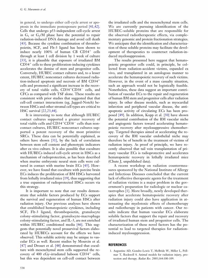
Culture of 400 cGy–irradiated BM CD34� cellsith TSF alone supported a 2.8-fold increase in totaliable cells compared with day 0 cells; however, aignificant decrement in the CD34�CD38� subset wasbserved by day 10 (Figure 1A and B). Conversely,UBEC contact cultures supported an 11.3-fold in-
rease in total cells at day 10 and were associated with aignificant increase in the percentage of CD34�CD38�
ells in culture (mean, 18.2%) compared with TSF alonemean, 0.5%; P � .005). This translated into a 29.4-foldncrease in CD34�CD38� cells in HUBEC contact cul-ures compared with input, as compared with a 4.9-foldecrease in CD34�CD38� cells with cytokines alone.ulture of irradiated BM CD34� cells with HUBECsnder noncontact conditions supported a 5.8-fold ex-ansion of total cells and a 4.8-fold increase in theD34� CD38� subset compared with day 0 cells (Fig-re 1A and B). Although this was significantly less thanhe recovery observed in HUBEC contact cultures, theecovery of total viable cells and CD34�CD38� cells inoncontact HUBEC cultures was significantly in-reased compared with TSF cultures alone (P � .01nd P � .01, respectively).
Irradiation of human CB CD34� cells yieldedimilar results as compared with BM CD34� cells.fter a 10-day culture with TSF alone, a 9.2-foldxpansion of total cells was observed, but a 3.9-foldecline in CD34�CD38� cells occurred by day 10Figure 1C and D). In contrast, HUBEC contact cul-ures supported a 29.5-fold increase in total cells and28.6-fold increase in CD34�CD38� cells comparedith input. Noncontact HUBEC cultures supported a7.7-fold increase in total cells and a 3.9-fold increasen CD34�CD38� cells (Figure 1C and D). The re-overy and expansion of total viable cells andD34�CD38� cells was significantly higher in both
UBEC contact and noncontact cultures compared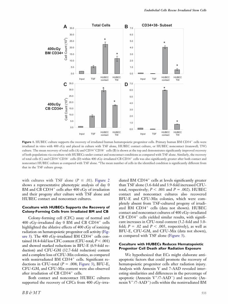
wsBaH
CC
4hrutadawdCa
s
dttcBpacCcfBa
CP
ahAea
Ficoont
Endothelial Cells Rescue Irradiated Stem Cells
B
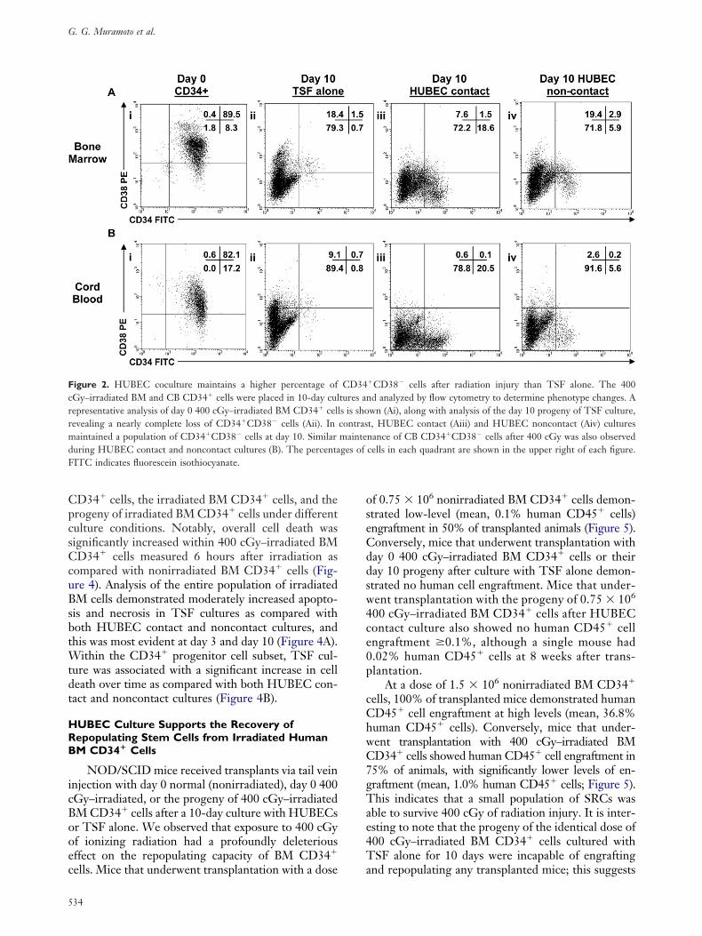
ith cultures with TSF alone (P � .01). Figure 2hows a representative phenotypic analysis of day 0M and CB CD34� cells after 400 cGy of irradiationnd their progeny after culture with TSF alone andUBEC contact and noncontact cultures.
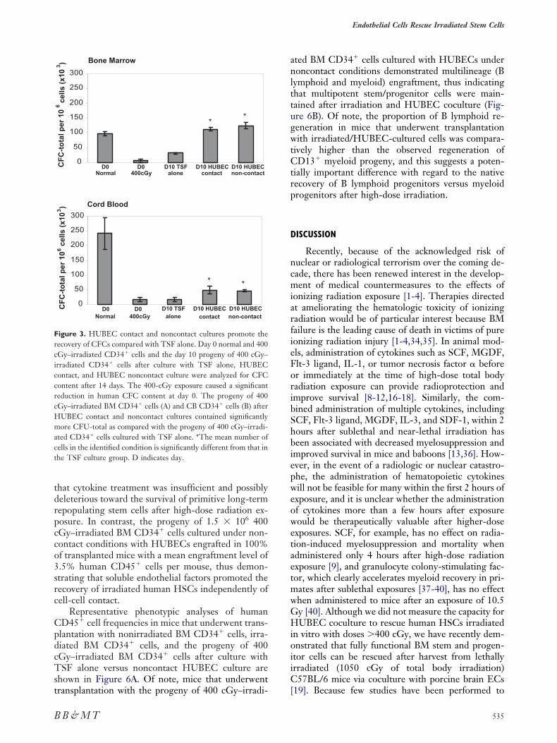
oculture with HUBECs Supports the Recovery ofolony-Forming Cells from Irradiated BM and CB
Colony-forming cell (CFC) assay of normal and00 cGy–irradiated day 0 BM and CB CD34� cellsighlighted the ablative effects of 400 cGy of ionizingadiation on hematopoietic progenitor cell activity (Fig-re 3). The 400 cGy–irradiated BM CD34� cells con-ained 18.4-fold less CFC content (CFU-total; P � .001)nd showed marked reductions in BFU-E (6.9-fold re-uction) and CFU-GM (32.7-fold reduction) contentnd a complete loss of CFU-Mix colonies, as comparedith nonirradiated BM CD34� cells. Significant re-uctions in CFU-total (P � .008; Figure 3), BFU-E,FU-GM, and CFU-Mix content were also observed
fter irradiation of CB CD34� cells.Both contact and noncontact HUBEC cultures
0.0
5.0
10.0
15.0
20.0
25.0
30.0
35.0
0.0
5.0
10.0
15.0
20.0
25.0
30.0
35.0
400cGy BM CD34+
400cGy CB CD34+
Total Cells
TSFInput HUBECcontact
TSFInput HUBECcontact
A
C
*
*
Cel
l Cou
nt (
105 )
Cel
l Cou
nt (
105 )
igure 1. HUBEC culture supports the recovery of irradiated humrradiated in vitro with 400 cGy and placed in culture with TSF aulture. The mean recovery of total cells (A) and CD34�CD38� celf both populations via coculture with HUBECs under contact and nf total cells (C) and CD34�CD38� cells (D) within 400 cGy–irradoncontact HUBEC culture as compared with TSF alone. *The mehat in the TSF culture group.
upported the recovery of CFCs from 400 cGy–irra- n
B & M T
iated BM CD34� cells at levels significantly greaterhan TSF alone (3.6-fold and 3.9-fold increased CFU-otal, respectively; P � .001 and P � .002). HUBEContact and noncontact cultures also recoveredFU-E and CFU-Mix colonies, which were com-letely absent from TSF-cultured progeny of irradi-ted BM CD34� cells (data not shown). HUBEContact and noncontact cultures of 400 cGy–irradiatedB CD34� cells yielded similar results, with signifi-
ant increases in CFU-total content (3.2-fold and 3.0-old; P � .02 and P � .005, respectively), as well asFU-E, CFU-GM, and CFU-Mix (data not shown),s compared with TSF alone (Figure 3).
oculture with HUBECs Reduces Hematopoieticrogenitor Cell Death after Radiation Exposure
We hypothesized that ECs might elaborate anti-poptotic factors that could promote the recovery ofematopoietic progenitor cells after radiation injury.nalysis with Annexin V and 7-AAD revealed inter-sting similarities and differences in the percentage ofpoptotic (Annexin V�/7-AAD�) and necrotic (An-
0.0
1.0
2.0
3.0
4.0
5.0
6.0
7.0
0.0
1.0
2.0
3.0
4.0
5.0
6.0
7.0
CD34+38- Subset
C
TSFInput HUBECcontact
HUBECTW
TSFInput HUBECcontact
HUBECTW
C
B
D*
*
*
*
atopoietic progenitor cells. Primary human BM CD34� cells wereUBEC contact culture, or HUBEC noncontact (transwell; TW)
shown at the top and demonstrates significantly improved recoverytact conditions as compared with TSF alone. Similarly, the recoveryB CD34� cells was also significantly greater after both contact andber of cells in the identified condition is significantly different from
HUBETW
HUBETW
*
*
an hemlone, H
ls (B) isoncon
iated Can num
exin V�/7-AAD�) cells within the nonirradiated BM
533
CpcsCcuBsbtWtdt
HRB
icBooec
oseCddsw4ce0p
cChwC7gTae4T
FcrrmdF
G. G. Muramoto et al.
5
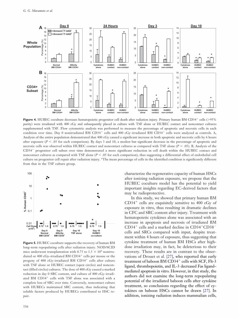
D34� cells, the irradiated BM CD34� cells, and therogeny of irradiated BM CD34� cells under differentulture conditions. Notably, overall cell death wasignificantly increased within 400 cGy–irradiated BMD34� cells measured 6 hours after irradiation as
ompared with nonirradiated BM CD34� cells (Fig-re 4). Analysis of the entire population of irradiatedM cells demonstrated moderately increased apopto-
is and necrosis in TSF cultures as compared withoth HUBEC contact and noncontact cultures, andhis was most evident at day 3 and day 10 (Figure 4A).
ithin the CD34� progenitor cell subset, TSF cul-ure was associated with a significant increase in celleath over time as compared with both HUBEC con-act and noncontact cultures (Figure 4B).
UBEC Culture Supports the Recovery ofepopulating Stem Cells from Irradiated HumanM CD34� Cells
NOD/SCID mice received transplants via tail veinnjection with day 0 normal (nonirradiated), day 0 400Gy–irradiated, or the progeny of 400 cGy–irradiatedM CD34� cells after a 10-day culture with HUBECsr TSF alone. We observed that exposure to 400 cGyf ionizing radiation had a profoundly deleteriousffect on the repopulating capacity of BM CD34�
igure 2. HUBEC coculture maintains a higher percentage ofGy–irradiated BM and CB CD34� cells were placed in 10-day culepresentative analysis of day 0 400 cGy–irradiated BM CD34� cellevealing a nearly complete loss of CD34�CD38� cells (Aii). Inaintained a population of CD34�CD38� cells at day 10. Similar
uring HUBEC contact and noncontact cultures (B). The percentaITC indicates fluorescein isothiocyanate.
ells. Mice that underwent transplantation with a dose a
34
f 0.75 � 106 nonirradiated BM CD34� cells demon-trated low-level (mean, 0.1% human CD45� cells)ngraftment in 50% of transplanted animals (Figure 5).onversely, mice that underwent transplantation withay 0 400 cGy–irradiated BM CD34� cells or theiray 10 progeny after culture with TSF alone demon-trated no human cell engraftment. Mice that under-ent transplantation with the progeny of 0.75 � 106
00 cGy–irradiated BM CD34� cells after HUBEContact culture also showed no human CD45� cellngraftment �0.1%, although a single mouse had.02% human CD45� cells at 8 weeks after trans-lantation.
At a dose of 1.5 � 106 nonirradiated BM CD34�
ells, 100% of transplanted mice demonstrated humanD45� cell engraftment at high levels (mean, 36.8%uman CD45� cells). Conversely, mice that under-ent transplantation with 400 cGy–irradiated BMD34� cells showed human CD45� cell engraftment in5% of animals, with significantly lower levels of en-raftment (mean, 1.0% human CD45� cells; Figure 5).his indicates that a small population of SRCs was
ble to survive 400 cGy of radiation injury. It is inter-sting to note that the progeny of the identical dose of00 cGy–irradiated BM CD34� cells cultured withSF alone for 10 days were incapable of engrafting
�CD38� cells after radiation injury than TSF alone. The 400d analyzed by flow cytometry to determine phenotype changes. Awn (Ai), along with analysis of the day 10 progeny of TSF culture,t, HUBEC contact (Aiii) and HUBEC noncontact (Aiv) culturesnance of CB CD34�CD38� cells after 400 cGy was also observedcells in each quadrant are shown in the upper right of each figure.
CD34tures ans is shocontrasmainteges of
nd repopulating any transplanted mice; this suggests
tdrpcco3src
CpdcTst
anlttugwtCtrp
D
ncmiarfieForibShbiepweowetaetmwGHioiiC
FrciccrcHmact
Endothelial Cells Rescue Irradiated Stem Cells
B
hat cytokine treatment was insufficient and possiblyeleterious toward the survival of primitive long-termepopulating stem cells after high-dose radiation ex-osure. In contrast, the progeny of 1.5 � 106 400Gy–irradiated BM CD34� cells cultured under non-ontact conditions with HUBECs engrafted in 100%f transplanted mice with a mean engraftment level of.5% human CD45� cells per mouse, thus demon-trating that soluble endothelial factors promoted theecovery of irradiated human HSCs independently ofell-cell contact.
Representative phenotypic analyses of humanD45� cell frequencies in mice that underwent trans-lantation with nonirradiated BM CD34� cells, irra-iated BM CD34� cells, and the progeny of 400Gy–irradiated BM CD34� cells after culture withSF alone versus noncontact HUBEC culture are
hown in Figure 6A. Of note, mice that underwent
0
50
100
150
200
250
300
0
50
100
150
200
250
300
D0Normal
D0400cGy
D10 TSF alone
D10 HUBEC contact
D10 HUBEC non-contact
D0Normal
D0400cGy
D10 TSF alone
D10 HUBEC contact
D10 HUBEC non-contact
CF
C-t
ota
l per
10
6 cel
ls (
x10
3 )C
FC
-to
tal p
er 1
06 c
ells
(x1
03)
Bone Marrow
Cord Blood
**
**
igure 3. HUBEC contact and noncontact cultures promote theecovery of CFCs compared with TSF alone. Day 0 normal and 400Gy–irradiated CD34� cells and the day 10 progeny of 400 cGy–rradiated CD34� cells after culture with TSF alone, HUBEContact, and HUBEC noncontact culture were analyzed for CFContent after 14 days. The 400-cGy exposure caused a significanteduction in human CFC content at day 0. The progeny of 400Gy–irradiated BM CD34� cells (A) and CB CD34� cells (B) afterUBEC contact and noncontact cultures contained significantlyore CFU-total as compared with the progeny of 400 cGy–irradi-
ted CD34� cells cultured with TSF alone. *The mean number ofells in the identified condition is significantly different from that inhe TSF culture group. D indicates day.
ransplantation with the progeny of 400 cGy–irradi- [
B & M T
ted BM CD34� cells cultured with HUBECs underoncontact conditions demonstrated multilineage (B
ymphoid and myeloid) engraftment, thus indicatinghat multipotent stem/progenitor cells were main-ained after irradiation and HUBEC coculture (Fig-re 6B). Of note, the proportion of B lymphoid re-eneration in mice that underwent transplantationith irradiated/HUBEC-cultured cells was compara-
ively higher than the observed regeneration ofD13� myeloid progeny, and this suggests a poten-
ially important difference with regard to the nativeecovery of B lymphoid progenitors versus myeloidrogenitors after high-dose irradiation.
ISCUSSION
Recently, because of the acknowledged risk ofuclear or radiological terrorism over the coming de-ade, there has been renewed interest in the develop-ent of medical countermeasures to the effects of
onizing radiation exposure [1-4]. Therapies directedt ameliorating the hematologic toxicity of ionizingadiation would be of particular interest because BMailure is the leading cause of death in victims of pureonizing radiation injury [1-4,34,35]. In animal mod-ls, administration of cytokines such as SCF, MGDF,lt-3 ligand, IL-1, or tumor necrosis factor � beforer immediately at the time of high-dose total bodyadiation exposure can provide radioprotection andmprove survival [8-12,16-18]. Similarly, the com-ined administration of multiple cytokines, includingCF, Flt-3 ligand, MGDF, IL-3, and SDF-1, within 2ours after sublethal and near-lethal irradiation haseen associated with decreased myelosuppression andmproved survival in mice and baboons [13,36]. How-ver, in the event of a radiologic or nuclear catastro-he, the administration of hematopoietic cytokinesill not be feasible for many within the first 2 hours ofxposure, and it is unclear whether the administrationf cytokines more than a few hours after exposureould be therapeutically valuable after higher-dosexposures. SCF, for example, has no effect on radia-ion-induced myelosuppression and mortality whendministered only 4 hours after high-dose radiationxposure [9], and granulocyte colony-stimulating fac-or, which clearly accelerates myeloid recovery in pri-ates after sublethal exposures [37-40], has no effecthen administered to mice after an exposure of 10.5y [40]. Although we did not measure the capacity forUBEC coculture to rescue human HSCs irradiated
n vitro with doses �400 cGy, we have recently dem-nstrated that fully functional BM stem and progen-tor cells can be rescued after harvest from lethallyrradiated (1050 cGy of total body irradiation)57BL/6 mice via coculture with porcine brain ECs
19]. Because few studies have been performed to
535
caHim
CeihiCcmcdrvtlmaptta
FpscAanCncfrom that in the TSF culture group.
Flmdpwtracwspair.
G. G. Muramoto et al.
536
haracterize the regenerative capacity of human HSCsfter ionizing radiation exposure, we propose that theUBEC coculture model has the potential to yield
mportant insights regarding EC-derived factors thatay be radioprotective.
In this study, we showed that primary human BMD34� cells are exquisitely sensitive to 400 cGy of
xposure in vitro, thus resulting in dramatic declinesn CFC and SRC content after injury. Treatment withematopoietic cytokines alone was associated with an
ncrease in apoptosis and necrosis of irradiated BMD34� cells and a marked decline in CD34�CD38�
ells and SRCs compared with input, despite treat-ent within 4 hours of exposure, thus suggesting that
ytokine treatment of human BM HSCs after high-ose irradiation may, in fact, be deleterious to theirecovery. These results are in contrast to the obser-ations of Drouet et al. [27], who reported that earlyreatment of baboon BM CD34� cells with SCF, Flt-3igand, thrombopoietin, and IL-3 decreased Fas ligand–
ediated apoptosis in vitro. However, in that study, theuthors did not examine the long-term repopulatingotential of the irradiated baboon cells after cytokinereatment, so conclusions regarding the effect of cy-okines on baboon HSCs cannot be drawn [27]. In
0
5
10
15
20
25
0
5
10
15
20
25
0
2
4
6
8
10
12
14
0
2
4
6
8
10
12
14
Day 3 Day 10
Cytokines alone
HUBECcontact
HUBECnon-contact
Cytokines alone
HUBECcontact
HUBECnon-contact
*
*
*
*
*
**
*
**
*
Cytokines alone
HUBECcontact
HUBECnon-contact
Cytokines alone
HUBECcontact
HUBECnon-contact
Ctact
Ctact
th after radiation injury. Primary human BM CD34� cells (�95%e with TSF alone or HUBEC contact and noncontact cultures
easure the percentage of apoptotic and necrotic cells in eachGy–irradiated BM CD34� cells were analyzed as controls. A,
gnificant increase in both apoptotic and necrotic cells by 6 hoursdest but significant decrease in the percentage of apoptotic andtures as compared with TSF alone (P � .05). B, Analysis of thecant reduction in cell death within the HUBEC contact andparison), thus suggesting a differential effect of endothelial cellntage of cells in the identified condition is significantly different
0
2
4
6
8
10
12
14
16
18
0
2
4
6
8
10
12
14
16
18
0
5
10
15
20
25
30
0
5
10
15
20
25
30
WholePopulation
CD34+Subset
Day 0 24 Hours =Annexin+/7-AAD-
=Annexin+/7-AAD+
NormalBM CD34+
400cGy BM CD34+
NormalBM CD34+
400cGy BM CD34+
*
*
*
*
*
Cytokines alone
HUBECcontact
HUBEnon-con
Cytokines alone
HUBECcontact
HUBEnon-con
A
B
Per
cent
age
(%)
Per
cent
age
(%)
igure 4. HUBEC coculture decreases hematopoietic progenitor cell deaurity) were irradiated with 400 cGy and subsequently placed in culturupplemented with TSF. Flow cytometric analysis was performed to mondition over time. Day 0 nonirradiated BM CD34� cells and 400 cnalysis of the entire population demonstrated that 400 cGy caused a sifter exposure (P � .05 for each comparison). By days 3 and 10, a moecrotic cells was observed within HUBEC contact and noncontact culD34� progenitor cell subset over time demonstrated a more signifioncontact cultures as compared with TSF alone (P � .05 for each comulture on progenitor cell repair after radiation injury. *The mean perce
0.01
0.1
1
10
100
Day 0 Normal
BM CD34+
Day 0 400cGy
BM CD34+
Day 10 TSF alone
Day 10 HUBEC
co-culture
Cell Dose (x106)
0.75 1.5 0.75 1.5 0.75 1.5 0.75 1.5
% H
um
an C
D45
+
igure 5. HUBEC coculture supports the recovery of human BMong-term repopulating cells after radiation injury. NOD/SCID
ice underwent transplantation with 0.75 to 1.5 � 106 nonirra-iated or 400 cGy–irradiated BM CD34� cells per mouse or therogeny of 400 cGy–irradiated BM CD34� cells after cultureith TSF alone or HUBEC contact (open circles) and noncon-
act (filled circles) cultures. The dose of 400 cGy caused a markededuction in day 0 SRC content, and culture of 400 cGy–irradi-ted BM CD34� cells with TSF alone was associated with aomplete loss of SRC over time. Conversely, noncontact cultureith HUBECs maintained SRC content, thus indicating that
oluble factors produced by HUBECs contributed to HSC re-
ddition, ionizing radiation induces mammalian cells,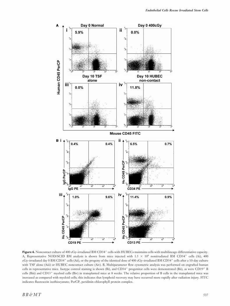
FAcwccii
Endothelial Cells Rescue Irradiated Stem Cells
B
igure 6. Noncontact culture of 400 cGy–irradiated BM CD34� cells with HUBECs maintains cells with multilineage differentiative capacity., Representative NOD/SCID BM analysis is shown from mice injected with 1.5 � 106 nonirradiated BM CD34� cells (Ai), 400Gy–irradiated day 0 BM CD34� cells (Aii), or the progeny of the identical dose of 400 cGy–irradiated BM CD34� cells after a 10-day cultureith TSF alone (Aiii) or HUBEC noncontact culture (Aiv). B, Multiparameter flow cytometric analysis was performed on engrafted human
ells in representative mice. Isotype control staining is shown (Bi), and CD34� progenitor cells were demonstrated (Bii), as were CD19� Bells (Biii) and CD13� myeloid cells (Biv) in transplanted mice at 8 weeks. The relative proportion of B cells in the transplanted mice wasncreased as compared with myeloid cells; this indicates that lymphoid recovery may have occurred more rapidly after radiation injury. FITC
ndicates fluorescein isothiocyanate; PerCP, peridinin-chlorophyll protein complex.537B & M T
ipCir[pit[CaCetceCcctH
ctcpSobawmwteEfat
strtSccwgroc[tcb
tWHtmWtod
plvaHsNboiiaptapactrcmh(
saloetartassocti
R
G. G. Muramoto et al.
5
n general, to undergo either cell-cycle arrest or apo-tosis in the immediate postexposure period [41,42].ells that undergo p53-independent cell-cycle arrest
n G1 or G2/M phase have the potential to repairadiation-induced DNA damage and avoid cell death43]. Because the cytokine combination of thrombo-oietin, SCF, and Flt-3 ligand has been shown to
nduce nearly 100% of human CB CD34� cellshrough at least 1 cell division by 1 week of culture33], it is plausible that exposure of irradiated BMD34� cells to these proliferation-inducing cytokines
ccelerates the demise of stem and progenitor cells.onversely, HUBEC contact cultures and, to a lesser
xtent, HUBEC noncontact cultures decreased radia-ion-induced apoptosis and necrosis of BM CD34�
ells and promoted a significant increase in the recov-ry of total viable cells, CD34�CD38� cells, andFCs as compared with TSF alone. These results are
onsistent with prior studies that suggested that thatell-cell contact interactions (eg, Jagged-Notch) be-ween HSCs and other stromal cell types are critical to
SC survival [22,23,44].It is interesting to note that although HUBEC
ontact cultures supported a greater recovery ofotal viable cells and CD34�CD38� cells than non-ontact cultures, HUBEC noncontact cultures sup-orted a potent recovery of the most primitiveRCs. These data can be potentially explained, asthers have shown [31], by the lack of correlationetween stem cell content and phenotypic indicatorsfter ex vivo culture. It is also possible that cocultureith HUBECs induced cell-cycle arrest in HSCs as aechanism of radioprotection, as has been describedhen murine embryonic neural stem cells were cul-
ured in contact with murine brain ECs [45]. How-ver, we have found that coculture with porcine brainCs induces the proliferation of BM HSCs harvested
rom lethally irradiated mice [19], thus suggesting thattrue expansion of radioprotected HSCs occurs via
his strategy.It is important to note that our results demon-
trate that soluble factors produced by ECs supporthe survival and regeneration of human HSCs afteradiation injury. Our previous analyses have shownhat established hematopoietic cytokines, includingCF, Flt-3 ligand, thrombopoietin, granulocyteolony-stimulating factor, granulocyte-macrophageolony-stimulating factor, and IL-3, are not enrichedithin HUBEC conditioned media [46]. This sug-ests that potentially novel prosurvival factors elabo-ated by HUBECs account for the effects we havebserved. This soluble activity may be unique to vas-ular ECs as well. Recent studies by Mourcin et al.47] and Drouet et al. [48] demonstrated that cocul-ure with mesenchymal stem cells supported the re-overy of 400 cGy–irradiated baboon CD34� cells,
ut this was dependent on cell-cell contact between38
he irradiated cells and the mesenchymal stem cells.e are currently pursuing identification of theUBEC-soluble proteins that are responsible for
he observed radiotherapeutic effects, via comple-entary genomic and protein fractionation strategies.e anticipate that the identification and characteriza-
ion of these soluble proteins may facilitate the devel-pment of therapeutics to counteract radiation-in-uced myelosuppression.
The results presented here suggest that hemato-oietic progenitor cells could, in principle, be col-ected from radiation-accident victims, expanded exivo, and transplanted in an autologous manner toccelerate the hematopoietic recovery of such victims.owever, in the event of a mass casualty situation,
uch an approach would not be logistically feasible.onetheless, these data suggest an important contri-
ution of vascular ECs to the repair and regenerationf human BM stem and progenitor cells after radiationnjury. In other disease models, such as myocardialnfarction and peripheral vascular disease, the anti-poptotic activity of circulating ECs has been pro-osed [49]. In addition, Kopp et al. [50] have shownhe potential contribution of the BM vascular nichend angiogenic factors toward accelerating hemato-oietic recovery after myelosuppressive chemother-py. Targeted therapies aimed at accelerating the re-overy of the BM vascular endothelial niche mayherefore be of benefit in the treatment of victims ofadiation injury. As proof of principle, we have re-ently observed that tail vein transplantation of pri-ary vascular ECs is radioprotective and accelerates
ematopoietic recovery in lethally irradiated miceChute J, unpublished data).
A recent workshop on radiation countermea-ures sponsored by the National Institute of Allergynd Infectious Diseases concluded that the currentack of effective therapeutic agents for the treatmentf radiation victims is a major problem in the gov-rnment’s preparation for radiologic or nuclear ca-astrophes [1]. More broadly, newly developed ther-pies that accelerate hematopoietic recovery afteradiation injury could also have application in at-enuating the myelotoxic effects of chemotherapynd radiotherapy in patients with cancer. Our re-ults indicate that human vascular ECs elaborateoluble factors that support the repair and recoveryf irradiated human stem and progenitor cells. Theharacterization of these novel factors has the po-ential to lead to targeted therapies for radiation-nduced myelosuppression.
EFERENCES
1. Augustine AD, Gondre-Lewis T, McBride W, Miller L, Pell-mar T, Rockwell S. Animal models for radiation injury, pro-
tection and therapy. Radiat Res. 2005;164:100-109.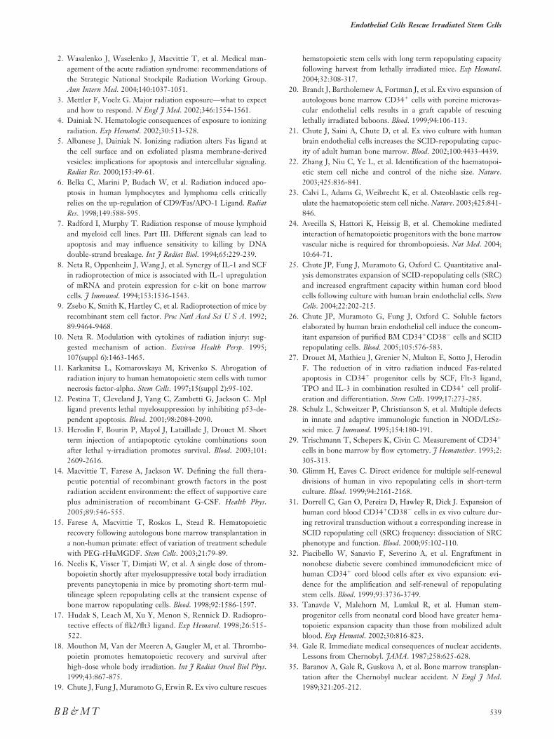
1
1
1
1
1
1
1
1
1
1
2
2
2
2
2
2
2
2
2
2
3
3
3
3
3
3
Endothelial Cells Rescue Irradiated Stem Cells
B
2. Wasalenko J, Waselenko J, Macvittie T, et al. Medical man-agement of the acute radiation syndrome: recommendations ofthe Strategic National Stockpile Radiation Working Group.Ann Intern Med. 2004;140:1037-1051.
3. Mettler F, Voelz G. Major radiation exposure—what to expectand how to respond. N Engl J Med. 2002;346:1554-1561.
4. Dainiak N. Hematologic consequences of exposure to ionizingradiation. Exp Hematol. 2002;30:513-528.
5. Albanese J, Dainiak N. Ionizing radiation alters Fas ligand atthe cell surface and on exfoliated plasma membrane-derivedvesicles: implications for apoptosis and intercellular signaling.Radiat Res. 2000;153:49-61.
6. Belka C, Marini P, Budach W, et al. Radiation induced apo-ptosis in human lymphocytes and lymphoma cells criticallyrelies on the up-regulation of CD9/Fas/APO-1 Ligand. RadiatRes. 1998;149:588-595.
7. Radford I, Murphy T. Radiation response of mouse lymphoidand myeloid cell lines. Part III. Different signals can lead toapoptosis and may influence sensitivity to killing by DNAdouble-strand breakage. Int J Radiat Biol. 1994;65:229-239.
8. Neta R, Oppenheim J, Wang J, et al. Synergy of IL-1 and SCFin radioprotection of mice is associated with IL-1 upregulationof mRNA and protein expression for c-kit on bone marrowcells. J Immunol. 1994;153:1536-1543.
9. Zsebo K, Smith K, Hartley C, et al. Radioprotection of mice byrecombinant stem cell factor. Proc Natl Acad Sci U S A. 1992;89:9464-9468.
0. Neta R. Modulation with cytokines of radiation injury: sug-gested mechanism of action. Environ Health Persp. 1995;107(suppl 6):1463-1465.
1. Karkanitsa L, Komarovskaya M, Krivenko S. Abrogation ofradiation injury to human hematopoietic stem cells with tumornecrosis factor-alpha. Stem Cells. 1997;15(suppl 2):95-102.
2. Pestina T, Cleveland J, Yang C, Zambetti G, Jackson C. Mplligand prevents lethal myelosuppression by inhibiting p53-de-pendent apoptosis. Blood. 2001;98:2084-2090.
3. Herodin F, Bourin P, Mayol J, Lataillade J, Drouet M. Shortterm injection of antiapoptotic cytokine combinations soonafter lethal -irradiation promotes survival. Blood. 2003;101:2609-2616.
4. Macvittie T, Farese A, Jackson W. Defining the full thera-peutic potential of recombinant growth factors in the postradiation accident environment: the effect of supportive careplus administration of recombinant G-CSF. Health Phys.2005;89:546-555.
5. Farese A, Macvittie T, Roskos L, Stead R. Hematopoieticrecovery following autologous bone marrow transplantation ina non-human primate: effect of variation of treatment schedulewith PEG-rHuMGDF. Stem Cells. 2003;21:79-89.
6. Neelis K, Visser T, Dimjati W, et al. A single dose of throm-bopoietin shortly after myelosuppressive total body irradiationprevents pancytopenia in mice by promoting short-term mul-tilineage spleen repopulating cells at the transient expense ofbone marrow repopulating cells. Blood. 1998;92:1586-1597.
7. Hudak S, Leach M, Xu Y, Menon S, Rennick D. Radiopro-tective effects of flk2/flt3 ligand. Exp Hematol. 1998;26:515-522.
8. Mouthon M, Van der Meeren A, Gaugler M, et al. Thrombo-poietin promotes hematopoietic recovery and survival afterhigh-dose whole body irradiation. Int J Radiat Oncol Biol Phys.1999;43:867-875.
9. Chute J, Fung J, Muramoto G, Erwin R. Ex vivo culture rescues
B & M T
hematopoietic stem cells with long term repopulating capacityfollowing harvest from lethally irradiated mice. Exp Hematol.2004;32:308-317.
0. Brandt J, Bartholemew A, Fortman J, et al. Ex vivo expansion ofautologous bone marrow CD34� cells with porcine microvas-cular endothelial cells results in a graft capable of rescuinglethally irradiated baboons. Blood. 1999;94:106-113.
1. Chute J, Saini A, Chute D, et al. Ex vivo culture with humanbrain endothelial cells increases the SCID-repopulating capac-ity of adult human bone marrow. Blood. 2002;100:4433-4439.
2. Zhang J, Niu C, Ye L, et al. Identification of the haematopoi-etic stem cell niche and control of the niche size. Nature.2003;425:836-841.
3. Calvi L, Adams G, Weibrecht K, et al. Osteoblastic cells reg-ulate the haematopoietic stem cell niche. Nature. 2003;425:841-846.
4. Avecilla S, Hattori K, Heissig B, et al. Chemokine mediatedinteraction of hematopoietic progenitors with the bone marrowvascular niche is required for thrombopoiesis. Nat Med. 2004;10:64-71.
5. Chute JP, Fung J, Muramoto G, Oxford C. Quantitative anal-ysis demonstrates expansion of SCID-repopulating cells (SRC)and increased engraftment capacity within human cord bloodcells following culture with human brain endothelial cells. StemCells. 2004;22:202-215.
6. Chute JP, Muramoto G, Fung J, Oxford C. Soluble factorselaborated by human brain endothelial cell induce the concom-itant expansion of purified BM CD34�CD38� cells and SCIDrepopulating cells. Blood. 2005;105:576-583.
7. Drouet M, Mathieu J, Grenier N, Multon E, Sotto J, HerodinF. The reduction of in vitro radiation induced Fas-relatedapoptosis in CD34� progenitor cells by SCF, Flt-3 ligand,TPO and IL-3 in combination resulted in CD34� cell prolif-eration and differentiation. Stem Cells. 1999;17:273-285.
8. Schulz L, Schweitzer P, Christianson S, et al. Multiple defectsin innate and adaptive immunologic function in NOD/LtSz-scid mice. J Immunol. 1995;154:180-191.
9. Trischmann T, Schepers K, Civin C. Measurement of CD34�
cells in bone marrow by flow cytometry. J Hematother. 1993;2:305-313.
0. Glimm H, Eaves C. Direct evidence for multiple self-renewaldivisions of human in vivo repopulating cells in short-termculture. Blood. 1999;94:2161-2168.
1. Dorrell C, Gan O, Pereira D, Hawley R, Dick J. Expansion ofhuman cord blood CD34�CD38� cells in ex vivo culture dur-ing retroviral transduction without a corresponding increase inSCID repopulating cell (SRC) frequency: dissociation of SRCphenotype and function. Blood. 2000;95:102-110.
2. Piacibello W, Sanavio F, Severino A, et al. Engraftment innonobese diabetic severe combined immunodeficient mice ofhuman CD34� cord blood cells after ex vivo expansion: evi-dence for the amplification and self-renewal of repopulatingstem cells. Blood. 1999;93:3736-3749.
3. Tanavde V, Malehorn M, Lumkul R, et al. Human stem-progenitor cells from neonatal cord blood have greater hema-topoietic expansion capacity than those from mobilized adultblood. Exp Hematol. 2002;30:816-823.
4. Gale R. Immediate medical consequences of nuclear accidents.Lessons from Chernobyl. JAMA. 1987;258:625-628.
5. Baranov A, Gale R, Guskova A, et al. Bone marrow transplan-tation after the Chernobyl nuclear accident. N Engl J Med.
1989;321:205-212.539
3
3
3
3
4
4
4
4
4
4
4
4
4
4
5
G. G. Muramoto et al.
5
6. Drouet M, Mourcin F, Grenier N, et al. Single administrationof stem cell factor, FLT-3 ligand, megakaryocyte growth anddevelopment factor, and interleukin-3 in combination soonafter irradiation prevents nonhuman primates from myelosup-pression: long-term follow up of hematopoiesis. Blood. 2004;103:878-885.
7. Farese A, Hunt P, Grab L, MacVittie T. Combined adminis-tration of recombinant human megakaryocyte growth and de-velopment factor and granulocyte colony stimulating factorenhances multilineage hematopoietic reconstitution in nonhu-man primates after radiation-induced marrow aplasia. J ClinInvest. 1996;97:2145-2151.
8. Farese A, Casey D, Vigneulle R, et al. A single dose ofpegylated leridistim significantly improves neutrophil recov-ery in sublethally irradiated rhesus macaques. Stem Cells.2001;19:514-521.
9. Farese A, Casey D, Smith W, et al. Leridistim, a chimeric dualGCSF and IL-3 receptor agonist, enhances multilineage hema-topoietic recovery in a nonhuman primate model of radiationinduced myelosuppression: effect of schedule, dose and route ofadministration. Stem Cells. 2001;19:522-533.
0. Tanikawa S, Nose M, Aoki Y, et al. Effects of recombinantgranulocyte colony stimulating factor on the hematologic re-covery and survival of irradiated mice. Blood. 1990;76:445-449.
1. Quelle F, Wang J, Feng J, et al. Cytokine rescue of p53-dependent apoptosis and cell cycle arrest is mediated by distinctJak kinase signaling pathways. Genes Dev. 1998;12:1099-1107.
2. Henry M, Lynch J, Eapen A, Quelle F. DNA damage induced cellcycle arrest of hematopoietic cells is overridden by activation ofthe PI-3 kinase/Akt signaling pathway. Blood. 2001;98:834-841.
3. Campanini F, Santucci M, Brusa G, et al. Expression of p21
40
(WAF1/CIP1/SID1) cyclin dependent kinase inhibitor inhematopoietic progenitor cells. Gene. 2001;273:173-180.
4. Tsai S, Fero J, Bartelmez S. Mouse jagged 2 is differentiallyexpressed in hematopoietic progenitors and endothelial cells andpromotes the survival and proliferation of hematopoietic progen-itors by direct cell-to-cell contact. Blood. 2000;96:950-957.
5. Mathieu C, Fouchet P, Gauthier L, Lassalle B, Boussin F,Mouthon M. Coculture with endothelial cells reduces the pop-ulation of cycling LeX neural precursors but increases that ofquiescent cells with a side population phenotype. Exp Cell Res.2006;312:707-718.
6. Chute J, Clark W, Johnson K, Saini A, Wells M, Harlan D.Hematopoietic cytokine production by human brain endothe-lial cells (HUBEC) is associated with the ex vivo expansion ofCD34�CD38� progenitor cells. Exp Hematol. 2000;28:96a.
7. Mourcin F, Grenier N, Mayol J, et al. Mesenchymal stem cellssupport expansion of in vitro irradiated CD34� cells in thepresence of SCF, Flt-3 ligand, TPO and IL-3: potential appli-cation to autologous cell therapy in accidentally irradiated vic-tims. Radiat Res. 2005;164:1-9.
8. Drouet M, Mourcin F, Grenier N, et al. Mesenchymal stemcells rescue CD34� cells from radiation-induced apoptosis andsustain hematopoietic reconstitution after coculture andcografting in lethally irradiated baboons: is autologous stem celltherapy in nuclear accident settings hype or reality? Bone Mar-row Transplant. 2005;35:1201-1209.
9. Kong D, Melo L, Gnecchi M, et al. Cytokine induced mobili-zation of circulating endothelial progenitor cells enhances re-pair of injured arteries. Circulation. 2004;110:239-246.
0. Kopp H, Avecilla S, Hooper A, et al. Tie-2 activation contrib-utes to hemangiogenic revascularization after myelosuppres-
sion. Blood. 2005;106:505-513.


