Embed Size (px)
Citation preview
This is the author manuscript accepted for publication and has undergone full peer review but has
not been through the copyediting, typesetting, pagination and proofreading process, which may
lead to differences between this version and the Version of Record. Please cite this article as doi:
10.1111/nph.14478
This article is protected by copyright. All rights reserved
Received Date : 30-Aug-2016
Revised Date : 13-Jan-2017
Accepted Date : 16-Jan-2017
Article type : MS - Regular Manuscript
Vascularization of the Selaginella rhizophore – anatomical fingerprints of polar auxin
transport with implications for the deep fossil record
Kelly K. S. Matsunaga1, Nevin P. Cullen2 and Alexandru M. F. Tomescu
3
1Department of Earth and Environmental Sciences, University of Michigan, Ann Arbor, MI
48109, USA; 2Department of Biology, San Francisco State University, San Francisco, CA 94132,
USA; 3
Department of Biological Sciences, Humboldt State University, Arcata, CA 95521, USA
Author for correspondence:
Kelly K. S. Matsunaga
Tel: +1 808 381 0328
Email: [email protected]
Received: 30 August 2016
Accepted: 16 January 2017
Auth
or
Manuscript

This article is protected by copyright. All rights reserved
Summary
• The Selaginella rhizophore is a unique and enigmatic organ whose homology with roots,
shoots (or neither of the two) remains unsettled. Nevertheless, rhizophore-like organs
have been documented in several fossil lycophytes. Here we test the homology of these
organs through comparisons with the architecture of rhizophore vascularization in
Selaginella.
• We document rhizophore vascularization in nine Selaginella species using cleared whole-
mounts and histological sectioning combined with three-dimensional reconstruction.
• Three patterns of rhizophore vascularization are present in Selaginella and each is
comparable to those observed in rhizophore-like organs of fossil lycophytes. More
compellingly, we found that all Selaginella species sampled exhibit tracheids that arc
backward from the stem and side branch into the rhizophore base. This tracheid
curvature is consistent with acropetal auxin transport previously documented in the
rhizophore and is indicative of the redirection of basipetal auxin from the shoot into the
rhizophore during development.
• The tracheid curvature observed in Selaginella rhizophores provides an anatomical
fingerprint for the patterns of auxin flow that underpin rhizophore development. Similar
tracheid geometry may be present and should be searched for in fossils to address
rhizophore homology and the conservation of auxin-related developmental mechanisms
from early stages of lycophyte evolution.
Key words: fossil, lycophyte, rhizophore, polar auxin transport, Selaginella, tracheid.
Introduction
Rhizophores, seen only in the lycophyte Selaginella, are downward growing axial organs
located at branching points of the shoot and which produce roots near their apex upon reaching
the soil (Jernstedt et al., 1994). Developmentally, rhizophores originate exogenously from
meristems positioned in the branching angle of the shoot, termed angle meristems. Two angle
meristems form at each branching point, a dorsal and a ventral one, either or both of which can
Auth
or
Manuscript
This article is protected by copyright. All rights reserved
develop into a rhizophore. The homology of the Selaginella rhizophore has puzzled plant
morphologists for well over a century, owing to a set of morphological and developmental
characters that combines root and stem features. Stem-like features of the rhizophore include
exogenous developmental origination (Jernstedt et al., 1992), the expression of class 1 KNOX
genes at the apex (like in the shoot apical meristem and unlike in the root apex of Selaginella;
Kawai et al., 2010), and the capacity of rhizophores and angle meristems to develop as shoots
under specific conditions (Harvey-Gibson, 1902; Williams, 1937; Webster, 1969; Wochok &
Sussex, 1976; Jernstedt et al., 1994; Sanders & Langdale, 2013). However, like roots, the
rhizophore does not typically produce leaves, it exhibits positive gravitropism, and has been
demonstrated to have acropetal polar auxin transport (Wochok & Sussex, 1974), an auxin
transport pattern which in euphyllophytes is seen only in roots. Various authors have argued for
or against interpreting the rhizophore as a stem homolog, a root, or as a novel sui generis organ,
but currently there is no scientific consensus on the nature of the rhizophore (Jernstedt et al.,
1994 and references therein).
Although no structures comparable to the selaginellalean rhizophore are known in extant
plants, the deep fossil record of the lycophyte clade includes several plants exhibiting two types
of structures that share some features with the rhizophore. These include subaxillary tubercles
and the branches derived from them, which have been reported in several zosterophyll-grade
lycophytes of the Devonian – Crenaticaulis, Gosslingia, Deheubarthia, Thrinkophyton, and
Anisophyton (Høeg, 1942; Banks & Davis, 1969; Edwards, 1970, 1994; Edwards & Kenrick,
1986; Hass & Remy, 1986; Kenrick & Edwards, 1988; Edwards et al., 1989). Such structures
are regularly positioned in the immediate vicinity of, and basal to, the angle formed at branching
points of axes, and for this reason they are also referred to as angular organs (Hass & Remy,
1986) (Fig. 1a). While the function and developmental fate of subaxillary tubercles is unknown
in many of these taxa, branches derived from them may have functioned as rooting organs in
some cases. Additionally, there are the root-bearing axes of some Devonian drepanophycalean
lycophytes (Schweitzer & Giesen, 1980; Xu et al., 2013; Matsunaga & Tomescu, 2016, 2017).
These are produced by two closely-spaced dichotomies of the shoot, in a manner that is referred
to as H- or K-branching (Fig. 1c), and resemble morphologically equivalent non-root-bearing
axes with inferred downward growth seen in some Devonian zosterophylls – Zosterophyllum,
Auth
or
Manuscript
This article is protected by copyright. All rights reserved
Sawdonia, and Bathurstia (Lang, 1927; Walton, 1964; Rayner, 1983; Gensel et al., 2001; Hao et
al., 2010).
Like the Selaginella rhizophore (Fig. 1b), both the subaxillary axes and those produced
by K-branching, documented in fossil plants, (1) arise consistently in association with branching
points of the subtending axis; and (2) develop, in many cases, in a direction different from that of
the regular axes or shoots of the parent plant. Subaxillary branches have been found to extend in
a direction perpendicular to the plane defined by the forking of the subtending axis (e.g., Banks
& Davis, 1969; Kenrick, 1988) and the axes arising from K-branching point away from the
regular axes or shoots of the parent plant, which sometimes means downwards (Walton, 1964;
Rayner, 1983; Gensel et al., 2001; Hao et al., 2010; Matsunaga & Tomescu, 2016). In such
cases, the downward direction of growth is probably cued by a positive gravitropic response
(Walton, 1964; Matsunaga & Tomescu, 2016), like that of the Selaginella rhizophore.
Furthermore, like the rhizophore, some drepanophycalean axes arising from K-branching bear
roots (Schweitzer & Giesen, 1980; Xu et al., 2013; Matsunaga & Tomescu, 2016).
The notable similarities between rhizophores and the organs described above have
prompted direct comparisons with Selaginella (Banks & Davis, 1969; Rayner, 1983; Hass &
Remy, 1986; Kenrick, 1988; Edwards et al., 1989). However, these similarities have not been
seriously explored due to the great phylogenetic distance between Selaginella and the fossil
lycophytes characterized by subaxillary branching and K-branching (Kenrick & Crane, 1997).
Nevertheless, while Selaginella is highly derived among lycophytes, the existence of shared
morphological patterns that span broad ranges of systematic diversity within the clade indicates
that fundamental body plan features can be conserved over hundreds of million years of
evolutionary history. For instance, morphological identity and homology of the rooting system
of extant Isoetes with that of Paleozoic rhizomorphic lycophytes (e.g. Paralycopodites and
Sigillaria; Stewart, 1947; Karrfalt, 1980; Jennings et al., 1983; Rothwell & Erwin, 1985;
Hetherington et al., 2016; Hetherington & Dolan, 2016) demonstrates conservation of
developmental characters and body plan organization over >300 million yr. Or, to consider an
even broader context, every major lineage of lycophytes exhibits rooting systems derived from or
incorporating downward growing stems or undifferentiated axes (Matsunaga & Tomescu, 2017).
Together, these observations imply that hypotheses of homology between the Selaginella
rhizophore and the rhizophore-like organs of early lycophytes should not be regarded as far-
Auth
or
Manuscript
This article is protected by copyright. All rights reserved
fetched, and that addressing them within a comparative morphology framework is a worthy
pursuit.
Branching patterns and vascular architecture have been documented in basal lycophytes
with rhizophore-like organs (e.g., Lang 1927; Banks & Davis, 1969; Edwards, 1970; Kenrick,
1988; Matsunaga & Tomescu, 2016), but comparable studies that can address the question of
homology have not been conducted on Selaginella. Here we test the hypothesis of homology
between the selaginellalean rhizophore and rhizophore-like organs of early lycophytes based on
comparative anatomy. We further explore whether anatomical signatures unique to rhizophore
development, related to the hormone auxin and applicable to studying the plant fossil record, are
present in Selaginella.
Materials and Methods
We surveyed the architecture of rhizophore vascularization in nine Selaginella species
kept in the Dennis K. Walker Greenhouse (Humboldt State University): S. involvens, S. wallacei,
S. kraussiana, S. pallescens, S. delicatula, S. doederleinii, S. wildenovii, and two additional
species. While the latter two could not be identified and are hereafter referred to as Selaginella
sp. 1 and Selaginella sp. 2, they are distinctly different from the other seven. Information on the
origin and identity of these two species was lost from the glasshouse records, and Selaginella
species (totaling c. 700–800 around the world; Zhou et al., 2016) are notoriously difficult to
identify in the absence of information on their geographic origin. The Selaginella species
sampled here span phylogenetic diversity within the family, covering six different clades as
resolved in the phylogeny of Weststrand & Korall (2016), and includes species with both dorsal
and ventral rhizophores and a range of stele types (Korall & Kenrick, 2002, 2004; Weststrand &
Korall, 2016).
Shoot segments centered around branching points were sampled from each species and
prepared by clearing and by paraffin embedding and serial sectioning on a rotary microtome.
For clearing we followed Ruzin’s (1999) sodium hydroxide-chloral hydrate protocol, including
full -strength commercial bleach (sodium hypochlorite), followed by safranin staining, and
dehydration in an ethanol series; slides were mounted in epoxy resin. Paraffin embedding
followed the standard protocol for plant tissue. Serial sections were cut at 10 µm thickness and
stained using Walker’s Sam quadruple stain protocol, which uses Weigert’s iron hematoxylin
Auth
or
Manuscript
This article is protected by copyright. All rights reserved
(1% in 10% ethanol), followed by Bismark Brown (1% in 50% ethanol), then phloxine (1% in
95% ethanol), and 1:1 Fast Green – Orange G (saturated in clove oil). Slides were mounted
using Eukitt mounting medium (O. Kindler GmbH, Freiburg, Germany).
Micrographs were taken using a Nikon Coolpix E8800 digital camera mounted on a
Nikon Eclipse E400 compound microscope and an Olympus DP73 digital camera on an
Olympus SZX16 microscope. High magnification images of tracheids in Selaginella were
digitally stacked along the z-axis to provide a consistent focal plane. Focal stacking and figure
construction were done in Photoshop CC (Adobe, San Jose, CA, USA). Volume renderings
based on serial sections were produced using Amira 3D software (FEI, Hillsboro, OR, USA).
Results
Three patterns of rhizophore vascularization
In the nine Selaginella species we surveyed, the architecture of the vascular supply of the
rhizophore falls into three main types: (1) the vascular bundle supplying the rhizophore
(rhizophore trace) diverges centrally, from the angle between stem stele and branch stele
(referred to as central divergence); (2) the rhizophore trace diverges from the stele of the main
stem, close to but above the divergence of the branch stele (stem divergence); (3) the rhizophore
trace diverges from the stele of the side branch above the divergence of the latter from the main
stem stele (branch divergence). These patterns are independent of the external position of
rhizophores or angle meristems, which are always positioned in the branching angle. Of the nine
species, seven exhibit central divergence, one species is characterized by stem divergence (S. sp.
2), and one by branch divergence (S. wallacei) (Table 1). While the number of meristeles (one to
three) forming the stele of the main stem (Table 1) adds complexity to the architecture of
vascularization in some cases, this does not interfere with the three main types of rhizophore
vascularization. We describe below the architecture of rhizophore vascularization in four species
that illustrate the range of variation observed: Selaginella kraussiana (two meristeles) and S.
wildenovii (three meristeles) for central divergence, S. sp. 2 (variable, one or two meristeles) for
stem divergence, and S. wallacei (one meristele) for branch divergence.
Selected species descriptions
Auth
or
Manuscript
This article is protected by copyright. All rights reserved
Selaginella kraussiana stems have two meristeles (Fig. 2a). At shoot branching points, one of
the meristeles of the main stem splits to produce two vascular segments. One of the segments
continues as one of the main stem meristeles while the other segment fuses with the second
meristele producing a plexus of vascular tissue. From this plexus three vascular segments
diverge and continue distal to the branching point: the second main stem meristele, the stele of
the side branch, and the stele of the rhizophore. The three diverge at the same level, with the
rhizophore stele diverging from the angle between the other two and extending laterally and
perpendicular to the plane defined by the main stem and lateral branch (Fig. 3a,b; Supporting
Information Video S1).
Selaginella wildenovii stems have three meristeles represented by plates of vascular
tissue flattened in the plane of shoot branching (defined by the main stem and lateral branch)
(Fig. 2b). At shoot branching points, each stem meristele diverges to give rise to one vascular
segments that becomes a meristele of the lateral branch. At the same level, the three meristeles
anastomose. The plexus of vascular tissues connecting them forms a bridge. This bridge
extends vertically in the angle between the stem and lateral branch, forming a ridge of vascular
tissue developed in a direction perpendicular to the plane of branching. The vascular supply of
the two rhizophores diverges from the two ends of this bridge, laterally and perpendicular to the
plane of branching (Fig. 3c,d; Video S2). Overall, the rhizophore vascular strands thus diverge
from a central position in the angle between the steles of the main stem and lateral branch.
In Selaginella sp. 2 rhizophores are produced at branching points of shoots that have
either one meristele (Fig. 2c) or two meristeles. In both cases, the vascular supply of the
rhizophore arises from the stele of the main stem, distal to the divergence of the lateral branch.
The rhizophore stele initially diverges from the stem stele in the plane of shoot branching and in
the same direction as the stele of the branch (Fig. 2c); distally, the rhizophore stele curves in a
direction perpendicular to the plane of branching (Fig. 3e,f; Video S3).
Selaginella wallacei stems are characterized by a simple stele consisting of a single
vascular segment (Fig. 2d). In this species, the rhizophore stele diverges from the stele of the
lateral branch, therefore distal to the divergence of the branch stele from the stele of the main
stem. Furthermore, the direction of rhizophore divergence is oblique to the plane of shoot
branching (Fig. 2d, 3g,h; Video S4).
Auth
or
Manuscript
This article is protected by copyright. All rights reserved
Rhizophore divergence and tracheid geometry
In all the species surveyed, the shape, arrangement and orientation of tracheids at the point of
rhizophore divergence exhibit a consistent geometry. The xylem bundle of the rhizophore is
connected to the subtending shoot by tracheids that arc from the stele of the main shoot and
branch shoot back into the rhizophore vascular supply, toward the rhizophore apex (Fig. 4a–c,g–
k). This consistent pattern is very conspicuous starting with early stages of rhizophore
development and, when two rhizophores are associated with one branching point, is seen in the
vascular supply of both rhizophores. It is worth noting that in some cases the high tracheid
density at branching points, especially when associated with thick sclerified cortical tissues that
hindered clearing, obscured fine details of tracheid arrangement and shape, making it difficult to
obtain good photographs that illustrate the curved tracheids clearly (Fig. 4l). Importantly, the
geometry of tracheids associated with rhizophore divergence is clearly different from the
geometry of tracheids documented around leaf traces and shoot branching points that lack
rhizophores. In these instances, tracheids consistently form a basipetally convergent pattern with
no tracheids that arc back apically (Fig. 4d–f).
Discussion
The architecture of the rhizophore vascular supply is variable
The rhizophore-like axes of Devonian lycophytes exhibit several patterns of vascular
architecture. These patterns provided the starting point for our comparative approach to the
homology of the Selaginella rhizophore. In some plants that produce subaxillary branches, such
as Gosslingia breconensis, the vascular supply of the subaxillary branch diverges from the stele
of the main axis distal to the divergence of the branch stele (Edwards, 1970) (Fig. 5a). In
lycophytes with K-branching, the vascular strand of the rhizophore-like organ diverges from the
stele of the side branch (Matsunaga & Tomescu, 2016) (Fig. 5b). Finally, in other lycophytes
with subaxillary branches, such as Deheubarthia splendens (Edwards et al., 1989), the vascular
supply of the subaxillary branch seems to diverge centrally, from in between and at the same
level as the stele of the main axis and the stele of the side branch (Fig. 5c).
The different patterns of vascular architecture at rhizophore divergence, documented in
the nine Selaginella species, cover all three types of architecture seen in the vascular supply of
Devonian rhizophore-like axes (Table 1). The pattern documented in Selaginella sp. 2 (stem
Auth
or
Manuscript
This article is protected by copyright. All rights reserved
divergence) corresponds to the architecture of Gosslingia subaxillary branches; the pattern seen
in S. wallacei (branch divergence) fits the vascular architecture of K-branching; the central
divergence observed in the other seven Selaginella species is comparable to the architecture of
the subaxillary branch vascular supply in Deheubarthia. This broad range of variation
documented within Selaginella, along with the taxonomic diversity of Devonian lycophytes that
exhibit matching architectures, indicate that vascular architecture cannot directly provide useful
evidence relevant to discussions of homology between Devonian rhizophore-like axes and the
Selaginella rhizophore. However, the variation seen in Selaginella indicates that a broad
spectrum of vascular architecture patterns can be produced by the same set of developmental
processes – in this case those associated with rhizophore development.
Tracheid geometry and patterns of auxin transport in the development of the Selaginella
rhizophore
Polar auxin transport (PAT) is the unidirectional flow of the hormone auxin and is mediated by
PIN auxin efflux carriers positioned in the plasma membrane. The localization of PIN proteins
at one end of the cell polarizes and canalizes the flow of auxin away from a source of higher
concentration, leading to elongation of those cells that transport auxin and their differentiation as
vascular tissues (Leyser & Day, 2003; Bennett et al., 2014). Polar gradients of auxin within the
plant body have been demonstrated, through numerous auxin application and inhibition
experiments, as sufficient for inducing vascular tissue differentiation in plants (e.g.
Wangermann, 1967; Sachs, 1969; Mattsson et al., 1999; Berleth et al., 2000). Moreover, the
auxin flux through tissues is responsible for the shape and orientation of xylem tracheary
elements, as documented in both natural and experimental conditions (Sachs, 1981; Sachs &
Cohen, 1982; Lev-Yadun & Aloni, 1990). Specifically, developing tracheids elongate along a
polar auxin gradient. The direct relationship between PAT and vascular architecture indicates
that vascular tissues provide an anatomical record of physiological processes, i.e. polar auxin
transport during development.
Patterns of polar auxin transport have been documented for both shoots and rhizophores
of Selaginella. In shoots, polar auxin transport is basipetal, consistent with patterns of PAT
documented in shoots of seed plants (Wochok & Sussex, 1973; Sanders & Langdale, 2013). By
contrast, PAT is acropetal in the rhizophore, like in the roots of seed plants (Wochok & Sussex,
Auth
or
Manuscript
This article is protected by copyright. All rights reserved
1974). These patterns of PAT are consistent with the orientation of tracheids at rhizophore
junctions, in which tracheids arc from the steles of the main stem and side branch into the
rhizophore vascular supply (Fig. 4). Together, these suggest the following developmental
scenario: at the rhizophore–shoot junction, auxin is redirected from its basipetal flow in the main
stem and lateral branch to flow acropetally into the rhizophore, and these changes in auxin flow
are recorded in the geometry of xylem tracheids (Fig. 6). This pattern is seen in all species
sampled, regardless of whether the rhizophore develops from a dorsal or ventral angle meristem.
Although the specific polarity of auxin flow between rhizophores and shoots (e.g. from shoot to
rhizophore vs rhizophore to shoot) cannot be directly inferred based on tracheid curvature alone,
our interpretation is the only one that is consistent with the patterns of auxin transport
documented in Selaginella.
There is a significant amount of experimental evidence suggesting that angle meristem
specification and rhizophore development are at least partially under the control of, and can be
manipulated by, auxins (Williams, 1937; Webster, 1969; Wochok & Sussex, 1973, 1974, 1976;
Jernstedt et al., 1994; Sanders & Langdale, 2013) – higher concentrations of auxin are associated
with development of the angle meristems into rhizophores and maintenance of rhizophore
identity, while lower auxin concentrations or auxin inhibition are correlated with development of
shoots. We suspect that the developmental identity of angle meristems (shoot vs rhizophore) is
specified, at least in part, by the local distribution of auxin concentrations around branching
points. At least one study has shown differences in auxin concentrations on dorsal vs ventral
sides of Selaginella shoots at branching points (Wochok & Sussex, 1973), where the pattern of
converging meristeles may generate an asymmetric distribution of auxin. It will be interesting to
test, in the future, (1) whether dorsi-ventral variation in auxin concentrations at shoot branching
points is correlated with dorsal and ventral rhizophore position, and (2) if auxin maxima are
formed at rhizophore apices, like they are in roots (e.g. Petrasek & Friml, 2009; Robert & Friml,
2009; Hayashi et al., 2014). Answers to these questions would provide important insights into
rhizophore development and, more broadly, into developmental pathways common to all rooting
structures, regardless of homology.
An anatomical fingerprint for testing hypotheses on the evolution of the lycophyte body plan
Auth
or
Manuscript
This article is protected by copyright. All rights reserved
The arced tracheid geometry we documented in Selaginella at the shoot–rhizophore junction is
consistent with vascular patterns associated with changes in the polarity of auxin transport in
flowering plants (Sachs, 1981). More broadly, this provides another example of tracheid
geometry serving as an anatomical fingerprint (e.g. Rothwell et al., 2014) for patterns of auxin
flow during organ development. A well-documented example of this uses the ‘auxin swirls’ seen
in the wood of several plant lineages that evolved secondary growth independently (lignophytes
[progymnosperms and seed plants], lycophytes, and sphenophytes; Lev-Yadun & Aloni, 1990;
Rothwell & Lev-Yadun, 2005; Rothwell et al., 2008; Decombeix et al., 2010) to infer a shared
underlying mechanism of polar auxin transport through the cambial layer (Sanders et al., 2011;
Rothwell & Lev-Yadun, 2005; Rothwell et al., 2014).
Sanders et al. (2011) used the same anatomical fingerprint, auxin swirls, to demonstrate
that rhizomorphs, the rooting organs of extinct lepidodendralean lycophytes, had acropetal PAT.
Aside from lepidodendralean rhizomorphs, acropetal PAT has only been demonstrated in the
Selaginella rhizophore (Wochok & Sussex, 1974) and is well known in the roots of seed plants.
Interestingly, whereas these three types of organs are not homologous (the rhizomorph is best
explained as a shoot-homolog modified for rooting – Rothwell & Erwin (1985) – and the
homology of the rhizophore is unresolved), they all serve rooting functions and exhibit positive
gravitropism. Based on these we hypothesize that acropetal PAT is independent of organ
identity and is correlated with positive gravitropic responses in organs with roles in absorption
and anchoring. An interesting test of this hypothesis would be to investigate the polarity of
auxin flow in the positively gravitropic rhizomes seen in the monocot Cordyline, for which
developmental plasticity of axillary buds borne on horizontal rhizomes is at least in part
controlled by auxin concentrations (Fisher, 1972), as well as in tubers of the lycophyte
Phylloglossum – downward growing shoots modified for starch storage and perennation (Eames,
1936; Bower, 1885).
Because auxin is an important regulator of plant development, anatomical signatures of
PAT in vascular tissues provide important means of studying developmental mechanisms in the
fossil record. If acropetal PAT is associated with positively gravitropic organs independent of
organ identity, then we can hypothesize that the rhizophore-like organs of early lycophytes –
subaxillary branches with rooting function and rooting axes derived from K-branches – must
have had acropetal PAT regardless of their developmental homology as stems or above-ground
Auth
or
Manuscript
This article is protected by copyright. All rights reserved
axes. The tracheid geometry of the shoot–rhizophore junction in Selaginella provides an
excellent anatomical fingerprint that can be used to test this hypothesis (Fig. 5). Xylem
tracheary elements have among the highest fossil preservation potential of all plant tissues and
are preserved even in the oldest known vascular plant fossils (Edwards et al., 1992). Thus,
arcing tracheids such as those seen at the base of the Selaginella rhizophore could, and should,
be searched for in the vascular tissues of anatomically-preserved fossils of basal lycophytes with
rhizophore-like organs (Fig. 5). The anatomy of subaxillary branch vascularization in Gosslingia
breconensis, as documented by Edwards (1970), is encouraging in this respect and re-
examination of those specimens could be a first step in the hypothesis-testing process. Should
such tracheids be discovered, they would provide evidence for shared developmental
mechanisms and patterns of PAT.
Conclusions
The rhizophore of Selaginella is a unique organ that has putative counterparts in fossil basal
lycophytes of the Devonian period. A survey of the anatomy of rhizophore vascularization in
nine Selaginella species revealed that the rhizophore stele can diverge in three positions in the
branching angle of a shoot, depending on the species: centrally, from the angle formed by the
steles of the main stem and lateral branch; from the stele of the main stem, distal to the
divergence of the branch stele; or from the stele of the lateral branch. This broad range of
anatomical variation within the genus precludes the use of the architecture of rhizophore
vascularization as a feature relevant to discussions of the homology of rhizophore-like axes
identified in Devonian basal lycophytes, but nevertheless indicates that the same set of
developmental processes can produce a wide range of vascular architectures.
Concurrently, this investigation revealed evidence for an anatomical fingerprint of
physiological processes involved in the development of the rhizophore. This fingerprint consists
of arced tracheids that connect the stele of the rhizophore to the steles of the main stem and
lateral branch. The geometry of these tracheids reflects redirection of the basipetal polar auxin
transport of the stem and branch into the acropetal auxin stream of the rhizophore. This
anatomical fingerprint has potential for applications in the study of Devonian lycophytes with
rhizophore-like organs, in which it could be used to infer patterns of PAT. Documenting
patterns of PAT is relevant to characterizing developmental patterns and mechanisms in these
Auth
or
Manuscript
This article is protected by copyright. All rights reserved
fossil plants. In turn such information is crucial for addressing hypotheses of homology over
broad taxonomic scales and the evolution of development in deep time.
Applying knowledge of plant development derived from studies of extant plants to the
study of fossils is a necessary step in advancing our understanding of the evolution of plant
development. In studies of animals, integration of morphological and anatomical data from the
fossil record with developmental and genetic data from extant organisms has played an important
role in the assembly of comprehensive evo-devo perspectives that span phylogeny and geologic
time, and wherein features of the fossils are used to understand the evolution of developmental
regulators and vice versa (e.g. Shubin, 2008; Erwin & Valentine, 2013). However, similar
progress in plant studies lags behind. As we advance our understanding of how plant physiology
and developmental processes are reflected in anatomy, investigating these aspects of plant
biology in the deep fossil record will become more feasible. This will provide an important
means for harnessing the rich store of data inherent in the fossil record, and enable us to both
generate and test hypotheses on development and homology in the evolution of plant body plans.
Acknowledgements
We thank Austin Browder for producing the three-dimensional renderings of vascular
architecture (Fig. 3 and Videos S1–S4) in Amira, the handling editor Dr Elena Kramer, and three
anonymous referees for helpful comments and suggestions.
Author contributions
K.K.S.M., N.P.C., and A.M.F.T. planned and designed the research. N.P.C. and A.M.F.T.
collected and analyzed the data. K.K.S.M. and A.M.F.T. analyzed the data and wrote the
manuscript. K.K.S.M. rendered figures and illustrations.
References
Banks HP, Davis MR. 1969. Crenaticaulis, a new genus of Devonian plants allied to
Zosterophyllum, and its bearing on the classification of early land plants. American
Journal of Botany 56: 436-449.
Bennett T, Hines G, Leyser O. 2014. Canalization: what the flux? Trends in Genetics 30: 41-
48.
Auth
or
Manuscript
This article is protected by copyright. All rights reserved
Berleth T, Mattsson J, Hardtke CS. 2000. Vascular continuity and auxin signals. Trends in
Plant Science 5: 387-393.
Bower FO. 1885. On the development and morphology of Phylloglossum drummondii.
Philosophical Transactions of the Royal Society of London 176: 665-678.
Decombeix A-L, Taylor EL, Taylor TN . 2010. Epicormic shoots in a Permian gymnosperm
from Antarctica. International Journal of Plant Sciences 171: 772-782.
Eames AJ. 1936. Morphology of vascular plants. New York, USA: McGraw-Hill.
Edwards D. 1970. Further observations on the Lower Devonian plant, Gosslingia breconensis
Heard. Philosophical Transactions of the Royal Society of London B 258: 225-243.
Edwards D. 1994. Toward an understanding of pattern and process in the growth of early
vascular plants. In: Ingram DS, Hudson A, eds. Shape and form in plants and fungi.
London, UK: Academic Press, 39-59.
Edwards D, Kenrick P. 1986. A new zosterophyll from the Lower Devonian of Wales.
Botanical Journal of the Linnean Society 92: 269-283.
Edwards D, Kenrick P, Carluccio LM. 1989. A reconsideration of cf. Psilophyton princeps
(Croft & Lang, 1942), a zosterophyll widespread in the Lower Old Red Sandstone of
South Wales. Botanical Journal of the Linnean Society 100: 293-318.
Edwards D, Davies KL, Axe L. 1992. A vascular conducting strand in the early land plant
Cooksonia. Nature 357: 683-685.
Erwin DH, Valentine JW. 2013. The Cambrian Explosion: the construction of animal
biodiversity. Englewood, CO, USA: Roberts and Company.
Fisher JB. 1972. Control of shoot–rhizome dimorphism in the woody monocotyledon, Cordyline
(Agavaceae). American Journal of Botany 59: 1000-1010.
Gensel PG, Kotyk ME, Basinger JF. 2001. Morphology of above- and belowground structures
in Early Devonian (Pragian-Emsian) plants. In: Gensel PG, Edwards D, eds. Plants
invade the land. New York, USA: Columbia University Press, 83-102.
Hao S, Xue J, Guo D, Wang D. 2010. Earliest rooting system and root : shoot ratio from a new
Zosterophyllum plant. New Phytologist 185: 217-225.
Harvey Gibson RJ. 1902. Contributions toward a knowledge of the anatomy of the genus
Selaginella. Annals of Botany 16: 449-466.
Auth
or
Manuscript
This article is protected by copyright. All rights reserved
Hass H, Remy W. 1986. Angular-organe – Ausdruck einer wenig beachteten
morphogenetischen Strategie devonischer Pflanzen. Argumenta Palaeobotanica 7: 155-
177.
Hayashi K, Nakamura S, Fukunaga S, Nishimura T, Jenness MK, Murphy AS, Motose H,
Nozaki H, Furutani M, Aoyama T. 2014. Auxin transport sites are visualized in planta
using fluorescent auxin analogs. Proceedings of the National Academy of Sciences 111:
1–6.
Hetherington AJ, Dolan L. 2016. The evolution of lycopsid rooting structures: conservatism
and disparity. New Phytologist. doi: 10.1111/nph.14324.
Hetherington AJ, Berry CM, Dolan L. 2016. Networks of highly branched stigmarian rootlets
developed on the first giant trees. Proceedings of the National Academy of Sciences, USA
113: 6695-6700.
Hø
Jennings JR, Karrfalt EE, Rothwell GW. 1983. Structure and affinities of Protostigmaria
eggertiana. American Journal of Botany 70: 963-974.
eg OA. 1942. The Downtonian and Devonian flora of Spitsbergen. Norges Svalbard- og
Ishavs-Undersokelser Skrifter 83: 1-228.
Jernstedt JA, Cutter EG, Gifford EM, Lu P. 1992. Angle meristem origin and development in
Selaginella martensii. Annals of Botany 69: 351-363.
Jernstedt JA, Cutter EG, Lu P. 1994. Independence of organogenesis and cell pattern in
developing angle shoots of Selaginella martensii. Annals of Botany 74: 343-355.
Karrfalt EE. 1980. A further comparison of Isoetes roots and stigmarian appendages. Canadian
Journal of Botany 58: 2318-2322.
Kawai JY, Tanabe Y, Soma S, Ito M. 2010. Class 1 KNOX gene expression supports the
Selaginella rhizophore concept. Journal of Plant Biology 53: 268-274.
Kenrick P. 1988. Studies on Lower Devonian plants from South Wales. PhD thesis, University
of Wales, Cardiff, UK.
Kenrick P, Crane PR. 1997. The origin and early diversification of land plants. Washington,
DC, USA: Smithsonian Institution Press.
Kenrick P, Edwards D. 1988. A new zosterophyll from a recently discovered exposure of the
Lower Devonian Senni Beds in Dyfed, Wales. Botanical Journal of the Linnean Society
98: 97-115.
Auth
or
Manuscript
This article is protected by copyright. All rights reserved
Korall P, Kenrick P . 2002. Phylogenetic relationships in Selaginellaceae based on rbcL
sequences. American Journal of Botany 89: 506-517.
Korall P, Kenrick P . 2004. The phylogenetic history of Selaginellaceae based on DNA
sequences from the plastid and nucleus: extreme substitution rates and rate heterogeneity.
Molecular Phylogenetics and Evolution 31: 852-864.
Lang WH. 1927. Contributions to the study of the Old Red Sandstone flora of Scotland. VI. On
Zosterophyllum myretonianum, Penh., and some other plant-remains from the Carmyllie
Beds of the Lower Old Red Sandstone. VII. On a specimen of Pseudosporochnus from
the Stromness Beds. Transactions of the Royal Society of Edinburgh 55: 443-455.
Lev-Yadun S, Aloni R. 1990. Vascular differentiation in branch junctions of trees: circular
patterns and functional significance. Trees 4: 49-54.
Leyser O, Day S. 2003. Mechanisms in plant development. Malden, MA, USA: Blackwell
Publishing.
Matsunaga KKS, Tomescu AMF. 2016. Root evolution at the base of the lycophyte clade:
insights from an Early Devonian lycophyte. Annals of Botany 117: 585-598.
Matsunaga KKS, Tomescu AMF. 2017. An organismal concept for Sengelia radicans gen. et
sp. nov. – morphology and natural history of an Early Devonian lycophyte. Annals of
Botany. doi: 10.1093/aob/mcw277. [Author, if possible, please update the doi with the
volume and page range.]
Petrasek J, Friml J. 2009. Auxin transport routes in plant development. Development 136:
2675–2688.
Rayner RJ. 1983. New observations on Sawdonia ornata from Scotland. Transactions of the
Royal Society of Edinburgh: Earth Sciences 74: 79-93.
Robert HS, Friml J. 2009. Auxin and other signals on the move in plants. Nature Chemical
Biology 5: 325–332.
Rothwell GW, Erwin DM. 1985. The rhizomorph apex of Paurodendron: implications for
homologies among the rooting organs of Lycopsida. American Journal of Botany 72: 86-
98.
Rothwell GW, Lev-Yadun S. 2005. Evidence of polar auxin flow in 375 million-year-old fossil
wood. American Journal of Botany 92: 903–906.
Auth
or
Manuscript
This article is protected by copyright. All rights reserved
Rothwell GW, Sanders H, Wyatt SE, Lev-Yadun S. 2008. A fossil record for growth
regulation: the role of auxin in wood evolution. Annals of the Missouri Botanical Garden
95: 121-134.
Rothwell GW, Wyatt SE, Tomescu AMF. 2014. Plant evolution at the interface of
paleontology and developmental biology: an organism-centered paradigm. American
Journal of Botany 101: 899-913.
Ruzin SE. 1999. Plant microtechinque and microscopy. Oxford, UK: Oxford University Press.
Sachs T. 1969. Polarity and the induction of organized vascular tissues. Annals of Botany 33:
263-275.
Sachs T. 1981. The control of the patterned differentiation of vascular tissues. Advances in
Botanical Research 9: 151-262.
Sachs T, Cohen D. 1982. Circular vessels and the control of vascular differentiation in plants.
Differentiation 21: 22-26.
Sanders HL, Langdale JA. 2013. Conserved transport mechanisms but distinct auxin responses
govern shoot patterning in Selaginella kraussiana. New Phytologist 198: 419-428.
Sanders H, Rothwell GW, Wyatt SE. 2011. Parallel evolution of auxin regulation in rooting
systems. Plant Systematics and Evolution 291: 221-225.
Schweitzer H-J, Giesen P. 1980. Uber Taeniophyton inopinatum, Protolycopodites devonicus
und Cladoxylon scoparium aus dem Mitteldevon von Wuppertal. Palaeontographica
Abteilung B 173: 1-25.
Shubin N. 2008. Your inner fish: a journey into the 3.5-billion-year history of the human body.
New York, USA: Pantheon Books.
Stewart WN. 1947. A comparative study of stigmarian appendages and Isoetes roots. American
Journal of Botany 34: 315-324.
Walton J. 1964. On the morphology of Zosterophyllum and some other Early Devonain plants.
Phytomorphology 14: 155-160.
Wangermann E. 1967. The effect of the leaf on differentiation of primary xylem in the
internode of Coleus blumei Benth. New Phytologist 66: 747-754.
Webster TR. 1969. An investigation of angle-meristem development in excised stem segments
of Selaginella martensii. Canadian Journal of Botany 47: 717-722.
Auth
or
Manuscript
This article is protected by copyright. All rights reserved
Weststrand S, Korall P. 2016. Phylogeny of Selaginellaceae: there is value in morphology after
all! American Journal of Botany 103: 2136-2159.
Williams S. 1937. Correlation phenomena and hormones in Selaginella. Nature 139: 966-966.
Wochok ZS, Sussex IM. 1973. Morphogenesis in Selaginella. Auxin transport in the stem. Plant
Physiology 51: 646-650.
Wochok ZS, Sussex IM. 1974. Morphogenesis in Selaginella. II. Auxin transport in the root
(rhizophore). Plant Physiology 53: 738-741.
Wochok ZS, Sussex IM. 1976. Redetermination of cultured root tips to leafy shoots in
Selaginella willdenovii. Plant Science Letters 6: 185-192.
Xu HH, Feng J, Jiang Q, Wang Y. 2013. Report of Drepanophycus Goppert (Lycopsida) from
the Middle Devonian of Xinjiang, China. Journal of Systematics and Evolution 51: 765-
772.
Zhou XM, Rothfels CJ, Zhang L, et al. 2016. A large-scale phylogeny of the lycophyte genus
Selaginella (Selaginellaceae: Lycopodiopsida) based on plastid and nuclear loci.
Cladistics 32: 360-389.
Supporting Information
Additional Supporting Information may be found online in the Supporting Information tab for
this article:
Video S1 Three-dimensional reconstruction of Selaginella kraussiana.
Video S2 Three-dimensional reconstruction of Selaginella wildenovii.
Video S3 Three-dimensional reconstruction of Selaginella sp. 2.
Video S4 Three-dimensional reconstruction of Selaginella wallacei.
Please note: Wiley Blackwell are not responsible for the content or functionality of any
supporting information supplied by the authors. Any queries (other than missing material) should
be directed to the New Phytologist Central Office.
Auth
or
Manuscript
This article is protected by copyright. All rights reserved
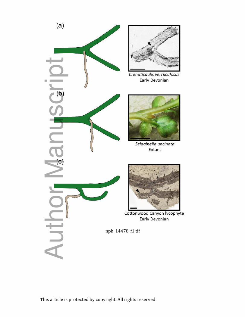
Fig. 1 Branching patterns associated with rhizophore-like structures among lycophytes. (a)
Angular organs of basal lycophytes. Diagrammatic example (at left) shows the divergence of
angular organs proximal to the branching angle of axes. In fossils of Crenaticaulis verruculosus
(at right) the divergence point of the angular organ, which overlaps the main axis, is indicated by
the arrowhead (Banks & Davis 1969, Fig. 18; published with permission from the Botanical
Society of America). Bar, 5 mm. (b) Divergence of the Selaginella uncinata rhizophore occurs
laterally in the immediate vicinity of the branching angle of the shoot. Bar, 2 mm. (c) Rooting
axes derived from K-branches arise from lateral branches of the shoot (arrowhead), shown here
in a drepanophycalean lycophyte from Cottonwood Canyon, Wyoming, USA. Bar, 10 mm.
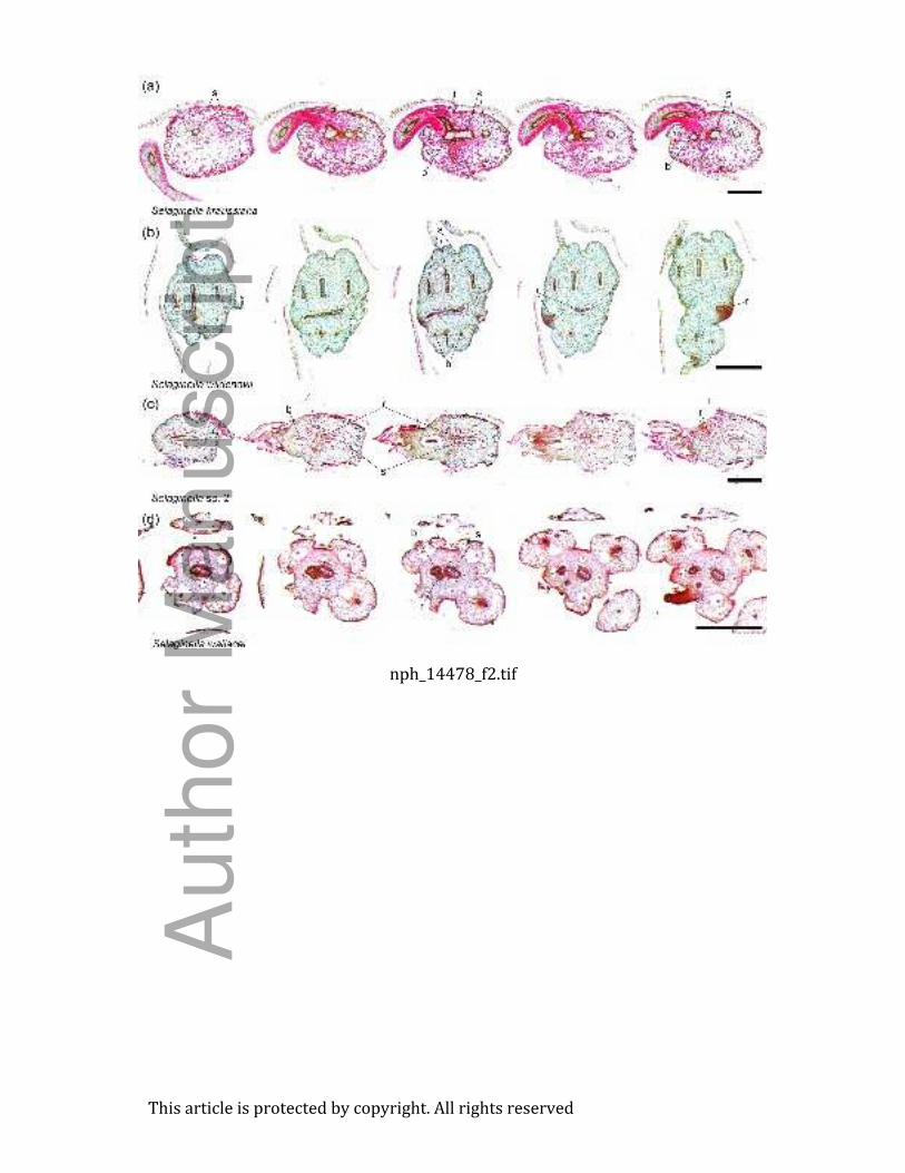
Fig. 2 Serial cross sections through branching points of four Selaginella species showing the
vascular architecture of rhizophore vascularization. (a) Selaginella kraussiana. The main stem
has two meristeles (s) and the rhizophore stele (r) arises from the divergence point of the main
stem and lateral branch (b) steles. Bar, 500 µm. (b) Selaginella wildenovii has a complex stelar
architecture consisting of three meristeles in both the main stem and lateral branch. Two
rhizophores are produced, and diverge centrally from the angle between the steles of the main
stem and lateral branch (see also Fig. 3g,h). Bar, 500 µm. (c) Selaginella sp. 2 showing
divergence of the rhizophore stele from the stele of the main stem, distal to the divergence of the
lateral branch. Bar, 500 µm. (d) Selaginella wallacei. The rhizophore stele diverges from the
stele of the lateral branch. Bar, 500 µm.
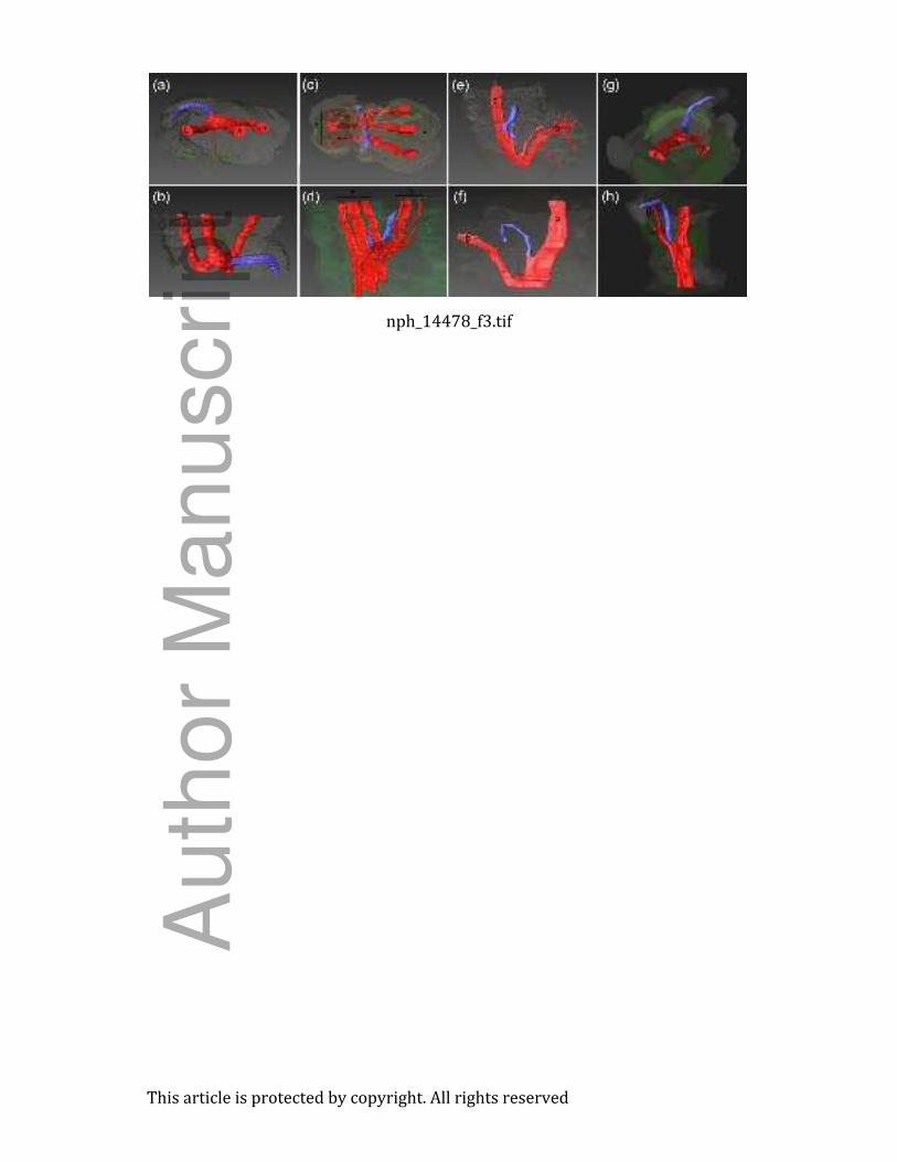
Fig. 3 Three-dimensional reconstructions of the vascular architecture at rhizophore divergence in
Selaginella kraussiana (a, b), Selaginella wildenovii (c, d), Selaginella sp. 2 (e, f), and
Selaginella wallacei (g, h). (a, c, g) Views looking down at branching points from positions
distal to them. (b, f, h) Views in horizontal plane from positions lateral to branching points. (d),
(3) [Author, please clarify ‘(3)’ – which panel are you referring to here?] are oblique views
from positions slightly above branching points. The stele of the main stem (s) and lateral branch
(b) are shown in red, while the rhizophore stele is shown in blue. Note the presence of more than
one rhizophore trace in S. wildenovii. Leaf traces are shown translucent (red) in (a, b, f–h) for
Auth
or
Manuscript
This article is protected by copyright. All rights reserved
clarity. Green structures on the periphery of the stems represent leaves, or parts of leaves, present
on the stem segments.
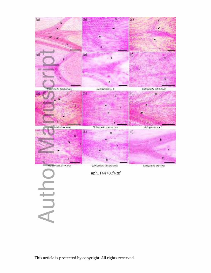
Fig. 4 Tracheid curvature at shoot–rhizophore junctions in nine Selaginella species.
Comparisons between branching points with rhizophores (a–c, g–l) and those without
rhizophores (d–f). Note the arcing of individual tracheids (arrows) from the stele of the main
stem (s) and lateral branches (b) into the rhizophore stele (r), and the absence of such tracheids in
branching points without rhizophores. The thin vascular strand (l) in (e) represents a leaf trace
and not a rhizophore trace. Also note that curvature is more apparent in some species than in
others, and cannot always be clearly shown (e.g., in l) owing to tracheid density, the orientation
of mounted specimens, or the limitations on photographing a thick three dimensional structure
through multiple focal planes. (a) Selaginella kraussiana; bar, 250 µm. (b) Selaginella sp. 2;
bar, 50 µm. (c) S. wildenovii; bar, 50 µm. (d) S. kraussiana branching point without a
rhizophore; bar, 100 µm. (e) Selaginella sp. 2, no rhizophore; bar, 50 µm. (f) S. wildenovii, no
rhizophore; bar, 50 µm. (g) S. delicatula; bar, 100 µm. (h) S. pallescens; bar, 50 µm. (i)
Selaginella sp. 1; bar, 100 µm. (j) S. involvens; bar, 50 µm. (k) S. doederleinii; bar, 50 µm. (l)
Selaginella wallacei; bar, 50 µm.
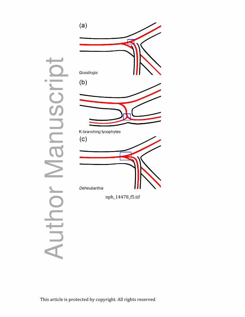
Fig. 5 Diagrammatic representations of vascular architecture in basal lycophytes with
rhizophore-like rooting structures: zosterophyll-grade lycophytes – Gosslingia (a) and
Deheubarthia (c) – with subaxillary branches (angular organs) and basal lycophytes with K-
branching (b). Gosslingia exhibits stem divergence of the vascular supply to the rhizophore-like
axis, K-branching involves branch divergence, and Deheubarthia exhibits central divergence. If
these rooting structures exhibit acropetal polar auxin transport resulting from redirection of auxin
from the shoot system, then curved tracheids should be found in the regions indicated by the blue
boxes.
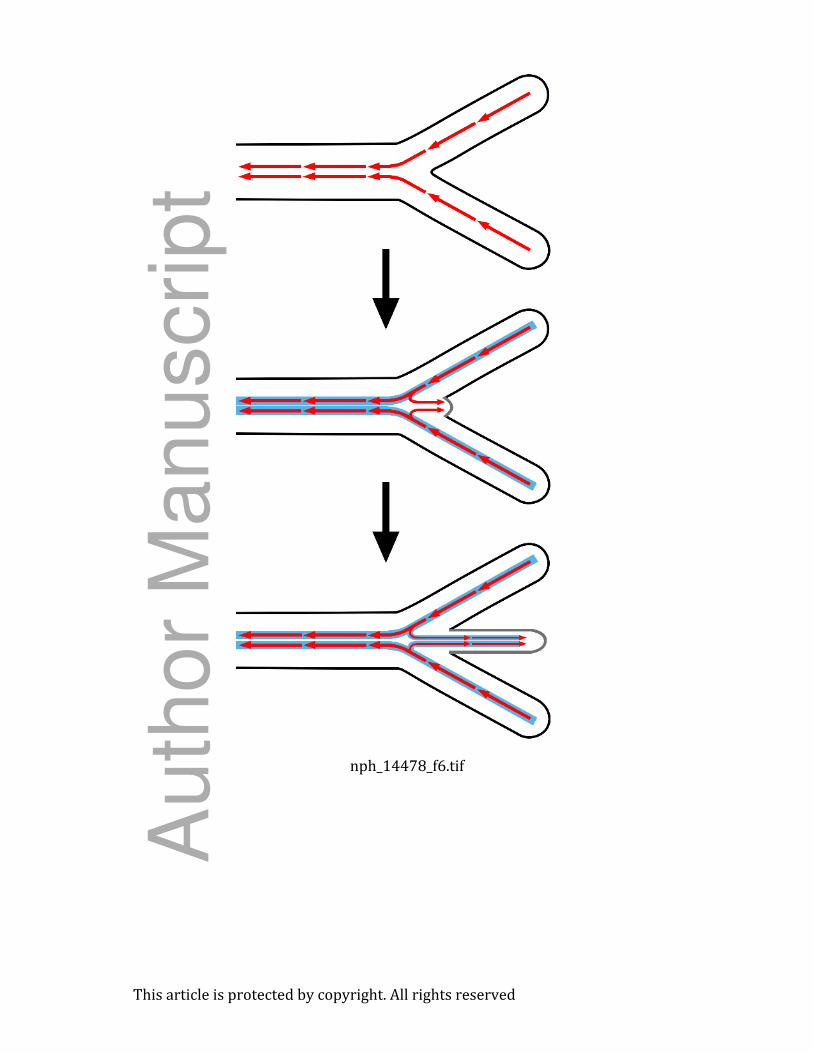
Fig. 6 Patterns of polar auxin transport at the rhizophore–shoot junction. Auxin is redirected
from its basipetal flow in the main stem and lateral branch to flow acropetally into the
rhizophore. This redirection is reflected in the geometry of tracheids at the base of the
rhizophore: these tracheids arch from the steles of the main stem and lateral branch into the
Auth
or
Manuscript
This article is protected by copyright. All rights reserved
rhizophore stele. Red arrows indicate the flow of auxin and solid blue lines represent
differentiated vascular tissues.
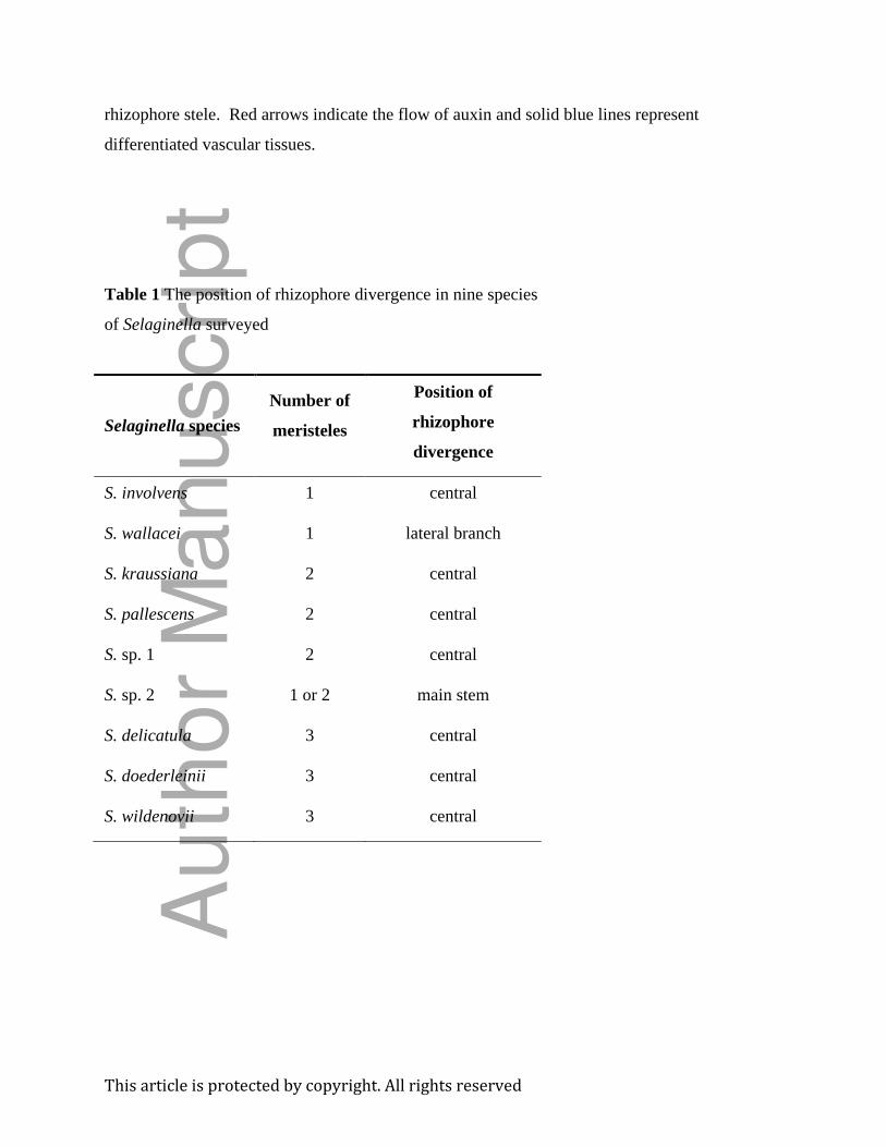
Table 1 The position of rhizophore divergence in nine species
of Selaginella surveyed
Selaginella species
Number of
meristeles
Position of
rhizophore
divergence
S. involvens 1 central
S. wallacei 1 lateral branch
S. kraussiana 2 central
S. pallescens 2 central
S. sp. 1 2 central
S. sp. 2 1 or 2 main stem
S. delicatula 3 central
S. doederleinii 3 central
S. wildenovii 3 central
Auth
or
Manuscript
nph_14478_f1.tif
Thisarticleisprotectedbycopyright.Allrightsreserved
Auth
or
Manuscript
nph_14478_f2.tif
Thisarticleisprotectedbycopyright.Allrightsreserved
Auth
or
Manuscript
nph_14478_f3.tif
Thisarticleisprotectedbycopyright.Allrightsreserved
Auth
or
Manuscript
nph_14478_f4.tif
Thisarticleisprotectedbycopyright.Allrightsreserved
Auth
or
Manuscript
nph_14478_f5.tif
Thisarticleisprotectedbycopyright.Allrightsreserved
Auth
or
Manuscript
nph_14478_f6.tif
Thisarticleisprotectedbycopyright.Allrightsreserved
Auth
or
Manuscript