Embed Size (px)
Citation preview
Vav1 Fine Tunes p53 Control of Apoptosis versusProliferation in Breast CancerShulamit Sebban1., Marganit Farago1., Dan Gashai1, Lena Ilan1, Eli Pikarsky2, Ittai Ben-Porath1,
Shulamit Katzav1*
1Departement of Developmental Biology and Cancer Research, Institute for Medical Research Israel-Canada, Hadassah Medical School - Hebrew University, Jerusalem,
Israel, 2Deaprtment of Immunology & Cancer Research and Department of Pathology, Institute for Medical Research Israel-Canada, Hadassah Medical School - Hebrew
University, Jerusalem, Israel
Abstract
Vav1 functions as a signal transducer protein in the hematopoietic system, where it is exclusively expressed. Vav1 wasrecently implicated in several human cancers, including lung, pancreatic and neuroblasoma. In this study, we analyzed theexpression and function of Vav1 in human breast tumors and breast cancer cell lines. Immunohistochemical analysis ofprimary human breast carcinomas indicated that Vav1 is expressed in 62% of 65 tumors tested and is correlated positivelywith estrogen receptor expression. Based on published gene profiling of 50 breast cancer cell lines, several Vav1-expressingcell lines were identified. RT-PCR confirmed Vav1 mRNA expression in several of these cell lines, yet no detectable levels ofVav1 protein were observed due to cbl-c proteasomal degradation. We used two of these lines, MCF-7 (Vav1 mRNAnegative) and AU565 (Vav1 mRNA positive), to explore the effect of Vav1 expression on breast cell phenotype and function.Vav1 expression had opposite effects on function in these two lines: it reduced proliferation and enhanced cell death inMCF-7 cells but enhanced proliferation in AU565 cells. Consistent with these findings, transcriptome analysis revealed anincrease in expression of proliferation-related genes in Vav1-expressing AU565 cells compared to controls, and an increasein apoptosis-related genes in Vav1-expressing MCF-7 cells compared with controls. TUNEL and c-H2AX foci assaysconfirmed that expression of Vav1 increased apoptosis in MCF-7 cells but not AU565 cells and shRNA experiments revealedthat p53 is required for this pro-apoptotic effect of Vav1 in these cells. These results highlight for the first time the potentialrole of Vav1 as an oncogenic stress activator in cancer and the p53 dependence of its pro-apoptotic effect in breast cells.
Citation: Sebban S, Farago M, Gashai D, Ilan L, Pikarsky E, et al. (2013) Vav1 Fine Tunes p53 Control of Apoptosis versus Proliferation in Breast Cancer. PLoSONE 8(1): e54321. doi:10.1371/journal.pone.0054321
Editor: Wael El-Rifai, Vanderbilt University Medical Center, United States of America
Received October 2, 2012; Accepted December 10, 2012; Published January 14, 2013
Copyright: � 2013 Sebban et al. This is an open-access article distributed under the terms of the Creative Commons Attribution License, which permitsunrestricted use, distribution, and reproduction in any medium, provided the original author and source are credited.
Funding: This work was supported by grants from the Israel Academy of Sciences, and the Israel Cancer Research Foundation, The Israeli Cancer Association andthe Hubert H. Humphrey Center for Experimental Medicine and Cancer Research. The funders had no role in study design, data collection and analysis, decision topublish, or preparation of the manuscript.
Competing Interests: The authors have read the journal’s policy and do not have any conflict of interest.
* E-mail: [email protected]
. These authors contributed equally to this work.
Introduction
The physiological function of Vav1 is restricted to the
hematopoietic system [1], where it plays a critical role in the
development and activation of T-cells. Following stimulation of
the TCR, Vav1 is phosphorylated at N-terminal tyrosine amino
acid residues, and this upregulates its Guanine Nucleotide
Exchange Factor (GEF) activity for specific Rho/RacGTPases,
leading to actin cytoskeletal reorganization [2]. Vav1 also
regulates calcium, ERK-MAP kinase, NFAT and NF- kBsignaling pathways in B and T-cells [3,4]. Recent studies revealed
that wild-type Vav1, which is normally tightly restricted to
hematopoietic cells, is expressed in several human tumor
malignancies, suggesting that it has a role in human cancer.
The involvement of wild type Vav1 in human tumors was first
demonstrated in the neuroblastoma SK-N-MC cell line [5]. A
subsequent screen of 42 primary human neuroblastomas revealed
that the majority expressed Vav1. Wild-type Vav1 was also
identified in more than 50% of 95-pancreatic ductal adenocarci-
noma (PDA) specimens examined and in several PDA cell lines
[6]. Patients with Vav1-positive tumors had a worse prognosis
than patients with Vav1-negative tumors [6]. Aberrant expression
of Vav1 was also found in over 40% of human primary lung
cancers and lung cancer cell lines examined [7] and in melanoma
tissue sections and cell lines [8]. Expression of Vav1 was also
shown in hematological malignancies such as B cell chronic
lymphocytic leukemia (B-CLL), occurring primarily in B-CLL
patients with 13q chromosomal deletions [9]. Depletion of Vav1
expression in pancreatic and lung cancer cell lines reduced colony
formation in soft agar and tumor size in nude mice. This effect of
Vav1 silencing was observed even in the presence of mutant K-
Ras, demonstrating the critical role of Vav1 in tumor development
[6,7].
Vav1 might contribute to malignancy by activating signaling
cascades through its GEF activity, resulting in cytoskeletal
reorganization and transcription [10–12]. Despite its physiological
restriction to hematopoietic cells, Vav1 can be phosphorylated on
tyrosine residues in cells of other tissue origins following
stimulation of growth factor receptors such as EGFR [13], platelet
derived growth factor receptor (PDGFR) [14], and the Nerve
Growth Factor (NGF) receptor, trk [15]. The additional Vav1-
PLOS ONE | www.plosone.org 1 January 2013 | Volume 8 | Issue 1 | e54321
triggered signaling may overwhelm cellular control mechanisms
and promote transformation.
To increase our understanding of Vav1 activity and regulation
in human cancers, we analyzed the involvement of Vav1 in human
breast cancer. In this study, we show that Vav1 is expressed in the
majority of breast carcinomas and that its ectopic expression in
breast cancer cell lines can induce significant changes in these cells,
causing either transformation or cell death.
Materials and Methods
Human Breast Tissue ArrayHuman breast paraffin tissue array (http://www.biochain.com/
biochain/datasheet/Z7020004-B410017.pdf) was purchased (Bio-
chain, CA, USA) and treated according to manufacturer’s
instructions.
ImmunohistochemistryImmunostainings were performed with anti-Vav1 mAbs,
1:10,000 (Upstate Biotechnology, NY, USA) and one with no
antibody using the labeled streptavidin biotin (LAB-SA) technique
(Zymed Laboratories, CA, USA) according to the manufacturer’s
instructions. Staining was evaluated by a board certified pathol-
ogist (E.P) and was quantified as described [7].
Gene ArraymRNA was isolated from cells using the RNeasy mini kit
(QIAGEN, Germany), and samples were subjected to GeneChipHHuman Exon 1.0 ST Array (Affymetrix, CA, USA). Each sample
was composed of a mixture of three independent mRNA
isolations. Data was read and RMA normalized using Partek
Genomic Suite 6.6. Statistical testing for significant genes and
clustering used this package, in addition to dedicated packages
written in Matlab R2011A.
RT-PCRTotal RNA and reverse transcription of Vav1 and GAPDH was
performed as previously described [7].
Quantitative Real-time PCRTotal RNA and cDNAs from cell lines were prepared as above.
Detection of Vav1 was performed using cyber green PCR master
mix (Tamar, Jerusalem, Israel) and the required primers (Table
S1). Analysis was performed using the ABI Prism 7300 real-time
PCR technology (Applied Biosystems, CA, USA). Three in-
dependent experiments were performed, each in triplicate.
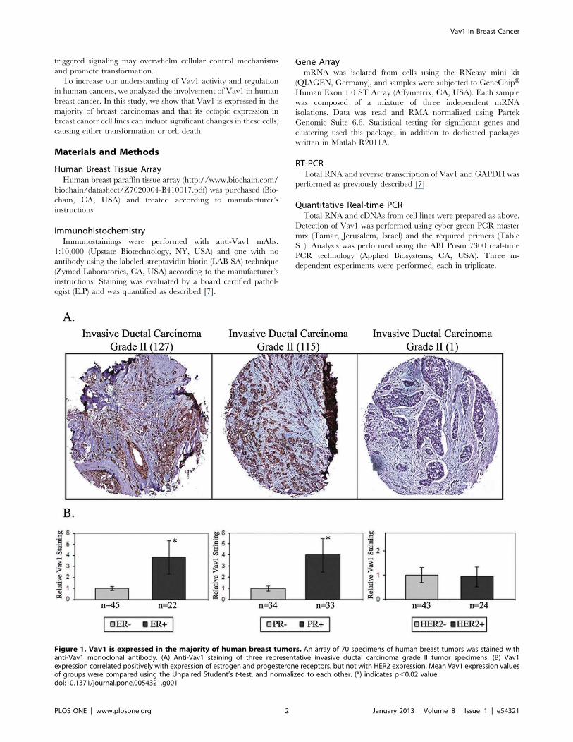
Figure 1. Vav1 is expressed in the majority of human breast tumors. An array of 70 specimens of human breast tumors was stained withanti-Vav1 monoclonal antibody. (A) Anti-Vav1 staining of three representative invasive ductal carcinoma grade II tumor specimens. (B) Vav1expression correlated positively with expression of estrogen and progesterone receptors, but not with HER2 expression. Mean Vav1 expression valuesof groups were compared using the Unpaired Student’s t-test, and normalized to each other. (*) indicates p,0.02 value.doi:10.1371/journal.pone.0054321.g001
Vav1 in Breast Cancer
PLOS ONE | www.plosone.org 2 January 2013 | Volume 8 | Issue 1 | e54321
Cell Culture, Cell Stimulation and Vav1 ExpressionJurkat (acute T cell leukemia, kindly given to us by Dr. Weiss
[16]), U937 (monocytes, histiocytic lymphoma [17]), H358
(bronchioalveolar Non-Small Lung Carcinoma, kindly given to
us by Drs. Gazdar and Minna [18]), AU565 [19] and SK-BR-3
[20] were grown in RPMI medium (Sigma); and MCF-7 cells [21]
were grown in Dulbecco’s modified Eagle’s medium (DMEM)
(Sigma). All media was supplemented with 10% Fetal Bovine
Serum (FBS), Penicillin-Streptomycin and L-Glutamine (Biological
Industries, Israel) and cells were maintained at 37uC with 5%
CO2. For stimulation with CSF1 or EGF, cells were grown to sub-
confluence, starved in serum-free medium for 48 h and treated
with medium containing 50 ng/ml human CSF1 (Peprotech, NJ,
USA) or 100 ng/ml human EGF (Cytolab, Rehovot, Israel) for 5,
15 and 30 min at 37uC. For expression of Vav1, cDNA encoding
the entire Vav1 coding region was generated by PCR and inserted
by in-frame cloning into the vector pcDNA6 (Invitrogen, NY,
USA) at two BstBI restriction sites. AU565 and MCF-7 cells were
stably transfected with either 2 mg of Vav1-pcDNA6 or 2 mg of
empty pcDNA6 vector, using the jetPEIH transfection reagent
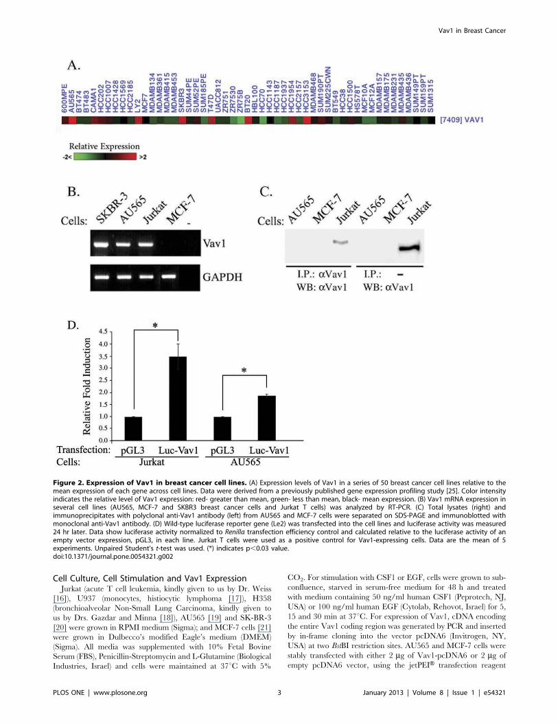
Figure 2. Expression of Vav1 in breast cancer cell lines. (A) Expression levels of Vav1 in a series of 50 breast cancer cell lines relative to themean expression of each gene across cell lines. Data were derived from a previously published gene expression profiling study [25]. Color intensityindicates the relative level of Vav1 expression: red- greater than mean, green- less than mean, black- mean expression. (B) Vav1 mRNA expression inseveral cell lines (AU565, MCF-7 and SKBR3 breast cancer cells and Jurkat T cells) was analyzed by RT-PCR. (C) Total lysates (right) andimmunoprecipitates with polyclonal anti-Vav1 antibody (left) from AU565 and MCF-7 cells were separated on SDS-PAGE and immunoblotted withmonoclonal anti-Vav1 antibody. (D) Wild-type luciferase reporter gene (Le2) was transfected into the cell lines and luciferase activity was measured24 hr later. Data show luciferase activity normalized to Renilla transfection efficiency control and calculated relative to the luciferase activity of anempty vector expression, pGL3, in each line. Jurkat T cells were used as a positive control for Vav1-expressing cells. Data are the mean of 5experiments. Unpaired Student’s t-test was used. (*) indicates p,0.03 value.doi:10.1371/journal.pone.0054321.g002
Vav1 in Breast Cancer
PLOS ONE | www.plosone.org 3 January 2013 | Volume 8 | Issue 1 | e54321
(Polyplus, CA, USA). Transfected cells were selected using 7 mg/ml blasticidine (Invitrogen, NY, USA). For luciferase assay,
AU565 cells were transiently transfected with pGL3-Vav1 or
pGL3 vector using the jetPEIH transfection reagent.
Luciferase Reporter AssayLuciferase reporter assay was performed with Dual- Luciferase
Reporter System (Promega, USA) using a Luminometer Mithras
(Berthold Technologies, Germany) as described [22].
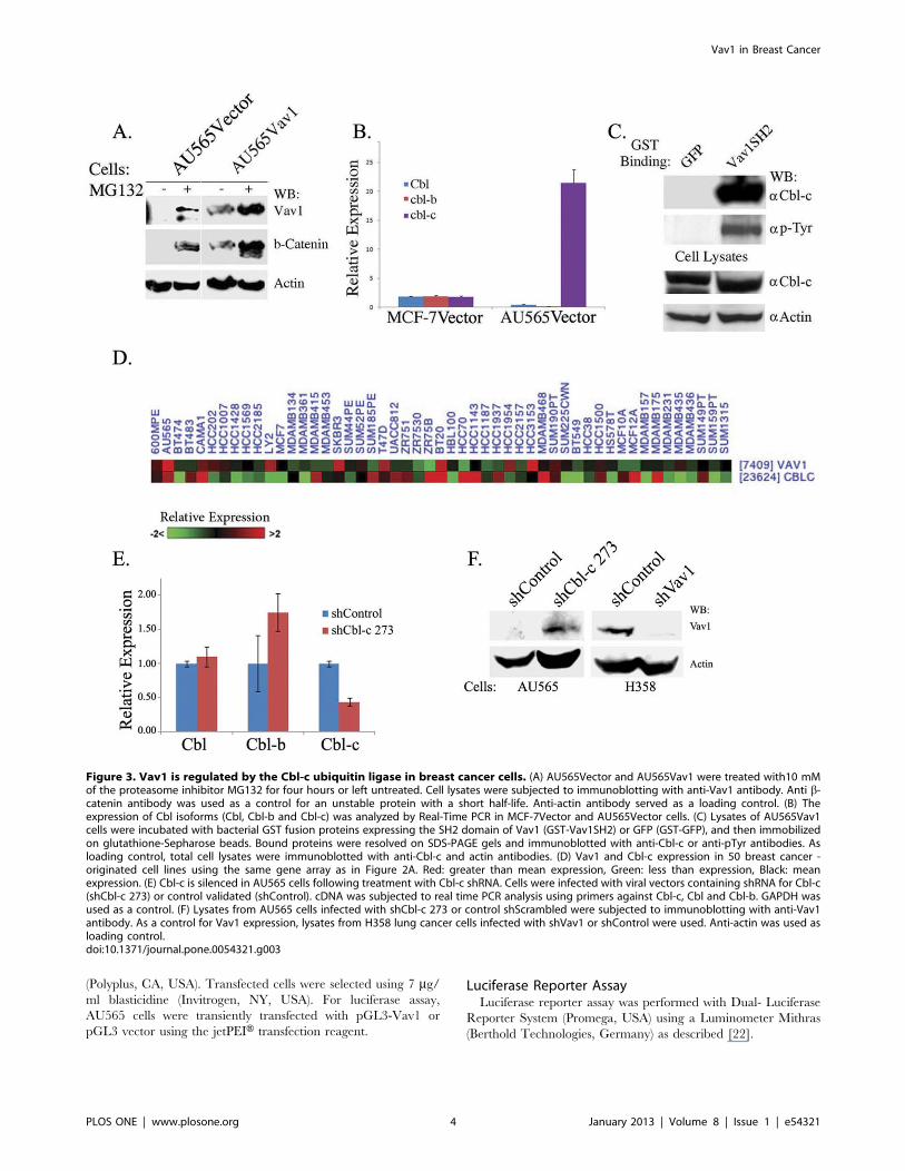
Figure 3. Vav1 is regulated by the Cbl-c ubiquitin ligase in breast cancer cells. (A) AU565Vector and AU565Vav1 were treated with10 mMof the proteasome inhibitor MG132 for four hours or left untreated. Cell lysates were subjected to immunoblotting with anti-Vav1 antibody. Anti b-catenin antibody was used as a control for an unstable protein with a short half-life. Anti-actin antibody served as a loading control. (B) Theexpression of Cbl isoforms (Cbl, Cbl-b and Cbl-c) was analyzed by Real-Time PCR in MCF-7Vector and AU565Vector cells. (C) Lysates of AU565Vav1cells were incubated with bacterial GST fusion proteins expressing the SH2 domain of Vav1 (GST-Vav1SH2) or GFP (GST-GFP), and then immobilizedon glutathione-Sepharose beads. Bound proteins were resolved on SDS-PAGE gels and immunoblotted with anti-Cbl-c or anti-pTyr antibodies. Asloading control, total cell lysates were immunoblotted with anti-Cbl-c and actin antibodies. (D) Vav1 and Cbl-c expression in 50 breast cancer -originated cell lines using the same gene array as in Figure 2A. Red: greater than mean expression, Green: less than expression, Black: meanexpression. (E) Cbl-c is silenced in AU565 cells following treatment with Cbl-c shRNA. Cells were infected with viral vectors containing shRNA for Cbl-c(shCbl-c 273) or control validated (shControl). cDNA was subjected to real time PCR analysis using primers against Cbl-c, Cbl and Cbl-b. GAPDH wasused as a control. (F) Lysates from AU565 cells infected with shCbl-c 273 or control shScrambled were subjected to immunoblotting with anti-Vav1antibody. As a control for Vav1 expression, lysates from H358 lung cancer cells infected with shVav1 or shControl were used. Anti-actin was used asloading control.doi:10.1371/journal.pone.0054321.g003
Vav1 in Breast Cancer
PLOS ONE | www.plosone.org 4 January 2013 | Volume 8 | Issue 1 | e54321
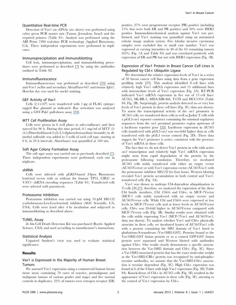
Quantitative Real-time PCRDetection of Vav1 on cDNAs (see above) was performed using
cyber green PCR master mix (Tamar, Jerusalem, Israel) and the
required primers (Table S1). Analysis was performed using the
ABI Prism 7300 real-time PCR technology (Applied Biosystems,
CA). Three independent experiments were performed in tripli-
cates.
Immunoprecipitation and ImmunoblottingCell lysis, immunoprecipitation, and immunoblotting proce-
dures were performed as described [7] by using the antibodies
outlined in Table S2.
ImmunofluorescenceImmunofluorescence was performed as described [23] using
anti-Vav1 mAbs and secondary AlexaFluor-647 anti-mouse IgG1.
Hoechst dye was used for nuclei staining.
GEF Activity of Vav1Cells (2.56106) were transfected with 1 mg of FLAG epitope-
tagged Rac plasmid as indicated. Rac activation was analyzed
using a GST-Pak1 pull-down assay [24].
MTT Cell Proliferation AssayCells were grown in 6 well plates to sub-confluence and then
starved for 96 h. During this time period, 0.1 mg/ml of MTT (3-
(4,5-Dimethylthiazol-2-yl)-2,5-diphenyltetrazolium bromide) in di-
methyl sulfoxide was added to 3 wells of each cell type, starting at
0 h, in 24 h intervals. Absorbance was quantified at 540 nm.
Soft Agar Colony Formation AssayThe soft agar assay was carried out as previously described [7].
Three independent experiments were performed, each one in
triplicate.
shRNACells were infected with pLKO-based (Open Biosystems)
lentiviral vector with or without the human TP53, CBLC or
VAV1- shRNA encoding sequences (Table S1). Transfected cells
were selected with puromycin.
Proteasome InhibitionProteasome inhibition was carried out using 10 mM MG132
(carbobenzoxy-Leu-Leu-leucinal) inhibitor (AGC Scientific, CA,
USA). Cells were lysed after 4 hr incubation and subjected to
immunoblotting as described above.
TUNEL AssayIn Situ Cell Death Detection Kit was purchased (Roche Applied
Science, USA) and used according to manufacturer’s instructions.
Statistical AnalysisUnpaired Student’s t-test was used to evaluate statistical
significance.
Results
Vav1 is Expressed in the Majority of Human BreastTumorsWe assessed Vav1 expression using a commercial human breast
tissue array containing 70 cases of reactive, premalignant and
malignant tumors of various grades and stages and five normal
controls in duplicates. 32% of tumors were estrogen receptor (ER)
positive, 27% were progesterone receptor (PR) positive (including
15% that were both ER and PR positive) and 50% were HER2
positive. Immunohistochemical analysis against Vav1 was per-
formed, and Vav1 staining was quantified using an automated
robotic image analysis system. Five lobular invasive carcinoma
samples were excluded due to small case number. Vav1 was
expressed at varying intensities in 40 of the 65 remaining tumors
(62%) (Fig. 1A and Table S3) and was correlated positively with
expression of ER and PR but not with HER2 expression (Fig. 1B).
Expression of Vav1 Protein in Breast Cancer Cell Lines isRegulated by Cbl-c Ubiquitin LigaseWe determined the relative expression levels of Vav1 in a series
of 50 breast cancer cell lines using data from a gene expression
profiling study [25]. This analysis identified 8 cell lines with
relatively high Vav1 mRNA expression and 15 additional lines
with intermediate levels of Vav1 expression (Fig. 2A). RT-PCR
indicated Vav1 mRNA expression in five out of 13 cell lines:
AU565, SK-BR-3, MDA-MB-468, T47D and HCC1954 (Table
S4; Fig. 2B). Surprisingly, protein analysis detected no or very low
levels of Vav1 protein in these cell lines (Fig. 2C; data not shown).
To assess the transcriptional activity of the vav1 promoter in
AU565 cells, we transfected these cells as well as Jurkat T cells with
a pGL3-vav1 reporter construct containing the minimal regulatory
sequences from the vav1 proximal promoter region upstream of
a luciferase reporter gene [22]. Luciferase expression in AU565
cells transfected with pGL3-vav1 was two-fold higher than in cells
transfected with the pGL3 vector control (Fig. 2D). These data
suggest the Vav1 promoter is active, consistent with the presence
of Vav1 mRNA in these cells.
The fact that we do not detect Vav1 protein in cells with active
vav1 transcription and relatively high Vav1 mRNA expression
might stem from rapid degradation of the protein by the
proteasome following translation. Therefore, we incubated
AU565 cells stably transfected with either an empty vector
(AU565Vector) or with Vav1 expression vector (AU565Vav1) with
the proteasome inhibitor MG132 for four hours. Western blotting
revealed Vav1 protein accumulation in both control and Vav1-
transfected cells (Fig. 3A).
Vav1 was shown to undergo Cbl-dependent ubiquitination in
T-cells [26,27]; therefore, we analyzed the expression of the three
Cbl family members, Cbl, Cbl-b and Cbl-c, in MCF-7Vector
(MCF-7 cells stably transfected with an empty vector) and
AU565Vector cells. While Cbl and Cbl-b were expressed at low
levels in MCF-7Vector cells and at lower levels in AU565Vector
cells, Cbl-c was 20-fold higher in AU565Vector compared with
MCF-7Vector cells (Fig. 3B). Similar results were obtained with
the cells stably expressing Vav1 (MCF-7Vav1 and AU565Vav1,
data not shown). To analyze whether Vav1 and Cbl-c physically
associate in these cells, we incubated lysates of AU565Vav1 cells
with a protein containing the SH2 domain of Vav1 fused to
gluthatione-S-transferase (Vav1SH2-GST). Proteins bound to the
Vav1SH2-GST fusion protein or to a control GFP-GST fusion
protein were separated and Western blotted with antibodies
against Cbl-c. Our results clearly demonstrate a specific associa-
tion between the Vav1SH2 domain and Cbl-c (Fig. 3C). Since
a Vav1SH2-associated protein that has the exact molecular weight
as the Vav1SH2-CBl-c protein was recognized by anti-phospho-
tyrosine antibodies, we assume that the Vav1SH2-Cbl-c associa-
tion is tyrosine dependent (Fig. 3C). High Cbl-c expression was
found in 6 of the 8 lines with high Vav1 expression (Fig. 3D; Table
S4). Knock-down of Cbl-c in AU565 cells (Fig. 3E) resulted in the
appearance of Vav1 expression in these cells (Fig. 3F), attesting to
the control of Vav1 expression by Cbl-c.
Vav1 in Breast Cancer
PLOS ONE | www.plosone.org 5 January 2013 | Volume 8 | Issue 1 | e54321
Vav1 in Breast Cancer
PLOS ONE | www.plosone.org 6 January 2013 | Volume 8 | Issue 1 | e54321
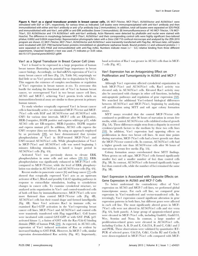
Vav1 as a Signal Transducer in Breast Cancer Cell LinesVav1 is found to be expressed in a large proportion of human
breast tumors illustrating its potential huge importance in breast
cancer biology. Accordingly, we find mRNA expression of Vav1 is
many breast cancer cell lines (Fig. 2A, Table S4); surprisingly we
find little or no Vav1 protein mainly due to degradation by Cbl-c.
This suggests the existence of complex mechanisms or regulation
of Vav1 expression in breast tumors in vivo. To overcome this
hurdle for studying the functional role of Vav1 in human breast
cancer, we overexpressed Vav1 in two breast cancer cell lines,
AU565 and MCF-7, achieving Vav1 protein levels which on
immunohistochemical assay are similar to those present in primary
human tumors.
To study whether ectopically expressed Vav1 in breast cancer
cells is functionally active, we stimulated MCF-7Vector and MCF-
7Vav1 cells with EGF and AU565Vector and AU565Vav1 with
CSF1 for various time intervals. MCF-7 cells are ER-positive,
HER-2-negative, EGFR positive and express wild-type p53, while
AU565 cells are ER-negative, HER-2-positive, EGFR negative
and express mutant p53. In addition, AU565 cells express the
CSF1 receptor (data not shown). By using an approach employed
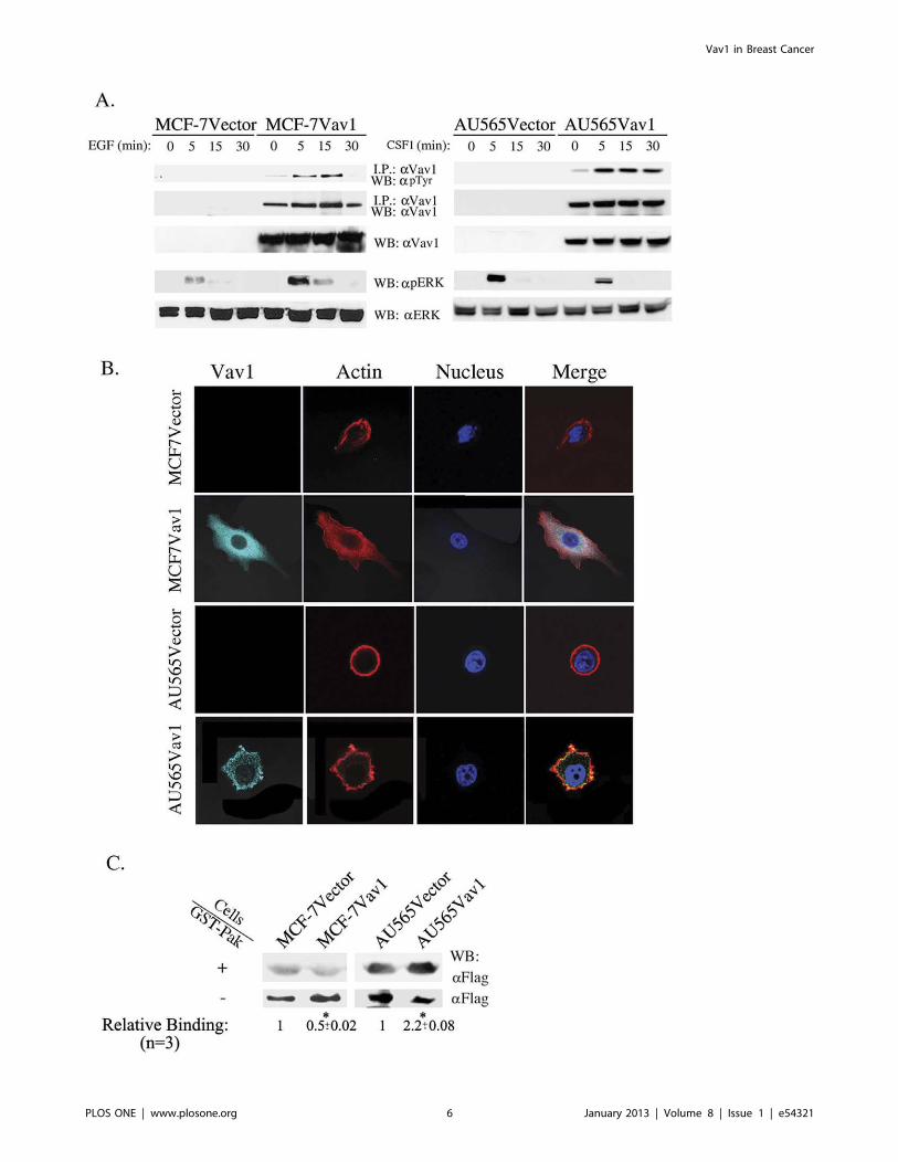
by us previously [28], we have demonstrated that tyrosine
phosphorylation of Vav1 in EGF-treated MCF-7Vav1 and
CSF1-treated AU565Vav1 cells. While phosphorylation of Vav1
in MCF-7Vav1 and AU565Vav1 cells was noted beginning 5
minutes following stimulation, it lasted a longer period in
AU565Vav1 cells (Fig. 4A).
Activated Vav1 was previously shown to elevate ERK
phosphorylation in some cells and not others [29–31]. ERK
phosphorylation was significantly enhanced in MCF-7Vav1 cells
compared to MCF-7Vector, while the level of ERK phosphory-
lation was similar in AU565Vav1 and AU565Vector cells (Fig. 4A).
Recent studies in pancreatic cancer [6] and lung cancer [7] cells
showed that ectopically expressed Vav1 acts as an upstream
activator of Rac1, RhoA and possibly Cdc42 signaling pathways in
response to extracellular stimulation, leading to cytoskeleton
changes in cancer cells. To examine cytoskeletal structure, we
analyzed actin organization in Vav1- and control-transfected cells
of both cell lines by immunofluorescence. MCF-7Vav1 cells were
more flattened than control MCF-7Vector cells (Fig. 4B).
AU565Vav1 cells lost their round shape and formed lamellipodia
(Fig. 4B). Since Vav1 activates Rac1 in immune cells, we
examined Rac1-GTP activation in the Vav1-expressing breast
cancer cell lines. MCF-7Vav1 and AU565Vav1 and control cells
were transiently transfected with Flag tagged-Rac1. Cell lysates
were incubated with control GST-GFP or with GST–PAK (p21
activated kinase 1), a fusion of GST with the Rac/Cdc42 binding
domain (PBD) of human PAK [24]. As expected, in AU565 cells,
expression of Vav1 induced activation of Rac as evident by
increased binding to GST-PAK. However, In MCF-7 cells, similar
expression downmodulated Rac activity (Fig. 4C). Importantly,
basal activation of Rac1 was greater in AU565 cells than in MCF-
7 cells (Fig. 4C).
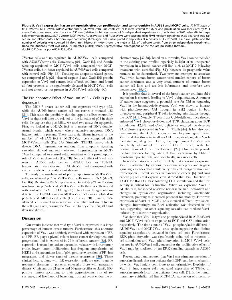
Vav1 Expression has an Antagonizing Effect onProliferation and Tumorigenicity in AU565 and MCF-7CellsAlthough Vav1 expression affected cytoskeletal organization in
both MCF-7Vav1 and AU565Vav1 cells, Rac1 activity was
elevated only in AU565Vav1 cells. Elevated Rac1 activity may
also be associated with changes in other cell functions, including
anti-apoptotic pathways and regulation of gene expression [32].
We searched for additional Vav1-related biological differences
between AU565Vav1 and MCF-7Vav1, beginning by analyzing
cell proliferation using MTT and soft agar colony formation
assays.
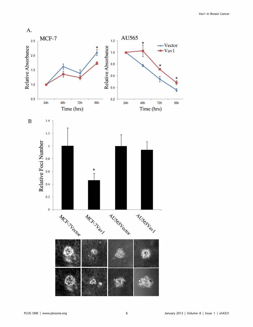
MTT assays revealed that the control MCF-7Vector cells
continued to proliferate after 96 hours of starvation in serum free
media, while control AU565Vector cells exhibited reduced growth
(Fig. 5A). These differences might stem from a disparity in secreted
cytokines/growth factors to the medium by these two cells lines
[33]. In addition, Vav1 expression had opposing effects on
proliferation in these two breast cell lines. At most time points
during starvation, MCF-7Vav1 cells had a lower growth rate than
the MCF-7Vector control cells. In contrast, AU565Vav1 cells had
a higher growth rate than AU565Vector cells after 96 hours of
starvation in serum free media (Fig. 5A).
Colony formation assays corroborated these MTT findings.
When grown on soft agar, MCF-7Vav1 cells formed significantly
smaller foci and a smaller number of foci than control cells
(Fig. 5B). In contrast, AU565Vav1 cells formed significantly larger
foci than control cells, while the number of foci remained the same
(Fig. 5B).
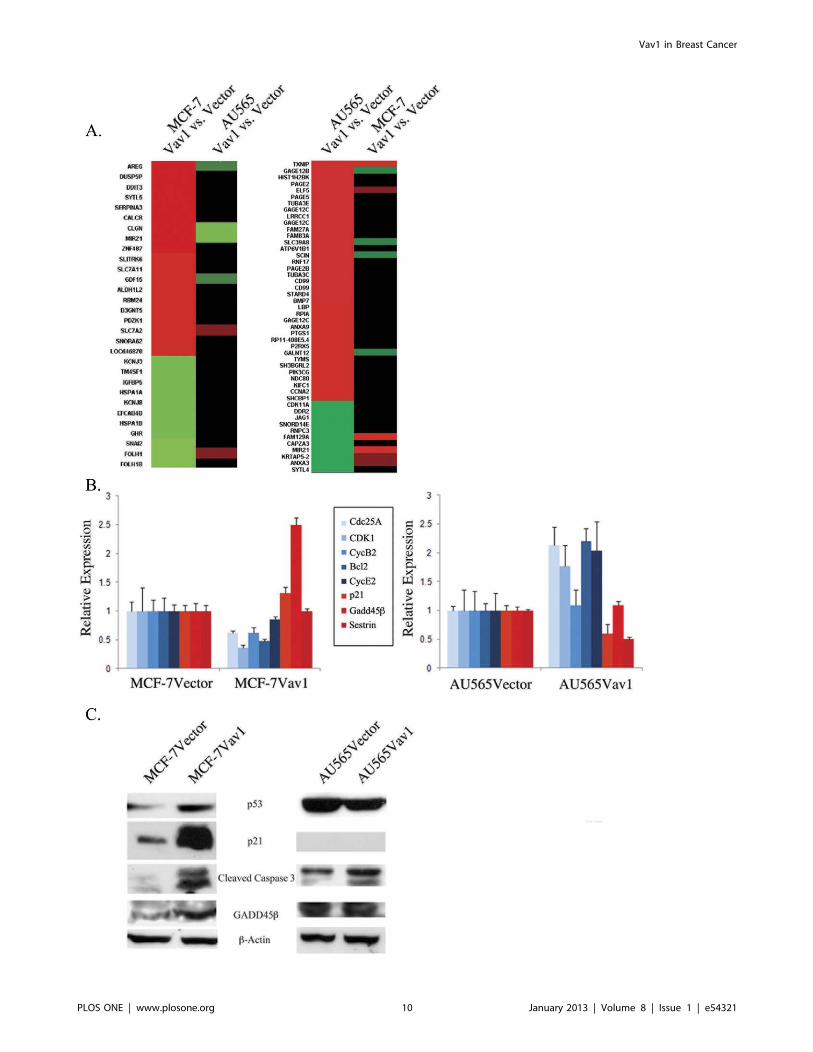
Vav1 Expression is Associated with Opposite Effects onGene Expression in AU565 and MCF-7 CellsTo better understand the contradictory effect of Vav1
expression on AU565 and MCF-7 cell lines, we performed global
transcriptome assays. For each cell line, we compared gene
expression in Vav1-transfected and vector-transfected cells. In-
terestingly, Vav1 expression caused significant alteration in gene
expression patterns in both lines, but different genes were affected
in each cell line. The most significantly altered genes in MCF-
7Vav1 cells were not altered in AU565Vav1 cells and vice versa
(Fig. 6A, both panels). A large group of apoptosis-related genes
were elevated in MCF-7Vav1 cells, including Gadd45, Gadd153,
Wee, Sestrins and Noxa. In contrast, a large number of
proliferation-related genes were elevated in AU565Vav1 cells,
including Cyclins A, B, D and E, Cdc25A, B and C, Cdk1 and 2
and PI3K. These observations were validated by quantitative RT-
PCR of selected genes. Cdc25A, Cdk1, Cyclin B2 and Cyclin E
were down-regulated in MCF-7Vav1 cells compared with MCF-
Figure 4. Vav1 as a signal transducer protein in breast cancer cells. (A) MCF-7Vector, MCF-7Vav1, AU565Vector and AU565Vav1 werestimulated with EGF or CSF1, respectively, for various times as indicated. Cell lysates were immunoprecipitated with anti-Vav1 antibody and thenimmunoblotted with either anti-Vav1 antibody or anti- pTyr antibody (top 2 immunoblots). In addition, total cell lysates were separated on SDS-PAGEand immunoblotted with anti-Vav1, anti-pERK or anti-ERK antibodies (lower 3 immunoblots). (B) Immunofluorescence of 145 MCF-7Vector, 176 MCF-7Vav1, 355 AU565Vector and 174 AU565Vav1 with anti-Vav1 antibody. Actin filaments were detected by phalloidin and nuclei were stained withHoechst. The difference in morphology between MCF-7Vav1, AU565Vav1 and their corresponding control cells were highly significant (two-tailoredpValue; 0.0002 and 0.0024 respectively). Representative photographs taken with a Zeiss LSM 710 confocal microscope and analyzed by the ZEN 2010program are shown. (C) MCF-7Vector, MCF-7Vav1, AU565Vector and AU565Vav1 were transiently transfected with Flag-Rac. 48 hours later, cell lysateswere incubated with GST–PAK bacterial fusion proteins immobilized on glutathione sepharose beads. Bound proteins (+) and unbound proteins (2)were separated on SDS–PAGE and immunoblotted with anti-Flag mAbs. Numbers indicate mean (+/2 S.E.) relative binding from three differentexperiments. Unpaired Student’s t-test was used. (*) indicates p,0.05 value.doi:10.1371/journal.pone.0054321.g004
Vav1 in Breast Cancer
PLOS ONE | www.plosone.org 7 January 2013 | Volume 8 | Issue 1 | e54321
Vav1 in Breast Cancer
PLOS ONE | www.plosone.org 8 January 2013 | Volume 8 | Issue 1 | e54321
7Vector cells and up-regulated in AU565Vav1 cells compared
with AU565Vector cells. Conversely, p21, Gadd45b and Sestrin
were up-regulated in MCF-7Vav1 cells compared with MCF-
7Vector cells, but down-regulated in AU565Vav1 cells compared
with control cells (Fig. 6B). Focusing on apoptosis-related genes,
we compared p53, p21, cleaved caspase 3 and Gadd45b protein
expression in Vav1 and control cells of both cell lines, and found
all four proteins to be significantly elevated in MCF-7Vav1 cells
and not altered or not present in AU565Vav1 cells (Fig. 6C).
The Pro-apoptotic Effect of Vav1 on MCF-7 Cells is p53-dependantThe MCF-7 breast cancer cell line expresses wild-type p53,
while the AU565 breast cancer cell line carries a mutated p53
[34]. This raises the possibility that the opposite effects exerted by
Vav1 in these cell lines are related to the function of p53 in these
cells. To explore this possibility, we used two assays for apoptosis.
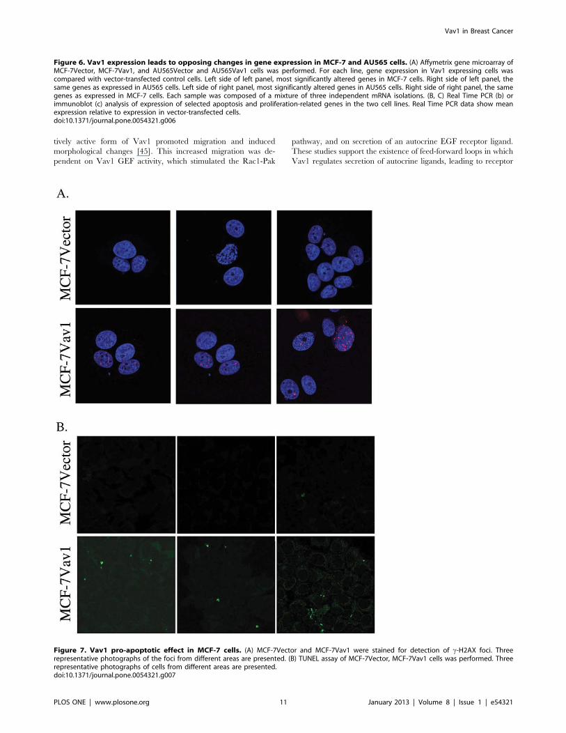
First, we looked at c-H2AX foci as a marker of DNA double-
strand breaks, which occur where extensive apoptotic DNA
fragmentation is present. There was a significant increase in the
number of c-H2AX foci in MCF-7Vav1 cells compared with
MCF-7Vector cells (Fig. 7A). Similarly, TUNEL assay, which
detects DNA fragmentation resulting from apoptotic signaling
cascades, showed markedly elevated fragmentation in MCF-
7Vav1 compared to control cells, consistent with a pro-apoptotic
role of Vav1 in these cells (Fig. 7B). No such effect of Vav1 was
seen in AU565 cells: neither c-H2AX foci nor TUNEL
fragmentation were elevated in AU565Vav1 cells compared with
vector transfected cells (data not shown).
To verify the involvement of p53 in apoptosis in MCF-7Vav1
cells, we silenced p53 in MCF-7Vav1 cells using shRNA (shp53;
Fig. 8A). Relative mRNA expression of Gadd45b, p21 and Sestrin
was lower in p53-silenced MCF-7Vav1 cells than in cells treated
with control shRNA (pLKO; Fig. 8B). The elevated fragmentation
detected by TUNEL assay in MCF-7Vav1 cells was abolished in
p53-silenced MCF-7Vav1 cells (Fig. 8C vs. 7B). Finally, p53-
silenced cells showed an increase in the number and size of foci in
the soft agar assay, erasing the Vav1 inhibitory effect (Fig. 8D and
data not shown).
Discussion
Our results indicate that wild-type Vav1 is expressed in a large
percentage of human breast tumors. Furthermore, this aberrant
expression of Vav1 was positively correlated with expression of ER
and PR. ER plays a pivotal role in breast cancer development and
progression, and is expressed in 75% of breast cancers [35]. ER
expression is related to patient age and correlates with lower tumor
grade, lower tumor proliferation, less frequent amplification of
HER2 and concomitant loss of p53, positive expression of PR, less
metastases, and slower rates of disease recurrence [36]. These
clinical factors, along with ER expression itself, are used to guide
treatment decisions in patients, especially those with metastatic
disease. Clinicians use 21-gene and 70-gene profiles to classify ER-
positive tumors according to their aggressiveness, risk of re-
currence, and likelihood of benefiting from adjuvant endocrine or
chemotherapy [37,38]. Based on our results, Vav1 can be included
in the existing gene profiles, especially in light of its unexpected
expression in a breast cancer cell line such as MCF-7 following
treatment with estradiol (Fig. S1); however its prognostic value
remains to be determined. Two previous attempts to associate
Vav1 with human breast cancer used smaller cohorts of breast
cancer specimens and a very small number of human breast
cancer cell lines and are less informative and therefore were
inconclusive [39,40].
It is possible that in several of the breast cancer cell lines cbl-c
expression is elevated, leading to Vav1 ubiquitination. A number
of studies have suggested a potential role for Cbl in regulating
Vav1 in the hematopoietic system. Vav1 was shown to interact
with phosphorylated Cbl through its SH2 region in both
thymocytes and peripheral T cells following stimulation through
the TCR [41]. Notably, T cells from Cbl-b-deficient mice showed
enhanced Vav1 phosphorylation and TCR clustering upon TCR
stimulation [42,43], and Cbl-b deficiency restored the defective
TCR clustering observed in Vav2/2 T cells [44]. It has also been
demonstrated that Cbl functions as an ubiquitin ligase toward
Vav1 and that this activity allows Cbl to negatively regulate Vav1-
mediated signaling [26]. Lastly, the requirement for Vav1 was
completely eliminated in Vav12/2Cbl2/2 mice, with full
normalization of T cell development [27]. Our results provide
the first evidence for regulation of Vav1 expression by Cbl-c in
non-hematopoietic cells, and specifically, in cancer cells.
In non-hematopoietic cells, it is likely that aberrantly expressed
Vav1 is activated by various membrane receptors and triggers
signaling cascades that result in cytoskeletal reorganization and
transcription. Recent studies in pancreatic cancer [6] and lung
cancer [7] cells that express Vav1 showed that Vav1 functions as
a GEF for Rac1 GTPase following EGF stimulation and that this
activity is critical for its function. When we expressed Vav1 in
AU565 cells, we indeed observed remarkable Rac1 activation and
changes in cytoskeleton organization including lamellipodia
formation, pointing to increased potential for motility. However,
expression of Vav1 in MCF-7 cells induced different cytoskeletal
changes. Interestingly, no Rac1 activation was observed in this
case, suggesting that other signaling cascades can mediate Vav1-
induced cytoskeleton reorganization.
We show that Vav1 is tyrosine phosphorylated in AU565Vav1
and MCF-7Vav1 cells in response to EGF and CSF1 stimulation
respectively. The time course of Vav1 phosphorylation differed in
AU565Vav1 and MCF-7Vav1 cells, again suggesting that distinct
signaling cascades are activated in these cell lines. Furthermore,
ERK phosphorylation was significantly enhanced in response to
cell stimulation and Vav1 phosphorylation in MCF-7Vav1 cells,
but not in AU565Vav1 cells, suggesting the proliferative effect of
Vav1 may be mediated by an ERK signaling cascade in AU565
cells.
Recent data demonstrated that Vav1 can stimulate secretion of
autocrine ligands that can activate the EGFR, another mechanism
by which Vav1 might contribute to tumorigenicity. Depletion of
Vav1 in lung cancer cells decreased expression of TGFa, an
autocrine growth factor that activates these cells [7]. In the human
mammary epithelial cell line MCF-10A, expression of a constitu-
Figure 5. Vav1 expression has an antagonistic effect on proliferation and tumorigenicity in AU565 and MCF-7 cells. (A) MTT assay ofMCF-7Vector, MCF-7Vav1, AU565Vector and AU565Vav1 cells. Sub-confluent cells were starved for 96 hr and proliferation was measured by MTTassay. Data show mean absorbance at 550 nm (relative to 24 hour value) of 3 independent experiments. (*) indicates p,0.05 value (B) Soft agarcolony formation assay. MCF-7Vector, MCF-7Vav1, AU565Vector and AU565Vav1 were suspended in RPMI medium containing 0.3% agar and 10% calfserum, and plated onto a bottom layer containing 0.8% agar. Cells were plated in triplicates at a density of 16105/well in a 6-well plate, and thenumber of colonies was counted 14 days later. Histogram (top) shows the mean 6 S.E. of triplicate values from three independent experiments.Unpaired Student’s t-test was used. (*) indicates p,0.05 value. Representative photographs of the foci are presented (bottom).doi:10.1371/journal.pone.0054321.g005
Vav1 in Breast Cancer
PLOS ONE | www.plosone.org 9 January 2013 | Volume 8 | Issue 1 | e54321
Vav1 in Breast Cancer
PLOS ONE | www.plosone.org 10 January 2013 | Volume 8 | Issue 1 | e54321
tively active form of Vav1 promoted migration and induced
morphological changes [45]. This increased migration was de-
pendent on Vav1 GEF activity, which stimulated the Rac1-Pak
pathway, and on secretion of an autocrine EGF receptor ligand.
These studies support the existence of feed-forward loops in which
Vav1 regulates secretion of autocrine ligands, leading to receptor
Figure 6. Vav1 expression leads to opposing changes in gene expression in MCF-7 and AU565 cells. (A) Affymetrix gene microarray ofMCF-7Vector, MCF-7Vav1, and AU565Vector and AU565Vav1 cells was performed. For each line, gene expression in Vav1 expressing cells wascompared with vector-transfected control cells. Left side of left panel, most significantly altered genes in MCF-7 cells. Right side of left panel, thesame genes as expressed in AU565 cells. Left side of right panel, most significantly altered genes in AU565 cells. Right side of right panel, the samegenes as expressed in MCF-7 cells. Each sample was composed of a mixture of three independent mRNA isolations. (B, C) Real Time PCR (b) orimmunoblot (c) analysis of expression of selected apoptosis and proliferation-related genes in the two cell lines. Real Time PCR data show meanexpression relative to expression in vector-transfected cells.doi:10.1371/journal.pone.0054321.g006
Figure 7. Vav1 pro-apoptotic effect in MCF-7 cells. (A) MCF-7Vector and MCF-7Vav1 were stained for detection of c-H2AX foci. Threerepresentative photographs of the foci from different areas are presented. (B) TUNEL assay of MCF-7Vector, MCF-7Vav1 cells was performed. Threerepresentative photographs of cells from different areas are presented.doi:10.1371/journal.pone.0054321.g007
Vav1 in Breast Cancer
PLOS ONE | www.plosone.org 11 January 2013 | Volume 8 | Issue 1 | e54321
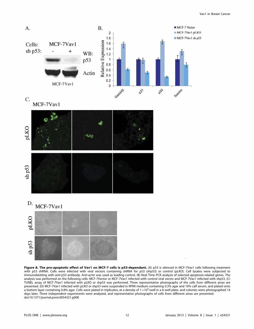
Figure 8. The pro-apoptotic effect of Vav1 on MCF-7 cells is p53-dependant. (A) p53 is silenced in MCF-7Vav1 cells following treatmentwith p53 shRNA. Cells were infected with viral vectors containing shRNA for p53 (shp53) or control (pLKO). Cell lysates were subjected toimmunoblotting with anti-p53 antibody. Anti-actin was used as loading control. (B) Real Time PCR analysis of selected apoptosis-related genes. Theanalysis was performed on the following cells: MCF-7Vector or MCF-7Vav1 infected with control viral vector and MCF-7Vav1 infected with shp53. (C)TUNEL assay of MCF-7Vav1 infected with pLKO or shp53 was performed. Three representative photographs of the cells from different areas arepresented. (D) MCF-7Vav1 infected with pLKO or shp53 were suspended in RPMI medium containing 0.3% agar and 10% calf serum, and plated ontoa bottom layer containing 0.8% agar. Cells were plated in triplicates, at a density of 16105/well in a 6-well plate, and colonies were photographed 14days later. Three independent experiments were analyzed, and representative photographs of cells from different areas are presented.doi:10.1371/journal.pone.0054321.g008
Vav1 in Breast Cancer
PLOS ONE | www.plosone.org 12 January 2013 | Volume 8 | Issue 1 | e54321
stimulation and subsequent increases in Vav1 activation. The
additional stimulatory input provided by Vav1 signaling in cells
where it is aberrantly expressed may overwhelm control mechan-
isms and tip the scales in favor of transformation.
Our soft agar and MTT assays showed opposing phenotypes of
AU565 and MCF-7 cells ectopically expressing Vav1. In
AU565Vav1 cells we observed an increase in foci number and
size, indicative of a higher proliferation rate in comparison to
AU565Vector cells. We observed the opposite in MCF-7Vav1
cells, which formed smaller foci than MCF-7Vector cells. These
surprising results raised the possibility that the expression of Vav1
affects gene expression in a different manner in these cell lines.
Indeed, Vav1 leads to an increase in expression of pro-pro-
liferation genes in AU565, while pro-apoptosis genes are elevated
in MCF-7 cells.
The anti-apoptotic effect of Vav1 has been shown in cancers of
hematopoietic origin. In vitro knockdown of Vav1 in anaplastic
large cell lymphoma was sufficient to cause cell cycle arrest and
apoptosis of these cells [46]. In the HL-60 and NB4 promyelocytic
cell lines, down-regulation of Vav1 affected expression of a number
of cell cycle/apoptosis-related proteins [47]. Lastly, Vav1 was
found to protect Jurkat T cells from Fas-mediated apoptosis by
promoting Bcl-2 transcription through its GEF activity toward
Rac2 [48]. Other studies have pointed to a pro-apoptotic role for
Vav1 in hematopoietic cells. For instance, during negative
selection, Vav1 promotes antigen-induced thymocyte apoptosis,
and inhibitors of the actin cytoskeleton or protein kinase C (PKC)
reverse the effect [49]. In activated CD4+ T cells, the Vav1-Rac
pathway is a critical component of TCR-induced cell death [50].
Vav1 was also shown to mediate apoptosis in L-MAT, a human
lymphoblastic T cell line [51]. In macrophages, the engulfment of
apoptotic cells requires the activation of Vav1/Rac1 and sub-
sequent actin polymerization to form the phagocytic cup [52].
We demonstrate here for the first time that Vav1 can influence
apoptosis in non-hematopoietic cancer cells. The normal cellular
response to oncogenic stress requires the tumor suppressor protein
p53. Nevertheless, the mechanisms linking oncogene activation to
p53 induction have remained controversial. Evidence from studies
of early-stage human tumors and animal models suggest that
oncogene-induced replication stress activates a DNA damage
response (DDR), which in turn activates p53 [53–55]. Using
cH2AX and TUNEL assays, we observed significant DDR in our
MCF-7Vav1 cells, as well as a remarkable increase in several
apoptosis-related proteins. We also demonstrated that the
apoptotic phenotype of MCF-7Vav1 cells is p53-dependant.
Several oncogenes and tumor suppressor genes have been shown
to have dual behavior in cancer, dependent on the cellular
environment. One such example is the transcription factor NF-kB.The role of the NF-kB signaling pathway in cancer is complex.
While in some cancers, NF-kB is oncogenic, and can serve as an
excellent target for tumor therapy, there is evidence it can also
suppress tumorigenesis [56]. Another example of a protein with
a dual role is Yap, a small protein that binds to many transcription
factors and modulates their activity. Yap increases the pro-
apoptotic function of p73 following DNA damage, and therefore
its activity favors tumor-suppression. However, other studies have
recently shown a role for Yap in cell differentiation, cell
transformation and in the regulation of organ size [57].
Whether Vav1 can play a dual role as a pro- or an anti-
apoptotic protein in cancer cells of non-hematopoietic origin has
never been tested directly, yet several studies point to such roles in
hematopoietic cells. While Vav1 was shown to protect Jurkat T
cells from Fas-mediated apoptosis by promoting Bcl-2 transcrip-
tion through its GEF activity [46], Gu et al., demonstrated that
oncogenic Vav1, which is constitutively active as a GEF, induces
Rac-dependent apoptosis via inhibition of Bcl-2 family proteins
and collaborates with p53 deficiency to promote hematopoietic
progenitor cell proliferation [58]. Thus, it is conceivable that in
non-hematopoietic cancer cells wild-type Vav1 might function in
a similar fashion to oncogenic Vav1 in hematopoietic cells due to
its constitutive activation by various aberrantly functional signaling
cascades. Moreover, its activity could depend on additional genetic
aberrations, such as the p53 pathway.
The fact that Vav1 is shown by us in this study to have opposite
effects when expressed in two breast cancer cell lines, MCF-7 and
AU565, clearly highlights the importance of the cellular environ-
ment on Vav1 function. Similarly, CKIa was recently shown to be
tumor suppressive when p53 is inactive. Combined ablation of
CKIa and p53 triggered high-grade dysplasia with extensive
proliferation [59].
NF-kB, Yap and CKIa represent three major developmental
pathways (NF-kB, Hippo and Wnt signaling, respectively) that can
lead to transformation when aberrantly expressed. Our results
highlight a similar role for Vav1, an important player in its own
signaling cascade in thymocytes, which contributes to cancer
development when aberrantly expressed in the breast. In addition,
our results indicate that the effect of ectopic Vav1 expression is
highly dependent on other cellular factors, including p53
availability.
Supporting Information
Figure S1 Vav1 expression in MCF-7 cells followingtreatment with estradiol. MCF-7 cells were treated for 48 hr
with 0, 10 and 20 nM of estrodiol (SIGMA). cDNA was subjected
to RT-PCR using Vav1 primers. Actin was used as a loading
control.
(TIF)
Table S1 Primers used for Real-Time PCR and shRNAsequences. This table details the sequences of primers used for
Real-Time PCR performed. Also, included are the sequences used
for shRNA.
(DOC)
Table S2 Antibodies used for Immunoprecipitation,Immunoblotting, Immunohistochemistry and Immuno-fluorescence. The antibodies for western blotting, immunopre-
cipitation, immunohistochemistry and immunofluorescence used
in the study are detailed, including the source for their purchase.
(DOC)
Table S3 Vav1 expression in breast cancer tissue array.The table details the various cancer tissues used in the study
including their receptor expression (ER, PR, and HER2 from – to
+++) and cancer staging (TNM) according to the manufecturere’s
information. Also, included is the level of Vav1 protein expression
calculated as detailed in the Material and Methods section.
(XLS)
Table S4 Vav1 (mRNA and protein) and Cbl-c (mRNA)expression in various breast cancer cell lines. The mRNA
and protein expression level of Vav1 and mRNA expression of
Cbl-c as assessed in our experiments (2; +/2; ++) in various
human breast cancer cell lines used in our experiments.
(XLS)
Acknowledgments
We are indebted to Dr. Susan Lewis for editing the manuscript. We thank
David Knigin for help in analysis of the tissue array, Roi Granit for help
Vav1 in Breast Cancer
PLOS ONE | www.plosone.org 13 January 2013 | Volume 8 | Issue 1 | e54321
with Vav1 mRNA array in cell lines and Lea Tzadik for mRNA
expression.Author Contributions
Conceived and designed the experiments: SS MF SK. Performed the
experiments: SS MF DG LI SK. Analyzed the data: SS MF EP IBP SK.
Contributed reagents/materials/analysis tools: EP IBP. Wrote the paper:
SS MF EP IBP SK.
References
1. Katzav S, Martin-Zanca D, Barbacid M, Hedge AM, Isfort R, et al. (1989) The
trk oncogene abrogates growth factor requirements and transforms hematopoi-etic cells. Oncogene 4: 1129–1135.
2. Lopez-Lago M, Lee H, Cruz C, Movilla N, Bustelo XR (2000) Tyrosine
phosphorylation mediates both activation and downmodulation of the biologicalactivity of Vav. Mol Cell Biol 20: 1678–1691.
3. Costello PS, Walters AE, Mee PJ, Turner M, Reynolds LF, et al. (1999) The
Rho-family GTP exchange factor Vav is a critical transducer of T cell receptorsignals to the calcium, ERK, and NF-kappaB pathways. Proc Natl Acad Sci U S A
96: 3035–3040.
4. Vigorito E, Gambardella L, Colucci F, McAdam S, Turner M (2005) Vavproteins regulate peripheral B-cell survival. Blood 106: 2391–2398.
5. Hornstein I, Pikarsky E, Groysman M, Amir G, Peylan-Ramu N, et al. (2003)The haematopoietic specific signal transducer Vav1 is expressed in a subset of
human neuroblastomas. Journal of Pathology 199: 526–533.
6. Fernandez-Zapico ME, Gonzalez-Paz NC, Weiss E, Savoy DN, Molina JR, etal. (2005) Ectopic expression of VAV1 reveals an unexpected role in pancreatic
cancer tumorigenesis. Cancer Cell 7: 39–49.
7. Lazer G, Idelchuk Y, Schapira V, Pikarsky E, Katzav S (2009) Thehaematopoietic specific signal transducer Vav1 is aberrantly expressed in lung
cancer and plays a role in tumourigenesis. Journal of Pathology 219: 25–34.
8. Bartolome RA, Molina-Ortiz I, Samaniego R, Sanchez-Mateos P, Bustelo XR,et al. (2006) Activation of Vav/Rho GTPase signaling by CXCL12 controls
membrane-type matrix metalloproteinase-dependent melanoma cell invasion.Cancer Research 66: 248–258.
9. Prieto-Sanchez RM, Hernandez JA, Garcia JL, Gutierrez NC, San Miguel J, et
al. (2006) Overexpression of the VAV proto-oncogene product is associated withB-cell chronic lymphocytic leukaemia displaying loss on 13q. British Journal of
Haematology 133: 642–645.
10. Fischer KD, Kong YY, Nishina H, Tedford K, Marengere LEM, et al. (1998)Vav is a regulator of cytoskeletal reorganization mediated by the T-cell receptor.
Current Biology 8: 554–562.
11. Holsinger LJ, Graef IA, Swat W, Chi T, Bautista DM, et al. (1998) Defects in
actin-cap formation in Vav-deficient mice implicate an actin requirement for
lymphocyte signal transduction. Current Biology 8: 563–572.
12. Hornstein I, Alcover A, Katzav S (2004) Vav proteins, masters of the world of
cytoskeleton organization. Cellular Signalling 16: 1–11.
13. Margolis B, Hu P, Katzav S, Li W, Oliver JM, et al. (1992) TyrosinePhosphorylation of Vav Protooncogene Product Containing Sh2 Domain and
Transcription Factor Motifs. Nature 356: 71–74.
14. Bustelo XR, Ledbetter JA, Barbacid M (1992) Product of Vav ProtooncogeneDefines a New Class of Tyrosine Protein-Kinase Substrates. Nature 356: 68–71.
15. Melamed I, Patel H, Brodie C, Gelfand EW (1999) Activation of Vav and Rasthrough the nerve growth factor and B cell receptors by different kinases.
Cellular Immunology 191: 83–89.
16. Weiss A, Wiskocil RL, Stobo JD (1984) The role of T3 surface molecules in theactivation of human T cells: a two-stimulus requirement for IL 2 production
reflects events occurring at a pre-translational level. J Immunol 133: 123–128.
17. Larrick JW, Fischer DG, Anderson SJ, Koren HS (1980) Characterization ofa human macrophage-like cell line stimulated in vitro: a model of macrophage
functions. J Immunol 125: 6–12.
18. Brower M, Carney DN, Oie HK, Gazdar AF, Minna JD (1986) Growth of celllines and clinical specimens of human non-small cell lung cancer in a serum-free
defined medium. Cancer Research 46: 798–806.
19. Marte BM, Graus-Porta D, Jeschke M, Fabbro D, Hynes NE, et al. (1995)
NDF/heregulin activates MAP kinase and p70/p85 S6 kinase during pro-
liferation or differentiation of mammary epithelial cells. Oncogene 10: 167–175.
20. Cameron DJ, Churchill WH (1979) Cytotoxicity of human macrophages for
tumor cells. Enhancement by human lymphocyte mediators. J Clin Invest 63:977–984.
21. Westley B, Rochefort H (1979) Estradiol induced proteins in the MCF7 human
breast cancer cell line. Biochem Biophys Res Commun 90: 410–416.
22. Ilan L, Katzav S (2012) Human Vav1 Expression in Hematopoietic and CancerCell Lines Is Regulated by c-Myb and by CpG Methylation. Plos One 7.
23. Groysman M, Hornstein I, Alcover A, Katzav S (2002) Vav1 and Ly-GDI tworegulators of Rho GTPases, function cooperatively as signal transducers in T cell
antigen receptor-induced pathways. Journal of Biological Chemistry 277:
50121–50130.
24. Manser E, Loo TH, Koh CG, Zhao ZS, Chen XQ, et al. (1998) PAK kinases are
directly coupled to the PIX family of nucleotide exchange factors. MolecularCell 1: 183–192.
25. Neve RM, Chin K, Fridlyand J, Yeh J, Baehner FL, et al. (2006) A collection of
breast cancer cell lines for the study of functionally distinct cancer subtypes.Cancer Cell 10: 515–527.
26. Miura-Shimura Y, Duan L, Rao NL, Reddi AL, Shimura H, et al. (2003) Cbl-
mediated ubiquitinylation and negative regulation of Vav. Journal of Biological
Chemistry 278: 38495–38504.
27. Chiang J, Hodes RJ (2011) Cbl Enforces Vav1 Dependence and a Restricted
Pathway of T Cell Development. Plos One 6.
28. Margolis B, Hu P, Katzav S, Li W, Oliver JM, et al. (1992) Tyrosine
phosphorylation of vav proto-oncogene product containing SH2 domain and
transcription factor motifs. Nature 356: 71–74.
29. Reynolds LF, de Bettignies C, Norton T, Beeser A, Chernoff J, et al. (2004) Vav1
transduces T cell receptor signals to the activation of the Ras/ERK pathway via
LAT, Sos, and RasGRP1. Journal of Biological Chemistry 279: 18239–18246.
30. Villalba M, Hernandez J, Deckert M, Tanaka Y, Altman A (2000) Vav
modulation of the Ras/MEK/ERK signaling pathway plays a role in NFAT
activation and CD69 up-regulation. European Journal of Immunology 30:
1587–1596.
31. Katzav S (2007) Flesh and blood: The story of Vav1, a gene that signals in
hematopoletic cells but can be transforming in human malignancies. Cancer
Letters 255: 241–254.
32. Wennerberg K, Der CJ (2004) Rho-family GTPases: it’s not only Rac and Rho
(and I like it). Journal of Cell Science 117: 1301–1312.
33. Kim J, Jung J, Lee SJ, Lee JS, Park MJ (2012) Cancer stem-like cells persist in
established cell lines through autocrine activation of EGFR signaling. Oncology
Letters 3: 607–612.
34. Lacroix M (2006) Significance, detection and markers of disseminated breast
cancer cells. Endocrine-Related Cancer 13: 1033–1067.
35. Clark GM, Osborne CK, McGuire WL (1984) Correlations between estrogen
receptor, progesterone receptor, and patient characteristics in human breast
cancer. J Clin Oncol 2: 1102–1109.
36. Osborne CK, Schiff R (2011) Mechanisms of endocrine resistance in breast
cancer. Annu Rev Med 62: 233–247.
37. van de Vijver MJ, He YD, van’t Veer LJ, Dai H, Hart AA, et al. (2002) A gene-
expression signature as a predictor of survival in breast cancer. N Engl J Med
347: 1999–2009.
38. Paik S, Shak S, Tang G, Kim C, Baker J, et al. (2004) A multigene assay to
predict recurrence of tamoxifen-treated, node-negative breast cancer.
N Engl J Med 351: 2817–2826.
39. Lane J, Martin TA, Watkins G, Mansel RE, Jiang WG (2008) The expression
and prognostic value of ROCK I and ROCK II and their role in human breast
cancer. International Journal of Oncology 33: 585–593.
40. Gjerstorff MF, Benoit VM, Laenkholm AV, Nielsen O, Johansen LE, et al.
(2006) Identification of genes with altered expression in medullary breast cancer
vs. ductal breast cancer and normal breast epithelia. International Journal of
Oncology 28: 1327–1335.
41. Marengere LEM, Mirtsos C, Kozieradzki I, Veillette A, Mak TW, et al. (1997)
Proto-oncoprotein Vav interacts with c-Cbl in activated thymocytes and
peripheral T cells. Journal of Immunology 159: 70–76.
42. Bachmaier K, Krawczyk C, Kozieradzki I, Kong YY, Sasaki T, et al. (2000)
Negative regulation of lymphocyte activation and autoimmunity by the
molecular adaptor Cbl-b. Nature 403: 211–216.
43. Chiang YPJ, Kole HK, Brown K, Naramura M, Fukuhara S, et al. (2000) Cbl-
b regulates the CD28 dependence of T-cell activation. Nature 403: 216–220.
44. Krawczyk C, Bachmaier K, Sasaki T, Jones RG, Snapper SB, et al. (2000) Cbl-
b is a negative regulator of receptor clustering and raft aggregation in T cells.
Immunity 13: 463–473.
45. Wilsbacher JL, Moores SL, Brugge JS (2006) An active form of Vav1 induces
migration of mammary epithelial cells by stimulating secretion of an epidermal
growth factor receptor ligand. Cell Commun Signal 4: 5.
46. Ambrogio C, Voena C, Manazza AD, Martinengo C, Costa C, et al. (2008) The
anaplastic lymphoma kinase controls cell shape and growth of anaplastic large
cell lymphoma through Cdc42 activation. Cancer Research 68: 8899–8907.
47. Bertagnolo V, Grassilli S, Bavelloni A, Brugnoli F, Piazzi M, et al. (2008) Vav1
modulates protein expression during ATRA-induced maturation of APL-derived
promyelocytes: A proteomic-based analysis. Journal of Proteome Research 7:
3729–3736.
48. Yin J, Wan YJ, Li SY, Du MJ, Zhang CZ, et al. (2011) The distinct role of
guanine nucleotide exchange factor Vav1 in Bcl-2 transcription and apoptosis
inhibition in Jurkat leukemia T cells. Acta Pharmacologica Sinica 32: 99–107.
49. Kong YY, Fischer KD, Bachmann MF, Mariathasan S, Kozieradzki I, et al.
(1998) Vav regulates peptide-specific apoptosis in thymocytes. Journal of
Experimental Medicine 188: 2099–2111.
50. Ramaswamy M, Dumont C, Cruz AC, Muppidi JR, Gomez TS, et al. (2007)
Cutting edge: Rac GTPases sensitize activated T Cells to die via Fas. Journal of
Immunology 179: 6384–6388.
Vav1 in Breast Cancer
PLOS ONE | www.plosone.org 14 January 2013 | Volume 8 | Issue 1 | e54321
51. Ishida M, Itsukaichi T, Kobayashi D, Kikuchi H (2010) Alteration of the PKC
theta-Vav1 complex and phosphorylation of Vav1 in TCDD-induced apoptosis
in the lymphoblastic T cell line, L-MAT. Toxicology 275: 72–78.
52. Mondal B, Saha S (2011) Inhibition of subcutaneous growth of Ehrlich ascites
carcinoma (EAC) tumor by post-immunization with EAC-cell gangliosides and
its anti-idiotype antibody in relation to tumor angiogenesis, apoptosis, cell cycle
and infiltration of CD4+, CD8+lymphocytes, NK cells, suppressor cells and
APC-cells in tumor. Indian Journal of Experimental Biology 49: 574–584.
53. Campisi J, di Fagagna FD (2007) Cellular senescence: when bad things happen
to good cells. Nature Reviews Molecular Cell Biology 8: 729–740.
54. Collado M, Serrano M (2010) SENESCENCE Senescence in tumours: evidence
from mice and humans. Nature Reviews Cancer 10: 51–57.
55. Kuilman T, Michaloglou C, Mooi WJ, Peeper DS (2010) The essence of
senescence. Genes & Development 24: 2463–2479.56. Hayden MS, Ghosh S (2008) Shared principles in NF-kappa B signaling. Cell
132: 344–362.
57. Bertini E, Oka T, Sudol M, Strano S, Blandino G (2009) YAP: at the crossroadbetween transformation and tumor suppression. Cell Cycle 8: 49–57.
58. Gu Y, Siefring JE, Wang L, Chae HD, Bailey JR, et al. (2006) Oncogenic Vav1induces Rac-dependent apoptosis via inhibition of Bcl-2 family proteins and
collaborates with p53 deficiency to promote hematopoietic progenitor cell
proliferation. Oncogene 25: 3963–3972.59. Elyada E, Pribluda A, Goldstein RE, Morgenstern Y, Brachya G, et al. (2011)
CKIalpha ablation highlights a critical role for p53 in invasiveness control.Nature 470: 409–413.
Vav1 in Breast Cancer
PLOS ONE | www.plosone.org 15 January 2013 | Volume 8 | Issue 1 | e54321


