Embed Size (px)
Citation preview
Sensory and Motor Systems
Vertebrate Evolution Conserves Hindbrain Circuitsdespite Diverse Feeding and Breathing ModesShun Li and Fan Wang
https://doi.org/10.1523/ENEURO.0435-20.2021
Department of Neurobiology, Duke University, Durham, NC 27710
Abstract
Feeding and breathing are two functions vital to the survival of all vertebrate species. Throughout the evolution,vertebrates living in different environments have evolved drastically different modes of feeding and breathingthrough using diversified orofacial and pharyngeal (oropharyngeal) muscles. The oropharyngeal structures are con-trolled by hindbrain neural circuits. The developing hindbrain shares strikingly conserved organizations and geneexpression patterns across vertebrates, thus begs the question of how a highly conserved hindbrain generates cir-cuits subserving diverse feeding/breathing patterns. In this review, we summarize major modes of feeding andbreathing and principles underlying their coordination in many vertebrate species. We provide a hypothesis for theexistence of a common hindbrain circuit at the phylotypic embryonic stage controlling oropharyngeal movementsthat is shared across vertebrate species; and reconfiguration and repurposing of this conserved circuit give rise tomore complex behaviors in adult higher vertebrates.
Key words: breathing; central rhythm generator; evolution; feeding; hindbrain
Significance Statement
Understanding how a highly conserved hindbrain generates diverse feeding/breathing patterns is important forelucidating neural mechanisms underlying the execution and coordination of these two vital behaviors. Here, wefirst briefly summarize key modes of vertebrates feeding/breathing, discuss main principles coordinating feed-ing/breathing, and provide a unifying hypothesis for the existence of a shared oropharyngeal movement controlcircuit across species. By synthesizing behavior, structural and neural mechanisms for feeding/breathing func-tions across evolution, we believe that this review and our hypothesis can open new research avenues for eluci-dating the precise hindbrain circuits controlling feeding, breathing, and other oropharyngeal functions.
IntroductionThe orofacial and pharyngeal regions of vertebrates,
derived from pharyngeal arches (PAs) in the embryo, arecritical for executing the two key survival functions,breathing and feeding. Vertebrates share a highly con-served embryonic hindbrain organization, both in terms ofgene expression profiles and stereotyped rhombomerearrangement (Kiecker and Lumsden, 2005; Parker andKrumlauf, 2020). Yet, they use widely different ap-proaches for feeding and breathing, that rely on a diversityof oropharyngeal structures and muscles (Bels andWhishaw, 2019). In this review, we first summarize majormodes of feeding and breathing and then examine differ-ent manners through which these two actions are coordi-nated throughout vertebrate evolution. Finally, we providea hypothesis for how a highly conserved embryonic
Received October 8, 2020; accepted January 12, 2021; First published March10, 2021.The authors declare no competing financial interests.Author contributions: S.L. and F.W. wrote the paper.This work was supported by a subcontract for the National Institutes of
Health Grant U19NS107466.F. Wang’s present address: McGovern Institute for Brain Research,
Department of Brain and Cognitive Sciences, Massachusetts Institute ofTechnology, Cambridge, MA 02139.Acknowledgements: We thank the Wang lab for many useful discussions,
especially Dr. Vincent Prevosto and Dr. Jun Takatoh, for careful reading andcommenting on this manuscript.Correspondence should be addressed to Fan Wang at [email protected]://doi.org/10.1523/ENEURO.0435-20.2021
Copyright © 2021 Li and Wang
This is an open-access article distributed under the terms of the CreativeCommons Attribution 4.0 International license, which permits unrestricted use,distribution and reproduction in any medium provided that the original work isproperly attributed.
March/April 2021, 8(2) ENEURO.0435-20.2021 1–15
Review

hindbrain can assemble neural circuits that control drasti-cally different feeding and breathing apparatuses and be-haviors across species. Important terminologies are listedand defined in Table 1.
FeedingSince animals are unable to directly exploit solar en-
ergy, they must obtain energy from resources available intheir habitat. Feeding is thus one of the primary and es-sential behaviors of survival. Vertebrate feeding is com-monly separated into four principle stages, namelyingestion, intraoral transport, processing, and swallowing(Schwenk, 2000a). These feeding stages can be viewedas solving two major subtasks, transportation and reduc-tion of food. The transportation process includes stagesof ingestion, intraoral transport, and swallowing that movefood (in original form or after reduction) from the outsideenvironment to the esophagus, while reduction is the pro-cess of mechanically breaking down food (and this step isnot needed for ingesting liquid). Vertebrates living in dif-ferent environment (e.g., aquatic vs terrestrial) developdistinct oropharyngeal musculoskeletal structures andfeeding mechanisms (Fig. 1; Table 2; for more details, seeBels and Whishaw, 2019).
Aquatic food transportationFood transportation in aquatic vertebrates is medium-
dependent, operated mainly by producing an inwardwater current that pulls the food into the mouth. The mostprimitive form of current generation is through directionalbeating of cilia (no muscles involved) to produce a slow in-ward flow and the use of a mucus filter to capture sus-pended food particles (Clark and Uyeno, 2019). Theevolution of branchial muscles enabled the production oflarger current flows. For example, lampreys use branchialmuscles to contract the branchial basket (the wall of thebuccal cavity), thereby squeezing the water rapidly out ofthe mouth (Rovainen, 1977; Missaghi et al., 2016). The
subsequent elastic (but passive) recoil of the buccal cavitybrings food-bearing water into the mouth. Then waterflows out through the pharyngeal slits, while food is re-tained and swallowed.This primitive form of compression-followed-by-pas-
sive expansion suction evolved into a more complex se-quence of active expansion-suction phases. Briefly, inmost sharks and fish, feeding starts with a sudden expan-sion of the oropharyngeal cavity produced by rapid andnearly synchronous activation of jaw opening and bran-chial muscles, capturing passing preys and moving the in-take caudally at high velocity (Grubich, 2001). Afteringestion, through rounds of sequential moderate cavityexpansions and relaxations, food is slowly transported to-ward the esophagus in a step-like pattern (van Meer et al.,2019; Weller et al., 2020). In addition to fish, aquatic mam-mals (e.g., seals and beaked whales) also secondarilyevolved suction using tongue retraction and depression(Marshall et al., 2014; Kienle and Berta, 2016; Kienle et al.,2017), pointing to suction as an optimal solution for trans-porting prey in the aquatic environment.Besides suction, other solutions of food transportation
in the aquatic environment emerge at various stages ofvertebrate evolution. For example, hagfish, which will bediscussed more thoroughly in later section, repeatedlyprotracts and retracts the tooth plate to rip off and movethe food to the esophagus (Clark et al., 2010; Zintzen etal., 2011). This cyclic pattern of protraction and retractionis aided by the posteriorly curved palatal tooth, whichacts as a rachet to prevent leakage of food (Clark andSummers, 2007). On the other hand, balaenid whalesconstantly keep their mouths open and “swim over”dense patches of food such as copepod. The prey-ladenwater then flows through the baleen system, formed ofstructures inside the mouth that uses close-knit hairfringes called baleen hairs to filter out water and collectpreys in front of the esophageal opening (Werth andPotvin, 2016; Goldbogen et al., 2017). Moreover, rorqualwhales (e.g., blue whales) lunge toward and engulf a huge
Table 1: Glossary
Buccal cavity Anterior portion of the digestive system that is bounded by the lips anteriorly and palatoglossal archposteriorly.
Pharynx Part of the throat posterior to the oral and nasal cavity, sitting above the esophagus.Larynx An organ that sits at the anterior neck, gating the entrance of trachea/lung and housing the vocal fold for
vocalization.Pharyngeal slits Series of openings in the pharynx. Originally assisted in filter-feeding in primitive chordates and have been
modified extensively throughout evolution.Hyolingual apparatus The hyoid (a U-shaped bone at the anterior neck anchoring the tongue and larynx) and tongue are collec-
tively referred to as the hyolingual apparatus.Glottis The space between the vocal folds, anatomically known as the rima glottidis.Epiglottis A cartilage flap in front of larynx that normally stands upright but close downwards to help airway protection
during swallowing.Palate The roof of the mouth that separates the nasal and oral cavity. In mammals, the anterior portion is bony (hard
palate) and the posterior portion is muscular (soft palate).Hox genes An evolutionary conserved group of homeobox genes that is crucial for specifying the anterior-posterior axis
of an animal.Central rhythmgenerator (CRG)
A neuronal circuit that produces rhythmic signals in the absence of sensory inputs. CRGs are assumed toparticipate in the generation of basic oropharyngeal and locomotor behaviors.
Review 2 of 15
March/April 2021, 8(2) ENEURO.0435-20.2021 eNeuro.org

volume of prey-laden water during feeding. Prey items arethen being filtered and transported to the esophagusmainly through hydraulic forces (Arnold et al., 2005;Werth, 2007; Werth and Ito, 2017).
Terrestrial food transportationIn terrestrial habitats, the air’s lower density means that
food transportation is no longer achievable through suc-tion. Instead, most tetrapods (amphibians, reptiles, birds,
mammals) possess a hyolingual apparatus (hyoid andtongue) that is fundamental to food transportation forthese terrestrial animals. Evolution of these complex oralstructures allow the animal to push or squeeze the food tothe esophagus in lieu of suction.Intraoral transport in most lizards and mammals involve
repetitive cycles of tongue protrusion and retraction.Importantly, these tongue movement cycles are alsotightly integrated with the cyclic jaw movements (i.e.,gape cycles). Based on the velocity and direction of jaw
Figure 1. Anatomy of the oropharyngeal region involved in feeding and breathing in major vertebrate species. Schematic represen-tation of gross oropharyngeal anatomy of different vertebrates in sagittal section. Major structures color-coded as indicated. Bluearrows indicate pathways for breathing, while brown arrows indicate pathways for feeding. A, Sagittal section of a hagfish. Differentgill pouches structures of Atlantic and Pacific hagfish are depicted. During breathing, the tooth plate is in its retracted position,while the velum repeatedly scrolls and unscrolls to allow water flowing in from the nostril. The water then flows through the gills andexits either at the common PCD or at individual gill slit. B, Schematic sagittal view of fish oral cavity with gills depicted. C,Schematic sagittal view of a frog oral cavity. D, Position of a feeding snake in sagittal plane (prey is not depicted for simplicity)showing the protruded glottis. E, Schematic sagittal view of a bird’s partially separated oral and nasal cavities. F, Sagittal represen-tation of an adult pilot whale (Globicephala melaena). The intranasal position of the larynx allows simultaneous feeding and vocaliza-tion. G, Sagittal view of a human infant. H, Sagittal view of a human adult. The larynx in adult is descended compared with theelevated position in infant. I, Oropharyngeal structure of human adult during swallowing. The food bolus (brown) pushes the epiglot-tis down, allowing it to contact with the elevated larynx to assist airway protection during swallowing in adult humans. Drawings arebased on or modified from Eom and Wood (2019), Ding et al. (2019), Mason et al. (2020), Cundall et al. (2014), Brown andStallknecht (2008), Laitman and Reidenberg (1997), and Arvedson and Lefton-Greif (1998).
Review 3 of 15
March/April 2021, 8(2) ENEURO.0435-20.2021 eNeuro.org
movements, gape cycles can be divided into slow open-ing of the jaw, fast opening, fast closing, and slow clos-ing-power stroke phases (Bramble and Wake, 1985;Schwenk, 2000a). During slow opening, the tonguemoves anteriorly and slides under the food. The tongueand hyoid then retract back along with the food at the endof the fast opening while the jaw rapidly closes (fast clos-ing) to prevent escape of the prey. The gape cycle thenenters the last phase (slow closing-power stroke) duringwhich food items are crushed by the teeth, which slowsthe jaw closing motion. Underlying mechanism of suchtongue-jaw coordination is still under extensive investiga-tions, and current results indicate contributions from bothneural mechanism via common premotor neurons (Staneket al., 2014, 2016) and mechanical linkages (for more de-tail, see Matsuo and Palmer, 2010).After the coordinated actions of tongue and jaw that move
the food through the oral cavity, swallowing is the final stepthat pushes food into the esophagus via a posteriorly-di-rected muscle activation sequence. Reptiles and mammalshave different swallowing procedures. In many reptile specieslike lizards and turtles, swallowing consists of two discretestages. First, the jaw closes and food items are being pushedto and accumulate in the pharynx in the pharyngeal packingstage by posterior and dorsal movements of the hyolingualapparatus (Schwenk and Rubega, 2005). This is then fol-lowed by the pharyngeal emptying stage that squeezes thefood bolus into the esophagus by constricting the pharynxthrough the constrictor colli muscle (Schwenk, 2000b). Inmammals, swallowing is rapidly executed within a processcalled deglutition because of the evolutionarily more recentinvention of internal pharyngeal muscles (Schwenk, 2000a).In deglutition, the jaw is closed when the food bolus isformed. The closing of the jaw stabilizes the mandible, andthe hyoid is elevated, which is accompanied by posteriortongue retraction that thrusts the food bolus posteriorly intothe pharynx (Fig. 1I). Hyoid elevation also leads to closure of
the glottis (the opening of trachea) for airway protection, i.e.,preventing food from entering into the airway. As the bolusenter the pharynx, the internal pharyngeal muscles initiate apowerful descending contraction wave of the pharyngealwall, pushing the bolus into the esophagus in a peristaltic mo-tion (Fig. 1I; Ertekin and Aydogdu, 2003; Thexton et al., 2007;Miller, 2008; Lang, 2009). After the passing of food, the hyoidresumes its normal position. Together, this highly choreo-graphed muscle activation sequence allows mammals to in-tegrate deglutition into food reduction (called mastication,see Food Reduction) and intraoral transport cycles ratherthan being a discrete and prolonged kinematic stage like thatin non-mammalian species (Schwenk, 2000a). Major oropha-ryngeal muscles, their functions, and neuronal innervationsare further explained in Table 3.In addition to this hyolingual apparatus-powered mode of
intraoral food transport, other land-dwelling species alsoevolved a diversity of methods for moving food from mouthto esophagus. For example, most frogs, crocodiles, andbirds do not develop tongue and hyoid structures compli-cated enough to be fully responsible for hyolingual transpor-tation. In crocodiles, the tongue lacks intrinsic musculatureand is entirely connected to the mandible (Iwasaki et al.,2019). Therefore, crocodiles elevate and protrude their headswhile their jaws release the food, allowing gravity along withthe retracting hyoid to drop the food toward the esophagus(Cleuren and de Vree, 1992). In frogs, the root of the tongue isusually attached at the anterior portion of the mouth’s floor(Fig. 1C). When feeding, frogs flip out their tongues, stick tothe prey, and flip back along with the prey into the mouth(Nishikawa, 2000; Herrel et al., 2019; Iwasaki et al., 2019).The subsequent swallowing is believed to be primarily drivenby tongue retraction, but also substantially aided by head ele-vations and even retraction of eyeballs, which help push thefood into the esophagus (Regal and Gans, 1976; Ritter andNishikawa, 1995; Tso et al., 1995; Levine et al., 2004). Insnakes, the jaw muscles and body both contribute to eating
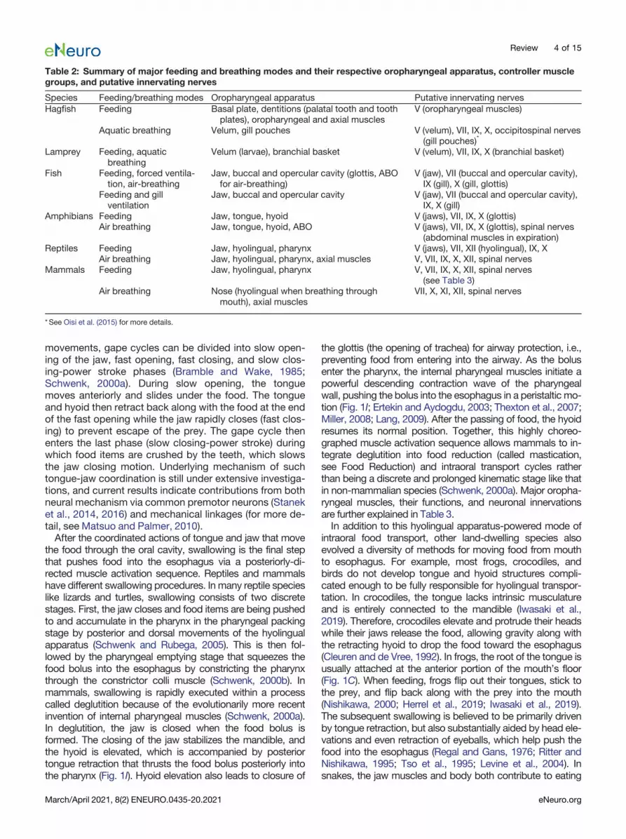
Table 2: Summary of major feeding and breathing modes and their respective oropharyngeal apparatus, controller musclegroups, and putative innervating nerves
Species Feeding/breathing modes Oropharyngeal apparatus Putative innervating nervesHagfish Feeding Basal plate, dentitions (palatal tooth and tooth
plates), oropharyngeal and axial musclesV (oropharyngeal muscles)
Aquatic breathing Velum, gill pouches V (velum), VII, IX, X, occipitospinal nerves(gill pouches)*
Lamprey Feeding, aquaticbreathing
Velum (larvae), branchial basket V (velum), VII, IX, X (branchial basket)
Fish Feeding, forced ventila-tion, air-breathing
Jaw, buccal and opercular cavity (glottis, ABOfor air-breathing)
V (jaw), VII (buccal and opercular cavity),IX (gill), X (gill, glottis)
Feeding and gillventilation
Jaw, buccal and opercular cavity V (jaw), VII (buccal and opercular cavity),IX, X (gill)
Amphibians Feeding Jaw, tongue, hyoid V (jaws), VII, IX, X (glottis)Air breathing Jaw, tongue, hyoid, ABO V (jaws), VII, IX, X (glottis), spinal nerves
(abdominal muscles in expiration)Reptiles Feeding Jaw, hyolingual, pharynx V (jaws), VII, XII (hyolingual), IX, X
Air breathing Jaw, hyolingual, pharynx, axial muscles V, VII, IX, X, XII, spinal nervesMammals Feeding Jaw, hyolingual, pharynx V, VII, IX, X, XII, spinal nerves
(see Table 3)Air breathing Nose (hyolingual when breathing through
mouth), axial musclesVII, X, XI, XII, spinal nerves
*See Oisi et al. (2015) for more details.
Review 4 of 15
March/April 2021, 8(2) ENEURO.0435-20.2021 eNeuro.org

and swallowing preys, without assistance from the tongue,which is completely reserved for chemosensory functions(Fig. 1D). Using highly kinetic joints, snakes can achieve ex-tremely large gap and cranial kinesis, providing substantialdegree of freedom to the upper jaw to move relatively inde-pendently of the head. Therefore, jaws of the snake can beopened and advanced unilaterally in an alternating pattern ofprotraction of the upper and lower jaw on either side, func-tioning as the primary vector of transportation/swallowing(Moon et al., 2019). This motion is aptly termed the “pterygoidwalk,” since the snake basically “walks over” the prey througha combination of ratchet-like jawmovements and concertina-like body undulation (Boltt and Ewer, 1964; Kley andBrainerd, 2002).
Food reductionEvolution of feeding motor programs is also dependent
on food size. While animals like lampreys and balaenidwhales are microphagous that transport food items in
bulk, wide variety of vertebrates are macrophagous, pos-ing the need of reduce the food into manipulatable sizes.Food reduction strategies of vertebrates, from agnathans(e.g., hagfish) to mammals, all predominantly rely on the useof teeth in both aquatic and terrestrial environment.Macrophagy perhaps first evolved in hagfish, one of the
most primitive members of existing vertebrates (Miyashitaet al., 2019). The feeding apparatus of a hagfish consistsof feeding musculature, a basal plate, and dentitions(tooth plates; Clark and Uyeno, 2019). The feeding mus-culature of hagfish are all innervated by the trigeminalnerve (Lindström, 1949) and can be divided into anteriorhard component for protraction and a posterior soft com-ponent for retraction (Clark et al., 2010; Clark and Uyeno,2019). During feeding, a hagfish repeatedly protractstooth plates to press into the food and retracts them intothe mouth along with the torn food (Clark et al., 2010).After retraction, the oral mucosa and palatal tooth dis-lodge the food from the teeth and transport it to theesophagus via additional protraction-retraction cycles
Table 3: Major action and cranial motor nerve supply of oropharyngeal muscles in human
Muscle group Muscle Nerve supply Major actionJaw (masticatory)muscles
Masseter V Elevates the mandible and closes the mouthTemporal V Elevates the mandible and closes the mouth (contraction of the entire
muscle); retruding the mandible (contraction of posterior fibers only)Medial pterygoid V Elevates the mandible and closes the mouthLateral pterygoid V Protrudes and depresses the mandible (bilateral contraction); lateral
excursion of the mandible to the opposite site (unilateral contraction)Hyoid muscles(suprahyoidgroup)
Digastric Anterior belly: V;Posteriorbelly: VII
Depresses the mandible when hyoid is fixed or elevates the hyoid andlarynx if the mandible is fixed
Mylohyoid V Depresses the mandible when the hyoid attachment is fixed or elevatesthe hyoid bone when the mandibular attachment is fixed
Geniohyoid C1 Assists in depression of the mandible, elevation and protrusion of thehyoid, and widening of the pharynx
Stylohyoid VII Elevates and retracts the hyolingual apparatus and keeps the pharynxopen during inspiration
Hyoid muscles (in-frahyoid group)
Omohyoid C2, C3 Depresses and retracts the hyoid and larynxSternohyoid C1–C3 Depresses the hyoidSternothyoid C1–C3 Depresses the hyoid and larynx when activated along with other infra-
hyoid muscles and opening the laryngeal inlet when activated aloneThyrohyoid C1 Depresses the hyoid when activated with other infrahyoid muscles and
elevates the larynx when the hyoid is stabilizedPharyngealmuscles (outercircular layer)
Inferior constrictor X Constricts the wall of pharynx during swallowingMiddle constrictor XSuperior constrictor X
Pharyngealmuscles (innerlongitudinallayer)
Stylopharyngeus IX Shortens and widens the pharynx during swallowingSalpingopharyngeus XPalatopharyngeus X
Tongue muscles(extrinsic)
Genioglossus XII Depresses and protrudes the tongue (bilateral contraction) or contralat-erally deviates the tongue (unilateral contraction)
Hyoglossus XII Depresses and retracts the tongueStyloglossus XII Retracts and elevates lateral portion of the tongue (midline depression
known as cupping)Palatoglossus X Elevates the lingual root during swallowing and depresses the soft
palateTongue muscles(intrinsic)
Superior longitudinal XII Retracts and broadens the tongue, elevates tongue apexInferior longitudinal XII Retracts and broadens the tongue, lowers tongue apexTransverse XII Narrows and elongates the tongueVertical XII Broadens and elongates the tongue
Review 5 of 15
March/April 2021, 8(2) ENEURO.0435-20.2021 eNeuro.org

(Clark and Summers, 2007). Because hagfish lacks theopposing jaw, to generate enough force to tear off piecesof food, the soft retractor musculature is arranged as amuscular hydrostat, which stiffens to provide a compres-sion-resistant support (Uyeno and Clark, 2015; Clubb etal., 2019). Additionally, hagfish can twist its bonelessbody into a knot (a movement called body knotting) toprovide further leverage (for details, see Haney et al.,2020). Thus, although hagfish is jawless, the turgid mus-cular hydrostat together with the body knot functions likea jaw that generates forceful bites similar to other jawedvertebrates (Clark and Summers, 2007).In contrast (to jawless fish), mammals possess well-de-
veloped jaw apparatus. The upper and lower teeth inmost mammals occlude such that they fit nicely together(Schwenk, 2000a). Occlusion within an advanced jaw en-ables a highly efficient form of food chewing called masti-cation, which allows food items to be fully comminutedbecause of the precise fit of the teeth. Chewing occursmostly during the ingestion and intraoral transport stagebefore swallowing. The masticatory muscles for rhythmicjaw opening and closing are described in Table 3. Inmammalian (e.g., sea otters) and non-mammalian gna-thostomes whose teeth do not occlude, macrophagicmethods like crushing and tearing become the commonsolution for reduction (Kienle et al., 2017). In this case,food items, usually hard-shell invertebrates, are punc-tured and crushed in the oral cavity so that salivary en-zymes are able to further soften the food. In addition tocrushing, tearing is used by carnivorous species like croc-odiles, orcas, or sharks. Similar to hagfish feeding, duringtearing the jaw together with the teeth grasp and rip offsmall pieces of food suitable for swallowing (for more de-tails, see Bels and Whishaw, 2019).
BreathingRespiration/breathing is also indispensable for verte-
brate survival as cells within the body need O2 replenish-ment and CO2 excretion to produce energy and properlymetabolize. Although breathing behaviors in vertebratesall includes active muscle contractions to move O2 andCO2 in or out of the body, respiration per se is based onpassive gas diffusion (Kardong, 2018). O2 is normally sub-ject to a higher partial pressure in the external environ-ment, allowing it to naturally diffuse into the blood while CO2
tends to diffuse out from the blood. The passive nature of gasexchanges means that all movements associated withbreathing have for purpose to maintain the partial pressuregradients across the exchange surfaces, by moving gas-con-taining medium (water or air). Throughout evolution, manysolutions for this task have emerged across different familiesof vertebrates, depending on the specific properties of the re-spiratory organ as well as the metabolic demand of theanimal.
Aquatic breathingThe emergence of active aquatic breathing involves
parallel evolution of water-transporting motor programsand specialized respiration structures. Active breathing inearly vertebrates is believed to be originated from feeding,
partially driven by the continued evolution of pharyngealslits (Alheid et al., 2004; Graham and Richardson, 2012;Gillis and Tidswell, 2017). In primitive chordates like am-phioxus, breathing occurs mostly through the skin (cuta-neous breathing; Schmitz et al., 2000; Milsom et al.,2004), and their pharyngeal slits, alongside the body,function to assist filter feeding (Graham and Richardson,2012). As the efficiency of feeding increased because ofthe innovation of the muscular suction pump, the pharyn-geal slits that were previously dedicated to feeding be-came free to take on respiratory functions. In someagnathans such as Pacific hagfish, each pharyngeal slit,also referred to as gill pouch, is separated and has its ownopening to the outside, whereas the gill pouches inAtlantic hagfish are connected to a common openingcalled pharyngo-cutaneous duct (PCD; Fig. 1A). Duringhagfish aquatic breathing, rhythmic contraction of velumbrings water into mouth through the nostril (inhalation),followed by active contractions of gill pouches to expelwater out either through individual gill slits or via the com-mon PCD (Fig. 1A; Malte and Lomholt, 1998; Eom andWood, 2019). Similar motor program is also observed inpremetamorphic larval lampreys (ammocoetes), in whichbreathing and feeding are accomplished within the sameaction: water enters from the mouth and exits through thepharyngeal slits by the scrolling and unscrolling action ofa velum (a muscular structure that is located at the midlineof pharynx), plus the compression and recoil of the bran-chial basket (Rovainen, 1996).Eventually, gills are evolved to replace the pharyngeal
slits in cartilaginous and teleost fish. In these species,breathing happens through gill ventilation, which uses adual pump mechanism, namely the successive expansionand compression of the buccal and opercular cavity con-trolled by branchial muscles (Fig. 1B; Kardong, 2018;Milsom, 2018). This dual pump mechanism, generated byrostral-caudally propagating wave of brainstem motor ac-tivity (Taylor, 1992; Taylor et al., 2009b), sequentially drivesthe water first into the buccal cavity (buccal expansion), thenacross the gill curtain (buccal compression along with oper-cular expansion) where O2 diffuses passively into the gill vas-culature. Finally, water containing CO2 to be excreted exitsfrom the opercular valve (through opercular compression). Inprinciple, the hydraulic nature of both fish breathing and feed-ing means that the same subset of cranial muscles can beused for both processes, although muscular activity duringgill ventilation alone (no feeding) is slower and less powerful(Taylor et al., 2010; Milsom, 2018). Under hypoxic conditions,fish recruit additional feedingmuscles to generate higher suc-tion power to increase water flow (Taylor et al., 2006).An important exception to the common dual pump ven-
tilation is ram ventilation. Here, the swimming motion itselfgenerates respiration-needed water flow across the gills,thereby saving energy costs associated to activating cra-nial muscles during normal gill ventilation (Roberts andRowell, 1988). Many elasmobranchs, such as great whitesharks or whale sharks, do not possess an operculum asan expiratory flap and are obligate ram ventilators thatneed to swim continuously for breathing (Mallatt, 1997).Other fish (e.g., nurse shark, trout) transition between gill
Review 6 of 15
March/April 2021, 8(2) ENEURO.0435-20.2021 eNeuro.org

and ram ventilation depending on swimming speed, watervelocity, and/or water O2 tension, displaying what mightbe the earliest form of breathing-locomotion coordination(Randall, 1982; Steffensen, 1985).
Air breathingAir breathing possibly first evolved in certain bony fish
species that dwell in O2-depleted aquatic habitats, bymodifying the existing suction-based feeding-breathingmechanism. Two of the most common air-breathing or-gans (ABOs) are the lung and the gas bladder. The open-ing of ABOs to the oral cavity is gated by the glottis nearthe floor of digestive tract. Air-breathing fish only comeout of water to breathe intermittently when they need to.During water immersion, the glottis is closed, and thecycles of buccal expansion and compression are used tomove water in and out of mouth for feeding as mentionedabove. During air breathing, the fish swims to the surface ofthe water, where the same cycles of buccal expansion andcompression coupled with the opening and closing of theglottis are used for drawing air into the buccal cavity and ex-changing the “old” air in the ABO with fresh air (Brainerd,1994; Brainerd and Ferry-Graham, 2005; Milsom, 2018). Thisbuccal pump mechanism for breathing is retained in anuranspecies like frogs and toads (Fig. 1C; Taylor et al., 2010).Subsequently during evolution, aspiration breathing sub-
stituted this primitive form of air breathing, with the recruit-ment of axial muscles for higher volume and a more efficientgas exchange. A first instance of aspiration breathing isexemplified by the traverse abdominal muscle that somesalamanders use during active expiration (Brainerd, 1999).Then both inspiration and expiration became powered byaxial muscles, thus drastically increasing the volume of gasexchange. For example, in lepidosaurs such as lizards andsnakes, intercostal muscles are used to create a rotation ofthe ribs that expands the thoracoabdominal cavity and thelung (Carrier, 1989). Similar aspiration pump mechanismhas been documented in birds, during which air sacs are in-flated by rib rotation and caudal sternum depression(Claessens, 2004). A unique aspect of avian breathing is thatwithin the air sac system airflow is unidirectional (Farmer,2015). Using two aerodynamic valves, inspiring air can onlyenter through one valve into one set of chambers, subse-quently the air moves in one direction, and expiring airleaves from the other valve back into the trachea (for moredetails, see Cieri and Farmer, 2016). This aerodynamic valv-ing systemmaximizes breathing efficiency.However, the reliance on axial muscles has its down-
sides. For instance, intercostal and abdominal muscles inlizards are also needed for locomotion. While breathingrequires bilateral and synchronous activation of bothmuscle groups, locomotion unilaterally and alternativelyactivates these muscles to bend the body or to stiffen thetrunk (Carrier, 1990, 1991). This functional conflict is es-pecially evident during high-speed sprinting, during whichaxial muscles are solely recruited for locomotion and thusreduce the lizard’s tidal volume to near zero (Carrier,1987). To solve this problem, many lizards, such as sav-annah monitor lizards, use gular (throat) pumping to cir-cumvent the breathing-locomotion conflict. In a highly
similar fashion to fish and amphibian’s buccal pumpingfor breathing, the gular cavity expands by retracting anddepressing the hyobranchial skeleton to assist in inspira-tion. The addition of this non-conflicting motor programallows these lizards to draw more than two times the airvolume than available through costal inspiration alone(Brainerd and Owerkowicz, 2006).Mammals solved the respiration-locomotion conflict by
evolving a novel respiratory muscle, the diaphragm, adome-shaped muscle. Contraction of the diaphragm flat-tens the curvature of the dome, leading to expansion ofthe thoracic cavity and air influx. The diaphragm allowsmammals to possess much greater stamina during loco-motion, such that ventilation volume can increase alongwith moving speed during locomotion. Testudines (turtlesand tortoises) and crocodilians also adopted similar strat-egies of using a dome-shaped muscle for aspirationbreathing. In turtles, the internal oblique muscle contractsduring inspiration, flattening the dome and increasing thethoracoabdominal cavity volume to draw air into the lung(Gaunt and Gans, 1969; Landberg et al., 2003). In croco-diles, the diaphragmaticus muscle retracts the liver thatdivides the thoracoabdominal cavity, thereby expandingthe cavity and bringing a large volume of air into the lungs(Naifeh et al., 1970; Gans and Clark, 1976; Kardong,2018).
Coordination between Feeding andBreathingBoth feeding and breathing are vital to survival, and
both food and O2 enter into the body from orofacial open-ings. The diverse modes of feeding and breathing throughevolution also involve various mechanisms for coordinat-ing these two behaviors.Aquatic suction feeding and aquatic ventilation (through
pharyngeal slit or gill) use many of the same muscles tobring water into the oral cavity, to push water out throughpharyngeal pore/gill for gas exchange while retainingfood. Thus, aquatic feeding and breathing can occur si-multaneously and harmoniously as part of the samemotor program. For species carrying out aquatic feedingand air-breathing, feeding (in water) and breathing (onsurface of water) never overlap in time, and the glottis isnormally closed to guard the lung until the time of breath-ing. Again, the same sets of muscles that draw water intobuccal cavity are used to draw air into the mouth and thelung.In contrast, for most tetrapods, the combination of ter-
restrial feeding and air breathing poses two main chal-lenges. (1) As feeding typically takes substantial time forintraoral transportation and reduction of food, for speciesthat depend on continuous air breathing (such as mostmammals), mechanisms are needed to allow concurrentrespiration and food processing (before swallowing) tooccur. (2) While both air and food enter via orofacial open-ings, air goes to the lung, whereas food needs to enter theesophagus. Thus, air-breathing tetrapod needs to preventfood from being swallowed into the lung. Different speciesevolved distinct musculoskeletal elements to solve thesetwo problems.
Review 7 of 15
March/April 2021, 8(2) ENEURO.0435-20.2021 eNeuro.org

First, to allow concurrent breathing and food process-ing, structural innovations were implemented during evo-lution to separate the transportation pathway of air andfood. For example, many snakes swallow the prey wholeregardless of the size, and thus require a prolonged pe-riod of intraoral transport and slow swallowing. To enablesimultaneous breathing and feeding, the snake’s oropha-ryngeal region has undergone significant rearrangements(Cundall et al., 2014). First, the glottis of snakes is situatedat the anterior region of the oral cavity and is able to pro-trude outside the oral cavity during feeding to maintainbreathing (Fig. 1D). Furthermore, the opening of theesophagus in snakes is distinctively anterior and thus ef-fectively shortens the length of the oral cavity, such thatthe prey is essentially transported directly into the esoph-agus (Fig. 1D).Another seminal structural innovation in tetropods re-
garding the oropharyngeal region is the development ofthe secondary palate (the posterior portion of hard palateand all the soft palate) that enabled the formation of a sep-arated nasal cavity dedicated to breathing, while foodonly enters into the oral cavity. The secondary palatalshelves in birds or lizards are not fully fused at the midlineleaving a midline cleft (Fig. 1E). By contrast, in mammals,crocodilians, and sea turtles, the secondary palate iscompletely fused at the midline, thereby fully isolating thenasal cavity from the oral cavity (Fig. 1G; Abramyan andRichman, 2015). This allows breathing to remain uninter-rupted during the majority of food processing steps be-fore swallowing, such as chewing and suckling inmammals or ingestion in crocodilians (who immerse theirwhole head except the nostrils into water during lurking).Second, to prevent the aspiration of food into the lung
during swallowing, either unique structural changes ornew motor coordination programs were added. In termsof structural changes, cetaceans (whales, dolphins, andporpoises) further developed a complete segregation oftheir airways from the feeding passage. In these species,the larynx (entry point to trachea and lung) inserts into andcompletely interlocks with the nasal cavity via the palato-pharyngeal sphincter (Fig. 1F; Reidenberg and Laitman,1987, 1994). Food enters the oral cavity and bypasses theairway to be swallowed. Thus, cetaceans are obligatorynasal breathers and can carry out breathing/vocalizationcompletely independent of feeding. A less extreme situa-tion is observed in most mammals (including human in-fants), in which the larynx is positioned above thepharyngeal floor where food will be passed into esopha-gus (Negus, 1949; Laitman et al., 1977; Wolfson andLaitman, 1990; Harrison, 1995; Crompton et al., 1997;Laitman and Reidenberg, 1997). In this configuration, theepiglottis that cover the entrance of the larynx is in con-tact with the posterior end of the soft palate, thereby pre-venting the food bolus from entering the glottis (Fig. 1G),akin to extending a snorkeling tube above the water. Thiselevated position of epiglottis/larynx allows human infantsto breathe and suckle milk simultaneously, and alsomeans that most mammals are essentially nasal breathers(Moss, 1965; Laitman and Reidenberg, 1993; Maynard etal., 2020).
As human infants develop, the larynx gradually de-scends to the adult position (below the level of pharyn-geal floor) around the second or third year (Fig. 1H;Lieberman et al., 2001). The descend of larynx acts as adouble-edged sword: it enables a much richer vocaliza-tion repertoire, removes the obligation for nasal breath-ing, but also greatly increases chances of food gettinginto the trachea, making humans the most susceptiblefor food aspiration among mammalian species (Laitmanand Reidenberg, 1997). To solve this problem, a reflex/motor program evolved in which swallowing temporarilysuppresses breathing, with concurrent activation ofmuscles that close the glottis, block the nasal cavity viathe soft palate, and elevate the larynx to contact the ep-iglottis (Fig. 1I).
A hypothesis: how conserved embryonichindbrain generates circuits for diversemodes of feeding and breathingUltraconserved hindbrain and PAs duringdevelopmentDespite the diverse modes of feeding and breathing,
these two vital behaviors are controlled by neural cir-cuits originated in the hindbrain in all vertebrates (seeFig. 2; Table 2). Importantly, all vertebrates share highlyconserved embryonic developmental programs duringthe phylotypic period of pharyngula stage, characterizedby conserved segmented structures of the embryonichindbrain and peripheral tissues called rhombomeres (r)and PAs, respectively (Fig. 2; Irie and Kuratani, 2011;Sugahara et al., 2017; Parker et al., 2019). Each rhombo-mere is characterized by its unique but conserved expres-sion of genes and transcription factors (in particular Hoxand Hox-regulating genes), where specific types of motorneurons, interneurons, and neural crest cells (precursorsto sensory neurons) are generated (Fig. 2; Lumsden andKeynes, 1989; Kiecker and Lumsden, 2005; Parker andKrumlauf, 2020). Each PA receives distinct yet stereo-typed sensorimotor innervation and forms arch-specificmusculoskeletal elements (Fig. 2). In most vertebrates,the first PA forms the jaw and is innervated by motoneur-ons of the Vth nerve derived from r2/r3. The second archforms the hyoid apparatus and facial muscles and is in-nervated by r4/r5-derived VIIth motoneurons. The moreposterior PAs either develop into gill in fish or pharyngealand laryngeal apparatuses in tetrapods and are inner-vated by IXth, Xth, and XIIth motoneurons derived fromr6/r7/r8 (Graham and Smith, 2001; Graham et al., 2019;Maynard et al., 2020). In other words, there is a roughlyordered anterior-to-posterior relationship between therhombomere origins of the motor nerves and the PAs thatthe nerves innervate in all vertebrates. To generate diversefeeding/breathing modes based on highly conserved em-bryonic hindbrain and PAs, we hypothesize that initially dur-ing development, the hindbrain produces a basic rhythmicmotor program common to all vertebrates, and that thisearly conserved circuit can be repurposed, reconfigured, orreplaced during later developmental stages to generate
Review 8 of 15
March/April 2021, 8(2) ENEURO.0435-20.2021 eNeuro.org

distinct patterns of feeding and breathing. Below we consid-er some evidence for this hypothesis.
A common vertebrate embryonic hindbrain circuit forrhythmic ingestion of water/amniotic fluidAs described above, aquatic breathing and aquatic feed-
ing share the same sets of cranial muscles and motor pro-grams. Notably, while fish and amphibians are born directlyinto water, amniotes like reptiles, birds, and mammals gothrough embryonic development encircled by amniotic fluid.Therefore, bringing fluid into the mouth to be swallowedis likely the fundamental process common to all devel-oping vertebrates. For example, it is known that fetalswallowing of amniotic fluid emerges right after majororganogenesis around 10–14weeks of gestation inhuman and is observed in early development of chicks,sheep, rodents, and monkeys (Ross and Nijland, 1998;Delaney and Arvedson, 2008; Mashayekhi et al., 2011;Gross and Trapani-Hanasewych, 2017; Maynard et al.,2020). This process of transporting fluid into the body isrepeatedly executed and requires coordinated activa-tion of oropharyngeal muscles (Sherman et al., 1990).Therefore, the developing hindbrain in all vertebratesmust contains a basic circuit that produces rhythmicexpansion and compression (or opening and closing) ofthe oral cavity for moving fluid into the mouth. We pro-pose that this basic circuit contains either one dominantcentral rhythm generator (CRG) in primitive vertebrates,or a series of sequentially coupled CRGs that each hasits own intrinsic rhythm in more evolved vertebrates,such that fluid is periodically transported into the mouthto be swallowed in a unidirectional manner (Fig. 3A,B).
Existence and locations of putative fluid-transportingCRGs in developing hindbrainWhere in embryonic hindbrain might the fluid-transport-
ing CRGs reside? Let us first consider the larval lampreys,which use rhythmic movement of the velum to pumpwater into the mouth for feeding and for breathing. Thevelum located at the midline of pharynx is innervated bythe Vth nerve. During larval lamprey aquatic breathing,muscles of velum and branchial basket (innervated byVIIth, IXth, Xth motoneurons) contract repeatedly in afixed sequence (Russell, 1986; Rovainen, 1996). Thisthen led to the discovery of a CRG adjacent to the Vthmotor nucleus termed paratrigeminal respiratory groupand a distributed CRG network around VIIth, IXth, Xthmotor nuclei (Fig. 3A; Martel et al., 2007; Missaghi etal., 2016). It is postulated that these two CRG networksare coupled such that their sequential outputs drive therepeated velar and basket pumping (and relaxation) ac-tions to bring in water (Fig. 3A). Since all later jawed ver-tebrates (Gnathostomes) use trigeminal (V)-innervatedjaw opening and closing to bring water/amniotic fluidinto the oral cavity, it is likely that the CRG located ini-tially adjacent to the Vth motor nucleus [peritrigeminalCRG (pT-CRG)] is conserved in all vertebrates (at leastin developmental stages; Fig. 3A,B).As vertebrates evolved more muscles, the lamprey-type
CRG configuration may be insufficient to drive coordi-nated movements of many muscles used for the repeatedand directional fluid transportation and swallowing proc-esses. Unlike agnathans such as lampreys and hagfishthat predominantly relies on trigeminal-innervated velumfor breathing, feeding/gill ventilation in fish starts from the
Figure 2. The highly conserved embryonic hindbrain and PAs and their associated circuits and peripheral structures. Left panel,Schematic representation of the vertebrate embryonic hindbrain. The developing hindbrain is segmented into rhombomeres (r1–r8),which is defined by combinatorial expression of different genes (e.g., Hox genes) and transcription factors (e.g., EGR2, also calledKrox-20), depicted in the middle. Locations of cranial sensory ganglia (gV, gVII–gXI) and otic vesicles (ov) are shown on the left sideof the hindbrain. The right side of the hindbrain shows motoneuron distribution of major cranial motor nuclei and their respectiveexit points. Neural crest cells form migratory streams (black arrows) that originate from rhombomeres to their respective PA. Rightpanel, Each PA is characterized by distinct nerve innervation, skeletal and muscular derivatives (table on the right). Drawings ofrhombomeres and PAs and their derivatives are based on Kiecker and Lumsden (2005) and Maynard et al. (2020).
Review 9 of 15
March/April 2021, 8(2) ENEURO.0435-20.2021 eNeuro.org

opening of the jaw (V), followed by the activation of bran-chial muscles (VII) that results in sequential expansionand compression of first the buccal and then the opercu-lar cavity. To swallow the food into the mouth, the se-quence starts after jaw closure, followed by the activationof branchial basket muscles (IX, X) that transports thefood to the esophagus through rounds of oropharyngealexpansions and compression. This sequential motor pat-tern likely requires serially coupled-CRGs in fish (Hyde,1904; Sundin et al., 2000; Taylor et al., 2009a). This hy-pothesis is further supported by “sheep-dip” experi-ments, in which isolated fish brainstems are graduallysubmerged into high magnesium solution that blocks ex-citatory transmission necessary for rhythmogenesis (seenext section). These experiments show that rhythmic out-puts of VII nerve degrade gradually rather than abruptly,indicating that the gill ventilation rhythm in fish is gener-ated by several mutually-coupled segmental CRGs dis-tributed throughout the hindbrain (Duchcherer et al.,2010). In the mammalian fetal swallowing process, thefluid transportation also similarly follows the sequentialactivation of jaw, hyolingual, and pharyngeal muscles(Sherman et al., 1990; Thexton et al., 2007).Furthermore, evidence suggests that in higher verte-
brates, each pair of rhombomeres contains its own CRG.In chick embryos, isolated hindbrain segment prepara-tions containing either V, VII, or XII motor nuclei all exhibitspontaneous rhythmic activities that drive their respectivesegmental motor nerves (Fig. 3B; Fortin et al., 1995;Borday et al., 2003). It was also revealed that odd-evenpairs of rhombomeres generate higher frequencies of rhyth-mic bursts (Coutinho et al., 2004). In rodent preparations,similar conclusion was drawn that rhythmic firings in V, VII,and XII motoneurons representing ingestive movements areinduced by their respective segmental CRG (Nakamura etal., 1999). We further speculate that the serial CRGs in pairsof rhombomeres form directionally biased connections suchthat the more anteriorly located CRG preferentially drivesthe activity of the posteriorly located CRG, thereby facilitat-ing the propagating wave of oropharyngeal muscle activitiesthat moves fluid from outside of the mouth to the esophagus(Fig. 3B). Notably, the rhombomeres form and differentiatein a rostral-to-caudal (from r1 to r8) order (Gavalas, 2002).Thus, the serial CRGs may also follow a developmental se-quential manner of maturation thereby allowing the anteriorCRGs to preferentially drive the posterior CRGs (Fig. 3B).The sequential coupling of CRGs is also likely dependent onand facilitated by the developing sensory neurons that formfeedback circuits in favor of sequential activation. In isolatedwhole embryonic hindbrain preparations, it was shown thatall cranial motor nerves exhibit synchronized rhythmic activ-ities (Fortin et al., 1995; Abadie et al., 2000), suggesting thatin the absence of sensory feedback, a single CRG (likely themost anterior pT-CRG) dominates.
Evolutionary conserved characteristics ofrhythm-generating mechanismInvestigations of rhythm-generating mechanisms behind
oropharyngeal behaviors in vertebrates have largely focusedon breathing, especially in lampreys, frogs, and rodents.
Figure 3. Hypothesized conserved vertebrate embryonic hind-brain circuit for intraoral fluid transportation and its later stagereconfigurations in mammals. The fluid-surrounding environ-ment and fluid ingestion behavior in aquatic and in embryonicterrestrial vertebrates suggest the existence of a conserved em-bryonic circuit that generates the fluid-transporting behavior inall vertebrates (A, B). A, In primitive vertebrates like lampreys,this fluid-transporting circuit consists of two CRGs, with pT-CRG as the dominant CRG, that drive rhythmic and sequentialactivations of downstream motor nuclei. B, In advanced verte-brates, a series of CRGs for each pair of rhombomeres work to-gether to support the directional fluid-transporting behavior.Hypothesized serial CRGs are sequentially coupled such thatan anterior CRG preferentially drives the activity of posteriorCRGs, thereby produces sequential activation of oropharyngealmuscles and directional movement of fluid. C, Splitting, reconfi-guration and repurposing of the embryonic fluid-transportingCRGs result in separated feeding-related and breathing-relatedCRGs in adult mammalian hindbrain. Only preBötC is high-lighted among the breathing CRGs for simplicity. The need ofcontinuous breathing makes preBötC the dominant CRG, whichbroadcasts its rhythm to other CRGs or motoneurons, ventralrespiratory group (VRG). Feeding-related CRGs control chewingand licking largely function independent of breathing CRGs be-cause of structural segregations of food and air intake path-ways. Swallowing inhibits breathing to prevent aspiration.
Review 10 of 15
March/April 2021, 8(2) ENEURO.0435-20.2021 eNeuro.org

These studies primarily examined the roles of excitatoryglutamatergic mechanisms, inhibitory GABAergic and glyci-nergic mechanisms, putative pacemaker properties, andneuromodulators in rhythm generations. Decades of studiessuggest that some rhythmogenic mechanisms are evolu-tionary conserved.First, excitatory glutamatergic system is essential for
rhythm generation in nearly all vertebrate breathingCRGs. Both lamprey’s pT-CRG and mammalian’s inspi-ration CRG, preBötzinger complex (preBötC), containlarge ensembles of glutamatergic neurons and are sensi-tive to modulations by opioids and substance P (Mutoloet al., 2010; Del Negro et al., 2018). In lampreys, microin-jections of AMPA and NMDA blockers in pT-CRG tempo-rary abolish its rhythm (Martel et al., 2007; Cinelli et al.,2013). On the other hand, manipulations that facilitateglutamatergic transmission (e.g., application of AMPA orperfusion of mGluRs I and II agonists) accelerate pT-CRG rhythm (Bongianni et al., 2002; Martel et al., 2007;Cinelli et al., 2013). In mammals, many neurons that ex-press transcription factor developing brain homeoboxprotein 1 (DBX1) during the pharyngula stage later be-come glutamatergic neurons, including neurons inpreBötC. Dbx1-knock-out mice do not breathe and dieat birth and optogenetic inhibition of DBX1 lineage cellsslows and stops breathing rhythm (for more detail, seeDel Negro et al., 2018). Glutamatergic transmission isalso found to be essential in CRGs responsible for lungventilation in turtles (Johnson and Mitchell, 1998) andfrogs (Chen and Hedrick, 2008; Baghdadwala et al.,2016). Interestingly, recent results from Ashhad andFeldman (2020) suggest that inspiratory rhythmogenesisin neonatal mammalian preBötC is critically driven byemergent network properties, where strong synchroniza-tion among excitatory rhythmogenic neurons withinpreBötC produces inspiratory bursts.Second, inhibitory systems heavily modulate the fre-
quency and amplitude of rhythm but may not be essentialfor rhythm generation per se. For example, respiratoryrhythm still persists after blockade of GABAergic and glyci-nergic transmission in lampreys (Bongianni et al., 2006;Cinelli et al., 2014, 2016), in frogs (lung ventilation; Leclère etal., 2012; Baghdadwala et al., 2016), turtles (Johnson et al.,2002, 2007), and in mammals (Janczewski et al., 2013;Sherman et al., 2015; Baertsch et al., 2018). However, in dif-ferent species or different brainstem nuclei, inhibition hasdifferent effects. Blocking GABAA in lamprey produce signif-icant increases in frequency and amplitude of breathingrhythm, while injection of GABAA agonist suppresses pT-CRG rhythm (Bongianni et al., 2006; Cinelli et al., 2014). Inrodents, it is proposed that GABAergic and glycinergictransmissions, respectively, regulate different aspects ofrhythm and pattern generated by preBötC (Ashhad andFeldman, 2020). Photostimulation of glycinergic preBötCneurons depresses breathing while inhibition of these neu-rons augments breathing amplitude and stops ongoingapnea (Sherman et al., 2015). Additionally, complex recipro-cal GABAergic transmissions are important for the couplingof inspiratory preBötC and expiratory parafacial oscillator(Mellen and Thoby-Brisson, 2012). By contrast, in chick
embryos, blocking GABA-mediated inhibition in segmentalCRGs abolishes the high frequency rhythmic bursts. Here,inhibition is required for generating high (but not low) fre-quency rhythms (Fortin et al., 1999).Many studies also examined whether pacemaker-like
properties are involved in hindbrain rhythmogenesisacross species. Specifically, these studies tested theroles of persistent sodium current (INaP) or calcium-acti-vated non-specific cation (ICAN) currents. In lampreys, tur-tles, and mammals, while breathing rhythm is abolishedby INaP and ICAN current blockers, application of an exog-enous excitatory agent (e.g., substance P) restores therhythm (Del Negro et al., 2005; Mutolo et al., 2010; Linand Onimaru, 2015; Johnson et al., 2016). These resultssuggest that pacemaker properties are not essential forrespiratory rhythm generation (Mellen and Thoby-Brisson,2012; Bongianni et al., 2016; Missaghi et al., 2016).However, under hypoxia condition, breathing-related ac-tivity are dependent on INaP (Peña et al., 2004; Paton etal., 2006). Furthermore, it was shown that in mouse, unlikethe primarily network-driven rhythmogenesis at and afterE18.5, INaP and ICAN dependent pacemaker properties arepurely required for inspiration rhythm in embryos beforeE16.5 (Chevalier et al., 2016). Thus, the rhythmogesismechanisms for the proposed fluid-transporting CRGs inearlier embryos could be different from mechanisms gov-erning CRGs in neonatal and adult higher vertebrates,and future studies with cellular identities are needed topinpoint the exact mechanisms.
Reconfiguration and repurposing embryonic CRGs toaccommodate parallel air breathing and feeding inhigher vertebratesThe evolutionary progression from one oral cavity
serving both feeding and breathing functions in aquaticvertebrates, to two separated food/fluid and air intakepathways, such as the separated nasal and oral cavitiesin mammals, allows air-breathing and feeding to be con-ducted, to a large extent, independently. We hypothesizethat peripheral structural separations are accompaniedby the split of conserved segmental fluid-transportingCRGs in the hindbrain into two separate sets: one set forbreathing and the other set remains as CRGs for feeding(Fig. 3C).Indeed, a series of breathing-related CRGs have been
discovered in mammals. They are, from rostral to caudal,the parafacial respiratory group functioning as the expir-atory CRG (pFRG), the postinspiration complex (PiCo)hypothesized to be the postinspiration CRG, and thepreBötC known as the inspiratory CRG. These respira-tory CRGs may have migrated during development fromtheir original birthplace to their final ventral locations inthe hindbrain. We speculate that the origin of preBötCmight be the segmental CRG near the Xth nerve (in r7)that is recruited during evolution to provide rhythmiccontrol of inspiration axial muscles (by projecting to neu-rons in the medullary ventral respiratory groups which inturn project to the spinal cord). Interestingly, it wasshown in mouse, that the constituent neurons of the em-bryonic parafacial (e-pF; originated in r4) and preBötC (in
Review 11 of 15
March/April 2021, 8(2) ENEURO.0435-20.2021 eNeuro.org

r7) CRGs are born and matured in a sequential fashion[e-pF cells are born at embryonic day (E)10.5 and ma-tured at E14.5, while preBötC neurons are born at E11.5and matured at E15.5], allowing the anterior-located e-pF to entrain the immature preBötC activity in early em-bryonic stages (Thoby-Brisson et al., 2009; Champagnatet al., 2011). This time period also coincides with the pe-riod of secondary palate formation (Yu et al., 2017). Afterthe complete fusion of the secondary palate at the mid-line to separate nasal and oral cavity in late embryonicstages, preBötC gradually becomes the dominant CRGin the hindbrain, entraining other respiratory CRGs(Ramirez et al., 2016; Del Negro et al., 2018), perhaps inpreparation for the need of continuous air-breathingafter birth. In addition, other secondary CRGs serve oro-facial actions using muscles evolutionarily involved inbreathing may be further separated/differentiated or re-purposed from embryonic ones. For example, parts ofposterior embryonic CRG neurons in r7/r8 are likely to berepurposed as CRGs for whisking in rodents and vocal-izations in mammals (Fig. 3C).On the other hand, some of the embryonic fluid-trans-
porting CRG neurons retain their feeding-related func-tions. These cells may stay in their conserved locationsnear feeding-related motor neurons. For example, the an-cient pT-CRG may become the mammalian CRG forsuckling (neonates) and chewing (adults). Indeed, neuronslocated dorsal to and near Vth nucleus are implicated incontrolling rhythmic jaw movements (Morquette et al.,2012). The caudal feeding-related embryonic CRGs likelyfurther differentiated to control licking and swallowing(Fig. 3C). Again, because of the separation of nasal andoral cavity, chewing and licking can operate to a large ex-tent independently of breathing until the step of swallow-ing in most mammals (Moore et al., 2014). On the otherhand, the continuously active inspiration CRG preBötC isshown to send strong projections throughout the hind-brain, broadcasting the breathing rhythm to neurons con-trolling feeding (Fig. 3C). This is likely because breathingrequires activity from certain feeding-related oropharyn-geal muscles to help maintain airway patency (Moore etal., 2014; Yang and Feldman, 2018). Finally, swallowing inmammals is typically executed is one rapid motion andthus it may not require a CRG. However, new airway pro-tection circuits must have evolved in mammals such thatcentrally-arisen or peripherally-arisen swallowing signalseffectively inhibit inspiration to prevent aspiration of food(Fig. 3C).The maturation and refinement of species-specific
hindbrain orofacial circuits likely depends on feedbacksignaling from peripheral muscles and other tissues tomotoneurons, then from motoneurons to upstream CRGsand other interneurons also through retrograde signaling,as well as depends on activity of sensory feedback.Gradually, synergistic muscles will be co-active in thesame phase of CRG rhythm, whereas antagonist muscleswill be co-activated in the opposite phase of CRG activity.Sequences are further consolidated and eventually di-verse motor patterns of coordinated feeding and breath-ing behaviors in higher vertebrates emerge.
Concluding remarksOne of the core questions related to the development
and evolution of motor behaviors is how evolutionary con-served developmental principles give rise to such a di-verse pool of behaviors. In this review, we discussed howvertebrates, while carrying out varied modes of feedingand breathing, all share a stage of living in a fluid and allexecute a homologous action of periodically transportingfluid from the external environment inside the body. We hy-pothesize that a conserved hindbrain circuit generating aposteriorly propagating rhythmic activity is initially formed inall vertebrates, that drives the basic intraoral fluid transportbehaviors. This circuit forms the basis for carrying outaquatic feeding/aquatic breathing, primitive air breathing,and terrestrial food transportation. Aspiration breathing andfood reduction (chewing) represent the splitting, repurpos-ing, and reconfiguration of the existing hindbrain CRGs. Wenote that other than preBötC/pF/PiCo, the neuronal identi-ties and exact locations of feeding-related CRGs (chewing,suckling, licking, swallowing, etc.) in mammals remainvague. Based on our unifying hypothesis, we propose thatstudies of hindbrain CRGs for feeding/gill ventilation in fishmay reveal the homologous neurons in mammals that driverhythmic suckling, chewing, and licking. Alternatively, if anew method can be developed that enables lineage tracingof hindbrain CRG neurons in mammalian embryos, followingthese neurons into postnatal development and adult shouldprovide insights for the locations and identities of adult CRGneurons.
References
Abadie V, Champagnat J, Fortin G (2000) Branchiomotor activities inmouse embryo. Neuroreport 11:141–145.
Abramyan J, Richman JM (2015) Recent insights into the morpholog-ical diversity in the amniote primary and secondary palates. DevDyn 244:1457–1468.
Alheid GF, MilsomWK, McCrimmon DR (2004) Pontine influences onbreathing: an overview. Respir Physiol Neurobiol 143:105–114.
Arnold P, Birtles R, Sobtzick S, Matthews M, Dunstan A (2005)Gulping behaviour in rorqual whales: underwater observations andfunctional interpretation. Mem Queensl Mus 51:309–332.
Arvedson JC, Lefton-Greif MA (1998) Pediatric videofluoroscopicswallow studies. A professional manual with caregiver guidelines.San Antonio: Communication Skill Builders.
Ashhad S, Feldman JL (2020) Emergent elements of inspiratoryrhythmogenesis: network synchronization and synchrony propa-gation. Neuron 106:482–497.
Baertsch NA, Baertsch HC, Ramirez JM (2018) The interdependenceof excitation and inhibition for the control of dynamic breathingrhythms. Nat Commun 9:843.
Baghdadwala MI, Duchcherer M, Trask WM, Gray PA, Wilson RJ(2016) Diving into the mammalian swamp of respiratory rhythmgeneration with the bullfrog. Respir Physiol Neurobiol 224:37–51.
Bels V, Whishaw IQ (2019) Feeding in vertebrates: evolution, mor-phology, behavior, biomechanics. Cham: Springer.
Boltt R, Ewer R (1964) The functional anatomy of the head of the puffadder, Bitis arietans (Merr.). J Morphol 114:83–105.
Bongianni F, Mutolo D, Carfì M, Pantaleo T (2002) Group I and II me-tabotropic glutamate receptors modulate respiratory activity in thelamprey. Eur J Neurosci 16:454–460.
Bongianni F, Mutolo D, Nardone F, Pantaleo T (2006) GABAergic andglycinergic inhibitory mechanisms in the lamprey respiratory con-trol. Brain Res 1090:134–145.
Review 12 of 15
March/April 2021, 8(2) ENEURO.0435-20.2021 eNeuro.org

Bongianni F, Mutolo D, Cinelli E, Pantaleo T (2016) Neural mecha-nisms underlying respiratory rhythm generation in the lamprey.Respir Physiol Neurobiol 224:17–26.
Borday C, Abadie V, Chatonnet F, Thoby-Brisson M, Champagnat J,Fortin G (2003) Developmental molecular switches regulatingbreathing patterns in CNS. Respir Physiol Neurobiol 135:121–132.
Brainerd EL (1994) The evolution of lung-gill bimodal breathing andthe homology of vertebrate respiratory pumps. Am Zool 34:289–299.
Brainerd EL (1999) New perspectives on the evolution of lung ventila-tion mechanisms in vertebrates. Exp Biol Online 4:1–28.
Brainerd EL, Ferry-Graham LA (2005) Mechanics of respiratorypumps. Fish Physiol 23:1–28.
Brainerd EL, Owerkowicz T (2006) Functional morphology and evolu-tion of aspiration breathing in tetrapods. Respir Physiol Neurobiol154:73–88.
Bramble DM, Wake DB (1985) Feeding mechanisms of lower tetra-pods. In: Functional vertebrate morphology (Hildebrand M,Bramble DM, Liem KF, Wake DB, eds), pp 230–261. Cambridge:Harvard University Press.
Brown JD, Stallknecht DE (2008) Wild bird surveillance for the avianinfluenza virus. Methods Mol Biol 436:85–97.
Carrier DR (1987) Lung ventilation during walking and running in fourspecies of lizards. Exp Biol 47:1.
Carrier DR (1989) Ventilatory action of the hypaxial muscles of the liz-ard Iguana iguana: a function of slow muscle. J Exp Biol 143:435–457.
Carrier DR (1990) Activity of the hypaxial muscles during walking inthe lizard Iguana iguana. J Exp Biol 152:453–470.
Carrier DR (1991) Conflict in the hypaxial musculo-skeletal system:documenting an evolutionary constraint. Am Zool 31:644–654.
Champagnat J, Morin-Surun M-P, Bouvier J, Thoby-Brisson M,Fortin G (2011) Prenatal development of central rhythm genera-tion. Respir Physiol Neurobiol 178:146–155.
Chen AK, Hedrick MS (2008) Role of glutamate and substance P inthe amphibian respiratory network during development. RespirPhysiol Neurobiol 162:24–31.
Chevalier M, Toporikova N, Simmers J, Thoby-Brisson M (2016)Development of pacemaker properties and rhythmogenic mecha-nisms in the mouse embryonic respiratory network. Elife 5:e16125.
Cieri RL, Farmer C (2016) Unidirectional pulmonary airflow in verte-brates: a review of structure, function, and evolution. J CompPhysiol B 186:541–552.
Cinelli E, Robertson B, Mutolo D, Grillner S, Pantaleo T, Bongianni F(2013) Neuronal mechanisms of respiratory pattern generation areevolutionary conserved. J Neurosci 33:9104–9112.
Cinelli E, Mutolo D, Robertson B, Grillner S, Contini M, Pantaleo T,Bongianni F (2014) GABAergic and glycinergic inputs modulaterhythmogenic mechanisms in the lamprey respiratory network. JPhysiol 592:1823–1838.
Cinelli E, Mutolo D, Contini M, Pantaleo T, Bongianni F (2016)Inhibitory control of ascending glutamatergic projections to thelamprey respiratory rhythm generator. Neuroscience 326:126–140.
Claessens LP (2004) Archosaurian respiration and the pelvic girdleaspiration breathing of crocodyliforms. Proc Biol Sci 271:1461–1465.
Clark AJ, Summers AP (2007) Morphology and kinematics of feedingin hagfish: possible functional advantages of jaws. J Exp Biol210:3897–3909.
Clark AJ, Uyeno TA (2019) Feeding in jawless fishes. In: Feeding invertebrates: evolution, morphology, behavior, biomechanics (BelsV, Whishaw IQ, eds), pp 189–230. Cham: Springer.
Clark AJ, Maravilla EJ, Summers AP (2010) A soft origin for a forcefulbite: motor patterns of the feeding musculature in Atlantic hagfish,Myxine glutinosa. Zoology (Jena) 113:259–268.
Cleuren J, de Vree F (1992) Kinematics of the jaw and hyolingual ap-paratus during feeding in Caiman crocodilus. J Morphol 212:141–154.
Clubb BL, Clark AJ, Uyeno TA (2019) Powering the hagfish “bite”:the functional morphology of the retractor complex of two hagfishfeeding apparatuses. J Morphol 280:827–840.
Coutinho AP, Borday C, Gilthorpe J, Jungbluth S, Champagnat J,Lumsden A, Fortin G (2004) Induction of a parafacial rhythm gener-ator by rhombomere 3 in the chick embryo. J Neurosci 24:9383–9390.
Crompton A, German R, Thexton A (1997) Mechanisms of swallow-ing and airway protection in infant mammals (Sus domesticus andMacaca fascicularis). J Zool 241:89–102.
Cundall D, Tuttman C, Close M (2014) A model of the anterior esoph-agus in snakes, with functional and developmental implications.Anat Rec (Hoboken) 297:586–598.
Del Negro CA, Morgado-Valle C, Hayes JA, Mackay DD, Pace RW,Crowder EA, Feldman JL (2005) Sodium and calcium current-mediated pacemaker neurons and respiratory rhythm generation.J Neurosci 25:446–453.
Del Negro CA, Funk GD, Feldman JL (2018) Breathing matters. NatRev Neurosci 19:351–367.
Delaney AL, Arvedson JC (2008) Development of swallowing andfeeding: prenatal through first year of life. Dev Disabil Res Rev14:105–117.
Ding Y, Vanselow DJ, Yakovlev MA, Katz SR, Lin AY, Clark DP,Vargas P, Xin X, Copper JE, Canfield VA, Ang KC, Wang Y, Xiao X,De Carlo F, van Rossum DB, La Riviere P, Cheng KC (2019)Computational 3D histologic phenotyping of whole zebrafish by X-ray histotomography. Elife 8:e44898.
Duchcherer M, Kottick A, Wilson R (2010) Evidence for a distributedrespiratory rhythm generating network in the goldfish (Carsssiusauratus). In: New frontiers in respiratory control (Homma I,Onimaru H, Fukuchi Y, eds), pp 3–7. New York: Springer.
Ertekin C, Aydogdu I (2003) Neurophysiology of swallowing. ClinNeurophysiol 114:2226–2244.
Eom J, Wood CM (2019) The ventilation mechanism of the Pacifichagfish Eptatretus stoutii. J Fish Biol 94:261–276.
Farmer C (2015) The evolution of unidirectional pulmonary airflow.Physiology (Bethesda) 30:260–272.
Fortin G, Kato F, Lumsden A, Champagnat J (1995) Rhythm genera-tion in the segmented hindbrain of chick embryos. J Physiol486:735–744.
Fortin G, Jungbluth S, Lumsden A, Champagnat J (1999) Segmentalspecification of GABAergic inhibition during development of hind-brain neural networks. Nat Neurosci 2:873–877.
Gans C, Clark BD (1976) Studies on ventilation of Caiman crocodilus(Crocodilia: Reptilia). Respir Physiol 26:285–301.
Gaunt AS, Gans C (1969) Mechanics of respiration in the snappingturtle, Chelydra serpentina (Linné). J Morphol 128:195–227.
Gavalas A (2002) ArRAnging the hindbrain. Trends Neurosci 25:61–64.
Gillis JA, Tidswell OR (2017) The origin of vertebrate gills. Curr Biol27:729–732.
Goldbogen J, Cade D, Calambokidis J, Friedlaender A, Potvin J,Segre P, Werth A (2017) How baleen whales feed: the biome-chanics of engulfment and filtration. Ann Rev Mar Sci 9:367–386.
Graham A, Smith A (2001) Patterning the pharyngeal arches.Bioessays 23:54–61.
Graham A, Richardson J (2012) Developmental and evolutionary ori-gins of the pharyngeal apparatus. Evodevo 3:24.
Graham A, Poopalasundaram S, Shone V, Kiecker C (2019) A reap-praisal and revision of the numbering of the pharyngeal arches. JAnat 235:1019–1023.
Gross RD, Trapani-Hanasewych M (2017) Breathing and swallowing:the next frontier. Semin Speech Lang 38:87–95.
Grubich JR (2001) Prey capture in actinopterygian fishes: a review ofsuction feeding motor patterns with new evidence from an elopo-morph fish,Megalops atlanticus. Am Zool 41:1258–1265.
Haney W, Clark A, Uyeno T (2020) Characterization of body knottingbehavior used for escape in a diversity of hagfishes. J Zool310:261–272.
Review 13 of 15
March/April 2021, 8(2) ENEURO.0435-20.2021 eNeuro.org

Harrison DFN (1995) The anatomy and physiology of the mammalianlarynx. Cambridge: Cambridge University Press.
Herrel A, O’Reilly JC, Fabre AC, Bardua C, Lowie A, Boistel R, GorbSN (2019) Feeding in amphibians: evolutionary transformationsand phenotypic diversity as drivers of feeding system diversity. In:Feeding in vertebrates: evolution, morphology, behavior, biome-chanics (Bels V, Whishaw IQ, eds), pp 431–467. Cham: Springer.
Hyde IH (1904) Localization of the respiratory centre in the skate. AmJ Physiol 10:236–258.
Irie N, Kuratani S (2011) Comparative transcriptome analysis revealsvertebrate phylotypic period during organogenesis. Nat Commun2:248.
Iwasaki SI, Erdogan S, Asami T (2019) Evolutionary specialization ofthe tongue in vertebrates: structure and function. In: Feeding invertebrates: evolution, morphology, behavior, biomechanics (BelsV, Whishaw IQ, eds), pp 333–384. Cham: Springer.
Janczewski WA, Tashima A, Hsu P, Cui Y, Feldman JL (2013) Role ofinhibition in respiratory pattern generation. J Neurosci 33:5454–5465.
Johnson SM, Mitchell GS (1998) N-methyl-d-aspartate-mediatedbulbospinal respiratory drive is pH/P(CO2)-insensitive in turtlebrainstem-spinal cord. Respir Physiol 113:201–212.
Johnson SM, Wilkerson JE, Wenninger MR, Henderson DR, MitchellGS (2002) Role of synaptic inhibition in turtle respiratory rhythmgeneration. J Physiol 544:253–265.
Johnson SM, Wiegel LM, Majewski DJ (2007) Are pacemaker proper-ties required for respiratory rhythm generation in adult turtle brainstems in vitro? Am J Physiol Regul Integr Comp Physiol 293:R901–R910.
Johnson SM, Hedrick MS, Krause BM, Nilles JP, Chapman MA(2016) Respiratory neuron characterization reveals intrinsic burst-ing properties in isolated adult turtle brainstems (Trachemys scrip-ta). Respir Physiol Neurobiol 224:52–61.
Kardong KV (2018) Vertebrates: comparative anatomy, function,evolution, Ed 8. New York: McGraw-Hill Education.
Kiecker C, Lumsden A (2005) Compartments and their boundaries invertebrate brain development. Nat Rev Neurosci 6:553–564.
Kienle SS, Berta A (2016) The better to eat you with: the comparativefeeding morphology of phocid seals (Pinnipedia, Phocidae). J Anat228:396–413.
Kienle SS, Law CJ, Costa DP, Berta A, Mehta RS (2017) Revisitingthe behavioural framework of feeding in predatory aquatic mam-mals. Proc Biol Sci 284:20171035.
Kley NJ, Brainerd EL (2002) Post-cranial prey transport mechanismsin the black pinesnake, Pituophis melanoleucus lodingi: an x-rayvideographic study. Zoology (Jena) 105:153–164.
Laitman JT, Reidenberg JS (1993) Specializations of the humanupper respiratory and upper digestive systems as seen throughcomparative and developmental anatomy. Dysphagia 8:318–325.
Laitman JT, Reidenberg JS (1997) The human aerodigestive tractand gastroesophageal reflux: an evolutionary perspective. Am JMed 103:2S–8S.
Laitman J, Crelin E, Conlogue G (1977) The function of the epiglottisin monkey and man. Yale J Biol Med 50:43–48.
Landberg T, Mailhot JD, Brainerd EL (2003) Lung ventilation duringtreadmill locomotion in a terrestrial turtle, Terrapene carolina. JExp Biol 206:3391–3404.
Lang IM (2009) Brain stem control of the phases of swallowing.Dysphagia 24:333–348.
Leclère R, Straus C, Similowski T, Bodineau L, Fiamma M-N (2012)Persistent lung oscillator response to CO2 after buccal oscillatorinhibition in the adult frog. Respir Physiol Neurobiol 183:166–169.
Levine RP, Monroy JA, Brainerd EL (2004) Contribution of eye retrac-tion to swallowing performance in the northern leopard frog, Ranapipiens. J Exp Biol 207:1361–1368.
Lieberman DE, McCarthy RC, Hiiemae KM, Palmer JB (2001)Ontogeny of postnatal hyoid and larynx descent in humans. ArchOral Biol 46:117–128.
Lin ST, Onimaru H (2015) Effects of riluzole on respiratory rhythmgeneration in the brainstem-spinal cord preparation from newbornrat. Neurosci Res 94:28–36.
Lindström T (1949) On the cranial nerves of the cyclostomes withspecial reference to n. trigeminus. Acta Zool 30:315–458.
Lumsden A, Keynes R (1989) Segmental patterns of neuronal devel-opment in the chick hindbrain. Nature 337:424–428.
Mallatt J (1997) Shark pharyngeal muscles and early vertebrate evo-lution. Acta Zool 78:279–294.
Malte H, Lomholt JP (1998) Ventilation and gas exchange. In: The bi-ology of hagfishes, pp 223–234. New York: Springer.
Marshall CD, Wieskotten S, Hanke W, Hanke FD, Marsh A, Kot B,Dehnhardt G (2014) Feeding kinematics, suction, and hydraulic jet-ting performance of harbor seals (Phoca vitulina). PLoS One 9:e86710.
Martel B, Guimond J, Gariépy J, Gravel J, Auclair F, Kolta A, Lund J,Dubuc R (2007) Respiratory rhythms generated in the lampreyrhombencephalon. Neuroscience 148:279–293.
Mashayekhi F, Dianati E, Moghadam LM (2011) Quantitative analysisof nerve growth factor in the amniotic fluid during chick embryonicdevelopment. Saudi J Biol Sci 18:209–212.
Mason KA, Losos JB, Duncan T (2020) Respiration. In: Biology, Ed12, pp 1053–1070. New York: McGraw-Hill Education.
Matsuo K, Palmer JB (2010) Kinematic linkage of the tongue, jaw,and hyoid during eating and speech. Arch Oral Biol 55:325–331.
Maynard TM, Zohn IE, Moody SA, LaMantia AS (2020) Suckling,feeding, and swallowing: behaviors, circuits, and targets for neuro-developmental pathology. Annu Rev Neurosci 43:315–336.
Mellen NM, Thoby-Brisson M (2012) Respiratory circuits: develop-ment, function and models. Curr Opin Neurobiol 22:676–685.
Miller AJ (2008) The neurobiology of swallowing and dysphagia. DevDisabil Res Rev 14:77–86.
Milsom WK (2018) Central control of air breathing in fishes. ActaHistochem 120:691–700.
MilsomWK, Chatburn J, Zimmer MB (2004) Pontine influences on re-spiratory control in ectothermic and heterothermic vertebrates.Respir Physiol Neurobiol 143:263–280.
Missaghi K, Le Gal JP, Gray PA, Dubuc R (2016) The neural controlof respiration in lampreys. Respir Physiol Neurobiol 234:14–25.
Miyashita T, Coates MI, Farrar R, Larson P, Manning PL, WogeliusRA, Edwards NP, Anné J, Bergmann U, Palmer AR, Currie PJ(2019) Hagfish from the Cretaceous Tethys Sea and a reconcilia-tion of the morphological–molecular conflict in early vertebratephylogeny. Proc Natl Acad Sci USA 116:2146–2151.
Moon BR, Penning DA, Segall M, Herrel A (2019) Feeding in snakes:form, function, and evolution of the feeding system. In: Feeding invertebrates: evolution, morphology, behavior, biomechanics (BelsV, Whishaw IQ, eds), pp 527–574. Cham: Springer.
Moore JD, Kleinfeld D, Wang F (2014) How the brainstem controls or-ofacial behaviors comprised of rhythmic actions. Trends Neurosci37:370–380.
Morquette P, Lavoie R, Fhima M-D, Lamoureux X, Verdier D, Kolta A(2012) Generation of the masticatory central pattern and its modu-lation by sensory feedback. Prog Neurobiol 96:340–355.
Moss ML (1965) The veloepiglottic sphincter and obligate. Nosebreathing in the neonate. J Pediatr 67:330–331.
Mutolo D, Bongianni F, Cinelli E, Pantaleo T (2010) Role of neurokininreceptors and ionic mechanisms within the respiratory network ofthe lamprey. Neuroscience 169:1136–1149.
Naifeh K, Huggins S, Hoff H, Hugg T, Norton R (1970) Respiratorypatterns in crocodilian reptiles. Respir Physiol 9:21–42.
Nakamura Y, Katakura N, Nakajima M (1999) Generation of rhythmi-cal ingestive activities of the trigeminal, facial, and hypoglossalmotoneurons in in vitro CNS preparations isolated from rats andmice. J Med Dent Sci 46:63–73.
Negus V (1949) The comparative anatomy and physiology of the lar-ynx. London: Heinemann.
Nishikawa KC (2000) Feeding in frogs. In: Feeding: form, functionand evolution in tetrapod vertebrates (Schwenk K, ed), pp 117–147. San Diego: Academic Press.
Review 14 of 15
March/April 2021, 8(2) ENEURO.0435-20.2021 eNeuro.org
Oisi Y, Fujimoto S, Ota KG, Kuratani S (2015) On the peculiar mor-phology and development of the hypoglossal, glossopharyngealand vagus nerves and hypobranchial muscles in the hagfish.Zoological Lett 1:6.
Parker HJ, Krumlauf R (2020) A Hox gene regulatory network forhindbrain segmentation. Curr Top Dev Biol 139:169–203.
Parker HJ, Bronner ME, Krumlauf R (2019) An atlas of anterior hoxgene expression in the embryonic sea lamprey head: hox-codeevolution in vertebrates. Dev Biol 453:19–33.
Paton JF, Abdala AP, Koizumi H, Smith JC, St-John WM (2006)Respiratory rhythm generation during gasping depends on persis-tent sodium current. Nat Neurosci 9:311–313.
Peña F, Parkis MA, Tryba AK, Ramirez J-M (2004) Differential contri-bution of pacemaker properties to the generation of respiratoryrhythms during normoxia and hypoxia. Neuron 43:105–117.
Ramirez JM, Dashevskiy T, Marlin IA, Baertsch N (2016)Microcircuits in respiratory rhythm generation: commonalities withother rhythm generating networks and evolutionary perspectives.Curr Opin Neurobiol 41:53–61.
Randall D (1982) The control of respiration and circulation in fish dur-ing exercise and hypoxia. J Exp Biol 100:275–288.
Regal PJ, Gans C (1976) Functional aspects of the evolution of frogtongues. Evolution 30:718–734.
Reidenberg JS, Laitman JT (1987) Position of the larynx in odontoceti(toothed whales). Anat Rec 218:98–106.
Reidenberg JS, Laitman JT (1994) Anatomy of the hyoid apparatus inodontoceti (toothed whales): specializations of their skeleton andmusculature compared with those of terrestrial mammals. AnatRec 240:598–624.
Ritter D, Nishikawa K (1995) The kinematics and mechanism of preycapture in the African pig-nosed frog (Hemisus marmoratum): de-scription of a radically divergent anuran tongue. J Exp Biol198:2025–2040.
Roberts JL, Rowell DM (1988) Periodic respiration of gill-breathingfishes. Can J Zool 66:182–190.
Ross MG, Nijland MJ (1998) Development of ingestive behavior. AmJ Physiol 274:R879–R893.
Rovainen CM (1977) Neural control of ventilation in the lamprey. FedProc 36:2386–2389.
Rovainen CM (1996) Feeding and breathing in lampreys. Brain BehavEvol 48:297–305.
Russell DF (1986) Respiratory pattern generation in adult lampreys(Lampetra fluviatilis): interneurons and burst resetting. J CompPhysiol A Neuroethol Sens Neural Behav Physiol 158:91–102.
Schmitz A, Gemmel M, Perry SF (2000) Morphometric partitioning ofrespiratory surfaces in amphioxus (Branchiostoma lanceolatumPallas). J Exp Biol 203:3381–3390.
Schwenk K (2000a) An introduction to tetrapod feeding. In: Feeding:form, function and evolution in tetrapod vertebrates (Schwenk K,ed), pp 21–61. San Diego: Academic Press.
Schwenk K (2000b) Feeding in lepidosaurs. In: Feeding: form, func-tion and evolution in tetrapod vertebrates (Schwenk K, ed), pp175–291. San Diego: Academic Press.
Schwenk K, Rubega M (2005) Diversity of vertebrate feeding sys-tems. In: Physiological and ecological adaptations to feeding invertebrates (Starck JM, Wang T, eds), pp 1–41. Enfield: SciencePublishers.
Sherman D, Ross MG, Day L, Ervin MG (1990) Fetal swallowing: cor-relation of electromyography and esophageal fluid flow. Am JPhysiol 258:R1386–R1394.
Sherman D, Worrell JW, Cui Y, Feldman JL (2015) Optogenetic per-turbation of preBötzinger complex inhibitory neurons modulatesrespiratory pattern. Nat Neurosci 18:408–414.
Stanek IE, Cheng S, Takatoh J, Han BX, Wang F (2014)Monosynaptic premotor circuit tracing reveals neural substratesfor oro-motor coordination. Elife 3:e02511.
Stanek IE, Rodriguez E, Zhao S, Han BX, Wang F (2016)Supratrigeminal bilaterally projecting neurons maintain basal tone
and enable bilateral phasic activation of jaw-closing muscles. JNeurosci 36:7663–7675.
Steffensen JF (1985) The transition between branchial pumping andram ventilation in fishes: energetic consequences and depend-ence on water oxygen tension. J Exp Biol 114:141–150.
Sugahara F, Murakami Y, Pascual-Anaya J, Kuratani S (2017)Reconstructing the ancestral vertebrate brain. Dev Growth Differ59:163–174.
Sundin L, Reid SG, Rantin FT, MilsomWK (2000) Branchial receptorsand cardiorespiratory reflexes in a neotropical fish, the tambaqui(Colossoma macropomum). J Exp Biol 203:1225–1239.
Taylor EW (1992) Nervous control of the heart and cardiorespiratoryinteractions. In: Fish physiology, pp 343–387. Amsterdam:Elsevier.
Taylor EW, Campbell HA, Levings JJ, Young MJ, Butler PJ, EggintonS (2006) Coupling of the respiratory rhythm in fish with activity inhypobranchial nerves and with heartbeat. Physiol Biochem Zool79:1000–1009.
Taylor EW, Leite CA, Florindo L, Beläo T, Rantin F (2009a) The basisof vagal efferent control of heart rate in a neotropical fish, thepacu, Piaractus mesopotamicus. J Exp Biol 212:906–913.
Taylor EW, Leite CA, Levings JJ (2009b) Central control of cardiores-piratory interactions in fish. Acta Histochem 111:257–267.
Taylor EW, Leite CA, McKenzie D, Wang T (2010) Control of respira-tion in fish, amphibians and reptiles. Braz J Med Biol Res 43:409–424.
Thexton AJ, Crompton A, German RZ (2007) Electromyographic ac-tivity during the reflex pharyngeal swallow in the pig: Doty andBosma (1956) revisited. J Appl Physiol (1985) 102:587–600.
Thoby-Brisson M, Karlén M, Wu N, Charnay P, Champagnat J, FortinG (2009) Genetic identification of an embryonic parafacial oscilla-tor coupling to the preBötzinger complex. Nat Neurosci 12:1028–1035.
Tso T, O’Reilly J, Nishikawa K (1995) Conservation of function of them. hyoglossus during feeding in frogs. Am Zool 35:123A.
Uyeno T, Clark A (2015) Muscle articulations: flexible jaw joints madeof soft tissues. Integr Comp Biol 55:193–204.
van Meer NM, Weller HI, Manafzadeh AR, Kaczmarek EB, Scott B,Gussekloo SW, Wilga CD, Brainerd EL, Camp AL (2019) Intra-oro-pharyngeal food transport and swallowing in white-spotted bam-boo sharks. J Exp Biol 222:jeb201426.
Weller HI, Olsen AM, Camp AL, Manafzadeh AR, Hernandez LP,Brainerd EL (2020) An XROMM study of food transport and swal-lowing in channel catfish. Integr Org Biol 2.
Werth AJ (2007) Adaptations of the cetacean hyolingual apparatusfor aquatic feeding and thermoregulation. Anat Rec (Hoboken)290:546–568.
Werth AJ, Potvin J (2016) Baleen hydrodynamics and morphology ofcross-flow filtration in balaenid whale suspension feeding. PLoSOne 11:e0150106.
Werth AJ, Ito H (2017) Sling, scoop, and squirter: anatomical featuresfacilitating prey transport, processing, and swallowing in rorqualwhales (Mammalia: Balaenopteridae). Anat Rec (Hoboken)300:2070–2086.
Wolfson VP, Laitman JT (1990) Ultrasound investigation of fetalhuman upper respiratory anatomy. Anat Rec 227:363–372.
Yang CF, Feldman JL (2018) Efferent projections of excitatory and in-hibitory preBötzinger Complex neurons. J Comp Neurol526:1389–1402.
Yu K, Deng M, Naluai-Cecchini T, Glass IA, Cox TC (2017)Differences in oral structure and tissue interactions during mousevs. human palatogenesis: implications for the translation of find-ings frommice. Front Physiol 8:154.
Zintzen V, Roberts CD, Anderson MJ, Stewart AL, Struthers CD,Harvey ES (2011) Hagfish predatory behaviour and slime defencemechanism. Sci Rep 1:131.
Review 15 of 15
March/April 2021, 8(2) ENEURO.0435-20.2021 eNeuro.org