Embed Size (px)
Citation preview

βIIPKC and εPKC isozymes as potential pharmacological targetsin cardiac hypertrophy and heart failure
Julio Cesar Batista Ferreiraa,b, Patricia Chakur Brumb, and Daria Mochly-Rosena,*
a Department of Chemical and Systems Biology, Stanford University School of Medicine, CCSR,Rm 3145A, 269 Campus Drive, Stanford, CA 94305-5174, USAb School of Physical Education and Sport, University of Sao Paulo, SP 05508-900, Brazil
AbstractCardiac hypertrophy is a complex adaptive response to mechanical and neurohumoral stimuli andunder continual stressor, it contributes to maladaptive responses, heart failure and death. Proteinkinase C (PKC) and several other kinases play a role in the maladaptative cardiac responses,including cardiomyocyte hypertrophy, myocardial fibrosis and inflammation. Identifying specifictherapies that regulate these kinases is a major focus of current research. PKC, a family of serine/threonine kinases, has emerged as potential mediators of hypertrophic stimuli associated withneurohumoral hyperactivity in heart failure. In this review, we describe the role of PKC isozymesthat are involved in cardiac hypertrophy and heart failure.
KeywordsPKC signaling pathways; cardiac remodeling; heart failure
1. IntroductionCardiac remodeling is a target organ response to cardiovascular diseases and is anindependent risk factor for coronary heart disease, stroke, arrhythmias, heart failure, andcardiovascular morbidity and mortality [1–3]. The process of cardiac hypertrophy involvesmultiple progressive alterations of heart geometry mediated by neurohumoral stimulatingstress (i.e. epinephrine, norepinephrine, angiotensin II and aldosterone). Various kinaseshave been described as candidate mediators of the cardiac biochemical stress and trophicresponse induced by activation of neurohormone receptors [4]. Subcellular changesassociated with cardiac hypertrophy and remodeling may be beneficial in the short term, butare often maladaptative and lead to functional decompensation in the long term. Along withthis, the potential molecular events underlying the transition from compensated cardiachypertrophy to failure are still under investigation. Thus a number of studies have focusedon identifying intracellular distal strategic nodes where signals converge and/or serve as
*Corresponding author. Stanford University School of Medicine, CCSR, Rm 3145A, 269 Campus Drive, Stanford, CA 94305-5174,USA, Tel:+ 1 650 725 7720, Fax:+ 1 650 723 2253. [email protected] is the founder of KAI Pharmaceuticals, Inc, a company that plans to bring PKC regulators to the clinic. However, none of thework described in this study is based on or supported by the company. Other authors have no disclosure.Publisher's Disclaimer: This is a PDF file of an unedited manuscript that has been accepted for publication. As a service to ourcustomers we are providing this early version of the manuscript. The manuscript will undergo copyediting, typesetting, and review ofthe resulting proof before it is published in its final citable form. Please note that during the production process errors may bediscovered which could affect the content, and all legal disclaimers that apply to the journal pertain.
NIH Public AccessAuthor ManuscriptJ Mol Cell Cardiol. Author manuscript; available in PMC 2012 October 1.
Published in final edited form as:J Mol Cell Cardiol. 2011 October ; 51(4): 479–484. doi:10.1016/j.yjmcc.2010.10.020.
NIH
-PA Author Manuscript
NIH
-PA Author Manuscript
NIH
-PA Author Manuscript

multi-effector brakes to suppress or reverse hypertrophy inside the cardiac muscle cell,which would become attractive targets for heart failure pharmacological therapy. In thisreview, we focus on βIIPKC and εPKC isozymes as potential intracellular nodes that playcritical roles in cardiac hypertrophy and failure.
2. PKC isozymes and their distribution in the heartIdentified in 1977 by Nishizuka and coworkers [5], protein kinase C (PKC) is a group ofclosely related phospholipid-dependent serine-threonine protein kinases, which are activatedas a result of receptor-dependent activation of phospholipase C and the hydrolysis ofmembrane phosphoinositides [6]. The physiological importance of PKC is underscored bythe existence of ten different isozymes. These enzymes are classified according to theirstructure and activation requirements into the following groups: classical or conventionalPKCs (α, βI, βII, and γ), which are Ca2+-dependent and activated by binding todiacylglycerol (DAG) and phosphatidylserine (PS); novel PKCs (δ, ε, θ, and η), which areCa2+-independent but are activated by DAG and PS; and the atypical PKCs (ζ, ι/λ), whichare Ca2+ and DAG independent but are PS sensitive. For all PKC isozymes, translocation tomembrane structures provides a mechanism to regulate access to substrate and has beentaken as the hallmark of activation [7]. At least for some PKC isozymes, auto-phosphorylation on particular sites can also be used as a hallmark of PKC activation [8].
PKC is ubiquitously expressed in all tissues, whereas its distribution is tissue, species andtime dependent. PKC isozymes α, δ, ε, η, and ζ have been identified in culturedcardiomyocytes [9,10]. While the mouse myocardium expresses low levels of both βI andβIIPKC [11], abundant expression of these β PKCs in human and rat cardiomyocytes hasbeen reported [12–14]. Immunoblotting analyses demonstrated the presence of PKCisozymes α, βI, βII, δ, ε, γ, μ, η, λ, and θ in human heart tissue [14]. Further species-specificdifferences in the expression of cardiac η, θ and εPKC were also reported [15]. Thus,caution must be taken when translating PKC findings from animal models, since expressionpatterns of specific PKC isozymes differs among species. Further, semi-quantitativeassessment of PKC isozymes levels using specific antibodies might provide misleadinginformation on the relative abundance of the PKC isozymes [16]. Using recombinantproteins as a standard, to provide absolute amounts of each PKC isozyme is essential,because of differences in relative immunoreactivity of the isozymes. Finally, temporalexpression and activation of different PKC isozymes during ageing and their contribution todisease progression should be considered [17]. For example, during heart failureprogression, εPKC and βIIPKC isozymes levels are increased at early (left ventricularhypertrophy) and late (cardiac dysfunction) stages, respectively [18].
3. PKC isozymes in cardiac hypertrophy and heart failureProlonged increase of workload triggers cardiac hypertrophy, an adaptative response tonormalize wall stress and compensate for the increased neurohormonal stimuli andhemodynamic load. At a cellular level, cardiac myocytes assume a hypertrophic phenotypeassociated with reactivation of fetal gene programs and quantitative/qualitative changes inthe contractile machinery, subcellular organelles, cellular signaling and myocardialmetabolism [19,20]. Several studies have addressed the intracellular mechanisms underlyingcardiac hypertrophy and PKC isozymes emerged as potential mediators of hypertrophicstimuli [21,22]. In fact, PKC activation with PMA (a non-selective PKC activator) causescardiac myocyte hypertrophy, whereas inhibitory peptides of the cpKCs display an anti-hypertrophic effect [23]. Neurohormones- and mechanical stress-induced cardiachypertrophic stimuli converge to PKC activation with increased PKC expression and activityin various in vivo models of cardiac hypertrophy [18,24–26]. Therefore, experimental
Ferreira et al. Page 2
J Mol Cell Cardiol. Author manuscript; available in PMC 2012 October 1.
NIH
-PA Author Manuscript
NIH
-PA Author Manuscript
NIH
-PA Author Manuscript

approaches using rat cultured myocytes, transgenic mice overexpressing PKC isozymes, andisozyme-selective agonist and antagonist peptides have been used to address the relativecontribution of PKC isozymes to cardiac hypertrophy and heart failure. Among differentPKC isozymes, εPKC and βIIPKC have been considered the main effectors. αPKC has alsobeen involved in rat cardiomyocyte growth and cardiac dysfunction [27,28].
εPKC is activated in response to hypertrophic stimuli in rat cultured cardiac myocyte and invivo, and its overexpression in mice leads to cardiac hypertrophy associated with concentricremodeling and preserved cardiac contractility [18,29]. Treatment with anti-sense εPKCdecreased myotrophin-induced stimulation of protein synthesis in neonatal ratcardiomyocytes [30]. In fact, cardiac myocyte-restricted εPKC activation in transgenic miceexpressing the εPKC-specific activator (ψεRACK) induces non-pathological hypertrophy,and inhibition of εPKC by a selective inhibitor, εV1 fragment, expressed in low levels intransgenic mice, results in a thin ventricular wall and myocyte hypertrophy [31,32].Additionally, high levels of εV1 expression lead to a lethal form of heart failure from dilatedcardiomyopathy [31]. We have also demonstrated that εPKC activation during transitionfrom compensated cardiac hypertrophy to heart failure increased mast cell degranulation-induced inflammatory responses, induced cardiac fibrosis and ventricular dysfunction, andsignificantly reduced animal survival, whereas sustained εPKC inhibition abrogated thispathological phenotype in a rat model [33,34].
βPKC isozymes also play an important role in cardiac hypertrophy and failure. Even thoughβPKC is restricted to embryonic and neonatal cardiac myocytes, its expression isupregulated in adult cardiac myocytes under hypertrophic stimuli and in human heart failure[12,14,18,24,35]. Of interest, increased βIIPKC levels in cultured mouse cardiac myocytesare paralleled by increased ANF and βMHC, genes involved in the transition from cardiacmaladaptative hypertrophy to heart failure in cardiac fetal reprogramming [35].
Targeted overexpression of βIIPKC in mouse cardiomyocytes results in left ventricularhypertrophy and fibrosis and oral treatment of the transgenic animal with LY333531, aβPKC inhibitor, prevents cardiac hypertrophy [35], supporting a direct relationship betweenβIIPKC and the pathological response. In contrast to these findings, another study usingβPKC knockout mice showed no role for βPKC in heart failure progression. Thus, the roleof βPKC in cardiac hypertrophy in genetically modified mice is controversial. Morerecently, hypertensive Dahl salt-sensitive rats treated with the βIIPKC-specific inhibitorpeptide, but not the βIPKC specific inhibitor, greatly delayed the development of heartfailure and improved survival [36]. Similar results were observed in myocardial infarction-induced heart failure in rats treated with βIIPKC-specific inhibitor peptide [36]. Finally,studies characterizing the level and activity of PKC isozymes in human heart failure found asignificant increase in level and activation of βPKC [12,14]. Together, these studies indicatethat changes in βIIPKC correlate with human heart failure, suggesting that focusing on thisPKC isozyme in considering therapeutic intervention is advisable. A summary of severalstudies using genetic and pharmacological approaches to determine the role of βIIPKC andεPKC isozymes in cardiac hypertrophy and heart failure is provided in Table 1.
4. PKC targets in cardiac hypertrophy and heart failureAs discussed above, βIIPKC and εPKC isozymes are suited to operate as molecular switchesat nodal points in signaling pathways leading to cardiac hypertrophy and heart failure, anddownstream mediators of PKC effects have been identified. Pro-hypertrophic stimulation ofadult cardiomyocyte cultures with endothelin-1 (ET-1), angiotensin II or phorbol myristateacetate (PMA) resulted in PKC-mediated phosphorylation with further activation of severalpro-survival kinases, including mTOR and S6K1[37] (Fig. 1). Indeed, expression of
Ferreira et al. Page 3
J Mol Cell Cardiol. Author manuscript; available in PMC 2012 October 1.
NIH
-PA Author Manuscript
NIH
-PA Author Manuscript
NIH
-PA Author Manuscript

dominant negative εPKC abrogated ET-1 stimulated mTOR and S6K1 phosphorylation,suggesting that εPKC activates mTOR and S6K1 leading to cardiomyocyte hypertrophy(Fig. 1).
Another kinase that has been implicated in PKC-mediated hypertrophic signaling is ERK, aMAPK involved in growth and cell survival (Fig. 1). Under hypertrophic stimuli, εPKC actsas an upstream regulator of the Ras/Raf/-ERK cascade [38] and mediates GPCR-dependentmobilization of transcription factors in cardiomyocyte cultures [39]. Indeed, using aconstitutively-activated εPKC mutant, Heidkamp et al. [40] demonstrated that εPKCselectively activates ERK, resulting in cardiomyocyte remodeling. Of interest, using thesame approach for δPKC, they observed that δPKC preferentially activates JNK and p38,which are implicated in stress-activated protein kinase cascades. Taken together thesestudies support the involvement of both εPKC and δPKC isozymes in MAPK cascadeactivation with distinctly different MAPK pathway downstream targets.
As described above, increased βPKC levels in rat primary cardiac myocytes underhypertrophic stimuli are paralleled by increased ANF expression, and the mechanismunderlying this response seems to be related to GATA-4, a transcription factor that mediatesβPKC activation of the ANF promoter in response to pro-hypertrophic Ang II, ultimatelyresulting in enhanced DNA binding activity [41]. In another study, Lim et al. showed thatPKC-dependent TAK1 phosphorylation and ATF2 (activating transcription factor 2)activation is involved in TGF-β1-induced cardiac hypertrophy [42]. Indeed, non-selectivePKC inhibition completely blocked TGF-β1-induced TAK1 kinase activity and subsequentdownstream signaling, such as inhibition of β-MHC gene induction and ANF promoteractivity. Lastly, protein kinase D, another downstream effector of PKC, directlyphosphorylates HDAC5 and stimulates its nuclear export and cardiac hypertrophy, whereasa non-selective PKC inhibitor (GF 109203X) prevents nuclear export of HDAC5 in responseto hypertrophic agonists [43]. According to these studies, neurohumoral activation of GPCRengages classical and novel PKC pathways towards cardiac hypertrophy-related genereprogramming (Fig. 1). Therefore, reduced myofilament responsiveness to Ca2+ associatedwith a significant increase in troponin I phosphorylation levels have been seen in themyocardium of transgenic mice overexpressing βIIPKC. The depressed cardiomyocytefunction improved after sequential perfusion of LY333531, a βPKC inhibitor. This studyshows that βIIPKC-mediated phosphorylation of troponin I in vivo may decrease the Ca2+
responsiveness of myofilaments, and thus lead to cardiac myocyte dysfunction [44].Depressed myofilament contractility-associated myocyte dysfunction was also observed inαPKC transgenic mice [28].
In addition to the effects of βIIPKC on cardiac hypertrophy-related gene reprogrammingproteins and contractile myofilaments, our laboratory has recently identified new βIIPKCtargets associated with protein quality control disruption in heart failure [45]. The cellularprotein quality control (PQC) is set to detect, repair and dispose of cytotoxic damagedproteins by multilayered control mechanisms including chaperones, the ubiquitin-proteasome system and autophagy. We found that increased βIIPKC activity leads toproteasome dysfunction, the main effector of the ubiquitin-proteasome system, andcontributes to heart failure development [45] (Ferreira et al in preparation). Therefore,blocking βIIPKC and proteasome interaction using a rationally designed peptide inhibitorfor βIIPKC improves cardiac function and survival in both ischemic and hypertensive-induced heart failure in rats. These phenomena were mediated by increasing proteasomalactivity and re-established cardiac protein quality control. A summary of βIIPKC and εPKCisozyme-mediated cellular responses in cardiac hypertrophy and heart failure is provided inFigure 2.
Ferreira et al. Page 4
J Mol Cell Cardiol. Author manuscript; available in PMC 2012 October 1.
NIH
-PA Author Manuscript
NIH
-PA Author Manuscript
NIH
-PA Author Manuscript

5. PKC inhibitorsBecause specific PKC isozymes contribute to a wide variety of human diseases andsometimes exert even opposing effects in the same disease, the need to produce highlyselective pharmacological PKC inhibitors is highlighted. A group of bisindolyl malelmide(BIM) compounds that are based on the scaffold of the nonspecific kinase inhibitorstaurosporine have been a focus of active research. These compounds were reported tofunction as βPKC selective inhibitors and reverse cardiac remodeling and ventriculardysfunction [44,46]. However, several staurosporine-originated PKC inhibitors have beentested in animal models and clinical trial and were found to have undesirable toxicity.Moreover, specific PKC inhibitors such as LV333531, BIM1and BMI2 inhibit the activityof several PKC isozymes as well as the activity of other kinases [47]. For example, theβPKC-selective inhibitor (LV333531) that is currently in clinical trial for diabeticretinopathy and diabetic macular edema has been reported to affect other PKC isozymes[48].
Our lab has taken another approach to generate PKC isozyme-selective pharmacologicaltools. We used a rational design to identity the interaction site between each PKC isozymeand its selective anchoring protein (RACK) and identified peptides corresponding to thesesites that selectively interfere with the interaction. Importantly, we showed that inhibition ofinteraction of a particular isozyme with its RACK causes a selective loss of the functionmediated by that isozyme without affecting the function of other PKC isozymes that arepresent in the same cells (reviewed elsewhere [49]). The peptides are delivered into cells andin vivo by cross-linking them to cell-permeating peptides, such as TAT47–57. These peptideinhibitors are highly selective and efficacious in treating animals used as models fordifferent human diseases where specific PKC isozyme is activated [50–54]. Relevant to thetopic of this review, selective inhibition of βIIPKC and εPKC with these peptide inhibitorssignificantly improved cardiac function and prolonged survival in different heart failureanimal models [22,33,45,53]. Therefore, although intermolecular proteins interactions areconsidered to be difficult to target for therapeutic, short peptides corresponding to sequencesthat mediate protein-protein interactions may offer a new class of drugs for treating anumber of diseases including cardiovascular diseases.
6. Summary and perspectivesTaken together, studies using cultured cardiomyocytes, transgenic animals and selectivepharmacological tools suggest that both βIIPKC and εPKC are key molecules involved incardiac hypertrophy and heart failure. Avoiding pathological cardiac remodeling is a goal ofheart failure therapy [55]. In this review, we describe a continuum of responses emanatingfrom βIIPKC and εPKC isozymes that contribute to decompensated hypertrophy and heartfailure (see Table 1 and Figure 1). Because specific modulators of PKC isozymes arealready in clinical trials for a variety of diseases [56–59], it may now be possible to considerusing βIIPKC and εPKC isozyme-specific inhibitors to treat human heart failure. Therefore,recent clinical trials suggest that systemic delivery of inhibitors of PKC isozymes is welltolerated [57,58,60]. Further, advances in drug delivery suggest that organ-selective deliverymay also be another possibility in the near future, for example by delivering slow drug-releasing molecules to specific organs [61]. Therefore, clinical trials with βIIPKC and εPKCinhibitors to treat heart failure should be considered using either systemic or cardiac-specificdrug delivery. Further in vivo studies using larger animal models will help to point out therelevance of βIIPKC and εPKC as therapeutic targets for the treatment of maladaptativecardiac remodeling and heart failure in humans.
Ferreira et al. Page 5
J Mol Cell Cardiol. Author manuscript; available in PMC 2012 October 1.
NIH
-PA Author Manuscript
NIH
-PA Author Manuscript
NIH
-PA Author Manuscript

AcknowledgmentsFunding
This study was supported by National Institute of Health Grant HL076675 and HL52141 to DMR. JCF holds apost-doctoral fellowship from Fundação de Amparo a Pesquisa do Estado de São Paulo - Brasil (FAPESP2009/03143-1). PCB holds a scholarship from Conselho Nacional de Pesquisa e Desenvolvimento, Brasil (CNPqBPQ 301519/2008-0).
Abbreviations
ANF atrial natriuretic factor
ATF 2 activating transcription factor 2
βMHC β myosin heavy chain
DAG diacylglycerol
ERK extracellular signal-regulated kinase
ET-1 endothelin-1
GATA 4 cardiac-restricted zinc finger protein
GPCR G protein-coupled receptors
HDAC histone deacetylase
JNK Jun N-terminal kinase
MAPK mitogen-activated protein kinase
mtor mammalian target of rapamycin
PKC protein kinase C
PMA phorbol myristate acetate
PS phosphatidylserine
Raf Raf proto-oncogene serine/threonine-protein kinase
Ras small guanosine triphosphate hydrolase
S6K1 p70 ribosomal protein S6 kinase 1
TAK1 TGF-β activated kinase 1
TGF-β transforming growth factor β
References1. Artham SM, Lavie CJ, Milani RV, Patel DA, Verma A, Ventura HO. Clinical impact of left
ventricular hypertrophy and implications for regression. Prog Cardiovasc Dis. 2009; 52:153–67.[PubMed: 19732607]
2. Levy D, Garrison RJ, Savage DD, Kannel WB, Castelli WP. Prognostic implications ofechocardiographically determined left ventricular mass in the Framingham Heart Study. N Engl JMed. 1990; 322:1561–6. [PubMed: 2139921]
3. Messerli FH, Ventura HO, Elizardi DJ, Dunn FG, Frohlich ED. Hypertension and sudden death.Increased ventricular ectopic activity in left ventricular hypertrophy. Am J Med. 1984; 77:18–22.[PubMed: 6234799]
4. Dorn GW 2nd, Force T. Protein kinase cascades in the regulation of cardiac hypertrophy. J ClinInvest. 2005; 115:527–37. [PubMed: 15765134]
Ferreira et al. Page 6
J Mol Cell Cardiol. Author manuscript; available in PMC 2012 October 1.
NIH
-PA Author Manuscript
NIH
-PA Author Manuscript
NIH
-PA Author Manuscript

5. Inoue M, Kishimoto A, Takai Y, Nishizuka Y. Studies on a cyclic nucleotide-independent proteinkinase and its proenzyme in mammalian tissues. II. Proenzyme and its activation by calcium-dependent protease from rat brain. J Biol Chem. 1977; 252:7610–6. [PubMed: 199594]
6. Dempsey EC, Newton AC, Mochly-Rosen D, Fields AP, Reyland ME, Insel PA, et al. Proteinkinase C isozymes and the regulation of diverse cell responses. Am J Physiol Lung Cell MolPhysiol. 2000; 279:L429–38. [PubMed: 10956616]
7. Mochly-Rosen D, Gordon AS. Anchoring proteins for protein kinase C: a means for isozymeselectivity. FASEB J. 1998; 12:35–42. [PubMed: 9438408]
8. Rybin VO, Guo J, Harleton E, Feinmark SJ, Steinberg SF. Regulatory autophosphorylation sites onprotein kinase C-delta at threonine-141 and threonine-295. Biochemistry. 2009; 48:4642–51.[PubMed: 19366211]
9. Erdbrugger W, Keffel J, Knocks M, Otto T, Philipp T, Michel MC. Protein kinase C isoenzymes inrat and human cardiovascular tissues. Br J Pharmacol. 1997; 120:177–86. [PubMed: 9117107]
10. Kohout TA, Rogers TB. Use of a PCR-based method to characterize protein kinase C isoformexpression in cardiac cells. Am J Physiol. 1993; 264:C1350–9. [PubMed: 7684564]
11. Schreiber KL, Paquet L, Allen BG, Rindt H. Protein kinase C isoform expression and activity inthe mouse heart. Am J Physiol Heart Circ Physiol. 2001; 281:H2062–71. [PubMed: 11668067]
12. Bowling N, Walsh RA, Song G, Estridge T, Sandusky GE, Fouts RL, et al. Increased proteinkinase C activity and expression of Ca2+-sensitive isoforms in the failing human heart.Circulation. 1999; 99:384–91. [PubMed: 9918525]
13. Shin HG, Barnett JV, Chang P, Reddy S, Drinkwater DC, Pierson RN, et al. Molecularheterogeneity of protein kinase C expression in human ventricle. Cardiovasc Res. 2000; 48:285–99. [PubMed: 11054475]
14. Simonis G, Briem SK, Schoen SP, Bock M, Marquetant R, Strasser RH. Protein kinase C in thehuman heart: differential regulation of the isoforms in aortic stenosis or dilated cardiomyopathy.Mol Cell Biochem. 2007; 305:103–11. [PubMed: 17594058]
15. Rouet-Benzineb P, Mohammadi K, Perennec J, Poyard M, Bouanani Nel H, Crozatier B. Proteinkinase C isoform expression in normal and failing rabbit hearts. Circ Res. 1996; 79:153–61.[PubMed: 8755991]
16. Pass JM, Gao J, Jones WK, Wead WB, Wu X, Zhang J, et al. Enhanced PKC beta II translocationand PKC beta II-RACK1 interactions in PKC epsilon-induced heart failure: a role for RACK1.Am J Physiol Heart Circ Physiol. 2001; 281:H2500–10. [PubMed: 11709417]
17. Goldspink P, Ruch S, Los T, Buttrick P, Garcia J. Maladaptation of calcium homoeostasis in agingcardiac myocytes. Pflugers Arch. 2008; 456:479–87. [PubMed: 18172603]
18. Inagaki K, Iwanaga Y, Sarai N, Onozawa Y, Takenaka H, Mochly-Rosen D, et al. Tissueangiotensin II during progression or ventricular hypertrophy to heart failure in hypertensive rats;differential effects on PKC epsilon and PKC beta. J Mol Cell Cardiol. 2002; 34:1377–85.[PubMed: 12392998]
19. Bernardo BC, Weeks KL, Pretorius L, McMullen JR. Molecular distinction between physiologicaland pathological cardiac hypertrophy: Experimental findings and therapeutic strategies. PharmacolTher. 2010; 128:191–27. [PubMed: 20438756]
20. Machackova J, Barta J, Dhalla NS. Myofibrillar remodeling in cardiac hypertrophy, heart failureand cardiomyopathies. Can J Cardiol. 2006; 22:953–68. [PubMed: 16971981]
21. Churchill E, Budas G, Vallentin A, Koyanagi T, Mochly-Rosen D. PKC isozymes in chroniccardiac disease: possible therapeutic targets? Annu Rev Pharmacol Toxicol. 2008; 48:569–99.[PubMed: 17919087]
22. Palaniyandi SS, Sun L, Ferreira JC, Mochly-Rosen D. Protein kinase C in heart failure: atherapeutic target? Cardiovasc Res. 2009; 82:229–39. [PubMed: 19168855]
23. Stebbins EG, Mochly-Rosen D. Binding specificity for RACK1 resides in the V5 region of beta IIprotein kinase C. J Biol Chem. 2001; 276:29644–50. [PubMed: 11387319]
24. Bogoyevitch MA, Parker PJ, Sugden PH. Characterization of protein kinase C isotype expressionin adult rat heart. Protein kinase C-epsilon is a major isotype present, and it is activated by phorbolesters, epinephrine, and endothelin. Circ Res. 1993; 72:757–67. [PubMed: 8443867]
Ferreira et al. Page 7
J Mol Cell Cardiol. Author manuscript; available in PMC 2012 October 1.
NIH
-PA Author Manuscript
NIH
-PA Author Manuscript
NIH
-PA Author Manuscript
25. Pass JM, Zheng Y, Wead WB, Zhang J, Li RC, Bolli R, et al. PKCepsilon activation inducesdichotomous cardiac phenotypes and modulates PKCepsilon-RACK interactions and RACKexpression. Am J Physiol Heart Circ Physiol. 2001; 280:H946–55. [PubMed: 11179034]
26. Takeishi Y, Ping P, Bolli R, Kirkpatrick DL, Hoit BD, Walsh RA. Transgenic overexpression ofconstitutively active protein kinase C epsilon causes concentric cardiac hypertrophy. Circ Res.2000; 86:1218–23. [PubMed: 10864911]
27. Braz JC, Bueno OF, De Windt LJ, Molkentin JD. PKC alpha regulates the hypertrophic growth ofcardiomyocytes through extracellular signal-regulated kinase1/2 (ERK1/2). J Cell Biol. 2002;156:905–19. [PubMed: 11864993]
28. Braz JC, Gregory K, Pathak A, Zhao W, Sahin B, Klevitsky R, et al. PKC-alpha regulates cardiaccontractility and propensity toward heart failure. Nat Med. 2004; 10:248–54. [PubMed: 14966518]
29. Chen L, Hahn H, Wu G, Chen CH, Liron T, Schechtman D, et al. Opposing cardioprotectiveactions and parallel hypertrophic effects of delta PKC and epsilon PKC. Proc Natl Acad Sci U SA. 2001; 98:11114–9. [PubMed: 11553773]
30. Sil P, Kandaswamy V, Sen S. Increased protein kinase C activity in myotrophin-induced myocytegrowth. Circ Res. 1998; 82:1173–88. [PubMed: 9633917]
31. Dorn GW 2nd, Souroujon MC, Liron T, Chen CH, Gray MO, Zhou HZ, et al. Sustained in vivocardiac protection by a rationally designed peptide that causes epsilon protein kinase Ctranslocation. Proc Natl Acad Sci U S A. 1999; 96:12798–803. [PubMed: 10536002]
32. Mochly-Rosen D, Wu G, Hahn H, Osinska H, Liron T, Lorenz JN, et al. Cardiotrophic effects ofprotein kinase C epsilon: analysis by in vivo modulation of PKCepsilon translocation. Circ Res.2000; 86:1173–9. [PubMed: 10850970]
33. Inagaki K, Koyanagi T, Berry NC, Sun L, Mochly-Rosen D. Pharmacological inhibition ofepsilon-protein kinase C attenuates cardiac fibrosis and dysfunction in hypertension-induced heartfailure. Hypertension. 2008; 51:1565–9. [PubMed: 18413490]
34. Palaniyandi SS, Inagaki K, Mochly-Rosen D. Mast cells and epsilonPKC: a role in cardiacremodeling in hypertension-induced heart failure. J Mol Cell Cardiol. 2008; 45:779–86. [PubMed:18804478]
35. Wakasaki H, Koya D, Schoen FJ, Jirousek MR, Ways DK, Hoit BD, et al. Targeted overexpressionof protein kinase C beta2 isoform in myocardium causes cardiomyopathy. Proc Natl Acad Sci U SA. 1997; 94:9320–5. [PubMed: 9256480]
36. Ferreira JC, Inagaki K, Fajardo G, Churchill E, Budas G, Disatnik M, et al. PharmacologicalβIIPKC inhibition is cardioprotective in late-stage hypertrophy and end-stage heart failure in tworat models. Circulation. 2008; 118:S535.
37. Moschella PC, Rao VU, McDermott PJ, Kuppuswamy D. Regulation of mTOR and S6K1activation by the nPKC isoforms, PKCepsilon and PKCdelta, in adult cardiac muscle cells. J MolCell Cardiol. 2007; 43:754–66. [PubMed: 17976640]
38. Clerk A, Bogoyevitch MA, Anderson MB, Sugden PH. Differential activation of protein kinase Cisoforms by endothelin-1 and phenylephrine and subsequent stimulation of p42 and p44 mitogen-activated protein kinases in ventricular myocytes cultured from neonatal rat hearts. J Biol Chem.1994; 269:32848–57. [PubMed: 7806510]
39. Singal T, Dhalla NS, Tappia PS. Regulation of c-Fos and c-Jun gene expression by phospholipaseC activity in adult cardiomyocytes. Mol Cell Biochem. 2009; 327:229–39. [PubMed: 19225867]
40. Heidkamp MC, Bayer AL, Martin JL, Samarel AM. Differential activation of mitogen-activatedprotein kinase cascades and apoptosis by protein kinase C epsilon and delta in neonatal ratventricular myocytes. Circ Res. 2001; 89:882–90. [PubMed: 11701615]
41. Wang J, Paradis P, Aries A, Komati H, Lefebvre C, Wang H, et al. Convergence of protein kinaseC and JAK-STAT signaling on transcription factor GATA-4. Mol Cell Biol. 2005; 25:9829–44.[PubMed: 16260600]
42. Lim JY, Park SJ, Hwang HY, Park EJ, Nam JH, Kim J, et al. TGF-beta1 induces cardiachypertrophic responses via PKC-dependent ATF-2 activation. J Mol Cell Cardiol. 2005; 39:627–36. [PubMed: 16125722]
Ferreira et al. Page 8
J Mol Cell Cardiol. Author manuscript; available in PMC 2012 October 1.
NIH
-PA Author Manuscript
NIH
-PA Author Manuscript
NIH
-PA Author Manuscript

43. Vega RB, Harrison BC, Meadows E, Roberts CR, Papst PJ, Olson EN, et al. Protein kinases C andD mediate agonist-dependent cardiac hypertrophy through nuclear export of histone deacetylase 5.Mol Cell Biol. 2004; 24:8374–85. [PubMed: 15367659]
44. Takeishi Y, Chu G, Kirkpatrick DM, Li Z, Wakasaki H, Kranias EG, et al. In vivo phosphorylationof cardiac troponin I by protein kinase Cbeta2 decreases cardiomyocyte calcium responsivenessand contractility in transgenic mouse hearts. J Clin Invest. 1998; 102:72–8. [PubMed: 9649559]
45. Ferreira, JC.; Boer, BN.; Grimberg, M.; Brum, PC.; Mochly-Rosen, D. Protein quality controldisruption by PKCβII in heart failure. ISHR North American Section Meeting; 2009.
46. Boyle AJ, Kelly DJ, Zhang Y, Cox AJ, Gow RM, Way K, et al. Inhibition of protein kinase Creduces left ventricular fibrosis and dysfunction following myocardial infarction. J Mol CellCardiol. 2005; 39:213–21. [PubMed: 15878171]
47. Komander D, Kular GS, Schuttelkopf AW, Deak M, Prakash KR, Bain J, et al. Interactions ofLY333531 and other bisindolyl maleimide inhibitors with PDK1. Structure. 2004; 12:215–26.[PubMed: 14962382]
48. Graff JR, McNulty AM, Hanna KR, Konicek BW, Lynch RL, Bailey SN, et al. The protein kinaseCbeta-selective inhibitor, Enzastaurin (LY317615.HCl), suppresses signaling through the AKTpathway, induces apoptosis, and suppresses growth of human colon cancer and glioblastomaxenografts. Cancer Res. 2005; 65:7462–9. [PubMed: 16103100]
49. Souroujon MC, Mochly-Rosen D. Peptide modulators of protein-protein interactions inintracellular signaling. Nat Biotechnol. 1998; 16:919–24. [PubMed: 9788346]
50. Chen CH, Budas GR, Churchill EN, Disatnik MH, Hurley TD, Mochly-Rosen D. Activation ofaldehyde dehydrogenase-2 reduces ischemic damage to the heart. Science. 2008; 321:1493–5.[PubMed: 18787169]
51. Churchill EN, Ferreira JC, Brum PC, Szweda LI, Mochly-Rosen D. Ischaemic preconditioningimproves proteasomal activity and increases the degradation of deltaPKC during reperfusion.Cardiovasc Res. 2010; 85:385–94. [PubMed: 19820255]
52. Kim J, Choi YL, Vallentin A, Hunrichs BS, Hellerstein MK, Peehl DM, et al. CentrosomalPKCbetaII and pericentrin are critical for human prostate cancer growth and angiogenesis. CancerRes. 2008; 68:6831–9. [PubMed: 18701509]
53. Koyanagi T, Noguchi K, Ootani A, Inagaki K, Robbins RC, Mochly-Rosen D. Pharmacologicalinhibition of epsilon PKC suppresses chronic inflammation in murine cardiac transplantationmodel. J Mol Cell Cardiol. 2007; 43:517–22. [PubMed: 17655859]
54. Qi X, Inagaki K, Sobel RA, Mochly-Rosen D. Sustained pharmacological inhibition of deltaPKCprotects against hypertensive encephalopathy through prevention of blood-brain barrier breakdownin rats. J Clin Invest. 2008; 118:173–82. [PubMed: 18097471]
55. Cohn JN, Ferrari R, Sharpe N. Cardiac remodeling--concepts and clinical implications: aconsensus paper from an international forum on cardiac remodeling. Behalf of an InternationalForum on Cardiac Remodeling. J Am Coll Cardiol. 2000; 35:569–82. [PubMed: 10716457]
56. Advani R, Peethambaram P, Lum BL, Fisher GA, Hartmann L, Long HJ, et al. A Phase II trial ofaprinocarsen, an antisense oligonucleotide inhibitor of protein kinase C alpha, administered as a21-day infusion to patients with advanced ovarian carcinoma. Cancer. 2004; 100:321–6. [PubMed:14716767]
57. Aiello LP, Davis MD, Girach A, Kles KA, Milton RC, Sheetz MJ, et al. Effect of ruboxistaurin onvisual loss in patients with diabetic retinopathy. Ophthalmology. 2006; 113:2221–30. [PubMed:16989901]
58. Bates E, Bode C, Costa M, Gibson CM, Granger C, Green C, et al. Intracoronary KAI-9803 as anadjunct to primary percutaneous coronary intervention for acute ST-segment elevation myocardialinfarction. Circulation. 2008; 117:886–96. [PubMed: 18250271]
59. Grossman SA, Alavi JB, Supko JG, Carson KA, Priet R, Dorr FA, et al. Efficacy and toxicity ofthe antisense oligonucleotide aprinocarsen directed against protein kinase C-alpha delivered as a21-day continuous intravenous infusion in patients with recurrent high-grade astrocytomas. NeuroOncol. 2005; 7:32–40. [PubMed: 15701280]
60. Effect of ruboxistaurin in patients with diabetic macular edema: thirty-month results of therandomized PKC-DMES clinical trial. Arch Ophthalmol. 2007; 125:318–24. [PubMed: 17353401]
Ferreira et al. Page 9
J Mol Cell Cardiol. Author manuscript; available in PMC 2012 October 1.
NIH
-PA Author Manuscript
NIH
-PA Author Manuscript
NIH
-PA Author Manuscript

61. Mayer CR, Bekeredjian R. Ultrasonic gene and drug delivery to the cardiovascular system. AdvDrug Deliv Rev. 2008; 60:1177–92. [PubMed: 18474407]
62. Shizukuda Y, Buttrick PM. Protein kinase C(epsilon) modulates apoptosis induced by beta -adrenergic stimulation in adult rat ventricular myocytes via extracellular signal-regulated kinase(ERK) activity. J Mol Cell Cardiol. 2001; 33:1791–803. [PubMed: 11603922]
63. Klein G, Schaefer A, Hilfiker-Kleiner D, Oppermann D, Shukla P, Quint A, et al. Increasedcollagen deposition and diastolic dysfunction but preserved myocardial hypertrophy after pressureoverload in mice lacking PKCepsilon. Circ Res. 2005; 96:748–55. [PubMed: 15761199]
64. Takeishi Y, Bhagwat A, Ball NA, Kirkpatrick DL, Periasamy M, Walsh RA. Effect of angiotensin-converting enzyme inhibition on protein kinase C and SR proteins in heart failure. Am J Physiol.1999; 276:H53–62. [PubMed: 9887017]
65. Koide Y, Tamura K, Suzuki A, Kitamura K, Yokoyama K, Hashimoto T, et al. Differentialinduction of protein kinase C isoforms at the cardiac hypertrophy stage and congestive heartfailure stage in Dahl salt-sensitive rats. Hypertens Res. 2003; 26:421–6. [PubMed: 12887134]
66. Bowman JC, Steinberg SF, Jiang T, Geenen DL, Fishman GI, Buttrick PM. Expression of proteinkinase C beta in the heart causes hypertrophy in adult mice and sudden death in neonates. J ClinInvest. 1997; 100:2189–95. [PubMed: 9410895]
67. Takeishi Y, Jalili T, Ball NA, Walsh RA. Responses of cardiac protein kinase C isoforms todistinct pathological stimuli are differentially regulated. Circ Res. 1999; 85:264–71. [PubMed:10436169]
68. De Windt LJ, Lim HW, Haq S, Force T, Molkentin JD. Calcineurin promotes protein kinase C andc-Jun NH2-terminal kinase activation in the heart. Cross-talk between cardiac hypertrophicsignaling pathways. J Biol Chem. 2000; 275:13571–9. [PubMed: 10788473]
69. Inoguchi T, Battan R, Handler E, Sportsman JR, Heath W, King GL. Preferential elevation ofprotein kinase C isoform beta II and diacylglycerol levels in the aorta and heart of diabetic rats:differential reversibility to glycemic control by islet cell transplantation. Proc Natl Acad Sci U SA. 1992; 89:11059–63. [PubMed: 1438315]
Ferreira et al. Page 10
J Mol Cell Cardiol. Author manuscript; available in PMC 2012 October 1.
NIH
-PA Author Manuscript
NIH
-PA Author Manuscript
NIH
-PA Author Manuscript
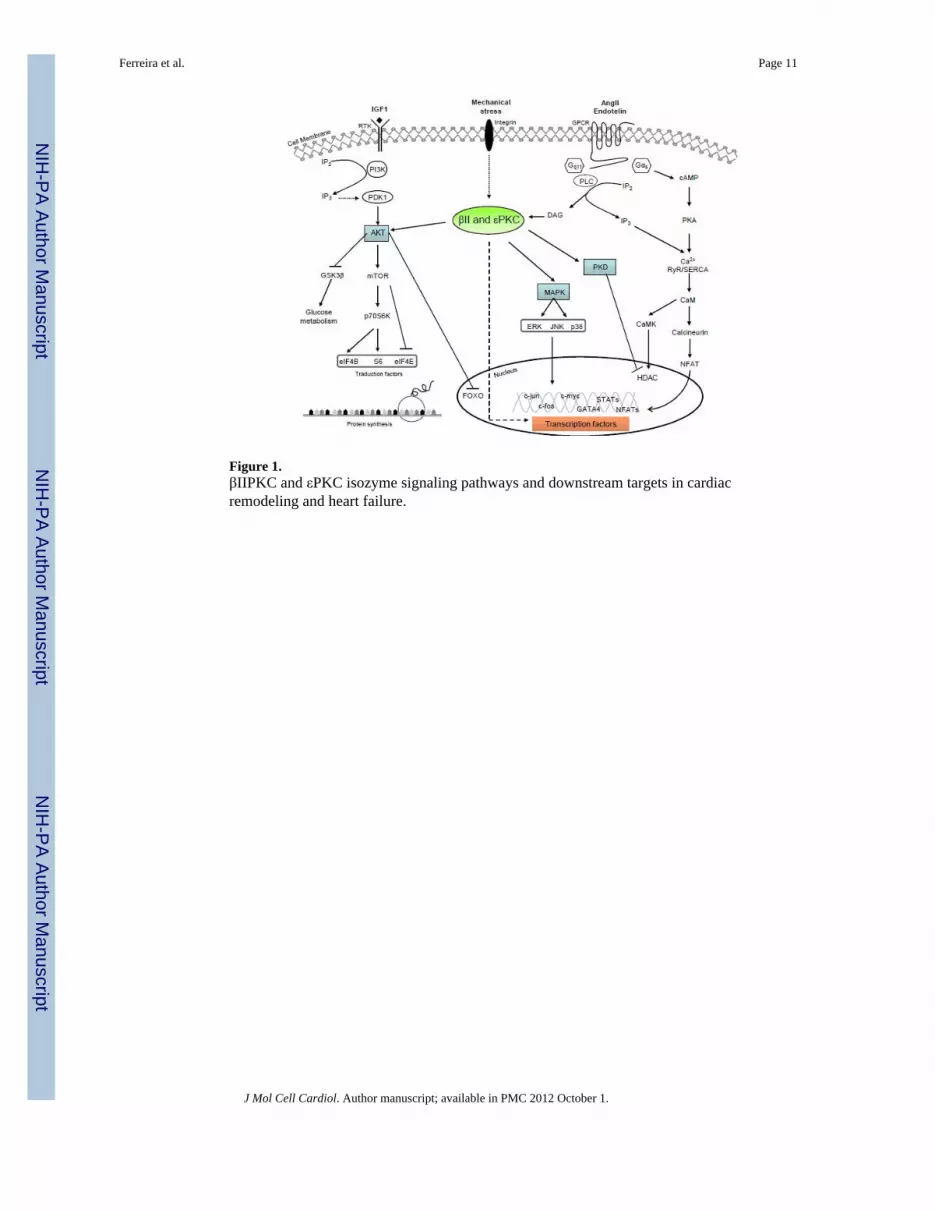
Figure 1.βIIPKC and εPKC isozyme signaling pathways and downstream targets in cardiacremodeling and heart failure.
Ferreira et al. Page 11
J Mol Cell Cardiol. Author manuscript; available in PMC 2012 October 1.
NIH
-PA Author Manuscript
NIH
-PA Author Manuscript
NIH
-PA Author Manuscript

Figure 2.Schematic βIIPKC and εPKC isozyme-mediated cellular responses in cardiac remodelingand heart failure.
Ferreira et al. Page 12
J Mol Cell Cardiol. Author manuscript; available in PMC 2012 October 1.
NIH
-PA Author Manuscript
NIH
-PA Author Manuscript
NIH
-PA Author Manuscript

NIH
-PA Author Manuscript
NIH
-PA Author Manuscript
NIH
-PA Author Manuscript
Ferreira et al. Page 13
Table 1
The role of isozyme-specific βIIPKC and εPKC in cardiac remodeling and heart failure
PKC isozyme Cardiac phenotype Model Authors Features
εPKC Hypertrophy Overexpression of constitutively activeεPKC
Takeishi etal. [26]
Concentric cardiac hypertrophy
εPKC Hypertrophy Cardiac-specific expression of εPKCinhibitor, εV1
Mochly-Rosen etal. [32]
Lethal dilated cardiomyopathy
εPKC Hypertrophy Cardiac-specific expression of εPKCactivator, ψεRACK
Mochly-Rosen etal. [32]
Concentric cardiac hypertrophy
εPKC Hypertrophy Overexpression of constitutively activeεPKC
Pass et al.[16]
Pathological cardiac hypertrophy
εPKC Hypertrophy Hypertensive rats-treated with εV1-2(specific εPKC isozyme inhibitor)
Shizukudaet al. [62]
Attenuated isoproterenol-inducedcell death
εPKC Hypertrophy Pressure-overload aortic banding εPKCknock-out mouse
Klein et al.[63]
Increased fibrosis
εPKC Hypertrophy/heart failure Pressure-overload heart failure rats Takeishi etal. [64]
ACE inhibitor attenuatesincreased αPKC and εPKC
translocation
εPKC/βII PKC Hypertrophy/heart failure Dahl Salt hypertensive rats Koide et al.[65]
Increased cardiac levels ofβIIPKC and εPKC
εPKC Heart failure Hypertensive rats-sustained treatmentwith εV1-2 (specific εPKC inhibitor)
Inagaki etal. [33]
Decreased cardiac fibrosis
βIIPKC Heart failure Heart failure rats-non-specific PKCinhibitor (LY333531)
Boyle et al.[46]
Decreased both fibrosis andTGFβ1 expression
βIIPKC Heart failure Cardiac-specific overexpression ofβIIPKC
Wakasakiet al. [35]
Pathologic cardiac hypertrophyand fibrosis
βIIPKC Heart failure Dahl Salt hypertensive rats Inagaki etal. [18]
Increased cardiac βIPKC, βIIPKClevels and translocation
βIIPKC Heart failure Human end-stage dilated or ischemiccardiomyopathy
Bowling etal. [12]
Increased cardiac βPKC activity
βIIPKC Heart failure Human end-stage dilated cardiomyopathy Simonis etal. [14]
Increased cardiac βIIPKC levels
βIIPKC Hypertrophy/heart Failure Cardiac-specific βIIPKC overexpression Bowman etal. [66]
Pathological cardiac hypertrophy
βIIPKC Hypertrophy Perfusion with angiotensin II-adult guineapig heart (ex vivo)
Takeishi etal. [67]
Increased cardiac βIIPKC level
βIIPKC Hypertrophy Transgenic mice-active calciunerinoverexpression
De Windtet al. [68]
Increased cardiac βPKCtranslocation
βIIPKC Hypertrophy Streptozotocin-induced diabetic rats Inoguchi etal. [69]
Increased cardiac βIIPKC activity
J Mol Cell Cardiol. Author manuscript; available in PMC 2012 October 1.


