Embed Size (px)
Citation preview


VIROLOGY


VIROLOGYPRINCIPLES AND APPLICATIONS
John B. Carter and Venetia A. Saunders
School of Biomolecular Sciences,Liverpool John Moores University, UK
Copyright 2007 John Wiley & Sons Ltd, The Atrium, Southern Gate, Chichester,West Sussex PO19 8SQ, England
Telephone (+44) 1243 779777
Email (for orders and customer service enquiries): [email protected] our Home Page on www.wileyeurope.com or www.wiley.com
All Rights Reserved. No part of this publication may be reproduced, stored in a retrieval system or transmitted in any form or byany means, electronic, mechanical, photocopying, recording, scanning or otherwise, except under the terms of the Copyright,Designs and Patents Act 1988 or under the terms of a licence issued by the Copyright Licensing Agency Ltd, 90 Tottenham CourtRoad, London W1T 4LP, UK, without the permission in writing of the Publisher. Requests to the Publisher should be addressedto the Permissions Department, John Wiley & Sons Ltd, The Atrium, Southern Gate, Chichester,West Sussex PO19 8SQ, England, or emailed to [email protected], or faxed to (+44) 1243 770620.
Designations used by companies to distinguish their products are often claimed as trademarks. All brand names and productnames used in this book are trade names, service marks, trademarks or registered trademarks of their respective owners. ThePublisher is not associated with any product or vendor mentioned in this book.
This publication is designed to provide accurate and authoritative information in regard to the subject matter covered. It is sold onthe understanding that the Publisher is not engaged in rendering professional services. If professional advice or other expertassistance is required, the services of a competent professional should be sought.
Other Wiley Editorial Offices
John Wiley & Sons Inc., 111 River Street, Hoboken, NJ 07030, USA
Jossey-Bass, 989 Market Street, San Francisco, CA 94103-1741, USA
Wiley-VCH Verlag GmbH, Boschstr. 12, D-69469 Weinheim, Germany
John Wiley & Sons Australia Ltd, 42 McDougall Street, Milton, Queensland 4064, Australia
John Wiley & Sons (Asia) Pte Ltd, 2 Clementi Loop #02-01, Jin Xing Distripark, Singapore 129809
John Wiley & Sons Canada Ltd, 6045 Freemont Blvd, Mississauga, Ontario, L5R 4J3, Canada
Wiley also publishes its books in a variety of electronic formats. Some content that appearsin print may not be available in electronic books.
Library of Congress Cataloging-in-Publication Data:
Carter, John, 1944–Virology : principles and applications / John Carter and Venetia
Saunders.p. ; cm.
Includes bibliographical references and index.ISBN 978-0-470-02386-0 (cloth)
1. Virology. 2. Viruses. 3. Virus diseases. I. Saunders, VenetiaA., 1949– II. Title.
[DNLM: 1. Viruses. 2. Virus Diseases. QW 160 C323v2007]QR360.C36 2007616.9′101 – dc22
2007017896
British Library Cataloguing in Publication Data
A catalogue record for this book is available from the British Library
ISBN: 978-0-470-02386-0 (HB)978-0-470-02387-7 (PB)
Typeset in 10/12pt Times by Laserwords Private Limited, Chennai, IndiaPrinted and bound by Printer Trento Srl., Trento, ItalyThis book is printed on acid-free paper responsibly manufactured from sustainable forestryin which at least two trees are planted for each one used for paper production.

To Myra, Robert, Jon and Mark

Contents
Preface xvAbbreviations used in this book xviiGreek letters used in this book xxiColour coding for molecules xxiii
1 Viruses and their importance 1At a glance 1
1.1 Viruses are ubiquitous on Earth 31.2 Reasons for studying viruses 31.3 The nature of viruses 41.4 The remainder of the book 7
Learning outcomes 7Sources of further information 7
2 Methods used in virology 9At a glance 9
2.1 Introduction to methods used in virology 112.2 Cultivation of viruses 122.3 Isolation of viruses 132.4 Centrifugation 132.5 Structural investigations of cells and virions 172.6 Electrophoretic techniques 172.7 Detection of viruses and virus components 182.8 Infectivity assays 222.9 Virus genetics 26
Learning outcomes 28Sources of further information 28
3 Virus structure 31At a glance 31
3.1 Introduction to virus structure 323.2 Virus genomes 323.3 Virus proteins 373.4 Capsids 393.5 Virion membranes 453.6 Occlusion bodies 47
viii CONTENTS
3.7 Other virion components 47Learning outcomes 48Sources of further information 48
4 Virus transmission 49At a glance 49
4.1 Introduction to virus transmission 504.2 Transmission of plant viruses 514.3 Transmission of vertebrate viruses 544.4 Transmission of invertebrate viruses 564.5 Permissive cells 57
Learning outcomes 58Sources of further information 58
5 Attachment and entry of viruses into cells 59At a glance 59
5.1 Overview of virus replication 605.2 Animal viruses 615.3 Bacteriophages 68
Learning outcomes 68Sources of further information 68
6 Transcription, translation and transport 69At a glance 69
6.1 Introduction to transcription, translation and transport 706.2 Transcription of virus genomes 706.3 Transcription in eukaryotes 726.4 Translation in eukaryotes 776.5 Transport in eukaryotic cells 816.6 Transcription and translation in bacteria 83
Learning outcomes 84Sources of further information 84
7 Virus genome replication 85At a glance 85
7.1 Overview of virus genome replication 867.2 Locations of virus genome replication in eukaryotic cells 887.3 Initiation of genome replication 887.4 Polymerases 897.5 DNA replication 907.6 Double-stranded RNA replication 917.7 Single-stranded RNA replication 927.8 Reverse transcription 92
Learning outcomes 93Sources of further information 93
CONTENTS ix
8 Assembly and exit of virions from cells 95At a glance 95
8.1 Introduction to assembly and exit of virions from cells 968.2 Nucleocapsid assembly 968.3 Formation of virion membranes 988.4 Virion exit from the infected cell 101
Learning outcomes 101Sources of further information 102
9 Outcomes of infection for the host 103At a glance 103
9.1 Introduction to outcomes of infection for the host 1059.2 Factors affecting outcomes of infection 1059.3 Non-productive infections 1109.4 Productive infections 112
Learning outcomes 114Sources of further information 114
10 Classification and nomenclature of viruses 115At a glance 11510.1 History of virus classification and nomenclature 11610.2 Modern virus classification and nomenclature 11610.3 Baltimore classification of viruses 119
Learning outcomes 119Sources of further information 120
11 Herpesviruses (and other dsDNA viruses) 121At a glance 12111.1 Introduction to herpesviruses 12211.2 The human herpesviruses 12211.3 The herpesvirus virion 12311.4 HSV-1 genome organization 12411.5 HSV-1 replication 12411.6 Latent herpesvirus infection 13011.7 Other dsDNA viruses 132
Learning outcomes 135Sources of further information 135
12 Parvoviruses (and other ssDNA viruses) 137At a glance 13712.1 Introduction to parvoviruses 13812.2 Examples of parvoviruses 13812.3 Parvovirus virion 13912.4 Parvovirus replication 141
x CONTENTS
12.5 Other ssDNA viruses 143Learning outcomes 146Sources of further information 146
13 Reoviruses (and other dsRNA viruses) 147At a glance 14713.1 Introduction to reoviruses 14813.2 Rotavirus virion 14813.3 Rotavirus replication 15013.4 Other dsRNA viruses 155
Learning outcomes 155Sources of further information 155
14 Picornaviruses (and other plus-strand RNA viruses) 157At a glance 15714.1 Introduction to picornaviruses 15814.2 Some important picornaviruses 15814.3 Picornavirus Virion 16014.4 Picornavirus replication 16114.5 Picornavirus recombination 16914.6 Picornavirus experimental systems 16914.7 Other plus-strand RNA viruses 170
Learning outcomes 172Sources of further information 172
15 Rhabdoviruses (and other minus-strand RNA viruses) 173At a glance 17315.1 Introduction to rhabdoviruses 17415.2 Some important rhabdoviruses 17515.3 The rhabdovirus virion and genome organization 17715.4 Rhabdovirus replication 17715.5 Other minus-strand RNA viruses 18315.6 Viruses with ambisense genomes 18315.7 Reverse genetics 183
Learning outcomes 184Sources of further information 184
16 Retroviruses 185At a glance 18516.1 Introduction to retroviruses 18616.2 Retrovirus virion 18616.3 Retrovirus replication 18816.4 Examples of retroviruses 19416.5 Retroviruses as gene vectors 19416.6 Endogenous retroviruses 194
Learning outcomes 196Sources of further information 196
CONTENTS xi
17 Human immunodeficiency viruses 197At a glance 19717.1 Introduction to HIV 19817.2 HIV virion 19817.3 HIV genome 19917.4 HIV-1 replication 19917.5 HIV-1 variability 20917.6 Progression of HIV infection 20917.7 Prevention of HIV transmission 210
Learning outcomes 211Sources of further information 211
18 Hepadnaviruses (and other reverse-transcribing DNAviruses) 213At a glance 21318.1 Introduction to hepadnaviruses 21418.2 Importance of HBV 21418.3 HBV virion 21518.4 Non-infectious particles 21618.5 Soluble virus protein 21618.6 HBV genome 21618.7 HBV genetic groups 21818.8 HBV replication 21818.9 Prevention and treatment of HBV infection 225
18.10 Other reverse-transcribing DNA viruses 225Learning outcomes 227Sources of further information 227
19 Bacterial viruses 229At a glance 22919.1 Introduction to bacterial viruses (bacteriophages) 230
RNA PHAGES 23119.2 Single-stranded RNA phages 23119.3 Double-stranded RNA phages 237
DNA PHAGES 23719.4 Single-stranded DNA phages 23719.5 Double-stranded DNA phages 246
Learning outcomes 255Sources of further information 255
20 Origins and evolution of viruses 257At a glance 25720.1 Introduction to origins and evolution of viruses 25820.2 Origins of viruses 25820.3 Evolution of viruses 261
Learning outcomes 270Sources of further information 270
xii CONTENTS
21 Emerging viruses 273At a glance 27321.1 Introduction to emerging viruses 27421.2 Viruses in new host species 27421.3 Viruses in new areas 27621.4 Viruses in new host species and in new areas 27821.5 New viruses 27921.6 Recently discovered viruses 28121.7 Re-emerging viruses 28121.8 Virus surveillance 28221.9 Dealing with outbreaks 282
Learning outcomes 283Sources of further information 283
22 Viruses and cancer 285At a glance 28522.1 Introduction to viruses and cancer 28622.2 Papillomavirus-linked cancers 28722.3 Polyomavirus-linked cancers 28722.4 Epstein-Barr virus-linked cancers 28922.5 Kaposi’s sarcoma 29022.6 Adult T cell leukaemia 29022.7 Hepatocellular carcinoma 29022.8 Virus-associated cancers in animals 29122.9 Cell lines derived from virus-associated cancers 291
22.10 How do viruses cause cancer? 29122.11 Prevention of virus-induced cancers 295
Learning outcomes 296Sources of further information 296
23 Survival of infectivity 297At a glance 29723.1 Preservation of virus infectivity 29823.2 Destruction of virus infectivity 29823.3 Inactivation targets in virions 29823.4 Inactivation kinetics 29823.5 Agents that inactivate virus infectivity 299
Learning outcomes 303Sources of further information 303
24 Virus vaccines 305At a glance 30524.1 Introduction to virus vaccines 30624.2 Live attenuated virus vaccines 30624.3 Inactivated virus vaccines 30724.4 Virion subunit vaccines 30924.5 Live recombinant virus vaccines 30924.6 Mass production of viruses for vaccines 310
CONTENTS xiii
24.7 Virus-like particles 31124.8 Synthetic peptide vaccines 31124.9 DNA vaccines 311
24.10 Storage and transport of vaccines 312Learning outcomes 313Sources of further information 313
25 Anti-viral drugs 315At a glance 31525.1 Introduction to anti-viral drugs 31625.2 Development of anti-viral drugs 31625.3 Examples of anti-viral drugs 31725.4 Drug resistance 32225.5 Anti-viral drug research 324
Learning outcomes 325Sources of further information 325
26 Prions 327At a glance 32726.1 Introduction to prions 32826.2 Transmissible spongiform encephalopathies 32826.3 The nature of prions 32826.4 Prion diseases 33026.5 Prion strains 33226.6 Prion transmission 33226.7 The protein-only hypothesis 333
Learning outcomes 333Sources of further information 333
Virologists’ vocabulary 335
Index 349

Preface
Virology is a fascinating and rapidly developing sub-ject, and is worthy of study purely because viruses areinteresting! Furthermore, virology is a branch of sci-ence that is of immense relevance to mankind for ahost of reasons, not least of which are the threats tohuman health caused by viruses, such as HIV, hep-atitis B virus, papillomaviruses, measles and influenzaviruses, to mention just a few. There is a continuingneed for trained virologists, and it is hoped that thisbook will play a small role in helping to fulfil this need.To a large extent the material in the book is based onvirology taught at Liverpool John Moores University.
This is not a textbook of fundamental virology,medical virology, veterinary virology, plant virologyor of bacteriophages, but a bit of each of these! Thegeneral pattern of the book is that principles of virologyare covered earlier and applications are covered later.There is no strict demarcation between the two,however, so the reader may be made aware of importantapplications while principles are being introduced.
The first 10 chapters cover basic aspects of virology.A chapter on methods used in virology comes early inthe book, but could be skimmed to gain an overview ofits contents and thereafter used for reference. There isone chapter on each of the seven Baltimore classes,concentrating mainly on animal viruses. There is achapter devoted entirely to HIV and an extendedchapter on phages, reflecting the renewed interest intheir biology and applications. After a chapter onorigins and evolution of viruses, there follow fivechapters covering various aspects of applied virology,including vaccines and antiviral drugs. The finalchapter is on prions, which are not viruses but are oftenconsidered along with the viruses.
Each chapter starts with ‘At a glance’, a briefsummary with the dual aim of giving a flavour ofwhat is coming up and providing a revision aid. Each
chapter ends with a list of learning outcomes and aguide to further reading in books and journals. Thereferences are mainly from the 21st century, but thereis a selection of important papers from the last century.
The book has a web site (www.wiley.com/go/carter),where you can find
• many references additional to those in the book;
• links to the journal references (to the full text wherethis is freely available, otherwise to the abstract);
• links to virology web sites;
• self-assessment questions and answers for eachchapter, to reinforce and extend concepts developedin the book.
A key feature of our book is a standard colour codeto differentiate various types of nucleic acid and proteinmolecule in the diagrams. The colour code is explainedon page xxiii. It is appreciated that colour coding maybe of limited value to individuals who have difficulty indifferentiating colours, so we have also labelled manyof the molecules.
A number of virus replication cycles are describedand the reader should be aware that these are modelsbased on evidence to date; the models may have to bemodified in the light of future evidence. We presentthe virus replication cycles as fitting within a generalframework of seven steps:
(1) Attachment of a virion to a cell
(2) Entry into the cell
(3) Transcription of virus genes into mRNAs
(4) Translation of virus mRNAs into virus proteins
(5) Genome replication
xvi PREFACE
(6) Assembly of the virus proteins and genomes intovirions
(7) Exit of the virions from the cell.
We hope that this helps in appreciating how virusreplication fits into a general pattern and in comparingthe replication cycles of different types of virus. Forsome groups of viruses the framework has to bemodified and we make clear when this is the case.
If you come across an unfamiliar term, pleaseconsult the ‘Virologists’ vocabulary’ at the back of thebook. This glossary includes not only virology-specificterms, but also a selection of terms from cell biology,molecular biology, immunology and medicine.
A list of the abbreviations that are used in this bookappears on the following pages.
We wish to thank the many people who have madethe production of this book possible. We thank all thosewho supplied images and those who gave permissionfor the use of their images; we are especially grate-ful to David Bhella, Tom Goddard, Kathryn Newtonand Jean-Yves Sgro. Thanks also to Robert Carter forassistance with images. We acknowledge the contribu-tions of the many students who have acted as guinea-pigs for our teaching materials and who have providedus with feedback. Grateful thanks also to those who
reviewed material for the book and provided valu-able feedback. We are sorry that we were unable toinclude all the topics suggested, but if we had doneso the book would have run to several volumes! Manythanks to Rachael Ballard, Robert Hambrook and all atJohn Wiley & Sons, Ltd who helped the book come tofruition. Finally, thanks to our families for their sup-port and for their patience during those many hours wespent ensconced in the study.
We hope you find the book useful and we would beinterested to hear what you think of it. We have triedto ensure that there are no errors, but it is probable thatsome have slipped through; if you come across anyerrors please inform us.
John B. [email protected]
Venetia A. [email protected]
School of Biomolecular Sciences,Liverpool John Moores University,
Byrom Street,Liverpool L3 3AF, UK
Abbreviations used in this book
(+) DNA plus strand (positive strand) DNA
(−) DNA minus strand (negative strand) DNA
(+) RNA plus strand (positive strand) RNA
(−) RNA minus strand (negative strand) RNA
A adenine
ADP adenosine diphosphate
AIDS acquired immune deficiency syndrome
AP-1 activator protein 1
ATP adenosine triphosphate
b base(s)
BL Burkitt’s lymphoma
bp base pair(s)
BSE bovine spongiform encephalitis
C cytosine
C terminus carboxy terminus
cccDNA covalently closed circular DNA
CD cluster of differentiation
cDNA copy DNA
CJD Creutzfeldt-Jakob disease
cos cohesive end
CP coat protein
CPE cytopathic effect
DIP defective interfering particle
DNA deoxyribose nucleic acid
ds double-stranded
DTR direct terminal repeat
E early
EBV Epstein-Barr virus
EF elongation factor
xviii ABBREVIATIONS USED IN THIS BOOK
ELISA enzyme-linked immunosorbent assayERV endogenous retrovirusE. coli Escherichia coliFf F-specific filamentousG guanineGFP green fluorescent proteinHAV hepatitis A virusHBV hepatitis B virusHBsAg hepatitis B surface antigenHCV hepatitis C virusHIV human immunodeficiency virusHPV human papillomavirusHSV herpes simplex virusHTLV-1 human T-lymphotropic virus 1IC50 50% inhibitory concentrationICTV International Committee on Taxonomy of VirusesICTVdB International Committee on Taxonomy of Viruses databaseIE immediate earlyIG intergenicIRES internal ribosome entry siteITR inverted terminal repeatkb kilobase(s)
kbp kilobase pair(s)
kD kiloDalton(s)
KSHV Kaposi’s sarcoma-associated herpesvirus
LDI long distance interaction
LE left end
LIN lysis inhibition
LPS lipopolysaccharide
LTR long terminal repeat
Mbp megabase pair(s)
MHC major histocompatibility complex
MJ Min Jou
m.o.i. multiplicity of infection
MP movement protein
mRNA messenger RNA
N terminus amino terminus
NF-κB nuclear factor kappa B
NK cell natural killer cell
ABBREVIATIONS USED IN THIS BOOK xix
nm nanometre(s) (10−9 metre)NPC nasopharyngeal carcinomaNSP non-structural proteinO operatorORF open reading frameori origin (replication)P promoterPBS primer binding sitePCR polymerase chain reactionpfu plaque-forming unitphage bacteriophagePS packaging signalRBS ribosome binding siteRE right endRF replicative formRI replicative intermediateRNA ribose nucleic acidRNAi RNA interferenceRNAse H ribonuclease HrRNA ribosomal RNART-PCR reverse transcriptase–polymerase chain reactionS Svedberg unitSARS severe acute respiratory syndromeS-D Shine-DalgarnoSI selectivity indexSIV simian immunodeficiency virusSp1 stimulatory protein 1ss single-strandedssb single-stranded bindingT thymineTCID50 virus dose that infects 50% of tissue culturesTK thymidine kinasetRNA transfer RNATSE transmissible spongiform encephalitisU uracilUV ultra-violetvCJD variant Creutzfeldt-Jakob diseaseVP virus proteinVPg virus protein, genome linkedVSV vesicular stomatitis virus

Greek letters used in this book
α alpha
β beta
γ gamma
ε epsilon
θ theta
κ kappa
λ lambda
σ sigma
ϕ phi
ψ psi

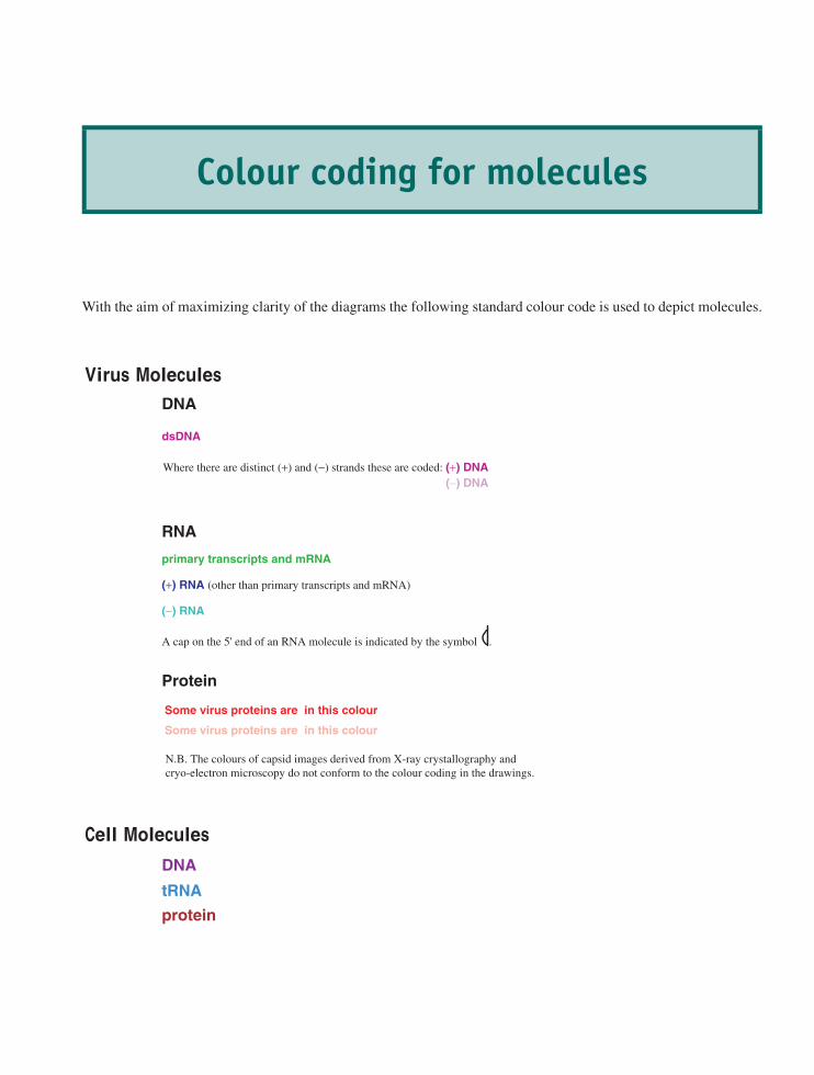
Colour coding for molecules
With the aim of maximizing clarity of the diagrams the following standard colour code is used to depict molecules.
Where there are distinct (+) and (−) strands these are coded: (+) DNA(−) DNA
Some virus proteins are in this colour
Some virus proteins are in this colour
tRNA
RNA
Protein
(−) RNA
(+) RNA (other than primary transcripts and mRNA)
primary transcripts and mRNA
DNA
protein
dsDNA
DNA
A cap on the 5' end of an RNA molecule is indicated by the symbol .
N.B. The colours of capsid images derived from X-ray crystallography andcryo-electron microscopy do not conform to the colour coding in the drawings.

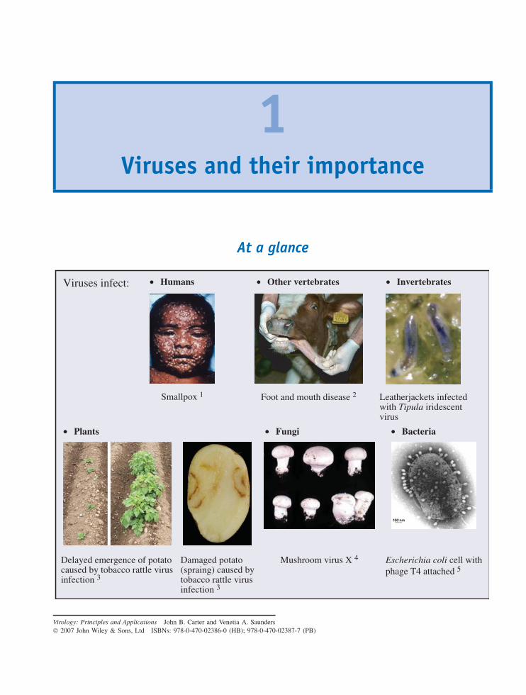
1Viruses and their importance
At a glance
Viruses infect: • Humans
Smallpox 1 Foot and mouth disease 2
• Other vertebrates
Leatherjackets infectedwith Tipula iridescentvirus
• Invertebrates
Mushroom virus X 4
• Fungi
Escherichia coli cell withphage T4 attached 5
• Bacteria• Plants
Delayed emergence of potato caused by tobacco rattle virusinfection 3
Damaged potato(spraing) caused bytobacco rattle virusinfection 3
Virology: Principles and Applications John B. Carter and Venetia A. Saunders 2007 John Wiley & Sons, Ltd ISBNs: 978-0-470-02386-0 (HB); 978-0-470-02387-7 (PB)
2 VIRUSES AND THEIR IMPORTANCE
At a glance (continued)
Some viruses are useful: ••••••
Phage typing of bacteria
Sources of enzymes
Pesticides
Anti-bacterial agents
Anti-cancer agents
Gene vectors
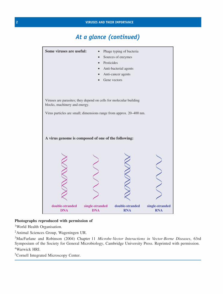
Viruses are parasites; they depend on cells for molecular building blocks, machinery and energy.
Virus particles are small; dimensions range from approx. 20–400 nm.
A virus genome is composed of one of the following:
double-strandedDNA
single-strandedDNA
double-strandedRNA
single-strandedRNA
Photographs reproduced with permission of1World Health Organisation.2Animal Sciences Group, Wageningen UR.3MacFarlane and Robinson (2004) Chapter 11 Microbe-Vector Interactions in Vector-Borne Diseases, 63rdSymposium of the Society for General Microbiology, Cambridge University Press. Reprinted with permission.4Warwick HRI.5Cornell Integrated Microscopy Center.
REASONS FOR STUDYING VIRUSES 3
1.1 Viruses are ubiquitous on EarthViruses infect all cellular life forms: eukaryotes (verte-brate animals, invertebrate animals, plants, fungi) andprokaryotes (bacteria and archaea). The viruses thatinfect prokaryotes are often referred to as bacterio-phages, or phages for short.
The presence of viruses is obvious in host organismsshowing signs of disease. Many healthy organisms,however, are hosts of non-pathogenic virus infections,some of which are active, while some are quiescent.Furthermore, the genomes of many organisms containremnants of ancient virus genomes that integrated intotheir host genomes long ago. As well being presentwithin their hosts, viruses are also found in soil, air andwater. Many aqueous environments contain very highconcentrations of viruses that infect the organisms thatlive in those environments.
There is a strong correlation between how inten-sively a species is studied and the number of virusesfound in that species. Our own species is the subject ofmost attention as we have a vested interest in learningabout agents and processes that affect our health. It isnot surprising that there are more viruses known thatinfect mankind than any other species, and new humanviruses continue to be found. The intestinal bacteriumEscherichia coli has also been the subject of muchstudy and many viruses have been found in this species.If other species received the same amount of attentionit is likely that many would be found to be hosts tosimilar numbers of viruses.
It is undoubtedly the case that the viruses that havebeen discovered represent only a tiny fraction of theviruses on the Earth. Most of the known plants, ani-mals, fungi, bacteria and archaea have yet to be inves-tigated for the presence of viruses, and new potentialhosts for viruses continue to be discovered. Further-more, the analysis of DNA from natural environmentspoints to the existence of many bacterial species thathave not yet been isolated in the laboratory; it is likelythat these ‘non-cultivable bacteria’ are also hosts toviruses.
1.2 Reasons for studying viruses1.2.1 Some viruses cause diseaseViruses are important agents of many human diseases,ranging from the trivial (e.g. common colds) to the
lethal (e.g. rabies), and viruses also play roles in thedevelopment of several types of cancer. As well ascausing individuals to suffer, virus diseases can alsoaffect the well-being of societies. Smallpox had agreat impact in the past and AIDS is having a greatimpact today.
There is therefore a requirement to understand thenature of viruses, how they replicate and how theycause disease. This knowledge permits the develop-ment of effective means for prevention, diagnosis andtreatment of virus diseases through the production ofvaccines, diagnostic reagents and techniques, and anti-viral drugs. These medical applications therefore con-stitute major aspects of the science of virology.
Veterinary virology and plant virology are alsoimportant because of the economic impact of the manyviruses that cause disease in domestic animals andcrop plants: foot and mouth disease virus and riceyellow mottle virus are just two examples. Anotherarea where viruses can cause economic damage is inthe dairy industry, where phages can infect the lacticacid bacteria that are responsible for the fermentationsthat produce cheese, yogurt and other milk products.
1.2.2 Some viruses are usefulSome viruses are studied because they have usefulcurrent or potential applications.
• Phage typing of bacteria. Some groups of bacteria,such as some Salmonella species, are classified intostrains on the basis of the spectrum of phages towhich they are susceptible. Identification of thephage types of bacterial isolates can provide usefulepidemiological information during outbreaks ofdisease caused by these bacteria.
• Sources of enzymes. A number of enzymes usedin molecular biology are virus enzymes. Examplesinclude reverse transcriptases from retroviruses andRNA polymerases from phages.
• Pesticides. Some insect pests are controlled withbaculoviruses and myxoma virus has been used tocontrol rabbits.
• Anti-bacterial agents. In the mid-20th centuryphages were used to treat some bacterial infectionsof humans. Interest waned with the discovery of
4 VIRUSES AND THEIR IMPORTANCE
antibiotics, but has been renewed with the emergenceof antibiotic-resistant strains of bacteria.
• Anti-cancer agents. Genetically modified strains ofviruses, such as herpes simplex virus and vacciniavirus, are being investigated for treatment of cancers.These strains have been modified so that they areable to infect and destroy specific tumour cells, butare unable to infect normal cells.
• Gene vectors for protein production. Viruses such ascertain baculoviruses and adenoviruses are used asvectors to take genes into animal cells growing inculture. This technology can be used to insert intocells genes encoding useful proteins, such as vaccinecomponents, and the cells can then be used for massproduction of the proteins.
• Gene vectors for treatment of genetic diseases.Children with severe combined immunodeficiency(baby in the bubble syndrome) have been suc-cessfully treated using retroviruses as vectors tointroduce into their stem cells a non-mutated copyof the mutated gene responsible for the disease(Section 16.5).
1.2.3 Virus studies have contributedto knowledge
Much of the basic knowledge of molecular biology,cell biology and cancer has been derived from studieswith viruses. Here are a few examples.
• A famous experiment carried by Alfred Hershey andMartha Chase, and published in 1952, used phage T2and E. coli to provide strong evidence that genes arecomposed of DNA.
• The first enhancers to be characterized were in genesof simian virus 40 (SV40).
• The first transcription factor to be characterized wasthe transplantation (T) antigen of SV40.
• The first nuclear localization signal of a protein wasidentified in the T antigen of SV40.
• Introns were discovered during studies of adenovirustranscription.
• The role of the cap structure at the 5′ end ofeukaryotic messenger RNA was discovered duringstudies with vaccinia virus and a reovirus.
• The first internal ribosomal entry site to be discov-ered was found in the RNA of poliovirus.
• The first RNA pseudoknot to be discovered was thatin the genome of turnip yellow mosaic virus.
1.3 The nature of viruses1.3.1 Viruses are small particles
Evidence for the existence of very small infectiousagents was first provided in the late 19th century by twoscientists working independently: Martinus Beijerinckin Holland and Dimitri Ivanovski in Russia. They madeextracts from diseased plants, which we now knowwere infected with tobacco mosaic virus, and passedthe extracts through fine filters. The filtrates containedan agent that was able to infect new plants, but nobacteria could be cultured from the filtrates. The agentremained infective through several transfers to newplants, eliminating the possibility of a toxin. Beijerinckcalled the agent a ‘virus’ and the term has been in useever since.
At around the same time, Freidrich Loeffler andPaul Frosch transmitted foot and mouth disease fromanimal to animal in inoculum that had been highlydiluted. A few years later Walter Reed and JamesCarroll demonstrated that the causative agent of yellowfever is a filterable agent.
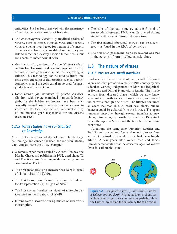
×107
×107
Figure 1.1 Comparative sizes of a herpesvirus particle,a balloon and the Earth. A large balloon is about tenmillion times larger than a herpesvirus particle, whilethe Earth is larger than the balloon by the same factor.
THE NATURE OF VIRUSES 5
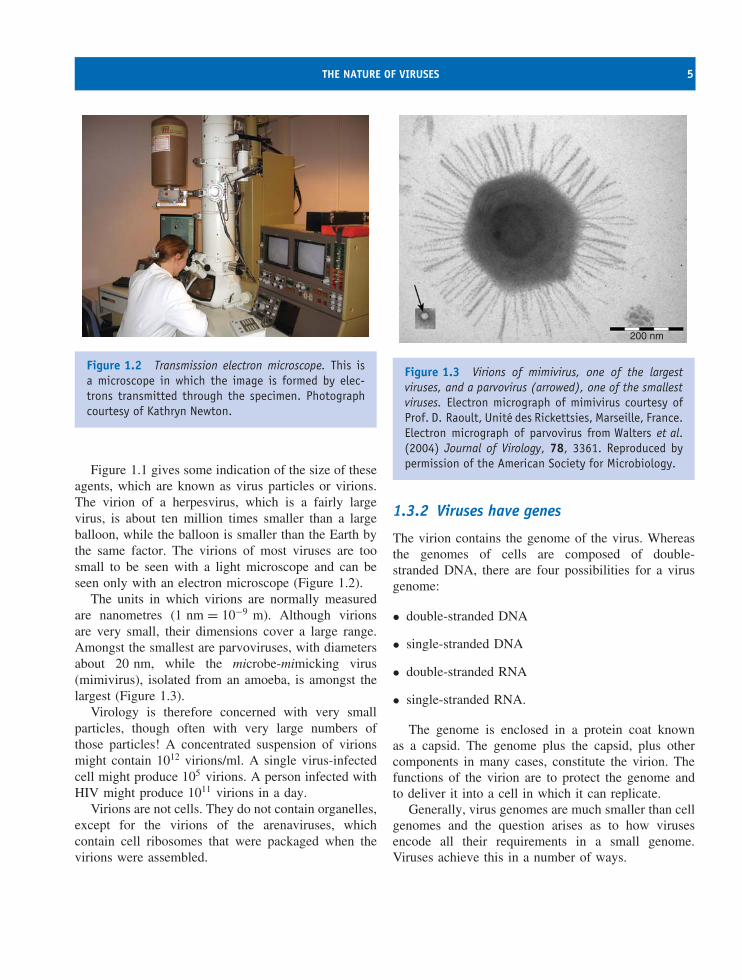
Figure 1.2 Transmission electron microscope. This isa microscope in which the image is formed by elec-trons transmitted through the specimen. Photographcourtesy of Kathryn Newton.
Figure 1.1 gives some indication of the size of theseagents, which are known as virus particles or virions.The virion of a herpesvirus, which is a fairly largevirus, is about ten million times smaller than a largeballoon, while the balloon is smaller than the Earth bythe same factor. The virions of most viruses are toosmall to be seen with a light microscope and can beseen only with an electron microscope (Figure 1.2).
The units in which virions are normally measuredare nanometres (1 nm = 10−9 m). Although virionsare very small, their dimensions cover a large range.Amongst the smallest are parvoviruses, with diametersabout 20 nm, while the microbe-mimicking virus(mimivirus), isolated from an amoeba, is amongst thelargest (Figure 1.3).
Virology is therefore concerned with very smallparticles, though often with very large numbers ofthose particles! A concentrated suspension of virionsmight contain 1012 virions/ml. A single virus-infectedcell might produce 105 virions. A person infected withHIV might produce 1011 virions in a day.
Virions are not cells. They do not contain organelles,except for the virions of the arenaviruses, whichcontain cell ribosomes that were packaged when thevirions were assembled.
200 nm
Figure 1.3 Virions of mimivirus, one of the largestviruses, and a parvovirus (arrowed), one of the smallestviruses. Electron micrograph of mimivirus courtesy ofProf. D. Raoult, Unite des Rickettsies, Marseille, France.Electron micrograph of parvovirus from Walters et al.(2004) Journal of Virology, 78, 3361. Reproduced bypermission of the American Society for Microbiology.
1.3.2 Viruses have genes
The virion contains the genome of the virus. Whereasthe genomes of cells are composed of double-stranded DNA, there are four possibilities for a virusgenome:
• double-stranded DNA
• single-stranded DNA
• double-stranded RNA
• single-stranded RNA.
The genome is enclosed in a protein coat knownas a capsid. The genome plus the capsid, plus othercomponents in many cases, constitute the virion. Thefunctions of the virion are to protect the genome andto deliver it into a cell in which it can replicate.
Generally, virus genomes are much smaller than cellgenomes and the question arises as to how virusesencode all their requirements in a small genome.Viruses achieve this in a number of ways.
6 VIRUSES AND THEIR IMPORTANCE
• Viruses use host cell proteins. The genomes oflarge viruses duplicate some of the functions ofthe host cell, but the small viruses rely veryheavily on functions of the host cell. There is,however, one function that an RNA virus mustencode, no matter how small its genome. Thatfunction is an RNA polymerase, because cellsdo not encode enzymes that can replicate virusRNA. A significant proportion of the genome of anRNA virus is taken up with the gene for an RNApolymerase.
• Viruses code efficiently. There may be overlappinggenes and genes encoded within genes. The smallgenome of hepatitis B virus is a good example (seeSection 18.6).
• Many virus proteins are multifunctional. A virusprotein may have several enzyme activities.
1.3.3 Viruses are parasitesViruses differ from cells in the way in which theymultiply. A new cell is always formed directly froma pre-existing cell, but a new virion is never formeddirectly from a pre-existing virion. New virions areformed by a process of replication, which takes placeinside a host cell and involves the synthesis ofcomponents followed by their assembly into virions.
Viruses are therefore parasites of cells, and aredependent on their hosts for most of their require-ments, including
• building-blocks such as amino acids and nucleo-sides;
• protein-synthesizing machinery (ribosomes);
• energy, in the form of adenosine triphosphate.
A virus modifies the intracellular environment of itshost in order to enhance the efficiency of the replicationprocess. Modifications might include production ofnew membranous structures, reduced expression ofcell genes or enhancement of a cell process. Somelarge phages encode proteins that boost photosynthesisin the cells of their photosynthetic bacterial hosts,thereby probably boosting the yields of virus from thecells.
A point has now been reached where the nature ofviruses can be summarized in a concise definition (seethe box).
Virus definition
A virus is a very small, non-cellular parasite of cells.Its genome, which is composed of either DNA orRNA, is enclosed in a protein coat.
1.3.4 Are viruses living or nonliving?
‘Viruses belong to biology because they possessgenes, replicate, evolve, and are adapted to par-ticular hosts, biotic habitats, and ecological niches.However, . . .they are nonliving infectious entitiesthat can be said, at best, to lead a kind of bor-rowed life.’
Marc van Regenmortel and Brian Mahy (2004)
‘It’s life, Jim, but not as we know it!’Dr. McCoy speaking to Captain Kirk of the
Starship Enterprise, Star Trek
There is an ongoing debate as to whether virusesare living or nonliving; the view taken depends onhow life is defined. Viruses have genes and when theyinfect cells these genes are replicated, so in this senseviruses are living. They are, however, very different tocellular life forms, so Dr. McCoy’s stock phrase (seethe box) on finding new life forms in the galaxy couldbe applied to viruses. When viruses are outside theirhost cells they exist as virus particles (virions), whichare inert, and could be described as nonliving, butviable bacterial spores are inert and are not consideredto be nonliving. You might form your own view as towhether viruses are living or nonliving as you progressthrough this book.
When Beijerinck selected the word ‘virus’ hechose the Latin word for poison. This term has nowbeen in use for over a century and virology hasdeveloped into a huge subject. More recently, theterm virus has acquired further meanings. Computers
SOURCES OF FURTHER INFORMATION 7
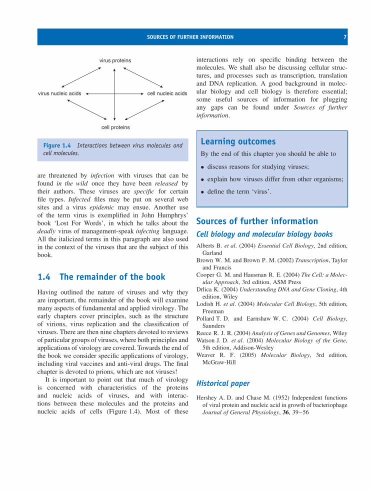
virus proteins
cell proteins
virus nucleic acids cell nucleic acids
Figure 1.4 Interactions between virus molecules andcell molecules.
are threatened by infection with viruses that can befound in the wild once they have been released bytheir authors. These viruses are specific for certainfile types. Infected files may be put on several websites and a virus epidemic may ensue. Another useof the term virus is exemplified in John Humphrys’book ‘Lost For Words’, in which he talks about thedeadly virus of management-speak infecting language.All the italicized terms in this paragraph are also usedin the context of the viruses that are the subject of thisbook.
1.4 The remainder of the book
Having outlined the nature of viruses and why theyare important, the remainder of the book will examinemany aspects of fundamental and applied virology. Theearly chapters cover principles, such as the structureof virions, virus replication and the classification ofviruses. There are then nine chapters devoted to reviewsof particular groups of viruses, where both principles andapplications of virology are covered. Towards the end ofthe book we consider specific applications of virology,including viral vaccines and anti-viral drugs. The finalchapter is devoted to prions, which are not viruses!
It is important to point out that much of virologyis concerned with characteristics of the proteinsand nucleic acids of viruses, and with interac-tions between these molecules and the proteins andnucleic acids of cells (Figure 1.4). Most of these
interactions rely on specific binding between themolecules. We shall also be discussing cellular struc-tures, and processes such as transcription, translationand DNA replication. A good background in molec-ular biology and cell biology is therefore essential;some useful sources of information for pluggingany gaps can be found under Sources of furtherinformation.
Learning outcomesBy the end of this chapter you should be able to
• discuss reasons for studying viruses;
• explain how viruses differ from other organisms;
• define the term ‘virus’.
Sources of further informationCell biology and molecular biology booksAlberts B. et al. (2004) Essential Cell Biology, 2nd edition,
GarlandBrown W. M. and Brown P. M. (2002) Transcription, Taylor
and FrancisCooper G. M. and Hausman R. E. (2004) The Cell: a Molec-
ular Approach, 3rd edition, ASM PressDrlica K. (2004) Understanding DNA and Gene Cloning, 4th
edition, WileyLodish H. et al. (2004) Molecular Cell Biology, 5th edition,
FreemanPollard T. D. and Earnshaw W. C. (2004) Cell Biology,
SaundersReece R. J. R. (2004) Analysis of Genes and Genomes, WileyWatson J. D. et al. (2004) Molecular Biology of the Gene,
5th edition, Addison-WesleyWeaver R. F. (2005) Molecular Biology, 3rd edition,
McGraw-Hill
Historical paper
Hershey A. D. and Chase M. (1952) Independent functionsof viral protein and nucleic acid in growth of bacteriophageJournal of General Physiology, 36, 39–56

8 VIRUSES AND THEIR IMPORTANCE
Recent papers
Breitbart M. and Rohwer F. (2005) Here a virus, there avirus, everywhere the same virus? Trends In Microbiology,13, 278–284
Chinen J. and Puck J. M. (2004) Successes and risks of genetherapy in primary immunodeficiencies Journal of Allergyand Clinical Immunology, 113, 595–603
Suttle C. A. (2005) Viruses in the sea Nature, 437, 356–361van Regenmortel M. H. V. and Mahy B. W. J. (2004)
Emerging issues in virus taxonomy Emerging InfectiousDiseases, 10(1) http://www.cdc.gov/ncidod/eid/vol10no1/03-0279.htm
Young L. S. et al. (2006) Viral gene therapy strategies: frombasic science to clinical application Journal of Pathology,208, 299–318
2Methods used in virology
At a glance
Virus Isolation and Culture
Animal virus plaquesin a cell culture 1
Phage plaques in a lawn of bacterial cells 2
Fluorescence Microscopy
Virus-infected cellsdetected using avirus-specific antibodylabelled with afluorescent dye 4
Confocal Microscopy
An endosome (labelled red) containing virus protein (labelled green)in an infected cell 5
Density Gradient Centrifugation
Separation of virus particlesin a density gradient 3
Virology: Principles and Applications John B. Carter and Venetia A. Saunders 2007 John Wiley & Sons, Ltd ISBNs: 978-0-470-02386-0 (HB); 978-0-470-02387-7 (PB)
10 METHODS USED IN VIROLOGY
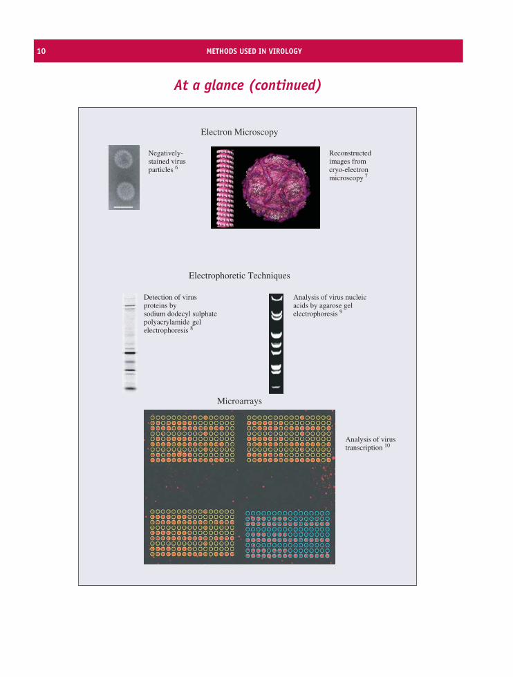
At a glance (continued)
Microarrays
Analysis of virustranscription 10
Electron Microscopy
Negatively-stained virusparticles 6
Electrophoretic Techniques
Detection of virus proteins bysodium dodecyl sulphate polyacrylamide gel electrophoresis 8
Analysis of virus nucleic acids by agarose gel electrophoresis 9
Reconstructedimages from cryo-electronmicroscopy 7
INTRODUCTION TO METHODS USED IN VIROLOGY 11
1Plaques formed by porcine reproductive and respiratory syndrome virus in a cell cultureFrom Lee and Yoo (2005) Journal of General Virology, 86, 3091.Reproduced by permission of the Society for General Microbiology and the author.2Plaques formed by phage MS2 in Escherichia coli cellsCourtesy of Kathryn Newton.3Separation of double-layered and triple-layered particles of rotavirus in a caesium chloride gradientFrom Lopez et al. (2005) Journal of Virology, 79, 184.Reproduced by permission of the American Society for Microbiology and the author.4Light micrograph of cells infected with influenza A virus treated with antibody labelled with a fluorescent dyeReproduced by permission of Argene SA, France.5An endosome (labelled with a red fluorescent dye) containing rabies virus protein (labelled with green fluorescentprotein) in the cytoplasm of an infected cellFrom Finke and Conzelmann (2005) Virus Research, 111, 120.Reproduced by permission of Elsevier Limited and the author.6Negatively stained particles of an orbivirus. The bar represents 50 nm.From Attoui et al. (2005) Journal of General Virology, 86, 3409.Reproduced by permission of the Society for General Microbiology and the author.7Reconstructed images from cryo-electron microscopy: measles virus nucleocapsid (left), echovirus type 12bound to a fragment of its cell receptor (right)Courtesy of Dr. David Bhella (MRC Virology Unit, Glasgow). Reinterpretations of data in Bhella et al. (2004)Journal of Molecular Biology, 340, 319 (by permission of Elsevier Limited) and Bhella et al. (2004) Journalof Biological Chemistry, 279, 8325 (by permission of The American Society for Biochemistry and MolecularBiology).8Rotavirus proteins from infected cellsFrom Lopez et al. (2005) Journal of Virology, 79, 184.Reproduced by permission of the American Society for Microbiology and the author.9Orbivirus RNA segments separated by electrophoresis through an agarose gelFrom Attoui et al. (2005) Journal of General Virology, 86, 3409.Reproduced by permission of the Society for General Microbiology and the author.10Analysis of varicella-zoster virus transcription using microarraysFrom Kennedy et al. (2005) Journal of General Virology, 86, 2673.Reproduced by permission of the Society for General Microbiology and the author.
2.1 Introduction to methods usedin virology
Methods used in virology are introduced early in thisbook in order to provide an appreciation of the nature ofthe techniques that have been used to achieve our cur-rent level of knowledge and understanding of viruses.Virology is a huge subject and uses a wide range ofmethods. Many of the techniques of molecular biologyand cell biology are used, and constraints on space
permit us to mention only some of them. Much of thefocus of this chapter is on methods that are unique tovirology. Many of these methods are used, not onlyin virus research, but also in the diagnosis of virusdiseases of humans, animals and plants.
Initially this chapter could be skimmed to gain anoverview of its contents and thereafter used for ref-erence. Details of the methods outlined here, and ofother methods important in virology, fill many vol-umes, some of which are listed at the end of the chapter.
12 METHODS USED IN VIROLOGY
2.2 Cultivation of viruses
Virologists need to be able to produce the objects oftheir study, so a wide range of procedures has beendeveloped for cultivating viruses. Virus cultivation isalso referred to as propagation or growth, all termsborrowed from horticulture! A few techniques havebeen developed for the cultivation of viruses in cell-free systems, but in the vast majority of cases it isnecessary to supply the virus with appropriate cells inwhich it can replicate.
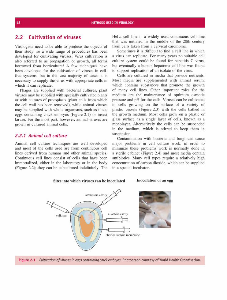
Phages are supplied with bacterial cultures, plantviruses may be supplied with specially cultivated plantsor with cultures of protoplasts (plant cells from whichthe cell wall has been removed), while animal virusesmay be supplied with whole organisms, such as mice,eggs containing chick embryos (Figure 2.1) or insectlarvae. For the most part, however, animal viruses aregrown in cultured animal cells.
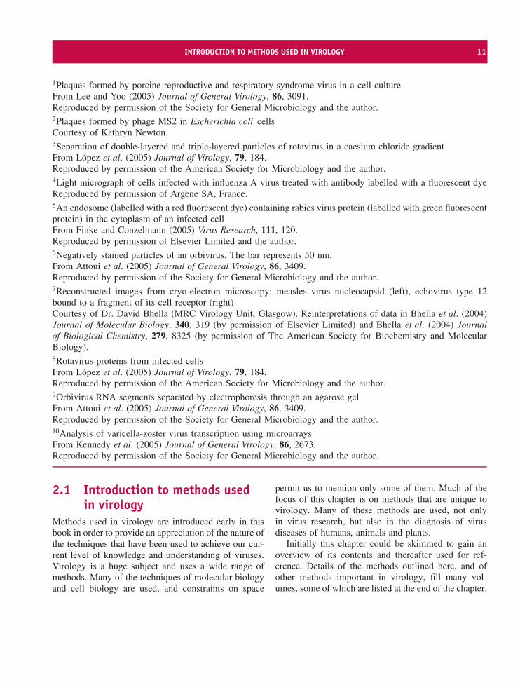
2.2.1 Animal cell culture
Animal cell culture techniques are well developedand most of the cells used are from continuous celllines derived from humans and other animal species.Continuous cell lines consist of cells that have beenimmortalized, either in the laboratory or in the body(Figure 2.2); they can be subcultured indefinitely. The
HeLa cell line is a widely used continuous cell linethat was initiated in the middle of the 20th centuryfrom cells taken from a cervical carcinoma.
Sometimes it is difficult to find a cell line in whicha virus can replicate. For many years no suitable cellculture system could be found for hepatitis C virus,but eventually a human hepatoma cell line was foundto support replication of an isolate of the virus.
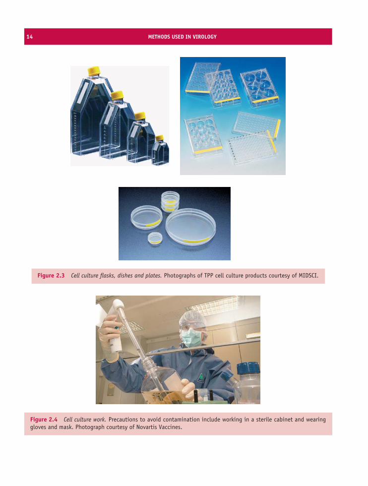
Cells are cultured in media that provide nutrients.Most media are supplemented with animal serum,which contains substances that promote the growthof many cell lines. Other important roles for themedium are the maintenance of optimum osmoticpressure and pH for the cells. Viruses can be cultivatedin cells growing on the surface of a variety ofplastic vessels (Figure 2.3) with the cells bathed inthe growth medium. Most cells grow on a plastic orglass surface as a single layer of cells, known as amonolayer. Alternatively the cells can be suspendedin the medium, which is stirred to keep them insuspension.
Contamination with bacteria and fungi can causemajor problems in cell culture work; in order tominimize these problems work is normally done ina sterile cabinet (Figure 2.4) and most media containantibiotics. Many cell types require a relatively highconcentration of carbon dioxide, which can be suppliedin a special incubator.
allantoic cavity
chorioallantoic membrane
amniotoic cavity
yolk sac
Sites into which viruses can be inoculated Inoculation of an egg
Figure 2.1 Cultivation of viruses in eggs containing chick embryos. Photograph courtesy of World Health Organisation.
CENTRIFUGATION 13
Primary cell culture
Cell line
Continuous cell line
Normal cells fromhuman or animal
Cancer cells fromhuman or animal
Immortalization usinga virus or a chemical
subculture
Figure 2.2 Derivation of continuous cell lines ofhuman and animal cells. Most types of cell taken fromthe body do not grow well in culture. If cells from aprimary culture can be subcultured they are growing asa cell line. They can be subcultured only a finite numberof times unless they are immortalized, in which casethey can be subcultured indefinitely as a continuouscell line. Cancer cells are already immortalized, andcontinuous cell lines may be established from thesewithout further treatment.
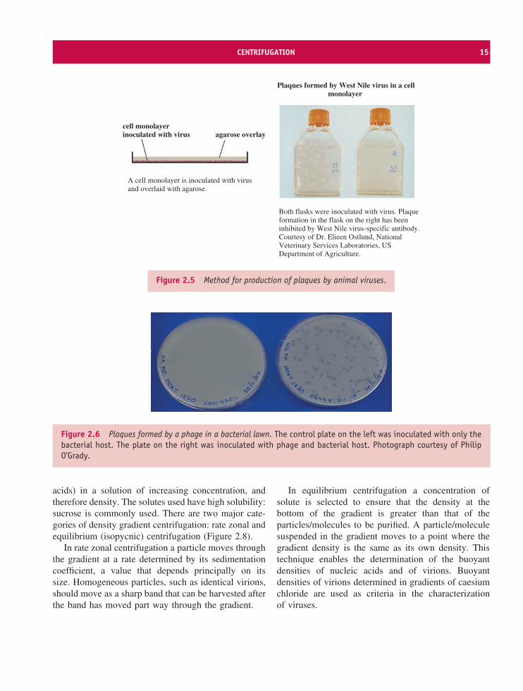
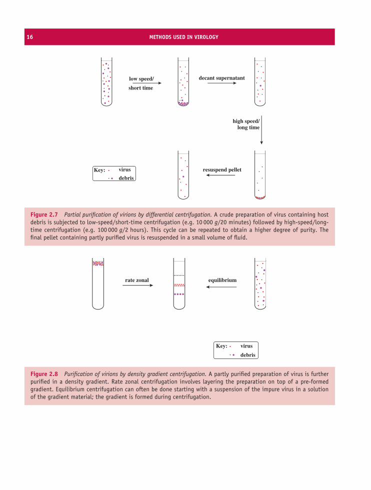
2.3 Isolation of virusesMany viruses can be isolated as a result of their abilityto form discrete visible zones (plaques) in layers ofhost cells. If a confluent layer of cells is inoculated withvirus at a concentration so that only a small proportionof the cells is infected, then plaques may form whereareas of cells are killed or altered by the virus infection.Each plaque is formed when infection spreads radiallyfrom an infected cell to surrounding cells.
Plaques can be formed by many animal virusesin monolayers if the cells are overlaid with agarosegel to maintain the progeny virus in a discrete zone(Figure 2.5). Plaques can also be formed by phages inlawns of bacterial growth (Figure 2.6).
It is generally assumed that a plaque is the resultof the infection of a cell by a single virion. If thisis the case then all virus produced from virus in the
plaque should be a clone, in other words it should begenetically identical. This clone can be referred to asan isolate, and if it is distinct from all other isolatesit can be referred to as a strain. This is analogous tothe derivation of a bacterial strain from a colony on anagar plate.
There is a possibility that a plaque might bederived from two or more virions so, to increase theprobability that a genetically pure strain of virus hasbeen obtained, material from a plaque can be inoculatedonto further monolayers and virus can be derived froman individual plaque. The virus is said to have beenplaque purified.
When a virus is first isolated it may replicate poorlyin cells in the laboratory, but after it has gone througha number of replication cycles it may replicate moreefficiently. Each time the virus is ‘sub-cultured’ (toborrow a term from bacteriology) it is said to havebeen passaged. After a number of passages the virusmay be genetically different to the original wild strain,in which case it is now a laboratory strain.
2.4 Centrifugation
After a virus has been propagated it is usually necessaryto remove host cell debris and other contaminantsbefore the virus particles can be used for laboratorystudies, for incorporation into a vaccine, or for someother purpose. Many virus purification proceduresinvolve centrifugation; partial purification can beachieved by differential centrifugation and a higherdegree of purity can be achieved by some form ofdensity gradient centrifugation.
2.4.1 Differential centrifugation
Differential centrifugation involves alternating cyclesof low-speed centrifugation, after which most ofthe virus is still in the supernatant, and high-speedcentrifugation, after which the virus is in the pellet(Figure 2.7).
2.4.2 Density gradient centrifugation
Density gradient centrifugation involves centrifugingparticles (such as virions) or molecules (such as nucleic
14 METHODS USED IN VIROLOGY
Figure 2.3 Cell culture flasks, dishes and plates. Photographs of TPP cell culture products courtesy of MIDSCI.
Figure 2.4 Cell culture work. Precautions to avoid contamination include working in a sterile cabinet and wearinggloves and mask. Photograph courtesy of Novartis Vaccines.
CENTRIFUGATION 15
Both flasks were inoculated with virus. Plaqueformation in the flask on the right has beeninhibited by West Nile virus-specific antibody.Courtesy of Dr. Elieen Ostlund, NationalVeterinary Services Laboratories, USDepartment of Agriculture.
A cell monolayer is inoculated with virusand overlaid with agarose.
cell monolayerinoculated with virus agarose overlay
Plaques formed by West Nile virus in a cellmonolayer
Figure 2.5 Method for production of plaques by animal viruses.
Figure 2.6 Plaques formed by a phage in a bacterial lawn. The control plate on the left was inoculated with only thebacterial host. The plate on the right was inoculated with phage and bacterial host. Photograph courtesy of PhilipO’Grady.
acids) in a solution of increasing concentration, andtherefore density. The solutes used have high solubility:sucrose is commonly used. There are two major cate-gories of density gradient centrifugation: rate zonal andequilibrium (isopycnic) centrifugation (Figure 2.8).
In rate zonal centrifugation a particle moves throughthe gradient at a rate determined by its sedimentationcoefficient, a value that depends principally on itssize. Homogeneous particles, such as identical virions,should move as a sharp band that can be harvested afterthe band has moved part way through the gradient.
In equilibrium centrifugation a concentration ofsolute is selected to ensure that the density at thebottom of the gradient is greater than that of theparticles/molecules to be purified. A particle/moleculesuspended in the gradient moves to a point where thegradient density is the same as its own density. Thistechnique enables the determination of the buoyantdensities of nucleic acids and of virions. Buoyantdensities of virions determined in gradients of caesiumchloride are used as criteria in the characterizationof viruses.
16 METHODS USED IN VIROLOGY
low speed/
resuspend pellet
decant supernatant
Key: virus
debris
high speed/long time
short time
Figure 2.7 Partial purification of virions by differential centrifugation. A crude preparation of virus containing hostdebris is subjected to low-speed/short-time centrifugation (e.g. 10 000 g/20 minutes) followed by high-speed/long-time centrifugation (e.g. 100 000 g/2 hours). This cycle can be repeated to obtain a higher degree of purity. Thefinal pellet containing partly purified virus is resuspended in a small volume of fluid.
rate zonal equilibrium
Key: virus
debris
Figure 2.8 Purification of virions by density gradient centrifugation. A partly purified preparation of virus is furtherpurified in a density gradient. Rate zonal centrifugation involves layering the preparation on top of a pre-formedgradient. Equilibrium centrifugation can often be done starting with a suspension of the impure virus in a solutionof the gradient material; the gradient is formed during centrifugation.
ELECTROPHORETIC TECHNIQUES 17
2.5 Structural investigationsof cells and virions
2.5.1 Light microscopy
The sizes of most virions are beyond the limits ofresolution of light microscopes, but light microscopyhas useful applications in detecting virus-infectedcells, for example by observing cytopathic effects(Section 2.7.2) or by detecting a fluorescent dye linkedto antibody molecules that have bound to a virus anti-gen (fluorescence microscopy).
Confocal microscopy is proving to be especiallyvaluable in virology. The principle of this techniqueis the use of a pinhole to exclude light from out-of-focus regions of the specimen. Most confocalmicroscopes scan the specimen with a laser, producingexceptionally clear images of thick specimens and offluorescing specimens. Furthermore, ‘optical slices’ ofa specimen can be collected and used to create a three-dimensional representation. The techniques can be usedwith live cells and can be applied to investigationsof protein trafficking, with the virus or cell proteinunder investigation carrying a suitable label, e.g. greenfluorescent protein (a jellyfish protein).
2.5.2 Electron microscopy
Many investigations of the structure of virions or ofvirus-infected cells involve electron microscopy. Largemagnifications are achievable with a transmissionelectron microscope but the specimen, whether it isa suspension of virions or an ultrathin section of avirus-infected cell, must be treated so that details canbe visualized.
Negative staining techniques generate contrast byusing heavy-metal-containing compounds, such aspotassium phosphotungstate and ammonium molyb-date. In electron micrographs of virions the stainsappear as dark areas around the virions, allowing theoverall virion shape and size to be determined. Furtherstructural detail may be apparent if the stains penetrateany crevices on the virion surface or any hollows withinthe virion. Negative staining techniques have generatedmany high quality electron micrographs, but the tech-niques have limitations, including structural distortionsresulting from drying.
Cryo-electron microscopy techniques are morerecent. In these a wet specimen is rapidly cooledto a temperature below −160 ◦C, freezing the wateras a glasslike material, as in the method outlinedin Figure 2.9. The images are recorded while thespecimen is frozen. They require computer process-ing in order to extract maximum detail, and datafrom multiple images are processed to reconstructthree-dimensional images of virus particles. This mayinvolve averaging many identical copies or combin-ing images into three-dimensional density maps (tomo-grams).
2.5.3 X-ray crystallography
X-ray crystallography is another technique thatis revealing detailed information about the three-dimensional structures of virions (and DNA, proteinsand DNA–protein complexes). This technique requiresthe production of a crystal of the virions or moleculesunder study. The crystal is placed in a beam of X-rays, which are diffracted by repeating arrangementsof molecules/atoms in the crystal. Analysis of thediffraction pattern allows the relative positions of thesemolecules/atoms to be determined.
Other techniques that are providing useful informa-tion about the structure of viruses are nuclear magneticresonance and atomic force microscopy.
2.6 Electrophoretic techniques
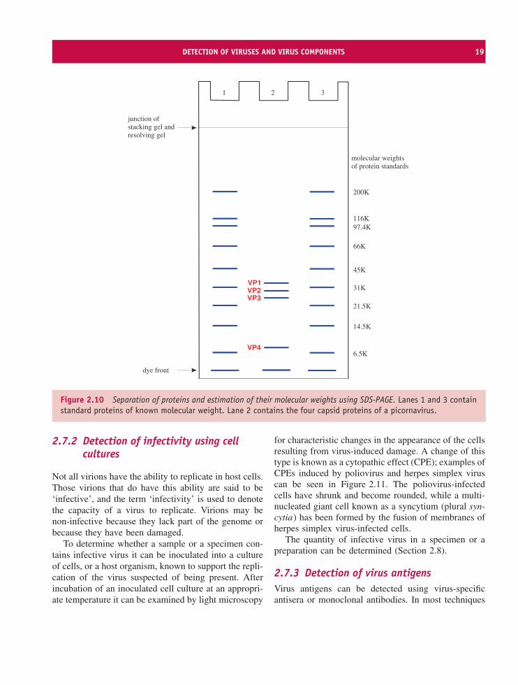
Mixtures of proteins or nucleic acids can be sepa-rated by electrophoresis in a gel composed of agaroseor polyacrylamide. In most electrophoretic techniqueseach protein or nucleic acid forms a band in thegel. Electrophoresis can be performed under condi-tions where the rate of movement through the geldepends on molecular weight. The molecular weightsof the protein or nucleic acid molecules can be esti-mated by comparing the positions of the bands withpositions of bands formed by molecules of knownmolecular weight electrophoresed in the same gel. Thetechnique for estimating molecular weights of proteinsis polyacrylamide gel electrophoresis in the presenceof the detergent sodium dodecyl sulphate (SDS-PAGE;Figure 2.10).
18 METHODS USED IN VIROLOGY
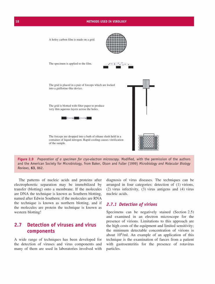
A holey carbon film is made on a grid.
The specimen is applied to the film.
The grid is placed in a pair of forceps which are locked into a guillotine-like device.
The grid is blotted with filter paper to produce very thin aqueous layers across the holes.
The forceps are dropped into a bath of ethane slush held in a container of liquid nitrogen. Rapid cooling causes vitrification of the sample.
Figure 2.9 Preparation of a specimen for cryo-electron microscopy. Modified, with the permission of the authorsand the American Society for Microbiology, from Baker, Olson and Fuller (1999) Microbiology and Molecular BiologyReviews, 63, 862.
The patterns of nucleic acids and proteins afterelectrophoretic separation may be immobilized bytransfer (blotting) onto a membrane. If the moleculesare DNA the technique is known as Southern blotting,named after Edwin Southern; if the molecules are RNAthe technique is known as northern blotting, and ifthe molecules are protein the technique is known aswestern blotting!
2.7 Detection of viruses and viruscomponents
A wide range of techniques has been developed forthe detection of viruses and virus components andmany of them are used in laboratories involved with
diagnosis of virus diseases. The techniques can bearranged in four categories: detection of (1) virions,(2) virus infectivity, (3) virus antigens and (4) virusnucleic acids.
2.7.1 Detection of virions
Specimens can be negatively stained (Section 2.5)and examined in an electron microscope for thepresence of virions. Limitations to this approach arethe high costs of the equipment and limited sensitivity;the minimum detectable concentration of virions isabout 106/ml. An example of an application of thistechnique is the examination of faeces from a patientwith gastroenteritis for the presence of rotavirusparticles.
DETECTION OF VIRUSES AND VIRUS COMPONENTS 19
VP4
VP1VP2VP3
200K
116K97.4K
66K
45K
31K
21.5K
14.5K
6.5K
21 3
dye front
junction ofstacking gel andresolving gel
molecular weightsof protein standards
Figure 2.10 Separation of proteins and estimation of their molecular weights using SDS-PAGE. Lanes 1 and 3 containstandard proteins of known molecular weight. Lane 2 contains the four capsid proteins of a picornavirus.
2.7.2 Detection of infectivity using cellcultures
Not all virions have the ability to replicate in host cells.Those virions that do have this ability are said to be‘infective’, and the term ‘infectivity’ is used to denotethe capacity of a virus to replicate. Virions may benon-infective because they lack part of the genome orbecause they have been damaged.
To determine whether a sample or a specimen con-tains infective virus it can be inoculated into a cultureof cells, or a host organism, known to support the repli-cation of the virus suspected of being present. Afterincubation of an inoculated cell culture at an appropri-ate temperature it can be examined by light microscopy
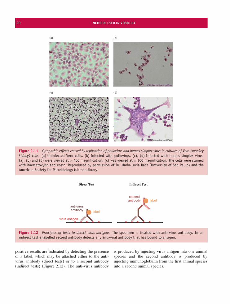
for characteristic changes in the appearance of the cellsresulting from virus-induced damage. A change of thistype is known as a cytopathic effect (CPE); examples ofCPEs induced by poliovirus and herpes simplex viruscan be seen in Figure 2.11. The poliovirus-infectedcells have shrunk and become rounded, while a multi-nucleated giant cell known as a syncytium (plural syn-cytia) has been formed by the fusion of membranes ofherpes simplex virus-infected cells.
The quantity of infective virus in a specimen or apreparation can be determined (Section 2.8).
2.7.3 Detection of virus antigensVirus antigens can be detected using virus-specificantisera or monoclonal antibodies. In most techniques
20 METHODS USED IN VIROLOGY
(a) (b)
(c) (d)
Figure 2.11 Cytopathic effects caused by replication of poliovirus and herpes simplex virus in cultures of Vero (monkeykidney) cells. (a) Uninfected Vero cells. (b) Infected with poliovirus. (c), (d) Infected with herpes simplex virus.(a), (b) and (d) were viewed at × 400 magnification; (c) was viewed at × 100 magnification. The cells were stainedwith haematoxylin and eosin. Reproduced by permission of Dr. Maria-Lucia Racz (University of Sao Paulo) and theAmerican Society for Microbiology MicrobeLibrary.
virus antigen
anti-virusantibody label
secondantibody label
Direct Test Indirect Test
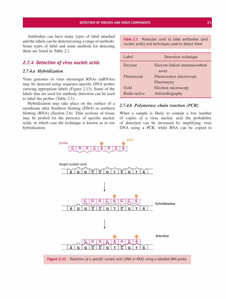
Figure 2.12 Principles of tests to detect virus antigens. The specimen is treated with anti-virus antibody. In anindirect test a labelled second antibody detects any anti-viral antibody that has bound to antigen.
positive results are indicated by detecting the presenceof a label, which may be attached either to the anti-virus antibody (direct tests) or to a second antibody(indirect tests) (Figure 2.12). The anti-virus antibody
is produced by injecting virus antigen into one animalspecies and the second antibody is produced byinjecting immunoglobulin from the first animal speciesinto a second animal species.
DETECTION OF VIRUSES AND VIRUS COMPONENTS 21
Antibodies can have many types of label attachedand the labels can be detected using a range of methods.Some types of label and some methods for detectingthem are listed in Table 2.1.
2.7.4 Detection of virus nucleic acids
2.7.4.a Hybridization
Virus genomes or virus messenger RNAs (mRNAs)may be detected using sequence-specific DNA probescarrying appropriate labels (Figure 2.13). Some of thelabels that are used for antibody detection can be usedto label the probes (Table 2.1).
Hybridization may take place on the surface of amembrane after Southern blotting (DNA) or northernblotting (RNA) (Section 2.6). Thin sections of tissuemay be probed for the presence of specific nucleicacids, in which case the technique is known as in situhybridization.
Table 2.1 Molecules used to label antibodies (andnucleic acids) and techniques used to detect them
Label Detection technique
Enzyme Enzyme-linked immunosorbentassay
Fluorescent Fluorescence microscopyFluorimetry
Gold Electron microscopyRadio-active Autoradiography
2.7.4.b Polymerase chain reaction (PCR)
When a sample is likely to contain a low numberof copies of a virus nucleic acid the probabilityof detection can be increased by amplifying virusDNA using a PCR, while RNA can be copied to
A G G
G G C A G C A
C
C
G G C A G C AC
G G C A G C AC
C G T C G T A
A G G C C G T C G T A
A G G C C G T C G T A
probe
target nucleic acid
hybridization
detection
label
Figure 2.13 Detection of a specific nucleic acid (DNA or RNA) using a labelled DNA probe.
22 METHODS USED IN VIROLOGY
DNA and amplified using a RT (reverse transcriptase)-PCR. The procedures require oligonucleotide primersspecific to viral sequences. An amplified product can bedetected by electrophoresis in an agarose gel, followedby transfer to a nitrocellulose membrane, which isincubated with a labelled probe.
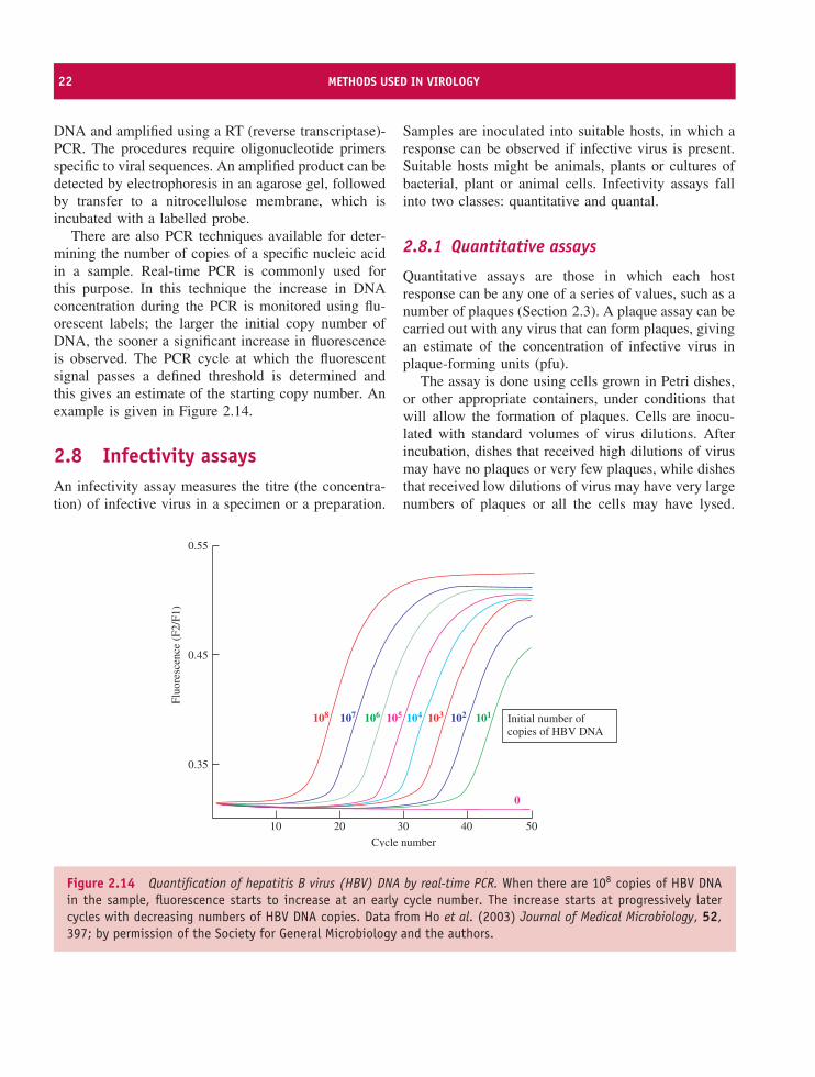
There are also PCR techniques available for deter-mining the number of copies of a specific nucleic acidin a sample. Real-time PCR is commonly used forthis purpose. In this technique the increase in DNAconcentration during the PCR is monitored using flu-orescent labels; the larger the initial copy number ofDNA, the sooner a significant increase in fluorescenceis observed. The PCR cycle at which the fluorescentsignal passes a defined threshold is determined andthis gives an estimate of the starting copy number. Anexample is given in Figure 2.14.
2.8 Infectivity assaysAn infectivity assay measures the titre (the concentra-tion) of infective virus in a specimen or a preparation.
Samples are inoculated into suitable hosts, in which aresponse can be observed if infective virus is present.Suitable hosts might be animals, plants or cultures ofbacterial, plant or animal cells. Infectivity assays fallinto two classes: quantitative and quantal.
2.8.1 Quantitative assays
Quantitative assays are those in which each hostresponse can be any one of a series of values, such as anumber of plaques (Section 2.3). A plaque assay can becarried out with any virus that can form plaques, givingan estimate of the concentration of infective virus inplaque-forming units (pfu).
The assay is done using cells grown in Petri dishes,or other appropriate containers, under conditions thatwill allow the formation of plaques. Cells are inocu-lated with standard volumes of virus dilutions. Afterincubation, dishes that received high dilutions of virusmay have no plaques or very few plaques, while dishesthat received low dilutions of virus may have very largenumbers of plaques or all the cells may have lysed.
0.55
Fluo
resc
ence
(F2
/F1)
0.45
0.35
10
108 107 106 105 104 103 102 101
20 30
Cycle number
Initial number ofcopies of HBV DNA
40
0
50
Figure 2.14 Quantification of hepatitis B virus (HBV) DNA by real-time PCR. When there are 108 copies of HBV DNAin the sample, fluorescence starts to increase at an early cycle number. The increase starts at progressively latercycles with decreasing numbers of HBV DNA copies. Data from Ho et al. (2003) Journal of Medical Microbiology, 52,397; by permission of the Society for General Microbiology and the authors.
INFECTIVITY ASSAYS 23
(c)
10−1 10−2 10−3 10−4 10−5 10−6
100
75
50
25
Virus dilution
One TCID50 is present in this dilution.
% p
ositi
ve
(a)
10−110−210−310−410−510−6 ControlVirus
1A
B
C
D
E
F
G
H
2 3 4 5 6 7 8
(b)
10−110−210−310−410−510−6
Control
1A + + + − − − −
+ − + − − − −+ + − + − − −+ + + − − − −+ + − + − − −+ + + − + − −+ + + + − − −+ + − − − − −
B
C
D
E
F
G
H
2 3 4 5 6 7 8
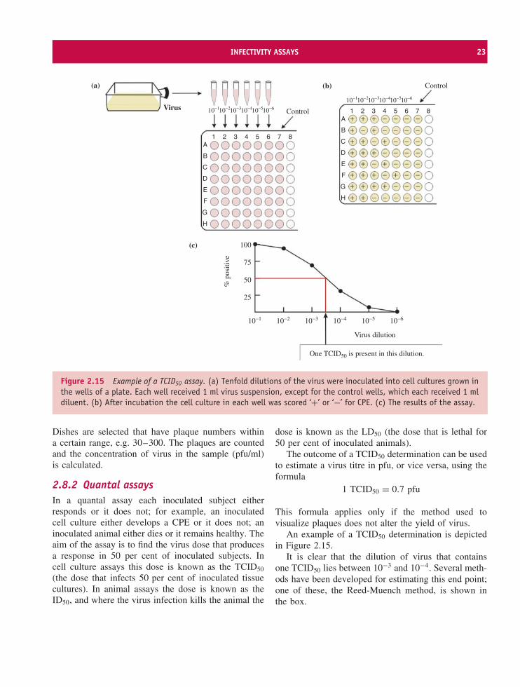
Figure 2.15 Example of a TCID50 assay. (a) Tenfold dilutions of the virus were inoculated into cell cultures grown inthe wells of a plate. Each well received 1 ml virus suspension, except for the control wells, which each received 1 mldiluent. (b) After incubation the cell culture in each well was scored ‘+’ or ‘−’ for CPE. (c) The results of the assay.
Dishes are selected that have plaque numbers withina certain range, e.g. 30–300. The plaques are countedand the concentration of virus in the sample (pfu/ml)is calculated.
2.8.2 Quantal assaysIn a quantal assay each inoculated subject eitherresponds or it does not; for example, an inoculatedcell culture either develops a CPE or it does not; aninoculated animal either dies or it remains healthy. Theaim of the assay is to find the virus dose that producesa response in 50 per cent of inoculated subjects. Incell culture assays this dose is known as the TCID50
(the dose that infects 50 per cent of inoculated tissuecultures). In animal assays the dose is known as theID50, and where the virus infection kills the animal the
dose is known as the LD50 (the dose that is lethal for50 per cent of inoculated animals).
The outcome of a TCID50 determination can be usedto estimate a virus titre in pfu, or vice versa, using theformula
1 TCID50 = 0.7 pfu
This formula applies only if the method used tovisualize plaques does not alter the yield of virus.
An example of a TCID50 determination is depictedin Figure 2.15.
It is clear that the dilution of virus that containsone TCID50 lies between 10−3 and 10−4. Several meth-ods have been developed for estimating this end point;one of these, the Reed-Muench method, is shown inthe box.
24 METHODS USED IN VIROLOGY
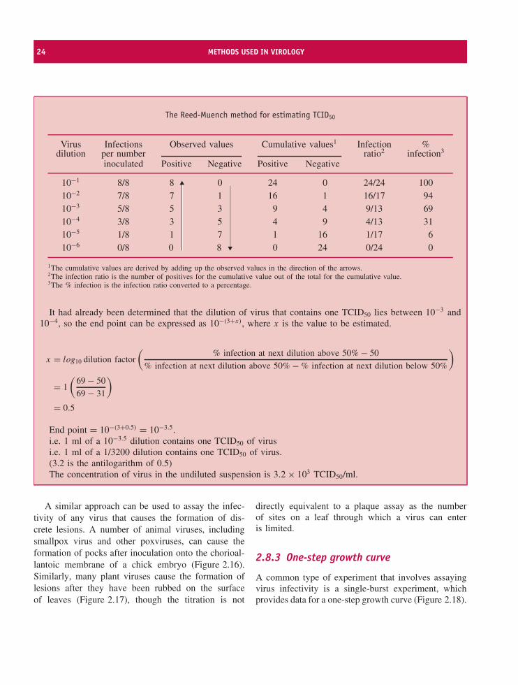
The Reed-Muench method for estimating TCID50
Virus Infections Observed values Cumulative values1 Infection %dilution per number ratio2 infection3
inoculated Positive Negative Positive Negative
10−1 8/8 8 0 24 0 24/24 10010−2 7/8 7 1 16 1 16/17 9410−3 5/8 5 3 9 4 9/13 6910−4 3/8 3 5 4 9 4/13 3110−5 1/8 1 7 1 16 1/17 610−6 0/8 0 8 0 24 0/24 0
1The cumulative values are derived by adding up the observed values in the direction of the arrows.2The infection ratio is the number of positives for the cumulative value out of the total for the cumulative value.3The % infection is the infection ratio converted to a percentage.
It had already been determined that the dilution of virus that contains one TCID50 lies between 10−3 and10−4, so the end point can be expressed as 10−(3+x), where x is the value to be estimated.
x = log10 dilution factor
(% infection at next dilution above 50% − 50
% infection at next dilution above 50% − % infection at next dilution below 50%
)
= 1
(69 − 50
69 − 31
)
= 0.5
End point = 10−(3+0.5) = 10−3.5.i.e. 1 ml of a 10−3.5 dilution contains one TCID50 of virusi.e. 1 ml of a 1/3200 dilution contains one TCID50 of virus.(3.2 is the antilogarithm of 0.5)The concentration of virus in the undiluted suspension is 3.2 × 10³ TCID50/ml.
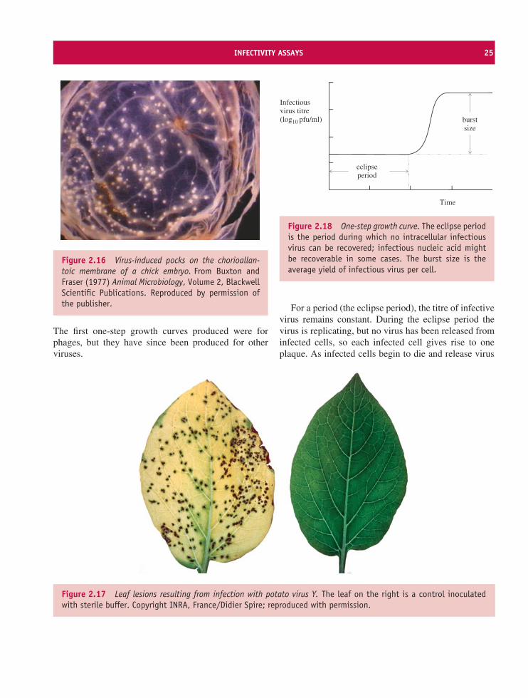
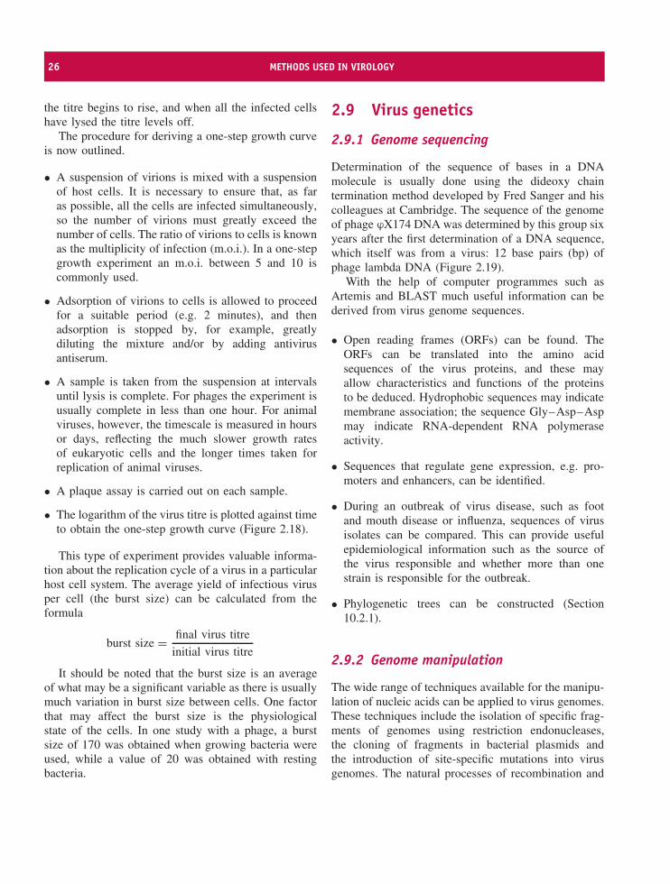
A similar approach can be used to assay the infec-tivity of any virus that causes the formation of dis-crete lesions. A number of animal viruses, includingsmallpox virus and other poxviruses, can cause theformation of pocks after inoculation onto the chorioal-lantoic membrane of a chick embryo (Figure 2.16).Similarly, many plant viruses cause the formation oflesions after they have been rubbed on the surfaceof leaves (Figure 2.17), though the titration is not
directly equivalent to a plaque assay as the numberof sites on a leaf through which a virus can enteris limited.
2.8.3 One-step growth curve
A common type of experiment that involves assayingvirus infectivity is a single-burst experiment, whichprovides data for a one-step growth curve (Figure 2.18).
INFECTIVITY ASSAYS 25
Figure 2.16 Virus-induced pocks on the chorioallan-toic membrane of a chick embryo. From Buxton andFraser (1977) Animal Microbiology, Volume 2, BlackwellScientific Publications. Reproduced by permission ofthe publisher.
The first one-step growth curves produced were forphages, but they have since been produced for otherviruses.
Time
burstsize
eclipseperiod
Infectiousvirus titre (log10 pfu/ml)
Figure 2.18 One-step growth curve. The eclipse periodis the period during which no intracellular infectiousvirus can be recovered; infectious nucleic acid mightbe recoverable in some cases. The burst size is theaverage yield of infectious virus per cell.
For a period (the eclipse period), the titre of infectivevirus remains constant. During the eclipse period thevirus is replicating, but no virus has been released frominfected cells, so each infected cell gives rise to oneplaque. As infected cells begin to die and release virus
Figure 2.17 Leaf lesions resulting from infection with potato virus Y. The leaf on the right is a control inoculatedwith sterile buffer. Copyright INRA, France/Didier Spire; reproduced with permission.
26 METHODS USED IN VIROLOGY
the titre begins to rise, and when all the infected cellshave lysed the titre levels off.
The procedure for deriving a one-step growth curveis now outlined.
• A suspension of virions is mixed with a suspensionof host cells. It is necessary to ensure that, as faras possible, all the cells are infected simultaneously,so the number of virions must greatly exceed thenumber of cells. The ratio of virions to cells is knownas the multiplicity of infection (m.o.i.). In a one-stepgrowth experiment an m.o.i. between 5 and 10 iscommonly used.
• Adsorption of virions to cells is allowed to proceedfor a suitable period (e.g. 2 minutes), and thenadsorption is stopped by, for example, greatlydiluting the mixture and/or by adding antivirusantiserum.
• A sample is taken from the suspension at intervalsuntil lysis is complete. For phages the experiment isusually complete in less than one hour. For animalviruses, however, the timescale is measured in hoursor days, reflecting the much slower growth ratesof eukaryotic cells and the longer times taken forreplication of animal viruses.
• A plaque assay is carried out on each sample.
• The logarithm of the virus titre is plotted against timeto obtain the one-step growth curve (Figure 2.18).
This type of experiment provides valuable informa-tion about the replication cycle of a virus in a particularhost cell system. The average yield of infectious virusper cell (the burst size) can be calculated from theformula
burst size = final virus titre
initial virus titre
It should be noted that the burst size is an averageof what may be a significant variable as there is usuallymuch variation in burst size between cells. One factorthat may affect the burst size is the physiologicalstate of the cells. In one study with a phage, a burstsize of 170 was obtained when growing bacteria wereused, while a value of 20 was obtained with restingbacteria.
2.9 Virus genetics
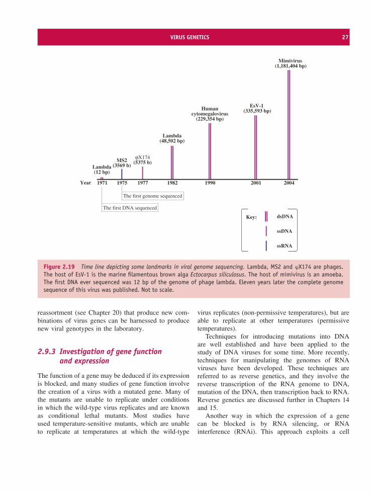
2.9.1 Genome sequencing
Determination of the sequence of bases in a DNAmolecule is usually done using the dideoxy chaintermination method developed by Fred Sanger and hiscolleagues at Cambridge. The sequence of the genomeof phage ϕX174 DNA was determined by this group sixyears after the first determination of a DNA sequence,which itself was from a virus: 12 base pairs (bp) ofphage lambda DNA (Figure 2.19).
With the help of computer programmes such asArtemis and BLAST much useful information can bederived from virus genome sequences.
• Open reading frames (ORFs) can be found. TheORFs can be translated into the amino acidsequences of the virus proteins, and these mayallow characteristics and functions of the proteinsto be deduced. Hydrophobic sequences may indicatemembrane association; the sequence Gly–Asp–Aspmay indicate RNA-dependent RNA polymeraseactivity.
• Sequences that regulate gene expression, e.g. pro-moters and enhancers, can be identified.
• During an outbreak of virus disease, such as footand mouth disease or influenza, sequences of virusisolates can be compared. This can provide usefulepidemiological information such as the source ofthe virus responsible and whether more than onestrain is responsible for the outbreak.
• Phylogenetic trees can be constructed (Section10.2.1).
2.9.2 Genome manipulation
The wide range of techniques available for the manipu-lation of nucleic acids can be applied to virus genomes.These techniques include the isolation of specific frag-ments of genomes using restriction endonucleases,the cloning of fragments in bacterial plasmids andthe introduction of site-specific mutations into virusgenomes. The natural processes of recombination and
VIRUS GENETICS 27
1971 1975 1977 1982 1990 2001 2004
The first DNA sequenced
The first genome sequenced
Mimivirus(1,181,404 bp)
EsV-1(335,593 bp)Human
cytomegalovirus(229,354 bp)
MS2(3569 b)
Year
Lambda(12 bp)
Lambda(48,502 bp)
ϕX174(5375 b)
Key: dsDNA
ssDNA
ssRNA
Figure 2.19 Time line depicting some landmarks in viral genome sequencing. Lambda, MS2 and ϕX174 are phages.The host of EsV-1 is the marine filamentous brown alga Ectocarpus siliculosus. The host of mimivirus is an amoeba.The first DNA ever sequenced was 12 bp of the genome of phage lambda. Eleven years later the complete genomesequence of this virus was published. Not to scale.
reassortment (see Chapter 20) that produce new com-binations of virus genes can be harnessed to producenew viral genotypes in the laboratory.
2.9.3 Investigation of gene functionand expression
The function of a gene may be deduced if its expressionis blocked, and many studies of gene function involvethe creation of a virus with a mutated gene. Many ofthe mutants are unable to replicate under conditionsin which the wild-type virus replicates and are knownas conditional lethal mutants. Most studies haveused temperature-sensitive mutants, which are unableto replicate at temperatures at which the wild-type
virus replicates (non-permissive temperatures), but areable to replicate at other temperatures (permissivetemperatures).
Techniques for introducing mutations into DNAare well established and have been applied to thestudy of DNA viruses for some time. More recently,techniques for manipulating the genomes of RNAviruses have been developed. These techniques arereferred to as reverse genetics, and they involve thereverse transcription of the RNA genome to DNA,mutation of the DNA, then transcription back to RNA.Reverse genetics are discussed further in Chapters 14and 15.
Another way in which the expression of a genecan be blocked is by RNA silencing, or RNAinterference (RNAi). This approach exploits a cell
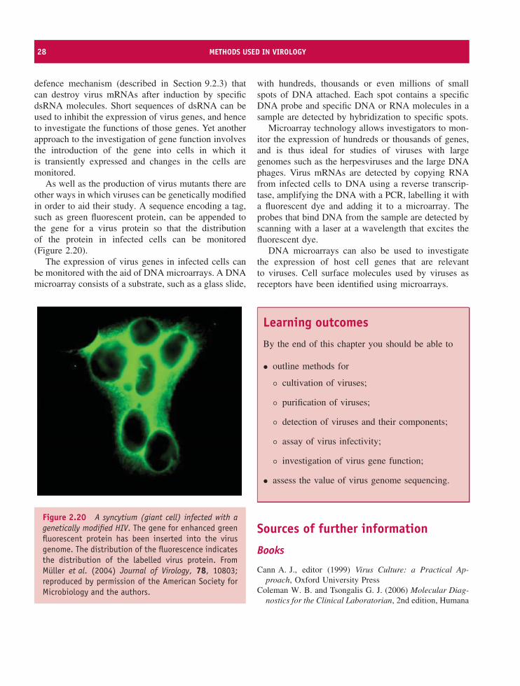
28 METHODS USED IN VIROLOGY
defence mechanism (described in Section 9.2.3) thatcan destroy virus mRNAs after induction by specificdsRNA molecules. Short sequences of dsRNA can beused to inhibit the expression of virus genes, and henceto investigate the functions of those genes. Yet anotherapproach to the investigation of gene function involvesthe introduction of the gene into cells in which itis transiently expressed and changes in the cells aremonitored.
As well as the production of virus mutants there areother ways in which viruses can be genetically modifiedin order to aid their study. A sequence encoding a tag,such as green fluorescent protein, can be appended tothe gene for a virus protein so that the distributionof the protein in infected cells can be monitored(Figure 2.20).
The expression of virus genes in infected cells canbe monitored with the aid of DNA microarrays. A DNAmicroarray consists of a substrate, such as a glass slide,
Figure 2.20 A syncytium (giant cell) infected with agenetically modified HIV. The gene for enhanced greenfluorescent protein has been inserted into the virusgenome. The distribution of the fluorescence indicatesthe distribution of the labelled virus protein. FromMuller et al. (2004) Journal of Virology, 78, 10803;reproduced by permission of the American Society forMicrobiology and the authors.
with hundreds, thousands or even millions of smallspots of DNA attached. Each spot contains a specificDNA probe and specific DNA or RNA molecules in asample are detected by hybridization to specific spots.
Microarray technology allows investigators to mon-itor the expression of hundreds or thousands of genes,and is thus ideal for studies of viruses with largegenomes such as the herpesviruses and the large DNAphages. Virus mRNAs are detected by copying RNAfrom infected cells to DNA using a reverse transcrip-tase, amplifying the DNA with a PCR, labelling it witha fluorescent dye and adding it to a microarray. Theprobes that bind DNA from the sample are detected byscanning with a laser at a wavelength that excites thefluorescent dye.
DNA microarrays can also be used to investigatethe expression of host cell genes that are relevantto viruses. Cell surface molecules used by viruses asreceptors have been identified using microarrays.
Learning outcomes
By the end of this chapter you should be able to
• outline methods for
◦ cultivation of viruses;
◦ purification of viruses;
◦ detection of viruses and their components;
◦ assay of virus infectivity;
◦ investigation of virus gene function;
• assess the value of virus genome sequencing.
Sources of further information
Books
Cann A. J., editor (1999) Virus Culture: a Practical Ap-proach, Oxford University Press
Coleman W. B. and Tsongalis G. J. (2006) Molecular Diag-nostics for the Clinical Laboratorian, 2nd edition, Humana
SOURCES OF FURTHER INFORMATION 29
Collier L. and Oxford J. (2006) Human Virology, 3rd edition,Chapter 36, Oxford University Press
Edwards K., Logan J. and Saunders N., editors (2004) Real-Time PCR: an Essential Guide, Horizon
Freshney R. I. (2005) Culture of Animal Cells: a Manual ofBasic Technique, 5th edition, Wiley-Liss
Payment P. (2002) Cultivation and assay of animal viruses.Chapter 8 in Hurst C. J. et al., editors, Manual of Environ-mental Microbiology, 2nd edition, ASM Press
Ye S. and Day I. N. M., editors (2003) Microarrays andMicroplates: Applications in Biomedical Sciences, BIOS
Historical paper
Ellis E. L. and Delbruck M. (1939) The growth of bacterio-phage Journal of General Physiology, 22, 365–384
Recent papers
Curry A, Appleton H. and Dowsett B. (2006) Application oftransmission electron microscopy to the clinical study ofviral and bacterial infections: present and future Micron,37, 91–106
DeFilippis V. et al. (2003) Functional genomics in virologyand antiviral drug discovery Trends in Biotechnology, 21,452–457
Domiati-Saada R. and Scheuermann R. H. (2006) Nucleicacid testing for viral burden and viral genotyping ClinicaChimica Acta, 363, 197–205
Kennedy P. G. E. et al. (2005) Transcriptomal analysis ofvaricella-zoster virus infection using long oligonucleotide-based microarrays Journal of General Virology, 86,2673–2684
Kuznetsov Yu. G. et al. (2001) Imaging of viruses byatomic force microscopy Journal of General Virology, 82,2025–2034
Natarajan P. et al. (2005) Exploring icosahedral virus struc-tures with VIPER Nature Reviews Microbiology, 3,809–817
Rossmann M. G. et al. (2005) Combining X-ray crystal-lography and electron microscopy Structure, 13, 355–362
Vernet G. (2004) Molecular diagnostics in virology Journalof Clinical Virology, 31, 239–247
Watzinger F., Ebner K. and Lion T. (2006) Detection andmonitoring of virus infections by real-time PCR MolecularAspects of Medicine, 27, 254–298

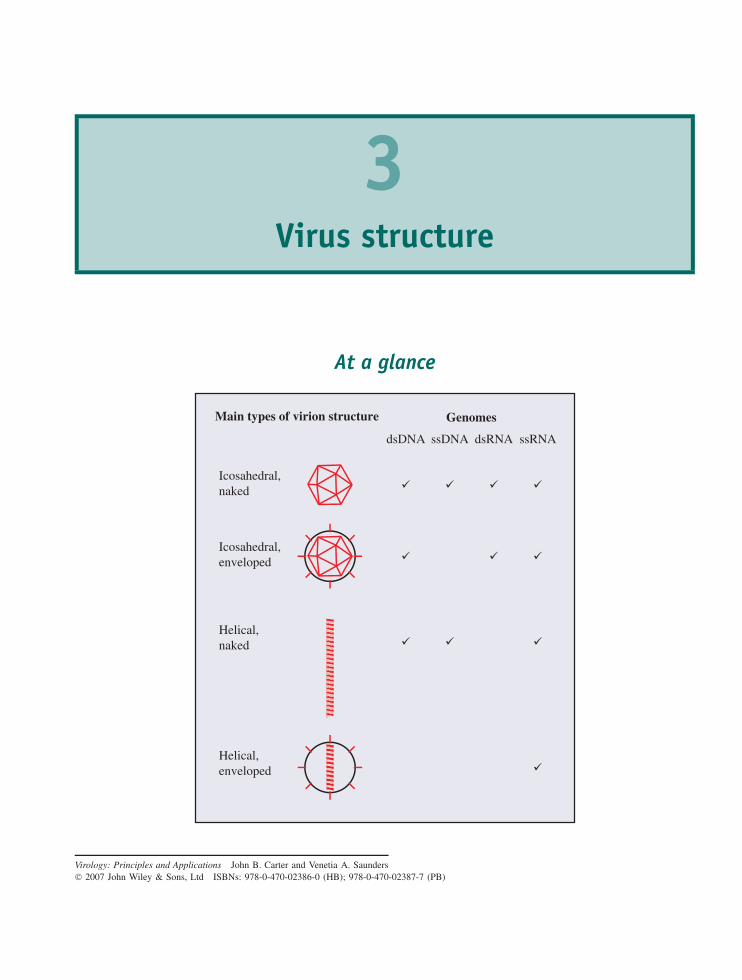
3Virus structure
At a glance
Main types of virion structure
Icosahedral,naked
Icosahedral,enveloped
Helical,naked
Helical,enveloped
Genomes
dsDNA ssDNA dsRNA ssRNA
Virology: Principles and Applications John B. Carter and Venetia A. Saunders 2007 John Wiley & Sons, Ltd ISBNs: 978-0-470-02386-0 (HB); 978-0-470-02387-7 (PB)
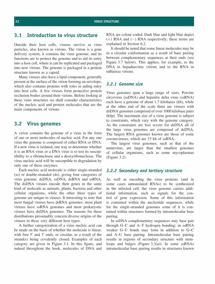
32 VIRUS STRUCTURE
3.1 Introduction to virus structure
Outside their host cells, viruses survive as virusparticles, also known as virions. The virion is a genedelivery system; it contains the virus genome, and itsfunctions are to protect the genome and to aid its entryinto a host cell, where it can be replicated and packagedinto new virions. The genome is packaged in a proteinstructure known as a capsid.
Many viruses also have a lipid component, generallypresent at the surface of the virion forming an envelope,which also contains proteins with roles in aiding entryinto host cells. A few viruses form protective proteinocclusion bodies around their virions. Before looking atthese virus structures we shall consider characteristicsof the nucleic acid and protein molecules that are themain components of virions.
3.2 Virus genomes
A virion contains the genome of a virus in the formof one or more molecules of nucleic acid. For any onevirus the genome is composed of either RNA or DNA.If a new virus is isolated, one way to determine whetherit is an RNA virus or a DNA virus is to test its suscep-tibility to a ribonuclease and a deoxyribonuclease. Thevirus nucleic acid will be susceptible to degradation byonly one of these enzymes.
Each nucleic acid molecule is either single-stranded(ss) or double-stranded (ds), giving four categories ofvirus genome: dsDNA, ssDNA, dsRNA and ssRNA.The dsDNA viruses encode their genes in the samekind of molecule as animals, plants, bacteria and othercellular organisms, while the other three types ofgenome are unique to viruses. It interesting to note thatmost fungal viruses have dsRNA genomes, most plantviruses have ssRNA genomes and most prokaryoticviruses have dsDNA genomes. The reasons for thesedistributions presumably concern diverse origins of theviruses in these very different host types.
A further categorization of a virus nucleic acid canbe made on the basis of whether the molecule is linear,with free 5′ and 3′ ends, or circular, as a result of thestrand(s) being covalently closed. Examples of eachcategory are given in Figure 3.1. In this figure, andindeed throughout the book, molecules of DNA and
RNA are colour coded. Dark blue and light blue depict(+) RNA and (−) RNA respectively; these terms areexplained in Section 6.2.
It should be noted that some linear molecules may bein a circular conformation as a result of base pairingbetween complementary sequences at their ends (seeFigure 3.7 below). This applies, for example, to theDNA in hepadnavirus virions and to the RNA ininfluenza virions.
3.2.1 Genome size
Virus genomes span a large range of sizes. Porcinecircovirus (ssDNA) and hepatitis delta virus (ssRNA)each have a genome of about 1.7 kilobases (kb), whileat the other end of the scale there are viruses withdsDNA genomes comprised of over 1000 kilobase pairs(kbp). The maximum size of a virus genome is subjectto constraints, which vary with the genome category.As the constraints are less severe for dsDNA all ofthe large virus genomes are composed of dsDNA.The largest RNA genomes known are those of somecoronaviruses, which are 33 kb of ssRNA.
The largest virus genomes, such as that of themimivirus, are larger than the smallest genomesof cellular organisms, such as some mycoplasmas(Figure 3.2).
3.2.2 Secondary and tertiary structure
As well as encoding the virus proteins (and insome cases untranslated RNAs) to be synthesizedin the infected cell, the virus genome carries addi-tional information, such as signals for the con-trol of gene expression. Some of this informationis contained within the nucleotide sequences, whilefor the single-stranded genomes some of it is con-tained within structures formed by intramolecular basepairing.
In ssDNA complementary sequences may base pairthrough G–C and A–T hydrogen bonding; in ssRNAweaker G–U bonds may form in addition to G–Cand A–U base pairing. Intramolecular base pairingresults in regions of secondary structure with stem-loops and bulges (Figure 3.3(a)). In some ssRNAsintramolecular base pairing results in structures known
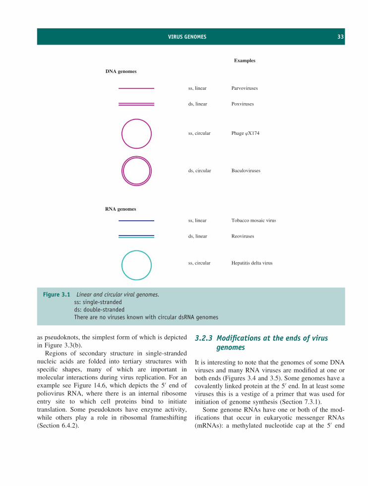
VIRUS GENOMES 33
Examples
DNA genomes
RNA genomes
ss, linear
ds, linear
ss, circular
ds, circular
ss, circular
ds, linear
ss, linear
Parvoviruses
Poxviruses
Phage jX174
Baculoviruses
Tobacco mosaic virus
Reoviruses
Hepatitis delta virus
Figure 3.1 Linear and circular viral genomes.ss: single-strandedds: double-strandedThere are no viruses known with circular dsRNA genomes
as pseudoknots, the simplest form of which is depictedin Figure 3.3(b).
Regions of secondary structure in single-strandednucleic acids are folded into tertiary structures withspecific shapes, many of which are important inmolecular interactions during virus replication. For anexample see Figure 14.6, which depicts the 5′ end ofpoliovirus RNA, where there is an internal ribosomeentry site to which cell proteins bind to initiatetranslation. Some pseudoknots have enzyme activity,while others play a role in ribosomal frameshifting(Section 6.4.2).
3.2.3 Modifications at the ends of virusgenomes
It is interesting to note that the genomes of some DNAviruses and many RNA viruses are modified at one orboth ends (Figures 3.4 and 3.5). Some genomes have acovalently linked protein at the 5′ end. In at least someviruses this is a vestige of a primer that was used forinitiation of genome synthesis (Section 7.3.1).
Some genome RNAs have one or both of the mod-ifications that occur in eukaryotic messenger RNAs(mRNAs): a methylated nucleotide cap at the 5′ end
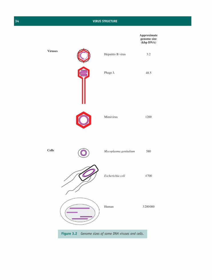
34 VIRUS STRUCTURE
Cells
VirusesHepatitis B virus 3.2
48.5
1200
580
4700
3200000
Approximategenome size(kbp DNA)
Mimivirus
Mycoplasma genitalium
Escherichia coli
Human
Phage λ
Figure 3.2 Genome sizes of some DNA viruses and cells.
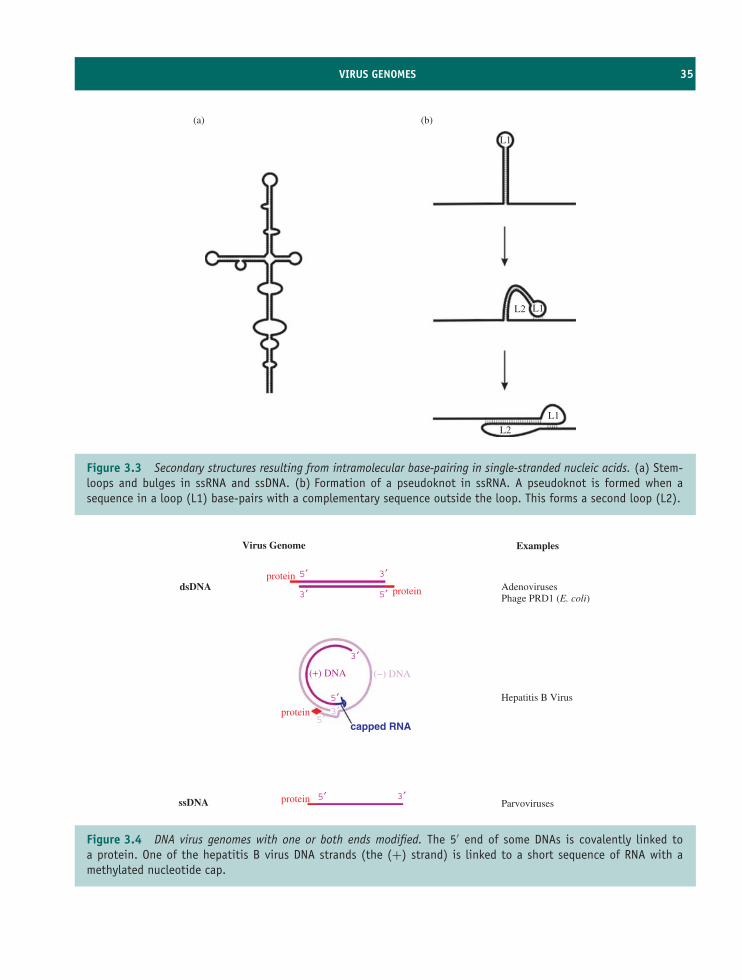
VIRUS GENOMES 35
(a) (b)
L2
L1
L2 L1
L1
Figure 3.3 Secondary structures resulting from intramolecular base-pairing in single-stranded nucleic acids. (a) Stem-loops and bulges in ssRNA and ssDNA. (b) Formation of a pseudoknot in ssRNA. A pseudoknot is formed when asequence in a loop (L1) base-pairs with a complementary sequence outside the loop. This forms a second loop (L2).
Examples
AdenovirusesPhage PRD1 (E. coli)
Hepatitis B Virus
Parvoviruses
Virus Genome
dsDNA
ssDNA protein
protein
(+) DNA (−) DNA
capped RNA
protein
protein
5′
5′
5′3′
3′
3′5′
3′ 5′
3′
Figure 3.4 DNA virus genomes with one or both ends modified. The 5′ end of some DNAs is covalently linked toa protein. One of the hepatitis B virus DNA strands (the (+) strand) is linked to a short sequence of RNA with amethylated nucleotide cap.
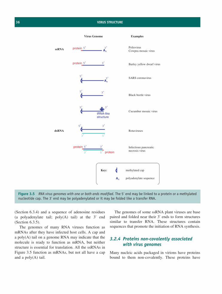
36 VIRUS STRUCTURE
Virus Genome Examples
ssRNA
dsRNA
PoliovirusCowpea mosaic virus
Barley yellow dwarf virus
SARS coronavirus
Black beetle virus
Cucumber mosaic virus
Rotaviruses
Infectious pancreaticnecrosis virus
5′
5′
5′
5′
5′
5′
5′
5′
3′
3′
3′
3′
3′
tRNA-likestructure
An
An
3′
3′
3′
5′ 3′
protein
protein
protein
protein
An
Key:
polyadenylate sequence
methylated cap
Figure 3.5 RNA virus genomes with one or both ends modified. The 5′ end may be linked to a protein or a methylatednucleotide cap. The 3′ end may be polyadenylated or it may be folded like a transfer RNA.
(Section 6.3.4) and a sequence of adenosine residues(a polyadenylate tail; poly(A) tail) at the 3′ end(Section 6.3.5).
The genomes of many RNA viruses function asmRNAs after they have infected host cells. A cap anda poly(A) tail on a genome RNA may indicate that themolecule is ready to function as mRNA, but neitherstructure is essential for translation. All the ssRNAs inFigure 3.5 function as mRNAs, but not all have a capand a poly(A) tail.
The genomes of some ssRNA plant viruses are basepaired and folded near their 3′ ends to form structuressimilar to transfer RNA. These structures containsequences that promote the initiation of RNA synthesis.
3.2.4 Proteins non-covalently associatedwith virus genomes
Many nucleic acids packaged in virions have proteinsbound to them non-covalently. These proteins have
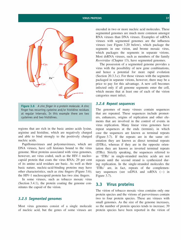
VIRUS PROTEINS 37
Cys
Cys His
His
Zn++
Figure 3.6 A zinc finger in a protein molecule. A zincfinger has recurring cysteine and/or histidine residuesat regular intervals. In this example there are twocysteines and two histidines.
regions that are rich in the basic amino acids lysine,arginine and histidine, which are negatively chargedand able to bind strongly to the positively chargednucleic acids.
Papillomaviruses and polyomaviruses, which areDNA viruses, have cell histones bound to the virusgenome. Most proteins associated with virus genomes,however, are virus coded, such as the HIV-1 nucleo-capsid protein that coats the virus RNA; 29 per centof its amino acid residues are basic. As well as theirbasic nature, nucleic-acid-binding proteins may haveother characteristics, such as zinc fingers (Figure 3.6);the HIV-1 nucleocapsid protein has two zinc fingers.
In some viruses, such as tobacco mosaic virus(Section 3.4.1), the protein coating the genome con-stitutes the capsid of the virion.
3.2.5 Segmented genomes
Most virus genomes consist of a single moleculeof nucleic acid, but the genes of some viruses are
encoded in two or more nucleic acid molecules. Thesesegmented genomes are much more common amongstRNA viruses than DNA viruses. Examples of ssRNAviruses with segmented genomes are the influenzaviruses (see Figure 3.20 below), which package thesegments in one virion, and brome mosaic virus,which packages the segments in separate virions.Most dsRNA viruses, such as members of the familyReoviridae (Chapter 13), have segmented genomes.
The possession of a segmented genome provides avirus with the possibility of new gene combinations,and hence a potential for more rapid evolution(Section 20.3.3.c). For those viruses with the segmentspackaged in separate virions, however, there may be aprice to pay for this advantage. A new cell becomesinfected only if all genome segments enter the cell,which means that at least one of each of the virioncategories must infect.
3.2.6 Repeat sequencesThe genomes of many viruses contain sequencesthat are repeated. These sequences include promot-ers, enhancers, origins of replication and other ele-ments that are involved in the control of events invirus replication. Many linear virus genomes haverepeat sequences at the ends (termini), in whichcase the sequences are known as terminal repeats(Figure 3.7). If the repeats are in the same ori-entation they are known as direct terminal repeats(DTRs), whereas if they are in the opposite orien-tation they are known as inverted terminal repeats(ITRs). Strictly speaking, the sequences referred toas ‘ITRs’ in single-stranded nucleic acids are notrepeats until the second strand is synthesized dur-ing replication. In the single-stranded molecules the‘ITRs’ are, in fact, repeats of the complemen-tary sequences (see ssDNA and ssRNA (−) inFigure 3.7).
3.3 Virus proteinsThe virion of tobacco mosaic virus contains only oneprotein species and the virions of parvoviruses containtwo to four protein species. These are viruses withsmall genomes. As the size of the genome increases,so the number of protein species tends to increase; 39protein species have been reported in the virion of
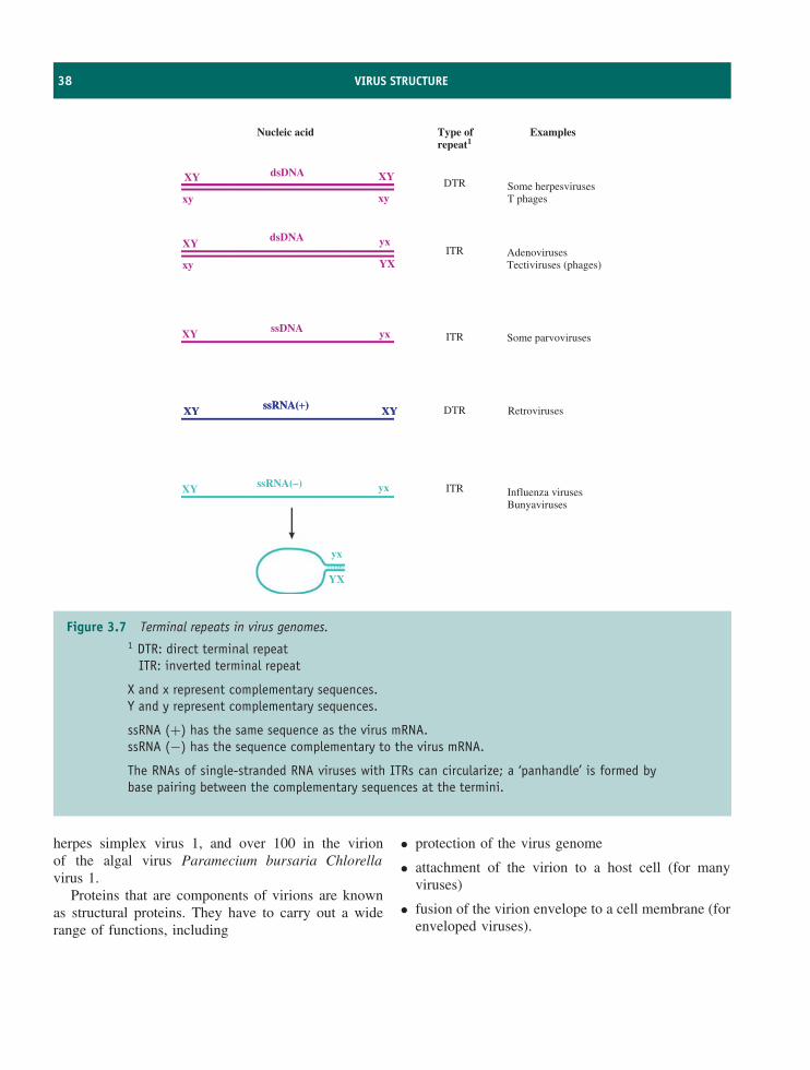
38 VIRUS STRUCTURE
Some herpesvirusesT phages
AdenovirusesTectiviruses (phages)
Some parvoviruses
Retroviruses
Influenza virusesBunyaviruses
ExamplesType ofrepeat1
Nucleic acid
DTR
DTR
ITR
ITR
ITR
XY XY
xy
yx
YX
yxXY
dsDNA
dsDNA
ssDNA
xy
XY
xy
ssRNA(+)ssRNA(+)XYXY XYXY
XY yx
yx
YX
ssRNA(–)
Figure 3.7 Terminal repeats in virus genomes.1 DTR: direct terminal repeat
ITR: inverted terminal repeat
X and x represent complementary sequences.Y and y represent complementary sequences.
ssRNA (+) has the same sequence as the virus mRNA.ssRNA (−) has the sequence complementary to the virus mRNA.
The RNAs of single-stranded RNA viruses with ITRs can circularize; a ‘panhandle’ is formed bybase pairing between the complementary sequences at the termini.
herpes simplex virus 1, and over 100 in the virionof the algal virus Paramecium bursaria Chlorellavirus 1.
Proteins that are components of virions are knownas structural proteins. They have to carry out a widerange of functions, including
• protection of the virus genome
• attachment of the virion to a host cell (for manyviruses)
• fusion of the virion envelope to a cell membrane (forenveloped viruses).
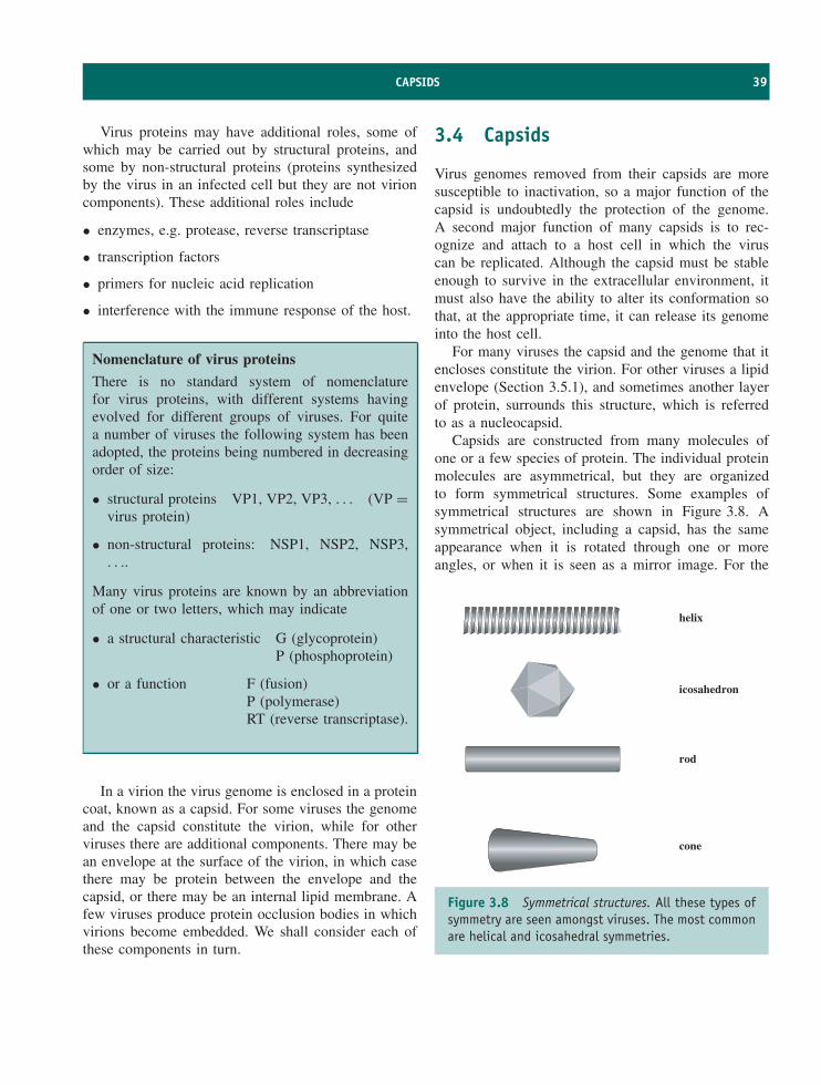
CAPSIDS 39
Virus proteins may have additional roles, some ofwhich may be carried out by structural proteins, andsome by non-structural proteins (proteins synthesizedby the virus in an infected cell but they are not virioncomponents). These additional roles include
• enzymes, e.g. protease, reverse transcriptase
• transcription factors
• primers for nucleic acid replication
• interference with the immune response of the host.
Nomenclature of virus proteins
There is no standard system of nomenclaturefor virus proteins, with different systems havingevolved for different groups of viruses. For quitea number of viruses the following system has beenadopted, the proteins being numbered in decreasingorder of size:
• structural proteins VP1, VP2, VP3, . . . (VP =virus protein)
• non-structural proteins: NSP1, NSP2, NSP3,. . ..
Many virus proteins are known by an abbreviationof one or two letters, which may indicate
• a structural characteristic G (glycoprotein)P (phosphoprotein)
• or a function F (fusion)P (polymerase)RT (reverse transcriptase).
In a virion the virus genome is enclosed in a proteincoat, known as a capsid. For some viruses the genomeand the capsid constitute the virion, while for otherviruses there are additional components. There may bean envelope at the surface of the virion, in which casethere may be protein between the envelope and thecapsid, or there may be an internal lipid membrane. Afew viruses produce protein occlusion bodies in whichvirions become embedded. We shall consider each ofthese components in turn.
3.4 Capsids
Virus genomes removed from their capsids are moresusceptible to inactivation, so a major function of thecapsid is undoubtedly the protection of the genome.A second major function of many capsids is to rec-ognize and attach to a host cell in which the viruscan be replicated. Although the capsid must be stableenough to survive in the extracellular environment, itmust also have the ability to alter its conformation sothat, at the appropriate time, it can release its genomeinto the host cell.
For many viruses the capsid and the genome that itencloses constitute the virion. For other viruses a lipidenvelope (Section 3.5.1), and sometimes another layerof protein, surrounds this structure, which is referredto as a nucleocapsid.
Capsids are constructed from many molecules ofone or a few species of protein. The individual proteinmolecules are asymmetrical, but they are organizedto form symmetrical structures. Some examples ofsymmetrical structures are shown in Figure 3.8. Asymmetrical object, including a capsid, has the sameappearance when it is rotated through one or moreangles, or when it is seen as a mirror image. For the
helix
icosahedron
rod
cone
Figure 3.8 Symmetrical structures. All these types ofsymmetry are seen amongst viruses. The most commonare helical and icosahedral symmetries.
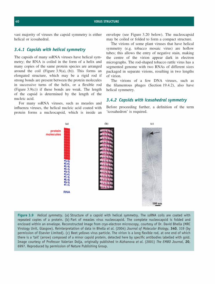
40 VIRUS STRUCTURE
vast majority of viruses the capsid symmetry is eitherhelical or icosahedral.
3.4.1 Capsids with helical symmetry
The capsids of many ssRNA viruses have helical sym-metry; the RNA is coiled in the form of a helix andmany copies of the same protein species are arrangedaround the coil (Figure 3.9(a), (b)). This forms anelongated structure, which may be a rigid rod ifstrong bonds are present between the protein moleculesin successive turns of the helix, or a flexible rod(Figure 3.9(c)) if these bonds are weak. The lengthof the capsid is determined by the length of thenucleic acid.
For many ssRNA viruses, such as measles andinfluenza viruses, the helical nucleic acid coated withprotein forms a nucleocapsid, which is inside an
envelope (see Figure 3.20 below). The nucleocapsidmay be coiled or folded to form a compact structure.
The virions of some plant viruses that have helicalsymmetry (e.g. tobacco mosaic virus) are hollowtubes; this allows the entry of negative stain, makingthe centre of the virion appear dark in electronmicrographs. The rod-shaped tobacco rattle virus has asegmented genome with two RNAs of different sizespackaged in separate virions, resulting in two lengthsof virion.
The virions of a few DNA viruses, such asthe filamentous phages (Section 19.4.2), also havehelical symmetry.
3.4.2 Capsids with icosahedral symmetryBefore proceeding further, a definition of the term‘icosahedron’ is required.
RNA
(a) (b) (c)
100 nm
proteinmolecules
Figure 3.9 Helical symmetry. (a) Structure of a capsid with helical symmetry. The ssRNA coils are coated withrepeated copies of a protein. (b) Part of measles virus nucleocapsid. The complete nucleocapsid is folded andenclosed within an envelope. Reconstructed image from cryo-electron microscopy, courtesy of Dr. David Bhella (MRCVirology Unit, Glasgow). Reinterpretation of data in Bhella et al. (2004) Journal of Molecular Biology, 340, 319 (bypermission of Elsevier Limited). (c) Beet yellows virus particle. The virion is a long flexible rod, at one end of whichthere is a ‘tail’ (arrow) composed of a minor capsid protein, detected here by specific antibodies labelled with gold.Image courtesy of Professor Valerian Dolja, originally published in Alzhanova et al. (2001) The EMBO Journal, 20,6997. Reproduced by permission of Nature Publishing Group.
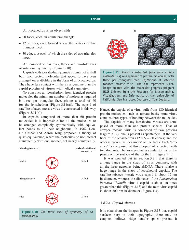
CAPSIDS 41
An icosahedron is an object with
• 20 faces, each an equilateral triangle;
• 12 vertices, each formed where the vertices of fivetriangles meet;
• 30 edges, at each of which the sides of two trianglesmeet.
An icosahedron has five-, three- and two-fold axesof rotational symmetry (Figure 3.10).
Capsids with icosahedral symmetry consist of a shellbuilt from protein molecules that appear to have beenarranged on scaffolding in the form of an icosahedron.They have less contact with the virus genome than thecapsid proteins of viruses with helical symmetry.
To construct an icosahedron from identical proteinmolecules the minimum number of molecules requiredis three per triangular face, giving a total of 60for the icosahedron (Figure 3.11(a)). The capsid ofsatellite tobacco mosaic virus is constructed in this way(Figure 3.11(b)).
In capsids composed of more than 60 proteinmolecules it is impossible for all the molecules tobe arranged completely symmetrically with equiva-lent bonds to all their neighbours. In 1962 Don-ald Caspar and Aaron Klug proposed a theory ofquasi-equivalence, where the molecules do not interactequivalently with one another, but nearly equivalently.
Viewing towards:
vertex
triangular face
edge
Axis of rotationalsymmetry
5-fold
3-fold
2-fold
Figure 3.10 The three axes of symmetry of anicosahedron.
(a) (b)
Figure 3.11 Capsid constructed from sixty proteinmolecules. (a) Arrangement of protein molecules, withthree per triangular face. (b) Virions of satellitetobacco mosaic virus. The bar represents 5 nm.Image created with the molecular graphics programUCSF Chimera from the Resource for Biocomputing,Visualization, and Informatics at the University ofCalifornia, San Francisco. Courtesy of Tom Goddard.
Hence, the capsid of a virus built from 180 identicalprotein molecules, such as tomato bushy stunt virus,contains three types of bonding between the molecules.
The capsids of many icosahedral viruses are com-posed of more than one protein species. That ofcowpea mosaic virus is composed of two proteins(Figure 3.12): one is present as ‘pentamers’ at the ver-tices of the icosahedron (12 × 5 = 60 copies) and theother is present as ‘hexamers’ on the faces. Each ‘hex-amer’ is composed of three copies of a protein withtwo domains. The arrangement is similar to that of thepanels on the surface of the football in Figure 3.12.
It was pointed out in Section 3.2.1 that there isa huge range in the sizes of virus genomes, withall the large genomes being dsDNA. There is also ahuge range in the sizes of icosahedral capsids. Thesatellite tobacco mosaic virus capsid is about 17 nmin diameter, whereas the diameter of the Parameciumbursaria Chlorella virus 1 capsid is about ten timesgreater than this (Figure 3.13) and the mimivirus capsidis about 300 nm in diameter (Figure 1.3).
3.4.2.a Capsid shapes
It is clear from the images in Figure 3.13 that capsidsurfaces vary in their topography; there may becanyons, hollows, ridges and/or spikes present. It
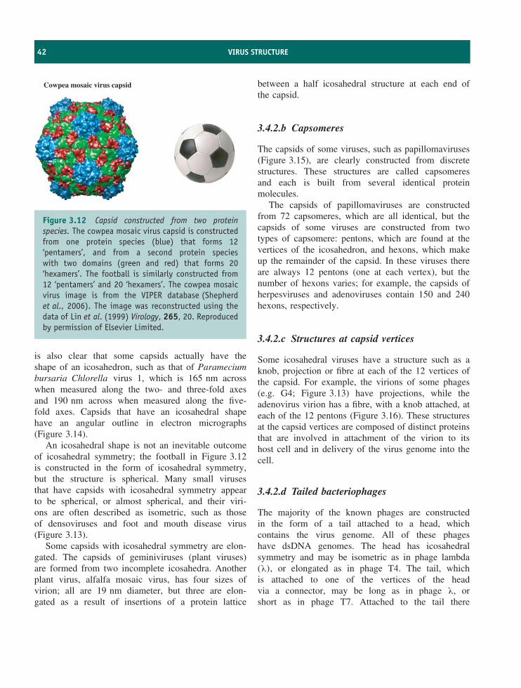
42 VIRUS STRUCTURE
Cowpea mosaic virus capsid
Figure 3.12 Capsid constructed from two proteinspecies. The cowpea mosaic virus capsid is constructedfrom one protein species (blue) that forms 12‘pentamers’, and from a second protein specieswith two domains (green and red) that forms 20‘hexamers’. The football is similarly constructed from12 ‘pentamers’ and 20 ‘hexamers’. The cowpea mosaicvirus image is from the VIPER database (Shepherdet al., 2006). The image was reconstructed using thedata of Lin et al. (1999) Virology, 265, 20. Reproducedby permission of Elsevier Limited.
is also clear that some capsids actually have theshape of an icosahedron, such as that of Parameciumbursaria Chlorella virus 1, which is 165 nm acrosswhen measured along the two- and three-fold axesand 190 nm across when measured along the five-fold axes. Capsids that have an icosahedral shapehave an angular outline in electron micrographs(Figure 3.14).
An icosahedral shape is not an inevitable outcomeof icosahedral symmetry; the football in Figure 3.12is constructed in the form of icosahedral symmetry,but the structure is spherical. Many small virusesthat have capsids with icosahedral symmetry appearto be spherical, or almost spherical, and their viri-ons are often described as isometric, such as thoseof densoviruses and foot and mouth disease virus(Figure 3.13).
Some capsids with icosahedral symmetry are elon-gated. The capsids of geminiviruses (plant viruses)are formed from two incomplete icosahedra. Anotherplant virus, alfalfa mosaic virus, has four sizes ofvirion; all are 19 nm diameter, but three are elon-gated as a result of insertions of a protein lattice
between a half icosahedral structure at each end ofthe capsid.
3.4.2.b Capsomeres
The capsids of some viruses, such as papillomaviruses(Figure 3.15), are clearly constructed from discretestructures. These structures are called capsomeresand each is built from several identical proteinmolecules.
The capsids of papillomaviruses are constructedfrom 72 capsomeres, which are all identical, but thecapsids of some viruses are constructed from twotypes of capsomere: pentons, which are found at thevertices of the icosahedron, and hexons, which makeup the remainder of the capsid. In these viruses thereare always 12 pentons (one at each vertex), but thenumber of hexons varies; for example, the capsids ofherpesviruses and adenoviruses contain 150 and 240hexons, respectively.
3.4.2.c Structures at capsid vertices
Some icosahedral viruses have a structure such as aknob, projection or fibre at each of the 12 vertices ofthe capsid. For example, the virions of some phages(e.g. G4; Figure 3.13) have projections, while theadenovirus virion has a fibre, with a knob attached, ateach of the 12 pentons (Figure 3.16). These structuresat the capsid vertices are composed of distinct proteinsthat are involved in attachment of the virion to itshost cell and in delivery of the virus genome into thecell.
3.4.2.d Tailed bacteriophages
The majority of the known phages are constructedin the form of a tail attached to a head, whichcontains the virus genome. All of these phageshave dsDNA genomes. The head has icosahedralsymmetry and may be isometric as in phage lambda(λ), or elongated as in phage T4. The tail, whichis attached to one of the vertices of the headvia a connector, may be long as in phage λ, orshort as in phage T7. Attached to the tail there

CAPSIDS 43
RibgrassMosaic Virus
Cowpea Chlorotic Mottle Virus
FelinePanleukopenia Virus
Bacteriophage MS2
Hepatitis B VirusFoot and Mouth Disease Virus
Bacteriophage G4Human PapillomavirusL1 Capsid
Human Rhinovirus 16& cellular receptor
Nudaurelia CapensisOmega Virus
Reovirus CoreBacteriophage PRD1
Bacteriophage HK97Rice Dwarf Virus
Paramecium Bursaria Chlorella Virus
Dengue Virus Bluetongue Virusinner layer
Human Papillomavirus
BacteriophagePhi-X174 procapsid
Norwalk Virus
DensovirusSatellite TobaccoMosaic Virus
50nm
Figure 3.13 Capsids with icosahedral symmetry. Some of the wide ranges of capsid architectures and sizes areillustrated. The images were created with the molecular graphics program UCSF Chimera using data from cryo-electronmicroscopy and X-ray diffraction. From Goddard et al. (2005) Structure, 13, 473. Reproduced by permission of ElsevierLimited.
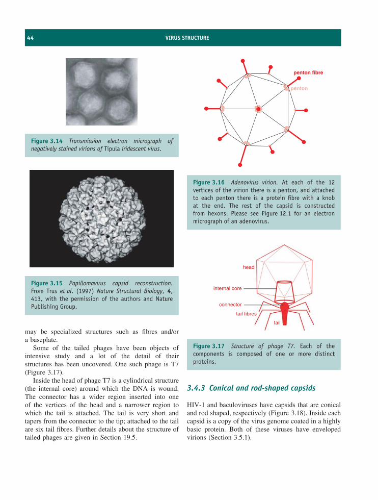
44 VIRUS STRUCTURE
Figure 3.14 Transmission electron micrograph ofnegatively stained virions of Tipula iridescent virus.
Figure 3.15 Papillomavirus capsid reconstruction.From Trus et al. (1997) Nature Structural Biology, 4,413, with the permission of the authors and NaturePublishing Group.
may be specialized structures such as fibres and/ora baseplate.
Some of the tailed phages have been objects ofintensive study and a lot of the detail of theirstructures has been uncovered. One such phage is T7(Figure 3.17).
Inside the head of phage T7 is a cylindrical structure(the internal core) around which the DNA is wound.The connector has a wider region inserted into oneof the vertices of the head and a narrower region towhich the tail is attached. The tail is very short andtapers from the connector to the tip; attached to the tailare six tail fibres. Further details about the structure oftailed phages are given in Section 19.5.
penton
penton fibre
Figure 3.16 Adenovirus virion. At each of the 12vertices of the virion there is a penton, and attachedto each penton there is a protein fibre with a knobat the end. The rest of the capsid is constructedfrom hexons. Please see Figure 12.1 for an electronmicrograph of an adenovirus.
head
internal core
connector
tail fibres
tail
Figure 3.17 Structure of phage T7. Each of thecomponents is composed of one or more distinctproteins.
3.4.3 Conical and rod-shaped capsids
HIV-1 and baculoviruses have capsids that are conicaland rod shaped, respectively (Figure 3.18). Inside eachcapsid is a copy of the virus genome coated in a highlybasic protein. Both of these viruses have envelopedvirions (Section 3.5.1).
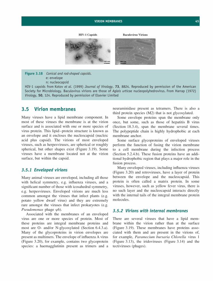
VIRION MEMBRANES 45
HIV-1 Capsids Baculovirus Virions
100 nm
Figure 3.18 Conical and rod-shaped capsids.e: envelopen: nucleocapsid
HIV-1 capsids from Kotov et al. (1999) Journal of Virology, 73, 8824. Reproduced by permission of the AmericanSociety for Microbiology. Baculovirus virions are those of Aglais urticae nucleopolyhedrovirus. From Harrap (1972)Virology, 50, 124. Reproduced by permission of Elsevier Limited
3.5 Virion membranesMany viruses have a lipid membrane component. Inmost of these viruses the membrane is at the virionsurface and is associated with one or more species ofvirus protein. This lipid–protein structure is known asan envelope and it encloses the nucleocapsid (nucleicacid plus capsid). The virions of most envelopedviruses, such as herpesviruses, are spherical or roughlyspherical, but other shapes exist (Figure 3.19). Someviruses have a membrane located not at the virionsurface, but within the capsid.
3.5.1 Enveloped virions
Many animal viruses are enveloped, including all thosewith helical symmetry, e.g. influenza viruses, and asignificant number of those with icosahedral symmetry,e.g. herpesviruses. Enveloped virions are much lesscommon amongst the viruses that infect plants (e.g.potato yellow dwarf virus) and they are extremelyrare amongst the viruses that infect prokaryotes (e.g.Pseudomonas phage ϕ6).
Associated with the membranes of an envelopedvirus are one or more species of protein. Most ofthese proteins are integral membrane proteins andmost are O- and/or N-glycosylated (Section 6.4.3.a).Many of the glycoproteins in virion envelopes arepresent as multimers. The envelope of influenza A virus(Figure 3.20), for example, contains two glycoproteinspecies: a haemagglutinin present as trimers and a
neuraminidase present as tetramers. There is also athird protein species (M2) that is not glycosylated.
Some envelope proteins span the membrane onlyonce, but some, such as those of hepatitis B virus(Section 18.3.4), span the membrane several times.The polypeptide chain is highly hydrophobic at eachmembrane anchor.
Some surface glycoproteins of enveloped virusesperform the function of fusing the virion membraneto a cell membrane during the infection process(Section 5.2.4.b). These fusion proteins have an addi-tional hydrophobic region that plays a major role in thefusion process.
Many enveloped viruses, including influenza viruses(Figure 3.20) and retroviruses, have a layer of proteinbetween the envelope and the nucleocapsid. Thisprotein is often called a matrix protein. In someviruses, however, such as yellow fever virus, there isno such layer and the nucleocapsid interacts directlywith the internal tails of the integral membrane proteinmolecules.
3.5.2 Virions with internal membranes
There are several viruses that have a lipid mem-brane within the virion rather than at the surface(Figure 3.19). These membranes have proteins asso-ciated with them and are present in the virions of,for example, Paramecium bursaria Chlorella virus 1(Figure 3.13), the iridoviruses (Figure 3.14) and thetectiviruses (phages).
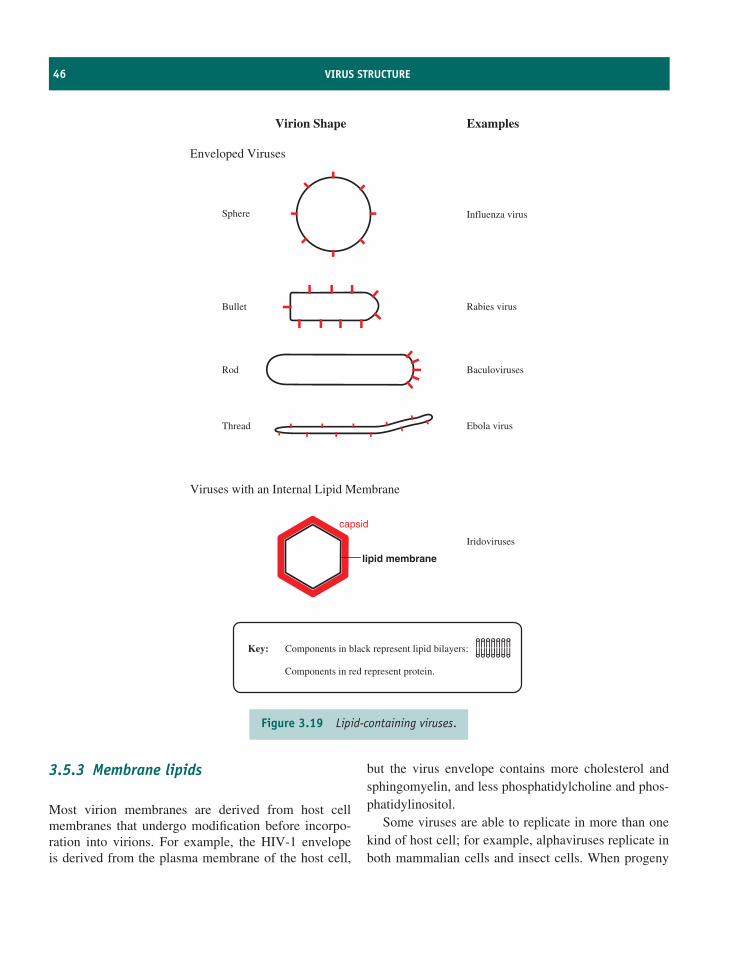
46 VIRUS STRUCTURE
Enveloped Viruses
Viruses with an Internal Lipid Membrane
capsid
lipid membrane
Sphere
Bullet
Thread
Rod
Virion Shape Examples
Influenza virus
Rabies virus
Baculoviruses
Ebola virus
Iridoviruses
Key: Components in black represent lipid bilayers:
Components in red represent protein.
Figure 3.19 Lipid-containing viruses.
3.5.3 Membrane lipids
Most virion membranes are derived from host cellmembranes that undergo modification before incorpo-ration into virions. For example, the HIV-1 envelopeis derived from the plasma membrane of the host cell,
but the virus envelope contains more cholesterol andsphingomyelin, and less phosphatidylcholine and phos-phatidylinositol.
Some viruses are able to replicate in more than onekind of host cell; for example, alphaviruses replicate inboth mammalian cells and insect cells. When progeny
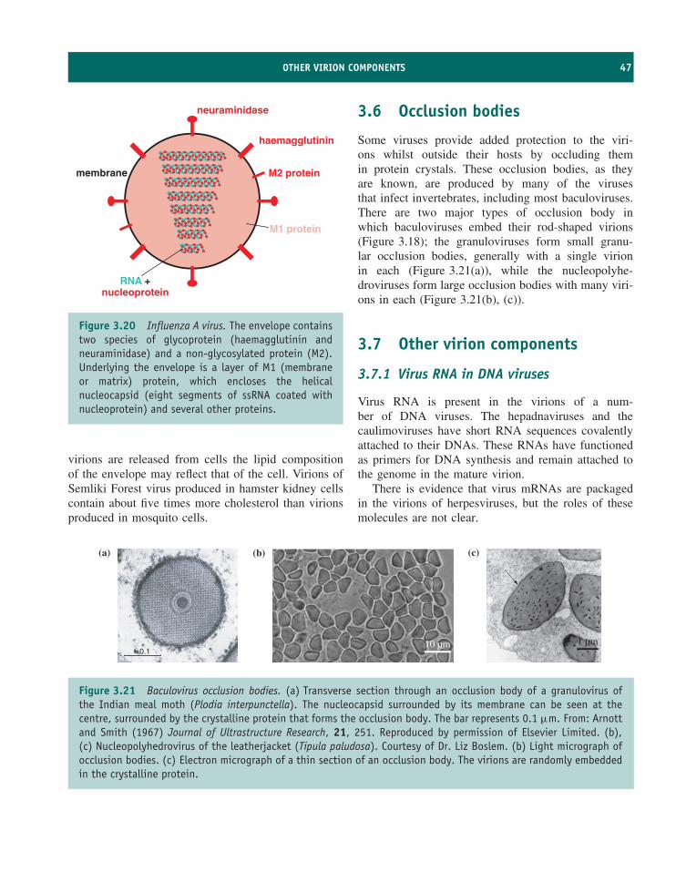
OTHER VIRION COMPONENTS 47
haemagglutinin
neuraminidase
M2 protein
M1 protein
membrane
RNA +nucleoprotein
Figure 3.20 Influenza A virus. The envelope containstwo species of glycoprotein (haemagglutinin andneuraminidase) and a non-glycosylated protein (M2).Underlying the envelope is a layer of M1 (membraneor matrix) protein, which encloses the helicalnucleocapsid (eight segments of ssRNA coated withnucleoprotein) and several other proteins.
virions are released from cells the lipid compositionof the envelope may reflect that of the cell. Virions ofSemliki Forest virus produced in hamster kidney cellscontain about five times more cholesterol than virionsproduced in mosquito cells.
3.6 Occlusion bodies
Some viruses provide added protection to the viri-ons whilst outside their hosts by occluding themin protein crystals. These occlusion bodies, as theyare known, are produced by many of the virusesthat infect invertebrates, including most baculoviruses.There are two major types of occlusion body inwhich baculoviruses embed their rod-shaped virions(Figure 3.18); the granuloviruses form small granu-lar occlusion bodies, generally with a single virionin each (Figure 3.21(a)), while the nucleopolyhe-droviruses form large occlusion bodies with many viri-ons in each (Figure 3.21(b), (c)).
3.7 Other virion components
3.7.1 Virus RNA in DNA viruses
Virus RNA is present in the virions of a num-ber of DNA viruses. The hepadnaviruses and thecaulimoviruses have short RNA sequences covalentlyattached to their DNAs. These RNAs have functionedas primers for DNA synthesis and remain attached tothe genome in the mature virion.
There is evidence that virus mRNAs are packagedin the virions of herpesviruses, but the roles of thesemolecules are not clear.
(c)
1 µm
(b)
10 µm
(a)
0.1
Figure 3.21 Baculovirus occlusion bodies. (a) Transverse section through an occlusion body of a granulovirus ofthe Indian meal moth (Plodia interpunctella). The nucleocapsid surrounded by its membrane can be seen at thecentre, surrounded by the crystalline protein that forms the occlusion body. The bar represents 0.1 µm. From: Arnottand Smith (1967) Journal of Ultrastructure Research, 21, 251. Reproduced by permission of Elsevier Limited. (b),(c) Nucleopolyhedrovirus of the leatherjacket (Tipula paludosa). Courtesy of Dr. Liz Boslem. (b) Light micrograph ofocclusion bodies. (c) Electron micrograph of a thin section of an occlusion body. The virions are randomly embeddedin the crystalline protein.
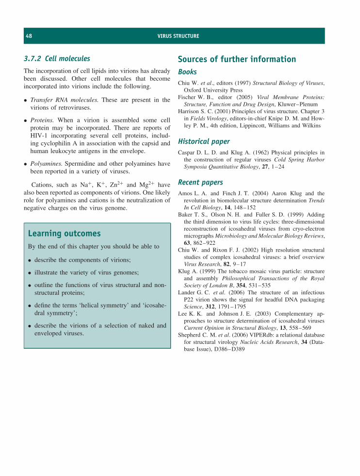
48 VIRUS STRUCTURE
3.7.2 Cell molecules
The incorporation of cell lipids into virions has alreadybeen discussed. Other cell molecules that becomeincorporated into virions include the following.
• Transfer RNA molecules. These are present in thevirions of retroviruses.
• Proteins. When a virion is assembled some cellprotein may be incorporated. There are reports ofHIV-1 incorporating several cell proteins, includ-ing cyclophilin A in association with the capsid andhuman leukocyte antigens in the envelope.
• Polyamines. Spermidine and other polyamines havebeen reported in a variety of viruses.
Cations, such as Na+, K+, Zn2+ and Mg2+ havealso been reported as components of virions. One likelyrole for polyamines and cations is the neutralization ofnegative charges on the virus genome.
Learning outcomesBy the end of this chapter you should be able to
• describe the components of virions;
• illustrate the variety of virus genomes;
• outline the functions of virus structural and non-structural proteins;
• define the terms ‘helical symmetry’ and ‘icosahe-dral symmetry’;
• describe the virions of a selection of naked andenveloped viruses.
Sources of further informationBooksChiu W. et al., editors (1997) Structural Biology of Viruses,
Oxford University PressFischer W. B., editor (2005) Viral Membrane Proteins:
Structure, Function and Drug Design, Kluwer–PlenumHarrison S. C. (2001) Principles of virus structure. Chapter 3
in Fields Virology, editors-in-chief Knipe D. M. and How-ley P. M., 4th edition, Lippincott, Williams and Wilkins
Historical paperCaspar D. L. D. and Klug A. (1962) Physical principles in
the construction of regular viruses Cold Spring HarborSymposia Quantitative Biology, 27, 1–24
Recent papersAmos L. A. and Finch J. T. (2004) Aaron Klug and the
revolution in biomolecular structure determination TrendsIn Cell Biology, 14, 148–152
Baker T. S., Olson N. H. and Fuller S. D. (1999) Addingthe third dimension to virus life cycles: three-dimensionalreconstruction of icosahedral viruses from cryo-electronmicrographs Microbiology and Molecular Biology Reviews,63, 862–922
Chiu W. and Rixon F. J. (2002) High resolution structuralstudies of complex icosahedral viruses: a brief overviewVirus Research, 82, 9–17
Klug A. (1999) The tobacco mosaic virus particle: structureand assembly Philosophical Transactions of the RoyalSociety of London B, 354, 531–535
Lander G. C. et al. (2006) The structure of an infectiousP22 virion shows the signal for headful DNA packagingScience, 312, 1791–1795
Lee K. K. and Johnson J. E. (2003) Complementary ap-proaches to structure determination of icosahedral virusesCurrent Opinion in Structural Biology, 13, 558–569
Shepherd C. M. et al. (2006) VIPERdb: a relational databasefor structural virology Nucleic Acids Research, 34 (Data-base Issue), D386–D389
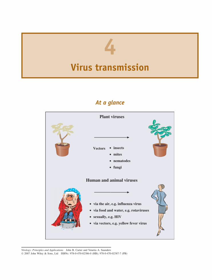
4Virus transmission
At a glance
Plant viruses
Vectors •
•
•
•
insects
mites
nematodes
fungi
Human and animal viruses
•
•
•
•
via the air, e.g. influenza virus
via food and water, e.g. rotaviruses
sexually, e.g. HIV
via vectors, e.g. yellow fever virus
Virology: Principles and Applications John B. Carter and Venetia A. Saunders 2007 John Wiley & Sons, Ltd ISBNs: 978-0-470-02386-0 (HB); 978-0-470-02387-7 (PB)
50 VIRUS TRANSMISSION
4.1 Introduction to virustransmission
A minimum proportion of the virions produced ininfected hosts must be transmitted to new hosts inwhich more virions can be manufactured. If this doesnot happen the virus will die out. The only otherpossibility for the survival of virus genes is for themto be maintained in cells as nucleic acids, which arereplicated and passed on to daughter cells when thecells divide.
Viruses of bacteria and other microbial hosts arereleased from infected cells into the environment ofthe host, where further susceptible cells are likely tobe present. These viruses are dependent upon chanceencounters with susceptible cells, to which they maybind if receptors on the surface of those cells comeinto contact with virus attachment sites.
Viruses of multicellular animals and plants must alsofind new cells to infect. An infection may spread toadjacent cells, or to cells in a distant part of the hostafter transport in the blood of an animal or in thephloem of a plant, but ultimately a virus must findnew hosts to infect if it is to survive.
Some virus infections modify the behaviour oftheir hosts in order to increase the probability oftransmission. Mammals infected with rabies virus oftenbecome aggressive; this change in behaviour increasesthe likelihood of the host biting another individual andtransmitting virus in the saliva. Some plant-feedinginsect larvae infected with baculoviruses become moremobile in the late stages of infection, thus aiding virusdispersal. If the virus-infected insect migrates towardsthe top of a plant then lower leaves, where other larvaewill feed, will become contaminated when the host diesand putrefies.
On their journeys between hosts viruses may haveto survive adverse conditions in an environment suchas air, water or soil; the survival of virus infectivityoutside the host is discussed in Chapter 23. Thereare some viruses, however, that can be transmitted tonew hosts without ‘seeing the light of day’, in otherwords without exposure to the outside environment.These are viruses that can be transmitted directly fromhost to host, for example during kissing or sexualintercourse, and viruses that are transmitted via vectors.Also included in this category are viruses that can be
transmitted directly from a parent to members of thenext generation. Transmission in these cases is said tobe vertical; otherwise it is described as horizontal.
Viruses may be moved over long distances in avariety of ways. Rivers and winds can move viruses tonew areas. The foot and mouth disease outbreak on theIsle of Wight, UK, in 1981 was initiated by virus thathad spread via the air from Brittany, more than 250 kmaway. Viruses of birds, fishes, humans and other hostsare transported within their hosts to other parts of theplanet as a result of migration, travel and animal export,for example
• bird migration (avian influenza viruses)
• human travel (SARS virus)
• animal export (monkeypox virus).
Furthermore, viruses can be transported on inani-mate materials, for example, foot and mouth diseasevirus on straw and farm vehicles.
Once virions have entered a multicellular organismthey may have further to travel before suitable hostcells are encountered. During this final stage of theirjourney the virions will encounter hazards in the formof host defence mechanisms, which must be survivedif the virions are to remain infective when they reachtheir destination.
In theory, a single virion can initiate an infection,but in practice it is often found that a host must beinoculated with a minimum number of virions in orderfor that host to become infected. The reasons for thisare probably many and varied, and may include somevirions being defective and some being inactivated bythe host’s immune systems. This minimum amount ofvirus required for infection of a host is known as theminimum infective dose.
In this chapter we shall look at the ways that virusesof plants and animals are transmitted from host to host,but first we must look at some general aspects of virustransmission by vectors.
4.1.1 Transmission via vectors: generalprinciples
Many viruses of plants and animals are transmittedbetween hosts by organisms that feed on them; these
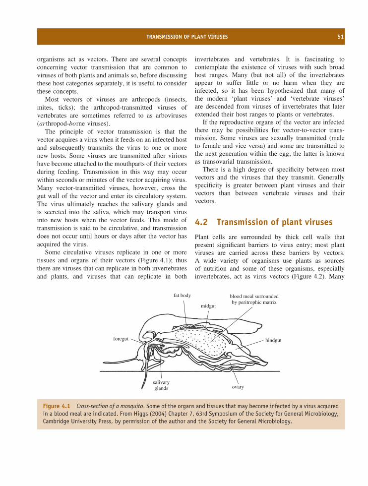
TRANSMISSION OF PLANT VIRUSES 51
organisms act as vectors. There are several conceptsconcerning vector transmission that are common toviruses of both plants and animals so, before discussingthese host categories separately, it is useful to considerthese concepts.
Most vectors of viruses are arthropods (insects,mites, ticks); the arthropod-transmitted viruses ofvertebrates are sometimes referred to as arboviruses(ar thropod-borne viruses).
The principle of vector transmission is that thevector acquires a virus when it feeds on an infected hostand subsequently transmits the virus to one or morenew hosts. Some viruses are transmitted after virionshave become attached to the mouthparts of their vectorsduring feeding. Transmission in this way may occurwithin seconds or minutes of the vector acquiring virus.Many vector-transmitted viruses, however, cross thegut wall of the vector and enter its circulatory system.The virus ultimately reaches the salivary glands andis secreted into the saliva, which may transport virusinto new hosts when the vector feeds. This mode oftransmission is said to be circulative, and transmissiondoes not occur until hours or days after the vector hasacquired the virus.
Some circulative viruses replicate in one or moretissues and organs of their vectors (Figure 4.1); thusthere are viruses that can replicate in both invertebratesand plants, and viruses that can replicate in both
invertebrates and vertebrates. It is fascinating tocontemplate the existence of viruses with such broadhost ranges. Many (but not all) of the invertebratesappear to suffer little or no harm when they areinfected, so it has been hypothesized that many ofthe modern ‘plant viruses’ and ‘vertebrate viruses’are descended from viruses of invertebrates that laterextended their host ranges to plants or vertebrates.
If the reproductive organs of the vector are infectedthere may be possibilities for vector-to-vector trans-mission. Some viruses are sexually transmitted (maleto female and vice versa) and some are transmitted tothe next generation within the egg; the latter is knownas transovarial transmission.
There is a high degree of specificity between mostvectors and the viruses that they transmit. Generallyspecificity is greater between plant viruses and theirvectors than between vertebrate viruses and theirvectors.
4.2 Transmission of plant viruses
Plant cells are surrounded by thick cell walls thatpresent significant barriers to virus entry; most plantviruses are carried across these barriers by vectors.A wide variety of organisms use plants as sourcesof nutrition and some of these organisms, especiallyinvertebrates, act as virus vectors (Figure 4.2). Many
fat body
midgut
foregut hindgut
blood meal surroundedby peritrophic matrix
salivaryglands ovary
Figure 4.1 Cross-section of a mosquito. Some of the organs and tissues that may become infected by a virus acquiredin a blood meal are indicated. From Higgs (2004) Chapter 7, 63rd Symposium of the Society for General Microbiology,Cambridge University Press, by permission of the author and the Society for General Microbiology.
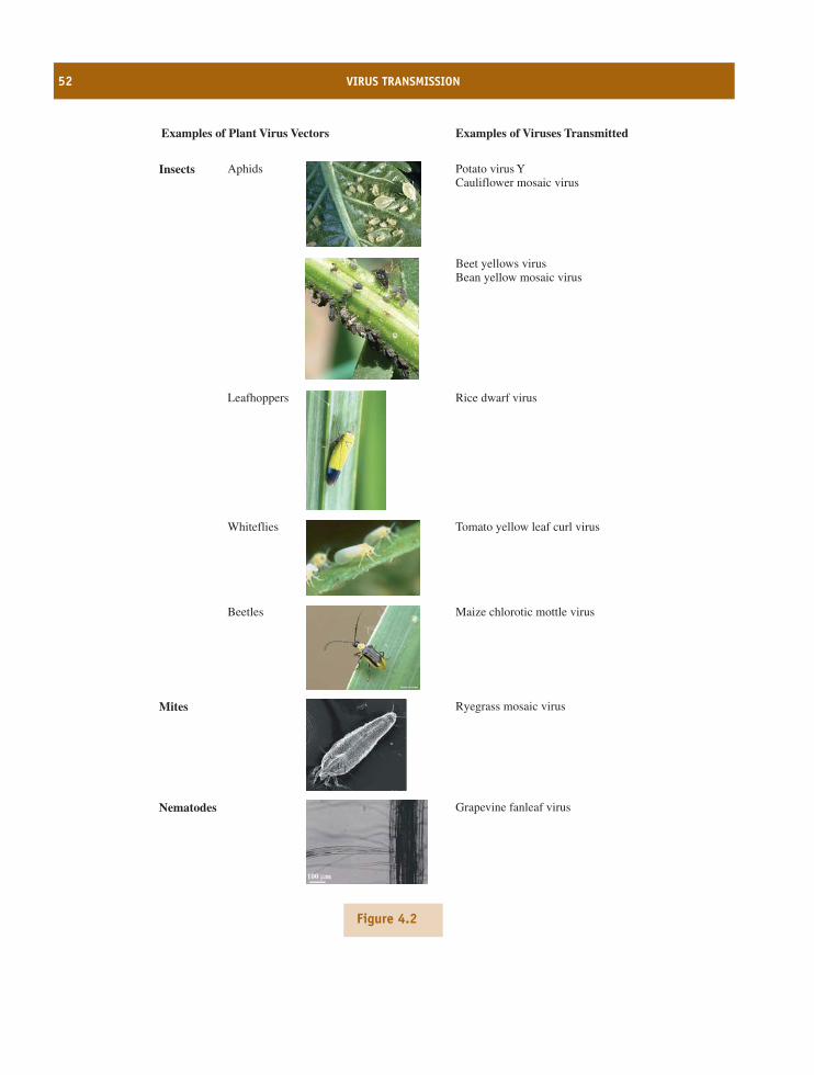
52 VIRUS TRANSMISSION
Examples of Plant Virus Vectors Examples of Viruses Transmitted
Insects Potato virus YCauliflower mosaic virus
Aphids
Rice dwarf virusLeafhoppers
Tomato yellow leaf curl virusWhiteflies
Maize chlorotic mottle virusBeetles
Mites Ryegrass mosaic virus
Nematodes Grapevine fanleaf virus
100 µm
Beet yellows virusBean yellow mosaic virus
Figure 4.2
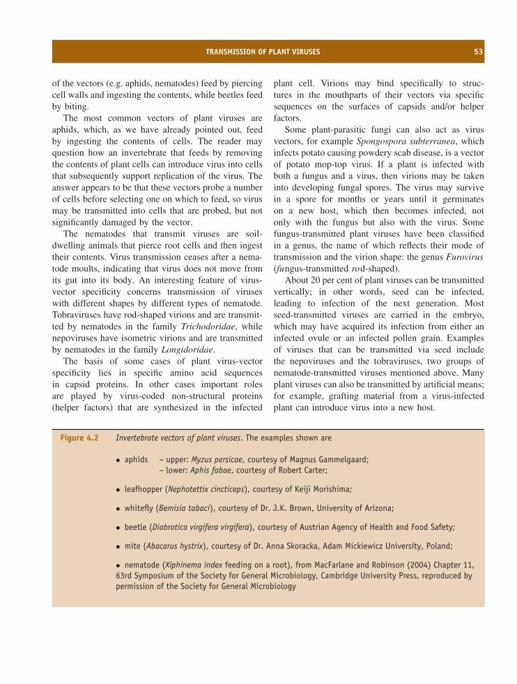
TRANSMISSION OF PLANT VIRUSES 53
of the vectors (e.g. aphids, nematodes) feed by piercingcell walls and ingesting the contents, while beetles feedby biting.
The most common vectors of plant viruses areaphids, which, as we have already pointed out, feedby ingesting the contents of cells. The reader mayquestion how an invertebrate that feeds by removingthe contents of plant cells can introduce virus into cellsthat subsequently support replication of the virus. Theanswer appears to be that these vectors probe a numberof cells before selecting one on which to feed, so virusmay be transmitted into cells that are probed, but notsignificantly damaged by the vector.
The nematodes that transmit viruses are soil-dwelling animals that pierce root cells and then ingesttheir contents. Virus transmission ceases after a nema-tode moults, indicating that virus does not move fromits gut into its body. An interesting feature of virus-vector specificity concerns transmission of viruseswith different shapes by different types of nematode.Tobraviruses have rod-shaped virions and are transmit-ted by nematodes in the family Trichodoridae, whilenepoviruses have isometric virions and are transmittedby nematodes in the family Longidoridae.
The basis of some cases of plant virus-vectorspecificity lies in specific amino acid sequencesin capsid proteins. In other cases important rolesare played by virus-coded non-structural proteins(helper factors) that are synthesized in the infected
plant cell. Virions may bind specifically to struc-tures in the mouthparts of their vectors via specificsequences on the surfaces of capsids and/or helperfactors.
Some plant-parasitic fungi can also act as virusvectors, for example Spongospora subterranea, whichinfects potato causing powdery scab disease, is a vectorof potato mop-top virus. If a plant is infected withboth a fungus and a virus, then virions may be takeninto developing fungal spores. The virus may survivein a spore for months or years until it germinateson a new host, which then becomes infected, notonly with the fungus but also with the virus. Somefungus-transmitted plant viruses have been classifiedin a genus, the name of which reflects their mode oftransmission and the virion shape: the genus Furovirus(fungus-transmitted rod-shaped).
About 20 per cent of plant viruses can be transmittedvertically; in other words, seed can be infected,leading to infection of the next generation. Mostseed-transmitted viruses are carried in the embryo,which may have acquired its infection from either aninfected ovule or an infected pollen grain. Examplesof viruses that can be transmitted via seed includethe nepoviruses and the tobraviruses, two groups ofnematode-transmitted viruses mentioned above. Manyplant viruses can also be transmitted by artificial means;for example, grafting material from a virus-infectedplant can introduce virus into a new host.
Figure 4.2 Invertebrate vectors of plant viruses. The examples shown are
• aphids – upper: Myzus persicae, courtesy of Magnus Gammelgaard;– lower: Aphis fabae, courtesy of Robert Carter;
• leafhopper (Nephotettix cincticeps), courtesy of Keiji Morishima;
• whitefly (Bemisia tabaci), courtesy of Dr. J.K. Brown, University of Arizona;
• beetle (Diabrotica virgifera virgifera), courtesy of Austrian Agency of Health and Food Safety;
• mite (Abacarus hystrix), courtesy of Dr. Anna Skoracka, Adam Mickiewicz University, Poland;
• nematode (Xiphinema index feeding on a root), from MacFarlane and Robinson (2004) Chapter 11,63rd Symposium of the Society for General Microbiology, Cambridge University Press, reproduced bypermission of the Society for General Microbiology
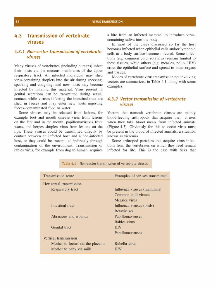
54 VIRUS TRANSMISSION
4.3 Transmission of vertebrateviruses
4.3.1 Non-vector transmission of vertebrateviruses
Many viruses of vertebrates (including humans) infecttheir hosts via the mucous membranes of the upperrespiratory tract. An infected individual may shedvirus-containing droplets into the air during sneezing,speaking and coughing, and new hosts may becomeinfected by inhaling this material. Virus present ingenital secretions can be transmitted during sexualcontact, while viruses infecting the intestinal tract areshed in faeces and may enter new hosts ingestingfaeces-contaminated food or water.
Some viruses may be released from lesions, forexample foot and mouth disease virus from lesionson the feet and in the mouth, papillomaviruses fromwarts, and herpes simplex virus from lesions on thelips. These viruses could be transmitted directly bycontact between an infected host and a non-infectedhost, or they could be transmitted indirectly throughcontamination of the environment. Transmission ofrabies virus, for example from dog to human, requires
a bite from an infected mammal to introduce virus-containing saliva into the body.
In most of the cases discussed so far the hostbecomes infected when epithelial cells and/or lymphoidcells at a body surface become infected. Some infec-tions (e.g. common cold, rotavirus) remain limited tothese tissues, while others (e.g. measles, polio, HIV)cross the epithelial surface and spread to other organsand tissues.
Modes of vertebrate virus transmission not involvingvectors are summarized in Table 4.1, along with someexamples.
4.3.2 Vector transmission of vertebrateviruses
Vectors that transmit vertebrate viruses are mainlyblood-feeding arthropods that acquire their viruseswhen they take blood meals from infected animals(Figure 4.3). Obviously for this to occur virus mustbe present in the blood of infected animals, a situationknown as viraemia.
Some arthropod parasites that acquire virus infec-tions from the vertebrates on which they feed remaininfected for life. This is the case with ticks that
Table 4.1 Non-vector transmission of vertebrate viruses
Transmission route Examples of viruses transmitted
Horizontal transmissionRespiratory tract Influenza viruses (mammals)
Common cold virusesMeasles virus
Intestinal tract Influenza viruses (birds)Rotaviruses
Abrasions and wounds PapillomavirusesRabies virus
Genital tract HIVPapillomaviruses
Vertical transmissionMother to foetus via the placenta Rubella virusMother to baby via milk HIV
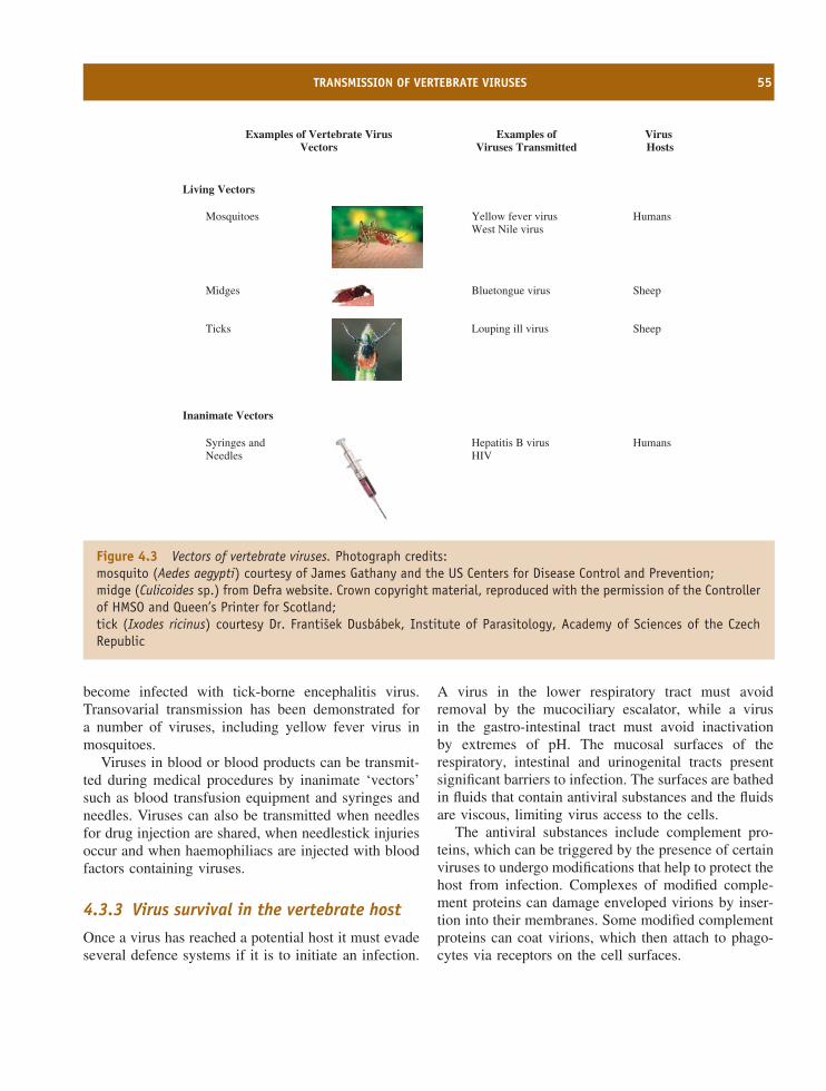
TRANSMISSION OF VERTEBRATE VIRUSES 55
Examples of Vertebrate VirusVectors
Examples of Viruses Transmitted
Virus Hosts
Living Vectors
Mosquitoes Yellow fever virusWest Nile virus
Humans
Midges Bluetongue virus Sheep
Ticks Louping ill virus Sheep
Inanimate Vectors
Syringes andNeedles
Hepatitis B virusHIV
Humans
Figure 4.3 Vectors of vertebrate viruses. Photograph credits:mosquito (Aedes aegypti) courtesy of James Gathany and the US Centers for Disease Control and Prevention;midge (Culicoides sp.) from Defra website. Crown copyright material, reproduced with the permission of the Controllerof HMSO and Queen’s Printer for Scotland;tick (Ixodes ricinus) courtesy Dr. Frantisek Dusbabek, Institute of Parasitology, Academy of Sciences of the CzechRepublic
become infected with tick-borne encephalitis virus.Transovarial transmission has been demonstrated fora number of viruses, including yellow fever virus inmosquitoes.
Viruses in blood or blood products can be transmit-ted during medical procedures by inanimate ‘vectors’such as blood transfusion equipment and syringes andneedles. Viruses can also be transmitted when needlesfor drug injection are shared, when needlestick injuriesoccur and when haemophiliacs are injected with bloodfactors containing viruses.
4.3.3 Virus survival in the vertebrate host
Once a virus has reached a potential host it must evadeseveral defence systems if it is to initiate an infection.
A virus in the lower respiratory tract must avoidremoval by the mucociliary escalator, while a virusin the gastro-intestinal tract must avoid inactivationby extremes of pH. The mucosal surfaces of therespiratory, intestinal and urinogenital tracts presentsignificant barriers to infection. The surfaces are bathedin fluids that contain antiviral substances and the fluidsare viscous, limiting virus access to the cells.
The antiviral substances include complement pro-teins, which can be triggered by the presence of certainviruses to undergo modifications that help to protect thehost from infection. Complexes of modified comple-ment proteins can damage enveloped virions by inser-tion into their membranes. Some modified complementproteins can coat virions, which then attach to phago-cytes via receptors on the cell surfaces.
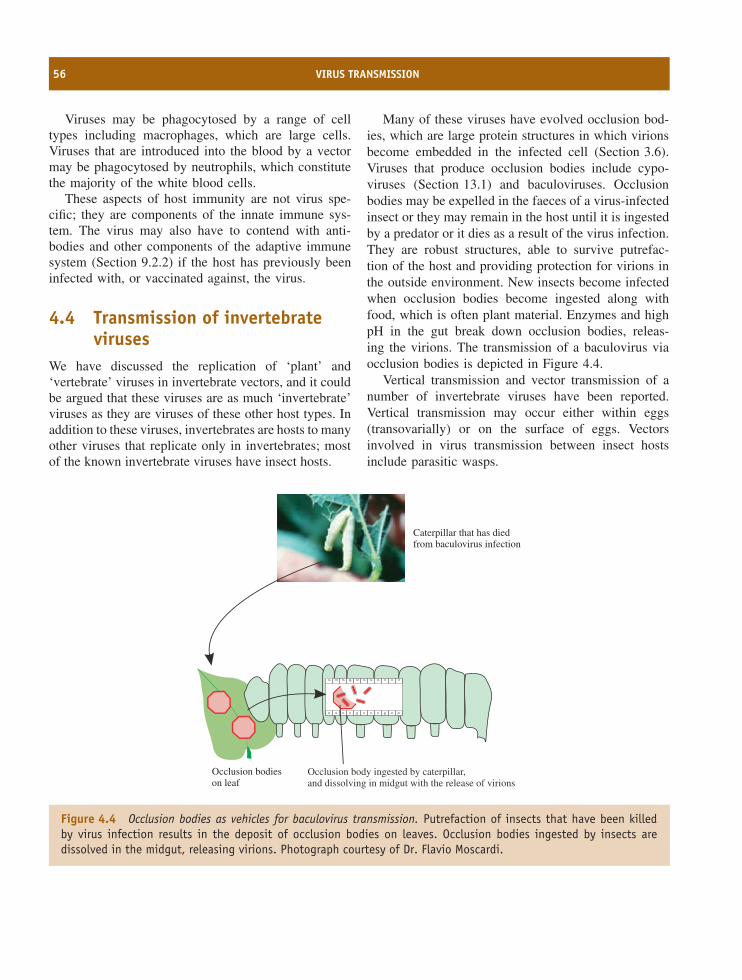
56 VIRUS TRANSMISSION
Viruses may be phagocytosed by a range of celltypes including macrophages, which are large cells.Viruses that are introduced into the blood by a vectormay be phagocytosed by neutrophils, which constitutethe majority of the white blood cells.
These aspects of host immunity are not virus spe-cific; they are components of the innate immune sys-tem. The virus may also have to contend with anti-bodies and other components of the adaptive immunesystem (Section 9.2.2) if the host has previously beeninfected with, or vaccinated against, the virus.
4.4 Transmission of invertebrateviruses
We have discussed the replication of ‘plant’ and‘vertebrate’ viruses in invertebrate vectors, and it couldbe argued that these viruses are as much ‘invertebrate’viruses as they are viruses of these other host types. Inaddition to these viruses, invertebrates are hosts to manyother viruses that replicate only in invertebrates; mostof the known invertebrate viruses have insect hosts.
Many of these viruses have evolved occlusion bod-ies, which are large protein structures in which virionsbecome embedded in the infected cell (Section 3.6).Viruses that produce occlusion bodies include cypo-viruses (Section 13.1) and baculoviruses. Occlusionbodies may be expelled in the faeces of a virus-infectedinsect or they may remain in the host until it is ingestedby a predator or it dies as a result of the virus infection.They are robust structures, able to survive putrefac-tion of the host and providing protection for virions inthe outside environment. New insects become infectedwhen occlusion bodies become ingested along withfood, which is often plant material. Enzymes and highpH in the gut break down occlusion bodies, releas-ing the virions. The transmission of a baculovirus viaocclusion bodies is depicted in Figure 4.4.
Vertical transmission and vector transmission of anumber of invertebrate viruses have been reported.Vertical transmission may occur either within eggs(transovarially) or on the surface of eggs. Vectorsinvolved in virus transmission between insect hostsinclude parasitic wasps.
Caterpillar that has diedfrom baculovirus infection
Occlusion bodieson leaf
Occlusion body ingested by caterpillar,and dissolving in midgut with the release of virions
Figure 4.4 Occlusion bodies as vehicles for baculovirus transmission. Putrefaction of insects that have been killedby virus infection results in the deposit of occlusion bodies on leaves. Occlusion bodies ingested by insects aredissolved in the midgut, releasing virions. Photograph courtesy of Dr. Flavio Moscardi.
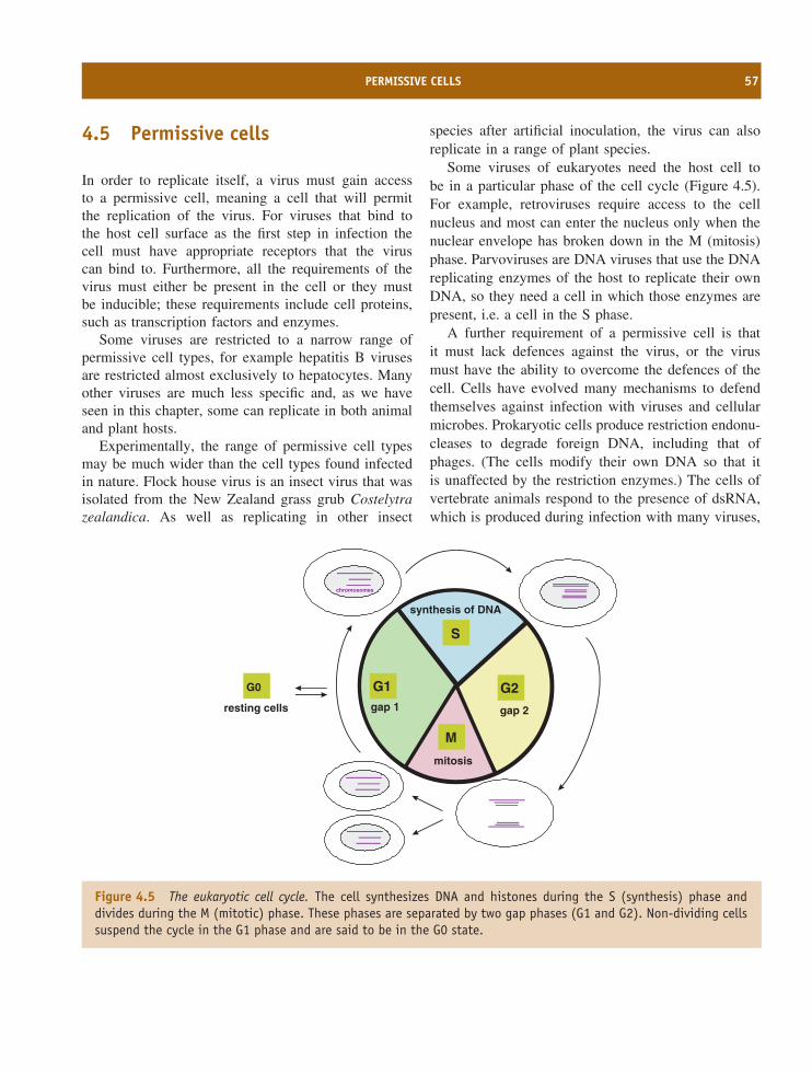
PERMISSIVE CELLS 57
4.5 Permissive cells
In order to replicate itself, a virus must gain accessto a permissive cell, meaning a cell that will permitthe replication of the virus. For viruses that bind tothe host cell surface as the first step in infection thecell must have appropriate receptors that the viruscan bind to. Furthermore, all the requirements of thevirus must either be present in the cell or they mustbe inducible; these requirements include cell proteins,such as transcription factors and enzymes.
Some viruses are restricted to a narrow range ofpermissive cell types, for example hepatitis B virusesare restricted almost exclusively to hepatocytes. Manyother viruses are much less specific and, as we haveseen in this chapter, some can replicate in both animaland plant hosts.
Experimentally, the range of permissive cell typesmay be much wider than the cell types found infectedin nature. Flock house virus is an insect virus that wasisolated from the New Zealand grass grub Costelytrazealandica. As well as replicating in other insect
species after artificial inoculation, the virus can alsoreplicate in a range of plant species.
Some viruses of eukaryotes need the host cell tobe in a particular phase of the cell cycle (Figure 4.5).For example, retroviruses require access to the cellnucleus and most can enter the nucleus only when thenuclear envelope has broken down in the M (mitosis)phase. Parvoviruses are DNA viruses that use the DNAreplicating enzymes of the host to replicate their ownDNA, so they need a cell in which those enzymes arepresent, i.e. a cell in the S phase.
A further requirement of a permissive cell is thatit must lack defences against the virus, or the virusmust have the ability to overcome the defences of thecell. Cells have evolved many mechanisms to defendthemselves against infection with viruses and cellularmicrobes. Prokaryotic cells produce restriction endonu-cleases to degrade foreign DNA, including that ofphages. (The cells modify their own DNA so that itis unaffected by the restriction enzymes.) The cells ofvertebrate animals respond to the presence of dsRNA,which is produced during infection with many viruses,
chromosomes
resting cells
G0
S
synthesis of DNA
G2
gap 2
mitosis
M
G1gap 1
Figure 4.5 The eukaryotic cell cycle. The cell synthesizes DNA and histones during the S (synthesis) phase anddivides during the M (mitotic) phase. These phases are separated by two gap phases (G1 and G2). Non-dividing cellssuspend the cycle in the G1 phase and are said to be in the G0 state.
58 VIRUS TRANSMISSION
by synthesizing cytokines, including interferons, inter-leukins and tumour necrosis factor. These proteins cantrigger a range of anti-viral defences.
In order to successfully infect a cell a virus mustdeliver its genome into this cell, the genome must sur-vive intact and all attempts by the host to terminatevirus replication must be thwarted. In the next fourchapters we shall consider the various stages that mustbe completed before a virus has successfully repli-cated itself.
Learning outcomesBy the end of this chapter you should be able to
• describe the modes of transmission of plantviruses and animal viruses;
• evaluate the roles of vectors in virus transmission;
• discuss the immune mechanisms encountered byan animal virus when it enters the body of a newhost.
Sources of further information
Books
Gillespie S. H., Smith G. L. and Osbourn A. (2004) Mi-crobe–Vector Interactions in Vector-Borne Diseases, 63rdSymposium of the Society for General Microbiology,Cambridge University Press
Taylor C. E. and Brown D. J. F. (1997) Nematode Vectors ofPlant Viruses, CAB
Journals
Bergelson J. M. (2003) Virus interactions with mucosal sur-faces: alternative receptors, alternative pathways CurrentOpinion in Microbiology, 6, 386–391
Pereira L. et al. (2005) Insights into viral transmission atthe uterine–placental interface Trends in Microbiology, 13,164–174
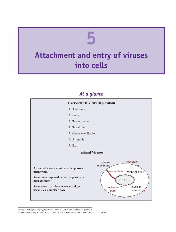
5Attachment and entry of viruses
into cells
At a glance
Animal Viruses
All animal viruses must cross the plasmamembrane.
Some must cross the nuclear envelope,usually via a nuclear pore.
Some are transported in the cytoplasm via microtubules.
microtubule
nuclearpore
CYTOPLASM
NUCLEUS
plasmamembrane
nuclearenvelope
receptors
Overview Of Virus Replication
1. Attachment
2. Entry
3. Transcription
4. Translation
5. Genome replication
6. Assembly
7. Exit
Virology: Principles and Applications John B. Carter and Venetia A. Saunders 2007 John Wiley & Sons, Ltd ISBNs: 978-0-470-02386-0 (HB); 978-0-470-02387-7 (PB)
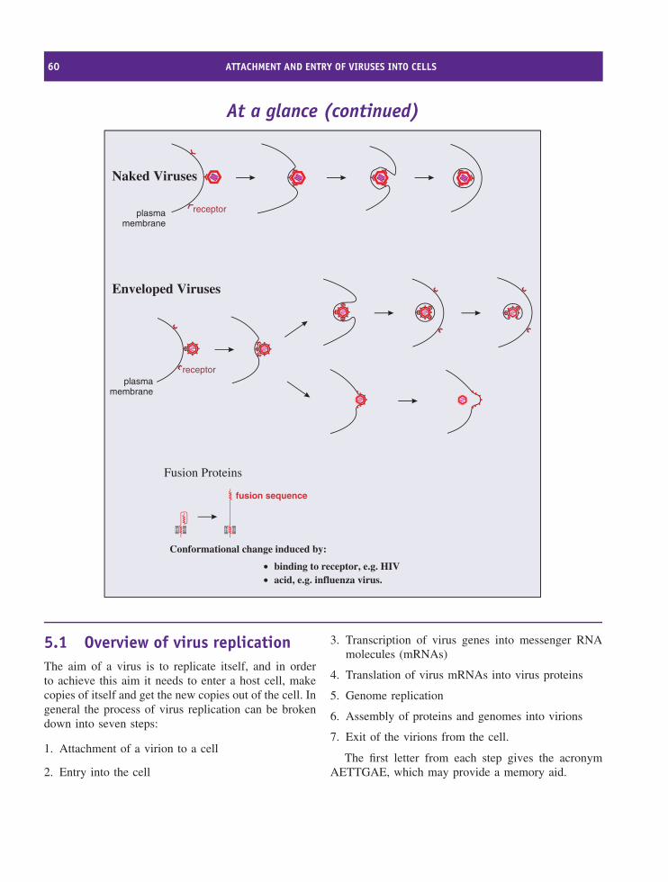
60 ATTACHMENT AND ENTRY OF VIRUSES INTO CELLS
At a glance (continued)
Naked Viruses
plasmamembrane
receptor
plasmamembrane
receptor
Enveloped Viruses
••
binding to receptor, e.g. HIVacid, e.g. influenza virus.
Fusion Proteins
fusion sequence
Conformational change induced by:
5.1 Overview of virus replicationThe aim of a virus is to replicate itself, and in orderto achieve this aim it needs to enter a host cell, makecopies of itself and get the new copies out of the cell. Ingeneral the process of virus replication can be brokendown into seven steps:
1. Attachment of a virion to a cell
2. Entry into the cell
3. Transcription of virus genes into messenger RNAmolecules (mRNAs)
4. Translation of virus mRNAs into virus proteins
5. Genome replication
6. Assembly of proteins and genomes into virions
7. Exit of the virions from the cell.
The first letter from each step gives the acronymAETTGAE, which may provide a memory aid.
ANIMAL VIRUSES 61
When trying to understand the modes of replicationof different types of virus these steps provide a usefultemplate. It is, however, a general template, becausenot all of the seven steps are relevant to all viruses, thesteps do not always occur in the same order and someviruses have an additional step! In the later stages ofreplication several steps occur concurrently. For manyviruses, transcription, translation, genome replication,virion assembly and exit can all be in progress at thesame time.
In the dialogue between the cell and the intruder,the cell provides critical cues that allow the virusto undergo molecular transformations that leadto successful internalization, intracellular transport,and uncoating.
Smith and Helenius (2004)
In this chapter we discuss the first two steps of thisgeneralized replication cycle: attachment and entry intothe host cell. We concentrate mainly on the mechanismsused by animal viruses to gain entry into host cells,but bacteriophages are also considered. The followingthree chapters deal with the remaining five steps of thegeneralized replication cycle.
The first step, attachment of a virion to a cell, appliesto viruses that infect animal and bacterial hosts. Beforethese viruses can cross the bounding membrane orwall of the host cell they must first bind to specificmolecules on the cell surface. Most plant viruses, onthe other hand, are delivered directly into a cell by avector (Section 4.2).
5.2 Animal viruses5.2.1 Cell receptors and co-receptorsA virion attaches via one or more of its surface proteinsto specific molecules on the surface of a host cell.These cellular molecules are known as receptors andthe recognition of a receptor by a virion is highlyspecific, like a key fitting in its lock. It has been foundthat some viruses need to bind to a second type ofcell surface molecule (a co-receptor) in order to infecta cell. In at least some cases binding to a receptor
causes a conformational change in the virus proteinthat enables it to bind to the co-receptor.
Receptors and co-receptors are cell surface mole-cules, usually glycoproteins, with a wide range offunctions that include
• acting as receptors for chemokines and growthfactors;
• mediating cell-to-cell contact and adhesion.
Some examples of cell receptors are given inTable 5.1.
A number of receptors are glycoproteins thatare folded into domains similar to those found inimmunoglobulin molecules. Three examples are shownin Figure 5.1; in each molecule the binding site for thevirus is located in the outermost domain.
Many of the cell surface molecules used by virusesas receptors are in regions of the plasma membrane thatare coated on the inner surface with one of the proteinsclathrin or caveolin.
5.2.1.a Evidence that a cell surface moleculeis a virus receptor
A search for the receptor for a particular virus mightbegin with the production of a panel of monoclonalantibodies against cell surface proteins. If one of theantibodies blocks virus binding and infectivity then thisis strong evidence that the corresponding antigen is thereceptor.
Further evidence that a molecule is a virus receptormight come from experiments that show one or moreof the following.
• Soluble derivatives of the molecule block virusbinding/infectivity.
• The normal ligand for the molecule blocks virusbinding/infectivity.
• Introduction of the gene encoding the molecule intovirus-resistant cells, and expression of that gene,makes those cells susceptible to infection.
5.2.2 Virus attachment sites
Each virion has multiple sites that can bind to receptors,and each site is made up of regions of one or more
62 ATTACHMENT AND ENTRY OF VIRUSES INTO CELLS
Table 5.1 Examples of cell receptors, virus proteins involved in attachment, and (for enveloped viruses) fusionproteins
Virus Cell receptor Virus protein(s) involved in
attachment toreceptor
fusion
Naked viruses
Approx. 90% of humanrhinoviruses
Intercellular adhesionmolecule-1 (ICAM-1)
VP1 + VP3
Approx. 10% of humanrhinoviruses
Low-density lipoproteinreceptors
VP1
Poliovirus CD155 VP1
Enveloped viruses
Murine leukaemia viruses Mouse cationic amino acidtransporter
SU (surfaceglycoprotein)
TM (transmembraneglycoprotein)
HIV-1 CD4 gp120 gp41
Influenza viruses A & B Sialic-acid-containingglycoproteins
Haemagglutinin Haemagglutinin
Measles virus Signalling lymphocyteactivation molecule(CD150)
Haemagglutinin Fusion
protein molecules. Some examples of proteins that bearvirus attachment sites are given in Table 5.1.
The virus attachment sites of naked viruses areon the capsid surface, sometimes within depressions(e.g. poliovirus) and sometimes on ridges (e.g. footand mouth disease virus). Poliovirus and foot andmouth disease virus are picornaviruses, and it has beendemonstrated for picornaviruses in general that bindingto receptors induces major structural changes in thevirion (Section 14.4.1).
The virus attachment sites of some naked virusesare on specialized structures, such as the fibresand knobs of adenoviruses (Section 3.4.2.c) and thespikes of rotaviruses (Chapter 13), while the virusattachment sites of enveloped viruses are on the surfaceglycoproteins.
Some virion surface proteins that bear the virusattachment sites are able to bind strongly to redblood cells of various species and cause them toclump, a phenomenon known as haemagglutination.The proteins responsible for haemagglutination arecalled haemagglutinins. Examples of viruses that canhaemagglutinate are influenza viruses (Figure 3.22)and measles virus.
5.2.3 Attachment of virions to receptors
The forces that bind a virus attachment site to areceptor include hydrogen bonds, ionic attractions andvan der Waals forces. Sugar moieties, when present onthe receptor and/or on the virus attachment site, arecommonly involved in the forces that bind the two.
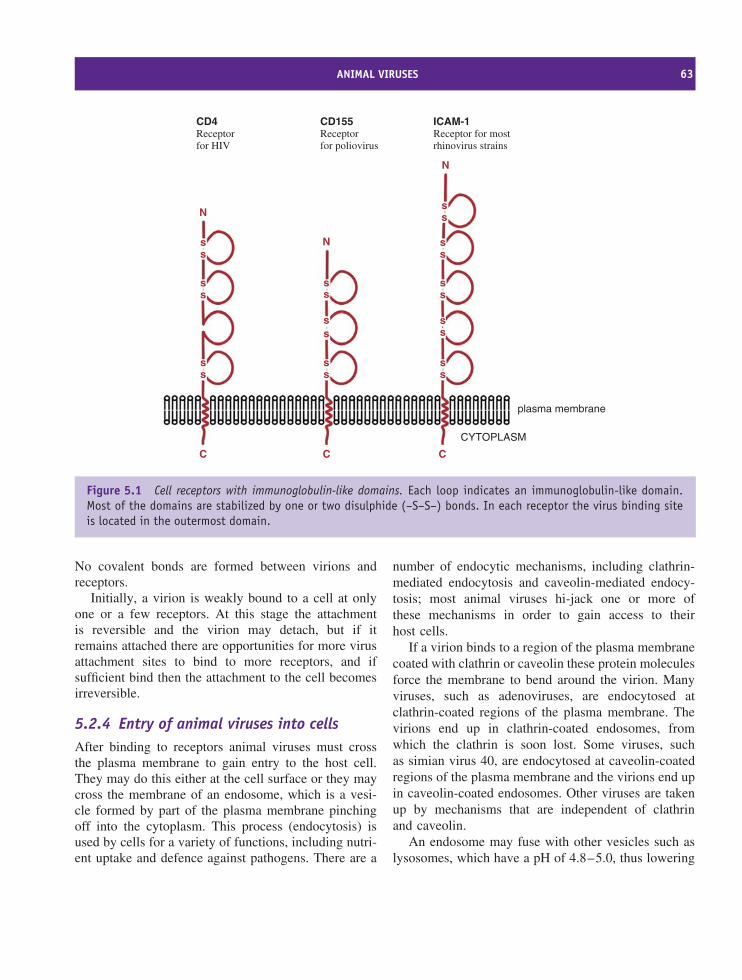
ANIMAL VIRUSES 63
ss
ss
ss
ss
ss
ss
s
plasma membrane
CYTOPLASM
s
N
C C C
N
N
ss
ss
ss
ss
CD4Receptorfor HIV
CD155Receptorfor poliovirus
ICAM-1Receptor for mostrhinovirus strains
Figure 5.1 Cell receptors with immunoglobulin-like domains. Each loop indicates an immunoglobulin-like domain.Most of the domains are stabilized by one or two disulphide (–S–S–) bonds. In each receptor the virus binding siteis located in the outermost domain.
No covalent bonds are formed between virions andreceptors.
Initially, a virion is weakly bound to a cell at onlyone or a few receptors. At this stage the attachmentis reversible and the virion may detach, but if itremains attached there are opportunities for more virusattachment sites to bind to more receptors, and ifsufficient bind then the attachment to the cell becomesirreversible.
5.2.4 Entry of animal viruses into cellsAfter binding to receptors animal viruses must crossthe plasma membrane to gain entry to the host cell.They may do this either at the cell surface or they maycross the membrane of an endosome, which is a vesi-cle formed by part of the plasma membrane pinchingoff into the cytoplasm. This process (endocytosis) isused by cells for a variety of functions, including nutri-ent uptake and defence against pathogens. There are a
number of endocytic mechanisms, including clathrin-mediated endocytosis and caveolin-mediated endocy-tosis; most animal viruses hi-jack one or more ofthese mechanisms in order to gain access to theirhost cells.
If a virion binds to a region of the plasma membranecoated with clathrin or caveolin these protein moleculesforce the membrane to bend around the virion. Manyviruses, such as adenoviruses, are endocytosed atclathrin-coated regions of the plasma membrane. Thevirions end up in clathrin-coated endosomes, fromwhich the clathrin is soon lost. Some viruses, suchas simian virus 40, are endocytosed at caveolin-coatedregions of the plasma membrane and the virions end upin caveolin-coated endosomes. Other viruses are takenup by mechanisms that are independent of clathrinand caveolin.
An endosome may fuse with other vesicles such aslysosomes, which have a pH of 4.8–5.0, thus lowering
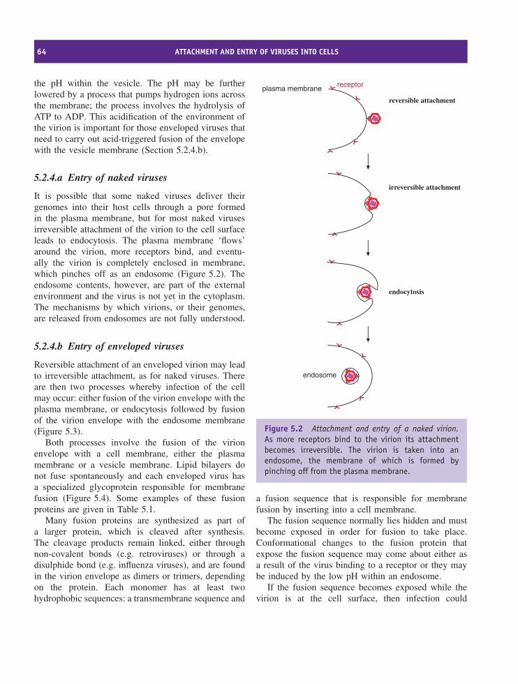
64 ATTACHMENT AND ENTRY OF VIRUSES INTO CELLS
the pH within the vesicle. The pH may be furtherlowered by a process that pumps hydrogen ions acrossthe membrane; the process involves the hydrolysis ofATP to ADP. This acidification of the environment ofthe virion is important for those enveloped viruses thatneed to carry out acid-triggered fusion of the envelopewith the vesicle membrane (Section 5.2.4.b).
5.2.4.a Entry of naked viruses
It is possible that some naked viruses deliver theirgenomes into their host cells through a pore formedin the plasma membrane, but for most naked virusesirreversible attachment of the virion to the cell surfaceleads to endocytosis. The plasma membrane ‘flows’around the virion, more receptors bind, and eventu-ally the virion is completely enclosed in membrane,which pinches off as an endosome (Figure 5.2). Theendosome contents, however, are part of the externalenvironment and the virus is not yet in the cytoplasm.The mechanisms by which virions, or their genomes,are released from endosomes are not fully understood.
5.2.4.b Entry of enveloped viruses
Reversible attachment of an enveloped virion may leadto irreversible attachment, as for naked viruses. Thereare then two processes whereby infection of the cellmay occur: either fusion of the virion envelope with theplasma membrane, or endocytosis followed by fusionof the virion envelope with the endosome membrane(Figure 5.3).
Both processes involve the fusion of the virionenvelope with a cell membrane, either the plasmamembrane or a vesicle membrane. Lipid bilayers donot fuse spontaneously and each enveloped virus hasa specialized glycoprotein responsible for membranefusion (Figure 5.4). Some examples of these fusionproteins are given in Table 5.1.
Many fusion proteins are synthesized as part ofa larger protein, which is cleaved after synthesis.The cleavage products remain linked, either throughnon-covalent bonds (e.g. retroviruses) or through adisulphide bond (e.g. influenza viruses), and are foundin the virion envelope as dimers or trimers, dependingon the protein. Each monomer has at least twohydrophobic sequences: a transmembrane sequence and
endosome
plasma membrane
reversible attachment
irreversible attachment
endocytosis
receptor
Figure 5.2 Attachment and entry of a naked virion.As more receptors bind to the virion its attachmentbecomes irreversible. The virion is taken into anendosome, the membrane of which is formed bypinching off from the plasma membrane.
a fusion sequence that is responsible for membranefusion by inserting into a cell membrane.
The fusion sequence normally lies hidden and mustbecome exposed in order for fusion to take place.Conformational changes to the fusion protein thatexpose the fusion sequence may come about either asa result of the virus binding to a receptor or they maybe induced by the low pH within an endosome.
If the fusion sequence becomes exposed while thevirion is at the cell surface, then infection could
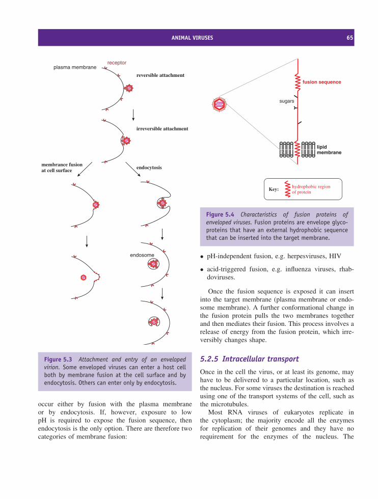
ANIMAL VIRUSES 65
endosome
plasma membrane
reversible attachment
irreversible attachment
endocytosismembrance fusionat cell surface
receptor
Figure 5.3 Attachment and entry of an envelopedvirion. Some enveloped viruses can enter a host cellboth by membrane fusion at the cell surface and byendocytosis. Others can enter only by endocytosis.
occur either by fusion with the plasma membraneor by endocytosis. If, however, exposure to lowpH is required to expose the fusion sequence, thenendocytosis is the only option. There are therefore twocategories of membrane fusion:
lipidmembrane
fusion sequence
hydrophobic regionof protein
Key:
sugars
Figure 5.4 Characteristics of fusion proteins ofenveloped viruses. Fusion proteins are envelope glyco-proteins that have an external hydrophobic sequencethat can be inserted into the target membrane.
• pH-independent fusion, e.g. herpesviruses, HIV
• acid-triggered fusion, e.g. influenza viruses, rhab-doviruses.
Once the fusion sequence is exposed it can insertinto the target membrane (plasma membrane or endo-some membrane). A further conformational change inthe fusion protein pulls the two membranes togetherand then mediates their fusion. This process involves arelease of energy from the fusion protein, which irre-versibly changes shape.
5.2.5 Intracellular transport
Once in the cell the virus, or at least its genome, mayhave to be delivered to a particular location, such asthe nucleus. For some viruses the destination is reachedusing one of the transport systems of the cell, such asthe microtubules.
Most RNA viruses of eukaryotes replicate inthe cytoplasm; the majority encode all the enzymesfor replication of their genomes and they have norequirement for the enzymes of the nucleus. The
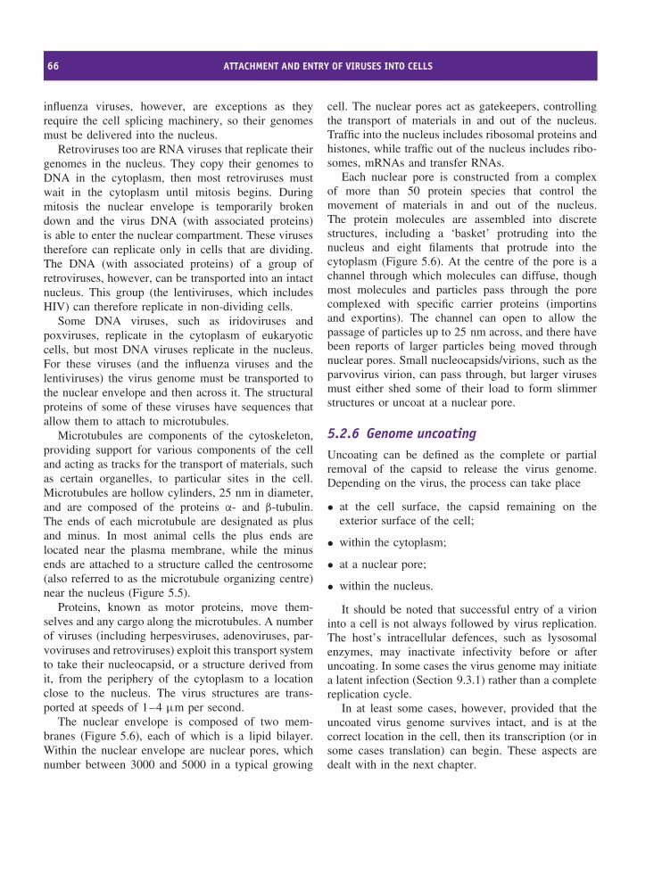
66 ATTACHMENT AND ENTRY OF VIRUSES INTO CELLS
influenza viruses, however, are exceptions as theyrequire the cell splicing machinery, so their genomesmust be delivered into the nucleus.
Retroviruses too are RNA viruses that replicate theirgenomes in the nucleus. They copy their genomes toDNA in the cytoplasm, then most retroviruses mustwait in the cytoplasm until mitosis begins. Duringmitosis the nuclear envelope is temporarily brokendown and the virus DNA (with associated proteins)is able to enter the nuclear compartment. These virusestherefore can replicate only in cells that are dividing.The DNA (with associated proteins) of a group ofretroviruses, however, can be transported into an intactnucleus. This group (the lentiviruses, which includesHIV) can therefore replicate in non-dividing cells.
Some DNA viruses, such as iridoviruses andpoxviruses, replicate in the cytoplasm of eukaryoticcells, but most DNA viruses replicate in the nucleus.For these viruses (and the influenza viruses and thelentiviruses) the virus genome must be transported tothe nuclear envelope and then across it. The structuralproteins of some of these viruses have sequences thatallow them to attach to microtubules.
Microtubules are components of the cytoskeleton,providing support for various components of the celland acting as tracks for the transport of materials, suchas certain organelles, to particular sites in the cell.Microtubules are hollow cylinders, 25 nm in diameter,and are composed of the proteins α- and β-tubulin.The ends of each microtubule are designated as plusand minus. In most animal cells the plus ends arelocated near the plasma membrane, while the minusends are attached to a structure called the centrosome(also referred to as the microtubule organizing centre)near the nucleus (Figure 5.5).
Proteins, known as motor proteins, move them-selves and any cargo along the microtubules. A numberof viruses (including herpesviruses, adenoviruses, par-voviruses and retroviruses) exploit this transport systemto take their nucleocapsid, or a structure derived fromit, from the periphery of the cytoplasm to a locationclose to the nucleus. The virus structures are trans-ported at speeds of 1–4 µm per second.
The nuclear envelope is composed of two mem-branes (Figure 5.6), each of which is a lipid bilayer.Within the nuclear envelope are nuclear pores, whichnumber between 3000 and 5000 in a typical growing
cell. The nuclear pores act as gatekeepers, controllingthe transport of materials in and out of the nucleus.Traffic into the nucleus includes ribosomal proteins andhistones, while traffic out of the nucleus includes ribo-somes, mRNAs and transfer RNAs.
Each nuclear pore is constructed from a complexof more than 50 protein species that control themovement of materials in and out of the nucleus.The protein molecules are assembled into discretestructures, including a ‘basket’ protruding into thenucleus and eight filaments that protrude into thecytoplasm (Figure 5.6). At the centre of the pore is achannel through which molecules can diffuse, thoughmost molecules and particles pass through the porecomplexed with specific carrier proteins (importinsand exportins). The channel can open to allow thepassage of particles up to 25 nm across, and there havebeen reports of larger particles being moved throughnuclear pores. Small nucleocapsids/virions, such as theparvovirus virion, can pass through, but larger virusesmust either shed some of their load to form slimmerstructures or uncoat at a nuclear pore.
5.2.6 Genome uncoatingUncoating can be defined as the complete or partialremoval of the capsid to release the virus genome.Depending on the virus, the process can take place
• at the cell surface, the capsid remaining on theexterior surface of the cell;
• within the cytoplasm;
• at a nuclear pore;
• within the nucleus.
It should be noted that successful entry of a virioninto a cell is not always followed by virus replication.The host’s intracellular defences, such as lysosomalenzymes, may inactivate infectivity before or afteruncoating. In some cases the virus genome may initiatea latent infection (Section 9.3.1) rather than a completereplication cycle.
In at least some cases, however, provided that theuncoated virus genome survives intact, and is at thecorrect location in the cell, then its transcription (or insome cases translation) can begin. These aspects aredealt with in the next chapter.
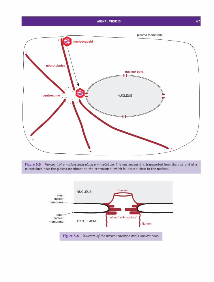
ANIMAL VIRUSES 67
NUCLEUS
microtubules
centrosome
nucleocapsid
++
+
+
+
–––
– –
nuclear pore
plasma membrane
Figure 5.5 Transport of a nucleocapsid along a microtubule. The nucleocapsid is transported from the plus end of amicrotubule near the plasma membrane to the centrosome, which is located close to the nucleus.
CYTOPLASM
NUCLEUSinner
nuclearmembrane
outernuclear
membrane
‘basket’
filament
‘wheel’ with ‘spokes’
Figure 5.6 Structure of the nuclear envelope and a nuclear pore.
68 ATTACHMENT AND ENTRY OF VIRUSES INTO CELLS
5.3 BacteriophagesLike animal viruses, phages bind specifically to cellsurface molecules that function as receptors and co-receptors. For many phages these molecules are on thesurface of the host cell wall, but for some phages theyare on the surface of other structures (pili, flagella orcapsules). Many of the virus attachment sites are onparticular virion structures, such as the tail fibres ofphage T4. As with animal viruses, the attachment ofa phage to its host is initially reversible, and becomesirreversible after attachment to further receptors and/orco-receptors.
Infection of a cell with a phage involves entry intothe cell of the virus genome and perhaps a few associ-ated proteins, the capsid and any associated appendagesremaining at the cell surface. This is in contrast to mostanimal and plant viruses, where the entire virion, or atleast the nucleocapsid, enters the host cell. Unless thehost is a mycoplasma (mycoplasmas do not have cellwalls), delivery of a phage genome into a cell requirespenetration of a cell wall and perhaps also a slime layeror capsule; the virions of many phages carry enzymes,such as lysozymes, to aid this process.
The attachment and entry of a number of phages arediscussed in Chapter 19.
Learning outcomesBy the end of this chapter you should be able to
• outline a generalized scheme of virus replicationinvolving seven steps;
• describe how animal viruses attach to and entertheir host cells;
• differentiate between the entry mechanisms ofnaked and enveloped animal viruses;
• describe the roles of cell components in thedelivery of some viral genomes to the nucleus;
• outline the infection mechanisms of phages.
Sources of further informationBooksMarsh M., editor (2005) Membrane Trafficking in Viral
Replication, SpringerPoranen M. et al. (2002) Common principles in viral entry.
Annual Review of Microbiology, 65, 521–538Young J. A. T. (2001) Virus entry and uncoating, Chapter 4
in Knipe D. M. and Howley P. M., editors-in-chief, FieldsVirology, 4th edition, Lippincott, Williams and Wilkins
JournalsDohner K., Nagel C.-H. and Sodeik B. (2005) Viral stop-
and-go along microtubules: taking a ride with dynein andkinesins Trends in Microbiology, 13, 320–327
Kemp P., Garcia L. R. and Molineux I. J. (2005) Changesin bacteriophage T7 virion structure at the initiation ofinfection Virology, 340, 307–317
Kielian M. and Rey F. A. (2006) Virus membrane-fusionproteins: more than one way to make a hairpin NatureReviews Microbiology, 4, 67–76
Molineux I. J. (2006) Fifty-three years since Hershey andChase; much ado about pressure but which pressure is it?Virology, 344, 221–229
Pelkmans L. (2005) Viruses as probes for systems analysisof cellular signalling, cytoskeleton reorganization andendocytosis Current Opinion in Microbiology, 8, 331–337
Smith A. E. and Helenius A. (2004) How viruses enteranimal cells Science, 304, 237–242
Whittaker G. R. (2003) Virus nuclear import Advanced DrugDelivery Reviews, 55, 733–747
Wu E. and Nemerow G. R. (2004) Virus yoga: the roleof flexibility in virus host cell recognition Trends inMicrobiology, 12, 162–169
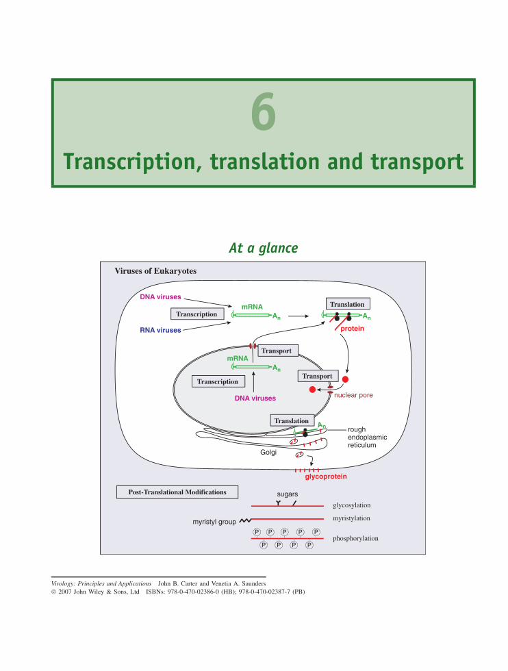
6Transcription, translation and transport
At a glance
An
mRNA
roughendoplasmicreticulum
Golgi
DNA viruses
RNA viruses
DNA viruses
An
P
P P P P
P P P P
sugars
myristyl group
glycosylation
myristylation
phosphorylation
glycoprotein
Viruses of Eukaryotes
Post-Translational Modifications
Translation
Translation
mRNATransport
An
An
protein
Transport
nuclear pore
Transcription
Transcription
Virology: Principles and Applications John B. Carter and Venetia A. Saunders 2007 John Wiley & Sons, Ltd ISBNs: 978-0-470-02386-0 (HB); 978-0-470-02387-7 (PB)
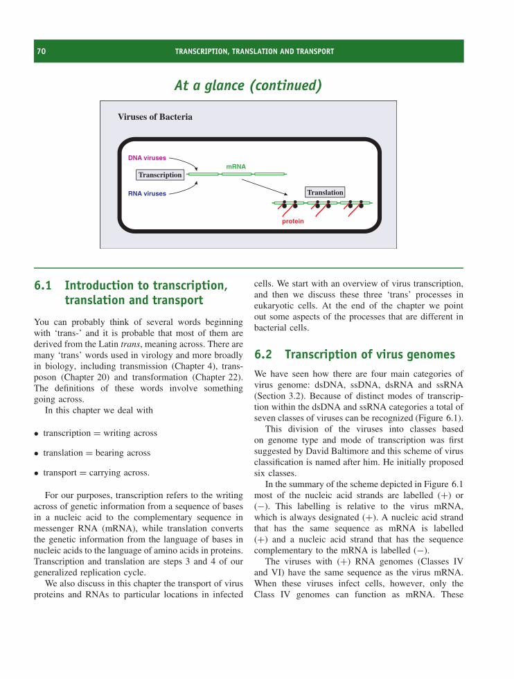
70 TRANSCRIPTION, TRANSLATION AND TRANSPORT
At a glance (continued)
protein
mRNADNA viruses
RNA viruses Translation
Viruses of Bacteria
Transcription
6.1 Introduction to transcription,translation and transport
You can probably think of several words beginningwith ‘trans-’ and it is probable that most of them arederived from the Latin trans, meaning across. There aremany ‘trans’ words used in virology and more broadlyin biology, including transmission (Chapter 4), trans-poson (Chapter 20) and transformation (Chapter 22).The definitions of these words involve somethinggoing across.
In this chapter we deal with
• transcription = writing across
• translation = bearing across
• transport = carrying across.
For our purposes, transcription refers to the writingacross of genetic information from a sequence of basesin a nucleic acid to the complementary sequence inmessenger RNA (mRNA), while translation convertsthe genetic information from the language of bases innucleic acids to the language of amino acids in proteins.Transcription and translation are steps 3 and 4 of ourgeneralized replication cycle.
We also discuss in this chapter the transport of virusproteins and RNAs to particular locations in infected
cells. We start with an overview of virus transcription,and then we discuss these three ‘trans’ processes ineukaryotic cells. At the end of the chapter we pointout some aspects of the processes that are different inbacterial cells.
6.2 Transcription of virus genomesWe have seen how there are four main categories ofvirus genome: dsDNA, ssDNA, dsRNA and ssRNA(Section 3.2). Because of distinct modes of transcrip-tion within the dsDNA and ssRNA categories a total ofseven classes of viruses can be recognized (Figure 6.1).
This division of the viruses into classes basedon genome type and mode of transcription was firstsuggested by David Baltimore and this scheme of virusclassification is named after him. He initially proposedsix classes.
In the summary of the scheme depicted in Figure 6.1most of the nucleic acid strands are labelled (+) or(−). This labelling is relative to the virus mRNA,which is always designated (+). A nucleic acid strandthat has the same sequence as mRNA is labelled(+) and a nucleic acid strand that has the sequencecomplementary to the mRNA is labelled (−).
The viruses with (+) RNA genomes (Classes IVand VI) have the same sequence as the virus mRNA.When these viruses infect cells, however, only theClass IV genomes can function as mRNA. These
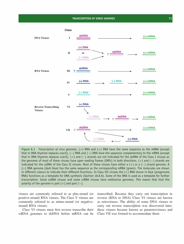
TRANSCRIPTION OF VIRUS GENOMES 71
( ) RNA– (+) mRNAV
dsDNA (+) mRNA
Class
IDNA Viruses
dsRNA (+) mRNAIII +–RNA Viruses
dsDNA (+) mRNAVII
(+) RNA( ) DNA–
+–
( ) DNA–
or dsDNA (+) mRNA
(+) DNA
II +–
( ) RNA–(+) RNA (+) mRNAIV
( ) DNA– (+) mRNA
(+) RNAVI
dsDNA+–
Reverse-TranscribingViruses
Figure 6.1 Transcription of virus genomes. (+) RNA and (+) DNA have the same sequence as the mRNA (exceptthat in DNA thymine replaces uracil). (−) RNA and (−) DNA have the sequence complementary to the mRNA (exceptthat in DNA thymine replaces uracil). (+) and (−) strands are not indicated for the dsDNA of the Class I viruses asthe genomes of most of these viruses have open reading frames (ORFs) in both directions. (+) and (−) strands areindicated for the ssDNA of the Class II viruses. Most of these viruses have either a (+) or a (−) strand genome. A(+) RNA genome (dark blue) has the same sequence as the corresponding mRNA (green). The molecules are shownin different colours to indicate their different functions. In Class VII viruses the (+) RNA shown in blue (pregenomeRNA) functions as a template for DNA synthesis (Section 18.8.6). Some of the DNA is used as a template for furthertranscription. Some ssDNA viruses and some ssRNA viruses have ambisense genomes. This means that that thepolarity of the genome is part (+) and part (−).
viruses are commonly referred to as plus-strand (orpositive-strand) RNA viruses. The Class V viruses arecommonly referred to as minus-strand (or negative-strand) RNA viruses.
Class VI viruses must first reverse transcribe theirssRNA genomes to dsDNA before mRNA can be
transcribed. Because they carry out transcription inreverse (RNA to DNA) Class VI viruses are knownas retroviruses. The ability of some DNA viruses tocarry out reverse transcription was discovered later;these viruses became known as pararetroviruses andClass VII was formed to accommodate them.
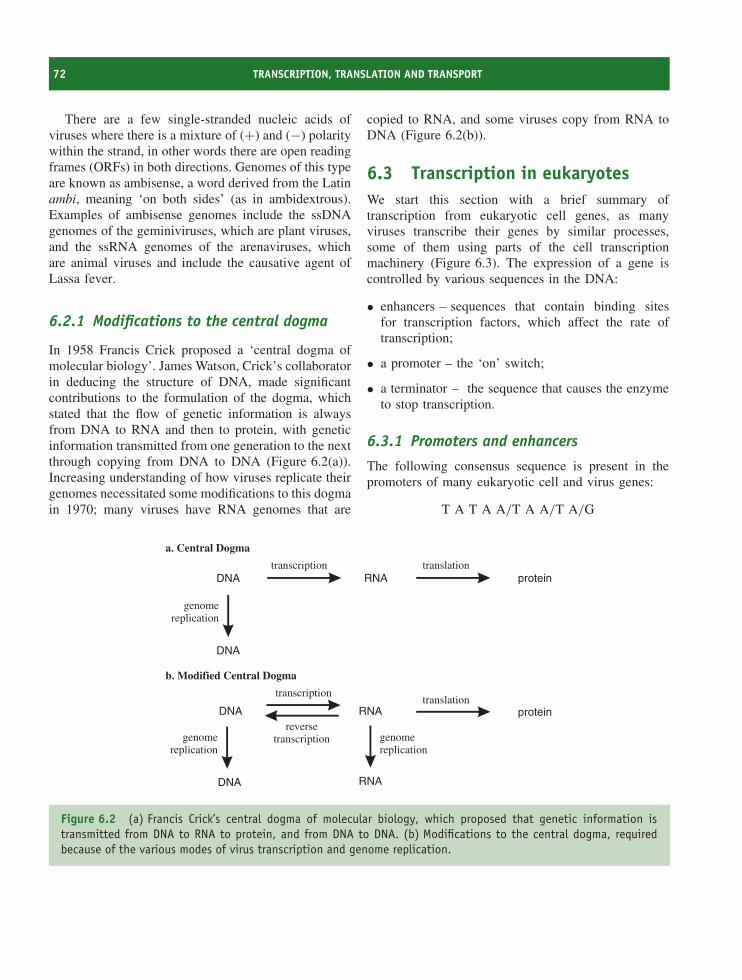
72 TRANSCRIPTION, TRANSLATION AND TRANSPORT
There are a few single-stranded nucleic acids ofviruses where there is a mixture of (+) and (−) polaritywithin the strand, in other words there are open readingframes (ORFs) in both directions. Genomes of this typeare known as ambisense, a word derived from the Latinambi, meaning ‘on both sides’ (as in ambidextrous).Examples of ambisense genomes include the ssDNAgenomes of the geminiviruses, which are plant viruses,and the ssRNA genomes of the arenaviruses, whichare animal viruses and include the causative agent ofLassa fever.
6.2.1 Modifications to the central dogma
In 1958 Francis Crick proposed a ‘central dogma ofmolecular biology’. James Watson, Crick’s collaboratorin deducing the structure of DNA, made significantcontributions to the formulation of the dogma, whichstated that the flow of genetic information is alwaysfrom DNA to RNA and then to protein, with geneticinformation transmitted from one generation to the nextthrough copying from DNA to DNA (Figure 6.2(a)).Increasing understanding of how viruses replicate theirgenomes necessitated some modifications to this dogmain 1970; many viruses have RNA genomes that are
copied to RNA, and some viruses copy from RNA toDNA (Figure 6.2(b)).
6.3 Transcription in eukaryotesWe start this section with a brief summary oftranscription from eukaryotic cell genes, as manyviruses transcribe their genes by similar processes,some of them using parts of the cell transcriptionmachinery (Figure 6.3). The expression of a gene iscontrolled by various sequences in the DNA:
• enhancers – sequences that contain binding sitesfor transcription factors, which affect the rate oftranscription;
• a promoter – the ‘on’ switch;
• a terminator – the sequence that causes the enzymeto stop transcription.
6.3.1 Promoters and enhancers
The following consensus sequence is present in thepromoters of many eukaryotic cell and virus genes:
T A T A A/T A A/T A/G
DNA RNA proteintranscription translation
DNA RNA protein
transcriptiontranslation
a. Central Dogma
b. Modified Central Dogma
DNA
genomereplication
genomereplication
genomereplication
DNA RNA
reversetranscription
Figure 6.2 (a) Francis Crick’s central dogma of molecular biology, which proposed that genetic information istransmitted from DNA to RNA to protein, and from DNA to DNA. (b) Modifications to the central dogma, requiredbecause of the various modes of virus transcription and genome replication.
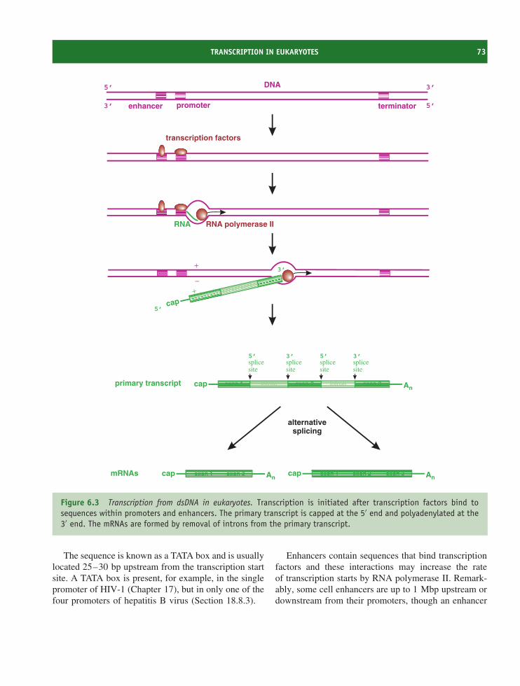
TRANSCRIPTION IN EUKARYOTES 73
DNA
enhancer promoter terminator
An
An
cap
cap Ancap
splicesite
primary transcript
RNA polymerase II
mRNAs
5’
5’ 5’3’ 3’
3’ 5’
3’
transcription factors
RNA
cap
+
−+
5’
3’
splicesite
splicesite
splicesite
alternativesplicing
exon 1 exon 2 exon 3intron intron
exon 3exon 1 exon 1 exon 2 exon 3
Figure 6.3 Transcription from dsDNA in eukaryotes. Transcription is initiated after transcription factors bind tosequences within promoters and enhancers. The primary transcript is capped at the 5′ end and polyadenylated at the3′ end. The mRNAs are formed by removal of introns from the primary transcript.
The sequence is known as a TATA box and is usuallylocated 25–30 bp upstream from the transcription startsite. A TATA box is present, for example, in the singlepromoter of HIV-1 (Chapter 17), but in only one of thefour promoters of hepatitis B virus (Section 18.8.3).
Enhancers contain sequences that bind transcriptionfactors and these interactions may increase the rateof transcription starts by RNA polymerase II. Remark-ably, some cell enhancers are up to 1 Mbp upstream ordownstream from their promoters, though an enhancer
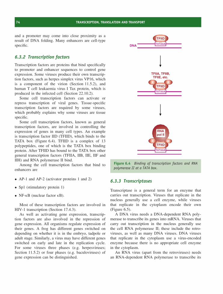
74 TRANSCRIPTION, TRANSLATION AND TRANSPORT
and a promoter may come into close proximity as aresult of DNA folding. Many enhancers are cell-typespecific.
6.3.2 Transcription factors
Transcription factors are proteins that bind specificallyto promoter and enhancer sequences to control geneexpression. Some viruses produce their own transcrip-tion factors, such as herpes simplex virus VP16, whichis a component of the virion (Section 11.5.2), andhuman T cell leukaemia virus I Tax protein, which isproduced in the infected cell (Section 22.10.2).
Some cell transcription factors can activate orrepress transcription of viral genes. Tissue-specifictranscription factors are required by some viruses,which probably explains why some viruses are tissuespecific.
Some cell transcription factors, known as generaltranscription factors, are involved in controlling theexpression of genes in many cell types. An exampleis transcription factor IID (TFIID), which binds to theTATA box (Figure 6.4). TFIID is a complex of 13polypeptides, one of which is the TATA box bindingprotein. After TFIID has bound to the TATA box othergeneral transcription factors (TFIIA, IIB, IIE, IIF andIIH) and RNA polymerase II bind.
Among the cell transcription factors that bind toenhancers are
• AP-1 and AP-2 (activator proteins 1 and 2)
• Sp1 (stimulatory protein 1)
• NF-κB (nuclear factor κB).
Most of these transcription factors are involved inHIV-1 transcription (Section 17.4.3).
As well as activating gene expression, transcrip-tion factors are also involved in the repression ofgene expression. All organisms regulate expression oftheir genes. A frog has different genes switched ondepending on whether it is in the embryo, tadpole oradult stage. Similarly, a virus may have different genesswitched on early and late in the replication cycle.For some viruses three phases (e.g. herpesviruses;Section 11.5.2) or four phases (e.g. baculoviruses) ofgene expression can be distinguished.
DNATATAAA
TFIIA, TFIIB,TFIIE, etc.
TATAAA
TATAAA
TFIID
TFIID
TFIID
RNApol II
Figure 6.4 Binding of transcription factors and RNApolymerase II at a TATA box.
6.3.3 Transcriptases
Transcriptase is a general term for an enzyme thatcarries out transcription. Viruses that replicate in thenucleus generally use a cell enzyme, while virusesthat replicate in the cytoplasm encode their own(Figure 6.5).
A DNA virus needs a DNA-dependent RNA poly-merase to transcribe its genes into mRNA. Viruses thatcarry out transcription in the nucleus generally usethe cell RNA polymerase II; these include the retro-viruses, as well as many DNA viruses. DNA virusesthat replicate in the cytoplasm use a virus-encodedenzyme because there is no appropriate cell enzymein the cytoplasm.
An RNA virus (apart from the retroviruses) needsan RNA-dependent RNA polymerase to transcribe its
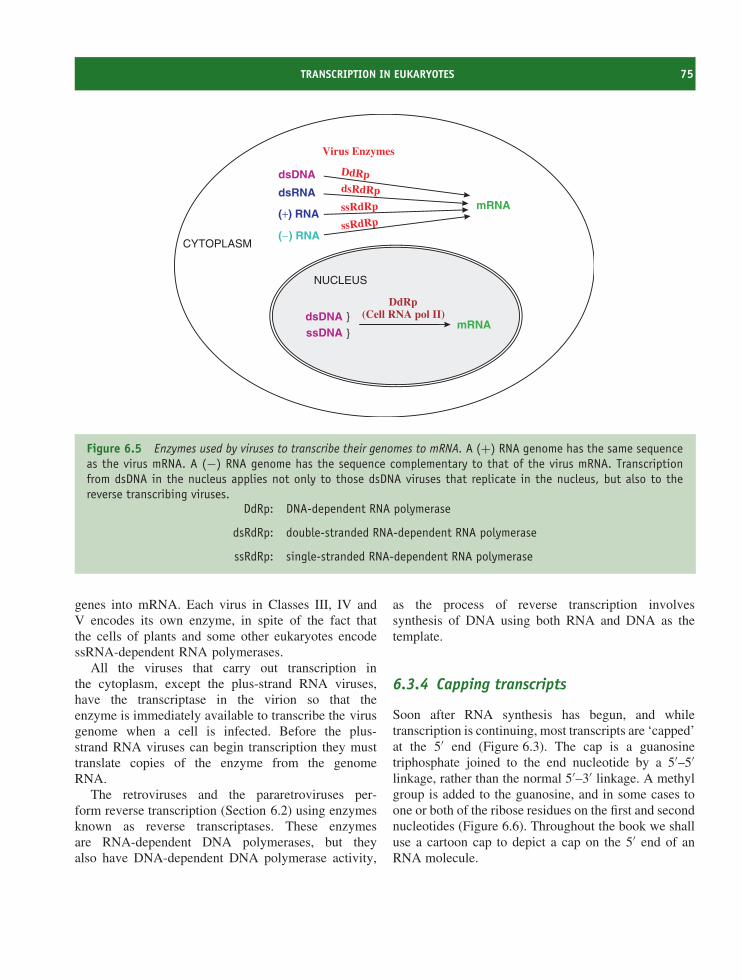
TRANSCRIPTION IN EUKARYOTES 75
mRNA
dsDNA
dsRNA
(+) RNA
( ) RNA−
DdRpdsRdRp
ssRdRp
ssRdRp
dsDNAssDNA
mRNA
DdRp(Cell RNA pol II)
CYTOPLASM
NUCLEUS
Virus Enzymes
}
}
Figure 6.5 Enzymes used by viruses to transcribe their genomes to mRNA. A (+) RNA genome has the same sequenceas the virus mRNA. A (−) RNA genome has the sequence complementary to that of the virus mRNA. Transcriptionfrom dsDNA in the nucleus applies not only to those dsDNA viruses that replicate in the nucleus, but also to thereverse transcribing viruses.
DdRp: DNA-dependent RNA polymerase
dsRdRp: double-stranded RNA-dependent RNA polymerase
ssRdRp: single-stranded RNA-dependent RNA polymerase
genes into mRNA. Each virus in Classes III, IV andV encodes its own enzyme, in spite of the fact thatthe cells of plants and some other eukaryotes encodessRNA-dependent RNA polymerases.
All the viruses that carry out transcription inthe cytoplasm, except the plus-strand RNA viruses,have the transcriptase in the virion so that theenzyme is immediately available to transcribe the virusgenome when a cell is infected. Before the plus-strand RNA viruses can begin transcription they musttranslate copies of the enzyme from the genomeRNA.
The retroviruses and the pararetroviruses per-form reverse transcription (Section 6.2) using enzymesknown as reverse transcriptases. These enzymesare RNA-dependent DNA polymerases, but theyalso have DNA-dependent DNA polymerase activity,
as the process of reverse transcription involvessynthesis of DNA using both RNA and DNA as thetemplate.
6.3.4 Capping transcripts
Soon after RNA synthesis has begun, and whiletranscription is continuing, most transcripts are ‘capped’at the 5′ end (Figure 6.3). The cap is a guanosinetriphosphate joined to the end nucleotide by a 5′–5′linkage, rather than the normal 5′–3′ linkage. A methylgroup is added to the guanosine, and in some cases toone or both of the ribose residues on the first and secondnucleotides (Figure 6.6). Throughout the book we shalluse a cartoon cap to depict a cap on the 5′ end of anRNA molecule.
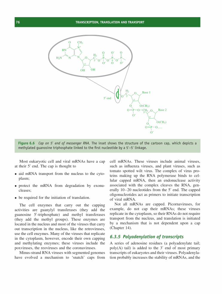
76 TRANSCRIPTION, TRANSLATION AND TRANSPORT
H2N
HN
N
O
N O
OH OH
H2C
OP
O
OP
O−
N+
OP
O
CH3
O− O O−5′3′
O
H2C O Base 1
O
P O
O(CH3)
O
O−
CH2 O Base 2
OPO O......
O−
O(CH3)
5′3′
5′
3′
5′ end
Figure 6.6 Cap on 5′ end of messenger RNA. The inset shows the structure of the cartoon cap, which depicts amethylated guanosine triphosphate linked to the first nucleotide by a 5′–5′ linkage.
Most eukaryotic cell and viral mRNAs have a capat their 5′ end. The cap is thought to
• aid mRNA transport from the nucleus to the cyto-plasm;
• protect the mRNA from degradation by exonu-cleases;
• be required for the initiation of translation.
The cell enzymes that carry out the cappingactivities are guanylyl transferases (they add theguanosine 5′-triphosphate) and methyl transferases(they add the methyl groups). These enzymes arelocated in the nucleus and most of the viruses that carryout transcription in the nucleus, like the retroviruses,use the cell enzymes. Many of the viruses that replicatein the cytoplasm, however, encode their own cappingand methylating enzymes; these viruses include thepoxviruses, the reoviruses and the coronaviruses.
Minus-strand RNA viruses with segmented genomeshave evolved a mechanism to ‘snatch’ caps from
cell mRNAs. These viruses include animal viruses,such as influenza viruses, and plant viruses, such astomato spotted wilt virus. The complex of virus pro-teins making up the RNA polymerase binds to cel-lular capped mRNA, then an endonuclease activityassociated with the complex cleaves the RNA, gen-erally 10–20 nucleotides from the 5′ end. The cappedoligonucleotides act as primers to initiate transcriptionof viral mRNA.
Not all mRNAs are capped. Picornaviruses, forexample, do not cap their mRNAs; these virusesreplicate in the cytoplasm, so their RNAs do not requiretransport from the nucleus, and translation is initiatedby a mechanism that is not dependent upon a cap(Chapter 14).
6.3.5 Polyadenylation of transcriptsA series of adenosine residues (a polyadenylate tail;poly(A) tail) is added to the 3′ end of most primarytranscripts of eukaryotes and their viruses. Polyadenyla-tion probably increases the stability of mRNAs, and the
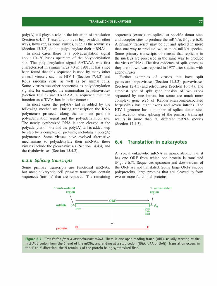
TRANSLATION IN EUKARYOTES 77
poly(A) tail plays a role in the initiation of translation(Section 6.4.1). These functions can be provided in otherways, however, as some viruses, such as the reoviruses(Section 13.3.2), do not polyadenylate their mRNAs.
In most cases there is a polyadenylation signalabout 10–30 bases upstream of the polyadenylationsite. The polyadenylation signal AATAAA was firstcharacterized in simian virus 40 in 1981. It has sincebeen found that this sequence is used by many otheranimal viruses, such as HIV-1 (Section 17.4.3) andRous sarcoma virus, as well as by animal cells.Some viruses use other sequences as polyadenylationsignals; for example, the mammalian hepadnaviruses(Section 18.8.3) use TATAAA, a sequence that canfunction as a TATA box in other contexts!
In most cases the poly(A) tail is added by thefollowing mechanism. During transcription the RNApolymerase proceeds along the template past thepolyadenylation signal and the polyadenylation site.The newly synthesized RNA is then cleaved at thepolyadenylation site and the poly(A) tail is added stepby step by a complex of proteins, including a poly(A)polymerase. Some viruses have evolved alternativemechanisms to polyadenylate their mRNAs; theseviruses include the picornaviruses (Section 14.4.4) andthe rhabdoviruses (Section 15.4.2).
6.3.6 Splicing transcriptsSome primary transcripts are functional mRNAs,but most eukaryotic cell primary transcripts containsequences (introns) that are removed. The remaining
sequences (exons) are spliced at specific donor sitesand acceptor sites to produce the mRNAs (Figure 6.3).A primary transcript may be cut and spliced in morethan one way to produce two or more mRNA species.Some primary transcripts of viruses that replicate inthe nucleus are processed in the same way to producethe virus mRNAs. The first evidence of split genes, asthey are known, was reported in 1977 after studies withadenoviruses.
Further examples of viruses that have splitgenes are herpesviruses (Section 11.5.2), parvoviruses(Section 12.4.3) and retroviruses (Section 16.3.4). Thesimplest type of split gene consists of two exonsseparated by one intron, but some are much morecomplex; gene K15 of Kaposi’s-sarcoma-associatedherpesvirus has eight exons and seven introns. TheHIV-1 genome has a number of splice donor sitesand acceptor sites; splicing of the primary transcriptresults in more than 30 different mRNA species(Section 17.4.3).
6.4 Translation in eukaryotes
A typical eukaryotic mRNA is monocistronic, i.e. ithas one ORF from which one protein is translated(Figure 6.7). Sequences upstream and downstream ofthe ORF are not translated. Some large ORFs encodepolyproteins, large proteins that are cleaved to formtwo or more functional proteins.
An
protein
mRNA
N C
5’untranslatedregion
3’untranslatedregion
ORF
AUG UGAUAAUAG
Figure 6.7 Translation from a monocistronic mRNA. There is one open reading frame (ORF), usually starting at thefirst AUG codon from the 5′ end of the mRNA, and ending at a stop codon (UGA, UAA or UAG). Translation occurs inthe 5′ to 3′ direction, the N terminus of the protein being synthesized first.
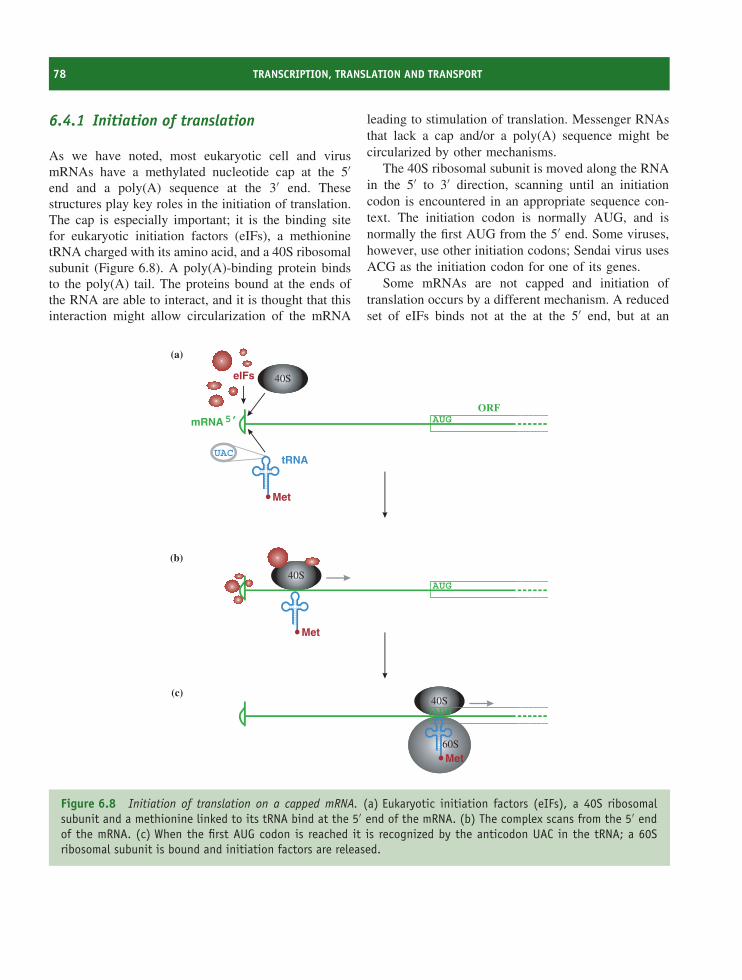
78 TRANSCRIPTION, TRANSLATION AND TRANSPORT
6.4.1 Initiation of translation
As we have noted, most eukaryotic cell and virusmRNAs have a methylated nucleotide cap at the 5′end and a poly(A) sequence at the 3′ end. Thesestructures play key roles in the initiation of translation.The cap is especially important; it is the binding sitefor eukaryotic initiation factors (eIFs), a methioninetRNA charged with its amino acid, and a 40S ribosomalsubunit (Figure 6.8). A poly(A)-binding protein bindsto the poly(A) tail. The proteins bound at the ends ofthe RNA are able to interact, and it is thought that thisinteraction might allow circularization of the mRNA
leading to stimulation of translation. Messenger RNAsthat lack a cap and/or a poly(A) sequence might becircularized by other mechanisms.
The 40S ribosomal subunit is moved along the RNAin the 5′ to 3′ direction, scanning until an initiationcodon is encountered in an appropriate sequence con-text. The initiation codon is normally AUG, and isnormally the first AUG from the 5′ end. Some viruses,however, use other initiation codons; Sendai virus usesACG as the initiation codon for one of its genes.
Some mRNAs are not capped and initiation oftranslation occurs by a different mechanism. A reducedset of eIFs binds not at the at the 5′ end, but at an
40SAUG
40SAUG
60S
40S
tRNA
5’ AUGmRNAORF
UAC
(a)
(b)
(c)
Met
Met
Met
eIFs
Figure 6.8 Initiation of translation on a capped mRNA. (a) Eukaryotic initiation factors (eIFs), a 40S ribosomalsubunit and a methionine linked to its tRNA bind at the 5′ end of the mRNA. (b) The complex scans from the 5′ endof the mRNA. (c) When the first AUG codon is reached it is recognized by the anticodon UAC in the tRNA; a 60Sribosomal subunit is bound and initiation factors are released.
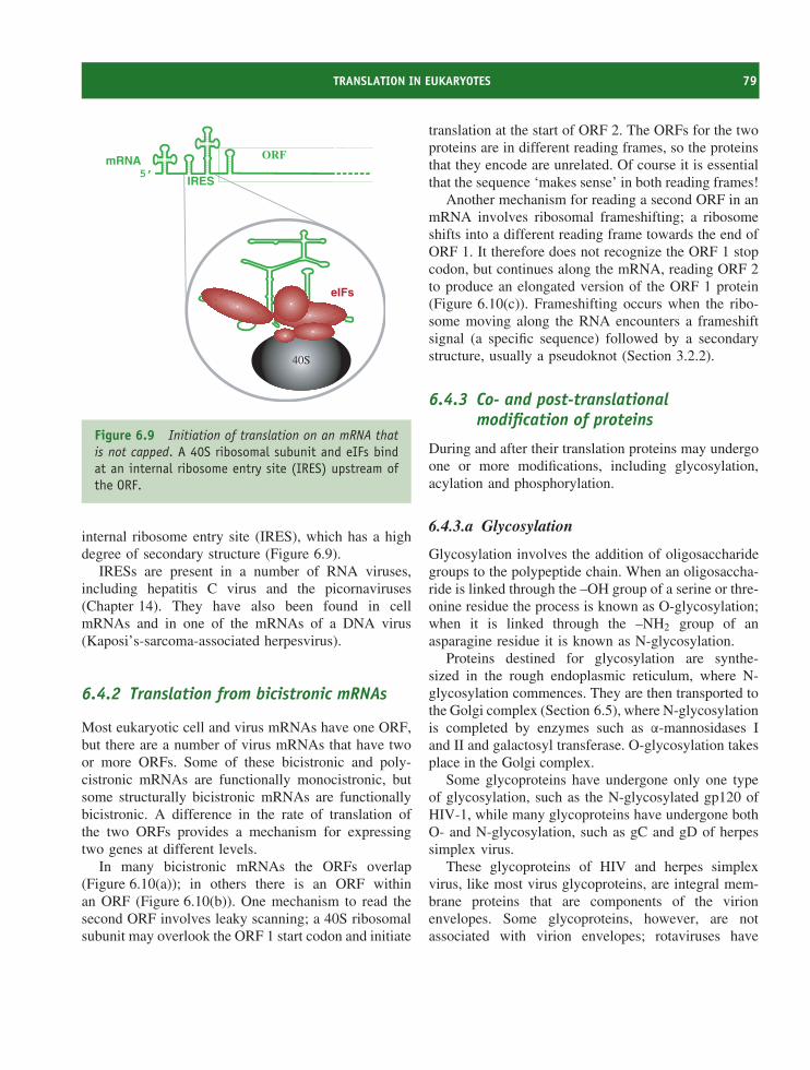
TRANSLATION IN EUKARYOTES 79
5’
eIFs
mRNA ORF
40S
IRES
Figure 6.9 Initiation of translation on an mRNA thatis not capped. A 40S ribosomal subunit and eIFs bindat an internal ribosome entry site (IRES) upstream ofthe ORF.
internal ribosome entry site (IRES), which has a highdegree of secondary structure (Figure 6.9).
IRESs are present in a number of RNA viruses,including hepatitis C virus and the picornaviruses(Chapter 14). They have also been found in cellmRNAs and in one of the mRNAs of a DNA virus(Kaposi’s-sarcoma-associated herpesvirus).
6.4.2 Translation from bicistronic mRNAs
Most eukaryotic cell and virus mRNAs have one ORF,but there are a number of virus mRNAs that have twoor more ORFs. Some of these bicistronic and poly-cistronic mRNAs are functionally monocistronic, butsome structurally bicistronic mRNAs are functionallybicistronic. A difference in the rate of translation ofthe two ORFs provides a mechanism for expressingtwo genes at different levels.
In many bicistronic mRNAs the ORFs overlap(Figure 6.10(a)); in others there is an ORF withinan ORF (Figure 6.10(b)). One mechanism to read thesecond ORF involves leaky scanning; a 40S ribosomalsubunit may overlook the ORF 1 start codon and initiate
translation at the start of ORF 2. The ORFs for the twoproteins are in different reading frames, so the proteinsthat they encode are unrelated. Of course it is essentialthat the sequence ‘makes sense’ in both reading frames!
Another mechanism for reading a second ORF in anmRNA involves ribosomal frameshifting; a ribosomeshifts into a different reading frame towards the end ofORF 1. It therefore does not recognize the ORF 1 stopcodon, but continues along the mRNA, reading ORF 2to produce an elongated version of the ORF 1 protein(Figure 6.10(c)). Frameshifting occurs when the ribo-some moving along the RNA encounters a frameshiftsignal (a specific sequence) followed by a secondarystructure, usually a pseudoknot (Section 3.2.2).
6.4.3 Co- and post-translationalmodification of proteins
During and after their translation proteins may undergoone or more modifications, including glycosylation,acylation and phosphorylation.
6.4.3.a Glycosylation
Glycosylation involves the addition of oligosaccharidegroups to the polypeptide chain. When an oligosaccha-ride is linked through the –OH group of a serine or thre-onine residue the process is known as O-glycosylation;when it is linked through the –NH2 group of anasparagine residue it is known as N-glycosylation.
Proteins destined for glycosylation are synthe-sized in the rough endoplasmic reticulum, where N-glycosylation commences. They are then transported tothe Golgi complex (Section 6.5), where N-glycosylationis completed by enzymes such as α-mannosidases Iand II and galactosyl transferase. O-glycosylation takesplace in the Golgi complex.
Some glycoproteins have undergone only one typeof glycosylation, such as the N-glycosylated gp120 ofHIV-1, while many glycoproteins have undergone bothO- and N-glycosylation, such as gC and gD of herpessimplex virus.
These glycoproteins of HIV and herpes simplexvirus, like most virus glycoproteins, are integral mem-brane proteins that are components of the virionenvelopes. Some glycoproteins, however, are notassociated with virion envelopes; rotaviruses have
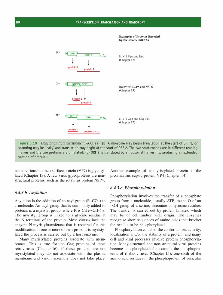
80 TRANSCRIPTION, TRANSLATION AND TRANSPORT
Rotavirus NSP5 and NSP6(Chapter 13)
HIV-1 Vpu and Env(Chapter 17)
ORF 1ORF 2
protein 2
protein 1
AnORF 1
ORF 2
protein 1protein 2
HIV-1 Gag and Gag-Pol(Chapter 17)
AnORF 1
ORF 2
protein 1protein 1 + 2
Examples of Proteins Encodedby Bicistronic mRNAs
(a)
(b)
(c)
Figure 6.10 Translation from bicistronic mRNAs. (a), (b) A ribosome may begin translation at the start of ORF 1, orscanning may be ‘leaky’ and translation may begin at the start of ORF 2. The two start codons are in different readingframes and the two proteins are unrelated. (c) ORF 2 is translated by a ribosomal frameshift, producing an extendedversion of protein 1.
naked virions but their surface protein (VP7) is glycosy-lated (Chapter 13). A few virus glycoproteins are non-structural proteins, such as the rotavirus protein NSP4.
6.4.3.b Acylation
Acylation is the addition of an acyl group (R–CO–) toa molecule. An acyl group that is commonly added toproteins is a myristyl group, where R is CH3–(CH2)12.The myristyl group is linked to a glycine residue atthe N terminus of the protein. Most viruses lack theenzyme N-myristyltransferase that is required for thismodification; if one or more of their proteins is myristy-lated the process is carried out by a host enzyme.
Many myristylated proteins associate with mem-branes. This is true for the Gag proteins of mostretroviruses (Chapter 16); if these proteins are notmyristylated they do not associate with the plasmamembrane and virion assembly does not take place.
Another example of a myristylated protein is thepicornavirus capsid protein VP4 (Chapter 14).
6.4.3.c Phosphorylation
Phosphorylation involves the transfer of a phosphategroup from a nucleotide, usually ATP, to the O of an–OH group of a serine, threonine or tyrosine residue.The transfer is carried out by protein kinases, whichmay be of cell and/or viral origin. The enzymesrecognize short sequences of amino acids that bracketthe residue to be phosphorylated.
Phosphorylation can alter the conformation, activity,localization and/or the stability of a protein, and manycell and viral processes involve protein phosphoryla-tion. Many structural and non-structural virus proteinsbecome phosphorylated, for example the phosphopro-teins of rhabdoviruses (Chapter 15); one-sixth of theamino acid residues in the phosphoprotein of vesicular
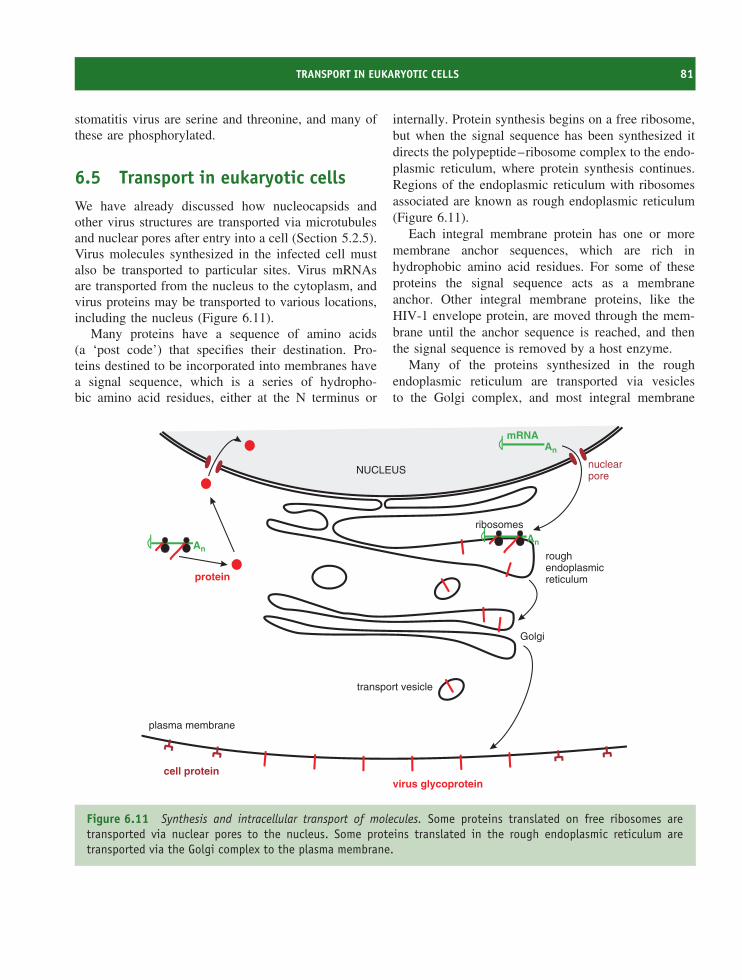
TRANSPORT IN EUKARYOTIC CELLS 81
stomatitis virus are serine and threonine, and many ofthese are phosphorylated.
6.5 Transport in eukaryotic cellsWe have already discussed how nucleocapsids andother virus structures are transported via microtubulesand nuclear pores after entry into a cell (Section 5.2.5).Virus molecules synthesized in the infected cell mustalso be transported to particular sites. Virus mRNAsare transported from the nucleus to the cytoplasm, andvirus proteins may be transported to various locations,including the nucleus (Figure 6.11).
Many proteins have a sequence of amino acids(a ‘post code’) that specifies their destination. Pro-teins destined to be incorporated into membranes havea signal sequence, which is a series of hydropho-bic amino acid residues, either at the N terminus or
internally. Protein synthesis begins on a free ribosome,but when the signal sequence has been synthesized itdirects the polypeptide–ribosome complex to the endo-plasmic reticulum, where protein synthesis continues.Regions of the endoplasmic reticulum with ribosomesassociated are known as rough endoplasmic reticulum(Figure 6.11).
Each integral membrane protein has one or moremembrane anchor sequences, which are rich inhydrophobic amino acid residues. For some of theseproteins the signal sequence acts as a membraneanchor. Other integral membrane proteins, like theHIV-1 envelope protein, are moved through the mem-brane until the anchor sequence is reached, and thenthe signal sequence is removed by a host enzyme.
Many of the proteins synthesized in the roughendoplasmic reticulum are transported via vesiclesto the Golgi complex, and most integral membrane
NUCLEUS
roughendoplasmicreticulum
Golgi
nuclearpore
AnAn
protein
An
virus glycoproteincell protein
mRNA
transport vesicle
plasma membrane
ribosomes
Figure 6.11 Synthesis and intracellular transport of molecules. Some proteins translated on free ribosomes aretransported via nuclear pores to the nucleus. Some proteins translated in the rough endoplasmic reticulum aretransported via the Golgi complex to the plasma membrane.
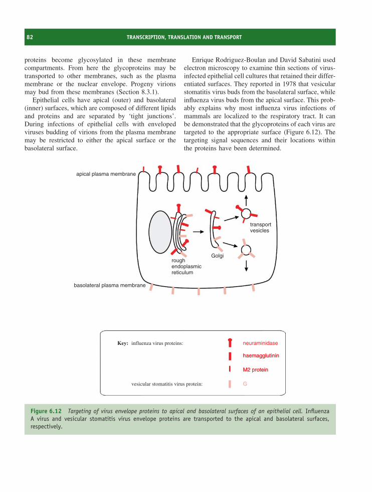
82 TRANSCRIPTION, TRANSLATION AND TRANSPORT
proteins become glycosylated in these membranecompartments. From here the glycoproteins may betransported to other membranes, such as the plasmamembrane or the nuclear envelope. Progeny virionsmay bud from these membranes (Section 8.3.1).
Epithelial cells have apical (outer) and basolateral(inner) surfaces, which are composed of different lipidsand proteins and are separated by ‘tight junctions’.During infections of epithelial cells with envelopedviruses budding of virions from the plasma membranemay be restricted to either the apical surface or thebasolateral surface.
Enrique Rodriguez-Boulan and David Sabatini usedelectron microscopy to examine thin sections of virus-infected epithelial cell cultures that retained their differ-entiated surfaces. They reported in 1978 that vesicularstomatitis virus buds from the basolateral surface, whileinfluenza virus buds from the apical surface. This prob-ably explains why most influenza virus infections ofmammals are localized to the respiratory tract. It canbe demonstrated that the glycoproteins of each virus aretargeted to the appropriate surface (Figure 6.12). Thetargeting signal sequences and their locations withinthe proteins have been determined.
apical plasma membrane
basolateral plasma membrane
Key: influenza virus proteins:
vesicular stomatitis virus protein:
haemagglutininhaemagglutinin
M2 proteinM2 protein
GG
roughendoplasmicreticulum
Golgi
transportvesicles
neuraminidase
Figure 6.12 Targeting of virus envelope proteins to apical and basolateral surfaces of an epithelial cell. InfluenzaA virus and vesicular stomatitis virus envelope proteins are transported to the apical and basolateral surfaces,respectively.
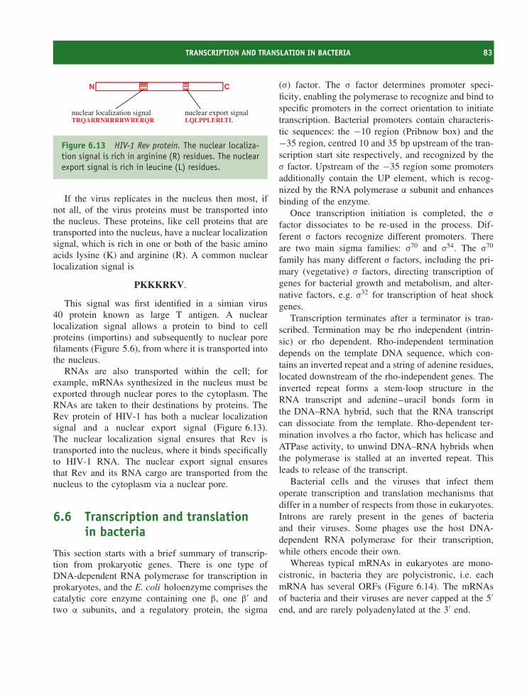
TRANSCRIPTION AND TRANSLATION IN BACTERIA 83
nuclear localization signalTRQARRNRRRRWRERQR
nuclear export signalLQLPPLERLTL
N C
Figure 6.13 HIV-1 Rev protein. The nuclear localiza-tion signal is rich in arginine (R) residues. The nuclearexport signal is rich in leucine (L) residues.
If the virus replicates in the nucleus then most, ifnot all, of the virus proteins must be transported intothe nucleus. These proteins, like cell proteins that aretransported into the nucleus, have a nuclear localizationsignal, which is rich in one or both of the basic aminoacids lysine (K) and arginine (R). A common nuclearlocalization signal is
PKKKRKV.
This signal was first identified in a simian virus40 protein known as large T antigen. A nuclearlocalization signal allows a protein to bind to cellproteins (importins) and subsequently to nuclear porefilaments (Figure 5.6), from where it is transported intothe nucleus.
RNAs are also transported within the cell; forexample, mRNAs synthesized in the nucleus must beexported through nuclear pores to the cytoplasm. TheRNAs are taken to their destinations by proteins. TheRev protein of HIV-1 has both a nuclear localizationsignal and a nuclear export signal (Figure 6.13).The nuclear localization signal ensures that Rev istransported into the nucleus, where it binds specificallyto HIV-1 RNA. The nuclear export signal ensuresthat Rev and its RNA cargo are transported from thenucleus to the cytoplasm via a nuclear pore.
6.6 Transcription and translationin bacteria
This section starts with a brief summary of transcrip-tion from prokaryotic genes. There is one type ofDNA-dependent RNA polymerase for transcription inprokaryotes, and the E. coli holoenzyme comprises thecatalytic core enzyme containing one β, one β′ andtwo α subunits, and a regulatory protein, the sigma
(σ) factor. The σ factor determines promoter speci-ficity, enabling the polymerase to recognize and bind tospecific promoters in the correct orientation to initiatetranscription. Bacterial promoters contain characteris-tic sequences: the −10 region (Pribnow box) and the−35 region, centred 10 and 35 bp upstream of the tran-scription start site respectively, and recognized by theσ factor. Upstream of the −35 region some promotersadditionally contain the UP element, which is recog-nized by the RNA polymerase α subunit and enhancesbinding of the enzyme.
Once transcription initiation is completed, the σ
factor dissociates to be re-used in the process. Dif-ferent σ factors recognize different promoters. Thereare two main sigma families: σ70 and σ54. The σ70
family has many different σ factors, including the pri-mary (vegetative) σ factors, directing transcription ofgenes for bacterial growth and metabolism, and alter-native factors, e.g. σ32 for transcription of heat shockgenes.
Transcription terminates after a terminator is tran-scribed. Termination may be rho independent (intrin-sic) or rho dependent. Rho-independent terminationdepends on the template DNA sequence, which con-tains an inverted repeat and a string of adenine residues,located downstream of the rho-independent genes. Theinverted repeat forms a stem-loop structure in theRNA transcript and adenine–uracil bonds form inthe DNA–RNA hybrid, such that the RNA transcriptcan dissociate from the template. Rho-dependent ter-mination involves a rho factor, which has helicase andATPase activity, to unwind DNA–RNA hybrids whenthe polymerase is stalled at an inverted repeat. Thisleads to release of the transcript.
Bacterial cells and the viruses that infect themoperate transcription and translation mechanisms thatdiffer in a number of respects from those in eukaryotes.Introns are rarely present in the genes of bacteriaand their viruses. Some phages use the host DNA-dependent RNA polymerase for their transcription,while others encode their own.
Whereas typical mRNAs in eukaryotes are mono-cistronic, in bacteria they are polycistronic, i.e. eachmRNA has several ORFs (Figure 6.14). The mRNAsof bacteria and their viruses are never capped at the 5′end, and are rarely polyadenylated at the 3′ end.
84 TRANSCRIPTION, TRANSLATION AND TRANSPORT
5’ 3’ORF 1 ORF 2 ORF 3
Figure 6.14 Characteristics of bacterial and phagemRNA. There are typically several ORFs, all of whichmay be translated at the same time. The 5′ end is notcapped and the 3′ end is rarely polyadenylated.
Bacterial translation differs from eukaryotic transla-tion in a number of features.
• Translation may start before transcription is com-plete. The lack of a nucleus allows transcription andtranslation to be coupled.
• The ribosomes are smaller (the ribosomal subunitshave sedimentation coefficients of 30S and 50S).
• The 30S ribosomal subunit binds directly to atranslation initiation region on the mRNA. Initi-ation generally involves interaction of the Shine-Dalgarno (S-D) sequence at the ribosome bindingsite (RBS) on mRNA and the anti-S-D sequenceat the 3′ end of 16S rRNA in the 30S subunit.The RBS is located just upstream of the AUG startcodon.
• The methionine of the initiator methionyl tRNA isgenerally formylated.
• A much smaller number of initiation factors isinvolved.
• All ORFs within an mRNA are translated and severalmay be translated concurrently.
A few phages have overlapping genes, which maybe translated by reading through a stop codon or byribosomal frameshifting (Section 6.4.2).
Learning outcomesBy the end of this chapter you should be able to
• explain how virus genes are transcribed andtranslated;
• describe the post-translational modifications thatsome virus proteins undergo;
• highlight differences in transcription and transla-tion between prokaryotic and eukaryotic cells;
• discuss the transport of virus proteins and RNAwithin cells.
Sources of further informationBooksBrown T. A. (2006) Genomes 3, 3rd edition, Garland ScienceBrown W. M. and Brown P. M. (2002) Transcription, Taylor
and FrancisLee D., Kapp L. D. and Lorsch J. R. (2004) The molecular
mechanics of eukaryotic translation. Annual Review ofBiochemistry, 73, 657–704
Smith G. L., Murphy B. J. and Law M. (2003) Vacciniavirus motility Annual Review of Microbiology, 57,323–342
Historical papersCrick F. (1970) Central dogma of molecular biology Nature,
227, 561–563Rodriguez-Boulan E. and Sabatini D. D. (1978) Asymmetricbudding of viruses in epithelial monolayers: a model systemfor study of epithelial polarity Proceedings of the NationalAcademy of Sciences USA, 75, 5071–5075
Recent papersAhlquist P. (2006) Parallels among positive-strand RNA
viruses, reverse-transcribing viruses and double-strandedRNA viruses Nature Reviews Microbiology, 4,371–382
Fechter P. and Brownlee G. G. (2005) Recognition ofmRNA cap structures by viral and cellular proteins Journalof General Virology, 86, 1239–1249
Kozak M. (2004) How strong is the case for regulation ofthe initiation step of translation by elements at the 3′ endof eukaryotic mRNAs? Gene, 343, 41–54
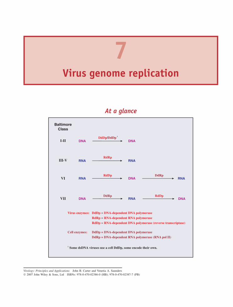
7Virus genome replication
At a glance
DdDp = DNA-dependent DNA polymerase
RdRp = RNA-dependent RNA polymerase
RdDp = RNA-dependent DNA polymerase (reverse transcriptase)
DNA
RNA RNA
DNA RNA
RNARNA DNADdRp
RdDp
RdDp
RdRp
VI
VII
III-V
DNA DNA
BaltimoreClass
I-II
Virus enzymes:
DdDp DNA-dependent DNA polymerase
DdRp = DNA-dependent RNA polymerase (RNA pol II)
=Cell enzymes:
DdRp
DdDp/DdDp 1
Some dsDNA viruses use a cell DdDp, some encode their own.1
Virology: Principles and Applications John B. Carter and Venetia A. Saunders 2007 John Wiley & Sons, Ltd ISBNs: 978-0-470-02386-0 (HB); 978-0-470-02387-7 (PB)

86 VIRUS GENOME REPLICATION
At a glance (continued)
some dsDNA viruses
(+) RNA viruses
ssDNA viruses
some dsDNA viruses
(−) RNA viruses (non-segmented genomes)
(−) RNA viruses (segmented genomes)
dsRNA viruses
retroviruses & pararetroviruses (DNA → RNA)
retroviruses & pararetroviruses (RNA → DNA)
CYTOPLASM
NUCLEUS
NUCLEUS
Locations of virus genome replication in eukaryotic cells
RNA
DNA
3’OH
protein OH
(self-priming)
Primers
3’OH
7.1 Overview of virus genomereplication
In this chapter we consider the fifth step of ourgeneralized replication cycle: genome replication. Thegenome of the infecting virus is replicated so that viraltranscription can be amplified and to provide copies ofthe genome for progeny virions.
Generally, DNA viruses copy their genomes directlyto DNA and RNA viruses copy their genomes directlyto RNA. There are, however, some DNA viruses thatreplicate their genomes via an RNA intermediate andsome RNA viruses that replicate their genomes via a
DNA intermediate. The various replication modes ofvirus genomes are summarized in Figure 7.1.
Single-stranded genomes are designated as plus orminus depending on their relationship to the virusmRNA. Plus strand genomes have the same sequenceas the mRNA (except that in DNA thymine replacesuracil), while minus-strand genomes have the sequencecomplementary to the mRNA. Single-stranded DNA isconverted to dsDNA prior to copying.
There are two classes of viruses with (+) RNAgenomes (Figure 7.1). Class IV viruses copy their (+)RNA genomes via a (−) RNA intermediate, whileClass VI viruses replicate via a DNA intermediate. The
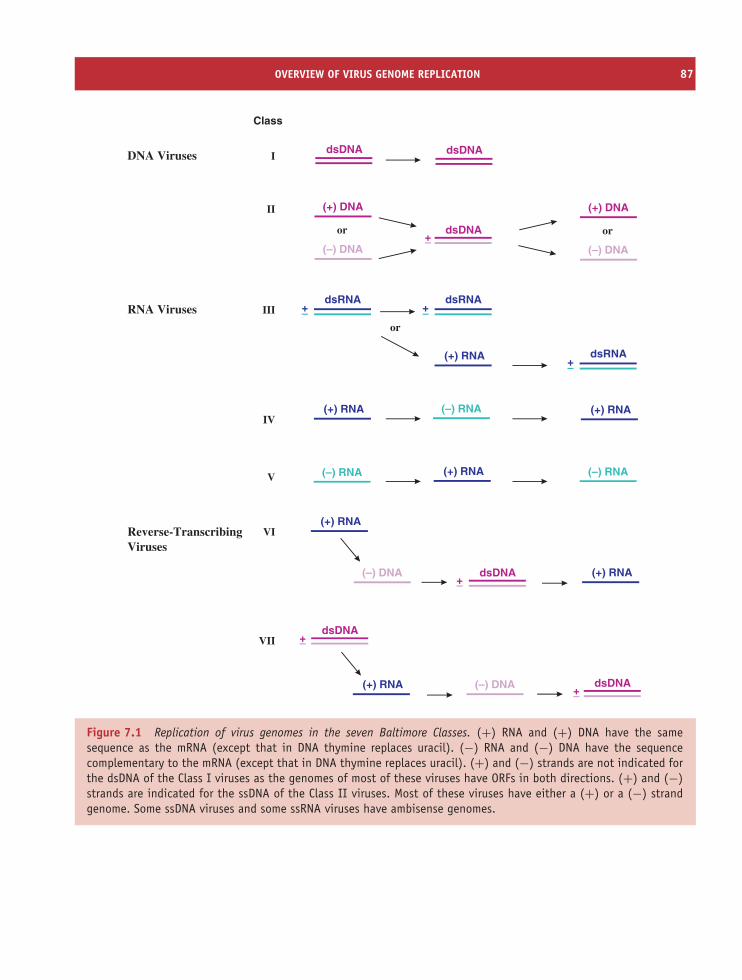
OVERVIEW OF VIRUS GENOME REPLICATION 87
(–) DNA
(+) RNA
dsDNA
Class
I
II
III
IV
V
VI
VII
dsDNA+–
+–
DNA Viruses
RNA Viruses
Reverse-TranscribingViruses
(–) RNA(+) RNA
(+) DNA
(–) DNA
or
dsDNA
(+) DNA
(–) DNA
or
(+) RNA
(+) RNA
(–) RNA (+) RNA (–) RNA
dsDNA
dsRNA+–
dsDNA+–
(+) RNA
(–) DNA dsDNA(+) RNA +–
or
dsRNA+–
dsRNA+–
Figure 7.1 Replication of virus genomes in the seven Baltimore Classes. (+) RNA and (+) DNA have the samesequence as the mRNA (except that in DNA thymine replaces uracil). (−) RNA and (−) DNA have the sequencecomplementary to the mRNA (except that in DNA thymine replaces uracil). (+) and (−) strands are not indicated forthe dsDNA of the Class I viruses as the genomes of most of these viruses have ORFs in both directions. (+) and (−)strands are indicated for the ssDNA of the Class II viruses. Most of these viruses have either a (+) or a (−) strandgenome. Some ssDNA viruses and some ssRNA viruses have ambisense genomes.
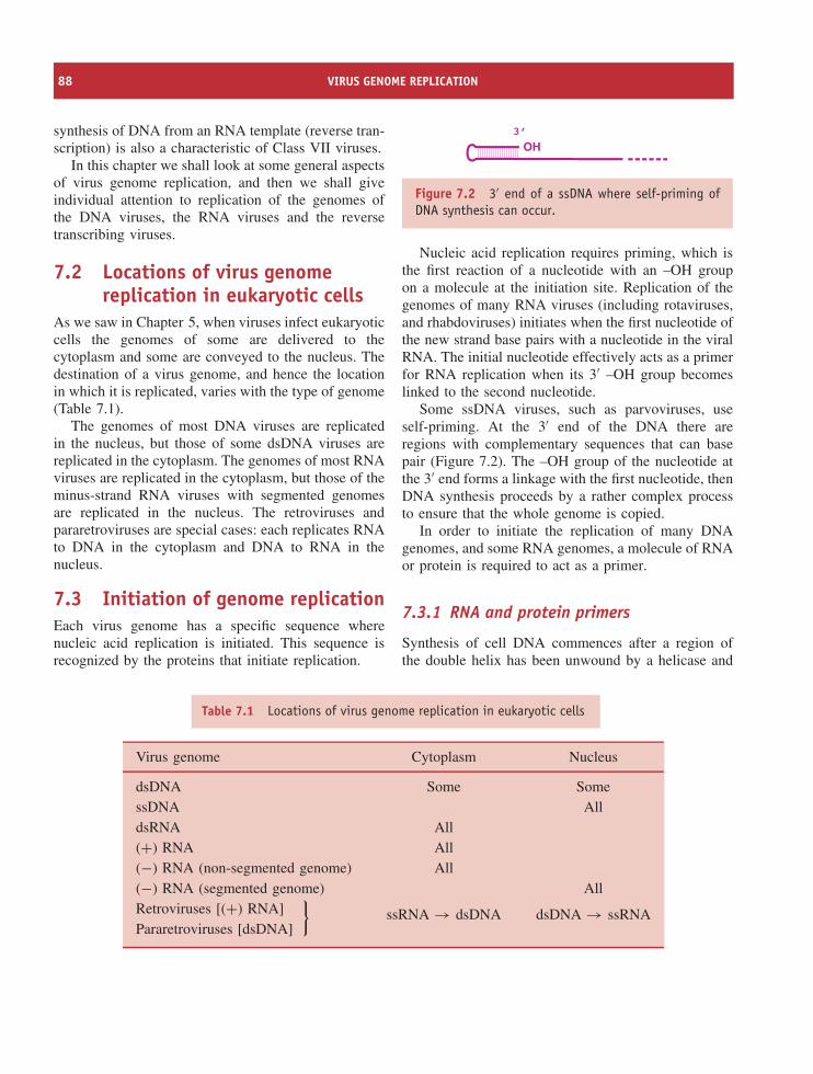
88 VIRUS GENOME REPLICATION
synthesis of DNA from an RNA template (reverse tran-scription) is also a characteristic of Class VII viruses.
In this chapter we shall look at some general aspectsof virus genome replication, and then we shall giveindividual attention to replication of the genomes ofthe DNA viruses, the RNA viruses and the reversetranscribing viruses.
7.2 Locations of virus genomereplication in eukaryotic cells
As we saw in Chapter 5, when viruses infect eukaryoticcells the genomes of some are delivered to thecytoplasm and some are conveyed to the nucleus. Thedestination of a virus genome, and hence the locationin which it is replicated, varies with the type of genome(Table 7.1).
The genomes of most DNA viruses are replicatedin the nucleus, but those of some dsDNA viruses arereplicated in the cytoplasm. The genomes of most RNAviruses are replicated in the cytoplasm, but those of theminus-strand RNA viruses with segmented genomesare replicated in the nucleus. The retroviruses andpararetroviruses are special cases: each replicates RNAto DNA in the cytoplasm and DNA to RNA in thenucleus.
7.3 Initiation of genome replicationEach virus genome has a specific sequence wherenucleic acid replication is initiated. This sequence isrecognized by the proteins that initiate replication.
OH3’
Figure 7.2 3′ end of a ssDNA where self-priming ofDNA synthesis can occur.
Nucleic acid replication requires priming, which isthe first reaction of a nucleotide with an –OH groupon a molecule at the initiation site. Replication of thegenomes of many RNA viruses (including rotaviruses,and rhabdoviruses) initiates when the first nucleotide ofthe new strand base pairs with a nucleotide in the viralRNA. The initial nucleotide effectively acts as a primerfor RNA replication when its 3′ –OH group becomeslinked to the second nucleotide.
Some ssDNA viruses, such as parvoviruses, useself-priming. At the 3′ end of the DNA there areregions with complementary sequences that can basepair (Figure 7.2). The –OH group of the nucleotide atthe 3′ end forms a linkage with the first nucleotide, thenDNA synthesis proceeds by a rather complex processto ensure that the whole genome is copied.
In order to initiate the replication of many DNAgenomes, and some RNA genomes, a molecule of RNAor protein is required to act as a primer.
7.3.1 RNA and protein primers
Synthesis of cell DNA commences after a region ofthe double helix has been unwound by a helicase and
Table 7.1 Locations of virus genome replication in eukaryotic cells
Virus genome Cytoplasm Nucleus
dsDNA Some SomessDNA AlldsRNA All(+) RNA All(−) RNA (non-segmented genome) All(−) RNA (segmented genome) AllRetroviruses [(+) RNA]
}Pararetroviruses [dsDNA]
ssRNA → dsDNA dsDNA → ssRNA
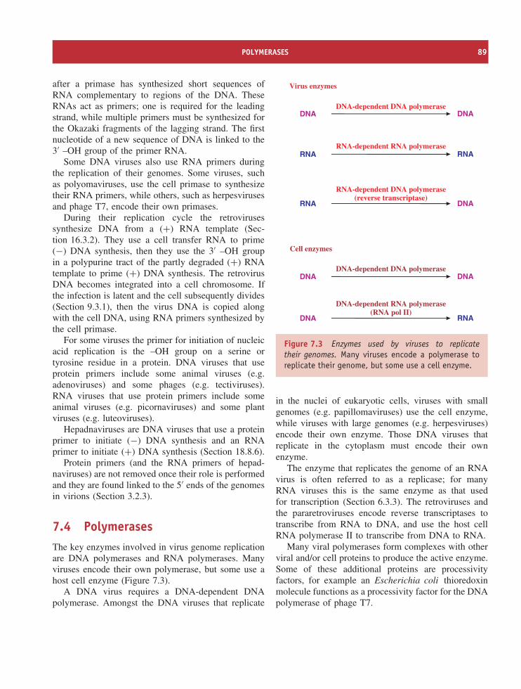
POLYMERASES 89
after a primase has synthesized short sequences ofRNA complementary to regions of the DNA. TheseRNAs act as primers; one is required for the leadingstrand, while multiple primers must be synthesized forthe Okazaki fragments of the lagging strand. The firstnucleotide of a new sequence of DNA is linked to the3′ –OH group of the primer RNA.
Some DNA viruses also use RNA primers duringthe replication of their genomes. Some viruses, suchas polyomaviruses, use the cell primase to synthesizetheir RNA primers, while others, such as herpesvirusesand phage T7, encode their own primases.
During their replication cycle the retrovirusessynthesize DNA from a (+) RNA template (Sec-tion 16.3.2). They use a cell transfer RNA to prime(−) DNA synthesis, then they use the 3′ –OH groupin a polypurine tract of the partly degraded (+) RNAtemplate to prime (+) DNA synthesis. The retrovirusDNA becomes integrated into a cell chromosome. Ifthe infection is latent and the cell subsequently divides(Section 9.3.1), then the virus DNA is copied alongwith the cell DNA, using RNA primers synthesized bythe cell primase.
For some viruses the primer for initiation of nucleicacid replication is the –OH group on a serine ortyrosine residue in a protein. DNA viruses that useprotein primers include some animal viruses (e.g.adenoviruses) and some phages (e.g. tectiviruses).RNA viruses that use protein primers include someanimal viruses (e.g. picornaviruses) and some plantviruses (e.g. luteoviruses).
Hepadnaviruses are DNA viruses that use a proteinprimer to initiate (−) DNA synthesis and an RNAprimer to initiate (+) DNA synthesis (Section 18.8.6).
Protein primers (and the RNA primers of hepad-naviruses) are not removed once their role is performedand they are found linked to the 5′ ends of the genomesin virions (Section 3.2.3).
7.4 PolymerasesThe key enzymes involved in virus genome replicationare DNA polymerases and RNA polymerases. Manyviruses encode their own polymerase, but some use ahost cell enzyme (Figure 7.3).
A DNA virus requires a DNA-dependent DNApolymerase. Amongst the DNA viruses that replicate
RNA RNA
DNA RNA
RNA DNA
DNA DNA
DNA DNA
Virus enzymes
Cell enzymes
RNA-dependent DNA polymerase(reverse transcriptase)
DNA-dependent RNA polymerase(RNA pol II)
DNA-dependent DNA polymerase
RNA-dependent RNA polymerase
DNA-dependent DNA polymerase
Figure 7.3 Enzymes used by viruses to replicatetheir genomes. Many viruses encode a polymerase toreplicate their genome, but some use a cell enzyme.
in the nuclei of eukaryotic cells, viruses with smallgenomes (e.g. papillomaviruses) use the cell enzyme,while viruses with large genomes (e.g. herpesviruses)encode their own enzyme. Those DNA viruses thatreplicate in the cytoplasm must encode their ownenzyme.
The enzyme that replicates the genome of an RNAvirus is often referred to as a replicase; for manyRNA viruses this is the same enzyme as that usedfor transcription (Section 6.3.3). The retroviruses andthe pararetroviruses encode reverse transcriptases totranscribe from RNA to DNA, and use the host cellRNA polymerase II to transcribe from DNA to RNA.
Many viral polymerases form complexes with otherviral and/or cell proteins to produce the active enzyme.Some of these additional proteins are processivityfactors, for example an Escherichia coli thioredoxinmolecule functions as a processivity factor for the DNApolymerase of phage T7.
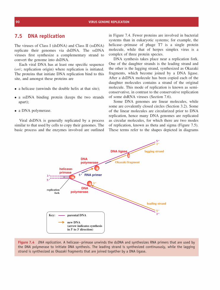
90 VIRUS GENOME REPLICATION
7.5 DNA replication
The viruses of Class I (dsDNA) and Class II (ssDNA)replicate their genomes via dsDNA. The ssDNAviruses first synthesize a complementary strand toconvert the genome into dsDNA.
Each viral DNA has at least one specific sequence(ori ; replication origin) where replication is initiated.The proteins that initiate DNA replication bind to thissite, and amongst these proteins are
• a helicase (unwinds the double helix at that site);
• a ssDNA binding protein (keeps the two strandsapart);
• a DNA polymerase.
Viral dsDNA is generally replicated by a processsimilar to that used by cells to copy their genomes. Thebasic process and the enzymes involved are outlined
in Figure 7.4. Fewer proteins are involved in bacterialsystems than in eukaryotic systems; for example, thehelicase–primase of phage T7 is a single proteinmolecule, while that of herpes simplex virus is acomplex of three protein species.
DNA synthesis takes place near a replication fork.One of the daughter strands is the leading strand andthe other is the lagging strand, synthesized as Okazakifragments, which become joined by a DNA ligase.After a dsDNA molecule has been copied each of thedaughter molecules contains a strand of the originalmolecule. This mode of replication is known as semi-conservative, in contrast to the conservative replicationof some dsRNA viruses (Section 7.6).
Some DNA genomes are linear molecules, whilesome are covalently closed circles (Section 3.2). Someof the linear molecules are circularized prior to DNAreplication, hence many DNA genomes are replicatedas circular molecules, for which there are two modesof replication, known as theta and sigma (Figure 7.5).These terms refer to the shapes depicted in diagrams
RNA primer
DNApolymerase
DNA ligase
DNApolymerase
Okazaki fragment
lagging strand
leading strand
5’
3’5’
3’
3’
5’3’
5’
helicase-primase
Key:
new DNA(arrow indicates synthesisin 5' to 3' direction)
parental DNA
replicationfork
Figure 7.4 DNA replication. A helicase–primase unwinds the dsDNA and synthesizes RNA primers that are used bythe DNA polymerase to initiate DNA synthesis. The leading strand is synthesized continuously, while the laggingstrand is synthesized as Okazaki fragments that are joined together by a DNA ligase.
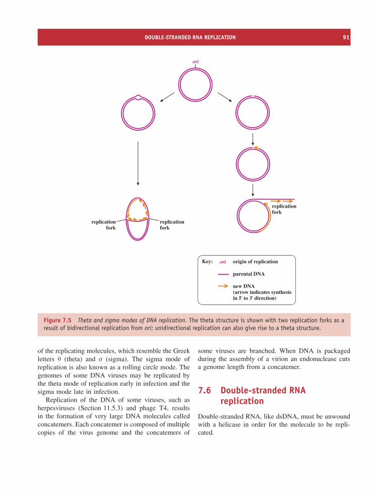
DOUBLE-STRANDED RNA REPLICATION 91
replicationfork
Key:
new DNA(arrow indicates synthesisin 5' to 3' direction)
parental DNA
ori
replicationfork
replicationfork
ori origin of replication
Figure 7.5 Theta and sigma modes of DNA replication. The theta structure is shown with two replication forks as aresult of bidirectional replication from ori; unidirectional replication can also give rise to a theta structure.
of the replicating molecules, which resemble the Greekletters θ (theta) and σ (sigma). The sigma mode ofreplication is also known as a rolling circle mode. Thegenomes of some DNA viruses may be replicated bythe theta mode of replication early in infection and thesigma mode late in infection.
Replication of the DNA of some viruses, such asherpesviruses (Section 11.5.3) and phage T4, resultsin the formation of very large DNA molecules calledconcatemers. Each concatemer is composed of multiplecopies of the virus genome and the concatemers of
some viruses are branched. When DNA is packagedduring the assembly of a virion an endonuclease cutsa genome length from a concatemer.
7.6 Double-stranded RNAreplication
Double-stranded RNA, like dsDNA, must be unwoundwith a helicase in order for the molecule to be repli-cated.
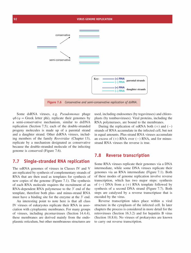
92 VIRUS GENOME REPLICATION
parental strands
conservative
semi-conservative
daughter strands
Key: (+) RNA(–) RNA
(+) RNA(–) RNA
Figure 7.6 Conservative and semi-conservative replication of dsRNA.
Some dsRNA viruses, e.g. Pseudomonas phageϕ6 (ϕ = Greek letter phi), replicate their genomes bya semi-conservative mechanism, similar to dsDNAreplication (Section 7.5); each of the double-strandedprogeny molecules is made up of a parental strandand a daughter strand. Other dsRNA viruses, includ-ing members of the family Reoviridae (Chapter 13),replicate by a mechanism designated as conservativebecause the double-stranded molecule of the infectinggenome is conserved (Figure 7.6).
7.7 Single-stranded RNA replicationThe ssRNA genomes of viruses in Classes IV and Vare replicated by synthesis of complementary strands ofRNA that are then used as templates for synthesis ofnew copies of the genome (Figure 7.1). The synthesisof each RNA molecule requires the recruitment of anRNA-dependent RNA polymerase to the 3′ end of thetemplate, therefore both plus- and minus-strand RNAmust have a binding site for the enzyme at the 3′ end.
An interesting point to note here is that all classIV viruses of eukaryotes replicate their RNA in asso-ciation with cytoplasmic membranes. For many groupsof viruses, including picornaviruses (Section 14.4.4),these membranes are derived mainly from the endo-plasmic reticulum, but other membranous structures are
used, including endosomes (by togaviruses) and chloro-plasts (by tombusviruses). Viral proteins, including theRNA polymerases, are bound to the membranes.
During the replication of ssRNA both (+) and (−)strands of RNA accumulate in the infected cell, but notin equal amounts. Plus-strand RNA viruses accumulatean excess of (+) RNA over (−) RNA, and for minus-strand RNA viruses the reverse is true.
7.8 Reverse transcription
Some RNA viruses replicate their genomes via a DNAintermediate, while some DNA viruses replicate theirgenomes via an RNA intermediate (Figure 7.1). Bothof these modes of genome replication involve reversetranscription, which has two major steps: synthesisof (−) DNA from a (+) RNA template followed bysynthesis of a second DNA strand (Figure 7.7). Bothsteps are catalysed by a reverse transcriptase that isencoded by the virus.
Reverse transcription takes place within a viralstructure in the cytoplasm of the infected cell. In laterchapters the process is considered in more detail for theretroviruses (Section 16.3.2) and for hepatitis B virus(Section 18.8.6). No viruses of prokaryotes are knownto carry out reverse transcription.
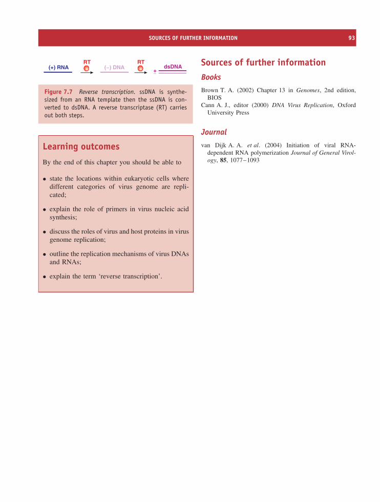
SOURCES OF FURTHER INFORMATION 93
(–) DNA dsDNA(+) RNA +–
RT RT
Figure 7.7 Reverse transcription. ssDNA is synthe-sized from an RNA template then the ssDNA is con-verted to dsDNA. A reverse transcriptase (RT) carriesout both steps.
Learning outcomes
By the end of this chapter you should be able to
• state the locations within eukaryotic cells wheredifferent categories of virus genome are repli-cated;
• explain the role of primers in virus nucleic acidsynthesis;
• discuss the roles of virus and host proteins in virusgenome replication;
• outline the replication mechanisms of virus DNAsand RNAs;
• explain the term ‘reverse transcription’.
Sources of further informationBooks
Brown T. A. (2002) Chapter 13 in Genomes, 2nd edition,BIOS
Cann A. J., editor (2000) DNA Virus Replication, OxfordUniversity Press
Journal
van Dijk A. A. et al. (2004) Initiation of viral RNA-dependent RNA polymerization Journal of General Virol-ogy, 85, 1077–1093

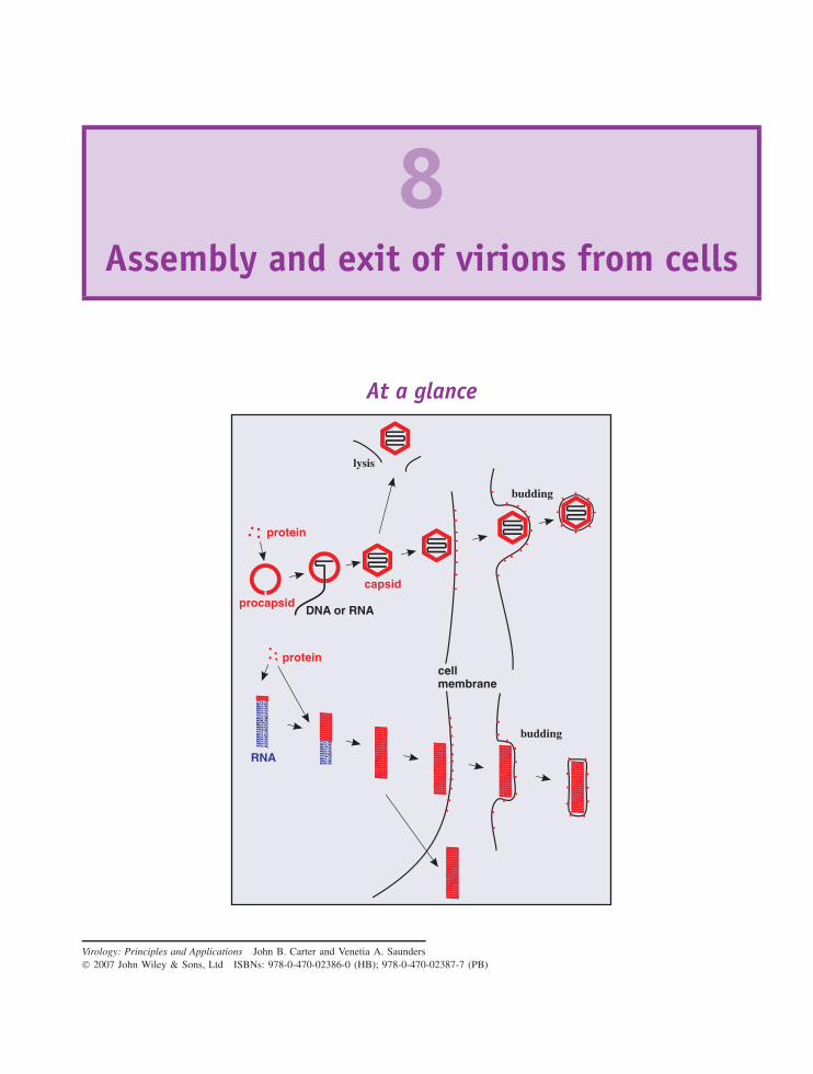
8Assembly and exit of virions from cells
At a glance
protein
protein
DNA or RNAprocapsid
capsid
cellmembrane
lysis
budding
RNA
budding
Virology: Principles and Applications John B. Carter and Venetia A. Saunders 2007 John Wiley & Sons, Ltd ISBNs: 978-0-470-02386-0 (HB); 978-0-470-02387-7 (PB)
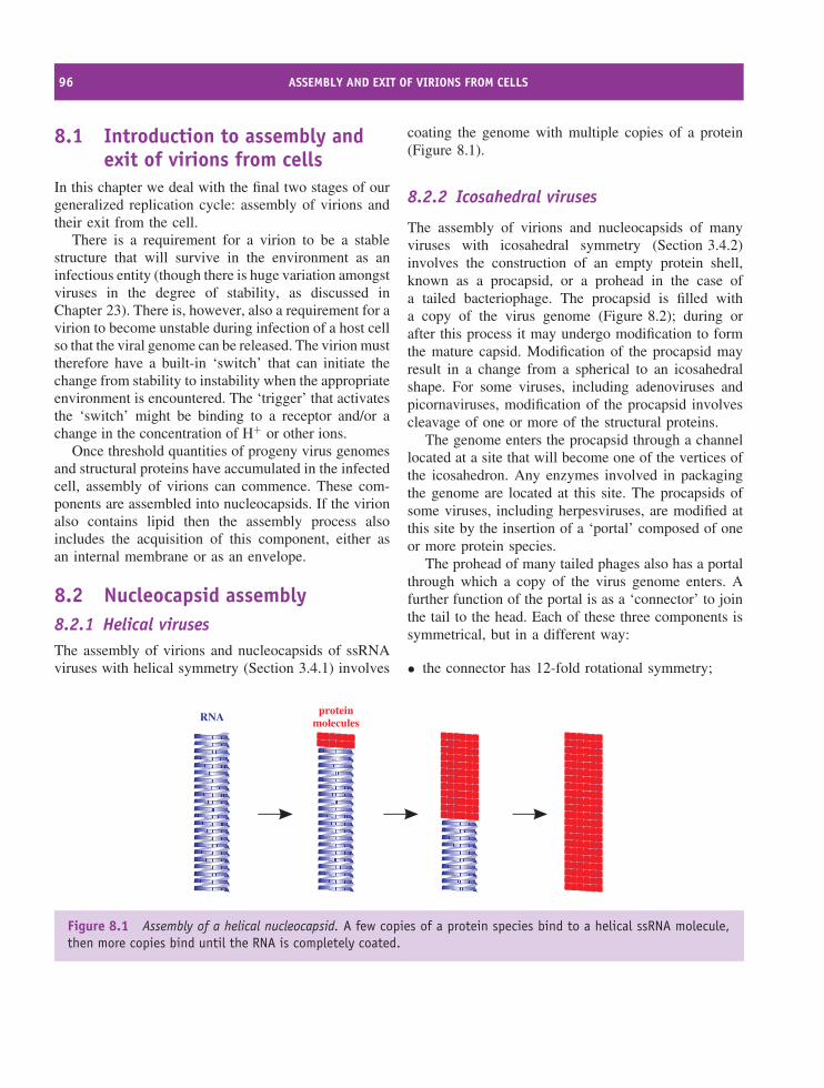
96 ASSEMBLY AND EXIT OF VIRIONS FROM CELLS
8.1 Introduction to assembly andexit of virions from cells
In this chapter we deal with the final two stages of ourgeneralized replication cycle: assembly of virions andtheir exit from the cell.
There is a requirement for a virion to be a stablestructure that will survive in the environment as aninfectious entity (though there is huge variation amongstviruses in the degree of stability, as discussed inChapter 23). There is, however, also a requirement for avirion to become unstable during infection of a host cellso that the viral genome can be released. The virion musttherefore have a built-in ‘switch’ that can initiate thechange from stability to instability when the appropriateenvironment is encountered. The ‘trigger’ that activatesthe ‘switch’ might be binding to a receptor and/or achange in the concentration of H+ or other ions.
Once threshold quantities of progeny virus genomesand structural proteins have accumulated in the infectedcell, assembly of virions can commence. These com-ponents are assembled into nucleocapsids. If the virionalso contains lipid then the assembly process alsoincludes the acquisition of this component, either asan internal membrane or as an envelope.
8.2 Nucleocapsid assembly8.2.1 Helical virusesThe assembly of virions and nucleocapsids of ssRNAviruses with helical symmetry (Section 3.4.1) involves
coating the genome with multiple copies of a protein(Figure 8.1).
8.2.2 Icosahedral viruses
The assembly of virions and nucleocapsids of manyviruses with icosahedral symmetry (Section 3.4.2)involves the construction of an empty protein shell,known as a procapsid, or a prohead in the case ofa tailed bacteriophage. The procapsid is filled witha copy of the virus genome (Figure 8.2); during orafter this process it may undergo modification to formthe mature capsid. Modification of the procapsid mayresult in a change from a spherical to an icosahedralshape. For some viruses, including adenoviruses andpicornaviruses, modification of the procapsid involvescleavage of one or more of the structural proteins.
The genome enters the procapsid through a channellocated at a site that will become one of the vertices ofthe icosahedron. Any enzymes involved in packagingthe genome are located at this site. The procapsids ofsome viruses, including herpesviruses, are modified atthis site by the insertion of a ‘portal’ composed of oneor more protein species.
The prohead of many tailed phages also has a portalthrough which a copy of the virus genome enters. Afurther function of the portal is as a ‘connector’ to jointhe tail to the head. Each of these three components issymmetrical, but in a different way:
• the connector has 12-fold rotational symmetry;
proteinmoleculesRNA
Figure 8.1 Assembly of a helical nucleocapsid. A few copies of a protein species bind to a helical ssRNA molecule,then more copies bind until the RNA is completely coated.
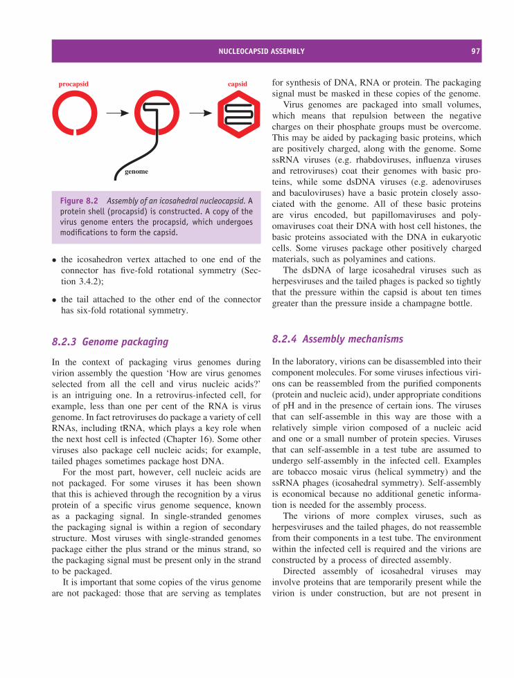
NUCLEOCAPSID ASSEMBLY 97
capsidprocapsid
genome
Figure 8.2 Assembly of an icosahedral nucleocapsid. Aprotein shell (procapsid) is constructed. A copy of thevirus genome enters the procapsid, which undergoesmodifications to form the capsid.
• the icosahedron vertex attached to one end of theconnector has five-fold rotational symmetry (Sec-tion 3.4.2);
• the tail attached to the other end of the connectorhas six-fold rotational symmetry.
8.2.3 Genome packaging
In the context of packaging virus genomes duringvirion assembly the question ‘How are virus genomesselected from all the cell and virus nucleic acids?’is an intriguing one. In a retrovirus-infected cell, forexample, less than one per cent of the RNA is virusgenome. In fact retroviruses do package a variety of cellRNAs, including tRNA, which plays a key role whenthe next host cell is infected (Chapter 16). Some otherviruses also package cell nucleic acids; for example,tailed phages sometimes package host DNA.
For the most part, however, cell nucleic acids arenot packaged. For some viruses it has been shownthat this is achieved through the recognition by a virusprotein of a specific virus genome sequence, knownas a packaging signal. In single-stranded genomesthe packaging signal is within a region of secondarystructure. Most viruses with single-stranded genomespackage either the plus strand or the minus strand, sothe packaging signal must be present only in the strandto be packaged.
It is important that some copies of the virus genomeare not packaged: those that are serving as templates
for synthesis of DNA, RNA or protein. The packagingsignal must be masked in these copies of the genome.
Virus genomes are packaged into small volumes,which means that repulsion between the negativecharges on their phosphate groups must be overcome.This may be aided by packaging basic proteins, whichare positively charged, along with the genome. SomessRNA viruses (e.g. rhabdoviruses, influenza virusesand retroviruses) coat their genomes with basic pro-teins, while some dsDNA viruses (e.g. adenovirusesand baculoviruses) have a basic protein closely asso-ciated with the genome. All of these basic proteinsare virus encoded, but papillomaviruses and poly-omaviruses coat their DNA with host cell histones, thebasic proteins associated with the DNA in eukaryoticcells. Some viruses package other positively chargedmaterials, such as polyamines and cations.
The dsDNA of large icosahedral viruses such asherpesviruses and the tailed phages is packed so tightlythat the pressure within the capsid is about ten timesgreater than the pressure inside a champagne bottle.
8.2.4 Assembly mechanisms
In the laboratory, virions can be disassembled into theircomponent molecules. For some viruses infectious viri-ons can be reassembled from the purified components(protein and nucleic acid), under appropriate conditionsof pH and in the presence of certain ions. The virusesthat can self-assemble in this way are those with arelatively simple virion composed of a nucleic acidand one or a small number of protein species. Virusesthat can self-assemble in a test tube are assumed toundergo self-assembly in the infected cell. Examplesare tobacco mosaic virus (helical symmetry) and thessRNA phages (icosahedral symmetry). Self-assemblyis economical because no additional genetic informa-tion is needed for the assembly process.
The virions of more complex viruses, such asherpesviruses and the tailed phages, do not reassemblefrom their components in a test tube. The environmentwithin the infected cell is required and the virions areconstructed by a process of directed assembly.
Directed assembly of icosahedral viruses mayinvolve proteins that are temporarily present while thevirion is under construction, but are not present in
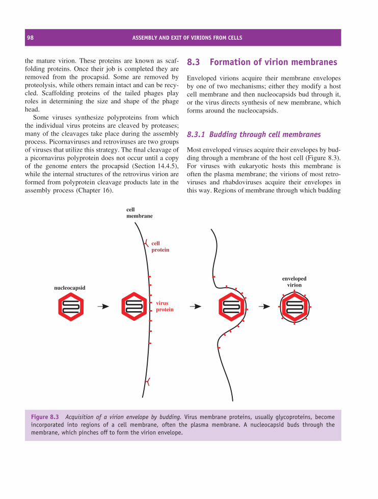
98 ASSEMBLY AND EXIT OF VIRIONS FROM CELLS
the mature virion. These proteins are known as scaf-folding proteins. Once their job is completed they areremoved from the procapsid. Some are removed byproteolysis, while others remain intact and can be recy-cled. Scaffolding proteins of the tailed phages playroles in determining the size and shape of the phagehead.
Some viruses synthesize polyproteins from whichthe individual virus proteins are cleaved by proteases;many of the cleavages take place during the assemblyprocess. Picornaviruses and retroviruses are two groupsof viruses that utilize this strategy. The final cleavage ofa picornavirus polyprotein does not occur until a copyof the genome enters the procapsid (Section 14.4.5),while the internal structures of the retrovirus virion areformed from polyprotein cleavage products late in theassembly process (Chapter 16).
8.3 Formation of virion membranes
Enveloped virions acquire their membrane envelopesby one of two mechanisms; either they modify a hostcell membrane and then nucleocapsids bud through it,or the virus directs synthesis of new membrane, whichforms around the nucleocapsids.
8.3.1 Budding through cell membranes
Most enveloped viruses acquire their envelopes by bud-ding through a membrane of the host cell (Figure 8.3).For viruses with eukaryotic hosts this membrane isoften the plasma membrane; the virions of most retro-viruses and rhabdoviruses acquire their envelopes inthis way. Regions of membrane through which budding
virusprotein
cellprotein
cellmembrane
nucleocapsid
envelopedvirion
Figure 8.3 Acquisition of a virion envelope by budding. Virus membrane proteins, usually glycoproteins, becomeincorporated into regions of a cell membrane, often the plasma membrane. A nucleocapsid buds through themembrane, which pinches off to form the virion envelope.
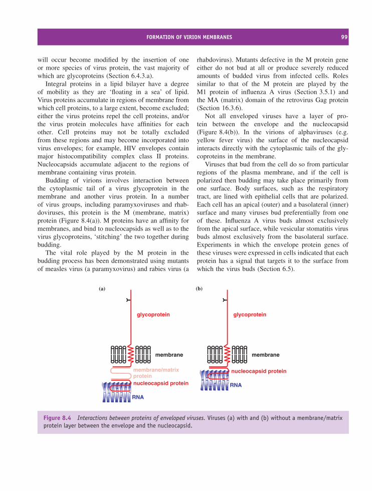
FORMATION OF VIRION MEMBRANES 99
will occur become modified by the insertion of oneor more species of virus protein, the vast majority ofwhich are glycoproteins (Section 6.4.3.a).
Integral proteins in a lipid bilayer have a degreeof mobility as they are ‘floating in a sea’ of lipid.Virus proteins accumulate in regions of membrane fromwhich cell proteins, to a large extent, become excluded;either the virus proteins repel the cell proteins, and/orthe virus protein molecules have affinities for eachother. Cell proteins may not be totally excludedfrom these regions and may become incorporated intovirus envelopes; for example, HIV envelopes containmajor histocompatibility complex class II proteins.Nucleocapsids accumulate adjacent to the regions ofmembrane containing virus protein.
Budding of virions involves interaction betweenthe cytoplasmic tail of a virus glycoprotein in themembrane and another virus protein. In a numberof virus groups, including paramyxoviruses and rhab-doviruses, this protein is the M (membrane, matrix)protein (Figure 8.4(a)). M proteins have an affinity formembranes, and bind to nucleocapsids as well as to thevirus glycoproteins, ‘stitching’ the two together duringbudding.
The vital role played by the M protein in thebudding process has been demonstrated using mutantsof measles virus (a paramyxovirus) and rabies virus (a
rhabdovirus). Mutants defective in the M protein geneeither do not bud at all or produce severely reducedamounts of budded virus from infected cells. Rolessimilar to that of the M protein are played by theM1 protein of influenza A virus (Section 3.5.1) andthe MA (matrix) domain of the retrovirus Gag protein(Section 16.3.6).
Not all enveloped viruses have a layer of pro-tein between the envelope and the nucleocapsid(Figure 8.4(b)). In the virions of alphaviruses (e.g.yellow fever virus) the surface of the nucleocapsidinteracts directly with the cytoplasmic tails of the gly-coproteins in the membrane.
Viruses that bud from the cell do so from particularregions of the plasma membrane, and if the cell ispolarized then budding may take place primarily fromone surface. Body surfaces, such as the respiratorytract, are lined with epithelial cells that are polarized.Each cell has an apical (outer) and a basolateral (inner)surface and many viruses bud preferentially from oneof these. Influenza A virus buds almost exclusivelyfrom the apical surface, while vesicular stomatitis virusbuds almost exclusively from the basolateral surface.Experiments in which the envelope protein genes ofthese viruses were expressed in cells indicated that eachprotein has a signal that targets it to the surface fromwhich the virus buds (Section 6.5).
nucleocapsid protein
glycoprotein
membrane/matrixprotein
membrane
RNA
glycoprotein
membrane
(a) (b)
nucleocapsid protein
RNA
Figure 8.4 Interactions between proteins of enveloped viruses. Viruses (a) with and (b) without a membrane/matrixprotein layer between the envelope and the nucleocapsid.
100 ASSEMBLY AND EXIT OF VIRIONS FROM CELLS
The late stage of budding involves the membranepinching off and the release of the newly formedvirion. There is evidence that host cell proteins bindto virus proteins and play vital roles in this process.Binding sites for these cell proteins have been identifiedin the Gag proteins of most retroviruses and in thematrix proteins of rhabdoviruses, paramyxoviruses andfiloviruses. Because they are involved in the late stageof budding the protein binding sites are known as late(L) domains. Mutations in these domains can resultin failure of virions to be released from the cellsurface.
The release of influenza virus particles from cellsrequires the activity of a virion enzyme: a neu-raminidase activity associated with an envelope gly-coprotein. At budding sites cleavage of neuraminicacid, the substrate for the enzyme, is essential for therelease of virions. Some anti-influenza virus drugs havebeen developed that act by inhibiting the neuraminidase(Section 25.3.5).
Some enveloped viruses bud from cell membranesother than the plasma membrane. Herpesvirus nucleo-capsids are constructed in the nucleus and begin theirjourney to the cytoplasm by budding through the inner
membrane of the nuclear envelope; the envelope ofthe mature virion is acquired in the cytoplasm by bud-ding into vesicles derived from the Golgi complex(Section 11.5.4). Hepadnaviruses bud into a membranecompartment between the endoplasmic reticulum andthe Golgi complex (Section 18.8.7).
One of the rare enveloped phages, Acholeplasmalaidlawii virus L2, acquires its envelope by buddingthrough the cell membrane of its mycoplasma host.
8.3.2 De novo synthesis of viral membranes
A minority of viruses direct the synthesis of lipidmembrane late in the replication cycle. In somecases the membrane forms a virion envelope (e.g.poxviruses); in other cases the membrane forms alayer below the surface of the capsid (e.g. iridoviruses;Section 3.5.2).
Baculoviruses produce two types of envelopedvirion during their replication. One type of virionhas the function of spreading the infection to othercells within the host, and this virion acquires itsenvelope by budding from the plasma membrane. Theother type of virion has the function of infecting new
Table 8.1 Summary of virion membrane acquisition
Site of virion Origin of virion Examplesassembly membrane
Virion envelope Internal virionmembrane
Eukaryotic cellcytoplasm
Plasma membrane Most retroviruses Mostrhabdoviruses
Post-RER membrane HepadnavirusesDe novo synthesis Poxviruses Iridoviruses
Eukaryotic cellnucleus
Inner nuclear membrane Nucleorhabdoviruses
De novo synthesis Baculovirus virions thatwill be occluded
Prokaryotic cell Cell membrane Acholeplasma laidlawiivirus L2
De novo synthesis Pseudomonas phage ϕ6 Alteromonas phagePM2
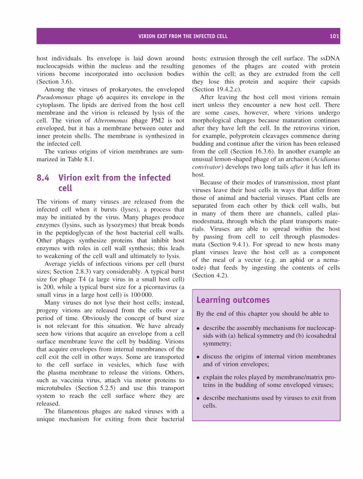
VIRION EXIT FROM THE INFECTED CELL 101
host individuals. Its envelope is laid down aroundnucleocapsids within the nucleus and the resultingvirions become incorporated into occlusion bodies(Section 3.6).
Among the viruses of prokaryotes, the envelopedPseudomonas phage ϕ6 acquires its envelope in thecytoplasm. The lipids are derived from the host cellmembrane and the virion is released by lysis of thecell. The virion of Alteromonas phage PM2 is notenveloped, but it has a membrane between outer andinner protein shells. The membrane is synthesized inthe infected cell.
The various origins of virion membranes are sum-marized in Table 8.1.
8.4 Virion exit from the infectedcell
The virions of many viruses are released from theinfected cell when it bursts (lyses), a process thatmay be initiated by the virus. Many phages produceenzymes (lysins, such as lysozymes) that break bondsin the peptidoglycan of the host bacterial cell walls.Other phages synthesize proteins that inhibit hostenzymes with roles in cell wall synthesis; this leadsto weakening of the cell wall and ultimately to lysis.
Average yields of infectious virions per cell (burstsizes; Section 2.8.3) vary considerably. A typical burstsize for phage T4 (a large virus in a small host cell)is 200, while a typical burst size for a picornavirus (asmall virus in a large host cell) is 100 000.
Many viruses do not lyse their host cells; instead,progeny virions are released from the cells over aperiod of time. Obviously the concept of burst sizeis not relevant for this situation. We have alreadyseen how virions that acquire an envelope from a cellsurface membrane leave the cell by budding. Virionsthat acquire envelopes from internal membranes of thecell exit the cell in other ways. Some are transportedto the cell surface in vesicles, which fuse withthe plasma membrane to release the virions. Others,such as vaccinia virus, attach via motor proteins tomicrotubules (Section 5.2.5) and use this transportsystem to reach the cell surface where they arereleased.
The filamentous phages are naked viruses with aunique mechanism for exiting from their bacterial
hosts: extrusion through the cell surface. The ssDNAgenomes of the phages are coated with proteinwithin the cell; as they are extruded from the cellthey lose this protein and acquire their capsids(Section 19.4.2.c).
After leaving the host cell most virions remaininert unless they encounter a new host cell. Thereare some cases, however, where virions undergomorphological changes because maturation continuesafter they have left the cell. In the retrovirus virion,for example, polyprotein cleavages commence duringbudding and continue after the virion has been releasedfrom the cell (Section 16.3.6). In another example anunusual lemon-shaped phage of an archaeon (Acidianusconvivator) develops two long tails after it has left itshost.
Because of their modes of transmission, most plantviruses leave their host cells in ways that differ fromthose of animal and bacterial viruses. Plant cells areseparated from each other by thick cell walls, butin many of them there are channels, called plas-modesmata, through which the plant transports mate-rials. Viruses are able to spread within the hostby passing from cell to cell through plasmodes-mata (Section 9.4.1). For spread to new hosts manyplant viruses leave the host cell as a componentof the meal of a vector (e.g. an aphid or a nema-tode) that feeds by ingesting the contents of cells(Section 4.2).
Learning outcomesBy the end of this chapter you should be able to
• describe the assembly mechanisms for nucleocap-sids with (a) helical symmetry and (b) icosahedralsymmetry;
• discuss the origins of internal virion membranesand of virion envelopes;
• explain the roles played by membrane/matrix pro-teins in the budding of some enveloped viruses;
• describe mechanisms used by viruses to exit fromcells.
102 ASSEMBLY AND EXIT OF VIRIONS FROM CELLS
Sources of further information
Books
Harrison S. C. (2005) Mechanism of membrane fusion byviral envelope proteins, pp 231–261 in Roy P., editorAdvances in Virus Research, Vol. 64, Elsevier
Hunter E. (2001) Virus assembly, Chapter 8 in Knipe D. M.and Howley P. M., editors-in-chief, Fields Virology, 4thedition, Lippincott, Williams and Wilkins
Schmitt A. P. and Lamb R. A. (2004) Escaping from thecell: assembly and budding of negative-strand RNAviruses, pp 145–196 in Yawaoka Y., editor, Biology ofNegative Strand RNA Viruses: the Power of Reverse Genet-ics, Springer
Journals
Bieniasz P. D. (2006) Late budding domains and hostproteins in enveloped virus release Virology, 344, 55–63
Chazal N. and Gerlier D. (2003) Virus entry, assembly,budding, and membrane rafts Microbiology and MolecularBiology Reviews, 67, 226–237
Herrero-Martınez E. et al. (2005) Vaccinia virus intracellularenveloped virions move to the cell periphery on micro-tubules in the absence of the A36R protein Journal ofGeneral Virology, 86, 2961–2968
Loessner M. J. (2005) Bacteriophage endolysins – currentstate of research and applications Current Opinion inMicrobiology, 8, 480–487
Steven A. C. et al. (2005) Virus maturation: dynamics andmechanism of a stabilizing structural transition that leadsto infectivity Current Opinion in Structural Biology, 15,227–236
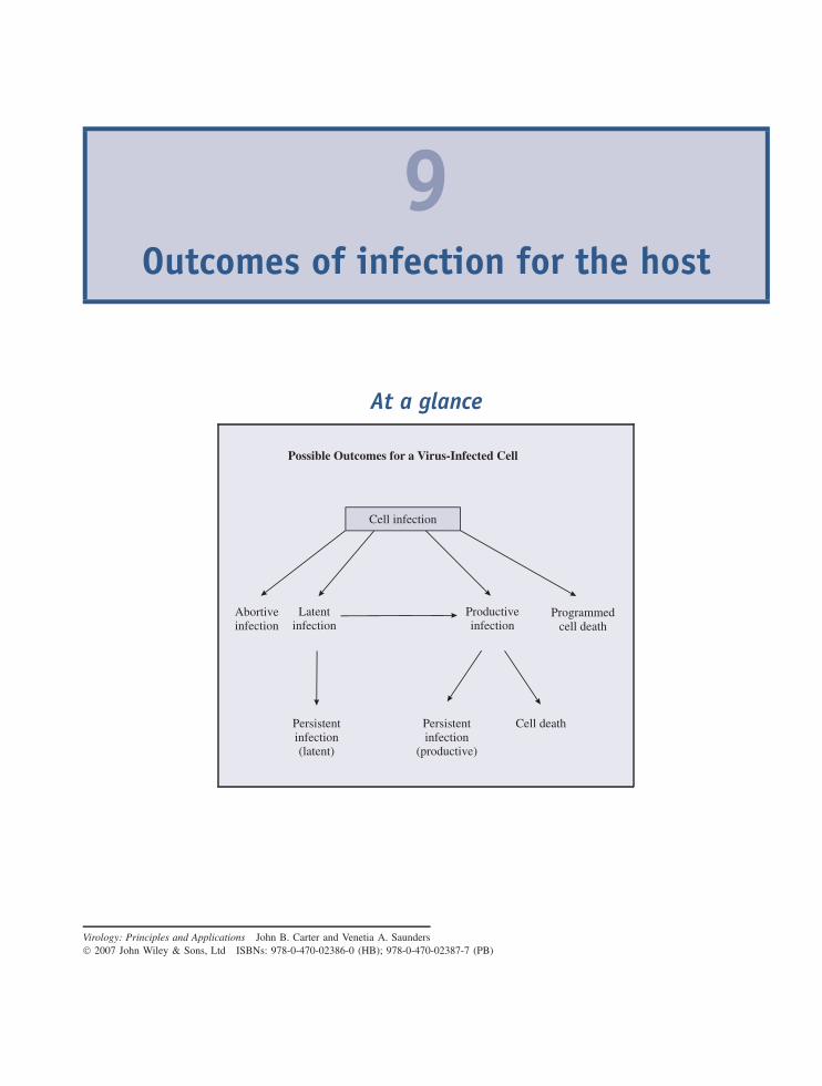
9Outcomes of infection for the host
At a glance
Cell infection
Abortiveinfection
Latentinfection
Persistentinfection
(productive)
Productiveinfection
Programmedcell death
Cell death
Possible Outcomes for a Virus-Infected Cell
Persistentinfection(latent)
Virology: Principles and Applications John B. Carter and Venetia A. Saunders 2007 John Wiley & Sons, Ltd ISBNs: 978-0-470-02386-0 (HB); 978-0-470-02387-7 (PB)
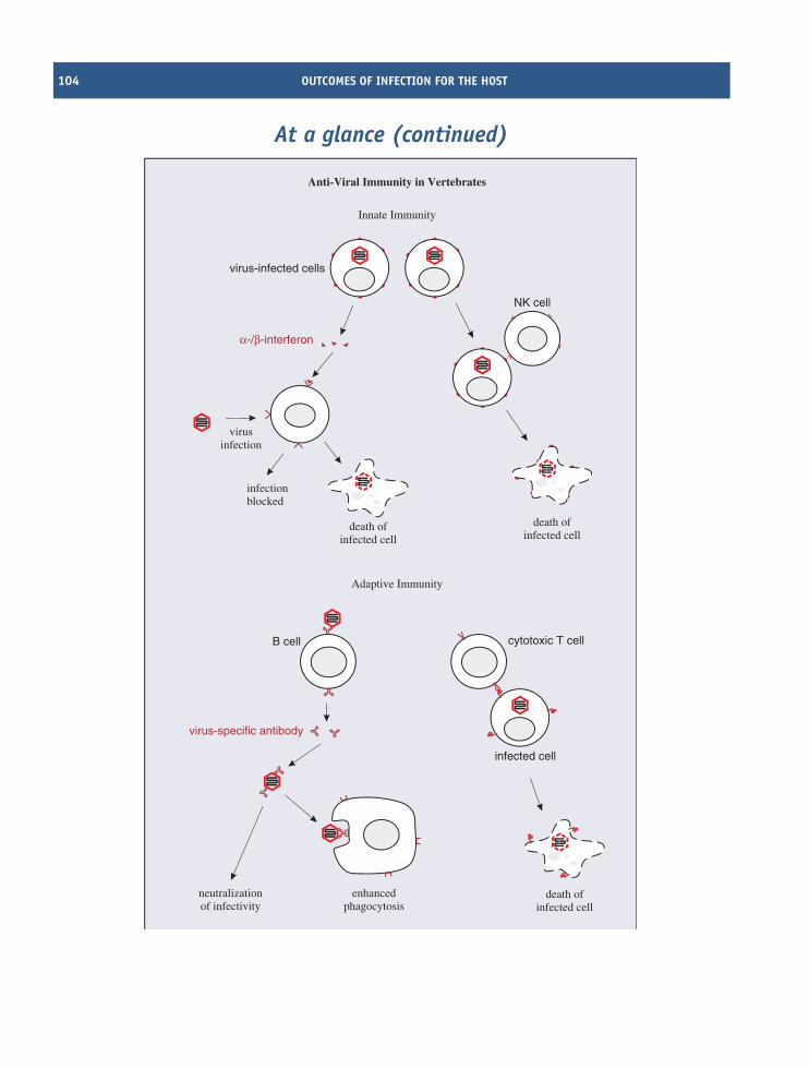
104 OUTCOMES OF INFECTION FOR THE HOST
At a glance (continued)
infected cell
death ofinfected cell
cytotoxic T cell
Innate Immunity
Adaptive Immunity
α-/β-interferon
virus-infected cells
infectionblocked
virusinfection
NK cell
neutralizationof infectivity
enhancedphagocytosis
virus-specific antibody
B cell
Anti-Viral Immunity in Vertebrates
death ofinfected cell
death ofinfected cell
FACTORS AFFECTING OUTCOMES OF INFECTION 105
9.1 Introduction to outcomesof infection for the host
In the previous few chapters we have looked at aspectsof the virus replication cycle that culminate in theexit of infective progeny virions from an infected cell.When this is the outcome the infection is said to beproductive. Virions may be released when the host celllyses, or the cell may survive releasing virions for aperiod, which may be short, as in the case of HIVinfection, or it may be long, as in the case of hepatitis Bvirus infection.
Some virus infections, however, are not productive,for a variety of reasons.
• An infection may become latent with the virusgenome persisting, perhaps for the lifetime of thecell, and perhaps in the daughter cells if the celldivides.
• An infection may be abortive, in which case neithera latent infection nor a productive infection isestablished. One cause of an abortive infection isa virus with a mutated genome; the virus is saidto be defective. It is unable to undergo a completereplication cycle, unless the cell is also infected witha virus that can provide the missing function(s). Avirus that is able to provide functions for a defectivevirus is known as a helper virus.
Some virus infections persist in their hosts for longperiods, sometimes for life. In some cases persistentinfections are productive, e.g. HIV; in other casespersistence may take the form of periods of latencyalternating with periods of productive infection, e.g.many herpesviruses. Long-term infection with someviruses may lead to the development of cancer in theinfected host (Chapter 22).
Some phages, including the filamentous phages(Section 19.4.2), initiate persistent productive infec-tions in cells of their bacterial hosts. Under appropriateconditions the cells can survive for long periods releas-ing progeny virions.
The outcome of a virus infection from the pointof view of the host may vary from harmless at oneend of the scale, through deleterious effects of varyingseverity, to death at the other end of the scale. The
outcome may hinge on complex interplay between avariety of host, viral and environmental factors.
Over time the organisms that are hosts to viruseshave evolved anti-viral defences, but the viruses havenot sat idly by. The relationship between a virus andits host usually involves an arms race and viruseshave developed various countermeasures against hostdefences. Some of these countermeasures are indicatedin boxes throughout this chapter.
In this chapter we concentrate mainly on outcomesof virus infections in vertebrate hosts, and we start byconsidering some aspects of host immunity that canaffect outcomes of infection.
9.2 Factors affecting outcomesof infection
A major factor affecting the outcome of infectionis the efficiency of the immune systems of thehost. In animals there are both innate and adaptiveimmune systems.
9.2.1 Innate immunity in vertebrates
Some aspects of innate immunity that viruses mightencounter prior to infecting cells were discussed inSection 4.3.3. Further components of innate immu-nity that might be encountered after cells havebecome infected are interferons and natural killer (NK)cells.
9.2.1.a Interferons
Interferons are proteins synthesized and secreted bycells in response to virus infection. A potent triggerfor interferon production is dsRNA, which is produced,not only by dsRNA viruses, but also by ssRNA virusesas they replicate.
The roles of interferons are to protect adjacentcells from infection and to activate T cell-mediatedimmunity (Section 9.2.2.b). There are a number oftypes of interferon. Alpha- and beta- (α- and β-)interferons are produced by most cell types whenthey become infected with viruses. After secretion, theinterferon molecules diffuse to nearby cells, where theycan trigger various anti-viral activities by binding tointerferon receptors (Figure 9.1).
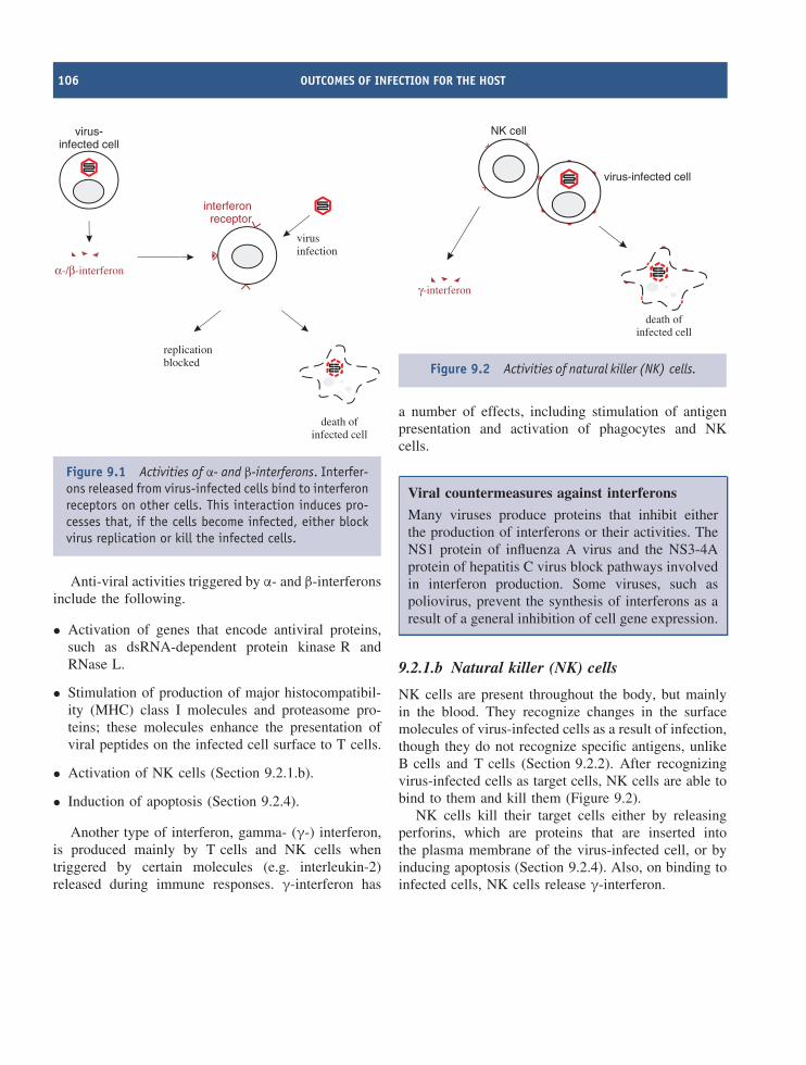
106 OUTCOMES OF INFECTION FOR THE HOST
α-/β-interferon
virus-infected cell
replicationblocked
interferonreceptor
virusinfection
death ofinfected cell
Figure 9.1 Activities of α- and β-interferons. Interfer-ons released from virus-infected cells bind to interferonreceptors on other cells. This interaction induces pro-cesses that, if the cells become infected, either blockvirus replication or kill the infected cells.
Anti-viral activities triggered by α- and β-interferonsinclude the following.
• Activation of genes that encode antiviral proteins,such as dsRNA-dependent protein kinase R andRNase L.
• Stimulation of production of major histocompatibil-ity (MHC) class I molecules and proteasome pro-teins; these molecules enhance the presentation ofviral peptides on the infected cell surface to T cells.
• Activation of NK cells (Section 9.2.1.b).
• Induction of apoptosis (Section 9.2.4).
Another type of interferon, gamma- (γ-) interferon,is produced mainly by T cells and NK cells whentriggered by certain molecules (e.g. interleukin-2)released during immune responses. γ-interferon has
virus-infected cell
NK cell
death ofinfected cell
γ-interferon
Figure 9.2 Activities of natural killer (NK) cells.
a number of effects, including stimulation of antigenpresentation and activation of phagocytes and NKcells.
Viral countermeasures against interferons
Many viruses produce proteins that inhibit eitherthe production of interferons or their activities. TheNS1 protein of influenza A virus and the NS3-4Aprotein of hepatitis C virus block pathways involvedin interferon production. Some viruses, such aspoliovirus, prevent the synthesis of interferons as aresult of a general inhibition of cell gene expression.
9.2.1.b Natural killer (NK) cells
NK cells are present throughout the body, but mainlyin the blood. They recognize changes in the surfacemolecules of virus-infected cells as a result of infection,though they do not recognize specific antigens, unlikeB cells and T cells (Section 9.2.2). After recognizingvirus-infected cells as target cells, NK cells are able tobind to them and kill them (Figure 9.2).
NK cells kill their target cells either by releasingperforins, which are proteins that are inserted intothe plasma membrane of the virus-infected cell, or byinducing apoptosis (Section 9.2.4). Also, on binding toinfected cells, NK cells release γ-interferon.
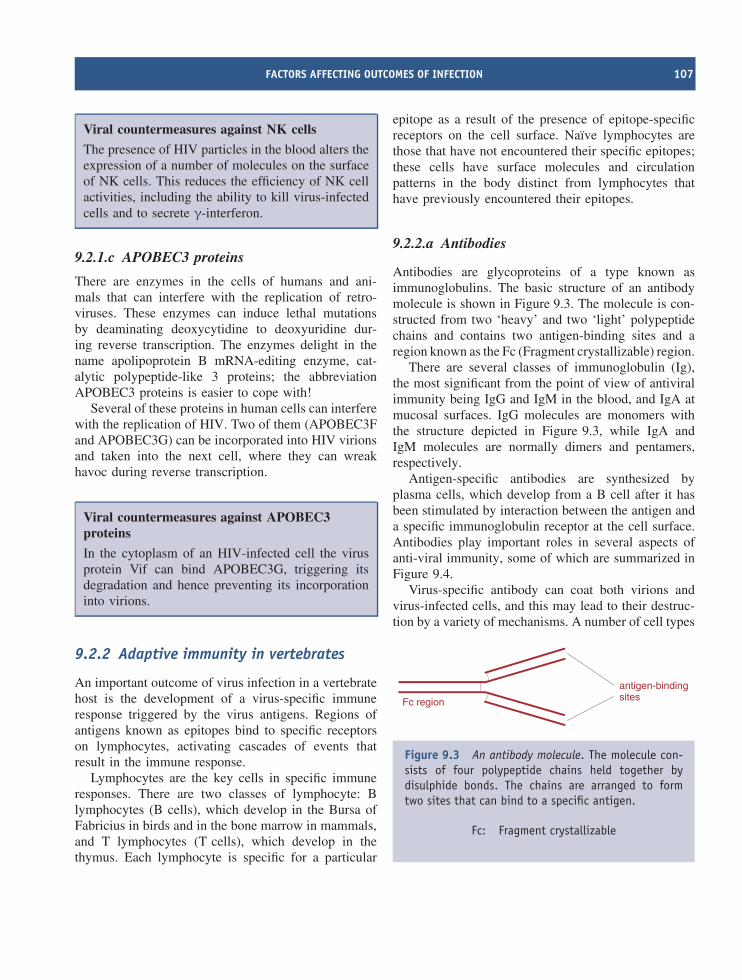
FACTORS AFFECTING OUTCOMES OF INFECTION 107
Viral countermeasures against NK cells
The presence of HIV particles in the blood alters theexpression of a number of molecules on the surfaceof NK cells. This reduces the efficiency of NK cellactivities, including the ability to kill virus-infectedcells and to secrete γ-interferon.
9.2.1.c APOBEC3 proteins
There are enzymes in the cells of humans and ani-mals that can interfere with the replication of retro-viruses. These enzymes can induce lethal mutationsby deaminating deoxycytidine to deoxyuridine dur-ing reverse transcription. The enzymes delight in thename apolipoprotein B mRNA-editing enzyme, cat-alytic polypeptide-like 3 proteins; the abbreviationAPOBEC3 proteins is easier to cope with!
Several of these proteins in human cells can interferewith the replication of HIV. Two of them (APOBEC3Fand APOBEC3G) can be incorporated into HIV virionsand taken into the next cell, where they can wreakhavoc during reverse transcription.
Viral countermeasures against APOBEC3proteins
In the cytoplasm of an HIV-infected cell the virusprotein Vif can bind APOBEC3G, triggering itsdegradation and hence preventing its incorporationinto virions.
9.2.2 Adaptive immunity in vertebrates
An important outcome of virus infection in a vertebratehost is the development of a virus-specific immuneresponse triggered by the virus antigens. Regions ofantigens known as epitopes bind to specific receptorson lymphocytes, activating cascades of events thatresult in the immune response.
Lymphocytes are the key cells in specific immuneresponses. There are two classes of lymphocyte: Blymphocytes (B cells), which develop in the Bursa ofFabricius in birds and in the bone marrow in mammals,and T lymphocytes (T cells), which develop in thethymus. Each lymphocyte is specific for a particular
epitope as a result of the presence of epitope-specificreceptors on the cell surface. Naıve lymphocytes arethose that have not encountered their specific epitopes;these cells have surface molecules and circulationpatterns in the body distinct from lymphocytes thathave previously encountered their epitopes.
9.2.2.a Antibodies
Antibodies are glycoproteins of a type known asimmunoglobulins. The basic structure of an antibodymolecule is shown in Figure 9.3. The molecule is con-structed from two ‘heavy’ and two ‘light’ polypeptidechains and contains two antigen-binding sites and aregion known as the Fc (Fragment crystallizable) region.
There are several classes of immunoglobulin (Ig),the most significant from the point of view of antiviralimmunity being IgG and IgM in the blood, and IgA atmucosal surfaces. IgG molecules are monomers withthe structure depicted in Figure 9.3, while IgA andIgM molecules are normally dimers and pentamers,respectively.
Antigen-specific antibodies are synthesized byplasma cells, which develop from a B cell after it hasbeen stimulated by interaction between the antigen anda specific immunoglobulin receptor at the cell surface.Antibodies play important roles in several aspects ofanti-viral immunity, some of which are summarized inFigure 9.4.
Virus-specific antibody can coat both virions andvirus-infected cells, and this may lead to their destruc-tion by a variety of mechanisms. A number of cell types
antigen-bindingsitesFc region
Figure 9.3 An antibody molecule. The molecule con-sists of four polypeptide chains held together bydisulphide bonds. The chains are arranged to formtwo sites that can bind to a specific antigen.
Fc: Fragment crystallizable
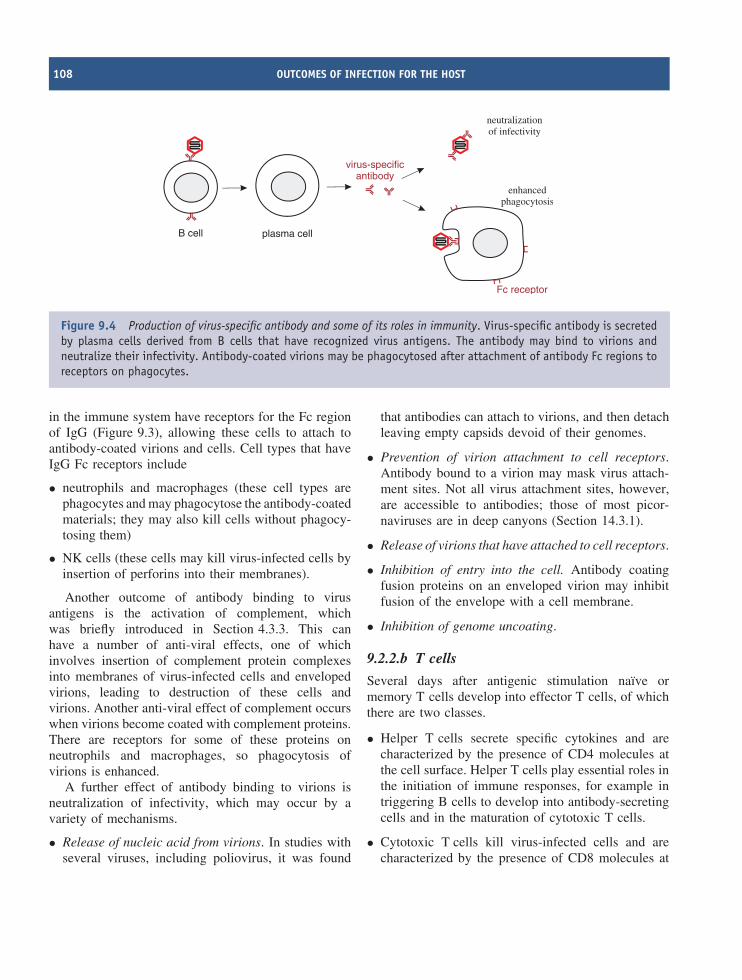
108 OUTCOMES OF INFECTION FOR THE HOST
plasma cell
neutralizationof infectivity
enhancedphagocytosis
virus-specificantibody
B cell
Fc receptor
Figure 9.4 Production of virus-specific antibody and some of its roles in immunity. Virus-specific antibody is secretedby plasma cells derived from B cells that have recognized virus antigens. The antibody may bind to virions andneutralize their infectivity. Antibody-coated virions may be phagocytosed after attachment of antibody Fc regions toreceptors on phagocytes.
in the immune system have receptors for the Fc regionof IgG (Figure 9.3), allowing these cells to attach toantibody-coated virions and cells. Cell types that haveIgG Fc receptors include
• neutrophils and macrophages (these cell types arephagocytes and may phagocytose the antibody-coatedmaterials; they may also kill cells without phagocy-tosing them)
• NK cells (these cells may kill virus-infected cells byinsertion of perforins into their membranes).
Another outcome of antibody binding to virusantigens is the activation of complement, whichwas briefly introduced in Section 4.3.3. This canhave a number of anti-viral effects, one of whichinvolves insertion of complement protein complexesinto membranes of virus-infected cells and envelopedvirions, leading to destruction of these cells andvirions. Another anti-viral effect of complement occurswhen virions become coated with complement proteins.There are receptors for some of these proteins onneutrophils and macrophages, so phagocytosis ofvirions is enhanced.
A further effect of antibody binding to virions isneutralization of infectivity, which may occur by avariety of mechanisms.
• Release of nucleic acid from virions. In studies withseveral viruses, including poliovirus, it was found
that antibodies can attach to virions, and then detachleaving empty capsids devoid of their genomes.
• Prevention of virion attachment to cell receptors.Antibody bound to a virion may mask virus attach-ment sites. Not all virus attachment sites, however,are accessible to antibodies; those of most picor-naviruses are in deep canyons (Section 14.3.1).
• Release of virions that have attached to cell receptors.
• Inhibition of entry into the cell. Antibody coatingfusion proteins on an enveloped virion may inhibitfusion of the envelope with a cell membrane.
• Inhibition of genome uncoating.
9.2.2.b T cells
Several days after antigenic stimulation naıve ormemory T cells develop into effector T cells, of whichthere are two classes.
• Helper T cells secrete specific cytokines and arecharacterized by the presence of CD4 molecules atthe cell surface. Helper T cells play essential roles inthe initiation of immune responses, for example intriggering B cells to develop into antibody-secretingcells and in the maturation of cytotoxic T cells.
• Cytotoxic T cells kill virus-infected cells and arecharacterized by the presence of CD8 molecules at
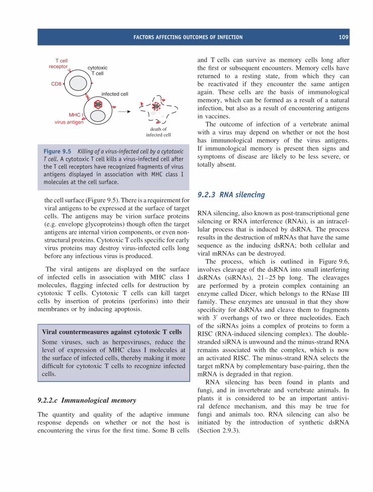
FACTORS AFFECTING OUTCOMES OF INFECTION 109
infected cell
death ofinfected cell
cytotoxicT cell
T cellreceptor
MHC Ivirus antigen
CD8
Figure 9.5 Killing of a virus-infected cell by a cytotoxicT cell. A cytotoxic T cell kills a virus-infected cell afterthe T cell receptors have recognized fragments of virusantigens displayed in association with MHC class Imolecules at the cell surface.
the cell surface (Figure 9.5). There is a requirement forviral antigens to be expressed at the surface of targetcells. The antigens may be virion surface proteins(e.g. envelope glycoproteins) though often the targetantigens are internal virion components, or even non-structural proteins. Cytotoxic T cells specific for earlyvirus proteins may destroy virus-infected cells longbefore any infectious virus is produced.
The viral antigens are displayed on the surfaceof infected cells in association with MHC class Imolecules, flagging infected cells for destruction bycytotoxic T cells. Cytotoxic T cells can kill targetcells by insertion of proteins (perforins) into theirmembranes or by inducing apoptosis.
Viral countermeasures against cytotoxic T cells
Some viruses, such as herpesviruses, reduce thelevel of expression of MHC class I molecules atthe surface of infected cells, thereby making it moredifficult for cytotoxic T cells to recognize infectedcells.
9.2.2.c Immunological memory
The quantity and quality of the adaptive immuneresponse depends on whether or not the host isencountering the virus for the first time. Some B cells
and T cells can survive as memory cells long afterthe first or subsequent encounters. Memory cells havereturned to a resting state, from which they canbe reactivated if they encounter the same antigenagain. These cells are the basis of immunologicalmemory, which can be formed as a result of a naturalinfection, but also as a result of encountering antigensin vaccines.
The outcome of infection of a vertebrate animalwith a virus may depend on whether or not the hosthas immunological memory of the virus antigens.If immunological memory is present then signs andsymptoms of disease are likely to be less severe, ortotally absent.
9.2.3 RNA silencing
RNA silencing, also known as post-transcriptional genesilencing or RNA interference (RNAi), is an intracel-lular process that is induced by dsRNA. The processresults in the destruction of mRNAs that have the samesequence as the inducing dsRNA; both cellular andviral mRNAs can be destroyed.
The process, which is outlined in Figure 9.6,involves cleavage of the dsRNA into small interferingdsRNAs (siRNAs), 21–25 bp long. The cleavagesare performed by a protein complex containing anenzyme called Dicer, which belongs to the RNase IIIfamily. These enzymes are unusual in that they showspecificity for dsRNAs and cleave them to fragmentswith 3′ overhangs of two or three nucleotides. Eachof the siRNAs joins a complex of proteins to form aRISC (RNA-induced silencing complex). The double-stranded siRNA is unwound and the minus-strand RNAremains associated with the complex, which is nowan activated RISC. The minus-strand RNA selects thetarget mRNA by complementary base-pairing, then themRNA is degraded in that region.
RNA silencing has been found in plants andfungi, and in invertebrate and vertebrate animals. Inplants it is considered to be an important antivi-ral defence mechanism, and this may be true forfungi and animals too. RNA silencing can also beinitiated by the introduction of synthetic dsRNA(Section 2.9.3).
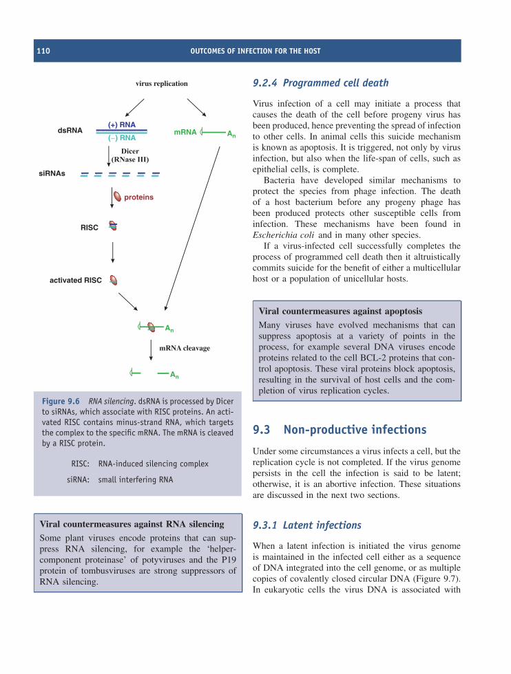
110 OUTCOMES OF INFECTION FOR THE HOST
An
An
dsRNA
siRNAs
virus replication
RISC
AnmRNA
proteins
activated RISC
mRNA cleavage
( ) RNA−
(+) RNA
Figure 9.6 RNA silencing. dsRNA is processed by Dicerto siRNAs, which associate with RISC proteins. An acti-vated RISC contains minus-strand RNA, which targetsthe complex to the specific mRNA. The mRNA is cleavedby a RISC protein.
RISC: RNA-induced silencing complex
siRNA: small interfering RNA
Viral countermeasures against RNA silencing
Some plant viruses encode proteins that can sup-press RNA silencing, for example the ‘helper-component proteinase’ of potyviruses and the P19protein of tombusviruses are strong suppressors ofRNA silencing.
9.2.4 Programmed cell death
Virus infection of a cell may initiate a process thatcauses the death of the cell before progeny virus hasbeen produced, hence preventing the spread of infectionto other cells. In animal cells this suicide mechanismis known as apoptosis. It is triggered, not only by virusinfection, but also when the life-span of cells, such asepithelial cells, is complete.
Bacteria have developed similar mechanisms toprotect the species from phage infection. The deathof a host bacterium before any progeny phage hasbeen produced protects other susceptible cells frominfection. These mechanisms have been found inEscherichia coli and in many other species.
If a virus-infected cell successfully completes theprocess of programmed cell death then it altruisticallycommits suicide for the benefit of either a multicellularhost or a population of unicellular hosts.
Viral countermeasures against apoptosis
Many viruses have evolved mechanisms that cansuppress apoptosis at a variety of points in theprocess, for example several DNA viruses encodeproteins related to the cell BCL-2 proteins that con-trol apoptosis. These viral proteins block apoptosis,resulting in the survival of host cells and the com-pletion of virus replication cycles.
9.3 Non-productive infections
Under some circumstances a virus infects a cell, but thereplication cycle is not completed. If the virus genomepersists in the cell the infection is said to be latent;otherwise, it is an abortive infection. These situationsare discussed in the next two sections.
9.3.1 Latent infections
When a latent infection is initiated the virus genomeis maintained in the infected cell either as a sequenceof DNA integrated into the cell genome, or as multiplecopies of covalently closed circular DNA (Figure 9.7).In eukaryotic cells the virus DNA is associated with
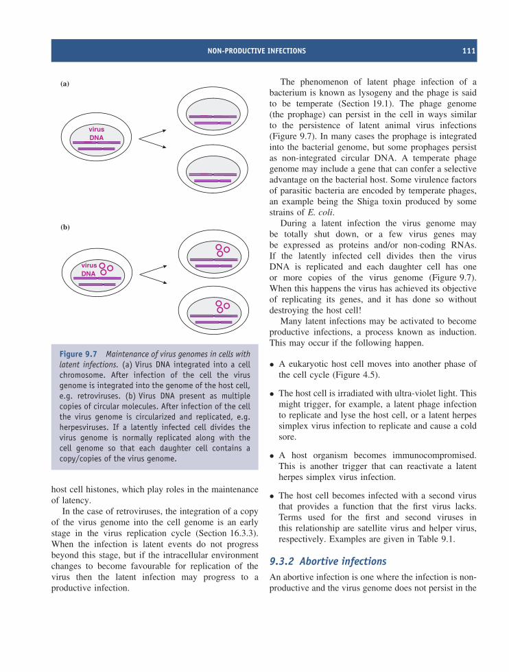
NON-PRODUCTIVE INFECTIONS 111
virusDNA
virusDNA
(a)
(b)
Figure 9.7 Maintenance of virus genomes in cells withlatent infections. (a) Virus DNA integrated into a cellchromosome. After infection of the cell the virusgenome is integrated into the genome of the host cell,e.g. retroviruses. (b) Virus DNA present as multiplecopies of circular molecules. After infection of the cellthe virus genome is circularized and replicated, e.g.herpesviruses. If a latently infected cell divides thevirus genome is normally replicated along with thecell genome so that each daughter cell contains acopy/copies of the virus genome.
host cell histones, which play roles in the maintenanceof latency.
In the case of retroviruses, the integration of a copyof the virus genome into the cell genome is an earlystage in the virus replication cycle (Section 16.3.3).When the infection is latent events do not progressbeyond this stage, but if the intracellular environmentchanges to become favourable for replication of thevirus then the latent infection may progress to aproductive infection.
The phenomenon of latent phage infection of abacterium is known as lysogeny and the phage is saidto be temperate (Section 19.1). The phage genome(the prophage) can persist in the cell in ways similarto the persistence of latent animal virus infections(Figure 9.7). In many cases the prophage is integratedinto the bacterial genome, but some prophages persistas non-integrated circular DNA. A temperate phagegenome may include a gene that can confer a selectiveadvantage on the bacterial host. Some virulence factorsof parasitic bacteria are encoded by temperate phages,an example being the Shiga toxin produced by somestrains of E. coli.
During a latent infection the virus genome maybe totally shut down, or a few virus genes maybe expressed as proteins and/or non-coding RNAs.If the latently infected cell divides then the virusDNA is replicated and each daughter cell has oneor more copies of the virus genome (Figure 9.7).When this happens the virus has achieved its objectiveof replicating its genes, and it has done so withoutdestroying the host cell!
Many latent infections may be activated to becomeproductive infections, a process known as induction.This may occur if the following happen.
• A eukaryotic host cell moves into another phase ofthe cell cycle (Figure 4.5).
• The host cell is irradiated with ultra-violet light. Thismight trigger, for example, a latent phage infectionto replicate and lyse the host cell, or a latent herpessimplex virus infection to replicate and cause a coldsore.
• A host organism becomes immunocompromised.This is another trigger that can reactivate a latentherpes simplex virus infection.
• The host cell becomes infected with a second virusthat provides a function that the first virus lacks.Terms used for the first and second viruses inthis relationship are satellite virus and helper virus,respectively. Examples are given in Table 9.1.
9.3.2 Abortive infectionsAn abortive infection is one where the infection is non-productive and the virus genome does not persist in the
112 OUTCOMES OF INFECTION FOR THE HOST
Table 9.1 Examples of satellite/helper viruses
Satellite virus Helper virus
Hepatitis delta virus Hepatitis B virusAdeno-associated viruses Adenovirus
cell as a latent infection. An infection might be abortivefor reasons concerning the cell, the environmental con-ditions and/or the virus. A virus may initiate productiveinfections in some cell types (permissive cells) andabortive infections in other cell types (non-permissivecells). Some abortive infections can kill the host cell.
Some virions may contain mutated genomes that maybe able to initiate the replication cycle, but may not havethe full set of functional genes necessary to complete it.Such virions are said to be defective, and if they infectcells without the means of complementing the mutatedor missing genes the result is an abortive infection.
There are a number of types of defective virus. Onetype is known as a defective interfering particle (DIP).DIPs often arise in the laboratory after animal viruseshave been passaged several times in cell culture at highmultiplicities of infection. DIPs have also been reportedafter passage in chick embryos and in mice, and DIPsof plant viruses have been observed.
Most DIPs contain less nucleic acid than thestandard virus from which they were derived. Bythemselves DIPs are either non-infectious or they caninitiate only abortive infections. The standard virus canact as helper virus to a DIP, so in cells that are co-infected with a DIP and the standard virus the DIP isable to replicate its genome and produce progeny DIPs.When this happens the replication of the DIP interfereswith the replication of the standard virus and reducesits yield, hence ‘interfering’ particle.
9.4 Productive infections9.4.1 Spread of infections within
multicellular hosts
Progeny virions from the first-infected cell of amulticellular host may infect cells nearby, for example
Table 9.2 Involvement of plant virus movementproteins (MPs) in the spread of infection viaplasmodesmata
Mode of transportthrough plasmodesmata
Virus examples
Virus RNA–MPcomplexes transported
Tobacco mosaicvirus
Cowpea chloroticmottle virus
Virus RNA–coatprotein–MPcomplexes transported
Cucumber mosaicvirus
Virions transportedthrough tubulescomposed of MP
Cowpea mosaicvirus
common cold viruses and rotaviruses might infect otherepithelial cells lining the respiratory tract and theintestinal tract, respectively. Most adjacent animal cellsare separated from each other only by their plasmamembranes, providing opportunities for direct cell-to-cell spread for some viruses.
Plant viruses are able to spread from cell to cellby passing through plasmodesmata, and each plantvirus encodes between one and four specialized pro-teins that enable them to do this. These proteinsare known as movement proteins (MPs) and theyact in a variety of ways. Some MPs form com-plexes either with virus RNA or with virus RNAplus coat protein molecules; other MPs form tubu-lar structures through which fully encapsidated virusRNA is transported (Table 9.2). MPs are multifunc-tional proteins with other roles in the virus replicationcycles.
In some circumstances progeny virions from infectedcells may be transported to distant sites in the host,where susceptible cells may become infected. In theanimal body the blood and the nerves may act as trans-port vehicles, while in plants transport may occur viathe phloem.
PRODUCTIVE INFECTIONS 113
9.4.2 DiseaseMany virus infections result in no disease in the host,while at the other end of the scale a virus infection mayresult in fatal disease, such as rabies or AIDS. Betweenthese extremes is a huge range of severity in diseasescaused by viruses.
Disease may be manifest as symptoms and/orsigns. In medical terminology symptoms are subjectivefeatures, such as abdominal pain and fatigue, whereassigns are objective features, such as blood in thefaeces and skin rashes. Symptoms can be recognizedonly by the infected individual, whereas signs canbe recognized by others. Plant virologists and insectvirologists sometimes use the word symptom inconnection with objective features, such as leaf lesionsin a plant (Figure 2.17) and iridescence in an insect(Chapter 1, at a glance).
Infection with some viruses, such as dependoviruses,some herpesviruses and some reoviruses, apparentlynever causes disease, while infection with otherviruses, such as human parvovirus B19, poliovirus andhepatitis B virus, may or may not result in disease.Infections that do not result in disease are said to besubclinical or asymptomatic.
The outcome of a complex interplay of factorsdetermines whether or not a virus infection results indisease, and if so the severity of the disease. Thesefactors may include virus factors, host factors andhuman intervention.
Not all viruses are pathogens (disease-causingagents) and some are pathogenic only under certaincircumstances. For pathogenic viruses factors that canaffect the outcome of infection include the following.
• The virulence of the virus strain. The virulence ofa virus (or any micro-organism) is a measure ofthe severity of disease it is capable of causing.Influenza A type H5N1 can be said to be morevirulent in humans than types H1N1 and H3N2
(Section 21.5.2), as type H5N1 causes more severedisease than the other two types.
• The dose of virus. A larger dose of virus may resultin a shorter incubation period (the time betweeninfection and the first appearance of signs and/orsymptoms).
Host factors that can affect the outcome of infec-tion include the effectiveness of immune systems (dis-cussed in Chapter 4 and earlier in this chapter), andthese in turn vary with age and nutritional status of thehost. It should be noted that a strong immune responseby the host does not guarantee the elimination of thevirus. HIV continues to replicate in the presence ofhigh levels of HIV-specific antibody and T cells. Insome cases signs and/or symptoms may result from thehost’s immune response against the virus. The measlesrash and herpes simplex lesions are clinical manifesta-tions of attempts by the human body to destroy virus-infected cells.
Human interventions that could influence the out-come of a virus infection in a human or an animalinclude the administration of anti-viral antibodies oranti-viral drugs (Chapter 25).
Recovery of the host from a virus disease may beaccompanied by elimination of the virus, as normallyoccurs after infection with common cold viruses inthe respiratory tract and rotavirus infections in theintestine. Alternatively, recovery may be accompaniedby establishment of a long-term infection, perhapsfor life.Such persistent infections, whether they areproductive or latent, may have no further consequencesfor the host, or they may cause disease when the hostis older. Childhood infection with varicella-zoster virusmay result in a lifetime latent infection, which maybe reactivated, causing shingles (Section 11.2.2). Long-term persistent infection of humans and animals withcertain viruses may lead to the development of cancer(Chapter 22).
114 OUTCOMES OF INFECTION FOR THE HOST
Learning outcomesBy the end of this chapter you should be able to
• describe the major components of innate andadaptive immunity in vertebrates;
• outline the process of RNA silencing;
• explain programmed cell death;
• explain the terms
◦ productive infection
◦ non-productive infection
◦ latent infection
◦ abortive infection
◦ defective virus;
• discuss the spread of virus infections withinanimal bodies and within plants;
• discuss the factors that determine whether virusinfection results in disease.
Sources of further informationBooks (general immunology)Mims C. A. et al. (2004) Sections 2 and 3 in Medical
Microbiology, 3rd edition, MosbyPlayfair J. H. L. and Bancroft G. J. (2004) Infection and
Immunity, 2nd edition, Oxford University PressRabson A., Roitt I. and Delves P. (2005) Really Essential
Medical Immunology, 2nd edition, Blackwell
Books (immune responses to virus infections)Digard P., Nash A. A. and Randall R. E., editors (2005)
Molecular Pathogenesis of Virus Infections, 64th Sympo-sium of the Society for General Microbiology, CambridgeUniversity Press
Chen Y.-B., Fannjang Y. and Hardwick J. M. (2004) Celldeath in viral infections, Chapter 17 in When Cells DieII, editors Lockshin R. A. and Zakeri Z., Wiley
Whitton J. L. and Oldstone M. B. A. (2001) The immuneresponse to viruses, Chapter 11 in Knipe D. M. and How-ley P. M., editors-in-chief, Fields Virology, 4th edition,Lippincott, Williams and Wilkins
Historical paperIsaacs A. and Lindenmann J. (1957) Virus interference. 1.
The interferon Proceedings of the Royal Society of LondonB, 147, 258–267
Recent papersCuconati A. and White E. (2002) Viral homologs of BCL-2:
role of apoptosis in the regulation of virus infection Genesand Development, 16, 2465–2478
Doehle B. P., Schafer A. and Cullen B. R. (2005) HumanAPOBEC3B is a potent inhibitor of HIV-1 infectivity andis resistant to HIV-1 Vif Virology, 339, 281–288
Fauci A. S., Mavilio D. and Kottilil S. (2005) NK cells inHIV infection: paradigm for protection or targets forambush Nature Reviews Immunology, 5, 835–843
Haller O., Kochs G. and Weber F. (2006) The interferonresponse circuit: induction and suppression by pathogenicviruses Virology, 344, 119–130
Iannello A. et al. (2006) Viral strategies for evading antiviralcellular immune responses of the host Journal of LeukocyteBiology, 79, 16–35
Lieberman P. M. (2006) Chromatin regulation of virusinfection Trends In Microbiology, 14, 132–140
Scholthof H. B. (2005) Plant virus transport: motions offunctional equivalence Trends in Plant Science, 10,376–382
Soosaar J. L. M. et al. (2005) Mechanisms of plant resis-tance to viruses Nature Reviews Microbiology, 3,789–798
Stram Y. and Kuzntzova L. (2006) Inhibition of viruses byRNA interference Virus Genes, 32, 299–306
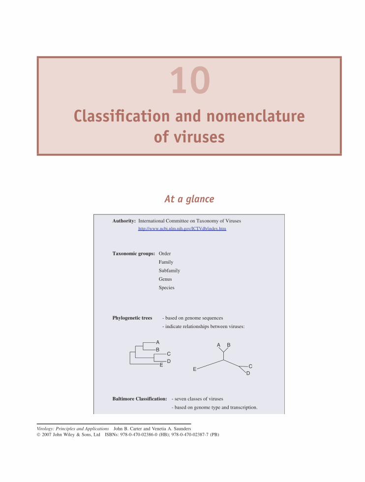
10Classification and nomenclature
of viruses
At a glance
Authority: International Committee on Taxonomy of Viruses
http://www.ncbi.nlm.nih.gov/ICTVdb/index.htm
Taxonomic groups: Order
Family
Subfamily
Genus
Species
Phylogenetic trees - based on genome sequences
- indicate relationships between viruses:
Baltimore Classification: - seven classes of viruses
- based on genome type and transcription.
AB
CD
E
A B
CD
E
Virology: Principles and Applications John B. Carter and Venetia A. Saunders 2007 John Wiley & Sons, Ltd ISBNs: 978-0-470-02386-0 (HB); 978-0-470-02387-7 (PB)
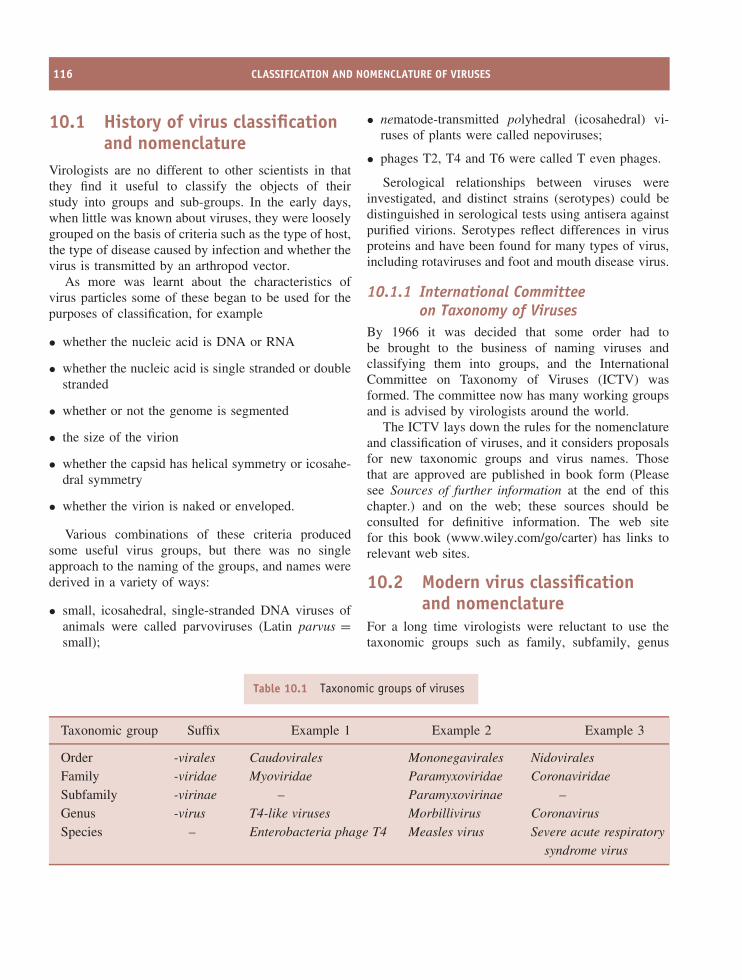
116 CLASSIFICATION AND NOMENCLATURE OF VIRUSES
10.1 History of virus classificationand nomenclature
Virologists are no different to other scientists in thatthey find it useful to classify the objects of theirstudy into groups and sub-groups. In the early days,when little was known about viruses, they were looselygrouped on the basis of criteria such as the type of host,the type of disease caused by infection and whether thevirus is transmitted by an arthropod vector.
As more was learnt about the characteristics ofvirus particles some of these began to be used for thepurposes of classification, for example
• whether the nucleic acid is DNA or RNA
• whether the nucleic acid is single stranded or doublestranded
• whether or not the genome is segmented
• the size of the virion
• whether the capsid has helical symmetry or icosahe-dral symmetry
• whether the virion is naked or enveloped.
Various combinations of these criteria producedsome useful virus groups, but there was no singleapproach to the naming of the groups, and names werederived in a variety of ways:
• small, icosahedral, single-stranded DNA viruses ofanimals were called parvoviruses (Latin parvus =small);
• nematode-transmitted polyhedral (icosahedral) vi-ruses of plants were called nepoviruses;
• phages T2, T4 and T6 were called T even phages.
Serological relationships between viruses wereinvestigated, and distinct strains (serotypes) could bedistinguished in serological tests using antisera againstpurified virions. Serotypes reflect differences in virusproteins and have been found for many types of virus,including rotaviruses and foot and mouth disease virus.
10.1.1 International Committeeon Taxonomy of Viruses
By 1966 it was decided that some order had tobe brought to the business of naming viruses andclassifying them into groups, and the InternationalCommittee on Taxonomy of Viruses (ICTV) wasformed. The committee now has many working groupsand is advised by virologists around the world.
The ICTV lays down the rules for the nomenclatureand classification of viruses, and it considers proposalsfor new taxonomic groups and virus names. Thosethat are approved are published in book form (Pleasesee Sources of further information at the end of thischapter.) and on the web; these sources should beconsulted for definitive information. The web sitefor this book (www.wiley.com/go/carter) has links torelevant web sites.
10.2 Modern virus classificationand nomenclature
For a long time virologists were reluctant to use thetaxonomic groups such as family, subfamily, genus
Table 10.1 Taxonomic groups of viruses
Taxonomic group Suffix Example 1 Example 2 Example 3
Order -virales Caudovirales Mononegavirales NidoviralesFamily -viridae Myoviridae Paramyxoviridae CoronaviridaeSubfamily -virinae – Paramyxovirinae –Genus -virus T4-like viruses Morbillivirus CoronavirusSpecies – Enterobacteria phage T4 Measles virus Severe acute respiratory
syndrome virus
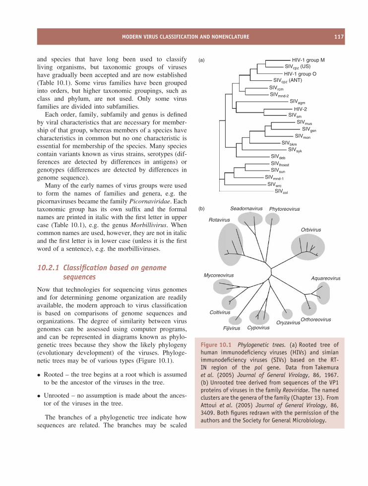
MODERN VIRUS CLASSIFICATION AND NOMENCLATURE 117
and species that have long been used to classifyliving organisms, but taxonomic groups of viruseshave gradually been accepted and are now established(Table 10.1). Some virus families have been groupedinto orders, but higher taxonomic groupings, such asclass and phylum, are not used. Only some virusfamilies are divided into subfamilies.
Each order, family, subfamily and genus is definedby viral characteristics that are necessary for member-ship of that group, whereas members of a species havecharacteristics in common but no one characteristic isessential for membership of the species. Many speciescontain variants known as virus strains, serotypes (dif-ferences are detected by differences in antigens) orgenotypes (differences are detected by differences ingenome sequence).
Many of the early names of virus groups were usedto form the names of families and genera, e.g. thepicornaviruses became the family Picornaviridae. Eachtaxonomic group has its own suffix and the formalnames are printed in italic with the first letter in uppercase (Table 10.1), e.g. the genus Morbillivirus. Whencommon names are used, however, they are not in italicand the first letter is in lower case (unless it is the firstword of a sentence), e.g. the morbilliviruses.
10.2.1 Classification based on genomesequences
Now that technologies for sequencing virus genomesand for determining genome organization are readilyavailable, the modern approach to virus classificationis based on comparisons of genome sequences andorganizations. The degree of similarity between virusgenomes can be assessed using computer programs,and can be represented in diagrams known as phylo-genetic trees because they show the likely phylogeny(evolutionary development) of the viruses. Phyloge-netic trees may be of various types (Figure 10.1).
• Rooted – the tree begins at a root which is assumedto be the ancestor of the viruses in the tree.
• Unrooted – no assumption is made about the ances-tor of the viruses in the tree.
The branches of a phylogenetic tree indicate howsequences are related. The branches may be scaled
Seadornavirus
Rotavirus
Mycoreovirus
Coltivirus
Fijivirus CypovirusOryzavirus
Orthoreovirus
Aquareovirus
Phytoreovirus
Orbivirus
(b)
(a)
SIVcol
SIVwrc
SIVmnd-1
SIVsun
SIVlhoest
SIVdeb
SIVsyk
SIVbkm
SIVmon
SIVgsn
SIVmus
SIVsm
HIV-2
HIV-1 group O
HIV-1 group M
SIVagm
SIVmnd-2
SIVrcm
SIVcpz (ANT)
SIVcpz (US)
Figure 10.1 Phylogenetic trees. (a) Rooted tree ofhuman immunodeficiency viruses (HIVs) and simianimmunodeficiency viruses (SIVs) based on the RT-IN region of the pol gene. Data from Takemuraet al. (2005) Journal of General Virology, 86, 1967.(b) Unrooted tree derived from sequences of the VP1proteins of viruses in the family Reoviridae. The namedclusters are the genera of the family (Chapter 13). FromAttoui et al. (2005) Journal of General Virology, 86,3409. Both figures redrawn with the permission of theauthors and the Society for General Microbiology.
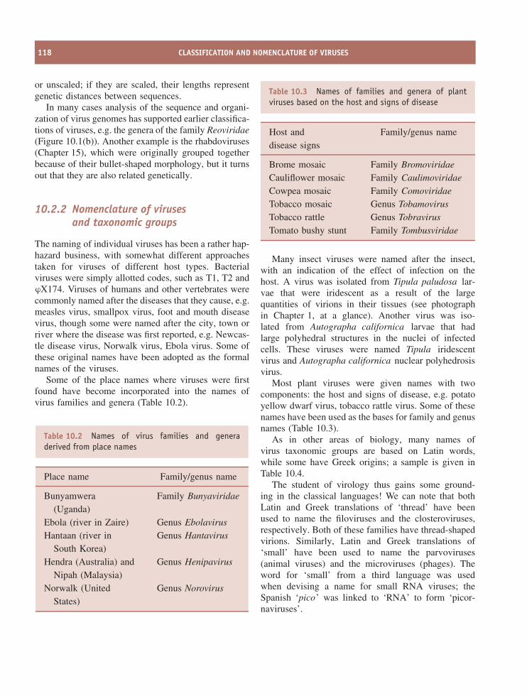
118 CLASSIFICATION AND NOMENCLATURE OF VIRUSES
or unscaled; if they are scaled, their lengths representgenetic distances between sequences.
In many cases analysis of the sequence and organi-zation of virus genomes has supported earlier classifica-tions of viruses, e.g. the genera of the family Reoviridae(Figure 10.1(b)). Another example is the rhabdoviruses(Chapter 15), which were originally grouped togetherbecause of their bullet-shaped morphology, but it turnsout that they are also related genetically.
10.2.2 Nomenclature of virusesand taxonomic groups
The naming of individual viruses has been a rather hap-hazard business, with somewhat different approachestaken for viruses of different host types. Bacterialviruses were simply allotted codes, such as T1, T2 andϕX174. Viruses of humans and other vertebrates werecommonly named after the diseases that they cause, e.g.measles virus, smallpox virus, foot and mouth diseasevirus, though some were named after the city, town orriver where the disease was first reported, e.g. Newcas-tle disease virus, Norwalk virus, Ebola virus. Some ofthese original names have been adopted as the formalnames of the viruses.
Some of the place names where viruses were firstfound have become incorporated into the names ofvirus families and genera (Table 10.2).
Table 10.2 Names of virus families and generaderived from place names
Place name Family/genus name
Bunyamwera(Uganda)
Family Bunyaviridae
Ebola (river in Zaire) Genus EbolavirusHantaan (river in
South Korea)Genus Hantavirus
Hendra (Australia) and Genus HenipavirusNipah (Malaysia)
Norwalk (UnitedStates)
Genus Norovirus
Table 10.3 Names of families and genera of plantviruses based on the host and signs of disease
Host anddisease signs
Family/genus name
Brome mosaic Family BromoviridaeCauliflower mosaic Family CaulimoviridaeCowpea mosaic Family ComoviridaeTobacco mosaic Genus TobamovirusTobacco rattle Genus TobravirusTomato bushy stunt Family Tombusviridae
Many insect viruses were named after the insect,with an indication of the effect of infection on thehost. A virus was isolated from Tipula paludosa lar-vae that were iridescent as a result of the largequantities of virions in their tissues (see photographin Chapter 1, at a glance). Another virus was iso-lated from Autographa californica larvae that hadlarge polyhedral structures in the nuclei of infectedcells. These viruses were named Tipula iridescentvirus and Autographa californica nuclear polyhedrosisvirus.
Most plant viruses were given names with twocomponents: the host and signs of disease, e.g. potatoyellow dwarf virus, tobacco rattle virus. Some of thesenames have been used as the bases for family and genusnames (Table 10.3).
As in other areas of biology, many names ofvirus taxonomic groups are based on Latin words,while some have Greek origins; a sample is given inTable 10.4.
The student of virology thus gains some ground-ing in the classical languages! We can note that bothLatin and Greek translations of ‘thread’ have beenused to name the filoviruses and the closteroviruses,respectively. Both of these families have thread-shapedvirions. Similarly, Latin and Greek translations of‘small’ have been used to name the parvoviruses(animal viruses) and the microviruses (phages). Theword for ‘small’ from a third language was usedwhen devising a name for small RNA viruses; theSpanish ‘pico’ was linked to ‘RNA’ to form ‘picor-naviruses’.
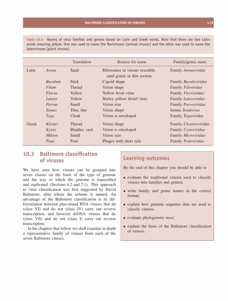
BALTIMORE CLASSIFICATION OF VIRUSES 119
Table 10.4 Names of virus families and genera based on Latin and Greek words. Note that there are two Latinwords meaning yellow. One was used to name the flaviviruses (animal viruses) and the other was used to name theluteoviruses (plant viruses)
Translation Reason for name Family/genus name
Latin Arena Sand Ribosomes in virions resemblesand grains in thin section
Family Arenaviridae
Baculum Stick Capsid shape Family BaculoviridaeFilum Thread Virion shape Family FiloviridaeFlavus Yellow Yellow fever virus Family FlaviviridaeLuteus Yellow Barley yellow dwarf virus Family LuteoviridaeParvus Small Virion size Family ParvoviridaeTenuis Thin, fine Virion shape Genus TenuivirusToga Cloak Virion is enveloped Family Togaviridae
Greek Kloster Thread Virion shape Family ClosteroviridaeKystis Bladder, sack Virion is enveloped Family CystoviridaeMikros Small Virion size Family MicroviridaePous Foot Phages with short tails Family Podoviridae
10.3 Baltimore classificationof viruses
We have seen how viruses can be grouped intoseven classes on the basis of the type of genomeand the way in which the genome is transcribedand replicated (Sections 6.2 and 7.1). This approachto virus classification was first suggested by DavidBaltimore, after whom the scheme is named. Anadvantage of the Baltimore classification is its dif-ferentiation between plus-strand RNA viruses that do(class VI) and do not (class IV) carry out reversetranscription, and between dsDNA viruses that do(class VII) and do not (class I) carry out reversetranscription.
In the chapters that follow we shall examine in deptha representative family of viruses from each of theseven Baltimore classes.
Learning outcomes
By the end of this chapter you should be able to
• evaluate the traditional criteria used to classifyviruses into families and genera;
• write family and genus names in the correctformat;
• explain how genome sequence data are used toclassify viruses;
• evaluate phylogenetic trees;
• explain the basis of the Baltimore classificationof viruses.

120 CLASSIFICATION AND NOMENCLATURE OF VIRUSES
Sources of further information
Book
Fauquet C. M. et al., editors (2005) Virus Taxonomy: EighthReport of the International Committee on Taxonomy ofViruses, Elsevier
JournalsBao Y. et al. (2004) National Center for Biotechnology
Information Viral Genomes Project Journal of Virology,78, 7291–7298
van Regenmortel M. H. V. & Mahy B. W. J. (2004) Emerg-ing issues in virus taxonomy Emerging Infectious Diseases,10(1) http://www.cdc.gov/ncidod/eid/vol10no1/03-0279.htm
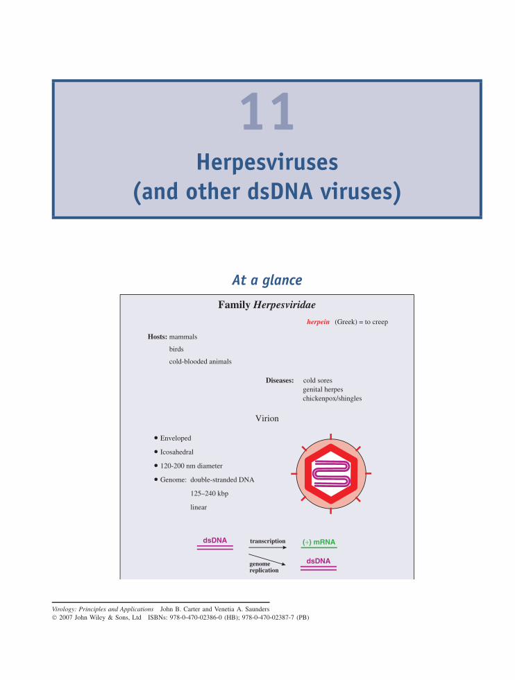
11Herpesviruses
(and other dsDNA viruses)
At a glance
Virion
• Enveloped
• Icosahedral
• 120-200 nm diameter
• Genome: double-stranded DNA
125–240 kbp
linear
Family Herpesviridae
Hosts: mammals
birds
cold-blooded animals
herpein
Diseases: cold soresgenital herpeschickenpox/shingles
dsDNA (+) mRNA
dsDNA
transcription
genomereplication
(Greek) = to creep
Virology: Principles and Applications John B. Carter and Venetia A. Saunders 2007 John Wiley & Sons, Ltd ISBNs: 978-0-470-02386-0 (HB); 978-0-470-02387-7 (PB)
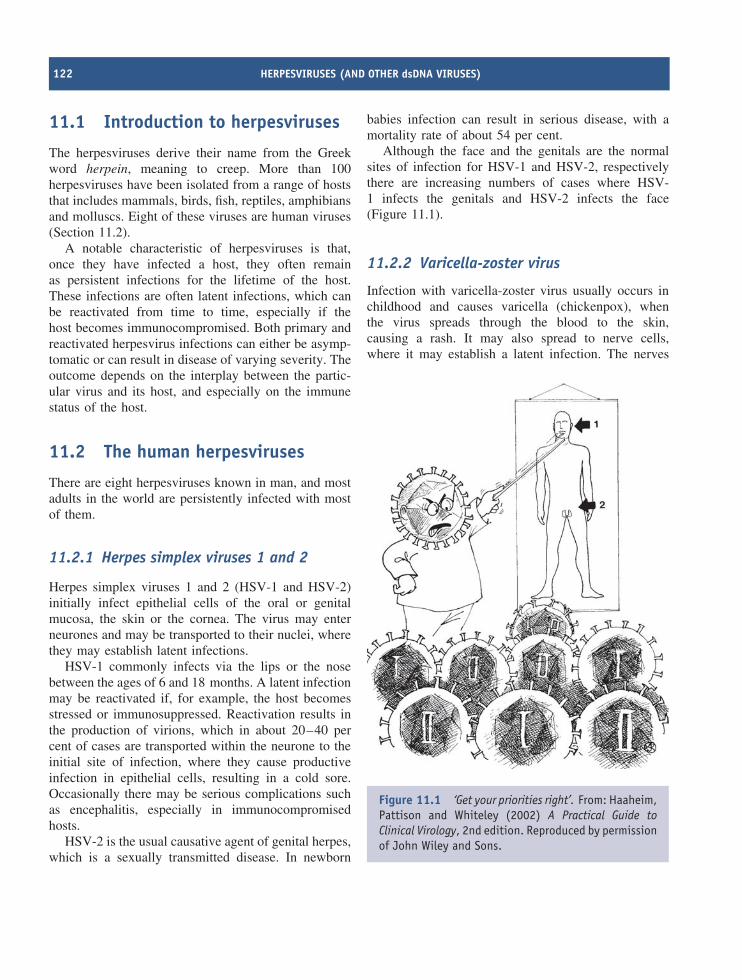
122 HERPESVIRUSES (AND OTHER dsDNA VIRUSES)
11.1 Introduction to herpesviruses
The herpesviruses derive their name from the Greekword herpein, meaning to creep. More than 100herpesviruses have been isolated from a range of hoststhat includes mammals, birds, fish, reptiles, amphibiansand molluscs. Eight of these viruses are human viruses(Section 11.2).
A notable characteristic of herpesviruses is that,once they have infected a host, they often remainas persistent infections for the lifetime of the host.These infections are often latent infections, which canbe reactivated from time to time, especially if thehost becomes immunocompromised. Both primary andreactivated herpesvirus infections can either be asymp-tomatic or can result in disease of varying severity. Theoutcome depends on the interplay between the partic-ular virus and its host, and especially on the immunestatus of the host.
11.2 The human herpesviruses
There are eight herpesviruses known in man, and mostadults in the world are persistently infected with mostof them.
11.2.1 Herpes simplex viruses 1 and 2
Herpes simplex viruses 1 and 2 (HSV-1 and HSV-2)initially infect epithelial cells of the oral or genitalmucosa, the skin or the cornea. The virus may enterneurones and may be transported to their nuclei, wherethey may establish latent infections.
HSV-1 commonly infects via the lips or the nosebetween the ages of 6 and 18 months. A latent infectionmay be reactivated if, for example, the host becomesstressed or immunosuppressed. Reactivation results inthe production of virions, which in about 20–40 percent of cases are transported within the neurone to theinitial site of infection, where they cause productiveinfection in epithelial cells, resulting in a cold sore.Occasionally there may be serious complications suchas encephalitis, especially in immunocompromisedhosts.
HSV-2 is the usual causative agent of genital herpes,which is a sexually transmitted disease. In newborn
babies infection can result in serious disease, with amortality rate of about 54 per cent.
Although the face and the genitals are the normalsites of infection for HSV-1 and HSV-2, respectivelythere are increasing numbers of cases where HSV-1 infects the genitals and HSV-2 infects the face(Figure 11.1).
11.2.2 Varicella-zoster virus
Infection with varicella-zoster virus usually occurs inchildhood and causes varicella (chickenpox), whenthe virus spreads through the blood to the skin,causing a rash. It may also spread to nerve cells,where it may establish a latent infection. The nerves
Figure 11.1 ‘Get your priorities right’. From: Haaheim,Pattison and Whiteley (2002) A Practical Guide toClinical Virology, 2nd edition. Reproduced by permissionof John Wiley and Sons.
THE HERPESVIRUS VIRION 123
most often affected are those in the face or thetrunk, and these are the areas most commonlyaffected in zoster (shingles) when a latent infection isreactivated.
11.2.3 Epstein-Barr virus
Epstein-Barr virus (EBV) is transmitted in saliva.Epithelial cells are infected first then the infec-tion spreads to B cells, which are the main hostcell type for this virus. More than 90 per centof people become infected with EBV, usually dur-ing the first years of life, when infection results infew or no symptoms. In developed countries someindividuals do not become infected until adoles-cence or adulthood. A proportion of these individu-als develop infectious mononucleosis (glandular fever),commonly called ‘the kissing disease’ by doctors.EBV is associated with a number of tumours in man(Chapter 22).
11.2.4 Human cytomegalovirus
In the vast majority of infections with humancytomegalovirus symptoms are either absent or theyare mild. In a pregnant woman, however, the virus caninfect the placenta and then the foetus, for whom theconsequences may be serious. In the US about oneper cent of babies are born infected with the virus(about 40 000 per year). In about seven per cent ofthese there is evidence of virus-induced damage atbirth, including small brain size and enlargement of theliver and spleen. In other individuals damage devel-ops at a later stage; the damage may be manifest ina number of ways, such as hearing loss and mentalretardation.
Human cytomegalovirus can also cause severe dis-ease (e.g. pneumonitis, hepatitis) in immunocompro-mised patients such as those with AIDS, those whohave received treatment for cancer and those whoare immunosuppressed because they have received anorgan transplant.
11.2.5 Human herpesvirus 6
There are two types of human herpesvirus 6, known asHHV-6A and HHV-6B. Infection of a child with the
latter can cause a fever and the sudden appearance ofa rash known as exanthem subitum.
11.2.6 Human herpesvirus 7
Human herpesvirus 7 was first isolated from a cultureof CD4 T cells that developed a cytopathic effect;the cells were from a healthy person. The virushas been associated with some cases of exanthemsubitum.
11.2.7 Kaposi’s sarcoma-associatedherpesvirus
Kaposi’s sarcoma-associated herpesvirus was discov-ered in 1994 and is named after the tumour with whichthe virus is associated (Chapter 22).
11.3 The herpesvirus virion
Herpesviruses have relatively complex virions com-posed of a large number of protein species organizedinto three distinct structures: capsid, tegument andenvelope (Figure 11.2). The virus genome is a lineardsDNA molecule, which varies in size within the her-pesvirus family from 125 to 240 kbp. The DNA ishoused in the capsid, which is icosahedral, and thecapsid is surrounded by the tegument. The HSV-1 tegu-ment contains at least 15 protein species and some virusmRNA molecules. The envelope contains a large num-ber of spikes (600–750 in HSV-1) composed of ten ormore glycoprotein species. There are several differentsizes of spike.
A number of schemes have evolved for the nomen-clature of herpesvirus proteins, with the result that anindividual protein may be referred to in the literatureby two or more different names. Most of the struc-tural proteins are commonly named VP (virus protein).In HSV-1 the most abundant proteins in the capsidand the tegument are VP5 and VP16, respectively. Inthe envelope there are at least 12 species of glycopro-tein, each of which is prefixed ‘g’, for example gB, gCand gD.
The capsid is constructed from 162 capsomeres, 12of which are pentons and the remainder of which are
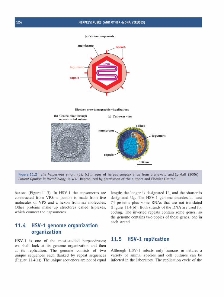
124 HERPESVIRUSES (AND OTHER dsDNA VIRUSES)
membrane spikes
DNA
capsid
tegument
(a) Virion components
(c) Cut-away view
spikes
membrane
tegument
capsid
100 nm
Electron cryo-tomographic visualizations
(b) Central slice throughreconstructed volume
Figure 11.2 The herpesvirus virion. (b), (c) Images of herpes simplex virus from Grunewald and Cyrklaff (2006)Current Opinion in Microbiology, 9, 437. Reproduced by permission of the authors and Elsevier Limited.
hexons (Figure 11.3). In HSV-1 the capsomeres areconstructed from VP5: a penton is made from fivemolecules of VP5 and a hexon from six molecules.Other proteins make up structures called triplexes,which connect the capsomeres.
11.4 HSV-1 genome organizationorganization
HSV-1 is one of the most-studied herpesviruses;we shall look at its genome organization and thenat its replication. The genome consists of twounique sequences each flanked by repeat sequences(Figure 11.4(a)). The unique sequences are not of equal
length: the longer is designated UL and the shorter isdesignated US. The HSV-1 genome encodes at least74 proteins plus some RNAs that are not translated(Figure 11.4(b)). Both strands of the DNA are used forcoding. The inverted repeats contain some genes, sothe genome contains two copies of these genes, one ineach strand.
11.5 HSV-1 replication
Although HSV-1 infects only humans in nature, avariety of animal species and cell cultures can beinfected in the laboratory. The replication cycle of the
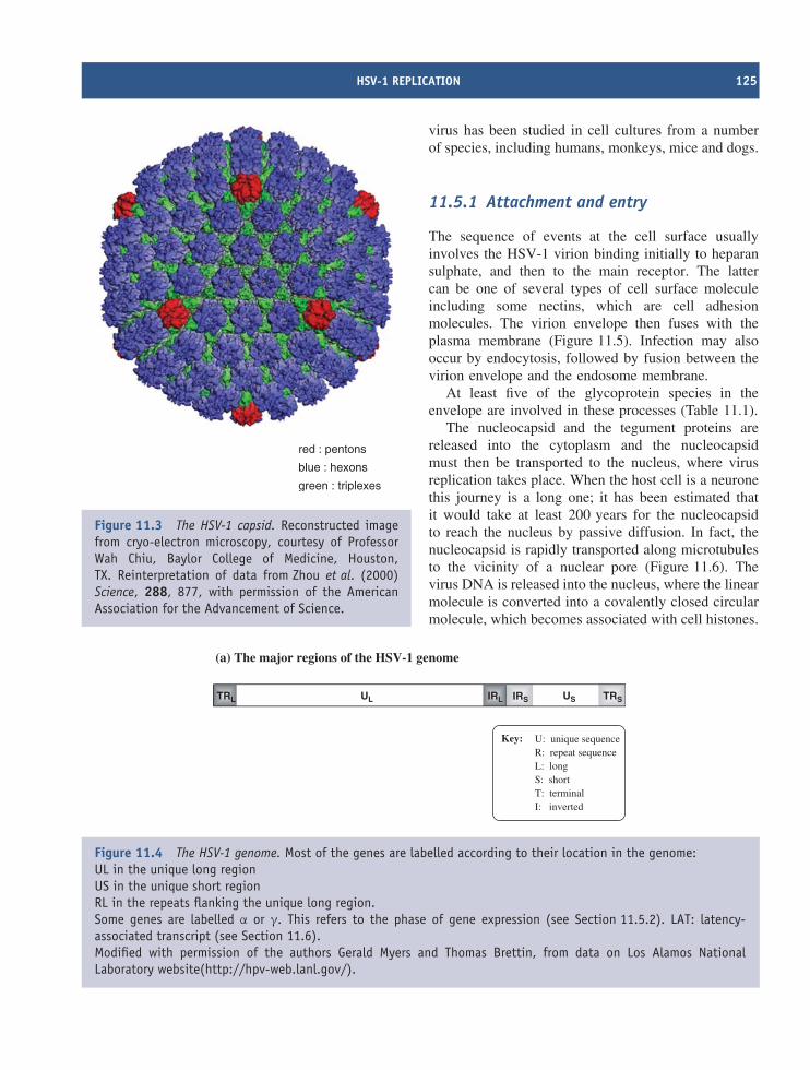
HSV-1 REPLICATION 125
red : pentons
blue : hexons
green : triplexes
Figure 11.3 The HSV-1 capsid. Reconstructed imagefrom cryo-electron microscopy, courtesy of ProfessorWah Chiu, Baylor College of Medicine, Houston,TX. Reinterpretation of data from Zhou et al. (2000)Science, 288, 877, with permission of the AmericanAssociation for the Advancement of Science.
virus has been studied in cell cultures from a numberof species, including humans, monkeys, mice and dogs.
11.5.1 Attachment and entry
The sequence of events at the cell surface usuallyinvolves the HSV-1 virion binding initially to heparansulphate, and then to the main receptor. The lattercan be one of several types of cell surface moleculeincluding some nectins, which are cell adhesionmolecules. The virion envelope then fuses with theplasma membrane (Figure 11.5). Infection may alsooccur by endocytosis, followed by fusion between thevirion envelope and the endosome membrane.
At least five of the glycoprotein species in theenvelope are involved in these processes (Table 11.1).
The nucleocapsid and the tegument proteins arereleased into the cytoplasm and the nucleocapsidmust then be transported to the nucleus, where virusreplication takes place. When the host cell is a neuronethis journey is a long one; it has been estimated thatit would take at least 200 years for the nucleocapsidto reach the nucleus by passive diffusion. In fact, thenucleocapsid is rapidly transported along microtubulesto the vicinity of a nuclear pore (Figure 11.6). Thevirus DNA is released into the nucleus, where the linearmolecule is converted into a covalently closed circularmolecule, which becomes associated with cell histones.
UL USTRL IRL TRSIRS
(a) The major regions of the HSV-1 genome
Key: U: unique sequenceR: repeat sequenceL: longS: shortT: terminalI: inverted
Figure 11.4 The HSV-1 genome. Most of the genes are labelled according to their location in the genome:UL in the unique long regionUS in the unique short regionRL in the repeats flanking the unique long region.Some genes are labelled α or γ. This refers to the phase of gene expression (see Section 11.5.2). LAT: latency-associated transcript (see Section 11.6).Modified with permission of the authors Gerald Myers and Thomas Brettin, from data on Los Alamos NationalLaboratory website(http://hpv-web.lanl.gov/).
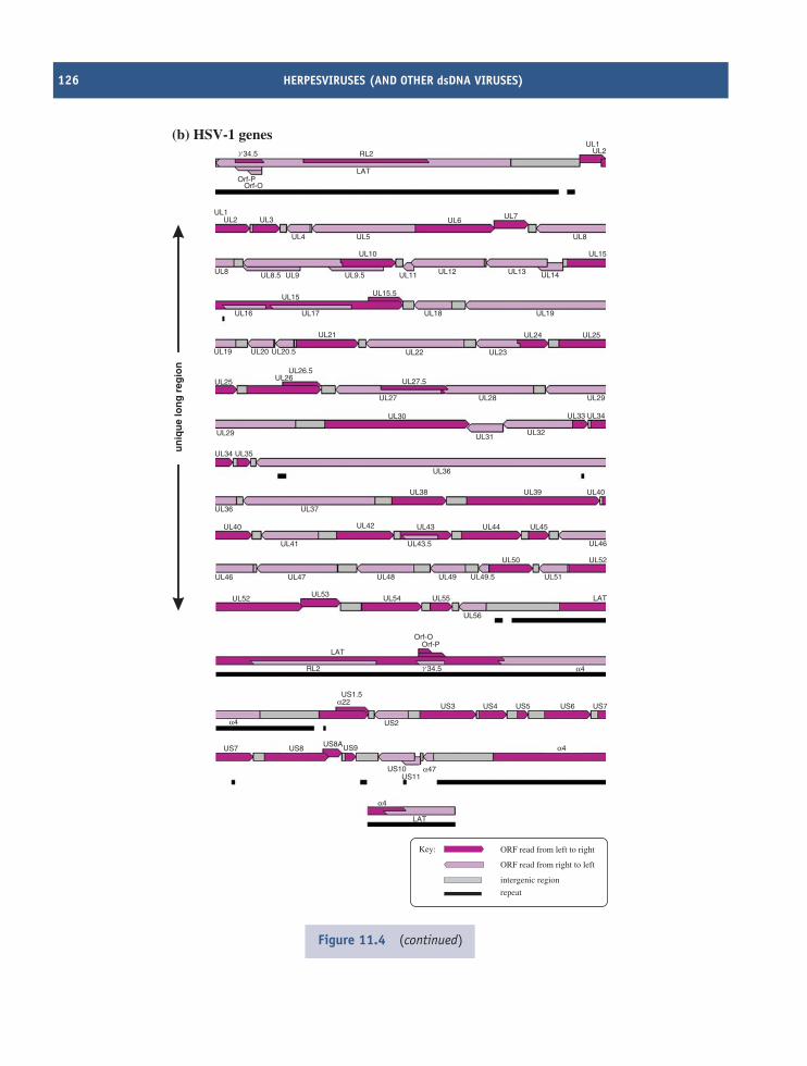
126 HERPESVIRUSES (AND OTHER dsDNA VIRUSES)
LAT
US11US10
US9US8AUS8US7
US2
RL2
LAT
Orf-OOrf-P
US3 US4 US5
UL56
UL55UL54UL53UL52
UL46 UL47 UL48 UL49 UL51
UL52
UL46
UL45
UL40UL39UL38
UL34 UL35
UL36
UL32
UL33 UL34
UL29UL28UL27
UL25 UL26UL26.5
UL20.5UL20
UL21
UL19
UL27.5
UL23
UL24 UL25
UL19
UL14
UL15
UL8
UL7
UL2UL1
UL6
UL13UL12UL11UL9.5UL9
UL4
UL3UL2UL1
UL5
LAT
RL2
UL10
UL8.5UL8
UL18UL17UL16
UL15 UL15.5
UL22
UL31UL29
UL30
UL44UL43UL42UL40
UL36 UL37
UL50
UL41 UL43.5
UL49.5
LAT
US6 US7
US1.5
α4
α4
α4
α22
α47
α4
34.5g
Orf-OOrf-P
34.5g
un
iqu
e lo
ng
reg
ion
(b) HSV-1 genes
ORF read from left to right
ORF read from right to left
intergenic region
repeat
Key:
Figure 11.4 (continued)
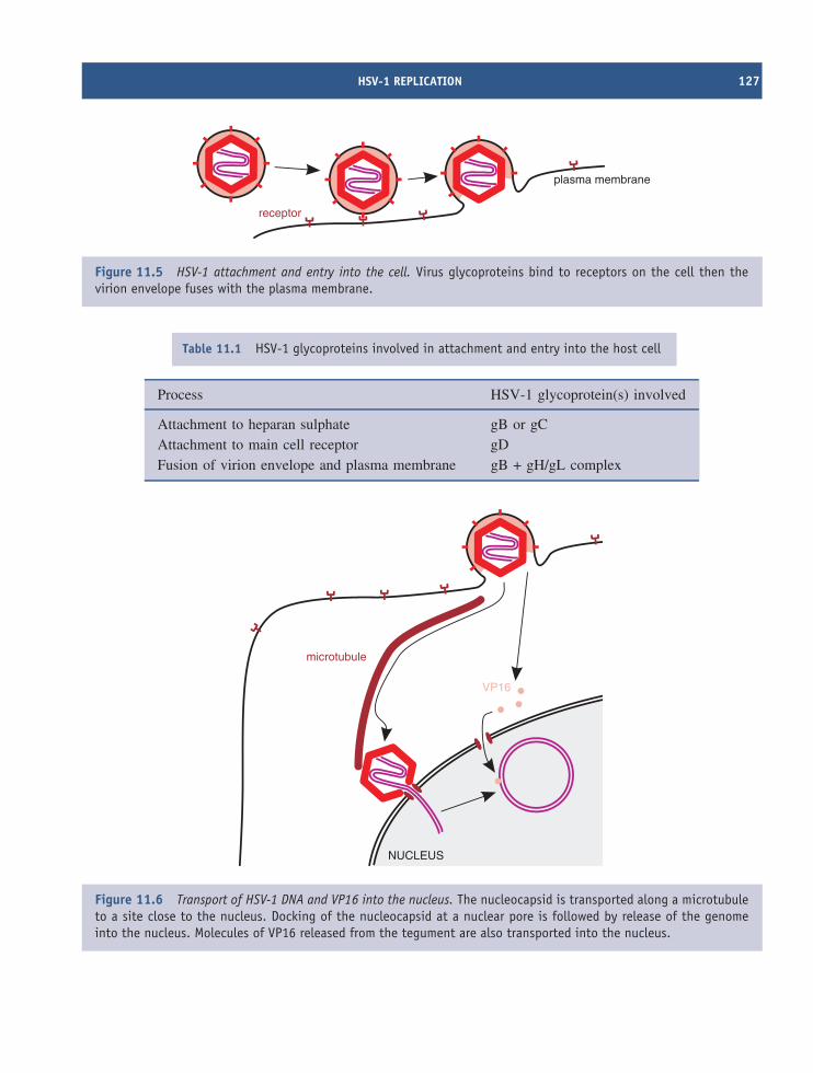
HSV-1 REPLICATION 127
plasma membrane
receptor
Figure 11.5 HSV-1 attachment and entry into the cell. Virus glycoproteins bind to receptors on the cell then thevirion envelope fuses with the plasma membrane.
Table 11.1 HSV-1 glycoproteins involved in attachment and entry into the host cell
Process HSV-1 glycoprotein(s) involved
Attachment to heparan sulphate gB or gCAttachment to main cell receptor gDFusion of virion envelope and plasma membrane gB + gH/gL complex
microtubule
NUCLEUS
VP16
Figure 11.6 Transport of HSV-1 DNA and VP16 into the nucleus. The nucleocapsid is transported along a microtubuleto a site close to the nucleus. Docking of the nucleocapsid at a nuclear pore is followed by release of the genomeinto the nucleus. Molecules of VP16 released from the tegument are also transported into the nucleus.
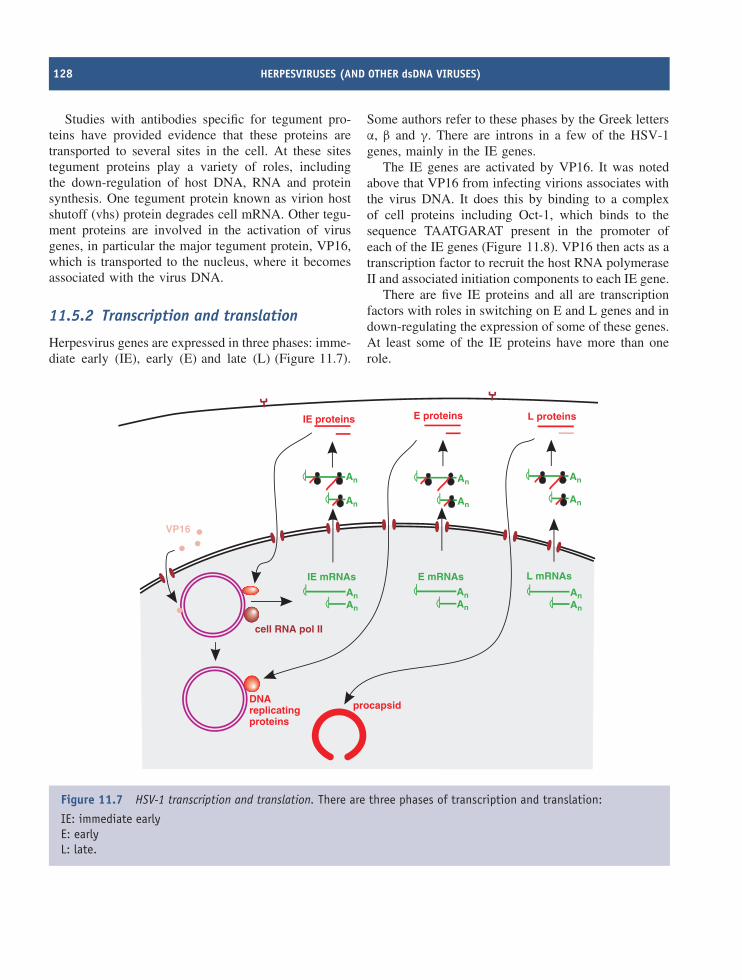
128 HERPESVIRUSES (AND OTHER dsDNA VIRUSES)
Studies with antibodies specific for tegument pro-teins have provided evidence that these proteins aretransported to several sites in the cell. At these sitestegument proteins play a variety of roles, includingthe down-regulation of host DNA, RNA and proteinsynthesis. One tegument protein known as virion hostshutoff (vhs) protein degrades cell mRNA. Other tegu-ment proteins are involved in the activation of virusgenes, in particular the major tegument protein, VP16,which is transported to the nucleus, where it becomesassociated with the virus DNA.
11.5.2 Transcription and translation
Herpesvirus genes are expressed in three phases: imme-diate early (IE), early (E) and late (L) (Figure 11.7).
Some authors refer to these phases by the Greek lettersα, β and γ. There are introns in a few of the HSV-1genes, mainly in the IE genes.
The IE genes are activated by VP16. It was notedabove that VP16 from infecting virions associates withthe virus DNA. It does this by binding to a complexof cell proteins including Oct-1, which binds to thesequence TAATGARAT present in the promoter ofeach of the IE genes (Figure 11.8). VP16 then acts as atranscription factor to recruit the host RNA polymeraseII and associated initiation components to each IE gene.
There are five IE proteins and all are transcriptionfactors with roles in switching on E and L genes and indown-regulating the expression of some of these genes.At least some of the IE proteins have more than onerole.
cell RNA pol II
E proteinsIE proteins
An
An
An
An
AnAn
An
An
An An
An
An
L proteins
L mRNAsE mRNAsIE mRNAs
VP16
DNAreplicatingproteins
procapsid
Figure 11.7 HSV-1 transcription and translation. There are three phases of transcription and translation:
IE: immediate earlyE: earlyL: late.
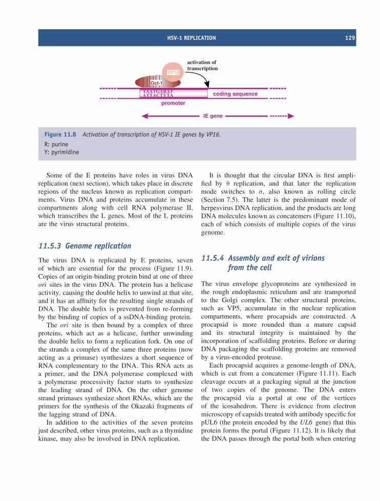
HSV-1 REPLICATION 129
activation oftranscription
VP16
TA
AT
AT
TA
GC
AT
RY
AT
TA
promoter
coding sequence
IE gene
Oct-1
Figure 11.8 Activation of transcription of HSV-1 IE genes by VP16.
R: purineY: pyrimidine
Some of the E proteins have roles in virus DNAreplication (next section), which takes place in discreteregions of the nucleus known as replication compart-ments. Virus DNA and proteins accumulate in thesecompartments along with cell RNA polymerase II,which transcribes the L genes. Most of the L proteinsare the virus structural proteins.
11.5.3 Genome replication
The virus DNA is replicated by E proteins, sevenof which are essential for the process (Figure 11.9).Copies of an origin-binding protein bind at one of threeori sites in the virus DNA. The protein has a helicaseactivity, causing the double helix to unwind at that site,and it has an affinity for the resulting single strands ofDNA. The double helix is prevented from re-formingby the binding of copies of a ssDNA-binding protein.
The ori site is then bound by a complex of threeproteins, which act as a helicase, further unwindingthe double helix to form a replication fork. On one ofthe strands a complex of the same three proteins (nowacting as a primase) synthesizes a short sequence ofRNA complementary to the DNA. This RNA acts asa primer, and the DNA polymerase complexed witha polymerase processivity factor starts to synthesizethe leading strand of DNA. On the other genomestrand primases synthesize short RNAs, which are theprimers for the synthesis of the Okazaki fragments ofthe lagging strand of DNA.
In addition to the activities of the seven proteinsjust described, other virus proteins, such as a thymidinekinase, may also be involved in DNA replication.
It is thought that the circular DNA is first ampli-fied by θ replication, and that later the replicationmode switches to σ, also known as rolling circle(Section 7.5). The latter is the predominant mode ofherpesvirus DNA replication, and the products are longDNA molecules known as concatemers (Figure 11.10),each of which consists of multiple copies of the virusgenome.
11.5.4 Assembly and exit of virionsfrom the cell
The virus envelope glycoproteins are synthesized inthe rough endoplasmic reticulum and are transportedto the Golgi complex. The other structural proteins,such as VP5, accumulate in the nuclear replicationcompartments, where procapsids are constructed. Aprocapsid is more rounded than a mature capsidand its structural integrity is maintained by theincorporation of scaffolding proteins. Before or duringDNA packaging the scaffolding proteins are removedby a virus-encoded protease.
Each procapsid acquires a genome-length of DNA,which is cut from a concatemer (Figure 11.11). Eachcleavage occurs at a packaging signal at the junctionof two copies of the genome. The DNA entersthe procapsid via a portal at one of the verticesof the icosahedron. There is evidence from electronmicroscopy of capsids treated with antibody specific forpUL6 (the protein encoded by the UL6 gene) that thisprotein forms the portal (Figure 11.12). It is likely thatthe DNA passes through the portal both when entering
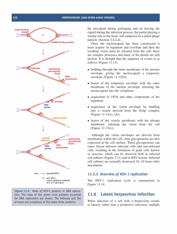
130 HERPESVIRUSES (AND OTHER dsDNA VIRUSES)
DNApolymerase
helicase polymeraseprocessivity
factor
3’5’
5’
3’
primase
leading strand
RNAprimer
3’5’
5’
3’
DNApolymerase
polymeraseprocessivityfactor
Okazakifragment
RNAprimer
3’5’
5’
3’
3’5’
ssDNA-binding protein
origin-bindingprotein
3’5’ori
Key:
new DNA(arrow indicates synthesisin 5' to 3' direction)
parental DNA
Figure 11.9 Roles of HSV-1 proteins in DNA replica-tion. The roles of the seven virus proteins essentialfor DNA replication are shown. The helicase and theprimase are complexes of the same three proteins.
the procapsid during packaging and on leaving thecapsid during the infection process, the portal playing asimilar role to the head–tail connector in a tailed phageparticle (Section 3.4.2.d).
Once the nucleocapsid has been constructed itmust acquire its tegument and envelope and then theresulting virion must be released from the cell; theseare complex processes and many of the details are stillunclear. It is thought that the sequence of events is asfollows (Figure 11.13):
• budding through the inner membrane of the nuclearenvelope, giving the nucleocapsid a temporaryenvelope (Figure 11.13(b));
• fusion of the temporary envelope with the outermembrane of the nuclear envelope, releasing thenucleocapsid into the cytoplasm;
• acquisition of VP16 and other components of thetegument;
• acquisition of the virion envelope by buddinginto a vesicle derived from the Golgi complex(Figure 11.13(c), (d));
• fusion of the vesicle membrane with the plasmamembrane, releasing the virion from the cell(Figure 11.13(e)).
Although the virion envelopes are derived frommembranes within the cell, virus glycoproteins are alsoexpressed at the cell surface. These glycoproteins cancause fusion between infected cells and non-infectedcells, resulting in the formation of giant cells knownas syncytia, which can be observed both in infectedcell cultures (Figure 2.11) and in HSV lesions. Infectedcell cultures are normally destroyed 18–24 hours afterinoculation.
11.5.5 Overview of HSV-1 replication
The HSV-1 replication cycle is summarized inFigure 11.14.
11.6 Latent herpesvirus infectionWhen infection of a cell with a herpesvirus resultsin latency rather than a productive infection, multiple
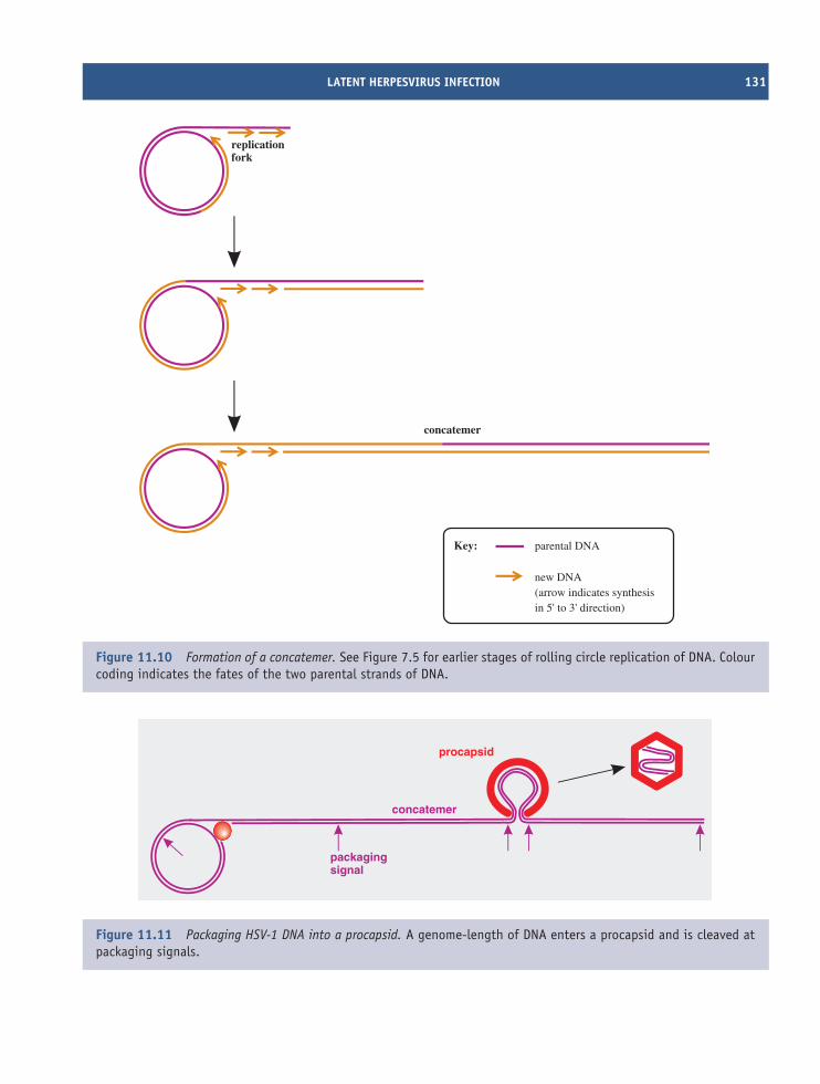
LATENT HERPESVIRUS INFECTION 131
concatemer
Key:
new DNA(arrow indicates synthesisin 5' to 3' direction)
parental DNA
replicationfork
Figure 11.10 Formation of a concatemer. See Figure 7.5 for earlier stages of rolling circle replication of DNA. Colourcoding indicates the fates of the two parental strands of DNA.
packagingsignal
concatemer
procapsid
Figure 11.11 Packaging HSV-1 DNA into a procapsid. A genome-length of DNA enters a procapsid and is cleaved atpackaging signals.
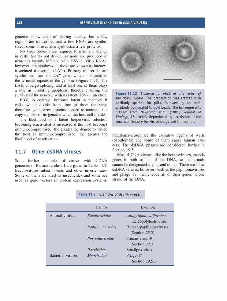
132 HERPESVIRUSES (AND OTHER dsDNA VIRUSES)
genome is switched off during latency, but a fewregions are transcribed and a few RNAs are synthe-sized; some viruses also synthesize a few proteins.
No virus proteins are required to maintain latencyin cells that do not divide, so none are produced inneurones latently infected with HSV-1. Virus RNAs,however, are synthesized; these are known as latency-associated transcripts (LATs). Primary transcripts aresynthesized from the LAT gene, which is located inthe terminal repeats of the genome (Figure 11.4). TheLATs undergo splicing, and at least one of them playsa role in inhibiting apoptosis, thereby ensuring thesurvival of the neurone with its latent HSV-1 infection.
EBV, in contrast, becomes latent in memory Bcells, which divide from time to time; the virustherefore synthesizes proteins needed to maintain thecopy number of its genome when the host cell divides.
The likelihood of a latent herpesvirus infectionbecoming reactivated is increased if the host becomesimmunocompromised; the greater the degree to whichthe host is immunocompromised, the greater thelikelihood of reactivation.
11.7 Other dsDNA virusesSome further examples of viruses with dsDNAgenomes in Baltimore class I are given in Table 11.2.Baculoviruses infect insects and other invertebrates.Some of them are used as insecticides and some areused as gene vectors in protein expression systems.
Figure 11.12 Evidence for pUL6 at one vertex ofthe HSV-1 capsid. The preparation was treated withantibody specific for pUL6 followed by an anti-antibody conjugated to gold beads. The bar represents100 nm. From Newcomb et al. (2001) Journal ofVirology, 75, 10923. Reproduced by permission of theAmerican Society for Microbiology and the author.
Papillomaviruses are the causative agents of warts(papillomas) and some of them cause human can-cers. The dsDNA phages are considered further inSection 19.5.
Most dsDNA viruses, like the herpesviruses, encodegenes in both strands of the DNA, so the strandscannot be designated as plus and minus. There are somedsDNA viruses, however, such as the papillomavirusesand phage T7, that encode all of their genes in onestrand of the DNA.
Table 11.2 Examples of dsDNA viruses
Family Example
Animal viruses Baculoviridae Autographa californicanucleopolyhedrovirus
Papillomaviridae Human papillomaviruses(Section 22.2)
Polyomaviridae Simian virus 40(Section 22.3)
Poxviridae Smallpox virusBacterial viruses Myoviridae Phage T4
(Section 19.5.1)
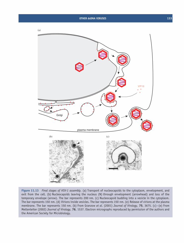
OTHER dsDNA VIRUSES 133
Golgi
plasma membrane
VP16
N
(b)
(a)
(c)
Figure 11.13 Final stages of HSV-1 assembly. (a) Transport of nucleocapsids to the cytoplasm, envelopment, andexit from the cell. (b) Nucleocapsids leaving the nucleus (N) through envelopment (arrowhead) and loss of thetemporary envelope (arrow). The bar represents 200 nm. (c) Nucleocapsid budding into a vesicle in the cytoplasm.The bar represents 150 nm. (d) Virions inside vesicles. The bar represents 150 nm. (e) Release of virions at the plasmamembrane. The bar represents 150 nm. (b) From Granzow et al. (2001) Journal of Virology, 75, 3675. (c)–(e) FromMettenleiter (2002) Journal of Virology, 76, 1537. Electron micrographs reproduced by permission of the authors andthe American Society for Microbiology.
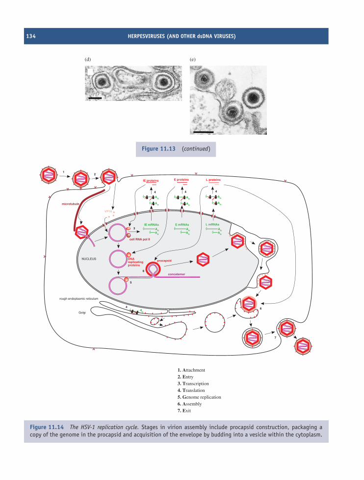
134 HERPESVIRUSES (AND OTHER dsDNA VIRUSES)
(d) (e)
Figure 11.13 (continued)
cell RNA pol II
NUCLEUS
rough endoplasmic reticulum
Golgi
microtubule
VP16
E proteinsIE proteins
An
An
An
AnAn
AnAn
An
An
An
An
An
L proteins
L mRNAsE mRNAsIE mRNAs
An
concatemer
procapsidDNAreplicatingproteins
12
3
4 4 4
5
6
6
7
4
1. Attachment
2. Entry
3. Transcription
4. Translation
5. Genome replication
6. Assembly
7. Exit
Figure 11.14 The HSV-1 replication cycle. Stages in virion assembly include procapsid construction, packaging acopy of the genome in the procapsid and acquisition of the envelope by budding into a vesicle within the cytoplasm.
SOURCES OF FURTHER INFORMATION 135
Learning outcomes
By the end of this chapter you should be able to
• list the human herpesviruses and explain theirimportance;
• describe the phenomenon of herpesvirus persis-tence and explain its importance;
• describe the HSV-1 virion;
• outline the main features of the HSV-1 genome;
• describe the replication cycle of HSV-1.
Sources of further information
Books
Freeman M. L., Decman V. and Hendricks R. L. (2005)Neurons and host immunity conspire to maintain herpessimplex virus in a latent state, pp. 203–213 in Molec-ular Pathogenesis of Virus Infections, editors Digard P.,Nash A. A. and Randall R. E., 64th Symposium of theSociety for General Microbiology, Cambridge UniversityPress
Knipe D. M. and Howley P. M., editors-in-chief (2001)Fields Virology, 4th edition, Chapters 71–83, Lippincott,Williams and Wilkins
Sandri-Goldin R. M., editor (2006) Alpha Herpesviruses:Molecular and Cellular Biology, Caister
Journals
Babb R. (2001) DNA recognition by the herpes simplexvirus transactivator VP16: a novel DNA-binding structureMolecular and Cellular Biology, 21, 4700–4712
De Bolle L., Naesens L. and De Clercq E. (2005) Update onhuman herpesvirus 6 biology, clinical features, and therapyClinical Microbiology Reviews, 18, 217–245
Efstathioua S. and Preston C. M. (2005) Towards an under-standing of the molecular basis of herpes simplex viruslatency Virus Research, 111, 108–119
Garner J. A. (2003) Herpes simplex virion entry into andintracellular transport within mammalian cells AdvancedDrug Delivery Reviews, 55, 1497–1513
Gupta A. et al. (2006) Anti-apoptotic function of a microRNAencoded by the HSV-1 latency-associated transcript Nature,442, 82–85
Hagglund R. and Roizman B. (2004) Role of ICP0 in thestrategy of conquest of the host cell by herpes simplexvirus 1 Journal of Virology, 78, 2169–2178
Kimberlin D. W. (2004) Neonatal herpes simplex infectionClinical Microbiology Reviews, 17, 1–13
McGeoch D. J., Rixon F. J. and Davison A. J. (2006) Topicsin herpesvirus genomics and evolution Virus Research,117, 90–104
Mettenleiter T. C., Klupp B. G. and Granzow H. (2006)Herpesvirus assembly: a tale of two membranes CurrentOpinion in Microbiology, 9, 423–429
Nicola A. V. and Straus S. E. (2004) Cellular and viralrequirements for rapid endocytic entry of herpes simplexvirus Journal of Virology, 78, 7508–7517
Nishiyama Y. (2004) Herpes simplex virus gene products:the accessories reflect her lifestyle well Reviews in MedicalVirology, 14, 33–46
Sinclair J. and Sissons P. (2006) Latency and reactivation ofhuman cytomegalovirus Journal of General Virology, 87,1763–1779

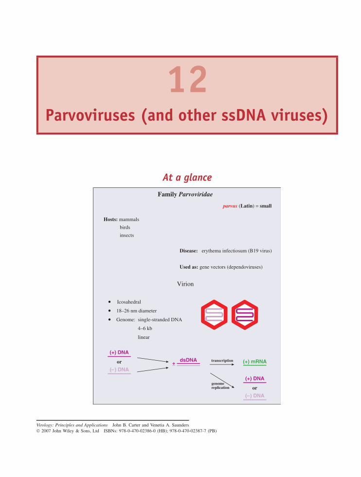
12Parvoviruses (and other ssDNA viruses)
At a glance
Virion
Icosahedral
18–26 nm diameter
Genome: single-stranded DNA
4–6 kb
linear
Family Parvoviridae
Hosts: mammals
birds
insects
parvus ( ) =Latin small
Disease: erythema infectiosum (B19 virus)
Used as: gene vectors (dependoviruses)
( ) DNA–
or dsDNA (+) mRNA
(+) DNA
( ) DNA–
or
(+) DNA
+–transcription
genomereplication
Virology: Principles and Applications John B. Carter and Venetia A. Saunders 2007 John Wiley & Sons, Ltd ISBNs: 978-0-470-02386-0 (HB); 978-0-470-02387-7 (PB)
138 PARVOVIRUSES (AND OTHER ssDNA VIRUSES)
12.1 Introduction to parvovirusesParvoviruses are amongst the smallest known viruses,with virions in the range 18–26 nm in diameter. Theyderive their name from the Latin parvus (= small).The family Parvoviridae has been divided into twosubfamilies: the Parvovirinae (vertebrate viruses) andthe Densovirinae (invertebrate viruses). Some of thegenera and species of the two subfamilies are shownin Table 12.1.
The subfamily Parvovirinae includes the genusDependovirus, the members of which are defec-tive, normally replicating only when the cell isco-infected with a helper virus. Other parvovirusesthat do not require helper viruses are known asautonomous parvoviruses.
12.2 Examples of parvoviruses12.2.1 DependovirusesThe first dependovirus to be discovered was observedin the electron microscope as a contaminant of anadenovirus preparation (Figure 12.1). The contaminantturned out to be a satellite virus (Section 9.3.1): adefective virus dependent on the help of the adenovirusfor replication. The satellite virus was therefore calledan adeno-associated virus. Other dependoviruses (dis-tinct serotypes) have since been found in adenoviruspreparations and in infected humans and other species.Results of surveys using serological methods and PCRdetection of virus DNA indicate that dependovirusinfections are widespread.
Not all dependoviruses have an absolute requirementfor the help of an adenovirus. Other DNA viruses (e.g.
Figure 12.1 Virions of adenovirus (arrowed) anddependovirus. Reproduced with permission of ProfessorM. Stewart McNulty, The Queen’s University of Belfast.
herpesviruses) may sometimes act as helpers, and somedependoviruses may replicate in the absence of a helpervirus under certain circumstances.
Dependoviruses are valuable gene vectors. They areused to introduce genes into cell cultures for mass-production of the proteins encoded by those genes,and they are being investigated as possible vectorsto introduce genes into the cells of patients for thetreatment of various genetic diseases and cancers.One of the advantages of dependoviruses for suchapplications is the fact that they are not known tocause any disease, in contrast to other viruses underinvestigation, such as retroviruses (Section 16.5).
Table 12.1 Some of the viruses in the family Parvoviridae
Subfamily Genus Species
Parvovirinae Dependovirus Adeno-associated virus 2Parvovirus Minute virus of mice
Feline panleukopenia virusErythrovirus B19 virusBocavirus Human bocavirus
Densovirinae Iteravirus Bombyx mori densovirus
PARVOVIRUS VIRION 139
Figure 12.2 Child with fifth disease. Reproduced bypermission of the New Zealand Dermatological Society.
12.2.2 Autonomous parvovirusesA parvovirus that does not require a helper virus wasdiscovered in serum from a healthy blood donor. Thevirus, named after a batch of blood labelled B19, infectsred blood cell precursors. Many infections with B19 arewithout signs or symptoms, but some result in disease,such as fifth disease (erythema infectiosum), in whichaffected children develop a ‘slapped-cheek’ appearance(Figure 12.2).
Other diseases caused by B19 virus include
• acute arthritis
• aplastic anaemia in persons with chronic haemolyticanaemia
• hydrops foetalis (infection may be transmitted froma pregnant woman to the foetus and may kill thefoetus).
In 2005 a new human parvovirus was discoveredusing a technique for molecular screening of nasopha-ryngeal aspirates from children with lower respira-tory tract disease. The virus is related to known par-voviruses in the genus Bocavirus.
Viruses in the subfamily Densovirinae cause theformation of dense inclusions in the nucleus of theinfected cell. Some of these viruses are pathogens ofthe silkworm (Bombyx mori ), and can cause economicdamage to the silk industry.
12.3 Parvovirus virionParvoviruses are small viruses of simple structure withthe ssDNA genome enclosed within a capsid that hasicosahedral symmetry (Figure 12.3).
12.3.1 CapsidThe parvovirus capsid has icosahedral symmetry andis built from 60 protein molecules. One protein speciesforms the majority of the capsid structure and thereare small amounts of between one and three otherprotein species, depending on the virus. The proteinsare numbered in order of size, with VP1 the largest;
capsid
ssDNA
(a) Virion components (b) Electron micrograph of negatively stained virions
(c) Reconstructed image from cryo- electron microscopy
Bar represents 50 nm.
Bar represents 10 nm.
Figure 12.3 The parvovirus virion. The virions in Figure 12.3(b), (c) are adeno-associated virus 5, from Walterset al. (2004) Journal of Virology, 78, 3361. Reproduced by permission of the American Society for Microbiology andthe authors.
140 PARVOVIRUSES (AND OTHER ssDNA VIRUSES)
the smaller proteins are shorter versions of VP1.Each protein species contains an eight-stranded β-barrelstructure that is common to many viral capsid proteins,including those of the picornaviruses (Chapter 14).
The virion is roughly spherical, with surface pro-trusions and canyons (Figure 12.3(c)). At each of thevertices of the icosahedron there is a protrusion with apore at the centre.
12.3.2 Genome
Parvoviruses have genomes composed of linear ssDNAin the size range 4–6 kb. At each end of a DNAmolecule there are a number of short complementarysequences that can base pair to form a secondarystructure (Figure 12.4). Some parvovirus genomes havesequences at their ends known as inverted terminal
repeats (ITRs), where the sequence at one end iscomplementary to, and in the opposite orientationto, the sequence at the other end (Section 3.2.6).As the sequences are complementary, the ends haveidentical secondary structures (Figure 12.4(a)). Otherparvoviruses have a unique sequence, and therefore aunique secondary structure, at each end of the DNA(Figure 12.4(b)).
During replication, parvoviruses with ITRs generateand package equal numbers of (+) and (−) strands ofDNA, while most viruses with unique sequences at thetermini do not. The percentages of virions containing(+) DNA and (−) DNA therefore vary with differentviruses (Table 12.2).
In a (−) DNA the genes for non-structural proteinsare towards the 3′ end and the structural protein genesare towards the 5′ end (Figure 12.5).
(a) ‘Inverted repeats’ at the termini
(b) Unique sequences at the termini
Figure 12.4 Base pairing at the termini of parvovirus DNA.
Table 12.2 Percentages of parvovirus virions containing (+) and (−) strand DNA
Virus Sequences at the % virions % virionsDNA termini1 containing containing
(+) DNA (−) DNA
B19 virus ITRs 50 50Dependoviruses ITRs 50 50Minute virus of mice Unique 1 99
1The termini of a parvovirus DNA are either ‘inverted repeats’ (ITRs; Figure 12.4(a)) or unique(Figure 12.4(b)).
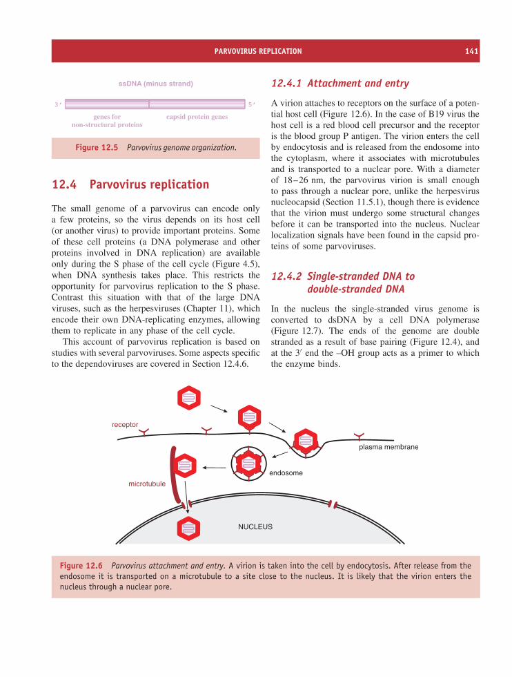
PARVOVIRUS REPLICATION 141
5’3’
genes fornon-structural proteins
capsid protein genes
ssDNA (minus strand)
Figure 12.5 Parvovirus genome organization.
12.4 Parvovirus replication
The small genome of a parvovirus can encode onlya few proteins, so the virus depends on its host cell(or another virus) to provide important proteins. Someof these cell proteins (a DNA polymerase and otherproteins involved in DNA replication) are availableonly during the S phase of the cell cycle (Figure 4.5),when DNA synthesis takes place. This restricts theopportunity for parvovirus replication to the S phase.Contrast this situation with that of the large DNAviruses, such as the herpesviruses (Chapter 11), whichencode their own DNA-replicating enzymes, allowingthem to replicate in any phase of the cell cycle.
This account of parvovirus replication is based onstudies with several parvoviruses. Some aspects specificto the dependoviruses are covered in Section 12.4.6.
12.4.1 Attachment and entry
A virion attaches to receptors on the surface of a poten-tial host cell (Figure 12.6). In the case of B19 virus thehost cell is a red blood cell precursor and the receptoris the blood group P antigen. The virion enters the cellby endocytosis and is released from the endosome intothe cytoplasm, where it associates with microtubulesand is transported to a nuclear pore. With a diameterof 18–26 nm, the parvovirus virion is small enoughto pass through a nuclear pore, unlike the herpesvirusnucleocapsid (Section 11.5.1), though there is evidencethat the virion must undergo some structural changesbefore it can be transported into the nucleus. Nuclearlocalization signals have been found in the capsid pro-teins of some parvoviruses.
12.4.2 Single-stranded DNA todouble-stranded DNA
In the nucleus the single-stranded virus genome isconverted to dsDNA by a cell DNA polymerase(Figure 12.7). The ends of the genome are doublestranded as a result of base pairing (Figure 12.4), andat the 3′ end the –OH group acts as a primer to whichthe enzyme binds.
NUCLEUS
microtubule
plasma membrane
endosome
receptor
Figure 12.6 Parvovirus attachment and entry. A virion is taken into the cell by endocytosis. After release from theendosome it is transported on a microtubule to a site close to the nucleus. It is likely that the virion enters thenucleus through a nuclear pore.
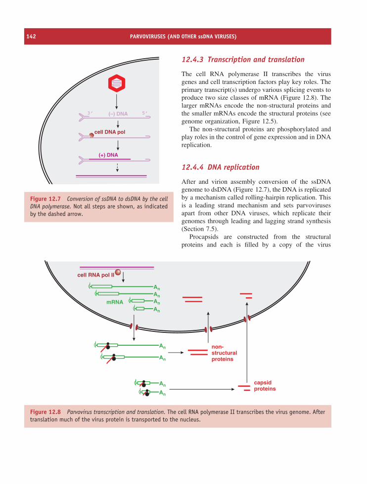
142 PARVOVIRUSES (AND OTHER ssDNA VIRUSES)
cell DNA pol
5’3’ (–) DNA
(+) DNA
Figure 12.7 Conversion of ssDNA to dsDNA by the cellDNA polymerase. Not all steps are shown, as indicatedby the dashed arrow.
12.4.3 Transcription and translation
The cell RNA polymerase II transcribes the virusgenes and cell transcription factors play key roles. Theprimary transcript(s) undergo various splicing events toproduce two size classes of mRNA (Figure 12.8). Thelarger mRNAs encode the non-structural proteins andthe smaller mRNAs encode the structural proteins (seegenome organization, Figure 12.5).
The non-structural proteins are phosphorylated andplay roles in the control of gene expression and in DNAreplication.
12.4.4 DNA replication
After and virion assembly conversion of the ssDNAgenome to dsDNA (Figure 12.7), the DNA is replicatedby a mechanism called rolling-hairpin replication. Thisis a leading strand mechanism and sets parvovirusesapart from other DNA viruses, which replicate theirgenomes through leading and lagging strand synthesis(Section 7.5).
Procapsids are constructed from the structuralproteins and each is filled by a copy of the virus
An
An
An
An capsidproteins
non-structuralproteins
An
An
An
An
mRNA
cell RNA pol II
Figure 12.8 Parvovirus transcription and translation. The cell RNA polymerase II transcribes the virus genome. Aftertranslation much of the virus protein is transported to the nucleus.
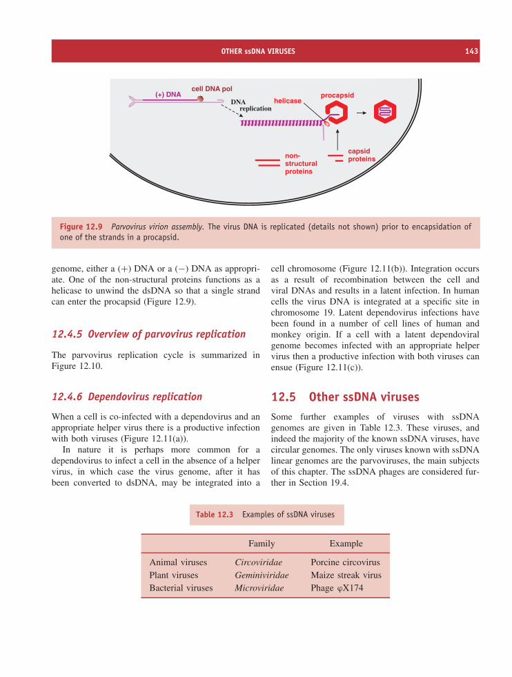
OTHER ssDNA VIRUSES 143
capsidproteinsnon-
structuralproteins
helicase(+) DNA
DNA replication
procapsidcell DNA pol
Figure 12.9 Parvovirus virion assembly. The virus DNA is replicated (details not shown) prior to encapsidation ofone of the strands in a procapsid.
genome, either a (+) DNA or a (−) DNA as appropri-ate. One of the non-structural proteins functions as ahelicase to unwind the dsDNA so that a single strandcan enter the procapsid (Figure 12.9).
12.4.5 Overview of parvovirus replication
The parvovirus replication cycle is summarized inFigure 12.10.
12.4.6 Dependovirus replication
When a cell is co-infected with a dependovirus and anappropriate helper virus there is a productive infectionwith both viruses (Figure 12.11(a)).
In nature it is perhaps more common for adependovirus to infect a cell in the absence of a helpervirus, in which case the virus genome, after it hasbeen converted to dsDNA, may be integrated into a
cell chromosome (Figure 12.11(b)). Integration occursas a result of recombination between the cell andviral DNAs and results in a latent infection. In humancells the virus DNA is integrated at a specific site inchromosome 19. Latent dependovirus infections havebeen found in a number of cell lines of human andmonkey origin. If a cell with a latent dependoviralgenome becomes infected with an appropriate helpervirus then a productive infection with both viruses canensue (Figure 12.11(c)).
12.5 Other ssDNA virusesSome further examples of viruses with ssDNAgenomes are given in Table 12.3. These viruses, andindeed the majority of the known ssDNA viruses, havecircular genomes. The only viruses known with ssDNAlinear genomes are the parvoviruses, the main subjectsof this chapter. The ssDNA phages are considered fur-ther in Section 19.4.
Table 12.3 Examples of ssDNA viruses
Family Example
Animal viruses Circoviridae Porcine circovirusPlant viruses Geminiviridae Maize streak virusBacterial viruses Microviridae Phage ϕX174
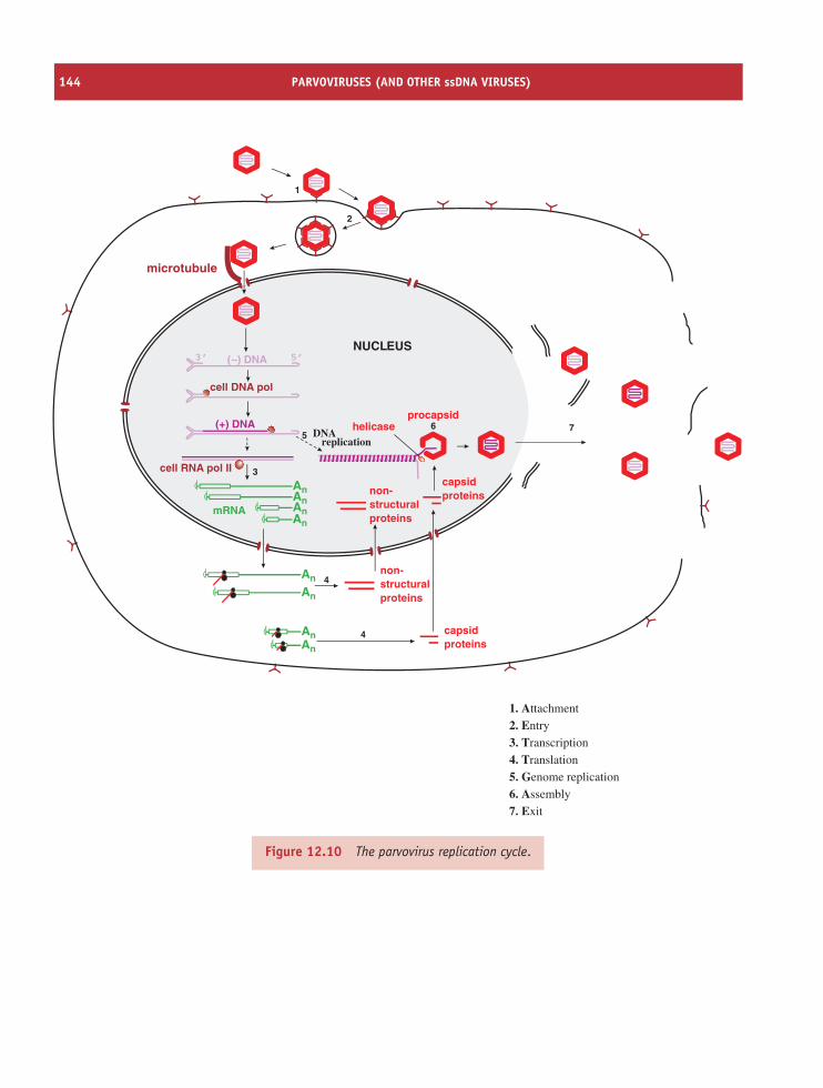
144 PARVOVIRUSES (AND OTHER ssDNA VIRUSES)
An
An
An
An
NUCLEUS5’3’
capsidproteins
capsidproteins
non-structuralproteins
non-structuralproteins
AnAn
mRNAAn
An
helicaseprocapsid
( ) DNA–
(+) DNA
microtubule
cell DNA pol
cell RNA pol II
1
2
3
4
5 DNA6 7
4
replication
1. Attachment
2. Entry
3. Transcription
4. Translation
5. Genome replication
6. Assembly
7. Exit
Figure 12.10 The parvovirus replication cycle.
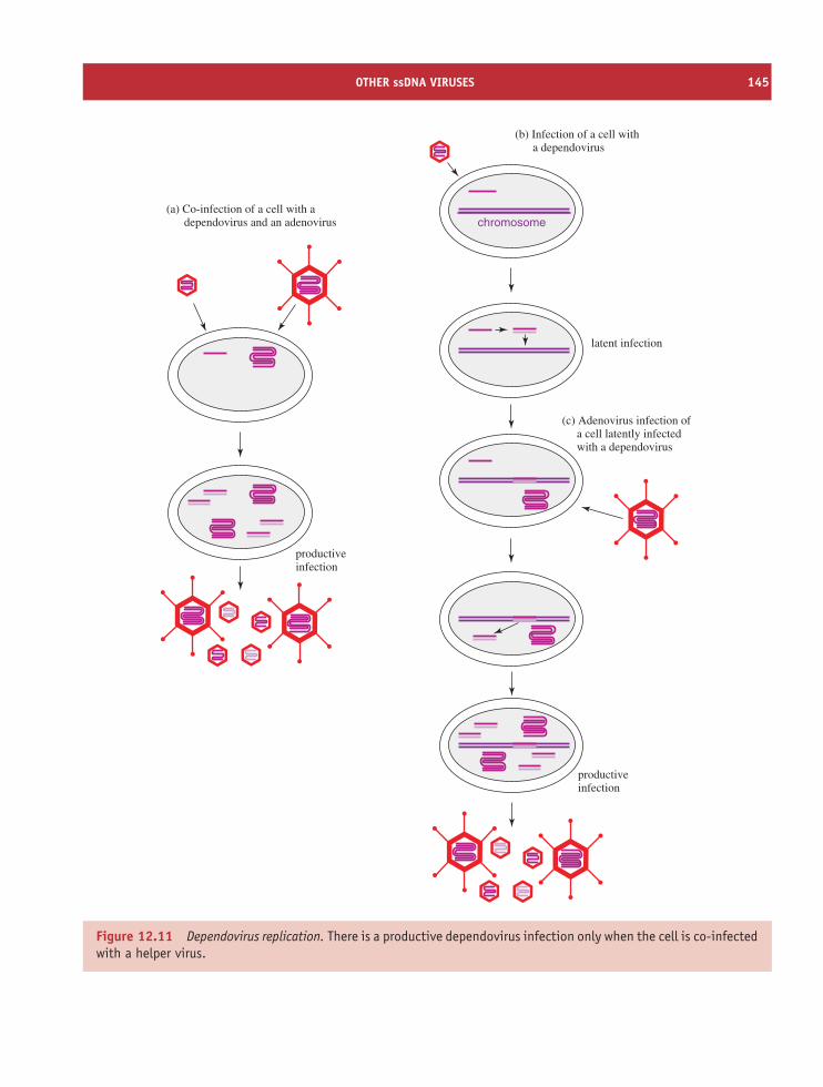
OTHER ssDNA VIRUSES 145
(a) Co-infection of a cell with a dependovirus and an adenovirus
(b) Infection of a cell with a dependovirus
(c) Adenovirus infection of a cell latently infected with a dependovirus
productiveinfection
latent infection
productiveinfection
chromosome
Figure 12.11 Dependovirus replication. There is a productive dependovirus infection only when the cell is co-infectedwith a helper virus.
146 PARVOVIRUSES (AND OTHER ssDNA VIRUSES)
Learning outcomesBy the end of this chapter you should be able to
• give examples of parvoviruses and explain theirimportance;
• describe the parvovirus virion;
• outline the main features of the parvovirusgenome;
• describe the replication cycle of parvoviruses;
• explain the difference between autonomous anddefective parvoviruses.
Sources of further informationBookKnipe D. M. and Howley P. M., editors-in-chief (2001)
Fields Virology, 4th edition, Chapters 69 and 70, Lippin-cott, Williams and Wilkins
JournalsAllander T. et al. (2005) Cloning of a human parvovirus
by molecular screening of respiratory tract samplesProceedings of the National Academy of Sciences of theUSA, 102, 12891–12896
Carter B. J. (2004) Adeno-associated virus and the develop-ment of adeno-associated virus vectors: a historical per-spective Molecular Therapy, 10, 981–989
Corcoran A. and Doyle S. (2004) Advances in the biology,diagnosis and host–pathogen interactions of parvovirusB19 Journal of Medical Microbiology, 53, 459–475
Flotte T. R. (2005) Adeno-associated virus-based gene ther-apy for inherited disorders Pediatric Research, 58,1143–1147
Gao G. et al. (2004) Clades of adeno-associated viruses arewidely disseminated in human tissues Journal of Virology,78, 6381–6388
Heegaard E. D. and Brown K. E. (2002) Human parvo-virus B19 Clinical Microbiology Reviews, 15, 485–505
Hueffer K. and Parrish C. R. (2003) Parvovirus host range,cell tropism and evolution Current Opinion in Microbiol-ogy, 6, 392–398
Vihinen-Ranta M. et al. (2004) Pathways of cell infectionby parvoviruses and adeno-associated viruses Journal ofVirology, 78, 6709–6714
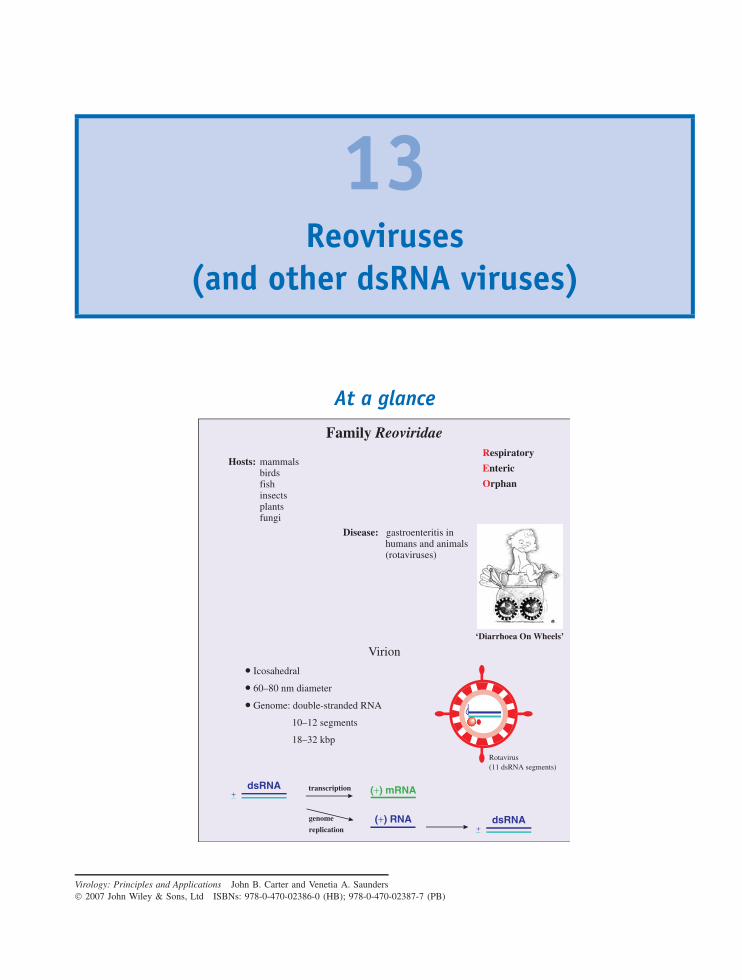
13Reoviruses
(and other dsRNA viruses)
At a glance
Virion
• Icosahedral
• 60–80 nm diameter
• Genome: double-stranded RNA
10–12 segments
18–32 kbp
Family Reoviridae
Hosts: mammalsbirdsfishinsectsplantsfungi
Respiratory
Enteric
Orphan
Disease: gastroenteritis in humans and animals (rotaviruses)
‘Diarrhoea On Wheels’
dsRNA (+) mRNA
(+) RNA
+−
dsRNA+−
transcription
genome
replication
Rotavirus(11 dsRNA segments)
Virology: Principles and Applications John B. Carter and Venetia A. Saunders 2007 John Wiley & Sons, Ltd ISBNs: 978-0-470-02386-0 (HB); 978-0-470-02387-7 (PB)
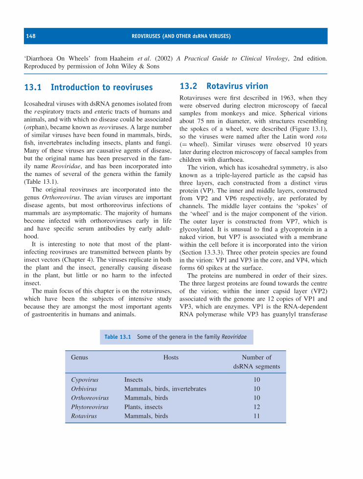
148 REOVIRUSES (AND OTHER dsRNA VIRUSES)
‘Diarrhoea On Wheels’ from Haaheim et al. (2002) A Practical Guide to Clinical Virology, 2nd edition.Reproduced by permission of John Wiley & Sons
13.1 Introduction to reoviruses
Icosahedral viruses with dsRNA genomes isolated fromthe respiratory tracts and enteric tracts of humans andanimals, and with which no disease could be associated(orphan), became known as reoviruses. A large numberof similar viruses have been found in mammals, birds,fish, invertebrates including insects, plants and fungi.Many of these viruses are causative agents of disease,but the original name has been preserved in the fam-ily name Reoviridae, and has been incorporated intothe names of several of the genera within the family(Table 13.1).
The original reoviruses are incorporated into thegenus Orthoreovirus. The avian viruses are importantdisease agents, but most orthoreovirus infections ofmammals are asymptomatic. The majority of humansbecome infected with orthoreoviruses early in lifeand have specific serum antibodies by early adult-hood.
It is interesting to note that most of the plant-infecting reoviruses are transmitted between plants byinsect vectors (Chapter 4). The viruses replicate in boththe plant and the insect, generally causing diseasein the plant, but little or no harm to the infectedinsect.
The main focus of this chapter is on the rotaviruses,which have been the subjects of intensive studybecause they are amongst the most important agentsof gastroenteritis in humans and animals.
13.2 Rotavirus virionRotaviruses were first described in 1963, when theywere observed during electron microscopy of faecalsamples from monkeys and mice. Spherical virionsabout 75 nm in diameter, with structures resemblingthe spokes of a wheel, were described (Figure 13.1),so the viruses were named after the Latin word rota(= wheel). Similar viruses were observed 10 yearslater during electron microscopy of faecal samples fromchildren with diarrhoea.
The virion, which has icosahedral symmetry, is alsoknown as a triple-layered particle as the capsid hasthree layers, each constructed from a distinct virusprotein (VP). The inner and middle layers, constructedfrom VP2 and VP6 respectively, are perforated bychannels. The middle layer contains the ‘spokes’ ofthe ‘wheel’ and is the major component of the virion.The outer layer is constructed from VP7, which isglycosylated. It is unusual to find a glycoprotein in anaked virion, but VP7 is associated with a membranewithin the cell before it is incorporated into the virion(Section 13.3.3). Three other protein species are foundin the virion: VP1 and VP3 in the core, and VP4, whichforms 60 spikes at the surface.
The proteins are numbered in order of their sizes.The three largest proteins are found towards the centreof the virion; within the inner capsid layer (VP2)associated with the genome are 12 copies of VP1 andVP3, which are enzymes. VP1 is the RNA-dependentRNA polymerase while VP3 has guanylyl transferase
Table 13.1 Some of the genera in the family Reoviridae
Genus Hosts Number ofdsRNA segments
Cypovirus Insects 10Orbivirus Mammals, birds, invertebrates 10Orthoreovirus Mammals, birds 10Phytoreovirus Plants, insects 12Rotavirus Mammals, birds 11
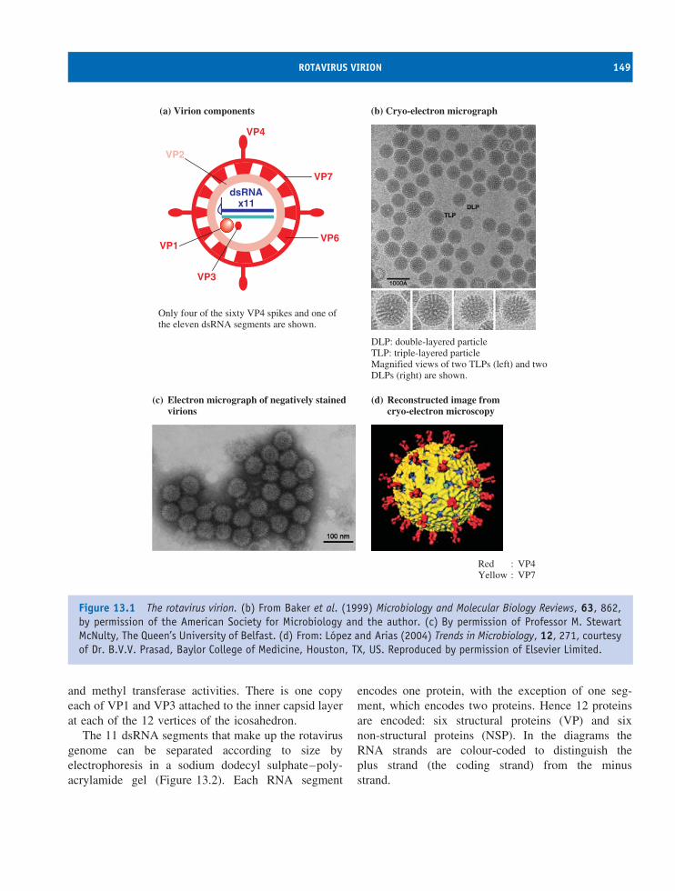
ROTAVIRUS VIRION 149
(a) Virion components
(c) Electron micrograph of negatively stained virions
(d) Reconstructed image from cryo-electron microscopy
(b) Cryo-electron micrograph
VP4
VP2
VP1
VP3
VP7
VP6
dsRNAx11
Only four of the sixty VP4 spikes and one ofthe eleven dsRNA segments are shown.
DLP: double-layered particleTLP: triple-layered particleMagnified views of two TLPs (left) and twoDLPs (right) are shown.
Red : VP4Yellow : VP7
Figure 13.1 The rotavirus virion. (b) From Baker et al. (1999) Microbiology and Molecular Biology Reviews, 63, 862,by permission of the American Society for Microbiology and the author. (c) By permission of Professor M. StewartMcNulty, The Queen’s University of Belfast. (d) From: Lopez and Arias (2004) Trends in Microbiology, 12, 271, courtesyof Dr. B.V.V. Prasad, Baylor College of Medicine, Houston, TX, US. Reproduced by permission of Elsevier Limited.
and methyl transferase activities. There is one copyeach of VP1 and VP3 attached to the inner capsid layerat each of the 12 vertices of the icosahedron.
The 11 dsRNA segments that make up the rotavirusgenome can be separated according to size byelectrophoresis in a sodium dodecyl sulphate–poly-acrylamide gel (Figure 13.2). Each RNA segment
encodes one protein, with the exception of one seg-ment, which encodes two proteins. Hence 12 proteinsare encoded: six structural proteins (VP) and sixnon-structural proteins (NSP). In the diagrams theRNA strands are colour-coded to distinguish theplus strand (the coding strand) from the minusstrand.
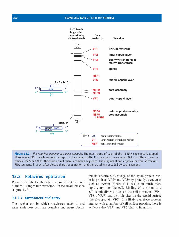
150 REOVIRUSES (AND OTHER dsRNA VIRUSES)
VP1
VP2
VP3
VP
VP4
VP6
VP7
NSP1
NSP2NSP3
NSP4
NSP
NSP5+ NSP6
RNA polymerase
inner capsid layer
guanylyl transferase;methyl transferase
spikes
core assembly
middle capsid layer
outer capsid layer
core assemblyouter capsid assembly
open reading frame
virus protein (structural protein)
non-structural protein
(−)
(+)5’
5’
3’
3’ORF
ORF(−)
(+)5’
5’
3’
3’NSP5NSP6
RNAs 1-10
RNA 11
RNA bandsin gel after
separation byelectrophoresis
Geneproduct(s) Function
Key:
Figure 13.2 The rotavirus genome and gene products. The plus strand of each of the 11 RNA segments is capped.There is one ORF in each segment, except for the smallest (RNA 11), in which there are two ORFs in different readingframes. NSP5 and NSP6 therefore do not share a common sequence. The diagram shows a typical pattern of rotavirusRNA segments in a gel after electrophoretic separation, and the protein(s) encoded by each segment.
13.3 Rotavirus replicationRotaviruses infect cells called enterocytes at the endsof the villi (finger-like extensions) in the small intestine(Figure 13.3).
13.3.1 Attachment and entryThe mechanisms by which rotaviruses attach to andenter their host cells are complex and many details
remain uncertain. Cleavage of the spike protein VP4to its products VP8* and VP5* by proteolytic enzymessuch as trypsin (Figure 13.4) results in much morerapid entry into the cell. Binding of a virion to acell is initially via sites on the spike proteins (VP4,VP8*, VP5*) and then via sites on the capsid surface(the glycoprotein VP7). It is likely that these proteinsinteract with a number of cell surface proteins; there isevidence that VP5* and VP7 bind to integrins.
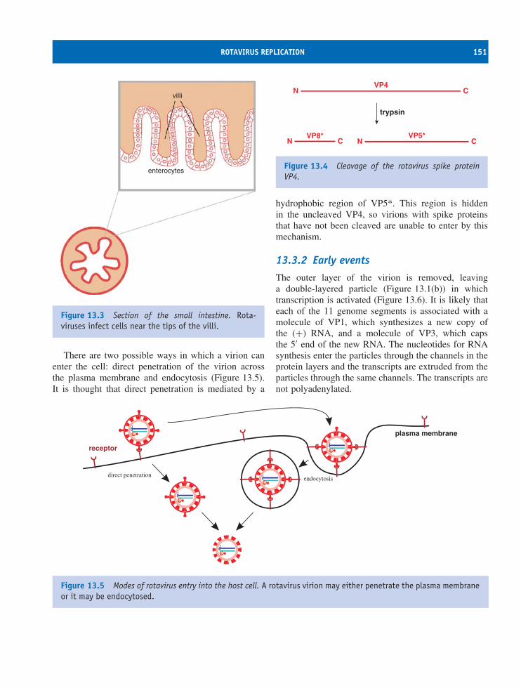
ROTAVIRUS REPLICATION 151
villi
enterocytes
Figure 13.3 Section of the small intestine. Rota-viruses infect cells near the tips of the villi.
There are two possible ways in which a virion canenter the cell: direct penetration of the virion acrossthe plasma membrane and endocytosis (Figure 13.5).It is thought that direct penetration is mediated by a
VP4N C
VP8*N C
VP5*N C
trypsin
Figure 13.4 Cleavage of the rotavirus spike proteinVP4.
hydrophobic region of VP5*. This region is hiddenin the uncleaved VP4, so virions with spike proteinsthat have not been cleaved are unable to enter by thismechanism.
13.3.2 Early events
The outer layer of the virion is removed, leavinga double-layered particle (Figure 13.1(b)) in whichtranscription is activated (Figure 13.6). It is likely thateach of the 11 genome segments is associated with amolecule of VP1, which synthesizes a new copy ofthe (+) RNA, and a molecule of VP3, which capsthe 5′ end of the new RNA. The nucleotides for RNAsynthesis enter the particles through the channels in theprotein layers and the transcripts are extruded from theparticles through the same channels. The transcripts arenot polyadenylated.
receptor
plasma membrane
direct penetrationendocytosis
Figure 13.5 Modes of rotavirus entry into the host cell. A rotavirus virion may either penetrate the plasma membraneor it may be endocytosed.
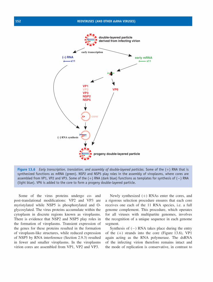
152 REOVIRUSES (AND OTHER dsRNA VIRUSES)
x11
VP1
VP3NSP2NSP5
VP2 VP6
early mRNAx11
core
double-layered particlederived from infecting virion
progeny double-layered particle
early transcription
(−) RNA synthesis
(+) RNA
Figure 13.6 Early transcription, translation, and assembly of double-layered particles. Some of the (+) RNA that issynthesized functions as mRNA (green). NSP2 and NSP5 play roles in the assembly of viroplasms, where cores areassembled from VP1, VP2 and VP3. Some of the (+) RNA (dark blue) functions as templates for synthesis of (−) RNA(light blue). VP6 is added to the core to form a progeny double-layered particle.
Some of the virus proteins undergo co- andpost-translational modifications: VP2 and VP3 aremyristylated while NSP5 is phosphorylated and O-glycosylated. The virus proteins accumulate within thecytoplasm in discrete regions known as viroplasms.There is evidence that NSP2 and NSP5 play roles inthe formation of viroplasms. Transient expression ofthe genes for these proteins resulted in the formationof viroplasm-like structures, while reduced expressionof NSP5 by RNA interference (Section 2.9.3) resultedin fewer and smaller viroplasms. In the viroplasmsvirion cores are assembled from VP1, VP2 and VP3.
Newly synthesized (+) RNAs enter the cores, anda rigorous selection procedure ensures that each corereceives one each of the 11 RNA species, i.e. a fullgenome complement. This procedure, which operatesfor all viruses with multipartite genomes, involvesthe recognition of a unique sequence in each genomesegment.
Synthesis of (−) RNA takes place during the entryof the (+) strands into the core (Figure 13.6), VP1again acting as the RNA polymerase. The dsRNAof the infecting virion therefore remains intact andthe mode of replication is conservative, in contrast to
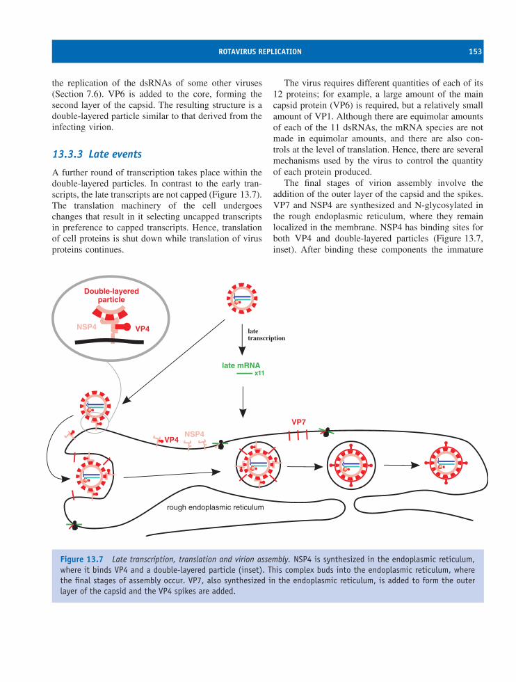
ROTAVIRUS REPLICATION 153
the replication of the dsRNAs of some other viruses(Section 7.6). VP6 is added to the core, forming thesecond layer of the capsid. The resulting structure is adouble-layered particle similar to that derived from theinfecting virion.
13.3.3 Late events
A further round of transcription takes place within thedouble-layered particles. In contrast to the early tran-scripts, the late transcripts are not capped (Figure 13.7).The translation machinery of the cell undergoeschanges that result in it selecting uncapped transcriptsin preference to capped transcripts. Hence, translationof cell proteins is shut down while translation of virusproteins continues.
The virus requires different quantities of each of its12 proteins; for example, a large amount of the maincapsid protein (VP6) is required, but a relatively smallamount of VP1. Although there are equimolar amountsof each of the 11 dsRNAs, the mRNA species are notmade in equimolar amounts, and there are also con-trols at the level of translation. Hence, there are severalmechanisms used by the virus to control the quantityof each protein produced.
The final stages of virion assembly involve theaddition of the outer layer of the capsid and the spikes.VP7 and NSP4 are synthesized and N-glycosylated inthe rough endoplasmic reticulum, where they remainlocalized in the membrane. NSP4 has binding sites forboth VP4 and double-layered particles (Figure 13.7,inset). After binding these components the immature
latetranscription
VP4
VP7
NSP4
late mRNAx11
rough endoplasmic reticulum
NSP4 VP4
Double-layeredparticle
Figure 13.7 Late transcription, translation and virion assembly. NSP4 is synthesized in the endoplasmic reticulum,where it binds VP4 and a double-layered particle (inset). This complex buds into the endoplasmic reticulum, wherethe final stages of assembly occur. VP7, also synthesized in the endoplasmic reticulum, is added to form the outerlayer of the capsid and the VP4 spikes are added.
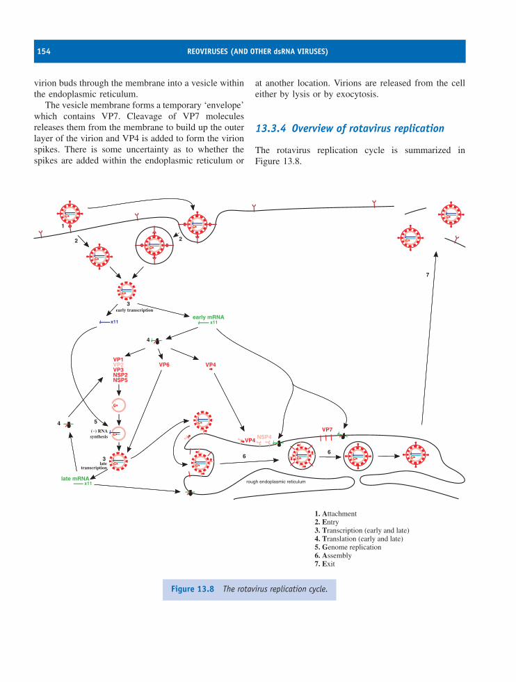
154 REOVIRUSES (AND OTHER dsRNA VIRUSES)
virion buds through the membrane into a vesicle withinthe endoplasmic reticulum.
The vesicle membrane forms a temporary ‘envelope’which contains VP7. Cleavage of VP7 moleculesreleases them from the membrane to build up the outerlayer of the virion and VP4 is added to form the virionspikes. There is some uncertainty as to whether thespikes are added within the endoplasmic reticulum or
at another location. Virions are released from the celleither by lysis or by exocytosis.
13.3.4 Overview of rotavirus replication
The rotavirus replication cycle is summarized inFigure 13.8.
1
3
2 2
early transcription
latetranscription
(−) RNAsynthesis
3
4
4
5
7
66
VP1
VP3NSP2NSP5
VP2 VP6 VP4
VP4
VP7NSP4
early mRNAx11x11
late mRNAx11 rough endoplasmic reticulum
1. Attachment2. Entry3. Transcription (early and late)4. Translation (early and late)5. Genome replication6. Assembly7. Exit
Figure 13.8 The rotavirus replication cycle.
SOURCES OF FURTHER INFORMATION 155
13.3.5 Rotavirus disease
Enterocytes on the villi are destroyed as a result ofrotavirus infection and this leads to reduced absorptionof water, salts and sugars from the gut. There isevidence that the tight junctions between cells aredamaged by the non-structural protein NSP4, allowingleakage of fluid into the gut. These effects of virusinfection, together with the secretion of water andsolutes by secretory cells, result in diarrhoea and thiscan lead to dehydration. Treatment is a relativelysimple matter of rehydrating the patient with a solutionof salts and sugar, but the means for this are notavailable in some parts of the world. It estimated thateach year there are around half a million deaths ofinfants and young children due to rotavirus infections.
Although most rotavirus infections are confined tothe gut, there is evidence that on occasion the virusmay cross the gut and infect other tissues.
13.4 Other dsRNA viruses
The virions of most dsRNA viruses have icosahedralsymmetry and most of them are naked; an exceptionis the family Cystoviridae, the members of which areenveloped bacteriophages (Section 19.3). Some furtherexamples of viruses with dsRNA genomes are given inTable 13.2.
A problem that dsRNA viruses must overcome is thefact that dsRNA is a potent inducer of a number of celldefence mechanisms including apoptosis, interferonproduction and RNA silencing (Chapter 9). Most ofthese viruses, including the rotaviruses, have solvedthis problem by ensuring that the viral dsRNA isalways enclosed within virus protein structures, and is
therefore never free in the cytoplasm to trigger thesedefences.
Learning outcomes
By the end of this chapter you should be able to
• explain how viruses in the family Reoviridae areclassified into genera;
• describe the rotavirus virion;
• discuss the main events of the rotavirus replica-tion cycle;
• explain how rotaviruses cause disease.
Sources of further information
Book
Roy P., editor (2006) Reoviruses: Entry, Assembly andMorphogenesis, Springer
Journals
Campagna M. et al. (2005) RNA interference of rotavirussegment 11 mRNA reveals the essential role of NSP5 inthe virus replicative cycle Journal of General Virology, 86,1481–1487
Chen D. et al. (2001) Features of the 3′-consensus sequenceof rotavirus mRNAs critical to minus strand synthesisVirology 282, 221–229
Table 13.2 Examples of dsRNA viruses
Family Example
Animal viruses Birnaviridae Infectious bursal disease virusPlant viruses Partitiviridae White clover cryptic virus 1Fungal viruses Chrysoviridae Penicillium chrysogenum virusBacterial viruses Cystoviridae Pseudomonas phage ϕ6

156 REOVIRUSES (AND OTHER dsRNA VIRUSES)
Lopez S. and Arias C. F. (2004) Multistep entry of rotavirusinto cells: a Versaillesque dance Trends in Microbiology,12, 271–278
Lopez T. et al. (2005) Silencing the morphogenesis ofrotavirus Journal of Virology, 79, 184–192
Mertens P. (2004) The dsRNA viruses Virus Research, 101,3–13
Pesavento J. B. et al. (2005) pH-induced conformationalchange of the rotavirus VP4 spike: implications for cell
entry and antibody neutralization Journal of Virology, 79,8572–8580
Taraporewala Z. F. and Patton J. T. (2004) Nonstructuralproteins involved in genome packaging and replicationof rotaviruses and other members of the Reoviridae VirusResearch, 101, 57–66
Widdowson M.-A. et al. (2005) Rotavirus disease and itsprevention Current Opinion in Gastroenterology, 21,26–31
14Picornaviruses (and other plus-strand
RNA viruses)
At a glance
Virion
• icosahedral
• 25–30 nm diameter
• genome: single-stranded RNA
Plus polarity
7–8 kb
Covalently linked protein (VPg)
Family PicornaviridaePico (= small) RNA viruses
Hosts: mammalsbirds
Diseases: common coldpoliohepatitis Afoot and mouth disease
(−) RNA(+) RNA (+) mRNA
(+) RNA
transcription
genomereplication
The virus genome can function as mRNA.
Virology: Principles and Applications John B. Carter and Venetia A. Saunders 2007 John Wiley & Sons, Ltd ISBNs: 978-0-470-02386-0 (HB); 978-0-470-02387-7 (PB)
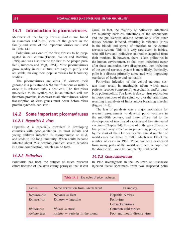
158 PICORNAVIRUSES (AND OTHER PLUS-STRAND RNA VIRUSES)
14.1 Introduction to picornavirusesMembers of the family Picornaviridae are foundin mammals and birds; some of the genera in thefamily and some of the important viruses are listedin Table 14.1.
Poliovirus was one of the first viruses to be prop-agated in cell culture (Enders, Weller and Robbins,1949) and was also one of the first to be plaque puri-fied (Dulbecco and Vogt, 1954). Most picornavirusesgrow readily in cell culture, are easy to purify andare stable, making them popular viruses for laboratorystudies.
The picornaviruses are class IV viruses; theirgenome is a plus-strand RNA that functions as mRNAonce it is released into a host cell. The first virusmolecules to be synthesized in an infected cell aretherefore proteins, in contrast to all other viruses, wheretranscription of virus genes must occur before virusprotein synthesis can start.
14.2 Some important picornaviruses14.2.1 Hepatitis A virusHepatitis A is especially prevalent in developingcountries with poor sanitation. In most infants andyoung children infection is asymptomatic or mild,and leads to life-long immunity. When adults becomeinfected about 75% develop jaundice; severe hepatitisis a rare complication, which can be fatal.
14.2.2 PoliovirusPoliovirus has been the subject of much researcheffort because of the devastating paralysis that it can
cause. In fact, the majority of poliovirus infectionsare relatively harmless infections of the oropharynxand the gut. Serious disease occurs only after othertissues become infected, resulting in viraemia (virusin the blood) and spread of infection to the centralnervous system. This is a very rare event in babies,who still have anti-poliovirus antibodies acquired fromtheir mothers. If, however, there is less poliovirus inthe human environment, so that most infections occurafter these antibodies have disappeared, then infectionof the central nervous system is more likely. Ironically,polio is a disease primarily associated with improvingstandards of hygiene and sanitation!
Poliovirus infection of the central nervous sys-tem may result in meningitis (from which mostpatients recover completely), encephalitis and/or para-lytic poliomyelitis. The latter is due to virus replicationin motor neurones of the spinal cord or the brain stem,resulting in paralysis of limbs and/or breathing muscles(Figure 14.1).
The fear of paralysis was a major motivation forresearch programmes to develop polio vaccines inthe mid-20th century, and these efforts led to thedevelopment of inactivated vaccines and live attenuatedvaccines (Chapter 24). The use of both types of vaccinehas proved very effective in preventing polio, so thatby the start of the 21st century the annual number ofworld cases had fallen to 3500, which was 1% of thenumber of cases in 1988. Polio has been eradicatedfrom many parts of the world and there is hope thatthe disease will soon be completely eradicated.
14.2.3 CoxsackievirusesIn 1948 investigators in the US town of Coxsackieinjected faecal specimens from two suspected polio
Table 14.1 Examples of picornaviruses
Genus Name derivation from Greek word Example(s)
Hepatovirus Hepatos = liver Hepatitis A virusEnterovirus Enteron = intestine Poliovirus
CoxsackievirusesRhinovirus Rhinos = nose Common cold virusesAphthovirus Aphtha = vesicles in the mouth Foot and mouth disease virus
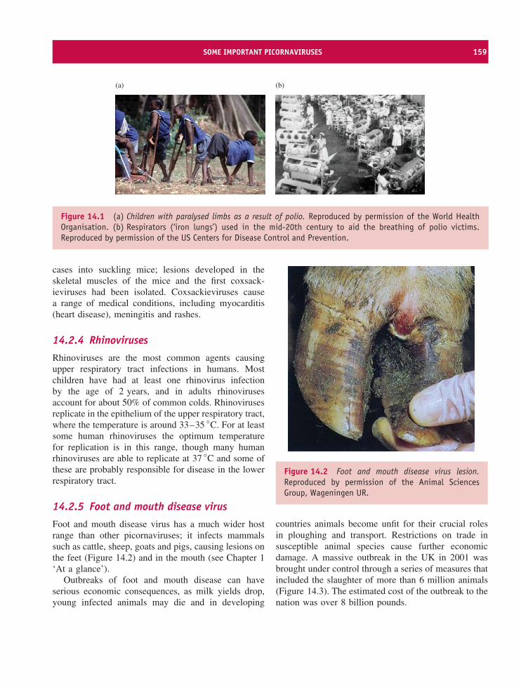
SOME IMPORTANT PICORNAVIRUSES 159
(a) (b)
Figure 14.1 (a) Children with paralysed limbs as a result of polio. Reproduced by permission of the World HealthOrganisation. (b) Respirators (‘iron lungs’) used in the mid-20th century to aid the breathing of polio victims.Reproduced by permission of the US Centers for Disease Control and Prevention.
cases into suckling mice; lesions developed in theskeletal muscles of the mice and the first coxsack-ieviruses had been isolated. Coxsackieviruses causea range of medical conditions, including myocarditis(heart disease), meningitis and rashes.
14.2.4 Rhinoviruses
Rhinoviruses are the most common agents causingupper respiratory tract infections in humans. Mostchildren have had at least one rhinovirus infectionby the age of 2 years, and in adults rhinovirusesaccount for about 50% of common colds. Rhinovirusesreplicate in the epithelium of the upper respiratory tract,where the temperature is around 33–35 ◦C. For at leastsome human rhinoviruses the optimum temperaturefor replication is in this range, though many humanrhinoviruses are able to replicate at 37 ◦C and some ofthese are probably responsible for disease in the lowerrespiratory tract.
14.2.5 Foot and mouth disease virus
Foot and mouth disease virus has a much wider hostrange than other picornaviruses; it infects mammalssuch as cattle, sheep, goats and pigs, causing lesions onthe feet (Figure 14.2) and in the mouth (see Chapter 1‘At a glance’).
Outbreaks of foot and mouth disease can haveserious economic consequences, as milk yields drop,young infected animals may die and in developing
Figure 14.2 Foot and mouth disease virus lesion.Reproduced by permission of the Animal SciencesGroup, Wageningen UR.
countries animals become unfit for their crucial rolesin ploughing and transport. Restrictions on trade insusceptible animal species cause further economicdamage. A massive outbreak in the UK in 2001 wasbrought under control through a series of measures thatincluded the slaughter of more than 6 million animals(Figure 14.3). The estimated cost of the outbreak to thenation was over 8 billion pounds.
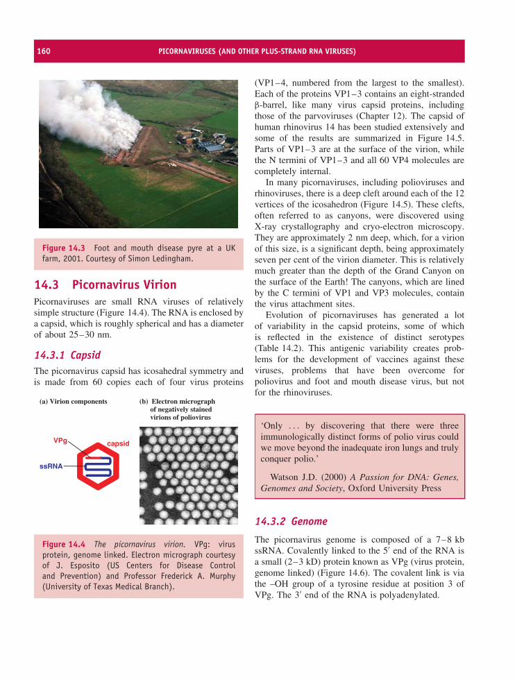
160 PICORNAVIRUSES (AND OTHER PLUS-STRAND RNA VIRUSES)
Figure 14.3 Foot and mouth disease pyre at a UKfarm, 2001. Courtesy of Simon Ledingham.
14.3 Picornavirus VirionPicornaviruses are small RNA viruses of relativelysimple structure (Figure 14.4). The RNA is enclosed bya capsid, which is roughly spherical and has a diameterof about 25–30 nm.
14.3.1 CapsidThe picornavirus capsid has icosahedral symmetry andis made from 60 copies each of four virus proteins
(a) Virion components (b) Electron micrograph of negatively stained virions of poliovirus
capsid
ssRNA
VPg
Figure 14.4 The picornavirus virion. VPg: virusprotein, genome linked. Electron micrograph courtesyof J. Esposito (US Centers for Disease Controland Prevention) and Professor Frederick A. Murphy(University of Texas Medical Branch).
(VP1–4, numbered from the largest to the smallest).Each of the proteins VP1–3 contains an eight-strandedβ-barrel, like many virus capsid proteins, includingthose of the parvoviruses (Chapter 12). The capsid ofhuman rhinovirus 14 has been studied extensively andsome of the results are summarized in Figure 14.5.Parts of VP1–3 are at the surface of the virion, whilethe N termini of VP1–3 and all 60 VP4 molecules arecompletely internal.
In many picornaviruses, including polioviruses andrhinoviruses, there is a deep cleft around each of the 12vertices of the icosahedron (Figure 14.5). These clefts,often referred to as canyons, were discovered usingX-ray crystallography and cryo-electron microscopy.They are approximately 2 nm deep, which, for a virionof this size, is a significant depth, being approximatelyseven per cent of the virion diameter. This is relativelymuch greater than the depth of the Grand Canyon onthe surface of the Earth! The canyons, which are linedby the C termini of VP1 and VP3 molecules, containthe virus attachment sites.
Evolution of picornaviruses has generated a lotof variability in the capsid proteins, some of whichis reflected in the existence of distinct serotypes(Table 14.2). This antigenic variability creates prob-lems for the development of vaccines against theseviruses, problems that have been overcome forpoliovirus and foot and mouth disease virus, but notfor the rhinoviruses.
‘Only . . . by discovering that there were threeimmunologically distinct forms of polio virus couldwe move beyond the inadequate iron lungs and trulyconquer polio.’
Watson J.D. (2000) A Passion for DNA: Genes,Genomes and Society, Oxford University Press
14.3.2 Genome
The picornavirus genome is composed of a 7–8 kbssRNA. Covalently linked to the 5′ end of the RNA isa small (2–3 kD) protein known as VPg (virus protein,genome linked) (Figure 14.6). The covalent link is viathe –OH group of a tyrosine residue at position 3 ofVPg. The 3′ end of the RNA is polyadenylated.
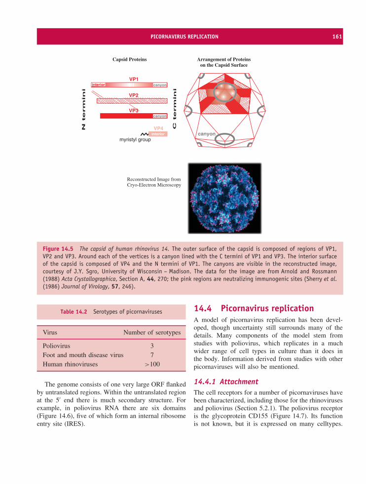
PICORNAVIRUS REPLICATION 161
myristyl group
VP1
VP2
VP3
VP4canyon
Reconstructed Image fromCryo-Electron Microscopy
canyoninterior
interior
canyon
Capsid Proteins Arrangement of Proteinson the Capsid Surface
Figure 14.5 The capsid of human rhinovirus 14. The outer surface of the capsid is composed of regions of VP1,VP2 and VP3. Around each of the vertices is a canyon lined with the C termini of VP1 and VP3. The interior surfaceof the capsid is composed of VP4 and the N termini of VP1. The canyons are visible in the reconstructed image,courtesy of J.Y. Sgro, University of Wisconsin – Madison. The data for the image are from Arnold and Rossmann(1988) Acta Crystallographica, Section A, 44, 270; the pink regions are neutralizing immunogenic sites (Sherry et al.(1986) Journal of Virology, 57, 246).
Table 14.2 Serotypes of picornaviruses
Virus Number of serotypes
Poliovirus 3Foot and mouth disease virus 7Human rhinoviruses >100
The genome consists of one very large ORF flankedby untranslated regions. Within the untranslated regionat the 5′ end there is much secondary structure. Forexample, in poliovirus RNA there are six domains(Figure 14.6), five of which form an internal ribosomeentry site (IRES).
14.4 Picornavirus replicationA model of picornavirus replication has been devel-oped, though uncertainty still surrounds many of thedetails. Many components of the model stem fromstudies with poliovirus, which replicates in a muchwider range of cell types in culture than it does inthe body. Information derived from studies with otherpicornaviruses will also be mentioned.
14.4.1 AttachmentThe cell receptors for a number of picornaviruses havebeen characterized, including those for the rhinovirusesand poliovirus (Section 5.2.1). The poliovirus receptoris the glycoprotein CD155 (Figure 14.7). Its functionis not known, but it is expressed on many celltypes.
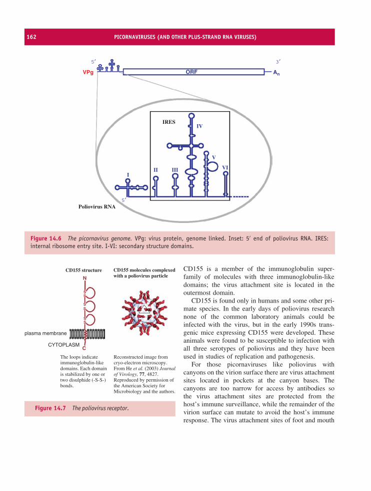
162 PICORNAVIRUSES (AND OTHER PLUS-STRAND RNA VIRUSES)
5′
5′
VPg
3′
AnORF
Poliovirus RNA
IRES
III
V
VI
IV
III
Figure 14.6 The picornavirus genome. VPg: virus protein, genome linked. Inset: 5′ end of poliovirus RNA. IRES:internal ribosome entry site. I-VI: secondary structure domains.
CD155 structure
The loops indicateimmunoglobulin-likedomains. Each domainis stabilized by one ortwo disulphide (-S-S-)bonds.
Reconstructed image fromcryo-electron microscopy.From He et al. (2003) Journalof Virology, 77, 4827.Reproduced by permission ofthe American Society forMicrobiology and the authors.
plasma membrane
CYTOPLASM
CD155 molecules complexedwith a poliovirus particleN
SS
SS
SS
C
Figure 14.7 The poliovirus receptor.
CD155 is a member of the immunoglobulin super-family of molecules with three immunoglobulin-likedomains; the virus attachment site is located in theoutermost domain.
CD155 is found only in humans and some other pri-mate species. In the early days of poliovirus researchnone of the common laboratory animals could beinfected with the virus, but in the early 1990s trans-genic mice expressing CD155 were developed. Theseanimals were found to be susceptible to infection withall three serotypes of poliovirus and they have beenused in studies of replication and pathogenesis.
For those picornaviruses like poliovirus withcanyons on the virion surface there are virus attachmentsites located in pockets at the canyon bases. Thecanyons are too narrow for access by antibodies sothe virus attachment sites are protected from thehost’s immune surveillance, while the remainder of thevirion surface can mutate to avoid the host’s immuneresponse. The virus attachment sites of foot and mouth
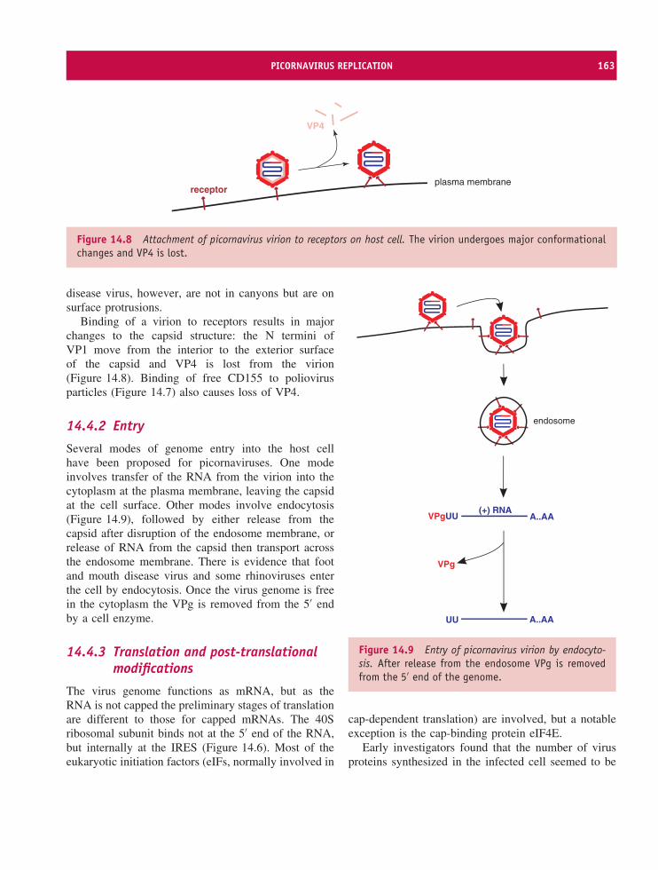
PICORNAVIRUS REPLICATION 163
VP4
plasma membranereceptor
Figure 14.8 Attachment of picornavirus virion to receptors on host cell. The virion undergoes major conformationalchanges and VP4 is lost.
disease virus, however, are not in canyons but are onsurface protrusions.
Binding of a virion to receptors results in majorchanges to the capsid structure: the N termini ofVP1 move from the interior to the exterior surfaceof the capsid and VP4 is lost from the virion(Figure 14.8). Binding of free CD155 to poliovirusparticles (Figure 14.7) also causes loss of VP4.
14.4.2 Entry
Several modes of genome entry into the host cellhave been proposed for picornaviruses. One modeinvolves transfer of the RNA from the virion into thecytoplasm at the plasma membrane, leaving the capsidat the cell surface. Other modes involve endocytosis(Figure 14.9), followed by either release from thecapsid after disruption of the endosome membrane, orrelease of RNA from the capsid then transport acrossthe endosome membrane. There is evidence that footand mouth disease virus and some rhinoviruses enterthe cell by endocytosis. Once the virus genome is freein the cytoplasm the VPg is removed from the 5′ endby a cell enzyme.
14.4.3 Translation and post-translationalmodifications
The virus genome functions as mRNA, but as theRNA is not capped the preliminary stages of translationare different to those for capped mRNAs. The 40Sribosomal subunit binds not at the 5′ end of the RNA,but internally at the IRES (Figure 14.6). Most of theeukaryotic initiation factors (eIFs, normally involved in
(+) RNAA..AAVPgUU
A..AAUU
VPg
endosome
Figure 14.9 Entry of picornavirus virion by endocyto-sis. After release from the endosome VPg is removedfrom the 5′ end of the genome.
cap-dependent translation) are involved, but a notableexception is the cap-binding protein eIF4E.
Early investigators found that the number of virusproteins synthesized in the infected cell seemed to be
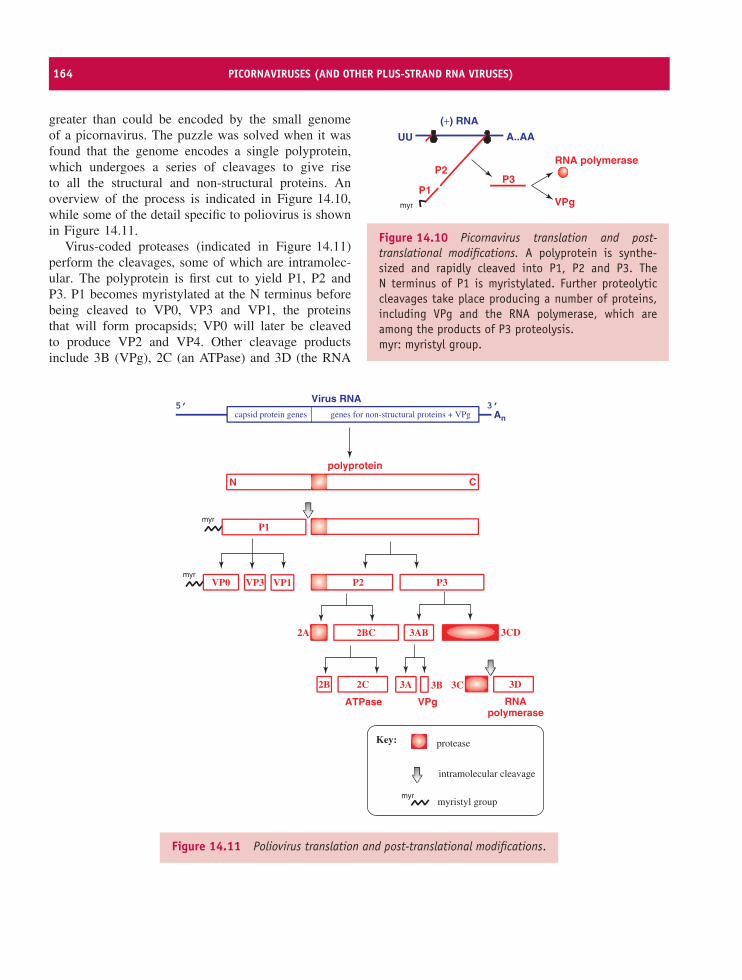
164 PICORNAVIRUSES (AND OTHER PLUS-STRAND RNA VIRUSES)
greater than could be encoded by the small genomeof a picornavirus. The puzzle was solved when it wasfound that the genome encodes a single polyprotein,which undergoes a series of cleavages to give riseto all the structural and non-structural proteins. Anoverview of the process is indicated in Figure 14.10,while some of the detail specific to poliovirus is shownin Figure 14.11.
Virus-coded proteases (indicated in Figure 14.11)perform the cleavages, some of which are intramolec-ular. The polyprotein is first cut to yield P1, P2 andP3. P1 becomes myristylated at the N terminus beforebeing cleaved to VP0, VP3 and VP1, the proteinsthat will form procapsids; VP0 will later be cleavedto produce VP2 and VP4. Other cleavage productsinclude 3B (VPg), 2C (an ATPase) and 3D (the RNA
VPg
P3
A..AAUU
P1
P2RNA polymerase
(+) RNA
myr
Figure 14.10 Picornavirus translation and post-translational modifications. A polyprotein is synthe-sized and rapidly cleaved into P1, P2 and P3. TheN terminus of P1 is myristylated. Further proteolyticcleavages take place producing a number of proteins,including VPg and the RNA polymerase, which areamong the products of P3 proteolysis.myr: myristyl group.
An
Virus RNA
capsid protein genes genes for non-structural proteins + VPg5’ 3’
polyprotein
N C
myrP1
P3P2
2BC2A 3CD3AB
2C2B 3A 3C 3D3B
VP0 VP3 VP1
ATPase RNApolymerase
VPg
myr
myr
protease
intramolecular cleavage
myristyl group
Key:
Figure 14.11 Poliovirus translation and post-translational modifications.
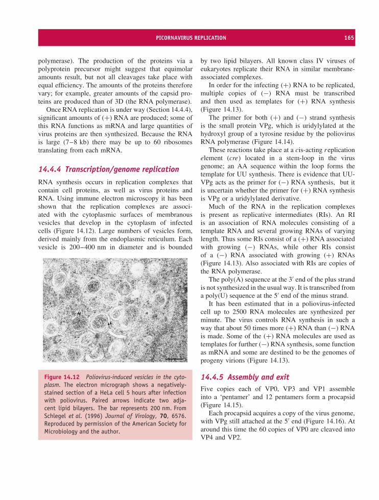
PICORNAVIRUS REPLICATION 165
polymerase). The production of the proteins via apolyprotein precursor might suggest that equimolaramounts result, but not all cleavages take place withequal efficiency. The amounts of the proteins thereforevary; for example, greater amounts of the capsid pro-teins are produced than of 3D (the RNA polymerase).
Once RNA replication is under way (Section 14.4.4),significant amounts of (+) RNA are produced; some ofthis RNA functions as mRNA and large quantities ofvirus proteins are then synthesized. Because the RNAis large (7–8 kb) there may be up to 60 ribosomestranslating from each mRNA.
14.4.4 Transcription/genome replication
RNA synthesis occurs in replication complexes thatcontain cell proteins, as well as virus proteins andRNA. Using immune electron microscopy it has beenshown that the replication complexes are associ-ated with the cytoplasmic surfaces of membranousvesicles that develop in the cytoplasm of infectedcells (Figure 14.12). Large numbers of vesicles form,derived mainly from the endoplasmic reticulum. Eachvesicle is 200–400 nm in diameter and is bounded
Figure 14.12 Poliovirus-induced vesicles in the cyto-plasm. The electron micrograph shows a negatively-stained section of a HeLa cell 5 hours after infectionwith poliovirus. Paired arrows indicate two adja-cent lipid bilayers. The bar represents 200 nm. FromSchlegel et al. (1996) Journal of Virology, 70, 6576.Reproduced by permission of the American Society forMicrobiology and the author.
by two lipid bilayers. All known class IV viruses ofeukaryotes replicate their RNA in similar membrane-associated complexes.
In order for the infecting (+) RNA to be replicated,multiple copies of (−) RNA must be transcribedand then used as templates for (+) RNA synthesis(Figure 14.13).
The primer for both (+) and (−) strand synthesisis the small protein VPg, which is uridylylated at thehydroxyl group of a tyrosine residue by the poliovirusRNA polymerase (Figure 14.14).
These reactions take place at a cis-acting replicationelement (cre) located in a stem-loop in the virusgenome; an AA sequence within the loop forms thetemplate for UU synthesis. There is evidence that UU-VPg acts as the primer for (−) RNA synthesis, but itis uncertain whether the primer for (+) RNA synthesisis VPg or a uridylylated derivative.
Much of the RNA in the replication complexesis present as replicative intermediates (RIs). An RIis an association of RNA molecules consisting of atemplate RNA and several growing RNAs of varyinglength. Thus some RIs consist of a (+) RNA associatedwith growing (−) RNAs, while other RIs consistof a (−) RNA associated with growing (+) RNAs(Figure 14.13). Also associated with RIs are copies ofthe RNA polymerase.
The poly(A) sequence at the 3′ end of the plus strandis not synthesized in the usual way. It is transcribed froma poly(U) sequence at the 5′ end of the minus strand.
It has been estimated that in a poliovirus-infectedcell up to 2500 RNA molecules are synthesized perminute. The virus controls RNA synthesis in such away that about 50 times more (+) RNA than (−) RNAis made. Some of the (+) RNA molecules are used astemplates for further (−) RNA synthesis, some functionas mRNA and some are destined to be the genomes ofprogeny virions (Figure 14.13).
14.4.5 Assembly and exitFive copies each of VP0, VP3 and VP1 assembleinto a ‘pentamer’ and 12 pentamers form a procapsid(Figure 14.15).
Each procapsid acquires a copy of the virus genome,with VPg still attached at the 5′ end (Figure 14.16). Ataround this time the 60 copies of VP0 are cleaved intoVP4 and VP2.
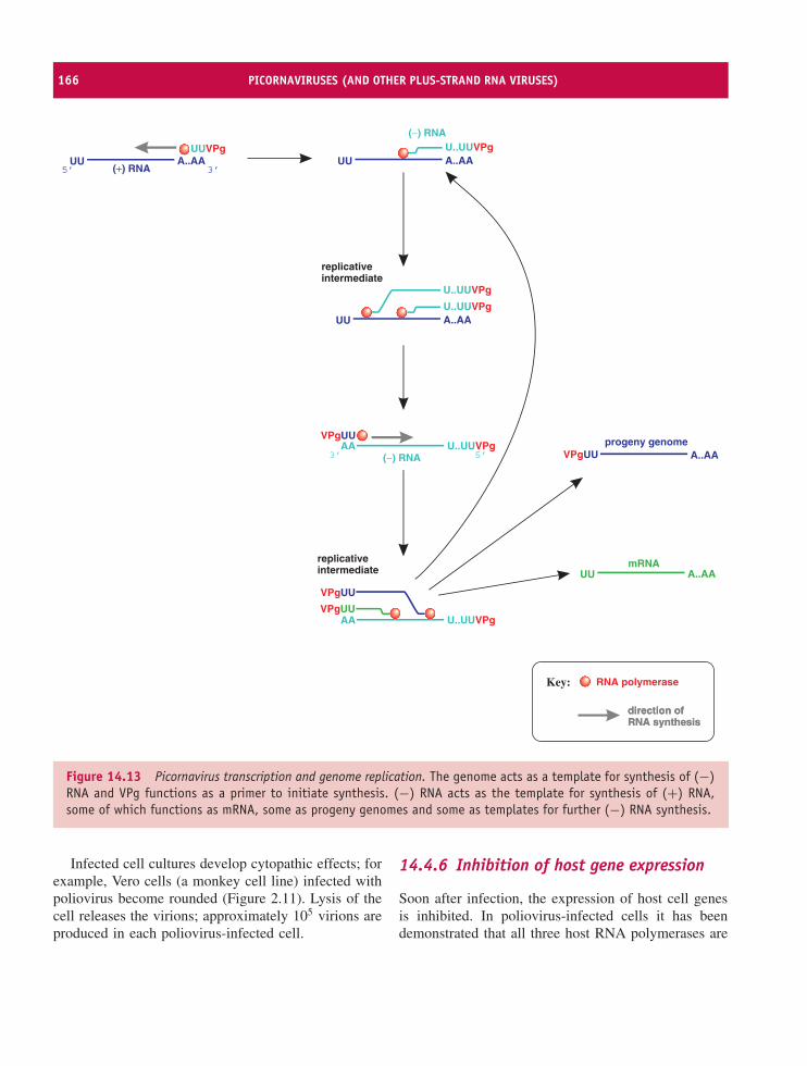
166 PICORNAVIRUSES (AND OTHER PLUS-STRAND RNA VIRUSES)
A..AAUUU..UUVPg
(−) RNA
A..AAUUU..UUVPg
U..UUVPg
AA
(−) RNA
U..UUVPgVPgUU
VPgUU
AA U..UUVPg
A..AAUU
progeny genomeA..AAVPgUU
mRNA
replicativeintermediate
replicativeintermediate
A..AAUUUUVPg
(+) RNA5’ 3’
5’3’
VPgUU
direction ofdirection ofRNA synthesisRNA synthesis
RNA polymeraseKey:
Figure 14.13 Picornavirus transcription and genome replication. The genome acts as a template for synthesis of (−)RNA and VPg functions as a primer to initiate synthesis. (−) RNA acts as the template for synthesis of (+) RNA,some of which functions as mRNA, some as progeny genomes and some as templates for further (−) RNA synthesis.
Infected cell cultures develop cytopathic effects; forexample, Vero cells (a monkey cell line) infected withpoliovirus become rounded (Figure 2.11). Lysis of thecell releases the virions; approximately 105 virions areproduced in each poliovirus-infected cell.
14.4.6 Inhibition of host gene expression
Soon after infection, the expression of host cell genesis inhibited. In poliovirus-infected cells it has beendemonstrated that all three host RNA polymerases are
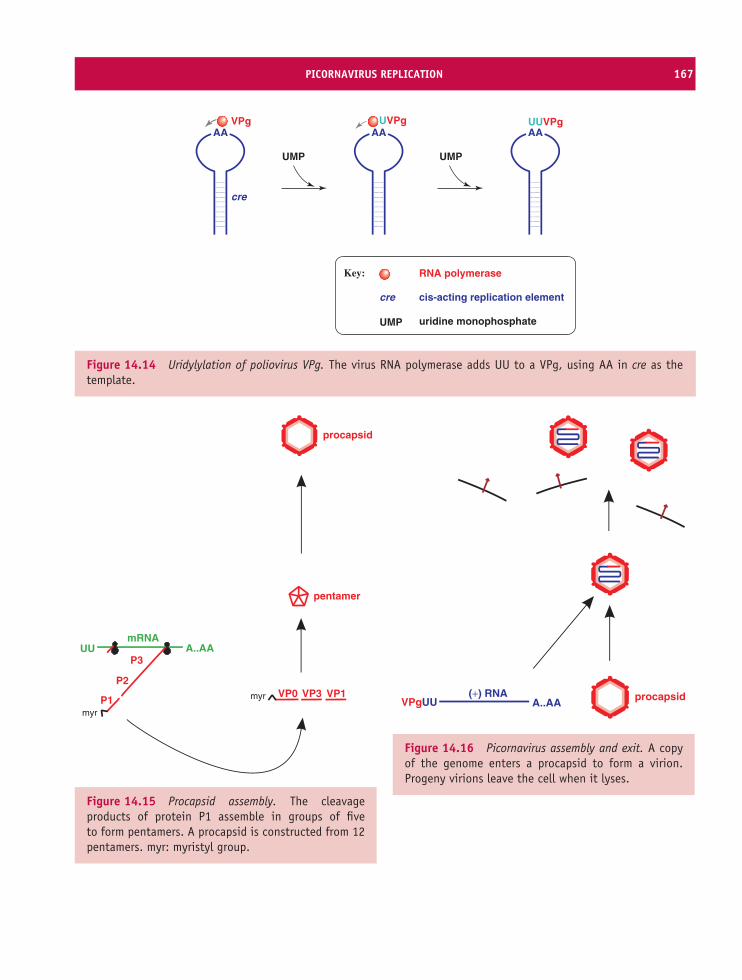
PICORNAVIRUS REPLICATION 167
VPgAA
cre
UVPgAA
UUVPgAA
UMP UMP
RNA polymerase
UMP uridine monophosphate
cre cis-acting replication element
Key:
Figure 14.14 Uridylylation of poliovirus VPg. The virus RNA polymerase adds UU to a VPg, using AA in cre as thetemplate.
A..AAUUmRNA
P1
P2
P3
VP0 VP1VP3
pentamer
procapsid
myr
myr
Figure 14.15 Procapsid assembly. The cleavageproducts of protein P1 assemble in groups of fiveto form pentamers. A procapsid is constructed from 12pentamers. myr: myristyl group.
(+) RNAA..AAVPgUU procapsid
Figure 14.16 Picornavirus assembly and exit. A copyof the genome enters a procapsid to form a virion.Progeny virions leave the cell when it lyses.
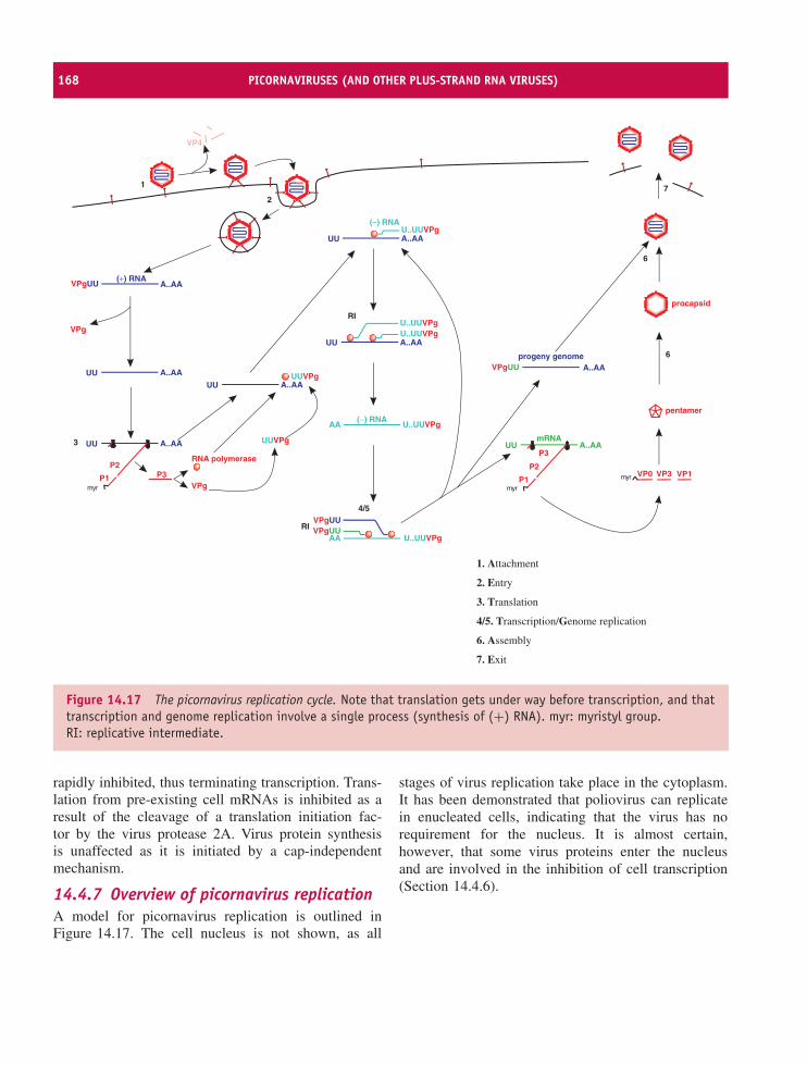
168 PICORNAVIRUSES (AND OTHER PLUS-STRAND RNA VIRUSES)
1
2
3
4/5
6
7
6
(+) RNAA..AAVPgUU
A..AAUU
VPg
A..AAUU
A..AAUU
RNA polymerase
VPg
UUVPg
UUVPg
A..AAUUU..UUVPg
(–) RNA
A..AAUUU..UUVPgU..UUVPg
AA
(−) RNA
U..UUVPgVPgUUVPgUU
AA U..UUVPg
progeny genomeA..AAVPgUU
mRNA
myrP1
P2P3
VP4
A..AAUU
myrP1
P2P3
VP0 VP1VP3myr
pentamer
RI
RI
procapsid
1. Attachment
2. Entry
3. Translation
4/5. Transcription/Genome replication
6. Assembly
7. Exit
Figure 14.17 The picornavirus replication cycle. Note that translation gets under way before transcription, and thattranscription and genome replication involve a single process (synthesis of (+) RNA). myr: myristyl group.RI: replicative intermediate.
rapidly inhibited, thus terminating transcription. Trans-lation from pre-existing cell mRNAs is inhibited as aresult of the cleavage of a translation initiation fac-tor by the virus protease 2A. Virus protein synthesisis unaffected as it is initiated by a cap-independentmechanism.
14.4.7 Overview of picornavirus replicationA model for picornavirus replication is outlined inFigure 14.17. The cell nucleus is not shown, as all
stages of virus replication take place in the cytoplasm.It has been demonstrated that poliovirus can replicatein enucleated cells, indicating that the virus has norequirement for the nucleus. It is almost certain,however, that some virus proteins enter the nucleusand are involved in the inhibition of cell transcription(Section 14.4.6).
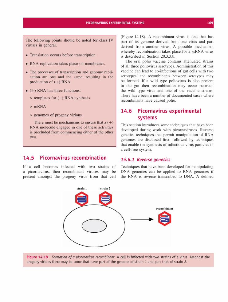
PICORNAVIRUS EXPERIMENTAL SYSTEMS 169
The following points should be noted for class IVviruses in general.
• Translation occurs before transcription.
• RNA replication takes place on membranes.
• The processes of transcription and genome repli-cation are one and the same, resulting in theproduction of (+) RNA.
• (+) RNA has three functions:
◦ templates for (–) RNA synthesis
◦ mRNA
◦ genomes of progeny virions.
There must be mechanisms to ensure that a (+)RNA molecule engaged in one of these activitiesis precluded from commencing either of the othertwo.
14.5 Picornavirus recombinationIf a cell becomes infected with two strains ofa picornavirus, then recombinant viruses may bepresent amongst the progeny virus from that cell
(Figure 14.18). A recombinant virus is one that haspart of its genome derived from one virus and partderived from another virus. A possible mechanismwhereby recombination takes place for a ssRNA virusis described in Section 20.3.3.b.
The oral polio vaccine contains attenuated strainsof all three poliovirus serotypes. Administration of thisvaccine can lead to co-infections of gut cells with twoserotypes, and recombinants between serotypes maybe formed. If a wild type poliovirus is also presentin the gut then recombination may occur betweenthe wild type virus and one of the vaccine strains.There have been a number of documented cases whererecombinants have caused polio.
14.6 Picornavirus experimentalsystems
This section introduces some techniques that have beendeveloped during work with picornaviruses. Reversegenetics techniques that permit manipulation of RNAgenomes are discussed first, followed by techniquesthat enable the synthesis of infectious virus particles ina cell-free system.
14.6.1 Reverse geneticsTechniques that have been developed for manipulatingDNA genomes can be applied to RNA genomes ifthe RNA is reverse transcribed to DNA. A defined
recombinant
strain 1 strain 2
Figure 14.18 Formation of a picornavirus recombinant. A cell is infected with two strains of a virus. Amongst theprogeny virions there may be some that have part of the genome of strain 1 and part that of strain 2.
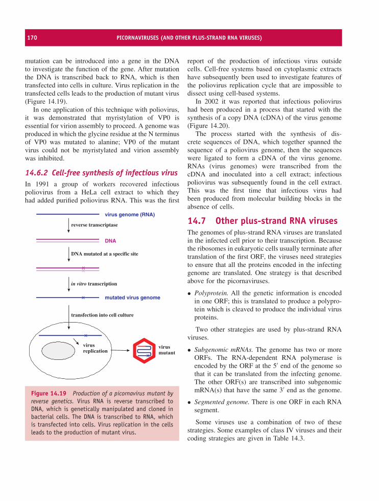
170 PICORNAVIRUSES (AND OTHER PLUS-STRAND RNA VIRUSES)
mutation can be introduced into a gene in the DNAto investigate the function of the gene. After mutationthe DNA is transcribed back to RNA, which is thentransfected into cells in culture. Virus replication in thetransfected cells leads to the production of mutant virus(Figure 14.19).
In one application of this technique with poliovirus,it was demonstrated that myristylation of VP0 isessential for virion assembly to proceed. A genome wasproduced in which the glycine residue at the N terminusof VP0 was mutated to alanine; VP0 of the mutantvirus could not be myristylated and virion assemblywas inhibited.
14.6.2 Cell-free synthesis of infectious virusIn 1991 a group of workers recovered infectiouspoliovirus from a HeLa cell extract to which theyhad added purified poliovirus RNA. This was the first
virus genome (RNA)
DNA
mutated virus genome
reverse transcriptase
DNA mutated at a specific site
transfection into cell culture
in vitro transcription
Figure 14.19 Production of a picornavirus mutant byreverse genetics. Virus RNA is reverse transcribed toDNA, which is genetically manipulated and cloned inbacterial cells. The DNA is transcribed to RNA, whichis transfected into cells. Virus replication in the cellsleads to the production of mutant virus.
report of the production of infectious virus outsidecells. Cell-free systems based on cytoplasmic extractshave subsequently been used to investigate features ofthe poliovirus replication cycle that are impossible todissect using cell-based systems.
In 2002 it was reported that infectious poliovirushad been produced in a process that started with thesynthesis of a copy DNA (cDNA) of the virus genome(Figure 14.20).
The process started with the synthesis of dis-crete sequences of DNA, which together spanned thesequence of a poliovirus genome, then the sequenceswere ligated to form a cDNA of the virus genome.RNAs (virus genomes) were transcribed from thecDNA and inoculated into a cell extract; infectiouspoliovirus was subsequently found in the cell extract.This was the first time that infectious virus hadbeen produced from molecular building blocks in theabsence of cells.
14.7 Other plus-strand RNA virusesThe genomes of plus-strand RNA viruses are translatedin the infected cell prior to their transcription. Becausethe ribosomes in eukaryotic cells usually terminate aftertranslation of the first ORF, the viruses need strategiesto ensure that all the proteins encoded in the infectinggenome are translated. One strategy is that describedabove for the picornaviruses.
• Polyprotein. All the genetic information is encodedin one ORF; this is translated to produce a polypro-tein which is cleaved to produce the individual virusproteins.
Two other strategies are used by plus-strand RNAviruses.
• Subgenomic mRNAs. The genome has two or moreORFs. The RNA-dependent RNA polymerase isencoded by the ORF at the 5′ end of the genome sothat it can be translated from the infecting genome.The other ORF(s) are transcribed into subgenomicmRNA(s) that have the same 3′ end as the genome.
• Segmented genome. There is one ORF in each RNAsegment.
Some viruses use a combination of two of thesestrategies. Some examples of class IV viruses and theircoding strategies are given in Table 14.3.
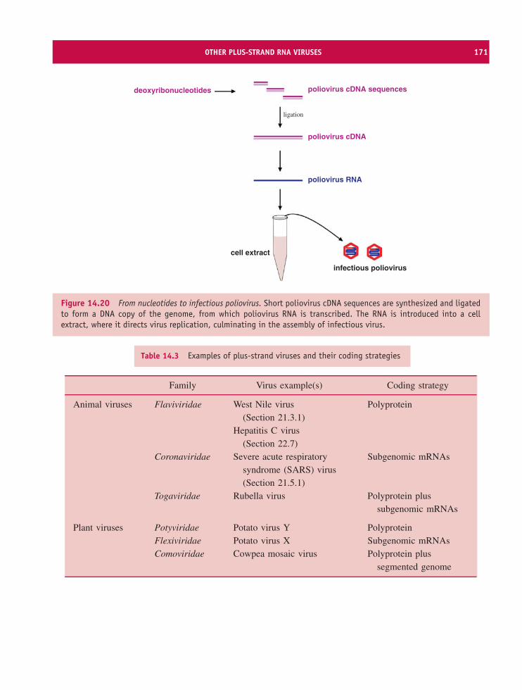
OTHER PLUS-STRAND RNA VIRUSES 171
deoxyribonucleotides poliovirus cDNA sequences
poliovirus cDNA
poliovirus RNA
ligation
cell extract
infectious poliovirus
Figure 14.20 From nucleotides to infectious poliovirus. Short poliovirus cDNA sequences are synthesized and ligatedto form a DNA copy of the genome, from which poliovirus RNA is transcribed. The RNA is introduced into a cellextract, where it directs virus replication, culminating in the assembly of infectious virus.
Table 14.3 Examples of plus-strand viruses and their coding strategies
Family Virus example(s) Coding strategy
Animal viruses Flaviviridae West Nile virus(Section 21.3.1)
Polyprotein
Hepatitis C virus(Section 22.7)
Coronaviridae Severe acute respiratorysyndrome (SARS) virus(Section 21.5.1)
Subgenomic mRNAs
Togaviridae Rubella virus Polyprotein plussubgenomic mRNAs
Plant viruses Potyviridae Potato virus Y PolyproteinFlexiviridae Potato virus X Subgenomic mRNAsComoviridae Cowpea mosaic virus Polyprotein plus
segmented genome
172 PICORNAVIRUSES (AND OTHER PLUS-STRAND RNA VIRUSES)
The only ssRNA phages that are known areclassified in the family Leviviridae (Section 19.2).Viruses in this family, such as phage MS2, havegenomes with characteristics of bacterial mRNA; thegenomes are polycistronic, with all of the ORFstranslated by internal initiation (Section 6.6).
Learning outcomesBy the end of this chapter you should be able to
• give examples of picornaviruses and explain theirimportance;
• describe the picornavirus virion;
• describe the picornavirus replication cycle;
• discuss picornavirus recombination;
• describe experimental systems used for picor-navirus studies.
Sources of further information
Books
Needham C. A. and Canning R. (2003) Polio: the rise andfall of a disease, in Global Disease Eradication: the Racefor the Last Child, American Society for Microbiology,pp. 77–115
Tyrrell D. and Fielder M. (2002) Cold Wars: the FightAgainst the Common Cold, Oxford University Press
Historical papers
Dulbecco R. and Vogt M. (1954) Plaque formation andisolation of pure lines with poliomyelitis viruses Journalof Experimental Medicine, 99, 167–182
Enders J. F., Weller T. H. and Robbins F. C. (1949) Culti-vation of the Lansing strain of poliomyelitis virus in cul-tures of various human embryonic tissues Science, 109,85–87
Recent papers
Ahlquist P. et al. (2003) Host factors in positive-strandRNA virus genome replication Journal of Virology, 77,8181–8186
Blomqvist S. et al. (2003) Characterization of a recombinanttype 3/type 2 poliovirus isolated from a healthy vaccineeand containing a chimeric capsid protein VP1 Journal ofGeneral Virology, 84, 573–580
Buenz E. J. and Howe C. L. (2006) Picornaviruses and celldeath Trends In Microbiology, 14, 28–36
Haydon D. T., Kao R. R. and Kitching R. P. (2004) The UKfoot-and-mouth disease outbreak – the aftermath NatureReviews Microbiology, 2, 675–681
Racaniello V. R. (2006) One hundred years of polioviruspathogenesis Virology, 344, 9–16
Whitton J. L., Cornell C. T. and Feuer R. (2005) Hostand virus determinants of picornavirus pathogenesis andtropism Nature Reviews Microbiology, 3, 765–776
Wimmer E. (2006) The test-tube synthesis of a chemicalcalled poliovirus: the simple synthesis of a virus hasfar-reaching societal implications EMBO Reports, 7 (SI),S3–S9
Xing L. et al. (2003) Structural analysis of human rhi-novirus complexed with ICAM-1 reveals the dynamics ofreceptor-mediated virus uncoating Journal of Virology, 77,6101–6107
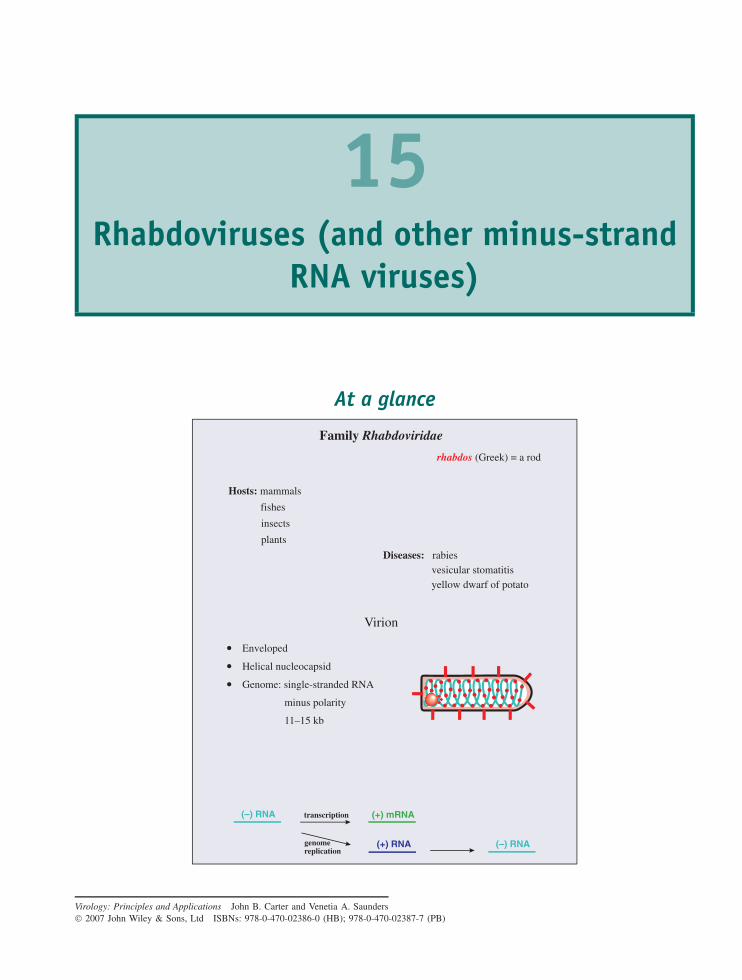
15Rhabdoviruses (and other minus-strand
RNA viruses)
At a glance
Virion
Enveloped
Helical nucleocapsid
Genome: single-stranded RNA
minus polarity
11–15 kb
Family Rhabdoviridae
Hosts: mammals
fishes
insects
plants
rhabdos (Greek) = a rod
Diseases: rabiesvesicular stomatitisyellow dwarf of potato
(–) RNA (+) mRNA
(+) RNA (–) RNA
transcription
genomereplication
Virology: Principles and Applications John B. Carter and Venetia A. Saunders 2007 John Wiley & Sons, Ltd ISBNs: 978-0-470-02386-0 (HB); 978-0-470-02387-7 (PB)
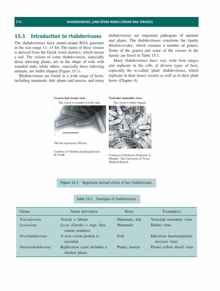
174 RHABDOVIRUSES (AND OTHER MINUS-STRAND RNA VIRUSES)
15.1 Introduction to rhabdovirusesThe rhabdoviruses have minus-strand RNA genomesin the size range 11–15 kb. The name of these virusesis derived from the Greek word rhabdos, which meansa rod. The virions of some rhabdoviruses, especiallythose infecting plants, are in the shape of rods withrounded ends, while others, especially those infectinganimals, are bullet shaped (Figure 15.1).
Rhabdoviruses are found in a wide range of hosts,including mammals, fish, plants and insects, and many
rhabdoviruses are important pathogens of animalsand plants. The rhabdoviruses constitute the familyRhabdoviridae, which contains a number of genera.Some of the genera and some of the viruses in thefamily are listed in Table 15.1.
Many rhabdoviruses have very wide host rangesand replicate in the cells of diverse types of host,especially the so-called ‘plant’ rhabdoviruses, whichreplicate in their insect vectors as well as in their planthosts (Chapter 4).
Festuca leaf streak virus
The virion is rounded at both ends.
The bar represents 200 nm.
Courtesy of Thorben Lundsgaard andICTVdB.
Vesicular stomatitis virus
The virion is bullet shaped.
Courtesy of Professor Frederick A.Murphy, The University of TexasMedical Branch.
Figure 15.1 Negatively stained virions of two rhabdoviruses.
Table 15.1 Examples of rhabdoviruses
Genus Name derivation Hosts Example(s)
Vesiculovirus Vesicle = blister Mammals, fish Vesicular stomatitis virusLyssavirus Lyssa (Greek) = rage, fury,
canine madnessMammals Rabies virus
Novirhabdovirus A non-vi rion protein isencoded.
Fish Infectious haematopoieticnecrosis virus
Nucleorhabdovirus Replication cycle includes anuclear phase.
Plants, insects Potato yellow dwarf virus
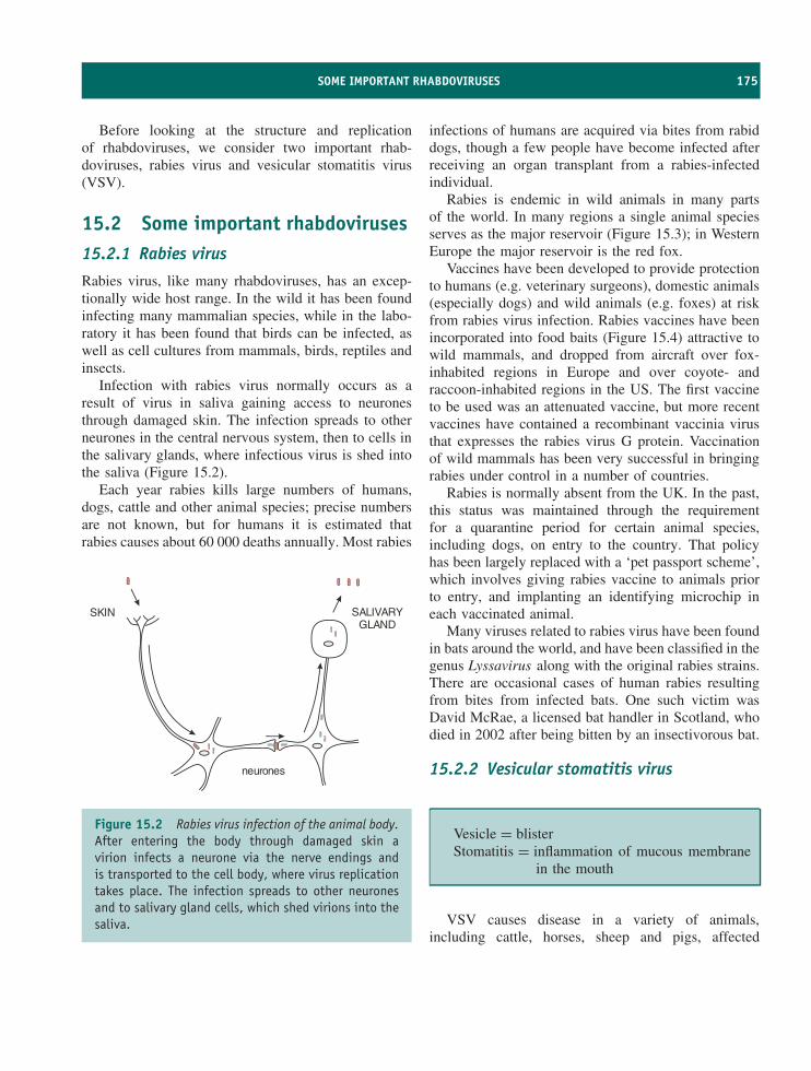
SOME IMPORTANT RHABDOVIRUSES 175
Before looking at the structure and replicationof rhabdoviruses, we consider two important rhab-doviruses, rabies virus and vesicular stomatitis virus(VSV).
15.2 Some important rhabdoviruses15.2.1 Rabies virus
Rabies virus, like many rhabdoviruses, has an excep-tionally wide host range. In the wild it has been foundinfecting many mammalian species, while in the labo-ratory it has been found that birds can be infected, aswell as cell cultures from mammals, birds, reptiles andinsects.
Infection with rabies virus normally occurs as aresult of virus in saliva gaining access to neuronesthrough damaged skin. The infection spreads to otherneurones in the central nervous system, then to cells inthe salivary glands, where infectious virus is shed intothe saliva (Figure 15.2).
Each year rabies kills large numbers of humans,dogs, cattle and other animal species; precise numbersare not known, but for humans it is estimated thatrabies causes about 60 000 deaths annually. Most rabies
SKIN SALIVARYGLAND
neurones
Figure 15.2 Rabies virus infection of the animal body.After entering the body through damaged skin avirion infects a neurone via the nerve endings andis transported to the cell body, where virus replicationtakes place. The infection spreads to other neuronesand to salivary gland cells, which shed virions into thesaliva.
infections of humans are acquired via bites from rabiddogs, though a few people have become infected afterreceiving an organ transplant from a rabies-infectedindividual.
Rabies is endemic in wild animals in many partsof the world. In many regions a single animal speciesserves as the major reservoir (Figure 15.3); in WesternEurope the major reservoir is the red fox.
Vaccines have been developed to provide protectionto humans (e.g. veterinary surgeons), domestic animals(especially dogs) and wild animals (e.g. foxes) at riskfrom rabies virus infection. Rabies vaccines have beenincorporated into food baits (Figure 15.4) attractive towild mammals, and dropped from aircraft over fox-inhabited regions in Europe and over coyote- andraccoon-inhabited regions in the US. The first vaccineto be used was an attenuated vaccine, but more recentvaccines have contained a recombinant vaccinia virusthat expresses the rabies virus G protein. Vaccinationof wild mammals has been very successful in bringingrabies under control in a number of countries.
Rabies is normally absent from the UK. In the past,this status was maintained through the requirementfor a quarantine period for certain animal species,including dogs, on entry to the country. That policyhas been largely replaced with a ‘pet passport scheme’,which involves giving rabies vaccine to animals priorto entry, and implanting an identifying microchip ineach vaccinated animal.
Many viruses related to rabies virus have been foundin bats around the world, and have been classified in thegenus Lyssavirus along with the original rabies strains.There are occasional cases of human rabies resultingfrom bites from infected bats. One such victim wasDavid McRae, a licensed bat handler in Scotland, whodied in 2002 after being bitten by an insectivorous bat.
15.2.2 Vesicular stomatitis virus
Vesicle = blisterStomatitis = inflammation of mucous membrane
in the mouth
VSV causes disease in a variety of animals,including cattle, horses, sheep and pigs, affected
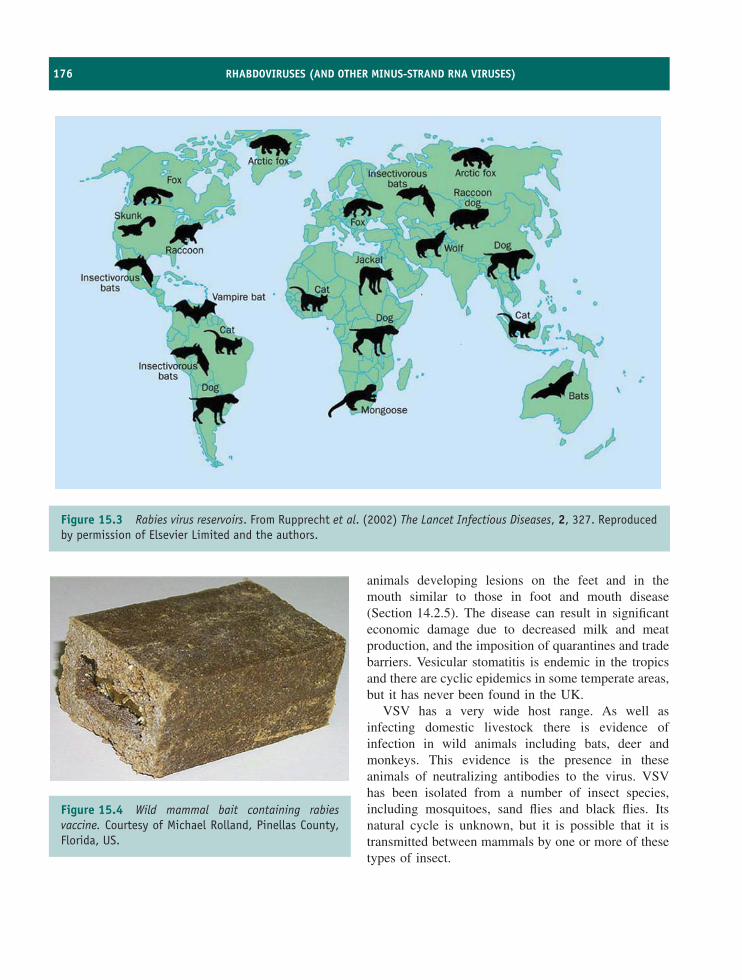
176 RHABDOVIRUSES (AND OTHER MINUS-STRAND RNA VIRUSES)
Figure 15.3 Rabies virus reservoirs. From Rupprecht et al. (2002) The Lancet Infectious Diseases, 2, 327. Reproducedby permission of Elsevier Limited and the authors.
Figure 15.4 Wild mammal bait containing rabiesvaccine. Courtesy of Michael Rolland, Pinellas County,Florida, US.
animals developing lesions on the feet and in themouth similar to those in foot and mouth disease(Section 14.2.5). The disease can result in significanteconomic damage due to decreased milk and meatproduction, and the imposition of quarantines and tradebarriers. Vesicular stomatitis is endemic in the tropicsand there are cyclic epidemics in some temperate areas,but it has never been found in the UK.
VSV has a very wide host range. As well asinfecting domestic livestock there is evidence ofinfection in wild animals including bats, deer andmonkeys. This evidence is the presence in theseanimals of neutralizing antibodies to the virus. VSVhas been isolated from a number of insect species,including mosquitoes, sand flies and black flies. Itsnatural cycle is unknown, but it is possible that it istransmitted between mammals by one or more of thesetypes of insect.
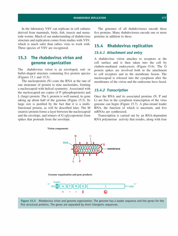
RHABDOVIRUS REPLICATION 177
In the laboratory VSV can replicate in cell culturesderived from mammals, birds, fish, insects and nema-tode worms. Much of our understanding of rhabdovirusstructure and replication comes from studies with VSV,which is much safer than rabies virus to work with.Three species of VSV are recognized.
15.3 The rhabdovirus virion andgenome organization
The rhabdovirus virion is an enveloped, rod- orbullet-shaped structure containing five protein species(Figures 15.1 and 15.5).
The nucleoprotein (N) coats the RNA at the rate ofone monomer of protein to nine nucleotides, forminga nucleocapsid with helical symmetry. Associated withthe nucleocapsid are copies of P (phosphoprotein) andL (large) protein. The L protein is well named, its genetaking up about half of the genome (Figure 15.5). Itslarge size is justified by the fact that it is a multi-functional protein, as will be described later. The M(matrix) protein forms a layer between the nucleocapsidand the envelope, and trimers of G (glycoprotein) formspikes that protrude from the envelope.
The genomes of all rhabdoviruses encode thesefive proteins. Many rhabdoviruses encode one or moreproteins in addition to these.
15.4 Rhabdovirus replication15.4.1 Attachment and entry
A rhabdovirus virion attaches to receptors at thecell surface and is then taken into the cell byclathrin-mediated endocytosis (Figure 15.6). The Gprotein spikes are involved both in the attachmentto cell receptors and in the membrane fusion. Thenucleocapsid is released into the cytoplasm after themembranes of the virion and the endosome have fused.
15.4.2 Transcription
Once the RNA and its associated proteins (N, P andL) are free in the cytoplasm transcription of the virusgenome can begin (Figure 15.7). A plus-strand leaderRNA, the function of which is uncertain, and fivemRNAs are synthesized.
Transcription is carried out by an RNA-dependentRNA polymerase activity that resides, along with four
N P M G L pppHO5’3’
leader
Virion components
Genome organization and gene products
N
PL
G
M
RNA
membrane
Figure 15.5 Rhabdovirus virion and genome organization. The genome has a leader sequence and the genes for thefive structural proteins. The genes are separated by short intergenic sequences.
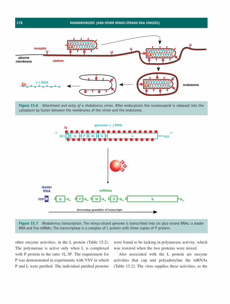
178 RHABDOVIRUSES (AND OTHER MINUS-STRAND RNA VIRUSES)
( ) RNA–
clathrin
receptor
plasmamembrane
endosome
Figure 15.6 Attachment and entry of a rhabdovirus virion. After endocytosis the nucleocapsid is released into thecytoplasm by fusion between the membranes of the virion and the endosome.
genome: (−) RNA
HO N
N
leaderRNA
ppp
decreasing quantities of transcripts
P M G L
N An An An An AnP M
mRNAs
G L
LP
PP
ppp3′ 5′
Figure 15.7 Rhabdovirus transcription. The minus-strand genome is transcribed into six plus-strand RNAs: a leaderRNA and five mRNAs. The transcriptase is a complex of L protein with three copies of P protein.
other enzyme activities, in the L protein (Table 15.2).
The polymerase is active only when L is complexed
with P protein in the ratio 1L:3P. The requirement for
P was demonstrated in experiments with VSV in which
P and L were purified. The individual purified proteins
were found to be lacking in polymerase activity, which
was restored when the two proteins were mixed.
Also associated with the L protein are enzyme
activities that cap and polyadenylate the mRNAs
(Table 15.2). The virus supplies these activities, as the
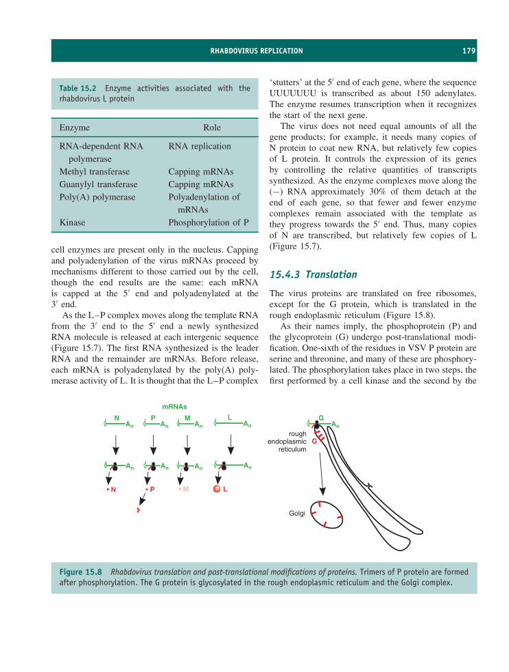
RHABDOVIRUS REPLICATION 179
Table 15.2 Enzyme activities associated with therhabdovirus L protein
Enzyme Role
RNA-dependent RNApolymerase
RNA replication
Methyl transferase Capping mRNAsGuanylyl transferase Capping mRNAsPoly(A) polymerase Polyadenylation of
mRNAsKinase Phosphorylation of P
cell enzymes are present only in the nucleus. Cappingand polyadenylation of the virus mRNAs proceed bymechanisms different to those carried out by the cell,though the end results are the same: each mRNAis capped at the 5′ end and polyadenylated at the3′ end.
As the L–P complex moves along the template RNAfrom the 3′ end to the 5′ end a newly synthesizedRNA molecule is released at each intergenic sequence(Figure 15.7). The first RNA synthesized is the leaderRNA and the remainder are mRNAs. Before release,each mRNA is polyadenylated by the poly(A) poly-merase activity of L. It is thought that the L–P complex
‘stutters’ at the 5′ end of each gene, where the sequenceUUUUUUU is transcribed as about 150 adenylates.The enzyme resumes transcription when it recognizesthe start of the next gene.
The virus does not need equal amounts of all thegene products; for example, it needs many copies ofN protein to coat new RNA, but relatively few copiesof L protein. It controls the expression of its genesby controlling the relative quantities of transcriptssynthesized. As the enzyme complexes move along the(−) RNA approximately 30% of them detach at theend of each gene, so that fewer and fewer enzymecomplexes remain associated with the template asthey progress towards the 5′ end. Thus, many copiesof N are transcribed, but relatively few copies of L(Figure 15.7).
15.4.3 Translation
The virus proteins are translated on free ribosomes,except for the G protein, which is translated in therough endoplasmic reticulum (Figure 15.8).
As their names imply, the phosphoprotein (P) andthe glycoprotein (G) undergo post-translational modi-fication. One-sixth of the residues in VSV P protein areserine and threonine, and many of these are phosphory-lated. The phosphorylation takes place in two steps, thefirst performed by a cell kinase and the second by the
An
mRNAs
An An
An An An
N PAn
M LAn
G
An
N P LM
Grough
endoplasmicreticulum
Golgi
Figure 15.8 Rhabdovirus translation and post-translational modifications of proteins. Trimers of P protein are formedafter phosphorylation. The G protein is glycosylated in the rough endoplasmic reticulum and the Golgi complex.
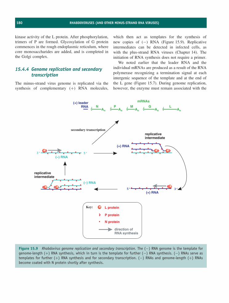
180 RHABDOVIRUSES (AND OTHER MINUS-STRAND RNA VIRUSES)
kinase activity of the L protein. After phosphorylation,trimers of P are formed. Glycosylation of G proteincommences in the rough endoplasmic reticulum, wherecore monosaccharides are added, and is completed inthe Golgi complex.
15.4.4 Genome replication and secondarytranscription
The minus-strand virus genome is replicated via thesynthesis of complementary (+) RNA molecules,
which then act as templates for the synthesis ofnew copies of (−) RNA (Figure 15.9). Replicativeintermediates can be detected in infected cells, aswith the plus-strand RNA viruses (Chapter 14). Theinitiation of RNA synthesis does not require a primer.
We noted earlier that the leader RNA and theindividual mRNAs are produced as a result of the RNApolymerase recognizing a termination signal at eachintergenic sequence of the template and at the end ofthe L gene (Figure 15.7). During genome replication,however, the enzyme must remain associated with the
direction ofRNA synthesisdirection ofRNA synthesis
secondary transcription
(+) RNA
replicativeintermediate
(–) RNA
5’ 3’(+) RNA
5’3’(–) RNA
P protein
L proteinKey:
replicativeintermediate
N protein
An
mRNAs
An AnN P
AnM
AnG L
(+) leaderRNA
Figure 15.9 Rhabdovirus genome replication and secondary transcription. The (−) RNA genome is the template forgenome-length (+) RNA synthesis, which in turn is the template for further (−) RNA synthesis. (−) RNAs serve astemplates for further (+) RNA synthesis and for secondary transcription. (−) RNAs and genome-length (+) RNAsbecome coated with N protein shortly after synthesis.
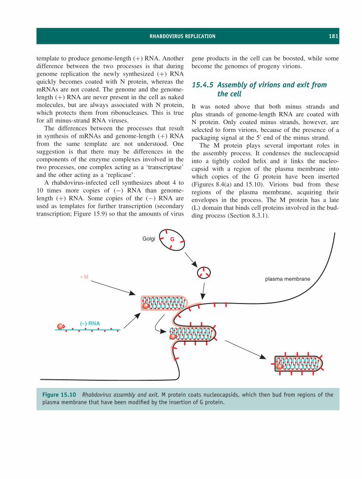
RHABDOVIRUS REPLICATION 181
template to produce genome-length (+) RNA. Anotherdifference between the two processes is that duringgenome replication the newly synthesized (+) RNAquickly becomes coated with N protein, whereas themRNAs are not coated. The genome and the genome-length (+) RNA are never present in the cell as nakedmolecules, but are always associated with N protein,which protects them from ribonucleases. This is truefor all minus-strand RNA viruses.
The differences between the processes that resultin synthesis of mRNAs and genome-length (+) RNAfrom the same template are not understood. Onesuggestion is that there may be differences in thecomponents of the enzyme complexes involved in thetwo processes, one complex acting as a ‘transcriptase’and the other acting as a ‘replicase’.
A rhabdovirus-infected cell synthesizes about 4 to10 times more copies of (−) RNA than genome-length (+) RNA. Some copies of the (−) RNA areused as templates for further transcription (secondarytranscription; Figure 15.9) so that the amounts of virus
gene products in the cell can be boosted, while somebecome the genomes of progeny virions.
15.4.5 Assembly of virions and exit fromthe cell
It was noted above that both minus strands andplus strands of genome-length RNA are coated withN protein. Only coated minus strands, however, areselected to form virions, because of the presence of apackaging signal at the 5′ end of the minus strand.
The M protein plays several important roles inthe assembly process. It condenses the nucleocapsidinto a tightly coiled helix and it links the nucleo-capsid with a region of the plasma membrane intowhich copies of the G protein have been inserted(Figures 8.4(a) and 15.10). Virions bud from theseregions of the plasma membrane, acquiring theirenvelopes in the process. The M protein has a late(L) domain that binds cell proteins involved in the bud-ding process (Section 8.3.1).
M
G
(–) RNA
Golgi
plasma membrane
Figure 15.10 Rhabdovirus assembly and exit. M protein coats nucleocapsids, which then bud from regions of theplasma membrane that have been modified by the insertion of G protein.
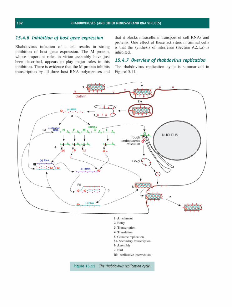
182 RHABDOVIRUSES (AND OTHER MINUS-STRAND RNA VIRUSES)
15.4.6 Inhibition of host gene expression
Rhabdovirus infection of a cell results in stronginhibition of host gene expression. The M protein,whose important roles in virion assembly have justbeen described, appears to play major roles in thisinhibition. There is evidence that the M protein inhibitstranscription by all three host RNA polymerases and
that it blocks intracellular transport of cell RNAs andproteins. One effect of these activities in animal cellsis that the synthesis of interferon (Section 9.2.1.a) isinhibited.
15.4.7 Overview of rhabdovirus replicationThe rhabdovirus replication cycle is summarized inFigure15.11.
1
2
3
4
56
7
5a An
mRNAs
An An
An An An
N P AnM An
G L(+) leader
RNA
RI
RI
AnG
An
N P LM
G
( ) RNA–
(+) RNA
( ) RNA–
(+) RNA
roughendoplasmic
reticulum
Golgi
NUCLEUS
clathrin
1. Attachment
2. Entry
3. Transcription
4. Translation
5. Genome replication5a. Secondary transcription6. Assembly
7. Exit
RI: replicative intermediate
Figure 15.11 The rhabdovirus replication cycle.
REVERSE GENETICS 183
There appears to be no significant role for thenucleus in the replication of most rhabdoviruses.Enucleated cells support VSV replication, with only asmall drop in yield compared with normal cells. This isnot true for all rhabdoviruses, however, as members ofthe genus Nucleorhabdovirus replicate in the nucleusof their plant and insect hosts, with virions buddingthrough the inner nuclear membrane.
15.5 Other minus-strand RNAviruses
Most viruses with minus-strand RNA genomes haveanimal hosts, while a few have plant hosts. Someexamples of minus-strand RNA viruses are given inTable 15.3. Amongst them are three of the world’smajor human pathogens: influenza, measles and res-piratory syncytial viruses. These three viruses causemillions of cases of serious disease and approximatelya million deaths each year.
Measles virus is one of the main causes ofchildhood death in developing countries and is stillresponsible for some deaths in industrialized countries(Section 21.7.1). Infection results in immunosuppres-sion, which renders the host more susceptible to sec-ondary infections with a range of bacterial and viralpathogens, and these cause most measles-associated
Table 15.3 Examples of minus-strand RNA viruses
Family Virus examples
Orthomyxoviridae Influenza virus1
Paramyxoviridae Measles virusRespiratory syncytial virus
Filoviridae Ebolavirus (Section 21.4.1)Bunyaviridae Hantaan virus (Section 21.2.1)
1A number of aspects of influenza virus are dealt with elsewhere:
• virion structure (Section 3.5.1)
• evolutionary mechanisms (Section 20.3.3.c) and the evolution ofnew virus strains (Section 21.5.2)
• vaccines (Chapter 24)
• anti-viral drugs (Section 25.3.5).
deaths. Measles virus can also cause lethal infectionsof the central nervous system.
Viruses in the families Paramyxoviridae andFiloviridae, like those in the Rhabdoviridae, have non-segmented genomes that are transcribed to mRNAs bytheir RNA-dependent RNA polymerases terminatingand reinitiating at intergenic sequences. These familiesform part of the order Mononegavirales, whichare viruses with monopartite genomes composedof negative sense RNA. Viruses in the familiesOrthomyxoviridae and Bunyaviridae, on the otherhand, have segmented genomes.
15.6 Viruses with ambisensegenomes
The RNA segments of some of the segmented genomeviruses are ambisense, where one or more of theRNA segments each encodes two genes, one in theplus sense and one in the minus sense. This is thecase for the two genome segments of viruses in thefamily Arenaviridae. It is also the case for some ofthe three genome segments of some members of thefamily Bunyaviridae, such as tomato spotted wilt virus;two of the three genome segments of this virus areambisense.
The minus-sense gene of an ambisense RNA isexpressed by transcription of a mRNA. The plus-sense gene is expressed by synthesis of an RNAcomplementary to the genome, followed by transcrip-tion of the mRNA for that gene (Figure 15.12).
15.7 Reverse geneticsReverse genetics have been used to investigate rhab-doviruses and other minus-strand RNA viruses, thoughthe procedures are not as straightforward as withthe plus-strand RNA viruses (Section 14.6.1). Minus-strand RNA is not infectious, so techniques had to bedevised that will not only generate virus genomes fromcDNA, but also supply the RNA polymerase and thenucleoprotein that coats the newly synthesized RNA(Section 15.4.4). After much painstaking work, thereare now procedures that enable the recovery of infec-tious minus-strand RNA virus from a cDNA.
As with the plus-strand RNA viruses, a definedmutation can be introduced into a gene in the cDNA
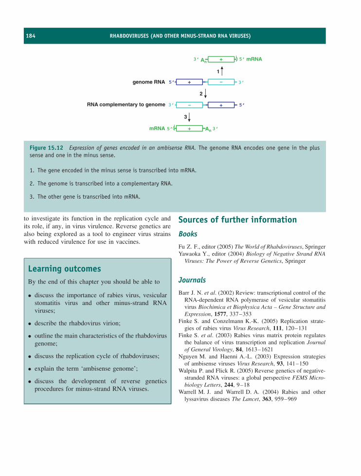
184 RHABDOVIRUSES (AND OTHER MINUS-STRAND RNA VIRUSES)
An
An mRNA
mRNA
+
+
genome RNA
1
2
3
5’ 3’
5’3’RNA complementary to genome
5’3’ +
+5’ 3’
Figure 15.12 Expression of genes encoded in an ambisense RNA. The genome RNA encodes one gene in the plussense and one in the minus sense.
1. The gene encoded in the minus sense is transcribed into mRNA.
2. The genome is transcribed into a complementary RNA.
3. The other gene is transcribed into mRNA.
to investigate its function in the replication cycle andits role, if any, in virus virulence. Reverse genetics arealso being explored as a tool to engineer virus strainswith reduced virulence for use in vaccines.
Learning outcomesBy the end of this chapter you should be able to
• discuss the importance of rabies virus, vesicularstomatitis virus and other minus-strand RNAviruses;
• describe the rhabdovirus virion;
• outline the main characteristics of the rhabdovirusgenome;
• discuss the replication cycle of rhabdoviruses;
• explain the term ‘ambisense genome’;
• discuss the development of reverse geneticsprocedures for minus-strand RNA viruses.
Sources of further information
Books
Fu Z. F., editor (2005) The World of Rhabdoviruses, SpringerYawaoka Y., editor (2004) Biology of Negative Strand RNA
Viruses: The Power of Reverse Genetics, Springer
Journals
Barr J. N. et al. (2002) Review: transcriptional control of theRNA-dependent RNA polymerase of vesicular stomatitisvirus Biochimica et Biophysica Acta – Gene Structure andExpression, 1577, 337–353
Finke S. and Conzelmann K.-K. (2005) Replication strate-gies of rabies virus Virus Research, 111, 120–131
Finke S. et al. (2003) Rabies virus matrix protein regulatesthe balance of virus transcription and replication Journalof General Virology, 84, 1613–1621
Nguyen M. and Haenni A.-L. (2003) Expression strategiesof ambisense viruses Virus Research, 93, 141–150
Walpita P. and Flick R. (2005) Reverse genetics of negative-stranded RNA viruses: a global perspective FEMS Micro-biology Letters, 244, 9–18
Warrell M. J. and Warrell D. A. (2004) Rabies and otherlyssavirus diseases The Lancet, 363, 959–969
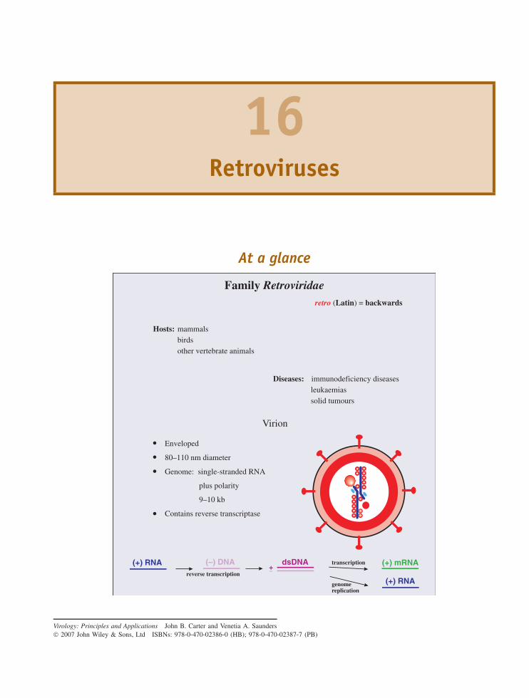
16Retroviruses
At a glance
Virion
Enveloped
80–110 nm diameter
Genome: single-stranded RNA
plus polarity
9–10 kb
Contains reverse transcriptase
Family Retroviridae
Hosts: mammalsbirdsother vertebrate animals
retro (Latin) = backwards
Diseases: immunodeficiency diseasesleukaemiassolid tumours
(–) DNA dsDNA (+) mRNA
(+) RNA
(+) RNA+–
transcription
reverse transcription
genomereplication
Virology: Principles and Applications John B. Carter and Venetia A. Saunders 2007 John Wiley & Sons, Ltd ISBNs: 978-0-470-02386-0 (HB); 978-0-470-02387-7 (PB)
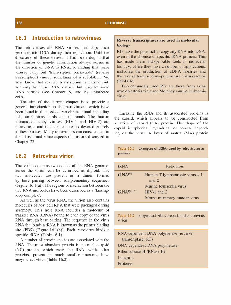
186 RETROVIRUSES
16.1 Introduction to retroviruses
The retroviruses are RNA viruses that copy theirgenomes into DNA during their replication. Until thediscovery of these viruses it had been dogma thatthe transfer of genetic information always occurs inthe direction of DNA to RNA, so finding that someviruses carry out ‘transcription backwards’ (reversetranscription) caused something of a revolution. Wenow know that reverse transcription is carried out,not only by these RNA viruses, but also by someDNA viruses (see Chapter 18) and by uninfectedcells.
The aim of the current chapter is to provide ageneral introduction to the retroviruses, which havebeen found in all classes of vertebrate animal, includingfish, amphibians, birds and mammals. The humanimmunodeficiency viruses (HIV-1 and HIV-2) areretroviruses and the next chapter is devoted entirelyto these viruses. Many retroviruses can cause cancer intheir hosts, and some aspects of this are discussed inChapter 22.
16.2 Retrovirus virion
The virion contains two copies of the RNA genome,hence the virion can be described as diploid. Thetwo molecules are present as a dimer, formedby base pairing between complementary sequences(Figure 16.1(a)). The regions of interaction between thetwo RNA molecules have been described as a ‘kissing-loop complex’.
As well as the virus RNA, the virion also containsmolecules of host cell RNA that were packaged duringassembly. This host RNA includes a molecule oftransfer RNA (tRNA) bound to each copy of the virusRNA through base pairing. The sequence in the virusRNA that binds a tRNA is known as the primer bindingsite (PBS) (Figure 16.1(b)). Each retrovirus binds aspecific tRNA (Table 16.1).
A number of protein species are associated with theRNA. The most abundant protein is the nucleocapsid(NC) protein, which coats the RNA, while otherproteins, present in much smaller amounts, haveenzyme activities (Table 16.2).
Reverse transcriptases are used in molecularbiology
RTs have the potential to copy any RNA into DNA,even in the absence of specific tRNA primers. Thishas made them indispensable tools in molecularbiology, where they have a number of applications,including the production of cDNA libraries andthe reverse transcription–polymerase chain reaction(RT-PCR).
Two commonly used RTs are those from avianmyeloblastosis virus and Moloney murine leukaemiavirus.
Encasing the RNA and its associated proteins isthe capsid, which appears to be constructed froma lattice of capsid (CA) protein. The shape of thecapsid is spherical, cylindrical or conical depend-ing on the virus. A layer of matrix (MA) protein
Table 16.1 Examples of tRNAs used by retroviruses asprimers
tRNA Retrovirus
tRNApro Human T-lymphotropic viruses 1and 2
Murine leukaemia virustRNAlys−3 HIV-1 and 2
Mouse mammary tumour virus
Table 16.2 Enzyme activities present in the retrovirusvirion
RNA-dependent DNA polymerase (reversetranscriptase; RT)
DNA-dependent DNA polymerase
Ribonuclease H (RNase H)
IntegraseProtease
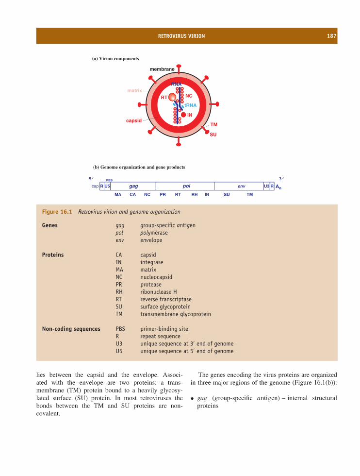
RETROVIRUS VIRION 187
RNA
tRNA
capsid
matrix
membrane
SU
TM
RT
IN
NC
(a) Virion components
(b) Genome organization and gene products
MA CA NC PR RT RH IN SU TM
An
PBS
U5 gag U3envR Rcap pol
5’ 3’
Figure 16.1 Retrovirus virion and genome organization
Genes gag group-specific antigenpol polymeraseenv envelope
Proteins CA capsidIN integraseMA matrixNC nucleocapsidPR proteaseRH ribonuclease HRT reverse transcriptaseSU surface glycoproteinTM transmembrane glycoprotein
Non-coding sequences PBS primer-binding siteR repeat sequenceU3 unique sequence at 3′ end of genomeU5 unique sequence at 5′ end of genome
lies between the capsid and the envelope. Associ-ated with the envelope are two proteins: a trans-membrane (TM) protein bound to a heavily glycosy-lated surface (SU) protein. In most retroviruses thebonds between the TM and SU proteins are non-covalent.
The genes encoding the virus proteins are organizedin three major regions of the genome (Figure 16.1(b)):
• gag (group-specific antigen) – internal structuralproteins
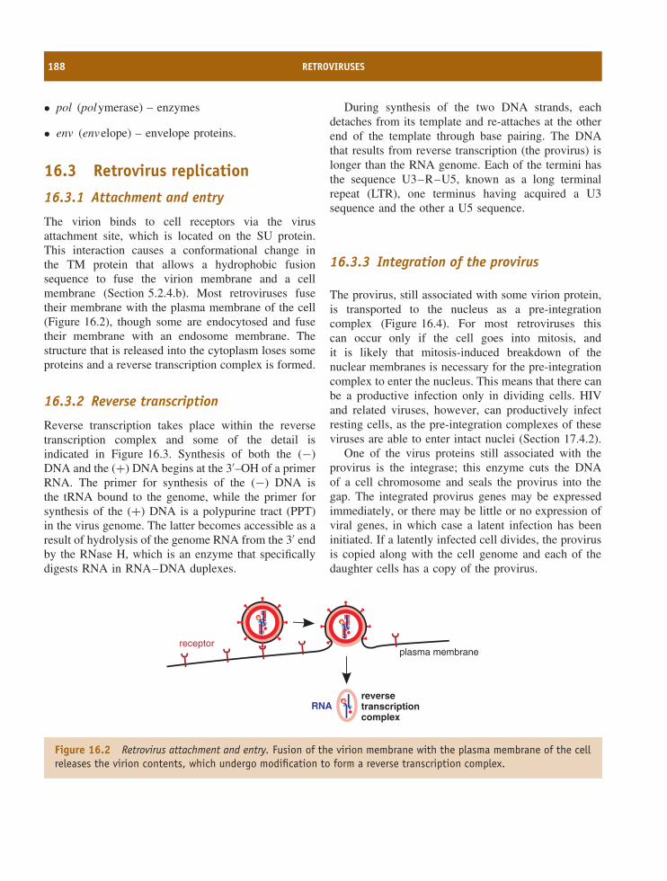
188 RETROVIRUSES
• pol (polymerase) – enzymes
• env (envelope) – envelope proteins.
16.3 Retrovirus replication
16.3.1 Attachment and entry
The virion binds to cell receptors via the virusattachment site, which is located on the SU protein.This interaction causes a conformational change inthe TM protein that allows a hydrophobic fusionsequence to fuse the virion membrane and a cellmembrane (Section 5.2.4.b). Most retroviruses fusetheir membrane with the plasma membrane of the cell(Figure 16.2), though some are endocytosed and fusetheir membrane with an endosome membrane. Thestructure that is released into the cytoplasm loses someproteins and a reverse transcription complex is formed.
16.3.2 Reverse transcription
Reverse transcription takes place within the reversetranscription complex and some of the detail isindicated in Figure 16.3. Synthesis of both the (−)DNA and the (+) DNA begins at the 3′–OH of a primerRNA. The primer for synthesis of the (−) DNA isthe tRNA bound to the genome, while the primer forsynthesis of the (+) DNA is a polypurine tract (PPT)in the virus genome. The latter becomes accessible as aresult of hydrolysis of the genome RNA from the 3′ endby the RNase H, which is an enzyme that specificallydigests RNA in RNA–DNA duplexes.
During synthesis of the two DNA strands, eachdetaches from its template and re-attaches at the otherend of the template through base pairing. The DNAthat results from reverse transcription (the provirus) islonger than the RNA genome. Each of the termini hasthe sequence U3–R–U5, known as a long terminalrepeat (LTR), one terminus having acquired a U3sequence and the other a U5 sequence.
16.3.3 Integration of the provirus
The provirus, still associated with some virion protein,is transported to the nucleus as a pre-integrationcomplex (Figure 16.4). For most retroviruses thiscan occur only if the cell goes into mitosis, andit is likely that mitosis-induced breakdown of thenuclear membranes is necessary for the pre-integrationcomplex to enter the nucleus. This means that there canbe a productive infection only in dividing cells. HIVand related viruses, however, can productively infectresting cells, as the pre-integration complexes of theseviruses are able to enter intact nuclei (Section 17.4.2).
One of the virus proteins still associated with theprovirus is the integrase; this enzyme cuts the DNAof a cell chromosome and seals the provirus into thegap. The integrated provirus genes may be expressedimmediately, or there may be little or no expression ofviral genes, in which case a latent infection has beeninitiated. If a latently infected cell divides, the provirusis copied along with the cell genome and each of thedaughter cells has a copy of the provirus.
RNAreversetranscriptioncomplex
plasma membranereceptor
Figure 16.2 Retrovirus attachment and entry. Fusion of the virion membrane with the plasma membrane of the cellreleases the virion contents, which undergo modification to form a reverse transcription complex.
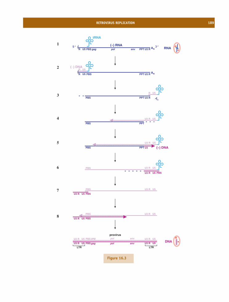
RETROVIRUS REPLICATION 189
U5R U3 Rgag envpol PBS PPT An
tRNA
(+) RNA5’ 3’
R U3 RPBS PPT An
(−) DNAR
U5
U5
PBS PPT U3 (+) DNA
RU3 U5
PBS U3
U3
R U5
PBSR U5
PBS U3
U3
R U5
PBSR U5
provirusU3
U3
R U5
PBSR U5
U3 PBSR U5
U3 R U5
LTR LTR
gag envpol
gag envpol
PBS PPT
RU3 U5
PBS RU3 U5
PBSRU3 U5
1
2
3
4
5
6
7
8
RNA
DNA
U3 RPBS PPT An
R U5
Figure 16.3
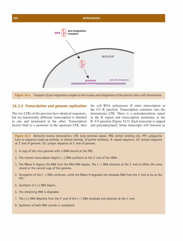
190 RETROVIRUSES
NUCLEUS
cell chromosome
DNA
provirus
pre-integrationcomplex
Figure 16.4 Transport of pre-integration complex to the nucleus and integration of the provirus into a cell chromosome.
16.3.4 Transcription and genome replication
The two LTRs of the provirus have identical sequences,but are functionally different; transcription is initiatedin one and terminated in the other. Transcriptionfactors bind to a promoter in the upstream LTR, then
the cell RNA polymerase II starts transcription atthe U3–R junction. Transcription continues into thedownstream LTR. There is a polyadenylation signalin the R region and transcription terminates at theR–U5 junction (Figure 16.5). Each transcript is cappedand polyadenylated. Some transcripts will function as
Figure 16.3 Retroviral reverse transcription. LTR: long terminal repeat. PBS: primer binding site. PPT: polypurinetract (a sequence made up entirely, or almost entirely, of purine residues). R: repeat sequence. U3: unique sequenceat 3′ end of genome. U5: unique sequence at 5′ end of genome.
1. A copy of the virus genome with a tRNA bound at the PBS.
2. The reverse transcriptase begins (−) DNA synthesis at the 3′ end of the tRNA.
3. The RNase H digests the RNA from the RNA-DNA duplex. The (−) DNA attaches at the 3′ end of either the samestrand or the second copy of the genome.
4. Elongation of the (−) DNA continues, while the RNase H degrades the template RNA from the 3′ end as far as thePPT.
5. Synthesis of (+) DNA begins.
6. The remaining RNA is degraded.
7. The (+) DNA detaches from the 5′ end of the (−) DNA template and attaches at the 3′ end.
8. Synthesis of both DNA strands is completed.
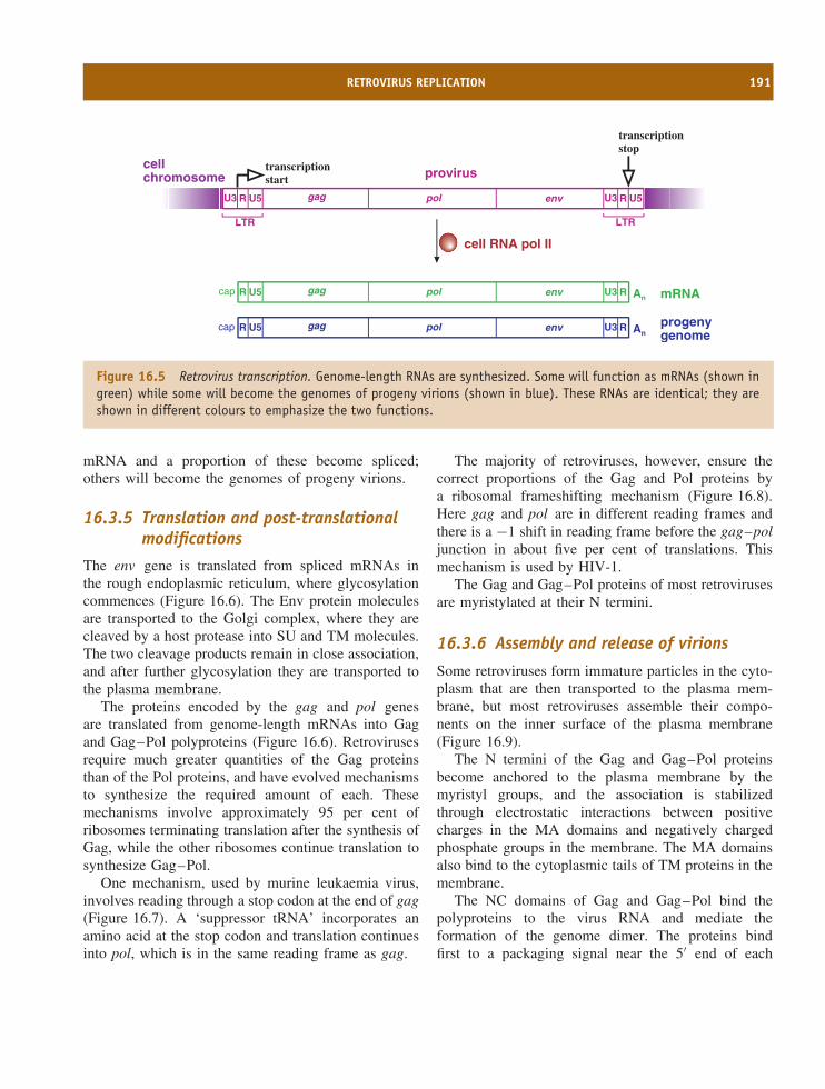
RETROVIRUS REPLICATION 191
gag U3pol env R U5U3 R U5
proviruscellchromosome
LTR LTR
gag U3pol env RR U5 An mRNA
gag U3pol env RR U5 Anprogenygenome
cell RNA pol II
cap
cap
transcriptionstart
transcriptionstop
Figure 16.5 Retrovirus transcription. Genome-length RNAs are synthesized. Some will function as mRNAs (shown ingreen) while some will become the genomes of progeny virions (shown in blue). These RNAs are identical; they areshown in different colours to emphasize the two functions.
mRNA and a proportion of these become spliced;others will become the genomes of progeny virions.
16.3.5 Translation and post-translationalmodifications
The env gene is translated from spliced mRNAs inthe rough endoplasmic reticulum, where glycosylationcommences (Figure 16.6). The Env protein moleculesare transported to the Golgi complex, where they arecleaved by a host protease into SU and TM molecules.The two cleavage products remain in close association,and after further glycosylation they are transported tothe plasma membrane.
The proteins encoded by the gag and pol genesare translated from genome-length mRNAs into Gagand Gag–Pol polyproteins (Figure 16.6). Retrovirusesrequire much greater quantities of the Gag proteinsthan of the Pol proteins, and have evolved mechanismsto synthesize the required amount of each. Thesemechanisms involve approximately 95 per cent ofribosomes terminating translation after the synthesis ofGag, while the other ribosomes continue translation tosynthesize Gag–Pol.
One mechanism, used by murine leukaemia virus,involves reading through a stop codon at the end of gag(Figure 16.7). A ‘suppressor tRNA’ incorporates anamino acid at the stop codon and translation continuesinto pol, which is in the same reading frame as gag.
The majority of retroviruses, however, ensure thecorrect proportions of the Gag and Pol proteins bya ribosomal frameshifting mechanism (Figure 16.8).Here gag and pol are in different reading frames andthere is a −1 shift in reading frame before the gag–poljunction in about five per cent of translations. Thismechanism is used by HIV-1.
The Gag and Gag–Pol proteins of most retrovirusesare myristylated at their N termini.
16.3.6 Assembly and release of virions
Some retroviruses form immature particles in the cyto-plasm that are then transported to the plasma mem-brane, but most retroviruses assemble their compo-nents on the inner surface of the plasma membrane(Figure 16.9).
The N termini of the Gag and Gag–Pol proteinsbecome anchored to the plasma membrane by themyristyl groups, and the association is stabilizedthrough electrostatic interactions between positivecharges in the MA domains and negatively chargedphosphate groups in the membrane. The MA domainsalso bind to the cytoplasmic tails of TM proteins in themembrane.
The NC domains of Gag and Gag–Pol bind thepolyproteins to the virus RNA and mediate theformation of the genome dimer. The proteins bindfirst to a packaging signal near the 5′ end of each
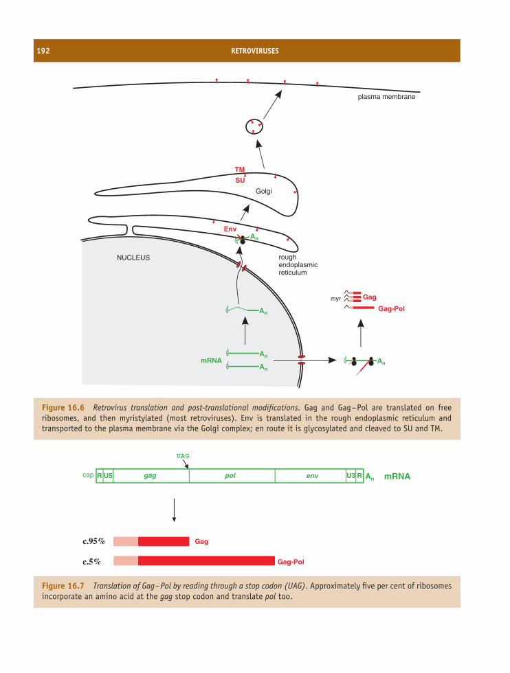
192 RETROVIRUSES
NUCLEUS roughendoplasmicreticulum
Golgi
plasma membrane
Gag-Pol
Gagmyr
An
An
An
An
mRNA
An
Env
SU
TM
Figure 16.6 Retrovirus translation and post-translational modifications. Gag and Gag–Pol are translated on freeribosomes, and then myristylated (most retroviruses). Env is translated in the rough endoplasmic reticulum andtransported to the plasma membrane via the Golgi complex; en route it is glycosylated and cleaved to SU and TM.
gag U3pol env RR U5 An mRNA
UAG
Gag-Pol
Gag
cap
c.95%
c.5%
Figure 16.7 Translation of Gag–Pol by reading through a stop codon (UAG). Approximately five per cent of ribosomesincorporate an amino acid at the gag stop codon and translate pol too.
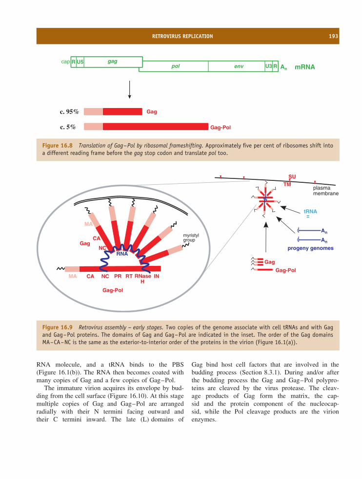
RETROVIRUS REPLICATION 193
U3pol env R An mRNAgagR U5
Gag-Pol
Gag
cap
c. 95%
c. 5%
Figure 16.8 Translation of Gag–Pol by ribosomal frameshifting. Approximately five per cent of ribosomes shift intoa different reading frame before the gag stop codon and translate pol too.
tRNA
SUTM
myristylgroup
GagNC
CA
MA
RNA
MA CA NC PR RT RNaseH
IN
Gag-Pol
An
An
progeny genomes
plasmamembrane
Gag-Pol
Gag
Figure 16.9 Retrovirus assembly – early stages. Two copies of the genome associate with cell tRNAs and with Gagand Gag–Pol proteins. The domains of Gag and Gag–Pol are indicated in the inset. The order of the Gag domainsMA–CA–NC is the same as the exterior-to-interior order of the proteins in the virion (Figure 16.1(a)).
RNA molecule, and a tRNA binds to the PBS(Figure 16.1(b)). The RNA then becomes coated withmany copies of Gag and a few copies of Gag–Pol.
The immature virion acquires its envelope by bud-ding from the cell surface (Figure 16.10). At this stagemultiple copies of Gag and Gag–Pol are arrangedradially with their N termini facing outward andtheir C termini inward. The late (L) domains of
Gag bind host cell factors that are involved in thebudding process (Section 8.3.1). During and/or afterthe budding process the Gag and Gag–Pol polypro-teins are cleaved by the virus protease. The cleav-age products of Gag form the matrix, the cap-sid and the protein component of the nucleocap-sid, while the Pol cleavage products are the virionenzymes.
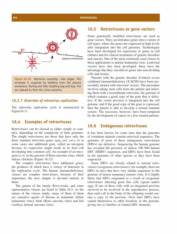
194 RETROVIRUSES
SU
TM
plasma membrane
Figure 16.10 Retrovirus assembly – late stages. Theenvelope is acquired by budding from the plasmamembrane. During and after budding Gag and Gag–Polare cleaved to form the virion proteins.
16.3.7 Overview of retrovirus replication
The retrovirus replication cycle is summarized inFigure16.11.
16.4 Examples of retrovirusesRetroviruses can be classed as either simple or com-plex, depending on the complexity of their genomes.The simple retroviruses are those that have only thethree standard retrovirus genes (gag, pol, env ), or insome cases one additional gene, called an oncogenebecause its expression might result in its host celldeveloping into a tumour cell. An example of an onco-gene is src in the genome of Rous sarcoma virus, whichinfects chickens (Figure 16.12).
The complex retroviruses have additional genes,the products of which have a variety of functions inthe replication cycle. The human immunodeficiencyviruses are complex retroviruses; because of theirimportance the next chapter is devoted entirely tothem.
The genera of the family Retroviridae and somerepresentative viruses are listed in Table 16.3. As thenames of the viruses imply, many of them of themare causative agents of disease in mammals (felineleukaemia virus), birds (Rous sarcoma virus) and fish(walleye dermal sarcoma virus).
16.5 Retroviruses as gene vectorsSome genetically modified retroviruses are used asgene vectors. They can introduce genes into a variety ofcell types, where the genes are expressed at high levelsafter integration into the cell genomes. Technologieshave been developed for expression of genes in cellcultures and for clinical treatments of genetic disordersand cancers. One of the most commonly used viruses inthese applications is murine leukaemia virus. Lentiviralvectors have also been developed; these have theadvantage that they can deliver genes into non-dividingcells and tissues.
Patients with the genetic disorder X-linked severecombined immunodeficiency (X-SCID) have been suc-cessfully treated with retroviral vectors. The procedureinvolves taking stem cells from the patient and infect-ing them with a recombinant retrovirus, the genome ofwhich contains a good copy of the gene that is defec-tive. If the vector provirus is integrated into the cellgenome, and if the good copy of the gene is expressed,then the patient is able to develop a normal immunesystem. The successes, however, have been temperedby the development of cancer in a few treated patients.
16.6 Endogenous retrovirusesIt has been known for some time that the genomesof vertebrate animals contain retroviral sequences. Thegenomes of most of these endogenous retroviruses(ERVs) are defective. Sequencing the human genomehas revealed the presence of almost 100 000 humanERV (HERV) sequences, and ERVs have been foundin the genomes of other species as they have beensequenced.
Some ERVs are closely related to normal retro-viruses (exogenous retroviruses); for example, there areERVs in mice that have very similar sequences to thegenome of mouse mammary tumour virus. It is highlylikely that ERVs originated as a result of exogenousretroviruses infecting germ line cells (sperm and/oregg). If one of these cells with an integrated provirussurvived to be involved in the reproductive process,then each cell in the body of the offspring would con-tain a copy of the provirus. Over time ERVs havecopied themselves to other locations in the genome,giving rise to families of related ERV elements.
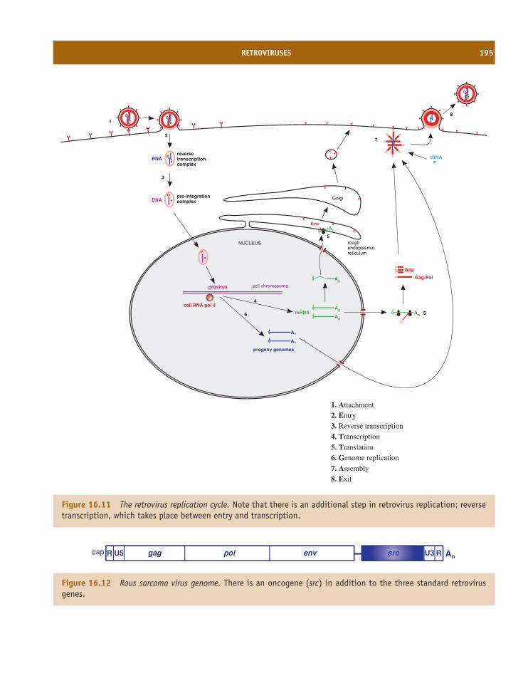
RETROVIRUSES 195
tRNA
NUCLEUS
cell RNA pol II
roughendoplasmicreticulum
Golgi
cell chromosome
Gag-Pol
Gag
An
An
An
An
An
An
mRNA
progeny genomes
RNA
DNA
An
provirus
reversetranscriptioncomplex
pre-integrationcomplex
Env
1
2
3
4
7
6
5
5
8
1. Attachment
2. Entry
3. Reverse transcription
4. Transcription
5. Translation
6. Genome replication
7. Assembly
8. Exit
Figure 16.11 The retrovirus replication cycle. Note that there is an additional step in retrovirus replication: reversetranscription, which takes place between entry and transcription.
gag pol envR U5 U3 R Ansrccap
Figure 16.12 Rous sarcoma virus genome. There is an oncogene (src) in addition to the three standard retrovirusgenes.
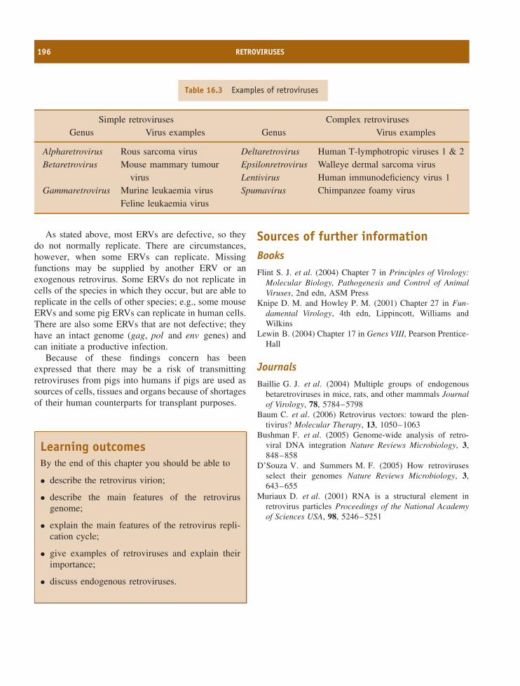
196 RETROVIRUSES
Table 16.3 Examples of retroviruses
Simple retroviruses Complex retrovirusesGenus Virus examples Genus Virus examples
Alpharetrovirus Rous sarcoma virus Deltaretrovirus Human T-lymphotropic viruses 1 & 2Betaretrovirus Mouse mammary tumour
virusEpsilonretrovirusLentivirus
Walleye dermal sarcoma virusHuman immunodeficiency virus 1
Gammaretrovirus Murine leukaemia virusFeline leukaemia virus
Spumavirus Chimpanzee foamy virus
As stated above, most ERVs are defective, so theydo not normally replicate. There are circumstances,however, when some ERVs can replicate. Missingfunctions may be supplied by another ERV or anexogenous retrovirus. Some ERVs do not replicate incells of the species in which they occur, but are able toreplicate in the cells of other species; e.g., some mouseERVs and some pig ERVs can replicate in human cells.There are also some ERVs that are not defective; theyhave an intact genome (gag, pol and env genes) andcan initiate a productive infection.
Because of these findings concern has beenexpressed that there may be a risk of transmittingretroviruses from pigs into humans if pigs are used assources of cells, tissues and organs because of shortagesof their human counterparts for transplant purposes.
Learning outcomesBy the end of this chapter you should be able to
• describe the retrovirus virion;
• describe the main features of the retrovirusgenome;
• explain the main features of the retrovirus repli-cation cycle;
• give examples of retroviruses and explain theirimportance;
• discuss endogenous retroviruses.
Sources of further informationBooksFlint S. J. et al. (2004) Chapter 7 in Principles of Virology:
Molecular Biology, Pathogenesis and Control of AnimalViruses, 2nd edn, ASM Press
Knipe D. M. and Howley P. M. (2001) Chapter 27 in Fun-damental Virology, 4th edn, Lippincott, Williams andWilkins
Lewin B. (2004) Chapter 17 in Genes VIII, Pearson Prentice-Hall
JournalsBaillie G. J. et al. (2004) Multiple groups of endogenous
betaretroviruses in mice, rats, and other mammals Journalof Virology, 78, 5784–5798
Baum C. et al. (2006) Retrovirus vectors: toward the plen-tivirus? Molecular Therapy, 13, 1050–1063
Bushman F. et al. (2005) Genome-wide analysis of retro-viral DNA integration Nature Reviews Microbiology, 3,848–858
D’Souza V. and Summers M. F. (2005) How retrovirusesselect their genomes Nature Reviews Microbiology, 3,643–655
Muriaux D. et al. (2001) RNA is a structural element inretrovirus particles Proceedings of the National Academyof Sciences USA, 98, 5246–5251
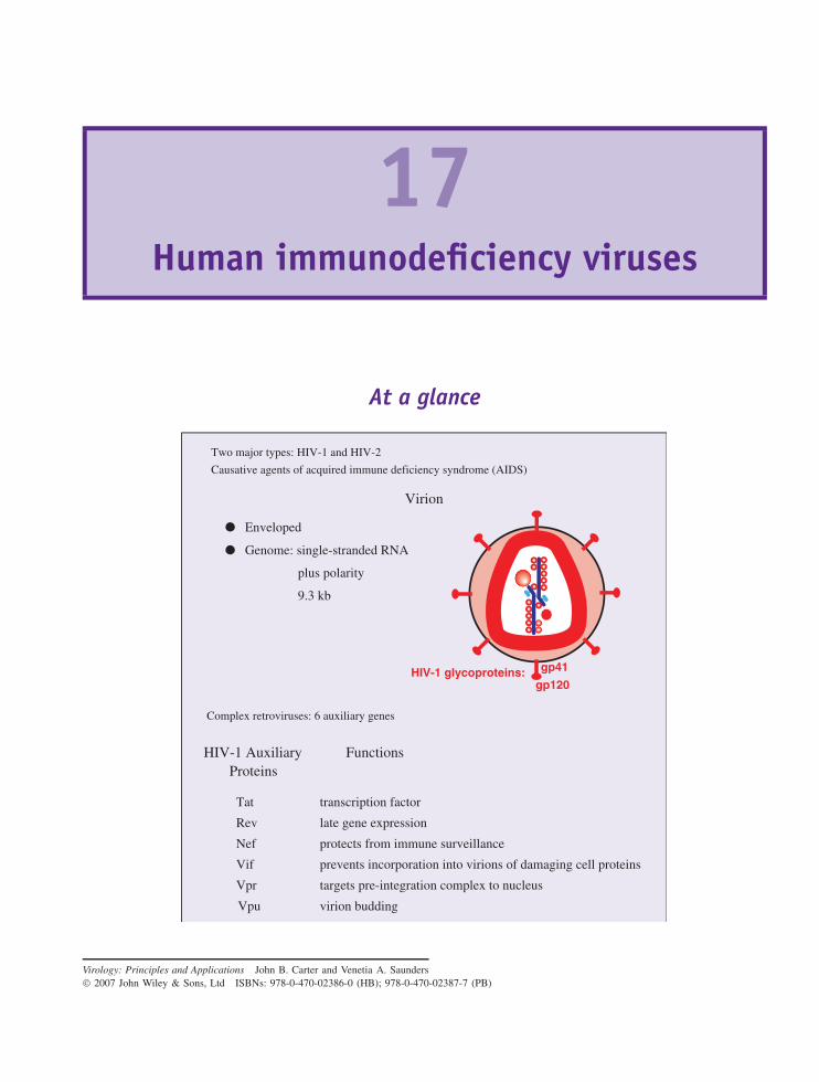
17Human immunodeficiency viruses
At a glance
Virion
Enveloped
Genome: single-stranded RNA
plus polarity
9.3 kb
Two major types: HIV-1 and HIV-2
Causative agents of acquired immune deficiency syndrome (AIDS)
gp41
gp120
HIV-1 Auxiliary Functions Proteins
Tat
Rev
Nef
Vif
Vpr
Vpu
transcription factor
late gene expression
protects from immune surveillance
prevents incorporation into virions of damaging cell proteins
targets pre-integration complex to nucleus
virion budding
HIV-1 glycoproteins:
Complex retroviruses: 6 auxiliary genes
Virology: Principles and Applications John B. Carter and Venetia A. Saunders 2007 John Wiley & Sons, Ltd ISBNs: 978-0-470-02386-0 (HB); 978-0-470-02387-7 (PB)
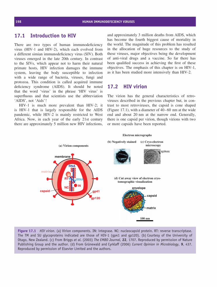
198 HUMAN IMMUNODEFICIENCY VIRUSES
17.1 Introduction to HIV
There are two types of human immunodeficiencyvirus (HIV-1 and HIV-2), which each evolved froma different simian immunodeficiency virus (SIV). Bothviruses emerged in the late 20th century. In contrastto the SIVs, which appear not to harm their naturalprimate hosts, HIV infection damages the immunesystem, leaving the body susceptible to infectionwith a wide range of bacteria, viruses, fungi andprotozoa. This condition is called acquired immunedeficiency syndrome (AIDS). It should be notedthat the word ‘virus’ in the phrase ‘HIV virus’ issuperfluous and that scientists use the abbreviation‘AIDS’, not ‘Aids’!
HIV-1 is much more prevalent than HIV-2; itis HIV-1 that is largely responsible for the AIDSpandemic, while HIV-2 is mainly restricted to WestAfrica. Now, in each year of the early 21st centurythere are approximately 5 million new HIV infections,
and approximately 3 million deaths from AIDS, whichhas become the fourth biggest cause of mortality inthe world. The magnitude of this problem has resultedin the allocation of huge resources to the study ofthese viruses, major objectives being the developmentof anti-viral drugs and a vaccine. So far there hasbeen qualified success in achieving the first of theseobjectives. The emphasis of this chapter is on HIV-1,as it has been studied more intensively than HIV-2.
17.2 HIV virion
The virion has the general characteristics of retro-viruses described in the previous chapter but, in con-trast to most retroviruses, the capsid is cone shaped(Figure 17.1), with a diameter of 40–60 nm at the wideend and about 20 nm at the narrow end. Generally,there is one capsid per virion, though virions with twoor more capsids have been reported.
RNA
tRNA
capsid
matrix
membrane
gp120
gp41
RT
INNC
(a) Virion components
(d) Cut away view of electron cryo-tomographic visualization
(b) Negatively stained (c) Cryo-electronmicroscopy
Electron micrographs
spikes
capsid
envelope
matrix
100 nm
Figure 17.1 HIV virion. (a) Virion components. IN: integrase. NC: nucleocapsid protein. RT: reverse transcriptase.The TM and SU glycoproteins indicated are those of HIV-1 (gp41 and gp120). (b) Courtesy of the University ofOtago, New Zealand. (c) From Briggs et al. (2003) The EMBO Journal, 22, 1707. Reproduced by permission of NaturePublishing Group and the author. (d) From Grunewald and Cyrklaff (2006) Current Opinion in Microbiology, 9, 437.Reproduced by permission of Elsevier Limited and the authors.
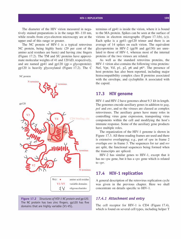
HIV-1 REPLICATION 199
The diameter of the HIV virion measured in nega-tively stained preparations is in the range 80–110 nm,while results from cryo-electron microscopy are at theupper end of this range or greater.
The NC protein of HIV-1 is a typical retrovirusNC protein, being highly basic (29 per cent of theamino acid residues are basic) and having zinc fingers(Figure 17.2). The TM and SU proteins have approxi-mate molecular weights of 41 and 120 kD, respectively,and are named gp41 and gp120 (gp = glycoprotein);gp120 is heavily glycosylated (Figure 17.2). The C
Zn++
Zn++
C terminusN terminus
V1V2
V3V4
V5
C terminus
N terminus
CD4 binding site
NC protein
gp120
Key: amino acid residue
V1-V5 variable domains
oligosaccharides
Figure 17.2 Structures of HIV-1 NC protein and gp120.The NC protein has two zinc fingers. gp120 has fivedomains that are highly variable (V1-V5).
terminus of gp41 is inside the virion, where it is boundto the MA protein. Spikes can be seen at the surface ofvirions in electron micrographs (Figure 17.1(b), (c)).Each spike is a gp41–gp120 trimer and there is anaverage of 14 spikes on each virion. The equivalentglycoproteins in HIV-2 (gp38 and gp130) are unre-lated to those of HIV-1, whereas most of the internalproteins of the two viruses are related.
As well as the standard retrovirus proteins, theHIV-1 virion also contains the following virus proteins:Nef, Vpr, Vif, p1, p2, p6 and p6*. The presence ofhost proteins has also been reported, including majorhistocompatibility complex class II proteins associatedwith the envelope, and cyclophilin A associated withthe capsid.
17.3 HIV genomeHIV-1 and HIV-2 have genomes about 9.3 kb in length.The genomes encode auxiliary genes in addition to gag,pol and env, and so the viruses are classed as complexretroviruses. The auxiliary genes have many roles incontrolling virus gene expression, transporting viruscomponents within the cell and modifying the host’simmune response. Some of the auxiliary gene productshave multiple roles.
The organization of the HIV-1 genome is shown inFigure 17.3. All three reading frames are used and thereis extensive overlapping; e.g., part of vpu in frame 2overlaps env in frame 3. The sequences for tat and revare split, the functional sequences being formed whenthe transcripts are spliced.
HIV-2 has similar genes to HIV-1, except that ithas no vpu gene, but it has a vpx gene which is relatedto vpr.
17.4 HIV-1 replicationA general description of the retrovirus replication cyclewas given in the previous chapter. Here we shallconcentrate on details specific to HIV-1.
17.4.1 Attachment and entry
The cell receptor for HIV-1 is CD4 (Figure 17.4),which is found on several cell types, including helper T
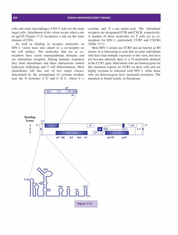
200 HUMAN IMMUNODEFICIENCY VIRUSES
cells and some macrophages; CD4 T cells are the maintarget cells. Attachment of the virion occurs when a siteon gp120 (Figure 17.2) recognizes a site on the outerdomain of CD4.
As well as binding to receptor molecules anHIV-1 virion must also attach to a co-receptor onthe cell surface. The molecules that act as co-receptors have seven transmembrane domains andare chemokine receptors. During immune responsesthey bind chemokines and these interactions controlleukocyte trafficking and T cell differentiation. Mostchemokines fall into one of two major classes,determined by the arrangement of cysteine residuesnear the N terminus: C–C and C–X–C, where C =
cysteine and X = any amino acid. The chemokinereceptors are designated CCR and CXCR, respectively.A number of these molecules on T cells act as co-receptors for HIV-1, particularly CCR5 and CXCR4(Table 17.1).
Most HIV-1 strains use CCR5 and are known as R5strains. It is interesting to note that in some individualswho have had multiple exposures to the virus, but havenot become infected, there is a 32-nucleotide deletionin the CCR5 gene. Individuals who are homozygous forthis mutation express no CCR5 on their cells and arehighly resistant to infection with HIV-1, while thosewho are heterozygous have increased resistance. Themutation is found mainly in Europeans.
gag
pol env
vpu
vpr
vif
tat
nef
R RU3
U5
Readingframe
123
MA CA
p2
NCp1
p6
rev
TARPoly-A
PBS
DIS SDPsi
AUG
5’ 3’
p6* PR RT RH IN gp120 gp41
Figure 17.3
HIV-1 REPLICATION 201
HIV-1 strains that use CXCR4 as a co-receptor areknown as X4 strains, and there are some strains (R5X4strains) that can use either co-receptor. R5 strains donot infect naıve T cells, but all three strains infectmemory T cells.
The interaction of gp120 with the receptor and co-receptor results in a dramatic re-arrangement of gp41,which proceeds to fuse the membranes of the virionand the cell. The contents of the virion envelope arereleased into the cytoplasm and develop into the reversetranscription complex, which contains the MA, Vpr, RTand IN proteins, as well as the virus genome.
17.4.2 Reverse transcription and transportto the nucleus
The reverse transcription complex associates rapidlywith microtubules (Figure 17.5). Reverse transcription
is primed by tRNAlys-3, and proceeds as outlined inSection 16.3.2.
Whereas the proviruses of most retroviruses areentirely dsDNA, those of HIV and other lentiviruseshave a short triple-stranded sequence known as acentral DNA flap. This comes about because, as wellas the polypurine tract (PPT) towards the 3′ end of thevirus genome, there is also a central PPT that acts as asecond initiation site of (+) DNA synthesis. Synthesisof the (+) DNA initiated at the 3′ PPT stops soonafter reaching the (+) DNA initiated at the centralPPT, resulting in a short overlapping ssDNA. ThisDNA flap plays a vital role in the early stages ofinfection.
After reverse transcription has been completed, thepre-integration complex, which contains host proteinsas well as virus proteins, is moved along microtubulestowards the nucleus. As discussed in Chapter 16,
Figure 17.3 HIV-1 genome organization
Main genes gag group-specific antigen (encodes matrix, capsid, p2,nucleocapsid, p1 and p6)
pol polymerase (encodes p6*, protease, reversetranscriptase, RNase H, integrase)
env envelope
Auxiliary genes nef negative regulatory factorrev regulator of expression of virion proteinstat transactivator of transcriptionvif virion infectivity factorvpr viral protein Rvpu viral protein U
Non-coding sequences R repeat sequenceU3 unique sequence at 3′ end of genomeU5 unique sequence at 5′ end of genome
Domains at the 5′ end of thegenome
TAR trans-acting response elementPoly-A polyadenylation signalPBS primer-binding siteDIS dimerization initiation site (involved in formation of
kissing loop complex)SD splice donor sitePsi (ψ) main part of the packaging signalAUG start codon of the gag gene
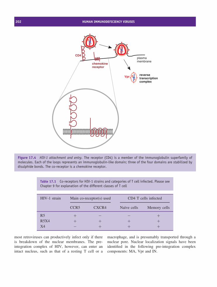
202 HUMAN IMMUNODEFICIENCY VIRUSES
reversetranscriptioncomplex
Vpr
CD4
chemokinereceptor
plasmamembrane
SS
SS
SS
Figure 17.4 HIV-1 attachment and entry. The receptor (CD4) is a member of the immunoglobulin superfamily ofmolecules. Each of the loops represents an immunoglobulin-like domain; three of the four domains are stabilized bydisulphide bonds. The co-receptor is a chemokine receptor.
Table 17.1 Co-receptors for HIV-1 strains and categories of T cell infected. Please seeChapter 9 for explanation of the different classes of T cell
HIV-1 strain Main co-receptor(s) used CD4 T cells infected
CCR5 CXCR4 Naıve cells Memory cells
R5 + − − +R5X4 + + + +X4 − + + +
most retroviruses can productively infect only if thereis breakdown of the nuclear membranes. The pre-integration complex of HIV, however, can enter anintact nucleus, such as that of a resting T cell or a
macrophage, and is presumably transported through anuclear pore. Nuclear localization signals have beenidentified in the following pre-integration complexcomponents: MA, Vpr and IN.
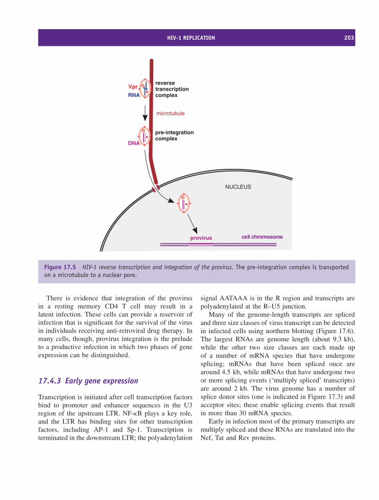
HIV-1 REPLICATION 203
NUCLEUS
cell chromosome
microtubule
RNA
DNA
provirus
reversetranscriptioncomplex
pre-integrationcomplex
Vpr
Figure 17.5 HIV-1 reverse transcription and integration of the provirus. The pre-integration complex is transportedon a microtubule to a nuclear pore.
There is evidence that integration of the provirusin a resting memory CD4 T cell may result in alatent infection. These cells can provide a reservoir ofinfection that is significant for the survival of the virusin individuals receiving anti-retroviral drug therapy. Inmany cells, though, provirus integration is the preludeto a productive infection in which two phases of geneexpression can be distinguished.
17.4.3 Early gene expression
Transcription is initiated after cell transcription factorsbind to promoter and enhancer sequences in the U3region of the upstream LTR. NF-κB plays a key role,and the LTR has binding sites for other transcriptionfactors, including AP-1 and Sp-1. Transcription isterminated in the downstream LTR; the polyadenylation
signal AATAAA is in the R region and transcripts arepolyadenylated at the R–U5 junction.
Many of the genome-length transcripts are splicedand three size classes of virus transcript can be detectedin infected cells using northern blotting (Figure 17.6).The largest RNAs are genome length (about 9.3 kb),while the other two size classes are each made upof a number of mRNA species that have undergonesplicing; mRNAs that have been spliced once arearound 4.5 kb, while mRNAs that have undergone twoor more splicing events (‘multiply spliced’ transcripts)are around 2 kb. The virus genome has a number ofsplice donor sites (one is indicated in Figure 17.3) andacceptor sites; these enable splicing events that resultin more than 30 mRNA species.
Early in infection most of the primary transcripts aremultiply spliced and these RNAs are translated into theNef, Tat and Rev proteins.
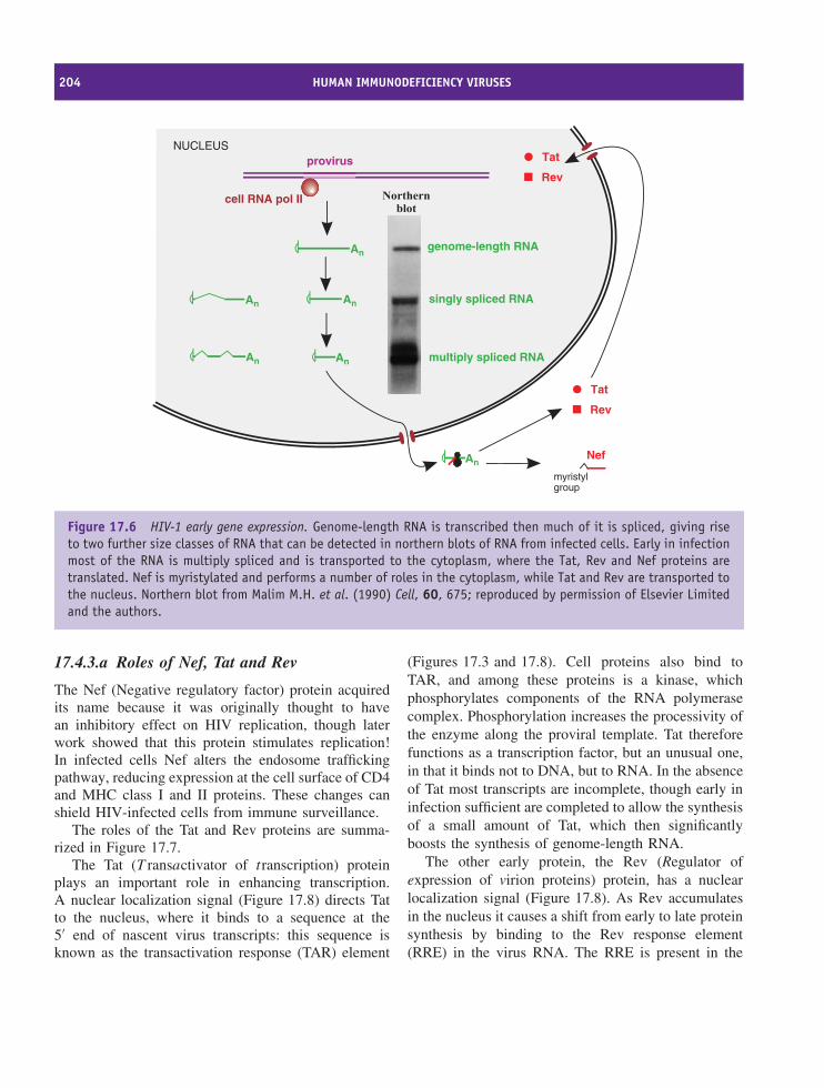
204 HUMAN IMMUNODEFICIENCY VIRUSES
cell RNA pol II
NUCLEUS
An
An
genome-length RNA
provirus
An
An
An
An
singly spliced RNA
multiply spliced RNA
Tat
Rev
Tat
Rev
myristylgroup
Nef
Figure 17.6 HIV-1 early gene expression. Genome-length RNA is transcribed then much of it is spliced, giving riseto two further size classes of RNA that can be detected in northern blots of RNA from infected cells. Early in infectionmost of the RNA is multiply spliced and is transported to the cytoplasm, where the Tat, Rev and Nef proteins aretranslated. Nef is myristylated and performs a number of roles in the cytoplasm, while Tat and Rev are transported tothe nucleus. Northern blot from Malim M.H. et al. (1990) Cell, 60, 675; reproduced by permission of Elsevier Limitedand the authors.
17.4.3.a Roles of Nef, Tat and Rev
The Nef (Negative regulatory factor) protein acquiredits name because it was originally thought to havean inhibitory effect on HIV replication, though laterwork showed that this protein stimulates replication!In infected cells Nef alters the endosome traffickingpathway, reducing expression at the cell surface of CD4and MHC class I and II proteins. These changes canshield HIV-infected cells from immune surveillance.
The roles of the Tat and Rev proteins are summa-rized in Figure 17.7.
The Tat (T ransactivator of transcription) proteinplays an important role in enhancing transcription.A nuclear localization signal (Figure 17.8) directs Tatto the nucleus, where it binds to a sequence at the5′ end of nascent virus transcripts: this sequence isknown as the transactivation response (TAR) element
(Figures 17.3 and 17.8). Cell proteins also bind toTAR, and among these proteins is a kinase, whichphosphorylates components of the RNA polymerasecomplex. Phosphorylation increases the processivity ofthe enzyme along the proviral template. Tat thereforefunctions as a transcription factor, but an unusual one,in that it binds not to DNA, but to RNA. In the absenceof Tat most transcripts are incomplete, though early ininfection sufficient are completed to allow the synthesisof a small amount of Tat, which then significantlyboosts the synthesis of genome-length RNA.
The other early protein, the Rev (Regulator ofexpression of virion proteins) protein, has a nuclearlocalization signal (Figure 17.8). As Rev accumulatesin the nucleus it causes a shift from early to late proteinsynthesis by binding to the Rev response element(RRE) in the virus RNA. The RRE is present in the
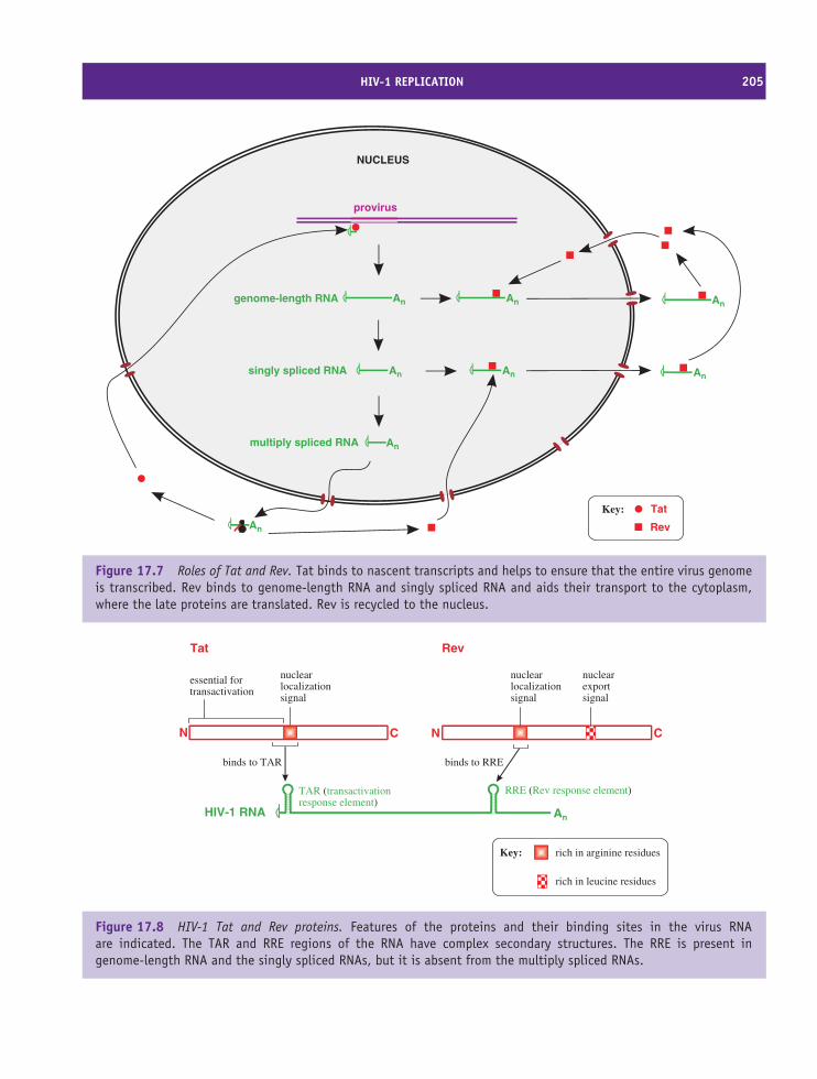
HIV-1 REPLICATION 205
NUCLEUS
An
Angenome-length RNA
provirus
An
An
singly spliced RNA
multiply spliced RNA
An
An
An
An
Tat
Rev
Key:
Figure 17.7 Roles of Tat and Rev. Tat binds to nascent transcripts and helps to ensure that the entire virus genomeis transcribed. Rev binds to genome-length RNA and singly spliced RNA and aids their transport to the cytoplasm,where the late proteins are translated. Rev is recycled to the nucleus.
N C
nuclearlocalizationsignal
nuclearexportsignal
N C
nuclearlocalizationsignal
essential fortransactivation
Tat Rev
AnHIV-1 RNA
binds to TAR binds to RRE
TAR (transactivationresponse element)
RRE (Rev response element)
rich in arginine residuesKey:
rich in leucine residues
Figure 17.8 HIV-1 Tat and Rev proteins. Features of the proteins and their binding sites in the virus RNAare indicated. The TAR and RRE regions of the RNA have complex secondary structures. The RRE is present ingenome-length RNA and the singly spliced RNAs, but it is absent from the multiply spliced RNAs.
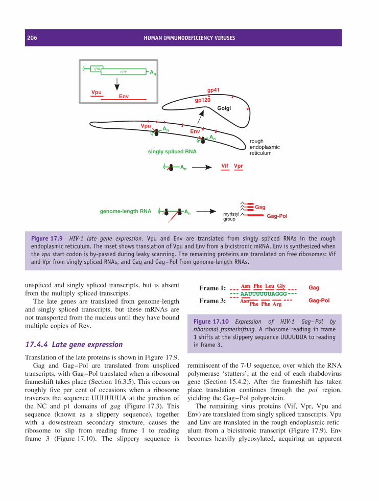
206 HUMAN IMMUNODEFICIENCY VIRUSES
roughendoplasmicreticulum
Golgi
An
Gag-Pol
Gagmyristylgroup
AnVprVif
genome-length RNA
EnvAnVpu
gp120
gp41
Anvpu
env
VpuEnv
singly spliced RNA
An
Figure 17.9 HIV-1 late gene expression. Vpu and Env are translated from singly spliced RNAs in the roughendoplasmic reticulum. The inset shows translation of Vpu and Env from a bicistronic mRNA. Env is synthesized whenthe vpu start codon is by-passed during leaky scanning. The remaining proteins are translated on free ribosomes: Vifand Vpr from singly spliced RNAs, and Gag and Gag–Pol from genome-length RNAs.
unspliced and singly spliced transcripts, but is absentfrom the multiply spliced transcripts.
The late genes are translated from genome-lengthand singly spliced transcripts, but these mRNAs arenot transported from the nucleus until they have boundmultiple copies of Rev.
17.4.4 Late gene expression
Translation of the late proteins is shown in Figure 17.9.Gag and Gag–Pol are translated from unspliced
transcripts, with Gag–Pol translated when a ribosomalframeshift takes place (Section 16.3.5). This occurs onroughly five per cent of occasions when a ribosometraverses the sequence UUUUUUA at the junction ofthe NC and p1 domains of gag (Figure 17.3). Thissequence (known as a slippery sequence), togetherwith a downstream secondary structure, causes theribosome to slip from reading frame 1 to readingframe 3 (Figure 17.10). The slippery sequence is
AAUUUUUUAGGGAAUUUUUUAGGGAsnAsn PhePhe LeuLeu GlyGly
AsnAsnPhePhe PhePhe ArgArg
GagGag
Gag-PolGag-Pol
Frame 1:
Frame 3:
Figure 17.10 Expression of HIV-1 Gag–Pol byribosomal frameshifting. A ribosome reading in frame1 shifts at the slippery sequence UUUUUUA to readingin frame 3.
reminiscent of the 7-U sequence, over which the RNApolymerase ‘stutters’, at the end of each rhabdovirusgene (Section 15.4.2). After the frameshift has takenplace translation continues through the pol region,yielding the Gag–Pol polyprotein.
The remaining virus proteins (Vif, Vpr, Vpu andEnv) are translated from singly spliced transcripts. Vpuand Env are translated in the rough endoplasmic retic-ulum from a bicistronic transcript (Figure 17.9). Envbecomes heavily glycosylated, acquiring an apparent
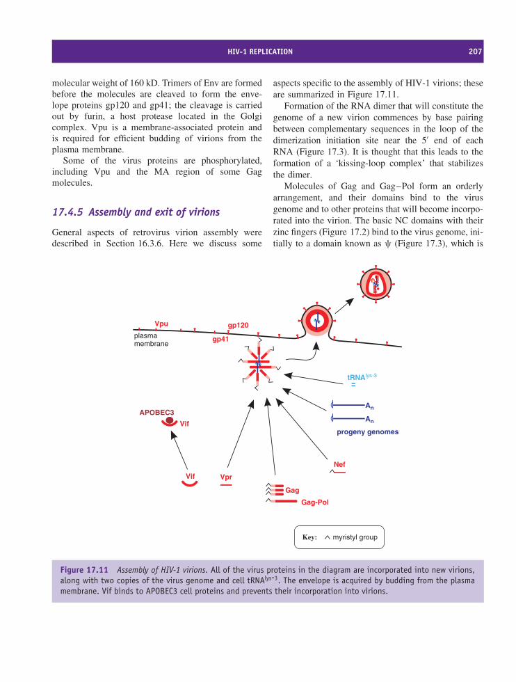
HIV-1 REPLICATION 207
molecular weight of 160 kD. Trimers of Env are formedbefore the molecules are cleaved to form the enve-lope proteins gp120 and gp41; the cleavage is carriedout by furin, a host protease located in the Golgicomplex. Vpu is a membrane-associated protein andis required for efficient budding of virions from theplasma membrane.
Some of the virus proteins are phosphorylated,including Vpu and the MA region of some Gagmolecules.
17.4.5 Assembly and exit of virions
General aspects of retrovirus virion assembly weredescribed in Section 16.3.6. Here we discuss some
aspects specific to the assembly of HIV-1 virions; theseare summarized in Figure 17.11.
Formation of the RNA dimer that will constitute thegenome of a new virion commences by base pairingbetween complementary sequences in the loop of thedimerization initiation site near the 5′ end of eachRNA (Figure 17.3). It is thought that this leads to theformation of a ‘kissing-loop complex’ that stabilizesthe dimer.
Molecules of Gag and Gag–Pol form an orderlyarrangement, and their domains bind to the virusgenome and to other proteins that will become incorpo-rated into the virion. The basic NC domains with theirzinc fingers (Figure 17.2) bind to the virus genome, ini-tially to a domain known as ψ (Figure 17.3), which is
tRNAlys-3
APOBEC3An
An
progeny genomes
Vif
Vif
Vpu gp120
gp41
Nef
Gag-Pol
Gag
Vpr
plasmamembrane
myristyl groupKey:
Figure 17.11 Assembly of HIV-1 virions. All of the virus proteins in the diagram are incorporated into new virions,along with two copies of the virus genome and cell tRNAlys-3. The envelope is acquired by budding from the plasmamembrane. Vif binds to APOBEC3 cell proteins and prevents their incorporation into virions.
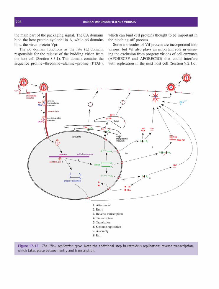
208 HUMAN IMMUNODEFICIENCY VIRUSES
the main part of the packaging signal. The CA domainsbind the host protein cyclophilin A, while p6 domainsbind the virus protein Vpr.
The p6 domain functions as the late (L) domain,responsible for the release of the budding virion fromthe host cell (Section 8.3.1). This domain contains thesequence proline–threonine–alanine–proline (PTAP),
which can bind cell proteins thought to be important inthe pinching off process.
Some molecules of Vif protein are incorporated intovirions, but Vif also plays an important role in ensur-ing the exclusion from progeny virions of cell enzymes(APOBEC3F and APOBEC3G) that could interferewith replication in the next host cell (Section 9.2.1.c).
tRNAlys-3
NUCLEUS
cell RNA pol II
roughendoplasmicreticulum
Golgi
cell chromosome
microtubule
An
An
An
An
mRNA
An
An
progeny genomes
RNA
DNA
An
provirus
reversetranscriptioncomplex
pre-integrationcomplex
Env
Gag-Pol
Gag
gp120
gp41
Vpr
An
VprVif
An An
Nef
AnVpu
Tat
Rev
CD4
chemokinereceptor
1
2
3
4
7
6
5
5
8
5early
late
1. Attachment2. Entry3. Reverse transcription4. Transcription5. Translation6. Genome replication7. Assembly8. Exit
Figure 17.12 The HIV-1 replication cycle. Note the additional step in retrovirus replication: reverse transcription,which takes place between entry and transcription.
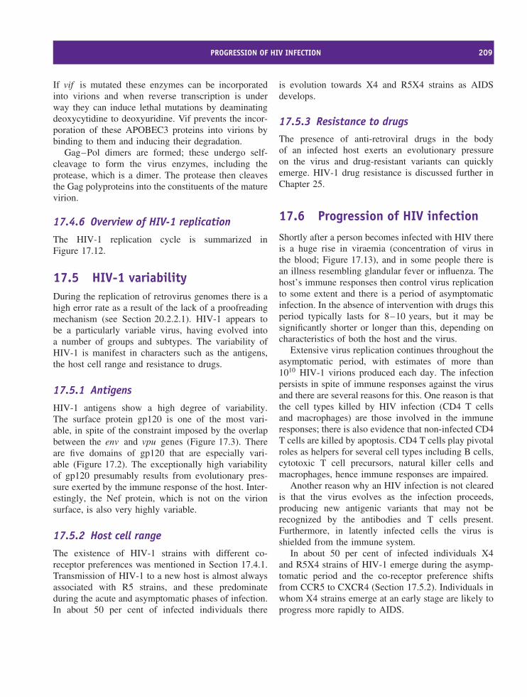
PROGRESSION OF HIV INFECTION 209
If vif is mutated these enzymes can be incorporatedinto virions and when reverse transcription is underway they can induce lethal mutations by deaminatingdeoxycytidine to deoxyuridine. Vif prevents the incor-poration of these APOBEC3 proteins into virions bybinding to them and inducing their degradation.
Gag–Pol dimers are formed; these undergo self-cleavage to form the virus enzymes, including theprotease, which is a dimer. The protease then cleavesthe Gag polyproteins into the constituents of the maturevirion.
17.4.6 Overview of HIV-1 replication
The HIV-1 replication cycle is summarized inFigure 17.12.
17.5 HIV-1 variabilityDuring the replication of retrovirus genomes there is ahigh error rate as a result of the lack of a proofreadingmechanism (see Section 20.2.2.1). HIV-1 appears tobe a particularly variable virus, having evolved intoa number of groups and subtypes. The variability ofHIV-1 is manifest in characters such as the antigens,the host cell range and resistance to drugs.
17.5.1 Antigens
HIV-1 antigens show a high degree of variability.The surface protein gp120 is one of the most vari-able, in spite of the constraint imposed by the overlapbetween the env and vpu genes (Figure 17.3). Thereare five domains of gp120 that are especially vari-able (Figure 17.2). The exceptionally high variabilityof gp120 presumably results from evolutionary pres-sure exerted by the immune response of the host. Inter-estingly, the Nef protein, which is not on the virionsurface, is also very highly variable.
17.5.2 Host cell range
The existence of HIV-1 strains with different co-receptor preferences was mentioned in Section 17.4.1.Transmission of HIV-1 to a new host is almost alwaysassociated with R5 strains, and these predominateduring the acute and asymptomatic phases of infection.In about 50 per cent of infected individuals there
is evolution towards X4 and R5X4 strains as AIDSdevelops.
17.5.3 Resistance to drugsThe presence of anti-retroviral drugs in the bodyof an infected host exerts an evolutionary pressureon the virus and drug-resistant variants can quicklyemerge. HIV-1 drug resistance is discussed further inChapter 25.
17.6 Progression of HIV infectionShortly after a person becomes infected with HIV thereis a huge rise in viraemia (concentration of virus inthe blood; Figure 17.13), and in some people there isan illness resembling glandular fever or influenza. Thehost’s immune responses then control virus replicationto some extent and there is a period of asymptomaticinfection. In the absence of intervention with drugs thisperiod typically lasts for 8–10 years, but it may besignificantly shorter or longer than this, depending oncharacteristics of both the host and the virus.
Extensive virus replication continues throughout theasymptomatic period, with estimates of more than1010 HIV-1 virions produced each day. The infectionpersists in spite of immune responses against the virusand there are several reasons for this. One reason is thatthe cell types killed by HIV infection (CD4 T cellsand macrophages) are those involved in the immuneresponses; there is also evidence that non-infected CD4T cells are killed by apoptosis. CD4 T cells play pivotalroles as helpers for several cell types including B cells,cytotoxic T cell precursors, natural killer cells andmacrophages, hence immune responses are impaired.
Another reason why an HIV infection is not clearedis that the virus evolves as the infection proceeds,producing new antigenic variants that may not berecognized by the antibodies and T cells present.Furthermore, in latently infected cells the virus isshielded from the immune system.
In about 50 per cent of infected individuals X4and R5X4 strains of HIV-1 emerge during the asymp-tomatic period and the co-receptor preference shiftsfrom CCR5 to CXCR4 (Section 17.5.2). Individuals inwhom X4 strains emerge at an early stage are likely toprogress more rapidly to AIDS.
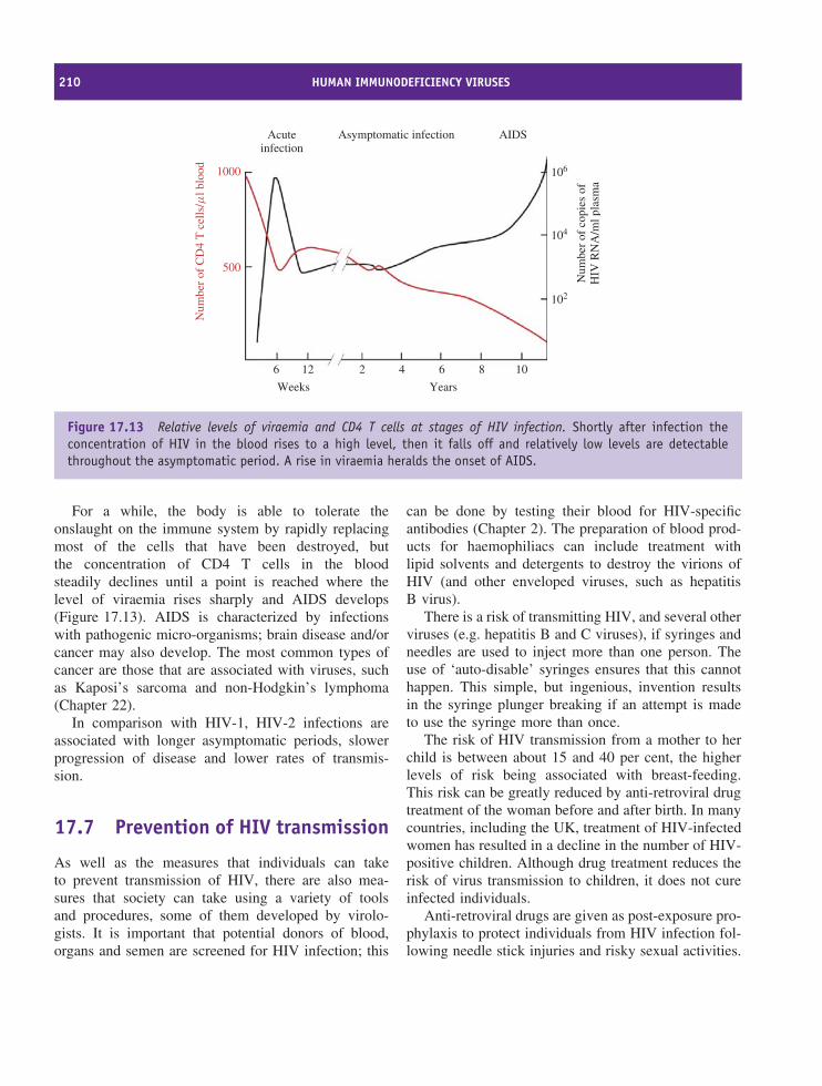
210 HUMAN IMMUNODEFICIENCY VIRUSES
Acuteinfection
Asymptomatic infection AIDS
1000
500
Num
ber
of C
D4
T c
ells
/ml b
lood 106
104
102
6 12 2 4 6 8 10
YearsWeeks
Num
ber
of c
opie
s of
HIV
RN
A/m
l pla
sma
Figure 17.13 Relative levels of viraemia and CD4 T cells at stages of HIV infection. Shortly after infection theconcentration of HIV in the blood rises to a high level, then it falls off and relatively low levels are detectablethroughout the asymptomatic period. A rise in viraemia heralds the onset of AIDS.
For a while, the body is able to tolerate theonslaught on the immune system by rapidly replacingmost of the cells that have been destroyed, butthe concentration of CD4 T cells in the bloodsteadily declines until a point is reached where thelevel of viraemia rises sharply and AIDS develops(Figure 17.13). AIDS is characterized by infectionswith pathogenic micro-organisms; brain disease and/orcancer may also develop. The most common types ofcancer are those that are associated with viruses, suchas Kaposi’s sarcoma and non-Hodgkin’s lymphoma(Chapter 22).
In comparison with HIV-1, HIV-2 infections areassociated with longer asymptomatic periods, slowerprogression of disease and lower rates of transmis-sion.
17.7 Prevention of HIV transmission
As well as the measures that individuals can taketo prevent transmission of HIV, there are also mea-sures that society can take using a variety of toolsand procedures, some of them developed by virolo-gists. It is important that potential donors of blood,organs and semen are screened for HIV infection; this
can be done by testing their blood for HIV-specificantibodies (Chapter 2). The preparation of blood prod-ucts for haemophiliacs can include treatment withlipid solvents and detergents to destroy the virions ofHIV (and other enveloped viruses, such as hepatitisB virus).
There is a risk of transmitting HIV, and several otherviruses (e.g. hepatitis B and C viruses), if syringes andneedles are used to inject more than one person. Theuse of ‘auto-disable’ syringes ensures that this cannothappen. This simple, but ingenious, invention resultsin the syringe plunger breaking if an attempt is madeto use the syringe more than once.
The risk of HIV transmission from a mother to herchild is between about 15 and 40 per cent, the higherlevels of risk being associated with breast-feeding.This risk can be greatly reduced by anti-retroviral drugtreatment of the woman before and after birth. In manycountries, including the UK, treatment of HIV-infectedwomen has resulted in a decline in the number of HIV-positive children. Although drug treatment reduces therisk of virus transmission to children, it does not cureinfected individuals.
Anti-retroviral drugs are given as post-exposure pro-phylaxis to protect individuals from HIV infection fol-lowing needle stick injuries and risky sexual activities.
SOURCES OF FURTHER INFORMATION 211
The use of drugs against HIV is discussed in detail inChapter 25.
A major goal of HIV research is the developmentof an effective vaccine. The deployment of such avaccine could dramatically reduce transmission rates ofthe virus, but after the expenditure of much effort thereis no sign on the horizon of an HIV vaccine suitablefor mass immunization. There are a number of reasonsfor this, including the ability of the virus to rapidlyevolve multiple antigenic variants as a result of its highmutation rate (Section 17.5).
Learning outcomes
By the end of this chapter you should be able to
• explain the importance of HIV;
• describe, with the aid of a labelled diagram, theHIV-1 virion;
• describe the HIV-1 genome;
• write an illustrated account of the replicationcycle of HIV-1;
• discuss the variability of HIV-1;
• discuss the effects of HIV infection on the host;
• evaluate approaches to the prevention of HIVtransmission.
Sources of further informationBooks
Richman D. D., editor (2003) Human ImmunodeficiencyVirus, International Medical Press
Turnbull E. and Borrow P. (2005) The immune response tohuman immunodeficiency virus type 1 (HIV-1) pp. 23–89in Molecular Pathogenesis of Virus Infections, editorsDigard P., Nash A. A. and Randall R. E., 64th Sympo-sium of the Society for General Microbiology, CambridgeUniversity Press
Journals
Adamson C. S. and Jones I. M. (2004) The molecular basisof HIV capsid assembly – five years of progress Reviewsin Medical Virology, 14, 107–121
Barboric M. and Peterlin B. M. (2005) A new paradigmin eukaryotic biology: HIV Tat and the control oftranscriptional elongation PLoS Biology, 3, e76
Persaud D. et al. (2003) Latency in human immunodefi-ciency virus type 1 infection: no easy answers Journalof Virology, 77, 1659–1665
Sierra S., Kupfer B. and Kaiser R. (2005) Basics of thevirology of HIV-1 and its replication Journal of ClinicalVirology, 34, 233–244
Trkola A. (2004) HIV–host interactions: vital to the virusand key to its inhibition Current Opinion in Microbiology,7, 555–559
Zheng Y. H., Lovsina N. and Peterlin B. M. (2005) Newlyidentified host factors modulate HIV replication Immunol-ogy Letters, 97, 225–234
Zhu P. et al. (2006) Distribution and three-dimensionalstructure of AIDS virus envelope spikes Nature, 441,847–852

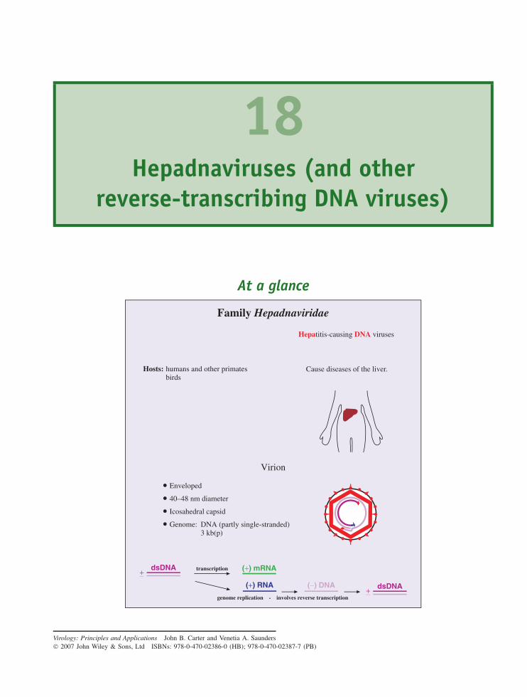
18Hepadnaviruses (and other
reverse-transcribing DNA viruses)
At a glance
Virion
• Enveloped
• 40–48 nm diameter
• Icosahedral capsid
• Genome: DNA (partly single-stranded) 3 kb(p)
Family Hepadnaviridae
Hosts: humans and other primates birds
Cause diseases of the liver.
Hepatitis-causing DNA viruses
dsDNA (+) mRNA
(+) RNA (−) DNA
+−dsDNA+−
transcription
genome replication - involves reverse transcription
Virology: Principles and Applications John B. Carter and Venetia A. Saunders 2007 John Wiley & Sons, Ltd ISBNs: 978-0-470-02386-0 (HB); 978-0-470-02387-7 (PB)
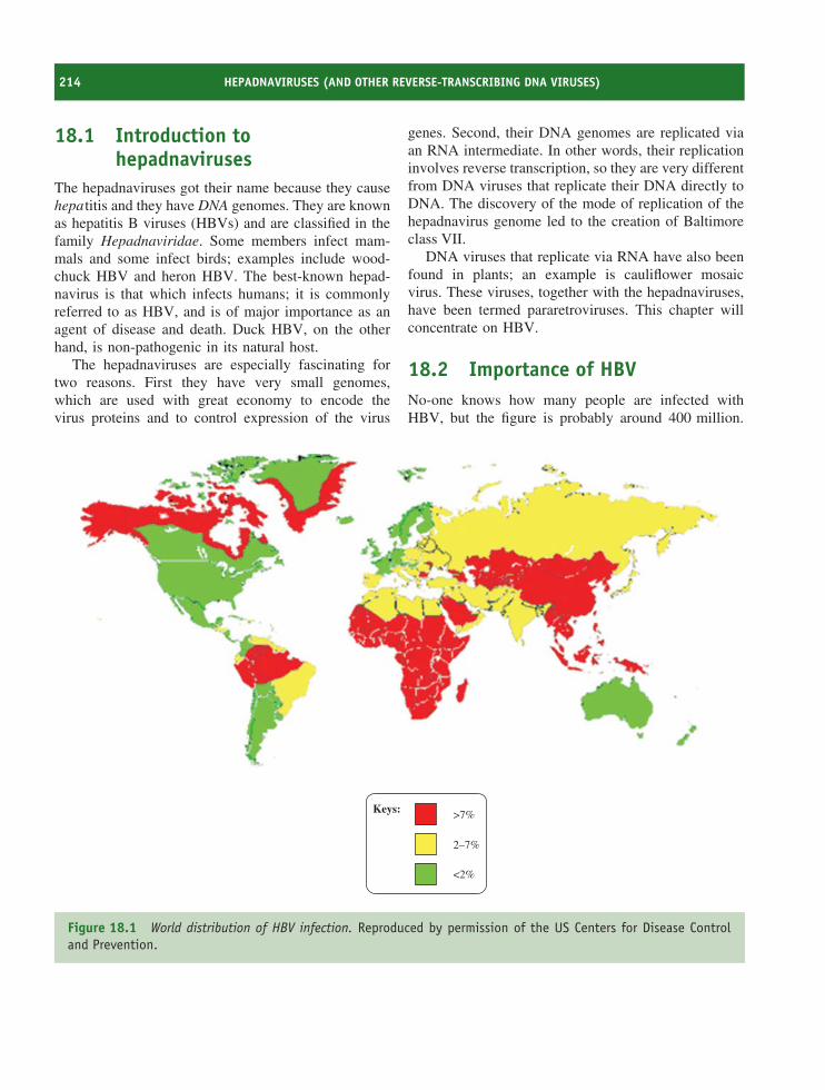
214 HEPADNAVIRUSES (AND OTHER REVERSE-TRANSCRIBING DNA VIRUSES)
18.1 Introduction tohepadnaviruses
The hepadnaviruses got their name because they causehepatitis and they have DNA genomes. They are knownas hepatitis B viruses (HBVs) and are classified in thefamily Hepadnaviridae. Some members infect mam-mals and some infect birds; examples include wood-chuck HBV and heron HBV. The best-known hepad-navirus is that which infects humans; it is commonlyreferred to as HBV, and is of major importance as anagent of disease and death. Duck HBV, on the otherhand, is non-pathogenic in its natural host.
The hepadnaviruses are especially fascinating fortwo reasons. First they have very small genomes,which are used with great economy to encode thevirus proteins and to control expression of the virus
genes. Second, their DNA genomes are replicated viaan RNA intermediate. In other words, their replicationinvolves reverse transcription, so they are very differentfrom DNA viruses that replicate their DNA directly toDNA. The discovery of the mode of replication of thehepadnavirus genome led to the creation of Baltimoreclass VII.
DNA viruses that replicate via RNA have also beenfound in plants; an example is cauliflower mosaicvirus. These viruses, together with the hepadnaviruses,have been termed pararetroviruses. This chapter willconcentrate on HBV.
18.2 Importance of HBVNo-one knows how many people are infected withHBV, but the figure is probably around 400 million.
Keys: >7%
2–7%
<2%
Figure 18.1 World distribution of HBV infection. Reproduced by permission of the US Centers for Disease Controland Prevention.
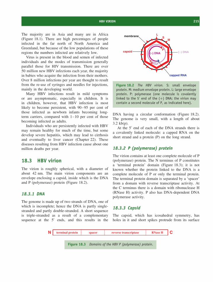
HBV VIRION 215
The majority are in Asia and many are in Africa(Figure 18.1). There are high percentages of peopleinfected in the far north of North America andGreenland, but because of the low populations of theseregions the numbers infected are relatively low.
Virus is present in the blood and semen of infectedindividuals and the modes of transmission generallyparallel those for HIV transmission. There are over50 million new HBV infections each year, the majorityin babies who acquire the infection from their mothers.Over 8 million infections per year are thought to resultfrom the re-use of syringes and needles for injections,mainly in the developing world.
Many HBV infections result in mild symptomsor are asymptomatic, especially in children. It isin children, however, that HBV infection is mostlikely to become persistent, with 90–95 per cent ofthose infected as newborn infants becoming long-term carriers, compared with 1–10 per cent of thosebecoming infected as adults.
Individuals who are persistently infected with HBVmay remain healthy for much of the time, but somedevelop severe hepatitis, which may lead to cirrhosisand eventually to liver cancer (Chapter 22). Thesediseases resulting from HBV infection cause about onemillion deaths per year.
18.3 HBV virionThe virion is roughly spherical, with a diameter ofabout 42 nm. The main virion components are anenvelope enclosing a capsid, inside which is the DNAand P (polymerase) protein (Figure 18.2).
18.3.1 DNA
The genome is made up of two strands of DNA, one ofwhich is incomplete; hence the DNA is partly single-stranded and partly double-stranded. A short sequenceis triple-stranded as a result of a complementarysequence at the 5′ ends, and this results in the
L
Mmembrane S
capped RNA
(+) DNA (−) DNAcapsid
P
P
Figure 18.2 The HBV virion. S: small envelopeprotein. M: medium envelope protein. L: large envelopeprotein. P: polymerase (one molecule is covalentlylinked to the 5′ end of the (+) DNA; the virion maycontain a second molecule of P, as indicated here).
DNA having a circular conformation (Figure 18.2).The genome is very small, with a length of about3.2 kb(p).
At the 5′ end of each of the DNA strands there isa covalently linked molecule: a capped RNA on theshort strand and a protein (P) on the long strand.
18.3.2 P (polymerase) protein
The virion contains at least one complete molecule of P(polymerase) protein. The N terminus of P constitutesa ‘terminal protein’ domain (Figure 18.3); it is notknown whether the protein linked to the DNA is acomplete molecule of P or only the terminal protein.The terminal protein domain is separated by a ‘spacer’from a domain with reverse transcriptase activity. Atthe C terminus there is a domain with ribonuclease H(RNase H) activity. P also has DNA-dependent DNApolymerase activity.
18.3.3 Capsid
The capsid, which has icosahedral symmetry, hasholes in it and short spikes protrude from its surface
N Cterminal protein spacer reverse transcriptase RNase H
Figure 18.3 Domains of the HBV P (polymerase) protein.
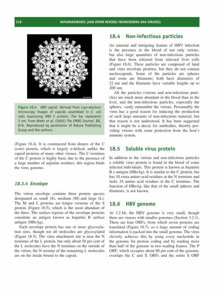
216 HEPADNAVIRUSES (AND OTHER REVERSE-TRANSCRIBING DNA VIRUSES)
Figure 18.4 HBV capsid. Derived from cryo-electronmicroscopy images of capsids assembled in E. colicells expressing HBV C protein. The bar represents5 nm. From Watts et al. (2002) The EMBO Journal, 21,876. Reproduced by permission of Nature PublishingGroup and the authors.
(Figure 18.4). It is constructed from dimers of the C(core) protein, which is largely α-helical, unlike thecapsid proteins of many other viruses. The C terminusof the C protein is highly basic due to the presence ofa large number of arginine residues; this region bindsthe virus genome.
18.3.4 Envelope
The virion envelope contains three protein speciesdesignated as small (S), medium (M) and large (L).The M and L proteins are longer versions of the Sprotein (Figure 18.5), which is the most abundant ofthe three. The surface regions of the envelope proteinsconstitute an antigen known as hepatitis B surfaceantigen (HBsAg).
Each envelope protein has one or more glycosyla-tion sites, though not all molecules are glycosylated(Figure 18.5). The virus attachment site is near the Nterminus of the L protein, but only about 50 per cent ofthe L molecules have the N terminus on the outside ofthe virion; the N termini of the remaining L moleculesare on the inside bound to the capsid.
18.4 Non-infectious particles
An unusual and intriguing feature of HBV infectionis the presence in the blood of not only virions,but also large quantities of non-infectious particlesthat have been released from infected liver cells(Figure 18.6). These particles are composed of lipidand virus envelope proteins, but they do not containnucleocapsids. Some of the particles are spheresand some are filaments; both have diameters of22 nm and the filaments have variable lengths up to200 nm.
All the particles (virions and non-infectious parti-cles) are much more abundant in the blood than in theliver, and the non-infectious particles, especially thespheres, vastly outnumber the virions. Presumably thevirus has a good reason for inducing the productionof such large amounts of non-infectious material, butthat reason is not understood. It has been suggestedthat it might be a decoy for antibodies, thereby pro-viding virions with some protection from the host’simmune system.
18.5 Soluble virus protein
In addition to the virions and non-infectious particlesa soluble virus protein is found in the blood of someinfected individuals. This protein is known as hepatitisB e antigen (HBeAg). It is similar to the C protein, buthas 10 extra amino acid residues at the N terminus andlacks 34 amino acid residues at the C terminus. Thefunction of HBeAg, like that of the small spheres andfilaments, is not known.
18.6 HBV genome
At 3.2 kb, the HBV genome is very small, thoughthere are viruses with smaller genomes (Section 3.2.1).There are four ORFs, from which seven proteins aretranslated (Figure 18.7), so a large amount of codinginformation is packed into the small genome. The viruscleverly achieves this by using every nucleotide inthe genome for protein coding and by reading morethan half of the genome in two reading frames. The PORF, which occupies about 80 per cent of the genome,overlaps the C and X ORFs and the entire S ORF
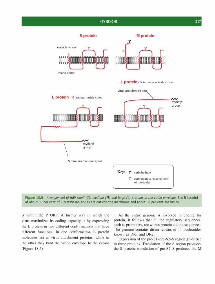
HBV GENOME 217
myristylgroup
myristylgroup
virus attachment site
outside virion
inside virion
S protein M protein
L protein - N terminus inside virion
L protein - N terminus outside virion
N terminus binds to capsid
carbohydrate
carbohydrate on about 50%of molecules
Key:
Figure 18.5 Arrangement of HBV small (S), medium (M) and large (L) proteins in the virion envelope. The N terminiof about 50 per cent of L protein molecules are outside the membrane and about 50 per cent are inside.
is within the P ORF. A further way in which the
virus maximizes its coding capacity is by expressing
the L protein in two different conformations that have
different functions. In one conformation L protein
molecules act as virus attachment proteins, while in
the other they bind the virion envelope to the capsid
(Figure 18.5).
As the entire genome is involved in coding forprotein, it follows that all the regulatory sequences,such as promoters, are within protein coding sequences.The genome contains direct repeats of 11 nucleotidesknown as DR1 and DR2.
Expression of the pre-S1–pre-S2–S region gives riseto three proteins. Translation of the S region producesthe S protein, translation of pre-S2–S produces the M
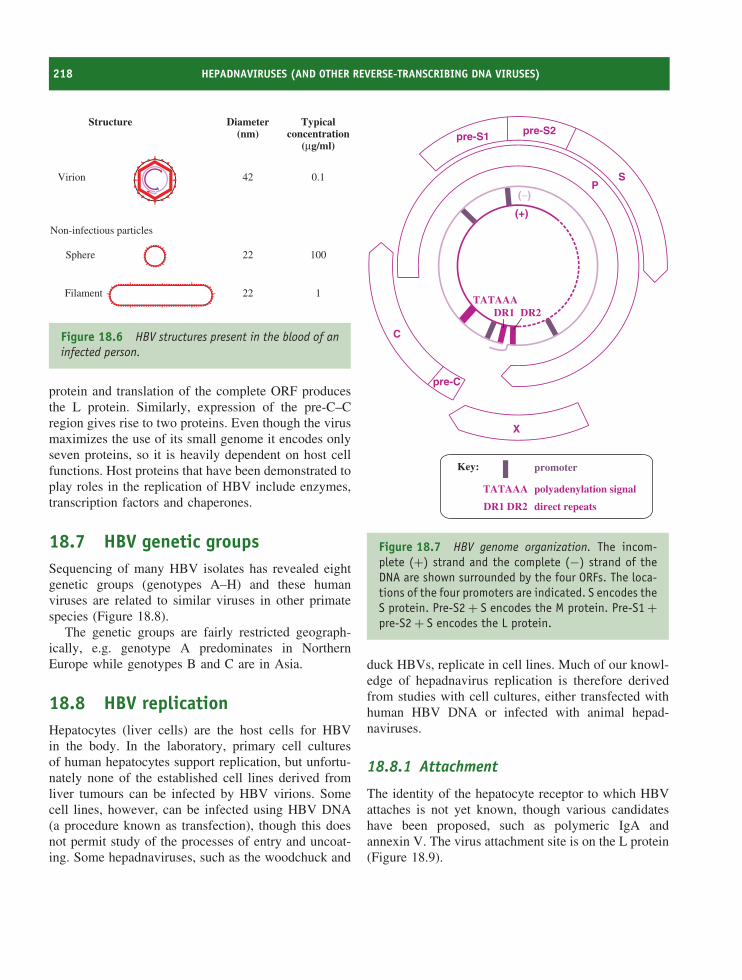
218 HEPADNAVIRUSES (AND OTHER REVERSE-TRANSCRIBING DNA VIRUSES)
Structure Diameter(nm)
Typicalconcentration
(µg/ml)
Virion 42
22
22 1
100
0.1
Non-infectious particles
Sphere
Filament
Figure 18.6 HBV structures present in the blood of aninfected person.
protein and translation of the complete ORF producesthe L protein. Similarly, expression of the pre-C–Cregion gives rise to two proteins. Even though the virusmaximizes the use of its small genome it encodes onlyseven proteins, so it is heavily dependent on host cellfunctions. Host proteins that have been demonstrated toplay roles in the replication of HBV include enzymes,transcription factors and chaperones.
18.7 HBV genetic groupsSequencing of many HBV isolates has revealed eightgenetic groups (genotypes A–H) and these humanviruses are related to similar viruses in other primatespecies (Figure 18.8).
The genetic groups are fairly restricted geograph-ically, e.g. genotype A predominates in NorthernEurope while genotypes B and C are in Asia.
18.8 HBV replicationHepatocytes (liver cells) are the host cells for HBVin the body. In the laboratory, primary cell culturesof human hepatocytes support replication, but unfortu-nately none of the established cell lines derived fromliver tumours can be infected by HBV virions. Somecell lines, however, can be infected using HBV DNA(a procedure known as transfection), though this doesnot permit study of the processes of entry and uncoat-ing. Some hepadnaviruses, such as the woodchuck and
(+)
(−)
C
PS
X
pre-S1 pre-S2
pre-C
TATAAADR1 DR2
promoter
TATAAA
DR1 DR2 direct repeats
polyadenylation signal
Key:
Figure 18.7 HBV genome organization. The incom-plete (+) strand and the complete (−) strand of theDNA are shown surrounded by the four ORFs. The loca-tions of the four promoters are indicated. S encodes theS protein. Pre-S2 + S encodes the M protein. Pre-S1 +pre-S2 + S encodes the L protein.
duck HBVs, replicate in cell lines. Much of our knowl-edge of hepadnavirus replication is therefore derivedfrom studies with cell cultures, either transfected withhuman HBV DNA or infected with animal hepad-naviruses.
18.8.1 Attachment
The identity of the hepatocyte receptor to which HBVattaches is not yet known, though various candidateshave been proposed, such as polymeric IgA andannexin V. The virus attachment site is on the L protein(Figure 18.9).
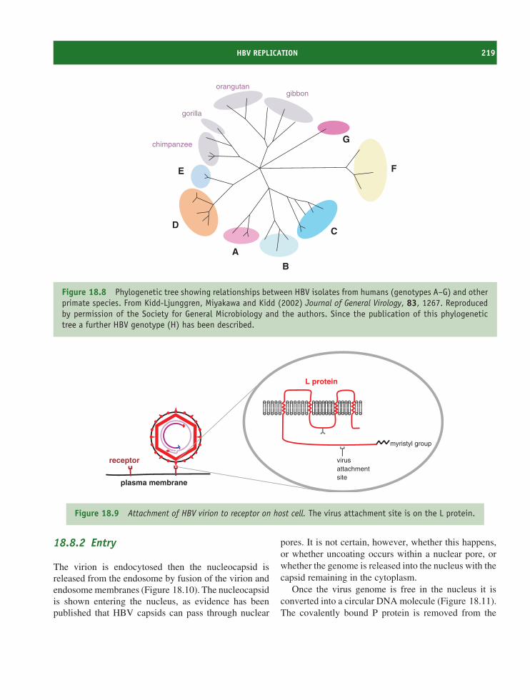
HBV REPLICATION 219
gibbonorangutan
gorilla
chimpanzee
A
B
CD
E F
G
Figure 18.8 Phylogenetic tree showing relationships between HBV isolates from humans (genotypes A–G) and otherprimate species. From Kidd-Ljunggren, Miyakawa and Kidd (2002) Journal of General Virology, 83, 1267. Reproducedby permission of the Society for General Microbiology and the authors. Since the publication of this phylogenetictree a further HBV genotype (H) has been described.
myristyl group
virusattachmentsite
L protein
plasma membrane
receptor
Figure 18.9 Attachment of HBV virion to receptor on host cell. The virus attachment site is on the L protein.
18.8.2 Entry
The virion is endocytosed then the nucleocapsid isreleased from the endosome by fusion of the virion andendosome membranes (Figure 18.10). The nucleocapsidis shown entering the nucleus, as evidence has beenpublished that HBV capsids can pass through nuclear
pores. It is not certain, however, whether this happens,or whether uncoating occurs within a nuclear pore, orwhether the genome is released into the nucleus with thecapsid remaining in the cytoplasm.
Once the virus genome is free in the nucleus it isconverted into a circular DNA molecule (Figure 18.11).The covalently bound P protein is removed from the
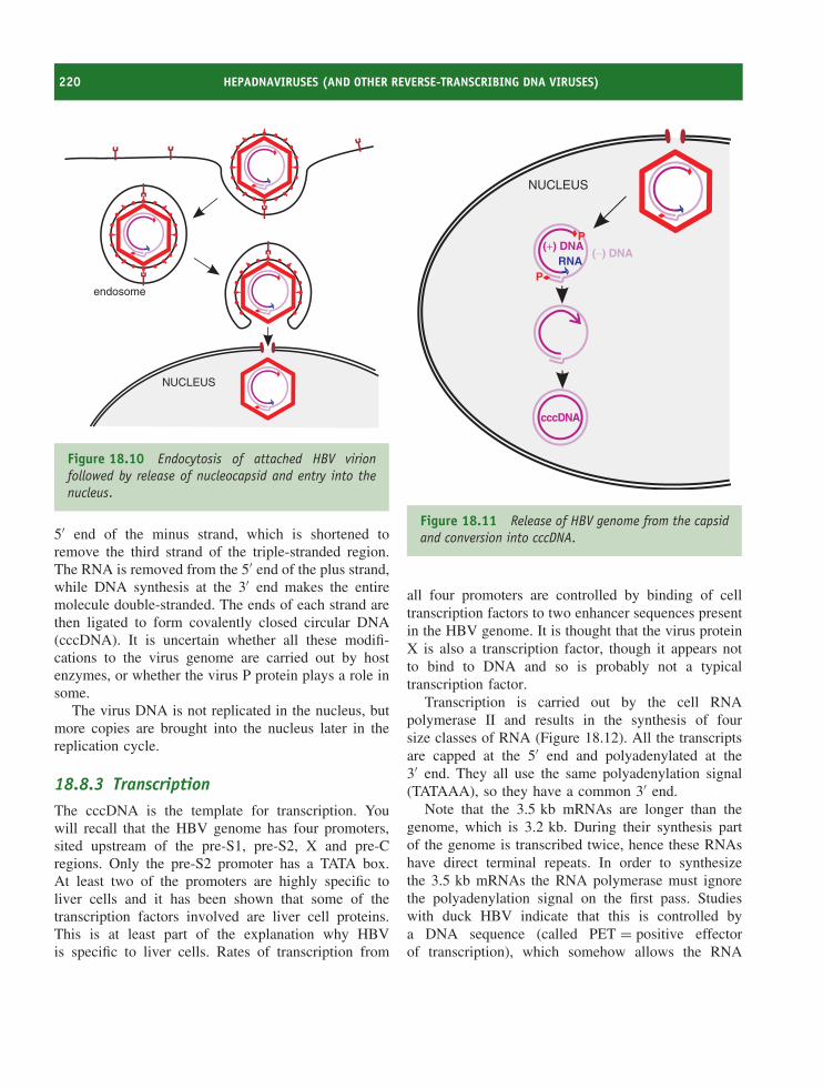
220 HEPADNAVIRUSES (AND OTHER REVERSE-TRANSCRIBING DNA VIRUSES)
NUCLEUS
endosome
Figure 18.10 Endocytosis of attached HBV virionfollowed by release of nucleocapsid and entry into thenucleus.
5′ end of the minus strand, which is shortened toremove the third strand of the triple-stranded region.The RNA is removed from the 5′ end of the plus strand,while DNA synthesis at the 3′ end makes the entiremolecule double-stranded. The ends of each strand arethen ligated to form covalently closed circular DNA(cccDNA). It is uncertain whether all these modifi-cations to the virus genome are carried out by hostenzymes, or whether the virus P protein plays a role insome.
The virus DNA is not replicated in the nucleus, butmore copies are brought into the nucleus later in thereplication cycle.
18.8.3 TranscriptionThe cccDNA is the template for transcription. Youwill recall that the HBV genome has four promoters,sited upstream of the pre-S1, pre-S2, X and pre-Cregions. Only the pre-S2 promoter has a TATA box.At least two of the promoters are highly specific toliver cells and it has been shown that some of thetranscription factors involved are liver cell proteins.This is at least part of the explanation why HBVis specific to liver cells. Rates of transcription from
NUCLEUS
cccDNA
(+) DNA(−) DNA
RNAP
P
Figure 18.11 Release of HBV genome from the capsidand conversion into cccDNA.
all four promoters are controlled by binding of celltranscription factors to two enhancer sequences presentin the HBV genome. It is thought that the virus proteinX is also a transcription factor, though it appears notto bind to DNA and so is probably not a typicaltranscription factor.
Transcription is carried out by the cell RNApolymerase II and results in the synthesis of foursize classes of RNA (Figure 18.12). All the transcriptsare capped at the 5′ end and polyadenylated at the3′ end. They all use the same polyadenylation signal(TATAAA), so they have a common 3′ end.
Note that the 3.5 kb mRNAs are longer than thegenome, which is 3.2 kb. During their synthesis partof the genome is transcribed twice, hence these RNAshave direct terminal repeats. In order to synthesizethe 3.5 kb mRNAs the RNA polymerase must ignorethe polyadenylation signal on the first pass. Studieswith duck HBV indicate that this is controlled bya DNA sequence (called PET = positive effectorof transcription), which somehow allows the RNA
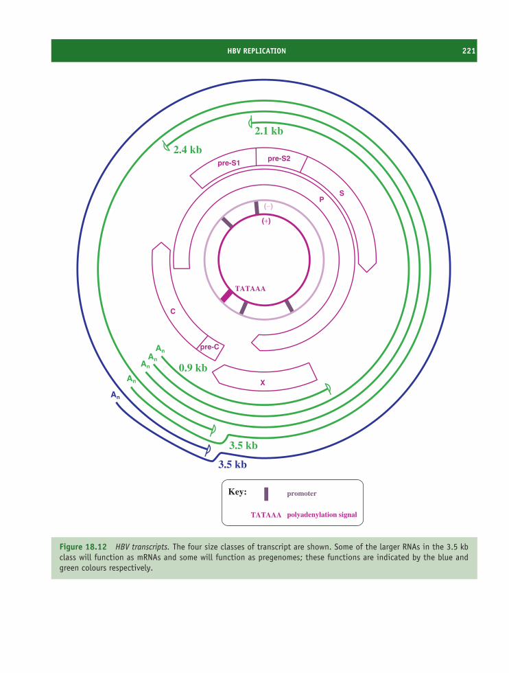
HBV REPLICATION 221
(+)
(−)
C
PS
X
pre-S1pre-S2
pre-CAnAn
An
An
An
0.9 kb
2.1 kb
2.4 kb
3.5 kb
3.5 kb
TATAAA
TATAAA
promoter
polyadenylation signal
Key:
Figure 18.12 HBV transcripts. The four size classes of transcript are shown. Some of the larger RNAs in the 3.5 kbclass will function as mRNAs and some will function as pregenomes; these functions are indicated by the blue andgreen colours respectively.
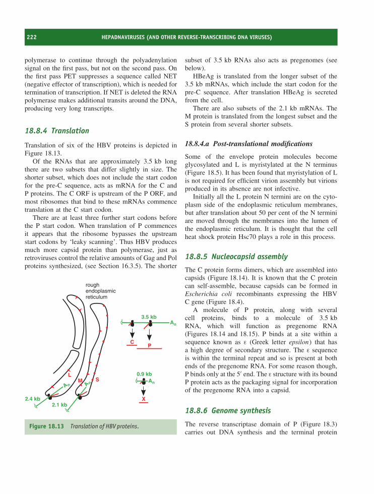
222 HEPADNAVIRUSES (AND OTHER REVERSE-TRANSCRIBING DNA VIRUSES)
polymerase to continue through the polyadenylationsignal on the first pass, but not on the second pass. Onthe first pass PET suppresses a sequence called NET(negative effector of transcription), which is needed fortermination of transcription. If NET is deleted the RNApolymerase makes additional transits around the DNA,producing very long transcripts.
18.8.4 Translation
Translation of six of the HBV proteins is depicted inFigure 18.13.
Of the RNAs that are approximately 3.5 kb longthere are two subsets that differ slightly in size. Theshorter subset, which does not include the start codonfor the pre-C sequence, acts as mRNA for the C andP proteins. The C ORF is upstream of the P ORF, andmost ribosomes that bind to these mRNAs commencetranslation at the C start codon.
There are at least three further start codons beforethe P start codon. When translation of P commencesit appears that the ribosome bypasses the upstreamstart codons by ‘leaky scanning’. Thus HBV producesmuch more capsid protein than polymerase, just asretroviruses control the relative amounts of Gag and Polproteins synthesized, (see Section 16.3.5). The shorter
roughendoplasmicreticulum
A n
L
An
A n
2.4 kb2.1 kb
M S
3.5 kb
An
0.9 kb
CP
X
Figure 18.13 Translation of HBV proteins.
subset of 3.5 kb RNAs also acts as pregenomes (seebelow).
HBeAg is translated from the longer subset of the3.5 kb mRNAs, which include the start codon for thepre-C sequence. After translation HBeAg is secretedfrom the cell.
There are also subsets of the 2.1 kb mRNAs. TheM protein is translated from the longest subset and theS protein from several shorter subsets.
18.8.4.a Post-translational modifications
Some of the envelope protein molecules becomeglycosylated and L is myristylated at the N terminus(Figure 18.5). It has been found that myristylation of Lis not required for efficient virion assembly but virionsproduced in its absence are not infective.
Initially all the L protein N termini are on the cyto-plasm side of the endoplasmic reticulum membranes,but after translation about 50 per cent of the N terminiare moved through the membranes into the lumen ofthe endoplasmic reticulum. It is thought that the cellheat shock protein Hsc70 plays a role in this process.
18.8.5 Nucleocapsid assembly
The C protein forms dimers, which are assembled intocapsids (Figure 18.14). It is known that the C proteincan self-assemble, because capsids can be formed inEscherichia coli recombinants expressing the HBVC gene (Figure 18.4).
A molecule of P protein, along with severalcell proteins, binds to a molecule of 3.5 kbRNA, which will function as pregenome RNA(Figures 18.14 and 18.15). P binds at a site within asequence known as ε (Greek letter epsilon) that hasa high degree of secondary structure. The ε sequenceis within the terminal repeat and so is present at bothends of the pregenome RNA. For some reason though,P binds only at the 5′ end. The ε structure with its boundP protein acts as the packaging signal for incorporationof the pregenome RNA into a capsid.
18.8.6 Genome synthesis
The reverse transcriptase domain of P (Figure 18.3)carries out DNA synthesis and the terminal protein
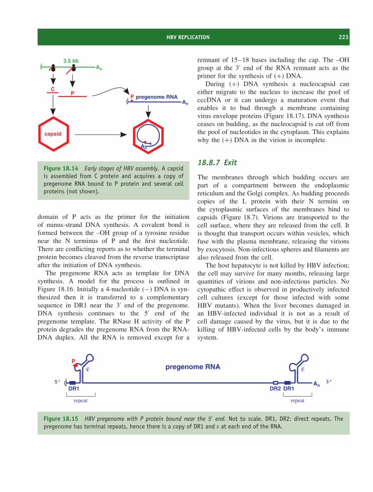
HBV REPLICATION 223
An
pregenome RNA
An
An
3.5 kb
CP
P
capsid
Figure 18.14 Early stages of HBV assembly. A capsidis assembled from C protein and acquires a copy ofpregenome RNA bound to P protein and several cellproteins (not shown).
domain of P acts as the primer for the initiationof minus-strand DNA synthesis. A covalent bond isformed between the –OH group of a tyrosine residuenear the N terminus of P and the first nucleotide.There are conflicting reports as to whether the terminalprotein becomes cleaved from the reverse transcriptaseafter the initiation of DNA synthesis.
The pregenome RNA acts as template for DNAsynthesis. A model for the process is outlined inFigure 18.16. Initially a 4-nucleotide (−) DNA is syn-thesized then it is transferred to a complementarysequence in DR1 near the 3′ end of the pregenome.DNA synthesis continues to the 5′ end of thepregenome template. The RNase H activity of the Pprotein degrades the pregenome RNA from the RNA-DNA duplex. All the RNA is removed except for a
remnant of 15–18 bases including the cap. The –OHgroup at the 3′ end of the RNA remnant acts as theprimer for the synthesis of (+) DNA.
During (+) DNA synthesis a nucleocapsid caneither migrate to the nucleus to increase the pool ofcccDNA or it can undergo a maturation event thatenables it to bud through a membrane containingvirus envelope proteins (Figure 18.17). DNA synthesisceases on budding, as the nucleocapsid is cut off fromthe pool of nucleotides in the cytoplasm. This explainswhy the (+) DNA in the virion is incomplete.
18.8.7 Exit
The membranes through which budding occurs arepart of a compartment between the endoplasmicreticulum and the Golgi complex. As budding proceedscopies of the L protein with their N termini onthe cytoplasmic surfaces of the membranes bind tocapsids (Figure 18.7). Virions are transported to thecell surface, where they are released from the cell. Itis thought that transport occurs within vesicles, whichfuse with the plasma membrane, releasing the virionsby exocytosis. Non-infectious spheres and filaments arealso released from the cell.
The host hepatocyte is not killed by HBV infection;the cell may survive for many months, releasing largequantities of virions and non-infectious particles. Nocytopathic effect is observed in productively infectedcell cultures (except for those infected with someHBV mutants). When the liver becomes damaged inan HBV-infected individual it is not as a result ofcell damage caused by the virus, but it is due to thekilling of HBV-infected cells by the body’s immunesystem.
An
ε
DR1
ε
DR1DR2
pregenome RNAP
5’ 3’
repeat repeat
Figure 18.15 HBV pregenome with P protein bound near the 5′ end. Not to scale. DR1, DR2: direct repeats. Thepregenome has terminal repeats, hence there is a copy of DR1 and ε at each end of the RNA.
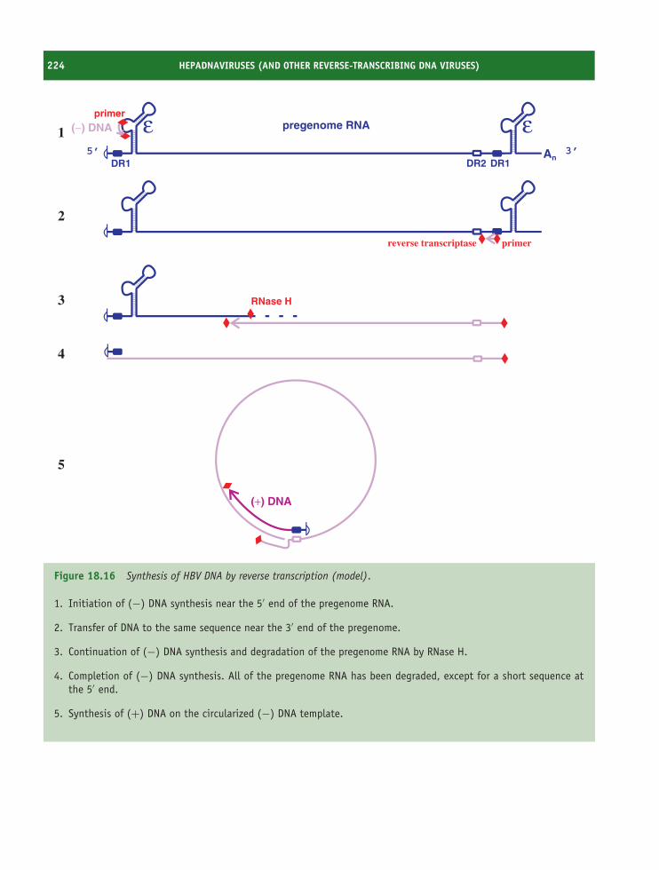
224 HEPADNAVIRUSES (AND OTHER REVERSE-TRANSCRIBING DNA VIRUSES)
AnDR1 DR1DR2
pregenome RNAprimer
5’ 3’
(−) DNA1
reverse transcriptase primer
2
RNase H3
4
5
(+) DNA
Figure 18.16 Synthesis of HBV DNA by reverse transcription (model).
1. Initiation of (−) DNA synthesis near the 5′ end of the pregenome RNA.
2. Transfer of DNA to the same sequence near the 3′ end of the pregenome.
3. Continuation of (−) DNA synthesis and degradation of the pregenome RNA by RNase H.
4. Completion of (−) DNA synthesis. All of the pregenome RNA has been degraded, except for a short sequence atthe 5′ end.
5. Synthesis of (+) DNA on the circularized (−) DNA template.
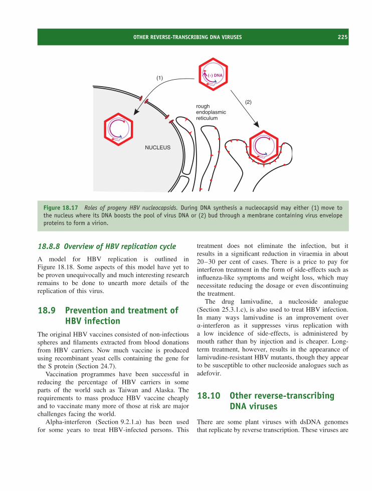
OTHER REVERSE-TRANSCRIBING DNA VIRUSES 225
NUCLEUS
roughendoplasmicreticulum
(+) DNA(1)
(2)
Figure 18.17 Roles of progeny HBV nucleocapsids. During DNA synthesis a nucleocapsid may either (1) move tothe nucleus where its DNA boosts the pool of virus DNA or (2) bud through a membrane containing virus envelopeproteins to form a virion.
18.8.8 Overview of HBV replication cycle
A model for HBV replication is outlined inFigure 18.18. Some aspects of this model have yet tobe proven unequivocally and much interesting researchremains to be done to unearth more details of thereplication of this virus.
18.9 Prevention and treatment ofHBV infection
The original HBV vaccines consisted of non-infectiousspheres and filaments extracted from blood donationsfrom HBV carriers. Now much vaccine is producedusing recombinant yeast cells containing the gene forthe S protein (Section 24.7).
Vaccination programmes have been successful inreducing the percentage of HBV carriers in someparts of the world such as Taiwan and Alaska. Therequirements to mass produce HBV vaccine cheaplyand to vaccinate many more of those at risk are majorchallenges facing the world.
Alpha-interferon (Section 9.2.1.a) has been usedfor some years to treat HBV-infected persons. This
treatment does not eliminate the infection, but itresults in a significant reduction in viraemia in about20–30 per cent of cases. There is a price to pay forinterferon treatment in the form of side-effects such asinfluenza-like symptoms and weight loss, which maynecessitate reducing the dosage or even discontinuingthe treatment.
The drug lamivudine, a nucleoside analogue(Section 25.3.1.c), is also used to treat HBV infection.In many ways lamivudine is an improvement overα-interferon as it suppresses virus replication witha low incidence of side-effects, is administered bymouth rather than by injection and is cheaper. Long-term treatment, however, results in the appearance oflamivudine-resistant HBV mutants, though they appearto be susceptible to other nucleoside analogues such asadefovir.
18.10 Other reverse-transcribingDNA viruses
There are some plant viruses with dsDNA genomesthat replicate by reverse transcription. These viruses are
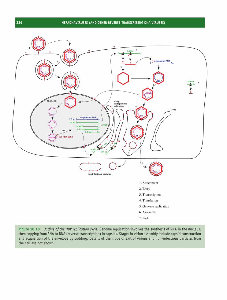
226 HEPADNAVIRUSES (AND OTHER REVERSE-TRANSCRIBING DNA VIRUSES)
NUCLEUS roughendoplasmicreticulum
Golgi
cell RNA pol II
An
cccDNA
An
An
An
An
L
Anpregenome RNA
Anpregenome RNA
An
An
3.5 kb
3.5 kb
2.4 kb2.1 kb
0.9 kb
An
2.4 kb2.1 kb
M S
3.5 kb
CP
non-infectious particles
P
An
0.9 kb
X
4
(+) DNA
1
2
3/5
7
6
5
6
4
4
mRNA
1. Attachment
2. Entry
3. Transcription
4. Translation
5. Genome replication
6. Assembly
7. Exit
Figure 18.18 Outline of the HBV replication cycle. Genome replication involves the synthesis of RNA in the nucleus,then copying from RNA to DNA (reverse transcription) in capsids. Stages in virion assembly include capsid constructionand acquisition of the envelope by budding. Details of the mode of exit of virions and non-infectious particles fromthe cell are not shown.
SOURCES OF FURTHER INFORMATION 227
classified in the family Caulimoviridae, which includesviruses with isometric virions, such as cauliflowermosaic virus, and viruses with rod-shaped virions, suchas rice tungro bacilliform virus.
Learning outcomes
By the end of this chapter you should be able to
• explain the importance of HBV;
• describe the HBV virion and non-infectiousparticles;
• outline the main features of the HBV genome;
• describe the HBV replication cycle;
• evaluate means of preventing and treating HBVinfection.
Sources of further information
Books
Knipe D. M. and Howley P. M. (2001) Chapter 36 inFundamental Virology, 4th edn, Lippincott, Williams andWilkins
Schultz U. et al. (2004) Duck hepatitis B virus: an invaluablemodel system for HBV infection. Advances In VirusResearch, Vol. 63, pp. 1–70, Elsevier
Steven A. C. et al. (2005) Structure, assembly, and anti-genicity of hepatitis B virus capsid proteins Virus Structureand Assembly, editor Roy P., Advances in Virus Research,Vol. 64, pp. 125–164, Elsevier
Journals
Hilleman M. R. (2003) Critical overview and outlook: patho-genesis, prevention, and treatment of hepatitis and hep-atocarcinoma caused by hepatitis B virus Vaccine, 21,4626–4649
Kidd-Ljunggren K., Miyakawa Y. and Kidd A. H. (2002)Genetic variability in hepatitis B viruses Journal ofGeneral Virology, 83, 1267–1280
Kramvisa A., Kewa M. and Francois G. (2005) Hepatitis Bvirus genotypes Vaccine, 23, 2409–2423
Lambert C. and Prange R. (2003) Chaperone action in theposttranslational topological reorientation of the hepatitisB virus large envelope protein: implications for transloca-tional regulation Proceedings of the National Academy ofSciences of the USA, 100, 5199–5204
Liu N. et al. (2004) cis-acting sequences that contribute tothe synthesis of relaxed-circular DNA of human hepatitisB virus Journal of Virology, 78, 642–649
Paran N., Cooper A. and Shaul Y. (2003) Interaction ofhepatitis B virus with cells Reviews in Medical Virology,13, 137–143
Seeger C. and Mason W. S. (2000) Hepatitis B virus biologyMicrobiology and Molecular Biology Reviews, 64, 51–68
Watts N. R. et al. (2002) The morphogenic linker peptide ofHBV capsid protein forms a mobile array on the interiorsurface The EMBO Journal, 21, 876–884
Wynne S. A., Crowther R. A. and Leslie A. G. (1999) Thecrystal structure of the human hepatitis B virus capsidMolecular Cell, 3, 771–780

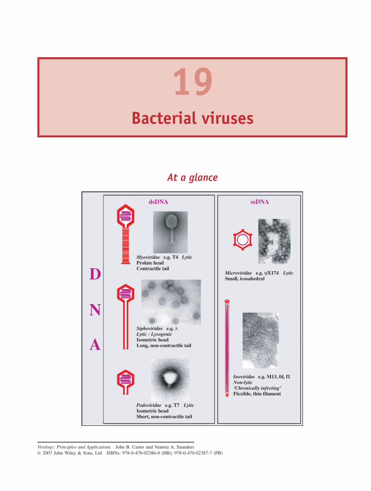
19Bacterial viruses
At a glance
dsDNA ssDNA
Microviridae e.g. ϕX174 LyticSmall, icosahedral
Siphoviridae e.g. λLytic : LysogenicIsometric headLong, non-contractile tail
Podoviridae e.g. T7 LyticIsometric headShort, non-contractile tail
Inoviridae e.g. M13, fd, f1Non-lytic‘Chronically infecting’Flexible, thin filament
D
N
A
Myoviridae e.g. T4 LyticProlate headContractile tail
Virology: Principles and Applications John B. Carter and Venetia A. Saunders 2007 John Wiley & Sons, Ltd ISBNs: 978-0-470-02386-0 (HB); 978-0-470-02387-7 (PB)
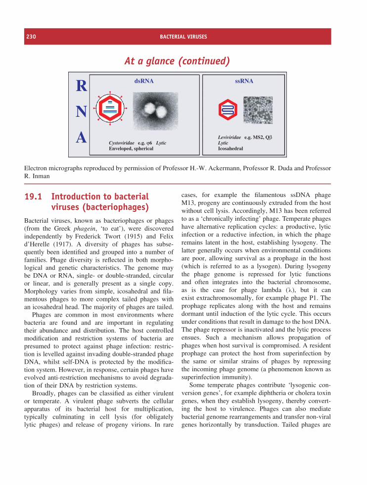
230 BACTERIAL VIRUSES
At a glance (continued)
dsRNA ssRNA
Cystoviridae e.g. ϕ6 LyticEnveloped, spherical
Leviviridae e.g. MS2, QβLyticIcosahedral
R
N
A
Electron micrographs reproduced by permission of Professor H.-W. Ackermann, Professor R. Duda and ProfessorR. Inman
19.1 Introduction to bacterialviruses (bacteriophages)
Bacterial viruses, known as bacteriophages or phages(from the Greek phagein, ‘to eat’), were discoveredindependently by Frederick Twort (1915) and Felixd’Herelle (1917). A diversity of phages has subse-quently been identified and grouped into a number offamilies. Phage diversity is reflected in both morpho-logical and genetic characteristics. The genome maybe DNA or RNA, single- or double-stranded, circularor linear, and is generally present as a single copy.Morphology varies from simple, icosahedral and fila-mentous phages to more complex tailed phages withan icosahedral head. The majority of phages are tailed.
Phages are common in most environments wherebacteria are found and are important in regulatingtheir abundance and distribution. The host controlledmodification and restriction systems of bacteria arepresumed to protect against phage infection: restric-tion is levelled against invading double-stranded phageDNA, whilst self-DNA is protected by the modifica-tion system. However, in response, certain phages haveevolved anti-restriction mechanisms to avoid degrada-tion of their DNA by restriction systems.
Broadly, phages can be classified as either virulentor temperate. A virulent phage subverts the cellularapparatus of its bacterial host for multiplication,typically culminating in cell lysis (for obligatelylytic phages) and release of progeny virions. In rare
cases, for example the filamentous ssDNA phageM13, progeny are continuously extruded from the hostwithout cell lysis. Accordingly, M13 has been referredto as a ‘chronically infecting’ phage. Temperate phageshave alternative replication cycles: a productive, lyticinfection or a reductive infection, in which the phageremains latent in the host, establishing lysogeny. Thelatter generally occurs when environmental conditionsare poor, allowing survival as a prophage in the host(which is referred to as a lysogen). During lysogenythe phage genome is repressed for lytic functionsand often integrates into the bacterial chromosome,as is the case for phage lambda (λ), but it canexist extrachromosomally, for example phage P1. Theprophage replicates along with the host and remainsdormant until induction of the lytic cycle. This occursunder conditions that result in damage to the host DNA.The phage repressor is inactivated and the lytic processensues. Such a mechanism allows propagation ofphages when host survival is compromised. A residentprophage can protect the host from superinfection bythe same or similar strains of phages by repressingthe incoming phage genome (a phenomenon known assuperinfection immunity).
Some temperate phages contribute ‘lysogenic con-version genes’, for example diphtheria or cholera toxingenes, when they establish lysogeny, thereby convert-ing the host to virulence. Phages can also mediatebacterial genome rearrangements and transfer non-viralgenes horizontally by transduction. Tailed phages are
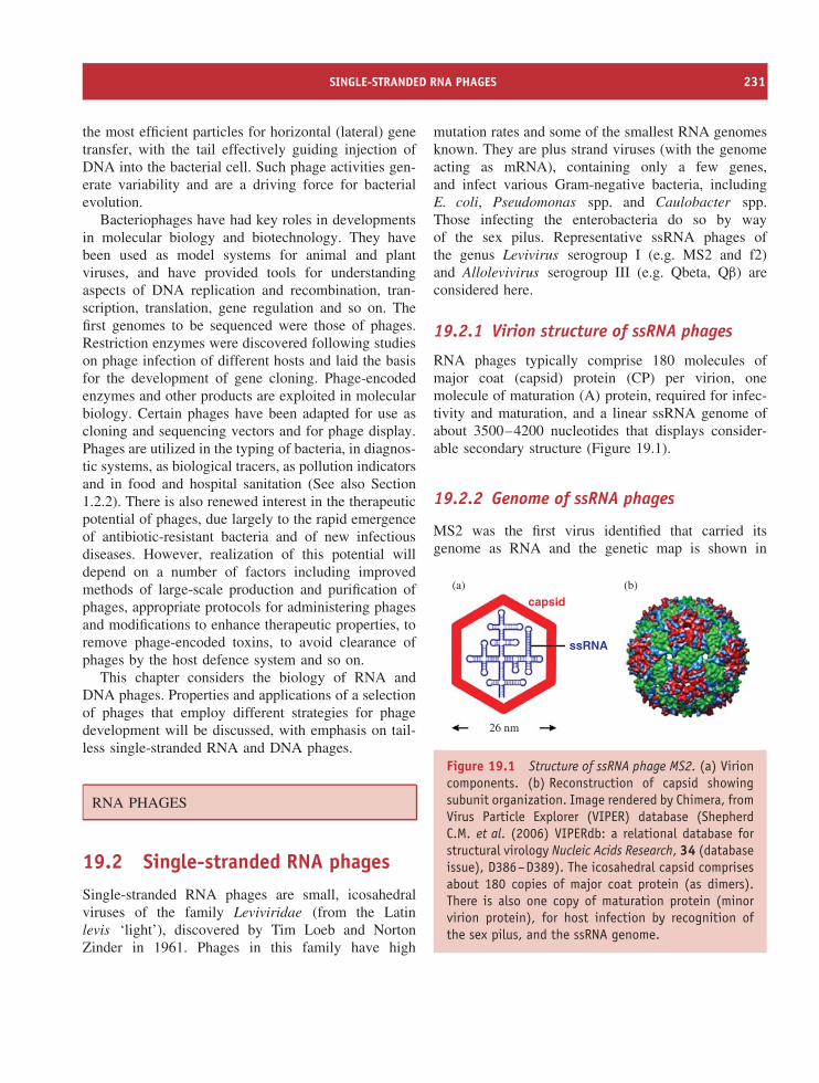
SINGLE-STRANDED RNA PHAGES 231
the most efficient particles for horizontal (lateral) genetransfer, with the tail effectively guiding injection ofDNA into the bacterial cell. Such phage activities gen-erate variability and are a driving force for bacterialevolution.
Bacteriophages have had key roles in developmentsin molecular biology and biotechnology. They havebeen used as model systems for animal and plantviruses, and have provided tools for understandingaspects of DNA replication and recombination, tran-scription, translation, gene regulation and so on. Thefirst genomes to be sequenced were those of phages.Restriction enzymes were discovered following studieson phage infection of different hosts and laid the basisfor the development of gene cloning. Phage-encodedenzymes and other products are exploited in molecularbiology. Certain phages have been adapted for use ascloning and sequencing vectors and for phage display.Phages are utilized in the typing of bacteria, in diagnos-tic systems, as biological tracers, as pollution indicatorsand in food and hospital sanitation (See also Section1.2.2). There is also renewed interest in the therapeuticpotential of phages, due largely to the rapid emergenceof antibiotic-resistant bacteria and of new infectiousdiseases. However, realization of this potential willdepend on a number of factors including improvedmethods of large-scale production and purification ofphages, appropriate protocols for administering phagesand modifications to enhance therapeutic properties, toremove phage-encoded toxins, to avoid clearance ofphages by the host defence system and so on.
This chapter considers the biology of RNA andDNA phages. Properties and applications of a selectionof phages that employ different strategies for phagedevelopment will be discussed, with emphasis on tail-less single-stranded RNA and DNA phages.
RNA PHAGES
19.2 Single-stranded RNA phagesSingle-stranded RNA phages are small, icosahedralviruses of the family Leviviridae (from the Latinlevis ‘light’), discovered by Tim Loeb and NortonZinder in 1961. Phages in this family have high
mutation rates and some of the smallest RNA genomesknown. They are plus strand viruses (with the genomeacting as mRNA), containing only a few genes,and infect various Gram-negative bacteria, includingE. coli, Pseudomonas spp. and Caulobacter spp.Those infecting the enterobacteria do so by wayof the sex pilus. Representative ssRNA phages ofthe genus Levivirus serogroup I (e.g. MS2 and f2)and Allolevivirus serogroup III (e.g. Qbeta, Qβ) areconsidered here.
19.2.1 Virion structure of ssRNA phages
RNA phages typically comprise 180 molecules ofmajor coat (capsid) protein (CP) per virion, onemolecule of maturation (A) protein, required for infec-tivity and maturation, and a linear ssRNA genome ofabout 3500–4200 nucleotides that displays consider-able secondary structure (Figure 19.1).
19.2.2 Genome of ssRNA phages
MS2 was the first virus identified that carried itsgenome as RNA and the genetic map is shown in
(a)
capsid
ssRNA
(b)
26 nm
Figure 19.1 Structure of ssRNA phage MS2. (a) Virioncomponents. (b) Reconstruction of capsid showingsubunit organization. Image rendered by Chimera, fromVirus Particle Explorer (VIPER) database (ShepherdC.M. et al. (2006) VIPERdb: a relational database forstructural virology Nucleic Acids Research, 34 (databaseissue), D386–D389). The icosahedral capsid comprisesabout 180 copies of major coat protein (as dimers).There is also one copy of maturation protein (minorvirion protein), for host infection by recognition ofthe sex pilus, and the ssRNA genome.
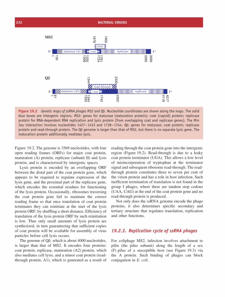
232 BACTERIAL VIRUSES
replicase
replicase
lysis
maturase (A)
maturase (A2)read-through
3569
17241761
13351308
1301 3395
42174118
1742
2352
13441320
611
1902
1678
2330
1344
5’ 3’
5’ 3’
Qβ
MS2
coat
coat
Figure 19.2 Genetic maps of ssRNA phages MS2 and Qβ. Nucleotide coordinates are shown along the maps. The solidblue boxes are intergenic regions. MS2: genes for maturase (maturation protein); coat (capsid) protein; replicaseprotein for RNA-dependent RNA replication and lysis protein (from overlapping coat and replicase genes). The MinJou interaction involves nucleotides 1427–1433 and 1738–1744. Qβ: genes for maturase; coat protein; replicaseprotein and read-through protein. The Qβ genome is larger than that of MS2, but there is no separate lysis gene. Thematuration protein additionally mediates lysis.
Figure 19.2. The genome is 3569 nucleotides, with fouropen reading frames (ORFs) for major coat protein,maturation (A) protein, replicase (subunit II) and lysisprotein, and is characterized by intergenic spaces.
Lysis protein is encoded by an overlapping ORFbetween the distal part of the coat protein gene, whichappears to be required to regulate expression of thelysis gene, and the proximal part of the replicase gene,which encodes the essential residues for functioningof the lysis protein. Occasionally, ribosomes traversingthe coat protein gene fail to maintain the correctreading frame so that once translation of coat proteinterminates they can reinitiate at the start of the lysisprotein ORF, by shuffling a short distance. Efficiency oftranslation of the lysis protein ORF by such reinitiationis low. Thus only small amounts of lysis protein aresynthesized, in turn guaranteeing that sufficient copiesof coat protein will be available for assembly of virusparticles before cell lysis occurs.
The genome of Qβ, which is about 4000 nucleotides,is larger than that of MS2. It encodes four proteins:coat protein, replicase, maturation (A2) protein, whichalso mediates cell lysis, and a minor coat protein (read-through protein, A1), which is generated as a result of
reading through the coat protein gene into the intergenicregion (Figure 19.2). Read-through is due to a leakycoat protein terminator (UGA). This allows a low levelof misincorporation of tryptophan at the terminatorsignal and subsequent ribosome read-through. The read-through protein constitutes three to seven per cent ofthe virion protein and has a role in host infection. Suchinefficient termination of translation is not found in thegroup I phages, where there are tandem stop codons(UAA, UAG) at the end of the coat protein gene and noread-through protein is produced.
Not only does the ssRNA genome encode the phageproteins, it also determines specific secondary andtertiary structure that regulates translation, replicationand other functions.
19.2.3. Replication cycle of ssRNA phages
For coliphage MS2, infection involves attachment topilin (the pilus subunit) along the length of a sex(F) pilus of a susceptible host (see Figure 19.3) viathe A protein. Such binding of phages can blockconjugation in E. coli.
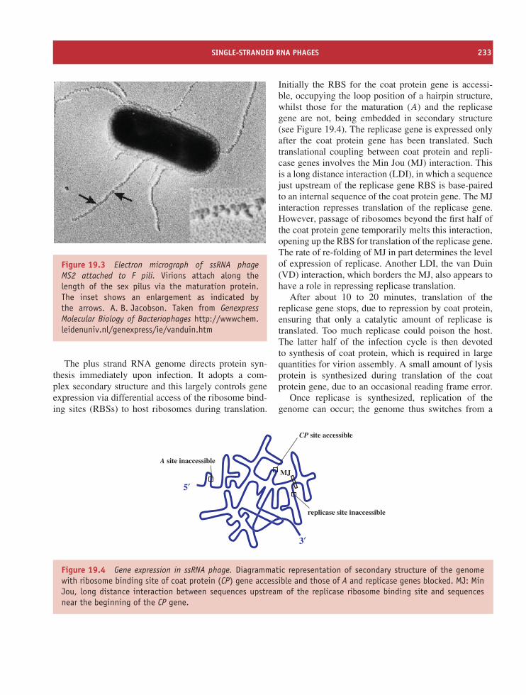
SINGLE-STRANDED RNA PHAGES 233
Figure 19.3 Electron micrograph of ssRNA phageMS2 attached to F pili. Virions attach along thelength of the sex pilus via the maturation protein.The inset shows an enlargement as indicated bythe arrows. A. B. Jacobson. Taken from GenexpressMolecular Biology of Bacteriophages http://wwwchem.leidenuniv.nl/genexpress/ie/vanduin.htm
The plus strand RNA genome directs protein syn-thesis immediately upon infection. It adopts a com-plex secondary structure and this largely controls geneexpression via differential access of the ribosome bind-ing sites (RBSs) to host ribosomes during translation.
Initially the RBS for the coat protein gene is accessi-ble, occupying the loop position of a hairpin structure,whilst those for the maturation (A) and the replicasegene are not, being embedded in secondary structure(see Figure 19.4). The replicase gene is expressed onlyafter the coat protein gene has been translated. Suchtranslational coupling between coat protein and repli-case genes involves the Min Jou (MJ) interaction. Thisis a long distance interaction (LDI), in which a sequencejust upstream of the replicase gene RBS is base-pairedto an internal sequence of the coat protein gene. The MJinteraction represses translation of the replicase gene.However, passage of ribosomes beyond the first half ofthe coat protein gene temporarily melts this interaction,opening up the RBS for translation of the replicase gene.The rate of re-folding of MJ in part determines the levelof expression of replicase. Another LDI, the van Duin(VD) interaction, which borders the MJ, also appears tohave a role in repressing replicase translation.
After about 10 to 20 minutes, translation of thereplicase gene stops, due to repression by coat protein,ensuring that only a catalytic amount of replicase istranslated. Too much replicase could poison the host.The latter half of the infection cycle is then devotedto synthesis of coat protein, which is required in largequantities for virion assembly. A small amount of lysisprotein is synthesized during translation of the coatprotein gene, due to an occasional reading frame error.
Once replicase is synthesized, replication of thegenome can occur; the genome thus switches from a
A site inaccessible
CP site accessible
replicase site inaccessible
MJ
5′
3′
Figure 19.4 Gene expression in ssRNA phage. Diagrammatic representation of secondary structure of the genomewith ribosome binding site of coat protein (CP) gene accessible and those of A and replicase genes blocked. MJ: MinJou, long distance interaction between sequences upstream of the replicase ribosome binding site and sequencesnear the beginning of the CP gene.
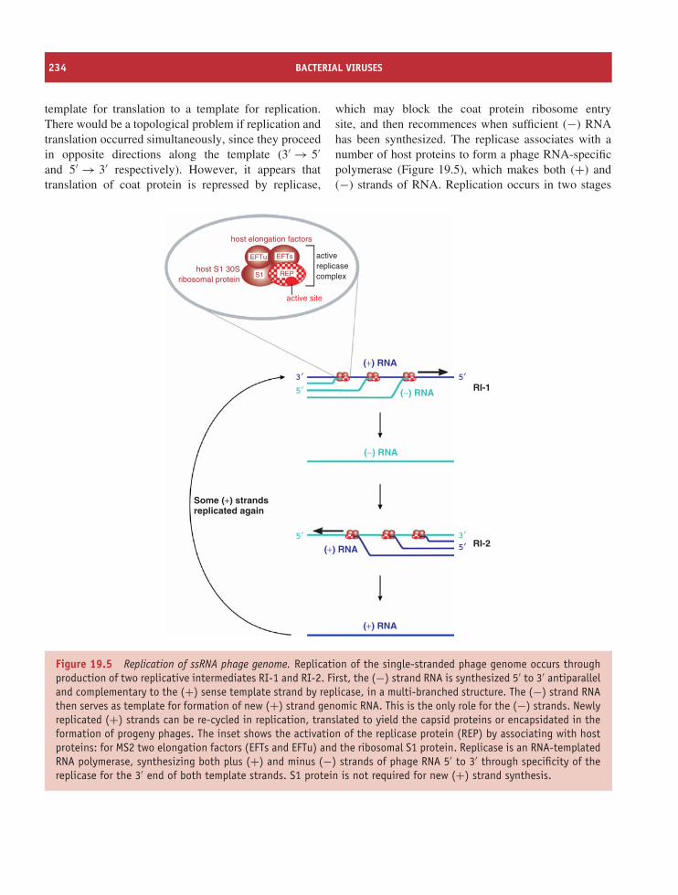
234 BACTERIAL VIRUSES
template for translation to a template for replication.There would be a topological problem if replication andtranslation occurred simultaneously, since they proceedin opposite directions along the template (3′ → 5′and 5′ → 3′ respectively). However, it appears thattranslation of coat protein is repressed by replicase,
which may block the coat protein ribosome entrysite, and then recommences when sufficient (−) RNAhas been synthesized. The replicase associates with anumber of host proteins to form a phage RNA-specificpolymerase (Figure 19.5), which makes both (+) and(−) strands of RNA. Replication occurs in two stages
host elongation factors
host S1 30Sribosomal protein
activereplicasecomplex
active site
(+) RNA
(+) RNA
(+) RNA
(−) RNA
(−) RNA
RI-1
RI-2
5′3′
5′
5′
3′5′
Some (+) strandsreplicated again
EFTu EFTs
S1 REP
Figure 19.5 Replication of ssRNA phage genome. Replication of the single-stranded phage genome occurs throughproduction of two replicative intermediates RI-1 and RI-2. First, the (−) strand RNA is synthesized 5′ to 3′ antiparalleland complementary to the (+) sense template strand by replicase, in a multi-branched structure. The (−) strand RNAthen serves as template for formation of new (+) strand genomic RNA. This is the only role for the (−) strands. Newlyreplicated (+) strands can be re-cycled in replication, translated to yield the capsid proteins or encapsidated in theformation of progeny phages. The inset shows the activation of the replicase protein (REP) by associating with hostproteins: for MS2 two elongation factors (EFTs and EFTu) and the ribosomal S1 protein. Replicase is an RNA-templatedRNA polymerase, synthesizing both plus (+) and minus (−) strands of phage RNA 5′ to 3′ through specificity of thereplicase for the 3′ end of both template strands. S1 protein is not required for new (+) strand synthesis.
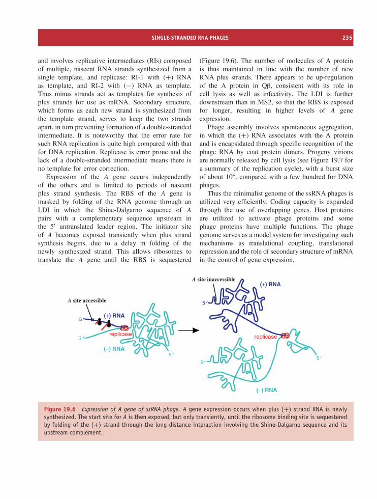
SINGLE-STRANDED RNA PHAGES 235
and involves replicative intermediates (RIs) composedof multiple, nascent RNA strands synthesized from asingle template, and replicase: RI-1 with (+) RNAas template, and RI-2 with (−) RNA as template.Thus minus strands act as templates for synthesis ofplus strands for use as mRNA. Secondary structure,which forms as each new strand is synthesized fromthe template strand, serves to keep the two strandsapart, in turn preventing formation of a double-strandedintermediate. It is noteworthy that the error rate forsuch RNA replication is quite high compared with thatfor DNA replication. Replicase is error prone and thelack of a double-stranded intermediate means there isno template for error correction.
Expression of the A gene occurs independentlyof the others and is limited to periods of nascentplus strand synthesis. The RBS of the A gene ismasked by folding of the RNA genome through anLDI in which the Shine-Dalgarno sequence of A
pairs with a complementary sequence upstream inthe 5′ untranslated leader region. The initiator siteof A becomes exposed transiently when plus strandsynthesis begins, due to a delay in folding of thenewly synthesized strand. This allows ribosomes totranslate the A gene until the RBS is sequestered
(Figure 19.6). The number of molecules of A proteinis thus maintained in line with the number of newRNA plus strands. There appears to be up-regulationof the A protein in Qβ, consistent with its role incell lysis as well as infectivity. The LDI is furtherdownstream than in MS2, so that the RBS is exposedfor longer, resulting in higher levels of A geneexpression.
Phage assembly involves spontaneous aggregation,in which the (+) RNA associates with the A proteinand is encapsidated through specific recognition of thephage RNA by coat protein dimers. Progeny virionsare normally released by cell lysis (see Figure 19.7 fora summary of the replication cycle), with a burst sizeof about 104, compared with a few hundred for DNAphages.
Thus the minimalist genome of the ssRNA phages isutilized very efficiently. Coding capacity is expandedthrough the use of overlapping genes. Host proteinsare utilized to activate phage proteins and somephage proteins have multiple functions. The phagegenome serves as a model system for investigating suchmechanisms as translational coupling, translationalrepression and the role of secondary structure of mRNAin the control of gene expression.
A site accessible
A site inaccessible
replicasereplicase
(+) RNA
(+) RNA
(−) RNA
(−) RNA
5’
5’
5’5’
3’
3’
Figure 19.6 Expression of A gene of ssRNA phage. A gene expression occurs when plus (+) strand RNA is newlysynthesized. The start site for A is then exposed, but only transiently, until the ribosome binding site is sequesteredby folding of the (+) strand through the long distance interaction involving the Shine-Dalgarno sequence and itsupstream complement.
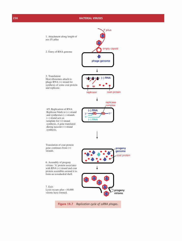
236 BACTERIAL VIRUSES
1. Attachment along length ofsex (F) pilus
2. Entry of RNA genome
3. Translation:Host ribosomes attach tophage RNA (+) strand forsynthesis of some coat proteinand replicase.
4/5. Replication of RNA:Replicase binds to (+) strandand synthesises (−) strands.(−) strand acts astemplate for (+) strandsynthesis. A gene translatedduring nascent (+) strandsynthesis.
Translation of coat proteingene continues from (+)strands.
6. Assembly of progenyvirions: 'A' protein associateswith RNA (+) strand and coatprotein assembles around it toform an icosahedral shell.
7. Exit:Lysis occurs after ~10,000 virions have formed.
(+) RNA
(+) RNA
progenygenome
progenyvirions
phage genome
(−) RNA
3’ 5’
5’
F pilus
empty capsid
replicase coat protein
coat protein
replicasecomplex
Figure 19.7 Replication cycle of ssRNA phages.
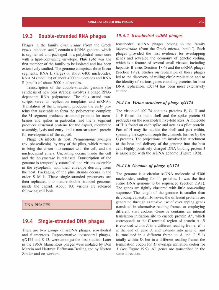
SINGLE-STRANDED DNA PHAGES 237
19.3 Double-stranded RNA phages
Phages in the family Cystoviridae (from the Greekkystis ‘bladder, sack’) contain a dsRNA genome, whichis segmented and packaged in a polyhedral inner corewith a lipid-containing envelope. Phi6 (ϕ6) was thefirst member of the family to be isolated and has beenextensively studied. The genome comprises three linearsegments: RNA L (large) of about 6400 nucleotides,RNA M (medium) of about 4000 nucleotides and RNAS (small) of about 3000 nucleotides.
Transcription of the double-stranded genome (forsynthesis of new plus strands) involves a phage RNA-dependent RNA polymerase. The plus strand tran-scripts serve as replication templates and mRNAs.Translation of the L segment produces the early pro-teins that assemble to form the polymerase complex;the M segment produces structural proteins for mem-branes and spikes in particular, and the S segmentproduces structural proteins for the capsid, membraneassembly, lysis and entry, and a non-structural proteinfor envelopment of the capsid.
Phage ϕ6 infects its host, Pseudomonas syringae(pv. phaseolicola), by way of the pilus, which retractsto bring the virion into contact with the cell, and thenucleocapsid enters. Uncoating occurs inside the celland the polymerase is released. Transcription of thegenome is temporally controlled and virions assemblein the cytoplasm, with their envelope deriving fromthe host. Packaging of the plus strands occurs in theorder S–M–L. These single-stranded precursors arethen replicated into mature double-stranded genomesinside the capsid. About 100 virions are releasedfollowing cell lysis.
DNA PHAGES
19.4 Single-stranded DNA phages
There are two groups of ssDNA phages, icosahedraland filamentous. Representative icosahedral phages,ϕX174 and S-13, were amongst the first studied. Laterin the 1960s filamentous phages were isolated by DonMarvin and Hartmut Hoffmann-Berling and by NortonZinder and co-workers.
19.4.1 Icosahedral ssDNA phages
Icosahedral ssDNA phages belong to the familyMicroviridae (from the Greek micros, ‘small’). Suchphages provided the first evidence for overlappinggenes and revealed the economy of genetic coding,which is a feature of several small viruses, includinghepatitis B virus (Section 18.6) and the ssRNA phages(Section 19.2). Studies on replication of these phagesled to the discovery of rolling circle replication and tothe identity of various genes encoding proteins for hostDNA replication. ϕX174 has been most extensivelystudied.
19.4.1.a Virion structure of phage ϕX174
The virion of ϕX174 contains proteins F, G, H andJ; F forms the main shell and the spike protein Gprotrudes on the icosahedral five-fold axes. A moleculeof H is found on each spike and acts as a pilot protein.Part of H may lie outside the shell and part within,spanning the capsid through the channels formed by theG proteins. The projections are involved in attachmentto the host and delivery of the genome into the hostcell. Highly positively charged DNA binding protein Jis associated with the ssDNA genome (Figure 19.8).
19.4.1.b Genome of phage ϕX174
The genome is a circular ssDNA molecule of 5386nucleotides, coding for 11 proteins. It was the firstentire DNA genome to be sequenced (Section 2.9.1).The genes are tightly clustered with little non-codingsequence. The length of the genome is smaller thanits coding capacity. However, the different proteins aregenerated through extensive use of overlapping genestranslated in alternative reading frames or employingdifferent start codons. Gene A contains an internaltranslation initiation site to encode protein A*, whichcorresponds to the C-terminal region of protein A; B
is encoded within A in a different reading frame; K isat the end of gene A and extends into gene C andis translated in a different frame to A and C; E istotally within D, but in a different reading frame; thetermination codon for D overlaps initiation codon forJ (see Figure 19.9). All genes are transcribed in thesame direction.
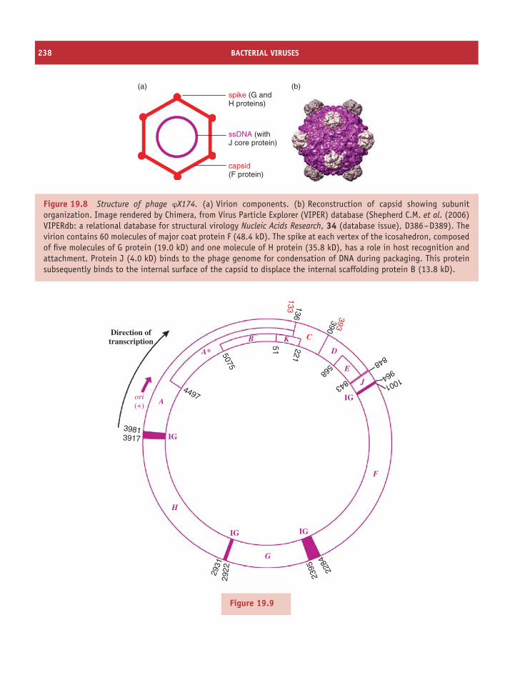
238 BACTERIAL VIRUSES
(b)spike (G andH proteins)
ssDNA (with J core protein)
capsid(F protein)
(a)
Figure 19.8 Structure of phage ϕX174. (a) Virion components. (b) Reconstruction of capsid showing subunitorganization. Image rendered by Chimera, from Virus Particle Explorer (VIPER) database (Shepherd C.M. et al. (2006)VIPERdb: a relational database for structural virology Nucleic Acids Research, 34 (database issue), D386–D389). Thevirion contains 60 molecules of major coat protein F (48.4 kD). The spike at each vertex of the icosahedron, composedof five molecules of G protein (19.0 kD) and one molecule of H protein (35.8 kD), has a role in host recognition andattachment. Protein J (4.0 kD) binds to the phage genome for condensation of DNA during packaging. This proteinsubsequently binds to the internal surface of the capsid to displace the internal scaffolding protein B (13.8 kD).
51 221136
390393
C
133
848964 1001
5075
4497
39813917
2931
2922
2395 22
84568
843
Direction oftranscription
ori(+) A
B
H
K
D
E
J
F
G
A*
IG
IG IG
IG
Figure 19.9
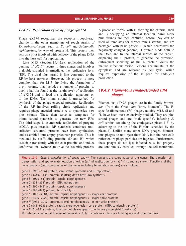
SINGLE-STRANDED DNA PHAGES 239
19.4.1.c Replication cycle of phage ϕX174
Phage ϕX174 recognizes the receptor lipopolysac-charide in the outer membrane of rough strains ofEnterobacteriaceae, such as E. coli and Salmonellatyphimurium, by way of protein H. This protein thenacts as a pilot involved with delivery of the phage DNAinto the host cell for replication.
Like M13 (Section 19.4.2.c), replication of thegenome of ϕX174 occurs in three stages and involvesa double-stranded intermediate, the replicative form(RF). The viral plus strand is first converted to theRF by host enzymes. However, this process is morecomplex than for M13, involving the formation ofa primosome, that includes a number of proteins toopen a hairpin found at the origin (ori ) of replicationof ϕX174 and to load the replication apparatus onto the DNA. The minus strand is transcribed forsynthesis of the phage-encoded proteins. Replicationof the RF involves rolling circle replication andrequires phage-encoded protein A to synthesize newplus strands. These then serve as templates forminus strand synthesis to generate the new RFs.The third stage is asymmetric replication of progenyssDNA plus strands. RF synthesis continues untilsufficient structural proteins have been synthesizedand assembled into empty precursor particles. This ismediated by scaffolding proteins (D and B), whichassociate transiently with the coat proteins and induceconformational switches to drive the assembly process.
A procapsid forms, with D forming the external shelland B occupying an internal location. Viral DNAplus strands are then captured, before they can beused as templates for further minus strands, and arepackaged with basic protein J (which neutralizes thenegatively charged genome). J protein binds both tothe DNA and to the internal surface of the capsid,displacing the B protein, to generate the provirion.Subsequent shedding of the D protein yields themature infectious virion. Virions accumulate in thecytoplasm and are released by cell lysis, whichrequires expression of the E gene for endolysinproduction.
19.4.2 Filamentous single-stranded DNAphages
Filamentous ssDNA phages are in the family Inoviri-dae (from the Greek ina ‘fibre, filament’). The F-specific filamentous (Ff) phages, notably M13, fd andf1, have been most extensively studied. They are plusstrand phages and are ‘male-specific’, infecting E.coli strains containing the conjugative plasmid F, byadsorbing to the tip of the F pilus (encoded by theplasmid). Unlike many other DNA phages, filamen-tous phages do not inject their DNA into the host cell;rather entire phage particles are ingested. Furthermore,these phages do not lyse infected cells, but progenyare continuously extruded through the cell membrane.
Figure 19.9 Genetic organization of phage ϕX174. The numbers are coordinates of the genes. The direction oftranscription and approximate location of origin (ori) of replication for viral (+) strand are shown. Functions of thegene products (with coordinates of the genes including termination codons) are as follows:
gene A (3981–136) protein, viral strand synthesis and RF replication;gene A∗ (4497–136) protein, shutting down host DNA synthesis;gene B (5075–51) protein, capsid morphogenesis;gene C (133–393) protein, DNA maturation;gene D (390–848) protein, capsid morphogenesis;gene E (568–843) protein, host cell lysis;gene F (1001–2284) protein, capsid morphogenesis – major coat protein;gene G (2395–2922) protein, capsid morphogenesis – major spike protein;gene H (2931–3917) protein, capsid morphogenesis – minor spike protein;gene J (848–964) protein, capsid morphogenesis – core protein (DNA condensing protein);gene K (51–221) protein, function not clear-appears to enhance phage yield (burst size).IG: intergenic region at borders of genes A, J, F, G, H contains a ribosome binding site and other features.
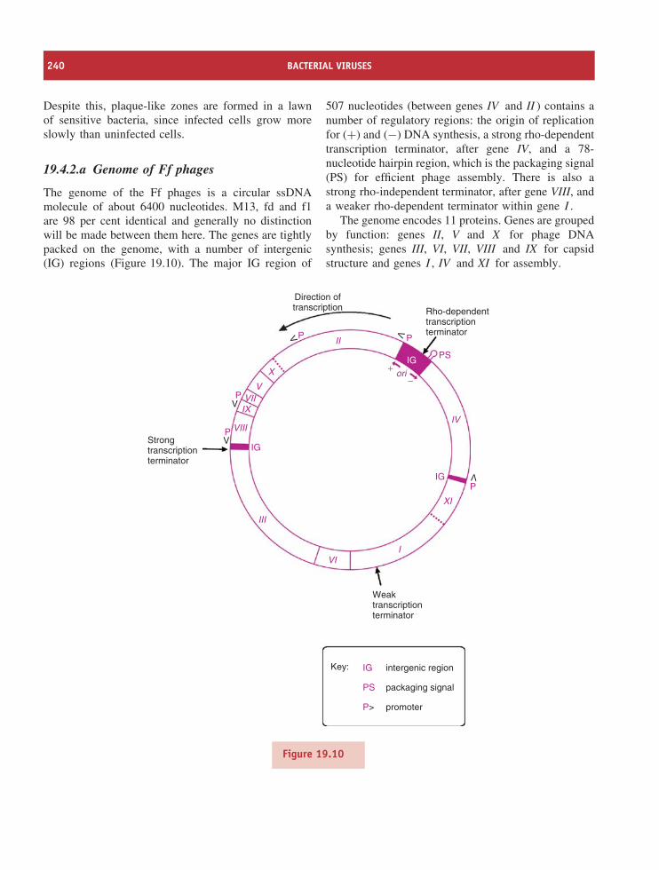
240 BACTERIAL VIRUSES
Despite this, plaque-like zones are formed in a lawnof sensitive bacteria, since infected cells grow moreslowly than uninfected cells.
19.4.2.a Genome of Ff phages
The genome of the Ff phages is a circular ssDNAmolecule of about 6400 nucleotides. M13, fd and f1are 98 per cent identical and generally no distinctionwill be made between them here. The genes are tightlypacked on the genome, with a number of intergenic(IG) regions (Figure 19.10). The major IG region of
507 nucleotides (between genes IV and II ) contains anumber of regulatory regions: the origin of replicationfor (+) and (−) DNA synthesis, a strong rho-dependenttranscription terminator, after gene IV, and a 78-nucleotide hairpin region, which is the packaging signal(PS) for efficient phage assembly. There is also astrong rho-independent terminator, after gene VIII, anda weaker rho-dependent terminator within gene I .
The genome encodes 11 proteins. Genes are groupedby function: genes II, V and X for phage DNAsynthesis; genes III, VI, VII, VIII and IX for capsidstructure and genes I , IV and XI for assembly.
Direction oftranscription
Strongtranscriptionterminator
P VIII
III
VI
XI
IV
I
IX
X
II
VIIV
IG
IG
P
P P
PSIGori
+−
V
V
V
V
P
V
Weaktranscriptionterminator
intergenic region
packaging signal
promoter
IG
PS
P>
Key:
Rho-dependenttranscriptionterminator
Figure 19.10
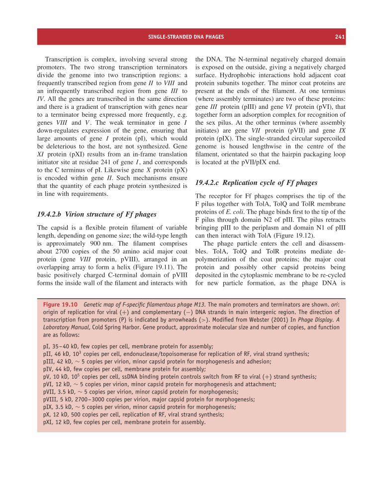
SINGLE-STRANDED DNA PHAGES 241
Transcription is complex, involving several strongpromoters. The two strong transcription terminatorsdivide the genome into two transcription regions: afrequently transcribed region from gene II to VIII andan infrequently transcribed region from gene III toIV. All the genes are transcribed in the same directionand there is a gradient of transcription with genes nearto a terminator being expressed more frequently, e.g.genes VIII and V . The weak terminator in gene I
down-regulates expression of the gene, ensuring thatlarge amounts of gene I protein (pI), which wouldbe deleterious to the host, are not synthesized. GeneXI protein (pXI) results from an in-frame translationinitiator site at residue 241 of gene I , and correspondsto the C terminus of pI. Likewise gene X protein (pX)is encoded within gene II. Such mechanisms ensurethat the quantity of each phage protein synthesized isin line with requirements.
19.4.2.b Virion structure of Ff phages
The capsid is a flexible protein filament of variablelength, depending on genome size; the wild-type lengthis approximately 900 nm. The filament comprisesabout 2700 copies of the 50 amino acid major coatprotein (gene VIII protein, pVIII), arranged in anoverlapping array to form a helix (Figure 19.11). Thebasic positively charged C-terminal domain of pVIIIforms the inside wall of the filament and interacts with
the DNA. The N-terminal negatively charged domainis exposed on the outside, giving a negatively chargedsurface. Hydrophobic interactions hold adjacent coatprotein subunits together. The minor coat proteins arepresent at the ends of the filament. At one terminus(where assembly terminates) are two of these proteins:gene III protein (pIII) and gene VI protein (pVI), thattogether form an adsorption complex for recognition ofthe sex pilus. At the other terminus (where assemblyinitiates) are gene VII protein (pVII) and gene IXprotein (pIX). The single-stranded circular supercoiledgenome is housed lengthwise in the centre of thefilament, orientated so that the hairpin packaging loopis located at the pVII/pIX end.
19.4.2.c Replication cycle of Ff phages
The receptor for Ff phages comprises the tip of theF pilus together with TolA, TolQ and TolR membraneproteins of E. coli. The phage binds first to the tip of theF pilus through domain N2 of pIII. The pilus retractsbringing pIII to the periplasm and domain N1 of pIIIcan then interact with TolA (Figure 19.12).
The phage particle enters the cell and disassem-bles. TolA, TolQ and TolR proteins mediate de-polymerization of the coat proteins; the major coatprotein and possibly other capsid proteins beingdeposited in the cytoplasmic membrane to be re-cycledfor new particle formation, as the phage DNA is
Figure 19.10 Genetic map of F-specific filamentous phage M13. The main promoters and terminators are shown. ori:origin of replication for viral (+) and complementary (−) DNA strands in main intergenic region. The direction oftranscription from promoters (P) is indicated by arrowheads (>). Modified from Webster (2001) In Phage Display. ALaboratory Manual, Cold Spring Harbor. Gene product, approximate molecular size and number of copies, and functionare as follows:
pI, 35–40 kD, few copies per cell, membrane protein for assembly;pII, 46 kD, 103 copies per cell, endonuclease/topoisomerase for replication of RF, viral strand synthesis;pIII, 42 kD, ∼ 5 copies per virion, minor capsid protein for morphogenesis and adhesion;pIV, 44 kD, few copies per cell, membrane protein for assembly;pV, 10 kD, 105 copies per cell, ssDNA binding protein controls switch from RF to viral (+) strand synthesis;pVI, 12 kD, ∼ 5 copies per virion, minor capsid protein for morphogenesis and attachment;pVII, 3.5 kD, ∼ 5 copies per virion, minor capsid protein for morphogenesis;pVIII, 5 kD, 2700–3000 copies per virion, major capsid protein for morphogenesis;pIX, 3.5 kD, ∼ 5 copies per virion, minor capsid protein for morphogenesis;pX, 12 kD, 500 copies per cell, replication of RF, viral strand synthesis;pXI, 12 kD, few copies per cell, membrane protein for assembly.
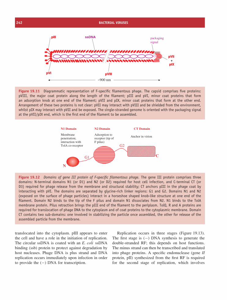
242 BACTERIAL VIRUSES
pVIIIpVIII
pVIIpVII+
pIXpIX
ssDNA
pVIpVI
pIIIpIII packagingsignal
~900 nm
Figure 19.11 Diagrammatic representation of F-specific filamentous phage. The capsid comprises five proteins:pVIII, the major coat protein along the length of the filament; pIII and pVI, minor coat proteins that forman adsorption knob at one end of the filament; pVII and pIX, minor coat proteins that form at the other end.Arrangement of these two proteins is not clear: pVII may interact with pVIII and be shielded from the environment,whilst pIX may interact with pVII and be exposed. The single-stranded genome is oriented with the packaging signalat the pVII/pIX end, which is the first end of the filament to be assembled.
N1 Domain CT Domain
Membranepenetration;interaction withTolA co-receptor
N2 Domain
Adsorption toreceptor (tip ofF pilus)
Anchor in virion
N
CG1
G2
Figure 19.12 Domains of gene III protein of F-specific filamentous phage. The gene III protein comprises threedomains: N-terminal domains N1 (or D1) and N2 (or D2) required for host cell infection, and C-terminal CT (orD3) required for phage release from the membrane and structural stability; CT anchors pIII in the phage coat byinteracting with pVI. The domains are separated by glycine-rich linker regions: G1 and G2. Domains N1 and N2(exposed on the surface of phage particles) interact in a horseshoe shaped knob-like structure at one end of thefilament. Domain N2 binds to the tip of the F pilus and domain N1 dissociates from N2. N1 binds to the TolAmembrane protein. Pilus retraction brings the pIII end of the filament to the periplasm. TolQ, R and A proteins arerequired for translocation of phage DNA to the cytoplasm and of coat proteins to the cytoplasmic membrane. DomainCT contains two sub-domains: one involved in stabilizing the particle once assembled, the other for release of theassembled particle from the membrane.
translocated into the cytoplasm. pIII appears to enterthe cell and have a role in the initiation of replication.The circular ssDNA is coated with an E. coli ssDNAbinding (ssb) protein to protect against degradation byhost nucleases. Phage DNA is plus strand and DNAreplication occurs immediately upon infection in orderto provide the (−) DNA for transcription.
Replication occurs in three stages (Figure 19.13).The first stage is (−) DNA synthesis to generate thedouble-stranded RF; this depends on host functions.The minus strand can then be transcribed and translatedinto phage proteins. A specific endonuclease (gene IIprotein, pII) synthesized from the first RF is requiredfor the second stage of replication, which involves
SINGLE-STRANDED DNA PHAGES 243
multiplication of the RF. The plus strand of the RFis nicked in the major intergenic region, providing aprimer (3′ end) for synthesis of a new plus strand,using the minus strand as template, by rolling circlereplication. As DNA polymerase III extends the 3′end, the old plus strand is displaced, cleaved andcircularized by pII and converted to a dsDNA (RF),which accumulates as a pool in the cell. As DNAreplication proceeds there is concomitant synthesis ofphage proteins. Gene V protein (pV) accumulates inthe cytoplasm and at a critical concentration bindscooperatively to displaced plus strands as they rolloff the circle, diverting them from their template role.This initiates stage 3 of replication, in which thereis a switch from RF to ssDNA synthesis. The 78-nucleotide hairpin region of the plus strand remains freeof pV and initiates the phage assembly reaction. pVadditionally serves as a translational repressor of pII,in turn regulating DNA synthesis. Another protein, pX,also located in the cytoplasm, has a role in synthesisof the single-stranded viral DNA. The nucleoproteincomplex of pV dimers and ssDNA forms a filamentousstructure and is targeted to the cell membrane, whereassembly takes place. Capsid proteins are inserted intothe cytoplasmic membrane as they form.
Assembly proteins pIV, pI and pXI are integralmembrane proteins. pIV forms an extrusion pore inthe outer membrane and interacts with pI and pX1of the inner membrane to form a channel across bothmembranes, large enough to accommodate extrudingphage particles. Assembly occurs where inner and outermembranes appear in close contact at an extrusionsite and requires ATP, proton motive force and thebacterial protein, thioredoxin. pV is displaced by coatproteins, which are packaged around the DNA asthe phage is extruded through the membrane. Theprocess is initiated by interaction of the packagingsignal with pVIII, pVII and pIX, possibly directed bypI. A conformational change induced in pI may allowinteraction with pIV to open the gate in the pIV exitpore. The pVII–pIX end of the particle emerges first.
Elongation proceeds as pV is displaced by pVIII,whose positively charged C-terminal region interactswith the negatively charged sugar–phosphate backboneof the DNA. Termination of assembly involves pIIIand pVI added to the other end of the filament as itis released, the length of the particle being dictated
by the size of the viral DNA. Without pIII, elongationwould continue with pVIII encapsidating another viralDNA to generate a polyphage. Thus pIII and pVIare important in maintaining structural integrity andstability of the virion. Particles are continuouslyextruded without killing the host cell (Figure 19.14).
This assembly process, occurring in the cell mem-brane and involving polymerization of coat proteinsaround the Ff phage genome, is in marked contrast tothat for dsDNA phages, such as T4, which occurs inthe cytoplasm and uses pre-formed heads for packagingDNA (Section 19.5.1).
The filamentous phage CTXϕ, which encodes thecholera toxin (the main virulence factor of Vib-rio cholerae), and a phage associated with invasivemeningococci are related to M13. However, unlikethe coliphage, these phages can establish lysogenyby site-specific integration into the host chromosomein a similar manner to that of the lambdoid phages(Section 19.5.3).
19.4.2.d Cloning vectors and phage display
The relatively small size and non-lytic infection cyclemake the filamentous phages easy to manipulate anduse as cloning vectors. Furthermore, since filament sizeis governed by size of the genomic DNA, insert DNAof variable length can be cloned. Vectors are basedon the RF, which is easily obtained from infectedcells. Cloned ssDNA can be recovered from the phageparticles for use in, for example, DNA sequencing,designing DNA probes and site-directed mutagenesis.
The intergenic (IG) region has been manipulated forthe development of cloning vectors. Joachim Messingand co-workers designed a set of vectors, the M13mp series, by inserting the α-fragment of the E. colilacZ gene containing different polylinkers (multiplecloning sites) into the IG region. Foreign DNA insertedinto one of the cloning sites can be identified byinsertional inactivation of lacZ. Other vectors derivedfrom filamentous phages include phagemids, whichcombine useful features of plasmids and ssDNAphages. Phagemids enable larger inserts than those usedin the M13 mp series to be cloned, double-strandedas plasmids, and amplified and recovered as ssDNAin virions. A phagemid typically contains a selectablemarker, an origin of plasmid replication, a cloning
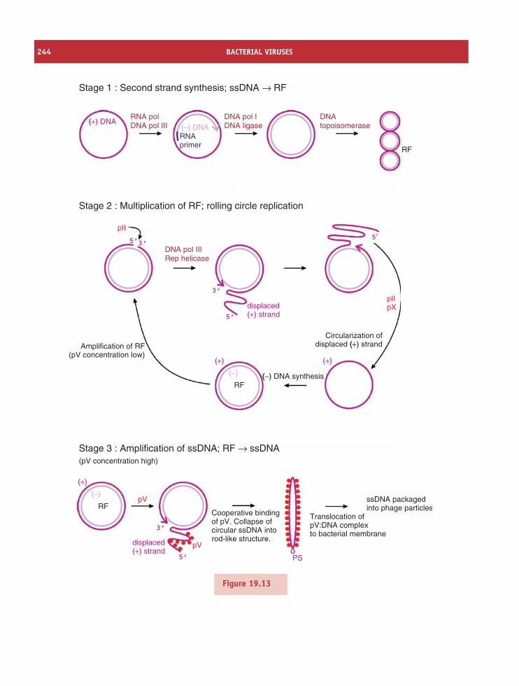
244 BACTERIAL VIRUSES
Stage 1 : Second strand synthesis; ssDNA → RF
Stage 2 : Multiplication of RF; rolling circle replication
Stage 3 : Amplification of ssDNA; RF → ssDNA
RFpV
pV
3’
5’
(pV concentration high)
Cooperative bindingof pV. Collapse ofcircular ssDNA intorod-like structure.
Translocation ofpV:DNA complexto bacterial membrane
PS
ssDNA packagedinto phage particles
pII
((+) DNARNA polDNA pol III
DNA pol IIIRep helicase
displaced(+) strand
(−) DNARNAprimer
pIIpX
5′
RF
(+)
(+)
(−)
(+)
(−) ((−) DNA synthesis
Circularization ofdisplaced ((+) strandAmplification of RF
(pV concentration low)
5’
5’
3’
3’
DNA pol IDNA ligase
DNAtopoisomerase
RF
displaced(+) strand
Figure 19.13
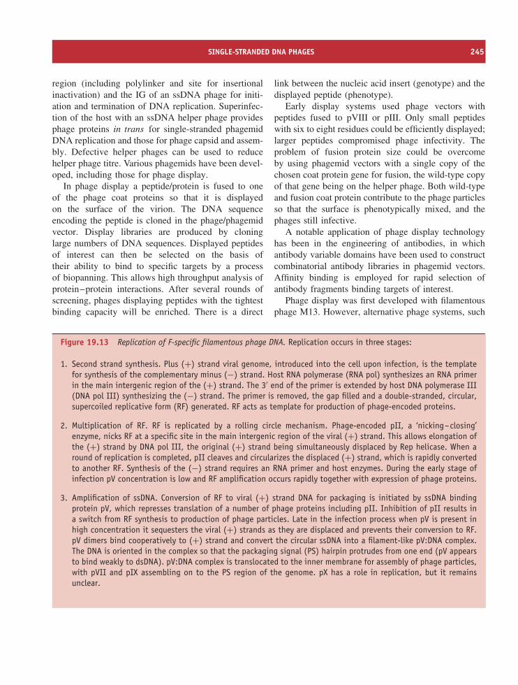
SINGLE-STRANDED DNA PHAGES 245
region (including polylinker and site for insertionalinactivation) and the IG of an ssDNA phage for initi-ation and termination of DNA replication. Superinfec-tion of the host with an ssDNA helper phage providesphage proteins in trans for single-stranded phagemidDNA replication and those for phage capsid and assem-bly. Defective helper phages can be used to reducehelper phage titre. Various phagemids have been devel-oped, including those for phage display.
In phage display a peptide/protein is fused to oneof the phage coat proteins so that it is displayedon the surface of the virion. The DNA sequenceencoding the peptide is cloned in the phage/phagemidvector. Display libraries are produced by cloninglarge numbers of DNA sequences. Displayed peptidesof interest can then be selected on the basis oftheir ability to bind to specific targets by a processof biopanning. This allows high throughput analysis ofprotein–protein interactions. After several rounds ofscreening, phages displaying peptides with the tightestbinding capacity will be enriched. There is a direct
link between the nucleic acid insert (genotype) and thedisplayed peptide (phenotype).
Early display systems used phage vectors withpeptides fused to pVIII or pIII. Only small peptideswith six to eight residues could be efficiently displayed;larger peptides compromised phage infectivity. Theproblem of fusion protein size could be overcomeby using phagemid vectors with a single copy of thechosen coat protein gene for fusion, the wild-type copyof that gene being on the helper phage. Both wild-typeand fusion coat protein contribute to the phage particlesso that the surface is phenotypically mixed, and thephages still infective.
A notable application of phage display technologyhas been in the engineering of antibodies, in whichantibody variable domains have been used to constructcombinatorial antibody libraries in phagemid vectors.Affinity binding is employed for rapid selection ofantibody fragments binding targets of interest.
Phage display was first developed with filamentousphage M13. However, alternative phage systems, such
Figure 19.13 Replication of F-specific filamentous phage DNA. Replication occurs in three stages:
1. Second strand synthesis. Plus (+) strand viral genome, introduced into the cell upon infection, is the templatefor synthesis of the complementary minus (−) strand. Host RNA polymerase (RNA pol) synthesizes an RNA primerin the main intergenic region of the (+) strand. The 3′ end of the primer is extended by host DNA polymerase III(DNA pol III) synthesizing the (−) strand. The primer is removed, the gap filled and a double-stranded, circular,supercoiled replicative form (RF) generated. RF acts as template for production of phage-encoded proteins.
2. Multiplication of RF. RF is replicated by a rolling circle mechanism. Phage-encoded pII, a ‘nicking–closing’enzyme, nicks RF at a specific site in the main intergenic region of the viral (+) strand. This allows elongation ofthe (+) strand by DNA pol III, the original (+) strand being simultaneously displaced by Rep helicase. When around of replication is completed, pII cleaves and circularizes the displaced (+) strand, which is rapidly convertedto another RF. Synthesis of the (−) strand requires an RNA primer and host enzymes. During the early stage ofinfection pV concentration is low and RF amplification occurs rapidly together with expression of phage proteins.
3. Amplification of ssDNA. Conversion of RF to viral (+) strand DNA for packaging is initiated by ssDNA bindingprotein pV, which represses translation of a number of phage proteins including pII. Inhibition of pII results ina switch from RF synthesis to production of phage particles. Late in the infection process when pV is present inhigh concentration it sequesters the viral (+) strands as they are displaced and prevents their conversion to RF.pV dimers bind cooperatively to (+) strand and convert the circular ssDNA into a filament-like pV:DNA complex.The DNA is oriented in the complex so that the packaging signal (PS) hairpin protrudes from one end (pV appearsto bind weakly to dsDNA). pV:DNA complex is translocated to the inner membrane for assembly of phage particles,with pVII and pIX assembling on to the PS region of the genome. pX has a role in replication, but it remainsunclear.
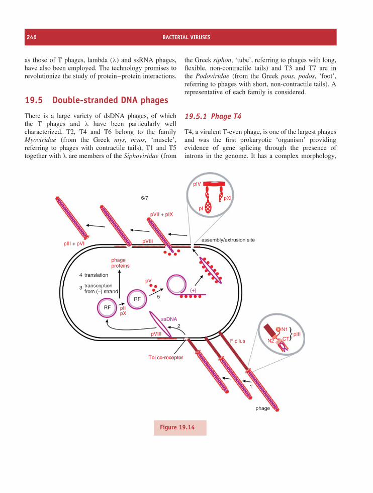
246 BACTERIAL VIRUSES
as those of T phages, lambda (λ) and ssRNA phages,have also been employed. The technology promises torevolutionize the study of protein–protein interactions.
19.5 Double-stranded DNA phages
There is a large variety of dsDNA phages, of whichthe T phages and λ have been particularly wellcharacterized. T2, T4 and T6 belong to the familyMyoviridae (from the Greek mys, myos, ‘muscle’,referring to phages with contractile tails), T1 and T5together with λ are members of the Siphoviridae (from
the Greek siphon, ‘tube’, referring to phages with long,flexible, non-contractile tails) and T3 and T7 are inthe Podoviridae (from the Greek pous, podos, ‘foot’,referring to phages with short, non-contractile tails). Arepresentative of each family is considered.
19.5.1 Phage T4
T4, a virulent T-even phage, is one of the largest phagesand was the first prokaryotic ‘organism’ providingevidence of gene splicing through the presence ofintrons in the genome. It has a complex morphology,
pIII + pVI
pVII + pIX
pVIII
pIV
pXI
pI
6/7
assembly/extrusion site
phageproteins
translation
transcriptionfrom (−) strand (+)
5
2
RF
RF
ssDNA
F pilus
phage
1
pIIIN2 CT
N1
pII
pV
pX
pVIII
Tol co-receptorTol co-receptor
4
3
Figure 19.14
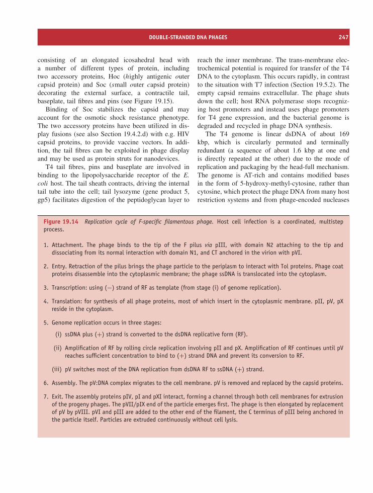
DOUBLE-STRANDED DNA PHAGES 247
consisting of an elongated icosahedral head witha number of different types of protein, includingtwo accessory proteins, Hoc (highly antigenic outercapsid protein) and Soc (small outer capsid protein)decorating the external surface, a contractile tail,baseplate, tail fibres and pins (see Figure 19.15).
Binding of Soc stabilizes the capsid and mayaccount for the osmotic shock resistance phenotype.The two accessory proteins have been utilized in dis-play fusions (see also Section 19.4.2.d) with e.g. HIVcapsid proteins, to provide vaccine vectors. In addi-tion, the tail fibres can be exploited in phage displayand may be used as protein struts for nanodevices.
T4 tail fibres, pins and baseplate are involved inbinding to the lipopolysaccharide receptor of the E.coli host. The tail sheath contracts, driving the internaltail tube into the cell; tail lysozyme (gene product 5,gp5) facilitates digestion of the peptidoglycan layer to
reach the inner membrane. The trans-membrane elec-trochemical potential is required for transfer of the T4DNA to the cytoplasm. This occurs rapidly, in contrastto the situation with T7 infection (Section 19.5.2). Theempty capsid remains extracellular. The phage shutsdown the cell; host RNA polymerase stops recogniz-ing host promoters and instead uses phage promotersfor T4 gene expression, and the bacterial genome isdegraded and recycled in phage DNA synthesis.
The T4 genome is linear dsDNA of about 169kbp, which is circularly permuted and terminallyredundant (a sequence of about 1.6 kbp at one endis directly repeated at the other) due to the mode ofreplication and packaging by the head-full mechanism.The genome is AT-rich and contains modified basesin the form of 5-hydroxy-methyl-cytosine, rather thancytosine, which protect the phage DNA from many hostrestriction systems and from phage-encoded nucleases
Figure 19.14 Replication cycle of F-specific filamentous phage. Host cell infection is a coordinated, multistepprocess.
1. Attachment. The phage binds to the tip of the F pilus via pIII, with domain N2 attaching to the tip anddissociating from its normal interaction with domain N1, and CT anchored in the virion with pVI.
2. Entry. Retraction of the pilus brings the phage particle to the periplasm to interact with Tol proteins. Phage coatproteins disassemble into the cytoplasmic membrane; the phage ssDNA is translocated into the cytoplasm.
3. Transcription: using (−) strand of RF as template (from stage (i) of genome replication).
4. Translation: for synthesis of all phage proteins, most of which insert in the cytoplasmic membrane. pII, pV, pXreside in the cytoplasm.
5. Genome replication occurs in three stages:
(i) ssDNA plus (+) strand is converted to the dsDNA replicative form (RF).
(ii) Amplification of RF by rolling circle replication involving pII and pX. Amplification of RF continues until pVreaches sufficient concentration to bind to (+) strand DNA and prevent its conversion to RF.
(iii) pV switches most of the DNA replication from dsDNA RF to ssDNA (+) strand.
6. Assembly. The pV:DNA complex migrates to the cell membrane. pV is removed and replaced by the capsid proteins.
7. Exit. The assembly proteins pIV, pI and pXI interact, forming a channel through both cell membranes for extrusionof the progeny phages. The pVII/pIX end of the particle emerges first. The phage is then elongated by replacementof pV by pVIII. pVI and pIII are added to the other end of the filament, the C terminus of pIII being anchored inthe particle itself. Particles are extruded continuously without cell lysis.
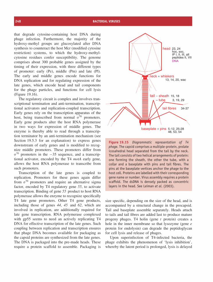
248 BACTERIAL VIRUSES
that degrade cytosine-containing host DNA duringphage infection. Furthermore, the majority of thehydroxy-methyl groups are glucosylated after DNAsynthesis to counteract the host Mcr (modified cytosinerestriction) systems, to which the hydroxy-methyl-cytosine residues confer susceptibility. The genomecomprises about 300 probable genes assigned by thetiming of their expression, with three different typesof promoter: early (Pe), middle (Pm) and late (Pl).The early and middle genes encode functions forDNA replication and for regulating expression of thelate genes, which encode head and tail componentsfor the phage particles, and functions for cell lysis(Figure 19.16).
The regulatory circuit is complex and involves tran-scriptional termination and anti-termination, transcrip-tional activators and replication-coupled transcription.Early genes rely on the transcription apparatus of thehost, being transcribed from normal σ70 promoters.Early gene products alter the host RNA polymerasein two ways for expression of middle genes. Theenzyme is thereby able to read through a transcrip-tion terminator by an anti-termination mechanism (seeSection 19.5.3 for an explanation) to express genesdownstream of early genes and is modified to recog-nize middle promoters. These promoters differ fromσ70 promoters in the −35 sequence, and a transcrip-tional activator, encoded by the T4 motA early gene,allows the host RNA polymerase to transcribe fromsuch promoters.
Transcription of the late genes is coupled toreplication. Promoters for these genes again differfrom σ70 promoters and require an alternative sigmafactor, encoded by T4 regulatory gene 55, to activatetranscription. Binding of gene 55 product to host RNApolymerase allows the enzyme to recognize specificallyT4 late gene promoters. Other T4 gene products,including those of genes 44, 45 and 62, which areinvolved in replication, are additionally required forlate gene transcription. RNA polymerase complexedwith gp55 seems to need an actively replicating T4DNA for effective transcription of the late genes. Suchcoupling between replication and transcription ensuresthat phage DNA becomes available for packaging asthe capsid proteins are synthesized from the late genes.The DNA is packaged into the pre-made heads. Theserequire a protein scaffold to assemble. Packaging is
head 23, 24hoc, soc,IP I, II, III, altpeptides II, VIIDNA
neck + whiskers
baseplate + pins 48, 53, 545-12, 25-28
tail − sheath 15, 183, 19, 29
34-37
− tube
tail fibres
13, 14, 20, wac
Figure 19.15 Diagrammatic representation of T4phage. The capsid comprises a multiple-protein, prolateicosahedral head separated from the tail by the neck.The tail consists of two helical arrangements of protein:one forming the sheath, the other the tube, with acollar and a baseplate with pins and tail fibres. Thepins at the baseplate vertices anchor the phage to thehost cell. Proteins are labelled with their correspondinggene name or number. Virus assembly requires a proteinscaffold. The dsDNA is densely packed as concentriclayers in the head. See Leiman et al. (2003).
size specific, depending on the size of the head, and isaccompanied by a structural change in the procapsid.Tail and baseplate assemble separately. Heads attachto tails and tail fibres are added last to produce matureprogeny phages. T4 holin (gene t protein) creates ahole in the inner membrane so that lysozyme (gene e
protein for endolysin) can degrade the peptidoglycanfor cell lysis and release of phages.
Upon superinfection of T4-infected bacteria, thephage exhibits the phenomenon of ‘lysis inhibition’,whereby the latent period is prolonged, lysis is delayed
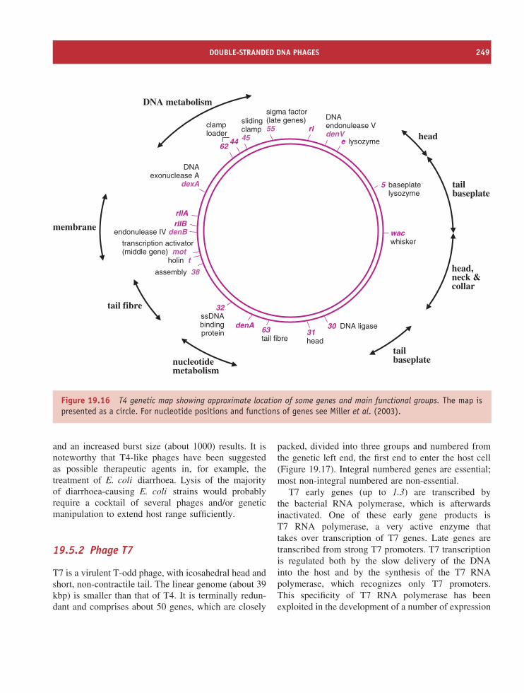
DOUBLE-STRANDED DNA PHAGES 249
DNAexonuclease A
dexA
lysozymee
rI
sigma factor(late genes)55
slidingclamp4544
62
clamploader
rIIArIIB
endonulease IV denB
holin t
transcription activator(middle gene) mot
assembly 38
32ssDNAbindingprotein
denA63tail fibre
31head
30 DNA ligase
DNA metabolism
membrane
tail fibre
nucleotidemetabolism
tailbaseplate
head,neck &collar
head
tailbaseplate
wacwhisker
5 baseplatelysozyme
DNAendonulease VdenV
Figure 19.16 T4 genetic map showing approximate location of some genes and main functional groups. The map ispresented as a circle. For nucleotide positions and functions of genes see Miller et al. (2003).
and an increased burst size (about 1000) results. It isnoteworthy that T4-like phages have been suggestedas possible therapeutic agents in, for example, thetreatment of E. coli diarrhoea. Lysis of the majorityof diarrhoea-causing E. coli strains would probablyrequire a cocktail of several phages and/or geneticmanipulation to extend host range sufficiently.
19.5.2 Phage T7
T7 is a virulent T-odd phage, with icosahedral head andshort, non-contractile tail. The linear genome (about 39kbp) is smaller than that of T4. It is terminally redun-dant and comprises about 50 genes, which are closely
packed, divided into three groups and numbered fromthe genetic left end, the first end to enter the host cell(Figure 19.17). Integral numbered genes are essential;most non-integral numbered are non-essential.
T7 early genes (up to 1.3) are transcribed bythe bacterial RNA polymerase, which is afterwardsinactivated. One of these early gene products isT7 RNA polymerase, a very active enzyme thattakes over transcription of T7 genes. Late genes aretranscribed from strong T7 promoters. T7 transcriptionis regulated both by the slow delivery of the DNAinto the host and by the synthesis of the T7 RNApolymerase, which recognizes only T7 promoters.This specificity of T7 RNA polymerase has beenexploited in the development of a number of expression
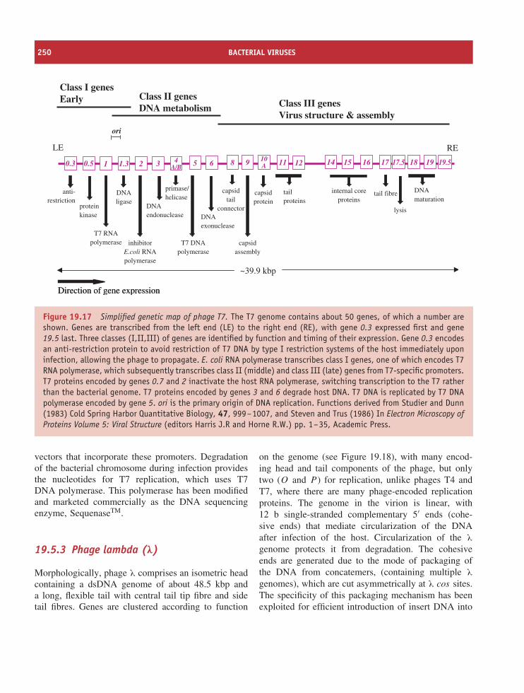
250 BACTERIAL VIRUSES
0.5 1 1.3 2 4A/B 5 6
10A 11 12 15 16 17 18 19 19.5
~39.9 kbp
Direction of gene expressionDirection of gene expression
Class I genesEarly Class II genes
DNA metabolism Class III genesVirus structure & assembly
anti-restriction
T7 RNApolymerase
DNAligase
proteinkinase
ori
LE RE
DNAexonuclease
3
DNAendonuclease
T7 DNApolymerase
tail fibretailproteins
14
internal coreproteins
capsidprotein
inhibitorE.coli RNApolymerase
DNAmaturation
17.5
lysis
9
capsidassembly
primase/helicase
8
capsidtail
connector
0.3
Figure 19.17 Simplified genetic map of phage T7. The T7 genome contains about 50 genes, of which a number areshown. Genes are transcribed from the left end (LE) to the right end (RE), with gene 0.3 expressed first and gene19.5 last. Three classes (I,II,III) of genes are identified by function and timing of their expression. Gene 0.3 encodesan anti-restriction protein to avoid restriction of T7 DNA by type I restriction systems of the host immediately uponinfection, allowing the phage to propagate. E. coli RNA polymerase transcribes class I genes, one of which encodes T7RNA polymerase, which subsequently transcribes class II (middle) and class III (late) genes from T7-specific promoters.T7 proteins encoded by genes 0.7 and 2 inactivate the host RNA polymerase, switching transcription to the T7 ratherthan the bacterial genome. T7 proteins encoded by genes 3 and 6 degrade host DNA. T7 DNA is replicated by T7 DNApolymerase encoded by gene 5. ori is the primary origin of DNA replication. Functions derived from Studier and Dunn(1983) Cold Spring Harbor Quantitative Biology, 47, 999–1007, and Steven and Trus (1986) In Electron Microscopy ofProteins Volume 5: Viral Structure (editors Harris J.R and Horne R.W.) pp. 1–35, Academic Press.
vectors that incorporate these promoters. Degradationof the bacterial chromosome during infection providesthe nucleotides for T7 replication, which uses T7DNA polymerase. This polymerase has been modifiedand marketed commercially as the DNA sequencingenzyme, SequenaseTM.
19.5.3 Phage lambda (λ)
Morphologically, phage λ comprises an isometric headcontaining a dsDNA genome of about 48.5 kbp anda long, flexible tail with central tail tip fibre and sidetail fibres. Genes are clustered according to function
on the genome (see Figure 19.18), with many encod-ing head and tail components of the phage, but onlytwo (O and P ) for replication, unlike phages T4 andT7, where there are many phage-encoded replicationproteins. The genome in the virion is linear, with12 b single-stranded complementary 5′ ends (cohe-sive ends) that mediate circularization of the DNAafter infection of the host. Circularization of the λ
genome protects it from degradation. The cohesiveends are generated due to the mode of packaging ofthe DNA from concatemers, (containing multiple λ
genomes), which are cut asymmetrically at λ cos sites.The specificity of this packaging mechanism has beenexploited for efficient introduction of insert DNA into
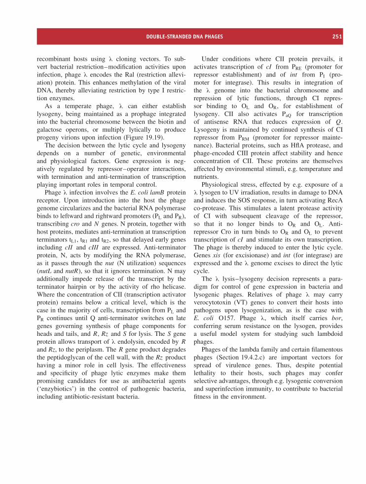
DOUBLE-STRANDED DNA PHAGES 251
recombinant hosts using λ cloning vectors. To sub-vert bacterial restriction–modification activities uponinfection, phage λ encodes the Ral (restriction allevi-ation) protein. This enhances methylation of the viralDNA, thereby alleviating restriction by type I restric-tion enzymes.
As a temperate phage, λ can either establishlysogeny, being maintained as a prophage integratedinto the bacterial chromosome between the biotin andgalactose operons, or multiply lytically to produceprogeny virions upon infection (Figure 19.19).
The decision between the lytic cycle and lysogenydepends on a number of genetic, environmentaland physiological factors. Gene expression is neg-atively regulated by repressor–operator interactions,with termination and anti-termination of transcriptionplaying important roles in temporal control.
Phage λ infection involves the E. coli lamB proteinreceptor. Upon introduction into the host the phagegenome circularizes and the bacterial RNA polymerasebinds to leftward and rightward promoters (PL and PR),transcribing cro and N genes. N protein, together withhost proteins, mediates anti-termination at transcriptionterminators tL1, tR1 and tR2, so that delayed early genesincluding cII and cIII are expressed. Anti-terminatorprotein, N, acts by modifying the RNA polymerase,as it passes through the nut (N utilization) sequences(nutL and nutR), so that it ignores termination. N mayadditionally impede release of the transcript by theterminator hairpin or by the activity of rho helicase.Where the concentration of CII (transcription activatorprotein) remains below a critical level, which is thecase in the majority of cells, transcription from PL andPR continues until Q anti-terminator switches on lategenes governing synthesis of phage components forheads and tails, and R, Rz and S for lysis. The S geneprotein allows transport of λ endolysin, encoded by R
and Rz, to the periplasm. The R gene product degradesthe peptidoglycan of the cell wall, with the Rz producthaving a minor role in cell lysis. The effectivenessand specificity of phage lytic enzymes make thempromising candidates for use as antibacterial agents(‘enzybiotics’) in the control of pathogenic bacteria,including antibiotic-resistant bacteria.
Under conditions where CII protein prevails, itactivates transcription of cI from PRE (promoter forrepressor establishment) and of int from PI (pro-moter for integrase). This results in integration ofthe λ genome into the bacterial chromosome andrepression of lytic functions, through CI repres-sor binding to OL and OR, for establishment oflysogeny. CII also activates PaQ for transcriptionof antisense RNA that reduces expression of Q.Lysogeny is maintained by continued synthesis of CIrepressor from PRM (promoter for repressor mainte-nance). Bacterial proteins, such as HflA protease, andphage-encoded CIII protein affect stability and henceconcentration of CII. These proteins are themselvesaffected by environmental stimuli, e.g. temperature andnutrients.
Physiological stress, effected by e.g. exposure of aλ lysogen to UV irradiation, results in damage to DNAand induces the SOS response, in turn activating RecAco-protease. This stimulates a latent protease activityof CI with subsequent cleavage of the repressor,so that it no longer binds to OR and OL. Anti-repressor Cro in turn binds to OR and OL to preventtranscription of cI and stimulate its own transcription.The phage is thereby induced to enter the lytic cycle.Genes xis (for excisionase) and int (for integrase) areexpressed and the λ genome excises to direct the lyticcycle.
The λ lysis–lysogeny decision represents a para-digm for control of gene expression in bacteria andlysogenic phages. Relatives of phage λ may carryverocytotoxin (VT) genes to convert their hosts intopathogens upon lysogenization, as is the case withE. coli O157. Phage λ, which itself carries bor,conferring serum resistance on the lysogen, providesa useful model system for studying such lambdoidphages.
Phages of the lambda family and certain filamentousphages (Section 19.4.2.c) are important vectors forspread of virulence genes. Thus, despite potentiallethality to their hosts, such phages may conferselective advantages, through e.g. lysogenic conversionand superinfection immunity, to contribute to bacterialfitness in the environment.
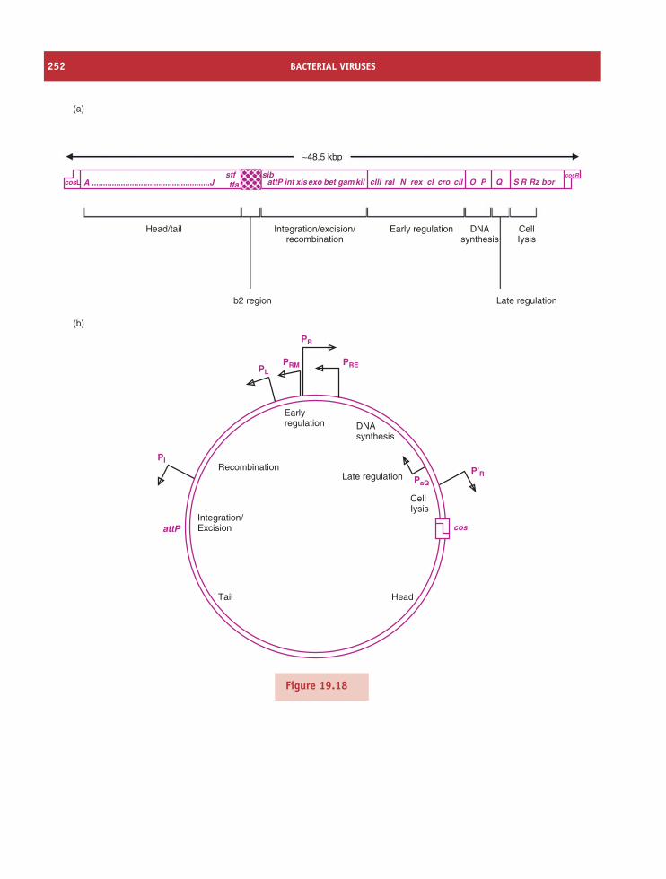
252 BACTERIAL VIRUSES
~48.5 kbp
sibstftfa attP
Head/tail
(a)
Integration/excision/recombination
b2 region Late regulation
Early regulation DNAsynthesis
CellIysis
int xisexo bet gam kil clll ral cl cro cll O P Q S R Rz borrexNcosL A .....................................................JcosR
(b)
PR
PL
PI
PaQ
P'RRecombination
Tail
Integration/ExcisionattP
Head
Earlyregulation
CellIysis
DNAsynthesis
Late regulation
PRM PRE
cos
Figure 19.18
DOUBLE-STRANDED DNA PHAGES 253
Figure 19.18 Genetic organization in phage lambda. (a) Simplified genetic map (non-integrated). Some of theessential λ genes are shown. Gene products/functions are as follows: A to J, head and tail proteins and assembly;stf and tfa, side tail fibres; b2 region, non-essential genes, part of the region replaced in λ cloning vectors; sib,regulatory sequence for int and xis expression – int can be transcribed from PI and PL. Expression from PL is inhibitedwhen sib is present. PL transcript terminates beyond sib and the site forms a secondary structure (hairpin) that isrecognized for cleavage by RNAse III, removing the int portion (= a form of retroregulation). PI transcript, whichterminates at tI (upstream of sib), is stable and int expressed. PL transcript from integrated prophage DNA is alsostable, since sib is removed, and int and xis expressed. attP (P.P′ or a.a′), λ attachment site – location of site-specificrecombination (15 b homology) with host chromosome integration site attB (B.B′ or b.b′) between gal and biooperons; int, integrase for integration and with xis λ prophage excision; xis, excisionase for λ prophage excision; exo,5′ to 3′ exonuclease for recombination; bet, promotes annealing of complementary ssDNA strands for recombination;gam, inhibits host RecBCD nuclease; kil, host killing by blocking cell division; cIII, required for establishment oflysogeny, CIII blocks degradation of CII by host proteases (HflA and HflB); ral (restriction alleviation), for partialalleviation of host B and K restriction systems; N, anti-terminator protein promotes expression of delayed early genes,causes RNA polymerase to overcome transcription terminators and not stop transcription, requires nut (N utilization)site and host factors; rex [rexB/rexA] (restriction of exogenous phage), blocks superinfection by phage such as P22,expressed in the lysogen; cI, λ repressor – binds to OL and OR to repress transcription from PL and PR, activatestranscription from PRM when bound to OR2, cI expressed from PRE to establish lysogeny and from PRM to maintainlysogeny; cro, anti-repressor – binds to OL and OR to repress early transcription, block activation of PRM and promotethe lytic cycle; cII, regulatory protein for establishment of lysogeny, activates transcription from PRE for CI synthesis,PI for integrase synthesis, PaQ for antisense Q resulting in reduced expression of Q. Stability of CII controlled byhost proteases and CIII. O, P, proteins for phage DNA replication; Q, anti-terminator protein promotes expression oflate genes from P′
R, requires qut site; S, holin protein, creates holes in cytoplasmic membrane permitting endolysin(R product) to gain access to periplasm for cell lysis – two forms of S protein are translated (from two in-frametranslation start sites) and the ratio determines the timing of lysis to ensure progeny phages are ready for releasebefore lysis occurs. R, an endolysin – a transglycosylase that digests peptidoglycan for cell lysis; Rz, for cell lysis;bor, lipoprotein in the membrane of λ lysogen conferring serum resistance on host. cosL,cosR, cohesive ends(leftand right respectively) – 12 b single-stranded complementary 5′ extensions, anneal to form circular genome in thehost. Note that the integrated prophage DNA is a linear permutation of the virion DNA, such that sib is no longerdownstream of att. (b) Simplified circular map. Circularization of genome by annealing of cohesive ends to form thecos site brings all late genes (cell lysis, head, tail genes) together. Main promoters are shown:
PL promoter leftwardPR promoter rightwardP′
R late promoterPRE promoter for λ repressor establishmentPRM promoter for λ repressor maintenancePI promoter for intPaQ promoter for anti-Q.
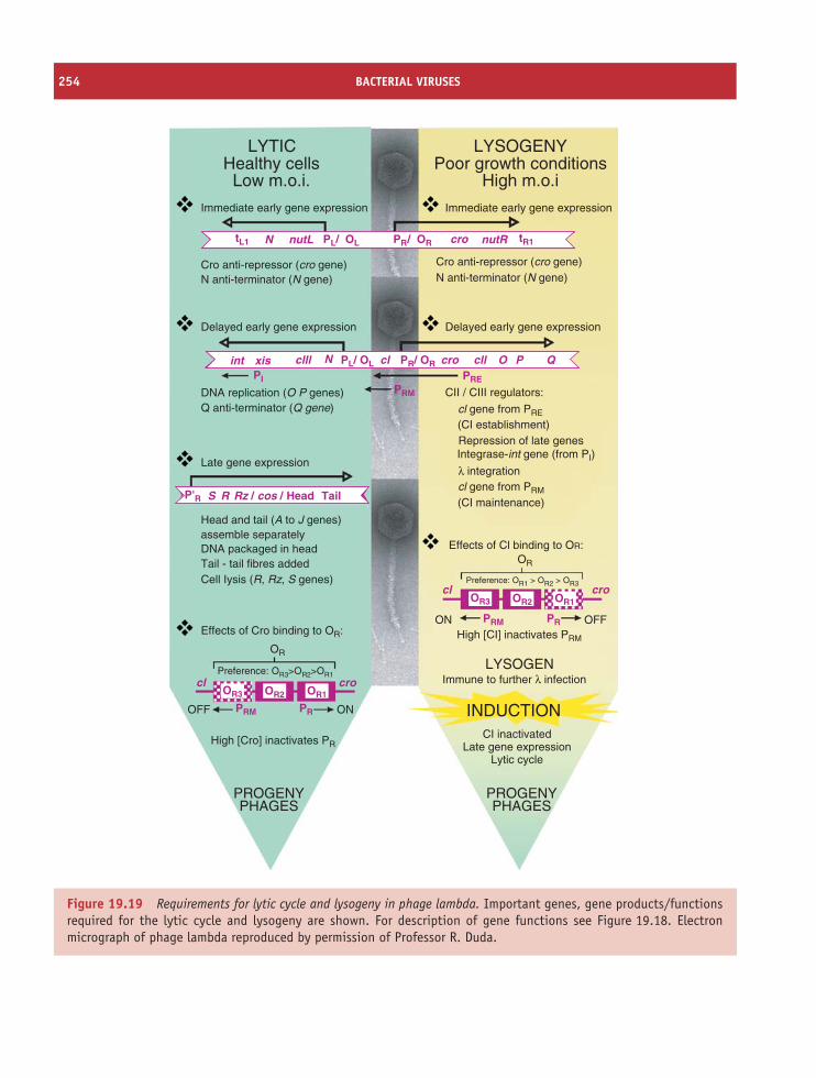
254 BACTERIAL VIRUSES
LYTIC LYSOGENYHealthy cellsLow m.o.i.
Poor growth conditionsHigh m.o.i
Immediate early gene expression
Cro anti-repressor (cro gene)N anti-terminator (N gene)
tL1 tR1N
NPI
PRM
PRE
xisint
nutL
clll cllcl
nutRPL/ OL cro
cro
PR/ OR
DNA replication (O P genes)
DNA packaged in headTail - tail fibres addedCell Iysis (R, Rz, S genes)
Q anti-terminator (Q gene)
Head and tail (A to J genes)assemble separately
Delayed early gene expression
Late gene expression
S R Head TailRz / cos /P'R
Effects of Cro binding to OR:
High [Cro] inactivates PR
High [CI] inactivates PRM
Effects of Cl binding to OR:
CII / CIII regulators:
Delayed early gene expression
Immediate early gene expression
N anti-terminator (N gene)Cro anti-repressor (cro gene)
cl gene from PRE
cl gene from PRM
(CI establishment)
(CI maintenance)
Repression of late genesIntegrase-int gene (from PI)
PROGENYPHAGES
PROGENYPHAGES
CI inactivatedLate gene expression
Lytic cycle
Immune to further λ infection
λ integration
INDUCTION
LYSOGENcl cro
OR
OR2
ONOFF
OR3 OR1
PRPRM
Preference: OR3>OR2>OR1
cl cro
OR
OR2
OFFON
OR3 OR1
PRPRM
Preference: OR1 > OR2 > OR3
PL/ OL PR/ OR O P Q
Figure 19.19 Requirements for lytic cycle and lysogeny in phage lambda. Important genes, gene products/functionsrequired for the lytic cycle and lysogeny are shown. For description of gene functions see Figure 19.18. Electronmicrograph of phage lambda reproduced by permission of Professor R. Duda.
SOURCES OF FURTHER INFORMATION 255
Learning outcomes
By the end of this chapter you should be able to
• discuss the replication cycle and control of geneexpression in ssRNA coliphages;
• outline the infection process of dsRNA phages;
• review the biology of the Ff and icosahedralssDNA phages;
• describe the structure and replication cycle ofdsDNA phages, T4 and T7;
• discuss the molecular events that govern thedecision between the lytic cycle and lysogeny inphage λ;
• explain some uses of phages in modern biotech-nology;
• appreciate the role of phages in the evolution ofbacterial diversity.
Sources of further informationGeneralCalendar R., editor (2006) The Bacteriophages, 2nd edition,
Oxford University PressKutter E. and Sulakvelidze A., editors (2005) Bacterio-
phages – Biology and Applications, CRC PressWackett L. P. (2005) Bacteriophage. An annotated selection
of world wide web sites relevant to the topics inenvironmental microbiology Environmental Microbiology,17, 299–300
Single-stranded RNA phagesvan Duin J. and Tsareva N. (2006) Single-stranded RNA
bacteriophages. In The Bacteriophages, 2nd edition,pp. 175–196 (Calendar R., editor), Oxford UniversityPress
Double-stranded RNA phagesBamford D.H. and Wickner R.B. (1994) Assembly of double-
stranded RNA viruses: bacteriophage φ6 and yeast virusL-A Seminars in Virology, 5, 61–69
Phage ϕX174
LeClerc J. E. (2002) Single-stranded DNA phages. In Mod-ern Microbial Genetics, 2nd edition, pp. 145–176 (StreipsU. N. and Yasbin R. E., editors) Wiley-Liss
Filamentous single-stranded DNA phages
Russel M., Lowman H. B. and Clackson T. (2004) Introduc-tion to phage biology and phage display. In Phage Display:a Practical Approach, pp. 1–26 (Clackson T. and Low-man H. B., editors), Oxford University Press
Webster R. (2001) Filamentous phage biology. In Phage Dis-play. A Laboratory Manual, pp. 1.1–1.37 (Barbas C. F.III, Burton D. R. and Scott, J. K., editors), Cold SpringHarbor Laboratory Press
Phage T4
Leiman P.G. et al. (2003) Structure and morphogenesis ofbacteriophage T4 Cellular and Molecular Life Sciences,60, 2356–2370
Miller E.S. et al. (2003) Bacteriophage T4 genome Microbi-ology and Molecular Biology Reviews, 67, 86–156
Phage T7
Dunn J.J. and Studier F.W. (1983) Complete nucleotidesequence of bacteriophage T7 DNA and locations ofT7 genetic elements Journal Molecular Biology, 166,477–535
Molineux I.J. (2001) No syringes please, ejection of phageT7 DNA from the virion is enzyme driven MolecularMicrobiology, 40, 1–8
Phage lambda
Gottesman M E. and Weisberg R. A. (2004) Little lambda,who made thee? Microbiology and Molecular BiologyReviews, 68, 796–813
Oppenheim A. B. et al. (2005) Switches in bacteriophagelambda development Annual Review of Genetics, 39,409–429
Ptashne M. (2004) A Genetic Switch. Phage Lambda Revis-ited, 3rd edn, Blackwell

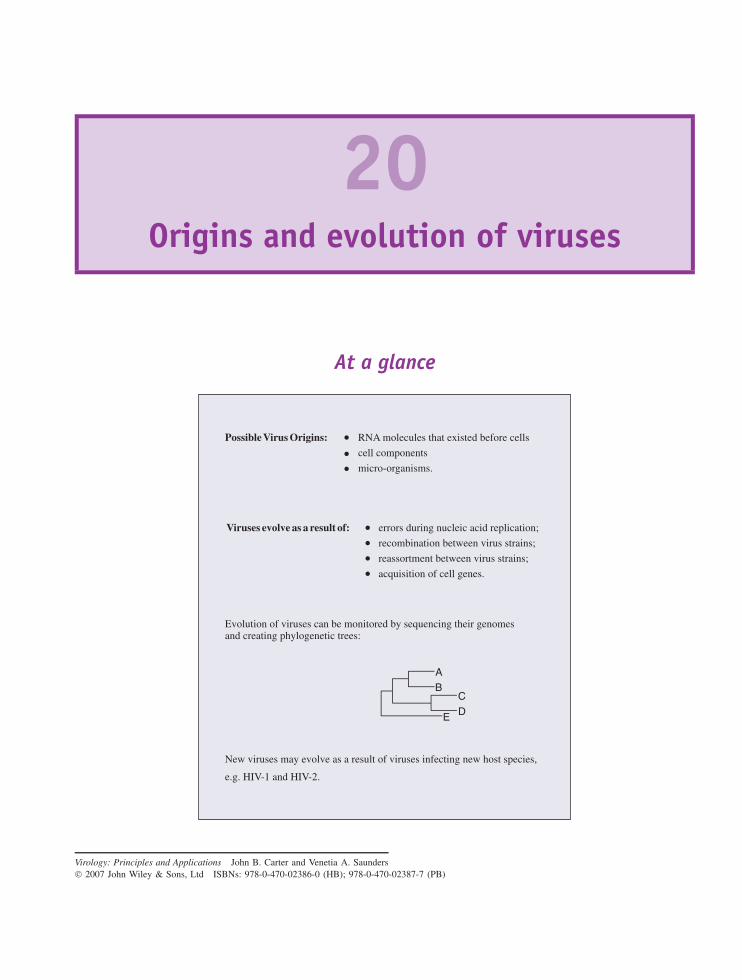
20Origins and evolution of viruses
At a glance
Possible Virus Origins: RNA molecules that existed before cells
cell components
micro-organisms.
Evolution of viruses can be monitored by sequencing their genomesand creating phylogenetic trees:
AB
CDE
Viruses evolve as a result of: errors during nucleic acid replication;
recombination between virus strains;
reassortment between virus strains;
acquisition of cell genes.
New viruses may evolve as a result of viruses infecting new host species,
e.g. HIV-1 and HIV-2.
Virology: Principles and Applications John B. Carter and Venetia A. Saunders 2007 John Wiley & Sons, Ltd ISBNs: 978-0-470-02386-0 (HB); 978-0-470-02387-7 (PB)
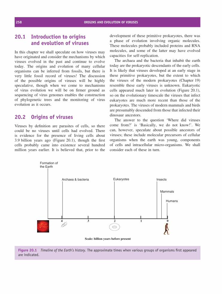
258 ORIGINS AND EVOLUTION OF VIRUSES
20.1 Introduction to originsand evolution of viruses
In this chapter we shall speculate on how viruses mayhave originated and consider the mechanisms by whichviruses evolved in the past and continue to evolvetoday. The origins and evolution of many cellularorganisms can be inferred from fossils, but there isvery little fossil record of viruses! The discussionof the possible origins of viruses will be highlyspeculative, though when we come to mechanismsof virus evolution we will be on firmer ground assequencing of virus genomes enables the constructionof phylogenetic trees and the monitoring of virusevolution as it occurs.
20.2 Origins of virusesViruses by definition are parasites of cells, so therecould be no viruses until cells had evolved. Thereis evidence for the presence of living cells about3.9 billion years ago (Figure 20.1), though the firstcells probably came into existence several hundredmillion years earlier. It is believed that, prior to the
development of these primitive prokaryotes, there wasa phase of evolution involving organic molecules.These molecules probably included proteins and RNAmolecules, and some of the latter may have evolvedcapacities for self-replication.
The archaea and the bacteria that inhabit the earthtoday are the prokaryotic descendants of the early cells.It is likely that viruses developed at an early stage inthese primitive prokaryotes, but the extent to whichthe viruses of the modern prokaryotes (Chapter 19)resemble these early viruses is unknown. Eukaryoticcells appeared much later in evolution (Figure 20.1),so on the evolutionary timescale the viruses that infecteukaryotes are much more recent than those of theprokaryotes. The viruses of modern mammals and birdsare presumably descended from those that infected theirdinosaur ancestors.
The answer to the question ‘Where did virusescome from?’ is ‘Basically, we do not know!’. Wecan, however, speculate about possible ancestors ofviruses; these include molecular precursors of cellularorganisms when the earth was young, componentsof cells and intracellular micro-organisms. We shallconsider each of these in turn.
Archaea & bacteria Eukaryotes Insects
Mammals
01234
Scale: billion years before present
Humans
Formation ofthe Earth
Figure 20.1 Timeline of the Earth’s history. The approximate times when various groups of organisms first appearedare indicated.
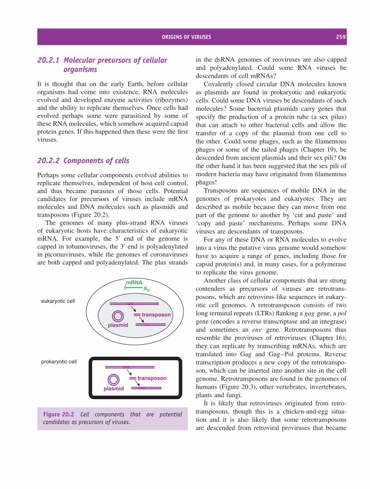
ORIGINS OF VIRUSES 259
20.2.1 Molecular precursors of cellularorganisms
It is thought that on the early Earth, before cellularorganisms had come into existence, RNA moleculesevolved and developed enzyme activities (ribozymes)and the ability to replicate themselves. Once cells hadevolved perhaps some were parasitized by some ofthese RNA molecules, which somehow acquired capsidprotein genes. If this happened then these were the firstviruses.
20.2.2 Components of cells
Perhaps some cellular components evolved abilities toreplicate themselves, independent of host cell control,and thus became parasites of those cells. Potentialcandidates for precursors of viruses include mRNAmolecules and DNA molecules such as plasmids andtransposons (Figure 20.2).
The genomes of many plus-strand RNA virusesof eukaryotic hosts have characteristics of eukaryoticmRNA. For example, the 5′ end of the genome iscapped in tobamoviruses, the 3′ end is polyadenylatedin picornaviruses, while the genomes of coronavirusesare both capped and polyadenylated. The plus strands
mRNAAn
transposon
plasmid
transposon
plasmid
eukaryotic cell
prokaryotic cell
Figure 20.2 Cell components that are potentialcandidates as precursors of viruses.
in the dsRNA genomes of reoviruses are also cappedand polyadenylated. Could some RNA viruses bedescendants of cell mRNAs?
Covalently closed circular DNA molecules knownas plasmids are found in prokaryotic and eukaryoticcells. Could some DNA viruses be descendants of suchmolecules? Some bacterial plasmids carry genes thatspecify the production of a protein tube (a sex pilus)that can attach to other bacterial cells and allow thetransfer of a copy of the plasmid from one cell tothe other. Could some phages, such as the filamentousphages or some of the tailed phages (Chapter 19), bedescended from ancient plasmids and their sex pili? Onthe other hand it has been suggested that the sex pili ofmodern bacteria may have originated from filamentousphages!
Transposons are sequences of mobile DNA in thegenomes of prokaryotes and eukaryotes. They aredescribed as mobile because they can move from onepart of the genome to another by ‘cut and paste’ and‘copy and paste’ mechanisms. Perhaps some DNAviruses are descendants of transposons.
For any of these DNA or RNA molecules to evolveinto a virus the putative virus genome would somehowhave to acquire a range of genes, including those forcapsid protein(s) and, in many cases, for a polymeraseto replicate the virus genome.
Another class of cellular components that are strongcontenders as precursors of viruses are retrotrans-posons, which are retrovirus-like sequences in eukary-otic cell genomes. A retrotransposon consists of twolong terminal repeats (LTRs) flanking a gag gene, a polgene (encodes a reverse transcriptase and an integrase)and sometimes an env gene. Retrotransposons thusresemble the proviruses of retroviruses (Chapter 16);they can replicate by transcribing mRNAs, which aretranslated into Gag and Gag–Pol proteins. Reversetranscription produces a new copy of the retrotranspo-son, which can be inserted into another site in the cellgenome. Retrotransposons are found in the genomes ofhumans (Figure 20.3), other vertebrates, invertebrates,plants and fungi.
It is likely that retroviruses originated from retro-transposons, though this is a chicken-and-egg situa-tion and it is also likely that some retrotransposonsare descended from retroviral proviruses that became
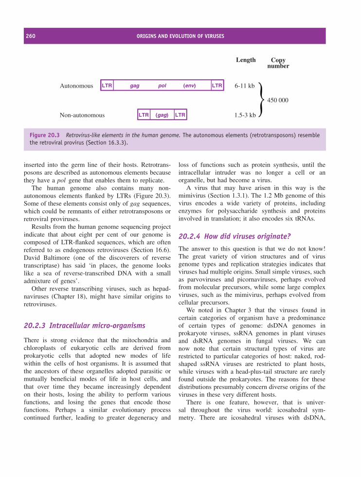
260 ORIGINS AND EVOLUTION OF VIRUSES
Autonomous
Length Copynumber
Non-autonomous
6-11 kb
1.5-3 kb
LTR gag pol (env)
(gag)
LTR
LTRLTR
450 000
Figure 20.3 Retrovirus-like elements in the human genome. The autonomous elements (retrotransposons) resemblethe retroviral provirus (Section 16.3.3).
inserted into the germ line of their hosts. Retrotrans-posons are described as autonomous elements becausethey have a pol gene that enables them to replicate.
The human genome also contains many non-autonomous elements flanked by LTRs (Figure 20.3).Some of these elements consist only of gag sequences,which could be remnants of either retrotransposons orretroviral proviruses.
Results from the human genome sequencing projectindicate that about eight per cent of our genome iscomposed of LTR-flanked sequences, which are oftenreferred to as endogenous retroviruses (Section 16.6).David Baltimore (one of the discoverers of reversetranscriptase) has said ‘in places, the genome lookslike a sea of reverse-transcribed DNA with a smalladmixture of genes’.
Other reverse transcribing viruses, such as hepad-naviruses (Chapter 18), might have similar origins toretroviruses.
20.2.3 Intracellular micro-organisms
There is strong evidence that the mitochondria andchloroplasts of eukaryotic cells are derived fromprokaryotic cells that adopted new modes of lifewithin the cells of host organisms. It is assumed thatthe ancestors of these organelles adopted parasitic ormutually beneficial modes of life in host cells, andthat over time they became increasingly dependenton their hosts, losing the ability to perform variousfunctions, and losing the genes that encode thosefunctions. Perhaps a similar evolutionary processcontinued further, leading to greater degeneracy and
loss of functions such as protein synthesis, until theintracellular intruder was no longer a cell or anorganelle, but had become a virus.
A virus that may have arisen in this way is themimivirus (Section 1.3.1). The 1.2 Mb genome of thisvirus encodes a wide variety of proteins, includingenzymes for polysaccharide synthesis and proteinsinvolved in translation; it also encodes six tRNAs.
20.2.4 How did viruses originate?
The answer to this question is that we do not know!The great variety of virion structures and of virusgenome types and replication strategies indicates thatviruses had multiple origins. Small simple viruses, suchas parvoviruses and picornaviruses, perhaps evolvedfrom molecular precursors, while some large complexviruses, such as the mimivirus, perhaps evolved fromcellular precursors.
We noted in Chapter 3 that the viruses found incertain categories of organism have a predominanceof certain types of genome: dsDNA genomes inprokaryote viruses, ssRNA genomes in plant virusesand dsRNA genomes in fungal viruses. We cannow note that certain structural types of virus arerestricted to particular categories of host: naked, rod-shaped ssRNA viruses are restricted to plant hosts,while viruses with a head-plus-tail structure are rarelyfound outside the prokaryotes. The reasons for thesedistributions presumably concern diverse origins of theviruses in these very different hosts.
There is one feature, however, that is univer-sal throughout the virus world: icosahedral sym-metry. There are icosahedral viruses with dsDNA,
EVOLUTION OF VIRUSES 261
ssDNA, dsRNA and ssRNA genomes, and icosahe-dral viruses infect bacterial, fungal, plant and ani-mal hosts. These viruses have evolved from a varietyof origins, but evolution has chosen the icosahedronfor capsid design because it is a particularly stablestructure.
One final thought on virus origins: we must entertainthe possibility that today new viruses are continuing toevolve from molecular and cellar precursors.
20.3 Evolution of viruses
‘. . . endless forms most beautiful and most won-derful have been, and are being, evolved’ CharlesDarwin (1859).
Charles Darwin’s concepts of over-reproduction andsurvival of the fittest that have been found to apply tocellular organisms also apply to viruses. Like a cellularorganism, a virus has genes that strive to perpetuatethemselves. The expression of the combination ofgenes that constitutes the genome permits the virusto replicate itself. Changes to individual genes andnew gene combinations continually throw up newgenotypes, most of which are less successful than theparental genotypes and do not survive. Occasionally,however, a new genotype is more successful thanthe parental genotype and might even supplant it.Sometimes a new virus genotype enables the infectionof a new host species.
20.3.1 Viruses from the past
The ancestors of modern viruses (‘virus fossils’)are not readily available, but the reverse transcrip-tion–polymerase chain reaction (RT-PCR) has beenused to recover and amplify genomes of at least twoRNA viruses from the past: influenza A virus from the1918–19 pandemic (see Section 20.3.3.c) and tomatomosaic virus. The latter virus is very stable and it canbe detected in the atmosphere. Its presence in Arcticice was predicted, and when cores of ice between 500
and 140 000 years old were examined the RNAs of 15strains of tomato mosaic virus were detected.
20.3.2 Monitoring virus evolution
Comparing sequences of virus genomes allows thedegrees of evolutionary relationship between virusesto be estimated, and even allows changes in rapidlyevolving viruses (e.g. HIV-1) to be followed. Sequencedata can be used to produce phylogenetic trees(Section 10.2.1), which indicate how modern virusstrains may have arisen from common ancestors(Figure 20.4).
20.3.3 Mechanisms of virus evolution
In many respects, the underlying processes that drivevirus evolution are the same as those that drivethe evolution of cellular organisms. These processesinvolve the generation of genome variants, the vastmajority of which are deleterious and do not survive,but a few provide an advantage in a particular niche.For a virus the niche might be a new host species orthe presence of an antiviral drug, and a variant mayproliferate in that niche as a new virus strain. Virusgenome variants arise as a result of mutations, newgene arrangements and acquisition of cell genes; weshall consider each process in turn.
20.3.3.a Mutations
When nucleic acids are copied by polymerases someerrors are made. If an error is in a protein-codingsequence and if the error results in a change inthe amino acid encoded, then the error results in amutation. Natural selection operates and the mutationsthat survive are those that best fit the virus for itscontinued survival.
There are many selection pressures on viruses.These pressures include the immune response of thehost; for example a new antigenic type of an animalvirus that the host’s immune system has not previouslyencountered is at an advantage compared with antigenictypes against which the host has acquired immunity.There is, therefore, heavy selection pressure on virusproteins (e.g. HIV-1 gp120) that are targets of the
262 ORIGINS AND EVOLUTION OF VIRUSES
L’hoest’s
Grivet-AGM Vervet-AGM
Chimp
Chimp
Sun-tailed
Mandrill-1
Mandrill-2Drill
RCM
Greaterspot-nosed
Moustached
Mona
Sabaeus-AGM
HIV-2
HIV-1 O Group
HIV-1 N Group
HIV-1M Group
HIV-2
MacaqueSooty mangabey
Sooty mangabey
Dent’s mona
De Brazza’s
Sykes’s
Guereza colobus
Tantalus-AGM
Figure 20.4 Phylogenetic tree showing relationships between primate lentiviruses based on pol sequences. Simianimmunodeficiency viruses (SIVs) are denoted by the name of the host species in black. Human immunodeficiencyviruses (HIVs) are indicated in red. AGM: African green monkey. RCM: red-capped mangabey. Adapted from Mullerand De Boer (2006) PLoS Pathogens, 2 (3), e15.
host’s immune response, and these proteins tendto be the least conserved. Those regions of theproteins that are targets for neutralizing antibodies areespecially variable. There are, however, constraints onthe evolutionary process. Virus attachment proteinsmust retain the configurations that enable them tobind to cell receptors, and enzymes, such as reversetranscriptases, must retain their catalytic abilities.
For some viruses there are additional constraints. Avirus such as potato yellow dwarf virus, which needsto replicate in both a plant host and an insect host, cantolerate a mutation that enhances its replication in theinsect host only if the mutation does not compromiseits ability to replicate in the plant host, and vice versa.
When virus DNA genomes are replicated, theerror rates are much lower than when RNA genomesare replicated (Figure 20.5). This is because DNA-dependent DNA polymerases have a proofreadingmechanism that corrects most of the errors, whereasRNA polymerases and reverse transcriptases have no
such mechanism. As a result, DNA viruses, such aspapillomaviruses, evolve much more slowly than RNAviruses, such as picornaviruses.
‘RNA viruses deserve their reputation as Nature’sswiftest evolvers’Michael Worobey and Edward Holmes (1999).
The high error rates during RNA replication andreverse transcription mean that for an RNA virus thereis no fixed sequence of bases for the genome. Instead,the virus genome exists as a large number of variants;the term ‘quasispecies’ has been coined to describethe group of variants that collectively constitute thegenome of an RNA virus (Figure 20.6). Many variantshave only a fleeting existence, while those best adaptedto a particular niche dominate in that niche. The highmutation rate produces some variants that may enable a
EVOLUTION OF VIRUSES 263
DNApolymerase
One error per107 to 1011 bases addedDNA Replication
RNApolymerase
RNA ReplicationOne error per103 to 106 bases added
reversetranscriptaseReverse
TranscriptionOne error per103 to 106 bases added
Figure 20.5 Error rates of DNA polymerases and RNA polymerases.
Quasispecies genomes
Figure 20.6 Quasispecies genomes of an RNA virus in an infected human. Virus genomes are represented as lines,and mutations as symbols on the lines. Mutations result in a spectrum of related genomes, termed viral quasispecies.Modified with the permission of the author and the publisher (Springer Verlag) from Domingo et al. (2006) Virusesas quasispecies: biological implications Current Topics in Microbiology and Immunology, Vol. 299, pp. 51–82.
264 ORIGINS AND EVOLUTION OF VIRUSES
virus to adapt to new niches, for example an expansionof its host range, should the opportunities arise.
The first step in the replication of HIV genomesinvolves reverse transcription, with an estimated errorrate around one error per 104 bases added. As the HIVgenome contains about 104 bases, this means that thereis an average of one error each time the genome isreverse transcribed. There is evidence that the humanimmunodeficiency viruses evolved from viruses thatjumped from chimpanzees and sooty mangabeys intohumans (Section 20.3.4). Since jumping species, theviruses have evolved rapidly, especially the M (main)group of HIV-1, which has radiated into many subtypesand sub-subtypes.
Rapid evolution of HIV can be detected within thebody of an infected individual, in which there maybe 1010 –1012 new virions produced each day. Thereis therefore constant production of a huge amount ofgenetic variability. We mentioned in Chapter 17 thatR5 strains of HIV-1 predominate in a newly infectedhost, but in many individuals as the infection proceedsthere is evolution towards X4 and R5X4 strains. Theconstant production of new variants also means thatthe virus can evolve rapidly in response to a selectionpressure, such as the presence of an anti-retroviral drug.
High diversity is also found in the genomes of otherRNA viruses, such as hepatitis C virus, where isolatesfrom a patient can have up to five per cent differencein their nucleotide sequences.
The capacity for rapid evolution of RNA virusescreates problems for the development and maintenanceof some vaccination programmes. Vaccination withattenuated poliovirus vaccine initiates infection inthe gut with one or more of the vaccine strains,and the infection normally lasts for 1–2 months.Some immunodeficient vaccinees, however, remaininfected for much longer periods of time, and in theseindividuals the vaccine viruses almost always evolve.There are differences of only a few bases between thegenomes of neurovirulent poliovirus strains and thoseof the vaccine strains (Section 24.2), so reversion toneurovirulence is common in these chronically infectedindividuals, who may develop paralytic poliomyelitisand may transmit virulent poliovirus to others.
Another example of RNA virus variability havingpractical consequences for vaccination is foot andmouth disease virus. Seven distinct serotypes of
this virus have evolved, and vaccination against oneserotype does not provide protection against the others.In addition, there are many subtypes of each serotypeand immunity to one subtype confers only partialimmunity to the related subtypes.
Hepatitis B virus (HBV) is a DNA virus, but,because it replicates its genome by reverse transcrip-tion, error rates similar to those of RNA viruses areobserved. More than half of the HBV genome is readin two reading frames and regulatory elements are allwithin protein coding regions (Section 18.6). There are,therefore, severe constraints on the ability of HBVto mutate and remain viable but, in spite of theseconstraints, variation is much greater than in DNAviruses that replicate their genomes solely with DNA-dependent DNA polymerases.
20.3.3.b Recombination
Recombination is a process that results in the pro-duction of a new genome, derived from two parentalgenomes. For a cellular organism recombination cantake place between DNA molecules within a cell.For viruses recombination may occur when a cell isinfected with two related viruses; the new virus that isproduced is referred to as a recombinant virus. BothDNA and RNA viruses can undergo recombination.
The first evidence for recombination in a viruscame from studies with the DNA phage T2, andsince then recombination has been documented forother DNA viruses. The formation of recombinantsbetween different baculoviruses (DNA viruses thatinfect invertebrates such as insects), and betweenherpes simplex virus 1 and herpes simplex virus2, has been reported. The recombination mechanismoperating for DNA viruses is cleavage of the parentalDNA molecules followed by ligation (Figure 20.7).The genomes of RNA viruses may be able to undergorecombination by a similar mechanism.
For ssRNA viruses such as picornaviruses, it isthought that recombinant genomes can also be pro-duced by a template switching mechanism. Thisinvolves displacement of an RNA polymerase tran-scribing a molecule of RNA, followed by associa-tion of the enzyme with another RNA molecule andresumption of RNA synthesis using the new template(Figure 20.8).
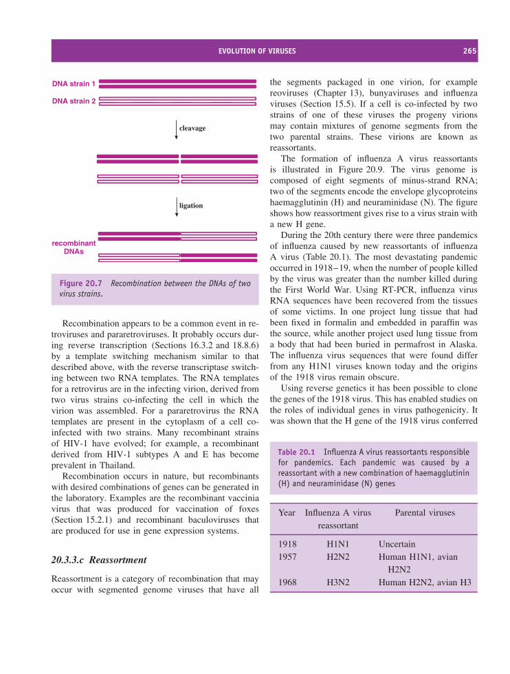
EVOLUTION OF VIRUSES 265
DNA strain 1
DNA strain 2
cleavage
ligation
recombinantDNAs
Figure 20.7 Recombination between the DNAs of twovirus strains.
Recombination appears to be a common event in re-troviruses and pararetroviruses. It probably occurs dur-ing reverse transcription (Sections 16.3.2 and 18.8.6)by a template switching mechanism similar to thatdescribed above, with the reverse transcriptase switch-ing between two RNA templates. The RNA templatesfor a retrovirus are in the infecting virion, derived fromtwo virus strains co-infecting the cell in which thevirion was assembled. For a pararetrovirus the RNAtemplates are present in the cytoplasm of a cell co-infected with two strains. Many recombinant strainsof HIV-1 have evolved; for example, a recombinantderived from HIV-1 subtypes A and E has becomeprevalent in Thailand.
Recombination occurs in nature, but recombinantswith desired combinations of genes can be generated inthe laboratory. Examples are the recombinant vacciniavirus that was produced for vaccination of foxes(Section 15.2.1) and recombinant baculoviruses thatare produced for use in gene expression systems.
20.3.3.c Reassortment
Reassortment is a category of recombination that mayoccur with segmented genome viruses that have all
the segments packaged in one virion, for examplereoviruses (Chapter 13), bunyaviruses and influenzaviruses (Section 15.5). If a cell is co-infected by twostrains of one of these viruses the progeny virionsmay contain mixtures of genome segments from thetwo parental strains. These virions are known asreassortants.
The formation of influenza A virus reassortantsis illustrated in Figure 20.9. The virus genome iscomposed of eight segments of minus-strand RNA;two of the segments encode the envelope glycoproteinshaemagglutinin (H) and neuraminidase (N). The figureshows how reassortment gives rise to a virus strain witha new H gene.
During the 20th century there were three pandemicsof influenza caused by new reassortants of influenzaA virus (Table 20.1). The most devastating pandemicoccurred in 1918–19, when the number of people killedby the virus was greater than the number killed duringthe First World War. Using RT-PCR, influenza virusRNA sequences have been recovered from the tissuesof some victims. In one project lung tissue that hadbeen fixed in formalin and embedded in paraffin wasthe source, while another project used lung tissue froma body that had been buried in permafrost in Alaska.The influenza virus sequences that were found differfrom any H1N1 viruses known today and the originsof the 1918 virus remain obscure.
Using reverse genetics it has been possible to clonethe genes of the 1918 virus. This has enabled studies onthe roles of individual genes in virus pathogenicity. Itwas shown that the H gene of the 1918 virus conferred
Table 20.1 Influenza A virus reassortants responsiblefor pandemics. Each pandemic was caused by areassortant with a new combination of haemagglutinin(H) and neuraminidase (N) genes
Year Influenza A virusreassortant
Parental viruses
1918 H1N1 Uncertain1957 H2N2 Human H1N1, avian
H2N21968 H3N2 Human H2N2, avian H3
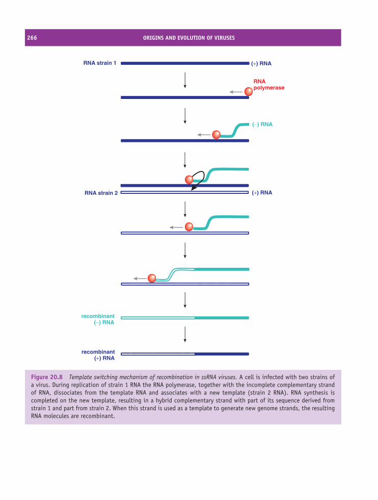
266 ORIGINS AND EVOLUTION OF VIRUSES
RNA strain 1
RNA strain 2
recombinant(+) RNA
RNApolymerase
(−) RNA
(+) RNA
(+) RNA
recombinant(−) RNA
Figure 20.8 Template switching mechanism of recombination in ssRNA viruses. A cell is infected with two strains ofa virus. During replication of strain 1 RNA the RNA polymerase, together with the incomplete complementary strandof RNA, dissociates from the template RNA and associates with a new template (strain 2 RNA). RNA synthesis iscompleted on the new template, resulting in a hybrid complementary strand with part of its sequence derived fromstrain 1 and part from strain 2. When this strand is used as a template to generate new genome strands, the resultingRNA molecules are recombinant.
EVOLUTION OF VIRUSES 267
pathogenicity to human influenza A virus strains thatwere non-pathogenic in mice.
The origins of the viruses responsible for the twomore recent influenza pandemics have been traced. Ineach case the virus was a reassortant between a humanvirus and an avian virus; the 1957 reassortant derivedthe H and N genes from an avian virus, while the 1968reassortant derived the H gene from an avian virus(Table 20.1).
20.3.3.d Acquisition of cell genes
The discovery of similarities between some virusproteins and cell proteins has led to the suggestionthat some viruses have ‘captured’ cell genes. Thegenomes of some retroviruses contain oncogenes thatthey probably acquired from their host cells, whichcontain similar genes (proto-oncogenes). An exampleis the src gene of Rous sarcoma virus (Section 16.4);a very similar gene (c-src) is present in the cells of allvertebrates. A retrovirus might acquire an oncogeneby recombination between the proviral DNA andhost DNA, or by RNA synthesis during transcriptioncontinuing from the provirus through into a proto-oncogene adjacent in the cell chromosome.
Some virus proteins modulate the immune systemof the host, often by mimicking cell proteins. Forexample, some of the human herpesviruses encodeproteins similar to cytokines and major histocompat-ibility complex proteins. Several large DNA virusesencode proteins similar to interleukin-10 and someof these proteins have been shown to suppress cell-mediated immune responses. Are some of the virusgenes that encode these proteins derived from cellgenes and, if so, how did the viruses acquire the genes?
One possible mechanism is recombination, thoughin order for this to occur the genomes of the virusand the cell must come into close proximity. Thismay happen with DNA viruses that replicate in thenucleus (e.g. herpesviruses), but there are many DNAviruses (e.g. poxviruses) that do not enter the nucleus,so cellular and viral genomes are never in closeproximity.
A second possible mechanism for viral acquisitionof a cell gene might be through synthesis of a DNAcopy of a cell mRNA, followed by insertion of the
DNA into the virus genome. The DNA synthesis wouldrequire a reverse transcriptase, possibly supplied by thecell or by a retrovirus co-infecting the cell.
20.3.4 Evolution of new viruses
From time to time a new virus appears, and mysterysurrounds its origin. Often there are suspects for theorigin of the virus and these suspects may be virusesin other host species. The genomes of the new virusand the suspects can be sequenced, and if a closematch is found between the new virus and one of thesuspects then the mystery has probably been solved.It is commonly found that a new virus is one thathas infected a new host species in which some furtherevolution has then taken place.
‘Crossing the species barrier – one small step toman, one giant leap to mankind’Mark Klempner and Daniel Shapiro (2004).
Many virus infections across species barriers aredead ends for the viruses as no transmissions to furtherindividuals occur, but sometimes a virus adapts toenable transmission from host to host. A virus mayhave to undergo one or more adaptations in order forit to
• replicate in the new host;
• evade immune responses of the new host;
• transmit to other individuals of the new host.
In 1978 a new parvovirus appeared in dogs andrapidly spread around the world. The genome sequenceof this canine parvovirus was found to be more than99 per cent identical to that of a parvovirus in cats(feline panleukopenia virus). Both viruses can infectfeline cells, but only the canine virus can infect caninecells. The feline virus is thought to be the precursor ofthe canine virus.
Similarly, the genome sequences of HIV-1 and HIV-2 are very similar to those of simian immunodeficiencyviruses found in chimpanzees (SIVcpz) and sooty
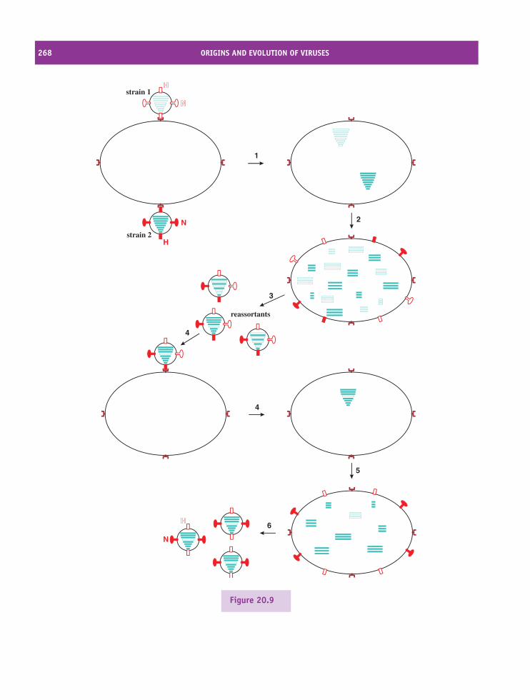
268 ORIGINS AND EVOLUTION OF VIRUSES
H
N
N
1
2
3
4
4
5
6
strain 1
strain 2
reassortants
Figure 20.9
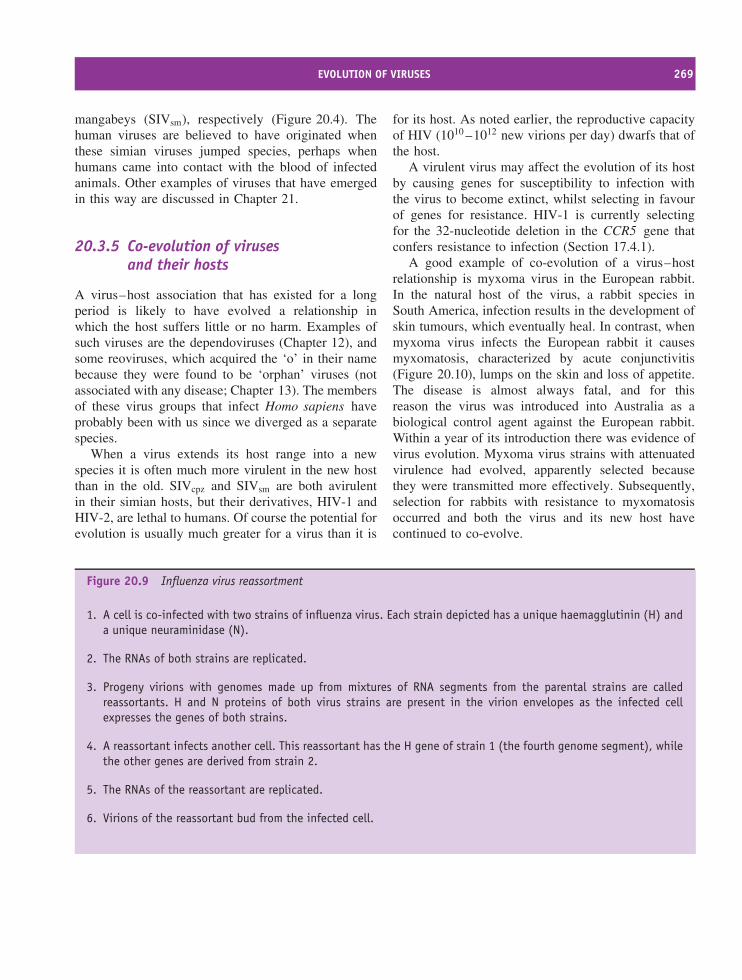
EVOLUTION OF VIRUSES 269
mangabeys (SIVsm), respectively (Figure 20.4). Thehuman viruses are believed to have originated whenthese simian viruses jumped species, perhaps whenhumans came into contact with the blood of infectedanimals. Other examples of viruses that have emergedin this way are discussed in Chapter 21.
20.3.5 Co-evolution of virusesand their hosts
A virus–host association that has existed for a longperiod is likely to have evolved a relationship inwhich the host suffers little or no harm. Examples ofsuch viruses are the dependoviruses (Chapter 12), andsome reoviruses, which acquired the ‘o’ in their namebecause they were found to be ‘orphan’ viruses (notassociated with any disease; Chapter 13). The membersof these virus groups that infect Homo sapiens haveprobably been with us since we diverged as a separatespecies.
When a virus extends its host range into a newspecies it is often much more virulent in the new hostthan in the old. SIVcpz and SIVsm are both avirulentin their simian hosts, but their derivatives, HIV-1 andHIV-2, are lethal to humans. Of course the potential forevolution is usually much greater for a virus than it is
for its host. As noted earlier, the reproductive capacityof HIV (1010 –1012 new virions per day) dwarfs that ofthe host.
A virulent virus may affect the evolution of its hostby causing genes for susceptibility to infection withthe virus to become extinct, whilst selecting in favourof genes for resistance. HIV-1 is currently selectingfor the 32-nucleotide deletion in the CCR5 gene thatconfers resistance to infection (Section 17.4.1).
A good example of co-evolution of a virus–hostrelationship is myxoma virus in the European rabbit.In the natural host of the virus, a rabbit species inSouth America, infection results in the development ofskin tumours, which eventually heal. In contrast, whenmyxoma virus infects the European rabbit it causesmyxomatosis, characterized by acute conjunctivitis(Figure 20.10), lumps on the skin and loss of appetite.The disease is almost always fatal, and for thisreason the virus was introduced into Australia as abiological control agent against the European rabbit.Within a year of its introduction there was evidence ofvirus evolution. Myxoma virus strains with attenuatedvirulence had evolved, apparently selected becausethey were transmitted more effectively. Subsequently,selection for rabbits with resistance to myxomatosisoccurred and both the virus and its new host havecontinued to co-evolve.
Figure 20.9 Influenza virus reassortment
1. A cell is co-infected with two strains of influenza virus. Each strain depicted has a unique haemagglutinin (H) anda unique neuraminidase (N).
2. The RNAs of both strains are replicated.
3. Progeny virions with genomes made up from mixtures of RNA segments from the parental strains are calledreassortants. H and N proteins of both virus strains are present in the virion envelopes as the infected cellexpresses the genes of both strains.
4. A reassortant infects another cell. This reassortant has the H gene of strain 1 (the fourth genome segment), whilethe other genes are derived from strain 2.
5. The RNAs of the reassortant are replicated.
6. Virions of the reassortant bud from the infected cell.
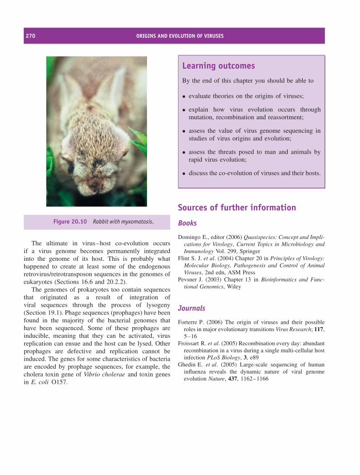
270 ORIGINS AND EVOLUTION OF VIRUSES
Figure 20.10 Rabbit with myxomatosis.
The ultimate in virus–host co-evolution occursif a virus genome becomes permanently integratedinto the genome of its host. This is probably whathappened to create at least some of the endogenousretrovirus/retrotransposon sequences in the genomes ofeukaryotes (Sections 16.6 and 20.2.2).
The genomes of prokaryotes too contain sequencesthat originated as a result of integration ofviral sequences through the process of lysogeny(Section 19.1). Phage sequences (prophages) have beenfound in the majority of the bacterial genomes thathave been sequenced. Some of these prophages areinducible, meaning that they can be activated, virusreplication can ensue and the host can be lysed. Otherprophages are defective and replication cannot beinduced. The genes for some characteristics of bacteriaare encoded by prophage sequences, for example, thecholera toxin gene of Vibrio cholerae and toxin genesin E. coli O157.
Learning outcomes
By the end of this chapter you should be able to
• evaluate theories on the origins of viruses;
• explain how virus evolution occurs throughmutation, recombination and reassortment;
• assess the value of virus genome sequencing instudies of virus origins and evolution;
• assess the threats posed to man and animals byrapid virus evolution;
• discuss the co-evolution of viruses and their hosts.
Sources of further information
Books
Domingo E., editor (2006) Quasispecies: Concept and Impli-cations for Virology, Current Topics in Microbiology andImmunology Vol. 299, Springer
Flint S. J. et al. (2004) Chapter 20 in Principles of Virology:Molecular Biology, Pathogenesis and Control of AnimalViruses, 2nd edn, ASM Press
Pevsner J. (2003) Chapter 13 in Bioinformatics and Func-tional Genomics, Wiley
Journals
Forterre P. (2006) The origin of viruses and their possibleroles in major evolutionary transitions Virus Research; 117,5–16
Froissart R. et al. (2005) Recombination every day: abundantrecombination in a virus during a single multi-cellular hostinfection PLoS Biology, 3, e89
Ghedin E. et al. (2005) Large-scale sequencing of humaninfluenza reveals the dynamic nature of viral genomeevolution Nature, 437, 1162–1166
SOURCES OF FURTHER INFORMATION 271
Holmes E. C. (2003) Error thresholds and the constraints toRNA virus evolution Trends In Microbiology, 11, 543–546
Hueffer K. et al. (2003) The natural host range shift andsubsequent evolution of canine parvovirus resulted fromvirus-specific binding to the canine transferrin receptorJournal of Virology, 77, 1718–1726
Klempner M. S. and Shapiro D. S. (2004) Crossing thespecies barrier – one small step to man, one giant leapto mankind New England Journal of Medicine, 350,1171–1172
Lukashev A. N. (2005) Role of recombination in evolu-tion of enteroviruses Reviews in Medical Virology; 15,157–167
Shackelton L. A. and Holmes E. C. (2004) The evolutionof large DNA viruses: combining genomic informationof viruses and their hosts Trends in Microbiology, 12,458–465
Worobey M. and Holmes E. C. (1999) Evolutionary aspectsof recombination in RNA viruses Journal of GeneralVirology, 80, 2535–2543


21Emerging viruses
At a glance
Avian influenzaEbola/Marburg
West Nile
Bluetongue
SARS
Nipah
Hendra
Human metapneumovirus
Monkeypox
Monkeypox
Mumps
Hantaviruspulmonarysyndrome
•
•
•
•
•
Viruses in new hosts
Viruses in new areas
Newly evolved viruses
Recently discovered viruses
Re-emerging viruses
Examples
Hantaviruspulmonarysyndrome
Emerging Viruses
Nipah virus in pigs and humans
West Nile virus in North America
Influenza virus reassortants
Human metapneumovirus
Mumps virus
Virology: Principles and Applications John B. Carter and Venetia A. Saunders 2007 John Wiley & Sons, Ltd ISBNs: 978-0-470-02386-0 (HB); 978-0-470-02387-7 (PB)
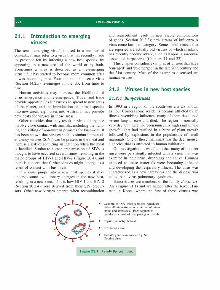
274 EMERGING VIRUSES
21.1 Introduction to emergingviruses
The term ‘emerging virus’ is used in a number ofcontexts: it may refer to a virus that has recently madeits presence felt by infecting a new host species, byappearing in a new area of the world or by both.Sometimes a virus is described as a ‘re-emergingvirus’ if it has started to become more common afterit was becoming rare. Foot and mouth disease virus(Section 14.2.5) re-emerges in the UK from time totime.
Human activities may increase the likelihood ofvirus emergence and re-emergence. Travel and tradeprovide opportunities for viruses to spread to new areasof the planet, and the introduction of animal speciesinto new areas, e.g. horses into Australia, may providenew hosts for viruses in those areas.
Other activities that may result in virus emergenceinvolve close contact with animals, including the hunt-ing and killing of non-human primates for bushmeat. Ithas been shown that viruses such as simian immunod-eficiency viruses (SIVs) can be present in the meat andthere is a risk of acquiring an infection when the meatis handled. Simian-to-human transmission of SIVs isthought to have occurred several times, resulting in themajor groups of HIV-1 and HIV-2 (Figure 20.4), andthere is concern that further viruses might emerge as aresult of contact with bushmeat.
If a virus jumps into a new host species it mayundergo some evolutionary changes in the new host,resulting in a new virus. This is how HIV-1 and HIV-2(Section 20.3.4) were derived from their SIV precur-sors. Other new viruses emerge when recombination
and reassortment result in new viable combinationsof genes (Section 20.3.3); new strains of influenza Avirus come into this category. Some ‘new’ viruses thatare reported are actually old viruses of which mankindhas recently become aware, such as Kaposi’s sarcoma-associated herpesvirus (Chapters 11 and 22).
This chapter considers examples of viruses that have‘emerged’ and ‘re-emerged’ in the late 20th century andthe 21st century. Most of the examples discussed arehuman viruses.
21.2 Viruses in new host species
21.2.1 Bunyaviruses
In 1993 in a region of the south-western US knownas Four Corners some residents became afflicted by anillness resembling influenza; many of them developedsevere lung disease and died. The region is normallyvery dry, but there had been unusually high rainfall andsnowfall that had resulted in a burst of plant growthfollowed by explosions in the populations of smallmammals. One of those mammals was the deer mouse,a species that is attracted to human habitation.
On investigation, it was found that many of the deermice were persistently infected with a virus that wasexcreted in their urine, droppings and saliva. Humansexposed to these materials were becoming infectedand developing the respiratory illness. The virus wascharacterized as a new hantavirus and the disease wascalled hantavirus pulmonary syndrome.
Hantaviruses are members of the family Bunyaviri-dae (Figure 21.1) and are named after the River Han-taan in Korea, where the first of these viruses was
•
•
•
•
Genome: ssRNA (three segments, which areeither all minus strand, or a mixture of minusstrand and ambisense). Each segment iscircular as a result of base pairing at its ends.
Capsid symmetry: helical
Enveloped virion
Includes genus Hantavirus, e.g. SinNombre virus
Figure 21.1 Family Bunyaviridae.
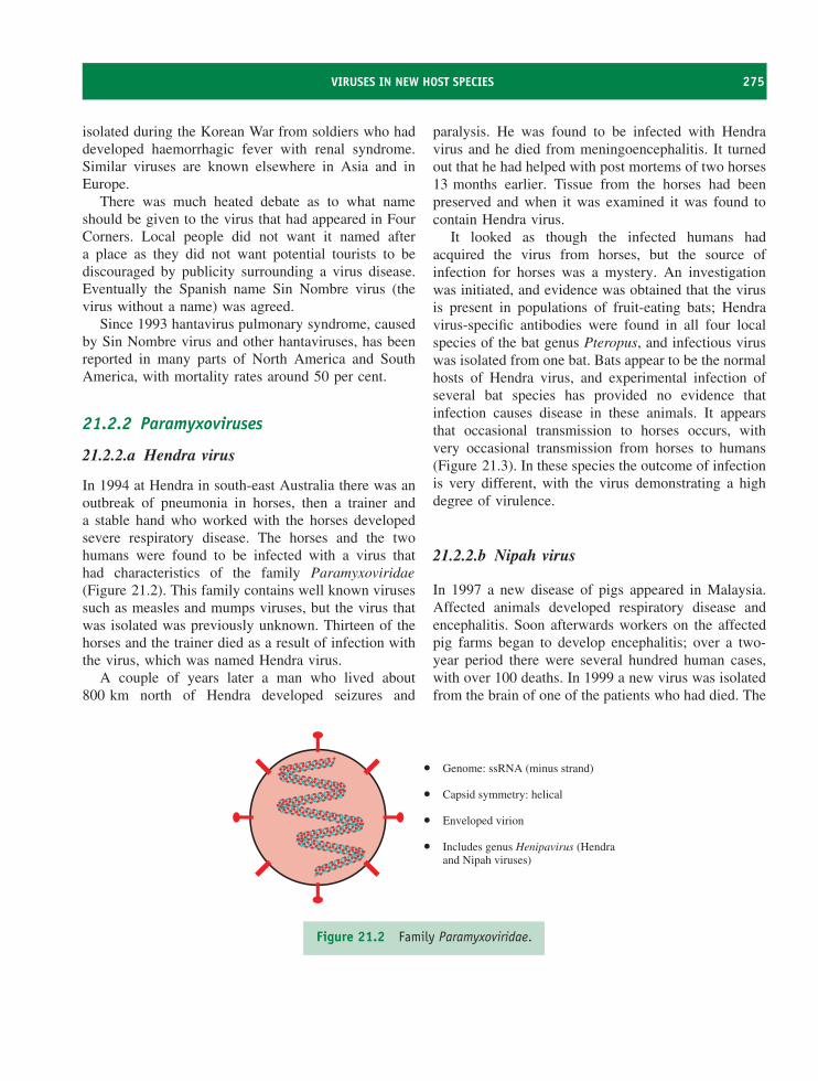
VIRUSES IN NEW HOST SPECIES 275
isolated during the Korean War from soldiers who haddeveloped haemorrhagic fever with renal syndrome.Similar viruses are known elsewhere in Asia and inEurope.
There was much heated debate as to what nameshould be given to the virus that had appeared in FourCorners. Local people did not want it named aftera place as they did not want potential tourists to bediscouraged by publicity surrounding a virus disease.Eventually the Spanish name Sin Nombre virus (thevirus without a name) was agreed.
Since 1993 hantavirus pulmonary syndrome, causedby Sin Nombre virus and other hantaviruses, has beenreported in many parts of North America and SouthAmerica, with mortality rates around 50 per cent.
21.2.2 Paramyxoviruses
21.2.2.a Hendra virus
In 1994 at Hendra in south-east Australia there was anoutbreak of pneumonia in horses, then a trainer anda stable hand who worked with the horses developedsevere respiratory disease. The horses and the twohumans were found to be infected with a virus thathad characteristics of the family Paramyxoviridae(Figure 21.2). This family contains well known virusessuch as measles and mumps viruses, but the virus thatwas isolated was previously unknown. Thirteen of thehorses and the trainer died as a result of infection withthe virus, which was named Hendra virus.
A couple of years later a man who lived about800 km north of Hendra developed seizures and
paralysis. He was found to be infected with Hendravirus and he died from meningoencephalitis. It turnedout that he had helped with post mortems of two horses13 months earlier. Tissue from the horses had beenpreserved and when it was examined it was found tocontain Hendra virus.
It looked as though the infected humans hadacquired the virus from horses, but the source ofinfection for horses was a mystery. An investigationwas initiated, and evidence was obtained that the virusis present in populations of fruit-eating bats; Hendravirus-specific antibodies were found in all four localspecies of the bat genus Pteropus, and infectious viruswas isolated from one bat. Bats appear to be the normalhosts of Hendra virus, and experimental infection ofseveral bat species has provided no evidence thatinfection causes disease in these animals. It appearsthat occasional transmission to horses occurs, withvery occasional transmission from horses to humans(Figure 21.3). In these species the outcome of infectionis very different, with the virus demonstrating a highdegree of virulence.
21.2.2.b Nipah virus
In 1997 a new disease of pigs appeared in Malaysia.Affected animals developed respiratory disease andencephalitis. Soon afterwards workers on the affectedpig farms began to develop encephalitis; over a two-year period there were several hundred human cases,with over 100 deaths. In 1999 a new virus was isolatedfrom the brain of one of the patients who had died. The
•
•
•
•
Genome: ssRNA (minus strand)
Capsid symmetry: helical
Enveloped virion
Includes genus Henipavirus (Hendraand Nipah viruses)
Figure 21.2 Family Paramyxoviridae.
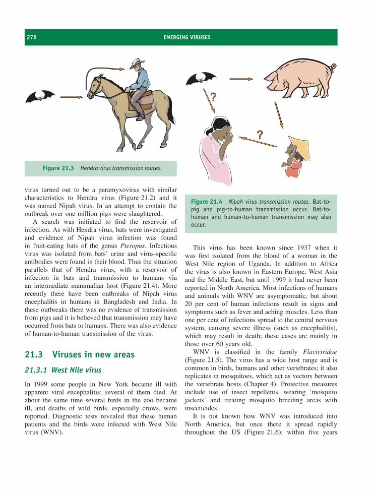
276 EMERGING VIRUSES
Figure 21.3 Hendra virus transmission routes.
virus turned out to be a paramyxovirus with similarcharacteristics to Hendra virus (Figure 21.2) and itwas named Nipah virus. In an attempt to contain theoutbreak over one million pigs were slaughtered.
A search was initiated to find the reservoir ofinfection. As with Hendra virus, bats were investigatedand evidence of Nipah virus infection was foundin fruit-eating bats of the genus Pteropus. Infectiousvirus was isolated from bats’ urine and virus-specificantibodies were found in their blood. Thus the situationparallels that of Hendra virus, with a reservoir ofinfection in bats and transmission to humans viaan intermediate mammalian host (Figure 21.4). Morerecently there have been outbreaks of Nipah virusencephalitis in humans in Bangladesh and India. Inthese outbreaks there was no evidence of transmissionfrom pigs and it is believed that transmission may haveoccurred from bats to humans. There was also evidenceof human-to-human transmission of the virus.
21.3 Viruses in new areas
21.3.1 West Nile virus
In 1999 some people in New York became ill withapparent viral encephalitis; several of them died. Atabout the same time several birds in the zoo becameill, and deaths of wild birds, especially crows, werereported. Diagnostic tests revealed that these humanpatients and the birds were infected with West Nilevirus (WNV).
Figure 21.4 Nipah virus transmission routes. Bat-to-pig and pig-to-human transmission occur. Bat-to-human and human-to-human transmission may alsooccur.
This virus has been known since 1937 when itwas first isolated from the blood of a woman in theWest Nile region of Uganda. In addition to Africathe virus is also known in Eastern Europe, West Asiaand the Middle East, but until 1999 it had never beenreported in North America. Most infections of humansand animals with WNV are asymptomatic, but about20 per cent of human infections result in signs andsymptoms such as fever and aching muscles. Less thanone per cent of infections spread to the central nervoussystem, causing severe illness (such as encephalitis),which may result in death; these cases are mainly inthose over 60 years old.
WNV is classified in the family Flaviviridae(Figure 21.5). The virus has a wide host range and iscommon in birds, humans and other vertebrates; it alsoreplicates in mosquitoes, which act as vectors betweenthe vertebrate hosts (Chapter 4). Protective measuresinclude use of insect repellents, wearing ‘mosquitojackets’ and treating mosquito breeding areas withinsecticides.
It is not known how WNV was introduced intoNorth America, but once there it spread rapidlythroughout the US (Figure 21.6); within five years
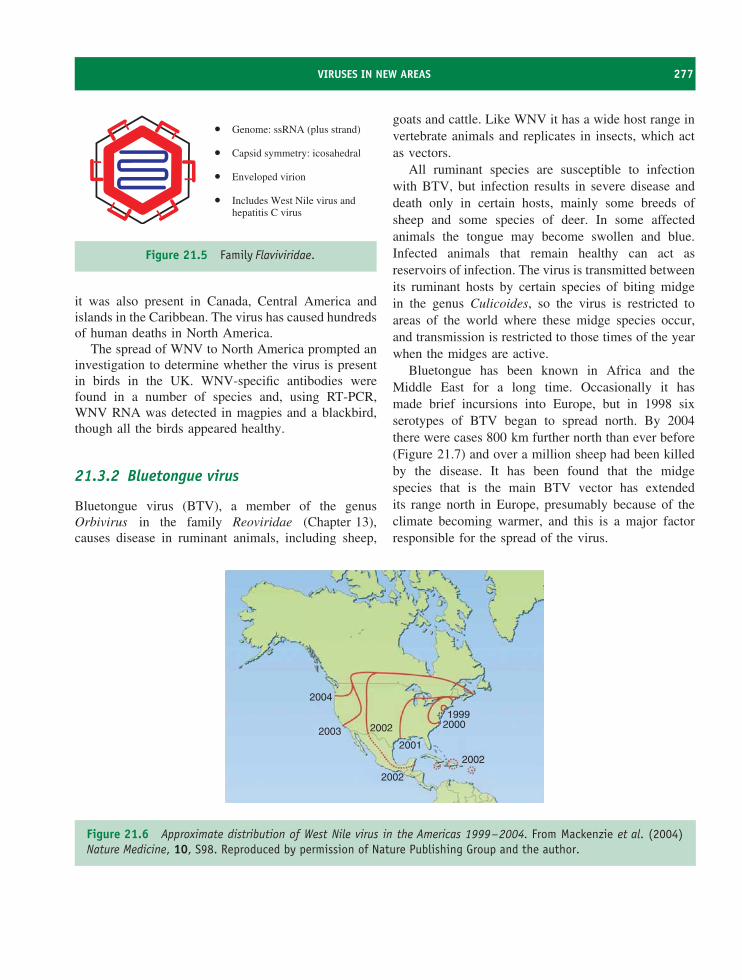
VIRUSES IN NEW AREAS 277
•
•
•
•
Genome: ssRNA (plus strand)
Capsid symmetry: icosahedral
Enveloped virion
Includes West Nile virus and hepatitis C virus
Figure 21.5 Family Flaviviridae.
it was also present in Canada, Central America andislands in the Caribbean. The virus has caused hundredsof human deaths in North America.
The spread of WNV to North America prompted aninvestigation to determine whether the virus is presentin birds in the UK. WNV-specific antibodies werefound in a number of species and, using RT-PCR,WNV RNA was detected in magpies and a blackbird,though all the birds appeared healthy.
21.3.2 Bluetongue virus
Bluetongue virus (BTV), a member of the genusOrbivirus in the family Reoviridae (Chapter 13),causes disease in ruminant animals, including sheep,
goats and cattle. Like WNV it has a wide host range invertebrate animals and replicates in insects, which actas vectors.
All ruminant species are susceptible to infectionwith BTV, but infection results in severe disease anddeath only in certain hosts, mainly some breeds ofsheep and some species of deer. In some affectedanimals the tongue may become swollen and blue.Infected animals that remain healthy can act asreservoirs of infection. The virus is transmitted betweenits ruminant hosts by certain species of biting midgein the genus Culicoides, so the virus is restricted toareas of the world where these midge species occur,and transmission is restricted to those times of the yearwhen the midges are active.
Bluetongue has been known in Africa and theMiddle East for a long time. Occasionally it hasmade brief incursions into Europe, but in 1998 sixserotypes of BTV began to spread north. By 2004there were cases 800 km further north than ever before(Figure 21.7) and over a million sheep had been killedby the disease. It has been found that the midgespecies that is the main BTV vector has extendedits range north in Europe, presumably because of theclimate becoming warmer, and this is a major factorresponsible for the spread of the virus.
2004
2003 2002
2002
20012002
20001999
Figure 21.6 Approximate distribution of West Nile virus in the Americas 1999–2004. From Mackenzie et al. (2004)Nature Medicine, 10, S98. Reproduced by permission of Nature Publishing Group and the author.
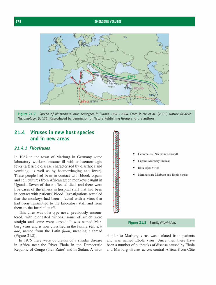
278 EMERGING VIRUSES
Figure 21.7 Spread of bluetongue virus serotypes in Europe 1998–2004. From Purse et al. (2005) Nature ReviewsMicrobiology, 3, 171. Reproduced by permission of Nature Publishing Group and the authors.
21.4 Viruses in new host speciesand in new areas
21.4.1 Filoviruses
In 1967 in the town of Marburg in Germany somelaboratory workers became ill with a haemorrhagicfever (a terrible disease characterized by diarrhoea andvomiting, as well as by haemorrhaging and fever).These people had been in contact with blood, organsand cell cultures from African green monkeys caught inUganda. Seven of those affected died, and there werefive cases of the illness in hospital staff that had beenin contact with patients’ blood. Investigations revealedthat the monkeys had been infected with a virus thathad been transmitted to the laboratory staff and fromthem to the hospital staff.
This virus was of a type never previously encoun-tered, with elongated virions, some of which werestraight and some were curved. It was named Mar-burg virus and is now classified in the family Filoviri-dae, named from the Latin filum, meaning a thread(Figure 21.8).
In 1976 there were outbreaks of a similar diseasein Africa near the River Ebola in the DemocraticRepublic of Congo (then Zaire) and in Sudan. A virus
•
•
•
•
Genome: ssRNA (minus strand)
Capsid symmetry: helical
Enveloped virion
Members are Marburg and Ebola viruses
Figure 21.8 Family Filoviridae.
similar to Marburg virus was isolated from patientsand was named Ebola virus. Since then there havebeen a number of outbreaks of disease caused by Ebolaand Marburg viruses across central Africa, from Cote
NEW VIRUSES 279
d’Ivoire in the west to Kenya in the east and to Angolain the south.
The way in which the outbreaks start has longbeen a mystery. There is increasing evidence thatsome outbreaks start when a human becomes infectedas a result of contact with the blood of an infectednon-human primate. It was known from the originaloutbreak in Germany that African green monkeyscan be infected with Marburg virus. It has sincebeen found that gorillas, chimpanzees and duikerscan be infected with Ebola virus, which may beresponsible for significant mortality of these species.Ebola and Marburg viruses are present in the blood ofinfected hosts and transmission to humans can occurthrough contact with the flesh of infected animals afterthey have been hunted and killed. Human-to-humantransmission readily occurs through contact with theblood of infected individuals.
There are several species of Ebola virus in Africa.A further species has appeared in the US and Italyin monkeys imported from the Philippines; there isserological evidence that animal handlers have beeninfected, but there have been no reports of this viruscausing disease.
It is thought likely that additional animal species actas ‘reservoirs’ of Marburg and Ebola viruses, and thatinfection in these species is likely to cause few or nosigns of disease. There have been many expeditionsto search for reservoir hosts, but very few producedpositive results; Ebola virus RNA has been detectedin some small mammals including fruit bats, and anti-Ebola virus antibodies have been detected in some fruitbats. There remain unanswered questions concerningreservoir species and host ranges of these viruses.
21.4.2 Monkeypox virus
Monkeypox is a disease of monkeys that is similar tosmallpox in man. The viruses that cause these diseasesare members of the family Poxviridae (Figure 21.9).Since 1970 there have been cases in Africa of humandisease caused by infection with monkeypox virus;some cases have been fatal.
In 2003 some people in the US developed a mysteryillness that involved a rash and a fever. Investigationsrevealed that they were infected with monkeypox virus,which had entered the country in small mammalsimported from Ghana. The virus had infected petprairie dogs and had then been transmitted to humans.
Sequencing the genomes of monkeypox virus iso-lates has revealed the existence of two strains thatappear to differ in virulence. The virus that wasimported into the US was the less virulent strain.
21.5 New viruses
21.5.1 SARS coronavirus
In 2002 a new human respiratory disease emerged insouthern China. The following year one of the doctorswho had been treating patients travelled to Hong Kong,where he became ill and died. Subsequently, peoplewho had stayed in the same hotel as the doctor travelledto Singapore, Vietnam, Canada and the US, taking theinfectious agent with them. The epidemic of severeacute respiratory syndrome (SARS) was under way.
The signs and symptoms of SARS resemble thoseof influenza and include fever, aching muscles, sorethroat, cough and shortness of breath. About 90 per
•
•
•
Genome: dsDNA
Large, complex virion
Members include smallpox andvaccinia viruses
Figure 21.9 Family Poxviridae.
280 EMERGING VIRUSES
•
•
•
•
Genome: ssRNA (plus strand)
Capsid symmetry: helical
Enveloped virion with a corona of glycoproteins
Includes SARS coronavirus
Figure 21.10 Family Coronaviridae.
cent of patients recovered, but for the remainder theinfection proved to be lethal. These were mainlyindividuals who had an underlying condition such asdiabetes, heart disease or a weakened immune system.On the face of it SARS is a respiratory tract disease,but in many patients the infection spread to other partsof the body. Diarrhoea developed in some patients andthe virus was shed in the faeces and urine for severalweeks.
The causative agent was found to be a newcoronavirus (Figure 21.10). No natural reservoir for theSARS coronavirus has been found, but coronaviruseswith very similar genome sequences were isolated fromanimals sold in markets in the region of China wherethe first SARS cases appeared. Antibodies to theseviruses were found in many workers in the markets,but none of the workers had any history of SARS. Asimilar virus has also been isolated from several batspecies. It is likely that coronaviruses have repeatedlycrossed into humans from other mammalian species,but in the majority of cases the virus has lacked theability for efficient human-to-human transmission. Itwould seem that the SARS coronavirus evolved froma rare virus that had this ability.
The SARS outbreak was brought under control byquarantine measures, but only after there had been over8000 cases with nearly 800 deaths.
21.5.2 New strains of influenza virus A
The family Orthomyxoviridae includes the influenzaA viruses (Figure 3.20). The virions have two species
of surface glycoprotein: a haemagglutinin (H) and aneuraminidase (N). There are 16 types of H antigen andnine types of N antigen, and there are many subtypesof each type. From time to time a virus emerges witha new combination of H and N genes formed byreassortment, and causes a pandemic (see Chapter 20).
The hosts of influenza A viruses are principally birdsthat frequent aquatic habitats. The birds (e.g. ducks,geese, gulls) acquire infections by ingestion or inhala-tion and the viruses infect their intestinal and/or respi-ratory tracts. Infection with most virus strains results infew or no signs of disease, but some strains are highlypathogenic and can kill their avian hosts. The virusescan be spread to new areas when the birds migrate.
Some influenza A viruses infect mammalian speciesincluding pigs, horses and humans; the respiratory tractis the main site of virus replication. Normally humansare infected only with viruses that have H type 1,2 or 3 and N type 1 or 2. Patients are commonlyvery ill and some die, either as a direct result of thevirus infection, or indirectly from secondary pathogens,which are able to infect as a result of damage to therespiratory epithelium.
The previous two paragraphs describe the normalsituation, but exceptions occur: some avian strains ofinfluenza A virus can be highly pathogenic in birds, canbe transmitted from wild birds to domestic poultry andcan be transmitted to humans (Figure 21.11). This wasthe situation in Hong Kong in 1997, when an H5N1virus caused an outbreak of serious disease in poultry.Eighteen people also became infected with the virusand six of them died. In order to bring the outbreak toan end all poultry in Hong Kong were slaughtered.
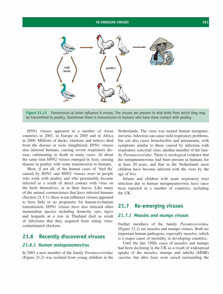
RE-EMERGING VIRUSES 281
wildbirds
poultry
Figure 21.11 Transmission of avian influenza A viruses. The viruses are present in wild birds from which they maybe transmitted to poultry. Sometimes there is transmission to humans who have close contact with poultry.
H5N1 viruses appeared in a number of Asiancountries in 2003, in Europe in 2005 and in Africain 2006. Millions of ducks, chickens and turkeys diedfrom the disease or were slaughtered; H5N1 virusesalso infected humans, causing severe respiratory dis-ease, culminating in death in many cases. At aboutthe same time H9N2 viruses emerged in Asia, causingdisease in poultry with some transmission to humans.
Most, if not all, of the human cases of ‘bird flu’caused by H5N1 and H9N2 viruses were in peoplewho work with poultry and who presumably becameinfected as a result of direct contact with virus onthe birds themselves, or in their faeces. Like manyof the animal coronaviruses that have infected humans(Section 21.5.1), these avian influenza viruses appearedto have little or no propensity for human-to-humantransmission. H5N1 viruses have also infected othermammalian species including domestic cats; tigersand leopards at a zoo in Thailand died as resultof infections that they acquired from eating virus-contaminated chickens.
21.6 Recently discovered viruses21.6.1 Human metapneumovirus
In 2001 a new member of the family Paramyxoviridae(Figure 21.2) was isolated from young children in the
Netherlands. The virus was named human metapneu-movirus. Infection can cause mild respiratory problems,but can also cause bronchiolitis and pneumonia, withsymptoms similar to those caused by infection withrespiratory syncytial virus, another member of the fam-ily Paramyxoviridae. There is serological evidence thatthe metapneumovirus had been present in humans forat least 50 years, and that in the Netherlands mostchildren have become infected with the virus by theage of five.
Infants and children with acute respiratory tractinfection due to human metapneumovirus have sincebeen reported in a number of countries, includingthe UK.
21.7 Re-emerging viruses
21.7.1 Measles and mumps viruses
Further members of the family Paramyxoviridae(Figure 21.2) are measles and mumps viruses. Both areimportant human pathogens, especially measles, whichis a major cause of mortality in developing countries.
Until the late 1990s cases of measles and mumpshad been declining in the UK as a result of widespreaduptake of the measles, mumps and rubella (MMR)vaccine, but after fears were raised surrounding the
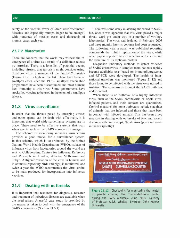
282 EMERGING VIRUSES
safety of the vaccine fewer children were vaccinated.Measles, and especially mumps, began to ‘re-emerge’,with hundreds of measles cases and thousands ofmumps cases each year.
21.7.2 Bioterrorism
There are concerns that the world may witness the re-emergence of a virus as a result of a deliberate releaseby terrorists. There is a long list of potential agents,including viruses, that terrorists might consider using.Smallpox virus, a member of the family Poxviridae(Figure 21.9), is high on the list. There have been nosmallpox cases since the 1970s, smallpox vaccinationprogrammes have been discontinued and most humanslack immunity to this virus. Some governments havestockpiled vaccine to be used in the event of a smallpoxrelease.
21.8 Virus surveillanceIn order that the threats posed by emerging virusesand other agents can be dealt with effectively, it isimportant that world-wide surveillance systems are inplace. There need to be effective systems that warnwhen agents such as the SARS coronavirus emerge.
The scheme for monitoring influenza virus strainsprovides a good model for a surveillance system.In this scheme, which is co-ordinated by the UnitedNations World Health Organisation (WHO), isolates ofinfluenza virus from laboratories around the world aresent to Collaborating Centres for Influenza Referenceand Research in London, Atlanta, Melbourne andTokyo. Antigenic variation of the virus in humans andin animals (especially birds and pigs) is monitored, andtwice a year the WHO recommends the virus strainsto be mass-produced for incorporation into influenzavaccines.
21.9 Dealing with outbreaksIt is important that resources for diagnosis, researchand treatment of infectious diseases are available whenthe need arises. A useful case study is provided bythe measures taken to deal with the emergence of theSARS coronavirus (Section 21.5.1).
There was some delay in alerting the world to SARSbut, once it was apparent that this virus posed a majorthreat, work got under way in a number of virologylaboratories. The virus was isolated in February 2003and three months later its genome had been sequenced.The following year a paper was published reportingcompounds that inhibit replication of the virus, whileother papers reported the cell receptor of the virus andthe structure of its replicase protein.
Diagnostic laboratory methods to detect evidenceof SARS coronavirus in samples from patients rapidlybecame available; tests based on immunofluorescenceand RT-PCR were developed. The health of inter-national travellers was monitored (Figure 21.12) andthose found to be infected with the virus were nursed inisolation. These measures brought the SARS outbreakunder control.
When there is an outbreak of a highly infectiousvirus, such as the SARS coronavirus or Ebola virus,infected patients and their contacts are quarantined.Control measures for some outbreaks include slaughterof animals that are infected and those that have beenin contact with infected animals. This has been a keymeasure in dealing with outbreaks of foot and mouthdisease (cattle and sheep), Nipah virus (pigs) and avianinfluenza (poultry).
Figure 21.12 Checkpoint for monitoring the healthof people crossing the Thailand–Burma borderduring the SARS outbreak, June 2003. Courtesyof Professor A.J.S. Whalley, Liverpool John MooresUniversity.
SOURCES OF FURTHER INFORMATION 283
Medical staff, veterinary staff and other personneldealing with humans and animals infected with high-risk viruses must take precautions to protect themselvesand to avoid spreading infection. Precautions includewearing protective clothing and breathing filteredair.
Learning outcomes
By the end of this chapter you should be able to
• evaluate the term ‘emerging virus’;
• discuss examples of viruses that have recentlyappeared in new host species;
• discuss examples of viruses that have recentlyappeared in new parts of the world;
• discuss examples of new viruses;
• discuss examples of re-emerging viruses;
• assess measures that can be taken to prevent andcontain outbreaks of infectious disease.
Sources of further information
Books
Hartman A. L., Towner J. S. and Nichol S. (2005) Pathogen-esis of Ebola and Marburg viruses, pp. 109–124 in Molec-ular Pathogenesis of Virus Infections, editors Digard P.,Nash A. A. and Randall R. E., 64th Symposium of the
Society for General Microbiology, Cambridge UniversityPress
Komar N. (2003) West Nile virus: epidemiology and ecologyin North America, pp. 185–234 in Advances in VirusResearch, Vol. 61, Elsevier
Parrish C. R. and Kawaoka Y. (2005) The origins of newpandemic viruses: the acquisition of new host ranges bycanine parvovirus and influenza A viruses, pp. 553–586 inAnnual Review of Microbiology, Vol. 59, Annual Reviews
Schmidt A., Wolff M. H. and Weber O., editors (2005)Coronaviruses with Special Emphasis on First InsightsConcerning SARS, Birkhauser
Smolinski M. S., Hamburg M. A. and Lederberg J., editors(2003) Microbial Threats to Health: Emergence, Detectionand Response, National Academies Press
Journals
Belshe R. B. (2005) The origins of pandemic influenza –lessons from the 1918 virus New England Journal ofMedicine, 353, 2209–2211
Eaton B. T. et al. (2005) Hendra and Nipah viruses: differentand dangerous Nature Reviews Microbiology, 4, 23–35
Horimoto T. and Kawaoka Y. (2005) Influenza: lessons frompast pandemics, warnings from current incidents NatureReviews Microbiology, 3, 591–600
Leroy E. M. et al. (2005) Fruit bats as reservoirs of Ebolavirus Nature, 438, 575–576
Likos A. M. et al. (2005) A tale of two clades: monkeypoxviruses Journal of General Virology, 86, 2661–2672
Purse B. V. et al. (2005) Climate change and the recentemergence of bluetongue in Europe Nature ReviewsMicrobiology, 3, 171–181
van den Hoogen B. G. et al. (2001) A newly discoveredhuman pneumovirus isolated from young children withrespiratory tract disease Nature Medicine, 7, 719–724
Walsh P. D., Biek R. and Real L. A. (2005) Wave-likespread of Ebola Zaire PLoS Biology, 3 (11), e371

22Viruses and cancer
At a glance
Burkitt’s lymphomanasopharyngeal carcinoma
(Epstein-Barr virus)
anogenital carcinomas(human papillomaviruses)
Kaposi’s sarcoma(Kaposi’s sarcoma-associated herpesvirus)
hepatocellular carcinoma(hepatitis B virus,hepatitis C virus)
adult T cell leukaemia(human T-lymphotropic virus 1)
B cell leukaemia/lymphomasarcoma
(retroviruses)
Virology: Principles and Applications John B. Carter and Venetia A. Saunders 2007 John Wiley & Sons, Ltd ISBNs: 978-0-470-02386-0 (HB); 978-0-470-02387-7 (PB)
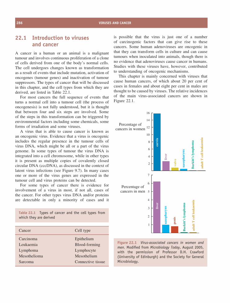
286 VIRUSES AND CANCER
22.1 Introduction to virusesand cancer
A cancer in a human or an animal is a malignanttumour and involves continuous proliferation of a cloneof cells derived from one of the body’s normal cells.The cell undergoes changes known as transformationas a result of events that include mutation, activation ofoncogenes (tumour genes) and inactivation of tumoursuppressors. The types of cancer that will be discussedin this chapter, and the cell types from which they arederived, are listed in Table 22.1.
For most cancers the full sequence of events thatturns a normal cell into a tumour cell (the process ofoncogenesis) is not fully understood, but it is thoughtthat between four and six steps are involved. Someof the steps in this transformation can be triggered byenvironmental factors including some chemicals, someforms of irradiation and some viruses.
A virus that is able to cause cancer is known asan oncogenic virus. Evidence that a virus is oncogenicincludes the regular presence in the tumour cells ofvirus DNA, which might be all or a part of the virusgenome. In some types of tumour the virus DNA isintegrated into a cell chromosome, while in other typesit is present as multiple copies of covalently closedcircular DNA (cccDNA), as discussed in the context oflatent virus infections (see Figure 9.7). In many casesone or more of the virus genes are expressed in thetumour cell and virus proteins can be detected.
For some types of cancer there is evidence forinvolvement of a virus in most, if not all, cases ofthe cancer. For other types virus DNA and/or proteinsare detectable in only a minority of cases and it
Table 22.1 Types of cancer and the cell types fromwhich they are derived
Cancer Cell type
Carcinoma EpitheliumLeukaemia Blood-formingLymphoma LymphocyteMesothelioma MesotheliumSarcoma Connective tissue
is possible that the virus is just one of a numberof carcinogenic factors that can give rise to thesecancers. Some human adenoviruses are oncogenic inthat they can transform cells in culture and can causetumours when inoculated into animals, though there isno evidence that adenoviruses cause cancer in humans.Studies with these viruses have, however, contributedto understanding of oncogenic mechanisms.
This chapter is mainly concerned with viruses thatcause human cancers, of which about 20 per cent ofcases in females and about eight per cent in males arethought to be caused by viruses. The relative incidencesof the main virus-associated cancers are shown inFigure 22.1.
2
4
6
8
10
12
14
16
Percentage ofcancers in women
liver
vulv
a
naso
phar
ynx
Bur
kitt
’s ly
mph
oma
adul
t T
cel
l leu
kaem
ia
cerv
ix
1
4
Percentage ofcancers in men
peni
s
naso
phar
ynx
Bur
kitt
’s ly
mph
oma
adul
t T
cel
l leu
kaem
ia
2
3
5
6
7
liver
Figure 22.1 Virus-associated cancers in women andmen. Modified from Microbiology Today, August 2005,with the permission of Professor D.H. Crawford(University of Edinburgh) and the Society for GeneralMicrobiology.
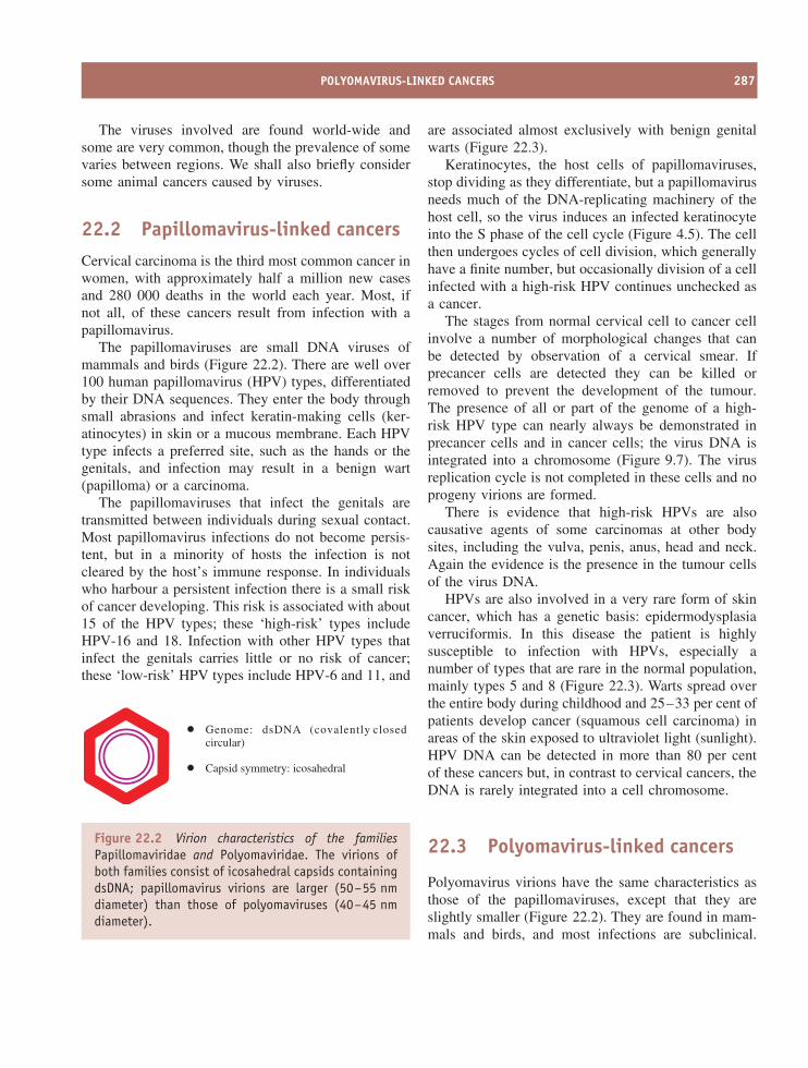
POLYOMAVIRUS-LINKED CANCERS 287
The viruses involved are found world-wide andsome are very common, though the prevalence of somevaries between regions. We shall also briefly considersome animal cancers caused by viruses.
22.2 Papillomavirus-linked cancersCervical carcinoma is the third most common cancer inwomen, with approximately half a million new casesand 280 000 deaths in the world each year. Most, ifnot all, of these cancers result from infection with apapillomavirus.
The papillomaviruses are small DNA viruses ofmammals and birds (Figure 22.2). There are well over100 human papillomavirus (HPV) types, differentiatedby their DNA sequences. They enter the body throughsmall abrasions and infect keratin-making cells (ker-atinocytes) in skin or a mucous membrane. Each HPVtype infects a preferred site, such as the hands or thegenitals, and infection may result in a benign wart(papilloma) or a carcinoma.
The papillomaviruses that infect the genitals aretransmitted between individuals during sexual contact.Most papillomavirus infections do not become persis-tent, but in a minority of hosts the infection is notcleared by the host’s immune response. In individualswho harbour a persistent infection there is a small riskof cancer developing. This risk is associated with about15 of the HPV types; these ‘high-risk’ types includeHPV-16 and 18. Infection with other HPV types thatinfect the genitals carries little or no risk of cancer;these ‘low-risk’ HPV types include HPV-6 and 11, and
Genome: dsDNA (covalently closedcircular)
Capsid symmetry: icosahedral
Figure 22.2 Virion characteristics of the familiesPapillomaviridae and Polyomaviridae. The virions ofboth families consist of icosahedral capsids containingdsDNA; papillomavirus virions are larger (50–55 nmdiameter) than those of polyomaviruses (40–45 nmdiameter).
are associated almost exclusively with benign genitalwarts (Figure 22.3).
Keratinocytes, the host cells of papillomaviruses,stop dividing as they differentiate, but a papillomavirusneeds much of the DNA-replicating machinery of thehost cell, so the virus induces an infected keratinocyteinto the S phase of the cell cycle (Figure 4.5). The cellthen undergoes cycles of cell division, which generallyhave a finite number, but occasionally division of a cellinfected with a high-risk HPV continues unchecked asa cancer.
The stages from normal cervical cell to cancer cellinvolve a number of morphological changes that canbe detected by observation of a cervical smear. Ifprecancer cells are detected they can be killed orremoved to prevent the development of the tumour.The presence of all or part of the genome of a high-risk HPV type can nearly always be demonstrated inprecancer cells and in cancer cells; the virus DNA isintegrated into a chromosome (Figure 9.7). The virusreplication cycle is not completed in these cells and noprogeny virions are formed.
There is evidence that high-risk HPVs are alsocausative agents of some carcinomas at other bodysites, including the vulva, penis, anus, head and neck.Again the evidence is the presence in the tumour cellsof the virus DNA.
HPVs are also involved in a very rare form of skincancer, which has a genetic basis: epidermodysplasiaverruciformis. In this disease the patient is highlysusceptible to infection with HPVs, especially anumber of types that are rare in the normal population,mainly types 5 and 8 (Figure 22.3). Warts spread overthe entire body during childhood and 25–33 per cent ofpatients develop cancer (squamous cell carcinoma) inareas of the skin exposed to ultraviolet light (sunlight).HPV DNA can be detected in more than 80 per centof these cancers but, in contrast to cervical cancers, theDNA is rarely integrated into a cell chromosome.
22.3 Polyomavirus-linked cancers
Polyomavirus virions have the same characteristics asthose of the papillomaviruses, except that they areslightly smaller (Figure 22.2). They are found in mam-mals and birds, and most infections are subclinical.
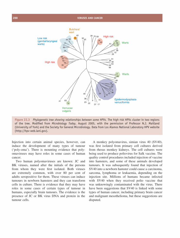
288 VIRUSES AND CANCER
Warts
High risk
High risk
Butchers'wartButchers'wart
Low risk(genital/laryngealwarts)
Warts465
6048
50192520
215
364712
8
24
15
1737
9
2223
38
49
63
3135
1652
3358
34
116b
13
44 55
411a
6656
3026
51
2910
283
272a57
61
18
597039
4232
407
45
53
Verrucaevulgaris
Epidermodysplasiaverruciformis
Figure 22.3 Phylogenetic tree showing relationships between some HPVs. The high risk HPVs cluster in two regionsof the tree. Modified from Microbiology Today, August 2005, with the permission of Professor N.J. Maitland(University of York) and the Society for General Microbiology. Data from Los Alamos National Laboratory HPV website(http://hpv-web.lanl.gov).
Injection into certain animal species, however, caninduce the development of many types of tumour(‘poly-oma’). There is mounting evidence that poly-omaviruses may have roles in some cases of humancancer.
Two human polyomaviruses are known: JC andBK viruses, named after the initials of the personsfrom whom they were first isolated. Both virusesare extremely common, with over 80 per cent ofadults seropositive for them. These viruses can inducetumours in newborn hamsters and they can transformcells in culture. There is evidence that they may haveroles in some cases of certain types of tumour inhumans, especially brain tumours. The evidence is thepresence of JC or BK virus DNA and protein in thetumour cells.
A monkey polyomavirus, simian virus 40 (SV40),was first isolated from primary cell cultures derivedfrom rhesus monkey kidneys. The cell cultures werebeing used to produce poliovirus for Salk vaccine. Thequality control procedures included injection of vaccineinto hamsters, and some of these animals developedtumours. It was subsequently found that injection ofSV40 into a newborn hamster could cause a carcinoma,sarcoma, lymphoma or leukaemia, depending on theinjection site. Millions of humans became infectedwith SV40 when they received polio vaccine thatwas unknowingly contaminated with the virus. Therehave been suggestions that SV40 is linked with sometypes of human cancer, including primary brain cancerand malignant mesothelioma, but these suggestions aredisputed.
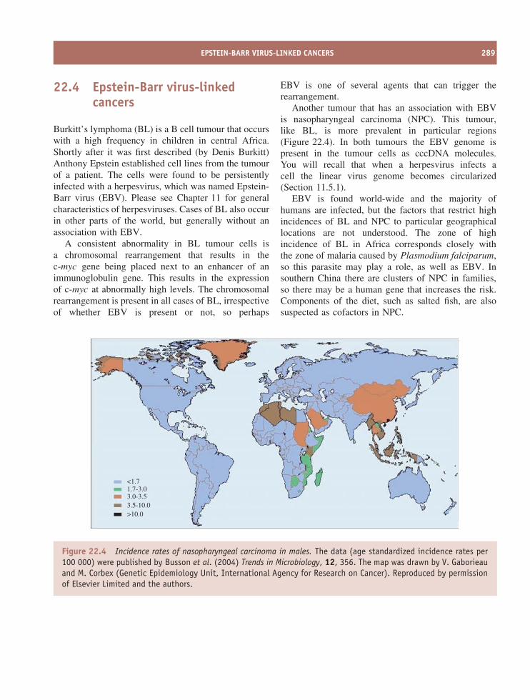
EPSTEIN-BARR VIRUS-LINKED CANCERS 289
22.4 Epstein-Barr virus-linkedcancers
Burkitt’s lymphoma (BL) is a B cell tumour that occurswith a high frequency in children in central Africa.Shortly after it was first described (by Denis Burkitt)Anthony Epstein established cell lines from the tumourof a patient. The cells were found to be persistentlyinfected with a herpesvirus, which was named Epstein-Barr virus (EBV). Please see Chapter 11 for generalcharacteristics of herpesviruses. Cases of BL also occurin other parts of the world, but generally without anassociation with EBV.
A consistent abnormality in BL tumour cells isa chromosomal rearrangement that results in thec-myc gene being placed next to an enhancer of animmunoglobulin gene. This results in the expressionof c-myc at abnormally high levels. The chromosomalrearrangement is present in all cases of BL, irrespectiveof whether EBV is present or not, so perhaps
EBV is one of several agents that can trigger therearrangement.
Another tumour that has an association with EBVis nasopharyngeal carcinoma (NPC). This tumour,like BL, is more prevalent in particular regions(Figure 22.4). In both tumours the EBV genome ispresent in the tumour cells as cccDNA molecules.You will recall that when a herpesvirus infects acell the linear virus genome becomes circularized(Section 11.5.1).
EBV is found world-wide and the majority ofhumans are infected, but the factors that restrict highincidences of BL and NPC to particular geographicallocations are not understood. The zone of highincidence of BL in Africa corresponds closely withthe zone of malaria caused by Plasmodium falciparum,so this parasite may play a role, as well as EBV. Insouthern China there are clusters of NPC in families,so there may be a human gene that increases the risk.Components of the diet, such as salted fish, are alsosuspected as cofactors in NPC.
<1.71.7-3.03.0-3.53.5-10.0>10.0
Figure 22.4 Incidence rates of nasopharyngeal carcinoma in males. The data (age standardized incidence rates per100 000) were published by Busson et al. (2004) Trends in Microbiology, 12, 356. The map was drawn by V. Gaborieauand M. Corbex (Genetic Epidemiology Unit, International Agency for Research on Cancer). Reproduced by permissionof Elsevier Limited and the authors.
290 VIRUSES AND CANCER
A number of other cancers have been found to haveassociations with EBV, including
• some cases of Hodgkin’s lymphoma
• non-Hodgkin’s lymphoma in AIDS
• post-transplant lymphoproliferative disorder.
In contrast to BL and NPC, Hodgkin’s lymphomais not restricted to particular geographical areas. Theother two cancers listed develop in patients whoare immunocompromised as a result of AIDS orimmunosuppressive treatment.
The oncogenic potential of EBV can be demon-strated when cultures of B cells are infected with thevirus. Infected cells synthesize a number of virus pro-teins that push the cells permanently into the cell cycle(Figure 4.5), resulting in the formation of lymphoblas-toid cell lines. In the body this B cell proliferation isnormally controlled by T cells, but in individuals whoare T cell immunocompromised the control may beinadequate and a B cell tumour may result.
22.5 Kaposi’s sarcomaKaposi’s sarcoma was first described in the 19thcentury as a rare skin cancer that affected elderly menin the Mediterranean region. Since the arrival of AIDSthis picture has changed. It is one of the most commoncancers in people with AIDS, and in these patientsthe cancer disseminates throughout the body and ismore aggressive. It is thought that the tumour cellsare derived from endothelial cells.
In 1994 it was discovered that the tumour cellscontain the DNA of a new herpesvirus. The viruswas subsequently isolated and was named Kaposi’s-sarcoma-associated herpesvirus (KSHV). The virus canbe found in most parts of the world, but serologicalstudies have shown that it is more common in certainregions, such as central Africa, and it is more commonin homosexual men. The risk of developing Kaposi’ssarcoma parallels the prevalence of the virus in thepopulation. As with EBV-associated tumours, copiesof the KSHV genome are present as cccDNA in thenuclei of the tumour cells.
There is also good evidence linking KSHV withtwo other human cancers: primary effusion lymphomasand multicentric Castleman’s disease. Both are tumoursderived from B cells.
22.6 Adult T cell leukaemia
Adult T cell leukaemia is associated with human T-lymphotropic virus 1 (HTLV-1) infection. Regions ofthe world with a high prevalence of the virus, suchas south-west Japan, have a high prevalence of thesetumours. HTLV-1 is a retrovirus and the tumour cellseach have a copy of the proviral DNA integrated intoa chromosome (Chapter 16).
22.7 Hepatocellular carcinoma
Hepatocellular carcinoma (liver cancer) accounts forfour to five per cent of cancer cases in the world. Anumber of factors are implicated as causative agents,including consumption of mould toxins in food, andtwo viruses: hepatitis B virus (HBV) and hepatitis Cvirus (HCV).
HBV is the most significant agent; the prevalenceof liver cancer closely parallels the prevalence ofpersistent HBV infection (Section 18.2), with thehighest incidences in Asia and in central and southernAfrica. In most of the tumours, HBV DNA is integratedinto the cell genome, and in most cases the virus DNAhas undergone rearrangements, including deletions.The P and C ORFs have generally been destroyed,but the S and X ORFs are often intact (please seeFigure 18.7 for a reminder of the HBV genes). Theonly viral gene product that is consistently present inthe tumour cells is the X protein.
The other virus associated with liver cancer, HCV,is a flavivirus (Figure 21.5). It is the only class IVvirus (plus-strand RNA virus) that is known to beoncogenic. Its genome is not found routinely in thecancer cells, in contrast to the other human oncogenicviruses.
Both HBV and HCV elicit immune responses whenthey infect the body. In some individuals the immune
HOW DO VIRUSES CAUSE CANCER? 291
response successfully eliminates the infection, but inmany cases the infection persists for life, as discussedin Section 18.2 for HBV. As far as HCV is concerned,it is estimated that about 80 per cent of individuals whobecome infected are unable to eliminate the infectionand that about 175 million people in the world areinfected. In some individuals who are persistentlyinfected with HBV or HCV liver cirrhosis develops,and for some this eventually leads on to cancer.
22.8 Virus-associated cancersin animals
A number of cancers in animal species have been foundto be associated with viruses. Outbreaks of some ofthese diseases can have serious economic consequencesin agriculture. A few examples are given here of animalcancers associated with retroviruses and a herpesvirus.
It was demonstrated early in the 20th centurythat filterable agents cause a leukosis (a B cellleukaemia/lymphoma) and a sarcoma in chickens.These agents were subsequently shown to be retro-viruses and the sarcoma-causing virus was namedRous sarcoma virus after its discoverer, Peyton Rous(Section 16.4). Other retroviruses were subsequentlyfound to be causative agents of leukaemia in cats andcattle, and a retrovirus known as Jaagsiekte sheep retro-virus was found to be responsible for adenocarcinomaof the lung in sheep.
Marek’s disease in chickens is a lymphoma, similarto that caused by retroviruses, but the causative agent isa herpesvirus and the transformed lymphocytes containmultiple copies of the viral genome. Papillomaviruses,already discussed as causative agents of human cancers(Section 22.2), cause cancers in cattle, horses andrabbits.
22.9 Cell lines derived fromvirus-associated cancers
Cancer cells, which by definition multiply continuouslyin the body, will often continue to multiply continu-ously if transferred into cell culture, when permanentcell lines may be established from them. Cell linesderived from most virus-induced cancers contain part
or all of the virus genome. The most famous of these,the HeLa cell line, was established from a cervicalcancer in the mid-20th century and is now used in lab-oratories around the world. Each HeLa cell contains acopy of part of the genome of HPV-18; this will bediscussed further in Section 22.10.1.
Cell lines derived from Burkitt’s lymphoma containcopies of the EBV genome and some produce infec-tious EBV; the virus was first discovered in one ofthese cell lines. Multiple copies of Marek’s diseasevirus genome are present in a cell line derived from aMarek’s tumour. A cell line derived from a liver cancercontains HBV DNA integrated at seven sites, and thecells secrete HBsAg. Cell lines derived from Kaposi’ssarcoma, however, do not contain KSHV DNA; thevirus DNA is lost from the tumour cells when theygrow in culture.
22.10 How do viruses cause cancer?Most virus-induced cancers develop after a long periodof persistent infection with an oncogenic virus; foradult T cell leukaemia this period is exceptionallylong (around 60 years). The virus infections persist intheir hosts in spite of immune responses, such as theproduction of virus-specific antibodies. Some persistentinfections are latent for much of the time (e.g. EBVand KSHV), with only small numbers of virus genesexpressed. Others, including HBV and HCV infections,are productive. Both of the latter viruses are able toevolve rapidly (Chapter 20), and this probably allowsthem to keep one step ahead of acquired immuneresponses.
Although many humans are persistently infectedby viruses that are potentially oncogenic, only smallpercentages develop virus-linked cancers. Relativelyfew people develop EBV-related tumours, though over90 per cent of adults worldwide are infected withthe virus. About three per cent of women persistentlyinfected with one of the high-risk strains of HPVdevelop cervical carcinoma, and similar percentages ofindividuals persistently infected with HBV, HCV orHTLV-1 develop a virus-linked cancer.
As cancer develops in only small percentages ofvirus-infected hosts it is clear that the virus infections
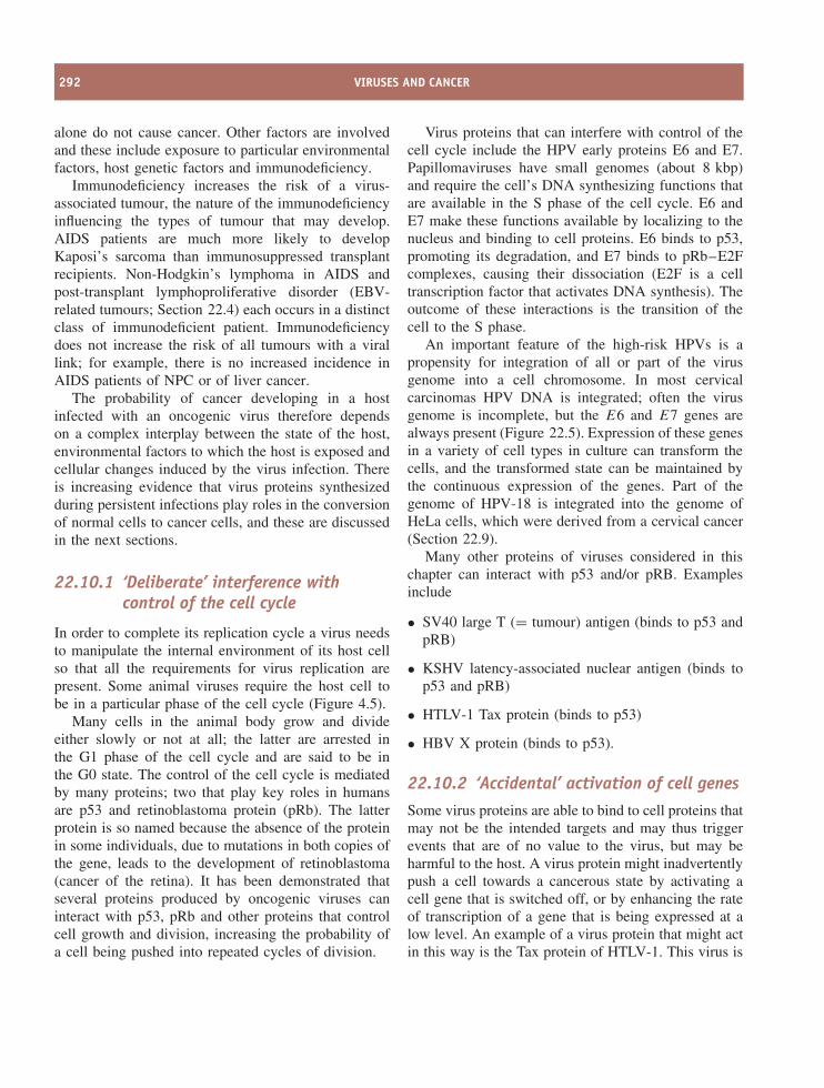
292 VIRUSES AND CANCER
alone do not cause cancer. Other factors are involvedand these include exposure to particular environmentalfactors, host genetic factors and immunodeficiency.
Immunodeficiency increases the risk of a virus-associated tumour, the nature of the immunodeficiencyinfluencing the types of tumour that may develop.AIDS patients are much more likely to developKaposi’s sarcoma than immunosuppressed transplantrecipients. Non-Hodgkin’s lymphoma in AIDS andpost-transplant lymphoproliferative disorder (EBV-related tumours; Section 22.4) each occurs in a distinctclass of immunodeficient patient. Immunodeficiencydoes not increase the risk of all tumours with a virallink; for example, there is no increased incidence inAIDS patients of NPC or of liver cancer.
The probability of cancer developing in a hostinfected with an oncogenic virus therefore dependson a complex interplay between the state of the host,environmental factors to which the host is exposed andcellular changes induced by the virus infection. Thereis increasing evidence that virus proteins synthesizedduring persistent infections play roles in the conversionof normal cells to cancer cells, and these are discussedin the next sections.
22.10.1 ‘Deliberate’ interference withcontrol of the cell cycle
In order to complete its replication cycle a virus needsto manipulate the internal environment of its host cellso that all the requirements for virus replication arepresent. Some animal viruses require the host cell tobe in a particular phase of the cell cycle (Figure 4.5).
Many cells in the animal body grow and divideeither slowly or not at all; the latter are arrested inthe G1 phase of the cell cycle and are said to be inthe G0 state. The control of the cell cycle is mediatedby many proteins; two that play key roles in humansare p53 and retinoblastoma protein (pRb). The latterprotein is so named because the absence of the proteinin some individuals, due to mutations in both copies ofthe gene, leads to the development of retinoblastoma(cancer of the retina). It has been demonstrated thatseveral proteins produced by oncogenic viruses caninteract with p53, pRb and other proteins that controlcell growth and division, increasing the probability ofa cell being pushed into repeated cycles of division.
Virus proteins that can interfere with control of thecell cycle include the HPV early proteins E6 and E7.Papillomaviruses have small genomes (about 8 kbp)and require the cell’s DNA synthesizing functions thatare available in the S phase of the cell cycle. E6 andE7 make these functions available by localizing to thenucleus and binding to cell proteins. E6 binds to p53,promoting its degradation, and E7 binds to pRb–E2Fcomplexes, causing their dissociation (E2F is a celltranscription factor that activates DNA synthesis). Theoutcome of these interactions is the transition of thecell to the S phase.
An important feature of the high-risk HPVs is apropensity for integration of all or part of the virusgenome into a cell chromosome. In most cervicalcarcinomas HPV DNA is integrated; often the virusgenome is incomplete, but the E6 and E7 genes arealways present (Figure 22.5). Expression of these genesin a variety of cell types in culture can transform thecells, and the transformed state can be maintained bythe continuous expression of the genes. Part of thegenome of HPV-18 is integrated into the genome ofHeLa cells, which were derived from a cervical cancer(Section 22.9).
Many other proteins of viruses considered in thischapter can interact with p53 and/or pRB. Examplesinclude
• SV40 large T (= tumour) antigen (binds to p53 andpRB)
• KSHV latency-associated nuclear antigen (binds top53 and pRB)
• HTLV-1 Tax protein (binds to p53)
• HBV X protein (binds to p53).
22.10.2 ‘Accidental’ activation of cell genesSome virus proteins are able to bind to cell proteins thatmay not be the intended targets and may thus triggerevents that are of no value to the virus, but may beharmful to the host. A virus protein might inadvertentlypush a cell towards a cancerous state by activating acell gene that is switched off, or by enhancing the rateof transcription of a gene that is being expressed at alow level. An example of a virus protein that might actin this way is the Tax protein of HTLV-1. This virus is
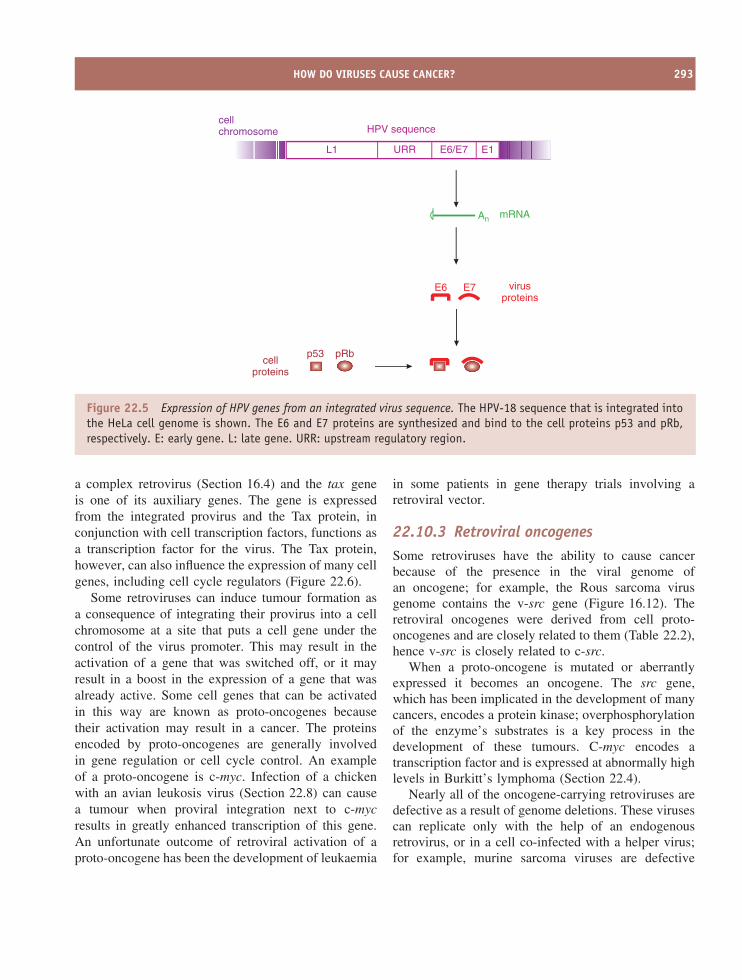
HOW DO VIRUSES CAUSE CANCER? 293
L1
HPV sequencecellchromosome
mRNA
URR E6/E7 E1
An
virusproteins
E6 E7
cellproteins
p53 pRb
Figure 22.5 Expression of HPV genes from an integrated virus sequence. The HPV-18 sequence that is integrated intothe HeLa cell genome is shown. The E6 and E7 proteins are synthesized and bind to the cell proteins p53 and pRb,respectively. E: early gene. L: late gene. URR: upstream regulatory region.
a complex retrovirus (Section 16.4) and the tax geneis one of its auxiliary genes. The gene is expressedfrom the integrated provirus and the Tax protein, inconjunction with cell transcription factors, functions asa transcription factor for the virus. The Tax protein,however, can also influence the expression of many cellgenes, including cell cycle regulators (Figure 22.6).
Some retroviruses can induce tumour formation asa consequence of integrating their provirus into a cellchromosome at a site that puts a cell gene under thecontrol of the virus promoter. This may result in theactivation of a gene that was switched off, or it mayresult in a boost in the expression of a gene that wasalready active. Some cell genes that can be activatedin this way are known as proto-oncogenes becausetheir activation may result in a cancer. The proteinsencoded by proto-oncogenes are generally involvedin gene regulation or cell cycle control. An exampleof a proto-oncogene is c-myc. Infection of a chickenwith an avian leukosis virus (Section 22.8) can causea tumour when proviral integration next to c-mycresults in greatly enhanced transcription of this gene.An unfortunate outcome of retroviral activation of aproto-oncogene has been the development of leukaemia
in some patients in gene therapy trials involving aretroviral vector.
22.10.3 Retroviral oncogenesSome retroviruses have the ability to cause cancerbecause of the presence in the viral genome ofan oncogene; for example, the Rous sarcoma virusgenome contains the v-src gene (Figure 16.12). Theretroviral oncogenes were derived from cell proto-oncogenes and are closely related to them (Table 22.2),hence v-src is closely related to c-src.
When a proto-oncogene is mutated or aberrantlyexpressed it becomes an oncogene. The src gene,which has been implicated in the development of manycancers, encodes a protein kinase; overphosphorylationof the enzyme’s substrates is a key process in thedevelopment of these tumours. C-myc encodes atranscription factor and is expressed at abnormally highlevels in Burkitt’s lymphoma (Section 22.4).
Nearly all of the oncogene-carrying retroviruses aredefective as a result of genome deletions. These virusescan replicate only with the help of an endogenousretrovirus, or in a cell co-infected with a helper virus;for example, murine sarcoma viruses are defective
294 VIRUSES AND CANCER
gag pol env LTR
HTLV-1 proviruscellchromosome
mRNAAn
Tax
celltranscription
factor
taxLTR
cell gene
mRNAAn
cell protein
Figure 22.6 HTLV-1 oncogenesis. The tax gene of the provirus is expressed. The Tax protein, complexed with cellproteins, activates transcription of the provirus and may also activate cell genes.
Table 22.2 Examples of retroviral oncogenes and the cell proto-oncogenes fromwhich they were derived. The names of some genes are derived from the virusnames. The names of viral oncogenes are prefixed by ‘v-’, while the names of cellproto-oncogenes are prefixed by ‘c-’
Retrovirus Viral oncogene Cell proto-oncogene
Rous sarcoma virus v-src c-srcAvian myelocytomatosis virus v-myc c-mycHarvey murine sarcoma virus v-H-ras c-H-ras
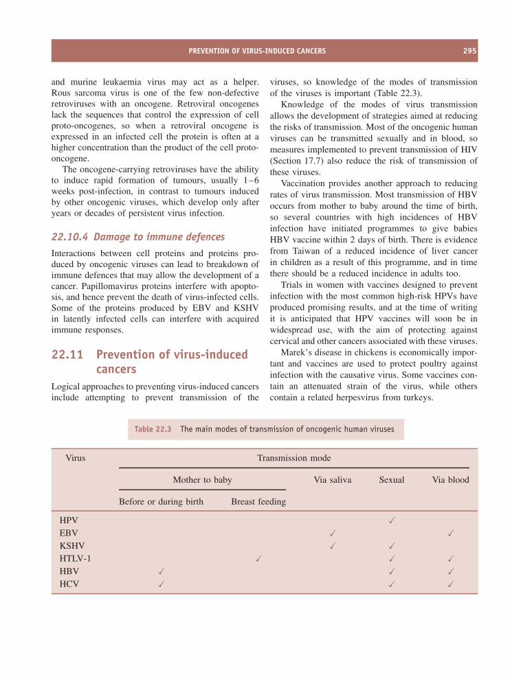
PREVENTION OF VIRUS-INDUCED CANCERS 295
and murine leukaemia virus may act as a helper.Rous sarcoma virus is one of the few non-defectiveretroviruses with an oncogene. Retroviral oncogeneslack the sequences that control the expression of cellproto-oncogenes, so when a retroviral oncogene isexpressed in an infected cell the protein is often at ahigher concentration than the product of the cell proto-oncogene.
The oncogene-carrying retroviruses have the abilityto induce rapid formation of tumours, usually 1–6weeks post-infection, in contrast to tumours inducedby other oncogenic viruses, which develop only afteryears or decades of persistent virus infection.
22.10.4 Damage to immune defencesInteractions between cell proteins and proteins pro-duced by oncogenic viruses can lead to breakdown ofimmune defences that may allow the development of acancer. Papillomavirus proteins interfere with apopto-sis, and hence prevent the death of virus-infected cells.Some of the proteins produced by EBV and KSHVin latently infected cells can interfere with acquiredimmune responses.
22.11 Prevention of virus-inducedcancers
Logical approaches to preventing virus-induced cancersinclude attempting to prevent transmission of the
viruses, so knowledge of the modes of transmissionof the viruses is important (Table 22.3).
Knowledge of the modes of virus transmissionallows the development of strategies aimed at reducingthe risks of transmission. Most of the oncogenic humanviruses can be transmitted sexually and in blood, someasures implemented to prevent transmission of HIV(Section 17.7) also reduce the risk of transmission ofthese viruses.
Vaccination provides another approach to reducingrates of virus transmission. Most transmission of HBVoccurs from mother to baby around the time of birth,so several countries with high incidences of HBVinfection have initiated programmes to give babiesHBV vaccine within 2 days of birth. There is evidencefrom Taiwan of a reduced incidence of liver cancerin children as a result of this programme, and in timethere should be a reduced incidence in adults too.
Trials in women with vaccines designed to preventinfection with the most common high-risk HPVs haveproduced promising results, and at the time of writingit is anticipated that HPV vaccines will soon be inwidespread use, with the aim of protecting againstcervical and other cancers associated with these viruses.
Marek’s disease in chickens is economically impor-tant and vaccines are used to protect poultry againstinfection with the causative virus. Some vaccines con-tain an attenuated strain of the virus, while otherscontain a related herpesvirus from turkeys.
Table 22.3 The main modes of transmission of oncogenic human viruses
Virus Transmission mode
Mother to baby Via saliva Sexual Via blood
Before or during birth Breast feeding
HPV �EBV � �KSHV � �HTLV-1 � � �HBV � � �HCV � � �
296 VIRUSES AND CANCER
An alternative approach to the prevention of virus-induced cancers is the attempted elimination from thebody of persistent infections with oncogenic viruses.So far this approach has had limited success. HCVinfection in some patients has been eliminated by treat-ment with α-interferon (Section 9.2.1.a), either alone orin combination with ribavirin. Persistent HBV infec-tions may be controlled, though not eliminated, usingα-interferon and/or lamivudine. Please see Chapter 25for more information on these anti-viral drugs.
Learning outcomes
By the end of this chapter you should be able to
• outline the characteristics of viruses that areassociated with cancers;
• evaluate the evidence for association of viruseswith some cancers;
• discuss possible mechanisms for virus inductionof cancer;
• suggest how virus-induced cancers may be pre-vented.
Sources of further information
Books
Basser R. L., Edwards S. and Frazer I. H. (2005) Develop-ment of vaccines against human papillomavirus (HPV),Chapter 12 in Moingeon P., editor Vaccines: Frontiers inDesign and Development, Horizon
Campo M. S., editor (2006) Papillomavirus Research: fromNatural History to Vaccines and Beyond, Caister
Gray L., Jolly C. and Herrington C. S. (2005) Human papil-lomaviruses and their effects on cell cycle control andapoptosis, pp. 235–251 in Molecular Pathogenesis ofVirus Infections, editors Digard P., Nash A. A. and Ran-dall R. E., 64th Symposium of the Society for GeneralMicrobiology, Cambridge University Press
Moore P. S. and Chang Y. (2003) Kaposi’s sarcoma-associated herpesvirus immunoevasion and tumorigenesis:
two sides of the same coin? pp. 609–639 in Annual Reviewof Microbiology, Vol. 57, Annual Reviews
Journals
Bekkers R. L. M. et al. (2004) Epidemiological and clinicalaspects of human papillomavirus detection in the preven-tion of cervical cancer Reviews in Medical Virology, 14,95–105
Butel J. S. (2000) Viral carcinogenesis: revelation of molec-ular mechanisms and etiology of human disease Carcino-genesis, 21, 405–426
Cathomas G. (2003) Kaposi’s sarcoma-associated herpes-virus (KSHV)/human herpesvirus 8 (HHV-8) as a tumourvirus Herpes, 10, 72–77
Collot-Teixeira S. et al. (2004) Human tumor suppressorp53 and DNA viruses Reviews in Medical Virology, 14,301–319
Guerrero R. B. and Roberts L. R. (2005) The role of hepati-tis B virus integrations in the pathogenesis of human hep-atocellular carcinoma Journal of Hepatology, 42, 760–777
Hammerschmidt W. and Sugden B. (2004) Epstein-Barrvirus sustains Burkitt’s lymphomas and Hodgkin’s diseaseTrends in Molecular Medicine, 10, 331–336
Hebner C. M. and Laimins L. A. (2005) Human papillo-maviruses: basic mechanisms of pathogenesis and onco-genicity Reviews in Medical Virology, 16, 83–89
Jarrett R. F. (2006) Viruses and lymphoma/leukaemia Jour-nal of Pathology, 208, 176–186
Longworth M. S. and Laimins L. A. (2004) Pathogenesis ofhuman papillomaviruses in differentiating epithelia Micro-biology and Molecular Biology Reviews, 68, 362–372
Lopes V., Young L. S. and Murray P. G. (2003) Epstein–Barr virus-associated cancers: aetiology and treatmentHerpes, 10, 78–82
Schulz T. F. (2006) The pleiotropic effects of Kaposi’ssarcoma herpesvirus Journal of Pathology, 208, 187–198
Snijders P. J. F. et al. (2006) HPV-mediated cervical car-cinogenesis: concepts and clinical implications Journal ofPathology, 208, 152–164
Thorley-Lawson D. A. (2005) EBV the prototypical humantumor virus – just how bad is it? Journal of Allergy andClinical Immunology, 116, 251–261
Pagano J. S. et al. (2004) Infectious agents and cancer:criteria for a causal relation Seminars in Cancer Biology,14, 453–471
Vilchez R. A. and Butel J. S. (2004) Emergent humanpathogen simian virus 40 and its role in cancer ClinicalMicrobiology Reviews, 17, 495–508
23Survival of infectivity
At a glance
• There may be two rates ofinfectivity loss.
%
infectivity
surviving
• In vaccine manufacture it is vital to ensure that maximuminfectivity is preserved in live vaccines, and that 100% ofinfectivity is destroyed in inactivated vaccines.
• Viruses vary greatly in the rate at which they lose infectivity.
• The rate of virus infectivity loss is affected by temperature, radiation and the chemical environment including pH.
Time
100
10
1
0.1
• Understanding the kinetics of infectivity loss is important in practicalsituations, such as devising disinfection and sterilization procedures,storage of virus preparations and vaccine manufacture.
Virology: Principles and Applications John B. Carter and Venetia A. Saunders 2007 John Wiley & Sons, Ltd ISBNs: 978-0-470-02386-0 (HB); 978-0-470-02387-7 (PB)
298 SURVIVAL OF INFECTIVITY
23.1 Preservation of virusinfectivity
There are several practical situations in which there isa need to preserve virus infectivity. A prime exampleis during the storage and distribution of live virusvaccines (Sections 24.2 and 24.5), when there is aneed to retain the potency of the vaccine. A secondexample is during the transport and storage of blood,faeces, saliva and other specimens from human andanimal patients prior to testing in a diagnostic virologylaboratory. Last, but by no means least, virologistsneed to preserve the infectivity of the viruses that theywork with.
23.2 Destruction of virus infectivity
There are many situations in which there is a require-ment to destroy virus infectivity. Procedures to min-imize the transmission of pathogenic viruses (andother micro-organisms) in hospitals and other healthcare facilities include the disinfection of surfaces andthe sterilization of equipment and materials. A goodexample of a situation requiring rigorous action isan outbreak of winter vomiting disease caused by anorovirus; patients’ vomit contains significant quanti-ties of virus, which must be inactivated if the outbreakis to be brought under control.
Inactivated (killed) virus vaccines (Section 24.3)are made from preparations of virulent viruses, soit is essential that all infectivity is destroyed. Bloodproducts, such as clotting factors for haemophiliacs andimmunoglobulins, are treated to destroy any viruses(e.g. HIV, hepatitis viruses) that might be present inthe donated blood. Continuing the theme of humanand animal health, water supplies that are free frompathogenic viruses (and other micro-organisms) are offundamental importance, so water treatment proceduresmust include processes that destroy these agents. Thesame applies to the treatment of swimming pool water.
The processes for production of cheese, yogurtand other dairy products utilize lactic acid bacteriato ferment the milk. There are many phages thatinfect these bacteria, and a phage infection during aproduction process can result in poor product quality,or in delay or failure of the fermentation. To minimize
the risk, procedures are required for treatment of themilk or whey to inactivate any phages present, and forthe disinfection of the factory environment.
Finally, virologists need supplies of pipettes, cellculture vessels and other laboratory materials thatare guaranteed free from viruses (and other micro-organisms), then after these materials have been usedany viruses present must be inactivated before disposal.
23.3 Inactivation targets in virions
The infectivity of a virion can be destroyed by thealteration or removal of its nucleic acid and/or one ormore of its proteins (Figure 23.1). A protein moleculemay be altered by the induction of a conformationalchange or, more drastically, by the breaking of covalentbonds, such as peptide bonds and disulphide bonds.
23.4 Inactivation kinetics
The rate at which a virus loses infectivity under definedconditions (for example, at a particular temperature andpH value) can be determined by incubating aliquotsof the virus preparation under those conditions. At
Naked Virion Enveloped Virion
internal protein
surface protein
nucleic acid
membrane
Figure 23.1 Inactivation targets in virions. Alterationof a surface protein might prevent a virion fromattaching to its host cell and/or from entering thecell. Stripping the envelope from an enveloped virionremoves the surface proteins and achieves the sameoutcome. Alteration of internal virion proteins candestroy properties, such as enzyme activities, essentialfor the replication of the virus.
AGENTS THAT INACTIVATE VIRUS INFECTIVITY 299
Time
%infectivitysurviving
100
10
1
0.1
Time
100
10
1
0.1
(a) (b)
Figure 23.2 Loss of virus infectivity at one rate (a) and at two rates (b).
various time intervals the surviving infectivity in analiquot is determined in an infectivity assay appropriatefor the virus (Section 2.8). The logarithm of thepercentage infectivity surviving is plotted against time(Figure 23.2).
In some circumstances infectivity is lost at aconstant rate until no further infectivity can be detected(Figure 23.2(a)). The slope of the line is a measure ofthe rate of infectivity loss. Sometimes there is a changein the rate of infectivity loss after most (usually morethan 99 per cent) of the infectivity has been destroyed(Figure 23.2(b)). It is not entirely clear why a smallfraction of some virus preparations loses infectivity ata slower rate. It has been suggested that this fractionmight contain virions that have a higher degree ofresistance to the inactivating factor and/or that thepreparation might contain virion clumps, with virionsat the centres of the clumps having some protection.
23.5 Agents that inactivate virusinfectivity
23.5.1 Physical agents
23.5.1.a Temperature
All infective viruses lose infectivity over time, therate of infectivity loss being highly dependent ontemperature. There is a lot of variability betweenviruses in the rates at which they are inactivated. Ingeneral, naked viruses are more stable than enveloped
viruses; most naked viruses lose little infectivity afterseveral hours at 37 ◦C, whereas most enveloped viruseslose significant infectivity under the same conditions.The generalization is, as stated, a broad one, as theremay be significant differences in stability betweenclosely related viruses. Under dry conditions varicella-zoster virus survives well, while the related herpessimplex viruses survive poorly.
At temperatures below −35 ◦C most viruses loseinfectivity very slowly, so for long-term storage ofviruses they are placed in freezers or in liquid nitrogen.Preservation of infectivity may be enhanced by storingvirus preparations in a freeze-dried form, rather than asaqueous suspensions. Moving to higher temperatures,most viruses are completely inactivated within 30minutes at 60 ◦C or within a few seconds at 100 ◦C.
Some of the bacteriophages of lactic acid bacteriathat cause problems in the dairy industry are quiteheat resistant, and some infectivity may survive theheat treatments used in the decontamination procedures(Figure 23.3).
The virions of some viruses, such as hepatitis Avirus (HAV) and parvoviruses, are exceptionally heatstable. Data for the minute virus of mice (a parvovirus)indicate some infectivity surviving after 60 minutes at80 ◦C (Figure 23.4).
Exceptions to the generalization concerning thestability of naked and enveloped virions include thoseof hepatitis B virus (HBV), which are enveloped buthave high stability, and those of poliovirus, which arenaked but have low stability. Interestingly, HAV (heat-stable virions) and poliovirus (heat-labile virions) are
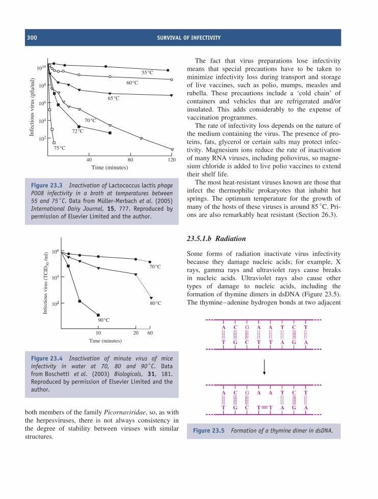
300 SURVIVAL OF INFECTIVITY
75°C
72°C
70°C
65°C
60°C
55°C
102
104
106
108
1010
40 80 120
Time (minutes)
Infe
ctio
us v
irus
(pf
u/m
l)
Figure 23.3 Inactivation of Lactococcus lactis phageP008 infectivity in a broth at temperatures between55 and 75 ◦C. Data from Muller-Merbach et al. (2005)International Dairy Journal, 15, 777. Reproduced bypermission of Elsevier Limited and the author.
70°C
102
104
106
10 20
Time (minutes)
Infe
ctio
us v
irus
(T
CID
50 /m
l)
60
80°C
90°C
Figure 23.4 Inactivation of minute virus of miceinfectivity in water at 70, 80 and 90 ◦C. Datafrom Boschetti et al. (2003) Biologicals, 31, 181.Reproduced by permission of Elsevier Limited and theauthor.
both members of the family Picornaviridae, so, as withthe herpesviruses, there is not always consistency inthe degree of stability between viruses with similarstructures.
The fact that virus preparations lose infectivitymeans that special precautions have to be taken tominimize infectivity loss during transport and storageof live vaccines, such as polio, mumps, measles andrubella. These precautions include a ‘cold chain’ ofcontainers and vehicles that are refrigerated and/orinsulated. This adds considerably to the expense ofvaccination programmes.
The rate of infectivity loss depends on the nature ofthe medium containing the virus. The presence of pro-teins, fats, glycerol or certain salts may protect infec-tivity. Magnesium ions reduce the rate of inactivationof many RNA viruses, including poliovirus, so magne-sium chloride is added to live polio vaccines to extendtheir shelf life.
The most heat-resistant viruses known are those thatinfect the thermophilic prokaryotes that inhabit hotsprings. The optimum temperature for the growth ofmany of the hosts of these viruses is around 85 ◦C. Pri-ons are also remarkably heat resistant (Section 26.3).
23.5.1.b Radiation
Some forms of radiation inactivate virus infectivitybecause they damage nucleic acids; for example, Xrays, gamma rays and ultraviolet rays cause breaksin nucleic acids. Ultraviolet rays also cause othertypes of damage to nucleic acids, including theformation of thymine dimers in dsDNA (Figure 23.5).The thymine–adenine hydrogen bonds at two adjacent
A C G
GG
A A T
T T
T
T
C
A C G A A T TC
C AA
GG T TT C AA
Figure 23.5 Formation of a thymine dimer in dsDNA.
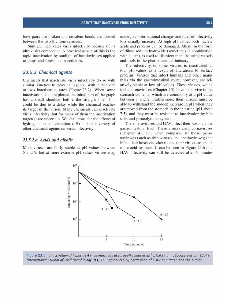
AGENTS THAT INACTIVATE VIRUS INFECTIVITY 301
base pairs are broken and covalent bonds are formedbetween the two thymine residues.
Sunlight inactivates virus infectivity because of itsultraviolet component. A practical aspect of this is therapid inactivation by sunlight of baculoviruses appliedto crops and forests as insecticides.
23.5.2 Chemical agents
Chemicals that inactivate virus infectivity do so withsimilar kinetics to physical agents, with either oneor two inactivation rates (Figure 23.2). When someinactivation data are plotted the initial part of the graphhas a small shoulder before the straight line. Thiscould be due to a delay while the chemical reachesits target in the virion. Many chemicals can inactivatevirus infectivity, but for many of them the inactivationtarget(s) are uncertain. We shall consider the effects ofhydrogen ion concentration (pH) and of a variety ofother chemical agents on virus infectivity.
23.5.2.a Acids and alkalis
Most viruses are fairly stable at pH values between5 and 9, but at more extreme pH values virions may
undergo conformational changes and rates of infectivityloss usually increase. At high pH values both nucleicacids and proteins can be damaged. Alkali, in the formof dilute sodium hydroxide (sometimes in combinationwith steam), is used to disinfect manufacturing vesselsand tools in the pharmaceutical industry.
The infectivity of some viruses is inactivated atlow pH values as a result of alterations to surfaceproteins. Viruses that infect humans and other mam-mals via the gastrointestinal route, however, are rel-atively stable at low pH values. These viruses, whichinclude rotaviruses (Chapter 13), have to survive in thestomach contents, which are commonly at a pH valuebetween 1 and 2. Furthermore, their virions must beable to withstand the sudden increase in pH when theyare moved from the stomach to the intestine (pH about7.5), and they must be resistant to inactivation by bilesalts and proteolytic enzymes.
The enteroviruses and HAV infect their hosts via thegastrointestinal tract. These viruses are picornaviruses(Chapter 14), but, when compared to those picor-naviruses (such as rhinoviruses and aphthoviruses) thatinfect their hosts via other routes, their virions are muchmore acid resistant. It can be seen in Figure 23.6 thatHAV infectivity can still be detected after 6 minutes
5 10 15
Time (minutes)
pH 3.8
pH 4.3
pH 3.3
102
104
106
108
Infe
ctio
us v
irus
(pf
u/m
l)
Figure 23.6 Inactivation of hepatitis A virus infectivity at three pH values at 85 ◦C. Data from Deboosere et al. (2004)International Journal of Food Microbiology, 93, 73. Reproduced by permission of Elsevier Limited and the author.
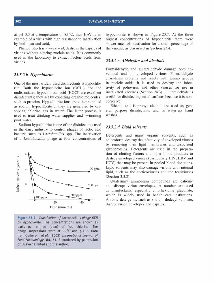
302 SURVIVAL OF INFECTIVITY
at pH 3.3 at a temperature of 85 ◦C, thus HAV is anexample of a virus with high resistance to inactivationby both heat and acid.
Phenol, which is a weak acid, destroys the capsids ofvirions without altering nucleic acids. It is commonlyused in the laboratory to extract nucleic acids fromvirions.
23.5.2.b Hypochlorite
One of the most widely used disinfectants is hypochlo-rite. Both the hypochlorite ion (OCl−) and theundissociated hypochlorous acid (HOCl) are excellentdisinfectants; they act by oxidizing organic molecules,such as proteins. Hypochlorite ions are either suppliedas sodium hypochlorite or they are generated by dis-solving chlorine gas in water. The latter process isused to treat drinking water supplies and swimmingpool water.
Sodium hypochlorite is one of the disinfectants usedin the dairy industry to control phages of lactic acidbacteria such as Lactobacillus spp. The inactivationof a Lactobacillus phage at four concentrations of
100 ppm
102
104
106
20 40
Time (minutes)
Infe
ctio
us v
irus
(pf
u/m
l)
200 ppm
300 ppm400 ppm
Figure 23.7 Inactivation of Lactobacillus phage BYMby hypochlorite. The concentrations are shown asparts per million (ppm) of free chlorine. Thephage suspensions were at 25 ◦C and pH 7. Datafrom Quiberoni et al. (2003) International Journal ofFood Microbiology, 84, 51. Reproduced by permissionof Elsevier Limited and the author.
hypochlorite is shown in Figure 23.7. At the threehighest concentrations of hypochlorite there wereslower rates of inactivation for a small percentage ofthe virions, as discussed in Section 23.4.
23.5.2.c Aldehydes and alcohols
Formaldehyde and glutaraldehyde damage both en-veloped and non-enveloped virions. Formaldehydecross-links proteins and reacts with amino groupsin nucleic acids; it is used to destroy the infec-tivity of poliovirus and other viruses for use ininactivated vaccines (Section 24.3). Glutaraldehyde isuseful for disinfecting metal surfaces because it is non-corrosive.
Ethanol and isopropyl alcohol are used as gen-eral purpose disinfectants and in waterless handwashes.
23.5.2.d Lipid solvents
Detergents and many organic solvents, such aschloroform, destroy the infectivity of enveloped virusesby removing their lipid membranes and associatedglycoproteins. Detergents are used in the prepara-tion of clotting factors and other blood products todestroy enveloped viruses (particularly HIV, HBV andHCV) that may be present in pooled blood donations.Lipid solvents may also damage virions with internallipid, such as the corticoviruses and the tectiviruses(Section 3.5.2).
Quaternary ammonium compounds are cationicand disrupt virion envelopes. A number are usedas disinfectants, especially chlorhexidine gluconate,which is widely used in health care institutions.Anionic detergents, such as sodium dodecyl sulphate,disrupt virion envelopes and capsids.
SOURCES OF FURTHER INFORMATION 303
Learning outcomes
By the end of this chapter you should be able to
• discuss situations in which preservation of virusinfectivity is important;
• discuss situations in which destruction of virusinfectivity is important;
• give examples of physical and chemical agentsthat damage virions and cause them to loseinfectivity;
• discuss the kinetics of inactivation of virusinfectivity.
Sources of further information
Books
Fraise A. P., Lambert P. A. and Maillard J.-Y., editors (2004)
Russell, Hugo and Aycliffe’s Principles and Practice ofDisinfection, Preservation and Sterilization, 4th edition,Blackwell
Wolff M. H. et al. (2005) Environmental survival and micro-bicide inactivation of coronaviruses, pp. 201–212 inSchmidt A., Wolff M.H. and Weber O., editors, Coron-aviruses with Special Emphasis on First Insights Concern-ing SARS, Birkhauser
Journals
Farshid M. et al. (2005) The clearance of viruses and trans-missible spongiform encephalopathy agents from biologi-cals Current Opinion in Biotechnology, 16, 561–567
Maillard J.-Y. (2001) Virus susceptibility to biocides: anunderstanding Reviews in Medical Microbiology, 12,63–74
McDonnell G. and Russell A. D. (1999) Antiseptics anddisinfectants: activity, action, and resistance ClinicalMicrobiology Reviews, 12, 147–179
Rzezutka A. and Cook N. (2004) Survival of human entericviruses in the environment and food FEMS MicrobiologyReviews, 28, 441–453

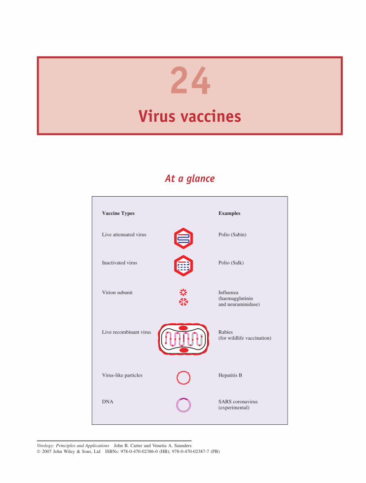
24Virus vaccines
At a glance
Live attenuated virus
Inactivated virus
Virion subunit
Live recombinant virus
Virus-like particles
DNA
Examples
Polio (Sabin)
Polio (Salk)
Influenza(haemagglutininand neuraminidase)
Rabies(for wildlife vaccination)
Vaccine Types
Hepatitis B
SARS coronavirus(experimental)
Virology: Principles and Applications John B. Carter and Venetia A. Saunders 2007 John Wiley & Sons, Ltd ISBNs: 978-0-470-02386-0 (HB); 978-0-470-02387-7 (PB)
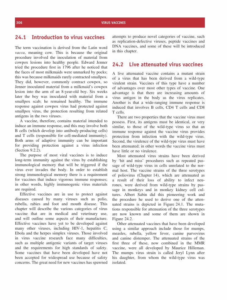
306 VIRUS VACCINES
24.1 Introduction to virus vaccines
The term vaccination is derived from the Latin wordvacca, meaning cow. This is because the originalprocedure involved the inoculation of material fromcowpox lesions into healthy people. Edward Jennertried the procedure first in 1796 after he noticed thatthe faces of most milkmaids were unmarked by pocks;this was because milkmaids rarely contracted smallpox.They did, however, commonly contract cowpox, soJenner inoculated material from a milkmaid’s cowpoxlesion into the arm of an 8-year-old boy. Six weekslater the boy was inoculated with material from asmallpox scab; he remained healthy. The immuneresponse against cowpox virus had protected againstsmallpox virus, the protection resulting from relatedantigens in the two viruses.
A vaccine, therefore, contains material intended toinduce an immune response, and this may involve bothB cells (which develop into antibody-producing cells)and T cells (responsible for cell-mediated immunity).Both arms of adaptive immunity can be importantfor providing protection against a virus infection(Section 9.2.2).
The purpose of most viral vaccines is to inducelong-term immunity against the virus by establishingimmunological memory that will be triggered if thevirus ever invades the body. In order to establishstrong immunological memory there is a requirementfor vaccines that induce vigorous immune responses;in other words, highly immunogenic virus materialsare required.
Effective vaccines are in use to protect againstdiseases caused by many viruses such as polio,rubella, rabies and foot and mouth disease. Thischapter will describe the various categories of virusvaccine that are in medical and veterinary use,and will outline some aspects of their manufacture.Effective vaccines have yet to be developed againstmany other viruses, including HIV-1, hepatitis C,Ebola and the herpes simplex viruses. Those involvedin virus vaccine research face many difficulties,such as multiple antigenic variants of target virusesand the requirements for high standards of safety.Some vaccines that have been developed have notbeen accepted for widespread use because of safetyconcerns. The great need for new vaccines has spawned
attempts to produce novel categories of vaccine, suchas replication-defective viruses, peptide vaccines andDNA vaccines, and some of these will be introducedin this chapter.
24.2 Live attenuated virus vaccinesA live attenuated vaccine contains a mutant strainof a virus that has been derived from a wild-typevirulent strain. Vaccines of this type have a numberof advantages over most other types of vaccine. Oneadvantage is that there are increasing amounts ofvirus antigen in the body as the virus replicates.Another is that a wide-ranging immune response isinduced that involves B cells, CD4 T cells and CD8T cells.
There are two properties that the vaccine virus mustpossess. First, its antigens must be identical, or verysimilar, to those of the wild-type virus so that animmune response against the vaccine virus providesprotection from infection with the wild-type virus.Second, the virulence of the wild-type virus must havebeen attenuated; in other words the vaccine virus musthave little or no virulence.
Most attenuated virus strains have been derivedby ‘hit and miss’ procedures such as repeated pas-sage of wild-type virus in cells unrelated to the nor-mal host. The vaccine strains of the three serotypesof poliovirus (Chapter 14), which are attenuated asa result of their loss of ability to infect neu-rones, were derived from wild-type strains by pas-sage in monkeys and in monkey kidney cell cul-tures. Albert Sabin did this pioneering work andthe procedure he used to derive one of the atten-uated strains is depicted in Figure 24.1. The muta-tions responsible for attenuation of the three serotypesare now known and some of them are shown inFigure 24.2.
Other attenuated vaccines that have been developedusing a similar approach include those for mumps,measles, rubella, yellow fever, canine parvovirusand canine distemper. The attenuated strains of thefirst three of these, now combined in the MMRvaccine, were all developed by Maurice Hilleman.The mumps virus strain is called Jeryl Lynn afterhis daughter, from whom the wild-type virus wasisolated.
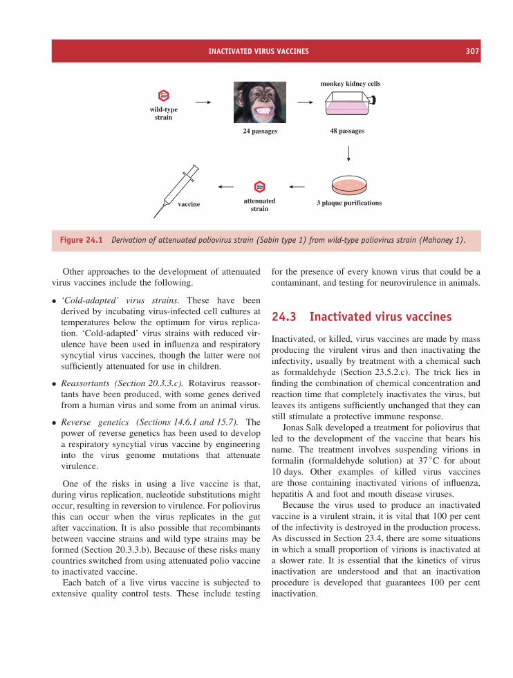
INACTIVATED VIRUS VACCINES 307
wild-typestrain
24 passages 48 passages
monkey kidney cells
3 plaque purificationsattenuatedstrain
vaccine
Figure 24.1 Derivation of attenuated poliovirus strain (Sabin type 1) from wild-type poliovirus strain (Mahoney 1).
Other approaches to the development of attenuatedvirus vaccines include the following.
• ‘Cold-adapted’ virus strains. These have beenderived by incubating virus-infected cell cultures attemperatures below the optimum for virus replica-tion. ‘Cold-adapted’ virus strains with reduced vir-ulence have been used in influenza and respiratorysyncytial virus vaccines, though the latter were notsufficiently attenuated for use in children.
• Reassortants (Section 20.3.3.c). Rotavirus reassor-tants have been produced, with some genes derivedfrom a human virus and some from an animal virus.
• Reverse genetics (Sections 14.6.1 and 15.7). Thepower of reverse genetics has been used to developa respiratory syncytial virus vaccine by engineeringinto the virus genome mutations that attenuatevirulence.
One of the risks in using a live vaccine is that,during virus replication, nucleotide substitutions mightoccur, resulting in reversion to virulence. For poliovirusthis can occur when the virus replicates in the gutafter vaccination. It is also possible that recombinantsbetween vaccine strains and wild type strains may beformed (Section 20.3.3.b). Because of these risks manycountries switched from using attenuated polio vaccineto inactivated vaccine.
Each batch of a live virus vaccine is subjected toextensive quality control tests. These include testing
for the presence of every known virus that could be acontaminant, and testing for neurovirulence in animals.
24.3 Inactivated virus vaccines
Inactivated, or killed, virus vaccines are made by massproducing the virulent virus and then inactivating theinfectivity, usually by treatment with a chemical suchas formaldehyde (Section 23.5.2.c). The trick lies infinding the combination of chemical concentration andreaction time that completely inactivates the virus, butleaves its antigens sufficiently unchanged that they canstill stimulate a protective immune response.
Jonas Salk developed a treatment for poliovirus thatled to the development of the vaccine that bears hisname. The treatment involves suspending virions informalin (formaldehyde solution) at 37 ◦C for about10 days. Other examples of killed virus vaccinesare those containing inactivated virions of influenza,hepatitis A and foot and mouth disease viruses.
Because the virus used to produce an inactivatedvaccine is a virulent strain, it is vital that 100 per centof the infectivity is destroyed in the production process.As discussed in Section 23.4, there are some situationsin which a small proportion of virions is inactivated ata slower rate. It is essential that the kinetics of virusinactivation are understood and that an inactivationprocedure is developed that guarantees 100 per centinactivation.
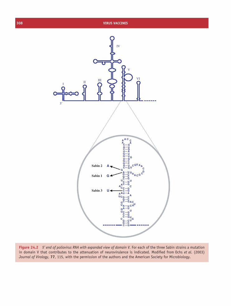
308 VIRUS VACCINES
IV
VI
V
IIII II
5′
Sabin 2
Sabin 1
Sabin 3
A
G
U
A A C C
C
C
C
CC
C
CC
C
CC
CCC
CCC
C
C CC
C CC
CCC
CC
CCG
GG
GGGG
G
G GGG
GG
GGG
GG
GG
G
GG
G
GG
GGGG
G
GU
UUU
U
UU
U
U
U U
UU
U UU
UUU
UU
A A
A
A
A
A
AA
AA AA
A
AA
A
AA
AA
Figure 24.2 5’ end of poliovirus RNA with expanded view of domain V. For each of the three Sabin strains a mutationin domain V that contributes to the attenuation of neurovirulence is indicated. Modified from Ochs et al. (2003)Journal of Virology, 77, 115, with the permission of the authors and the American Society for Microbiology.
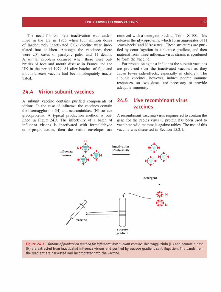
LIVE RECOMBINANT VIRUS VACCINES 309
The need for complete inactivation was under-lined in the US in 1955 when four million dosesof inadequately inactivated Salk vaccine were inoc-ulated into children. Amongst the vaccinees therewere 204 cases of paralytic polio and 11 deaths.A similar problem occurred when there were out-breaks of foot and mouth disease in France and theUK in the period 1979–81 after batches of foot andmouth disease vaccine had been inadequately inacti-vated.
24.4 Virion subunit vaccines
A subunit vaccine contains purified components ofvirions. In the case of influenza the vaccines containthe haemagglutinin (H) and neuraminidase (N) surfaceglycoproteins. A typical production method is out-lined in Figure 24.3. The infectivity of a batch ofinfluenza virions is inactivated with formaldehydeor β-propiolactone, then the virion envelopes are
removed with a detergent, such as Triton X-100. Thisreleases the glycoproteins, which form aggregates of H‘cartwheels’ and N ‘rosettes’. These structures are puri-fied by centrifugation in a sucrose gradient, and thenmaterial from three influenza virus strains is combinedto form the vaccine.
For protection against influenza the subunit vaccinesare preferred over the inactivated vaccines as theycause fewer side-effects, especially in children. Thesubunit vaccines, however, induce poorer immuneresponses, so two doses are necessary to provideadequate immunity.
24.5 Live recombinant virusvaccines
A recombinant vaccinia virus engineered to contain thegene for the rabies virus G protein has been used tovaccinate wild mammals against rabies. The use of thisvaccine was discussed in Section 15.2.1.
H
N
H
Ninfluenza
virions
inactivationof infectivity
detergent
vaccine
sucrosegradient
Figure 24.3 Outline of production method for influenza virus subunit vaccine. Haemagglutinin (H) and neuraminidase(N) are extracted from inactivated influenza virions and purified by sucrose gradient centrifugation. The bands fromthe gradient are harvested and incorporated into the vaccine.
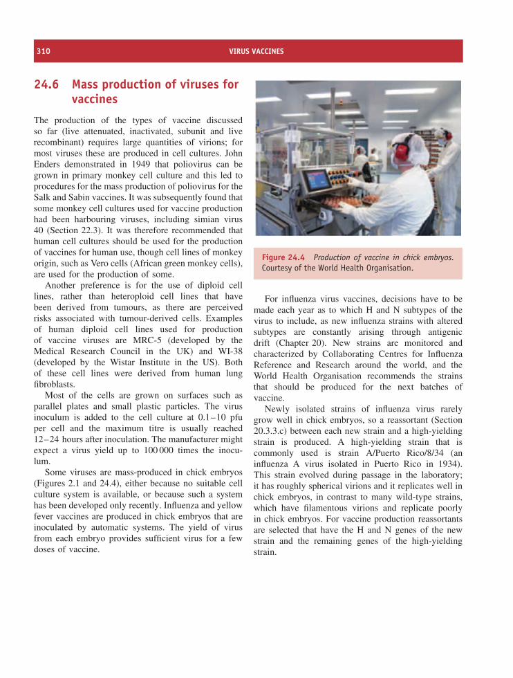
310 VIRUS VACCINES
24.6 Mass production of viruses forvaccines
The production of the types of vaccine discussedso far (live attenuated, inactivated, subunit and liverecombinant) requires large quantities of virions; formost viruses these are produced in cell cultures. JohnEnders demonstrated in 1949 that poliovirus can begrown in primary monkey cell culture and this led toprocedures for the mass production of poliovirus for theSalk and Sabin vaccines. It was subsequently found thatsome monkey cell cultures used for vaccine productionhad been harbouring viruses, including simian virus40 (Section 22.3). It was therefore recommended thathuman cell cultures should be used for the productionof vaccines for human use, though cell lines of monkeyorigin, such as Vero cells (African green monkey cells),are used for the production of some.
Another preference is for the use of diploid celllines, rather than heteroploid cell lines that havebeen derived from tumours, as there are perceivedrisks associated with tumour-derived cells. Examplesof human diploid cell lines used for productionof vaccine viruses are MRC-5 (developed by theMedical Research Council in the UK) and WI-38(developed by the Wistar Institute in the US). Bothof these cell lines were derived from human lungfibroblasts.
Most of the cells are grown on surfaces such asparallel plates and small plastic particles. The virusinoculum is added to the cell culture at 0.1–10 pfuper cell and the maximum titre is usually reached12–24 hours after inoculation. The manufacturer mightexpect a virus yield up to 100 000 times the inocu-lum.
Some viruses are mass-produced in chick embryos(Figures 2.1 and 24.4), either because no suitable cellculture system is available, or because such a systemhas been developed only recently. Influenza and yellowfever vaccines are produced in chick embryos that areinoculated by automatic systems. The yield of virusfrom each embryo provides sufficient virus for a fewdoses of vaccine.
Figure 24.4 Production of vaccine in chick embryos.Courtesy of the World Health Organisation.
For influenza virus vaccines, decisions have to bemade each year as to which H and N subtypes of thevirus to include, as new influenza strains with alteredsubtypes are constantly arising through antigenicdrift (Chapter 20). New strains are monitored andcharacterized by Collaborating Centres for InfluenzaReference and Research around the world, and theWorld Health Organisation recommends the strainsthat should be produced for the next batches ofvaccine.
Newly isolated strains of influenza virus rarelygrow well in chick embryos, so a reassortant (Section20.3.3.c) between each new strain and a high-yieldingstrain is produced. A high-yielding strain that iscommonly used is strain A/Puerto Rico/8/34 (aninfluenza A virus isolated in Puerto Rico in 1934).This strain evolved during passage in the laboratory;it has roughly spherical virions and it replicates well inchick embryos, in contrast to many wild-type strains,which have filamentous virions and replicate poorlyin chick embryos. For vaccine production reassortantsare selected that have the H and N genes of the newstrain and the remaining genes of the high-yieldingstrain.
DNA VACCINES 311
Table 24.1 Examples of viruses that are massproduced for vaccine manufacture
Vaccines for humans Vaccines for animals
Polio Foot and mouth diseaseInfluenza Marek’s diseaseMeasles Newcastle diseaseMumps Canine distemperRubella RabiesRabiesYellow fever
Although influenza virus has traditionally been pro-duced in chick embryos for incorporation into inac-tivated and subunit vaccines, cell culture systemsfor virus production have now been developed.Furthermore, live attenuated influenza virus vac-cines have been licensed in some countries, so thetypes of influenza virus vaccine and the ways inwhich they are mass produced may change in thefuture.
There are major enterprises around the worldproducing vaccines for human and veterinary use.Some examples of the vaccines produced are given inTable 24.1.
24.7 Virus-like particles
Virus-like particles are structures assembled from virusproteins. The particles resemble virions, but they aredevoid of any nucleic acid and can therefore be deemedsafer than vaccines containing attenuated or inactivatedvirions.
Hepatitis B virus (HBV) vaccine is produced inrecombinant yeast cells that have the gene for theHBsAg inserted into the genome. The cells are grownin bulk and then broken to release the virus protein.After purification the HBsAg molecules receive achemical treatment that causes them to aggregate
into spherical structures similar to the non-infectiousparticles produced in the HBV-infected human host(Section 18.4).
The major capsid protein of papillomaviruses canself-assemble into virus-like particles that bear the epi-topes required for generating neutralizing antibodies.(An epitope is a short sequence of amino acids criti-cal for inducing an immune response.) Human papill-omavirus vaccines (Section 22.11) contain virus-likeparticles that are produced either in insect cells usinga baculovirus vector or in yeast cells.
24.8 Synthetic peptide vaccines
Each protein antigen has one or more epitopes. Theseshort amino acid sequences can be synthesized in amachine and it was suggested that the resulting peptidesmight be used as vaccines. Compared with traditionalvaccines it would be easier to ensure the absence ofcontaminants such as viruses and proteins.
A lot of work has been done to try to develop peptidevaccines against foot and mouth disease virus. In thisvirus there is an important epitope within the virionprotein VP1 (Section 14.3.1). Synthetic peptides of thissequence induced reasonable levels of neutralizing andprotective antibodies in laboratory animals, but whenvaccine trials were done in farm animals the resultswere disappointing.
24.9 DNA vaccines
The most revolutionary approach to vaccination isthe introduction into the vaccinee of DNA encodingan antigen, with the aim of inducing cells of thevaccinee to synthesize the antigen. One advantageof this approach is that there is a steady supplyof new antigen to stimulate the immune system, aswith live virus vaccines. Because the antigen (a virusprotein in this case) is produced within the cells ofthe vaccinee, it is likely to stimulate efficient T-cell-mediated responses.
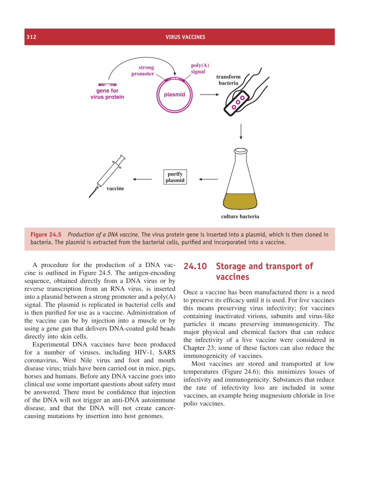
312 VIRUS VACCINES
gene forvirus protein plasmid
strongpromoter
poly(A)signal
transformbacteria
vaccine
purifyplasmid
culture bacteria
Figure 24.5 Production of a DNA vaccine. The virus protein gene is inserted into a plasmid, which is then cloned inbacteria. The plasmid is extracted from the bacterial cells, purified and incorporated into a vaccine.
A procedure for the production of a DNA vac-cine is outlined in Figure 24.5. The antigen-encodingsequence, obtained directly from a DNA virus or byreverse transcription from an RNA virus, is insertedinto a plasmid between a strong promoter and a poly(A)signal. The plasmid is replicated in bacterial cells andis then purified for use as a vaccine. Administration ofthe vaccine can be by injection into a muscle or byusing a gene gun that delivers DNA-coated gold beadsdirectly into skin cells.
Experimental DNA vaccines have been producedfor a number of viruses, including HIV-1, SARScoronavirus, West Nile virus and foot and mouthdisease virus; trials have been carried out in mice, pigs,horses and humans. Before any DNA vaccine goes intoclinical use some important questions about safety mustbe answered. There must be confidence that injectionof the DNA will not trigger an anti-DNA autoimmunedisease, and that the DNA will not create cancer-causing mutations by insertion into host genomes.
24.10 Storage and transport ofvaccines
Once a vaccine has been manufactured there is a needto preserve its efficacy until it is used. For live vaccinesthis means preserving virus infectivity; for vaccinescontaining inactivated virions, subunits and virus-likeparticles it means preserving immunogenicity. Themajor physical and chemical factors that can reducethe infectivity of a live vaccine were considered inChapter 23; some of these factors can also reduce theimmunogenicity of vaccines.
Most vaccines are stored and transported at lowtemperatures (Figure 24.6); this minimizes losses ofinfectivity and immunogenicity. Substances that reducethe rate of infectivity loss are included in somevaccines, an example being magnesium chloride in livepolio vaccines.
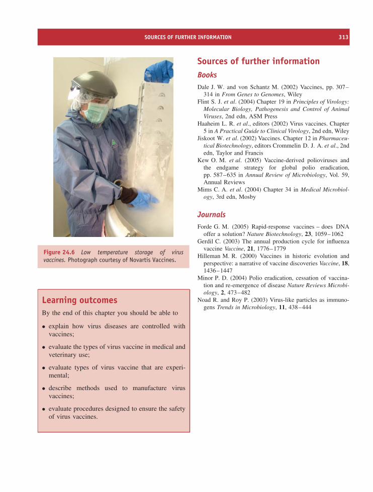
SOURCES OF FURTHER INFORMATION 313
Figure 24.6 Low temperature storage of virusvaccines. Photograph courtesy of Novartis Vaccines.
Learning outcomesBy the end of this chapter you should be able to
• explain how virus diseases are controlled withvaccines;
• evaluate the types of virus vaccine in medical andveterinary use;
• evaluate types of virus vaccine that are experi-mental;
• describe methods used to manufacture virusvaccines;
• evaluate procedures designed to ensure the safetyof virus vaccines.
Sources of further informationBooksDale J. W. and von Schantz M. (2002) Vaccines, pp. 307–
314 in From Genes to Genomes, WileyFlint S. J. et al. (2004) Chapter 19 in Principles of Virology:
Molecular Biology, Pathogenesis and Control of AnimalViruses, 2nd edn, ASM Press
Haaheim L. R. et al., editors (2002) Virus vaccines. Chapter5 in A Practical Guide to Clinical Virology, 2nd edn, Wiley
Jiskoot W. et al. (2002) Vaccines. Chapter 12 in Pharmaceu-tical Biotechnology, editors Crommelin D. J. A. et al., 2ndedn, Taylor and Francis
Kew O. M. et al. (2005) Vaccine-derived polioviruses andthe endgame strategy for global polio eradication,pp. 587–635 in Annual Review of Microbiology, Vol. 59,Annual Reviews
Mims C. A. et al. (2004) Chapter 34 in Medical Microbiol-ogy, 3rd edn, Mosby
JournalsForde G. M. (2005) Rapid-response vaccines – does DNA
offer a solution? Nature Biotechnology, 23, 1059–1062Gerdil C. (2003) The annual production cycle for influenza
vaccine Vaccine, 21, 1776–1779Hilleman M. R. (2000) Vaccines in historic evolution and
perspective: a narrative of vaccine discoveries Vaccine, 18,1436–1447
Minor P. D. (2004) Polio eradication, cessation of vaccina-tion and re-emergence of disease Nature Reviews Microbi-ology, 2, 473–482
Noad R. and Roy P. (2003) Virus-like particles as immuno-gens Trends in Microbiology, 11, 438–444

25Anti-viral drugs
At a glance
fusion
reversetranscription
DNAreplication
budding
maturation
Replication targets of anti-viral drugs [with examples of drugs]
[enfuvirtide]
[AZT, lamivudine,nevirapine]
[aciclovir,ganciclovir]
[oseltamivir]
[ritonavir]
Virology: Principles and Applications John B. Carter and Venetia A. Saunders 2007 John Wiley & Sons, Ltd ISBNs: 978-0-470-02386-0 (HB); 978-0-470-02387-7 (PB)
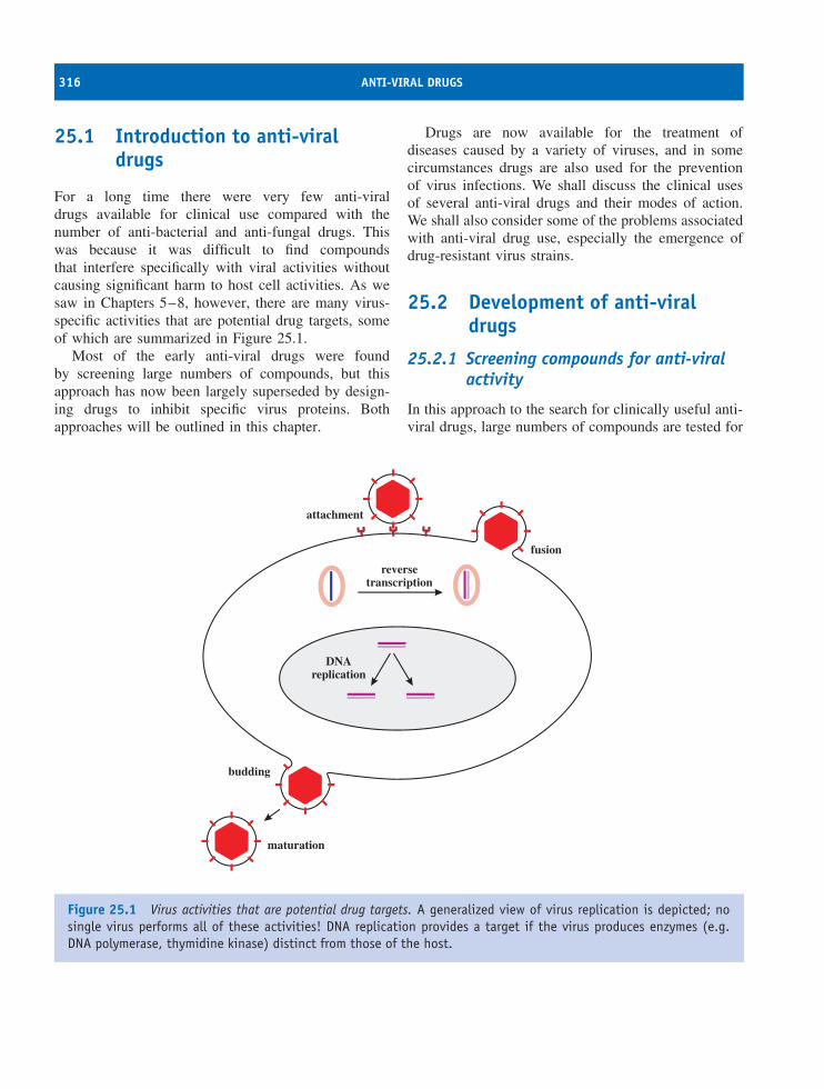
316 ANTI-VIRAL DRUGS
25.1 Introduction to anti-viraldrugs
For a long time there were very few anti-viraldrugs available for clinical use compared with thenumber of anti-bacterial and anti-fungal drugs. Thiswas because it was difficult to find compoundsthat interfere specifically with viral activities withoutcausing significant harm to host cell activities. As wesaw in Chapters 5–8, however, there are many virus-specific activities that are potential drug targets, someof which are summarized in Figure 25.1.
Most of the early anti-viral drugs were foundby screening large numbers of compounds, but thisapproach has now been largely superseded by design-ing drugs to inhibit specific virus proteins. Bothapproaches will be outlined in this chapter.
Drugs are now available for the treatment ofdiseases caused by a variety of viruses, and in somecircumstances drugs are also used for the preventionof virus infections. We shall discuss the clinical usesof several anti-viral drugs and their modes of action.We shall also consider some of the problems associatedwith anti-viral drug use, especially the emergence ofdrug-resistant virus strains.
25.2 Development of anti-viraldrugs
25.2.1 Screening compounds for anti-viralactivity
In this approach to the search for clinically useful anti-viral drugs, large numbers of compounds are tested for
attachment
fusion
reversetranscription
DNAreplication
budding
maturation
Figure 25.1 Virus activities that are potential drug targets. A generalized view of virus replication is depicted; nosingle virus performs all of these activities! DNA replication provides a target if the virus produces enzymes (e.g.DNA polymerase, thymidine kinase) distinct from those of the host.
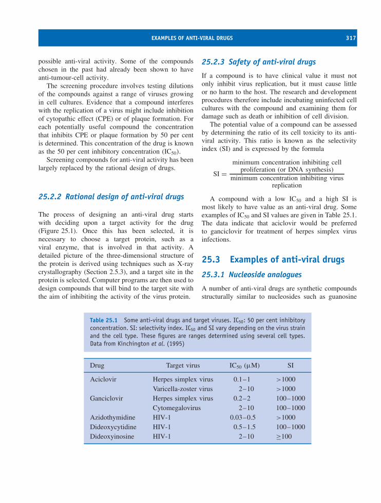
EXAMPLES OF ANTI-VIRAL DRUGS 317
possible anti-viral activity. Some of the compoundschosen in the past had already been shown to haveanti-tumour-cell activity.
The screening procedure involves testing dilutionsof the compounds against a range of viruses growingin cell cultures. Evidence that a compound interfereswith the replication of a virus might include inhibitionof cytopathic effect (CPE) or of plaque formation. Foreach potentially useful compound the concentrationthat inhibits CPE or plaque formation by 50 per centis determined. This concentration of the drug is knownas the 50 per cent inhibitory concentration (IC50).
Screening compounds for anti-viral activity has beenlargely replaced by the rational design of drugs.
25.2.2 Rational design of anti-viral drugs
The process of designing an anti-viral drug startswith deciding upon a target activity for the drug(Figure 25.1). Once this has been selected, it isnecessary to choose a target protein, such as aviral enzyme, that is involved in that activity. Adetailed picture of the three-dimensional structure ofthe protein is derived using techniques such as X-raycrystallography (Section 2.5.3), and a target site in theprotein is selected. Computer programs are then used todesign compounds that will bind to the target site withthe aim of inhibiting the activity of the virus protein.
25.2.3 Safety of anti-viral drugs
If a compound is to have clinical value it must notonly inhibit virus replication, but it must cause littleor no harm to the host. The research and developmentprocedures therefore include incubating uninfected cellcultures with the compound and examining them fordamage such as death or inhibition of cell division.
The potential value of a compound can be assessedby determining the ratio of its cell toxicity to its anti-viral activity. This ratio is known as the selectivityindex (SI) and is expressed by the formula
SI =minimum concentration inhibiting cell
proliferation (or DNA synthesis)minimum concentration inhibiting virus
replication
A compound with a low IC50 and a high SI ismost likely to have value as an anti-viral drug. Someexamples of IC50 and SI values are given in Table 25.1.The data indicate that aciclovir would be preferredto ganciclovir for treatment of herpes simplex virusinfections.
25.3 Examples of anti-viral drugs
25.3.1 Nucleoside analogues
A number of anti-viral drugs are synthetic compoundsstructurally similar to nucleosides such as guanosine
Table 25.1 Some anti-viral drugs and target viruses. IC50: 50 per cent inhibitoryconcentration. SI: selectivity index. IC50 and SI vary depending on the virus strainand the cell type. These figures are ranges determined using several cell types.Data from Kinchington et al. (1995)
Drug Target virus IC50 (µM) SI
Aciclovir Herpes simplex virus 0.1–1 >1000Varicella-zoster virus 2–10 >1000
Ganciclovir Herpes simplex virus 0.2–2 100–1000Cytomegalovirus 2–10 100–1000
Azidothymidine HIV-1 0.03–0.5 >1000Dideoxycytidine HIV-1 0.5–1.5 100–1000Dideoxyinosine HIV-1 2–10 ≥100
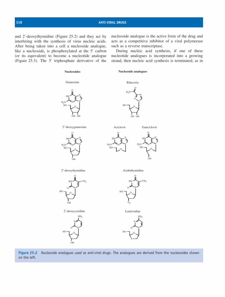
318 ANTI-VIRAL DRUGS
and 2′-deoxythymidine (Figure 25.2) and they act byinterfering with the synthesis of virus nucleic acids.After being taken into a cell a nucleoside analogue,like a nucleoside, is phosphorylated at the 5′ carbon(or its equivalent) to become a nucleotide analogue(Figure 25.3). The 5′ triphosphate derivative of the
nucleoside analogue is the active form of the drug andacts as a competitive inhibitor of a viral polymerasesuch as a reverse transcriptase.
During nucleic acid synthesis, if one of thesenucleotide analogues is incorporated into a growingstrand, then nucleic acid synthesis is terminated, as in
Nucleosides
Guanosine
Nucleoside analogues
Ribavirin
OO
O
N
NN
NHN
H2N
H2N
N N
OH OHOH
HO
OH
OHO
O
NHN
H2N N N
OH
OHO
O
HN CH3
N
OH
O
OHO
O
N
N
NH2
OH
OHO
O
N
N
NH2
S
OHO
O
HN CH3
N
N3
O
O
HO
O
N
HN HN
H2N
N
N
OHO
O
N
OH
H2N
N
N
OHO
2′-deoxyguanosine
2′-deoxythymidine
2′-deoxycytidine
Aciclovir Ganciclovir
Azidothymidine
Lamivudine
Figure 25.2 Nucleoside analogues used as anti-viral drugs. The analogues are derived from the nucleosides shownon the left.
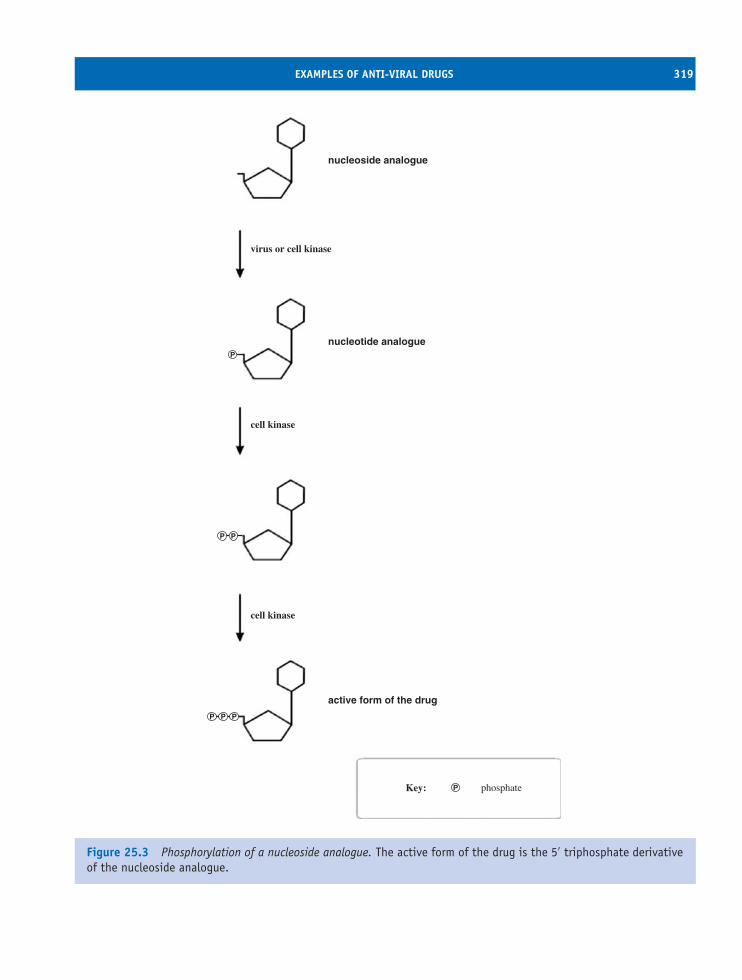
EXAMPLES OF ANTI-VIRAL DRUGS 319
nucleoside analogue
nucleotide analogue
virus or cell kinase
cell kinase
cell kinase
active form of the drug
P
P P P
P
P
Key: phosphateP
Figure 25.3 Phosphorylation of a nucleoside analogue. The active form of the drug is the 5′ triphosphate derivativeof the nucleoside analogue.
320 ANTI-VIRAL DRUGS
the dideoxy method of DNA sequencing. The structureof the nucleotide analogue prevents it from acceptingthe next nucleotide. In some cases the analogue lacksthe hydroxyl group on the 3′ carbon (or lacks the3′ carbon) necessary for the addition of the nextnucleotide.
25.3.1.a Ribavirin
Ribavirin is an analogue of guanosine (Figure 25.2). Itis used for the treatment of infection with several RNAviruses, especially persistent infections with hepatitis Cvirus (HCV; Section 22.11). Combined treatment withribavirin and α-interferon has eradicated HCV infectionin many patients.
Ribavirin has also been recommended for treatmentof young children infected with respiratory syncytialvirus (Section 15.6), one of the most important causesof severe respiratory tract disease in children. The drugis delivered to the patient in the form of an aerosol for12–18 hours daily. This treatment reduces the severityof the disease, but the mode of delivery is expensiveand inconvenient, so it is not often used.
The mode of action of ribavirin is not completelyunderstood and at least five hypotheses have beenproposed to explain its anti-viral activity. The drug hasbeen shown to inhibit several activities, including viralRNA synthesis and mRNA capping, and it is likely thatit has more than one mode of action.
25.3.1.b Nucleoside analogues that inhibitherpesvirus DNA synthesis
Aciclovir (also spelt ‘acyclovir’) is an analogue ofguanosine (Figure 25.2). It is a very safe drug,with almost no side-effects, widely used for thetreatment and prevention of diseases caused by threeof the herpesviruses: herpes simplex viruses 1 and 2(HSV-1 and HSV-2) and varicella-zoster virus (VZV)(Chapter 11). Uses of aciclovir include
• prevention of cold sores;
• prevention and treatment of genital herpes;
• treatment of HSV encephalitis (early treatmentreduces the mortality rate);
• protection of immunocompromised patients afterexposure to VZV;
• treatment of shingles (pain is reduced significantly).
Aciclovir strongly inhibits virus DNA synthesis buthas very little effect on cell DNA synthesis. In aherpesvirus-infected cell the first phosphorylation ofaciclovir (Figure 25.3) is carried out by the virusthymidine kinase (TK) (Section 11.5.3). The cell TKis produced only at certain stages of the cell cycleand it has a much lower affinity for aciclovir than theviral enzyme, so there is very little phosphorylation ofaciclovir in uninfected cells.
During DNA synthesis, aciclovir triphosphate com-petes with deoxyguanosine triphosphate (dGTP) forincorporation into the new strand. When aciclovirbecomes incorporated, DNA synthesis is terminateddue to the lack of a 3′–OH group.
Ganciclovir, like aciclovir, is an analogue of guano-sine, and the first phosphorylation in a cell is carriedout by a virus enzyme, in this case a protein kinase.Ganciclovir is more likely than aciclovir to becomeincorporated into cell DNA. As a result ganciclovir hasa lower SI than aciclovir (Table 25.1) and can causesignificant side-effects, especially reduced numbers ofblood cells because of damage to bone marrow cells.
Ganciclovir is used to treat infections with cyto-megalovirus, as aciclovir is less effective againstthis virus than it is against the other herpesviruses.Examples of cytomegalovirus diseases that are treatedwith ganciclovir include pneumonia and retinitis inimmunocompromised patients, such as those who havereceived organ transplants and those with AIDS.
25.3.1.c Nucleoside analogues that inhibitreverse transcription
Azidothymidine (AZT), also known as zidovudine, isan analogue of thymidine (Figure 25.2). It had beeninvestigated as a possible anti-cancer drug and it wasfound to inhibit the reverse transcriptase of HIV. Likeother nucleoside analogues, it is phosphorylated to the5′ triphosphate after uptake into a cell (Figure 25.3).
AZT triphosphate binds more strongly to the viralreverse transcriptase than to the cell DNA polymerase,and the reverse transcriptase binds AZT triphosphate inpreference to deoxythymidine triphosphate. AZT thus
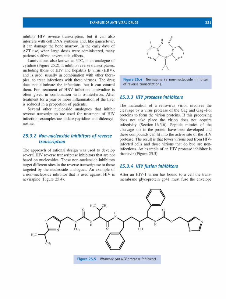
EXAMPLES OF ANTI-VIRAL DRUGS 321
inhibits HIV reverse transcription, but it can alsointerfere with cell DNA synthesis and, like ganciclovir,it can damage the bone marrow. In the early days ofAZT use, when large doses were administered, manypatients suffered severe side-effects.
Lamivudine, also known as 3TC, is an analogue ofcytidine (Figure 25.2). It inhibits reverse transcriptases,including those of HIV and hepatitis B virus (HBV),and is used, usually in combination with other thera-pies, to treat infections with these viruses. The drugdoes not eliminate the infections, but it can controlthem. For treatment of HBV infection lamivudine isoften given in combination with α-interferon. Aftertreatment for a year or more inflammation of the liveris reduced in a proportion of patients.
Several other nucleoside analogues that inhibitreverse transcription are used for treatment of HIVinfection; examples are dideoxycytidine and dideoxyi-nosine.
25.3.2 Non-nucleoside inhibitors of reversetranscription
The approach of rational design was used to developseveral HIV reverse transcriptase inhibitors that are notbased on nucleosides. These non-nucleoside inhibitorstarget different sites in the reverse transcriptase to thosetargeted by the nucleoside analogues. An example ofa non-nucleoside inhibitor that is used against HIV isnevirapine (Figure 25.4).
N
N
N N
OH
Figure 25.4 Nevirapine (a non-nucleoside inhibitorof reverse transcription).
25.3.3 HIV protease inhibitors
The maturation of a retrovirus virion involves thecleavage by a virus protease of the Gag and Gag–Polproteins to form the virion proteins. If this processingdoes not take place the virion does not acquireinfectivity (Section 16.3.6). Peptide mimics of thecleavage site in the protein have been developed andthese compounds can fit into the active site of the HIVprotease. The result is that fewer virions bud from HIV-infected cells and those virions that do bud are non-infectious. An example of an HIV protease inhibitor isritonavir (Figure 25.5).
25.3.4 HIV fusion inhibitors
After an HIV-1 virion has bound to a cell the trans-membrane glycoprotein gp41 must fuse the envelope
H3C
H3C
CH3
CH3
CH3
N
N
N
O
OH
NH
H
NS
OH
O
O
S
N
Figure 25.5 Ritonavir (an HIV protease inhibitor).
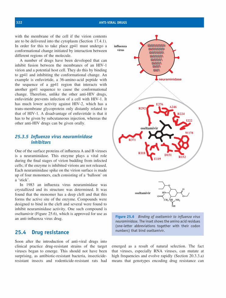
322 ANTI-VIRAL DRUGS
with the membrane of the cell if the virion contentsare to be delivered into the cytoplasm (Section 17.4.1).In order for this to take place gp41 must undergo aconformational change initiated by interaction betweendifferent regions of the molecule.
A number of drugs have been developed that caninhibit fusion between the membranes of an HIV-1virion and a potential host cell. They do this by bindingto gp41 and inhibiting the conformational change. Anexample is enfuvirtide, a 36-amino-acid peptide withthe sequence of a gp41 region that interacts withanother gp41 sequence to cause the conformationalchange. Therefore, unlike the other anti-HIV drugs,enfuvirtide prevents infection of a cell with HIV-1. Ithas much lower activity against HIV-2, which has atrans-membrane glycoprotein only distantly related tothat of HIV-1. A disadvantage of enfuvirtide is that ithas to be given by subcutaneous injection, whereas theother anti-HIV drugs can be given orally.
25.3.5 Influenza virus neuraminidaseinhibitors
One of the surface proteins of influenza A and B virusesis a neuraminidase. This enzyme plays a vital roleduring the final stages of virion budding from infectedcells; if the enzyme is inhibited virions are not released.Each neuraminidase spike on the virion surface is madeup of four monomers, each consisting of a ‘balloon’ ona ‘stick’.
In 1983 an influenza virus neuraminidase wascrystallized and its structure was determined. It wasfound that the monomer has a deep cleft and that thisforms the active site of the enzyme. Compounds weredesigned to bind in the cleft and several were found toinhibit neuraminidase activity. One such compound isoseltamivir (Figure 25.6), which is approved for use asan anti-influenza virus drug.
25.4 Drug resistanceSoon after the introduction of anti-viral drugs intoclinical practice drug-resistant strains of the targetviruses began to emerge. This should not have beensurprising, as antibiotic-resistant bacteria, insecticide-resistant insects and rodenticide-resistant rats had
R292E276
A246
R224
I222
W178
R152D151
E119
R118
R371
neuraminidase
O
NH
O
OO
NH
HN
NH2CH3
oseltamivir
oseltamivir
influenzavirus
Figure 25.6 Binding of oseltamivir to influenza virusneuraminidase. The inset shows the amino acid residues(one-letter abbreviations together with their codonnumbers) that bind oseltamivir.
emerged as a result of natural selection. The factthat viruses, especially RNA viruses, can mutate athigh frequencies and evolve rapidly (Section 20.3.3.a)means that genotypes encoding drug resistance can
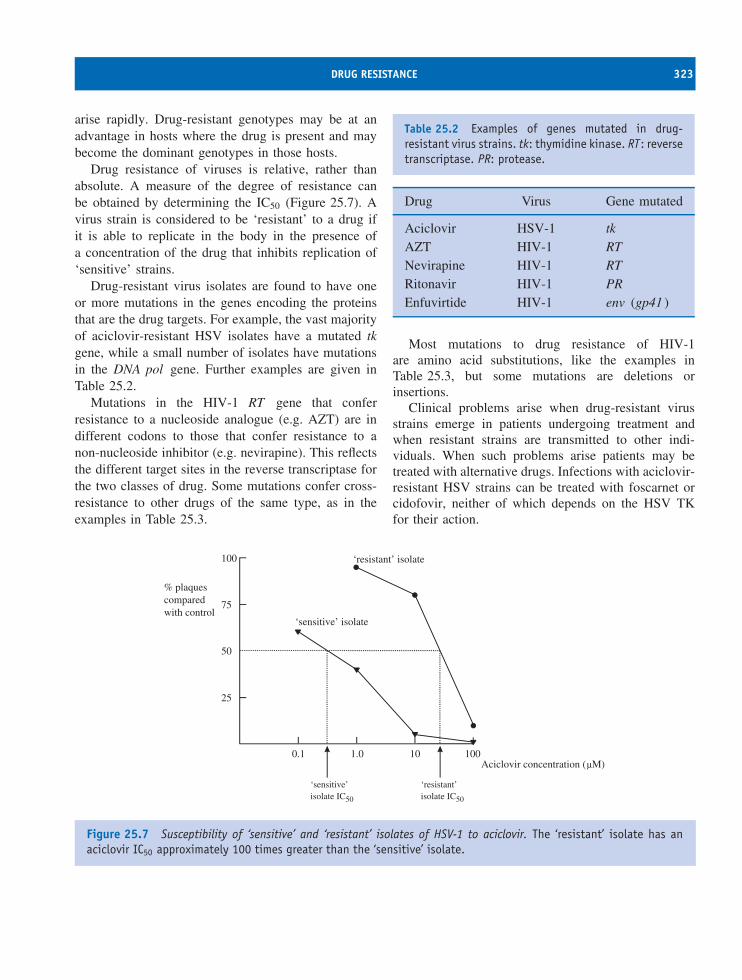
DRUG RESISTANCE 323
arise rapidly. Drug-resistant genotypes may be at anadvantage in hosts where the drug is present and maybecome the dominant genotypes in those hosts.
Drug resistance of viruses is relative, rather thanabsolute. A measure of the degree of resistance canbe obtained by determining the IC50 (Figure 25.7). Avirus strain is considered to be ‘resistant’ to a drug ifit is able to replicate in the body in the presence ofa concentration of the drug that inhibits replication of‘sensitive’ strains.
Drug-resistant virus isolates are found to have oneor more mutations in the genes encoding the proteinsthat are the drug targets. For example, the vast majorityof aciclovir-resistant HSV isolates have a mutated tkgene, while a small number of isolates have mutationsin the DNA pol gene. Further examples are given inTable 25.2.
Mutations in the HIV-1 RT gene that conferresistance to a nucleoside analogue (e.g. AZT) are indifferent codons to those that confer resistance to anon-nucleoside inhibitor (e.g. nevirapine). This reflectsthe different target sites in the reverse transcriptase forthe two classes of drug. Some mutations confer cross-resistance to other drugs of the same type, as in theexamples in Table 25.3.
Table 25.2 Examples of genes mutated in drug-resistant virus strains. tk: thymidine kinase. RT: reversetranscriptase. PR: protease.
Drug Virus Gene mutated
Aciclovir HSV-1 tkAZT HIV-1 RTNevirapine HIV-1 RTRitonavir HIV-1 PREnfuvirtide HIV-1 env (gp41 )
Most mutations to drug resistance of HIV-1are amino acid substitutions, like the examples inTable 25.3, but some mutations are deletions orinsertions.
Clinical problems arise when drug-resistant virusstrains emerge in patients undergoing treatment andwhen resistant strains are transmitted to other indi-viduals. When such problems arise patients may betreated with alternative drugs. Infections with aciclovir-resistant HSV strains can be treated with foscarnet orcidofovir, neither of which depends on the HSV TKfor their action.
‘resistant’ isolate
25
50
75
100
1.0 10 100Aciclovir concentration ( M)
0.1
‘sensitive’ isolate
% plaquescomparedwith control
‘sensitive’isolate IC50
‘resistant’isolate IC50
µ
Figure 25.7 Susceptibility of ‘sensitive’ and ‘resistant’ isolates of HSV-1 to aciclovir. The ‘resistant’ isolate has anaciclovir IC50 approximately 100 times greater than the ‘sensitive’ isolate.
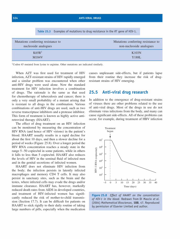
324 ANTI-VIRAL DRUGS
Table 25.3 Examples of mutations to drug resistance in the RT gene of HIV-1.
Mutations conferring resistance to Mutations conferring resistance tonucleoside analogues non-nucleoside analogues
K65R1 K103NM184V Y188L
1Codon 65 mutated from lysine to arginine. Other mutations are indicated similarly.
When AZT was first used for treatment of HIVinfection, AZT-resistant strains of HIV rapidly emergedand a similar problem was encountered when otheranti-HIV drugs were used alone. Now the standardtreatment for HIV infection involves a combinationof drugs. The rationale is the same as that usedfor chemotherapy of tuberculosis and cancer; there isonly a very small probability of a mutant arising thatis resistant to all drugs in the combination. Variouscombinations of anti-HIV drugs are used, such as tworeverse transcriptase inhibitors and a protease inhibitor.This form of treatment is known as highly active anti-retroviral therapy (HAART).
The effect of drug treatment on an HIV infectioncan be monitored by measuring the concentration ofHIV RNA (and hence of HIV virions) in the patient’sblood. HAART usually results in a rapid decline forabout the first 10 days, and then a slower decline for aperiod of weeks (Figure 25.8). Over a longer period theHIV RNA concentration reaches a steady state in therange 5–50 copies/ml in some patients, while in othersit falls to less than 5 copies/ml. HAART also reducesthe levels of HIV in the seminal fluid of infected menand in the genital secretions of infected women.
HAART does not eliminate HIV infection fromthe body; the infection persists in latently infectedmacrophages and memory CD4 T cells. It may alsopersist in sanctuary sites, such as the brain and thetestes, where infected cells may evade the drugs and/orimmune clearance. HAART has, however, markedlyreduced death rates from AIDS in developed countries,and treatment of HIV-infected women has signifi-cantly reduced the risk of mother-to-child transmis-sion (Section 17.7). It can be difficult for patients onHAART to stick rigidly to their daily routine of takinglarge numbers of pills, especially when the medication
causes unpleasant side-effects, but if patients lapsefrom their routine they increase the risk of drug-resistant strains of HIV emerging.
25.5 Anti-viral drug research
In addition to the emergence of drug-resistant strainsof viruses there are other problems related to the useof anti-viral drugs. Most of the drugs in use do noteliminate virus infections from the body, and many cancause significant side-effects. All of these problems canoccur, for example, during treatment of HBV infection
102
104
105
107
5
Time (days)
HIV
-1 R
NA
(co
pies
/ml)
10 15 20 25 30 35 400−5
Treatmentbegan
103
106
Figure 25.8 Effect of HAART on the concentrationof HIV-1 in the blood. Redrawn from Di Mascio et al.(2004) Mathematical Biosciences, 188, 47. Reproducedby permission of Elsevier Limited and author.
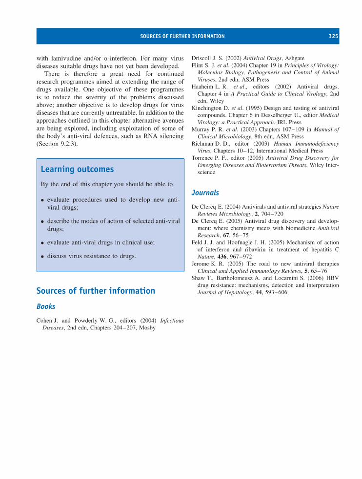
SOURCES OF FURTHER INFORMATION 325
with lamivudine and/or α-interferon. For many virusdiseases suitable drugs have not yet been developed.
There is therefore a great need for continuedresearch programmes aimed at extending the range ofdrugs available. One objective of these programmesis to reduce the severity of the problems discussedabove; another objective is to develop drugs for virusdiseases that are currently untreatable. In addition to theapproaches outlined in this chapter alternative avenuesare being explored, including exploitation of some ofthe body’s anti-viral defences, such as RNA silencing(Section 9.2.3).
Learning outcomes
By the end of this chapter you should be able to
• evaluate procedures used to develop new anti-viral drugs;
• describe the modes of action of selected anti-viraldrugs;
• evaluate anti-viral drugs in clinical use;
• discuss virus resistance to drugs.
Sources of further information
Books
Cohen J. and Powderly W. G., editors (2004) InfectiousDiseases, 2nd edn, Chapters 204–207, Mosby
Driscoll J. S. (2002) Antiviral Drugs, AshgateFlint S. J. et al. (2004) Chapter 19 in Principles of Virology:
Molecular Biology, Pathogenesis and Control of AnimalViruses, 2nd edn, ASM Press
Haaheim L. R. et al., editors (2002) Antiviral drugs.Chapter 4 in A Practical Guide to Clinical Virology, 2ndedn, Wiley
Kinchington D. et al. (1995) Design and testing of antiviralcompounds. Chapter 6 in Desselberger U., editor MedicalVirology: a Practical Approach, IRL Press
Murray P. R. et al. (2003) Chapters 107–109 in Manual ofClinical Microbiology, 8th edn, ASM Press
Richman D. D., editor (2003) Human ImmunodeficiencyVirus, Chapters 10–12, International Medical Press
Torrence P. F., editor (2005) Antiviral Drug Discovery forEmerging Diseases and Bioterrorism Threats, Wiley Inter-science
Journals
De Clercq E. (2004) Antivirals and antiviral strategies NatureReviews Microbiology, 2, 704–720
De Clercq E. (2005) Antiviral drug discovery and develop-ment: where chemistry meets with biomedicine AntiviralResearch, 67, 56–75
Feld J. J. and Hoofnagle J. H. (2005) Mechanism of actionof interferon and ribavirin in treatment of hepatitis CNature, 436, 967–972
Jerome K. R. (2005) The road to new antiviral therapiesClinical and Applied Immunology Reviews, 5, 65–76
Shaw T., Bartholomeusz A. and Locarnini S. (2006) HBVdrug resistance: mechanisms, detection and interpretationJournal of Hepatology, 44, 593–606

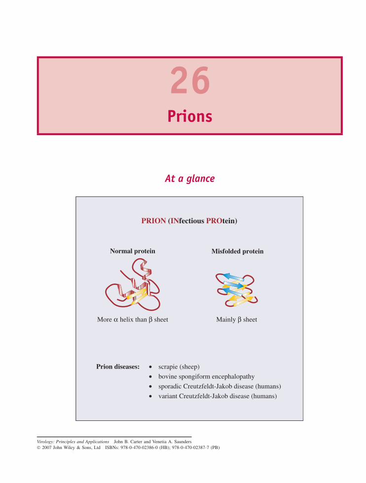
26Prions
At a glance
PRION (INfectious PROtein)
Mainly β sheet
Normal protein
••••
scrapie (sheep)
bovine spongiform encephalopathy
sporadic Creutzfeldt-Jakob disease (humans)
variant Creutzfeldt-Jakob disease (humans)
Misfolded protein
More α helix than β sheet
Prion diseases:
Virology: Principles and Applications John B. Carter and Venetia A. Saunders 2007 John Wiley & Sons, Ltd ISBNs: 978-0-470-02386-0 (HB); 978-0-470-02387-7 (PB)
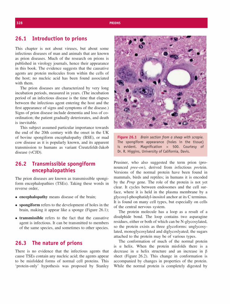
328 PRIONS
26.1 Introduction to prions
This chapter is not about viruses, but about someinfectious diseases of man and animals that are knownas prion diseases. Much of the research on prions ispublished in virology journals, hence their appearancein this book. The evidence suggests that the causativeagents are protein molecules from within the cells ofthe host; no nucleic acid has been found associatedwith them.
The prion diseases are characterized by very longincubation periods, measured in years. (The incubationperiod of an infectious disease is the time that elapsesbetween the infectious agent entering the host and thefirst appearance of signs and symptoms of the disease.)Signs of prion disease include dementia and loss of co-ordination; the patient gradually deteriorates, and deathis inevitable.
This subject assumed particular importance towardsthe end of the 20th century with the onset in the UKof bovine spongiform encephalopathy (BSE), or madcow disease as it is popularly known, and its apparenttransmission to humans as variant Creutzfeldt-Jakobdisease (vCJD).
26.2 Transmissible spongiformencephalopathies
The prion diseases are known as transmissible spongi-form encephalopathies (TSEs). Taking these words inreverse order,
• encephalopathy means disease of the brain;
• spongiform refers to the development of holes in thebrain, making it appear like a sponge (Figure 26.1);
• transmissible refers to the fact that the causativeagent is infectious. It can be transmitted to membersof the same species, and sometimes to other species.
26.3 The nature of prionsThere is no evidence that the infectious agents thatcause TSEs contain any nucleic acid; the agents appearto be misfolded forms of normal cell proteins. This‘protein-only’ hypothesis was proposed by Stanley
Figure 26.1 Brain section from a sheep with scrapie.The spongiform appearance (holes in the tissue)is evident. Magnification × 500. Courtesy ofDr. R. Higgins, University of California, Davis.
Prusiner, who also suggested the term prion (pro-nounced pree-on), derived from infectious protein.Versions of the normal protein have been found inmammals, birds and reptiles; in humans it is encodedby the Prnp gene. The role of the protein is not yetclear. It cycles between endosomes and the cell sur-face, where it is held in the plasma membrane by aglycosyl-phosphatidyl-inositol anchor at its C terminus.It is found on many cell types, but especially on cellsof the central nervous system.
The protein molecule has a loop as a result of adisulphide bond. The loop contains two asparagineresidues, either or both of which can be N-glycosylated,so the protein exists as three glycoforms: unglycosy-lated, monoglycosylated and diglycoslyated; the sugarsattached to the protein may be of various types.
The conformation of much of the normal proteinis α helix. When the protein misfolds there is adecrease in α helix structure and an increase in β
sheet (Figure 26.2). This change in conformation isaccompanied by changes in properties of the protein.While the normal protein is completely digested by
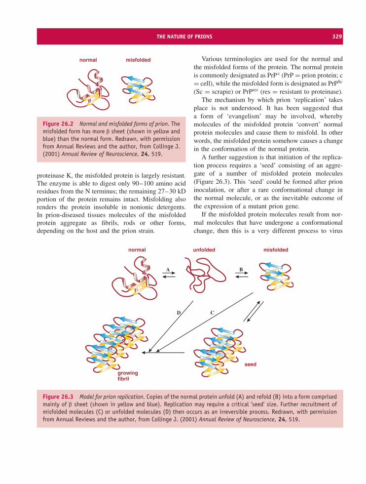
THE NATURE OF PRIONS 329
normal misfolded
Figure 26.2 Normal and misfolded forms of prion. Themisfolded form has more β sheet (shown in yellow andblue) than the normal form. Redrawn, with permissionfrom Annual Reviews and the author, from Collinge J.(2001) Annual Review of Neuroscience, 24, 519.
proteinase K, the misfolded protein is largely resistant.The enzyme is able to digest only 90–100 amino acidresidues from the N terminus; the remaining 27–30 kDportion of the protein remains intact. Misfolding alsorenders the protein insoluble in nonionic detergents.In prion-diseased tissues molecules of the misfoldedprotein aggregate as fibrils, rods or other forms,depending on the host and the prion strain.
Various terminologies are used for the normal andthe misfolded forms of the protein. The normal proteinis commonly designated as PrP c (PrP = prion protein; c= cell), while the misfolded form is designated as PrPSc
(Sc = scrapie) or PrPres (res = resistant to proteinase).The mechanism by which prion ‘replication’ takes
place is not understood. It has been suggested thata form of ‘evangelism’ may be involved, wherebymolecules of the misfolded protein ‘convert’ normalprotein molecules and cause them to misfold. In otherwords, the misfolded protein somehow causes a changein the conformation of the normal protein.
A further suggestion is that initiation of the replica-tion process requires a ‘seed’ consisting of an aggre-gate of a number of misfolded protein molecules(Figure 26.3). This ‘seed’ could be formed after prioninoculation, or after a rare conformational change inthe normal molecule, or as the inevitable outcome ofthe expression of a mutant prion gene.
If the misfolded protein molecules result from nor-mal molecules that have undergone a conformationalchange, then this is a very different process to virus
normal unfolded misfolded
seed
A B
CD
growingfibril
Figure 26.3 Model for prion replication. Copies of the normal protein unfold (A) and refold (B) into a form comprisedmainly of β sheet (shown in yellow and blue). Replication may require a critical ‘seed’ size. Further recruitment ofmisfolded molecules (C) or unfolded molecules (D) then occurs as an irreversible process. Redrawn, with permissionfrom Annual Reviews and the author, from Collinge J. (2001) Annual Review of Neuroscience, 24, 519.
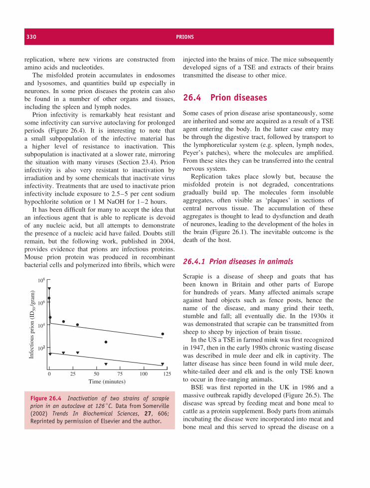
330 PRIONS
replication, where new virions are constructed fromamino acids and nucleotides.
The misfolded protein accumulates in endosomesand lysosomes, and quantities build up especially inneurones. In some prion diseases the protein can alsobe found in a number of other organs and tissues,including the spleen and lymph nodes.
Prion infectivity is remarkably heat resistant andsome infectivity can survive autoclaving for prolongedperiods (Figure 26.4). It is interesting to note thata small subpopulation of the infective material hasa higher level of resistance to inactivation. Thissubpopulation is inactivated at a slower rate, mirroringthe situation with many viruses (Section 23.4). Prioninfectivity is also very resistant to inactivation byirradiation and by some chemicals that inactivate virusinfectivity. Treatments that are used to inactivate prioninfectivity include exposure to 2.5–5 per cent sodiumhypochlorite solution or 1 M NaOH for 1–2 hours.
It has been difficult for many to accept the idea thatan infectious agent that is able to replicate is devoidof any nucleic acid, but all attempts to demonstratethe presence of a nucleic acid have failed. Doubts stillremain, but the following work, published in 2004,provides evidence that prions are infectious proteins.Mouse prion protein was produced in recombinantbacterial cells and polymerized into fibrils, which were
102
104
106
108
25 100
Time (minutes)
Infe
ctio
us p
rion
(ID
50/g
ram
)
50 75 1250
Figure 26.4 Inactivation of two strains of scrapieprion in an autoclave at 126 ◦C. Data from Somerville(2002) Trends In Biochemical Sciences, 27, 606;Reprinted by permission of Elsevier and the author.
injected into the brains of mice. The mice subsequentlydeveloped signs of a TSE and extracts of their brainstransmitted the disease to other mice.
26.4 Prion diseases
Some cases of prion disease arise spontaneously, someare inherited and some are acquired as a result of a TSEagent entering the body. In the latter case entry maybe through the digestive tract, followed by transport tothe lymphoreticular system (e.g. spleen, lymph nodes,Peyer’s patches), where the molecules are amplified.From these sites they can be transferred into the centralnervous system.
Replication takes place slowly but, because themisfolded protein is not degraded, concentrationsgradually build up. The molecules form insolubleaggregates, often visible as ‘plaques’ in sections ofcentral nervous tissue. The accumulation of theseaggregates is thought to lead to dysfunction and deathof neurones, leading to the development of the holes inthe brain (Figure 26.1). The inevitable outcome is thedeath of the host.
26.4.1 Prion diseases in animals
Scrapie is a disease of sheep and goats that hasbeen known in Britain and other parts of Europefor hundreds of years. Many affected animals scrapeagainst hard objects such as fence posts, hence thename of the disease, and many grind their teeth,stumble and fall; all eventually die. In the 1930s itwas demonstrated that scrapie can be transmitted fromsheep to sheep by injection of brain tissue.
In the US a TSE in farmed mink was first recognizedin 1947, then in the early 1980s chronic wasting diseasewas described in mule deer and elk in captivity. Thelatter disease has since been found in wild mule deer,white-tailed deer and elk and is the only TSE knownto occur in free-ranging animals.
BSE was first reported in the UK in 1986 and amassive outbreak rapidly developed (Figure 26.5). Thedisease was spread by feeding meat and bone meal tocattle as a protein supplement. Body parts from animalsincubating the disease were incorporated into meat andbone meal and this served to spread the disease on a
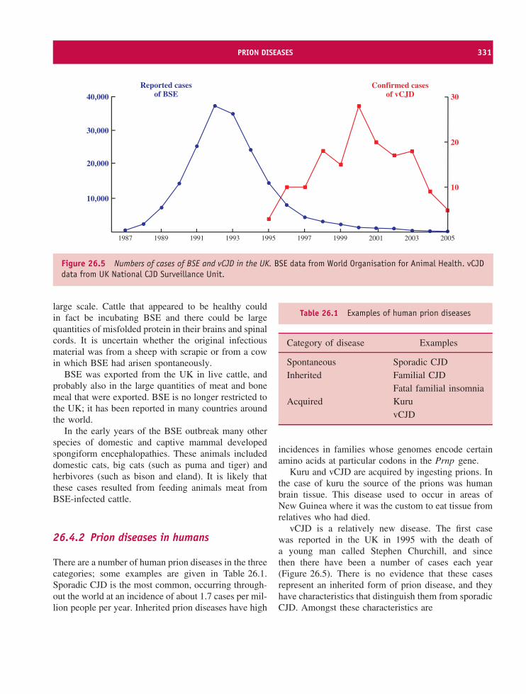
PRION DISEASES 331
1987 1989 1991 1993 1995 1997 1999 2001 2003 2005
40,000
30,000
20,000
10,00010
20
30
Reported casesof BSE
Confirmed casesof vCJD
Figure 26.5 Numbers of cases of BSE and vCJD in the UK. BSE data from World Organisation for Animal Health. vCJDdata from UK National CJD Surveillance Unit.
large scale. Cattle that appeared to be healthy couldin fact be incubating BSE and there could be largequantities of misfolded protein in their brains and spinalcords. It is uncertain whether the original infectiousmaterial was from a sheep with scrapie or from a cowin which BSE had arisen spontaneously.
BSE was exported from the UK in live cattle, andprobably also in the large quantities of meat and bonemeal that were exported. BSE is no longer restricted tothe UK; it has been reported in many countries aroundthe world.
In the early years of the BSE outbreak many otherspecies of domestic and captive mammal developedspongiform encephalopathies. These animals includeddomestic cats, big cats (such as puma and tiger) andherbivores (such as bison and eland). It is likely thatthese cases resulted from feeding animals meat fromBSE-infected cattle.
26.4.2 Prion diseases in humans
There are a number of human prion diseases in the threecategories; some examples are given in Table 26.1.Sporadic CJD is the most common, occurring through-out the world at an incidence of about 1.7 cases per mil-lion people per year. Inherited prion diseases have high
Table 26.1 Examples of human prion diseases
Category of disease Examples
Spontaneous Sporadic CJDInherited Familial CJD
Fatal familial insomniaAcquired Kuru
vCJD
incidences in families whose genomes encode certainamino acids at particular codons in the Prnp gene.
Kuru and vCJD are acquired by ingesting prions. Inthe case of kuru the source of the prions was humanbrain tissue. This disease used to occur in areas ofNew Guinea where it was the custom to eat tissue fromrelatives who had died.
vCJD is a relatively new disease. The first casewas reported in the UK in 1995 with the death ofa young man called Stephen Churchill, and sincethen there have been a number of cases each year(Figure 26.5). There is no evidence that these casesrepresent an inherited form of prion disease, and theyhave characteristics that distinguish them from sporadicCJD. Amongst these characteristics are
332 PRIONS
• the relatively young age of the victims;
• the relatively short duration of the disease;
• distinct pathological changes in the brain;
• the presence of infective prion in the spleen, lymphnodes and tonsils.
These features indicated that vCJD was a newdisease, and because its appearance coincided withthe BSE outbreak in the UK it was suggested that ithad resulted from humans ingesting the BSE prion.Further evidence that the BSE prion is the causativeagent of vCJD came from laboratory studies, whichdemonstrated that the BSE prion and the vCJD prionhave identical molecular characteristics, and that theyinduce identical responses in inoculated mice. Afew cases of vCJD appear to have resulted fromtransmission of the agent via blood transfusion, ratherthan through ingestion.
There have been cases of vCJD in countries otherthan the UK. Some of these victims had lived in theUK, so may have acquired the disease before movingabroad. Others had never visited the UK, but they couldhave become infected from meat exported from the UKor from BSE in local cattle.
26.5 Prion strains
For some TSE agents a number of strains canbe differentiated, each having phenotypic charactersthat are consistently transmitted from generation togeneration. For example, different strains of the scrapieagent can induce different clinical syndromes in goats.Differences can also be observed in laboratory studieswhen prion strains may be distinguished by propertiessuch as incubation period in mice after injection intothe brain and degree of heat resistance. Figure 26.4illustrates two strains of scrapie agent that differ inheat resistance.
Virus strains are defined by differences in thesequences of their nucleic acids, but if TSE agents donot contain nucleic acids how can strains with differentphenotypic characters be explained? One proposalis that the prion protein can misfold into multipleconformations, one for each strain. Alternatively,
prion strain differences may result from the extentto which the molecules are glycosylated at the twoglycosylation sites; in other words, a particular strainmay have a particular ratio of the three glycoforms(Section 26.3).
26.6 Prion transmissionAs we have already discussed, prions can be trans-mitted to other members of the same species andoften to other species. The efficiency of transmissiondepends on the route by which the prion moleculesenter the body; injection into the brain is much moreefficient than ingestion. Dose is also important; thelarger the dose, the more likely it is that transmissionwill occur.
Another factor that affects the efficiency of priontransmission is the existence of small variations inthe prion gene sequence, which result in variationin susceptibility between genotypes. The presence ofcertain amino acids at particular codons can renderindividuals more or less susceptible to infection. Forexample, sheep are more susceptible to scrapie if theprion gene has valine rather than alanine at codon 136.Small variations in the prion gene sequence can alsohave a strong influence on the incubation period andon the clinical course of the disease.
Many attempts to transmit TSE agents to distantlyrelated species have been reported as difficult. Whentransmission does occur the minimum infective doseis commonly larger than for the same or closely-related species, and incubation periods are commonlylonger. This effect has been called the ‘species barrier’.Most tests for cross-species transmission have usedthe development of prion disease in recipient animalsas the criterion for successful transmission. There is,however, evidence for subclinical infections, whereprions replicate without development of disease duringthe lifespan of the infected animal. It is thereforepossible that the strength of the ‘species barrier’ hasbeen over-estimated.
If a TSE agent is transmitted to a different species,and then passaged in that species, progressive changestake place; the incubation period becomes shorter overa number of passages and then stabilizes.
The transmission of BSE into humans as vCJDinvolved a ‘species barrier’ being crossed. As the BSE
SOURCES OF FURTHER INFORMATION 333
outbreak progressed, the UK government introducedincreasingly strict measures, to reduce the risk of theBSE prion being present in food. Animal materialslikely to constitute a risk were banned; these includebrain, spinal cord and mechanically recovered meatfrom the spine. Governments introduced additionalmeasures, including the testing of cattle and meat forthe BSE prion using serological methods.
The possibility of human-to-human transmission isalso a matter for concern. Measures are taken tominimize the risk of prion transmission via donatedblood and blood products, and via surgical instru-ments.
The cases of TSE in cats and other species,coincident with the BSE outbreak, are thought to havebeen transmitted in meat containing the BSE agent(Section 26.4.1). A number of additional species havebeen infected with the BSE agent under experimentalconditions, thus the BSE agent appears to have a wide‘host range’.
26.7 The protein-only hypothesis
If the protein-only hypothesis is correct, and theinfectious agent is misfolded prion protein, thentransmission of a prion disease implies that theintroduction of the misfolded protein into the body of anew host initiates the misfolding of protein moleculesin that host. If the agent is ‘transmitted’ to otherspecies then, because each species makes a specificprion protein, the molecules that become misfoldedhave the amino acid sequence of the recipient prion,not that of the donor.
The protein-only hypothesis suggests that the agentis derived from one of the body’s own proteins, and thiscan explain the inherited forms of TSEs (Table 26.1).According to the hypothesis, these diseases develop asa consequence of the inheritance of a gene encoding aprion protein with an amino acid sequence that has ahigh probability of misfolding.
There remain many controversies and there is stillmuch to be learnt about the TSEs and their causativeagents. Progress in research is slow as a result of thelong incubation periods of the diseases.
Learning outcomes
By the end of this chapter you should be able to
• define the terms ‘prion’ and ‘transmissible spongi-form encephalopathy’;
• evaluate the ‘protein-only’ hypothesis;
• describe the characteristics of prions;
• discuss a theory of prion replication;
• describe prion diseases in animals and man;
• discuss the transmission of prions.
Sources of further information
Books
Bishop M. and Ironside J. W. (2004) Genetic susceptibilityto prion diseases. Chapter 13 in Susceptibility to InfectiousDiseases, editor Bellamy R., Cambridge University Press
Dormont D. (2004) Prions. Chapter 223 in Infectious Dis-eases, 2nd edn, editors Cohen J. and Powderly W. G.,Mosby
Harris D., editor (2004) Mad Cow Disease and RelatedEncephalopathies, Springer
Manson J. C. and Barron R. M. (2005) The transmissiblespongiform encephalopathies. pp. 137–158 in Molecu-lar Pathogenesis of Virus Infections, editors Digard P.,Nash A. A. and Randall R. E., 64th Symposium of theSociety for General Microbiology, Cambridge UniversityPress
Pennington H. (2003) When Food Kills: BSE, E. coli andDisaster Science, Oxford University Press
Prusiner S. B., editor (2004) Prion Biology and Diseases, 2ndedn, Cold Spring Harbor Laboratory Press
Journals
Baylis M. et al. (2004) Risk of scrapie in British sheepof different prion protein genotype Journal of GeneralVirology, 85, 2735–2740
334 PRIONS
Collinge J. et al. (2006) Kuru in the 21st century – anacquired human prion disease with very long incubationperiods The Lancet, 367, 2068–2074
Henry C. and Knight R. (2002) Clinical features of variantCreutzfeldt-Jakob disease Reviews in Medical Virology, 12,143–150
Hill A. F. and Collinge J. (2003) Subclinical prion infectionTrends in Microbiology, 11, 578–584
Hilton D. A. (2006) Pathogenesis and prevalence of variantCreutzfeldt-Jakob disease Journal of Pathology, 208,134–141
Legname G. et al. (2004) Synthetic mammalian prionsScience, 305, 673–676
Mabbott N. A. and MacPherson G. G. (2006) Prions andtheir lethal journey to the brain Nature Reviews Micro-biology, 4, 201–211
Silveira J. R. et al. (2005) The most infectious prion proteinparticles Nature, 437, 257–261
Watts J. C., Balachandran A. and Westaway D. (2006) Theexpanding universe of prion diseases PLoS Pathogens, 2,e26
Virologists’ vocabulary
Brief definitions of terms used in virology, plus a selection of relevant terms from cell biology, molecularbiology, immunology and medicine.
Abortive infection Infection of a cell where the virus replication cycle is not completedand no progeny virions are formed.
Ambisense genome A virus genome composed of ssRNA or ssDNA that is partly plussense and partly minus sense.
Antibody A glycoprotein synthesized in a plasma cell, which is derived from a Bcell that has interacted with a specific antigen. The antibody moleculecan bind specifically to this antigen.
Antigen A molecule that(a) triggers synthesis of antibody and/or a T cell response;(b) binds specifically to an antibody or a lymphocyte receptor.
Antiserum (plural antisera) Blood serum from an animal that has been injected with an antigen.The serum contains antibodies specific to that antigen.
Anti-termination A mechanism involved in the control of transcription in whichtermination is overcome at specific terminator sites. Anti-terminatorproteins allow RNA polymerase to read through these sites into genesdownstream.
Apoptosis Programmed cell death. Cell suicide. A process controlled by a cellthat results in the death of the cell.
Archaea One of two major groups of prokaryotes, the other being the bacteria.
Assembly The stage in the virus replication cycle when components cometogether to form virions.
Asymptomatic infection An infection without symptoms or signs of disease.
Attachment Binding of a virion to specific receptors on the host cell.
Attenuated strain ofa virus
A strain of a pathogenic virus that has much reduced virulence, but canstill replicate in its host. The virulence is said to have been attenuated.
Avirulent strain of a virus A strain of a virus that lacks virulence, but can still replicate in its host.
Virology: Principles and Applications John B. Carter and Venetia A. Saunders 2007 John Wiley & Sons, Ltd ISBNs: 978-0-470-02386-0 (HB); 978-0-470-02387-7 (PB)
336 VIROLOGISTS’ VOCABULARY
B lymphocyte (B cell) A cell with surface receptors that can recognize a specific antigen.Antigen binding can trigger a B cell to develop into anantibody-secreting plasma cell.
Bacteriophage (phage) A type of virus that infects bacteria.
Baltimore classification A scheme that classifies viruses into seven groups on the basis of thenature of the genome and the way in which it is transcribed.
Bicistronic mRNA An mRNA with two open reading frames.
Bronchiolitis Inflammation of the bronchioles.
Burst size The average number of new virions released from a single infected cell.
Cap A methylated guanosine triphosphate joined to the 5′ end of an mRNAby a 5′ –5′ bond.
Capsid The protein coat that encloses the nucleic acid of a virus.
Capsomere A discrete component of a capsid, constructed from several identicalprotein molecules.
CD (cluster ofdifferentiation)
CD antigens are cell surface molecules recognized by monoclonalantibodies. CD4 is the receptor for HIV.
Chemokine A cytokine that stimulates the migration and activation of cells in theanimal body, especially cells involved in inflammation.
Cirrhosis Liver disease characterized by scarring and loss of function.
Cohesive end (cos) A single-stranded end of a dsDNA molecule with a base sequencecomplementary to another single-stranded end of the same or adifferent DNA molecule, allowing hydrogen bonding between the twoends.
Coliphage A bacteriophage that infects E. coli.
Complement A series of proteins that is activated when the body is infected.Activation has a number of anti-viral effects, including lysis of infectedcells and enhancement of phagocytosis.
Concatemer A very long DNA molecule composed of multiple repeats of anucleotide sequence.
Conditional lethal mutant A mutant virus that expresses wild-type characteristics under certain(permissive) conditions, but mutant (lethal) characteristics under other(non-permissive/restrictive) conditions, e.g. a temperature-sensitivemutant.
Confocal microscopy The use of a microscope that excludes light from out-of-focus regionsof the specimen, producing clear images of thick and fluorescentlylabelled specimens.
Conservative replication The replication mode of some dsRNAs. The parental double strand isconserved and both strands of the progeny molecule are newlysynthesized.
Continuous cell line A clone of cells that is immortal and can be subcultured indefinitely.
VIROLOGISTS’ VOCABULARY 337
Co-receptor A second cell receptor to which some viruses must bind in order toinfect a cell.
Cryo-electron microscopy Examination of frozen specimens in an electron microscope with acold stage.
Cytokine A protein that is secreted from a cell and has a specific effect on othercells, including cells of the immune system.
Cytopathic effect (CPE) Change in the appearance of a cell culture induced by virus infection.
Defective interferingparticle
A virus that lacks part of its genome and interferes with the replicationof standard virus.
Defective virus A virus that lacks part of its genome and is unable to complete thereplication cycle without the aid of a helper virus.
Diploid Having two copies of the genome.
DNA-dependent RNApolymerase
An enzyme that synthesizes RNA from a DNA template.
Domain A discrete portion of a protein or a nucleic acid with its own structureand function.
Elongation factor (EF) A specific protein required for polypeptide elongation duringtranslation.
Encephalitis Inflammation of the brain.
Endemic Describes a disease that is constantly present or commonly present in ageographical area.
Endocytosis A process whereby eukaryotic cells take in extracellular materials byengulfing them in endosomes.
Endogenous retroviruses Retrovirus sequences present in the genomes of vertebrate animals.
Endosome A vesicle formed during endocytosis by membrane pinching off fromthe plasma membrane.
Enhancer A short DNA sequence that can increase the frequency of transcriptioninitiation.
Envelope A lipid bilayer and associated protein forming the outer component ofan enveloped virion.
Enzyme-linkedimmunosorbent assay(ELISA)
A serological method used to assay antigens and antibodies. Positiveresults detect an enzyme label.
Epidemic A rapid increase in the number of cases of a disease that spreads overa larger geographical area.
Epitope The part of an antigen that binds to an antibody or a lymphocytereceptor.
Escherichia coli (E. coli) Gram-negative bacterium that inhabits the human colon.Exogenous retrovirus A normal infectious retrovirus, cf. endogenous retrovirus.
338 VIROLOGISTS’ VOCABULARY
Exon Part of a eukaryotic gene that is transcribed and usually translated intoprotein.
Exonuclease An enzyme that digests a DNA or RNA strand one nucleotide at a timefrom either the 5′ end or the 3′ end.
F pilus A specialized pilus, encoded by the F plasmid, required for conjugationin E. coli.
Fluorescence microscopy The use of a microscope fitted with an appropriate source of light, e.g.an arc lamp or a laser, to examine fluorescent specimens.
Fusion protein (a) A virus protein that fuses the membrane of an enveloped viruswith a cell membrane.
(b) A protein generated by joining two genes together e.g. a foreignprotein fused to a coat protein in phage display.
Genome The DNA or RNA that encodes the genes of an organism or a virus.
Genotype The complete set of genes of an organism or a virus.
Glycoprotein A protein with one or more oligosaccharide groups covalently attached.
Glycosylation The process of adding oligosaccharide groups to proteins.
Golgi complex A membranous organelle found in most eukaryotic cells. Its primaryfunction is to process and sort proteins.
Green fluorescent protein(GFP)
A jellyfish protein that fluoresces green. The GFP gene can be fused toa gene of interest, producing a ‘tagged’ fusion protein, and thelocalization or movement of the protein in living cells can bevisualized. The GFP gene can also be used to monitor the expressionof a gene of interest in living cells; the control sequences for the geneof interest are linked to the GFP gene.
Guanylyl transferase An enzyme that adds guanosine 5′-monophosphate to the 5′ end ofmRNA when it is capped.
Hairpin A structure formed by hydrogen bonding within a single-strandednucleic acid molecule, producing a double-stranded stem and a loop ofunpaired nucleotides.
Helical symmetry A type of capsid symmetry present in many ssRNA viruses where theRNA forms a helix that is coated with protein.
Helicase An enzyme that unwinds a DNA duplex at replication forks.
Helper virus A virus that can provide function(s) missing from a defective virus,thereby enabling the latter to complete its replication cycle.
Hepatocyte The main cell type in the liver.
Heteroploid cell A cell with an abnormal number of chromosomes.Hexon A type of capsomere surrounded by six other capsomeres.Histones Proteins comprised mainly of basic amino acids, associated with DNA
in the cells of eukaryotes and some archaea. Histones play roles ingene regulation.
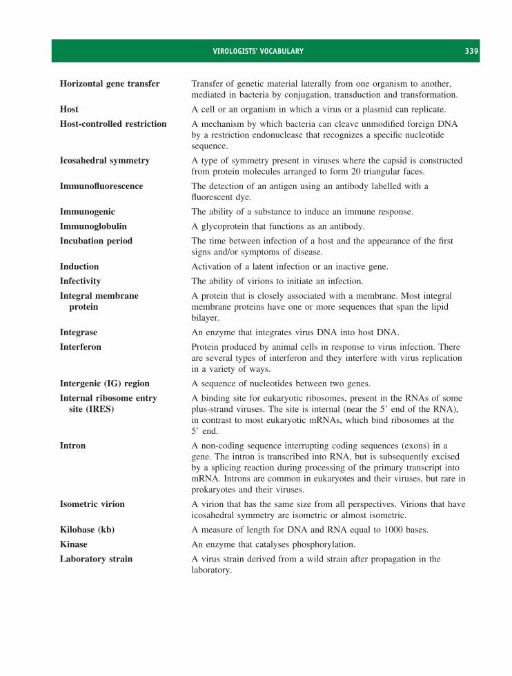
VIROLOGISTS’ VOCABULARY 339
Horizontal gene transfer Transfer of genetic material laterally from one organism to another,mediated in bacteria by conjugation, transduction and transformation.
Host A cell or an organism in which a virus or a plasmid can replicate.
Host-controlled restriction A mechanism by which bacteria can cleave unmodified foreign DNAby a restriction endonuclease that recognizes a specific nucleotidesequence.
Icosahedral symmetry A type of symmetry present in viruses where the capsid is constructedfrom protein molecules arranged to form 20 triangular faces.
Immunofluorescence The detection of an antigen using an antibody labelled with afluorescent dye.
Immunogenic The ability of a substance to induce an immune response.
Immunoglobulin A glycoprotein that functions as an antibody.
Incubation period The time between infection of a host and the appearance of the firstsigns and/or symptoms of disease.
Induction Activation of a latent infection or an inactive gene.
Infectivity The ability of virions to initiate an infection.
Integral membraneprotein
A protein that is closely associated with a membrane. Most integralmembrane proteins have one or more sequences that span the lipidbilayer.
Integrase An enzyme that integrates virus DNA into host DNA.
Interferon Protein produced by animal cells in response to virus infection. Thereare several types of interferon and they interfere with virus replicationin a variety of ways.
Intergenic (IG) region A sequence of nucleotides between two genes.
Internal ribosome entrysite (IRES)
A binding site for eukaryotic ribosomes, present in the RNAs of someplus-strand viruses. The site is internal (near the 5’ end of the RNA),in contrast to most eukaryotic mRNAs, which bind ribosomes at the5’ end.
Intron A non-coding sequence interrupting coding sequences (exons) in agene. The intron is transcribed into RNA, but is subsequently excisedby a splicing reaction during processing of the primary transcript intomRNA. Introns are common in eukaryotes and their viruses, but rare inprokaryotes and their viruses.
Isometric virion A virion that has the same size from all perspectives. Virions that haveicosahedral symmetry are isometric or almost isometric.
Kilobase (kb) A measure of length for DNA and RNA equal to 1000 bases.
Kinase An enzyme that catalyses phosphorylation.
Laboratory strain A virus strain derived from a wild strain after propagation in thelaboratory.
340 VIROLOGISTS’ VOCABULARY
Latent infection Infection of a cell where the replication cycle is not completed, but thevirus genome is maintained in the cell.
Ligase An enzyme that can catalyse bond formation between two similar typesof molecule. A DNA ligase can join two DNA molecules by catalysingthe formation of a phosphodiester bond between the 5’ end of apolynucleotide chain and the 3’ end of another (or the same)polynucleotide chain.
Ligation The joining of two molecules, e.g. DNA.Lipopolysaccharide (LPS) A compound comprising lipid and polysaccharide that is a key
component of the outer membrane of Gram-negative bacteria.Lysis Destruction of a cell caused by the rupture of its membrane and release
of contents, e.g. the bursting of a cell and release of progeny virions atthe end of the replication cycle.
Lysogen A bacterium containing the genome of a phage that is in the prophagestate and thus repressed for lytic functions. Such a bacterium is said tobe lysogenic for (name of the phage).
Lysogenic conversion(phage conversion)
When a cell becomes lysogenized, occasionally extra genes (unrelatedto the lytic cycle or lysogeny) carried by the prophage are expressed inthe host cell. These genes can change the phenotype of the host.
Lysogeny Latent infection of a bacterial cell with a phage.Lysosome Membrane-bounded organelle in the cytoplasm of eukaryotic cells.
Lysosomes contain enzymes, e.g. proteases and nucleases, that candigest virions.
Major histocompatibilitycomplex (MHC)
A region of the vertebrate genome that encodes majorhistocompatibility proteins. MHC class I and class II molecules are cellsurface proteins that play important roles in immune responses.
Meningitis Inflammation of the meninges (membranes covering the brain and thespinal cord).
Meningoencephalitis Inflammation of the meninges and the brain.Methyl transferase An enzyme that catalyses the transfer of methyl groups from one
molecule to another. Methyl transferases play a role in cappingeukaryotic mRNA.
Microarray A substrate with an orderly array of small spots of material (e.g. DNAor protein) attached. Each spot contains a specific probe that can detectspecific target molecules in a sample.
Microtubule Protein structure in eukaryotic cells, forming a component of thecytoskeleton, with roles in intracellular transport and mitosis.
Minus strand (negativestrand, (−) strand)
A nucleic acid strand that has the nucleotide sequence complementaryto that of the mRNA.
Monocistronic mRNA An mRNA with one open reading frame.Monoclonal antibody A single type of antibody produced by a clone of identical cells.Monolayer A layer of cells growing on the surface of a plastic or glass vessel.
Multiplicity of infection(m.o.i.)
The ratio of virions to cells in a virion–cell mixture.
VIROLOGISTS’ VOCABULARY 341
Mutation An alteration in the sequence of DNA or RNA. Mutations may occurspontaneously or they may be induced.
Myristylation(myristoylation)
The addition of a myristyl (myristoyl) group to a molecule.
Naked virus A virus that does not have an envelope on the virion.
Negative staining The use of a compound containing a heavy metal to reveal thestructure of specimens in a transmission electron microscope.
Neurone The main type of cell in the nervous system.
Neurovirulence A measure of the severity of nervous system disease that a virus(or any micro-organism) is capable of causing.
Neutralization The inactivation of virus infectivity by reaction with specific antibody.
Non-structural protein A virus protein that is not a component of the virion, but has one ormore roles in the replication cycle.
Northern blotting The transfer of RNA molecules to a membrane after gelelectrophoresis. Specific RNAs can be detected on the membrane usingprobes.
Nuclear envelope The structure, composed of two membranes, that separates the nucleusfrom the cytoplasm in a eukaryotic cell.
Nuclear localization signal A positively charged sequence in a protein that directs the proteinthrough nuclear pores into the nucleus of a cell.
Nuclear pore A protein complex in the nuclear envelope of a eukaryotic cell throughwhich materials are transported in and out of the nucleus.
Nucleic acid hybridization The formation of double-stranded nucleic acids by base-pairingbetween complementary single strands.
Nucleocapsid The virus genome enclosed in a protein capsid.
Nucleoside A nitrogen base (purine or pyrimidine) joined to a sugar (ribose ordeoxyribose).
Nucleotide A nitrogen base (purine or pyrimidine) joined to a sugar, which isjoined to one or more phosphate groups.
Okazaki fragment A short fragment of DNA with the RNA primer attached, producedduring discontinuous DNA synthesis. Okazaki fragments are joined bya DNA ligase to form the lagging strand.
Oncogene A cell or virus gene, the expression of which can cause a cell tobecome transformed. This may lead to the development of a cancer.
Open reading frame(ORF)
A sequence of nucleotides, starting with an initiation codon andfinishing with a termination codon, that encodes the amino acids ofa protein.
Origin (ori) A specific sequence of DNA at which replication is initiated.
Packaging signal (PS) A nucleotide sequence in a virus genome that is recognized by a virusprotein during virion assembly.
Pandemic A disease outbreak throughout the world.
342 VIROLOGISTS’ VOCABULARY
Pararetrovirus A virus with a DNA genome that is replicated via RNA.Passage ‘Subculture’ of a virus in cell culture or in an organism. ‘Subculture’
of a prion in an organism.Penton A type of capsomere surrounded by five other capsomeres.Perforin Protein present in CD8 T cells and NK cells. These cells can kill a
virus-infected cell by releasing perforins, which form pores in theplasma membrane of the target cell.
Persistent infection A long-term, possibly life-long, infection of a host. A persistent virusinfection may be productive or latent.
Phage display Display of recombinant proteins on the surface of a phage e.g. M13.Phagemid A cloning vector that combines features of a phage and plasmid
replicon and can replicate in either mode. The vector carries a plasmidori for replication in plasmid mode, and a filamentous phage ori,leading to production of single-stranded copies of the phagemid whenthe cell is infected with the relevant helper phage.
Phenotype The observable characteristics of an organism or virus, determined byits genotype and environmental factors.
Phosphoprotein A protein with one or more phosphate groups covalently attached.Phosphorylation The addition of one or more phosphate groups to a molecule.Phylogenetic tree A diagram showing evolutionary relationships between viruses or other
organisms.Pilin The protein subunit that is polymerized into the pilus in bacteria.Plaque (a) An area of lysis in a layer of cells, usually initiated by a single
virion infecting a cell, followed by the spread of infection tosurrounding cells.
(b) An aggregate of prion fibrils in the central nervous system of ahuman or animal with a transmissible spongiform encephalopathy.
Plaque-forming unit The quantity of virus (often a single virion) that can initiate formationof one plaque.
Plaque-purified virus A clone of virus derived from an individual plaque.Plasma membrane The membrane at the surface of a eukaryotic cell.Plasmid A self-replicating, extrachromosomal dsDNA molecule that is
generally circular, though can be linear. Plasmids are common inprokaryotes and rare in eukaryotes. Artificial plasmids can be createdto clone DNA sequences.
Plus strand (positivestrand, (+) strand)
A nucleic acid strand that has the same sequence as the mRNA.
Poly(A) polymerase An enzyme that adds adenylate residues to the 3′ end of a eukaryoticmRNA.
Polyadenylation The process of adding adenylate residues to the 3′ end of a eukaryoticmRNA.
Polycistronic mRNA An mRNA with more than one open reading frame.
VIROLOGISTS’ VOCABULARY 343
Polymerase chain reaction(PCR)
An in vitro technique for amplifying specific DNA sequences.
Polyprotein A large protein that is cleaved to form smaller functional proteins.Post-translational
modificationA modification to a protein after it has been translated. Examples:glycosylation, cleavage.
Primary transcript An RNA molecule synthesized during transcription. Introns may beremoved from the primary transcript to form functional RNAs.
Primase An enzyme that synthesizes RNA primers for DNA synthesis.Primer A molecule (often RNA, sometimes DNA or protein) that provides a
free –OH group at which a polymerase can initiate synthesis of DNAor RNA.
Primosome A complex of proteins involved in the priming action that initiatesDNA replication on ϕX-type origins.
Probe A specific sequence of DNA or RNA used to detect nucleic acids byhybridization. A probe may be labelled, e.g. with a fluorescentmolecule, to enable detection and quantification of the hybrid.
Procapsid A precursor of a capsid.Processivity factor A protein that increases the efficiency of DNA synthesis by enhancing
the ability of a DNA polymerase to remain associated with, and toprocess along, its template.
Productive infection Infection of a cell that results in the production of progeny virions.Promoter A specific DNA sequence, usually upstream of the transcription start
point of a gene, to which DNA-dependent RNA polymerase binds toinitiate transcription.
Proofreading A mechanism for correcting errors in nucleic acid or protein synthesis.Prophage A phage genome that resides in a bacterial host in a latent state. The
prophages of most phages are integrated into the bacterial genome, butsome are not.
Protease An enzyme that cleaves protein molecules by breaking peptide bonds.Proto-oncogene A cell gene that can become an oncogene (tumour gene) if activated or
over-expressed.Provirus The dsDNA copy of a retrovirus genome RNA.Pseudoknot An ssRNA secondary structure with two loops, formed when a
sequence in a loop base-pairs with a complementary sequence outsidethe loop.
Reading frame The phase in which the nucleotides of a nucleic acid are read in triplets.Reassortment A category of recombination that may occur with those segmented
genome viruses that have all the segments packaged in one virion.Reassortment occurs in a cell co-infected with two virus strains, and isthe formation of progeny virions containing mixtures of genomesegments from the two parental strains.
Receptor A molecule on a cell surface to which a virus specifically attaches.
344 VIROLOGISTS’ VOCABULARY
Recombinant An organism or a virus with a new genome, produced as a result ofrecombination. May also be used to describe the genome of theorganism or virus.
Recombination A process of combining genetic sequences that results in theproduction of a new genome, derived from two parental genomes. Fora cellular organism, recombination can take place between DNAmolecules within a cell. For viruses, recombination may occur when acell is co-infected with two related viruses.
Replicase An RNA polymerase that replicates the genome of an RNA virus bysynthesizing both (+) RNA and (−) RNA.
Replication cycle The process whereby a virus is replicated. For most viruses the processstarts with attachment to a host cell and this is followed by entry intothe cell. The process ends with exit of progeny virions from the cell.Some authors use the terms ‘infection cycle’ and ‘life cycle’.
Replicative intermediate A structure formed during ssRNA replication, consisting of a templateRNA associated with nascent RNAs of varying length. The nascentRNAs have the opposite polarity to the template RNA.
Restriction endonuclease An enzyme, usually from a bacterium, that cuts dsDNA at a specificsite.
Retrovirus A member of the family Retroviridae, so named because these virusescarry out reverse transcription.
Reverse genetics The generation of an RNA virus genome from a cloned copy DNA.Reverse transcriptase An enzyme that can synthesize DNA using (a) an RNA template and
(b) a DNA template.Reverse transcription Synthesis of DNA from an RNA template.Reverse transcription-
polymerase chainreaction (RT-PCR)
An in vitro technique for amplifying the data in RNA sequences byfirst copying the RNA to DNA using a reverse transcriptase. The DNAis then amplified by a PCR.
Ribonuclease (RNase) An enzyme that hydrolyses RNA.
Ribonuclease H (RNase H) A ribonuclease that specifically digests the RNA in an RNA–DNAduplex.
Ribosomal frameshifting A mechanism that allows a ribosome to read two overlapping openreading frames (ORF1 and ORF2) in an mRNA. A ribosome readingORF 1 shifts into a different reading frame towards the end of theORF. The ORF 1 stop codon is therefore not recognized and theribosome now reads ORF 2 to produce an elongated version of theORF 1 protein.
RNA-dependent RNApolymerase
An enzyme that synthesizes RNA from an RNA template.
RNA interference (RNAi,RNA silencing)
A process that interferes with the expression of a specific gene. Theprocess is induced by a dsRNA and results in the destruction of mRNAthat has the same sequence as the dsRNA.
RNA polymerase An enzyme that synthesizes RNA.
VIROLOGISTS’ VOCABULARY 345
RNA polymerase II The eukaryotic cell enzyme that synthesizes mRNA.
Rough endoplasmicreticulum
A system of membranes and ribosomes in the cytoplasm of eukaryoticcells.
Satellite virus A defective virus that depends on a helper virus to provide one ormore functions.
Scaffolding protein A protein that facilitates the assembly of a procapsid. The scaffoldingprotein is removed in the final stages of assembly and is therefore notpresent in the mature virion.
Segmented genome A virus genome that is composed of two or more nucleic acidmolecules.
Self-assembly The ability of a biological structure, e.g. a virus particle, to formspontaneously from its component parts.
Semi-conservativereplication
The replication mode of dsDNA and some dsRNAs in which theparental double strand is not conserved. Each progeny moleculeconsists of one parental strand and one newly synthesized strand.
Serology The study of antigen–antibody reactions and their use in tests to detectspecific antigens and antibodies.
Seropositive A person or an animal is said to be seropositive for an antigen, e.g. avirus, if their blood contains antibodies specific for that antigen.
Serotype A strain of a virus or micro-organism distinguished by its antigens.
Sex pilus A thin protein appendage for bacterial mating.
Shine-Dalgarno(S-D) sequence
A purine-rich sequence in prokaryotic mRNA just upstream of thetranslation start. The sequence can base-pair with a sequence near the3′-end of 16S ribosomal RNA and facilitates the initiation of proteinsynthesis.
Sigma (σ) factor A subunit of bacterial RNA polymerase, responsible for the recognitionof a specific promoter. Different sigma factors allow recognition ofdifferent promoter sequences.
Single-stranded binding(ssb) protein
A basic protein with a high affinity for ssDNA. Ssb protein protectsssDNA from nuclease attack and prevents it from re-annealing intodsDNA.
Southern blotting The transfer of DNA molecules to a membrane after gelelectrophoresis. Specific DNA can be detected on the membrane usingprobes.
Splicing The process of removing introns from primary transcripts and joiningthe exons to form mRNA.
Start (initiation) codon A codon on mRNA for initiation of protein synthesis: commonlyAUG, less commonly GUG and rarely UUG.
Stop (termination) codon A codon on mRNA for termination of protein synthesis: UAG, UAAor UGA. When a ribosome encounters a stop codon a terminationfactor interacts with the ribosome, causing polypeptide synthesis tostop and the ribosome to dissociate from the mRNA.
346 VIROLOGISTS’ VOCABULARY
Structural protein A protein that is a virion component.
Superinfection Infection by a virus of a cell that is already infected (often as a latentinfection) with that virus or a related virus.
Superinfection immunity(homoimmunity)
The immunity of a lysogen to superinfection by a phage with a similarregulatory mechanism.
Synchronous infection Near simultaneous infection of all cells in a culture with a virus. Thiscan be achieved by e.g. using a high multiplicity of infection, or bylimiting the time of attachment then diluting the culture so that virionsand cells are unlikely to make contact.
Syncytium (plural syncytia) A multinucleated giant cell formed by the fusion of membranes of anumber of individual cells. A number of viruses can cause theformation of syncytia when they infect cells.
T lymphocyte (T cell) A cell with surface receptors that can recognize a specific antigen.Antigen binding can trigger a T cell to perform one of several roles,including helper T cell or cytotoxic T cell.
Tailed phage A phage that has a tail attached to the head, which contains thegenome.
TATA box A DNA sequence in a eukaryotic promoter, typically 15–25nucleotides upstream from the transcription start.
TCID50 The dose of virus that infects 50 per cent of tissue cultures inoculatedwith aliquots of a virus preparation.
Tegument A layer of protein and RNA between the capsid and the envelope of aherpesvirus particle.
Temperate phage A phage capable of either establishing a lysogenic state in asusceptible bacterial host or of entering the lytic cycle.
Temperature-sensitivemutant
A virus mutant that is unable to replicate at some temperatures(non-permissive temperatures) at which the wild-type virus canreplicate, but is able to replicate at other temperatures (permissivetemperatures).
Titre The concentration of virus, antibody or other material in a preparation,determined by titration.
Transcriptase An enzyme that carries out transcription.
Transcription Synthesis of RNA from a DNA or an RNA template.
Transcription factor A protein that binds specifically to a promoter or enhancer to controlgene expression.
Transfection A process for introducing nucleic acids (e.g. a virus genome) into cells.
Transformation (a) Changes in an animal cell that result in it developing into a tumourcell.
(b) Bacterial gene transfer process involving uptake of naked DNAand acquisition of an altered genotype.
Translation Synthesis of protein from the genetic information in mRNA.
Transmission electronmicroscope
A microscope in which the image is formed by electrons transmittedthrough the specimen.

VIROLOGISTS’ VOCABULARY 347
Transovarial transmission Transmission of a virus from a female, e.g. insect, to the nextgeneration within eggs.
Uncoating The release of the virus genome (and associated protein in some cases)from a virion when a cell is infected.
Upstream The region of a nucleic acid extending in the 5′ direction from a gene;in the opposite direction to that of transcription.
Vector (a) An organism that transmits a virus from an infected host to anuninfected host.
(b) A virus or plasmid DNA, into which foreign DNA can be insertedfor the purpose of transferring it to a host cell foramplification/cloning or gene therapy.
Vertical transmission Transmission of a genetic element, virus or micro-organism to the nextgeneration of the host.
Viraemia The presence of infectious virus in the bloodstream.
Virion Virus particle.
Viroplasm A morphologically distinct region in which virus replication occurswithin an infected cell.
Virulence A measure of the severity of disease that a virus (or micro-organism) iscapable of causing.
Virulent strain of a virus A virus strain that can cause disease when it infects a host.
Virulent phage A phage that can produce only a lytic infection; it is unable to inducelysogeny.
Virus attachment site The surface regions of one or more virion proteins, which form a sitethat attaches to the receptor on a host cell.
Western blotting The transfer of protein molecules to a membrane after gelelectrophoresis. Specific proteins can be detected on the membrane.
Wild strain A virus strain isolated from a naturally infected host, cf. laboratorystrain.
Wild-type virus The parental strain of a mutant or laboratory-adapted virus.
Zinc finger A region of a nucleic acid-binding protein that resembles a finger andbinds a zinc ion.

INDEX
3TC, 32150 per cent inhibitory concentration, 317,
323
Abortive infection, 111, 112Acholeplasma laidlawii virus L2, budding,
100Aciclovir, 317, 320, 323Acid-triggered membrane fusion, 64, 65Acidianus convivator phage, 101Acquired immune deficiency syndrome,
198, 209, 210cytomegalovirus, 123
Acylation, 80Adaptive immunity, 107Adefovir, 225Adeno-associated virus, 112, 138
gene vector, 138Adenovirus
DNA, 35endocytosis, 63genome packaging, 97helper virus, 138oncogenic, 286primer, 89structure, 42
Adult T cell leukaemia, 290AIDS. See Acquired immune deficiency
syndromeAlfalfa mosaic virus, virions, 42Alpha-interferon, 105–106Alpharetrovirus, 196Alphavirus
budding, 99hosts, 46
Ambisense genome, 71, 72, 183Animal cell culture, 12Anti-restriction, 230Anti-termination, 248, 251Antibody, 19, 107, 108Antigen, 19Aphid vectors, 52, 53
Aphthovirus, 158Apical surface of epithelial cell, 82Aplastic anaemia, 139APOBEC3 proteins, 107, 208Apoptosis, 106, 109, 110, 155, 295Arbovirus, 51Arenaviridae, 119, 183Arenavirus, 5
genome, 72Assay, 22–24
infectivity, 22quantal, 23quantitative, 22
Assemblynucleocapsid, 96virion, 96–98, 101
Asymptomatic infection, 113Attachment (virion to receptor), 62Attenuated strain, 306Autographa californica nuclear polyhedrosis
virus, 118Autonomous element, 260Autonomous parvovirus, 139Autoradiography, 21Avian influenza, 280Avian leukosis virus, 291, 293Avian myeloblastosis virus, 186Avian myelocytomatosis virus, 294Azidothymidine (AZT), 317, 320, 323, 324
B lymphocyte (B cell), 107Epstein-Barr virus, 123, 290
B19 parvovirus, 139–140host cell, 141
β-propiolactone, 309Bacteriophage. See PhageBaculoviridae, 119, 132Baculovirus, 132
budding, 100capsid, 44envelope, 46, 100gene expression, 74
genome packaging, 97inactivation, 301occlusion body, 47, 56pesticide, 3recombinant, 264, 265transmission, 50
Baltimore classification, 70, 87Baltimore, David, 70, 119, 260Barley yellow dwarf virus, RNA, 36Basolateral surface of epithelial cell, 82Bats, viruses in
coronaviruses, 280Ebola virus, 279Hendra virus, 275Nipah virus, 276rabies virus, 175, 176
BCL-2 protein, 110Bean yellow mosaic virus, transmission, 52Beet yellows virus
structure, 40transmission, 52
Beetle vectors, 52, 53Beijerinck, Martinus, 4Beta-interferon, 105–106Betaretrovirus, 196Bicistronic messenger RNA, 79, 206Biological control, 269Bioterrorism, 282Birnaviridae, 155BK virus, 288Black beetle virus, RNA, 36Blood group P antigen, 141Blood products, 298Blood transfusion, prion transmission, 332Blood, virus in, 210, 295Bluetongue virus, 277Bocavirus, 139Bombyx mori densovirus, 138Bovine spongiform encephalopathy, 330,
331Brain cancer, 288Brome mosaic virus, 37
Virology: Principles and Applications John B. Carter and Venetia A. Saunders 2007 John Wiley & Sons, Ltd ISBNs: 978-0-470-02386-0 (HB); 978-0-470-02387-7 (PB)
350 INDEX
Bromoviridae, 118BSE, 330, 331Budding of virions, 98–100
hepatitis B virus, 223retrovirus, 193rhabdovirus, 181, 183
Bunyaviridae, 118, 183, 274Bunyavirus, 265, 274
genome, 38Buoyant density, 15Burkitt’s lymphoma, 289, 291Burkitt, Denis, 289Burst size, 26, 101Bushmeat, 274
c-myc gene, 289, 293Cancer, 4, 285–296Canine parvovirus, 267Canyon, virion surface, 160Cap on RNA
messenger RNA, 76, 78transcript, 75virus genome, 33
Capsid, 39–45hepatitis B virus, 215herpesvirus, 123HIV, 198parvovirus, 139picornavirus, 160retrovirus, 186rotavirus, 148
Capsomere, 42Carcinoma, 286Carroll, James, 4Caspar, Donald, 41Cations, virion components, 48Caudovirales, 116Caulimoviridae, 118, 227Cauliflower mosaic virus, 227
transmission, 52Caveolin, 61Caveolin-mediated endocytosis, 63CCR5, 200CCR5 gene, 269CD4, 8, 108, 155, 161–163, 199, 204CD4 T cell, 200, 209Cell culture, 12
virus production, 310Cell cycle, 57
cancer, 292parvoviruses, 141
Cell line, 291Cell-free synthesis of virus, 170Central DNA flap, 201Central dogma, 72Centrifugation, 9, 13, 15
density gradient, 9, 13differential, 13equilibrium, 15
isopycnic, 15rate zonal, 15
Cervical carcinoma, 287, 292Cervical smear, 287Chase, Martha, 4Chemokine receptor, 200Chick embryo, 12, 310Chicken viruses, 291Chickenpox, 122Chimpanzee foamy virus, 196Cholera toxin gene, 270Chronic wasting disease, 330Chrysoviridae, 155Cidofovir, 323Circoviridae, 143Circular nucleic acid, 32Circulative transmission, 51Cirrhosis, 215, 291Cis-acting replication element, 165Classification of viruses, 115–120Clathrin, 61Clathrin-mediated endocytosis, 63Cloning vector, 243, 245, 251
M13 mp series, 243phagemid, 243, 245
Closteroviridae, 119Closterovirus, 118Co-evolution of virus and host, 270Co-receptor, 61Cold sore, 122, 320Cold-adapted strain, 307Collaborating Centres for Influenza
Reference and Research, 310Common cold, 159Comoviridae, 118, 171Complement, 55, 108Complex retroviruses, 194Concatemer, 91, 250Connector, 42, 96, 97Conservative replication of double-stranded
RNA, 92, 152Continuous cell line, 12Coronaviridae, 116, 171Coronavirus, 76, 116
capping enzymes, 76Cowpea chlorotic mottle virus movement
protein, 112Cowpea mosaic virus, 171
RNA, 36capsid, 41movement protein, 112
Cowpox, 306Coxsackieviruses, 158
medical conditions, 159cre, 165Creutzfeldt-Jakob disease, 331, 332
familial, 331sporadic, 331variant, 331, 332
Crick, Francis, 72
Cucumber mosaic virusmovement protein, 112RNA, 12, 36
Culicoides, 277CXCR4, 200Cyclophilin A, 199Cypovirus, 56Cypovirus, 148Cytokines, 108, 267Cytomegalovirus, 123, 320Cytopathic effect, 19Cytotoxic T cell, 108, 109Cystoviridae, 155, 229, 2g37
Darwin, Charles, 261d’Herelle, Felix, 230Deer mouse, 274Defective interfering particle, 112Defective virus, 112Deltaretrovirus, 196Densovirinae, 138, 139Dependovirus, 138
DNA integration, 143gene vector, 138genome, 140latent infection, 143replication, 143
Detergent, effect on infectivity, 302Diarrhoea, 155Dicer, 109Dideoxycytidine, 317, 321Dideoxyinosine, 317, 321Dimerization initiation site, 207DIP, 112Diploid cell line, 310Direct terminal repeat, 37Directed assembly, 97Disinfection, 298, 301
hypochlorite, 302DNA dependent RNA polymerase
eukaryotic, 74bacterial, 83
DNA phage, 237–254DNA replication, 90, 91
filamentous single-stranded DNA phage,242, 243
herpesvirus, 129parvovirus, 142–143phage T7, 89, 90phage ϕX174, 239
DNA-dependent DNA polymerase, 74, 83,89, 262
Double-layered particle, rotavirus, 149, 151,152, 153
Double-stranded RNA phage, 237Double-stranded RNA replication, 91,
152Drugs, anti-viral, 315–325
design, 317
INDEX 351
development, 316HIV, 210resistance, 225, 322–324safety, 317targets, 316
DTR, 37Duck hepatitis B virus, 214, 218
E6 and E7 proteins, papillomavirus, 292E. coli, 231, 239, 247, 251
O157 toxin genes, 270Ebola virus, 118, 183, 278–279Eclipse period, 25Electron microscope, 5Electron microscopy, 17Electrophoresis, 17–18Emerging virus, 273–283Encephalitis, 275, 276
herpes simplex virus, 122, 320Enders, John, 310Endocytosis, 63, 64, 151Endogenous retrovirus, 194, 196, 260Enfuvirtide, 322, 323Enhancer, 72–74
hepatitis B virus, 220Enterocyte, 151, 155Enterovirus, 158Entry of animal viruses into cells, 63env gene
HIV, 199, 206retrotransposon, 259retrovirus, 188
Envelope, 45–47, 98–101hepatitis B virus, 216herpesvirus, 123HIV, 199retrovirus, 187rhabdovirus, 177
Enzyme-linked immunosorbent assay, 21Enzymes, virus, 262Epidermodysplasia verruciformis, 287Epitope, 107Epsilonretrovirus, 196Epstein, Anthony, 289Epstein-Barr virus, 123, 291
cancer, 289–290latent infection, 132
Erythema infectiosum, 139Erythrovirus, 138Eukaryotic initiation factors, 78Evolution of viruses, 261–270Exanthem subitum, 123Exit of virion from cell, 101Exocytosis, 223Exogenous retrovirus, 194Exon, 77
F pilus. See Sex pilusF protein, 39
F-specific filamentous phage. SeeFilamentous single-stranded DNAphage
Family (taxonomic group), 116, 117Fc (Fragment crystallizable), 107Feline leukaemia virus, 196Feline panleukopenia virus, 138, 267Festuca leaf streak virus, 174Ff phage. See Filamentous single-stranded
DNA phageFifth disease, 139Filamentous single-stranded DNA phage,
239–247assembly, 243DNA replication, 242, 243genome, 240, 241replication cycle, 241transcription, 241virion, 241–242
Filoviridae 119, 183, 278Filovirus, 118, 278Flaviviridae, 119, 171, 276Flexiviridae, 171Flock house virus, 57Foot and mouth disease, 50
economic consequences, 159vaccine, 309
Foot and mouth disease virus, 159evolution, 264serotypes, 160transmission, 50virus attachment site, 163
Formaldehyde, 302, 307, 309Formalin, 307Foscarnet, 323Fox, rabies, 175Frosch, Paul, 4Fungal viruses, 155Fungus-transmitted plant viruses, 53Furin, 207Furovirus, 53Fusion inhibitor, 321Fusion protein, 45, 62, 64, 65, 321
gag generetrotransposon, 259retrovirus, 187
Gag, Gag-Pol proteinsretrotransposon, 259retrovirus, 191
Gamma-interferon, 106Gammaretrovirus, 196Ganciclovir, 317, 320Geminiviridae, 143Geminivirus
capsid, 42genome, 72
Gene expression, controlHIV, 204
herpesvirus, 128rhabdovirus, 179
Gene therapy, 138, 194, 293Gene vector, 4, 138, 194Genetics, 26, 27
reverse, 27Genital herpes, 122, 320Genome, virus, 5, 32–37
manipulation, 26packaging, 97sequencing, 26, 117size, 32
Genome, virus, replication, 85–93hepatitis B virus, 226HIV, 208herpesvirus, 129initiation, 88location in cell, 88parvovirus, 144picornavirus, 168retrovirus, 195rhabdovirus, 182rotavirus, 154
Genotype, 117Genus (taxonomic group), 116, 117Glandular fever, 123Glycoform, 328Glycoprotein, 45, 99
hepatitis B virus, 216HIV, 199herpesvirus, 123, 125, 130retrovirus, 187rotavirus, 148
Glycosylation of proteins, 79HIV, 191rhabdovirus, 180rotavirus, 152, 153
Golgi complex, 79, 81Granulovirus, 47Grapevine fanleaf virus, transmission, 52Green fluorescent protein, 11, 17, 28Guanylyl transferase, 76
rhabdovirus, 179rotavirus, 148
H5N1 influenza virus, 280HAART, 324HAV. See Hepatitis A virusHaemagglutination, 62Haemagglutinin, 62, 265, 280, 309Haemorrhagic fever, 278
with renal syndrome, 275Hantaan virus, 183Hantavirus, 118, 274Hantavirus pulmonary syndrome, 274, 275Harvey murine sarcoma virus, 294HBV. See Hepatitis B virusHCV. See Hepatitis C virusHeat shock protein, 222
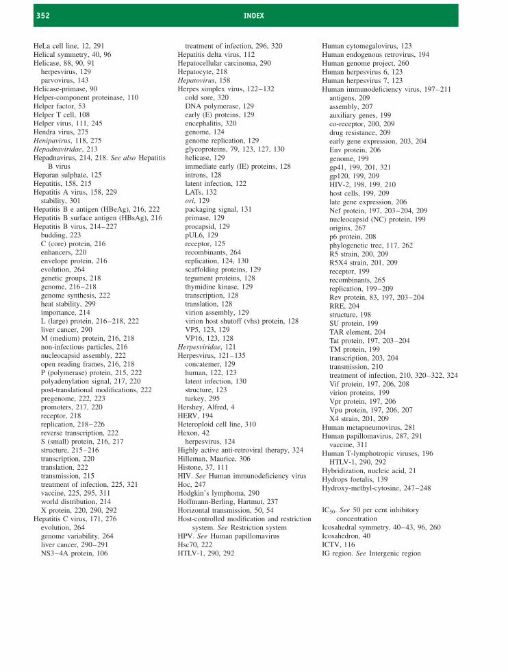
352 INDEX
HeLa cell line, 12, 291Helical symmetry, 40, 96Helicase, 88, 90, 91
herpesvirus, 129parvovirus, 143
Helicase-primase, 90Helper-component proteinase, 110Helper factor, 53Helper T cell, 108Helper virus, 111, 245Hendra virus, 275Henipavirus, 118, 275Hepadnaviridae, 213Hepadnavirus, 214, 218. See also Hepatitis
B virusHeparan sulphate, 125Hepatitis, 158, 215Hepatitis A virus, 158, 229
stability, 301Hepatitis B e antigen (HBeAg), 216, 222Hepatitis B surface antigen (HBsAg), 216Hepatitis B virus, 214–227
budding, 223C (core) protein, 216enhancers, 220envelope protein, 216evolution, 264genetic groups, 218genome, 216–218genome synthesis, 222heat stability, 299importance, 214L (large) protein, 216–218, 222liver cancer, 290M (medium) protein, 216, 218non-infectious particles, 216nucleocapsid assembly, 222open reading frames, 216, 218P (polymerase) protein, 215, 222polyadenylation signal, 217, 220post-translational modifications, 222pregenome, 222, 223promoters, 217, 220receptor, 218replication, 218–226reverse transcription, 222S (small) protein, 216, 217structure, 215–216transcription, 220translation, 222transmission, 215treatment of infection, 225, 321vaccine, 225, 295, 311world distribution, 214X protein, 220, 290, 292
Hepatitis C virus, 171, 276evolution, 264genome variability, 264liver cancer, 290–291NS3–4A protein, 106
treatment of infection, 296, 320Hepatitis delta virus, 112Hepatocellular carcinoma, 290Hepatocyte, 218Hepatovirus, 158Herpes simplex virus, 122–132
cold sore, 320DNA polymerase, 129early (E) proteins, 129encephalitis, 320genome, 124genome replication, 129glycoproteins, 79, 123, 127, 130helicase, 129immediate early (IE) proteins, 128introns, 128latent infection, 122LATs, 132ori, 129packaging signal, 131primase, 129procapsid, 129pUL6, 129receptor, 125recombinants, 264replication, 124, 130scaffolding proteins, 129tegument proteins, 128thymidine kinase, 129transcription, 128translation, 128virion assembly, 129virion host shutoff (vhs) protein, 128VP5, 123, 129VP16, 123, 128
Herpesviridae, 121Herpesvirus, 121–135
concatemer, 129human, 122, 123latent infection, 130structure, 123turkey, 295
Hershey, Alfred, 4HERV, 194Heteroploid cell line, 310Hexon, 42
herpesvirus, 124Highly active anti-retroviral therapy, 324Hilleman, Maurice, 306Histone, 37, 111HIV. See Human immunodeficiency virusHoc, 247Hodgkin’s lymphoma, 290Hoffmann-Berling, Hartmut, 237Horizontal transmission, 50, 54Host-controlled modification and restriction
system. See Restriction systemHPV. See Human papillomavirusHsc70, 222HTLV-1, 290, 292
Human cytomegalovirus, 123Human endogenous retrovirus, 194Human genome project, 260Human herpesvirus 6, 123Human herpesvirus 7, 123Human immunodeficiency virus, 197–211
antigens, 209assembly, 207auxiliary genes, 199co-receptor, 200, 209drug resistance, 209early gene expression, 203, 204Env protein, 206genome, 199gp41, 199, 201, 321gp120, 199, 209HIV-2, 198, 199, 210host cells, 199, 209late gene expression, 206Nef protein, 197, 203–204, 209nucleocapsid (NC) protein, 199origins, 267p6 protein, 208phylogenetic tree, 117, 262R5 strain, 200, 209R5X4 strain, 201, 209receptor, 199recombinants, 265replication, 199–209Rev protein, 83, 197, 203–204RRE, 204structure, 198SU protein, 199TAR element, 204Tat protein, 197, 203–204TM protein, 199transcription, 203, 204transmission, 210treatment of infection, 210, 320–322, 324Vif protein, 197, 206, 208virion proteins, 199Vpr protein, 197, 206Vpu protein, 197, 206, 207X4 strain, 201, 209
Human metapneumovirus, 281Human papillomavirus, 287, 291
vaccine, 311Human T-lymphotropic viruses, 196
HTLV-1, 290, 292Hybridization, nucleic acid, 21Hydrops foetalis, 139Hydroxy-methyl-cytosine, 247–248
IC50. See 50 per cent inhibitoryconcentration
Icosahedral symmetry, 40–43, 96, 260Icosahedron, 40ICTV, 116IG region. See Intergenic region
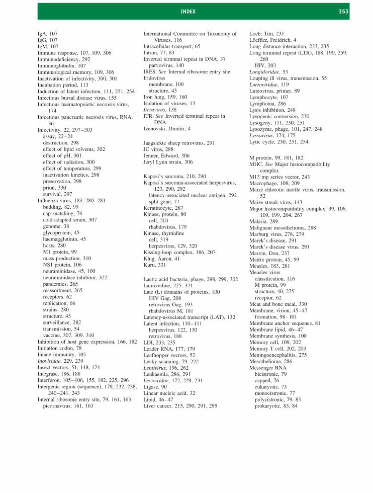
INDEX 353
IgA, 107IgG, 107IgM, 107Immune response, 107, 109, 306Immunodeficiency, 292Immunoglobulin, 107Immunological memory, 109, 306Inactivation of infectivity, 300, 301Incubation period, 113Induction of latent infection, 111, 251, 254Infectious bursal disease virus, 155Infectious haematopoietic necrosis virus,
174Infectious pancreatic necrosis virus, RNA,
36Infectivity, 22, 297–303
assay, 22–24destruction, 298effect of lipid solvents, 302effect of pH, 301effect of radiation, 300effect of temperature, 299inactivation kinetics, 298preservation, 298prion, 330survival, 297
Influenza virus, 183, 280–281budding, 82, 99cap snatching, 76cold-adapted strain, 307genome, 38glycoprotein, 45haemagglutinin, 45hosts, 280M1 protein, 99mass production, 310NS1 protein, 106neuraminidase, 45, 100neuraminidase inhibitor, 322pandemics, 265reassortment, 265receptors, 62replication, 66strains, 280structure, 45surveillance, 282transmission, 54vaccine, 307, 309, 310
Inhibition of host gene expression, 166, 182Initiation codon, 78Innate immunity, 105Inoviridae, 229, 239Insect vectors, 51, 148, 174Integrase, 186, 188Interferon, 105–106, 155, 182, 225, 296Intergenic region (sequence), 179, 232, 238,
240–241, 243Internal ribosome entry site, 79, 161, 163
picornavirus, 161, 163
International Committee on Taxonomy ofViruses, 116
Intracellular transport, 65Intron, 77, 83Inverted terminal repeat in DNA, 37
parvovirus, 140IRES. See Internal ribosome entry siteIridovirus
membrane, 100structure, 45
Iron lung, 159, 160Isolation of viruses, 13Iteravirus, 138ITR. See Inverted terminal repeat in
DNAIvanovski, Dimitri, 4
Jaagsiekte sheep retrovirus, 291JC virus, 288Jenner, Edward, 306Jeryl Lynn strain, 306
Kaposi’s sarcoma, 210, 290Kaposi’s sarcoma-associated herpesvirus,
123, 290, 292latency-associated nuclear antigen, 292split gene, 77
Keratinocyte, 287Kinase, protein, 80
cell, 204rhabdovirus, 179
Kinase, thymidinecell, 319herpesvirus, 129, 320
Kissing-loop complex, 186, 207Klug, Aaron, 41Kuru, 331
Lactic acid bacteria, phage, 298, 299, 302Lamivudine, 225, 321Late (L) domains of proteins, 100
HIV Gag, 208retrovirus Gag, 193rhabdovirus M, 181
Latency-associated transcript (LAT), 132Latent infection, 110–111
herpesvirus, 122, 130retrovirus, 188
LDI, 233, 235Leader RNA, 177, 179Leafhopper vectors, 52Leaky scanning, 79, 222Lentivirus, 196, 262Leukaemia, 286, 291Leviviridae, 172, 229, 231Ligase, 90Linear nucleic acid, 32Lipid, 46–47Liver cancer, 215, 290, 291, 295
Loeb, Tim, 231Loeffler, Freidrich, 4Long distance interaction, 233, 235Long terminal repeat (LTR), 188, 190, 259,
260HIV, 203
Longidoridae, 53Louping ill virus, transmission, 55Luteoviridae, 119Luteovirus, primer, 89Lymphocyte, 107Lymphoma, 286Lysis inhibition, 248Lysogenic conversion, 230Lysogeny, 111, 230, 251Lysozyme, phage, 101, 247, 248Lyssavirus, 174, 175Lytic cycle, 230, 251, 254
M protein, 99, 181, 182MHC. See Major histocompatibility
complexM13 mp series vector, 243Macrophage, 108, 209Maize chlorotic mottle virus, transmission,
52Maize streak virus, 143Major histocompatibility complex, 99, 106,
109, 199, 204, 267Malaria, 289Malignant mesothelioma, 288Marburg virus, 278, 279Marek’s disease, 291Marek’s disease virus, 291Marvin, Don, 237Matrix protein, 45, 99Measles, 183, 281Measles virus
classification, 116M protein, 99structure, 40, 275receptor, 62
Meat and bone meal, 330Membrane, virion, 45–47
formation, 98–101Membrane anchor sequence, 81Membrane lipid, 46–47Membrane synthesis, 100Memory cell, 109, 202Memory T cell, 202, 203Meningoencephalitis, 275Mesothelioma, 286Messenger RNA
bicistronic, 79capped, 76eukaryotic, 73monocistronic, 77polycistronic, 79, 83prokaryotic, 83, 84
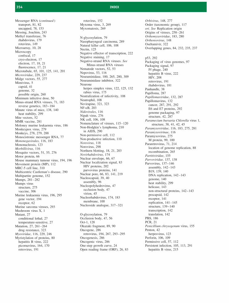
354 INDEX
Messenger RNA (continued )transport, 81, 82uncapped, 78, 153
Messing, Joachim, 243Methyl transferase, 76
rhabdovirus, 179rotavirus, 149
Microarray, 10, 28Microscopy
confocal, 17cryo-electron, 17electron, 17, 18, 21fluorescence, 17, 21
Microtubule, 65, 101, 125, 141, 201Microviridae, 229, 237Midge vectors, 55, 277Mimivirus, 5
capsid, 41genome, 32possible origin, 260
Minimum infective dose, 50Minus-strand RNA viruses, 71, 183
reverse genetics, 183–184Minute virus of mice, 138, 140
heat stability, 299Mite vectors, 52MMR vaccine, 281Moloney murine leukaemia virus, 186Monkeypox virus, 279Monkeys, 278, 279, 288Monocistronic messenger RNA, 77Mononegavirales, 116, 183Mononucleosis, 123Morbillivirus, 116Mosquito vectors, 51, 55, 276Motor protein, 66Mouse mammary tumour virus, 194, 196Movement protein (MP), 112MRC-5 cell line, 310Multicentric Castleman’s disease, 290Multipartite genome, 152Mumps, 281–282Mumps virus
structure, 275vaccine, 306
Murine leukaemia virus, 196, 295gene vector, 194receptor, 62
Murine sarcoma viruses, 293Mushroom virus X, 1Mutant, 27
conditional lethal, 27temperature-sensitive, 27
Mutation, 27, 261–264drug resistance, 323
Myoviridae, 116, 229, 246Myristylation of proteins, 80
hepatitis B virus, 222picornavirus, 164, 170retrovirus, 191
rotavirus, 152Myxoma virus, 3, 269Myxomatosis, 269
N-glycosylation, 79Nasopharyngeal carcinoma, 289Natural killer cell, 106, 108Nectin, 125Negative effector of transcription, 222Negative staining, 17Negative-strand RNA viruses. See
Minus-strand RNA virusesNematode vectors, 52, 53Nepovirus, 53, 116Neuraminidase, 100, 265, 280, 309Neuraminidase inhibitor, 322Neurone
herpes simplex virus, 122, 125, 132rabies virus, 175
Neutralization of infectivity, 108Neutrophil, 108Nevirapine, 321, 323NF-κB, 203Nidovirales, 116Nipah virus, 276NK cell, 106, 108Nomenclature of viruses, 115–120Non-Hodgkin’s lymphoma, 210
in AIDS, 290Non-permissive cell, 112Non-productive infection, 110Norovirus, 118Norovirus, 298Northern blotting, 18, 21, 203Novirhabdovirus, 174Nuclear envelope, 66, 67Nuclear localization signal, 83
HIV proteins, 202parvovirus proteins, 141
Nuclear pore, 66, 83, 141, 219Nucleocapsid, 39, 40
assembly, 96Nucleopolyhedrovirus, 47
occlusion body, 47virion, 45
Nucleorhabdovirus, 174, 183membrane, 100
Nucleoside analogue, 317–321
O-glycosylation, 79Occlusion body, 47, 56Oct-1, 128Okazaki fragment, 89, 90Oncogene, 286
retrovirus, 194, 267, 293–295Oncogenesis, 286Oncogenic virus, 286One-step growth curve, 24Open reading frame (ORF), 26, 83
Orbivirus, 148, 277Order (taxonomic group), 117ori. See Replication originOrigins of viruses, 258–261Orthomyxoviridae, 183, 280Orthoreovirus, 148Oseltamivir, 322Overlapping genes, 84, 232, 235, 237
p53, 292Packaging of virus genomes, 97Packaging signal, 97
Ff phage, 240hepatitis B virus, 222HIV, 208retrovirus, 191rhabdovirus, 181
Panhandle, 38Papilloma, 287Papillomaviridae, 132, 287Papillomavirus, 132
cancer, 287, 291, 292E6 and E7 proteins, 292genome packaging, 97structure, 42, 287
Paramecium bursaria Chlorella virus 1,structure, 38, 41, 42, 45
Paramyxoviridae, 116, 183, 275, 281Paramyxovirinae, 116Paramyxovirus, 275
M protein, 99, 100Pararetrovirus, 71, 214
location of genome replication, 88recombination, 265
Partitiviridae, 155Parvoviridae, 137, 138Parvovirus, 137–146
assembly, 142–143B19, 139, 140DNA replication, 142–143genome, 140heat stability, 299helicase, 143non-structural proteins, 142–143procapsid, 142receptor, 141replication, 141–145structure, 139–140transcription, 142translation, 142
PBS, 186PCR, 21Penicillium chrysogenum virus, 155Penton, 42
herpesvirus, 123Perforin, 106, 109Permissive cell, 57, 112Persistent infection, 105, 113, 291
hepatitis B virus, 215
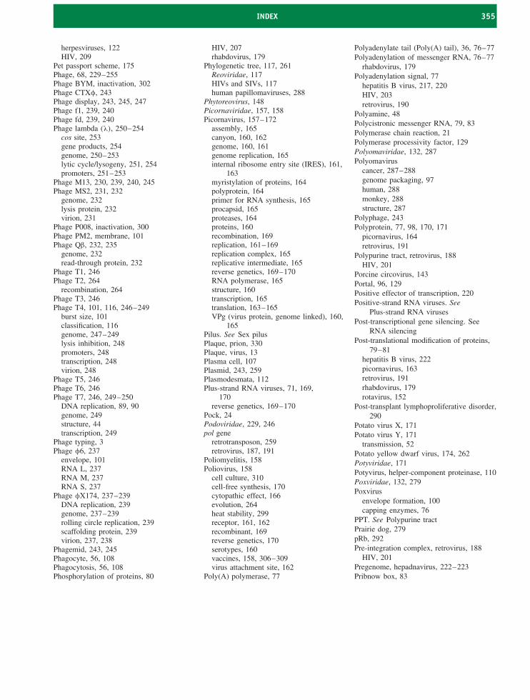
INDEX 355
herpesviruses, 122HIV, 209
Pet passport scheme, 175Phage, 68, 229–255Phage BYM, inactivation, 302Phage CTXφ, 243Phage display, 243, 245, 247Phage f1, 239, 240Phage fd, 239, 240Phage lambda (λ), 250–254
cos site, 253gene products, 254genome, 250–253lytic cycle/lysogeny, 251, 254promoters, 251–253
Phage M13, 230, 239, 240, 245Phage MS2, 231, 232
genome, 232lysis protein, 232virion, 231
Phage P008, inactivation, 300Phage PM2, membrane, 101Phage Qβ, 232, 235
genome, 232read-through protein, 232
Phage T1, 246Phage T2, 264
recombination, 264Phage T3, 246Phage T4, 101, 116, 246–249
burst size, 101classification, 116genome, 247–249lysis inhibition, 248promoters, 248transcription, 248virion, 248
Phage T5, 246Phage T6, 246Phage T7, 246, 249–250
DNA replication, 89, 90genome, 249structure, 44transcription, 249
Phage typing, 3Phage φ6, 237
envelope, 101RNA L, 237RNA M, 237RNA S, 237
Phage φX174, 237–239DNA replication, 239genome, 237–239rolling circle replication, 239scaffolding protein, 239virion, 237, 238
Phagemid, 243, 245Phagocyte, 56, 108Phagocytosis, 56, 108Phosphorylation of proteins, 80
HIV, 207rhabdovirus, 179
Phylogenetic tree, 117, 261Reoviridae, 117HIVs and SIVs, 117human papillomaviruses, 288
Phytoreovirus, 148Picornaviridae, 157, 158Picornavirus, 157–172
assembly, 165canyon, 160, 162genome, 160, 161genome replication, 165internal ribosome entry site (IRES), 161,
163myristylation of proteins, 164polyprotein, 164primer for RNA synthesis, 165procapsid, 165proteases, 164proteins, 160recombination, 169replication, 161–169replication complex, 165replicative intermediate, 165reverse genetics, 169–170RNA polymerase, 165structure, 160transcription, 165translation, 163–165VPg (virus protein, genome linked), 160,
165Pilus. See Sex pilusPlaque, prion, 330Plaque, virus, 13Plasma cell, 107Plasmid, 243, 259Plasmodesmata, 112Plus-strand RNA viruses, 71, 169,
170reverse genetics, 169–170
Pock, 24Podoviridae, 229, 246pol gene
retrotransposon, 259retrovirus, 187, 191
Poliomyelitis, 158Poliovirus, 158
cell culture, 310cell-free synthesis, 170cytopathic effect, 166evolution, 264heat stability, 299receptor, 161, 162recombinant, 169reverse genetics, 170serotypes, 160vaccines, 158, 306–309virus attachment site, 162
Poly(A) polymerase, 77
Polyadenylate tail (Poly(A) tail), 36, 76–77Polyadenylation of messenger RNA, 76–77
rhabdovirus, 179Polyadenylation signal, 77
hepatitis B virus, 217, 220HIV, 203retrovirus, 190
Polyamine, 48Polycistronic messenger RNA, 79, 83Polymerase chain reaction, 21Polymerase processivity factor, 129Polyomaviridae, 132, 287Polyomavirus
cancer, 287–288genome packaging, 97human, 288monkey, 288structure, 287
Polyphage, 243Polyprotein, 77, 98, 170, 171
picornavirus, 164retrovirus, 191
Polypurine tract, retrovirus, 188HIV, 201
Porcine circovirus, 143Portal, 96, 129Positive effector of transcription, 220Positive-strand RNA viruses. See
Plus-strand RNA virusesPost-transcriptional gene silencing. See
RNA silencingPost-translational modification of proteins,
79–81hepatitis B virus, 222picornavirus, 163retrovirus, 191rhabdovirus, 179rotavirus, 152
Post-transplant lymphoproliferative disorder,290
Potato virus X, 171Potato virus Y, 171
transmission, 52Potato yellow dwarf virus, 174, 262Potyviridae, 171Potyvirus, helper-component proteinase, 110Poxviridae, 132, 279Poxvirus
envelope formation, 100capping enzymes, 76
PPT. See Polypurine tractPrairie dog, 279pRb, 292Pre-integration complex, retrovirus, 188
HIV, 201Pregenome, hepadnavirus, 222–223Pribnow box, 83
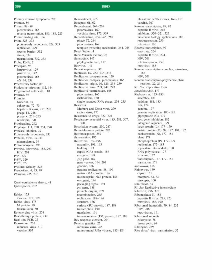
356 INDEX
Primary effusion lymphoma, 290Primase, 89Primer, 88–89
picornavirus, 165reverse transcription, 186, 188, 223
Primer binding site, 186Prion, 328–333
protein-only hypothesis, 328, 333replication, 329species barrier, 332strain, 332transmission, 332, 333
Probe, DNA, 21Procapsid, 96
herpesvirus, 129parvovirus, 142picornavirus, 165φX174, 239
Processivity factor, 89Productive infection, 112, 114Programmed cell death, 110Prohead, 96Promoter
bacterial, 83eukaryote, 72–73hepatitis B virus, 217, 220phage T4, 248phage λ, 251–253retrovirus, 190
Proofreading, 262Prophage, 111, 230, 251, 270Protease inhibitor, 321Protein-only hypothesis, 333Proteins, virus, 37–39
nomenclature, 39Proto-oncogene, 293Provirus, retrovirus, 188, 293
HIV, 201PrPc, 329PrPres, 329PrPSc, 329Prusiner, Stanley, 328Pseudoknot, 4, 33, 79Pteropus, 275, 276
Quasi-equivalence theory, 41Quasispecies, 262
Rabies, 175vaccine, 175, 309
Rabies virus, 175M protein, 99transmission, 50
Re-emerging virus, 274Read-through protein, 232Real-time PCR, 22Reassortant, 265
influenza virus, 310vaccine, 307
Reassortment, 265Receptor, 61, 62Recombinant, 264–265
picornavirus, 169vaccinia virus, 175, 309
Recombination, 264–265, 267phage T2, 264picornavirus, 169template switching mechanism, 264, 265
Reed, Walter, 4Reed-Muench method, 23Reoviridae, 147
phylogenetic tree, 117Reovirus, 148Repeat sequences, 37Replicase, 89, 232, 233–235Replication compartments, 129Replication complex, picornavirus, 165Replication origin, 90, 129, 238–239Replicative form, 239, 242, 243Replicative intermediate, 165
picornavirus, 165rhabdovirus, 180single-stranded RNA phage, 234–235
ReservoirMarburg and Ebola virus, 279rabies virus, 175
Resistance to drugs, 322–324Respiratory syncytial virus, 183, 281, 307,
320Restriction system, 230, 247Retinoblastoma protein, 292Retrotransposon, 259Retroviridae, 185Retrovirus, 185–196
assembly, 191, 193budding, 193capsid (CA) protein, 186env gene, 188gag gene, 187gene vectors, 194, 293genome, 186genome replication, 88, 190matrix (MA) protein, 186nucleocapsid (NC) protein, 186oncogene, 194packaging signal, 191pol gene, 188possible origins, 259recombination, 265replication, 188–194structure, 186surface (SU) protein, 187, 188transcription, 190translation, 191transmembrane (TM) protein, 187, 188
Rev response element, 204Reverse genetics, 27
influenza virus, 265minus-strand RNA viruses, 183–184
plus-strand RNA viruses, 169–170vaccine, 307
Reverse transcriptase, 89, 92hepatitis B virus, 215inhibitors, 320–321, 323molecular biology applications, 186retrotransposon, 259retrovirus, 186
Reverse transcription, 92error rate, 264hepatitis B virus, 224HIV, 201retrotransposon, 259retrovirus, 188
Reverse transcription complex, retrovirus,188
HIV, 201Reverse transcription-polymerase chain
reaction, 22, 261RF. See Replicative formRhabdoviridae, 173Rhabdovirus, 173–183
assembly, 181budding, 181, 183fish, 174genome, 177genome replication, 180–181glycoprotein (G), 177host gene inhibition, 182intergenic sequence, 179large protein (L), 177–179matrix protein (M), 99, 177, 181, 182nucleoprotein (N), 177, 181plant, 174phosphoprotein (P), 177–179replication, 177–183replicative intermediate, 180RNA polymerase, 177structure, 177transcription, 177, 179–181translation, 179
Rhinovirus, 158Rhinovirus, 159
capsid, 161receptors, 62, 63serotypes, 160
Rho factor, 83RI. See Replicative intermediateRibavirin, 296, 320Ribonuclease H, 188
hepatitis B virus, 215, 223retrovirus, 186, 190
Ribosomal frameshift, 79, 84, 232HIV, 206retroviruses, 191
Ribosomal subunitseukaryotic, 78prokaryotic, 84
Ribozyme, 259Rice dwarf virus, transmission, 52
INDEX 357
Rice tungro bacilliform virus, 227RISC, 109Ritonavir, 321, 323RNA in DNA viruses, 47RNA interference (RNAi), 27, 109, 152RNA phage, 231–237RNA polymerase, 89, 262.
See also DNA-dependent RNApolymerase
See also RNA-dependent RNApolymerase
RNA polymerase II, 73, 74, 89RNA replication, 91–92
picornavirus, 165rhabdovirus, 180–181rotavirus, 151, 152single-stranded RNA phages, 234–235
RNA silencing, 27, 109, 110, 155RNase H. See Ribonuclease HRNA-dependent RNA polymerase, 26,
74–75picornavirus, 165rhabdovirus, 177rotavirus, 148
RNA-induced silencing complex, 109Rolling circle replication, 91
herpesvirus, 129DNA phages, 237, 239, 243, 244–245
Rolling hairpin replication, 142Rooted phylogenetic tree, 117Rotavirus, 148–155
disease, 155double-layered particle, 151, 153replication, 150–154RNA polymerase, 148stability, 301structure, 148–149transcription, 151, 153translation, 152, 153triple-layered particle, 148
Rous sarcoma virus, 194, 196, 291oncogene, 194, 267, 293, 295
Rous, Peyton, 291RRE, 204RT-PCR, 22, 261Rubella virus, 54, 171Ryegrass mosaic virus, transmission, 52
Sabin, Albert, 306Salk, Jonas, 307S-D. See Shine-Dalgarno sequenceSanger, Fred, 26Sarcoma, 286SARS, 279, 282SARS coronavirus, 171, 280, 282
classification, 116RNA, 36
Satellite tobacco mosaic virus, capsid, 41Satellite virus, 111
Scaffolding protein, 98herpesvirus, 129phage T4, 248phage φX174, 239
Scrapie, 330SDS-PAGE, 17
rotavirus RNA, 149Secondary structure of nucleic acid, 32,
231, 233, 235Seed-transmitted viruses, 53Segmented genome, 37
ambisense RNA viruses, 183influenza virus, 47minus-strand RNA viruses, 183plus-strand RNA viruses, 170reassortants, 265reoviruses, 148tobacco rattle virus, 40
Selectivity index, 317Self-assembly of virion, 97Self-priming of DNA replication, 88Semi-conservative replication
double-stranded DNA, 90double-stranded RNA, 92
Semliki Forest virus, 47Sequencing genomes, 26, 117, 267Serotype, 116, 117, 160Severe acute respiratory syndrome, 279, 282Sex pilus, 231, 232, 236, 241Sexual transmission of viruses, 54, 295Shiga toxin, 111Shine-Dalgarno sequence, 84, 235Shingles, 123, 320Sigma factor, 83Sigma replication of DNA, 90, 91, 129Sign of disease, 113Signal sequence, 81, 99Simian immunodeficiency virus, 198, 267,
269, 274phylogenetic tree, 117, 262
Simian virus 40, 4, 288, 310endocytosis, 63T antigen, 83, 292
Simple retroviruses, 194Sin Nombre virus, 275Single-stranded DNA binding protein, 90,
129Single-stranded DNA phage, 237–247Single-stranded RNA phage, 172, 231–236
Min Jou (MJ) interaction, 232, 233replicase, 233–235replication cycle, 232, 235van Duin (VD) interaction, 233
Single-stranded RNA replication, 92, 165,180–181, 234–235
Siphoviridae, 229, 246SIV. See Simian immunodeficiency virusSlippery sequence, 206Smallpox, 1, 279
vaccination, 306
virus, 282Soc, 247Southern blotting, 18, 21Species (taxonomic group), 117Spike, envelope
herpesvirus, 123HIV, 199rhabdovirus, 177
Spliced transcript, 77HIV, 203parvovirus, 142retrovirus, 191
Split gene, 77Spongospora subterranea, 53Spraing, 1Spumavirus, 196Squamous cell carcinoma, 287Steps in virus replication, 60Sterilization, 298Stomatitis, 175Storage of viruses, 299Structure of viruses, 17, 31–48Subclinical infection, 113Subfamily (taxonomic group), 116, 117Subgenomic messenger RNAs, 170, 171Superinfection immunity, 230, 248, 251Suppressor transfer RNA, 191Surveillance of virus disease, 282SV40. See Simian virus 40Symmetry
helical, 40icosahedral, 40–43
Symptom, definition, 113Syncytium, 28, 130
T even phage, 116T lymphocyte (T cell), 107–109
HIV infection, 200–201, 209T7 DNA polymerase, 250T7 RNA polymerase, 249Tailed phage, 42, 246–254TAR element, 204TATA box, 73, 74, 220Tax, 292–293Taxonomic groups, 116TCID50, 23Tectivirus
genome, 38primer, 89
Tegument, 123Temperate phage, 111, 230, 251Tenuivirus, 119Terminally redundant, 247, 249Terminator, 72Theta replication of DNA, 90, 91, 129Thymidine kinase
cell, 319herpesvirus, 129, 320
Thymine dimer, 300
358 INDEX
Tick-borne encephalitis virus, 55Tick vectors, 54Tipula iridescent virus, 1, 118
virion, 44Tobacco mosaic virus
assembly, 97genome, 33transport through plasmodesmata, 112structure, 37, 40
Tobacco rattle virus, 1, 40, 118virions, 40
Tobamovirus, 118Tobravirus, 118Tobraviruses, transmission, 53Togaviridae, 119, 171Tomato bushy stunt virus, capsid, 41Tomato mosaic virus, 261Tomato spotted wilt virus
ambisense genome, 183cap snatching, 76
Tomato yellow leaf curl virus, transmission,52
Tombusviridae, 118Tombusvirus P19 protein, 110Transactivation response element, 204Transcriptase, 74. See also Reverse
transcriptaseTranscription, 70
bacterial, 83eukaryotic, 72–77hepatitis B virus, 220–222herpesvirus, 128HIV, 203, 206initiation, 73, 83parvovirus, 142phage T4, 248phage T7, 249picornavirus, 165retrovirus, 190rhabdovirus, 177–179rotavirus, 151, 153termination, 72, 83virus, 70–72
Transcription factor, 74, 203, 220, 293Transcription factor IID (TFIID), 74Transduction, 230Transfection, 218Transfer RNA in retrovirus virion, 186Transformation, 286Transgenic mice, 162Translation, 70
bacterial, 83–84eukaryotic, 77–79hepatitis B virus, 222herpesvirus, 128, 134HIV, 206initiation, 78–79, 84parvovirus, 142picornavirus, 163–165retrovirus, 191
rhabdovirus, 179rotavirus, 152, 153
Translational coupling, 233Transmissible spongiform encephalopathy,
328Transmission of prions 332–333Transmission of viruses 49–56, 295
hepatitis B virus, 215HIV, 210invertebrate viruses, 56plant viruses, 51vertebrate viruses, 54
Transovarial transmission, 51, 55Transport in eukaryotic cells, 81Transposon, 259Trichodoridae, 53Triple-layered particle, rotavirus, 148, 149Turnip yellow mosaic virus, 4Twort, Frederick, 230
U3 sequence, 187U5 sequence, 187Ultraviolet radiation, effect on infectivity,
300, 301Uncoating, 66Unrooted phylogenetic tree, 117Uridylylation of poliovirus VPg, 165
Vaccination, 295, 306wild mammals, 175
Vaccine, 305–313contamination, 307, 311DNA, 311–312hepatitis B virus, 225, 295, 311HIV, 211human papillomavirus, 295, 311inactivated, 298, 307influenza, 309, 310, 311live, attenuated, 306–307Marek’s disease virus, 295MMR, 306papillomavirus, 295, 311peptide, 311poliovirus, 306, 307preservation, 298, 312quality control, 307recombinant, 309subunit, 309virus-like particle, 311
Vaccinia virus, 279recombinant, 175, 309
Variant Creutzfeldt-Jakob disease (vCJD),331, 332
Varicella, 122Varicella-zoster virus, 122, 320Vector
transmission of viruses, 50, 51, 53–55See also Cloning vector.See also Gene vector.
Vero cells, 20, 310Vertical transmission, 50
plant viruses, 53vertebrate viruses, 54invertebrate viruses, 56
Vesicular stomatitis virus, 174, 175budding, 82, 99phosphoprotein, 80
Vesiculovirus, 174Viraemia, 54
HIV, 209, 210Virion
assembly, 95–101definition, 5, 32detection, 18size, 5structure, 17, 39–47
Viroplasm, 152Virulence, 113, 306, 307Virulence factor, bacterial, 111Virulent phage, 230, 246, 249Virus attachment protein, 262Virus attachment site, 61–63
hepatitis B virus, 216, 218phage, 68picornavirus, 160, 162retrovirus, 188
Virus definition, 6Virus replication, overview, 60Virus-like particle, 311VPg (virus protein, genome linked), 160,
165
Walleye dermal sarcoma virus, 196Wart, 287Water treatment, 298, 302Watson, James, 72West Nile virus, 171, 276Western blotting, 18White clover cryptic virus 1, 155Whitefly vectors, 52WI-38 cell line, 310Winter vomiting disease, 298Woodchuck HBV, 218World Health Organisation, 310
X-linked severe combinedimmunodeficiency (X-SCID), 194
X-ray crystallography, 17
Yellow fever virusbudding, 99structure, 45transmission, 55vaccine, 310
Zidovudine, 320Zinc finger, 37, 199Zinder, Norton, 231, 237Zoster, 123


