Embed Size (px)
Citation preview
NeuroImage 108 (2015) 124–137
Contents lists available at ScienceDirect
NeuroImage
j ourna l homepage: www.e lsev ie r .com/ locate /yn img
Virtual dissection and comparative connectivity of the superiorlongitudinal fasciculus in chimpanzees and humans
Erin E. Hecht a,⁎, David A. Gutman b, Bruce A. Bradley c, Todd M. Preuss d, Dietrich Stout a
a Department of Anthropology, Emory University, 1557 Dickey Drive, Rm 114, Atlanta, GA 30322, USAb Department of Biomedical Informatics, Emory University School of Medicine, 36 Eagle Row, PAIS Building, 5th Floor South, Atlanta, GA 30322, USAc Department of Archaeology, University of Exeter, Laver Building, North Park Road, Exeter EX4 4QE, UKd Yerkes National Primate Research Center, Div. Neuropharmacology & Neurologic Diseases & Center for Translational Social Neuroscience, Emory University,954 Gatewood Rd., Atlanta, GA 30329, USA
⁎ Corresponding author at: Department of PsychNeuroscience, Georgia State University, P.O. Box 5010, Atl
E-mail addresses: [email protected], [email protected]@emory.edu (D.A. Gutman), B.A.Bradley@[email protected] (T.M. Preuss), [email protected] (D
http://dx.doi.org/10.1016/j.neuroimage.2014.12.0391053-8119/© 2014 Elsevier Inc. All rights reserved.
a b s t r a c t
a r t i c l e i n f oArticle history:Accepted 8 December 2014Available online 20 December 2014
Keywords:LateralityCerebral asymmetryEvolutionWhite matterDiffusion tensor imagingTractography
Many of the behavioral capacities that distinguish humans from other primates rely on fronto-parietal circuits.The superior longitudinal fasciculus (SLF) is the primary whitematter tract connecting lateral frontal with lateralparietal regions; it is distinct from the arcuate fasciculus, which interconnects the frontal and temporal lobes.Here we report a direct, quantitative comparison of SLF connectivity using virtual in vivo dissection of the SLFin chimpanzees and humans. SLF I, the superior-most branch of the SLF, showed similar patterns of connectivitybetween humans and chimpanzees, andwasproportionally volumetrically larger in chimpanzees. SLF II, themid-dle branch, and SLF III, the inferior-most branch, showed species differences in frontal connectivity. In humans,SLF II showed greater connectivity with dorsolateral prefrontal cortex, whereas in chimps SLF II showed greaterconnectivity with the inferior frontal gyrus. SLF III was right-lateralized and proportionally volumetrically largerin humans, and human SLF III showed relatively reduced connectivity with dorsal premotor cortex and greaterextension into the anterior inferior frontal gyrus, especially in the right hemisphere. These results have implica-tions for the evolution of fronto-parietal functions including spatial attention to observed actions, social learning,and tool use, and are in line with previous research suggesting a unique role for the right anterior inferior frontalgyrus in the evolution of human fronto-parietal network architecture.
© 2014 Elsevier Inc. All rights reserved.
Introduction
Many of the behaviors that distinguish humans from otherprimates – including social learning and tool use – rely on activationof, and communication between, frontal and parietal cortical regions(Johnson-Frey, 2004; Fabbri-Destro and Rizzolatti, 2008; Peeters et al.,2009; Caspers et al., 2010). Evidence for human specializations inthese circuits is accumulating from a growing number of comparativestudies. For example, action observation involves inferior frontal andinferior parietal regions in macaques, chimpanzees, and humans(Fabbri-Destro and Rizzolatti, 2008; Caspers et al., 2010; Rilling andStout, 2014), but the type of detailed, methods-oriented social learningthat is uniquely developed in humans may be related to increasedactivation and connectivity in inferior fronto-parietal cortex (Hechtet al., 2013a,b). Similarly, tool use involves homologous inferior frontaland inferior parietal regions in monkeys and humans (Johnson-Frey,
ology, Center for Behavioralanta, GA 30302, USA.(E.E. Hecht),
.ac.uk (B.A. Bradley),. Stout).
2004; Ferrari et al., 2005; Hihara et al., 2006; Obayashi et al., 2007;Quallo et al., 2009; Orban and Rizzolatti, 2012), but a region of humananterior inferior parietal cortex has unique response properties thatmay support uniquely human capacities for causal understanding(Peeters et al., 2009; Orban and Rizzolatti, 2012). More generally,there is evidence for organizational changes and expansion of grayand white matter in the frontal lobes (Smaers et al., 2010; Preuss,2011; Passingham and Smaers, 2014), changes in frontal and parietalwhite and gray matter asymmetry (Schenker et al., 2010; Gilissen andHopkins, 2013; Hopkins and Avants, 2013; Van Essen and Glasser,2014) and emergence of new functional response properties in inferiorfrontal (Neubert et al., 2014) and parietal cortex (Peeters et al., 2009).Together, these studies suggest that fronto-parietal circuitswere a likelylocus of structural–functional adaptation in human brain evolution.Here we report a direct, quantitative comparison between humansand chimpanzees in the superior longitudinal fasciculus (SLF), the pri-mary white matter tract connecting lateral frontal with lateral parietalregions.
The SLF is an antero-posteriorly oriented tract located in the lateralaspect of the cerebral white matter. The label “superior longitudinalfasciculus” is sometimes used interchangeablywith “arcuate fasciculus,”but distinct bundles of fronto-parietal and fronto-temporalfibers can be
125E.E. Hecht et al. / NeuroImage 108 (2015) 124–137
recognized in both macaques and humans (Makris et al., 2005;Fernandez-Miranda et al., 2008; Gharabaghi et al., 2009; Petrides andPandya, 2009; Thiebaut de Schotten et al., 2011a; Martino and Marcode Lucas, 2014). Here we use the term “SLF” to refer specifically todirect fronto-parietal connections and consider the arcuate to consistof fronto-temporal connections (see the Comparison to previousstudies section for a more extensive discussion of terminology). Studiesin humans (Makris et al., 2005; Thiebaut de Schotten et al., 2011a) andmacaques (Petrides and Pandya, 1984, 2002; Schmahmann et al., 2007;Thiebaut de Schotten et al., 2012) have identified3 sub-tractswithin theSLF. The superior-most branch is SLF I, which links the superior parietallobule with the supplementary motor area, posterior dorsolateralprefrontal cortex, dorsal premotor cortex, and the rostral part of prima-ry motor cortex. SLF II is located inferior and lateral to SLF I and linksposterior inferior parietal cortex with dorsal premotor cortex anddorsolateral prefrontal cortex. SLF III is the inferior- and lateral-mostof these tracts, traveling in the opercular white matter. It connects theposterior inferior prefrontal and ventral premotor cortex with anteriorinferior parietal cortex. Functionally, SLF has been linked with motorplanning and visuospatial processing in humans and monkeys(Petrides and Pandya, 2002; Thiebaut de Schotten et al., 2011a) and isthus one likely locus of evolutionary changes supporting uniquelyhuman capacities for tool-use and social learning of observed actions.
Althoughmacaques and chimpanzees are capable of simple tool-use,humans are distinguished by the complexity of their tool-use and tool-making, including the use of tools to make other tools, the constructionof multi-component tools, and the accumulation of complexity in tooldesign through social learning (Johnson-Frey, 2003; Frey, 2007). Inhumans, tool use involves a distributed network of interconnectedfrontal, parietal, and occipitotemporal regions (Johnson-Frey, 2004;Ramayya et al., 2010; Rilling and Stout, 2014). This network overlapswith an evolutionarily ancient fronto-parietal network for object-directed grasping (Rizzolatti and Fadiga, 1998) but human tool-use net-works are undoubtedly more complex than macaque object-graspingnetworks. It has been proposed that use of “complex” tools (those thatalter the functional properties of the hand) requires additional causalunderstanding resulting from an integration of dorsal (“how”) andventral (“what”) processing streams in a left-lateralized network oftemporal, frontal and parietal areas (Frey, 2007). This capacity may besupported by the evolution of new functional response properties inleft anterior inferior parietal cortex (Peeters et al., 2009) and by theexpansion of gray matter and extension of white matter in lateraltemporal cortex, particularly the middle temporal gyrus, which playsan important role in semantic representation (Orban et al., 2004;Rilling et al., 2008; Hecht et al., 2013a).
Beyond tool-use, actual tool-making involves longer action chainswith more complex, abstract goals. There has been relatively littlestudy of such multi-step technological actions, but lesion (Hartmannet al., 2005) and neuroimaging (Frey and Gerry, 2006; Hamilton andGrafton, 2008) evidence implicate right frontoparietal cortex in therepresentation of action sequences and goals. Experimental studies ofstone tool-making, a behavior practiced by human ancestors for morethan 2.5 million years, have reported left anterior inferior parietal–ven-tral premotor activation during simple tool-making and increased rightinferior parietal–inferior frontal (ventral premotor, pars triangularis ofthe inferior frontal gyrus) duringmore complex tool-making. A longitu-dinal study of stone tool-making skill acquisition identified training-related changes (increased fractional anisotropy) in white matterunderlying these fronto-parietal cortical regions, including right parstriangularis (Hecht et al., 2014). A “mirror-system” or “simulation” ac-count of action understanding suggests that similar neural systemswould be involved in the social learning of tool-making methods, andthis has been supported by an fMRI study of stone tool-making actionobservation (Stout et al., 2011).
Comparative evidence relevant to understanding the anatomy andevolution of these left and right fronto-parietal circuits is limited. In a
previous comparative DTI study, we used probabilistic tractography tocompare frontal–parietal–temporal connectivity in macaques,chimpanzees, and humans and found a gradient in the pattern of net-work organization (Hecht et al., 2013a). In macaques, frontal–temporalconnections via the extreme and/or external capsules dominated thisnetwork, while in humans, frontal–parietal–temporal connections viathe superior and middle longitudinal fasciculi were more prominent;chimpanzees were intermediate. Thiebaut de Schotten et al. (2012)employed a “virtual dissection” approach to obtain more detailedanatomical reconstructions. They concluded that SLF is “highly con-served” between humans and macaques. However, they also reportedapparent differences, including more anterior frontal terminations ofSLF III in humans. The chimpanzee condition is unknown. Human SLFIII is right lateralized (Thiebaut de Schotten et al., 2011a,b), but the sym-metry/asymmetry of SLF branches in both macaques and chimpanzeesis again unknown. Ramayya et al. (2010) used deterministictractography to examine asymmetries of a putative human tool-usenetwork, confirming the presence of leftwardly-asymmetric connec-tions between the middle temporal gyrus, anterior inferior parietallobe and inferior frontal cortex but also finding a strongly rightwardlyasymmetric pathway between the posterior inferior parietal and frontalcortex. It is tempting to conclude that these patterns of asymmetry andenhanced fronto-parietal connectivity reflect uniquely human adapta-tions for the execution and social transmission of tool-use and tool-making, butmore detailed information on comparative anatomy is need-ed, particularly from our closest living relative, the chimpanzee. We thusconducted a virtual dissection study of humans and chimpanzees to as-sess the presence/absence of differences in the relative size, lateralization,and connections of SLF I, II, and III.
Materials and methods
Subjects and data acquisition
ChimpanzeesThe current study analyzed archived chimpanzee datasets from
previous studies. Chimpanzee subjects were 2 males and 47 femaleshoused at the Yerkes National Primate Research Center. The scans ana-lyzed in the current study were acquired at the Yerkes National PrimateResearch Center under propofol anesthesia (10 mg/kg/h) using previ-ously described procedures (Chen et al., 2013; Hecht et al., 2013b). Allprocedures were carried out in accordance with protocols approvedby YNPRC and the Emory University Institutional Animal Care and UseCommittee (approval no. YER-2001206). 60-direction DTI images withisotropic 1.8mm3 voxels were acquired on a Siemens Trio 3.0 T scanner(TR: 5900 ms; TE: 86 ms; 41 slices). 5 B0 volumes were acquiredwith no diffusion weighting. T1-weighted images were acquired onthe same scanner with isotropic 0.8 mm3 voxels (TR: 2600 ms; TE:3.06 ms; slice thickness: 0.8 mm).
HumansOne group of human subjects consisted of 5 males and 1 female
recruited from theundergraduate and graduate programs at theUniver-sity of Exeter, all right-handed by self-report, with no neurological orpsychiatric illness. Scans were acquired at the Wellcome Departmentof Imaging Neuroscience at University College London. All subjectsprovided written consent. The National Hospital for Neurology andNeurosurgery and Institute of Neurology Joint Research Ethics Commit-tee approved the study. 61-direction DTI imageswith isotropic 1.7mm3
voxels were acquired on a Siemens Trio 3.0 T scanner (TR: 1820 ms;TE:102 ms; 80 slices). 6 B0 volumes were acquired with no diffusionweighting. T1-weighted images were acquired on the same scannerwith isotropic 1 mm3 voxels (TR: 1820 ms; TE: 102 ms; 80 slices).
A second group of human subjects consisted of 58 females, 2 left-handed and the rest right-handed by self-report, with no known neuro-logical or psychiatric illness. Scanswere acquired at the Yerkes National
126 E.E. Hecht et al. / NeuroImage 108 (2015) 124–137
Primate Research Center at Emory University. All subjects providedwritten consent. The Emory Institutional Review Board approvedthe study. 60-direction DTI images with isotropic 2.0 mm3 voxelswere acquired on a Siemens Trio 3.0 T scanner. 4 B0 volumes wereacquired with no diffusion weighting (TR: 8500 ms; TE: 95 ms; 64slices). T1-weighted images were acquired on the same scannerwith isotropic 1 mm3 voxels (TR: 2600ms; TE: 3.02ms; slice thickness:1 mm).
Image processing and registration
Image processing and analysis were carried out using the FSLsoftware package (Smith et al., 2004; Woolrich et al., 2009;Jenkinson et al., 2012). T1 images underwent noise reduction usingSUSAN (Smith and Brady, 1997) and bias correction using FAST(Zhang et al., 2001). DTI data underwent correction for distortioncaused by eddy currents using EDDY (the new tool which replacesEDDY_CORRECT). Both T1 images and the averaged B0 imagesunderwent brain extraction using BET (Smith, 2002). BEDPOSTX,part of the FDT software package (Behrens et al., 2003, 2007), wasused to build up a Bayesian distribution of diffusion information in3D space for each voxel, modeling 3 fibers at each voxel. BEDPOSTXautomatically estimates the number of crossing fibers at eachvoxel. Linear transformations with 6 degrees of freedom werecomputed from each subject's DTI dataset to their T1-weightedstructural image using FLIRT, a linear registration algorithm(Jenkinson and Smith, 2001; Jenkinson et al., 2002). Nonlineartransformations were computed from each subject's T1 image to atemplate brain for that species – i.e., a chimpanzee template (Li et al.,2010) or the human 1 mm nonlinearly-registered T1 MNI template –
using FNIRT, a nonlinear registration algorithm (Andersson et al.,2007). Registration of images from native diffusion space to templatespace was achieved by combining these linear diffusion-to-structuralplus nonlinear structural-to-template transformations and applyingthem in a single step in order to avoid repeated reslicing. Registrationof images from template space to native diffusion space was achievedby inverting each of these transformations, combining them, and apply-ing the combined transformation in a single step. Tractography analysesused PROBTRACKX, a probabilistic algorithm that samples from Bayes-ian distributions of multiple diffusion directions in order to facilitatetracking through crossing fibers and into gray matter (Behrens et al.,2003, 2007).
Control tractography
The comparative tractography methods used here have previouslybeen shown to identify species differences in tracts generallyacknowledged to have undergone anatomical change in the humanlineage while producing a lack of species differences in tractsthought not to have undergone major changes in the human lineage(Hecht et al., 2013a). In order to additionally ensure that anymeasured between-species laterality differences in the SLF werenot due to between-species differences in imaging parameters orimage quality, we performed tractography in the left and rightretinogeniculostriate tracts. Seeds were placed at the optic chiasmand in coronal cross-sections of the occipital lobe (SupplementaryFig. 1a). TheseROIswere drawnon individual subjects' B0 images in na-tive diffusion space. We ran symmetric waypoints-mode tractographywith the following parameters: 25,000 samples/voxel, loopchecksenabled, curvature threshold at 0.2 (±78.45°), steplength at 0.5, fiberthreshold at 0.1, and tractography not explicitly constrained byfractional anisotropy. We mapped streamlines that passed throughboth masks in each subject. Tractography results were thresholded to0.1% of the waytotal (a measure of the total number of streamlines ina particular tractography analysis) and registered to template space asdescribed above in the Image processing and registration section.
Proportional tract volume was quantified in the native-spacethresholded tractrography images as the number of above-thresholdvoxels in the tract divided by the number of total voxels in the brain.Template-space tracts were binarized and combined to producegroup-composite images where the intensity at a given voxel corre-sponds to the number of subjects with above-threshold connectivityat that location (Hecht et al., 2013a). This produced no qualitativedifferences in tract anatomy (Supplementary Fig. 1b) or quantitativedifferences in proportional tract volume (Supplementary Fig. 1c).
Segmentation of SLF based on frontal connectivity
In a previous study, Thiebaut de Schotten et al. performed virtualdissection of the human SLF using differential frontal connectivity(Thiebaut de Schotten et al., 2011a). The same method was later alsoused in a macaque–human comparative study (Thiebaut de Schottenet al., 2012). We used a similar method here. Seeds were placed in theinferior, middle, and superior frontal gyri of each hemisphere, and insuperior parietal cortex, anterior inferior parietal cortex, and posteriorinferior parietal cortex. SLF I was defined as the tract connectingsuperior frontal cortex with superior parietal cortex; SLF II was definedas the tract connecting the middle frontal gyrus with posterior inferiorparietal cortex; and SLF III was defined as the tract connecting theinferior frontal gyruswith anterior inferior parietal cortex. Also, becausewe have previously observed tractography results that track acrossknown synapses (i.e., from the optic chiasm through the LGN to thevisual cortex (Hecht et al., 2013a)), we took some additional steps toreduce the number of streamlines representing multi-synaptic connec-tions. Specifically, a large, general inclusion mask was placed in thewhite matter between the frontal and parietal lobes at the level of thecentral sulcus in order to retain only streamlines that passed throughthis route. The entire mid-sagittal plane of each scan was used as an ex-clusionmask in order to exclude interhemispheric connections betweenthe seeds so thatwe could analyze connectivitywithin each hemisphereseparately. We also placed exclusion masks in the temporal cortex andin a coronal slice in the white matter in the vicinity of the extreme/external capsules in order to exclude connections passing throughthese tracts. Supplementary Figs. 2 and 3 show the tractographyseeds and inclusion and exclusion masks in chimpanzees andhumans. These ROIs were drawn on the chimpanzee and human(MNI) templates and registered to individuals' native diffusionspace as described above in the Image processing and registrationsection. We ran symmetric waypoints-mode tractography with thefollowing parameters: 25,000 samples/voxel, loopchecks enabled,curvature threshold at 0.2, steplength at 0.5, fiber threshold at 0.1,and tractography not explicitly constrained by fractional anisotropy(but later steps that measured white matter tract volume did soafter white matter/gray matter segmentation). Tractography resultswere thresholded to 0.1% of the waytotal and registered to templatespace as described above in the Image processing and registrationsection. Template-space tracts were binarized and combined toproduce group-composite images. Quantification of proportionaltract volume and cortical connectivity occurred in the thresholdednative-space tractography images. Proportional tract volume wasmeasured as the number of above-threshold voxels within thewhite matter of each thresholded tract, expressed as a percentageof that subject's total SLF volume. Cortical connectivity was measuredas the number of voxels in each target region that received above-threshold connectivity from each tract. Target regions includeddorsolateral prefrontal cortex, inferior frontal gyrus, dorsal precentralgyrus (dorsal premotor cortex), and ventral precentral gyrus (ventralpremotor cortex). Homologous chimpanzee and human ROIs for eachof these regions were produced as part of a previous study (Hechtet al., 2013b). These ROIs are illustrated in Fig. 4a and describedanatomically in Table 1.
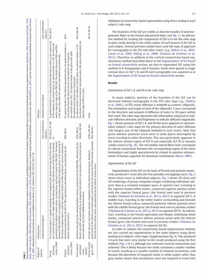
Table1
Ana
tomicalde
finition
sof
homolog
oushu
man
andch
impa
nzee
region
sof
interestforq
uantify
ingfron
talSLF
term
inations
.Chimpa
nzee
ROIsweredraw
nby
hand
basedon
prev
ious
anatom
icalresearch
(Brodm
ann,
1909
;Eco
nomoan
dPa
rker,192
9;Ba
iley,19
48;V
onBo
nin,
1948
;Baileyan
dVon
Bonin,
1950
;Sch
enke
ret
al.,20
10).Hum
anRO
IswerecreatedusingtheJulic
hprob
abilisticcytoarch
itectonicatlas(Eickh
offe
tal.,20
07)an
dtheHarva
rd/O
xfordprob
abilisticstructural
atlas(D
esikan
etal.,20
06).Re
prod
uced
withpe
rmission
andmod
ified
from
Hecht
etal.,JN
eurosci2
01333
(35):141
17-34.
Region
ofinterest
Chim
panz
ees
Hum
ans
Ana
tomical
description
Cyto-architecton
icregion
(s)
Ana
tomical
description
Cyto-architecton
icregion
(s)
Dorsal
prem
otor
cortex
(PMd)
Atitsdo
rsal
aspe
ct,itex
tend
san
teriorly
toan
imag
inarylin
edraw
nfrom
thetipof
theinferior
pre-centralsulcu
sat
a90
°an
glewiththelateralsulcu
s.Th
einferior
part
oftheRO
Iisbo
rdered
anteriorly
attheinferior
fron
talsulcu
s,cu
rvingdo
wnan
dba
ckto
mee
tthePM
vRO
I.Th
ebo
rder
betw
eenPM
dan
dPM
visan
imag
inarylin
edraw
npa
ralle
ltothelateralsulcu
sat
thedo
rsal
tipof
thefron
to-occipital
sulcus
sothat
thesu
perior
bordersof
PMvan
dBroc
a'sarea
areco
ntinuo
us.
FB(B
A6)
,FC(B
A8)
Itspo
steriorbo
rder
isave
rtical
linefrom
thelateralsulcu
sto
thesu
perior
tip
ofthesu
perior
pre-centralsulcu
s.Itsan
terior
border
isa45
°lin
efrom
the
antero-sup
eriored
geof
thePM
vRO
I.Th
ebo
rder
betw
eenPM
dan
dPM
visthegy
rusthat
splitsthesu
perior
andinferior
precen
tral
sulci.
BA6,
BA8
Ven
tral
prem
otor
cortex
(PMv)
Bordered
posteriorlyby
theM1/S1
ROI,su
periorly
asde
scribe
dab
ove,
andan
teriorly
bythe
inferior
precen
tral
sulcus
.FB
A(B
A6)
Itsan
terior
border
istheinferior
precen
tral
sulcus
.Its
posteriorbo
rder
isa
vertical
linefrom
thelateralsulcu
sto
thesu
perior
tipof
supe
rior
pre-centralsulcu
s(M
1).Its
supe
rior
border
isthegy
rusthat
splits
theinferior
andsu
perior
pre-centralg
yri.
BA6
Dorso-lateral
prefrontal
cortex
(DLP
FC)
Bordered
dorsally
bytheinterh
emisph
eric
fissure,
posteriorlyby
thePM
dRO
I,inferiorly
bythe
Broc
a'sarea
ROI,an
dan
teriorly
byan
imag
inarylin
ewhich
isan
extens
ionof
theorbitalsulcu
sdraw
npa
stthetipof
themiddlefron
talsulcu
s.
FDm
(BA9)
,Fd
delta(B
A46
)Itsinferior
border
istheinferior
fron
talsulcu
s.Itsan
terior
border
isa45
°lin
edraw
nfrom
tipof
anterior
horizo
ntal
ramus
(the
sulcus
that
bordersthe
anterior
edge
ofBroc
a'sarea
).
BA9,
BA46
Inferior
fron
tal
gyrus(IFG
)Includ
esthepa
rsop
ercu
larisan
dpa
rstriang
ularisof
theinferior
fron
talg
yrus
.Borde
red
posteriorlyby
theinferior
precen
tral
sulcus
,anteriorlyby
thesm
allsulcu
sthat
extend
san
teriorly
from
thefron
to-orbital
sulcus
,and
supe
riorly
bytheinferior
fron
talsulcu
s.
FCBm
(BA44
),FD
p(B
A45
)Sa
me.
BA44
,BA45
127E.E. Hecht et al. / NeuroImage 108 (2015) 124–137
Validation of connectivity-based segmentation using direct seeding in eachsubject's color map
The branches of the SLF are visible as discrete bundles of anterior–posterior fibers in the frontal and parietal lobes (see Fig. 1). An alterna-tive method for tracking the components of SLF is to use the color mapto place seeds directly in the white matter of each branch of the SLF ineach subject. Several previous studies have used this type of approachfor tractography in the SLF and other tracts (e.g., Makris et al., 2005;Catani et al., 2005; Rilling et al., 2008; Thiebaut de Schotten et al.,2012). Therefore, in addition to the cortical-connectivity-based seg-mentation method described above in the Segmentation of SLF basedon frontal connectivity section, we also re-segmented SLF using thismethod in 8 chimpanzees and 6 humans. Seeds were placed in singlecoronal slices in SLF I, II, and III and tractography was acquired as inthe Segmentation of SLF based on frontal connectivity section.
Results
Examination of SLF I, II, and III in the color map
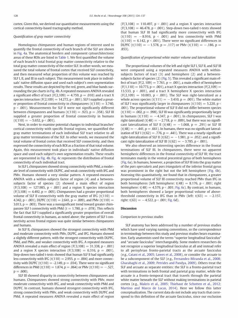
In many subjects, portions of the branches of the SLF can bediscerned without tractography in the DTI color map (e.g., (Makriset al., 2005)). In DTI, water diffusion is modeled as a tensor (ellipsoid).The orientation and length of each of the ellipsoid's 3 axes correspondto the direction and amount of diffusion of water in 3D space withinthat voxel. The color map represents this information using hue to indi-cate diffusion direction and brightness to indicate diffusion magnitude.Fig. 1 shows portions of SLF I, II, and III that were apparent in represen-tative subjects' color maps for the primary direction of water diffusion(the longest axis of the ellipsoid modeled in each voxel). Note thatgreen anterior–posterior tracts were in some places interrupted bytracts traveling in other directions. This was particularly apparent inthe inferior frontal region of SLF II and especially SLF III in humans(white arrow in Fig. 1b). The red medial–lateral fibers here correspondto callosal connections between the corresponding region of the otherhemisphere and might speculatively be related to putative enhance-ments of human capacities for bimanual coordination (Byrne, 2005).
Segmentation of the SLF
Segmentation of the SLF on the basis of frontal and parietal connec-tivity produced 3 semi-discrete but partially overlapping tracts. Fig. 2shows these tracts in individual subjects; Fig. 3 shows 2D slices and3D renderings of group composite images combining individual sub-jects' data in a common template space. A superior tract, traveling inthe superior frontal white matter, connected superior parietal cortexwith the superior frontal gyrus (the frontal seed used in previousstudies (Thiebaut de Schotten et al., 2011a, 2012) to segment SLF I). Amiddle tract, traveling in the white matter surrounding and beneaththe inferior frontal sulcus, connected posterior inferior parietal cortexwith themiddle frontal gyrus (the frontal seed used in previous studies(Thiebaut de Schotten et al., 2011a, 2012) to segment SLF II). An inferiortract, traveling in the frontal operculum and deeper underlying whitematter, connected anterior inferior parietal cortex with the inferiorfrontal gyrus (the frontal seed used in previous studies (Thiebaut deSchotten et al., 2011a, 2012) to segment SLF III).
In order to validate the connectivity-based segmentation method,we also carried out segmentation in the same subjects using directseeding in 6 subjects' color maps (Supplementary Fig. 4). This produced3 tracts that were very similar to the results produced using the firstmethod (Figs. 2 & 3), although less extensive cortical connectivity wasachieved. This is likely because the seeds contained a smaller numberof voxels, resulting in a smaller number of initiated streamlines, andbecause the placement of waypoint masks in white matter rather thangray matter meant that streamlines were not required to travel into
128 E.E. Hecht et al. / NeuroImage 108 (2015) 124–137
cortex. Given this, we derived our quantitativemeasurements using thecortical connectivity-based tractography method.
Quantification of gray matter connectivity
Homologous chimpanzee and human regions of interest used toquantify the frontal connectivity of each branch of the SLF are shownin Fig. 4a. The anatomical borders and component cytoarchitectonicareas of these ROIs are listed in Table 1. We first quantified the volumeof each branch's total frontal gray matter connectivity relative to thetotal graymatter connectivity of the entire SLF. In other words, wemea-sured the total volume of frontal cortex that received SLF connectivity,and then measured what proportion of this volume was reached bySLF I, II, and III in each subject. This measurement took place in individ-uals' native diffusion space and used each subject's own segmentationresults. These results are depicted by the red, green, and blue bands sur-rounding the pie charts in Fig. 4b. A repeatedmeasures ANOVA revealeda significant effect of tract (F(2,109) = 4.759, p = .010) and a tract Xspecies interaction (F(2,109)= 14.272, p b .001). SLF I supplied a great-er proportion of frontal connectivity in chimpanzees (t(110) = 3.740,p b .001). Measurements for SLF II were not significantly differentbetween chimpanzees and humans (t(110) = .923, p = .358). SLF IIIsupplied a greater proportion of frontal connectivity in humans(t(110) = −5.652, p b .001).
Next, in order to examine potential changes to individual branches'cortical connectivity with specific frontal regions, we quantified thegray matter terminations of each individual SLF tract relative to allgray matter terminations of the SLF. In other words, we measured thetotal volume of frontal cortex that received SLF connectivity, and thenexpressed the connectivity of each ROI as a fraction of that total volume.Again, this measurement took place in individuals' native diffusionspace and used each subject's own segmentation results. These resultsare represented in Fig. 4b. Fig. 4c represents the distribution of frontalconnectivity of each individual tract.
In SLF I, chimpanzees showed strong connectivitywith PMd, amoder-ate level of connectivity with DLPFC, andweak connectivity with IFG andPMv. Humans showed a very similar pattern. A repeated-measuresANOVA with a within-subjects factor of region (4) and a between-subjects factor of species (2) showed a main effect of region(F(3,108) = 127.985, p b .001) and a region X species interaction(F(3,108) = 4.492, p = .005). Chimpanzees had a greater proportionalvolume of SLF II connectivity with the gray matter of IFG (t(110) =4.342, p b .001), DLPFC (t(110) = 2.641, p = .009), and PMv (t(110) =3.013, p = .003). There was a nonsignificant trend toward greater chim-panzee SLF I connectivity with PMd (t = 1.780, p = .078). This reflectsthe fact that SLF I supplied a significantly greater proportion of overallfrontal connectivity in humans, as noted above; the pattern of SLF I con-nectivity across frontal regions was quite similar between chimpanzeesand humans.
In SLF II, chimpanzees showed the strongest connectivity with PMdand moderate connectivity with PMv, DLPFC, and IFG. Humans showeda slightly different pattern, with the strongest connectivity with DLPFC,PMd, and PMv, and weaker connectivity with IFG. A repeated measuresANOVA revealed a main effect of region (F(3,108) = 31.338, p b .001)and a region X species interaction (F(3,108) = 6.316, p = .001).Step-down two-tailed t-tests showed that human SLF II had significantlyless connectivity with IFG (t(110) = 2.955, p = .004) and more connec-tivity with DLPFC (t(110) = -2.149, p= .034). There were no significantdifferences in PMd (t(110)= 1.874, p= .064) or PMv (t(110)=− .527,p = .600).
SLF III showed disparity in connectivity between chimpanzees andhumans. Chimpanzees showed strong connectivity with PMv, moremoderate connectivity with IFG, and weak connectivity with PMd andDLPFC. In contrast, humans showed strongest connectivity with IFG,strong connectivity with PMv, and weak connectivity with DLPFC andPMd. A repeated measures ANOVA revealed a main effect of region
(F(3,108) = 110.497, p b .001) and a region X species interaction(F(3,108) = 46.478, p b .001). Step-down two-tailed t-tests showedthat human SLF III had significantly more connectivity with IFG(t(110) = −8.916, p b .001) and less connectivity with PMd(t(110) = 6.142, p b .001). There were no significant differences inDLPFC (t(110) = −1.578, p = .117) or PMv (t(110) = − .186, p =.853).
Quantification of proportional white matter volume and lateralization
The proportional volumes of the left and right SLF I, SLF II, and SLF IIIwere compared using a repeated measures ANOVA with within-subjects factors of tract (3) and hemisphere (2) and a between-subjects factor of species (2) (Fig. 5). This revealed a significantmain ef-fect of tract (F(2, 109) = 7.761, p = .001), a main effect of hemisphere(F(1,110)=10.773, p= .001), a tract X species interaction (F(2,109)=13.533, p b .001), and a tract X hemisphere X species interaction(F(2,109) = 10.003, p b .001). The SLF was right-lateralized as awhole across species (t(111) = −3.410, p = .001). The white matterof SLF I was significantly larger in chimpanzees (t(110) = 5.220, p b
.001). The proportional volume of SLF II did not differ between species(t(110)= .002, p= .998). SLF III was significantly proportionally largerin humans (t(110) = −4.347, p b .001). In chimpanzees, SLF I wasright-lateralized (t(48) = −2.718, p = .009), but there was no signifi-cant lateralization of SLF II (t(48) = −1.068, p = .291) or SLF III(t(48) =− .441, p= .661). In humans, there was no significant lateral-ization of SLF I (t(62) = .776, p = .441). There was a nearly-significantright-lateralization of SLF II (t(62) = −1.981, p = .052) and a signifi-cant right-lateralization of SLF III (t(62) = −3.785, p b .001).
We also observed an interesting species difference in the frontalterminations of SLF III. In chimpanzees, there were no apparenthemispheric differences in the frontal connectivity of SLF III; the tractterminates mainly in the ventral precentral gyrus of both hemispheres(Fig. 6a). In humans, however, a projection of SLF III into the graymatterof the pars opercularis and pars triangularis of the inferior frontal gyruswas prominent in the right but not the left hemisphere (Fig. 6b).Assessing this quantitatively, we found that in chimpanzees, a greaterproportional volume of SLF III connectivity reached PMv than IFG inboth hemispheres (left hemisphere: t(48) = 8.176, p b .001; righthemisphere: t(48) = 4.579, p b .001; Fig. 6c). By contrast, in humans,both hemispheres showed a larger proportional volume of above-threshold connectivity in IFG than in PMv (left: t(63) = −2.157,right: t(63) = −4.522, p b .001; Fig. 6d).
Discussion
Comparison to previous studies
SLF anatomy has been addressed by a number of previous studieswhich have used varying naming conventions, so the correspondencein terminology between this study and previous studies bears examina-tion. Early anatomists used the terms “superior longitudinal fasciculus”and “arcuate fasciculus” interchangeably. Some modern researchers donot recognize a superior longitudinal fasciculus at all and instead referto all perisylvian fronto-parietal tracts as the arcuate fasciculus(e.g., Catani et al., 2005; Lawes et al., 2008), or consider the arcuate tobe a subcomponent of the SLF (e.g., Fernandez-Miranda et al., 2008;Gharabaghi et al., 2009; Petrides and Pandya, 2009). Others treat theSLF and arcuate as separate entities: the SLF is a fronto-parietal tractwith terminations in both frontal and parietal gray matter, while thearcuate is a fronto-temporal tract that travels through the parietalwhite matter beneath the SLF without making terminations in parietalcortex (e.g., Makris et al., 2005; Thiebaut de Schotten et al., 2012;Martino and Marco de Lucas, 2014). Here we follow this latterconceptualization. The current data do not include any tracts that corre-spond to this definition of the arcuate fasciculus, since our exclusion
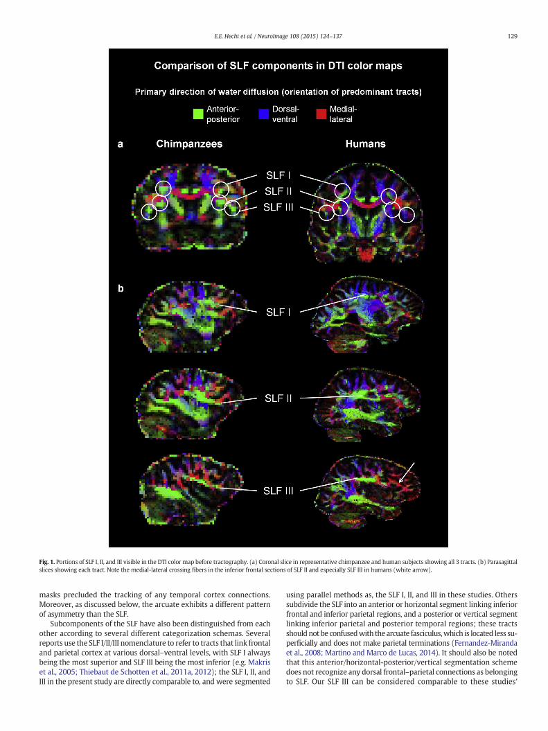
Fig. 1. Portions of SLF I, II, and III visible in the DTI color map before tractography. (a) Coronal slice in representative chimpanzee and human subjects showing all 3 tracts. (b) Parasagittalslices showing each tract. Note the medial-lateral crossing fibers in the inferior frontal sections of SLF II and especially SLF III in humans (white arrow).
129E.E. Hecht et al. / NeuroImage 108 (2015) 124–137
masks precluded the tracking of any temporal cortex connections.Moreover, as discussed below, the arcuate exhibits a different patternof asymmetry than the SLF.
Subcomponents of the SLF have also been distinguished from eachother according to several different categorization schemas. Severalreports use the SLF I/II/III nomenclature to refer to tracts that link frontaland parietal cortex at various dorsal–ventral levels, with SLF I alwaysbeing the most superior and SLF III being the most inferior (e.g. Makriset al., 2005; Thiebaut de Schotten et al., 2011a, 2012); the SLF I, II, andIII in the present study are directly comparable to, and were segmented
using parallel methods as, the SLF I, II, and III in these studies. Otherssubdivide the SLF into an anterior or horizontal segment linking inferiorfrontal and inferior parietal regions, and a posterior or vertical segmentlinking inferior parietal and posterior temporal regions; these tractsshouldnotbe confusedwith the arcuate fasciculus,which is located less su-perficially and does not make parietal terminations (Fernandez-Mirandaet al., 2008; Martino and Marco de Lucas, 2014). It should also be notedthat this anterior/horizontal-posterior/vertical segmentation schemedoes not recognize any dorsal frontal–parietal connections as belongingto SLF. Our SLF III can be considered comparable to these studies'
Fig. 2. SLF tracts in individual subjects. Parasagittal slices in representative chimpanzeeand human subjects showing SLF I (top, blue), SLF II (middle, green), and SLF III (bottom,red).
130 E.E. Hecht et al. / NeuroImage 108 (2015) 124–137
anterior/horizontal segment, while our SLF I and II have no correlates inthis classification scheme. Some researchers refer to the posterior, su-perficial, vertically-oriented parietal–temporal tract as the middle lon-gitudinal fasciculus (Makris et al., 2009; Petrides and Pandya, 2009) oras a posterior segment of the arcuate fasciculus (Catani et al., 2005;Lawes et al., 2008). This tract does not correspond to any in the currentstudy, since our exclusionmasks specifically precluded the possibility ofmeasuring any tracts with cortical terminations in temporal cortex.
Limitations of the present study
Several limitations to the present study should be noted. First, whilethe homological relationships between human and chimpanzee corticalregions discussed here are well supported by existing architectonicdata, these data are not as extensive as those bearing on human–ma-caque homologies. There were several extensive cytoarchitectonicstudies of the chimpanzee brain several decades ago (Brodmann,1909; Economo and Parker, 1929; Bailey, 1948; Von Bonin, 1948;Bailey and Von Bonin, 1950; Schenker et al., 2010). Bonin and Bailey,who produced a map of chimpanzee cortex, did so in the context of abroader, explicitly comparative project which was intended to identifyhomologous areas across primate species (Bailey and Von Bonin, 1947,
1951). More recent, focused studies have also been performed, includ-ing in inferior frontal cortex (Schenker et al., 2008, 2010). Glasseret al. produced myeloarchitectonic maps using MRI imaging, and thedistribution of myelin density is consistent with the homologies usedhere (Glasser et al., 2014). For many chimpanzee cortical regions it ispossible to identify a relatively straightforward correspondence withhuman and macaque cytoarchitectonic regions. However, a morecomplete account of homological correspondence would also takefunctional activation and connectivity into account, and these types ofstudies have been rare in chimpanzees.
A second limitation of the present study is that the voxel size:brainsize ratio is lower in the human scans than the chimpanzee scans inthe present study, meaning that the human scans have relatively higherspatial resolution. Thus, our chimpanzee tractography results may beanatomically coarser than our human results. Also, both our chimpan-zee and human datasets had a relatively low representation of males,so potential sex differences could not be investigated. Finally, it mustbe remembered that DTI does not track individual axons, and that therelationship between quantitative DTI measures and actual anatomicalconnectivity is still unclear (see Jones et al., 2013). DTI does notapproach the sensitivity or specificity of gold-standard techniques likeinjection tract tracing and it should be considered a reflection offascicle-level rather than cellular-level connectivity. Terms like “differ-ence in connectivity” in the current paper should be taken to refer todifferences in the morphology and trajectory of white matter tracts;DTI cannot shed light on finer-grained levels of connectivity like thenumber of axons or synapses in an area. However, it is the onlywhole-brain in vivo anatomical connectivity method available forspecies like chimpanzees and humans.
Species comparisons in SLF anatomy
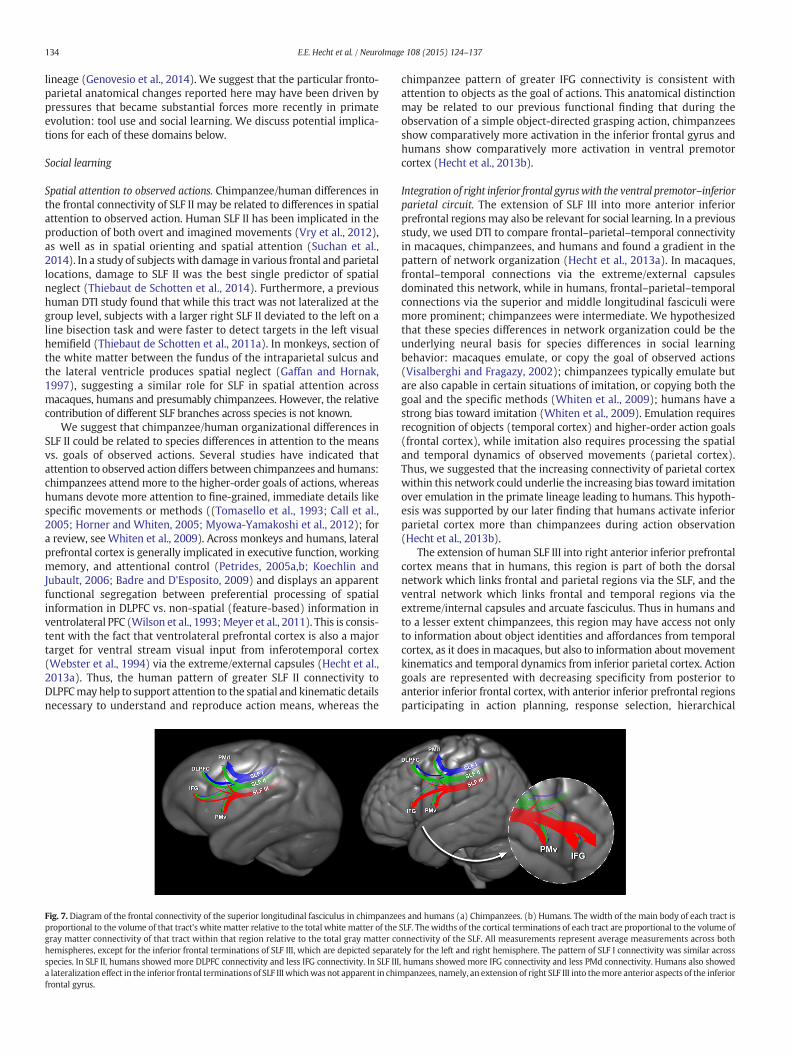
The present study found both similarities and differences in theconnectivity, size, and laterality of the superior longitudinal fasciculusin chimpanzees and humans. These findings are summarized in Fig. 7.We will discuss these separately for SLF I, II, and III.
SLF ISLF I provided a significantly larger proportional volume of frontal
cortical connectivity in chimpanzees than in humans, and it wasright-lateralized in chimpanzees. However, the pattern of SLF I's frontalconnectivity was quite similar between chimpanzees and humans. Thistract is thought to facilitate information transfer related to the higher-order regulation of motor behavior (Petrides and Pandya, 2002). Ourresults suggest that this tract may be related to some aspect of motorregulation that is or more developed in chimpanzees than in humansand/or may be more prominent in chimpanzees due to the relativehuman expansion of SLFIII.
SLF IISLF II did not show evidence of asymmetry or species differences in
theproportional volumeof themain stemofwhitematter. However,wedid observe species differences in the cortical terminations of this tract:in humans, SLF II had relatively weaker connectivity with the inferiorfrontal gyrus and relatively stronger connectivity with dorsolateralprefrontal cortex.
SLF IIIThis tract comprised a significantly greater fraction of the propor-
tional volume of the main body of SLF white matter in humans. It wasalso right-lateralized in humans, in agreement with previous research(Thiebaut de Schotten et al., 2011a,b). Note that this is in contrast tothe arcuate fasciculus, which is left-lateralized both in humans(Nucifora et al., 2005; Vernooij et al., 2007; Glasser and Rilling, 2008)and chimpanzees (Rilling et al., 2011). Humans showed stronger SLF
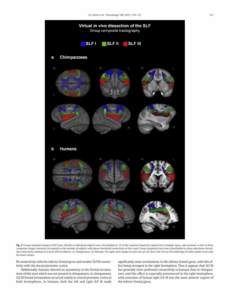
Fig. 3. Group composite images of SLF tracts. Results in individual subjects were thresholded at .1% of the waytotal, binarized, registered to template space, and summed, so that in thesecomposite images, intensity corresponds to the number of subjects with above-threshold connectivity at that voxel. Group composite tracts were thresholded to show only above-thresh-old connectivity common to at least 50% of subjects. (a) Chimpanzees. (b) Humans. The right-most images in each row are 2D slices; the rest are 3D renderings of whitematter tracts ontothe brain surface.
131E.E. Hecht et al. / NeuroImage 108 (2015) 124–137
III connectivitywith the inferior frontal gyrus andweaker SLF III connec-tivity with the dorsal premotor cortex.
Additionally, humans showed an asymmetry in the frontal termina-tions of this tractwhichwas not present in chimpanzees. In chimpanzees,SLF III frontal terminations occurredmainly in ventral premotor cortex inboth hemispheres. In humans, both the left and right SLF III made
significantly more terminations in the inferior frontal gyrus, with this ef-fect being strongest in the right hemisphere. Thus it appears that SLF IIIhas generally more prefrontal connectivity in humans than in chimpan-zees, and this effect is especially pronounced in the right hemisphere,with extension of human right SLF III into the more anterior regions ofthe inferior frontal gyrus.
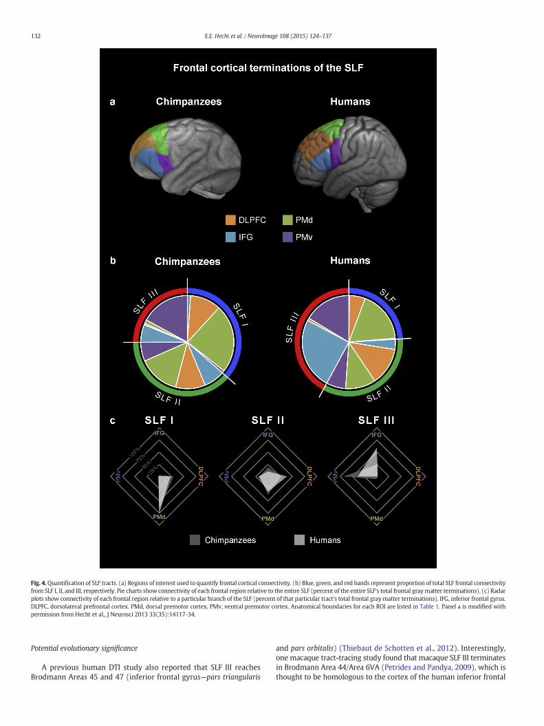
Fig. 4.Quantification of SLF tracts. (a) Regions of interest used to quantify frontal cortical connectivity. (b) Blue, green, and red bands represent proportion of total SLF frontal connectivityfrom SLF I, II, and III, respectively. Pie charts show connectivity of each frontal region relative to the entire SLF (percent of the entire SLF's total frontal graymatter terminations). (c) Radarplots show connectivity of each frontal region relative to a particular branch of the SLF (percent of that particular tract's total frontal graymatter terminations). IFG, inferior frontal gyrus.DLPFC, dorsolateral prefrontal cortex. PMd, dorsal premotor cortex. PMv, ventral premotor cortex. Anatomical boundaries for each ROI are listed in Table 1. Panel a is modified withpermission from Hecht et al., J Neurosci 2013 33(35):14117-34.
132 E.E. Hecht et al. / NeuroImage 108 (2015) 124–137
Potential evolutionary significance
A previous human DTI study also reported that SLF III reachesBrodmann Areas 45 and 47 (inferior frontal gyrus—pars triangularis
and pars orbitalis) (Thiebaut de Schotten et al., 2012). Interestingly,one macaque tract-tracing study found that macaque SLF III terminatesin Brodmann Area 44/Area 6VA (Petrides and Pandya, 2009), which isthought to be homologous to the cortex of the human inferior frontal
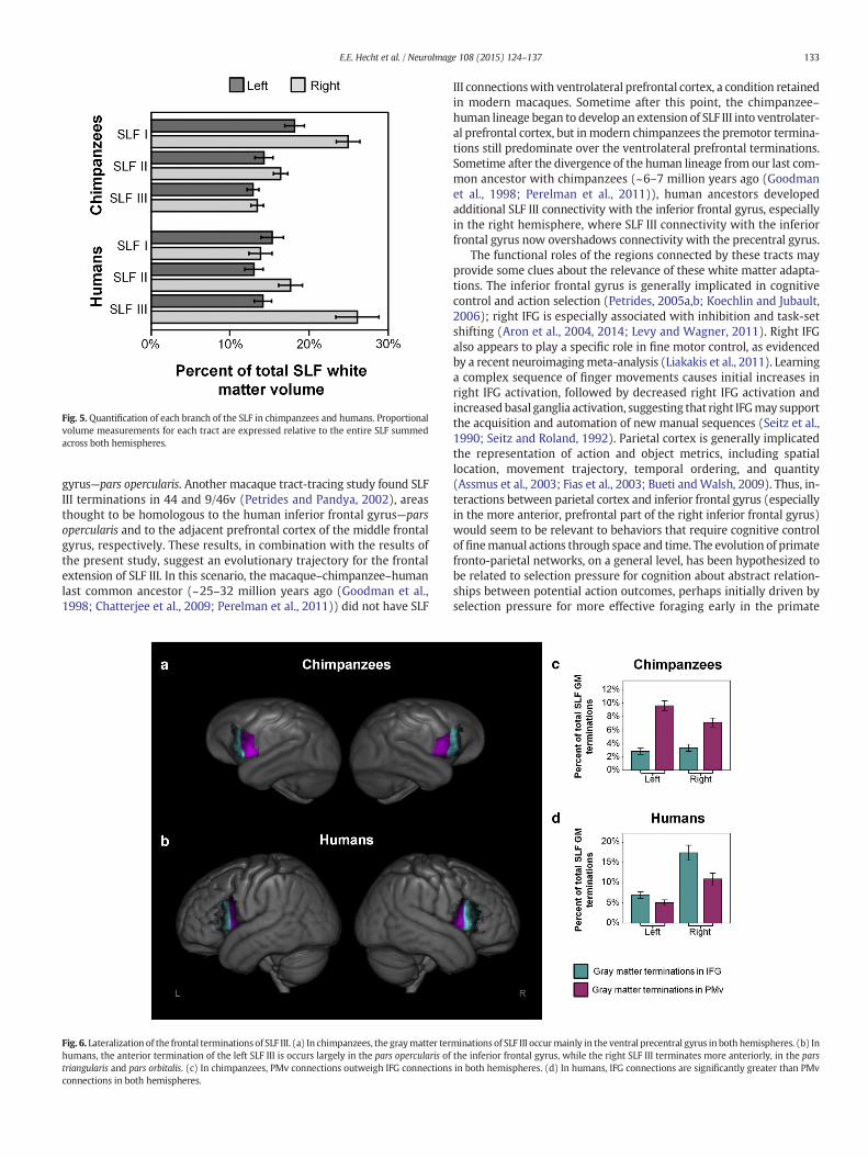
Fig. 5. Quantification of each branch of the SLF in chimpanzees and humans. Proportionalvolume measurements for each tract are expressed relative to the entire SLF summedacross both hemispheres.
133E.E. Hecht et al. / NeuroImage 108 (2015) 124–137
gyrus—pars opercularis. Another macaque tract-tracing study found SLFIII terminations in 44 and 9/46v (Petrides and Pandya, 2002), areasthought to be homologous to the human inferior frontal gyrus—parsopercularis and to the adjacent prefrontal cortex of the middle frontalgyrus, respectively. These results, in combination with the results ofthe present study, suggest an evolutionary trajectory for the frontalextension of SLF III. In this scenario, the macaque–chimpanzee–humanlast common ancestor (~25–32 million years ago (Goodman et al.,1998; Chatterjee et al., 2009; Perelman et al., 2011)) did not have SLF
Fig. 6. Lateralization of the frontal terminations of SLF III. (a) In chimpanzees, the graymatter terhumans, the anterior termination of the left SLF III is occurs largely in the pars opercularis oftriangularis and pars orbitalis. (c) In chimpanzees, PMv connections outweigh IFG connectionsconnections in both hemispheres.
III connectionswith ventrolateral prefrontal cortex, a condition retainedin modern macaques. Sometime after this point, the chimpanzee–human lineage began to develop an extension of SLF III into ventrolater-al prefrontal cortex, but inmodern chimpanzees the premotor termina-tions still predominate over the ventrolateral prefrontal terminations.Sometime after the divergence of the human lineage from our last com-mon ancestor with chimpanzees (~6–7 million years ago (Goodmanet al., 1998; Perelman et al., 2011)), human ancestors developedadditional SLF III connectivity with the inferior frontal gyrus, especiallyin the right hemisphere, where SLF III connectivity with the inferiorfrontal gyrus now overshadows connectivity with the precentral gyrus.
The functional roles of the regions connected by these tracts mayprovide some clues about the relevance of these white matter adapta-tions. The inferior frontal gyrus is generally implicated in cognitivecontrol and action selection (Petrides, 2005a,b; Koechlin and Jubault,2006); right IFG is especially associated with inhibition and task-setshifting (Aron et al., 2004, 2014; Levy and Wagner, 2011). Right IFGalso appears to play a specific role in fine motor control, as evidencedby a recent neuroimagingmeta-analysis (Liakakis et al., 2011). Learninga complex sequence of finger movements causes initial increases inright IFG activation, followed by decreased right IFG activation andincreased basal ganglia activation, suggesting that right IFGmay supportthe acquisition and automation of new manual sequences (Seitz et al.,1990; Seitz and Roland, 1992). Parietal cortex is generally implicatedthe representation of action and object metrics, including spatiallocation, movement trajectory, temporal ordering, and quantity(Assmus et al., 2003; Fias et al., 2003; Bueti andWalsh, 2009). Thus, in-teractions between parietal cortex and inferior frontal gyrus (especiallyin the more anterior, prefrontal part of the right inferior frontal gyrus)would seem to be relevant to behaviors that require cognitive controlof finemanual actions through space and time. The evolution of primatefronto-parietal networks, on a general level, has been hypothesized tobe related to selection pressure for cognition about abstract relation-ships between potential action outcomes, perhaps initially driven byselection pressure for more effective foraging early in the primate
minations of SLF III occurmainly in the ventral precentral gyrus in both hemispheres. (b) Inthe inferior frontal gyrus, while the right SLF III terminates more anteriorly, in the parsin both hemispheres. (d) In humans, IFG connections are significantly greater than PMv
134 E.E. Hecht et al. / NeuroImage 108 (2015) 124–137
lineage (Genovesio et al., 2014). We suggest that the particular fronto-parietal anatomical changes reported here may have been driven bypressures that became substantial forces more recently in primateevolution: tool use and social learning. We discuss potential implica-tions for each of these domains below.
Social learning
Spatial attention to observed actions. Chimpanzee/human differences inthe frontal connectivity of SLF II may be related to differences in spatialattention to observed action. Human SLF II has been implicated in theproduction of both overt and imagined movements (Vry et al., 2012),as well as in spatial orienting and spatial attention (Suchan et al.,2014). In a study of subjects with damage in various frontal and parietallocations, damage to SLF II was the best single predictor of spatialneglect (Thiebaut de Schotten et al., 2014). Furthermore, a previoushuman DTI study found that while this tract was not lateralized at thegroup level, subjects with a larger right SLF II deviated to the left on aline bisection task and were faster to detect targets in the left visualhemifield (Thiebaut de Schotten et al., 2011a). In monkeys, section ofthe white matter between the fundus of the intraparietal sulcus andthe lateral ventricle produces spatial neglect (Gaffan and Hornak,1997), suggesting a similar role for SLF in spatial attention acrossmacaques, humans and presumably chimpanzees. However, the relativecontribution of different SLF branches across species is not known.
We suggest that chimpanzee/human organizational differences inSLF II could be related to species differences in attention to the meansvs. goals of observed actions. Several studies have indicated thatattention to observed action differs between chimpanzees and humans:chimpanzees attend more to the higher-order goals of actions, whereashumans devote more attention to fine-grained, immediate details likespecific movements or methods ((Tomasello et al., 1993; Call et al.,2005; Horner and Whiten, 2005; Myowa-Yamakoshi et al., 2012); fora review, see Whiten et al., 2009). Across monkeys and humans, lateralprefrontal cortex is generally implicated in executive function, workingmemory, and attentional control (Petrides, 2005a,b; Koechlin andJubault, 2006; Badre and D'Esposito, 2009) and displays an apparentfunctional segregation between preferential processing of spatialinformation in DLPFC vs. non-spatial (feature-based) information inventrolateral PFC (Wilson et al., 1993;Meyer et al., 2011). This is consis-tent with the fact that ventrolateral prefrontal cortex is also a majortarget for ventral stream visual input from inferotemporal cortex(Webster et al., 1994) via the extreme/external capsules (Hecht et al.,2013a). Thus, the human pattern of greater SLF II connectivity toDLPFCmay help to support attention to the spatial and kinematic detailsnecessary to understand and reproduce action means, whereas the
Fig. 7. Diagram of the frontal connectivity of the superior longitudinal fasciculus in chimpanzeeproportional to the volume of that tract's white matter relative to the total white matter of thegray matter connectivity of that tract within that region relative to the total gray matter cohemispheres, except for the inferior frontal terminations of SLF III, which are depicted separatspecies. In SLF II, humans showed more DLPFC connectivity and less IFG connectivity. In SLF IIIa lateralization effect in the inferior frontal terminations of SLF III whichwas not apparent in chimfrontal gyrus.
chimpanzee pattern of greater IFG connectivity is consistent withattention to objects as the goal of actions. This anatomical distinctionmay be related to our previous functional finding that during theobservation of a simple object-directed grasping action, chimpanzeesshow comparatively more activation in the inferior frontal gyrus andhumans show comparatively more activation in ventral premotorcortex (Hecht et al., 2013b).
Integration of right inferior frontal gyruswith the ventral premotor–inferiorparietal circuit. The extension of SLF III into more anterior inferiorprefrontal regions may also be relevant for social learning. In a previousstudy, we used DTI to compare frontal–parietal–temporal connectivityin macaques, chimpanzees, and humans and found a gradient in thepattern of network organization (Hecht et al., 2013a). In macaques,frontal–temporal connections via the extreme/external capsulesdominated this network, while in humans, frontal–parietal–temporalconnections via the superior and middle longitudinal fasciculi weremore prominent; chimpanzees were intermediate. We hypothesizedthat these species differences in network organization could be theunderlying neural basis for species differences in social learningbehavior: macaques emulate, or copy the goal of observed actions(Visalberghi and Fragazy, 2002); chimpanzees typically emulate butare also capable in certain situations of imitation, or copying both thegoal and the specific methods (Whiten et al., 2009); humans have astrong bias toward imitation (Whiten et al., 2009). Emulation requiresrecognition of objects (temporal cortex) and higher-order action goals(frontal cortex), while imitation also requires processing the spatialand temporal dynamics of observed movements (parietal cortex).Thus, we suggested that the increasing connectivity of parietal cortexwithin this network could underlie the increasing bias toward imitationover emulation in the primate lineage leading to humans. This hypoth-esis was supported by our later finding that humans activate inferiorparietal cortex more than chimpanzees during action observation(Hecht et al., 2013b).
The extension of human SLF III into right anterior inferior prefrontalcortex means that in humans, this region is part of both the dorsalnetwork which links frontal and parietal regions via the SLF, and theventral network which links frontal and temporal regions via theextreme/internal capsules and arcuate fasciculus. Thus in humans andto a lesser extent chimpanzees, this region may have access not onlyto information about object identities and affordances from temporalcortex, as it does in macaques, but also to information about movementkinematics and temporal dynamics from inferior parietal cortex. Actiongoals are represented with decreasing specificity from posterior toanterior inferior frontal cortex, with anterior inferior prefrontal regionsparticipating in action planning, response selection, hierarchical
s and humans (a) Chimpanzees. (b) Humans. The width of the main body of each tract isSLF. The widths of the cortical terminations of each tract are proportional to the volume ofnnectivity of the SLF. All measurements represent average measurements across bothely for the left and right hemisphere. The pattern of SLF I connectivity was similar across, humans showed more IFG connectivity and less PMd connectivity. Humans also showedpanzees, namely, an extension of right SLF III into themore anterior aspects of the inferior
135E.E. Hecht et al. / NeuroImage 108 (2015) 124–137
sequencing, and cognitive control (Petrides, 2005a,b; Badre andD'Esposito, 2009). Therefore, we hypothesize that the extension ofhuman SLF III into right anterior inferior frontal gyrus could enableincreased incorporation of kinematic detail into processing of higher-order action goals. This function would be especially important forbehaviors where the achievement of hierarchical action goals is causallydependent on kinematics, as it is in stone toolmaking (Nonaka et al.,2010; Stout, 2013).
Tool useChimpanzee/human differences in the relative size and anterior
terminations of SLF III may be related to differences in tool-use ability.Chimpanzee tool-use is impressive, and includes the dexterous use,making, and sequential combination of multiple tools to achieve a goal(Sanz and Morgan, 2010). It is widely assumed that the commonancestor of chimpanzees and humans had similar tool-using capacities(e.g. Panger et al., 2002). The earliest known uniquely hominin toolsare 2.6 million years old, consist of sharp stone flakes struck from rivercobbles using another stone, andwere likely used to butcher animal car-casses (Semaw et al., 2003). There is some debate whether these simple“Oldowan” tools represent a departure from shared, “ape-grade” cogni-tive capacities (Wynn et al., 2011), but their manufacture does requirebimanual coordination of accurate, forceful blows that appears difficultor impossible for apes (Byrne, 2005). In keepingwith this, studies of ac-tual Oldowan tool-making by modern human subjects using FDG–PETmethodology, which enables imaging of behavior that occurs outsidethe scanner (Stout and Chaminade, 2007; Stout et al., 2008), documenttask-related activations in a distributed frontal–parietal–occipitotemporal network for visually-guided object manipulation sim-ilar to that identified in other studies of complex tool-use (e.g. Frey,2007), but no activations in more anterior portions of prefrontal cortextypically associated with cognitive control functions. Intriguingly, thecurrent study did find evidence of increased medial–lateral fibers inhuman inferior prefrontal cortex (Fig. 1b), which if they do indeed cor-respond to interhemispheric callosal connections, may play a rolesupporting enhanced human capacities for bimanual coordination.
After approximately 1.7million years ago (Lepre et al., 2011; Beyeneet al., 2013), Oldowan technology began to be replaced by “Acheulean”tool-making methods based the intentional shaping of stones to pro-duce large cutting tools known as ‘picks’, ‘handaxes’ and ‘cleavers’.Such shaping requires more extended sequences of contingent actionsorganized with respect to a distal goal (Stout, 2011) and is commonlythought to represent a major increase in cognitive complexity. By500,000 years ago, some Acheulean tools exhibited a high level of re-finement requiring even more complex production sequences includ-ing, for example, the careful preparation of edges and surfaces prior toflake removal (Stout et al., 2014). FDG–PET data show that, unlikeOldowan tool-making, such refined Acheulean tool-making is associat-ed with increased activation of right fronto-parietal cortex generallyand right IFG—pars triangularis specifically (Stout et al., 2008). A similarpreferential response of right pars triangularis to Acheulean tool-makingwas observed in an fMRI study of action observation. This study also re-ported an effect of technology (Acheulean N Oldowan) in left inferiorfrontal sulcus bordering IFG—pars triangularis (Stout et al., 2011). Finally,a recent longitudinal DTI study of stone tool-making (especially Acheule-an) skill acquisition over a two-year period found training-related chang-es (increased fractional anisotropy) in SLF III underlying inferior parietaland frontal cortex, again including right IFG—pars triangularis (Hechtet al., 2014).
Experimental evidence thus links increased IFG functional responseand white matter connectivity to archeologically documented variationin tool-making complexity over the course of human evolution.We pro-pose that the enhanced human SLF III connectivity with IFG reportedhere, and especially that of right SLF III, comprises part of the anatomicalbasis for the unique technological capacities of Homo sapiens. The IFGbilaterally is associated with the hierarchical control of behavior
(Koechlin and Jubault, 2006) and right IFG in particular is associatedwith inhibitory and set-shifting functions (Aron et al., 2004, 2014;Levy and Wagner, 2011) that are important to execution of multi-stepaction plans (e.g. Hartmann et al., 2005). Right parietal cortex isinvolved in representing the sequential order of behavior (Frey andGerry, 2006; Jubault et al., 2007), and the right hemisphere in generalappears to be specialized for integration of perception and action acrosslarger spatio-temporal frames (Deacon, 1997; Serrien et al., 2006; Stoutand Chaminade, 2012). Increased human connectivity between theright inferior frontal gyrus and parietal cortex via SLF III may providean anatomical basis for enhanced cognitive control over the complexgoals and multi-step action sequences characteristic of human technol-ogy (Stout, 2013).
Conclusions
Our results indicate that during human evolution, SLF II underwentselection for increased connectivity with dorsolateral prefrontal cortex,whereas SLF III underwent selection for increased relative size and in-creased connectivity with the inferior frontal gyrus, in particular themore anterior aspects of the right inferior frontal gyrus. Thus the neuralsubstrate for integrating right inferior frontal cortex with the lateralfrontal–parietal–occipitotemporal network likely emerged relatively re-cently in primate evolution, after humans' divergence from chimpan-zees. We suggest that the driving force behind these changes mayhave been the intertwined selective pressures for spatial attention toobserved actions, toolmaking, and social learning, all of which likely de-pend on integration of spatial, kinematic and sequential information foraction perception and control.
Supplementary data to this article can be found online at http://dx.doi.org/10.1016/j.neuroimage.2014.12.039.
Acknowledgments
We appreciate the work of the animal care, veterinary, and imagingstaff at the Yerkes National Primate Research Center and the imagingstaff at the Wellcome Trust Centre for Neuroimaging at UniversityCollege London. We would also like to extend our gratitude to thevolunteer subjects whose dedication, good humor and reliability madethis project possible. This research was funded by the LeverhulmeTrust F/00 144/BP to BB and DS, theWenner-Gren Foundation Disserta-tion Fieldwork Grant and Osmundsen Initiative Award 7699 (Ref#3681) to EH, The John Templeton Foundation (Award 40463 to TMP),and the National Institutes of Health RR-00165 to Yerkes NationalPrimate Research Center (superseded by the Office of Research Infra-structure Programs/OD P51OD11132).
Conflicts of interest
The authors declare that there are no conflicts of interest.
References
Andersson, J.L.R., Jenkinson,M., Smith, S., 2007. Non-linear Optimisation: FMRIB TechnicalReport (TR07JA1 from www.fmrib.ox.ac.uk/analysis/techrep).
Aron, A.R., Robbins, T.W., Poldrack, R.A., 2004. Inhibition and the right inferior frontalcortex. Trends Cogn. Sci. 8 (4), 170–177.
Aron, A.R., Robbins, T.W., Poldrack, R.A., 2014. Inhibition and the right inferior frontal cor-tex: one decade on. Trends Cogn. Sci 18 (4), 177–185.
Assmus, A., Marshall, J.C., Ritzl, A., Noth, J., Zilles, K., Fink, G.R., 2003. Left inferior parietalcortex integrates time and space during collision judgments. Neuroimage 20(Suppl. 1), S82–S88.
Badre, D., D'Esposito, M., 2009. Is the rostro-caudal axis of the frontal lobe hierarchical?Nat. Rev. Neurosci. 10 (9), 659–669.
Bailey, P., 1948. Concerning cytoarchitecture of the frontal lobe of chimpanzee (Pansatyrus) and man (Homo sapiens). In: Fulton, J.F. (Ed.), The Frontal Lobes. Williamsand Wilkins, Baltimore.
Bailey, P., Von Bonin, G., 1947. The Neocortex of Macaca mulatta. University of IllinoisPress, Urbana, Illinois.
136 E.E. Hecht et al. / NeuroImage 108 (2015) 124–137
Bailey, P., Von Bonin, G., 1950. The Isocortex of the Chimpanzee. The University of Illinois,Urbana, Illinois.
Bailey, P., Von Bonin, G., 1951. The Isocortex of Man. University of Illinois Press, Urbana,Illinois.
Behrens, T.E., Woolrich, M.W., Jenkinson, M., Johansen-Berg, H., Nunes, R.G., Clare, S.,Matthews, P.M., Brady, J.M., Smith, S.M., 2003. Characterization and propagation ofuncertainty in diffusion-weighted MR imaging. Magn. Reson. Med. 50 (5),1077–1088.
Behrens, T.E., Berg, H.J., Jbabdi, S., Rushworth, M.F., Woolrich, M.W., 2007. Probabilisticdiffusion tractography with multiple fibre orientations: what can we gain?Neuroimage 34 (1), 144–155.
Beyene, Y., Katoh, S., WoldeGabriel, G., Hart, W.K., Uto, K., Sudo, M., Kondo, M., Hyodo, M.,Renne, P.R., Suwa, G., Asfaw, B., 2013. The characteristics and chronology of theearliest Acheulean at Konso, Ethiopia. Proc. Natl. Acad. Sci. 110 (5), 1584–1591.
Brodmann, K., 1909. Vergleichende Lokalisationslehre der Grosshirnrhinde. Barth, Leipzig((reprinted as Brodmannn's Localisation in the Cereb Cortex, 1994). London,Smith-Gordon).
Bueti, D., Walsh, V., 2009. The parietal cortex and the representation of time, space,number and other magnitudes. Philos. Trans. R. Soc. Lond. B Biol. Sci. 364 (1525),1831–1840.
Byrne, R.W., 2005. The maker not the tool: the cognitive significance of great ape manualskills. In: Roux, V., Bril, B. (Eds.), Stone Knapping : The Necessary Conditions for aUniquely Hominid Behaviour. McDonald Institute, pp. 159–169.
Call, J., Carpenter, M., Tomasello, M., 2005. Copying results and copying actions in theprocess of social learning: chimpanzees (Pan troglodytes) and human children(Homo sapiens). Anim. Cogn. 8 (3), 151–163.
Caspers, S., Zilles, K., Laird, A.R., Eickhoff, S.B., 2010. ALE meta-analysis of action observa-tion and imitation in the human brain. Neuroimage 50 (3), 1148–1167.
Catani, M., Jones, D.K., Ffytche, D.H., 2005. Perisylvian language networks of the humanbrain. Ann. Neurol. 57 (1), 8–16.
Chatterjee, H.J., Ho, S.Y., Barnes, I., Groves, C., 2009. Estimating the phylogeny anddivergence times of primates using a supermatrix approach. BMC Evol. Biol. 9, 259.
Chen, X., Errangi, B., Li, L., Glasser, M.F., Westlye, L.T., Fjell, A.M., Walhovd, K.B., Hu, X.,Herndon, J.G., Preuss, T.M., Rilling, J.K., 2013. Brain aging in humans, chimpanzees(Pan troglodytes), and rhesus macaques (Macaca mulatta): magnetic resonanceimaging studies of macro- and microstructural changes. Neurobiol. Aging 34 (10),2248–2260.
Deacon, T.W., 1997. The Symbolic Species: The Co-evolution of Language and the Brain.W.W. Norton, New York.
Desikan, R.S., Ségonne, F., Fischl, B., Quinn, B.T., Dickerson, B.C., Blacker, D., Buckner, R.L.,Dale, A.M., Maguire, R.P., Hyman, B.T., Albert, M.S., Killiany, R.J., 2006. An automatedlabeling system for subdividing the human cerebral cortex on MRI scans into gyralbased regions of interest. Neuroimage 31 (3), 968–980.
Economo, C., Parker, S., 1929. The Cytoarchitectonics of the Human Cerebral Cortex.Oxford University Press, London, Humphrey Milford.
Eickhoff, S.B., Paus, T., Caspers, S., Grosbras, M.H., Evans, A.C., Zilles, K., Amunts, K., 2007.Assignment of functional activations to probabilistic cytoarchitectonic areas revisited.Neuroimage 36 (3), 511–521.
Fabbri-Destro, M., Rizzolatti, G., 2008.Mirror neurons andmirror systems inmonkeys andhumans. Physiology (Bethesda) 23, 171–179.
Fernandez-Miranda, J.C., Rhoton Jr., A.L., Alvarez-Linera, J., Kakizawa, Y., Choi, C., deOliveira, E.P., 2008. Three-dimensional microsurgical and tractographic anatomy ofthe white matter of the human brain. Neurosurgery 62 (6 Suppl. 3), 989–1026(discussion 1026–1028).
Ferrari, P.F., Rozzi, S., Fogassi, L., 2005. Mirror neurons responding to observation ofactions made with tools in monkey ventral premotor cortex. J. Cogn. Neurosci. 17(2), 212–226.
Fias, W., Lammertyn, J., Reynvoet, B., Dupont, P., Orban, G.A., 2003. Parietal representationof symbolic and nonsymbolic magnitude. J. Cogn. Neurosci. 15 (1), 47–56.
Frey, S.H., 2007. What puts the how in where? Tool use and the divided visual streamshypothesis. Cortex 43 (3), 368–375.
Frey, S.H., Gerry, V.E., 2006. Modulation of neural activity during observational learning ofactions and their sequential orders. J. Neurosci. 26 (51), 13194–13201.
Gaffan, D., Hornak, J., 1997. Visual neglect in the monkey. Representation and disconnec-tion. Brain 120 (Pt 9), 1647–1657.
Genovesio, A., Wise, S.P., Passingham, R.E., 2014. Prefrontal–parietal function: fromforaging to foresight. Trends Cogn. Sci. 18 (2), 72–81.
Gharabaghi, A., Kunath, F., Erb, M., Saur, R., Heckl, S., Tatagiba, M., Grodd, W., Karnath,H.O., 2009. Perisylvian white matter connectivity in the human right hemisphere.BMC Neurosci. 10, 15.
Gilissen, E.P., Hopkins, W.D., 2013. Asymmetries of the parietal operculum in chimpan-zees (Pan troglodytes) in relation to handedness for tool use. Cereb. Cortex 23 (2),411–422.
Glasser, M.F., Rilling, J.K., 2008. DTI tractography of the human brain's language pathways.Cereb. Cortex 18 (11), 2471–2482.
Glasser, M.F., Goyal, M.S., Preuss, T.M., Raichle, M.E., Van Essen, D.C., 2014. Trends andproperties of human cerebral cortex: correlations with cortical myelin content.Neuroimage 93 (Pt 2), 165–175.
Goodman, M., Porter, C.A., Czelusniak, J., Page, S.L., Schneider, H., Shoshani, J., Gunnell, G.,Groves, C.P., 1998. Toward a phylogenetic classification of primates based on DNAevidence complemented by fossil evidence. Mol. Phylogenet. Evol. 9 (3), 585–598.
Hamilton, A.F., Grafton, S.T., 2008. Action outcomes are represented in human inferiorfrontoparietal cortex. Cereb. Cortex 18 (5), 1160–1168.
Hartmann, K., Goldenberg, G., Daumuller, M., Hermsdorfer, J., 2005. It takes the wholebrain to make a cup of coffee: the neuropsychology of naturalistic actions involvingtechnical devices. Neuropsychologia 43 (4), 625–637.
Hecht, E.E., Gutman, D.A., Preuss, T.M., Sanchez, M.M., Parr, L.A., Rilling, J.K., 2013a. Processversus product in social learning: comparative diffusion tensor imaging of neuralsystems for action execution–observation matching in macaques, chimpanzees, andhumans. Cereb. Cortex 23 (5), 1014–1024.
Hecht, E.E., Murphy, L.E., Gutman, D.A., Votaw, J.R., Schuster, D.M., Preuss, T.M., Orban,G.A., Stout, D., Parr, L.A., 2013b. Differences in neural activation for object-directedgrasping in chimpanzees and humans. J. Neurosci. 33 (35), 14117–14134.
Hecht, E.E., Gutman, D.A., Khreisheh, N., Taylor, S.V., Kilner, J., Faisal, A.A., Bradley, B.B.,Chaminade, T., Stout, D., 2014. Acquisition of Paleolithic toolmaking abilitiesinvolves structural remodeling to inferior frontoparietal regions. Brain Struct. Funct.[Epub ahead of print] PMID: 24859884.
Hihara, S., Notoya, T., Tanaka, M., Ichinose, S., Ojima, H., Obayashi, S., Fujii, N., Iriki, A.,2006. Extension of corticocortical afferents into the anterior bank of theintraparietal sulcus by tool-use training in adult monkeys. Neuropsychologia 44 (13),2636–2646.
Hopkins,W.D., Avants, B.B., 2013. Regional and hemispheric variation in cortical thicknessin chimpanzees (Pan troglodytes). J. Neurosci. 33 (12), 5241–5248.
Horner, V., Whiten, A., 2005. Causal knowledge and imitation/emulation switching inchimpanzees (Pan troglodytes) and children (Homo sapiens). Anim. Cogn. 8 (3),164–181.
Jenkinson, M., Smith, S., 2001. A global optimisation method for robust affine registrationof brain images. Med. Image Anal. 5 (2), 143–156.
Jenkinson, M., Bannister, P., Brady, M., Smith, S., 2002. Improved optimization for therobust and accurate linear registration and motion correction of brain images.Neuroimage 17 (2), 825–841.
Jenkinson, M., Beckmann, C.F., Behrens, T.E., Woolrich, M.W., Smith, S.M., 2012. Fsl.Neuroimage 62 (2), 782–790.
Johnson-Frey, S.H., 2003. What's so special about human tool use? Neuron 39 (2),201–204.
Johnson-Frey, S.H., 2004. The neural bases of complex tool use in humans. Trends Cogn.Sci. 8 (2), 71–78.
Jones, D.K., Knosche, T.R., Turner, R., 2013. White matter integrity, fiber count, and otherfallacies: the do's and don'ts of diffusion MRI. Neuroimage 73, 239–254.
Jubault, T., Ody, C., Koechlin, E., 2007. Serial organization of human behavior in theinferior parietal cortex. J. Neurosci. 27 (41), 11028–11036.
Koechlin, E., Jubault, T., 2006. Broca's area and the hierarchical organization of humanbehavior. Neuron 50 (6), 963–974.
Lawes, I.N., Barrick, T.R., Murugam, V., Spierings, N., Evans, D.R., Song, M., Clark, C.A., 2008.Atlas-based segmentation of white matter tracts of the human brain using diffusiontensor tractography and comparison with classical dissection. Neuroimage 39 (1),62–79.
Lepre, C.J., Roche, H., Kent, D.V., Harmand, S., Quinn, R.L., Brugal, J.-P., Texier, P.-J., Lenoble,A., Feibel, C.S., 2011. An earlier origin for the Acheulian. Nature 477 (7362),82–85.
Levy, B.J., Wagner, A.D., 2011. Cognitive control and right ventrolateral prefrontal cortex:reflexive reorienting, motor inhibition, and action updating. Ann. N. Y. Acad. Sci.1224, 40–62.
Li, L., Preuss, T.M., Rilling, J.K., Hopkins, W.D., Glasser, M.F., Kumar, B., Nana, R., Zhang, X.,Hu, X., 2010. Chimpanzee (Pan troglodytes) precentral corticospinal system asymme-try and handedness: a diffusion magnetic resonance imaging study. PLoS One 5 (9),e12886.
Liakakis, G., Nickel, J., Seitz, R.J., 2011. Diversity of the inferior frontal gyrus—ameta-analysis of neuroimaging studies. Behav. Brain Res. 225 (1), 341–347.
Makris, N., Kennedy, D.N., McInerney, S., Sorensen, A.G., Wang, R., Caviness Jr., V.S.,Pandya, D.N., 2005. Segmentation of subcomponents within the superior longitudinalfascicle in humans: a quantitative, in vivo, DT-MRI study. Cereb. Cortex 15 (6),854–869.
Makris, N., Papadimitriou, G.M., Kaiser, J.R., Sorg, S., Kennedy, D.N., Pandya, D.N., 2009.Delineation of the middle longitudinal fascicle in humans: a quantitative, in vivo,DT-MRI study. Cereb. Cortex 19 (4), 777–785.
Martino, J., Marco de Lucas, E., 2014. Subcortical anatomy of the lateral associationfascicles of the brain: a review. Clin. Anat. 27 (4), 563–569.
Meyer, T., Qi, X.L., Stanford, T.R., Constantinidis, C., 2011. Stimulus selectivity in dorsal andventral prefrontal cortex after training in working memory tasks. J. Neurosci. 31 (17),6266–6276.
Myowa-Yamakoshi, M., Scola, C., Hirata, S., 2012. Humans and chimpanzees attenddifferently to goal-directed actions. Nat. Commun. 3, 693.
Neubert, F.X., Mars, R.B., Thomas, A.G., Sallet, J., Rushworth, M.F., 2014. Comparison ofhuman ventral frontal cortex areas for cognitive control and language with areas inmonkey frontal cortex. Neuron 81 (3), 700–713.
Nonaka, T., Bril, B., Rein, R., 2010. How do stone knappers predict and control the outcomeof flaking? Implications for understanding early stone tool technology. J. Hum. Evol.59 (2), 155–167.
Nucifora, P.G., Verma, R., Melhem, E.R., Gur, R.E., Gur, R.C., 2005. Leftward asymmetry inrelative fiber density of the arcuate fasciculus. Neuroreport 16 (8), 791–794.
Obayashi, S., Matsumoto, R., Suhara, T., Nagai, Y., Iriki, A., Maeda, J., 2007. Functionalorganization of monkey brain for abstract operation. Cortex 43 (3), 389–396.
Orban, G.A., Rizzolatti, G., 2012. An area specifically devoted to tool use in human leftinferior parietal lobule. Behav. Brain Sci. 35 (4), 234.
Orban, G.A., Van Essen, D., Vanduffel, W., 2004. Comparative mapping of higher visualareas in monkeys and humans. Trends Cogn. Sci. 8 (7), 315–324.
Panger, M.A., Brooks, A.S., Richmond, B.G., Wood, B., 2002. Older than the Oldowan?Rethinking the emergence of hominin tool use. Evol. Anthropol. 11, 235–245.
Passingham, R.E., Smaers, J.B., 2014. Is the prefrontal cortex especially enlarged in thehuman brain allometric relations and remapping factors. Brain Behav. Evol. 84 (2),156–166.
137E.E. Hecht et al. / NeuroImage 108 (2015) 124–137
Peeters, R., Simone, L., Nelissen, K., Fabbri-Destro, M., Vanduffel, W., Rizzolatti, G., Orban,G.A., 2009. The representation of tool use in humans and monkeys: common anduniquely human features. J. Neurosci. 29 (37), 11523–11539.
Perelman, P., Johnson, W.E., Roos, C., Seuanez, H.N., Horvath, J.E., Moreira, M.A., Kessing,B., Pontius, J., Roelke, M., Rumpler, Y., Schneider, M.P., Silva, A., O'Brien, S.J.,Pecon-Slattery, J., 2011. A molecular phylogeny of living primates. PLoS Genet. 7(3), e1001342.
Petrides, M., 2005a. Lateral prefrontal cortex: architectonic and functional organization.Philos. Trans. R. Soc. Lond. B Biol. Sci. 360 (1456), 781–795.
Petrides, M., 2005b. Lateral prefrontal cortex: architectonic and functional organization.Philos. Trans. R. Soc. B Biol. Sci. 360 (1456), 781–795.
Petrides, M., Pandya, D.N., 1984. Projections to the frontal cortex from the posteriorparietal region in the rhesus monkey. J. Comp. Neurol. 228 (1), 105–116.
Petrides, M., Pandya, D.N., 2002. Comparative cytoarchitectonic analysis of the human andthe macaque ventrolateral prefrontal cortex and corticocortical connection patternsin the monkey. Eur. J. Neurosci. 16 (2), 291–310.
Petrides, M., Pandya, D.N., 2009. Distinct parietal and temporal pathways to thehomologues of Broca's area in the monkey. PLoS Biol. 7 (8), e1000170.
Preuss, T.M., 2011. The human brain: rewired and running hot. Ann. N. Y. Acad. Sci. 1225(Suppl. 1), E182–E191.
Quallo, M.M., Price, C.J., Ueno, K., Asamizuya, T., Cheng, K., Lemon, R.N., Iriki, A., 2009. Grayand white matter changes associated with tool-use learning in macaque monkeys.Proc. Natl. Acad. Sci. U. S. A. 106 (43), 18379–18384.
Ramayya, A.G., Glasser, M.F., Rilling, J.K., 2010. A DTI investigation of neural substratessupporting tool use. Cereb. Cortex 20 (3), 507–516.
Rilling, J.K., Stout, D., 2014. Evolution of the neural bases of higher cognitive function inhumans. In: Gazzaniga, M.S., Mangun, G.R. (Eds.), The Cognitive Neurosciences. MITPress, Cambridge, MA, pp. 41–49.
Rilling, J.K., Glasser, M.F., Preuss, T.M., Ma, X., Zhao, T., Hu, X., Behrens, T.E., 2008. Theevolution of the arcuate fasciculus revealed with comparative DTI. Nat. Neurosci. 11(4), 426–428.
Rilling, J.K., Glasser, M.F., Jbabdi, S., Andersson, J., Preuss, T.M., 2011. Continuity,divergence, and the evolution of brain language pathways. Front. Evol. Neurosci. 3, 11.
Rizzolatti, G., Fadiga, L., 1998. Grasping objects and grasping action meanings: the dualrole of monkey rostroventral premotor cortex (area F5). Novartis Found. Symp.218, 81–95 (discussion 95–103).
Sanz, C., Morgan, D., 2010. The complexity of chimpanzee tool-use behaviors. In: Goodall,J., Lonsdorf, E.V., Ross, S.R., Matsuzawa, T. (Eds.), The Mind of the Chimpanzee:Ecological and Experimental Perspectives. University of Chicago Press, Chicago,Illinois, USA, pp. 127–140.
Schenker, N.M., Buxhoeveden, D.P., Blackmon, W.L., Amunts, K., Zilles, K., Semendeferi, K.,2008. A comparative quantitative analysis of cytoarchitecture and minicolumnarorganization in Broca's area in humans and great apes. J. Comp. Neurol. 510 (1),117–128.
Schenker, N.M., Hopkins, W.D., Spocter, M.A., Garrison, A.R., Stimpson, C.D., Erwin, J.M.,Hof, P.R., Sherwood, C.C., 2010. Broca's area homologue in chimpanzees (Pantroglodytes): probabilistic mapping, asymmetry, and comparison to humans. Cereb.Cortex 20 (3), 730–742.
Schmahmann, J.D., Pandya, D.N., Wang, R., Dai, G., D'Arceuil, H.E., de Crespigny, A.J.,Wedeen, V.J., 2007. Association fibre pathways of the brain: parallel observationsfrom diffusion spectrum imaging and autoradiography. Brain 130 (Pt 3), 630–653.
Seitz, R.J., Roland, P.E., 1992. Learning of sequential fingermovements inman: a combinedkinematic and positron emission tomography (PET) study. Eur. J. Neurosci. 4 (2),154–165.
Seitz, R.J., Roland, E., Bohm, C., Greitz, T., Stone-Elander, S., 1990. Motor learning inman: apositron emission tomographic study. Neuroreport 1 (1), 57–60.
Semaw, S., Rogers, M.J., Quade, J., Renne, P.R., Butler, R.F., Dominguez-Rodrigo, M., Stout,D., Hart, W.S., Pickering, T., Simpson, S.W., 2003. 2.6-million-year-old stone toolsand associated bones from OGS-6 and OGS-7, Gona, Afar, Ethiopia. J. Hum. Evol. 45(2), 169–177.
Serrien, D.J., Ivry, R.B., Swinnen, S.P., 2006. Dynamics of hemispheric specialization andintegration in the context of motor control. Nat. Rev. Neurosci. 7 (2), 160–166.
Smaers, J.B., Schleicher, A., Zilles, K., Vinicius, L., 2010. Frontal white matter volume isassociated with brain enlargement and higher structural connectivity in anthropoidprimates. PLoS One 5 (2), e9123.
Smith, S.M., 2002. Fast robust automated brain extraction. Hum. Brain Mapp. 17 (3),143–155.
Smith, S.M., Brady, J.M., 1997. SUSAN — a new approach to low level image processing.Int. J. Comput. Vis. 23 (1), 45–78.
Smith, S.M., Jenkinson, M., Woolrich, M.W., Beckmann, C.F., Behrens, T.E., Johansen-Berg,H., Bannister, P.R., De Luca, M., Drobnjak, I., Flitney, D.E., Niazy, R.K., Saunders, J.,Vickers, J., Zhang, Y., De Stefano, N., Brady, J.M., Matthews, P.M., 2004. Advances in
functional and structural MR image analysis and implementation as FSL. Neuroimage23 (Suppl. 1), S208–S219.
Stout, D., 2011. Stone toolmaking and the evolution of human culture and cognition.Philos. Trans. R. Soc. Lond. B Biol. Sci. 366 (1567), 1050–1059.
Stout, D., 2013. Neuroscience of technology. In: Richerson, P.J., Christiansen, M.H. (Eds.),Cultural Evolution: Society, Technology, Language, and Religion 12. MIT Press,Cambridge, MA.
Stout, D., Chaminade, T., 2007. The evolutionary neuroscience of tool making.Neuropsychologia 45 (5), 1091–1100.
Stout, D., Chaminade, T., 2012. Stone tools, language and the brain in human evolution.Philos. Trans. R. Soc. Lond. B Biol. Sci. 367 (1585), 75–87.
Stout, D., Toth, N., Schick, K., Chaminade, T., 2008. Neural correlates of Early Stone Agetoolmaking: technology, language and cognition in human evolution. Philos. Trans.R. Soc. Lond. B Biol. Sci. 363 (1499), 1939–1949.
Stout, D., Passingham, R., Frith, C., Apel, J., Chaminade, T., 2011. Technology, expertise andsocial cognition in human evolution. Eur. J. Neurosci. 33 (7), 1328–1338.
Stout, D., Apel, J., Commander, J., Roberts, M., 2014. Late Acheulean technology andcognition at Boxgrove, UK. J. Archaeol. Sci. 41, 576–590.
Suchan, J., Umarova, R., Schnell, S., Himmelbach, M., Weiller, C., Karnath, H.O., Saur, D.,2014. Fiber pathways connecting cortical areas relevant for spatial orienting andexploration. Hum. Brain Mapp. 35 (3), 1031–1043.
Thiebaut de Schotten, M., Dell'Acqua, F., Forkel, S.J., Simmons, A., Vergani, F., Murphy, D.G.,Catani, M., 2011a. A lateralized brain network for visuospatial attention. Nat.Neurosci. 14 (10), 1245–1246.
Thiebaut de Schotten, M., Ffytche, D.H., Bizzi, A., Dell'Acqua, F., Allin, M., Walshe, M.,Murray, R., Williams, S.C., Murphy, D.G., Catani, M., 2011b. Atlasing location, asymme-try and inter-subject variability of white matter tracts in the human brain with MRdiffusion tractography. Neuroimage 54 (1), 49–59.
Thiebaut de Schotten, M., Dell'Acqua, F., Valabregue, R., Catani, M., 2012. Monkey tohuman comparative anatomy of the frontal lobe association tracts. Cortex 48 (1),82–96.
Thiebaut de Schotten, M., Tomaiuolo, F., Aiello, M., Merola, S., Silvetti, M., Lecce, F.,Bartolomeo, P., Doricchi, F., 2014. Damage to white matter pathways in subacuteand chronic spatial neglect: a group study and 2 single-case studies with completevirtual “in vivo” tractography dissection. Cereb. Cortex 24 (3), 691–706.
Tomasello, M., Savage-Rumbaugh, S., Kruger, A.C., 1993. Imitative learning of actions onobjects by children, chimpanzees, and enculturated chimpanzees. Child Dev. 64 (6),1688–1705.
Van Essen, D.C., Glasser, M.F., 2014. In vivo architectonics: a cortico-centric perspective.Neuroimage 93 (Pt 2), 157–164.
Vernooij, M.W., Smits, M., Wielopolski, P.A., Houston, G.C., Krestin, G.P., van der Lugt, A.,2007. Fiber density asymmetry of the arcuate fasciculus in relation to functionalhemispheric language lateralization in both right- and left-handed healthy subjects:a combined fMRI and DTI study. Neuroimage 35 (3), 1064–1076.
Visalberghi, E., Fragazy, D., 2002. Do monkeys ape? In: Dautenhahn, C.N.K. (Ed.), TenYears After. Imitation in Animals and Artefacts. MIT Press, Cambridge, MA,pp. 471–499
Von Bonin, G., 1948. The frontal lobe of primates; cytoarchitectural studies. Res. Publ.Assoc. Res. Nerv. Ment. Dis. 22, 67–83.
Vry, M.S., Saur, D., Rijntjes, M., Umarova, R., Kellmeyer, P., Schnell, S., Glauche, V., Hamzei,F., Weiller, C., 2012. Ventral and dorsal fiber systems for imagined and executedmovement. Exp. Brain Res. 219 (2), 203–216.
Webster, M.J., Bachevalier, J., Ungerleider, L.G., 1994. Connections of inferior temporalareas TEO and TE with parietal and frontal cortex inmacaque monkeys. Cereb. Cortex4 (5), 470–483.
Whiten, A., McGuigan, N., Marshall-Pescini, S., Hopper, L.M., 2009. Emulation, imitation,over-imitation and the scope of culture for child and chimpanzee. Philos. Trans. R.Soc. Lond. B Biol. Sci. 364 (1528), 2417–2428.
Wilson, F.A., Scalaidhe, S.P., Goldman-Rakic, P.S., 1993. Dissociation of object and spatialprocessing domains in primate prefrontal cortex. Science 260 (5116), 1955–1958.
Woolrich, M.W., Jbabdi, S., Patenaude, B., Chappell, M., Makni, S., Behrens, T., Beckmann,C., Jenkinson, M., Smith, S.M., 2009. Bayesian analysis of neuroimaging data in FSL.Neuroimage 45 (1 Suppl.), S173–S186.
Wynn, T., Hernandez‐Aguilar, R.A., Marchant, L.F., Mcgrew, W.C., 2011. “An ape's view ofthe Oldowan” revisited. Evolutionary Anthropology: Issues, News, and Reviews20(5), pp. 181–197.
Zhang, Y., Brady, M., Smith, S., 2001. Segmentation of brain MR images through a hiddenMarkov random field model and the expectation–maximization algorithm. IEEETrans. Med. Imaging 20 (1), 45–57.