Embed Size (px)
Citation preview
Cerebral Cortex January 2009;19:176--186
doi:10.1093/cercor/bhn068
Advance Access publication May 2, 2008
Visuokinesthetic Perception of HandMovement is Mediated by Cerebro--Cerebellar Interaction between the LeftCerebellum and Right Parietal Cortex
Nobuhiro Hagura1,2,7, Yutaka Oouchida1,3, Yu Aramaki4,5,
Tomohisa Okada4, Michikazu Matsumura1, Norihiro Sadato4,6
and Eiichi Naito1,3,5
1Graduate School of Human and Environmental Studies, Kyoto
University, Kyoto 606-8501, Japan, 2The Japan Society for the
Promotion of Science, Tokyo 102-8472, Japan, 3Advanced
Telecommunications Research Institute International,
Computational Neuroscience Laboratories, Kyoto 619-0288,
Japan, 4National Institute for Physiological Sciences, Okazaki
444-8585, Japan, 5National Institute of Information and
Communication Technology, Research Department 1, Kobe
Advanced ICT Research Center, Biophysical ICT Group, Kyoto
619-0288, Japan and 6Japan Science and Technology
Corporation/Research Institute of Science and Technology for
Society, Kawaguchi 332-0012, Japan
7Current address: National Institute of Information and
Communication Technology, Research Department 1, Kobe
Advanced ICT Research Center, Biophysical ICT Group, ATR
Computational Neuroscience Laboratories, 2-2-2 Hikaridai,
Seika-cho, Soraku-gun, Kyoto, 619-0288, Japan
Combination of visual and kinesthetic information is essential toperceive bodily movements. We conducted behavioral andfunctional magnetic resonance imaging experiments to investigatethe neuronal correlates of visuokinesthetic combination in percep-tion of hand movement. Participants experienced illusory flexionmovement of their hand elicited by tendon vibration while theyviewed video-recorded flexion (congruent: CONG) or extension(incongruent: INCONG) motions of their hand. The amount ofillusory experience was graded by the visual velocities only whenvisual information regarding hand motion was concordant withkinesthetic information (CONG). The left posterolateral cerebellumwas specifically recruited under the CONG, and this left cerebellaractivation was consistent for both left and right hands. The leftcerebellar activity reflected the participants’ intensity of illusoryhand movement under the CONG, and we further showed thatcoupling of activity between the left cerebellum and the ‘‘right’’parietal cortex emerges during this visuokinesthetic combination/perception. The ‘‘left’’ cerebellum, working with the anatomicallyconnected high-order bodily region of the ‘‘right’’ parietal cortex,participates in online combination of exteroceptive (vision) andinteroceptive (kinesthesia) information to perceive hand movement.The cerebro--cerebellar interaction may underlie updating of one’s‘‘body image,’’ when perceiving bodily movement from visual andkinesthetic information.
Keywords: cerebro--cerebellar interaction, functional magnetic resonanceimaging (fMRI), kinesthesia, limb movement, multisensory, tendon vibration
Introduction
Perception of the continually changing spatial location of
a body part during movement is required for accurate motor
control of the body parts (limbs) (Rothwell et al. 1982; Bard
et al. 1995; Ghez and Sainburg 1995; Sainburg et al. 1995). As
visual and kinesthetic systems can both signal information
about limb movement to the brain, simultaneous processing of
these 2 sources of information, which are eventually combined,
is particularly important in perceiving limb movement (Head
and Holmes 1911; Graziano and Gross 1998). Earlier studies
have suggested that activations of the premotor and parietal
cortices are associated with multisensory perception of spatial
location of a stationary limb (Graziano 1999; Graziano et al.
2000; Ehrsson et al. 2004). However, the neuronal substrates
involved in the multisensory combinative process that sub-
serves limb movement perception have not been investigated
directly.
In the present study, right-handed participants experienced
illusory flexion movements of their right hand while simulta-
neously viewing video-recorded flexion (congruent: CONG) or
extension (incongruent: INCONG) motions of their right hand
(Fig. 1a). The illusion was elicited by vibratory stimulation on
the tendon of the wrist extensor (extensor carpi ulnaris: ECU)
muscle (Goodwin et al. 1972a, 1972b; Naito 2004), which
excites the muscle spindle afferents (Burke et al. 1976; Roll and
Vedel 1982; Gandevia 1985; Roll et al. 1989). This illusion is not
associated with any actual movement, intention to move, or
sense of effort (Kito et al. 2006). Thus, by examining how visual
information modulates the kinesthetic experience and mea-
suring brain activity during these situations, the brain processes
associated with online combination of visual and kinesthetic
information for perception of hand movement can be
investigated.
In the behavioral experiment, 3 different velocities of visual
hand motions were prepared for each flexion (CONG) and
extension (INCONG) hand movement. Because directions of
hand movements sensed by visual and kinesthetic systems are
tightly coupled in our daily experience, we expect that visual
velocity will be combined with the illusion and thus grade
the kinesthetic sensation only under the CONG condition
(Saunders and Knill 2004). Vision may also affect kinesthesia
under the INCONG condition, but it is anticipated that the
effect would differ from that under the CONG condition, given
the directional discrepancy between information provided by
the 2 senses, which is apparent and opposes regular experience.
We also examined brain activity to identify brain areas
associated with the simultaneous processing of visual and
kinesthetic information derived from hand movement (visuo-
kinesthetic processing). Previous studies suggest that neurons
� 2008 The Authors
This is an Open Access article distributed under the terms of the Creative Commons Attribution Non-Commercial License (http://creativecommons.org/licenses/by-nc/2.0/uk/) which
permits unrestricted non-commercial use, distribution, and reproduction in any medium, provided the original work is properly cited.
by guest on January 5, 2014http://cercor.oxfordjournals.org/
Dow
nloaded from
in the frontoparietal cortices participate in processing of
multisensory information derived from the body (Graziano
1999; Graziano et al. 2000; Ehrsson et al. 2004). Thus,
frontoparietal activations are most likely to be common
contributors for visuokinesthetic processing under both the
CONG and INCONG conditions. Given our prediction that
visuokinesthetic combination only occurs under the CONG
condition (see above), we further expect activation in
additional brain structures that are specifically involved in
visuokinesthetic processing under the CONG condition.
Several studies have shown that the human lateral cerebellum
plays crucial roles in visuomotor tasks that require online
combination of visual and kinesthetic information (Beppu et al.
1984; Haggard et al. 1995; Liu et al. 1999; Imamizu et al. 2000).
Thus, cerebellar activation is likely to emerge under the CONG
condition.
Because it is likely that the cerebellum participates in the
online combination of visual and kinesthetic input, we further
examined whether cerebellar activity also mediates visuoki-
nesthetic perception. This is important because the classical
view is that the cerebral (frontoparietal) cortices are pre-
dominantly engaged in multisensory processing and perception
of the body (Berlucchi and Aglioti 1997; Berti et al. 2005;
Haggard and Wolpert 2005; Committeri et al. 2007). Finally,
prompted by the idea that the cerebro--cerebellar interaction
plays an important role in processing various sensory inputs
(Middleton and Strick 1998), we sought to determine whether
cerebro--cerebellar interaction underlies the present visuoki-
nesthetic combination (perception). We addressed these issues
using functional magnetic resonance imaging (fMRI).
Materials and Methods
ParticipantsSeventeen volunteers (12 males and 5 females, 21--28 years old)
participated in the behavioral experiment, 16 volunteers (11 males and
5 females, 21--28 years old) participated in the fMRI experiment 1, 7
male volunteers (23--40 years old) participated in the fMRI experiment
2, and 12 volunteers (7 males and 5 females, 18--33 years old)
participated in the fMRI experiment 3. All the participants were right-
handed (Oldfield 1971) and had no history of neurological or other
disease. They gave written informed consent prior to the experiments,
and the Ethical Committee of the National Institute of Physiological
Science approved the study. The fMRI experiments were carried out
following the principles and guidelines of the Declaration of Helsinki
(1975).
A pretest was performed before the start of each experiment. The
participants were asked to relax their vibrated hand and to check if
they could experience an illusory hand movement. Most of the
participants had no knowledge about kinesthetic illusion, but within
a couple of trials (15-s vibration to the tendon of the wrist extensor
muscle in each trial), all reported that they felt their hands flexing. After
this procedure, the real experiment was performed.
Behavioral Experiment
Task
The participants received vibratory stimulation that elicited illusory
flexion movement of their right hand while viewing video-recorded
flexion (CONG) or extension (INCONG) hand motion of their right
hand through a head-mounted display (Eye-Trek FMD-150W, Olympus,
Tokyo, Japan) (Fig. 1a). The participants were seated on a comfortable
chair, and their right arms were placed on a table beside them. The
forearms were completely relaxed in this position, the hands were
pronated, and the wrists were naturally flexed and relaxed without
touching anything. The participants viewed their hand motions
through the head-mounted display while they faced the front. For
some participants, we also asked them to view the hand motions while
they faced the actual location of their right hand.
The vibratory stimulation was given to the tendon of the ECU muscle
of the right wrist at 110 Hz. The vibrator was nonmagnetic (ILLUSOR,
Umihira Ltd., Kyoto, Japan) and driven by constant air pressure
provided by an air compressor (AIR KING GTAC 1525, GREAT TOOL,
Taipei, Taiwan) (Hagura et al. 2007). The amplitude of the vibratory
stimulus was approximately ±3.5 mm. An experienced experimenter
operated the vibrator manually by applying it to the skin with a constant
light pressure that was the minimum required to induce the illusory
movement in each participant.
For the visual hand motions, 3 different velocities (Fast, Medium, and
Slow) were used for each flexion or extension hand motion. The
velocities of these motions were determined as follows. First, blind-
folded participants experienced an illusory flexion movement elicited
by tendon vibration of the right ECU muscle for 10 s and were asked to
remember the average velocity of the illusory movement. Immediately
after the trial, they were required to replicate the illusory movement at
the velocity they remembered by actually moving their hand. This
movement was video recorded (VIDEO WALKMAN GU-D900, Sony,
Tokyo, Japan) and defined as the Fast condition. Next, the participants
were requested to replicate the hand movement at approximately half
and approximately a quarter of the remembered velocity. These
movements were also video recorded and were defined as the Medium
and Slow conditions, respectively. After these procedures for flexion
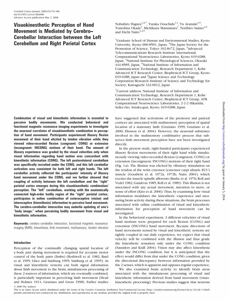
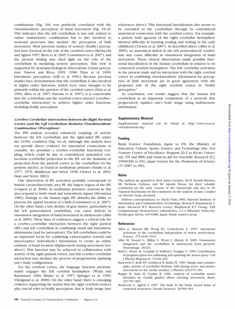
Figure 1. Conditions (a) and results (b) in the behavioral experiment. (a) Participantsexperienced illusory flexions of their right hands while viewing their video-recordedhand flexion (CONG) or extension motion (INCONG). Crosses on the wrist jointsindicate fixation points. The open arrow indicates the direction of illusory movement,and filled arrows indicate the directions of visual hand motions. Three differentvelocities were used for each hand motion. (b) Filled bars represent the mean illusoryangles across all participants under CONG conditions, and open bars indicate thoseunder INCONG conditions. Error bars indicate standard errors of means across allparticipants. *P\ 0.05.
Cerebral Cortex January 2009, V 19 N 1 177
by guest on January 5, 2014http://cercor.oxfordjournals.org/
Dow
nloaded from
hand motions were completed, an illusory extension movement was
elicited by vibrating the tendon of the right flexor carpi ulnaris (FCU)
muscle in another trial (in this situation, the vibrated hands were
semipronated without touching anything to allow the stimulation on
the tendon of FCU). The same procedure was used to determine the 3
velocities for visual extension hand motion.
Replications (see above) of the flexion hand movements were started
from a straight wrist position (0 degree), and replications of the
extension movements were started from a wrist-flexed position (40
degrees). Both replications were performed for 10 s, and neither of the
replicated movements reached the maximum rotation of the individual
wrist joint. The mean velocities of these replicated flexion and
extension movements for all participants were approximately 6.6
degrees per second (Fast), 4.0 degrees per second (Medium), and 2.4
degrees per second (Slow). The distance between the camera and the
hand was approximately 65 cm, which ensured that the position and
size of the hand image (radial view) on the display stayed constant for
all conditions. The video-recorded flexion hand motions were used
for the CONG condition, and the extension hand motions were used
for the INCONG condition. Thus, there were a total of 6 conditions.
Three trials were performed for each of the 6 conditions (3 velocities
for CONG and 3 for INCONG), and the order of the conditions was
randomized. In a given trial, the tendon was vibrated for 10 s while the
participants viewed 1 of the 6 video-recorded hand motions for 10 s.
The start of tendon vibration was synchronized with the start of hand
motion in the video. The participants were asked to be aware of illusory
hand movements while they viewed their hand motions. After each
trial, they were instructed to replicate the experienced maximum angle
of the illusory flexion by flexing their wrists (Hagura et al. 2007), and
this angle (i.e., total angular displacement) was measured with
a protractor. The mean value was calculated for the 3 trials for each
condition per participant. To evaluate the effect of the 3 different visual
velocities on the illusory wrist angular displacement, a t-test with
a Bonferroni correction was used for each pair among the 3 conditions.
This test was conducted separately for the CONG and INCONG
conditions.
We also recorded electromyographic (EMG) activity from the wrist
FCU, which is the agonistic muscle for the illusory wrist flexion and
from the vibrated ECU muscle in 16 of the 17 participants. The
integrated EMG was calculated for each visual condition and compared
across conditions (see detailed method in the Supplementary
Materials).
fMRI Experiment 1
Scanner
A 3.0 T Siemens scanner (Magnetom Allegra) with a head coil provided
T1-weighted anatomical images (3-dimensional magnetization-
prepared rapid-acquisition-echo sequence) and functional T2*-
weighted echoplanar images (64 3 64 matrix, 3.0 3 3.0 mm, time
echo [TE] = 40 ms). One functional image volume of the brain was
collected every 4 s (time repetiton [TR] = 4,000 ms) and comprised
44 slices of 3 mm in thickness, which ensured that the whole brain
was within the field of view (192 3 192 mm).
Tasks
The 16 participants rested comfortably in a supine position inside the
magnetic resonance scanner with the arms extended in a relaxed
position parallel to the trunk. The hands were pronated and the wrists
were naturally flexed and relaxed (ca., 40 degrees of flexion) without
touching anything. The arms were fixed with an arm brace. The
participants were instructed to relax their body completely and not to
move during scanning and were also requested to be aware of the
sensations from the vibrated hand as they viewed video-recorded
motions of their hand.
The experiment was conducted with a 3 (somatosensory) 3 3
(visual) factorial design. For the somatosensory conditions, we vibrated
either the tendon of the wrist extensor muscles, which elicit illusory
hand flexion movements (Tendon vibration: T), or the skin surface over
the bone just beside the tendon (e.g., the processus styloideus ulnae)
(Bone vibration: B) that only elicited a sensation of skin vibration with
no illusion (Naito and Ehrsson 2006). The latter was used as a control
condition for the former. Although the tendon vibration excites muscle
spindle afferents that elicit illusory hand movement, it may simulta-
neously recruit vibrotactile receptors that do not directly contribute to
the kinesthetic experience. Therefore, to evaluate the effect of
kinesthetic input alone, the effects in the B condition were subtracted
from those in the T condition. We have shown the validity of this
somatosensory control condition in previous tendon vibration studies
(Naito and Ehrsson 2001, 2006; Naito, Kochiyama, et al. 2002; Naito,
Roland, Ehrsson 2002; Hagura et al. 2007; Naito et al. 2007, 2008). The
skin contact surface was approximately 1 cm2 for both vibratory (T and
B) conditions, with use of the same nonmagnetic vibrator as in the
behavioral experiment. We also included a condition in which no
vibratory stimuli were delivered (No vibration: N).
For the visual conditions, the participants viewed flexion (F) or
extension (E) of their hands. These images were video recorded in
advance, and the participants were thus completely passive during
scanning, that is, they did not generate any movements. The velocities
of the hand motions were set at approximately 3 degrees per second.
This velocity was selected based on our previous observation that this is
the average velocity for illusory hand movements when the tendon is
vibrated for a relatively long time ( >30 s) (e.g., Naito, Kochiyama, et al.
2002). For the flexion hand motion, the participants viewed video-
recordings of flexion of their hands from a starting position of
20-degree extension to a final position of 70-degree flexion. For the
extension motion, they viewed their hands extending from a starting
position of 50-degree flexion to an end point of 40-degree extension.
The range of hand motion was thus approximately 90 degrees under
both conditions, and this range was consistent across participants. In
addition, we also included a visual control condition in which the
participants viewed an inanimate fixation point (Fi).
During video-recording of hand motions, a mark was put on the
radial side of the wrist joint. The position of this point does not vary
with hand movement (Fig. 1a). During scanning while viewing hand
motion, the participants were requested to fixate on this point to
restrict eye movement. In the scanner, the visual images were viewed
through a mirror located just in front of the eyes, and the images were
projected from outside the scanner room. Before scanning, we ensured
that participants could see the whole image of the visual hand motion.
An experimenter in the scanner room operated the vibrator manually
by lightly applying it to the skin, as in the behavioral experiment.
Instructions regarding the stimulus conditions and onset/offset timing
of the vibration were given to the experimenter by computer-
generated visual cues projected onto the white surface of the scanner
(the participants could not see this visual information).
Each participant underwent 6 sessions, and each session included 9
conditions (F--T, F--B, F--N, E--T, E--B, E--N, Fi--T, Fi--B, and Fi--N). For the
order of the 3 somatosensory conditions [tendon vibration (T), bone
vibration (B), and rest (R)], we prepared 6 combinations, that is, T--B--R,
T--R--B, B--T--R, B--R--T, R--T--B, and R--B--T. All 6 combinations were
tested once with each participant. One combination (e.g., T--B--R) was
assigned to 1 session and repeated 3 times in a session, and other
combinations were used in different sessions. The repetition of 1
combination in a session (e.g., T--B--R--T--B--R--T--B--R) enables avoidance
of a successive vibratory stimulation on the tendon, which may reduce
the sensitivity of muscle spindle afferents (Ribot-Ciscar and Roll 1998).
Similarly, for the 3 visual conditions (FLEX [F], EXT [E], FIX [Fi]), 6
combinations were prepared for each session, and all 6 combinations
were tested once with each participant. One combination was assigned
to 1 session and repeated 3 times in a session. As a whole, a total of 36
conditions [6 (somatosensory) 3 6 (visual)] resulted from the
interaction of combinations of visual and somatosensory conditions.
We could not perfectly randomize these interaction patterns of
combinations across participants, but we took special care to avoid
the concentrating usage of particular types. Each condition lasted for
32 s (in each session, 8 functional images were collected); use of this
epoch length has provided reliable results in terms of reproducibility of
activation patterns related to kinesthetic illusory hand movements in
our previous tendon vibration studies (Naito and Ehrsson 2001, 2006;
Naito, Kochiyama, et al. 2002; Naito, Roland, Ehrsson 2002; Hagura et al.
2007; Naito et al. 2007, 2008). A period of 8 s before the start of each
condition was allotted for positioning of the vibrator; this period was
178 Neuronal Correlates of Visuokinesthetic Combination d Hagura et al.
by guest on January 5, 2014http://cercor.oxfordjournals.org/
Dow
nloaded from
defined as a condition of no interest. We collected 90 functional
volumes in each session, and a total of 6 3 90 volumes were collected
per participant. The F--T and E--T conditions are referred to as the
CONG and INCONG conditions, respectively.
Data Analysis
The fMRI data were analyzed using Statistical Parametric Mapping
software (SPM99; http://www.fil.ion.ucl.ac.uk/spm; Wellcome Depart-
ment of Cognitive Neurology, London). The functional images were
realigned to correct for head movements, coregistered with each
participant’s anatomical magnetic resonance imaging, and transformed
(through both linear and nonlinear transformation) to the format of the
Montreal Neurological Institute (MNI) standard brain. The functional
images were scaled to 100 and spatially smoothed with an 8-mm full
width at half maximum (FWHM) isotropic Gaussian kernel and
smoothed in time by a 4-s FWHM Gaussian kernel.
A linear regression model (general linear model) was fitted to the
data for each participant. Each condition was modeled with a boxcar
function delayed by 4 s and convoluted with the standard SPM99
hemodynamic response function. Because our behavioral experiment
indicated that the illusion starts a few seconds after the vibration onset,
we modeled the first 4 s of all experimental conditions as an effect of
no interest.
First, to identify activity related to the simultaneous processing of
visual and kinesthetic information (visuokinesthetic processing), we
defined a linear contrast of [(CONG + INCONG) vs. (F--B + E--B)] (main
effect of visuokinesthetic processing in a 2 3 2 factorial design). The
rationale of this contrast is that effects related to pure visual processing,
vibrotactile processing, and the interactionbetween themcanbe removed,
allowing elucidation of areas related to visuokinesthetic processing.
Next, we identified activity that was exclusively related to
visuokinesthetic processing under the CONG condition by examining
the interaction contrast between the site of vibration and the direction
of vision in a 2 3 2 factorial design [(CONG vs. F--B) vs. (INCONG vs. E--
B)]. The contrast (CONG vs. F--B) was used as an inclusive mask (P <
0.05, uncorrected) to ensure that only voxels showing activation from
the corresponding control condition were included. The rationale for
this design is to ensure that the resultant interaction term is related
only to the interaction of concordant visual and kinesthetic information
(CONG) and that the activation is not confounded by effects such as
simple differences in tendon vibration, cutaneous vibrotactile stimula-
tion, or types of visual hand motions. We also searched for brain regions
that were specifically active in the INCONG condition using the
contrast [(INCONG vs. E--B) vs. (CONG vs. F--B)].
The results obtained from these analyses were estimated blood
oxygen level--dependent (BOLD) signals for the contrasts from each of
the 16 participants (contrast images). To accommodate interparticipant
variability, the contrast images from all participants were subjected to
random-effect group analysis (second-level analysis) (Friston et al.
1999). One-sample t-tests were used (15 degrees of freedom [df]) with
a voxelwise threshold of P < 0.001, uncorrected (t > 3.73) used to
generate the cluster images. For the statistical inference, we used
a threshold of P < 0.05 at the cluster level after correction for multiple
comparisons in the whole-brain space.
For anatomical identification of activation in the cytoarchitectonic
areas 4, 6, 44, and 45, the activated regions were related to the
cytoarchitectonic 30% probably maps in the MNI reference brain space
using the SPM Anatomy toolbox (version 1.2) (Eickhoff et al. 2005; Naito,
Roland, Ehrsson 2002; Naito et al. 2005). For identification ofmotor areas
in the medial wall (supplementary motor area [SMA], pre-SMA, cingulate
motor area [CMA]), we referred to the definitions of Roland and Zilles
(1996) and Picard and Strick (2001). For identification of the areas in the
cerebellum, we referred to Schmahmann et al. (2000).
Flip Analysis
Flip analysis was performed to examine whether the right frontopar-
ietal regions related to both the CONG and INCONG conditions were
predominantly active in the right hemisphere (see Naito et al. 2005)
and if they corresponded to the previously identified right frontopar-
ietal regions, which were active during the illusions for both left and
right hands (nonsomatotopical region; Naito et al. 2005, 2007; Naito
and Ehrsson 2006).
First, the functional images (normalized and smoothed) for all scans
of each participant in fMRI experiment 1 were flipped (a left-to-right
transformation on the x-axis) to make left--right reversed images
(flipped images). This allowed direct comparison of the activity in
a voxel in the left side of the brain with the corresponding voxel on the
right side. Then, a new general linear model that included both flipped
and unflipped data was defined in each participant. The right-side
dominance of frontoparietal activity was tested by the contrast
[[(CONG + INCONG) vs. (F--B + E--B)] vs. flip – [(CONG + INCONG)
vs. (F--B + E--B)]] in each participant. After the individual analysis, the
individual images were incorporated into a second-level random-effect
model to evaluate population inference (Friston et al. 1999). A 1-sample
t-test was used (15 df) with a voxelwise threshold of P < 0.001,
uncorrected (t > 3.73).
To confirm that the right-sided regions corresponded to the
frontoparietal areas active during the illusions for both left and right
hands in our previous studies (see above references), a small volume
correction (P < 0.05 at the cluster-level corrected; see Worsley et al.
1996) was used to restrict the search space. The search space was
defined as all voxels in a sphere with a 10-mm radius around the right
frontoparietal peaks obtained in our previous study (Naito et al. 2005).
fMRI Experiment 2
Tasks
In fMRI experiment 1, the left cerebellum was specifically activated
under the CONG condition for the right hand (see Results and Fig. 2e).
In fMRI experiment 2, we wanted to determine whether the activity of
the left cerebellum during visuokinesthetic combination has non-
somatotopical characteristics, that is, common activation under the
CONG condition for left and right hands. Another group of 7 volunteers
participated in this study, and the same 3.0-T scanner was used.
A similar procedure to that used in fMRI experiment 1 was used in
this study. The main difference was that the experimental conditions
were prepared for left and right hands. In the left-hand conditions,
vibratory stimulation was applied to the left hand (T and B) while the
participants viewed video-recorded flexion (F) or extension (E) hand
motion of their left hand (visual velocity of ca., 3 degrees per second).
Each participant underwent 8 sessions: in 4 of these sessions
vibrations were applied to the right hand, and in the other 4 sessions,
the vibrations were applied to the left hand. The left-hand and right-
hand sessions were alternated. For each hand, we assigned 2 sessions
for the F condition and 2 for the E condition, and each session included
T and B conditions. Each condition lasted for 21 s (TR = 3,000 ms, TE =30 ms, 7 functional images) and was repeated 3 times in 1 session. The
order of conditions was randomized according to a balanced schedule.
We collected 42 functional images per condition for each hand (CONG,
INCONG, F--B, and E--B) of each participant.
Data Analysis
Image preprocessing for the fMRI data was performed similarly to that
in the first fMRI experiment. A linear regression model (general linear
model) was fitted to the pooled data from all participants to increase
the sensitivity of the analysis (fixed-effect model, as in Hagura et al.
2007).
To test whether left cerebellar activation specific to the CONG
condition is consistently observed for the left and right hands, we
defined a linear contrast of [(CONG vs. F--B) vs. (INCONG vs. E--B)] for
each hand and performed a conjunction analysis [left (CONG vs. F--B)
vs. (INCONG vs. E--B)] \ [right (CONG vs. F--B) vs. (INCONG vs. E--B)]
(Price and Friston 1997; Friston et al. 2005). The conjunction contrast
[left (CONG vs. F--B)] \ [right (CONG vs. F--B)] was used as an inclusive
mask (P < 0.05, uncorrected) to ensure that only voxels showing
activation under CONG conditions for the left and right hands were
included. Because we had an a priori anatomical hypothesis in the left
cerebellum, we restricted the search space and used a small volume
correction (Worsley et al. 1996). The search space was defined by the
left cerebellar activation map obtained in fMRI experiment 1 [(CONG
vs. F--B) vs. (INCONG vs. E--B)] (P < 0.001, uncorrected) (Fig. 2e). The
definition of the search space was statistically independent because the
data were obtained in another group of volunteers. In the conjunction
Cerebral Cortex January 2009, V 19 N 1 179
by guest on January 5, 2014http://cercor.oxfordjournals.org/
Dow
nloaded from
analysis, a voxelwise threshold of P < 0.005, corrected was used, which
was a conservative threshold, and we confirmed that the activation
exists around the region revealed in the conjunction analysis in each of
the 2 interaction contrasts (P < 0.001, uncorrected).
Single-Subject Analysis
The above statistical analysis was based on the functional data pooled
across participants using a fixed-effect analysis, in which the results
may be biased by a minority of participants showing strong effects. To
make sure that the group results were representative of all 7
participants, we analyzed the individual data for each hand (see Hagura
et al. 2007). All image-processing steps were identical to those used in
the group analysis (see above). The same GLM as in the group analysis
was used, with the only difference being that we considered the
functional data from each participant separately. A linear contrast of
[(CONG vs. F--B) vs. (INCONG vs. E--B)] was performed separately for
each hand of each participant, and increases of the BOLD signal (P <
0.05, uncorrected) were probed in a volume of radius 10-mm around
peaks of activation detected in the group analysis. We report the
number of participants that exhibited a BOLD signal increases in the
relevant areas and the results were tabulated in the Supplementary
Table 1 in the Supplementary Materials.
fMRI Experiment 3
Tasks
In fMRI experiment 3, we examined if the left cerebellar activity under
the CONG condition is associated with subjective visuokinesthetic
perception of hand movement. An independent group of 12 volunteers
participated in the study, and the same 3.0 T scanner and a similar
procedure to fMRI experiment 1 were used. The main difference was
that brain activity was measured during vibration of the right hand (T
and B) while the participants viewed video-recorded flexion motions of
their right hand at 3 different velocities (Fast, Medium, and Slow). The
hand motions of each participant were video recorded before the scan,
during which hand flexing was started from a 20-degree extension
position for all velocity conditions. For this video-recording, each
participant was asked to continuously flex the hand up to an individual
maximum angle of wrist flexion for 15, 20, or 30 s. The mean velocities
across participants were approximately 5.8 degrees per second (Fast),
4.3 degrees per second (Medium), and 2.9 degrees per second (Slow).
In the experiment, hand motions were presented for the first 15 s only
in all velocity conditions. Thus, the participants viewed their hand
motions in the ranges of approximately 90, 60, and 45 degrees,
respectively.
All participants underwent 12 sessions, and each session included
Fast-T, Fast-B, Medium-T, Medium-B, Slow-T, and Slow-B conditions.
The order of the conditions was randomized as in fMRI experiment 1.
Each condition lasted for 15 s (TR = 3,000 ms, TE = 30ms, 5 functional
images) and was repeated once per session. During the scanning, the
participants were asked to be aware of illusory hand movements while
they viewed the flexion hand motions. After each session, the
participants scored the degree of perceived hand flexion for each
T condition on a scale from 0 to 10 (illusion score): a score of 10
indicated illusory movements at the maximally flexed angle and a score
of 0 indicated no experience of illusory movement. As shown
previously, the illusion score is correlated with the replicated angular
magnitude of illusory hand movement (Hagura et al. 2007) and can be
used in statistical analysis.
Data Analysis
In the statistical analysis of the behavioral ratings (illusion score), the
mean value of scores for each condition (Fast, Medium, or Slow) was
calculated for each participant. A t-test was performed on the mean
values for each pair of velocity conditions, with a Bonferroni correction
for the number of comparisons.
Image preprocessing for the fMRI data was similar to that in fMRI
experiment 1. To reveal brain areas with activities covarying with
the subjective perception of illusory flexion experience under the
T condition, a parametric modulation analysis was performed (Buchel
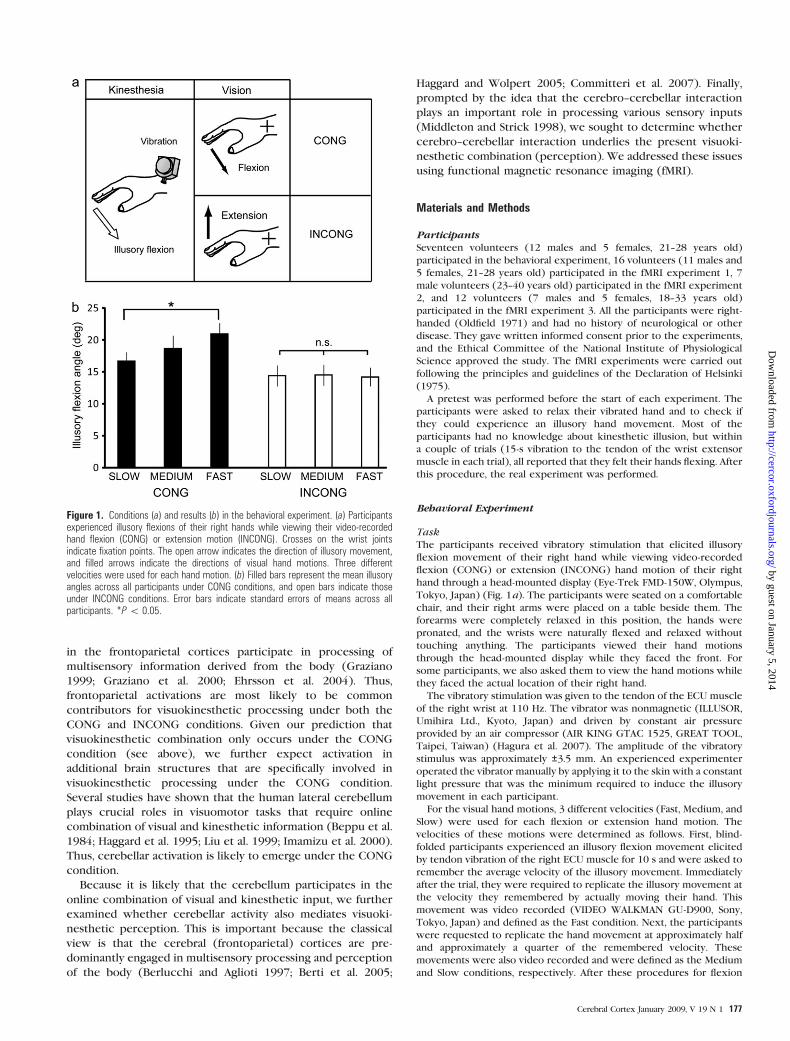
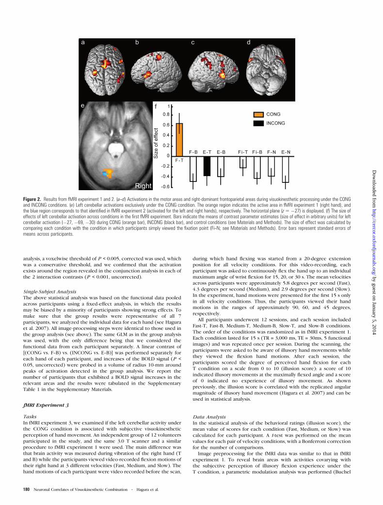
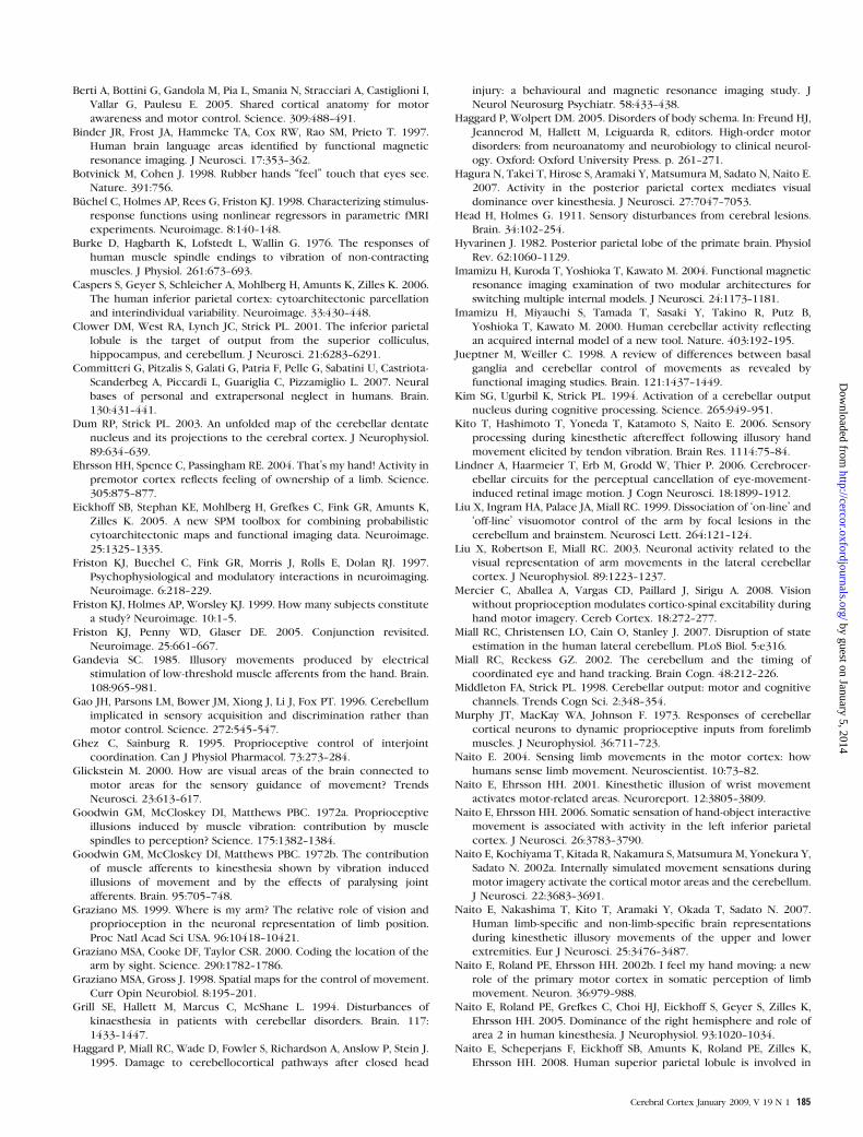
Figure 2. Results from fMRI experiment 1 and 2. (a--d) Activations in the motor areas and right-dominant frontoparietal areas during visuokinesthetic processing under the CONGand INCONG conditions. (e) Left cerebellar activations exclusively under the CONG condition. The orange region indicates the active area in fMRI experiment 1 (right hand), andthe blue region corresponds to that identified in fMRI experiment 2 (activated for the left and right hands), respectively. The horizontal plane (z5 �27) is displayed. (f) The size ofeffects of left cerebellar activation across conditions in the first fMRI experiment. Bars indicate the means of contrast parameter estimates (size of effect in arbitrary units) for leftcerebellar activation (�27, �69, �30) during CONG (orange bar), INCONG (black bar), and control conditions (see Materials and Methods). The size of effect was calculated bycomparing each condition with the condition in which participants simply viewed the fixation point (Fi--N; see Materials and Methods). Error bars represent standard errors ofmeans across participants.
180 Neuronal Correlates of Visuokinesthetic Combination d Hagura et al.
by guest on January 5, 2014http://cercor.oxfordjournals.org/
Dow
nloaded from
et al. 1998). A linear regression model (general linear model) was fitted
to the individual data, including all 3 somatosensory conditions (T, B,
and N). A regressor for parametric modulation was added to the
T condition for each session. The linear parameters were used as
parametric modulators for the illusion scores.
Brain regions that showed a linear relationship with the illusion
scores were tested by applying a t-contrast (1 for the parametric
modulation regressor and 0 for elsewhere) in each participant. Next,
the individual images were incorporated into the second-level random-
effect model to evaluate population inference (Friston et al. 1999). A 1-
sample t-test (11 df) was used with a voxelwise threshold of P < 0.001,
uncorrected (t > 3.73).
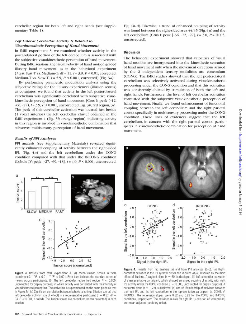
Psychophysiological Interaction Analysis
In the series of analyses described above, we found that the
posterolateral portion of the left cerebellum was specifically activated
during visuokinesthetic combination (perception) under the CONG
condition and that this cerebellar activation was left-side dominant
(activated irrespective of left and right hands) (see Results, Fig. 2e). In
contrast, we found right-side dominant frontoparietal activation during
visuokinesthetic processing under both the CONG and INCONG
conditions (main effect of visuokinesthetic processing; see Fig. 2a--d
and 4a), in which the locations corresponded to the right-sided
regions active during illusions of the left and right hands in our
previous studies (Naito et al. 2005, 2007; Naito and Ehrsson 2006).
Prompted by the anatomical evidence in nonhuman primates that
the cerebral cortices and the cerebellum are mainly contralaterally
interconnected (Glickstein 2000; Strick 2004), we examined if the
cerebro--cerebellar interaction takes place under the CONG condi-
tion by reanalyzing the data obtained from fMRI experiment 1. We
expected that coupling of activity between the right frontoparietal
cortices and the left cerebellum is enhanced only under the CONG
condition, in which the brain is required to process concordant
visuokinesthetic information. We performed a psychophysiological
interaction (PPI) analysis (Friston et al. 1997) by extracting the data
from the right frontal and parietal regions (see details in Supplemen-
tary Materials).
Results
Velocity of Visual Hand Motion Grades Illusory Flexionunder the CONG Condition
In the behavioral experiment, no overt hand movements
appeared in any trials. The visual hand motions affected the
sensation of illusory hand flexion under both the CONG and
INCONG conditions; however, the visual effects were clearly
distinct between the conditions. This was also confirmed when
some participants viewed their hand motions while they faced
the actual location of the right hand.
Under the CONG condition only, the velocity of the visual
hand motion graded the intensity of illusory hand movement.
Identical vibratory stimuli were given to the same tendon site,
but the illusions were evenly attenuated irrespective of the
viewed velocity of wrist motion under the INCONG condition
(Fig. 1b). Under the CONG condition, the angle of illusory,
perceived hand displacement at the Fast velocity was signifi-
cantly greater than that at the Slow velocity (t-test, df = 16, t =2.8, P < 0.05, corrected), but no such difference was observed
under the INCONG condition. Furthermore, under the CONG
condition, some participants reported that they felt as if they
were viewing their actual hands passively flexing on the
display, but none reported such an experience under the
INCONG condition. Thus, the results suggest that velocity of
visual hand motion was incorporated into the kinesthetic
experience under the CONG condition but not under the
INCONG condition.
Although we found a slight increase of EMG activities from
the FCU muscle (the agonistic muscle for illusory wrist flexion)
and from the vibrated ECU muscle during tendon vibration, the
levels of EMG activities were not graded by the velocities of
visual hand motions. In addition, the activities were not
correlated with the angle of illusory hand displacement in
any of the visual conditions, suggesting that the muscular
activities have no direct relationship with the graded illusory
sensation (see details in Supplementary Materials).
Frontoparietal Activation under the CONG and INCONGConditions
In fMRI experiment 1, all participants experienced illusory
hand flexion only during tendon vibration and no illusion
during the bone vibration (B). Examination of the main effect of
visuokinesthetic processing using the following contrast
[(CONG + INCONG) vs. (F--B + E--B)] showed activation in
the hand sections of cortical motor areas and in the
frontoparietal cortices (P < 0.05, corrected) (Fig. 2a--d). In
the motor areas, we found peaks of activation in the left
(contralateral) cytoarchitectonic area 4a, dorsal part of area 6
(dorsal premotor cortex), medial aspect of area 6 (SMA), caudal
part of the CMA, and bilateral pre-SMA. In the frontal cortices,
peaks of activations were located in the bilateral cytoarchitec-
tonic areas 44/45, frontal operculum, and the anterior part of
the right middle frontal gyrus. In the parietal cortices, the
peaks were located in the right lateralmost part of the inferior
parietal lobule (IPL) and in the right intraparietal sulcus area. A
flip analysis performed to test whether activation of the right
frontoparietal cortices is right-side dominant, as found in our
previous studies (Naito et al. 2005, 2007; Naito and Ehrsson
2006), showed right-sided activations in the IPL [peak
coordinates (x, y, z) = (66, –30, 33); t = 4.1, P < 0.05, corrected]
and in areas 44/45 [peak (60, 21, 18); t = 4.0, P < 0.05,
corrected] (Fig. 4a).
Exclusive Activation of the Left Lateral Cerebellum underthe CONG Condition
The CONG condition [(CONG vs. F--B) vs. (INCONG vs. E--B)]
exclusively activated the posterolateral portion of the left
cerebellum (Crus I: peak [–12, –81, –24], t = 5.5; Lobule IV: peak
[–27, –69, –30], t = 4.2; P < 0.05, corrected; Fig. 2e [orange
region]). This was the only region that showed significant
activation (interaction) in the entire brain and was not
activated under the other 3 control conditions (F--B, INCONG,
or E--B; see Fig. 2f). Furthermore, this area was not activated
when the participants experienced illusions while viewing an
inanimate fixation point or when they viewed their hand
motion without receiving vibratory stimuli (see Materials and
Methods and Fig. 2f). No specific activation for the INCONG
condition was found in the entire brain [(INCONG vs. E--B) vs.
(CONG vs. F--B)].
In fMRI experiment 2, we confirmed that the same left
cerebellar region was activated under the CONG conditions for
both left and right hands (Crus I: peak [–21, –78, –30], P < 0.005,
corrected after small volume correction; Fig. 2e [blue region]).
The left cerebellum was the only region that was activated in
the entire brain (cluster size > 10 voxels), and no significant
activation was observed in the right corresponding cerebellar
region (P > 0.05, uncorrected). Finally, single-subject analysis
revealed that all 7 participants had increased activity in this left
Cerebral Cortex January 2009, V 19 N 1 181
by guest on January 5, 2014http://cercor.oxfordjournals.org/
Dow
nloaded from
cerebellar region for both left and right hands (see Supple-
mentary Table 1).
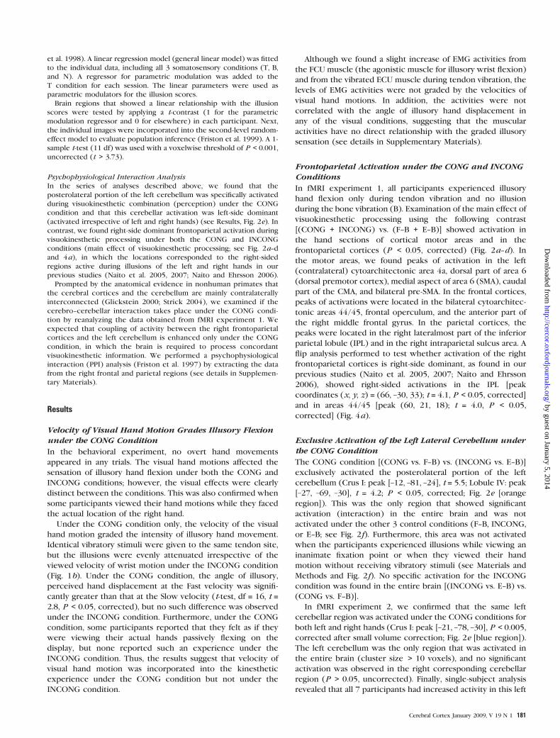
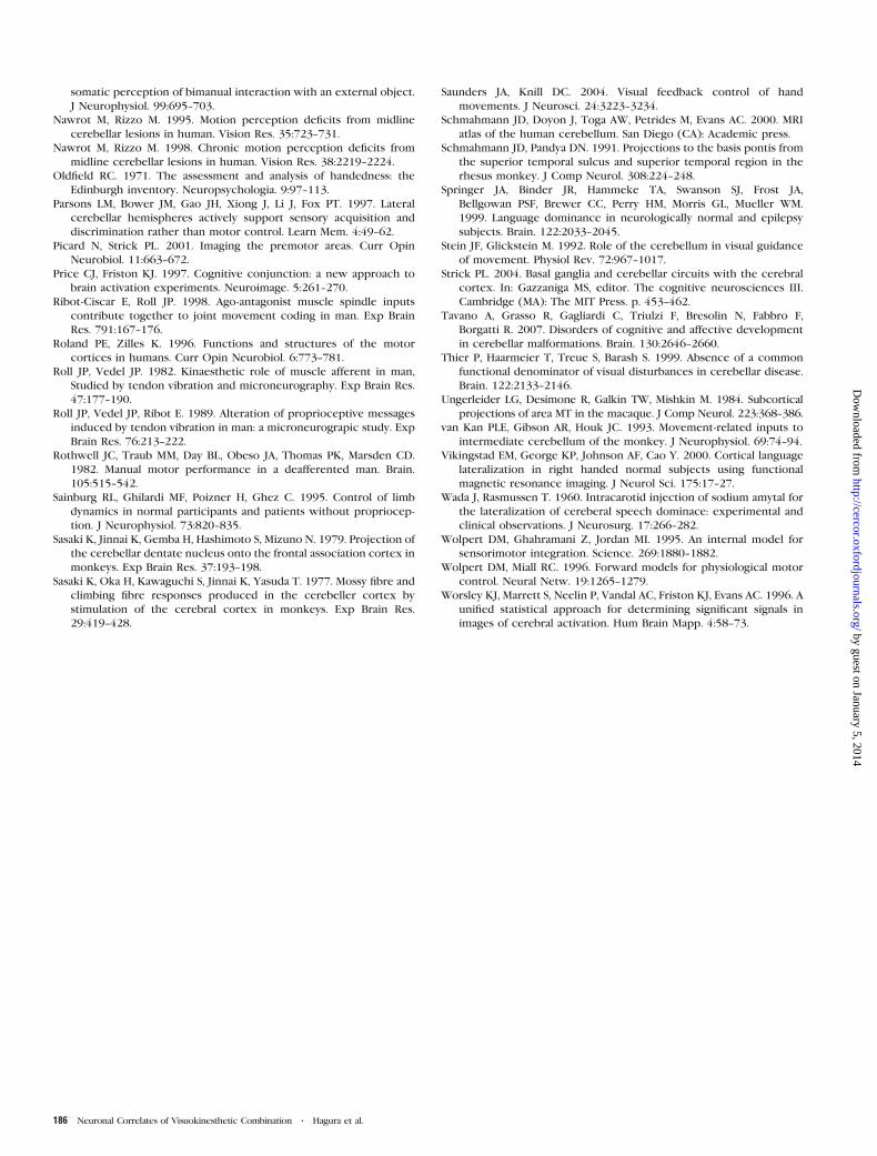
Left Lateral Cerebellar Activity Is Related toVisuokinesthetic Perception of Hand Movement
In fMRI experiment 3, we examined whether activity in the
posterolateral portion of the left cerebellum is associated with
the subjective visuokinesthetic perception of hand movement.
During fMRI sessions, the visual velocity of hand motion graded
illusory hand movement, as in the behavioral experiment
(t-test, Fast-T vs. Medium-T: df = 11, t = 3.8, P < 0.01, corrected;
Medium-T vs. Slow-T: t = 5.9, P < 0.001, corrected) (Fig. 3a).
By performing parametric modulation analysis using the
subjective ratings for the illusory experiences (illusion scores)
as covariates, we found that activity in the left posterolateral
cerebellum was significantly correlated with subjective visuo-
kinesthetic perception of hand movement [Crus I: peak (–12,
–66, –27), t = 3.9, P < 0.001, uncorrected; Fig. 3b, red region, 3c].
The peak of this cerebellar activation was located just beside
(1 voxel anterior) the left cerebellar cluster obtained in the
fMRI experiment 1 (Fig. 3b, orange region), indicating activity
in this region is involved in visuokinesthetic combination that
subserves multisensory perception of hand movement.
Results of PPI Analyses
PPI analysis (see Supplementary Materials) revealed signifi-
cantly enhanced coupling of activity between the right-sided
IPL (Fig. 4a) and the left cerebellum under the CONG
condition compared with that under the INCONG condition
(Lobule IV: peak [–27, –69, –18], t = 4.0, P < 0.001, uncorrected;
Fig. 4b--d). Likewise, a trend of enhanced coupling of activity
was found between the right-sided area 44/45 (Fig. 4a) and the
left cerebellum (Crus I: peak [–36, –72, –27], t = 3.0, P = 0.005,
uncorrected).
Discussion
The behavioral experiment showed that velocities of visual
hand motions are incorporated into the kinesthetic sensation
of hand movement only when the movement directions sensed
by the 2 independent sensory modalities are concordant
(CONG). The fMRI studies showed that the left posterolateral
cerebellum was selectively activated during visuokinesthetic
processing under the CONG condition and that this activation
was consistently elicited by stimulation of both the left and
right hands. Furthermore, the level of left cerebellar activation
correlated with the subjective visuokinesthetic perception of
hand movement. Finally, we found enhancement of functional
coupling between the left cerebellum and the right parietal
cortex specifically in multisensory processing under the CONG
condition. These lines of evidences suggest that the left
cerebellum, in concert with the right parietal cortex, partic-
ipates in visuokinesthetic combination for perception of hand
movement.
Figure 3. Results from fMRI experiment 3. (a) Mean illusion scores in fMRIexperiment 3. **P\ 0.01, ***P\ 0.001. Error bars indicate the standard errors ofmeans across participants. (b) The left cerebellar region (red region; P \ 0.005,uncorrected for display purpose) in which activity was correlated with the intensity ofvisuokinesthetic perception. The activation is superimposed on the same plane as thatin Figure 2e. (c) Significant correlation between behavioral ratings (illusion scores) andleft cerebellar activity (size of effect) in a representative participant (r 5 0.57, df 534, P\ 0.001, 1-tailed). The illusion scores are normalized (mean corrected) in eachsession.
Figure 4. Results from flip analysis (a) and from PPI analyses (b--d). (a) Right-dominant activities in the IPL (yellow circle) and in areas 44/45 revealed by the maineffect of illusions. A sagittal plane (x 5 60) is displayed. (b) Left cerebellar activationof a representative participant, which showed enhanced coupling of activity with rightIPL activity under the CONG condition (P\0.005, uncorrected for display purpose). Ahorizontal plane (z 5 �27) is displayed. (c) and (d) Relationship of activities betweenthe right IPL and the left cerebellum in the representative participant (c: CONG; d:INCONG). The regression slopes were 0.52 and 0.29 for the CONG and INCONGconditions, respectively. The activities (x-axis for right IPL; y-axis for left cerebellum)are mean adjusted (arbitrary units).
182 Neuronal Correlates of Visuokinesthetic Combination d Hagura et al.
by guest on January 5, 2014http://cercor.oxfordjournals.org/
Dow
nloaded from
Effects of Vision on Illusory Hand Movement
In our behavioral experiment, visual information regarding hand
motions affected the kinesthetic illusion of hand flexion under
both the CONG and INCONG conditions (Fig. 1b). This finding is
consistent with the notion of ‘‘visual dominance over kinesthe-
sia’’ (Botvinick and Cohen 1998; Hagura et al. 2007). The striking
difference between the conditions was that the velocity of visual
flexion motions graded the angular magnitude of the illusory
flexion movement only under the CONG condition (Fig. 1b).
This grading of illusion by the directionally concordant visual
hand motion was also replicated in the CONG condition of fMRI
experiment 3 (Fig. 3a), in which the participants viewed the
hand motions displayed on the monitor in front of their face in
the scanner. Thus, the location where the visual hand motions
was displayed did not change this effect. These results indicate
that, in addition to the general effect of visual dominance over
kinesthesia (Hagura et al. 2007), a specific neuronal process to
incorporate information on visual velocity into the kinesthetic
experience is present when the directions sensed by vision and
kinesthesia are concordant (CONG). Because participants
reported that the hand motions they viewed were felt as actual
state of their own vibrated hand only under the CONG
condition, it is plausible that the brain continuously matches
and combines visual and kinesthetic information to maintain
perceptual coherence, linking seen and felt movements. This
context-specific combination of vision and kinesthesia may be
brought about because movement directions sensed by vision
and kinesthesia are always matched during ordinary motor
behavior (cf., Mercier et al. 2008).
Visuokinesthetic Combination in the Left LateralCerebellum
The hand sections of cortical motor areas were activated under
both CONG and INCONG conditions (Fig. 2a--b). These areas
participate in limb-specific kinesthetic processing even in
situations when vision is unavailable (Naito et al. 2005, 2007).
The 2 conditions also activated the frontoparietal cortices
(mostly in the right hemisphere; areas 44/45 and IPL; see
Figs 2a--d and 4a), which also process general kinesthetic
information (limb nonspecific); it has been suggested that these
areas contribute to creation of higher order body representa-
tions (Berlucchi and Aglioti 1997; Berti et al. 2005; Naito et al.
2005, 2007; Committeri et al. 2007). Therefore, the frontopar-
ietal activation observed in the present study may be related to
higher order visuokinesthetic processing, an interpretation
consistent with previous findings (Ehrsson et al. 2004).
The left posterolateral cerebellum was specifically activated
under the CONG condition (Fig. 2e). Activation of this region
during illusory hand movements was not found in our previous
studies (see above), suggesting that this cerebellar region is
exclusively involved in multisensory processing of concordant
visual and kinesthetic information regarding hand movements
(also see Fig. 2f). It is unlikely that this cerebellar activity
represents the positional discrepancy sensed by vision and
kinesthesia (cf., Imamizu et al. 2000) because we observed no
significant increase of activity in the cerebellum under the
INCONG condition, where the discrepancy should be more
prominent (Fig. 2e--f). Just as the behavioral results suggest that
vision and kinesthesia are combined only under the CONG
condition, it follows that the activity in the left posterolateral
cerebellum is associated with a neuronal process of visuoki-
nesthetic combination.
The tasks used in the experiments were completely passive,
where no actual movement, no intention to move, and no sense
of effort are required (Kito et al. 2006). Thus, it is reasonable to
interpret our observation of cerebellar activity as a ‘‘multisen-
sory’’ process of visuokinesthetic combination. This type of
sensory combination could reasonably take place in the
cerebellum because it has been demonstrated in nonhuman
primates that both visual (Ungerleider et al. 1984; Schmahmann
and Pandya 1991; Stein and Glickstein 1992; Glickstein 2000)
and kinesthetic (Murphy et al. 1973; Bauswein et al. 1983;
van Kan et al. 1993) information reaches the cerebellum, either
indirectly via the cerebro--pontine--cerebellar pathway or
directly via the spinocerebellar pathway. This view also fits
with the notion that the cerebellum plays a sensory role when
the brain acquires multisensory information that is relevant to
the tasks to be performed (Gao et al. 1996; Parsons et al. 1997;
Miall and Reckess 2002).
The human posterolateral cerebellum is activated by visuo-
motor tasks that require multisensory (integrative) processing of
visual and kinesthetic information (Jueptner and Weiller 1998;
Imamizu et al. 2000, 2004). Patients with lesions in the lateral
cerebellum often exhibit deficits in visuomotor tracking tasks,
and these deficits worsen during execution of tasks that include
visual feedback of hand motion. This indicates that cerebellar
damage disturbs computation of online visuokinesthetic combi-
nation in estimating the state of hand (Beppu et al. 1984;
Haggard et al. 1995; Liu et al. 1999). In nonhuman primates,
neurons in the lateral cerebellum seem to process online visual
feedback from the monkey’s own hand movements (Liu et al.
2003). All these observations support our claim that the
cerebellum is involved in continuous visuokinesthetic combina-
tion to update limb position, which can be utilized for adaptive
online motor control of limb position.
The left cerebellum has not been stressed as a locus for
visuokinesthetic combination because most previous studies
have focused more on the aspect of visuomotor control of hand
movement (Jueptner and Weiller 1998; Imamizu et al. 2000,
2004) rather than on the process of multisensory combination
that subserves the spatial perception of hand movement. A
previous report of activity of the left cerebellum during the
course of multisensory combination regarding a static hand
(Ehrsson et al. 2004) may support our present interpretation.
Thus, left cerebellar activation may be important as the brain
configures the limb location/displacement by combining multi-
sensory afferent information.
The precise mechanism of the cerebellar activation observed
in the present study is still uncertain. However, the cerebellum
has been reported to participate in the process of prediction
of the sensory consequence of action (Wolpert et al. 1995;
Wolpert and Miall 1996), especially regarding the visual
consequence (perception) (Lindner et al. 2006) and also in
the estimation of the state of the effector for online correction
of action (Miall et al. 2007). Thus, one may speculate that the
cerebellar activation is related to a process that matches the 2
states of hand position/displacement estimated by continuous
visual and kinesthetic inputs to further predict the forthcoming
state of the hand.
Left Cerebellar Activity Is Associated with VisuokinestheticPerception
The activity in the left posterolateral cerebellum, which was
similar to the region associated with visuokinesthetic
Cerebral Cortex January 2009, V 19 N 1 183
by guest on January 5, 2014http://cercor.oxfordjournals.org/
Dow
nloaded from
combination (Fig. 3b), was positively correlated with the
visuokinesthetic perception of hand movement (Fig. 3b--d).
This indicates that the left cerebellum is not only related to
online multisensory combination but is also involved in
neuronal processes that mediate the perception of limb
movement. Most previous studies of sensory (bodily) percep-
tion have focused on the role of the cerebral cortex (Berlucchi
and Aglioti 1997; Berti et al. 2005; Committeri et al. 2007), and
the present finding may shed light on the role of the
cerebellum in mediating sensory perception. This view is
supported by neuropsychological observations (visual percep-
tion: Nawrot and Rizzo 1995, 1998; Thier et al. 1999;
kinesthetic perception: Grill et al. 1994). Because previous
studies have demonstrated that the cerebellum is also involved
in higher order functions, which were once thought to be
primarily within the purview of the cerebral cortex (Kim et al.
1994; Allen et al. 1997; Parsons et al. 1997), it is conceivable
that the cerebellum and the cerebral cortex interact (cerebro--
cerebellar interaction) to achieve higher order functions,
including bodily perception.
Cerebro--Cerebellar Interaction between the Right ParietalCortex and the Left Cerebellum Mediates VisuokinestheticCombination (Perception)
The PPI analysis revealed enhanced coupling of activity
between the left cerebellum and the right-sided IPL under
the CONG condition (Fig. 4a--d). Although this analysis does
not provide direct evidence for anatomical connections or
causality, we postulate a cerebro--cerebellar functional cou-
pling, which could be due to contralateral anatomical con-
nections (cerebellar projection to the IPL via the thalamus or
projection from the parietal cortex to the cerebellum via the
pontine nuclei), as found in nonhuman primates (Sasaki et al.
1977, 1979; Middleton and Strick 1998; Clower et al. 2001;
Dum and Strick 2003).
Our observation of IPL activation probably corresponds to
human cytoarchitectonic area PF, the largest region of the IPL
(Caspers et al. 2006). In nonhuman primates, neurons in this
area respond to both visual and somesthetic inputs (Hyvarinen
1982). Damage to the human right IPL disturbs the ability to
process the spatial location of a limb (Committeri et al. 2007).
On the other hand, a low density of gray matter, particularly in
the left posterolateral cerebellum, can cause deficits in
visuomotor integration of hand movement in adolescents (Allin
et al. 2005). These lines of evidences suggest a critical role for
a cerebro--cerebellar interaction between the right parietal
(IPL) and left cerebellum in combining visual and kinesthetic
information (and for perception). The left cerebellum could be
an important locus for combining exteroceptive (visual) and
interoceptive (kinesthetic) information to create an online
estimate of hand location/displacement during movement (see
above). This function may be achieved in collaboration with
activity of the right parietal cortex, and this cerebro--cerebellar
interaction may mediate the process of progressively updating
one’s body configuration.
For the cerebral cortex, the language function predomi-
nantly engages the left cerebral hemisphere (Wada and
Rasmussen 1960; Binder et al. 1997; Springer et al. 1999;
Vikingstad et al. 2000). On the other hand, there is emerging
evidence supporting the notion that the right cerebral cortices
play crucial roles in bodily perception, that is, body image (see
references above). This functional lateralization also seems to
be extended to the cerebellum through its contralateral
anatomical connection with the cerebral cortex. For example,
a patient with agenesis of the right cerebellar hemisphere
showed difficulty in learning reading and writing in his early
childhood (Tavano et al. 2007). As described above (Allin et al.
2005), an anatomical deficit in the left posterolateral cerebel-
lum may cause difficulty in visuomotor integration of hand
movement. These clinical observations imply possible func-
tional lateralization in the human cerebellum in relation to its
connected cerebral hemisphere. The left cerebellar activation
in the present study and its interaction with the right cerebral
cortex in combining visuokinesthetic information for percep-
tion of limb movement are in good agreement with the
proposed role of the right cerebral cortex in ‘‘bodily
perception.’’
In conclusion, our results suggest that the human left
cerebellum is an important component of a network that
progressively updates one’s body image using multisensory
information.
Supplementary Material
Supplementary material can be found at: http://www.cercor.
oxfordjournals.org/.
Funding
Brain Science Foundation, Japan to EN; the Ministry of
Education, Culture, Sports, Science, and Technology (the 21st
Century Center of Excellence Program [D-2 to Kyoto Univer-
sity, EN and MM) and Grant-in-Aid for Scientific Research [C],
19500290 to EN), Japan Society for the Promotion of Science
(17-2087 to NH).
Notes
The authors are grateful to Prof. James Lackner, Dr H. Henrik Ehrsson,
Mr Nicholas Cothros, and Mr Satoshi Hirose for their valuable
comments on the early version of the manuscript and also to Dr
Takanori Kochiyama for his comments on the analysis of data. Conflict
of interest: None declared.
Address correspondence to: Eiichi Naito, PhD, National Institute of
Information and Communication Technology, Research Department 1,
Kobe Advanced ICT Research Center, Biophysical ICT Group, ATR
Computational Neuroscience Laboratories, 2-2-2 Hikaridai, Seika-cho,
Soraku-gun, Kyoto, 619-0288, Japan. Email: [email protected].
References
Allen G, Buxton RB, Wong EC, Courchesne E. 1997. Attentional
activation of the cerebellum independent of motor involvement.
Science. 275:1940--1943.
Allin M, Nosarti C, Rifkin L, Wyatt J, Murray R. 2005. Visuomotor
integration and the cerebellum in adolescents born preterm.
Neuroimage. 26:S23.
Bard C, Fleury M, Teasdale N, Paillard J, Nougier V. 1995. Contribution
of proprioception for calibrating and updating the motor space. Can
J Physiol Pharmacol. 73:246--254.
Bauswein E, Kolb FP, Leimbeck B, Rubia FJ. 1983. Simple and complex
spike activity of cerebellar Purkinje cells during active and passive
movements in the awake monkey. J Physiol. 339:379--394.
Beppu H, Suda M, Tanaka R. 1984. Analysis of cerebellar motor
disorders by visually guided elbow tracking movement. Brain.
107:787--809.
Berlucchi G, Aglioti S. 1997. The body in the brain: neural bases of
corporeal awareness. Trends Neurosci. 20:560--564.
184 Neuronal Correlates of Visuokinesthetic Combination d Hagura et al.
by guest on January 5, 2014http://cercor.oxfordjournals.org/
Dow
nloaded from
Berti A, Bottini G, Gandola M, Pia L, Smania N, Stracciari A, Castiglioni I,
Vallar G, Paulesu E. 2005. Shared cortical anatomy for motor
awareness and motor control. Science. 309:488--491.
Binder JR, Frost JA, Hammeke TA, Cox RW, Rao SM, Prieto T. 1997.
Human brain language areas identified by functional magnetic
resonance imaging. J Neurosci. 17:353--362.
Botvinick M, Cohen J. 1998. Rubber hands ‘‘feel’’ touch that eyes see.
Nature. 391:756.
Buchel C, Holmes AP, Rees G, Friston KJ. 1998. Characterizing stimulus-
response functions using nonlinear regressors in parametric fMRI
experiments. Neuroimage. 8:140--148.
Burke D, Hagbarth K, Lofstedt L, Wallin G. 1976. The responses of
human muscle spindle endings to vibration of non-contracting
muscles. J Physiol. 261:673--693.
Caspers S, Geyer S, Schleicher A, Mohlberg H, Amunts K, Zilles K. 2006.
The human inferior parietal cortex: cytoarchitectonic parcellation
and interindividual variability. Neuroimage. 33:430--448.
Clower DM, West RA, Lynch JC, Strick PL. 2001. The inferior parietal
lobule is the target of output from the superior colliculus,
hippocampus, and cerebellum. J Neurosci. 21:6283--6291.
Committeri G, Pitzalis S, Galati G, Patria F, Pelle G, Sabatini U, Castriota-
Scanderbeg A, Piccardi L, Guariglia C, Pizzamiglio L. 2007. Neural
bases of personal and extrapersonal neglect in humans. Brain.
130:431--441.
Dum RP, Strick PL. 2003. An unfolded map of the cerebellar dentate
nucleus and its projections to the cerebral cortex. J Neurophysiol.
89:634--639.
Ehrsson HH, Spence C, Passingham RE. 2004. That’s my hand! Activity in
premotor cortex reflects feeling of ownership of a limb. Science.
305:875--877.
Eickhoff SB, Stephan KE, Mohlberg H, Grefkes C, Fink GR, Amunts K,
Zilles K. 2005. A new SPM toolbox for combining probabilistic
cytoarchitectonic maps and functional imaging data. Neuroimage.
25:1325--1335.
Friston KJ, Buechel C, Fink GR, Morris J, Rolls E, Dolan RJ. 1997.
Psychophysiological and modulatory interactions in neuroimaging.
Neuroimage. 6:218--229.
Friston KJ, Holmes AP, Worsley KJ. 1999. How many subjects constitute
a study? Neuroimage. 10:1--5.
Friston KJ, Penny WD, Glaser DE. 2005. Conjunction revisited.
Neuroimage. 25:661--667.
Gandevia SC. 1985. Illusory movements produced by electrical
stimulation of low-threshold muscle afferents from the hand. Brain.
108:965--981.
Gao JH, Parsons LM, Bower JM, Xiong J, Li J, Fox PT. 1996. Cerebellum
implicated in sensory acquisition and discrimination rather than
motor control. Science. 272:545--547.
Ghez C, Sainburg R. 1995. Proprioceptive control of interjoint
coordination. Can J Physiol Pharmacol. 73:273--284.
Glickstein M. 2000. How are visual areas of the brain connected to
motor areas for the sensory guidance of movement? Trends
Neurosci. 23:613--617.
Goodwin GM, McCloskey DI, Matthews PBC. 1972a. Proprioceptive
illusions induced by muscle vibration: contribution by muscle
spindles to perception? Science. 175:1382--1384.
Goodwin GM, McCloskey DI, Matthews PBC. 1972b. The contribution
of muscle afferents to kinesthesia shown by vibration induced
illusions of movement and by the effects of paralysing joint
afferents. Brain. 95:705--748.
Graziano MS. 1999. Where is my arm? The relative role of vision and
proprioception in the neuronal representation of limb position.
Proc Natl Acad Sci USA. 96:10418--10421.
Graziano MSA, Cooke DF, Taylor CSR. 2000. Coding the location of the
arm by sight. Science. 290:1782--1786.
Graziano MSA, Gross J. 1998. Spatial maps for the control of movement.
Curr Opin Neurobiol. 8:195--201.
Grill SE, Hallett M, Marcus C, McShane L. 1994. Disturbances of
kinaesthesia in patients with cerebellar disorders. Brain. 117:
1433--1447.
Haggard P, Miall RC, Wade D, Fowler S, Richardson A, Anslow P, Stein J.
1995. Damage to cerebellocortical pathways after closed head
injury: a behavioural and magnetic resonance imaging study. J
Neurol Neurosurg Psychiatr. 58:433--438.
Haggard P, Wolpert DM. 2005. Disorders of body schema. In: Freund HJ,
Jeannerod M, Hallett M, Leiguarda R, editors. High-order motor
disorders: from neuroanatomy and neurobiology to clinical neurol-
ogy. Oxford: Oxford University Press. p. 261--271.
Hagura N, Takei T, Hirose S, Aramaki Y, Matsumura M, Sadato N, Naito E.
2007. Activity in the posterior parietal cortex mediates visual
dominance over kinesthesia. J Neurosci. 27:7047--7053.
Head H, Holmes G. 1911. Sensory disturbances from cerebral lesions.
Brain. 34:102--254.
Hyvarinen J. 1982. Posterior parietal lobe of the primate brain. Physiol
Rev. 62:1060--1129.
Imamizu H, Kuroda T, Yoshioka T, Kawato M. 2004. Functional magnetic
resonance imaging examination of two modular architectures for
switching multiple internal models. J Neurosci. 24:1173--1181.
Imamizu H, Miyauchi S, Tamada T, Sasaki Y, Takino R, Putz B,
Yoshioka T, Kawato M. 2000. Human cerebellar activity reflecting
an acquired internal model of a new tool. Nature. 403:192--195.
Jueptner M, Weiller C. 1998. A review of differences between basal
ganglia and cerebellar control of movements as revealed by
functional imaging studies. Brain. 121:1437--1449.
Kim SG, Ugurbil K, Strick PL. 1994. Activation of a cerebellar output
nucleus during cognitive processing. Science. 265:949--951.
Kito T, Hashimoto T, Yoneda T, Katamoto S, Naito E. 2006. Sensory
processing during kinesthetic aftereffect following illusory hand
movement elicited by tendon vibration. Brain Res. 1114:75--84.
Lindner A, Haarmeier T, Erb M, Grodd W, Thier P. 2006. Cerebrocer-
ebellar circuits for the perceptual cancellation of eye-movement-
induced retinal image motion. J Cogn Neurosci. 18:1899--1912.
Liu X, Ingram HA, Palace JA, Miall RC. 1999. Dissociation of ‘on-line’ and
‘off-line’ visuomotor control of the arm by focal lesions in the
cerebellum and brainstem. Neurosci Lett. 264:121--124.
Liu X, Robertson E, Miall RC. 2003. Neuronal activity related to the
visual representation of arm movements in the lateral cerebellar
cortex. J Neurophysiol. 89:1223--1237.
Mercier C, Aballea A, Vargas CD, Paillard J, Sirigu A. 2008. Vision
without proprioception modulates cortico-spinal excitability during
hand motor imagery. Cereb Cortex. 18:272--277.
Miall RC, Christensen LO, Cain O, Stanley J. 2007. Disruption of state
estimation in the human lateral cerebellum. PLoS Biol. 5:e316.
Miall RC, Reckess GZ. 2002. The cerebellum and the timing of
coordinated eye and hand tracking. Brain Cogn. 48:212--226.
Middleton FA, Strick PL. 1998. Cerebellar output: motor and cognitive
channels. Trends Cogn Sci. 2:348--354.
Murphy JT, MacKay WA, Johnson F. 1973. Responses of cerebellar
cortical neurons to dynamic proprioceptive inputs from forelimb
muscles. J Neurophysiol. 36:711--723.
Naito E. 2004. Sensing limb movements in the motor cortex: how
humans sense limb movement. Neuroscientist. 10:73--82.
Naito E, Ehrsson HH. 2001. Kinesthetic illusion of wrist movement
activates motor-related areas. Neuroreport. 12:3805--3809.
Naito E, Ehrsson HH. 2006. Somatic sensation of hand-object interactive
movement is associated with activity in the left inferior parietal
cortex. J Neurosci. 26:3783--3790.
Naito E, Kochiyama T, Kitada R, Nakamura S, Matsumura M, Yonekura Y,
Sadato N. 2002a. Internally simulated movement sensations during
motor imagery activate the cortical motor areas and the cerebellum.
J Neurosci. 22:3683--3691.
Naito E, Nakashima T, Kito T, Aramaki Y, Okada T, Sadato N. 2007.
Human limb-specific and non-limb-specific brain representations
during kinesthetic illusory movements of the upper and lower
extremities. Eur J Neurosci. 25:3476--3487.
Naito E, Roland PE, Ehrsson HH. 2002b. I feel my hand moving: a new
role of the primary motor cortex in somatic perception of limb
movement. Neuron. 36:979--988.
Naito E, Roland PE, Grefkes C, Choi HJ, Eickhoff S, Geyer S, Zilles K,
Ehrsson HH. 2005. Dominance of the right hemisphere and role of
area 2 in human kinesthesia. J Neurophysiol. 93:1020--1034.
Naito E, Scheperjans F, Eickhoff SB, Amunts K, Roland PE, Zilles K,
Ehrsson HH. 2008. Human superior parietal lobule is involved in
Cerebral Cortex January 2009, V 19 N 1 185
by guest on January 5, 2014http://cercor.oxfordjournals.org/
Dow
nloaded from
somatic perception of bimanual interaction with an external object.
J Neurophysiol. 99:695--703.
Nawrot M, Rizzo M. 1995. Motion perception deficits from midline
cerebellar lesions in human. Vision Res. 35:723--731.
Nawrot M, Rizzo M. 1998. Chronic motion perception deficits from
midline cerebellar lesions in human. Vision Res. 38:2219--2224.
Oldfield RC. 1971. The assessment and analysis of handedness: the
Edinburgh inventory. Neuropsychologia. 9:97--113.
Parsons LM, Bower JM, Gao JH, Xiong J, Li J, Fox PT. 1997. Lateral
cerebellar hemispheres actively support sensory acquisition and
discrimination rather than motor control. Learn Mem. 4:49--62.
Picard N, Strick PL. 2001. Imaging the premotor areas. Curr Opin
Neurobiol. 11:663--672.
Price CJ, Friston KJ. 1997. Cognitive conjunction: a new approach to
brain activation experiments. Neuroimage. 5:261--270.
Ribot-Ciscar E, Roll JP. 1998. Ago-antagonist muscle spindle inputs
contribute together to joint movement coding in man. Exp Brain
Res. 791:167--176.
Roland PE, Zilles K. 1996. Functions and structures of the motor
cortices in humans. Curr Opin Neurobiol. 6:773--781.
Roll JP, Vedel JP. 1982. Kinaesthetic role of muscle afferent in man,
Studied by tendon vibration and microneurography. Exp Brain Res.
47:177--190.
Roll JP, Vedel JP, Ribot E. 1989. Alteration of proprioceptive messages
induced by tendon vibration in man: a microneurograpic study. Exp
Brain Res. 76:213--222.
Rothwell JC, Traub MM, Day BL, Obeso JA, Thomas PK, Marsden CD.
1982. Manual motor performance in a deafferented man. Brain.
105:515--542.
Sainburg RL, Ghilardi MF, Poizner H, Ghez C. 1995. Control of limb
dynamics in normal participants and patients without propriocep-
tion. J Neurophysiol. 73:820--835.
Sasaki K, Jinnai K, Gemba H, Hashimoto S, Mizuno N. 1979. Projection of
the cerebellar dentate nucleus onto the frontal association cortex in
monkeys. Exp Brain Res. 37:193--198.
Sasaki K, Oka H, Kawaguchi S, Jinnai K, Yasuda T. 1977. Mossy fibre and
climbing fibre responses produced in the cerebeller cortex by
stimulation of the cerebral cortex in monkeys. Exp Brain Res.
29:419--428.
Saunders JA, Knill DC. 2004. Visual feedback control of hand
movements. J Neurosci. 24:3223--3234.
Schmahmann JD, Doyon J, Toga AW, Petrides M, Evans AC. 2000. MRI
atlas of the human cerebellum. San Diego (CA): Academic press.
Schmahmann JD, Pandya DN. 1991. Projections to the basis pontis from
the superior temporal sulcus and superior temporal region in the
rhesus monkey. J Comp Neurol. 308:224--248.
Springer JA, Binder JR, Hammeke TA, Swanson SJ, Frost JA,
Bellgowan PSF, Brewer CC, Perry HM, Morris GL, Mueller WM.
1999. Language dominance in neurologically normal and epilepsy
subjects. Brain. 122:2033--2045.
Stein JF, Glickstein M. 1992. Role of the cerebellum in visual guidance
of movement. Physiol Rev. 72:967--1017.
Strick PL. 2004. Basal ganglia and cerebellar circuits with the cerebral
cortex. In: Gazzaniga MS, editor. The cognitive neurosciences III.
Cambridge (MA): The MIT Press. p. 453--462.
Tavano A, Grasso R, Gagliardi C, Triulzi F, Bresolin N, Fabbro F,
Borgatti R. 2007. Disorders of cognitive and affective development
in cerebellar malformations. Brain. 130:2646--2660.
Thier P, Haarmeier T, Treue S, Barash S. 1999. Absence of a common
functional denominator of visual disturbances in cerebellar disease.
Brain. 122:2133--2146.
Ungerleider LG, Desimone R, Galkin TW, Mishkin M. 1984. Subcortical
projections of area MT in the macaque. J Comp Neurol. 223:368--386.
van Kan PLE, Gibson AR, Houk JC. 1993. Movement-related inputs to
intermediate cerebellum of the monkey. J Neurophysiol. 69:74--94.
Vikingstad EM, George KP, Johnson AF, Cao Y. 2000. Cortical language
lateralization in right handed normal subjects using functional
magnetic resonance imaging. J Neurol Sci. 175:17--27.
Wada J, Rasmussen T. 1960. Intracarotid injection of sodium amytal for
the lateralization of cereberal speech dominace: experimental and
clinical observations. J Neurosurg. 17:266--282.
Wolpert DM, Ghahramani Z, Jordan MI. 1995. An internal model for
sensorimotor integration. Science. 269:1880--1882.
Wolpert DM, Miall RC. 1996. Forward models for physiological motor
control. Neural Netw. 19:1265--1279.
Worsley KJ, Marrett S, Neelin P, Vandal AC, Friston KJ, Evans AC. 1996. A
unified statistical approach for determining significant signals in
images of cerebral activation. Hum Brain Mapp. 4:58--73.
186 Neuronal Correlates of Visuokinesthetic Combination d Hagura et al.
by guest on January 5, 2014http://cercor.oxfordjournals.org/
Dow
nloaded from


