Embed Size (px)
Citation preview
©INTERNATIONAL CENTRE FOR DIARRHOEALDISEASE RESEARCH, BANGLADESH
J HEALTH POPUL NUTR 2010 Oct;28(5):458-469ISSN 1606-0997 | $ 5.00+0.20
Correspondence and reprint requests should be addressed to:Dr. Abdullah H. BaquiAssociate ProfessorDepartment of International HealthBloomberg School of Public HealthJohns Hopkins University615 North Wolfe Street, Room E8138 Baltimore, MD 21205USAEmail: [email protected] Phone: 410-955-3850
Vitamin D Status of Infants in Northeastern Rural Bangladesh: Preliminary Observations and a Review of
Potential Determinants
Daniel E. Roth1, M. Rashed Shah1,2, Robert E. Black1, and Abdullah H. Baqui1
1Department of International Health, Johns Hopkins Bloomberg School of Public Health, 615 North Wolfe Street,
Baltimore, MD 21205, USA, and 2Projahnmo, Sylhet, Bangladesh
ABSTRACT
Vitamin D deficiency is a global public-health concern, even in tropical regions where the risk of deficiency was previously assumed to be low due to cutaneous vitamin D synthesis stimulated by exposure to sun. Poor vitamin D status, indicated by low serum concentrations of 25-hydroxyvitamin D [25(OH)D], has been observed in South Asian populations. However, limited information is available on the vitamin D status of young infants in this region. Therefore, to gain preliminary insights into the vitamin D status of infants in rural Bangladesh, 25(OH)D was assessed in a group of community-sampled control participants in a pneu-monia case-control study in rural Sylhet, Bangladesh (25°N) during the winter dry season (January-Febru-ary). Among 29 infants aged 1-6 months, the mean 25(OH)D was 36.7 nmol/L [95% confidence interval (CI) 30.2-43.2]. The proportion of infants with vitamin D deficiency defined by 25(OH)D <25 nmol/L was 28% (95% CI 10-45), 59% (95% CI 40-78) had 25(OH)D<40 nmol/L, and all were below 80 nmol/L. From one to six months, there was a positive correlation between age and 25(OH)D (Spearman=0.65; p=0.0001). Within a larger group of 74 infants and toddlers aged 1-17 months (cases and controls recruited for the pneumonia study), young age was the only significant risk factor for vitamin D deficiency [25(OH)D <25 nmol/L]. Since conservative maternal clothing practices (i.e. veiling) and low frequency of intake of foods from animal source (other than fish) were common among the mothers of the participants, determinants of low maternal-infant 25(OH)D in Bangladesh deserve more detailed consideration in future studies. In conclusion, the vitamin D status in young infants in rural Sylhet, Bangladesh, was poorer than might be expected based on geographic considerations. The causes and consequences of low 25(OH)D in infancy and early childhood in this setting remain to be established.
Key words: Risk factors; 25-hydroxyvitamin D; Vitamin D; Vitamin D deficiency; Bangladesh
INTRODUCTION
Throughout most of the previous century, vitamin D deficiency and rickets were predominantly per-ceived as problems of industrialized countries at northern latitudes, where insufficient exposure to sun and intake of vitamin D were linked to inade- quate intestinal absorption of calcium and im-
paired skeletal mineralization (1). However, since vitamin D status has become readily estimable based on serum/plasma 25-hydroxyvitamin D con-centration [25(OH)D] (2), epidemiologic research aimed at identifying previously-uncharacterized at-risk populations and characterizing novel dis-ease associations with vitamin D status has been greatly facilitated. Thus, vitamin D deficiency has re-emerged as a global public-health concern and is now presumptively linked to a range of infectious, inflammatory and neoplastic diseases throughout the life course and around the world (1).
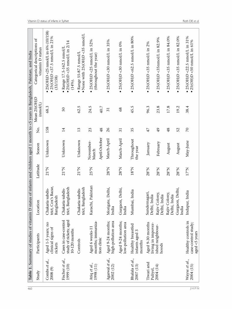
Low 25(OH)D is surprisingly common in South Asia, where systemic vitamin D deficits would be expected to be prevented by cutaneous vitamin D synthesis stimulated by exposure to sun at relatively low latitudes (3). Few studies on vitamin D status in infancy have been conducted in South Asia (Table
Roth DE et al.
Volume 28 | Number 5 | October 2010 459
Vitamin D status of infants in Sylhet
1). In Bangladesh, two published reports on child-hood 25(OH)D are available but neither reported 25(OH)D in early infancy (Table 1).
Knowledge of the vitamin D status of young chil-dren and infants is needed to design studies target-ing the aetiologic mechanisms and potential health implications of deficiency. A case-control study on the association between acute lower respiratory tract infection (ALRI) and vitamin D status in in-fants and young children conducted in Zakiganj subdistrict of Sylhet district in Bangladesh, during January-February 2008, provided an opportunity to gain preliminary insights into the vitamin D status of infants in northeastern rural Bangladesh (4). Here, we aimed to describe the vitamin D sta- tus of the source population and briefly review the potential determinants of low infant 25(OH)D in this setting.
MATERIALS AND METHODS
Setting
Zakiganj subdistrict (upazila) is in Sylhet district of northeastern Bangladesh (25°N), on the border with India. This region has low average household income and maternal literacy and has limited ac-cess to healthcare compared to neighbouring sub-districts in Sylhet (5). The study was facilitated by strong existing partnerships with local community organizations, the subdistrict health complex, and the Ministry of Health and Family Welfare, based on an ongoing collaborative neonatal health inter-vention trial infrastructure (Projahnmo) (6).
Participants
ALRI cases who met a clinical definition of ALRI were recruited from among infants and young children, aged one month to two years, admitted to the Zakijang subdistrict hospital. Control par-ticipants were selected by sampling from among children who lived in the same villages as the cases, were matched to a case on age (±2 months) and gender, and had no signs of ALRI at recruit-ment or reported past history of ALRI/pneumonia. To identify controls, a rapid household census was conducted in the village of residence of each case participant to generate a list of eligible controls aged 1-23 months. In an order based on closeness in age to the index case, caregivers of the listed children were approached until a control partici-pant was recruited, consent was obtained, and a blood specimen was collected. If an eligible control was not enrolled, the census and eligible control identification process was repeated in the nearest neighbouring village. Some recruited children not
considered eligible for the primary case-control study were included in the present analysis. The major reasons for this difference were that children enrolled during a one-week pilot phase were not included in the case-control study but were in-cluded here, and the strict requirement that gross haemolysis be absent on visual inspection of serum specimens included in the case-control study was relaxed for the present analysis. This latter deci-sion was made based on post-hoc findings that the mean 25(OH)D of grossly haemolyzed specimens was only slightly and non-significantly lower than that of non-haemolyzed serum specimens (dif-ference of means=3.6 nmol/L, p=0.335, after ad-justment for case-control status).
Collection of data
Caregivers (mothers) of participants were adminis-tered a questionnaire that addressed selected infant, maternal and household characteristics potentially associated with vitamin D status. Maternal intake of foods from animal sources, which included po-tential sources of vitamin D and rich sources of cal-cium, was assessed based on the reported frequency of consumption of food items/categories over the seven days preceding enrollment. Participants were further categorized as to whether the mother had consumed each food item/category at least once in the preceding seven days. Weight of infant was the average of two measurements recorded to the near-est 0.1 kg (Seca 354 infant scale), and length was the average of two measurements, to the nearest 0.5 cm (Seca 210 measuring mat). Gender-specific weight-for-age (WA), length-for-age (LA), and body mass index (BMI) z-scores were calculated accord-ing to the growth standards of the World Health Organization (7). According to convention, partici-pants with z-score values of less than -2 for each of the anthropometric indices were considered to have stunting (LA), underweight (WA), and low body mass index (BMI). Since reliable information on gestational age at birth was unavailable, anthro-pometric measures were interpreted under the as-sumption of term gestation.
A venous blood specimen was collected by standard methods, separated into serum aliquots, and stored at -20 °C or less. At the completion of the study, sera were shipped to the laboratory of Dr. Bruce Hollis, Medical University of South Carolina, Charleston, USA, for measurement of the total serum 25(OH)D concentration by radio-immunoassay (8).
Outcomes
The primary outcome of the study was the esti-mated mean 25(OH)D among infants and children
Roth DE et al. Vitamin D status of infants in Sylhet
JHPN460
Tab
le 1
. Su
mm
ary
of s
tud
ies
of v
itam
in D
sta
tus
of i
nfa
nts
an
d c
hil
dre
n a
ged
1 m
onth
to
<5 y
ears
in
Ban
glad
esh
, Pak
ista
n, a
nd
In
dia
Stu
dy
Part
icip
ants
Loca
tion
Lati
tud
eSe
ason
No.
Mea
n 2
5(O
H)D
(n
mol
/L)
Cat
egor
ical
des
crip
tion
s of
vi
tam
in D
sta
tus
Com
bs e
t al.,
20
08 (9
)A
ged
1-5
yea
rs; n
o cl
inic
al s
ign
s of
ri
cket
s
Ch
akar
ia s
ubdi
s-tr
ict,
Cox
’s B
azar
, B
angl
ades
h
21°N
Un
know
n15
868
.3•
25(O
H)D
<25
nm
ol/L
in 6
% (1
0/15
8)•
25(O
H)D
<37
.5 n
mol
/L i
n 2
1%
(33/
158)
Fisc
her
et
al.,
1999
(10
)C
ases
in c
ase-
con
trol
st
udy
of r
icke
ts; a
ged
10
-120
mon
ths
Ch
akar
ia s
ubdi
s-tr
ict,
Ban
glad
esh
21°N
Un
know
n14
50•
Ran
ge 1
7.5-
162.
5 n
mol
/L•
25(O
H)D
<35
nm
ol/L
in 2
/14
(14%
).
Con
trol
sC
hak
aria
sub
dis-
tric
t, B
angl
ades
h21
°NU
nkn
own
1362
.5•
Ran
ge 5
5-87
.5 n
mol
/L•
Non
e w
ith
25(
OH
)D <
35 n
mol
/L
Ati
q e
t al
., 19
98 (
11)
Age
d 6
wee
ks-1
1 m
onth
s; im
mun
iza-
tion
clin
ic
Kar
ach
i, P
akis
tan
25°N
Nov
embe
r-M
arch
2324
.5•
25(O
H)D
<25
nm
ol/L
in
52%
(t
hro
ugh
out
the
year
)A
pril-
Oct
ober
4840
.7
Aga
rwal
et a
l.,
2002
(12
)A
ged
9-2
4 m
onth
s;
hig
h-p
ollu
tion
are
aM
orig
ate,
Del
hi,
In
dia
28°N
Mar
ch-A
pri
l26
31•
25(O
H)D
<30
nm
ol/L
in
35%
Age
d 9
-24
mon
ths;
lo
w-p
ollu
tion
are
aG
urg
aon
, Del
hi,
In
dia
28°N
Mar
ch-A
pri
l31
68•
25(O
H)D
<30
nm
ol/L
in
0%
Bh
alal
a et
al.,
20
07 (
13)
Hea
lth
y br
east
fed
in
fan
ts a
ged
3
mon
ths
Mu
mba
i, I
nd
ia18
°NT
hro
ugh
out
the
year
3545
.5•
25(O
H)D
<62
.5 n
mol
/L i
n 8
0%
Tiw
ari
and
Pu
liye
l,
2004
(14
)
Age
d 9
-30
mon
ths
livi
ng
in i
mp
over
-is
hed
nei
ghbo
ur-
hoo
ds
Sun
der
nag
ari,
Del
hi,
In
dia
28°N
Jan
uar
y47
96.3
• 25
(OH
)D <
35 n
mol
/L i
n 2
%
Raj
iv C
olon
y,
Del
hi,
In
dia
28°N
Febr
uar
y49
23.8
• 25
(OH
)D <
35n
mol
/L i
n 8
2.9%
Raj
iv C
olon
y,
Del
hi,
In
dia
28°N
Au
gust
4817
.8•
25(O
H)D
<35
nm
ol/L
in
84.
0%
Gu
rgao
n, D
elh
i,
Ind
ia28
°NA
ugu
st52
19.2
• 25
(OH
)D <
35 n
mol
/L i
n 8
2.0%
Way
se e
t al
., 20
04 (
15)
Hea
lth
y co
ntr
ols
in
case
-con
trol
stu
dy;
aged
<5
year
s
Ind
apu
r, I
nd
ia17
°NM
ay-J
un
e70
38.4
• 25
(OH
)D <
22.5
nm
ol/L
in
31%
• 25
(OH
)D <
50 n
mol
/L i
n 6
1%
Roth DE et al.
Volume 28 | Number 5 | October 2010 461
Vitamin D status of infants in Sylhet
Table 2. Characteristics of a sample of infants aged 1-6 months in rural Sylhet distri- ct, Bangladesh
CharacteristicsMean±SD or
no. (%)No. 29Age (days) 71±32Boys 25 (86)Age (years) of mothers 23.8±4.2Mother attended any school 19 (66)Father attended any school 18 (62)Family owns their own home 19 (66)Lives in a house with components made of manufactured materials (i.e. cement, brick, tin) Floor Walls Roof
1 (3)9 (31)
27 (93)
Occupation of primary income-earner of household Day labourer Farmer on leased land Land owner Non-agricultural business-owner Salaried non-agricultural job
15 (52)2 (7)2 (7)
5 (17)5 (17)
Muslim religion 27 (93)Mother wears a burka when in public 27 (93)Exclusively breastfeeding 23 (79)Anthropometric measures Weight-for-age z-score Length-for-age z-score Body mass index z-score
-1.58±1.29-1.08±1.57-1.40±1.67
SD=Standard deviation
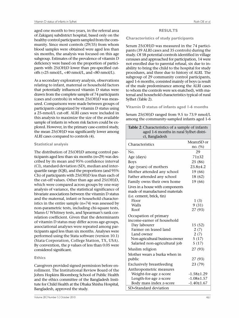
aged one month to two years, in the referral area of Zakiganj subdistrict hospital, based only on the healthy control participants sampled from the com-munity. Since most controls (29/35) from whom blood samples were obtained were aged less than six months, the analysis was focused on this age subgroup. Estimates of the prevalence of vitamin D deficiency were based on the proportion of partici-pants with 25(OH)D lower than pre-specified cut-offs (<25 nmol/L, <40 nmol/L, and <80 nmol/L).
As a secondary exploratory analysis, observations relating to infant, maternal or household factors that potentially influenced vitamin D status were drawn from the complete sample of 74 participants (cases and controls) in whom 25(OH)D was meas-ured. Comparisons were made between groups of participants categorized by vitamin D status using a 25-nmol/L cut-off. ALRI cases were included in this analysis to maximize the size of the available sample of infants in whom risk factors could be ex-plored. However, in the primary case-control study, the mean 25(OH)D was significantly lower among ALRI cases compared to controls (4).
Statistical analysis
The distribution of 25(OH)D among control par-ticipants aged less than six months (n=29) was des- cribed by its mean and 95% confidence interval (CI), standard deviation (SD), median and inter-quartile range (IQR), and the proportions (and 95% CIs) of participants with 25(OH)D less than each of the cut-off values. Other than age and 25(OH)D, which were compared across groups by one-way analysis of variance, the statistical significance of bivariate associations between the vitamin D status and the maternal, infant or household character-istics in the entire sample (n=74) was assessed by non-parametric tests, including chi-square tests, Mann-U Whitney tests, and Spearman’s rank cor-relation coefficient. Given that the determinants of vitamin D status may differ across age-groups, associational analyses were repeated among par-ticipants aged less than six months. Analyses were performed using the Stata software (version 10.1) (Stata Corporation, College Station, TX, USA). By convention, the p values of less than 0.05 were considered significant.
Ethics
Caregivers provided signed permission before en-rollment. The Institutional Review Board of the Johns Hopkins Bloomberg School of Public Health and the ethics committee of the Bangladesh Insti-tute for Child Health at the Dhaka Shishu Hospital, Bangladesh, approved the study.
RESULTS
Characteristics of study participants
Serum 25(OH)D was measured in the 74 partici-pants (39 ALRI cases and 35 controls) during the study. Of 58 potential controls identified in village censuses and approached for participation, 14 were not enrolled due to parental refusal, six due to in-ability to bring the child to the hospital for study procedures, and three due to history of ALRI. The subgroup of 29 community control participants, aged 1-6 months, consisted mainly of boys (a result of the male predominance among the ALRI cases to whom the controls were sex-matched), with ma-ternal and household characteristics typical of rural Sylhet (Table 2).
Vitamin D status of infants aged 1-6 months
Serum 25(OH)D ranged from 9.5 to 73.9 nmol/L among the community-sampled infants aged 1-6
Roth DE et al. Vitamin D status of infants in Sylhet
JHPN462
8060
4020
0
1 2 3 4 65Age (months)
25(O
H)D
(n
mol
/L)
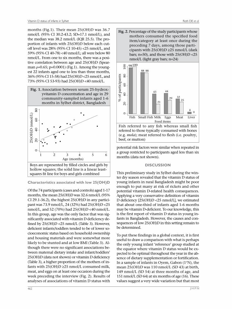
Fig. 1. Association between serum 25-hydrox-yvitamin D concentration and age in 29 community-sampled infants aged 1-6 months in Sylhet district, Bangladesh
Boys are represented by filled circles and girls by hollow squares; the solid line is a linear least-squares fit line for boys and girls combined
months (Fig.1). Their mean 25(OH)D was 36.7 nmol/L (95% CI 30.2-43.2; SD=17.1 nmol/L), and the median was 38.2 nmol/L (IQR 25.5). The pro-portion of infants with 25(OH)D below each cut-off level was 28% (95% CI 10-45) <25 nmol/L, and 59% (95% CI 40-78) <40 nmol/L; all were below 80 nmol/L. From one to six months, there was a posi-tive correlation between age and 25(OH)D (Spear-man ρ=0.65; p=0.0001) (Fig.1). Among the young-est 22 infants aged one to less than three months, 36% (95% CI 15-58) had 25(OH)D <25 nmol/L, and 73% (95% CI 53-93) had 25(OH)D <40 nmol/L.
Characteristics associated with low 25(OH)D
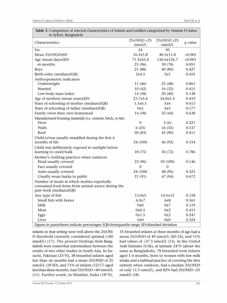
Of the 74 participants (cases and controls) aged 1-17 months, the mean 25(OH)D was 32.6 nmol/L (95% CI 29.1-36.2), the highest 25(OH)D in any partici-pant was 73.9 nmol/L, 24 (32%) had 25(OH)D <25 nmol/L, and 52 (70%) had 25(OH)D <40 nmol/L. In this group, age was the only factor that was sig-nificantly associated with vitamin D deficiency de-fined by 25(OH)D <25 nmol/L (Table 3). However, deficient infants/toddlers tended to be of lower so-cioeconomic status based on household ownership and housing materials and were somewhat more likely to be stunted and at low BMI (Table 3). Al-though there were no significant associations be-tween maternal dietary intake and infant/toddlers’ 25(OH)D (data not shown) or vitamin D deficiency (Table 3), a higher proportion of the mothers of in-fants with 25(OH)D ≥25 nmol/L consumed milk, meat, and eggs on at least one occasion during the week preceding the interview (Fig. 2). Results of analyses of associations of vitamin D status with
100
8060
4020
0
Fish Small Fish Milk EggsFood items
% o
f p
arti
cip
ants
04
253229
4443
21
8888
10098
Meat Liver
Fish referred to any fish whereas small fish referred to those typically consumed with bones (e.g. mola); meat referred to flesh (i.e. poultry, beef, or mutton)
Fig. 2. Percentage of the study participants whose mothers consumed the specified food item/category at least once during the preceding 7 days, among those parti-cipants with 25(OH)D ≥25 nmol/L (dark bars; n=50), and those with 25(OH)D <25 nmol/L (light gray bars; n=24)
potential risk factors were similar when repeated in a group restricted to participants aged less than six months (data not shown).
DISCUSSION
This preliminary study in Sylhet during the win-ter dry season revealed that the vitamin D status of young infants in rural Bangladesh might be poor enough to put many at risk of rickets and other potential vitamin D-related health consequences. Applying a very conservative definition of vitamin D deficiency [25(OH)D <25 nmol/L], we estimated that about one-third of infants aged 1-6 months may be vitamin D-deficient. To our knowledge, this is the first report of vitamin D status in young in-fants in Bangladesh. However, the causes and con-sequences of low 25(OH)D in this setting remain to be determined.
To put these findings in a global context, it is first useful to draw a comparison with what is perhaps the only young infant ‘reference’ group studied at the equator where vitamin D status would be ex-pected to be optimal throughout the year in the ab-sence of dietary supplementation or fortification. In a sample of infants in Oyem, Gabon (1°N), the mean 25(OH)D was 110 nmol/L (SD 43) at birth, 149 nmol/L (SD 54) at three months of age, and 151 nmol/L (SD 64) at six months of age (16). These values suggest a very wide variation but that most
Roth DE et al.
Volume 28 | Number 5 | October 2010 463
Vitamin D status of infants in Sylhet
Table 3. Comparison of selected characteristics of infants and toddlers categorized by vitamin D status in Sylhet, Bangladesh
Characteristics25(OH)D <25
nmol/L25(OH)D ≥25
nmol/Lp value
No. 24 50Mean 25(OH)D±SD 16.3±5.8 40.5±11.8 <0.001Age (mean days±SD) 71.3±43.4 136.6±126.7 <0.001
<6 months 23 (96) 39 (78) 0.051Boys 21 (88) 40 (80) 0.427Birth-order (median±IQR) 2±4.5 2±5 0.455Anthropometric indicators Underweight 11 (46) 25 (48) 0.861 Stunted 10 (42) 16 (32) 0.415 Low body mass index 14 (58) 20 (40) 0.138Age of mothers (mean years±SD) 23.7±5.6 24.8±5.3 0.410Years of schooling of mother (median±IQR) 1.5±6.5 5±6 0.613Years of schooling of father (median±IQR) 0±5 4±5 0.177Family owns their own homestead 14 (58) 32 (64) 0.638Manufactured housing materials (i.e. cement, brick, or tin) Floor 0 3 (6) 0.221 Walls 6 (25) 16 (32) 0.537 Roof 20 (83) 45 (90) 0.411Child is/was usually swaddled during the first 6 months of life 24 (100) 46 (92) 0.154Child was deliberately exposed to sunlight before learning to crawl/walk 18 (75) 36 (72) 0.786Mother’s clothing practices when outdoors Head usually covered 23 (96) 50 (100) 0.146 Face usually covered 0 0 - Arms usually covered 24 (100) 48 (96) 0.321 Usually wears burka in public 21 (91) 47 (94) 0.672Number of meals at which mother reportedly consumed food items from animal source during the past week (median±IQR)Any type of fish 13.0±5 14.5±12 0.158 Small fish with bones 6.0±7 6±8 0.561 Milk 0±0 0±7 0.119 Meat 0±0.5 0±2 0.415 Eggs 0±1.5 0±2 0.247 Liver 0±0 0±0 0.324Figures in parentheses indicate percentages; IQR=Interquartile range; SD=Standard deviation
infants in that setting were well above the 25(OH)D threshold currently considered optimal (>80 nmol/L) (17). The present findings from Bang-ladesh were somewhat intermediary between the results of two other studies in South Asia. In Ka-rachi, Pakistan (25°N), 38 breastfed infants aged less than six months had a mean 25(OH)D of 25 nmol/L (18 SD), and 71% of infants (12/17) aged less than three months, had 25(OH)D <40 nmol/L (11). Further south, in Mumbai, India (18°N),
35 breastfed infants at three months of age had a mean 25(OH)D of 49 nmol/L (SD 24), and 51% had values of <37.5 nmol/L (13). In the United Arab Emirates (UAE), at latitude 24°N (about the same as Bangladesh), 78 breastfed term infants aged 1-4 months, born to women with low milk intake and a habitual practice of covering the skin entirely when outdoors, had a median 25(OH)D of only 11.5 nmol/L, and 82% had 25(OH)D <25 nmol/L (18).
Roth DE et al. Vitamin D status of infants in Sylhet
JHPN464
In Iowa, USA (41°N), in a longitudinal study of predominantly white infants who were all exclu-sively breastfed and not receiving supplements, the mean 25(OH)D at about three and half months of age was 33 nmol/L for those assessed in the sum-mer (50% at <27.5 nmol/L) and 17 nmol/L in the winter (79% at <27.5 nmol/L); at approximately six months of age, the mean 25(OH)D increased to 45 nmol/L in the summer (32% at <27.5 nmol/L) but remained at 17 nmol/L for those measured in the winter (82% <27.5 nmol/L) (19). An exogenous vitamin D source is recommended for all infants in North America. So, it is worthwhile noting that, among formula-fed or vitamin D-supplemented in-fants (mean vitamin D intake of ~370 IU per day) aged 1-6 months (n=37) enrolled in a hospital-based study during the winter in Alberta, Canada (53°N) (20), the mean 25(OH)D was 78 nmol/L (Roth D et al. unpublished observations). Although there was a wide variation (17-152 nmol/L), only one infant aged 1.3 months had 25(OH)D <40 nmol/L, dem-onstrating the real-world effect of supplementa-tion/fortification policies.
Therefore, these data from rural Bangladesh, in combination with earlier findings from infants in urban Pakistan, India, and UAE, have demon-strated that the vitamin D status of young infants in South Asia and the Middle East may be no bet-ter than that of unsupplemented infants at much higher northern latitudes in North America, where guidelines support the provision of routine vitamin D supplementation to all breastfed infants (21,22). This implies that a tropical climate does not neces-sarily protect against low 25(OH)D in early infancy. Although cutaneous pre-vitamin D3 synthesis is expected to occur year-round in South Asia on the basis of latitude (23), there is a substantial seasonal variation in ultraviolet B irradiance (24). In fact, a seasonal differential in vitamin D status was observed among Pakistani infants (11). There-fore, the representativeness of our data is limited because they reflect vitamin D status during the winter, when ultraviolet radiation exposure is at its nadir, and when cutaneous vitamin D synthesis would be expected to be relatively minimal (24). Moreover, in the Bengal region, the attenuation of actual summer-time ultraviolet radiation exposure due to monsoon cloud-cover (24) may prevent sufficient endowment of vitamin D stores during the summer and, thus, further increase the risk of deficiency during the winter. Our cross-sectional observations suggest that 25(OH)D in Bangladeshi infants may rise within the first few months of life. However, these age-dependent differences may have been confounded by seasonal timing of ges-
tation—a younger age implied that the third tri-mester coincided with the expected seasonal nadir of vitamin D synthesis, when maternal vitamin D stores might be relatively depleted and, thus, when transfer of vitamin D metabolites to the foetus may have been minimized.
To further explain the apparent ‘vitamin D para-dox’ in South Asia (3), a range of hypothetical mechanisms can be proposed (Table 3). Young in-fants depend almost entirely on the transplacental transfer of vitamin D and 25(OH)D, which explains the consistent association between maternal and cord-blood 25(OH)D (25) and the observation that maternal antenatal vitamin D supplementation augments both maternal and cord-blood 25(OH)D (26). Therefore, the major reason that Bangladeshi infants start life with poor vitamin D stores is low maternal antenatal 25(OH)D, which has been do- cumented in urban and rural Bangladeshi women of reproductive age (27). Islam et al. recently stud-ied female workers in a garment factory in Dhaka and speculated that their long day-time hours in indoors, brief exposure to low-intensity sunlight in the early morning, outdoor air pollution, and wide-spread sunscreen use, in combination with darkly-pigmented skin, may contribute to their poor vita-min D status (mean 25(OH)D of 37 nmol/L, and 15% of the participants had 25(OH)D <25 nmol/L) (28). Conservative dress, including almost com-plete skin coverage by traditional veils or cloaks, has been emphasized as a contributor to vitamin D deficits in Muslim women in South Asia and the Middle East because it limits cutaneous vitamin D synthesis regardless of the intensity of ambient ul-traviolet B (29).
Inferences regarding the determinants of infant/toddlers’ vitamin D status in this study were limi- ted, largely because of the small sample-size and substantial uniformity with respect to selected ma-ternal clothing and dietary practices. We also ac-knowledge that pooling of ALRI cases and controls to maximize our available sample-size may have led to selection biases. However, the observation that mothers of infants with relatively low 25(OH)D seemed less likely to consume foods from animal sources (other than fish) deserves consideration in future studies. The amount of vitamin D in the local diet is unknown but low calcium intake (or reduced absorption of calcium due to high phytate intake), typical of low-income diets in Bangladesh (30,31), may accelerate 25(OH)D use, leading to relatively-increased vitamin D demands (32). Since the con-centration of vitamin D metabolites in breastmilk is determined by maternal vitamin D status, ma-ternal vitamin D deficiency during lactation may
Roth DE et al.
Volume 28 | Number 5 | October 2010 465
Vitamin D status of infants in Sylhet
cause ongoing deficits in postnatal infants’ vitamin D intake (42); yet, maternal factors cannot entirely account for the persistence of low 25(OH)D among toddlers (Table 1) who experience direct exposure to sun and should be unaffected by the conserva-tive clothing practices of their mothers. Therefore, much remains to be learnt about the determinants of vitamin D status throughout infancy and child-hood, particularly where 25(OH)D appears discrep-ant from that which would be expected based on latitude.
Despite the current enthusiasm for vitamin D sup-plementation in the USA (22), the effects of vitamin D deficits during early infancy are still not fully understood, and meaningful inflection points in 25(OH)D-outcome relationships have not been es-tablished. The most widely-accepted manifestation of severe vitamin D deficiency in infancy is rick-ets, the classical childhood metabolic bone disease associated with skeletal hypomineralization and deformities, muscle weakness, and growth impair-
ment (1). Young infants with severe congenital vi-tamin D deficiency may present with hypocalcae-mic tetany or seizures, with absent or subtle skeletal pathology (43-45). Greer noted that there is no clear or consistent association between 25(OH)D and the risk of rickets or other functional outcomes (46). Although 25(OH)D <25 nmol/L is typical of clinically-apparent rickets, early stages of the dis-ease may occur at higher 25(OH)D (~40 nmol/L) (47), with declines in 25(OH)D occurring as the dis-ease progresses and vitamin D stores are depleted. The frequent occurrence of 25(OH)D >25 nmol/L among toddlers and older children with rickets has been presumptively attributed to dietary calcium deficits (48); however, rickets among breastfed in-fants with 25(OH)D >25 nmol/L (49) suggests that factors other than calcium intake may be impli- cated.
In Bangladesh, rickets may be more common than previously thought, based on surveys of lower limb
Table 4. Hypothetical mechanisms that may contribute to risk of maternal-infant vitamin D deficiency in South Asia
General mechanism Examples of specific hypothesized mechanisms
Endogenous cutaneous vitamin D production
• Conservative clothing practices (33)• Limited time spent outdoors• Dark skin pigmentation (34)• Cloud-cover and outdoor air pollution (12)• Geographic/genetic/dietary factors that alter epidermal
7-dehydrocholesterol composition or concentration
Dietary vitamin D intake • Inadequate intake of liver, eggs, fish, or fish oils, or fortified dairy products
• Diminished natural vitamin D content of farmed fish (35)• Reduction in vitamin D content of fish due to cooking (e.g.
frying) or processing
Intestinal vitamin D absorption and storage
• Nutrient-nutrient interactions or competition, e.g. taurine (36)• Inadequate consumption of dietary fatty acid• Chronic intestinal inflammation (37)• Cholestatic liver disease (and reduced bile acid excretion)• Low fat mass, leading to reduced capacity to store vitamin D
Hepatic conversion of vitamin D to 25-hydroyxyvitamin D
• Chronic hepatitis or hepatic dysfunction, e.g. aflatoxin exposure (38)
• Dietary/environmental exposure to P450 cytochrome inhibitors
Destruction or use of 25-hydroxyvitamin D or 1,25-dihydroxyvitamin D
• Dietary calcium deficit and/or phytate-rich diet, driving secondary hyperparathyroidism
• Areca nut consumption (39)• Protein-energy malnutrition (40) • Nutrient-nutrient interactions, e.g. vitamin A (41)• Genetic polymorphisms in genes encoding proteins involved
in vitamin D metabolism, e.g. vitamin D receptor, 24-hydroxylase
Roth DE et al. Vitamin D status of infants in Sylhet
JHPN466
deformities in ambulating children (50,51). Recent data from a nationwide survey suggest that about 0.6% of Bangladeshi children, aged 1-15 years, may have radiologic evidence of rickets, with the high-est prevalence in Chittagong and Sylhet divisions (51). The incidence of symptomatic hypocalcaemia secondary to vitamin D deficiency in early infancy is unknown. Some investigators have played down the role of vitamin D in rickets in Bangladesh (10, 52), instead blaming dietary calcium deficits or other mineral deficiencies or excesses, e.g. alumi-num (53). However, in case-control studies, dietary calcium intake between rickets-affected and unaf-fected households did not differ (54) whereas the mean 25(OH)D in cases was significantly lower than in controls (10). A plausible hypothesis is that vitamin D deficiency acts synergistically with oth-er causes of inadequate bone mineralization and that an individual’s 25(OH)D concentration below which clinical signs emerge depends on the severi- ty and multiplicity of other genetic and environ-mental factors.
Beyond rickets, speculation abounds regarding the potential extra-skeletal consequences of suboptimal vitamin D status during foetal development and infancy. The vitamin D receptor has been found within virtually every organ-system (55), and the active metabolite of vitamin D is well-described as a potent mediator of cell proliferation and differ-entiation, particularly noted for its range of effects on immune function in laboratory models (56). The case-control study for which the data for the present analysis were primarily collected revealed an inverse association between 25(OH)D and the odds of hospitalization for ALRI (4), corroborat-ing the findings in neonates in Turkey (57) and children in India (15). If vitamin D deficiency is confirmed as a risk factor for pneumonia, interven-tions to improve maternal-infant vitamin D status could reduce the global burden of ALRI, the single most important cause of early childhood death in the world (58). Other postulated consequences of antenatal or infant vitamin D deficiency include growth faltering (59,60), type 1 diabetes (61), and asthma (62). However, rigorous studies of the broad health benefits of interventions to improve the an-tenatal or postnatal vitamin D status in South Asian mothers and infants have yet to be reported.
Limitations
This study was limited by its small sample-size, res- tricted geographic scope, and cross-sectional de-sign. We aimed to select control participants for the main case-control study in a manner that would
enable inferences about the source population. Selection of control was necessarily non-random from the perspective of age and gender and, thus, unfortunately led to an over-representation of boys; however, there is unlikely to be a gender differen-tial in vitamin D status during early infancy (63). Also, the requirement for an absence of reported history of ALRI probably produced negligible bias since only three otherwise eligible children were excluded for this reason. Aside from these caveats, the community-based sampling was likely random with respect to most determinants of vitamin D status. The group of infants aged 1-6 months was small but adequate to estimate the mean 25(OH)D within a 13-nmol/L range with 95% confidence. However, data were insufficient to yield precise estimates of the associations between infant and maternal characteristics and 25(OH)D. Another limitation was the lack of ancillary biochemical or radiographic data that may have revealed evidence of adverse consequences of low 25(OH)D. We did not report the findings of physical examinations because scoring systems for rickets are not very use-ful in early infancy, and a protocol for a standard-ized musculoskeletal clinical examination was not satisfactorily implemented. However, none of the toddlers had any clinical evidence of rickets ac-cording to the physician’s examination (data not shown).
Conclusions
This study provides initial observations on the vita-min D status of young infants in northeastern rural Bangladesh. However, it remains to be determined whether the relatively left-shifted distribution of 25(OH)D in this study sample is representative of the broader population and causally associated with an excess burden of rickets, symptomatic hypocalcaemia, growth faltering, or extra-skeletal health outcomes. Therefore, recommendations for universal antenatal and/or infant vitamin D sup-plementation in Bangladesh based on biochemical data alone would be premature. The causes and consequences of low 25(OH)D in young infants in South Asia must be further investigated.
ACKNOWLEDGEMENTS
The authors thank the participants and caregivers for their kind cooperation and also thank the study field staff, the Projahnmo Sylhet team, and per-sonnel at the Zakiganj subdistrict hospital. They particularly thank Kazi Moksedur Rahman (Deputy Executive Director, SHIMANTIK), Dr. Arun Kumar Roy (Project Research Physician, Projahnmo), Dr. Daniel Hossain (Project Research Manager, Projah-
Roth DE et al.
Volume 28 | Number 5 | October 2010 467
Vitamin D status of infants in Sylhet
nmo), Dr. Sirajul Islam (Upazila Health and Fam-ily Planning Officer), and Dr. Jonme Joy Dutta Shankar (Medical Officer, Zakiganj subdistrict hos-pital). The authors are grateful to Dr. Bruce Hol-lis and Dr. Carole Wagner (Medical University of South Carolina, USA), Dr. Samir K. Saha (Bang-ladesh Institute of Child Health, Dhaka Shishu Hospital, Dhaka, Bangladesh), and the Depart-ment of International Health at the Johns Hopkins Bloomberg School of Public Health. D. Roth was supported by training grants from the Canadian Institutes for Health Research and the Alberta Her-itage Foundation for Medical Research.
REFERENCES
1. Holick MF. Resurrection of vitamin D deficiency and rickets. J Clin Invest 2006;116:2062-72.
2. Zerwekh JE. Blood biomarkers of vitamin D status. Am J Clin Nutr 2008;87:1087S-91S.
3. Joshi SR. Vitamin D paradox in plenty sunshine in rural India—an emerging threat. J Assoc Physicians In-dia 2008;56:749-52.
4. Roth DE, Shah R, Black RE, Baqui AH. Vitamin D status and acute lower respiratory infection in early childhood in Sylhet, Bangladesh. Acta Paediatr 2010;99:389-93.
5. Baqui, AH, Arifeen SE, Darmstadt GL, Ahmed S, Seraji HR, Winch PJ et al.; Bangladesh Projahnmo Study Group. Differentials in neonatal mortality in two adjacent rural areas of Bangladesh: lessons for neonatal health interventions. Global Public Health 2008;3:366-82.
6. Baqui AH, Arifeen SE, Darmstadt GL, Ahmed S, Wil-liams EK, Seraji HR et al. Effect of community-based newborn-care intervention package implemented through two service-delivery strategies in Sylhet dis-trict, Bangladesh: a cluster-randomised controlled trial. Lancet 2008;371:1936-44.
7. World Health Organization. WHO child growth stand- ards: length/height-for-age, weight-for-age, weigh- t-for-length, weight-for-height and body mass index-for-age: methods and development. Geneva: World Health Organization, 2006. 312 p.
8. Hollis BW, Kamerud JQ, Selvaag SR, Lorenz JD, Na-poli JL. Determination of vitamin D status by radio-immunoassay with a 125I-labeled tracer. Clin Chem 1993;39:529-33.
9. Combs GF, Jr., Hassan N, Dellagana N, Staab D, Fischer P, Hunt C et al. Apparent efficacy of food-based calcium supplementation in preventing rickets in Bangladesh. Biol Trace Elem Res 2008;121:193-204.
10. Fischer PR, Rahman A, Cimma JP, Kyaw-Myint TO, Kabir AR, Talukder K et al. Nutritional rickets with-
out vitamin D deficiency in Bangladesh. J Trop Pediatr 1999;45:291-3.
11. Atiq M, Suria A, Nizami SQ, Ahmed I. Maternal vi-tamin-D deficiency in Pakistan. Acta Obstet Gynecol Scand 1998;77:970-3.
12. Agarwal KS, Mughal MZ, Upadhyay P, Berry JL, Maw-er EB, Puliyel JM. The impact of atmospheric pollu-tion on vitamin D status of infants and toddlers in Delhi, India. Arch Dis Child 2002;87:111-3.
13. Bhalala U, Desai M, Parekh P, Mokal R, Chheda B. Subclinical hypovitaminosis D among exclusively breastfed young infants. Indian Pediatr 2007;44:897-901.
14. Tiwari L, Puliyel JM. Vitamin D level in slum children of Delhi. Indian Pediatr 2004;41:1076-7.
15. Wayse V, Yousafzai A, Mogale K, Filteau S. Association of subclinical vitamin D deficiency with severe acute lower respiratory infection in Indian children under 5 y. Eur J Clin Nutr 2004;58:563-7.
16. Nguema-Asseko B, Ganga-Zandzou PS, Ovono F, Lendoye E, Lemamy GJ, Akendengue B et al. [Vi-tamin D status in Gabonese children]. Arch Pediatr 2005;12:1587-90 [French].
17. Heaney RP. Vitamin D: criteria for safety and efficacy. Nutr Rev 2008;66(10 Suppl 2):S178-81.
18. Dawodu A, Agarwal M, Hossain M, Kochiyil J, Zayed R. Hypovitaminosis D and vitamin D deficiency in ex-clusively breast-feeding infants and their mothers in summer: a justification for vitamin D supplementa-tion of breast-feeding infants. J Pediatr 2003;142:169-73.
19. Ziegler EE, Hollis BW, Nelson SE, Jeter JM. Vitamin D deficiency in breastfed infants in Iowa. Pediatrics 2006;118:603-10.
20. Roth DE, Jones AB, Prosser C, Robinson JL, Vohra S. Vitamin D status is not associated with the risk of hospitalization for acute bronchiolitis in early child-hood. Eur J Clin Nutr 2009;63:297-9.
21. Canadian Paediatric Society. Vitamin D supplemen-tation: recommendations for Canadian mothers and infants. Paediatr Child Health 2007;12:583-98.
22. Wagner CL, Greer FR; American Academy of Pedi-atrics Section on Breastfeeding; American Academy of Pediatrics Committee on Nutrition. Prevention of rickets and vitamin D deficiency in infants, children, and adolescents. Pediatrics 2008;122:1142-52.
23. Holick MF. Environmental factors that influence the cutaneous production of vitamin D. Am J Clin Nutr 1995;61(3 Suppl):638S-645S.
24. Bachelet D, Barnes PW, Brown D, Brown M. Latitudi-nal and seasonal variation in calculated ultraviolet-B irradiance for rice-growing regions of Asia. Photochem Photobiol 1991;54:411-22.
Roth DE et al. Vitamin D status of infants in Sylhet
JHPN468
25. Hollis BW, Pittard WB, 3rd. Evaluation of the total fetomaternal vitamin D relationships at term: evi-dence for racial differences. J Clin Endocrinol Metab 1984;59:652-7.
26. Specker B. Vitamin D requirements during pregnan-cy. Am J Clin Nutr 2004;80(6 Suppl):1740S-7S.
27. Islam MZ, Lamberg-Allardt C, Kärkkäinen M, Outila T, Salamatullah Q, Shamim AA. Vitamin D deficien-cy: a concern in premenopausal Bangladeshi women of two socio-economic groups in rural and urban re-gion. Eur J Clin Nutr 2002;56:51-6.
28. Islam MZ, Shamim AA, Kemi V, Nevanlinna A, Akhtaruzzaman M, Laaksonen M et al. Vitamin D deficiency and low bone status in adult female garment factory workers in Bangladesh. Br J Nutr 2008;99:1322-9.
29. Dawodu A, Absood G, Patel M, Agarwal M, Ezimokhai M, Abdulrazzaq Y et al. Biosocial factors affecting vi-tamin D status of women of childbearing age in the United Arab Emirates. J Biosoc Sci 1998;30:431-7.
30. Islam MZ, Lamberg-Allardt C, Kärkkäinen M, Ali SM. Dietary calcium intake in premenopausal Bangla-deshi women: do socio-economic or physiological factors play a role? Eur J Clin Nutr 2003;57:674-80.
31. Hels O, Hassan N, Tetens I, Haraksingh Thilsted S. Food consumption, energy and nutrient intake and nutritional status in rural Bangladesh: changes from 1981-1982 to 1995-96. Eur J Clin Nutr 2003;57:586-94.
32. Clements MR, Johnson L, Fraser DR. A new mecha-nism for induced vitamin D deficiency in calcium deprivation. Nature 1987;325:62-5.
33. Matsuoka LY, Wortsman J, Dannenberg MJ, Hollis BW, Lu Z, Holick MF. Clothing prevents ultraviolet-B radiation-dependent photosynthesis of vitamin D3. J Clin Endocrinol Metab 1992;75:1099-103.
34. Clemens TL, Adams JS, Henderson SL, Holick MF. In-creased skin pigment reduces the capacity of skin to synthesise vitamin D3. Lancet 1982;1:74-6.
35. Lu Z, Chen TC, Zhang A, Persons KS, Kohn N, Berkow-itz R et al. An evaluation of the vitamin D3 content in fish: is the vitamin D content adequate to satisfy the dietary requirement for vitamin D? J Steroid Biochem Mol Biol 2007;103:642-4.
36. Zamboni G, Piemonte G, Bolner A, Antoniazzi F, Dall‘Agnola A, Messner H et al. Influence of dietary taurine on vitamin D absorption. Acta Paediatr 1993;82:811-5.
37. Leichtmann GA, Bengoa JM, Bolt MJ, Sitrin MD. In-testinal absorption of cholecalciferol and 25-hydrox-ycholecalciferol in patients with both Crohn‘s disease and intestinal resection. Am J Clin Nutr 1991;54:548-52.
38. Glahn RP, Beers KW, Bottje WG, Wideman RF, Jr., Huff WE, Thomas W. Aflatoxicosis alters avian renal function, calcium, and vitamin D metabolism. J Toxi-col Environ Health 1991;34:309-21.
39. Ogunkolade WB, Boucher BJ, Bustin SA, Burrin JM, Noonan K, Mannan N et al. Vitamin D metabolism in peripheral blood mononuclear cells is influenced by chewing “betel nut” (Areca catechu) and vitamin D status. J Clin Endocrinol Metab 2006;91:2612-7.
40. Fraser DR. Vitamin D-deficiency in Asia. J Steroid Bio-chem Mol Biol 2004;89-90:491-5.
41. Allegretto EA, Shevde N, Zou A, Howell SR, Boehm MF, Hollis BW et al. Retinoid X receptor acts as a hormone receptor in vivo to induce a key metabolic enzyme for 1,25-dihydroxyvitamin D3. J Biol Chem 1995;270:23906-9.
42. Hollis BW, Wagner CL. Assessment of dietary vitamin D requirements during pregnancy and lactation. Am J Clin Nutr 2004;79:717-26.
43. Ahmed I, Atiq M, Iqbal J, Khurshid M, Whittaker P. Vitamin D deficiency rickets in breast-fed infants pre-senting with hypocalcaemic seizures. Acta Paediatr 1995;84:941-2.
44. Hatun S, Ozkan B, Orbak Z, Doneray H, Cizmecioglu F, Toprak D et al. Vitamin D deficiency in early in-fancy. J Nutr 2005;135:279-82.
45. Balasubramanian S, Shivbalan S, Kumar PS. Hypoc-alcemia due to vitamin D deficiency in exclusively breastfed infants. Indian Pediatr 2006;43:247-51.
46. Greer FR. 25-Hydroxyvitamin D: functional out-comes in infants and young children. Am J Clin Nutr 2008;88:529S-33S.
47. Arnaud SB, Stickler GB, Haworth JC. Serum 25-hydroxyvitamin D in infantile rickets. Pediatrics 1976;57:221-5.
48. DeLucia MC, Mitnick ME, Carpenter TO. Nutritional rickets with normal circulating 25-hydroxyvitamin D: a call for reexamining the role of dietary calcium intake in North American infants. J Clin Endocrinol Metab 2003;88:3539-45.
49. Kreiter SR, Schwartz RP, Kirkman HN, Jr., Charlton PA, Calikoglu AS, Davenport ML. Nutritional rick-ets in African American breast-fed infants. J Pediatr 2000;137:153-7.
50. Hellen Keller International. Rickets in Bangladeshi children: a small focus or a widespread problem?. Dhaka: Hellen Keller International, 2001. 4 p. (Nutri-tion surveillance bulletin no. 4).
51. International Centre for Diarrhoeal Disease Research, Bangladesh. National rickets survey in Bangladesh, 2008. Health Sci Bull 2009;7:7-11.
52. Craviari T, Pettifor JM, Thacher TD, Meisner C, Ar-
Roth DE et al.
Volume 28 | Number 5 | October 2010 469
Vitamin D status of infants in Sylhet
naud J, Fischer PR; Rickets Convergence Group. Rick-ets: an overview and future directions, with special reference to Bangladesh. A summary of the Rickets Convergence Group meeting, Dhaka, 26-27 January 2006. J Health Popul Nutr 2008;26:112-21.
53. Cimma JP, Arnaud J, Labarere J, Guillard O, Nugues F, Marrauld A et al. Effect of consumption of food cooked in aluminium or stainless-steel pots on Bang-ladeshi children with calcium-deficient rickets: an eight month trial. J Trace Elem Med Biol 2004;17:249-53.
54. Combs GF, Hassan N. The Chakaria food system study: household-level, case-control study to identify risk factor for rickets in Bangladesh. Eur J Clin Nutr 2005;59:1291-301.
55. Norman AW. From vitamin D to hormone D: funda-mentals of the vitamin D endocrine system essential for good health. Am J Clin Nutr 2008;88:491S-9S.
56. van Etten E, Stoffels K, Gysemans C, Mathieu C, Over-bergh L. Regulation of vitamin D homeostasis: impli-cations for the immune system. Nutr Rev 2008;66(10 Suppl 2):S125-34.
57. Karatekin G, Kaya A, Salihoğlu O, Balci H, Nuhoğlu A. Association of subclinical vitamin D deficiency in newborns with acute lower respiratory infection and
their mothers. Eur J Clin Nutr 2009;63:473-7.
58. Black RE, Cousens S, Johnson HL, Lawn JE, Rudan I, Bassani DG et al. Global, regional, and national caus-es of child mortality in 2008: a systematic analysis. Lancet 2010;375:1969-87.
59. Brooke OG, Butters F, Wood C. Intrauterine vitamin D nutrition and postnatal growth in Asian infants. Br Med J (Clin Res Ed) 1981;283:1024.
60. Rajah J, Jubeh JA, Haq A, Shalash A, Parsons H. Nutritional rickets and z scores for height in the United Arab Emirates: to D or not to D? Pediatr Int 2008;50:424-8.
61. Zipitis CS, Akobeng AK. Vitamin D supplementa-tion in early childhood and risk of type 1 diabetes: a systematic review and meta-analysis. Arch Dis Child 2008;93:512-7.
62. Camargo CA, Jr., Rifas-Shiman SL, Litonjua AA, Rich-Edwards JW, Weiss ST, Gold DR et al. Maternal intake of vitamin D during pregnancy and risk of recur-rent wheeze in children at 3 y of age. Am J Clin Nutr 2007;85:788-95.
63. Gordon CM, Feldman HA, Sinclair L, Williams AL, Kleinman PK, Perez-Rossello J et al. Prevalence of vi-tamin D deficiency among healthy infants and tod-dlers. Arch Pediatr Adolesc Med 2008;162:505-12.


