Embed Size (px)
Citation preview

AD_________________ Award Number:
W81XWH-06-1-0289 TITLE:
Scavenger Receptors and Resistance to Inhaled Allergens PRINCIPAL INVESTIGATOR:
Lester Kobzik, M.D. CONTRACTING ORGANIZATION:
Harvard School of Public Health Boston, MA 02115 REPORT DATE: February 2010 TYPE OF REPORT:
Final PREPARED FOR: U.S. Army Medical Research and Materiel Command Fort Detrick, Maryland 21702-5012 DISTRIBUTION STATEMENT: XX Approved for public release; distribution unlimited The views, opinions and/or findings contained in this report are those of the author(s) and should not be construed as an official Department of the Army position, policy or decision unless so designated by other documentation.

REPORT DOCUMENTATION PAGE Form Approved
OMB No. 0704-0188 Public reporting burden for this collection of information is estimated to average 1 hour per response, including the time for reviewing instructions, searching existing data sources, gathering and maintaining the data needed, and completing and reviewing this collection of information. Send comments regarding this burden estimate or any other aspect of this collection of information, including suggestions for reducing this burden to Department of Defense, Washington Headquarters Services, Directorate for Information Operations and Reports (0704-0188), 1215 Jefferson Davis Highway, Suite 1204, Arlington, VA 22202-4302. Respondents should be aware that notwithstanding any other provision of law, no person shall be subject to any penalty for failing to comply with a collection of information if it does not display a currently valid OMB control number. PLEASE DO NOT RETURN YOUR FORM TO THE ABOVE ADDRESS. 1. REPORT DATE (DD-MM-YYYY) 01-02-2010
2. REPORT TYPE Final
3. DATES COVERED (From - To) 17 JAN 2006 - 16 JAN 2010
4. TITLE AND SUBTITLE Scavenger Receptors and Resistance to Inhaled Allergens
5a. CONTRACT NUMBER
5b. GRANT NUMBER W81XWH-06-1-0289
5c. PROGRAM ELEMENT NUMBER
6. AUTHOR(S) Lester Kobzik
5d. PROJECT NUMBER
email: [email protected]
5e. TASK NUMBER
5f. WORK UNIT NUMBER 7. PERFORMING ORGANIZATION NAME(S) AND ADDRESS(ES)
AND ADDRESS(ES)
8. PERFORMING ORGANIZATION REPORT NUMBER
Harvard School of Public Health . Boston, MA 02115
9. SPONSORING / MONITORING AGENCY NAME(S) AND ADDRESS(ES) 10. SPONSOR/MONITOR’S ACRONYM(S) U.S. Army Medical Research and Material C
ommand Fort Detrich, MD 21702 11. SPONSOR/MONITOR’S REPORT NUMBER(S) 12. DISTRIBUTION / AVAILABILITY STATEMENT Approved for public release; distribution unlimited
13. SUPPLEMENTARY NOTES
14. ABSTRACT Our central hypothesis was that lung macrophage scavenger receptors normally function to bind and clear inhaled allergens and pathogens, thereby preventing allergic responses and infections. The purpose of the project was to determine whether 1) decreased levels of SRAs (mediated by environmental stresses) increase susceptibility to asthma; and 2) modulating levels of scavenger receptors will affect resistance to asthma. The results indicate that scavenger receptors modulate the allergic response, primarily by modifying dendritic cell trafficking to thoracic lymph nodes in the setting of asthma. Dendritic cells deficient in the scavenger receptors SRA I/II or MARCO traveled to the lymph nodes faster, and generated a more robust asthmatic response when allergen was delivered into the lungs. Scavenger receptors are linked to oxidant air pollution responses in a complex manner. Oxidant air pollution increases expression of MARCO which serves to scavenge oxidized pro-inflammatory lipids, thereby dampening, but not eradicating, acute inflammatory responses.
15. SUBJECT TERMS asthma, allergy, scavenger receptors, dendritic cells
16. SECURITY CLASSIFICATION OF:
17. LIMITATION OF ABSTRACT
18. NUMBER OF PAGES
19a. NAME OF RESPONSIBLE PERSON USAMRMC
a. REPORT U
b. ABSTRACT U
c. THIS PAGE U
UU 50
19b. TELEPHONE NUMBER (include area code) Standard Form 298 (Rev. 8-98)
Prescribed by ANSI Std. Z39.18

Table of Contents
INTRODUCTION:! 2
BODY:! 2
Task 1: Determine susceptibility of SRA ʻknockoutʼ (KO) mice to asthma.! 2
Task 2: Determine role of SRAs on DCs in responses to inhaled allergen:! 2
Task 3: Determine role of SRAs on AMs in responses to inhaled allergen:! 12
Task 4: Modulation of SRAs and Air Pollution Responses! 13
Key Research Accomplishments:! 14
Reportable Outcomes:! 14
Appendices:! 15
Personnel supported by this grant:! 16

INTRODUCTION:
Our central hypothesis is that lung macrophage scavenger receptors normally function to bind and clear inhaled allergens and pathogens, thereby preventing allergic responses and infections. The purpose of the project is to determine whether 1) decreased levels of SRAs (mediated by environmental stresses) increase susceptibility to asthma or pneumonia; and 2) therapy to increase or maintain normal levels of scavenger receptors will increase resistance to asthma and pneumonia. The scope of the research includes studies using in vivo mouse models (Aim 1), studies of the specific role of alveolar macrophages (Aim 2) and dendritic cells (Aim 3) and studies of the effects of pollutants on scavenger receptors (Aim 4).
BODY:
Task 1: Determine susceptibility of SRA ʻknockoutʼ (KO) mice to asthma.
In this work we report that the class A scavenger receptors (SRAs) MARCO and SR-AI/II are expressed on lung macrophages (MΦ) and dendritic cells (DC) and function in innate defenses against inhaled pathogens and particles. Increased expression of SRAs in the lungs of mice in an OVA-asthma model suggested an additional role in modulating responses to inhaled allergen. After OVA sensitization and aerosol challenge, SR-AI/II and MARCO-deficient mice exhibited greater eosinophilic airway inflammation and airway hyperresponsiveness compared to wild-type mice. A role for simple SRA-mediated antigen clearance (‘scavenging’) by lung macrophages was excluded by observation of comparable uptake of fluorescent OVA by wild-type and SRA-deficient lung MΦs and DCs. In contrast, airway instillation of fluorescent antigen revealed significantly higher traffic of labelled DCs to thoracic lymph nodes in SRA-deficient mice than in controls. The increased migration of SRA-deficient DCs was accompanied by enhanced proliferation in thoracic lymph nodes of adoptively transferred OVA-specific T cells after airway OVA challenge. The data identify a novel role for SRAs expressed on lung DCs in down-regulation of specific immune responses to aero-allergens by reduction of DC migration from the site of antigen uptake to the draining lymph nodes. This studied is detailed in the publication which is Appendix 1.
Task 2: Determine role of SRAs on DCs in responses to inhaled allergen:
This work sought to test the hypothesis that DC SRAs act to down-regulate allergic immune responses. While we made good progress in some areas, we must also report some disappointing results in other areas, despite considerable effort.
To study dendritic cells, we learned and optimized in vitro culture protocols to grow bone-marrow derived dendritic cells from both wild-type and SRA knockout mice. This produces a large number of cells suitable for easy analysis of phenotype and function.
2

We have confirmed purity of the cultured dendritic cells by immunolabeling with CD11c marker (>95% +).
Initial results with functional assays to compare DCs from wild-type and knockout mice were promising, as indicated in an earlier annual report. However, as we repeated and expanded these analyses, we have consistently observed an absence of significant differences in the wild-type vs. knockout groups. For example, to test whether scavenger receptors reduce cell motility (as observed in vivo as increased DC accumulation in lymph nodes in our asthma model), we have developed microscopic and live cell imaging assays to quantitate cell movement. The first assay uses modified Boyden migration chambers and results in counting of cells that move from an upper chamber to the lower chamber. The second assay measures random migration by tracing cell movement over 12-24 hours in a live cell system. The details of these assays and the results are presented next, followed by conclusions and discussion.
Dendritic cell isolation and differentiation:Bone marrow progenitor cells were isolated from normal female wild-type or MARCO-/- adult (8-12 wk) Balb/c mice and cultured in 6-well tissue culture plates at 3e6/well in RPMI-10 (RPMI 1640 + 10% FBS + 1 mM l-glutamine + penicillin/streptomycin) with 20 ng/ml rGM-CSF (Peprotech) at 37° C, 5% CO2. Media was changed every 2-3 d without disturbing the cells for 7 d. For motility and chemotaxis assays, on day 7-8 of culture, immature DC were replenished with media with or without maturation for 24 h using 1 µg/ml LPS, then harvested by pipetting and replated in assay dishes or wells. For surface receptor expression assays, immature cells were removed from plates by pipetting, replated in fresh media in low-adherence tissue culture dishes, and matured for 24-48 h with 20 ng/ml TNFα or 1 µg/ml LPS; cells were then removed by pipetting and stained for flow cytometry.
Random motility assay:Immature and mature DC were harvested on day 8-9 of culture as indicated and stained with Hoescht (Molecular Probes) in normal saline + 10 mM HEPES + 0.5% BSA for 5 min at 37° C. Cells were then washed and adhered to glass-bottom tissue culture plates (MatTek) coated with 50 µg/ml fibronectin (Sigma) for 4 h at 37° C, 5% CO2 in RPMI-10 + 10 mM HEPES + 20 ng/ml rGM-CSF. Cells were then imaged at 5 min intervals for 16 h using a Nikon confocal microscope fitted with an environmental chamber at 37° C. Cell tracks were generated by tracking nuclei positions using the MTrackJ plugin (Erik Meijering, University Medical Center, Rotterdam, Netherlands) for ImageJ (NIH). Graphs and analysis were performed using the Chemotaxis Tool plugin (Ibid) for ImageJ.
Chemotaxis:Immature and mature DC were harvested on day 8-9 of culture as indicated and resuspended in RPMI-10 at 1e7/ml, then 100 µl cell suspension was placed into the top well of 8 µm pore transwells (BD Falcon) in 24-well tissue culture plates (BD Falcon) containing 0.5 ml RPMI-10 ± 0.1-100 nM CCL21, 50 ng/ml MIP-1α, or 100 ng/ml MCP-1 (all chemokines obtained from Peprotech) in triplicate wells per condition. Cells were allowed to migrate for 4 h at 37° C, 5% CO2; transwells were then removed and cells
3

that had migrated to the lower well were harvested using 2 mM EDTA. Migrated cells were counted using flow cytometric cell counting, and percent migrated cells was determined for each well using cell counts obtained from parallel wells containing cells without the transwell inserts. Chemotactic indices were calculated by normalizing all data relative to the percentage of migrated cells (matched type and maturation state) in wells with no chemokine.
Results:No significant differences were observed in DC migration, either in random movement in the absence of stimulation or in chemotaxis in response to CCL21 or MIP-1α. This suggests that MARCO does not act as a molecular “brake” on DC movement from the lung to the lymph nodes.
A
B C
4
Figure 1: Random migration by BMD-DC. Bone marrow progenitor cells were isolated from normal adult Balb/c mice and cultured in RPMI-10 with rGM-CSF. Cells were cultured 7-8 d, matured for 24 h with LPS, and then labeled with Hoescht and plated on fibronectin-coated, glass-bottom dishes. Cell movement was tracked for 16 h by tracing nuclei using time-lapse microscopy. Data were analyzed using ImageJ. Representative data from n = 4 experiments. A, Cell tracks over time. B, Total distance traveled. C,

Figure 2: Chemotaxis by BMD-DC. Bone marrow progenitor cells were isolated from normal adult Balb/c mice and cultured in RPMI-10 with rGM-CSF. Cells were cultured 7-8 d, matured for 24 h with LPS, then placed in the upper well of 8 µm transwells with (+) or without (-) 100 nM CCL21 in the upper and lower wells as indicated, and allowed to migrate for 4 h at 37° C. Transwells were then removed and cells that had migrated to the lower well were harvested and counted using flow cytometric cell counting. The percent migrated cells were determined using cell counts obtained from parallel wells containing cells without the transwell inserts, and chemotactic indices were calculated by normalizing all data relative to the percentage of migrated cells in wells with no chemokine. Cumulative data are mean ± standard error of n = 8 experiments. All values obtained from wells with CCL21 in the lower well only were significant compared to wells with no chemokine. N.S., not significant (p > 0.3).We hypothesized that MARCO alters DC migration by affecting DC motility, DC maturation, or both. In parallel to the evaluation of migration, results of which reported above, we examined the effects of MARCO on DC maturation in response to two maturation stimuli, TNFα and LPS, using flow cytometry to measure changes in surface marker expression prior to and following maturation caused by these agents.
Although surface expression of key DC maturation markers (CD80, CD86, MHC-II), adhesion molecules (CD11b, CD11c, CD54), and antigen presentation/costimulation
5

proteins (CD40, CD80, CD86, MHC-I, MHC-II) was increased in LPS- and TNFα-matured DC, no significant differences were observed in their expression on MARCO-/- versus wild-type DC. We next report the methods used and the results of these studies.
Cell surface receptor expression by flow cytometry: Immature or mature DC were harvested on day 9 of culture, resuspended in Facs Buffer (PBS + 10 mM HEPES + 2 mM EDTA + 0.5% BSA), and stained for surface markers using specific labeled antibodies against CD11c, CD11b, CD19, CD40, CD54, CD80, CD83, CD86, MARCO, MHC-I, and MHC-II, or isotype controls (all antibodies obtained from eBioscience). Cells were then washed, fixed, and cellular fluorescence measured using a FACSCalibur flow cytometer (BD Biosciences). Data were analyzed using FlowJo software (TreeStar).24h LPS
24h TNFα
48h LPS
48h TNF
6
Figure 3: Surface receptor expression by BMD-DC. Bone marrow progenitor cells were isolated from normal adult Balb/c mice and cultured in RPMI-10 with rGM-CSF. Cells were cultured 7 d, matured for 24-48 h with TNFα or LPS, and then stained for surface markers using specific labeled antibodies against CD11c, MHC-II, MHC-I, CD86, CD80, CD11b, CD40, CD54, CD19, CD83, or isotype controls. Representative data from n = 4 experiments. Shaded line, isotype staining of BMD-DC. Thin dotted or dashed lines, immature cells. Thick dotted or dashed lines, LPS- or TNFα-matured cells. Dotted lines, MARCO-/- DC. Dashed lines, wild-type DC.
In vivo component of task 2-4. We made reasonable progress in a number of experimental areas related to the in vivo components of tasks 2-4. First, we have established effective adoptive transfer protocols using either primary DCs harvested from spleen or bone-marrow derived DCs. These protocols show clear acquisition of asthma susceptibility in otherwise normal recipients of DCs harvested from ‘asthmatic’ donor mice, but not from normal non-asthmatic DCs. The development of these protocols is a key step, especially the ability to cause increased asthma susceptibility using bone-marrow derived DCs. The latter allows us to more easily prepare the large number of DCs from wild-type and knockout mice needed for the adoptive transfer into sufficient replicate animals. A second area of good progress has been in the analysis of environmental exposures on airway responses and the role of MARCO and SRA in this response (Task 4). We have begun the LPS exposures with promising results, and have also observed relevant differences in responses to oxidant air pollutant exposure in MARCO/SRA knockout mice exposed to ozone. These methods and results are presented next.
Adoptive Transfer of Primary DCs
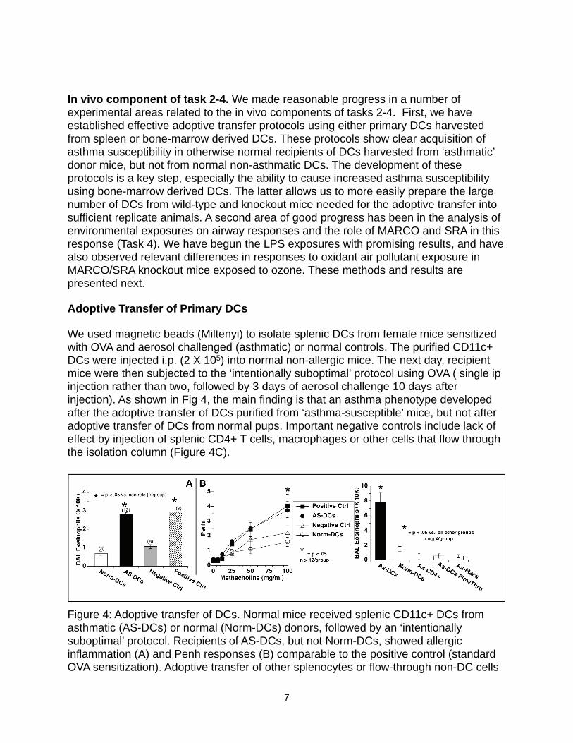
We used magnetic beads (Miltenyi) to isolate splenic DCs from female mice sensitized with OVA and aerosol challenged (asthmatic) or normal controls. The purified CD11c+ DCs were injected i.p. (2 X 10⁵) into normal non-allergic mice. The next day, recipient mice were then subjected to the ‘intentionally suboptimal’ protocol using OVA ( single ip injection rather than two, followed by 3 days of aerosol challenge 10 days after injection). As shown in Fig 4, the main finding is that an asthma phenotype developed after the adoptive transfer of DCs purified from ‘asthma-susceptible’ mice, but not after adoptive transfer of DCs from normal pups. Important negative controls include lack of effect by injection of splenic CD4+ T cells, macrophages or other cells that flow through the isolation column (Figure 4C).
Figure 4: Adoptive transfer of DCs. Normal mice received splenic CD11c+ DCs from asthmatic (AS-DCs) or normal (Norm-DCs) donors, followed by an ‘intentionally suboptimal’ protocol. Recipients of AS-DCs, but not Norm-DCs, showed allergic inflammation (A) and Penh responses (B) comparable to the positive control (standard OVA sensitization). Adoptive transfer of other splenocytes or flow-through non-DC cells
7

had minimal effect (C).
Immunophenotyping of DCs. To evaluate the surface expression of MHC-II and relevant co-stimulatory molecules and subpopulation markers in the DCs from two groups, flow cytometric analysis was performed using a FACSCanto II (Beckton-Dickinson) flow cytometer with dedicated data acquisition system and software for analysis. Labelling was performed at 4 0C for 40 minutes, in presence of 20% normal mouse serum to block non-specific binding. Concentration of labelling antibody depended on the manufacturer's instructions (Miltenyi Biotec, Auburn, CA; or eBioscience, San Diego, CA). Specificity of labeling was verified using isotype controls.We found no significant differences in surface expression of these markers: both for NoBa and AsBa the CD11c+ cells were about 95 % positive for MHC-II, 83% positive for CD86 and 25% positive for CD8a. A summary of these data is shown in Figure 5.
Figure 5
Adoptive Transfer of Cultured DCs.
To facilitate adoptive transfer of DCs from SRA knockout mice, we sought to develop a source from bone-marrow derived DCs. Moreover, since cytokine skewing of DC towards Th1- versus Th2-inducing DC (DC1 and DC2, respectively) has been studied by many laboratories, we also investigated this protocol to increase our chances of testing the influence of SRA receptors in a DC2 type population.
Dendritic cell isolation and differentiation:We followed a protocol designed to produce DC0, DC1, and DC2 subtypes of DCs. Bone marrow progenitor cells were isolated from normal adult (8-12 wk) Balb/c mice and cultured in 6-well tissue culture plates at 3e6/well in RPMI-10 (RPMI 1640 + 10%
8

FBS + 1 mM l-glutamine + penicillin/streptomycin) with 20 ng/ml rGM-CSF + 20 ng/ml rIL-3 (Peprotech) at 37° C, 5% CO2. To these cultures were added no cytokines (DC0), 20 ng/ml IFNγ + 20 U/ml rIL-12 (DC1), or 20 ng/ml rIL-4 (DC2). Media was changed every 2 d without disturbing the cells. On day 4 of culture, loosely adherent cells were removed by pipetting, washed twice in PBS, and counted. DC were resuspended in PBS at 2e6 live cells/ml (by trypan blue exclusion), where dead cells typically constituted less than 20% of total cells. DC were adoptively transferred to normal P4 Balb/c pups by i.p. injection of 100 µl cells (2e5 DC). Remaining DC were resuspended in Facs Buffer (PBS + 10 mM HEPES + 2 mM EDTA + 0.5% BSA) and stained for surface markers using specific labeled antibodies against CD11c, CD11b, CD40, CD80, CD86, MHC-I, and MHC-II, or isotype controls (all antibodies obtained from eBioscience).
Figure 6: Adoptive transfer of in vitro bone marrow-derived dendritic cells. Normal mice received DCs differentiated to favor DC0, DC1 or DC2 phenotypes, and were subjected to the ‘intentionally suboptimal’ protocol. Recipients of DC2, but not DC0 or DC1 cells, showed enhanced AI (A) and Penh responses (B), consistent with re-creation of the ‘asthma-susceptible’ DC phenotype.
Gene expression and Epigenomic profiling of asthma-susceptible vs normal DCs. To determine if pro-allergic DCs were linked to increased expression of SRAs and other related genes, we performed microarray analysis of total cell RNA isolated from asthma-susceptible or control DCs. Repeated trials using two different platforms (Illumina, Affymetrix) did not reveal any substantial or reproducible changes. Most fold-changes for genes were small e.g (1.3X) and the list of genes showing these small differences (~20-200 depending on stringency) was completely different in the datasets produced by the two platforms. (data not shown). We concluded that there was no difference in expression and that the small differences observed were ‘noise’.
9

To determine if epigenetic differences in regulation of genes could contribute to the different phenotype of the pro-asthmatic vs. normal DCs. we conducted genome-wide DNA methylation analysis.! Epigenome-wide DNA methylation scanning was performed by Switchgear Genomics (Menlo Park, CA). This method exploits sensitivity to restriction endonucleases conferred by the absence of methylation on cytosine residues. A cocktail of methylation-sensitive nucleases (with 99.9% efficiency and specificity founmethylated DNA) is used to digest half of each genomic DNA sample, prior to differential fluorescent labeling (Cy3 green for the treated sample; Cy5 red for theuntreated). Competitive hybridization is performed onto tiled arrays that cover the human genome, resulting in changes in the ratio of fluorescence depending onmethylation-driven nuclease sensitivity. (Decreased methylation leads to increased binding of the red-labeled fragments). The competitive hybridization uses a 400,000 oligo probe array which measures methylation status at ~42,000 unique regions in the mouse genome; 92% of the ~22,000 predicted CpG islands are covered, and 20,000 additional CpG-rich regions that are not annotated as CpG islands are also covered. The array includes 1,000 negative control regions (absent of CpGs) to build an error model for analysis.! For epigenomic data, the Switchgear Genomics company provides a normalized data matrix containing zscores. The data transformation was done as follows. The log2 ratio (untreated-cy5/treated-cy3) was calculated for each probe. The sample treatment decreases the signal from unmethylated DNA but does not change the signal from methylated DNA. Therefore a large ratio means the target is unmethylated in the sample whereas a log2 ratio of 0 means the target is methylated. The median log2 ratio of the negative control probes was subtracted from the experimental and control ratios to center the data. The data were then smoothed by averaging across a sliding window of 3 neighboring probes shifting 1 probe at a time. This helps to minimize noise from single probes. The negative control probes (designed to regions that are not affected by the treatment) serve as a background distribution to assign a confidence limit to eachexperimental probe. The mean and standard deviation were calculated from the negative control probes for each sample. These statistical measures were then used to calculate a z-score for each experimental probe within that experiment. The z-score was calculated as:! [(exp log2 ratio)-(mean log2 ratio of negs)] / (stdev log2 ratio of negs). This statistical measure takes into account and normalizes for the variation in each individual sample, thus making comparisons between samples more reliable. Each z-score measure is the number of stdev units from the mean of the negative control distribution. Based on the area under the curve of a standard distribution, a z-score of 1.6 means that there is 95% chance that the measurement lies outside of the negative control distribution and therefore is unmethylated.! Expression or methylation data values from experimental groups were further compared in TM4 package via several high-level methods, including SignificanceAnalysis for Microarrays (SAM) and/or Analysis of Variance (ANOVA). SAM is initially performed at false discovery rate (FDR) rate of 0% for maximum stringency; p-value stringency for ANOVA-based comparisons will include several levels (e.g. 0.05, 0.01, 0.005) depending on the size of the output list of sites and the desired flexibility ofdownstream functional enrichment analysis. Additionally cluster analysis (hierarchical
10

clustering, HCL) was performed to identify interrelated traits of expression or methylation changes across the groups.
Entire outputs and raw data of the DNA methylation arrays are publicly available as NCBIʼs Gene Expression Omnibus (GEO) database record # GSE13380, which includes information on approximately 450,000 50-mer probes. This is the firstsubmission of such genome-wide methylation data to our knowledge.
Genomic DNA from the pro-asthmatic vs. normal DCs showed remarkable differences in the degree of methylation at a number of CpG sites throughout the genome, including both annotated CpG islands and other locations. These findings areillustrated in Figure 7, which shows a subset of a larger hierarchical clustering data set, presented as a heatmap comparing the methylation status of DC DNA from pro-asthmatic vs. normal DCs. Panel 7B (right) illustrates the fold difference in normalized fluorescence of the top 40 di ferentially methylated sites.
Figure 7:
11

We next evaluated the overall level of methylation or demethylation, using the sites identified as significantly d ferent between the two groups. This list can be larger or smaller depending on the stringency of statistic criteria (p-value cutoff 0.05 vs 0.005, etc). However independent of the stringency we found that overall there was a higher level of methylation in the DCs of asthma-susceptible neonates compared to that of controls (which were, respectively, more demethylated; Figure 8A). As shown in Figure 8D, each of the samples in the ʻasthma-susceptibleʼ group had higher level of methylation; concordantly, the overall number of demethylated sites in all 9 samples was higher in the normal group (not shown)
Figure 8A! ! ! ! ! ! ! Figure 8B
The exciting part of these findings is the novel observation that di ferences in how dendritic cells behave in terms of allergic predisposition can be linked to epigenetic control mechanisms. Although many differences were observed, none were found in the area of the genes for the SRAs. This does not exclude a regulatory role but additional studies are needed to sort this out. One limitation of these studies is that the gene expression analyses used resting dendritic cells. It is possible and worthy of investigation that activated DCs would in fact show a distinct profile in the pro-asthmati vs. normal groups.
Task 3: Determine role of SRAs on AMs in responses to inhaled allergen: This work sought to test the hypothesis that AM SRAs also mediate down-regulation of immune responses to allergens by use of adoptive transfer experiments. Pilot experiments failed to show any effect of wild type or knockout AMs at the doses used. We put these unpromising studies on hold to concentrate on the dendritic cell work, which did allow the productive results shown above. Because the work for in vitro with dendritic cells took more time and effort than anticipated, we were not able to make further progress on this aim.
12

Task 4: Modulation of SRAs and Air Pollution ResponsesOur goal in these experiments was to expose allergic mice to inhaled air pollutants to investigate how pollutant-generated oxidant stress modulates SRAs and how that, in turn, could affect asthma severity. For air pollution studies, we made significant progress using ozone as a model oxidant. The data obtained using ozone support the hypothesis that down-regulation of SRAs will lead to harmful increased acute inflammation.
As shown in Fig. 9, oxidant pollutant exposure increased MARCO expression at the mRNA level, using both array and PCR analysis, and at the protein level (Western blot and cell surface analysis). Additional experimentation has shown that both MARCO and SRA KO mice demonstrate increased acute inflammation in response to the mild ozone challenge used here, linked to defective clearance of pro-inflammatory oxidized phospholipids, such cholesterol epoxide or oxidized surfactant phospholipids (PON-GPC) in the absence of the scavenger receptors Fig. 10 below.
13
Figure 10: MARCO decreases inflammation in lungs of mice exposed to β-epoxide or PON-GPC i.t. BAL samples obtained from MARCO–/– mice 7 hours after i.t. instillation of 1 μg β-epoxide (Exp) or PON-GPC (PON) showed higher levels of neutrophils (A), MIP-2 (B), and total protein (C) compared with controls. Number of mice per group is shown for each bar. *P < 0.05 versus respective treated MARCO+/+ group; #P < 0.05 versus

We have begun studies using exposure to solid air pollutants. As shown in Fig. 11 (left), we find similar enhanced acute inflammation in MARCO deficient mice exposed to air pollution samples. However, this difference is only evident when the soluble component of the air pollution particles is instilled, a finding that is under current further investigation. Many of these findings are detailed in the publication which is Appendix 2.
Key Research Accomplishments: • Completed experiments to compare random migration and directed chemotaxis
in dendritic cells from wild-type and MARCO-deficient mice
• Completed experiments to compare cell surface immunophenotype before and after in vitro maturation with TNF and LPS in dendritic cells from wild-type and MARCO-deficient mice
• Established system for adoptive transfer experiments in vivo to test ability of wild-type vs. knockout dendritic cells to alter susceptibility to asthma
• Established in vitro system to generate TH2 vs TH1 skewing dendritic cells to more efficiently test ability of wild-type vs. knockout dendritic cells to alter susceptibility to asthma in adoptive transfer experiments
• Identified novel epigenetic differences in pro-asthmatic dendritic cells vs. normal dendritic cells
• Confirmed predicted increased sensitivity of SRA deficient mice (MARCO and SRA) to inflammation caused by oxidant pollutants with gaseaous ozone and identified mechanism as delayed clearance of pro-inflammatory oxidized lipids.
Reportable Outcomes:Three publications related to this research are:
1! Arredouani MS, Franco F, Imrich A, Fedulov A, Lu X, Perkins D, et al. Scavenger Receptors SR-AI/II and MARCO limit pulmonary dendritic cell migration and allergic airway inflammation. J Immunol. 2007;178(9):5912-22.! Dahl M, Bauer AK, Arredouani M, Soininen R, Tryggvason K, Kleeberger SR, et al. Protection against inhaled oxidants through scavenging of oxidized lipids by macrophage receptors MARCO and SR-AI/II. J Clin Invest. 2007;117(3):757-64
14

3. Sulahian TH, Imrich A, Deloid G, Winkler AR, Kobzik L. Signaling pathways required for macrophage scavenger receptor-mediated phagocytosis: analysis by scanning cytometry. Respir Res. 2008;9:59
Abstract related to this work
1. Scavenger receptors and resistance to allergies L. Kobzik. presented at Military Health Research Forum, Aug/Sept 2009, Kansas City, MO
15! The results of our studies indicate that scavenger receptors modulate the allergic response, primarily by modifying dendritic cell trafficking to thoracic lymph nodes in thesetting of asthma. We found that dendritic cells deficient in the scavenger receptorsSRA I/II or MARCO traveled to the lymph nodes faster, and generated a more robust and intense asthmatic response when allergen was delivered into the lungs. We also observed that scavenger receptors are linked to oxidant air pollution responses in a complex manner. Oxidant air pollution increases expression of MARCO which serves to scavenge oxidized pro-inflammatory lipids, thereby dampening, but not eradicating,acute inflammatory responses. e also developed scanning cytometry methods for measuring macrophage phenotype in a high-throughput manner which will enable future studies. We plan to continue two especially interesting aspects of these studies. First, the epigenetic control of dendritic cell and macrophage function, and how this might impact lung host defense against allergens, merits further investigation. We are continuing these studies, stimulated by the findings above. Second, the ability of modulation of SRAs by oxidants suggests that discovery of other drugs or agents to modulate these receptors could be of therapeutic value. We are pursuing screening platforms, such as those illustrated in Appendix 3, to search for increased SRA expression after drug modulation.
Appendices: Three publications related to this research are attached as appendices:
1! Arredouani MS, Franco F, Imrich A, Fedulov A, Lu X, Perkins D, et al. Scavenger Receptors SR-AI/II and MARCO limit pulmonary dendritic cell migration and allergic airway inflammation. J Immunol. 2007;178(9):5912-22.! Dahl M, Bauer AK, Arredouani M, Soininen R, Tryggvason K, Kleeberger SR, et al. Protection against inhaled oxidants through scavenging of oxidized lipids by macrophage receptors MARCO and SR-AI/II. J Clin Invest. 2007;117(3):757-643. Sulahian TH, Imrich A, Deloid G, Winkler AR, Kobzik L. Signaling pathways
required for macrophage scavenger receptor-mediated phagocytosis: analysis by scanning cytometry. Respir Res. 2008;9:59
15

Personnel supported by this grant:
Lester Kobzik, P.I.M. ArredouaniT. SulahianR. LimA. FedulovA. Bedugnis
16

Scavenger Receptors SR-AI/II and MARCO Limit PulmonaryDendritic Cell Migration and Allergic Airway Inflammation1
Mohamed S. Arredouani,2,3* Francesca Franco,3†‡ Amy Imrich,* Alexey Fedulov,*Xin Lu,§ David Perkins,¶ Raija Soininen,� Karl Tryggvason,# Steven D. Shapiro,†
and Lester Kobzik4*
The class A scavenger receptors (SR-A) MARCO and SR-AI/II are expressed on lung macrophages (M�s) and dendritic cells(DCs) and function in innate defenses against inhaled pathogens and particles. Increased expression of SR-As in the lungs of micein an OVA-asthma model suggested an additional role in modulating responses to an inhaled allergen. After OVA sensitizationand aerosol challenge, SR-AI/II and MARCO-deficient mice exhibited greater eosinophilic airway inflammation and airwayhyperresponsiveness compared with wild-type mice. A role for simple SR-A-mediated Ag clearance (“scavenging”) by lung M�swas excluded by the observation of a comparable uptake of fluorescent OVA by wild-type and SR-A-deficient lung M�s and DCs.In contrast, airway instillation of fluorescent Ag revealed a significantly higher traffic of labeled DCs to thoracic lymph nodesin SR-A-deficient mice than in controls. The increased migration of SR-A-deficient DCs was accompanied by the enhancedproliferation in thoracic lymph nodes of adoptively transferred OVA-specific T cells after airway OVA challenge. The dataidentify a novel role for SR-As expressed on lung DCs in the down-regulation of specific immune responses to aeroallergensby the reduction of DC migration from the site of Ag uptake to the draining lymph nodes. The Journal of Immunology, 2007,178: 5912–5920.
T he lung is constantly exposed to numerous inhaled parti-cles and pathogens and relies on the broad ligand speci-ficity of scavengers and other pattern recognition recep-
tors for innate immune defense (1–3). The scavenger receptor(SR)5 family includes two members in the SR-A subclass that areexpressed on lung macrophages (M�s) and dendritic cells (DCs),MARCO (M� receptor with collagenous structure), and SR-AI/II(SR-A, types I and II) (1, 2, 4). MARCO, like SR-AI/II, binds
acetylated low-density lipoprotein and bacteria but not yeast (5–7).MARCO and SR-AI/II expressed on alveolar macrophages func-tion to promote the uptake and clearance of inhaled particles andbacteria (7–10).
Aeroallergens constitute another common inhaled challengeto the lung’s immune cells. Stimulated in part by gene expres-sion profiling that shows increased expression of MARCO andSR-AI/II in the lungs of mice in an OVA-asthma model, wesought to determine whether SR-As contributed to defense ofthe lung against inhaled allergens using receptor-deficient miceand a model of allergic asthma. We found that sensitized micelacking SR-A develop more severe airway inflammation andairway hyperresponsiveness (AHR) in response to inhaledaeroallergen. Because SR-As mediate macrophage binding andclearance of modified proteins, we initially expected that de-creased clearance of Ag (OVA) by SR-A-deficient alveolarmacrophages (AMs) would be a mechanism for increased al-lergic responses, but this postulate proved incorrect. We nextevaluated the effect of SR-A-deficiency on the ability of Ag-loaded pulmonary DCs to migrate to the draining lymph nodes(LNs) and generate specific T cell responses. The data indicatethat MARCO and SR-AI/II function in a novel mechanism todown-regulate migration of pulmonary DCs to thoracic LNs andthereby diminish T cell responses to specific aeroallergens.
Materials and MethodsAnimals
Six- to eight-week-old mice genetically deficient in MARCO(MARCO�/�) or SR-AI/II (SR-AI/II�/�) were used in all experiments.Age- and sex-matched wild-type (WT) (C57BL/6 and BALB/c) micepurchased from Charles River Laboratories were used as controls.MARCO�/� mice were generated using targeted homologous recombina-tion (9) and were backcrossed for at least 10 generations to the C57BL/6background. SR-AI/II�/� mice were generated by disrupting exon 4 of theSR-A gene, which is essential for the formation of functional trimeric
*Department of Environmental Health, Harvard School of Public Health, Boston, MA02115; †Pulmonary and Critical Care Medicine, Brigham and Women’s Hospital,Harvard Medical School, Boston, MA 02115; ‡Clinica di Malattie dell’Apparato Res-piratorio, Dipartimento Integrato di Oncologia ed Ematologia, Universita degli Studidi Modena e Reggio Emilia, Modena, Italy; §Department of Family and PreventiveMedicine, University of California San Diego, CA 92101; ¶Department of Medicine,University of California San Diego, CA 92101; �Department of Medical Biochemistryand Molecular Biology, Biocenter Oulu, University of Oulu, Oulu, Finland; and #De-partment of Medical Biochemistry and Biophysics, Karolinska Institutet, Stockholm,Sweden
Received for publication September 5, 2006. Accepted for publication February9, 2007.
The costs of publication of this article were defrayed in part by the payment of pagecharges. This article must therefore be hereby marked advertisement in accordancewith 18 U.S.C. Section 1734 solely to indicate this fact.1 This work was supported by National Institutes of Health Grants ES0002 andES11008, and by Department of Defense Grant W81XWH-06-1-0289. M.S.A. is arecipient of the Jere Mead fellowship.2 Current address: Beth Israel Deaconess Medical Center, Harvard Medical School,77 Avenue Louis Pasteur, HIM-1039, Boston, MA 02115.3 M.S.A. and F.F. contributed equally to this work.4 Address correspondence and reprint requests to Dr. Lester Kobzik, Department ofEnvironmental Health, Harvard School of Public Health, 665 Huntington Avenue,Boston, MA 02115. E-mail address: [email protected] Abbreviations used in this paper: SR, scavenger receptor; SR-AI/II, SR class A, typeI and type II; AHR, airway hyperresponsiveness; AM, alveolar macrophage; BAL,bronchoalveolar lavage; BALF, BAL fluid; Ct, threshold cycle; C3H, C3H/HeJ; DC,dendritic cell; i.t., intratracheal; KO, knockout; LN, lymph node; MARCO, macro-phage receptor with collagenous structure; M�, macrophage; Penh, enhanced pause;WT, wild type.
Copyright © 2007 by The American Association of Immunologists, Inc. 0022-1767/07/$2.00
The Journal of Immunology
www.jimmunol.org

receptors (11). Double knockout (KO) mice were obtained in our labora-tory by intercross of single KO mice.
Both single KO mice were backcrossed in our laboratory to the BALB/cbackground for eight generations. DO11.10 mice, which are transgenic forthe TCR recognizing OVA peptide 323–339, A/J, C3H/HeJ (C3H), andC3H/HeOuJ mice were from The Jackson Laboratory. All animals werehoused in sterile microisolator cages and had no evidence of spontaneousinfection. Approval before all experimentation was obtained from HarvardSchool of Public Health institutional animal use review committee.
Mouse model of airway allergic inflammation1
To compare allergic responses in SR-A and normal mice, groups ofMARCO�/�, SR-AI/II�/�, and C57BL/6 WT control mice were sensitizedi.p. with 8 �g OVA in 1 mg of alum gel in 0.5 ml of PBS on days 0 and7. On day 14 the sensitized mice were challenged with aerosolized 0.5%OVA or PBS for 1 h. Mice were sacrificed 72 h postchallenge, blood wascollected through heart puncture, and the lungs were lavaged with PBSbefore they were harvested, inflated with formalin, and processed for his-tologic analysis.
In BALB/c mice, 10 �g OVA in 2 mg of Alum powder were adminis-tered i.p. at days 0 and 7 followed by aerosol challenge with either PBS or1% OVA for 30 min on days 14 and 15.
Microarray data analysis
Microarray data was acquired from the Public Expression Profiling Re-source (http://pepr.cnmcresearch.org; see project 108). Gene expressiondata were calculated by using the GeneChip-Robust Multiarray Analysisalgorithm (12) from the Bioconductor project (http://www.bioconductor.org/). The fold change in MARCO and SR-A gene expression was calcu-lated as the ratio of the level in the OVA/control sample.
The raw p values were adjusted by false discovery rate correction and anadjusted p value �0.05 was interpreted as significant.
RT and real-time PCR
Total lung RNA was extracted from normal and OVA-sensitized and chal-lenged BALB/c mice using a Qiagen RNAeasy kit according to manufac-turer’s instructions. RNA purity was controlled by OD measurement. RNAconcentrations were adjusted and the samples were reverse transcribed tocDNA using the novel SuperScript III first-strand cDNA synthesis kit (In-vitrogen Life Technologies). cDNA samples were analyzed in duplicate ina quantitative real-time PCR using the SYBR Green Supermix (Bio-Rad)for MARCO and SR-A message with the following primer sequences (In-tegrated DNA Technologies): SR-A sense (5�-AGAATTTCAGCATGGCAACTG-3�) and SR antisense (5�-ACGGACTCTGACATGCAGTG-3�); and MARCO sense (5�-GAAACAAAGGGGACATGGG-3�) andMARCO antisense (5�-TTCACACCTGCAATCCCTG-3�). Murine�-actin was used as housekeeping control and a no-template sample wasused as a negative control. Data are represented as �Ct (threshold cy-cle) values, with the lower values indicating a greater abundance ofmRNA in the sample.
Measurement of airway hyperresponsiveness
AHR was measured in MARCO�/�, SR-AI/II�/�, and BALB/c WT miceusing whole body plethysmography (Buxco; EMKA Technologies) 24hafter the last of two daily OVA or PBS aerosol challenges. The response ofthe airways to inhaled methacholine (Sigma-Aldrich) at concentrationsranging from 6.25 to 100 mg/ml (13) was recorded. AHR was expressed asenhanced pause (Penh), a calculated value that correlates with airwayresistance.
Bronchoalveolar lavage (BAL)
BAL was performed in situ with a 20-gauge catheter inserted into theproximal trachea, flushing the lower airways six times with 0.8 ml of PBS.The fluid retrieved from the first flushing was kept for ELISA. The BALfluid cells were separated from the BAL fluid by centrifugation, resus-pended in PBS, and counted and a fraction was cytospun on microscopicslides for staining with Diff-Quick (Baxter Scientific Products) for subse-quent leukocyte differential counts.
OVA uptake studies
To prepare the OVA-FITC conjugate, OVA was dissolved in carbonatebuffer (pH 9.2). Freshly prepared FITC in DMSO (10 mg/ml) was added ata ratio of 10 mg per 200 mg of OVA and the mixture was incubated at
room temperature in the dark for 1 h. To remove free FITC, the mixturewas dialyzed for 24 h against PBS. MARCO�/�, SR-AI/II�/�, andC57BL/6 mice were given a 15-min aerosol treatment with a 10 mg/mlsolution of OVA-FITC. BALs were performed 1 h later and cells wereanalyzed by flow cytometry. To test the binding of OVA to AMs in vitro,BAL fluid (BALF) cells (200 � 103/well) from C57BL/6 WT and doubleKO mice were pretreated for 5 min with 5 �M cytochalasin D and thenincubated with 5 �g/ml OVA-Alexa Fluor 488 for 40 min at 37°C andanalyzed by flow cytometry.
Instillation of macromolecule solutions into the trachea
Mice were anesthetized by i.p. injection of 2.5% Avertin and received anintratracheal (i.t.) injection of 600 �g of OVA-FITC in a volume of 60 �lof sterile PBS. The trachea was carefully exposed via a small midlineincision and the solution was inoculated. The incision was then closed withsterile silk and the mice were allowed to fully recover before being re-turned to the cages.
Preparation of single-cell suspensions and immunofluorescentlabeling
Lung digestion medium consisted of RPMI 1640 (from Invitrogen LifeTechnologies) supplemented with 1 mg/ml collagenase type IV (Sigma-Aldrich) and 0.5 mg/ml DNase (deoxyribonuclease I from a bovine pan-creas; Sigma-Aldrich). LN digestion medium consisted of 1 � HBSS(Cellgro; Mediatech) and 2% EDTA-treated FBS (HyClone) supplementedwith 2.5 mg/ml collagenase type IV (Sigma-Aldrich). EDTA-treated FBSwas prepared by adding 20 �l of 0.5 M EDTA per ml of FBS. FACSstaining buffer consisted of PBS (free of Ca2� or Mg2�) supplemented with5% FBS, 0.1% sodium azide, and 5 mM EDTA.
Preparation of lung and LN single-cell suspensions
Lung. Animals were euthanized by CO2 narcosis. Following a thoracot-omy, right heart catheterization was performed using a 21-gauge, 0.75-inchsiliconized needle (SURFLO winged infusion set; Terumo) and the pul-monary circulation was perfused with at least 20 ml of sterile PBS toremove the intravascular pool of cells. Two milliliters of digestion me-dium were then injected in the trachea using a 22-gauge catheter and thetrachea was quickly sealed with silk suture after the catheter was re-moved. The trachea and lungs were then removed and the lungs werecarefully separated from the heart, thymus, and trachea and incubated at37°C in additional 3 ml of digestion medium for 30 min. Incubation wasthen prolonged for an additional 30 min with vigorous pipetting of thesamples at 10-min intervals with a 5-ml serological pipet. Subsequently,samples were passed through a 70-�m nylon cell strainer, subjected toRBC lysis, incubated in calcium- and magnesium-free PBS containing10 mM EDTA for 5 min at room temperature on a shaker, and finallyresuspended in FACS staining buffer and kept on ice until immunoflu-orescent labeling.Lymph nodes. For migration studies, animals were euthanized by CO2
narcosis 24 h after an i.t. injection of OVA-FITC. For T cell proliferationstudies, animals were euthanized by CO2 narcosis 96 h after the injectionof OVA. Following a thoracotomy, paratracheal and parathymic intratho-racic LNs were removed under a stereo microscope (Olympus SZ 60) andincubated at 37°C in 3 ml of LN digestion medium. After 10 min ofincubation, LNs were minced with 20-gauge, 1.5-inch and 25-gauge,0.625-inch needles (BD Biosciences) and incubation was prolonged foranother 10 min. Subsequently, samples were passed through a 7-�mnylon cell strainer, incubated in calcium- and magnesium-free PBS con-taining 10 mM EDTA for 5 min at room temperature on a shaker, andfinally resuspended in FACS staining buffer and kept on ice until im-munofluorescent labeling.
Labeling of single cell suspensions for flow cytometry
All staining procedures were performed at 4°C. Cells were preincubated for20 min with a Fc receptor blocking Ab (anti-CD16/CD32; BD Biosciences)to reduce nonspecific binding. For lung studies, cells were subsequentlystained with a PE-Cy5.5-conjugated hamster anti-mouse CD11c mAb(Caltag Laboratories) and data acquisition was performed using the FL1/FL3 template to allow assessment of the distribution of CD11c-bright cellswith regard to autofluorescence. A PE-Cy 5.5-conjugated hamster IgG iso-type control was used to determine background staining (Caltag Labora-tories). Rat anti-F4/80 (IgG2a; clone 6F12) and rat anti Mac-3 (IgG1; cloneM3/84) were from BD Biosciences. For migration studies, cells werestained with a PE-Cy5.5-conjugated hamster anti-mouse CD11c mAb(Caltag Laboratories). For T cell proliferation studies, cells were stained
5913The Journal of Immunology

with PE mouse anti-mouse DO11.10 TCR mAb (clone KJ1-26) (CaltagLaboratories). A PE-conjugated mouse IgG2a isotype control was used todetermine background staining (Caltag Laboratories). Flow cytometry dataacquisition was performed on a BD FACScan running CellQuest software(BD Biosciences). Flow Jo software (Tree Star) was used for data analysis.For lung and migration studies 50,000 total events were acquired for eachsample. For T cell proliferation studies 500,000 total events were acquiredfor each sample. Dead cells were gated out based on light scatterproperties.
In vivo assessment of T cell proliferation
CD4� T cells were enriched from the spleens of DO11.10 mice by mag-netic bead separation under sterile conditions using a mixture of biotin-conjugated mAbs against CD8a (rat IgG2a; Ly-2), CD11b (rat IgG2b;Mac-1), CD45R (rat IgG2a; B220), CD49b (rat IgM; DX5), and Ter-119(rat IgG2b), followed by anti-biotin microbeads (colloidal superparamag-netic microbeads conjugated to a monoclonal anti-biotin Ab, mouse IgG1;clone Bio3-18E7.2) (Miltenyi Biotec). CD4� DO11.10 T cells were sub-sequently labeled with 10 �M CFSE (Sigma-Aldrich) at 37°C for 10 minas described by Lyons et al. (14) and then resuspended in sterile PBS. Micereceived an i.v. injection of 10 � 106 CFSE-labeled DO11.10 T cells 24 hbefore an i.t. injection of 600 �g of OVA in a volume of 60 �l of PBS.Four days later T cell responses were analyzed in the draining mediastinalLNs by observing the CFSE division profiles of live KJ1-26� CD4� Tcells. The number of transgenic T cells in each LN was calculated aspercentage of KJ1-26�CFSE� cells among the total cell number.
Statistical analysis
Student’s t test (unpaired, two-tailed) was used to calculate significancelevels for all measurements. Data are presented as mean � SEM or SD.Differences were considered significant when p � 0.05.
ResultsIncreased MARCO and SR-AI/II gene expression in a murinemodel of asthma
To identify genes modulated in asthma, we analyzed publicdatabases of microarray expression profiling in experimentalmurine asthma models. In a project conducted by M. Wills-
Karp (Cincinnati Children’s Hospital Medical Center, Cincin-nati, OH; data openly available online at the Public ExpressionProfiling Resource (http://pepr.cnmcresearch.org/)), the re-sponse to OVA exposure at 6 and 24 h following allergen chal-lenge in both the A/J and C3H strains was determined by usingfive replicates of whole lung RNA from each experimentalgroup. We processed the data as described in Materials andMethods. A comparison of allergen-challenged mice to saline-chal-lenged mice revealed a significant up-regulation of MARCO and SR-AI/II after exposure to OVA in both strains (Fig. 1, A and B). A
FIGURE 1. Augmented expression of MARCO and SR-AI/II in allergicasthma. Microarray analysis of RNA transcripts for MARCO (A) and SR-AI/II (SRA) (B) was performed on asthmatic and control A/J (AJ) and C3Hmice at 6 h and 24 h after allergen or PBS challenge. Data are expressedas mean � SD of normalized gene expression obtained from five mice.�, p � 0.05 vs same time point after PBS challenge.
FIGURE 2. BAL cell yields after OVA-sensitization and challenge.Control (C57BL/6 or BALB/c), MARCO�/� and SR-AI/II�/� (SRA) micewere sensitized twice with OVA in alum and exposed once to 1% aero-solized OVA (�OVA). As a negative control, mice were immunized withOVA and challenged with PBS aerosol (�PBS). Seventy-two hours afteraerosol exposure, lungs were lavaged and then fixed in formalin for H&Estaining. Total leukocyte counts were determined in BALF of MARCO�/�,SR-AI/II�/�, and their WT counterparts in both C57BL/6 (A and B) andBALB/c (C) backgrounds. Data shown here are representative of 18 (A), 12(B), and 6 mice (C) per group. ��, p � 0.01; ���, p � 0.001; for OVA vsPBS challenge.
5914 CLASS A SCAVENGER RECEPTORS AND ALLERGIC ASTHMA

similar trend was found in studies using the C57BL/6 strain(search for GDS348 on the Gene Expression Omnibus DataSetssite: http://www.ncbi.nlm.nih.gov/entrez/query.fcgi?dbgds).
We also observed increased MARCO and SR-A gene expres-sion in RT-PCR analysis of lung samples from OVA-sensitizedand exposed mice compared with controls (e.g., �CT values for
FIGURE 3. Response to OVA challenge in WT vs SR-A-deficient mice. Differential counts showing the amounts of BALF macrophages, eosinophils, andlymphocytes were determined on stained cytospin slides from MARCO�/�, SR-AI/II�/�, and their WT counterparts in both C57BL/6 (A and B) and BALB/cbackgrounds (E). Data shown here are representative of 18 mice per group (A), 12 mice per group (B), and 6 mice per group (E). �, p � 0.05; ��, p � 0.01; and���, p � 0.001; for OVA vs PBS challenge. Representative photomicrographs of tissue histopathology are shown from WT (C and D), MARCO�/� (C), andSR-AI/II�/� (D) mice challenged with either PBS or OVA. Inset (C, lower right panel) shows higher magnification (�600) of eosinophils and mononuclear cellscomprising the inflammatory infiltrates. Images are shown at a �200 original magnification for MARCO�/� and �100 for SR-AI/II�/� mice. SR-A-deficient micealso show enhanced airway hyperreactivity (F) as measured by whole body plethysmography in conscious OVA-sensitized and challenged BALB/c mice. Penh,an index of airway obstruction, was recorded after aerosolization of either PBS or 50 mg/ml methacholine. Data represent mean Penh values � SEM (six miceper group for OVA/OVA and three mice per group for OVA/PBS). #, p � 0.05 vs PBS challenge; ��, p � 0.01 vs WT mice.
5915The Journal of Immunology

OVA vs controls were 5.3 � 0.6 vs 6.6 � 0.4 (SR-A) and 7.2 �1.1 vs 9.2 � 1.5 (MARCO); lower �CT values indicate agreater abundance of mRNA targets).
Increased severity of airway inflammation in SR-A deficient miceduring allergic asthma
We next directly analyzed the physiologic relevance of the twoSR-A receptors, MARCO and SR-AI/II, in vivo in a murine modelof allergic airway inflammation caused by OVA sensitization andaerosol challenge. Due to the unavailability of SR-A KO mice inthe susceptible A/J and resistant C3H backgrounds used in themicroarray studies, we used the C57BL/6 and BALB/c strains,both known to show pulmonary expression of SR-As (8, 9) andto be prone to OVA-induced airway inflammation (15, 16). Sev-enty-two hours after the aerosol challenge, sham-challengedmice (OVA/PBS groups) showed no sign of inflammation,whereas all OVA/OVA groups showed a remarkable increase inthe total number of leukocytes recruited to the airways (Fig. 2,A and B). Notably, the total number of eosinophils and lym-phocytes in the BAL samples from OVA/OVA groups was sub-stantially greater in the SR-A-deficient mice relative to theircontrol counterparts (Fig. 3, A and B). OVA/PBS mice, in con-trast, did not show any recruitment of eosinophils into theirairways. Consistent with the increased leukocyte numbers inlavage samples of OVA-challenged SR-A-deficient mice, his-tologic analysis of lungs harvested from these mice showedallergic inflammation consisting of peribronchial and perivas-cular cell infiltrates of eosinophils and mononuclear cells (Fig.3, C and D).
Unlike C57BL/6 mice, allergen-sensitized BALB/c mice de-velop easily detectable AHR following exposure to inhaled aller-gen (15, 16). MARCO�/� and SR-AI/II�/� mice on the BALB/cbackground also showed a significant increase in eosinophils andmacrophages (Fig. 3E) and total cell number (Fig. 2C) in theBALF following OVA challenge, compared with their WTcounterparts. The basis for the increased macrophage number inthe KO mice on the BALB/c background is unknown. It is alsoworth mentioning that the discrepancy in both the intensity andthe nature of cellular inflammatory responses between C57BL/6and BALB/c strains after exposure to inhaled OVA is an ex-pected result of the different induction protocols we have usedto achieve significant eosinophilic recruitment and the Ag dose-dependent response in these strains. Whole body plethysmog-raphy was used to evaluate pulmonary function changes afterOVA challenge in WT vs MARCO�/� and SR-AI/II�/� mice.Following aerosolized bronchoconstrictor (methacholine) chal-lenge, WT mice showed a slight, but significant, increase inAHR relative to the baseline ( p � 0.05). In contrast,MARCO�/� and SR-AI/II�/� mice showed a much more robustresponse ( p � 0.01; Fig. 3F), consistent with their greater al-lergic inflammatory response.
WT and SR-A-deficient lung M�s show normal uptake ofinhaled OVA allergen
AMs can efficiently bind and internalize unopsonized particles andbacteria through SR-As, leading to the clearance of inhaled matterfrom the airways and the reduction of the resulting inflammation(7–9) and are known to similarly bind modified proteins (17–19).To determine whether SR-As could reduce allergic inflammationby simply “scavenging” aeroallergen with a resulting decrease inallergen dose, we measured their ability to internalize inhaled al-lergens using FITC-OVA. WT and KO mice were exposed to in-haled fluorescent OVA, the airways were lavaged 1 h later, and thetotal fluorescence of AMs was evaluated by flow cytometry.
Similar amounts of FITC-OVA were found associated withAMs in WT, MARCO�/�, and SR-AI/II�/� mice ( p 0.05),indicating essentially identical uptake in vivo (Fig. 4A). In par-allel experiments, FITC-OVA was administered i.t. to the miceand the amount of OVA associated with the M� population wasdetermined on the cells isolated from whole lung homogenates.The total amount of FITC-OVA on M�s, as discriminated bygating of the CD11c�, F4/80�, or MAC3� populations, wassimilar in both WT and MARCO�/� mice ( p 0.05; Fig. 4B).In vitro assays confirmed that the absence of receptors did notaffect AM binding of OVA, as double-deficient AMs boundAlexa Fluor 488-OVA to nearly the same extent as did controlAMs (data not shown). These findings are consistent with pre-vious reports indicating that, unlike chemically modified albu-min, native albumin binding to M�s is not mediated throughSR-As (17–19), and they also indicate that nebulization doesnot per se denature the allergenic proteins sufficiently to createSR-A binding domains.
Allergen-loaded SR-A-deficient DCs show increased migrationfrom the lungs to the draining LNs
We next sought to investigate whether another SR-A-expressingcell type, the lung DC, was involved in the increased asthmaticphenotype seen in SR-A-deficient mice. Airway DCs capture Agsin the lungs and migrate to the regional LNs where they present theAg to the specific T cells. To track DC migration from the lungsto the draining LNs, we administered OVA-FITC i.t. and analyzedcell suspensions prepared from mediastinal LNs 24 h later. DCswere labeled with anti-CD11c Ab and the number of cells express-ing the CD11c and also carrying FITC was determined by flowcytometry. Although there are no significant differences in LN cel-lularity under basal conditions (Fig. 5A), OVA challenge of theairways resulted in an increase in LN cellularity, an increase which
FIGURE 4. Absence of SR-As on AMs does not affect the clearanceof inhaled allergen. A, Control (C57BL/6), MARCO�/�, and SR-AI/II�/� mice were challenged with aerosolized OVA-FITC (10 mg/ml) for15 min. The lungs were lavaged 1 h later with PBS and cells wereanalyzed by flow cytometry to compare the uptake of fluorescent OVA.Data shown represent the mean � SEM of four mice per group. B, Inseparate experiments, three WT and three MARCO�/� mice received600 �g of OVA-FITC i.t. The lungs were harvested 4 h later and cellsuspensions prepared by homogenization were labeled with Abs to ei-ther CD11c, F4/80, or MAC3 Ags and the mean green fluorescenceintensity (MFI) of the double positive cells was determined.
5916 CLASS A SCAVENGER RECEPTORS AND ALLERGIC ASTHMA

is greater in the SR-A-deficient mice (Fig. 5B). A striking findingwas that SR-A-deficient mice showed a significantly greater num-ber of Ag-loaded DCs in the thoracic LNs (Fig. 5C), indicating thatDC migration is more efficient in the KO mice. Double KO miceshowed an even greater migration of airway DCs after OVAchallenge compared with control mice and single deficient mice(Fig. 5D). These studies were performed in unimmunized mice.We next assessed DC migration in OVA-sensitized WT andMARCO�/� mice. Notably, although the sensitized WT miceshowed an elevated migration of airway DCs to the LNs afterOVA exposure (note the expanded range of the y-axis), the in-crease was even more marked in the MARCO�/� mice (Fig. 5E).To determine whether differences in Ag (OVA) uptake by WT orSR-A-deficient DCs could mediate the enhanced allergic re-sponses in SR-A-deficient mice, we also evaluated the OVA-FITC content of the DCs that reach the LNs after Ag challenge(measured as green fluorescence). We observed the sameamounts of Ag in the DCs reaching the LNs in both WT andMARCO�/� mice (Fig. 5F). This indicates that SR-A-defi-ciency does not alter the uptake of OVA-FITC Ag by DCs, afinding similar to data obtained with macrophages (Fig. 4). Toevaluate the potential of trace endotoxin in the OVA prepara-tion to modulate DC migration, we performed OVA-FITC in-
stillation into endotoxin-sensitive and resistant (C3H/Ouj andC3H/HeJ respectively). No differences were observed in thenumbers of migrated FITC�CD11C� DCs found in thoracicLNs in the two strains of mice (data not shown).
To evaluate the possibility that the enhanced DC migration inKO mice was due to a higher basal number of DCs in the lungs, wequantified the lung DC population in naive MARCO�/� and con-trol mice. Lung DCs were defined as bright CD11c� cells with lowautofluorescence, as described by Vermaelen and Pauwels (20).We found that the number of lung DCs was not statistically dif-ferent between MARCO�/� and their control WT mice (data notshown).
Allergen challenged SR-A deficient mice show enhanced T cellpriming in the draining LNs
To more directly test the functional significance of augmented Ag-loaded DC migration in SR-A-deficient mice, we used an adoptivetransfer model to assess T cell proliferation in the draining LNsafter Ag challenge. BALB/c WT, MARCO�/�, and SR-AI/II�/�
mice were injected i.v. with CFSE-labeled OVA-specific CD4� Tlymphocytes from DO11.10 transgenic mice. Recipient mice werechallenged i.t. with OVA 24 h later. The mediastinal LNs were
FIGURE 5. Increased migration ofMARCO�/� and SR-AI/II�/� pulmo-nary DCs in response to an inhaledAg challenge. A and B, The total cellcontent of homogenized mediastinalLNs from C57BL/6, MARCO�/�,and SR-AI/II�/� mice was deter-mined before (A) or 24 h after (B)they received 600 �g of OVA-FITCi.t. C–F, LN suspensions from OVA-FITC-challenged mice were stainedfor CD11c and the fraction of FITC�
DCs was determined by flow cytom-etry (C). A similar protocol was ap-plied to double KO (dKO) mice (D)and OVA-sensitized C57BL/6 andMARCO�/� mice (E). Also, theamount of green fluorescence associ-ated with the DCs was determined(F). Data represent the mean � SDfrom two or more separate experi-ments with at least six mice per ge-notype, �, p � 0.05; ��, p � 0.01.
5917The Journal of Immunology

harvested 96 h after OVA challenge for an analysis of dye dilutionas a function of cell division.
Comparable numbers of adoptively transferred DO11.10 T cellsreached the mediastinal LNs in all three groups of mice (data notshown) and, similarly, comparable fractions underwent at least onedivision (percentage of cells showing decreased CFSE was 91,93.5, and 92%, respectively, in WT, MARCO�/�, and SR-AI/II�/� mice). However, there was a greater proliferative response inthe LNs of MARCO�/� mice (295 � 83 � 103; mean � SD)compared with WT mice (140 � 83 � 103), indicating that ahigher absolute number of T cells had undergone a greater num-ber of divisions in the MARCO�/� mice (Fig. 6). This indicatesthat the larger numbers of Ag-loaded DCs that migrate to thedraining LNs of the MARCO�/� mice result in a greater pro-liferative response by Ag-specific T lymphocytes. SR-AI/II�/�
mice showed a similar trend that did not reach statistical sig-nificance in T cell proliferation (206 � 92 � 103) comparedwith control mice.
DiscussionThe data presented identify a novel role for SR-As expressed onlung DCs in modulating pulmonary responses to aeroallergens.The context for our findings includes the recognition of the im-portant role of DCs in the pathogenesis of asthma (21) and asprofessional APCs that bridge innate and adaptive immunity (22,23). DCs express SR-AI/II, which functions in Ag presentation andadaptive immunity (17, 19, 24–28). For example, SR-AI/II�/�
mice are deficient in mounting an efficient T cell response to ma-leylated murine serum albumin, a known SR-AI/II ligand (29). In
contrast, the role of MARCO receptors in modulating adaptiveimmunity has not been examined.
It has been postulated that MARCO expression is induced uponDC maturation (30, 31). Although we did not directly address thematuration state of pulmonary DCs in naive WT mice, we knowthe following: 1) only immature DCs can take up and process Ag(32); 2) immunohistochemical studies show the expression ofMARCO only on M�s in the normal lung (8) with an absence ofMARCO labeling in normal airways that contain CD11c� airwayDCs; and 3) mediastinal LN DCs express MARCO after OVAchallenge (data not shown). This suggests that pulmonary DCsstart to express MARCO after allergen encounter, consistent withthe increased MARCO gene expression observed after OVA chal-lenge in microarray studies.
Some limitations of the study merit discussion. For some controlexperiments, only MARCO-deficient mice were analyzed (e.g., themigration of DCs in OVA-sensitized mice; Fig. 5E). Hence, thefull extent to which SR-AI/II deficiency mirrors the findings withMARCO-deficient mice requires further characterization. One po-tential problem to be considered is the confounding effects of traceendotoxin in the OVA allergen. Two lines of evidence argueagainst this possibility. First, no differences were observed in thenumbers of migrated FITC�CD11C� DCs found in either endo-toxin-sensitive or endotoxin-resistant thoracic LNs (C3H/Ouj andC3H/HeJ respectively). Second, we have previously reported sim-ilar levels of cytokine release (TNF-� and MIP-2) by AMs fromWT and KO mice in response to LPS in vitro (9), arguing againstdifferential responses on this basis.
FIGURE 6. Enhanced Ag-in-duced T lymphocyte proliferationin mediastinal LNs of SR-A-defi-cient mice. CFSE-labeled spleenCD4� T cells from DO11.10 micewere transferred i.v. into BALB/c,MARCO�/�, and SR-AI/II�/�
mice 24 h before i.t. administrationof OVA. Ninety-six hours later,cell suspensions were preparedfrom draining LNs and stainedwith KJ-126-PE Ab. Representa-tive histograms (A; cells in the M2zone have undergone at least onedivision) and dot plots (B; cells inthe rectangles have undergone atleast one division) are shown forcontrol, MARCO�/�, and SR-AI/II�/� mouse groups. The deducedabsolute number of cells that un-derwent at least one division isshown in C. Data represent themean � SD from eight(MARCO�/�) and 12 mice (SR-AI/II�/�). �, p � 0.05.
5918 CLASS A SCAVENGER RECEPTORS AND ALLERGIC ASTHMA

In peripheral tissues such as the lungs, DCs exist normally in animmature state and provide a sentinel function for foreign Ags(32). Upon Ag encounter, DCs undergo a process of maturationthat triggers their migration to draining LNs and enhances theirAg-presenting capacity (23). The migration of Ag-loaded DCsfrom peripheral tissues to the LNs is a critical step in generating anoptimal immune response (33, 34) and, hence, a potential regula-tory point.
SR-As may inhibit DC migration through a number of mecha-nisms. SR-As have been shown to promote adhesion to matrixmolecules (35, 36) and to other cells, e.g., marginal zone macro-phages to B cells (37), and either of these interactions could po-tentially reduce cell migration. Pikkarainen et al. (38) have previ-ously shown that fibroblastic cell lines transfected with MARCOundergo significant morphologic changes through the induction ofdendritic plasma membrane processes. These processes include theappearance of large lamellipodia-like structures and long plasmamembrane extensions. Moreover, a clear correlation exists be-tween MARCO expression and the rearranged actin cytoskeletonof mature DCs (30), although in this study MARCO expressionupon maturation was associated with a decrease in filopodia and around phenotype. The morphologic changes induced by simpleMARCO expression do not require its interaction with any givenligand. The rearrangements induced by MARCO in fibroblasticcell lines were shown to be partially dependent on Rac1 (38). Rac,together with Rho and Cdc42, represent a group of small GTPasesinvolved in the formation of filopodia and podosomes in immatureDCs (39), structural changes that could increase adhesion and re-duce migration. To test the speculation that adhesion to a matrixmight mediate some of the observed effects, studies of the kineticsof induction of MARCO expression on airway and LN DCs afterallergen challenge are warranted, as well as a comparison of themigration capacity of MARCO-deficient and WT DCs. Additionalmechanisms are suggested by data indicating the ability of scav-enger receptors, when present, to skew the cytokine milieu andimmune response toward Th1-type immunity (40). Indeed, one ofthe mechanisms leading to increased AHR in allergic asthma is theenhanced recruitment of eosinophils into the allergen-challengedairways as we observed in the lungs of SR-A deficient mice. Thismay be a consequence of the increased recruitment of Th lympho-cytes and an altered cytokine milieu.
Innate immune responses are increasingly recognized as crit-ical modifiers of adaptive immunity (41, 42). In the examplepresented here, the innate pattern recognition receptors, the SR-As, mediate reduced amounts of total Ag delivery to LNs(through decreased numbers of DCs carrying similar amounts ofAg per cell). SR-A-mediated down-regulation of lung immuneresponses likely contributes to the reduction of unwanted im-mune responses to commonly encountered environmentalaeroallergens.
DisclosuresThe authors have no financial conflict of interest.
References1. Pearson, A. M. 1996. Scavenger receptors in innate immunity. Curr. Opin. Im-
munol. 8: 20–28.2. Krieger, M., and J. Herz. 1994. Structures and functions of multiligand lipopro-
tein receptors: macrophage scavenger receptors and LDL receptor-related protein(LRP). Annu. Rev. Biochem. 63: 601–637.
3. Gough, P. J., and S. Gordon. 2000. The role of scavenger receptors in the innateimmune system. Microbes Infect. 2: 305–311.
4. Arredouani, M., and L. Kobzik. 2004. The structure and function of MARCO, amacrophage class a scavenger receptor. Cell. Mol. Biol. 50: OL657–OL665.
5. van der Laan, L. J., M. Kangas, E. A. Dopp, E. Broug-Holub, O. Elomaa,K. Tryggvason, and G. Kraal. 1997. Macrophage scavenger receptor MARCO: invitro and in vivo regulation and involvement in the anti-bacterial host defense.Immunol. Lett. 57: 203–208.
6. Gough, P. J., D. R. Greaves, and S. Gordon. 1998. A naturally occurring isoformof the human macrophage scavenger receptor (SR-A) gene generated by alter-native splicing blocks modified LDL uptake. J. Lipid Res. 39: 531–543.
7. Arredouani, M. S., A. Palecanda, H. Koziel, Y. C. Huang, A. Imrich,T. H. Sulahian, Y. Y. Ning, Z. Yang, T. Pikkarainen, M. Sankala, et al. 2005.MARCO is the major binding receptor for unopsonized particles and bacteria onhuman alveolar macrophages. J. Immunol. 175: 6058–6064.
8. Palecanda, A., J. Paulauskis, E. Al-Mutairi, A. Imrich, G. Qin, H. Suzuki,T. Kodama, K. Tryggvason, H. Koziel, and L. Kobzik. 1999. Role of the scav-enger receptor MARCO in alveolar macrophage binding of unopsonized envi-ronmental particles. J. Exp. Med. 189: 1497–1506.
9. Arredouani, M., Z. Yang, Y. Y. Ning, G. Qin, R. Soininen, K. Tryggvason, andL. Kobzik. 2004. The scavenger receptor MARCO is required for normal lungdefense against pneumococcal pneumonia and inhaled particles. J. Exp. Med.200: 267–272.
10. Arredouani, M. S., Z. Yang, A. Imrich, Y. Ning, G. Qin, and L. Kobzik. 2006.The macrophage scavenger receptor SR-AI/II and lung defense against pneumo-cocci and particles. Am. J. Respir. Cell Mol. Biol. 35: 474–478.
11. Suzuki, H., Y. Kurihara, M. Takeya, N. Kamada, M. Kataoka, K. Jishage,O. Ueda, H. Sakaguchi, T. Higashi, T. Suzuki, et al. 1997. A role for macrophagescavenger receptors in atherosclerosis and susceptibility to infection. Nature 386:292–296.
12. Wu, Z., and R. A. Irizarry. 2004. Preprocessing of oligonucleotide array data.Nat. Biotechnol. 22: 656–658.
13. Hamada, K., Y. Suzaki, A. Goldman, Y. Y. Ning, C. Goldsmith, A. Palecanda,B. Coull, C. Hubeau, and L. Kobzik. 2003. Allergen-independent maternal trans-mission of asthma susceptibility. J. Immunol. 170: 1683–1689.
14. Lyons, A. B., J. Hasbold, and P. D. Hodgkin. 2001. Flow cytometric analysis ofcell division history using dilution of carboxyfluorescein diacetate succinimidylester, a stably integrated fluorescent probe. Methods Cell Biol. 63: 375–398.
15. Takeda, K., A. Haczku, J. J. Lee, C. G. Irvin, and E. W. Gelfand. 2001. Straindependence of airway hyperresponsiveness reflects differences in eosinophil lo-calization in the lung. Am. J. Physiol. 281: L394–L402.
16. Shinagawa, K., and M. Kojima. 2003. Mouse model of airway remodeling: straindifferences. Am. J. Respir. Crit. Care Med. 168: 959–967.
17. Shakushiro, K., Y. Yamasaki, M. Nishikawa, and Y. Takakura. 2004. Efficientscavenger receptor-mediated uptake and cross-presentation of negatively chargedsoluble antigens by dendritic cells. Immunology 112: 211–218.
18. Haberland, M. E., and A. M. Fogelman. 1985. Scavenger receptor-mediated rec-ognition of maleyl bovine plasma albumin and the demaleylated protein in humanmonocyte macrophages. Proc. Natl. Acad. Sci. USA 82: 2693–2697.
19. Abraham, R., N. Singh, A. Mukhopadhyay, S. K. Basu, V. Bal, and S. Rath.1995. Modulation of immunogenicity and antigenicity of proteins by maleylationto target scavenger receptors on macrophages. J. Immunol. 154: 1–8.
20. Vermaelen, K., and R. Pauwels. 2004. Accurate and simple discrimination ofmouse pulmonary dendritic cell and macrophage populations by flow cytometry:methodology and new insights. Cytometry A. 61: 170–177.
21. Lambrecht, B. N., M. De Veerman, A. J. Coyle, J. C. Gutierrez-Ramos,K. Thielemans, and R. A. Pauwels. 2000. Myeloid dendritic cells induce Th2responses to inhaled antigen, leading to eosinophilic airway inflammation.J. Clin. Invest. 106: 551–559.
22. Banchereau, J., and R. M. Steinman. 1998. Dendritic cells and the control ofimmunity. Nature 392: 245–252.
23. Banchereau, J., F. Briere, C. Caux, J. Davoust, S. Lebecque, Y. J. Liu,B. Pulendran, and K. Palucka. 2000. Immunobiology of dendritic cells. Annu.Rev. Immunol. 18: 767–811.
24. Geng, Y. J., and G. K. Hansson. 1995. High endothelial cells of postcapillaryvenules express the scavenger receptor in human peripheral lymph nodes. Scand.J. Immunol. 42: 289–296.
25. Abraham, R., A. Choudhury, S. K. Basu, V. Bal, and S. Rath. 1997. Disruptionof T cell tolerance by directing a self antigen to macrophage-specific scavengerreceptors. J. Immunol. 158: 4029–4035.
26. Bansal, P., P. Mukherjee, S. K. Basu, A. George, V. Bal, and S. Rath. 1999. MHCclass I-restricted presentation of maleylated protein binding to scavenger recep-tors. J. Immunol. 162: 4430–4437.
27. Singh, N., S. Bhatia, R. Abraham, S. K. Basu, A. George, V. Bal, and S. Rath.1998. Modulation of T cell cytokine profiles and peptide-MHC complex avail-ability in vivo by delivery to scavenger receptors via antigen maleylation. J. Im-munol. 160: 4869–4880.
28. Yang, G., J. Addai, W. H. Tian, A. Frolov, T. M. Wheeler, and T. C. Thompson.2004. Reduced infiltration of class A scavenger receptor positive antigen-pre-senting cells is associated with prostate cancer progression. Cancer Res. 64:2076–2082.
29. Nicoletti, A., G. Caligiuri, I. Tornberg, T. Kodama, S. Stemme, andG. K. Hansson. 1999. The macrophage scavenger receptor type A directs mod-ified proteins to antigen presentation. Eur. J. Immunol. 29: 512–521.
30. Granucci, F., F. Petralia, M. Urbano, S. Citterio, F. Di Tota, L. Santambrogio, andP. Ricciardi-Castagnoli. 2003. The scavenger receptor MARCO mediates cy-toskeleton rearrangements in dendritic cells and microglia. Blood 102:2940–2947.
31. Re, F., S. L. Belyanskaya, R. J. Riese, B. Cipriani, F. R. Fischer, F. Granucci,P. Ricciardi-Castagnoli, C. Brosnan, L. J. Stern, J. L. Strominger, and
5919The Journal of Immunology

L. Santambrogio. 2002. Granulocyte-macrophage colony-stimulating factor in-duces an expression program in neonatal microglia that primes them for antigenpresentation. J. Immunol. 169: 2264–2273.
32. Cella, M., F. Sallusto, and A. Lanzavecchia. 1997. Origin, maturation and antigenpresenting function of dendritic cells. Curr. Opin. Immunol. 9: 10–16.
33. Vermaelen, K. Y., I. Carro-Muino, B. N. Lambrecht, and R. A. Pauwels. 2001.Specific migratory dendritic cells rapidly transport antigen from the airways tothe thoracic lymph nodes. J. Exp. Med. 193: 51–60.
34. Gunn, M. D., S. Kyuwa, C. Tam, T. Kakiuchi, A. Matsuzawa, L. T. Williams, andH. Nakano. 1999. Mice lacking expression of secondary lymphoid organ che-mokine have defects in lymphocyte homing and dendritic cell localization.J. Exp. Med. 189: 451–460.
35. el Khoury, J., C. A. Thomas, J. D. Loike, S. E. Hickman, L. Cao, andS. C. Silverstein. 1994. Macrophages adhere to glucose-modified basementmembrane collagen IV via their scavenger receptors. J. Biol. Chem. 269:10197–10200.
36. Gowen, B. B., T. K. Borg, A. Ghaffar, and E. P. Mayer. 2000. Selective adhesionof macrophages to denatured forms of type I collagen is mediated by scavengerreceptors. Matrix Biol. 19: 61–71.
37. Karlsson, M. C., R. Guinamard, S. Bolland, M. Sankala, R. M. Steinman, andJ. V. Ravetch. 2003. Macrophages control the retention and trafficking of B lym-phocytes in the splenic marginal zone. J. Exp. Med. 198: 333–340.
38. Pikkarainen, T., A. Brannstrom, and K. Tryggvason. 1999. Expression of mac-rophage MARCO receptor induces formation of dendritic plasma membrane pro-cesses. J. Biol. Chem. 274: 10975–10982.
39. Burns, S., A. J. Thrasher, M. P. Blundell, L. Machesky, and G. E. Jones. 2001.Configuration of human dendritic cell cytoskeleton by Rho GTPases, the WASprotein, and differentiation. Blood 98: 1142–1149.
40. Bhatia, S., S. Mukhopadhyay, E. Jarman, G. Hall, A. George, S. K. Basu, S. Rath,J. R. Lamb, and V. Bal. 2002. Scavenger receptor-specific allergen delivery elic-its IFN-�-dominated immunity and directs established TH2-dominated responsesto a nonallergic phenotype. J. Allergy Clin. Immunol. 109: 321–328.
41. Janeway, C. A., Jr., and R. Medzhitov. 2002. Innate immune recognition. Annu.Rev. Immunol. 20: 197–216.
42. Reis e Sousa, C. 2004. Toll-like receptors and dendritic cells: for whom the bugtolls. Semin. Immunol. 16: 27–34.
5920 CLASS A SCAVENGER RECEPTORS AND ALLERGIC ASTHMA

Research article
TheJournalofClinicalInvestigation http://www.jci.org Volume 117 Number 3 March 2007 757
Protection against inhaled oxidants through scavenging of oxidized lipids by macrophage
receptors MARCO and SR-AI/IIMorten Dahl,1 Alison K. Bauer,2 Mohamed Arredouani,1 Raija Soininen,3
Karl Tryggvason,4 Steven R. Kleeberger,2 and Lester Kobzik1
1Department of Environmental Health, Harvard School of Public Health, Harvard Medical School, Boston, Massachusetts, USA. 2Laboratory of Respiratory Biology, National Institute of Environmental Health Sciences, NIH, Research Triangle Park, North Carolina, USA.
3Department of Medical Biochemistry and Molecular Biology, Biocenter Oulu, University of Oulu, Oulu, Finland. 4Division of Matrix Biology, Department of Medical Biochemistry and Biophysics, Karolinska Institutet, Stockholm, Sweden.
Alveolarmacrophages(AMs)expresstheclassAscavengerreceptors(SRAs)macrophagereceptorwithcol-lagenousstructure(MARCO)andscavengerreceptorAI/II(SRA-I/II),whichrecognizeoxidizedlipidsandprovideinnatedefenseagainstinhaledpathogensandparticles.IncreasedMARCOexpressioninlungsofozone-resistantmicesuggestedanadditionalroleprotectingagainstinhaledoxidants.Afterozoneexposure,MARCO–/–miceshowedgreaterlunginjurythandidMARCO+/+mice.Ozoneisknowntogenerateoxidized,proinflammatorylipidsinlungliningfluid,suchas5β,6β-epoxycholesterol(β-epoxide)and1-palmitoyl-2-(9′-oxo-nonanoyl)-glycerophosphocholine(PON-GPC).Intratrachealinstillationofeitherlipidcausedsubstan-tialneutrophilinfluxinMARCO–/–mice,buthadnoeffectinMARCO+/+mice.NormalAMsshowedgreateruptakeinvitroofβ-epoxidecomparedwithMARCO–/–AMs,consistentwithSRAfunctioninbindingoxidizedlipids.SR-AI/II–/–miceshowedsimilarenhancedacutelunginflammationafterβ-epoxideoranotherinhaledoxidant(aerosolizedleachateofresidualoilflyash).Incontrast,subacuteozoneexposuredidnotenhanceinflammationinSR-AI/II–/–versusSR-AI/II+/+mice,reflectingincreasedAMexpressionofMARCO.ThesedataidentifywhatwebelievetobeanovelfunctionforAMSRAsindecreasingpulmonaryinflammationafteroxidantinhalationbyscavengingproinflammatoryoxidizedlipidsfromlungliningfluids.
IntroductionInhaled oxidants are important causes of environmental lung inju-ry, and the oxidative stress caused by air pollution and tobacco smoke can contribute to the pathogenesis of chronic obstructive pulmonary disease (COPD) and asthma (1, 2). The first point of contact between lung tissue and inhaled oxidants is the epithe-lial lining fluid. If levels of antioxidants in the lining fluid are inadequate to control activities of the inhaled oxidants, second-ary oxidation products arise, which can pass on the oxidative stress to the surrounding milieu and underlying epithelium (3). By exposing bovine surfactant to ozone, Murphy and colleagues identified 5β,6β-epoxycholesterol (β-epoxide) and 1-palmitoyl-2-(9′-oxo-nonanoyl)-glycerophosphocholine (PON-GPC) as 2 major surfactant-derived oxidation products (4, 5). Both lipids are pro-inflammatory and may contribute to lung inflammation after ozone inhalation (4, 5). As surfactant covers a large fraction of the pulmonary epithelium, β-epoxide and PON-GPC can reach rela-tively high concentrations on the surface and be widely distributed during conditions of oxidative stress. Rapid clearance of oxidized surfactant lipids from the lung lining fluid therefore seems critical for a successful host response against inhaled oxidants.
Macrophage receptor with collagenous structure (MARCO) and scavenger receptor AI/II (SR-AI/II) are class A scavenger receptors (SRAs) on macrophages with potential functions in host defense against modified lipids (6). MARCO and SR-AI share the same overall domain structure, but differ in that MARCO has a longer extracellular domain and lacks an α–heli-cal coiled coil domain (7). SR-AI and SR-AII are 2 similar recep-tors generated through alternative splicing of a single gene. Macrophage scavenger receptors have long been known to clear modified lipids and contribute to foam cell formation during atherogenesis (8). They also function as part of innate defense systems in the lung, as scavenger receptors on alveolar macro-phages (AMs) have been shown to bind environmental particles and bacteria (9–11). However, their role in protecting the lung against oxidized surfactant lipids generated by inhaled oxidants has not to our knowledge been examined previously.
Gene expression profiling identified increased MARCO expression in lungs of ozone-resistant mice, suggesting a pro-tective role for this receptor. Using mice with genetic deletion of MARCO or SR-AI/II, we analyzed their specific in vivo roles in regulating lung inflammation in response to 2 inhaled oxidants, ozone and residual oil fly ash (ROFA). We also examined the in vivo role of MARCO and SR-AI/II in lungs exposed to oxidized surfactant lipids such as β-epoxide and PON-GPC. Absence of MARCO increased BAL markers of lung inflammation and injury after inhalation of ozone or ROFA leachate and after instillation of B-epoxie or PON-GPC. In vitro studies confirmed diminished uptake of oxidized lipids by MARCO-deficient AMs. Our data identify what we believe to be a novel role for MARCO
Nonstandardabbreviationsused: AM, alveolar macrophage; BAL, bronchoalveolar lavage; COPD, chronic obstructive pulmonary disease; β-epoxide, 5β,6β-epoxycholes-terol; i.t., intratracheal(ly); MARCO, macrophage receptor with collagenous structure; MIP-2, macrophage inflammatory protein–2; PMN, polymorphonuclear leukocyte; PON-GPC, 1-palmitoyl-2-(9′-oxo-nonanoyl)-glycerophosphocholine; ROFA, residual oil fly ash; SRA, scavenger receptor class A; SR-AI/II, scavenger receptor AI/II.
Conflictofinterest: The authors have declared that no conflict of interest exists.
Citationforthisarticle: J. Clin. Invest. 117:757–764 (2007). doi:10.1172/JCI29968.
Related Commentary, page 601

research article
758 TheJournalofClinicalInvestigation http://www.jci.org Volume 117 Number 3 March 2007
and SR-AI/II in innate defenses against inhaled oxidants: ben-eficial scavenging of oxidized surfactant lipids from damaged lung lining fluids.
ResultsIncreased expression of MARCO after ozone exposure in vivo. In order to identify potential mediators of the ozone resistance previously observed in C3H/HeJ mice (12), we used microarray profiling and RT-PCR to analyze gene expression in lungs of ozone-resistant (C3H/HeJ) and consomic, ozone-sensitive (C3H/OuJ) mice. Analy-sis after exposure to 0.3 ppm ozone for 48 hours showed increased MARCO mRNA expression at 24 and 48 hours in C3H/HeJ compared with C3H/OuJ mice (Figure 1, A and B). Western blot analysis of lung tissue from C3H/HeJ mice after 48 hours of 0.3-ppm ozone exposure confirmed the increased MARCO expression at the protein level (Figure 1C).
Because the increased MARCO expression in whole-lung homogenate samples could be caused by increased lung macro-phage numbers after ozone inhalation, we next isolated AMs from C57BL/6 mice also exposed to 0.3 ppm ozone for 48 hours and immunostained them for cell surface MARCO. We found that MARCO was significantly upregulated on the surface of AMs from C57BL/6 mice in response to ozone exposure (Figure 1D), facilitating further analysis using C57BL/6 mice with a specific deletion of the MARCO gene (9, 13).
MARCO decreases inflammation in the lungs of mice exposed to ozone. To examine whether MARCO deficiency modulates lung inflamma-tion in response to ozone, we exposed MARCO+/+ and MARCO–/– mice to 0.3 ppm ozone for 48 hours and performed bronchoalveo-lar lavage (BAL). As expected, ozone caused increased neutrophil numbers in BAL fluid from MARCO+/+ mice; however, the increase was 2-fold higher in MARCO–/– than in MARCO+/+ mice (20 × 104 versus 8 × 10⁴; P = 0.003; Figure 2A). Ozone mediates oxidative damage to the alveolocapillary membrane, and proteins leak from plasma into alveoli. Ozone caused increased BAL protein levels in
MARCO+/+ mice, but the ozone-associated increase in BAL protein was slightly higher in MARCO–/– than MARCO+/+ mice (Figure 2B). Ozone exposure also cause increased oxidative stress, which may result in the formation of 8-isoprostane from arachidonic acid phospholipids (14, 15). The relatively low concentration of ozone used in our exposures (0.3 ppm) had no detectable effect on the level of 8-isoprostane in BAL samples from MARCO+/+ mice, but increased BAL 8-isoprostane levels in MARCO–/– mice (Figure 2C). Hence, the presence of MARCO reduced the intensity of injury in lungs of MARCO+/+ mice exposed to ozone, as measured by levels of neutrophils, protein, and 8-isoprostane.
MARCO decreases neutrophil accumulation in the lungs of mice exposed to aerosolized ROFA. To examine whether MARCO inhibits inflamma-tion in lungs exposed to other inhaled oxidants besides ozone, we next exposed MARCO–/– mice to aerosolized leachate from ROFA, a surrogate for air pollution particulates (100 mg/ml for 1 hour). The soluble metals present in dissolved ROFA (leachate) constitute most of the mass of ROFA samples and provide a useful model of acute lung injury following aerosol exposure (16). As expected, brief exposure to ROFA aerosol (1 hour) caused increased neutrophilia in MARCO+/+ mice upon BAL analysis 18 hours later. Strikingly, the increase was 5-fold higher in MARCO–/– than in MARCO+/+ mice (5 × 10⁴ versus 1 × 10⁴; P < 0.001; Figure 2D). BAL isoprostane levels were minimal and not different between the 2 groups after these ROFA exposures which cause a lower absolute level of BAL neu-trophilia than seen after ozone exposures (data not shown). After exposure to a higher concentration of ROFA (leachate of 300 mg/ml for 1.5 hours), MARCO–/– mice showed increased levels — similar to those following ozone exposure — of BAL polymorphonuclear leukocytes (PMNs) compared with MARCO+/+ mice (20 × 104 versus 14 × 104, average of 5 mice per group) and a corresponding trend for increased BAL isoprostanes (8.2 pg/ml versus 5.5 pg/ml).
MARCO decreases inflammation in the lungs of mice exposed to oxidized lipids in vivo. Ozone exposure of lung lining fluid cholesterol creates β-epoxide, an oxidized surfactant lipid that is cytotoxic in vitro
Figure 1Ozone upregulates MARCO in lungs from ozone-resistant HeJ mice. HeJ or congenic ozone-sensitive OuJ mice were exposed to 0.3 ppm ozone for up to 48 hours. Microarray analysis (A) and RT-PCR (B) were performed on total RNA isolated from lung samples and showed increased MARCO mRNA expression in HeJ mice (filled symbols) compared with OuJ mice (open symbols). (C) Western blot analysis of lung tissue obtained after 48 hours of ozone expo-sure also showed increased MARCO protein expression. (D) Ozone upregu-lates MARCO on the surface of AMs of C57BL/6 mice exposed to 0.3 ppm ozone for 48 hours, as shown by increased fluorescence after flow cyto-metric analysis. Results shown are representative of 3 independent experi-ments. MFI, mean fluorescence intensi-ty. *P < 0.05 versus ozone-treated OuJ; #P < 0.05 versus air-exposed control.

research article
TheJournalofClinicalInvestigation http://www.jci.org Volume 117 Number 3 March 2007 759
and may contribute to lung inflammation after ozone inhalation (4, 17). Also, ozonolysis of unsaturated surfactant glycerophos-phocholines creates PON-GPC (5). To determine whether MARCO protects against these oxidized surfactant lipids, we instilled 1 μg β-epoxide or PON-GPC intratracheally (i.t.) into MARCO–/– mice. We found that i.t. instillation of either β-epoxide or PON-GPC into MARCO–/– mice caused substantial neutrophil influx, whereas these agents had no effect in MARCO+/+ mice at this dose (Figure 3A). Instillation of a 100-fold greater dose of either β-epoxide or PON-GPC into MARCO+/+ mice did cause BAL neutrophilia (2.3 ± 2.8 × 10⁴ and 17 ± 21 × 104, n = 5 and 2, respectively), con-firming the proinflammatory potential of these agents when present in sufficient concentration. Instillation of β-epoxide and PON-GPC i.t. into MARCO–/– mice also caused increased levels of the neutrophil chemoattractant macrophage inflammatory pro-
tein–2 (MIP-2) and total protein in BAL fluid, while no such effects were observed in MARCO+/+ mice (Figure 3, B and C).
MARCO promotes lipid accumulation in AMs exposed to β-epoxide in vitro. To examine the role of MARCO in scavenging β-epoxide, we incubated MARCO–/– and MARCO+/+ AMs with β-epoxide for 24 hours and quantitated cellular lipid content by flow cytometry after labeling with the fluorescent dye Nile Red. We found that MARCO–/– AMs exhibited diminished uptake compared with MARCO+/+ AMs, as evidenced by lower staining for cellular lip-ids after β-epoxide incubation (relative fluorescence, MARCO+/+, 30.8 ± 10.7; MARCO–/–, 10.3 ± 10.3). Viability of MARCO–/– and MARCO+/+ AMs were similar at 95% and 94%, respectively, after 18 hours of β-epoxide incubation.
SR-AI/II decreases neutrophil accumulation in the lungs of mice exposed to ROFA and β-epoxide in vivo. In order to determine whether the other major macrophage SRA, SRA-I/II, also protects against inhaled oxidants, we exposed SR-AI/II+/+ and SR-AI/II–/– mice to ROFA aerosols or β-epoxide by i.t. instillation and measured acute lung inflammatory responses by BAL analysis 7 hours later. While the ROFA aerosol caused a small increase in BAL neutrophils in SR-AI/II+/+ mice, the increase was substantially higher in SR-AI/II–/– mice (9.9 × 10⁴ versus 0.8 × 10⁴; P = 0.002; Figure 4A). Similarly, i.t. instillation of β-epoxide into SR-AI/II–/– mice caused substan-tial neutrophil influx, whereas it had no effect in SR-AI/II+/+ mice (11 × 104 versus 0.2 × 10⁴; P < 0.001; Figure 4B).
We also evaluated responses after the longer exposures to inhaled ozone (0.3 ppm for 48 hours). In contrast to findings with MARCO–/– mice, ozone caused similar small increases in neutrophils in SR-AI/II+/+ and SR-AI/II–/– mice (Figure 4C). Because ozone exposure was asso-ciated with increased expression of MARCO in normal or wild-type mice (Figure 1), we sought to determine whether increased expression of MARCO also occurs in SR-AI/II–/– mice. We isolated AMs from SR-AI/II–/– mice exposed to 0.3 ppm ozone for 48 hours, immunostained for cell surface MARCO, and quantified the expres-sion using flow cytometry. We found that SR-AI/II–/– AMs exhibited substantially higher staining for MARCO after ozone exposure compared with air-exposed controls (Figure 5).
DiscussionOur interest in the role of the macrophage SRAs MARCO and SR-AI/II in regulating lung inflammation after ozone inhala-tion began with microarray data showing increased pulmonary
Figure 2MARCO decreases inflammation in lungs of mice exposed to ozone and ROFA. (A–C) BAL samples obtained from MARCO–/– mice after exposure to 0.3 ppm ozone for 48 hours showed higher levels of neu-trophils (A), total protein (B), and 8-isoprostane (C) compared with controls. (D) BAL samples obtained from MARCO–/– mice 18 hours after exposure to aerosolized leachate from ROFA (100 mg/ml, 1 hour) showed higher levels of neutrophils compared with controls. Number of mice per group is shown for each bar. *P < 0.05 versus treated MARCO+/+ group; #P < 0.05 versus untreated.
Figure 3MARCO decreases inflammation in lungs of mice exposed to β-epoxide or PON-GPC i.t. BAL samples obtained from MARCO–/– mice 7 hours after i.t. instillation of 1 μg β-epoxide (Exp) or PON-GPC (PON) showed higher levels of neutrophils (A), MIP-2 (B), and total protein (C) com-pared with controls. Number of mice per group is shown for each bar. *P < 0.05 versus respective treated MARCO+/+ group; #P < 0.05 versus cholesterol (Chol) control.

research article
760 TheJournalofClinicalInvestigation http://www.jci.org Volume 117 Number 3 March 2007
MARCO mRNA expression in ozone-resistant HeJ mice, but not in ozone-sensitive OuJ mice (Figure 1). While this initial hypoth-esis-generating data came from HeJ and OuJ mice, we subse-quently determined that ozone inhalation also increased MARCO expression on AMs from C57BL/6 mice, allowing more detailed analyses using mice deficient in MARCO (available on a C57BL/6 background). We found that absence of MARCO increased lung inflammation after inhalation of ozone and another inhaled oxidant, aerosolized ROFA leachate. These findings prompted further examination of the role of lung macrophage MARCO in uptake and removal of oxidized surfactant lipids such as β-epox-ide and PON-GPC. Our analyses showed that MARCO mediated intracellular lipid accumulation after incubation of lung macro-phages with β-epoxide in vitro. MARCO acted to protect lungs against inflammation after β-epoxide and PON-GPC instilla-tion in vivo, as shown by increased inflammatory responses in MARCO–/– mice. Taken together, these data suggest what we believe to be a previously unrecognized role for lung macrophage SRAs in lung defense against inhaled oxidants through clearance of otherwise proinflammatory oxidized surfactant lipids from damaged lung lining fluids.
Our data show similar, but not identical, functions for the 2 receptors studied, MARCO and SR-AI/II. We found that SR-AI/II, also contributed to protection of the lungs against acute inflam-mation, as determined by the number of BAL neutrophils after ROFA and β-epoxide exposure. In contrast to MARCO–/– mice, however, SR-AI/II–/– mice did not show increased inflammation in experiments using 48 hours of exposure to ozone. After such ozone exposure, we observed increased expression of MARCO on AMs from both SR-AI/II+/+ and SR-AI/II–/– mice, suggesting a protective effect of MARCO in both groups. Moreover, it is note-worthy that ozone exposure did not cause increased expression of the gene encoding SR-AI/II in the same microarray analyses that revealed increased MARCO expression in the ozone-resistant HeJ mice (data not shown). One interpretation is that basal levels of SR-AI/II contribute to clearance of proinflammatory oxidized lip-ids generated by the acute challenges of ROFA aerosols or direct instillation of β-epoxide. Comparison of data from these more acute exposures with those of the 48-hour ozone experiments suggest that the basal and unchanged level of SR-AI/II is insuf-ficient for optimal clearance of oxidized lipids generated during the longer exposure to ozone, a task mediated by the increased MARCO expression we observed in the ozone model. It is also
possible that during repair of inflammation, scavenger receptors may be involved in binding and clearing cellular debris, thereby hampering further amplification of the inflammation in the lung. Hence, to the extent that such repair processes are initiated at the 48-hour time point of the ozone exposure, MARCO and SR-AI/II may limit inflammation through mechanisms beyond the pro-posed binding of oxidized lipids.
It is worth noting that ozone and ROFA generate different lipid oxidation products. Ozone produces specific lipid ozonation products plus nonspecific lipid autoxidation from both ozone-initiated reactions within the epithelial lining fluid and after the onset of the inflammatory response. On the other hand, ROFA likely generates oxidized lipids via transition metal redox cycling as well as inflammation. Production of β-epoxide occurs in both scenarios, while PON-GCP should be relatively specific to ozone reactions. To facilitate comparison of ROFA to ozone exposures, we also analyzed BAL PMNs and isoprostanes using a higher ROFA concentration, which caused levels of PMNs comparable to those seen with ozone. Under these conditions, MARCO–/– mice showed a higher number of BAL PMNs and a trend toward higher 8-iso-prostane levels. These data further support the conclusion that MARCO can scavenge a range of oxidatively modified lipids rather than being strictly specific for autoxidation-derived lipids.
When β-epoxide and PON-GCP were administered to MARCO+/+ mice, MARCO appeared to inhibit the inflammatory response to the oxidized lipids in full. When MARCO+/+ mice were exposed to ROFA or ozone, MARCO only partly decreased the inflammatory
Figure 4SR-AI/II decreases neutrophil accumulation in lungs of mice exposed to ROFA and i.t. β-epoxide, but not ozone. SR-AI/II–/– mice received aero-solized leachate from ROFA (100 mg/ml for 1 hour; A), 1 μg i.t. β-epoxide (B), or 0.3 ppm ozone (C) for 48 hours. Number of mice per group is shown for each bar. *P < 0.05 versus treated SR-AI/II+/+ group; #P < 0.05 versus untreated control.
Figure 5Ozone upregulates MARCO on the surface of SR-AI/II–/– AMs. SR-AI/II–/– mice were exposed to 0.3 ppm ozone for 48 hours, after which the AMs were isolated, labeled for MARCO, and analyzed by flow cytometry. Results shown are representative of 2 independent experiments.

research article
TheJournalofClinicalInvestigation http://www.jci.org Volume 117 Number 3 March 2007 761
response, indicating that other oxidized products are capable of inducing inflammation in MARCO+/+ mice. At 100-fold greater doses, both β-epoxide and PON-GCP caused neutrophil influx in MARCO+/+ mice 7 hours after instillation (2.3 × 10⁴ and 17 × 104, respectively). At lower doses, the oxidized surfactant lipids did not seem critical to the inflammatory response in MARCO+/+ mice, probably because MARCO and other scavenger receptors were capable of clearing the lipids sufficiently from the lung surface.
We have previously shown important functions for MARCO and SR-AI/II in innate immune responses against bacteria and environ-mental particles (9–11). The current data indicate that MARCO and SR-AI/II are also involved in clearing oxidized surfactant lip-ids in the lung. In atherosclerosis, scavenger receptors have long been known to take up oxidized lipids and contribute to foam cell formation (8, 18, 19). While SR-AI/II is implicated in lipid loading of macrophage-derived foam cells during atherogenesis (18, 20–22), the receptor is also expressed on AMs, indicating that it may be engaged in metabolism of modified lipids in the lung. MARCO is upregulated on human foam cells (23) and on foam cells in atherosclerotic lesions from mice (24, 25), and a high-fat diet causes increased MARCO mRNA expression in the lungs and livers of C57BL/6 mice (25), further supporting a role for MARCO in lipid uptake in the lung.
Lipid ozonation products from surfactant are essential for the transmission of toxic signals to the pulmonary epithelium after ozone inhalation (3, 26, 27). The oxidized surfactant lipids β-epox-ide and PON-GPC are important lipid ozonation products implicat-ed in this process. They are created from cholesterol and oleate- and palmitoyl-containing glycerophosphocholines, which constitute about 5% and 20%, respectively, of the lung surfactant (28, 29). Also, β-epoxide is created from cholesterol in cell membranes. Both lipids are detected in vitro after exposing bovine surfactant to ozone (4, 5), and β-epoxide and nonanals are detected in vivo in BALs after expos-ing rodents to ozone (17, 30). We are not aware of previous studies measuring β-epoxide and PON-GPC using the 48-hour exposure conditions of the present study. However, exposing C57BL/6 mice to 0.5 ppm ozone for 3 hours creates 2.5 ng β-epoxide per ml BAL, and unexposed lungs from C57BL/6 mice have β-epoxide levels of approximately 95 ng per lung (17). Our data using bolus instillation of 1 μg β-epoxide and PON-GPC provide a proof-of-principle, but future studies would benefit by more detailed measurement of the BAL levels of oxidized lipids during the 48-hour exposure to ozone.
Macrophages have previously been shown to take up β-epox-ide (31), whereas PON-GPC uptake by macrophages is less well described. Lipid ozonation products regulate several proinflam-matory cytokines, chemokines, and adhesion molecules (5, 32–37). Both β-epoxide and PON-GPC have specifically been associated with cytotoxic activities and expression of IL-8 (5, 32), an impor-tant neutrophil chemoattractant after ozone exposure (3, 38, 39). In the present study, viability of AMs was relatively unaffected by β-epoxide incubation (16 μg/ml), in contrast to previous studies of cell lines in which this dose has been shown to have cytotoxic effects (4, 32, 40). One explanation might be that the primary cells used in our assay resist the cytotoxic effect better than cell lines do. Alternatively, differences in our assay compared with previous studies may also account for the better cell survival we observed. Consistent with prior observations of β-epoxide and PON-GPC as potential stimulators of IL-8 release (5, 32), MIP-2, a rodent homo-log of human IL-8, was markedly increased after i.t. instillation of β-epoxide or PON-GPC into MARCO–/– mice.
Both lung epithelial cells and macrophages express CXC chemokine mRNA and protein after ozone exposure in vivo (38, 41). MIP-2 expression after oxidant exposure has been local-ized immunohistochemically to both AMs and alveolar epithelial cells (38, 42). However, these data do not allow quantitation of the relative contribution of epithelial cells versus macrophages to MIP-2 release. One mechanism suggested by our present data is protection of lung epithelial cells from oxidized lipids by AM scav-enging. Another possibility is that AM scavenger receptors divert oxidized lipids away from other, more proinflammatory receptors on the same cell and thereby reduce AM-derived MIP-2 and the inflammatory response that follows. In order to begin to address this question, we compared the capacity of AMs from MARCO+/+ and MARCO–/– mice to respond to oxidants by secretion of MIP-2. We observed similar fold increases in MIP-2 release in AMs exposed in vitro to H2O2 generated by a glucose oxidase–glucose system (43) (fold increase at 50 μg/ml glucose oxidase, MARCO+/+, 1.9 ± 0.6; MARCO–/–, 1.8 ± 0.4; n = 3; our unpublished observa-tions). These data suggest that MARCO–/– AMs retain the ability to respond to oxidants with proinflammatory cytokines, similar to previous observations using LPS (9). The question of the rela-tive contribution of epithelial versus macrophage cytokine release when AM scavenger receptors are absent could be further analyzed in vitro. However, extrapolation to in vivo biology will remain dif-ficult, as previous coculture experiments show enhanced release of CXC chemokines in response to environmental oxidants when AMs and lung epithelial cells are in contact (44).
Oxidants are present in air pollution and tobacco smoke or are released from activated leukocytes during lung inflammation. Increased oxidative stress in terms of increased BAL 8-isoprostane levels has been detected in lungs of smokers, patients with asthma, and patients with COPD (45). Therefore, we exposed MARCO–/– and MARCO+/+ mice to the smoke from 4 unfiltered cigarettes and after 6 hours found higher BAL neutrophils in MARCO–/– than in MARCO+/+ mice (0.51 versus 0.07 × 10⁴; n = 8 and n = 11, respectively; P = 0.02; our unpublished observations). These data indicate that MARCO protects the lung from acute cigarette smoke exposure. However, because cigarette smoke contains both particulate and gaseous proinflammatory components, whether MARCO protects by scavenging particles, smoke-generated oxi-dized lipids, or both, remains to be determined in future studies. When the lung lining fluid is under oxidative attack in injuries other than ozone-induced lung disease, significant concentrations of β-epoxide and PON-GPC may be produced and contribute to inflammation. In support of this idea, genetic studies show that the reduced activity of microsomal epoxide hydrolase, an enzyme that metabolizes β-epoxide (46), is associated with a higher risk of COPD and emphysema (47, 48). Other members of the epoxide hydrolase family besides microsomal epoxide hydrolase are also expressed in the lung (49–51), but their relation to the risk of lung disease is less well described.
In conclusion, our data indicate what we believe to be a pre-viously unrecognized role for MARCO and SR-AI/II in innate defenses against inhaled oxidants, scavenging oxidized surfactant lipids from damaged lung lining fluids. This mechanism may apply to proinflammatory oxidized surfactant lipids other than the β-epoxide and PON-GPC we studied. We also speculate that MARCO and SR-AI/II could be involved in a general mechanism to dampen inflammation caused by the oxidized surfactant lipids generated during many forms of pulmonary inflammation and

research article
762 TheJournalofClinicalInvestigation http://www.jci.org Volume 117 Number 3 March 2007
injury. Similar to the role for scavenger receptors in atherosclerosis (8, 52), SRAs internalize potentially proinflammatory oxidized lip-ids without engaging the typical phlogistic response in the lungs.
MethodsAnimals. Mice (8–12 weeks old) genetically deficient in MARCO or SR-AI/AII on the C57BL/6 background were used in all experiments but the initial gene expression profiling experiments, in which C3H/OuJ and C3H/HeJ mice were used. Age- and sex-matched MARCO+/+ and SR-AI/II+/+ C57BL/6 mice were used as controls. MARCO–/– mice were developed using homologous recombination and were backcrossed for at least 8 generations to the C57BL/6 background (9, 13). SR-AI/II–/– C57BL/6 mice were generated by disrupting exon 4 of the gene encoding SR-AI/II (also known as macrophage scavenger receptor 1; Msr1) as previously described (18) and were kindly provided by T. Kodama (University of Tokyo, Tokyo, Japan). All animals were housed in sterile microisolator cages and had no evidence of spontaneous infection. Ethical approval before all experimen-tation was obtained from the Harvard Medical Area Animal Use Commit-tee, Boston, Massachusetts, USA.
Ozone exposure. Mice were placed in cages in a 145-l stainless steel cham-ber with a Plexiglass door and then exposed to 0.3 ppm ozone for 48 hours. As a control, mice were exposed to atmospheric air in a neighboring cham-ber in the same room. During exposures, mice had continuous access to food and water. Ozone was generated by passing oxygen (Airgas East) through UV light, which was subsequently mixed with room air in the chamber. By the constant drawing of a sample of the chamber atmosphere through a sampling port, the ozone concentration within the chamber was monitored constantly by a UV photometric ozone analyzer (model 49; Thermo Electron Corp.), which was calibrated by a UV photometric ozone calibrator (model 49PS; Thermo Electron Corp.). In this ozone-induced lung disease model, there is acute lung inflammation with primary influx of neutrophils and production of proinflammatory mediators similar to ozone-induced lung injury in humans. BAL was performed within 1 hour after cessation of ozone exposure.
Aerosol exposure to ROFA leachate. A single sample (1 kg) of ROFA, obtained from the precipitator unit of a local power plant, was gener-ously provided by J. Godleski (Harvard School of Public Health, Boston, Massachusetts, USA). ROFA was suspended at 100 mg/ml in PBS, pH 7.4, and sonicated for 10 minutes. After sitting for 30 minutes at room tem-perature, the ROFA suspension was incubated at 37°C with rotation for 4 hours and then centrifuged at 3,000 g for 10 minutes. The supernatant (leachate) was removed and used for aerosol exposures. Mice were exposed to a nebulized aerosol of ROFA leachate for 1 hour within individual compartments of a mouse “pie” chamber (Braintree Scientific) using a Pari IS2 nebulizer (SUN Medical Supply) connected to an air compressor (PulmoAID; DeVilbiss). BAL was performed 18 hours after the aerosol exposure, a time point previously determined to be most representative of maximal neutrophil accumulation and airway hyperresponsiveness in normal or Balb/C mice (16).
Lipid i.t. instillation. Mice were anesthetized by injecting 100–200 μl of 20 mg/ml ketamine hydrochloride (Fort Dodge Laboratories Inc.) intra-muscularly into the right thigh. For each mouse, 50 μl of 20 μg/ml β-epox-ide (C2648; Sigma-Aldrich), 50 μl of 20 μg/ml PON-GPC (870605; Avanti Polar Lipids), or 50 μl of 20 μg/ml cholesterol (C8667; Sigma-Aldrich) was instilled i.t. BAL was performed 7 hours after i.t. lipid installation.
Affymetrix GeneChip array analysis. The left lungs of C3H/OuJ and C3H/HeJ mice exposed to 0.3 ppm ozone or air (n = 3 per group) for 6, 24, or 48 hours of continuous exposure were processed for Affymetrix microarray analysis. Total RNA was isolated using TRIzol reagent (Invitrogen). cDNA was synthesized using SuperScript choice system (Invitrogen), followed
by purification via phenol/chloroform extraction and labeling using an ENZO BioArray RNA transcript labeling kit. Purified biotin-labeled cRNA was then generated with QIAGEN’s RNeasy kit and fragmented randomly to about 200 bp (200 mM Tris-acetate, pH 8.2; 500 mM KOAc; and 150 mM MgOAc). Each fragmented cRNA sample was hybridized to Affymetrix Murine Genome U74Av2 oligonucleotide arrays according to the manufacturer’s instructions. Fluorescent images were read using a Hewlett-Packard G2500A Gene Array Scanner. Each GeneChip underwent a stringent quality-control regime using Microarray Analysis Software (version 5; Affymetrix) and the following parameters: cRNA fold changes (amount of cRNA obtained from starting RNA); scaling factor; percentage of “Present” calls; signal intensity; housekeeping genes; internal probe set controls; and visual inspection of the .dat files for hybridization artifacts. The expression value (mean difference) for each gene was determined by calculating the mean of differences of intensity (perfect match intensity minus mismatch intensity) between its probe pairs. The expression analy-sis files created by Microarray Analysis Software (version 5) were trans-ferred to GeneSpring 7.0 (Silicon Genetics) for statistical analyses and characterization of data. All samples were normalized in GeneSpring to OuJ air-exposed controls, similar to previous studies (53). After a stringent filtering of genes, including elimination of absent flags in all groups and inclusion of only those genes with significant interactions for strain and time (P < 0.05, 2-way ANOVA), we then identified changes in MARCO expression in OuJ versus HeJ mice at each time point.
RT-PCR. The same total RNA used for the microarray analysis (1 μg) was reverse transcribed into cDNA in a volume of 50 μl. We then used 2.0 μl of cDNA for quantitative PCR using a purchased probe set for MARCO accord-ing to the manufacturer’s instructions (Mm00440265_m1; PerkinElmer). MARCO expression was normalized to 18S (Hs99999901_s1; PerkinElmer).
Western blotting. A piece of the right lung from the same mice as in the microarray analysis was homogenized in RIPA buffer, and super-natants containing total lung protein were obtained. Total protein (100 μg) was denatured, fractionated by a 10% SDS-PAGE gel, and trans-ferred onto PVDF membranes. After blocking with 5% nonfat dried milk in Tris-buffered saline, pH 7.4, the membrane was incubated with rat anti-MARCO antibody (diluted 1:100; Cell Sciences). The membranes were then incubated with an anti-rat HRP-labeled secondary antibody (Santa Cruz Biotechnology Inc.), and protein signals were visualized with a chemiluminescent reagent (ECL Plus Western blotting detection system; Amersham Biosciences).
Cell isolation and BAL analyses. Mice were euthanized by an overdose of pentobarbital, and their lungs were lavaged 6 times with 0.8 ml ice-cold PBS. Total cell yield was determined by hemocytometer, and differential counts were determined by microscopic evaluation of cytocentrifuge prep-arations stained with modified Wright-Giemsa. BAL protein levels were determined using the Bio-Rad protein assay, BAL 8-isoprostane was deter-mined by competitive enzyme immunoassay (Cayman Chemical), and BAL MIP-2 was determined by ELISA assay (R&D Systems) according to the manufacturers’ instructions.
Flow cytometry. Lavaged AMs from nonexposed MARCO+/+ and MARCO–/– mice were plated at 160 × 103/well in cell culture medium, 50 μg/ml choles-terol, or 16 μg/ml β-epoxide in 1% heat-inactivated fetal bovine serum/peni-cillin/streptomycin/RPMI. After 24 hours, cells were washed 2 times in PBS, resuspended in 200 μl PBS, and incubated on ice with 200–400 ng/ml Nile Red for 20–30 minutes. At least 90% of the cells were viable, as determined by Trypan Blue staining. After incubation, yellow-gold fluorescence emis-sion was collected at 530–560 nm on a Coulter Epics Elite flow cytometer (Beckman Coulter). LPS level in the β-epoxide stock solution (20 μg/ml) was found to be below detection limit (less than 6.25 pg/ml) using the Limulus amebocyte lysate assay (QCL-1000; BioWhittaker).

research article
TheJournalofClinicalInvestigation http://www.jci.org Volume 117 Number 3 March 2007 763
Lavaged AMs from SR-AI/II+/+ and SR-AI/II–/– mice exposed to air or 0.3 ppm ozone for 48 hours were suspended at 2 × 10⁶ per ml and incu-bated in 20 μg/ml rat anti-mouse CD16/CD32 (catalog no. 553140; BD Biosciences — Pharmingen) for 45 minutes on ice. After 45 minutes, 10 μg FITC-conjugated rat anti-mouse MARCO antibody (catalog no. MCA1849FA; Serotec), 10 μg FITC-conjugated rat anti-mouse IgG antibody (catalog no. MCA1211F; Serotec), or medium was added to each 400-μl cell suspension. After 55 minutes of incubation on ice, cells were washed 2 times in 1.2 ml PBS and resuspended in 5 μg/ml propidium iodine, 1% BSA, and PBS, after which fluorescence emission was collected at 530 nm on the flow cytometer using propidium iodine gating for live cells.
Statistics. Statistical analysis was performed with SPSS version 12. Mann-Whitney U test or 2-tailed Student’s t test was used for comparisons of 2 groups. Results are reported as mean ± SEM. A P value less than 0.05 was considered significant.
AcknowledgmentsThis study was supported by NIH grants ES011008 and ES 00002 (to L. Kobzik) as well as by the Danish Medical Research Council (22-02-0549) to M. Dahl.
Received for publication August 4, 2006, and accepted in revised form December 12, 2006.
Address correspondence to: Lester Kobzik, Department of Envi-ronmental Health, Harvard School of Public Health, 665 Hun-tington Avenue, Boston, Massachusetts 02115, USA. Phone: (617) 432-2247; Fax: (617) 432-0014; E-mail: [email protected].
Morten Dahl and Alison K. Bauer contributed equally to this work.
1. MacNee, W., and Rahman, I. 2001. Is oxidative stress central to the pathogenesis of chronic obstructive pulmonary disease? Trends Mol. Med. 7:55–62.
2. Bowler, R.P. 2004. Oxidative stress in the pathogen-esis of asthma. Curr. Allergy Asthma Rep. 4:116–122.
3. Mudway, I.S., and Kelly, F.J. 2000. Ozone and the lung: a sensitive issue. Mol. Aspects Med. 21:1–48.
4. Pulfer, M.K., and Murphy, R.C. 2004. Formation of biologically active oxysterols during ozonolysis of cholesterol present in lung surfactant. J. Biol. Chem. 279:26331–26338.
5. Uhlson, C., et al. 2002. Oxidized phospholipids derived from ozone-treated lung surfactant extract reduce macrophage and epithelial cell viability. Chem. Res. Toxicol. 15:896–906.
6. Elomaa, O., et al. 1995. Cloning of a novel bacteria-binding receptor structurally related to scavenger receptors and expressed in a subset of macrophages. Cell. 80:603–609.
7. Arredouani, M.S., and Kobzik, L. 2004. The struc-ture and function of marco, a macrophage class a scavenger receptor. Cell. Mol. Biol. (Noisy-le-grand). 50:OL657–OL665.
8. Murphy, J.E., Tedbury, P.R., Homer-Vanniasinkam, S., Walker, J.H., and Ponnambalam, S. 2005. Biochemistry and cell biology of mammalian scav-enger receptors. Atherosclerosis. 182:1–15.
9. Arredouani, M., et al. 2004. The scavenger receptor MARCO is required for lung defense against pneu-mococcal pneumonia and inhaled particles. J. Exp. Med. 200:267–272.
10. Arredouani, M.S., et al. 2005. MARCO is the major binding receptor for unopsonized particles and bac-teria on human alveolar macrophages. J. Immunol. 175:6058–6064.
11. Arredouani, M.S., et al. 2006. The macrophage scav-enger receptor SR-AI/II and lung defense against pneumococci and particles. Am. J. Respir. Cell Mol. Biol. 35:474–478.
12. Kleeberger, S.R., Reddy, S., Zhang, L.Y., and Jedlicka, A.E. 2000. Genetic susceptibility to ozone-induced lung hyperpermeability: role of toll-like receptor 4. Am. J. Respir. Cell Mol. Biol. 22:620–627.
13. Mukhopadhyay, S., et al. 2006. MARCO, an innate activation marker of macrophages, is a class A scavenger receptor for Neisseria meningitidis. Eur. J. Immunol. 36:940–949.
14. Montuschi, P., Nightingale, J.A., Kharitonov, S.A., and Barnes, P.J. 2002. Ozone-induced increase in exhaled 8-isoprostane in healthy subjects is resis-tant to inhaled budesonide. Free Radic. Biol. Med. 33:1403–1408.
15. Long, N.C., et al. 2001. Ozone causes lipid peroxi-dation but little antioxidant depletion in exercis-ing and nonexercising hamsters. J. Appl. Physiol. 91:1694–1700.
16. Hamada, K., Goldsmith, C.A., Suzaki, Y., Goldman,
A., and Kobzik, L. 2002. Airway hyperresponsive-ness caused by aerosol exposure to residual oil fly ash leachate in mice. J. Toxicol. Environ. Health A. 65:1351–1365.
17. Pulfer, M.K., Taube, C., Gelfand, E., and Murphy, R.C. 2005. Ozone exposure in vivo and formation of biologically active oxysterols in the lung. J. Pharmacol. Exp. Ther. 312:256–264.
18. Suzuki, H., et al. 1997. A role for macrophage scav-enger receptors in atherosclerosis and susceptibil-ity to infection. Nature. 386:292–296.
19. Moore, K.J., et al. 2005. Loss of receptor-medi-ated lipid uptake via scavenger receptor A or CD36 pathways does not ameliorate atherosclerosis in hyperlipidemic mice. J. Clin. Invest. 115:2192–2201. doi:10.1172/JCI24061.
20. Babaev, V.R., Patel, M.B., Semenkovich, C.F., Fazio, S., and Linton, M.F. 2000. Macrophage lipoprotein lipase promotes foam cell formation and athero-sclerosis in low density lipoprotein receptor-defi-cient mice. J. Biol. Chem. 275:26293–26299.
21. Takahashi, K., et al. 1992. Expression of macrophage scavenger receptors in various human tissues and atherosclerotic lesions. Clin. Biochem. 25:365–368.
22. Naito, M., Kodama, T., Matsumoto, A., Doi, T., and Takahashi, K. 1991. Tissue distribution, intracellular localization, and in vitro expression of bovine macrophage scavenger receptors. Am. J. Pathol. 139:1411–1423.
23. Tyson, K.L., Weissberg, P.L., and Shanahan, C.M. 2002. Heterogeneity of gene expression in human atheroma unmasked using cDNA representational difference analysis. Physiol. Genomics. 9:121–130.
24. Sakaguchi, H., et al. 1998. Role of macrophage scavenger receptors in diet-induced atherosclerosis in mice. Lab. Invest. 78:423–434.
25. Yoshimatsu, M., et al. 2004. Induction of macro-phage scavenger receptor MARCO in nonalcoholic steatohepatitis indicates possible involvement of endotoxin in its pathogenic process. Int. J. Exp. Pathol. 85:335–343.
26. Pryor, W.A., Squadrito, G.L., and Friedman, M. 1995. A new mechanism for the toxicity of ozone. Toxicol. Lett. 82–83:287–293.
27. Connor, L.M., Ballinger, C.A., Albrecht, T.B., and Postlethwait, E.M. 2004. Interfacial phospholip-ids inhibit ozone-reactive absorption-mediated cytotoxicity in vitro. Am. J. Physiol. Lung Cell. Mol. Physiol. 286:L1169–L1178.
28. Notter, R.H. 2000. Lung surfactants: basic science and clinical applications. Marcel Dekker. New York, New York, USA. 130–176.
29. Postle, A.D., Heeley, E.L., and Wilton, D.C. 2001. A comparison of the molecular species composi-tions of mammalian lung surfactant phospho-lipids. Comp. Biochem. Physiol. A. Mol. Integr. Physiol. 129:65–73.
30. Pryor, W.A., Bermudez, E., Cueto, R., and Squadrito,
G.L. 1996. Detection of aldehydes in bronchoalveo-lar lavage of rats exposed to ozone. Fundam. Appl. Toxicol. 34:148–156.
31. Cao, J., Fales, H.M., and Schaffner, C.P. 1995. Cellu-lar sterol accumulation stimulated by cholesterol 5 beta,6 beta-epoxide in J774 macrophages. Proc. Soc. Exp. Biol. Med. 209:195–204.
32. Lemaire-Ewing, S., et al. 2005. Comparison of the cytotoxic, pro-oxidant and pro-inflammatory char-acteristics of different oxysterols. Cell Biol. Toxicol. 21:97–114.
33. Kafoury, R.M., et al. 1999. Induction of inflamma-tory mediators in human airway epithelial cells by lipid ozonation products. Am. J. Respir. Crit. Care Med. 160:1934–1942.
34. Kafoury, R.M., et al. 1998. Lipid ozonation prod-ucts activate phospholipases A2, C, and D. Toxicol. Appl. Pharmacol. 150:338–349.
35. Leitinger, N., et al. 1999. Structurally similar oxidized phospholipids differentially regulate endothelial binding of monocytes and neutrophils. Proc. Natl. Acad. Sci. U.S.A. 96:12010–12015.
36. Yeh, M., et al. 2001. Increased transcription of IL-8 in endothelial cells is differentially regulated by TNF-alpha and oxidized phospholipids. Arterio-scler. Thromb. Vasc. Biol. 21:1585–1591.
37. Leonarduzzi, G., et al. 2005. Oxysterol-induced upregulation of MCP-1 expression and synthe-sis in macrophage cells. Free Radic. Biol. Med. 39:1152–1161.
38. Chang, M.M., Wu, R., Plopper, C.G., and Hyde, D.M. 1998. IL-8 is one of the major chemokines produced by monkey airway epithelium after ozone-induced injury. Am. J. Physiol. 275:L524–L532.
39. Jaspers, I., Flescher, E., and Chen, L.C. 1997. Ozone-induced IL-8 expression and transcription factor binding in respiratory epithelial cells. Am. J. Physiol. 272:L504–L511.
40. Ryan, L., O’Callaghan, Y.C., and O’Brien, N.M. 2004. Comparison of the apoptotic processes induced by the oxysterols 7beta-hydroxycholesterol and cholesterol-5beta,6beta-epoxide. Cell Biol. Toxicol. 20:313–323.
41. Driscoll, K.E., Simpson, L., Carter, J., Hassenbein, D., and Leikauf, G.D. 1993. Ozone inhalation stim-ulates expression of a neutrophil chemotactic pro-tein, macrophage inflammatory protein 2. Toxicol. Appl. Pharmacol. 119:306–309.
42. Deng, H., Mason, S.N., and Auten, R.L., Jr. 2000. Lung inflammation in hyperoxia can be prevented by antichemokine treatment in newborn rats. Am. J. Respir. Crit. Care Med. 162:2316–2323.
43. Ning, Y., et al. 2004. Particle-epithelial interac-tion: effect of priming and bystander neutrophils on interleukin-8 release. Am. J. Respir. Cell Mol. Biol. 30:744–750.
44. Tao, F., and Kobzik, L. 2002. Lung macrophage-epithelial cell interactions amplify particle-medi-

research article
764 TheJournalofClinicalInvestigation http://www.jci.org Volume 117 Number 3 March 2007
ated cytokine release. Am. J. Respir. Cell Mol. Biol. 26:499–505.
45. Montuschi, P., Barnes, P.J., and Roberts, L.J., 2nd. 2004. Isoprostanes: markers and mediators of oxi-dative stress. FASEB J. 18:1791–1800.
46. Aringer, L., and Eneroth, P. 1974. Formation and metabolism in vitro of 5,6-epoxides of cholesterol and beta-sitosterol. J. Lipid Res. 15:389–398.
47. Smith, C.A., and Harrison, D.J. 1997. Association between polymorphism in gene for microsomal epoxide hydrolase and susceptibility to emphysema.
Lancet. 350:630–633. 48. Hersh, C.P., et al. 2006. Genetic association analy-
sis of functional impairment in chronic obstruc-tive pulmonary disease. Am. J. Respir. Crit. Care Med. 173:977–984.
49. Newman, J.W., Morisseau, C., and Hammock, B.D. 2005. Epoxide hydrolases: their roles and interac-tions with lipid metabolism. Prog. Lipid Res. 44:1–51.
50. Petruzzelli, S., et al. 1992. Cigarette smoke inhibits cytosolic but not microsomal epoxide hydrolase of human lung. Hum. Exp. Toxicol. 11:99–103.
51. Willey, J.C., et al. 1997. Quantitative RT-PCR mea-surement of cytochromes p450 1A1, 1B1, and 2B7, microsomal epoxide hydrolase, and NADPH oxido-reductase expression in lung cells of smokers and nonsmokers. Am. J. Respir. Cell Mol. Biol. 17:114–124.
52. Savill, J. 1998. Apoptosis. Phagocytic docking with-out shocking. Nature. 392:442–443.
53. Cho, H.Y., Reddy, S.P., Debiase, A., Yamamoto, M., and Kleeberger, S.R. 2005. Gene expression profil-ing of NRF2-mediated protection against oxidative injury. Free Radic. Biol. Med. 38:325–343.

BioMed Central
Page 1 of 15(page number not for citation purposes)
Respiratory Research
Open AccessResearchSignaling pathways required for macrophage scavenger receptor-mediated phagocytosis: analysis by scanning cytometryTimothy H Sulahian*1, Amy Imrich1, Glen DeLoid1, Aaron R Winkler2,3 and Lester Kobzik1
Address: 1Harvard School of Public Health, Molecular and Integrative Physiological Sciences Program, 655 Huntington Ave, Building II, 2nd Floor, Boston, MA 02115, USA, 2Department of Inflammation, Wyeth Research, 35 Cambridge Park Dr., Cambridge, MA 02140, USA and 3Department of Pharmacology and Experimental Therapeutics, Boston University School of Medicine, 715 Albany St., L-603, Boston, MA 02118, USA
Email: Timothy H Sulahian* - [email protected]; Amy Imrich - [email protected]; Glen DeLoid - [email protected]; Aaron R Winkler - [email protected]; Lester Kobzik - [email protected]
* Corresponding author
AbstractBackground: Scavenger receptors are important components of the innate immune system in thelung, allowing alveolar macrophages to bind and phagocytose numerous unopsonized targets. Micewith genetic deletions of scavenger receptors, such as SR-A and MARCO, are susceptible toinfection or inflammation from inhaled pathogens or dusts. However, the signaling pathwaysrequired for scavenger receptor-mediated phagocytosis of unopsonized particles have not beencharacterized.
Methods: We developed a scanning cytometry-based high-throughput assay of macrophagephagocytosis that quantitates bound and internalized unopsonized latex beads. This assay allowedthe testing of a panel of signaling inhibitors which have previously been shown to target opsonin-dependent phagocytosis for their effect on unopsonized bead uptake by human in vitro-derivedalveolar macrophage-like cells. The non-selective scavenger receptor inhibitor poly(I) and the actindestabilizer cytochalasin D were used to validate the assay and caused near complete abrogationof bead binding and internalization, respectively.
Results: Microtubule destabilization using nocodazole dramatically inhibited bead internalization.Internalization was also significantly reduced by inhibitors of tyrosine kinases (genistein andherbimycin A), protein kinase C (staurosporine, chelerythrine chloride and Gö 6976),phosphoinositide-3 kinase (LY294002 and wortmannin), and the JNK and ERK pathways. Incontrast, inhibition of phospholipase C by U-73122 had no effect.
Conclusion: These data indicate the utility of scanning cytometry for the analysis of phagocytosisand that phagocytosis of unopsonized particles has both shared and distinct features whencompared to opsonin-mediated phagocytosis.
Published: 7 August 2008
Respiratory Research 2008, 9:59 doi:10.1186/1465-9921-9-59
Received: 27 March 2008Accepted: 7 August 2008
This article is available from: http://respiratory-research.com/content/9/1/59
© 2008 Sulahian et al; licensee BioMed Central Ltd. This is an Open Access article distributed under the terms of the Creative Commons Attribution License (http://creativecommons.org/licenses/by/2.0), which permits unrestricted use, distribution, and reproduction in any medium, provided the original work is properly cited.
Respiratory Research 2008, 9:59 http://respiratory-research.com/content/9/1/59
BackgroundLung infection is responsible for more disability-adjustedlife years lost than any other disease [1] and high levels ofinhaled dusts have been linked in several epidemiologicalstudies to increases in ear and airway infections, cardio-vascular disease, lung cancer and mortality [2-5]. Alveolarmacrophages (AMs) are a first line of defense againstinhaled bacteria and environmental dusts. Therefore,understanding the mechanism by which AMs defendagainst inhaled insults is crucial. Since contact withinhaled particles often takes place before an antibodyresponse has occurred or with particles for which specificantibodies are not readily made, the AM relies on innatereceptors to recognize inhaled particles.
Scavenger receptors (SRs) are a key component of theinnate immune system. In addition to their well-knownrole in low-density lipoprotein metabolism, SRs play acritical role in AM clearance of inhaled particles by bind-ing and allowing the cells to internalize unopsonizedmicroorganisms, apoptotic bodies and environmentaldusts [6,7]. General blockade of SRs using polyanionicinhibitors results in a dramatic reduction of AM uptake ofresidual oil fly ash, ambient air particles, diesel dust, ironoxide, titanium dioxide, silica, Escherichia coli Staphy-lococcus aureus 8-11]. Specific blockade and transfectionof members of the SR family have shown these receptorsto be capable of binding several Gram-positive and Gram-negative bacteria as well as isolated lipopolysaccharideand lipotechoic acid [12-21]. In addition, mice deficientin SR-A or MARCO demonstrate reduced bacterial clear-ance, increased pulmonary inflammation and increasedmortality following an intranasal challenge with Strepto-coccus Pneumoniae [10,22]. Furthermore, MARCO canbind CpG DNA [23], whereas blockade of MARCO with amonoclonal antibody dramatically reduces AM uptake oftitanium dioxide, iron oxide, silica and latex beads[24,22,25]. SR-A and MARCO, therefore, are clearly criti-cal components of pulmonary host defense. However, it isimportant to point out that AMs also express several otherless well-characterized SRs including LOX-1, SR-PSOXand SRCL [10]. These SRs are capable of binding bacteria[26-28] and might also contribute to the AM response toinhaled insults.
While it is clear that SR-initiated uptake of inhaled parti-cles is critically important for lung defense, it is currentlynot known which signaling pathways are necessary for SR-mediated phagocytosis. In contrast, phagocytosis ofopsonized particles (via Fc or complement receptors) hasbeen well characterized [29]. Many characteristics ofopsonin-mediated phagocytosis are shared by both Fc andcomplement receptors (such as signaling by tyrosinekinase, protein kinase C (PKC), phosphoinositide-3kinase (PI-3K), mitogen activated protein kinases (MAPK)
and phospholipase Cγ (PLCγ)). In contrast, some charac-teristics are unique to one receptor pathway (such as sen-sitivity of complement-mediated uptake to microtubuleinhibitors) [30]. Many of these opsonin-mediated phago-cytic signaling pathways have also been implicated innon-phagocytic SR-mediated responses such as cytokineproduction and lipoprotein endocytosis [31-38]. Wehypothesized that these pathways would also be necessaryfor SR-mediated phagocytosis. To test this, we employed abattery of well-established signaling inhibitors and anovel high-throughput fluorescence phagocytosis assay.
AMs are known to express a wide array of SRs with over-lapping ligand specificities. Therefore, it is likely thatinhaled particles are simultaneously bound by multipleSR family members. Since the underlying biology of theparticle-AM interaction is more complicated than a simpleone ligand/one receptor interaction, we chose a target par-ticle (latex spheres) that likewise binds multiple SRs tomore closely model the true physiology of particle-AMinteractions. It should be noted that the latex sphere haslong been used as a model for inhaled particulates and issimilar to 'real world' particles in terms of its SR-mediateduptake by AM [10,39,9,40,41,25,42].
MethodsCell isolation, differentiation and characterizationDiscarded platelet apheresis collars were obtained fromthe Kraft Family Blood Donor Center at the Dana-FarberCancer Institute (Boston, MA, USA). Buffy coats were har-vested from these collars and enriched for monocytesusing the RosetteSep Monocyte Enrichment kit (Stem CellTechnologies, Vancouver, BC, Canada). Monocytes werethen cultured in Vuelife bags (American Fluoroseal,Gaithersburg, MD, USA) for 11 days at 5% CO2 and 37°Cin RPMI/10% FBS/20 μg/ml gentamicin supplementedwith 20 ng/ml human granulocyte/macrophage-colonystimulating factor (GM-CSF, Peprotech, Rocky Hill, NJ,USA). GM-CSF matured MØ (GM-MØ) were then har-vested and resuspended at 1 × 10⁶/ml in RPMI/10% FBS.1 × 105 cells were dispensed into black-walled 96 wellMicro-Clear plates (Greiner Bio-One, Monroe, NC, USA).For some experiments, the number of cells per well wasaltered but the volume remained constant. After platingand adherence, GM-MØ were incubated for 40–44 hours,and then used to measure particle binding and internali-zation.
Some GM-MØ were characterized by flow cytometrybefore being plated for experiments. Cells were stainedwith anti-PSOX (10 μg/ml, provided by Dr. KimihisaIchikawa, Sankyo, Tokyo, Japan), anti-LOX-1 (10 μg/ml,Cell Sciences, Inc., Canton, MA, USA), anti-SR-A (10 μg/ml, provided by Dr. Motohiro Takeya, Kumamoto Univer-sity, Kumamoto, Japan), anti-CD68 (10 μg/ml, Dako,
Page 2 of 15(page number not for citation purposes)

Respiratory Research 2008, 9:59 http://respiratory-research.com/content/9/1/59
Carpinteria, CA, USA), anti-CD14 (3.3 μg/ml), anti-HLA-DR (10 μg/ml), anti-HLA-DQ (10 μg/ml) or equal con-centrations of isotype matched control antibodies (allfrom BD Biosciences, Rockville, MD, USA) in PBS with 2mg/ml bovine serum albumin (BSA) and 4 mg/ml humanIgG (both from Sigma, St. Louis, MO, USA). This step wasfollowed by staining with 20 μg/ml Alexafluor 488labeled F(ab')2 goat anti-mouse antibodies (Invitrogen,Carlsbad, CA, USA) and fixation in PBS with 1% parafor-maldehyde. Other cells were stained with 10 μg/ml PLK-1(anti-MARCO [10]) or control IgG that had been bioti-nylated using biotin-X-NHS (Calbiochem, San Diego, CA,USA). This was followed by secondary staining with 7.5μg/ml streptavidin-phycoerythrin (Invitrogen) and fixa-tion as described above. Cellular fluorescence was meas-ured using a Coulter Epics Elite flow cytometer (BeckmanCoulter, Miami, FL, USA).
Cells were also evaluated for their ability to bind unop-sonized latex beads in the presence or absence of SRinhibitors. One hundred microliters of GM-MØ (sus-pended at 2 × 10⁶/ml in HBSS/0.3% BSA) were plated ineach well of a low adherence 96-well plate (Corning,Corning NY, USA). One hundred microliters of 20 μg/mlpolyinosinic acid (poly (I)), 20 μg/ml chondroitin sulfate(both from Sigma), 20 μg/ml PLK-1 mAb or 20 μg/mlmIgG3 isotype control (eBioscience, San Diego, CA, USA)were added and cells were allowed to incubate for 10 min-utes at 37°C. One hundred microliters of green fluores-cent latex beads (1 μm, Invitrogen) were added at aconcentration of 1 × 10⁸/ml in HBSS/0.3%BSA with orwithout 10 μg/ml poly(I), 10 μg/ml chondroitin sulfate,10 μg/ml PLK-1 or 10 μg/ml mIgG3. This corresponds to a50:1 bead to cell ratio. Cells were incubated for 30 min-utes at 37°C, with gentle pipetting every 10 minutes toresuspend the cells and beads. After incubation, the assaywas stopped by chilling cells on ice and analyzing fluores-cence by flow cytometry.
For mouse studies, primary AMs were isolated fromC57BL/6J mice (The Jackson Laboratory, Bar Harbor, ME,USA). Immediately before bronchialveolar lavage, micewere euthanized by an overdose of Phenobarbital. Thelungs were lavaged six times with 0.8 ml of ice-cold PBS.Cell purity and yield was determined using a hemocytom-eter. Murine AMs were cultured in black-walled 96 wellMicro-Clear plates in RPMI/10% FBS for 40–44 hoursbefore phagocytosis assays were performed as describedfor GM-MØ.
Preparation of biotinylated latex beadsBiotin-BSA was generated by incubating 50 mg of tissueculture grade BSA (Sigma) with 30 mg biotin-X-NHS in 10ml PBS for one hour at room temperature. Unconjugatedbiotin was removed by extensive dialysis. Green fluores-
cent carboxylated latex beads (1 μm, Invitrogen) were cen-trifuged at high speed and washed twice in 2-(N-morpholino)ethanesulfonic acid (MES, Calbiochem)buffer (19.2 mg/ml, pH 6.0). Beads were suspended at 5 ×10⁹ per ml in MES buffer. Water-soluble carbodimide(WSC, Calbiochem) was freshly dissolved in MES bufferand beads were incubated at room temperature for onehour with 10 mg/ml WSC. Beads were washed twice in 0.5× PBS and resuspended in water. An equal volume ofbiotin-BSA was added for a final concentration of 2 mg/ml BSA in 0.5 × PBS. Beads were incubated overnight atroom temperature and then centrifuged at high speed.Beads were then resuspended in 0.5 × PBS with 40 mMglycine and incubated for one hour. Finally, beads werewashed twice in PBS containing 0.2% BSA and 0.01%sodium azide and stored at 4°C.
Internalization assayAll reagents and buffers were at room temperature whenadded to cells and all incubations were performed inwarm (37°C) humid air unless otherwise noted. All fluo-rescent dyes were purchased from Invitrogen. Cells wereincubated with CellTracker Blue at 100 μM in HBSS withCa++ and Mg++ (Cambrex, East Rutherford, NJ, USA) for 40minutes followed by a 30 minute recovery period in assaybuffer (HBSS/0.3% BSA). Inhibitors (Table 1) or DMSOwere then added for 20 minutes. Poly(I), cytochalasin D,nocodazole, staurosporine, wortmannin and herbimycinA were purchased from Sigma. All other inhibitors werepurchased from Calbiochem. GM-MØ were then incu-bated for 20 minutes with bead suspension (2 × 10⁸
beads/ml) +/- inhibitors for bead binding and internaliza-tion. Cells were then washed 2 × 250 μl with assay buffer,covered with fresh buffer +/- inhibitors and incubated foran additional 20 minutes to allow for further bead inter-nalization (the cells were, therefore, incubated with inhib-itors for a grand total of 60 minutes). After this the cellswere washed and extracellular beads were labeled on icefor 30 minutes using streptavidin-Texas Red (20 μg/ml inassay buffer). After a final wash with 250 μl assay buffer,cells were fixed with 4% paraformaldehyde in PBS. Thefixative was removed after 30 minutes and cell nuclei werestained for 30 minutes with 3 μg/ml of Hoechst 33342.The Hoechst dye was then removed and wells were filledwith 100 μl of 4% paraformaldehyde in PBS for storage.
Image Acquisition and Data AnalysisImages of adherent cells were collected using the PathwayHT bioimager (BD Biosciences). Cells were both illumi-nated through and fluorescence emission was collectedfrom the bottom of the plate using a 20 × NA075 lens(Olympus, Center Valley, PA, USA) and a field size ofapproximately 300 μm square. All images were collectedusing flat field correction and 2 × 2 binning of pixels. Autofocus was carried out using the fluorescence emission of
Page 3 of 15(page number not for citation purposes)

Respiratory Research 2008, 9:59 http://respiratory-research.com/content/9/1/59
Hoechst and CellTracker Blue, which share the same exci-tation and emission spectra. Confocal images of bead flu-orescence (488 BP excitation, 515 LP dichroic, 515 LPemission filters), Texas Red (560 BP excitation, 595 LPdichroic, 645 LP emission filters) and Hoechst/Cell-Tracker Blue (380 BP excitation, 400 LP dichroic, 435 LPemission filters) were collected every 1.7 μm for a total of10 sections. The dyes were illuminated sequentially andthe confocal images collected were collapsed, creatingnew images with clear definition of all beads within eachcell.
Cell segmentation for each image was achieved using acombination of the Hoechst signal (to identify singlecells) and the CellTracker Blue signal (to define the cellborders). Using the collapsed stacks of confocal images,software was developed to define the cells (blue emissionimage), count the number of beads per cell (green emis-sion image) and determine if the beads are outside the cell(red emission image) using custom software developed inMATLAB (The Mathworks, Inc., Natick, MA, USA).Hoechst/CellTracker Blue images were processed toreduce noise, enhance contrast and correct for non-uni-form field brightness. A gradient-facilitated watershed seg-mentation algorithm was used to identify and labelindividual cells. Cell sizes (profile areas) were calculatedas the number of pixels in segmented cell objects (col-lapsed stack images). Cell volumes were calculated as thesum of the cell profile areas of the individual confocalimages comprising collapsed stacks. Green fluorescent (allbeads) and red fluorescent (external beads) images weresharpened and contrast enhanced. Watershed segmenta-tion was used to identify and label individual beadobjects. Labeled bead objects within the "all beads" imagewere classified as "internal" if they had less than 20%overlap with an external bead object. Bead objects sharingone or more pixel with any cell object were considered to
be associated with that cell. All partial cell images alongthe edges of the field were omitted from analysis.
Bead binding was calculated as the average number ofcell-associated beads per cell. Typically between 1200 and1800 total cells were counted per donor per condition.Percent internalization was calculated as the number ofinternalized beads divided by the total number of cell-associated beads for each cell, then multiplied by 100. Sig-nificant differences were calculated for the poly(I) datausing Students paired t-test. For the cell density data, theSpearman correlation test was performed. For all otherdata, significant differences were calculated using one-wayANOVA followed by Bonferroni's multiple comparison ofall means. An unpaired ANOVA was used in the analysisof the protein tyrosine kinase data in Figure 8. For allother data, a paired ANOVA was used. Prism 4 for theMacintosh (Graphpad Software, San Diego, CA, USA) wasused for all graphing and statistical calculations.
ResultsCharacterization of GM-MØMonocytes are typically matured into MØ in vitro using M-CSF. However, AM are unusual in that they require GM-CSF, but not M-CSF, for their development in vivo [43-47].Therefore, we followed the GM-CSF-based differentiationprotocol of Akagawa, et al., designed to produce mono-cyte-derived MØ with a distinctly AM-like phenotype(GM-MØ) [48]. Both AM and GM-MØ have been shownto produce lower levels of H2O2, express higher levels ofcatalase and are more resistant to H2O2 toxicity whencompared to M-CSF derived MØ. Furthermore, AM andGM-MØ (but not M-CSF derived MØ) express HLA-DQand are resistant to HIV infection, but susceptible to Myco-bacterium tuberculosis infection [48,49]. Finally, we areconfident that GM-MØ are an appropriate model for pri-mary AMs in that several of the inhibitors described in this
Table 1: Summary of Inhibitors Tested.
Inhibitor Target Final Concentration Vehicle
Poly(I) SR binding 10 μg/ml PBSCytochalasin D filamentous actin 15 μM DMSONocodazole microtubules 25 μM DMSOStaurosporine protein kinases 1 μM DMSOChelerythrine Cl PKC 25 μM DMSOGö 6976 PKC 10 μM DMSOWortmannin PI-3K 0.04 μM DMSOLY294002 PI-3K 200 μM DMSOGenistein tyrosine kinases 100 μM DMSOHerbimycin A tyrosine kinases 80 μM DMSOMEK inhibitor I MEK 200 μM DMSOJNK inhibitor I JNK 4 μM PBSJNK control inactive analog of JNK inhibitor I 4 μM PBSU-73122 phospholipase C 10 μM DMSOU-73343 inactive analog of U-73122 10 μM DMSO
Page 4 of 15(page number not for citation purposes)

Respiratory Research 2008, 9:59 http://respiratory-research.com/content/9/1/59
communication (genistein, herbimycin A, wortmannin,nocodazole and staurosporine) were also tested for theirability to inhibit phagocytosis of beads by primary murineAM. In all cases, the results were comparable to thoseobtained using GM-MØ (data not shown).
It should be noted that, unlike murine bone marrow,incubation of human monocytes with GM-CSF alonedoes not produce dendritic cells, as evidenced by the mor-phology and surface marker expression of GM-MØ. GM-MØ were harvested after 11 days of culture in GM-CSF-supplemented media and immunolabeled to measuresurface expression of general macrophage markers as wellas markers which can differentiate between alveolar/GM-MØ and the more traditional M-CSF matured MØ. Asshown in Figure 1, greater than 90% of GM-MØ stain pos-itive for the MØ surface proteins CD14 and HLA-DR anddemonstrate a MØ-like morphology when analyzed bylight microscopy, confirming their identity as MØ. Thesecells are also positive for both HLA-DQ and MARCO (Fig-ure 2), a phenotype consistent with both GM-MØ and pri-mary AMs [10,48-51,25,52]. In addition, GM-MØ werelabeled for SRs known to be present on primary AMs[53,54,10]. As shown in Figure 2, GM-MØ are weakly pos-itive for CD68 and strongly positive for MARCO, PSOXand SR-A.
Our findings also confirm that SRs are involved in thebinding of unopsonized latex beads. As shown in Figures2F and 2G, bead uptake is dramatically inhibited by eitherthe broad SR blocker poly(I) or the MARCO-specific SRblocker mAb PLK-1. These agents reduced the fluorescentbead signal by 80% and 62% respectively, whereas theircontrol reagents (chondroitin sulfate and mIgG3) had noeffect. Taken together, these data suggest that GM-MØaccurately model primary AMs in their expression of awide range of SRs and that their interaction with unop-sonized beads involves MARCO (and likely other SRs aswell).
High throughput direct measurement of phagocytosisA high throughput phagocytosis assay was developed toprovide rapid and direct measurement of both particlebinding and internalization. For this assay, GM-MØ arefirst incubated with CellTracker Blue, which provides auniform label of the whole cell to facilitate cytometricidentification. The GM-MØ are then allowed to bind andingest biotinylated green fluorescent latex beads, followedby incubation with streptavidin-Texas Red to label exter-nal beads. Analysis with a scanning cytometer producesimages in which beads that are bound, but not internal-ized, are clearly distinguishable from those which areinternalized. Figures 3A–D are typical examples of imagesproduced by this technique. In Figures 3A and 3B, phago-cytosis has been inhibited by cytochalasin D treatment. As
a result, all of the beads are extracellular and appear as yel-low, due to the colocalization of red and green fluores-cence. In contrast, the cells in Figures 3C and 3D havebeen allowed to internalize beads. In these images, somebeads are extracellular (appearing as yellow) while othershave been internalized (appearing as green). The cells inthese images can be automatically identified ('seg-mented') and the number of beads per cell counted usinga combination of commercial and custom software (Fig-ure 3B and 3D).
In order to validate this technique, GM-MØ were culturedwith known inhibitors of SR binding and phagocytosisbefore being incubated with fluorescent beads. Weobserved a nearly complete (96%) reduction in thenumber of beads bound by cells in the presence of the SRblocker poly(I) (Figure 4A). In contrast, the actin destabi-lizer cytochalasin D has no effect on total bead binding,but decreases the number of beads internalized by 90%when compared to the DMSO control (Figures 4B and4C). To compare the results of software image analysis tohuman quantitation of the same images, beads per cellwere manually counted for 50 cells in both the controland cytochalasin D treated conditions. These results werequite similar to those obtained by software analysis andare shown in Table 2. Hence, our software quantificationtechnique is capable of accurately counting and distin-guishing between beads that have been internalized andbeads that have been bound, but not internalized.
Binding and internalization are differentially affected by cell densityTo determine the optimal cell concentration for this assay,we compared results using a range of cell densities. Thedata collected indicate that cell density affects cell size,and has considerable and opposing effects on bead bind-ing and internalization (Figure 5). Higher plating densi-ties are associated with reduced cell size, as measured bypixels per cell profile in collapsed stack images, (r = -0.972, p < 0.001). Comparison of cell sizes and cell vol-umes calculated from confocal slices confirmed that cellsize accurately reflects cell volume (n = 95 cells, r = 0.971,p < 0.0001, data not shown). As cell density increases, thenumber of beads bound per cell decreases significantly (r= -0.853, p < 0.001). This could be due to reduced cell sizeand/or increased cell-to-cell contact, thereby reducing thecellular surface area available for bead binding. In con-trast, increasing the cell density dramatically augments thepercentage of bound beads that are internalized (e.g.,increasing the density of the cells from 54 to 180 cells perfield resulted in a 60% increase in the percentage of inter-nalized beads (r = 0.622, p < 0.05)), an observation thatcannot easily be explained by a reduction in cell size.Taken together, these data indicate that the density usedfor in vitro analysis of GM-MØ has a significant influence
Page 5 of 15(page number not for citation purposes)

Respiratory Research 2008, 9:59 http://respiratory-research.com/content/9/1/59
Page 6 of 15(page number not for citation purposes)
Characterization of GM-MØFigure 1Characterization of GM-MØ. CD14 (A), HLA-DR (B) and HLA-DQ (C) expression were evaluated by flow cytometry. Solid lines represent the fluorescence of stained cells, while dashed lines represent the results from control antibodies. Data are representative of experiments performed on cells from three donors. Cytocentrifuge preparations illustrating monocyte and macrophage morphology before (D) and after (E) maturation with GM-CSF were captured at equal magnification (200×).
C.)
0
5
10
15
20
25
30 controlanti-HLA-DQ
Co
un
ts
0.1 1 10 100 1000
Fluorescence Intensity
A.) B.)
0
5
10
15
20
25
30 controlanti-HLA-DR
Co
un
ts
0.1 1 10 100 10000
5
10
15
20
25
30 controlanti-CD14
Co
un
ts
0.1 1 10 100 1000
Fluorescence Intensity Fluorescence Intensity
E.)D.)

Respiratory Research 2008, 9:59 http://respiratory-research.com/content/9/1/59
Page 7 of 15(page number not for citation purposes)
Characterization of SRs expressed by GM-MØFigure 2Characterization of SRs expressed by GM-MØ. MARCO (A), PSOX (B), LOX-1 (C), SR-A (D) and CD68 (E) expression as well as fluorescent bead binding in the presence of poly(I) (F) or an anti-MARCO mAb (G) were evaluated by flow cytome-try. Data are representative of experiments performed on cells from three (A-E) or five (F and G) donors.
A.)
0.1 1 10 100 10000
5
10
15
20
25
30 controlanti-MARCO
Co
un
ts
Fluorescence Intensity
0
10
20
30
40 controlanti-CD68
Co
un
ts
0.1 1 10 100 1000
Fluorescence Intensity
E.)
B.)
C.) D.)
Fluorescence Intensity
Fluorescence Intensity
Fluorescence Intensity
0.1 1 10 100 10000
10
20
30
40 control
anti-SR-A
Co
un
ts
0.1 1 10 100 10000
10
20
30
40 controlanti-LOX-1
Co
un
ts
0.1 1 10 100 10000
10
20
30
40
50
60 controlanti-PSOX
Co
un
ts
F.)
Fluorescence Intensity
0 250 500 750 10000
25
50
75beads onlybeads + poly(I)beads + CS
Co
un
ts
0 250 500 750 10000
25
50
75beads only
beads + anti-MARCObeads + mIgG3
Co
un
ts
Fluorescence Intensity
G.)

Respiratory Research 2008, 9:59 http://respiratory-research.com/content/9/1/59
on phagocytic parameters. For all subsequent experi-ments, cells were plated at 1 × 10⁵ cells per well.
Microtubule destabilization inhibits SR mediated internalizationAlthough filamentous actin is required for phagocytosis ingeneral, the requirement for microtubules depends uponwhich phagocytic receptor is involved. For example,inhibiting microtubule function blocks complementreceptor-mediated, but not Fc receptor-mediated, particle
internalization [30,55]. In order to determine if SR-medi-ated phagocytosis requires microtubules, GM-MØ wereanalyzed for their ability to bind and internalize latexbeads in the presence of the microtubule destabilizer noc-odazole. Nocodazole treatment has no effect on the totalnumber of beads bound per cell (data not shown), sug-gesting that SRs do not require microtubules for particlebinding. In contrast, nocodazole treatment reduces theproportion of internalized beads by 50% when comparedto the DMSO control (Figure 6). We conclude that SR-mediated internalization is similar to complement recep-tor-mediated phagocytosis in that they both require func-tional microtubules.
Effect of signaling pathway inhibitors on SR-mediated phagocytosisA large number of signaling molecules have been impli-cated in MØ phagocytosis [56,57]. However, most of thiswork has been performed using IgG or (to a lesser extent)complement opsonized particles. Very little is knownabout which signaling pathways are required for SR-medi-ated phagocytosis. Our strategy was to analyze these path-ways using a panel of relevant pharmacologic inhibitors,an approach facilitated by the high throughput assaydescribed above.
Tyrosine kinases and PKC are both known to be involvedin Fc-receptor mediated phagocytosis [57]. Therefore, wetested the effect of protein tyrosine kinase and PKC inhib-
Binding and phagocytosis assayFigure 3Binding and phagocytosis assay. Adherent GM-MØ were treated with cytochalasin D (A and B) to block internalization or vehicle control (DMSO, C and D) before incubation with biotinylated green fluorescent latex beads and labeling of extracellular beads with streptavidin-Texas Red. Images obtained by scanning cytometry show intracellular beads in green, while extracellular beads are in yellow (due to the colocalization of red and green fluorescence). Panels A and C depict the fluorescence images obtained by the scanning cytometer, while panels B and D depict the same images after automated segmenting and bead identification. Scale bars represent 20 μm.
A.)
C.) D.)
B.)
Table 2: Manual vs. Computer Enumeration of Cell-Associated Beads.
Internalized Beads Extracellular Beads
Control Cytochalasin D Control Cytochalasin D
Manual 126 10 57 221Computer 130 11 69 234
Quantification of bead binding and internalizationFigure 4Quantification of bead binding and internalization. Adherent GM-MØ were pretreated with either poly(I) (A) or cytochalasin D (B and C) before incubation with fluores-cent latex beads. Cells were analyzed for total bead binding (A and B) and percent internalization (C). Bars represent the means of four (A) or three (B and C) donors +/- the standard deviation. For each donor, three fields from each of three replicate wells were analyzed. *p < 0.01 **p < 0.001 when compared to either the control or DMSO conditions.
Control Poly (I)0
1
2
3
4
5
6
**Bin
din
g (
bea
ds/
cell)
Control DMSO Cytochalasin D0
102030405060708090
100
*
Bea
d In
tern
aliz
atio
n (
%)
Control DMSO Cytochalasin D0
1
2
3
4
5
6
Bin
din
g (
bea
ds/
cell)
A.)
C.)
B.)
Page 8 of 15(page number not for citation purposes)

Respiratory Research 2008, 9:59 http://respiratory-research.com/content/9/1/59
itors on SR-mediated phagocytosis (Figures 7 and 8). Inhi-bition of PKC with staurosporine results in a significantreduction in the number of beads internalized. However,staurosporine is known to inhibit a number of other pro-tein kinases in addition to PKC. In order to definitivelyshow that PKC is required, the PKC specific inhibitorschelerythrine chloride and Gö 6976 were used. Theseinhibitors cause dramatic (77% and 86%, respectively)reductions in bead internalization. Similarly, treatmentwith the protein tyrosine kinase inhibitors genistein andherbimycin A result in a 51% and 64% reduction in inter-nalization, respectively. These data show that PKC andtyrosine kinase activities are important for non-opsonicphagocytosis.
The MAPK family of protein kinases is critical for Fc recep-tor mediated phagocytosis as well as cell cycle progressionand a number of other cytoskeletal processes. Since PKCand tyrosine kinases are known to stimulate MAPK [58],inhibitors of the JNK and ERK MAPK pathways weretested for their ability to inhibit SR-mediated phagocyto-sis. Inhibition of either of these MAPK pathways blocksinternalization. The JNK inhibitor reduces bead internali-
zation by 28% while the inactive analog used as a controldoes not cause a statistically significant reduction (Figure9A). Inhibition of the ERK pathway was achieved using aninhibitor of the upstream kinase, MEK. Treatment withthis inhibitor reduces phagocytosis by 42% when com-pared to DMSO control (Figure 9B).
In addition to the protein kinases mentioned above, thelipid modifying enzymes PI-3K and PLCγ have also beenshown to play a role in MØ phagocytosis [57]. Therefore,the PI-3K inhibitors wortmannin and LY294002 and thePLCγ inhibitor U-73122 were used to block these enzymesbefore challenging GM-MØ with latex beads As shown inFigure 10A, wortmannin inhibits bead internalization by59%, while LY294002 causes an even greater inhibition(78%) (Figure 10B). These data demonstrate that PI-3K isrequired for optimal SR-mediated phagocytosis. However,unlike PI-3K, PLCγ does not appear to be necessary, as U-73122 is unable to block internalization at the concentra-tion tested (Figure 11).
Interestingly, while most of the inhibitors shown in Fig-ures 7, 8, 9, 10, 11 block internalization, none of themhave a significant effect on particle binding, cell size or thenumber of cells per field (data not shown). This indicatesthat SRs do not require PKC, tyrosine kinase, MAPK, PI-3Kor PLCγ signaling to effectively bind unopsonized parti-cles. In addition, the fact that cell size and number areunaffected by the inhibitors used demonstrates that theseinhibitors did not affect cell viability. This is confirmed by
Bead binding and internalization are cell density dependentFigure 5Bead binding and internalization are cell density dependent. GM-MØ were plated at 5 × 10⁴ to 1.25 × 10⁵
cells/well and cultured overnight before incubation with fluo-rescent latex beads. At the end of the experiment, cell den-sity for each well was calculated as the average number of cells per field from three fields. Cells were analyzed for total bead binding (A), cell size (B) and bead internalization (C). Points represent the means of three wells each. Lines repre-sent the best-fit linear regression. Data are pooled from three donors.
A.) B.)
C.)
25 50 75 100 125 150 175 200400
600
800
1000
1200
1400
Cells/fieldC
ell S
ize
(pix
els/
cell)
25 50 75 100 125 150 175 2000
10
20
30
40
50
60
70
Cells/field
Bea
d In
tern
aliz
atio
n (
%)
25 50 75 100 125 150 175 2000
1
2
3
4
5
6
7
Cells/field
Bin
din
g (
bea
ds/
cell)
Microtubule destabilization inhibits SR mediated phagocytosisFigure 6Microtubule destabilization inhibits SR mediated phagocytosis. Adherent GM-MØ were pretreated with nocodazole before incubation with fluorescent latex beads. Cells were analyzed for the percent internalization of bound beads. Bars represent the means of five donors +/- the stand-ard deviation. For each donor, three fields from each of three replicate wells were analyzed. **p < 0.001 when com-pared to either the control or DMSO conditions.
Control DMSO Nocodazole0
10
20
30
40
50
60
70
80
**
Bea
d In
tern
aliz
atio
n (
%)
Page 9 of 15(page number not for citation purposes)

Respiratory Research 2008, 9:59 http://respiratory-research.com/content/9/1/59
the observation that the inhibitors used do not alter cellu-lar morphology or increase staining with propidiumiodide (data not shown).
DiscussionWhile the ligand binding characteristics of SRs have beencharacterized [6], very little is known about the signalingpathways utilized during SR-mediated phagocytosis. Inorder to address this, we developed a high-throughputphagocytosis assay capable of distinguishing betweeninternalized and non-internalized cell-associated parti-cles. Using this assay, we tested a battery of signalinginhibitors that are known to block opsonin-mediatedphagocytosis for their effect on opsonin-independentphagocytosis. We found that microtubules, PKC, tyrosinekinases, MAPKs and PI-3K are required for optimal SR-mediated phagocytosis. Furthermore, cell density has asignificant impact on both particle binding and internali-zation.
As primary human AM are difficult to obtain in largequantities, we took advantage of a previously published invitro human monocyte differentiation scheme that pro-duces MØ that are phenotypically and physiologicallysimilar to human AM. In order to confirm our findings,we tested a subset of inhibitors (genistein, herbimycin A,wortmannin, nocodazole and staurosporine) for theireffect on bead phagocytosis by primary murine AMs.Every inhibitor tested significantly decreased bead inter-nalization. This demonstrates that, at the very least, pro-tein tyrosine kinases, PKC, PI-3K and microtubules arenecessary for bead phagocytosis by primary murine AM.These findings are identical to those obtained using GM-MØ and further establish these cells as a useful model ofprimary AM.
Most currently available phagocytosis assays rely on sub-tracting the number of particles associated with cells inwhich internalization has been blocked from the numberof particles associated with cells in which internalizationhas not been blocked. The agents used to block phagocy-tosis are typically cytoskeletal or mitochondrial poisonssuch as cytochalasin D or sodium azide (although incuba-tion at low temperature has also been used) [59-61]. Builtinto these indirect techniques is the assumption that theagent used to block internalization is effective in the par-ticular cells being studied, yet does not alter the numberof bound extracellular beads.
In some cases (particularly for receptors of unopsonizedtargets), this assumption is erroneous, resulting in eitheran under- or overestimation of particle internalization.
Protein kinase C blockers inhibit SR mediated phagocytosisFigure 7Protein kinase C blockers inhibit SR mediated phago-cytosis. Adherent GM-MØ were pretreated with either staurosporine (A), chelerythryine chloride or Gö 6976 (B) before incubation with fluorescent latex beads. Cells were analyzed for the percent internalization of bound beads. Bars represent the means of five (A) or three (B) donors +/- the standard deviation. For each donor, three fields from each of three replicate wells were analyzed. *p < 0.01 and **p < 0.001 when compared to either the control or DMSO condi-tions.
Control DMSO Staurosporine0
102030405060708090
**
Bea
d In
tern
aliz
atio
n (
%)
A.)
B.)
Control DMSO Chelerythrine Cl Gš 69760
102030405060708090
*
*
Bea
d In
tern
aliz
atio
n (
%)
o..
Protein tyrosine kinase blockers inhibit SR mediated phago-cytosisFigure 8Protein tyrosine kinase blockers inhibit SR mediated phagocytosis. Adherent GM-MØ were pretreated with either genistein or herbimycin A before incubation with fluo-rescent latex beads. Cells were analyzed for the percent internalization of bound beads. Bars represent the means of at least four donors +/- the standard deviation. For each donor, three fields from each of three replicate wells were analyzed. **p < 0.001 when compared to either the control or DMSO conditions.
Control DMSO Genistein Herbimycin A0
10
20
30
40
50
60
70
80
**
**
Bea
d In
tern
aliz
atio
n (
%)
Page 10 of 15(page number not for citation purposes)

Respiratory Research 2008, 9:59 http://respiratory-research.com/content/9/1/59
For example, our two-color direct approach definitivelydemonstrates that cytochalasin D is an extremely effectiveblocker of phagocytosis in GM-MØ (Figure 3D). How-ever, it does not alter the total number of cell-associatedbeads (Figure 3C). Since the total number of cell-associ-ated beads is the sum of the internalized beads and thebeads that have been bound but not internalized, thesedata indicate that cytochalasin D treatment does indeedalter the number of bound extracellular beads under ourexperimental conditions. In this case, using the indirectsingle-color technique would have led to a dramaticunderestimation of bead internalization by the untreatedcells. The opposite problem would have been encoun-tered if a low temperature incubation had been used toblock internalization. This is because, unlike opsonized
particles, the binding of unopsonized beads is tempera-ture dependent ([42] and our unpublished results).
Given the limitations of the indirect assays mentionedabove, we chose to utilize a direct phagocytosis assaybased on previously developed two-color fluorescenceassays [62,42]. These assays use one intrinsic fluorescentdye to identify all particles and a second non-cell permea-ble stain applied after internalization to identify particlesthat have not been internalized. These techniques allowthe investigator to distinguish between internalized andextracellular particles without relying on interventionsthat alter the biology of the cell. While these assays over-come the pitfalls of the indirect assays, they introduce newdifficulties for data collection. For example, analysis by
MAPK blockers inhibit SR mediated phagocytosisFigure 9MAPK blockers inhibit SR mediated phagocytosis. Adherent GM-MØ were pretreated with inhibitors of either JNK (A) or MEK (B) before incubation with fluorescent latex beads. Cells were analyzed for the percent internalization of bound beads. Bars represent the means of five (A) or six (B) donors +/- the standard deviation. For each donor, three fields from each of three replicate wells were analyzed. **p < 0.001 when compared to either the control, JNK control or DMSO conditions.
Control DMSO MEK inhibitor0
10
20
30
40
50
60
70
80
**
Bea
d In
tern
aliz
atio
n (
%)
Control JNK Control JNK Inhibitor0
10
20
30
40
50
60
70
80
**
Bea
d In
tern
aliz
atio
n (
%)
A.)
B.)
Effect of PI-3K inhibitors on SR mediated phagocytosisFigure 10Effect of PI-3K inhibitors on SR mediated phagocyto-sis. Adherent GM-MØ were pretreated with wortmannin (A) or LY294002 (B) before being fed fluorescent latex beads. Cells were analyzed for the percent internalization of bound beads. Bars represent the means of five (A) or three (B) donors +/- the standard deviation. For each donor, three fields from each of three replicate wells were analyzed. **p < 0.001 when compared to either the control or DMSO condi-tions. *p < 0.05 when compared to the control condition.
Control DMSO Wortmannin0
10
20
30
40
50
60
70
**
Bea
d In
tern
aliz
atio
n (
%)
A.)
B.)
Control DMSO LY2940020
10
20
30
40
50
60
70
**
*
Bea
d In
tern
aliz
atio
n (
%)
Page 11 of 15(page number not for citation purposes)

Respiratory Research 2008, 9:59 http://respiratory-research.com/content/9/1/59
flow cytometry can provide exact bead per cell counts forup to three (or perhaps four) cell-associated beads per cell.This is due to the high intensity and low bead-to-bead var-iability of the intrinsic fluorescent dye. However, at higherbead loads, the absolute number of beads per cell cannotbe determined, as the fluorescent peaks begin to overlap[63]. Furthermore, the higher variability and lower inten-sity of staining with the extracellular dye precludes precisebead per cell counts at even very low bead loads (ourunpublished data). As a result of these issues, results aretypically reported as a ratio of fluorescence intensities(not absolute bead number) when flow cytometry is usedas a read out. The alternative to flow cytometry (countingbeads by eye using a fluorescent microscope) is tediousand incompatible with high throughput.
In order to overcome these limitations, we developed asystem using scanning cytometer technology that canautomatically count the number of beads associated withany given cell and distinguish between internalized andextracellular beads. This system allows the investigator toexpress his or her data as the number of beads per cell andnot simply as fluorescence intensity, even for cells withhigh bead loads. While this assay is similar in manyrespects to one recently developed by Steinberg and col-leagues for analysis of opsonized phagocytosis [64], it dif-fers in that our method involves collecting a set ofconfocal images spanning the entire thickness of the cellthat are then collapsed into a single image for analysis.This technique allows all of the cell-associated beads to bein focus for the final analysis. In contrast, we have found
that using conventional fluorescence microscopy does notallow all of the cell-associated beads to remain in focussimultaneously and therefore excludes some beads fromanalysis (data not shown).
The confocal-based phagocytosis assay described in thisreport was used to test the hypothesis that SR-mediatedphagocytosis is similar to complement-mediated phago-cytosis in respect to its sensitivity to a microtubule inhib-itor. Phagocytosis of opsonized particles by Fc orcomplement receptors share a number of characteristics,including dependence on actin filaments and the accumu-lation of signaling and actin binding proteins at the site ofthe forming phagosome [56]. However, fundamental dif-ferences exist between these two modes of phagocytosis[65,66,55,67,68]. These differences have led some to char-acterize them as type I (Fc receptor-mediated) and type II(complement receptor-mediated). Microtubule poisonssuch as nocodazole paralyze complement-mediated, butnot Fc receptor-mediated, particle internalization [55,30].In this report we present the first evidence that SR-medi-ated phagocytosis exhibits a characteristic of type IIphagocytosis in that nocodazole significantly inhibitsinternalization.
This report is also the first to show that tyrosine kinases,PKC, PI-3K and MAPKs are necessary for SR-mediatedphagocytosis by MØ. The requirement for PI-3K and tyro-sine kinases is consistent with a recent report showing thatPI-3K and the Src kinase Lyn are both required for SR-A-mediated MØ spreading [69]. Furthermore, treatment ofMØ cell lines with soluble SR ligands results in the tyro-sine phosphorylation of Src kinases, PLCγ and PI-3K aswell as a tyrosine kinase dependant activation of PKC[34,33,31,32], suggesting that tyrosine kinase activationmay occur relatively early in the SR signaling cascade.Consistent with the inhibition of phagocytosis reportedhere, inhibition of tyrosine kinases blocks the inductionof urokinase-type plasminogen activator (uPA) and IL-1expression by THP-1 cells in response to SR ligands[31,32]. Similarly, pharmacological blockade of PKCinhibits SR-mediated increases in uPA expression, myelinendocytosis, prostaglandin E2 release and ERK activation[32,36,35].
It is surprising to note that the PLCγ inhibitor U-73122does not affect bead internalization, as U-71322 has pre-viously been shown to inhibit myelin endocytosis by CR3-
/- microglia [35] and PKC activation in response to oxi-dized LDL (oxLDL) [33]. However, the experimental con-ditions in these reports differ greatly from those describedhere as the authors use either primary murine microglia orLPS primed P388D1 cells. The signaling pathways andreceptors utilized by these murine cells could be quite dif-ferent from those utilized by our primary unprimed
Effect of a PLCγ inhibitor on SR mediated phagocytosisFigure 11Effect of a PLCγ inhibitor on SR mediated phagocyto-sis. Adherent GM-MØ were pretreated with U-73122 or the inactive analog U-73343 before being fed fluorescent latex beads. Cells were analyzed for the percent internalization of bound beads. Bars represent the means of four donors +/- the standard deviation. For each donor, three fields from each of three replicate wells were analyzed.
Page 12 of 15(page number not for citation purposes)
Respiratory Research 2008, 9:59 http://respiratory-research.com/content/9/1/59
human GM-MØ. Furthermore, while PLCγ is an impor-tant activator of conventional PKC, atypical PLCγ-inde-pendent PKC isoenzymes have been shown to beimportant in a number of immune cell functions [70].Our finding that PKC blockers inhibit internalization, buta PLCγ blocker does not, raises the possibility that GM-MØ utilize atypical PKC isoenzymes as second messengersignals for SR-mediated phagocytosis. While this has yetto be formally demonstrated, it is supported by our find-ing that an inhibitor of the atypical PKC isoenzyme acti-vator PI-3K [70] blocks internalization.
Finally, the MAPK family of proteins are known to play animportant role in MØ phagocytosis and have been impli-cated as downstream signaling molecules for SRs. Stimu-lation of SRs with fucoidan, oxLDL or poly(I) results inthe activation of JNK and ERK MAPK pathways[37,31,38]. Furthermore, Lamprou and colleaguesreported that inhibition of these pathways results in areduction of latex bead internalization by medfly hemo-cytes [71,72]. The results of our experiments are consistentwith these reports in that the inhibition of JNK and ERKpathways results in a reduction of bead internalization.This suggests that some of the pathways utilized duringSR-mediated phagocytosis are conserved across a broadspectrum of species.
It is important to note that none of the signaling inhibi-tors tested in this report had any measurable effect on cellviability, size, density or bead binding. It is known thatSR-A-mediated acetylated low density lipoprotein bindingand cell adhesion require G proteins [73,74]. This, com-bined with the previous observation that particle bindingby SRs is highly temperature dependent, suggests that itcontains an active component. However, our data sug-gests that this active binding mechanism does not requireactin filaments, microtubules, PKC, PI-3K, tyrosinekinases, MAPKs or PLCγ even though many of these path-ways are necessary for internalization. Our finding thatcytochalasin D has no effect on bead binding stands incontrast to the report of Post, et al. in which cytochalasinD was shown to inhibit SR-A-mediated cell attachment by35% [73]. This discrepancy may reflect the differencesbetween the cytoskeletal requirements for particle bindingvs. firm anchorage to a substrate.
ConclusionWe have developed a novel high-throughput assay for par-ticle phagocytosis that we used to test the signaling path-ways and cytoskeletal components required forunopsonized phagocytosis by human monocyte-derivedMØ. We found that filamentous actin, microtubules, PKC,tyrosine kinases, PI-3K, MEK and JNK are required foroptimal particle internalization while an inhibitor ofPLCγ has no effect.
Competing interestsThe authors declare that they have no competing interests.
Authors' contributionsTHS conceived and designed the experiments, generatedthe biotin-conjugated beads and authored the manu-script. AI participated in experimental design and manu-script authoring and carried out the binding andphagocytosis assays. GD developed the custom data anal-ysis software and participated in manuscript authoring.ARW generated the GM-MØ protocol and participated inmanuscript revision. LK contributed to the conceptionand design of the experiments and played a significantrole in the revision of the manuscript. All authors readand approved the final manuscript.
AcknowledgementsThe authors would like to thank Jean Lai for her assistance with scanning cytometry. This study was supported by grants from the National Institutes of Health (P30ES000002, R01ES011008, S10RR021132, R01ES011903 and F32ES013689).
References1. Mizgerd JP: Lung infection--a public health priority. PLoS Med
2006, 3(2):e76.2. Abbey DE, Nishino N, McDonnell WF, Burchette RJ, Knutsen SF,
Lawrence Beeson W, Yang JX: Long-term inhalable particles andother air pollutants related to mortality in nonsmokers. AmJ Respir Crit Care Med 1999, 159(2):373-382.
3. Pope CA, Burnett RT, Thun MJ, Calle EE, Krewski D, Ito K, ThurstonGD: Lung cancer, cardiopulmonary mortality, and long-termexposure to fine particulate air pollution. JAMA 2002,287(9):1132-1141.
4. Dockery DW, Pope CA, Xu X, Spengler JD, Ware JH, Fay ME, FerrisBG Jr., Speizer FE: An association between air pollution andmortality in six U.S. cities. N Engl J Med 1993,329(24):1753-1759.
5. Heinrich J: Nonallergic respiratory morbidity improved alongwith a decline of traditional air pollution levels: a review. EurRespir J Suppl 2003, 40:64s-69s.
6. Peiser L, Mukhopadhyay S, Gordon S: Scavenger receptors ininnate immunity. Curr Opin Immunol 2002, 14(1):123-128.
7. Palecanda A, Kobzik L: Receptors for unopsonized particles: therole of alveolar macrophage scavenger receptors. Curr MolMed 2001, 1(5):589-595.
8. Goldsmith CA, Frevert C, Imrich A, Sioutas C, Kobzik L: Alveolarmacrophage interaction with air pollution particulates. Envi-ron Health Perspect 1997, 105 Suppl 5:1191-1195.
9. Kobzik L: Lung macrophage uptake of unopsonized environ-mental particulates. Role of scavenger-type receptors. JImmunol 1995, 155(1):367-376.
10. Arredouani MS, Palecanda A, Koziel H, Huang YC, Imrich A, SulahianTH, Ning YY, Yang Z, Pikkarainen T, Sankala M, Vargas SO, Takeya M,Tryggvason K, Kobzik L: MARCO is the major binding receptorfor unopsonized particles and bacteria on human alveolarmacrophages. J Immunol 2005, 175(9):6058-6064.
11. Iyer R, Hamilton RF, Li L, Holian A: Silica-induced apoptosismediated via scavenger receptor in human alveolar macro-phages. Toxicol Appl Pharmacol 1996, 141(1):84-92.
12. Dunne DW, Resnick D, Greenberg J, Krieger M, Joiner KA: The typeI macrophage scavenger receptor binds to gram-positivebacteria and recognizes lipoteichoic acid. Proc Natl Acad Sci US A 1994, 91(5):1863-1867.
13. Hampton RY, Golenbock DT, Penman M, Krieger M, Raetz CR: Rec-ognition and plasma clearance of endotoxin by scavengerreceptors. Nature 1991, 352(6333):342-344.
14. Peiser L, De Winther MP, Makepeace K, Hollinshead M, Coull P,Plested J, Kodama T, Moxon ER, Gordon S: The class A macro-
Page 13 of 15(page number not for citation purposes)

Respiratory Research 2008, 9:59 http://respiratory-research.com/content/9/1/59
phage scavenger receptor is a major pattern recognitionreceptor for Neisseria meningitidis which is independent oflipopolysaccharide and not required for secretory responses.Infect Immun 2002, 70(10):5346-5354.
15. Peiser L, Gough PJ, Kodama T, Gordon S: Macrophage class Ascavenger receptor-mediated phagocytosis of Escherichiacoli: role of cell heterogeneity, microbial strain, and cultureconditions in vitro. Infect Immun 2000, 68(4):1953-1963.
16. Thomas CA, Li Y, Kodama T, Suzuki H, Silverstein SC, El Khoury J:Protection from lethal gram-positive infection by macro-phage scavenger receptor-dependent phagocytosis. J ExpMed 2000, 191(1):147-156.
17. Elshourbagy NA, Li X, Terrett J, Vanhorn S, Gross MS, Adamou JE,Anderson KM, Webb CL, Lysko PG: Molecular characterizationof a human scavenger receptor, human MARCO. Eur J Bio-chem 2000, 267(3):919-926.
18. Mukhopadhyay S, Chen Y, Sankala M, Peiser L, Pikkarainen T, Kraal G,Tryggvason K, Gordon S: MARCO, an innate activation markerof macrophages, is a class A scavenger receptor for Neisseriameningitidis. Eur J Immunol 2006, 36(4):940-949.
19. van der Laan LJ, Dopp EA, Haworth R, Pikkarainen T, Kangas M, Elo-maa O, Dijkstra CD, Gordon S, Tryggvason K, Kraal G: Regulationand functional involvement of macrophage scavenger recep-tor MARCO in clearance of bacteria in vivo. J Immunol 1999,162(2):939-947.
20. van der Laan LJ, Kangas M, Dopp EA, Broug-Holub E, Elomaa O, Try-ggvason K, Kraal G: Macrophage scavenger receptor MARCO:in vitro and in vivo regulation and involvement in the anti-bacterial host defense. Immunol Lett 1997, 57(1-3):203-208.
21. Peiser L, Makepeace K, Pluddemann A, Savino S, Wright JC, Pizza M,Rappuoli R, Moxon ER, Gordon S: Identification of Neisseriameningitidis nonlipopolysaccharide ligands for class A mac-rophage scavenger receptor by using a novel assay. InfectImmun 2006, 74(9):5191-5199.
22. Arredouani M, Yang Z, Ning Y, Qin G, Soininen R, Tryggvason K,Kobzik L: The scavenger receptor MARCO is required forlung defense against pneumococcal pneumonia and inhaledparticles. J Exp Med 2004, 200(2):267-272.
23. Jozefowski S, Sulahian TH, Arredouani M, Kobzik L: Role of scaven-ger receptor MARCO in macrophage responses to CpG oli-godeoxynucleotides. J Leukoc Biol 2006, 80(4):870-879.
24. Hamilton RF, Thakur SA, Mayfair JK, Holian A: MARCO mediatessilica uptake and toxicity in alveolar macrophages fromC57BL/6 mice. J Biol Chem 2006, 281(45):34218-34226.
25. Palecanda A, Paulauskis J, Al-Mutairi E, Imrich A, Qin G, Suzuki H,Kodama T, Tryggvason K, Koziel H, Kobzik L: Role of the scaven-ger receptor MARCO in alveolar macrophage binding ofunopsonized environmental particles. J Exp Med 1999,189(9):1497-1506.
26. Nakamura K, Funakoshi H, Miyamoto K, Tokunaga F, Nakamura T:Molecular cloning and functional characterization of ahuman scavenger receptor with C-type lectin (SRCL), anovel member of a scavenger receptor family. Biochem BiophysRes Commun 2001, 280(4):1028-1035.
27. Shimaoka T, Kume N, Minami M, Hayashida K, Sawamura T, Kita T,Yonehara S: LOX-1 supports adhesion of Gram-positive andGram-negative bacteria. J Immunol 2001, 166(8):5108-5114.
28. Shimaoka T, Nakayama T, Kume N, Takahashi S, Yamaguchi J, MinamiM, Hayashida K, Kita T, Ohsumi J, Yoshie O, Yonehara S: SR-PSOX/CXC chemokine ligand 16 mediates bacterial phagocytosisby APCs through its chemokine domain. J Immunol 2003,171(4):1647-1651.
29. García-García E, Rosales C: Signal transduction during Fc recep-tor-mediated phagocytosis. J Leukoc Biol 2002, 72(6):1092-1108.
30. Newman SL, Mikus LK, Tucci MA: Differential requirements forcellular cytoskeleton in human macrophage complementreceptor- and Fc receptor-mediated phagocytosis. J Immunol1991, 146(3):967-974.
31. Hsu HY, Chiu SL, Wen MH, Chen KY, Hua KF: Ligands of macro-phage scavenger receptor induce cytokine expression via dif-ferential modulation of protein kinase signaling pathways. JBiol Chem 2001, 276(31):28719-28730.
32. Hsu HY, Hajjar DP, Khan KM, Falcone DJ: Ligand binding to mac-rophage scavenger receptor-A induces urokinase-type plas-minogen activator expression by a protein kinase-dependentsignaling pathway. J Biol Chem 1998, 273(2):1240-1246.
33. Claus R, Fyrnys B, Deigner HP, Wolf G: Oxidized low-density lipo-protein stimulates protein kinase C (PKC) and inducesexpression of PKC-isotypes via prostaglandin-H-synthase inP388D1 macrophage-like cells. Biochemistry 1996,35(15):4911-4922.
34. Miki S, Tsukada S, Nakamura Y, Aimoto S, Hojo H, Sato B, YamamotoM, Miki Y: Functional and possible physical association of scav-enger receptor with cytoplasmic tyrosine kinase Lyn inmonocytic THP-1-derived macrophages. FEBS Lett 1996,399(3):241-244.
35. Cohen G, Makranz C, Spira M, Kodama T, Reichert F, Rotshenker S:Non-PKC DAG/phorbol-ester receptor(s) inhibit comple-ment receptor-3 and nPKC inhibit scavenger receptor-AI/II-mediated myelin phagocytosis but cPKC, PI3k, andPLCgamma activate myelin phagocytosis by both. Glia 2006,53(5):538-550.
36. Falcone DJ, McCaffrey TA, Vergilio JA: Stimulation of macro-phage urokinase expression by polyanions is protein kinaseC-dependent and requires protein and RNA synthesis. J BiolChem 1991, 266(33):22726-22732.
37. Campa VM, Iglesias JM, Carcedo MT, Rodriguez R, Riera J, Ramos S,Lazo PS: Polyinosinic acid induces TNF and NO production aswell as NF-kappaB and AP-1 transcriptional activation in themonocytemacrophage cell line RAW 264.7. Inflamm Res 2005,54(8):328-337.
38. Ricci R, Sumara G, Sumara I, Rozenberg I, Kurrer M, Akhmedov A,Hersberger M, Eriksson U, Eberli FR, Becher B, Boren J, Chen M,Cybulsky MI, Moore KJ, Freeman MW, Wagner EF, Matter CM,Luscher TF: Requirement of JNK2 for scavenger receptor A-mediated foam cell formation in atherogenesis. Science 2004,306(5701):1558-1561.
39. Kenoyer JL, Phalen RF, Davis JR: Particle clearance from the res-piratory tract as a test of toxicity: effect of ozone on shortand long term clearance. Exp Lung Res 1981/05/01 edition. 1981,2(2):111-120.
40. Lehnert BE, Tech C: Quantitative evaluation of opsonin-inde-pendent phagocytosis by alveolar macrophages in monol-ayer using polystyrene microspheres. J Immunol Methods 1985/04/22 edition. 1985, 78(2):337-344.
41. Lehnert BE, Valdez YE, Bomalaski SH: Lung and pleural "free-cellresponses" to the intrapulmonary deposition of particles inthe rat. J Toxicol Environ Health 1985/01/01 edition. 1985,16(6):823-839.
42. Parod RJ, Brain JD: Immune opsonin-independent phagocytosisby pulmonary macrophages. J Immunol 1986, 136(6):2041-2047.
43. Akagawa KS, Kamoshita K, Tokunaga T: Effects of granulocyte-macrophage colony-stimulating factor and colony-stimulat-ing factor-1 on the proliferation and differentiation ofmurine alveolar macrophages. J Immunol 1988,141(10):3383-3390.
44. Dranoff G, Crawford AD, Sadelain M, Ream B, Rashid A, Bronson RT,Dickersin GR, Bachurski CJ, Mark EL, Whitsett JA, Mulligan RC:Involvement of granulocyte-macrophage colony-stimulatingfactor in pulmonary homeostasis. Science 1994,264(5159):713-716.
45. Robb L, Drinkwater CC, Metcalf D, Li R, Kontgen F, Nicola NA, Beg-ley CG: Hematopoietic and lung abnormalities in mice with anull mutation of the common beta subunit of the receptorsfor granulocyte-macrophage colony-stimulating factor andinterleukins 3 and 5. Proc Natl Acad Sci U S A 1995,92(21):9565-9569.
46. Witmer-Pack MD, Hughes DA, Schuler G, Lawson L, McWilliam A,Inaba K, Steinman RM, Gordon S: Identification of macrophagesand dendritic cells in the osteopetrotic (op/op) mouse. J CellSci 1993, 104 ( Pt 4):1021-1029.
47. Zsengeller ZK, Reed JA, Bachurski CJ, LeVine AM, Forry-Schaudies S,Hirsch R, Whitsett JA: Adenovirus-mediated granulocyte-mac-rophage colony-stimulating factor improves lung pathologyof pulmonary alveolar proteinosis in granulocyte-macro-phage colony-stimulating factor-deficient mice. Hum GeneTher 1998, 9(14):2101-2109.
48. Akagawa KS: Functional heterogeneity of colony-stimulatingfactor-induced human monocyte-derived macrophages. Int JHematol 2002, 76(1):27-34.
49. Akagawa KS, Komuro I, Kanazawa H, Yamazaki T, Mochida K, KishiF: Functional heterogeneity of colony-stimulating factor-
Page 14 of 15(page number not for citation purposes)

Publish with BioMed Central and every scientist can read your work free of charge
"BioMed Central will be the most significant development for disseminating the results of biomedical research in our lifetime."
Sir Paul Nurse, Cancer Research UK
Your research papers will be:
available free of charge to the entire biomedical community
peer reviewed and published immediately upon acceptance
cited in PubMed and archived on PubMed Central
yours — you keep the copyright
Submit your manuscript here:http://www.biomedcentral.com/info/publishing_adv.asp
BioMedcentral
Respiratory Research 2008, 9:59 http://respiratory-research.com/content/9/1/59
Page 15 of 15(page number not for citation purposes)
induced human monocyte-derived macrophages. Respirology2006, 11 Suppl:S32-6.
50. Granucci F, Petralia F, Urbano M, Citterio S, Di Tota F, SantambrogioL, Ricciardi-Castagnoli P: The scavenger receptor MARCOmediates cytoskeleton rearrangements in dendritic cells andmicroglia. Blood 2003, 102(8):2940-2947.
51. Jozefowski S, Arredouani M, Sulahian T, Kobzik L: Disparate regu-lation and function of the class A scavenger receptors SR-AI/II and MARCO. J Immunol 2005, 175(12):8032-8041.
52. Re F, Belyanskaya SL, Riese RJ, Cipriani B, Fischer FR, Granucci F, Ric-ciardi-Castagnoli P, Brosnan C, Stern LJ, Strominger JL, SantambrogioL: Granulocyte-macrophage colony-stimulating factorinduces an expression program in neonatal microglia thatprimes them for antigen presentation. J Immunol 2002,169(5):2264-2273.
53. Stanton LA, Fenhalls G, Lucas A, Gough P, Greaves DR, Mahoney JA,Helden P, Gordon S: Immunophenotyping of macrophages inhuman pulmonary tuberculosis and sarcoidosis. Int J Exp Pathol2003, 84(6):289-304.
54. Tomokiyo R, Jinnouchi K, Honda M, Wada Y, Hanada N, Hiraoka T,Suzuki H, Kodama T, Takahashi K, Takeya M: Production, charac-terization, and interspecies reactivities of monoclonal anti-bodies against human class A macrophage scavengerreceptors. Atherosclerosis 2002, 161(1):123-132.
55. Allen LA, Aderem A: Molecular definition of distinct cytoskele-tal structures involved in complement- and Fc receptor-mediated phagocytosis in macrophages. J Exp Med 1996,184(2):627-637.
56. Aderem A, Underhill DM: Mechanisms of phagocytosis in mac-rophages. Annu Rev Immunol 1999, 17:593-623.
57. Swanson JA, Hoppe AD: The coordination of signaling during Fcreceptor-mediated phagocytosis. J Leukoc Biol 2004,76(6):1093-1103.
58. Liebmann C: Regulation of MAP kinase activity by peptidereceptor signalling pathway: paradigms of multiplicity. CellSignal 2001, 13(11):777-785.
59. Oda T, Maeda H: A new simple fluorometric assay for phago-cytosis. J Immunol Methods 1986, 88(2):175-183.
60. Steinkamp JA, Wilson JS, Saunders GC, Stewart CC: Phagocytosis:flow cytometric quantitation with fluorescent microspheres.Science 1982, 215(4528):64-66.
61. Santos JL, Montes MJ, Gutierrez F, Ruiz C: Evaluation of phago-cytic capacity with a modified flow cytometry technique.Immunol Lett 1995, 45(1-2):1-4.
62. Ogle JD, Noel JG, Sramkoski RM, Ogle CK, Alexander JW: Phago-cytosis of opsonized fluorescent microspheres by humanneutrophils. A two-color flow cytometric method for thedetermination of attachment and ingestion. J Immunol Methods1988, 115(1):17-29.
63. Rassias AJ, Givan AL, Marrin CA, Whalen K, Pahl J, Yeager MP: Insu-lin increases neutrophil count and phagocytic capacity aftercardiac surgery. Anesth Analg 2002, 94(5):1113-9, table of con-tents.
64. Steinberg BE, Scott CC, Grinstein S: High-throughput assays ofphagocytosis, phagosome maturation, and bacterial inva-sion. Am J Physiol Cell Physiol 2007, 292(2):C945-52.
65. Caron E, Hall A: Identification of two distinct mechanisms ofphagocytosis controlled by different Rho GTPases. Science1998, 282(5394):1717-1721.
66. Hall AB, Gakidis MA, Glogauer M, Wilsbacher JL, Gao S, Swat W,Brugge JS: Requirements for Vav guanine nucleotide exchangefactors and Rho GTPases in FcgammaR- and complement-mediated phagocytosis. Immunity 2006, 24(3):305-316.
67. May RC, Caron E, Hall A, Machesky LM: Involvement of the Arp2/3 complex in phagocytosis mediated by FcgammaR or CR3.Nat Cell Biol 2000, 2(4):246-248.
68. Olazabal IM, Caron E, May RC, Schilling K, Knecht DA, Machesky LM:Rho-kinase and myosin-II control phagocytic cup formationduring CR, but not FcgammaR, phagocytosis. Curr Biol 2002,12(16):1413-1418.
69. Nikolic DM, Cholewa J, Gass C, Gong MC, Post SR: Class A scav-enger receptor-mediated cell adhesion requires the sequen-tial activation of Lyn and PI3-kinase. Am J Physiol Cell Physiol2007, 292(4):C1450-8.
70. Tan SL, Parker PJ: Emerging and diverse roles of protein kinaseC in immune cell signalling. Biochem J 2003, 376(Pt 3):545-552.
71. Lamprou I, Tsakas S, Theodorou GL, Karakantza M, LampropoulouM, Marmaras VJ: Uptake of LPS/E. coli/latex beads via distinctsignalling pathways in medfly hemocytes: the role of MAPkinases activation and protein secretion. Biochim Biophys Acta2005, 1744(1):1-10.
72. Lamprou I, Mamali I, Dallas K, Fertakis V, Lampropoulou M, MarmarasVJ: Distinct signalling pathways promote phagocytosis of bac-teria, latex beads and lipopolysaccharide in medfly haemo-cytes. Immunology 2007, 121(3):314-327.
73. Post SR, Gass C, Rice S, Nikolic D, Crump H, Post GR: Class A scav-enger receptors mediate cell adhesion via activation of G(i/o) and formation of focal adhesion complexes. J Lipid Res 2002,43(11):1829-1836.
74. Whitman SC, Daugherty A, Post SR: Regulation of acetylated lowdensity lipoprotein uptake in macrophages by pertussistoxin-sensitive G proteins. J Lipid Res 2000, 41(5):807-813.


![06 Primo Contrabbasso[1]](https://img.pdfslide.net/doc/110x75/634d922f7c06afa1b60d2a75/06-primo-contrabbasso1.jpg)


