Embed Size (px)
Citation preview

This article appeared in a journal published by Elsevier. The attachedcopy is furnished to the author for internal non-commercial researchand education use, including for instruction at the authors institution
and sharing with colleagues.
Other uses, including reproduction and distribution, or selling orlicensing copies, or posting to personal, institutional or third party
websites are prohibited.
In most cases authors are permitted to post their version of thearticle (e.g. in Word or Tex form) to their personal website orinstitutional repository. Authors requiring further information
regarding Elsevier’s archiving and manuscript policies areencouraged to visit:
http://www.elsevier.com/copyright

Author's personal copy
d Original Contribution
WAVE SIMULATION IN BIOLOGIC MEDIA BASED ON THE KELVIN-VOIGTFRACTIONAL-DERIVATIVE STRESS-STRAIN RELATION
MICHELE CAPUTO,* JOS�E M. CARCIONE,y and FABIO CAVALLINIy
*Department of Physics, University ‘‘La Sapienza’’, Rome, Italy; and y Istituto Nazionale di Oceanografia e diGeofisica Sperimentale (OGS), Trieste, Italy
(Received 20 December 2010; revised 21 March 2011; in final form 22 March 2011)
Abstract—The acoustic behavior of biologicmedia can be describedmore realistically using a stress-strain relationbased on fractional time derivatives of the strain, since the fractional exponent is an additional fitting parameter.Weconsider a generalization of the Kelvin-Voigt rheology to the case of rational orders of differentiation, the so-calledKelvin-Voigt fractional-derivative (KVFD) constitutive equation, and introduce a novel modeling method to solvethe wave equation by means of the Gr€unwald-Letnikov approximation and the staggered Fourier pseudospectralmethod to compute the spatial derivatives. The algorithm can handle complex geometries and general material-property variability.Weverify the results by comparisonwith the analytical solution obtained forwave propagationin homogeneous media. Moreover, we illustrate the use of the algorithm by simulation of wave propagation innormal and cancerous breast tissue. (E-mail: [email protected]) � 2011 World Federation for Ultrasound inMedicine & Biology.
Key Words: Biologic media, Anelasticity, Fractional derivatives, Waves, Kelvin-Voigt, Dissipation.
INTRODUCTION
The description of the physical and chemical behavior ofliving matter by using fractional derivatives has recentlygained increasing interest in the medical communityfor the characterization of pathologies. New imagingmethods are based on fractional stress-strain relations tointerpret data obtained with ultrasound elastography(Coussot et al. 2009), where the shear and Young’smoduliare the relevant elastic constants. Fractional derivativeshave been used to describe the viscoelastic characteriza-tion of liver (Taylor et al. 2002), the flow of small mole-cules across biologic membranes (Caputo and Cametti2008a 2008b; Caputo et al. 2009; Cesarone et al. 2005)and breast-tissue attenuation in ultrasound propagation(Bouna€ım et al 2007; Bouna€ım and Chen 2008). Maginet al. (2009) solved the Bloch equation, which relatesa macroscopic model of magnetization to applied radio-frequency in gradient and static magnetic fields, to detectand characterize neurodegenerative, malignant andischemic diseases. The overview of the methods based
on fractional calculus and used in bioengineering is givenin Magin (2006).
Stress-strain relations based on fractional derivativesprovide a suitable model of wave attenuation in anelasticmedia. Bland (1960), Caputo (1967), Kjartansson (1979)and Caputo and Mainardi (1971) described the anelasticbehavior of general materials over wide frequencyranges by using fractional derivatives, in particularconsidering propagation with constant-Q characteristics.In this case, Mainardi and Tomirotti (1997) obtained theone-dimensional (1-D) Green’s function based on theMittag-Leffler functions.
One of the most used stress-strain relations for bio-logic tissues is the Kelvin-Voigt fractional-derivative(KVFD) model, first introduced by Caputo (1981) tomodel underground nuclear explosions (Taylor et al.2002; Kiss et al. 2004; Coussot et al. 2009; Kelly andMcGough 2009). Since then, several authors studiedand used the properties of the KVFD model. Schiesselet al. (1995) obtained analytical solutions in terms ofFoxH- and Mittag-Leffer functions. Eldred et al. (1995)fit experimental data for both rubbery and a glassyviscoelastic material. Important applications includebiomedical engineering. Zhang et al. (2008) showedthat stress relaxation tests on prostate samples producedrepeatable results that fit a viscoelastic KVFD model.
Address correspondence to: Jos�e M. Carcione, Istituto Nazionaledi Oceanografia e di Geofisica Sperimentale (OGS), Borgo Grotta Gi-gante 42c, 34010 Sgonico, Trieste, Italy. E-mail: [email protected]
996
Ultrasound in Med. & Biol., Vol. 37, No. 6, pp. 996–1004, 2011Copyright � 2011 World Federation for Ultrasound in Medicine & Biology
Printed in the USA. All rights reserved0301-5629/$ - see front matter
doi:10.1016/j.ultrasmedbio.2011.03.009

Author's personal copy
Similarly, Coussot et al. (2009) showed that the KVFDrelation can characterize the viscoelastic properties of hy-dropolymers, in particular normal and cancerous breasttissues, stating that this approach may ultimately beapplied to tumor differentiation. Here, we consider theKVFD stress-strain relation that is based on three freeparameters to describe the viscoelastic behavior of bio-logic media. Combining this relation with Newton’sequation yields the so-called ‘‘Caputo wave equation’’(Caputo 1967) studied by Holm and Sinkus (2010).
Numerical simulations of wave propagation in anaxisymmetric three-dimensional (3-D) domain, basedon the Caputo wave equation, have been performed byWismer (2006) in the low-frequency range, using a finiteelement method. Regarding other numerical simulationsin more than one dimension, Caputo and Carcione(2010) generalized the one-term stress-strain relation(spring or dashpot) to the fractional case, which includesHooke’s law at the lower limit of the fractional order ofdifferentiation and the constitutive relation of a dashpotat the corresponding upper limit. In this case, theseauthors considered a spectrum of orders of differentia-tion. The numerical simulation of two-dimensional (2-D)seismic compressional (P)-wave propagation in heteroge-neous media for one order of differentiation has been im-plemented by Carcione et al. (2002), while the 3-D P-S(shear) case has been developed and solved numericallyin two dimensions by Carcione (2009). To our knowl-edge, there are a few works that use the KVFD approachto solve the wave equation in more than one dimension.Besides Wismer (2006), Dikmen (2005) applied themodel to simulate 2-D seismic wave attenuation in soilstructures and employed the finite-element algorithm tosolve the wave equation. In bioacoustics, there is thework of Bouna€ım et al (2007) and Bouna€ım and Chen(2008), who used a finite-element method to perform2-D numerical simulations to investigate the detectabilityof breast tumors. They have used a fractional Laplacian(Chen and Holm 2004) instead of fractional time deri-vates. It is important to point out that attenuation canalso be described by using spatial fractional derivatives(Carcione 2010; Treeby and Cox 2010).
Here, we propose to solve the differential equationswith a direct method, where the spatial derivatives arecomputed by using the staggered Fourier pseudospectralmethod (e.g., Carcione 2007; Caputo and Carcione 2010).Fractional time derivatives are computed with theGr€unwald-Letnikov (GL) approximation (Gr€unwald1867; Letnikov 1868; Caputo 1967; Carcione et al.2002), which is an extension of the standard finite-difference approximation for derivatives of integer order.
In the first part of this work, we introduce the stress-strain relation and calculate the complex moduli, phasevelocities and attenuation and quality factors vs.
frequency. We then recast the wave equation in thetime-domain in terms of fractional derivatives and obtainthe GL approximation. The model is discretized on amesh and the spatial derivatives are calculated with theFourier method by using the fast Fourier transform.Finally, we perform numerical experiments in breast fattytissue and breast cancer to study the influence of anelas-ticity on the wave field. The experiments simulate theclinical amplitude/velocity reconstruction imaging(CARI) technique, which is an ultrasonic method forthe detection of breast cancer (Richter 1994). It is basedon the reflection of waves at a metallic plate. In CARI,reflection through the breast without the tumor showsa uniform pattern, while in the presence of tumor the fieldarrives earlier and shows more attenuation.
MATERIALS AND METHODS
The stress-strain relationAttenuation can be described by means of additional
first-order time differential equations (e.g., Carcione et al.1988; Wojcik et al. 1999) or by using power laws inthe form of fractional derivatives. This approachapproximates better the behavior of real media. Caputoand Mainardi (1971) describe the anelastic behavior ofmanymaterials over wide frequency ranges by using frac-tional derivatives. We consider the generalization of theKelvin-Voigt stress (s)-strain (e) relation as
s5Me1hvqe
vtq; 0#q#1; (1)
whereM is the stiffness, andh is a pseudo-viscosity,whichis a stiffness for q5 0 and a viscosity for q5 1. The limitsq5 0 and q5 1 give Hooke’s law and the constitutiverelation of a spring in parallel connection with a dashpot,i.e., the Kelvin-Voigt model (Carcione 2007).
In the frequency domain, we obtain
s5M3; (2)
where
M5M1hðiuÞq (3)
is the complex stiffness, withu the angular frequency.Wemay write (Carcione 2009)
h5 h0u2q0 ; (4)
where u0 is a reference frequency. Then,
M5M1h0
�iu
u0
�q
: (5)
Note that h has the units [Pa sq]. The complexmodulus M given by eqn (5) reduces to the real modulus
Wave simulation in biologic media d M. CAPUTO et al. 997

Author's personal copy
M at zero angular frequency; thus, the quasi-static elasticlimit is represented by this model.
Phase velocity, and attenuation and quality factorWe define the complex velocity as
v5
ffiffiffiffiffiM
r
s; (6)
where r is the mass density. Then, the phase velocity (vp),attenuation factor (a) and Q factor are obtained as(Carcione 2007).
vp 5�Re
�v21
��21; a52uIm
�v21
�(7)
and
Q5Reðv2ÞImðv2Þ; (8)
respectively, where ‘‘Re’’ and ‘‘Im’’ denote real and imag-inary parts, respectively.
Using eqn (3), we obtain from eqn (6)
v2 51
r½M1huqexpðiqÞ� ; q5p
2q; (9)
and
Q5Mh21u2q1cosq
sinq: (10)
Equation (9) is equivalent to the dispersion relation of the‘‘Caputo wave equation’’ studied by Holm and Sinkus(2010) and introduced by Caputo (1967). Holm andSinkus (2010) show that in the low-frequency range, theattenuation factor a is proportional to juj11q
while atthe high frequencies it is proportional to juj12q=2
, wherethe low and high frequencies are determined by the condi-tions ðutÞq,,1 and ðutÞq..1, respectively, where t isthe relaxation time t5 ðh=MÞ1=q.
If M 5 0, Q is constant (independent of frequency),given by
Q51
tanq; (11)
and we obtain the rheology considered by Carcione et al.(2002).
Two-dimensional dynamical equationsThe conservation of linear momentum for a 2-D
linear anelastic medium, describing dilatational deforma-tions, can be written as
rv2t ui 5 viðs1f Þ; i5 1ðxÞ; 2ðyÞ (12)
(Auld 1990; Carcione 2007), where ui are the componentsof the displacement vector, f is the source and vi computes
the spatial derivative with respect to xi. The initialconditions are uið0; xÞ5 0, vtuið0; xÞ5 0 anduiðt; xÞ5 0, for t,0, where x is the position vector. Thestrain-displacement relation is e5 v1u11v2u2. Then, thecomplete set of equations describing the propagation is
v2t u1 5 r21v1ðs1f Þ;v2t u2 5 r21v2ðs1f Þ;s5M e1h
vqe
vtq;
e5 v1u11v2u2;
(13)
Numerical algorithmThe computation of the fractional derivative is based
on the Gr€unwald-Letnikov (GL) approximation(Podlubny 1999; Carcione et al. 2002). The fractionalderivative of order q of a function g is
vqg
vtqzDqg5
1
hq
XJ
j50
ð21Þj�qj
�gðt2jhÞ; (14)
where h is the time step, and J5t=h21. The derivation ofthis expression can be found, for instance, in Carcioneet al. (2002). The fractional derivative of g at time tdepends on all the previous values of g. This is thememoryproperty of the fractional derivative, related to field atten-uation. The binomial coefficients are negligible for jexceeding an integer J. This allows us to truncate thesumat j5 L,L#J, whereL is the effectivememory length.
Fractional derivatives of order q,,1 require largememory resources and computational time, because thedecay of the binomial coefficients in eqn (14) is slow(Carcione et al. 2002; Carcione 2009) and the effectivememory length L is large. We increase the order of thederivative by applying a time derivative of order m toeqn (13). The result is
Dm12u1 5 r21v1t;Dm12u2 5 r21v2t;t5MDme1hDm1qe1s;
(15)
where we have introduced a causal source terms5 sðt; xÞ5Dmf . It is enough to take m 5 1 to have aconsiderable saving in memory storage compared withm 5 0. In this case, t5 vts is the stress rate.
We discretize eqn (15) at t 5 nh with m5 1. Usingthe notation un5uðnhÞ, the left-hand side of the firsttwo equations in (15) can be approximated using
h3D3ui�n5 un11
i 23uni13un21i 2un22
i ; i5 1; 2; (16)
where we have used a right-shifted finite-differenceexpression for the third derivative.
998 Ultrasound in Medicine and Biology Volume 37, Number 6, 2011

Author's personal copy
Using eqn (14), the GL derivative in the third equa-tion in (15) can be approximated as
Dm1qez1
hm1q
XJ
j50
ð21Þj�m1qj
�eðt2jhÞ: (17)
Finally, we obtain for m 51,
un111 5 h3ðr21v1t
nÞ13un123un211 1un22
1 ;un112 5 h3ðr21v2t
nÞ13un223un212 1un22
2 ;
tn 5M
h
�en2en21
�1
h
hq11
XJ
j50
ð21Þj�q11j
�en2j1sn;
(18)
The spatial derivatives are calculated with the stag-gered Fourier method by using the fast Fourier transform(FFT) (Carcione 1999; Carcione 2007, 2009). TheFourier pseudospectral method has spectral accuracyfor band-limited signals. Then, the results are not affectedby spatial numerical dispersion. Grid staggering requiresaveraging the material properties to remove diffractionsarising from the discretization of the interfaces. At half-grid points, we average the values defined at regularpoints. In this case, we apply an arithmetic averaging tothe density and the stiffness.
Since we use Fourier basis functions to compute thespatial derivatives, eqn (18) satisfy periodic boundaryconditions at the edges of the numerical mesh.
RESULTS
Analytical solutions of wave propagation problemsare exact and conceptually appealing, but can be obtainedonly under rather restrictive assumptions about the geom-etry and the nature of the propagation medium. On theother hand, numerical solutions can cope with complexmedia and arbitrary boundary conditions but are errorprone and hence require verification (i.e., tests withsynthetic data) and validation (i.e., tests with realisticdata).
In this article, we verify our numerical algorithm bycomparing numerical and analytical solutions arisingfrom a model that assumes an unbounded homogeneousmodel. Moreover, we validate it by simulating theCARI experimental technique.
Unbounded homogeneous mediumWe consider two breast tissues, specifically, (1)
breast fatty tissue and (2) breast cancer, with thefollowing properties: c0 5 1475 m/s, h0 5 0.01 M andq 5 1.7 and c0 5 1527 m/s, h0 5 0.04 M and q 5 1.3,respectively. These values have been taken from the liter-ature (D’astous and Foster 1986; Weiwad et al. 2000;Bouna€ım et al 2007; Bouna€ım and Chen 2008). From
eqn (9), M5rc20, where c0 is the velocity at the low-frequency limit. The density for both media is r 51020 kg/m3 (ICRU 1998) and the reference frequencyis u0 5 (2 p) 3 MHz. Figure 1a and b show the phasevelocity and quality factor as a function of frequency,where the solid and dashed lines correspond to breastfatty tissue and breast cancer, respectively. It is clearthat the second medium is much lossier than the first one.
To compute the numerical transient responses, weuse the following time-dependence for the source
sðtÞ5�a2
1
2
�expð2aÞ; a5
pðt2tsÞ
tp
2; (19)
where tp is the period of thewave and we take ts51:4tp. Itsfrequency spectrum is
SðuÞ5�
tpffiffiffip
p�
aexpð2a2iutsÞ ; a5�u
up
�2
; up 52p
tp:
(20)
a
b
Fig. 1. Phase velocity (a) and quality factor (b) as a function offrequency, where the solid and dashed lines correspond to breast
cancer and breast fatty tissue, respectively.
Wave simulation in biologic media d M. CAPUTO et al. 999
Author's personal copy
The peak frequency is fp 5 1=tp.We now perform simulations to compare snapshots
between a hypothetical lossless medium and the actualmedium. A sample of breast cancer is discretized ona numerical mesh, with uniform vertical and horizontalgrid spacings of 60 mm, and 231 3 231 grid points. Adilatational source is applied at the center of the meshwith a peak frequency of 3 MHz. At this frequency Qz25, according to the dashed line in Figure 1b. We usea memory length L 5 70 and a time step h 5 5 ns.Figure 2 shows the snapshots, where the strong attenua-tion in the real medium is evident (Fig. 2b).
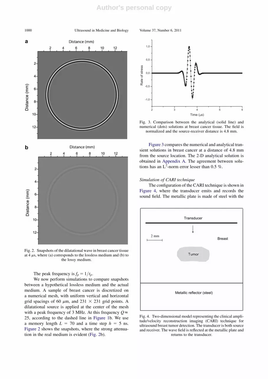
Figure 3 compares the numerical and analytical tran-sient solutions in breast cancer at a distance of 4.8 mmfrom the source location. The 2-D analytical solution isobtained in Appendix A. The agreement between solu-tions has an L2-norm error lesser than 0.5 %.
Simulation of CARI techniqueThe configuration of the CARI technique is shown in
Figure 4, where the transducer emits and records thesound field. The metallic plate is made of steel with the
Fig. 2. Snapshots of the dilatational wave in breast cancer tissueat 4 ms, where (a) corresponds to the lossless medium and (b) to
the lossy medium.
Fig. 3. Comparison between the analytical (solid line) andnumerical (dots) solutions at breast cancer tissue. The field is
normalized and the source-receiver distance is 4.8 mm.
2 mmBreast
Tumor
Metallic reflector (steel)
Transducer
Fig. 4. Two-dimensional model representing the clinical ampli-tude/velocity reconstruction imaging (CARI) technique forultrasound breast tumor detection. The transducer is both sourceand receiver. The wave field is reflected at the metallic plate and
returns to the transducer.
1000 Ultrasound in Medicine and Biology Volume 37, Number 6, 2011

Author's personal copy
properties c0 5 5900 m/s, r 5 7850 kg/m3 and no atten-uation. The mesh has absorbing boundary conditions atthe top and bottom of the grid (20 grid cells) (Cerjanet al. 1985). Due to the periodicity of the Fourier method,the absorbing strip of the top boundary is located at thebottom of the mesh. To be effective, the damping isapplied to all the temporal levels, un; n522;21; 0; 1.We use a memory length L5 70 and a time step h5 2 ns.
Figure 5 shows snapshots of the wave field at twodifferent propagation times. In Figure 5a, the picture
shows how the down-going plane wave is diffracted bythe tumor, while in Figure 5b the plane wave has been re-flected at the breast-steel interface and is traveling back(up) to the transducer line. As can be seen, the field isfaster in the region where the tumor is present. Thetime history recorded at the transducer is represented inFigure 6. The first horizontal event is the initial planewave, while the reflection event can be seen at nearly12 ms, with the signal below the tumor arriving slightlyearlier and showing more dissipation.
To show the sensitivity of the technique to the pres-ence of a tumor and attenuation, we represent in Figure 7the maximum stress rate of the reflection event corre-sponding to the following cases: (1) without tumor, loss-less media; (2) with tumor, lossless media; (3) withouttumor, lossy media; and (4) with tumor, lossy media.When there is no tumor the response is flat, while theeffect of attenuation is clear in the much lower amplitudeof the signal. The simulations show that the CARI tech-nique is suitable for tumor detection.
DISCUSSION
To our knowledge, the numerical algorithms used tosimulate ultrasound in biologic media involving frac-tional derivatives are based on the finite element (FE)method (Dikmen 2005; Wisman 2003; Bouna€ım et al2007; Bouna€ım and Chen 2008). In other fields, thefinite difference (FD) method is also used (e.g., Rekanosand Papadopoulos 2010). These algorithms and the Four-ier pesudospectral (PS)method have no restrictions on the
Fig. 5. Snapshots at 4 ms (a) and 10 ms (b). The thick line repre-sents the breast/steel interface and the transducer is located at2.4 mm (vertical distance). A diffraction event arising from
the tumor can be seen at 4 ms.
Fig. 6. Time history recorded at the transducer. The reflectionevent at 12 ms has a smaller travel time and lower amplitude
at the location of the tumor.
Wave simulation in biologic media d M. CAPUTO et al. 1001

Author's personal copy
type of constitutive equation, boundary conditions orsource-type and allow general material variability. FDsimulations are simple to program and, under not verystrict accuracy requirements, are very efficient. Pseudo-spectral methods can, in some cases, be more expensive,but guarantee high accuracy and relatively lower back-ground noise when staggered differential operators areused. These operators are also suitable when large varia-tions of Poisson’s ratio are present in the model (e.g.,a fluid-solid interface). On the other hand, FE methodsare best suited for engineering problems, where interfaceshave well defined geometrical features, in contrast withbiologic interfaces. Moreover, use of nonstructured grids,mainly in 3-D space, is one of the main disadvantages offinite-element methods because of the topological prob-lems to be solved when constructing the model.
In particular, the PS method is suitable when thesignal propagates long distances. For instance, a 3 MHzsignal traveling a distance 10 cm propagates hundred ofwavelengths. At these ranges, low-order FE and FD algo-rithms distort signals unacceptably, while it is known thatPS methods are highly accurate (Carcione 2007). In addi-tion, the PSmethod permits the use of coarse grid sizes. In3-D space, pseudospectral methods require a minimum ofgrid points, compared with finite differences and can bethe best choice when limited computer storage is avail-able. Fornberg (1996) showed a formal equivalencebetween the PS method and n-th order FD stencils, wheren is the number of grid points. This is the reason for thehigh accuracy. This property, computational efficiencyand parallelism using a large-scale bioacoustic model isillustrated in Wojcik et al. (1999). Optimal performancesin geophysics are shown in Carcione et al. (2002) andCarcione (2009).
Another use of the present modeling method couldbe the simulation of ultrasonic pulse-echo data. Doyleet al. (2010) showed that normal and malignant cellsproduce time-domain signals and spectral features thatare significantly different. The method may have otherpotential applications in medicine and biology, besidesmodeling of breast tissue. For example, it could improvethe simulation study of contrast microbubbles witha KVFD-type shell instead of the classical KV visco-elastic shell of encapsulation (Chen et al. 2009).
We note that a final test of the method requiresa direct comparison of the numerical results with experi-mental measurements. Also, we should take into accountthe limitations of the CARI simulations in relation to‘‘realistic’’ experiments, given the more complex realityof breast cancer assessment with ultrasound (e.g., ductalor lobular carcinoma, natural variations in tissue densityand stiffness in normal breast tissue, sources of experi-mental noise, etc.).
Themethodcanbeusefulwhere other techniquesmayfail, for instance travel time tomography (Schreiman et al.1984), since the difference invelocity between breast tissueand breast cancer is small.
CONCLUSIONS
We have presented a numerical algorithm to modelultrasound in biologic tissues based on a generalizationof the Kelvin-Voigt model to the case of fractional timederivatives of the strain. This stress-strain relation hasthree parameters that can be obtained by fitting realdata, namely, the stiffness, the pseudo-viscosity and thefractional order. The wave field is computed in thetime-space domain using the Gr€unwald-Letnikov approx-imation and the staggered Fourier pseudospectralmethod. The method is successfully tested against ananalytical solution and applied to breast cancer detection.
The classical amplitude-velocity reconstructionimaging technique data modeled with the Kelvin-Voigtstress-strain relation, modified with the introduction offractional derivatives, may possibly indicate the presenceof tumor. A further analysis of the physical properties ofvarious tumors and more detailed data are required. Then,the method may lead to the possibility to distinguishbetween malignant and nonmalignant tumors.
Acknowledgments—The authors thank two anonymous reviewers andthe editor for useful and detailed comments.
REFERENCES
Auld BA. Acoustic fields and waves in solids, Vol. 1. Malabar: Robert E.Krieger, Publishing Co.; 1990.
Bland DR. The theory of linear viscoelasticity. Oxford: Pergamon Press;1960.
Fig. 7. Maximum stress rate field of the reflection event corre-sponding to the following cases: without tumor, lossless media(1); with tumor, lossless media (2); without tumor, lossy media
(3); and with tumor, lossy media (4).
1002 Ultrasound in Medicine and Biology Volume 37, Number 6, 2011

Author's personal copy
Bouna€ımA, Holm S, ChenW, Ødeg�ard A. Detectability of breast lesionswith CARI ultrasonography using a bioacoustic computationalapproach. Comput Math Appl 2007;54:96–106.
Bouna€ımA, ChenW. Computations for a breast ultrasonic imaging tech-nique and finite element approach for a fractional derivativemodeling the breast tissue acoustic attenuation. Int J Tomogr Stat2008;10:31–43.
Caputo M. Linear models of dissipation whose Q is almost frequencyindependent-II. Geophys J Royal Astronomic Soc 1967;3:529–539.
Caputo M. Elastic radiation from a source in a medium with an almostfrequency independent. J Phys Earth 1981;29:487–497.
Caputo M, Cametti C. Memory diffusion in two cases of biologicalinterest. Theor Biol 2008;254:697–703.
Caputo M, Cametti C. The memory formalism in the diffusion of drugsthrough skin membrane. Physica D 2009;42:125505–125511.
Caputo M, Cametti C, Ruggiero V. Time and spatial concentrationprofile of glucose inside an eritrocyte membrane by means ofa memory formalism. Physica A 2008;387:210–218.
CaputoM, Carcione JM.Wave simulation in dissipativemedia describedby distributed-order fractional time derivatives. J Vibration Control2010, in press. [Epub ahead of print].
Caputo M, Mainardi F. A new dissipation model based on memorymechanism. Pure Appl Geophys 1971;91:134–147.
Carcione JM. Staggered mesh for the anisotropic and viscoelastic waveequation. Geophysics 1999;64:1863–1866.
Carcione JM. Wave fields in real media. Theory and numerical simula-tion of wave propagation in anisotropic, anelastic, porous and elec-tromagnetic media, Amsterdam: Elsevier Science, (Second edition,revised and extended) 2007.
Carcione JM. Theory and modeling of constant-Q P- and S-waves usingfractional time derivatives. Geophysics 2009;74:T1–T11.
Carcione JM. A generalization of the Fourier pseudospectral method.Geophysics 2010;75:53–56.
Carcione JM, Cavallini F, Mainardi F, Hanyga A. Time-domain seismicmodeling of constant Q-wave propagation using fractional deriva-tive. Pure Appl Geophys 2002;159:1719–1736.
Carcione JM, Kosloff D, Kosloff R. Viscoacoustic wave propagationsimulation in the earth. Geophysics 1988;53:769–777.
Cerjan C, Kosloff D, Kosloff R, Reshef M. A nonreflectng boundarycondition for discrete acoustic and elastic wave equations.Geophysics 1985;50:705–708.
Cesarone F, Caputo M, Cametti C. Memory formalism in the passivediffusion across a biological membrane. J Membrane Sci 2005;250:79–84.
ChenW, Holm S. Fractional Laplacian time-space models for linear andnonlinear lossy media exhibiting arbitrary frequency power-lawdependency. J Acoust Soc Am 2004;115:1424–1430.
Chen J, Hunter KS, Shandas R. Wave scattering from encapsulated mi-crobubbles subject to high-frequency ultrasound: Contribution ofhigher-order scattering modes. J Acoust Soc Am 2009;126:1766–1775.
Coussot C, Kalyanam S, Yapp R, Insana MF. Fractional derivativemodels for ultrasonic characterization of polymer and breast tissueviscoelasticity. IEEE Trans Ultrason Ferroelectr Freq Control2009;56:715–726.
D’astous FT, Foster FS. Frequency dependence of ultrasound attenua-tion and backscatter in breast tissue. Ultrasound Med Biol 1986;12:795–808.
Dikmen €U. Modeling of seismic wave attenuation in soil structuresusing fractional derivative scheme. J Balkan Geophys Soc 2005;8:175–188.
Doyle TE, Goodrich JB, Ambrose BJ, Patel H, Kwon S, Pearson LH.Ultrasonic differentiation of normal versus malignant breast epithe-lial cells in monolayer cultures. J Acoust Soc Am 2010;128:EL229–EL235.
Eldred LB, Baker WP, Palazotto AN. Kelvin-Voigt vs. fractional deriv-ative model as constitutive relations for viscoelastic materials.AIAA J 1995;33:547–550.
Fornberg B. A practical guide to pseudospectral methods. Cambridge:Cambridge University Press; 1996.
Gr€unwald AK. €Uber ‘‘begrenzte’’ Derivationen und deren Anwendung.Zeitschrift f€urAngewandteMathematikundPhysik1867;12:441–480.
Holm S, Sinkus RA. Unifying fractional wave equation for compres-sional and shear waves. J Acoust Soc Am 2010;127:542–548.
ICRU. Conversion coefficients for use in radiological protection againstexternal radiation. Report 1998;57. Bethesda: ICRU Publications,1998.
Kelly JF, McGough RJ. Two fractal ladder models and power law waveequations. J. Acoust Soc Am 2009;126:2072–2081.
Kiss MZ, Varghese T, Hall TJ. Viscoelastic characterization of in vitrocanine tissue. Phys Med Biol 2004;49:4207–4218.
Kjartansson E. Constant Q-wave propagation and attenuation.J Geophys Res 1979;84:4737–4748.
Letnikov AV. Theory of differentiation of fractional order. Matematice-skij Sbornik 1868;3:1–68 (in Russian).
Magin R. Fractional calculus in bioengineering. Chicago, IL: Universityof Illinois at Chicago Press; 2006.
Magin R, Feng X, Baleanu D. Solving the fractional order Bloch equa-tion. Concepts Magn Reson 2009;34A:16–23.
Mainardi F, TomirottiM. Seismic pulse propagationwith constant Q andstable probability distributions. Annali di Geofisica 1997;40:1311–1328.
Podlubny I. Fractional differential equations. San Diego: AcademicPress; 1999.
Rekanos IT, Papadopoulos TG. FDTDmodeling of wave propagation inCole-Cole media with multiple relaxation times. Antennas WirelessPropagation Lett IEEE 2010;9:67–69.
Richter K. Technique for detecting and evaluating breast lesions. J.Ultrasound Med 1994;13:782–797.
Schiessel H, Metzler R, Blumen A, Nonnenmacher TF. Generalizedviscoelastic models: Their fractional equations with solutions.J Phys A Math Gen 1995;28:6567–6584.
Schreiman JS, Gisvold J, Greenleaf JF, Bahn RC. Ultrasound transmis-sion computed tomography of the breast. Radiology 1984;150:523–530.
Taylor LS, Lerner AL, Rubens DJ, Parker KJ. A Kelvin-Voight frac-tional derivative model for viscoelastic characterization of livertissue. Adv Bioeng 2002;53:1–2.
Treeby BE, Cox BT. Modeling power law absorption and dispersion foracoustic propagation using the fractional Laplacian. J Acoust SocAm 2010;127:2741–2748.
Weiwad W, Heinig A, Goetz L, Hartmann H, Lampe D, Buchmann J,Millner R, Spielmann RP, Heywang- Kbrunner SH. Direct measure-ment of sound velocity in various specimens of breast tissue. InvestRadiol 2000;35:721–726.
Wismer MG. Finite element analysis of broadband acoustic pulsesthrough inhomogenous media with power law attenuation.J Acoust Soc Am 2006;120:3493–3502.
Wojcik G, Mould J, Carcione LM, Ostromogilsky M, Vaughan D.Combined transducer and nonlinear tissue propagation simulations.ASME International Mechanical Engineering Congress and Exposi-tion. November 14-19, 1999, Nashville, TN, USA.
Zhang M, Nigwekar P, Castaneda B, Hoyt K, Joseph JV, diSant’Agnese A, Messing EM, Strang JG, Rubens DJ, Parker KJ.Quantitative characterization of viscoelastic properties of humanprostate correlated with histology. Ultrasound Med Biol 2008;34:1033–1042.
APPENDIX A
GREEN’S FUNCTION AND ANALYTICALSOLUTION
A 2-D analytical solution corresponding to eqn (15) withm5 1 ina homogeneous medium can easily be obtained. Combining the equa-tions, we have
v3t e51
rDt: (21)
Wave simulation in biologic media d M. CAPUTO et al. 1003

Author's personal copy
In the frequency domain, s5Me, according to eqn (2), and using(15), eqn (21) becomes a Helmholtz equation,
De1p2e521
iurv2Ds52
1
iuMDs; p5
u
v; (22)
where p is the wave number and v is given by eqn (6). If v is real, themedium is lossless. The solution to the acoustic (lossless) equationðD1p2ÞG528dðrÞ is the Green function G522iH
ð2Þ0 ðprÞ, with v5c0,
where Hð2Þ0 is the zero-order Hankel function of the second kind (e.g.,
Carcione, 2007). More precisely,
Gðx; y; x0; y0;u; c0Þ522iHð2Þ0
�ur
c0
�(23)
where ðx0; y0Þ is the source location, and
r5ffiffiffiffiffiffiffiffiffiffiffiffiffiffiffiffiffiffiffiffiffiffiffiffiffiffiffiffiffiffiffiffiffiffiffiffiðx2x0Þ21ðy2y0Þ2
q: (24)
The anelastic solution is obtained by invoking the correspondenceprinciple (Bland 1960), i.e., by substituting the acoustic velocity c0 withthe complex velocity v. The differential operator D=ðiuMÞ acts on thesource in eqn (22). Thus, the Green’s function for the strain is
Ge 51
iuMDG: (25)
Since DG52p2G away from the source and s5Me, the Green’sfunction for the stress is
Gs 5MGe 52p2G
iu: (26)
We setGð2uÞ5G�ðuÞ, where the superscript * denotes complexconjugation. This equation ensures that the inverse Fourier transform ofthe Green’s function is real. The frequency-domain solution is thengiven by sðuÞ51
8GsðuÞFðuÞ, where F is the Fourier transform of the
source time history. Since we are solving the dynamical equation withm 5 1, our solution is not s but the stress rate t5vts. Hence,
tðx; y; x0; y0;uÞ5 1
8iuGsF52
1
8p2Gjðx; y; x0; y0;u; vÞFðuÞ; (27)
Because the Hankel function has a singularity at u 5 0, weassumeG50 foru5 0, an approximation that does not have a significanteffect on the solution (note, moreover, that Fð0Þ5 0). The time-domainsolution tðtÞ is obtained by a discrete inverse Fourier transform. Wehave tacitly assumed that t and dt=dt are zero at time t5 0.
1004 Ultrasound in Medicine and Biology Volume 37, Number 6, 2011