Embed Size (px)
Citation preview

CHAPTER 6
What Has fMRI Thught UsAbout Object Recognition?
Kalanit Grill-Spector
6.L lntroduction
Humans can effortlessly recognize objects in a fraction of a second despite large vari-ability in the appearance of objects (Thorpe et al. 1996). What are the underlyingrepresentations and computations that enable this remarkable human ability? One wa1to answer these questions is to investigate the neural mechanisms of object recognitionin the human brain. With the advent of functional magnetic resonance imaging (fMRI Iabout 15 years ago, neuroscientists and psychologists began to examine the neuralbases of object recognition in humans. Functional magnetic resonance imaging (fMRI Iis an attractive method because it is a noninvasive technique that allows multiple mea-surements of brain activation in the same awake behaving human. Among noninvasivetechniques, it provides the best spatial resolution currently available, enabling us tolocalize cortical activations in the spatial resolution of millimeters (as fine as I mm Iand at a reasonable time scale (in the order of seconds).
Before the advent of fMRI, knowledge about the function of the ventral stream wasbased on single-unit electrophysiology measurements in monkeys and on lesion studies.These studies showed that neurons in the monkey inferotemporal (IT) cortex respond toshapes (Fujita etal.1992) and complex objects such as faces (Desimone et al. 1984), andthat lesions to the ventral stream can produce specific deficits in object recognition, suchas agnosia (inability to recognize objects) and prosopagnosia (inability to recognizefaces, Farah 1995). However, interpreting lesion data is complicated because lesionsare typically diffuse (usually more than one region is damaged), typically disrupt botha cortical region and its connectivity, and are not replicable across patients. Therefore.the primary knowledge gained from fMRI research was which cortical sites in thenormal human brain are involved in object recognition. The first set of fMRI studiesof object and face recognition in humans identified the regions in the human brain thatrespond selectivity to objects and faces (Malach et al. 1995; Kanwisher et al. 1997Grill-Spector et al. 1998b). Then a series of studies demonstrated that activation inobject- and face-selective regions correlates with success at recognizing object andfaces, respectively, providing striking evidence for the involvement of these regions in
recogn
researct-ocus t
that ar
recogrI n t
about
tt'rpic
u:ed I
Fttr e:
oblecr[1r\\ i
rrrtru:oh1e,'
rcco!.rf ttxrr her
, lloP
rmpl
l r C O
..,f Fl r
:iur.\
o f
r I
I
I rrf
:cl.:
::E
cio
:NU
.-rf
-rJ
3 l- t l
T1. i
JI
102

tUs
wHAT nls flrnr TAUGHT us ABour oBJEcr REcocNrrron? 103
recognition (Grill-Spector et al. 2000; Bar et al.200l; Grill-Spector et al. 2004). Onceresearchers found which regions in the cortex are involved in object recognition, thefocus of research shifted to examining the nature of representations and computationsthat are implemented in these regions to understand how they enable efficient objectrecognition in humans. "'
In this chapter I will review fMRI research that provided important knowledgeabout the nature of object representations in the human brain. I chose to focus on thistopic because results from these experiments provide important insights that can beused by computer scientists when they design artificial object recognition systems.For example, one of the fundamental problems in recognition is how to recognize anobject across variations in its appearance (invariant object recognition). Understandinghow a biological system has solved this problem may give clues for how to build arobust artificial recognition system. Funher, fMRI is more adequate for measuringobject representations than the temporal sequence of computations en route to objectrecognition because the time scale of fMRI measurements is longer than the time scaleof the recognition process (the temporal resolution of fMRI is in the order of seconds,whereas object recognition takes about 100-250 ms). Nevertheless, combining psy-chophysics with fMRI may give us some clues about what kind of visual processing isimplemented in distinct cortical regions. For example, finding regions whose activationis correlated with success at some tasks, but not others, may suggest the involvementof particular cortical regions in one computation, but not another.
In discussing how fMRI has impacted our current understanding of object represen-tations, I will focus on results pertaining to two aspects of object representation:
. How do the underlying representations provide for invariant object recognition?
. How is category information represented in the ventral stream?
I have chosen these topics because (i) they are central topics in object recognitionfor which fMRI has substantially advanced our understanding. (ii) Some findingsrelated to these topics stirred considerable debate (see sect. 6.7), and (iii) some ofthe fMRI findings in humans are surprising given prior knowledge from single-unitelectrophysiology in monkeys. In organizing the chapter, I will begin with a briefintroduction of the functionalorganization of the human ventral stream and a definitionof object-selective cortex. Then I will describe research that elucidated the propertiesof these regions with respect to basic coding principles. I will continue with findingsrelated to invariant object recognition, and end with research and theories regardingcategory representation and specialization in the human ventral stream.
6.2 The Functional Organization of the Human Ventral Stream
The first set of fMRI studies on object and face recognition in humans was devoted toidentifying the regions in the brain that are object- and face-selective. Electrophysiologyresearch in monkeys suggested that neurons in higher-level regions respond to shapesand objects more than simple stimuli such as lines, edges, and patterns (Desimoneet al. 1984; Fujita et al. 1992; Logothetis et al. 1995). Based on these findings, fMRIstudies measured brain activation when people viewed pictures of objects compared to
ion?
rd despite large vari-are the underlying
an ability? One wayrf object recognitionnce imaging (fMRI)examine the neuralnce imaging (fMRI)llows multiple mea-\mong noninvasiveble. enabling us to; (as fine as I mm)
ventral stream wasd on lesion studies.) cortex respond tone et al. 1984), andt recognition, suchrility to recognized because lesionscally disrupt bothrtients. Therefore,nical sites in thet of fMRI studieshuman brain thatisher et al. 1997;:hat activation inizing object and'these regions in

t04 KALANIT GRILL-SPECTOR
II;;IIII
I
IaII
III
IIIIII
(b)E t otE axl oE6 r oa
f '-..traa|raalrahlJla
*rftdrt
+Ut
Puonlrtlon Dwrlbo (mr)
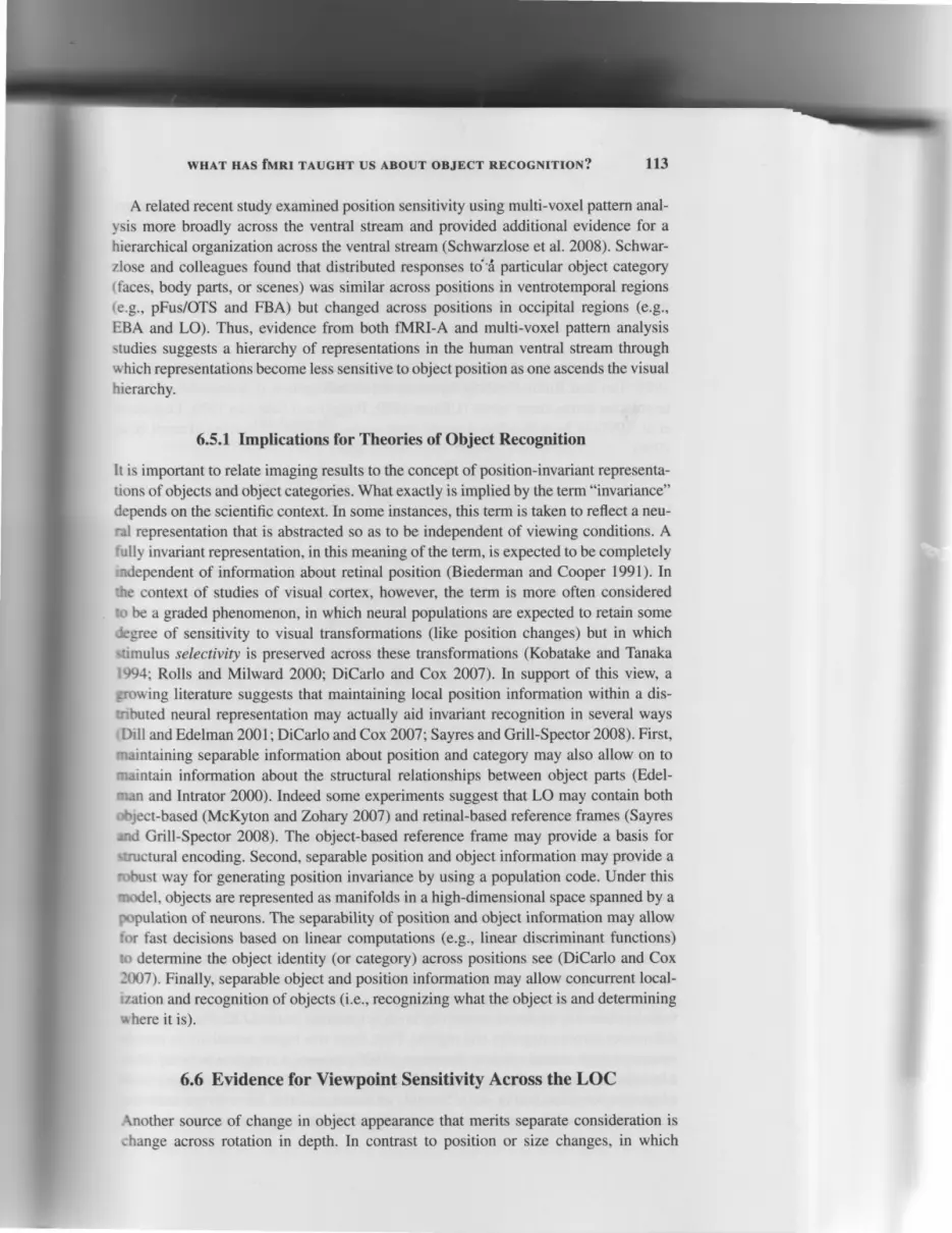
Figure 6.1. Object-, face-, and place-selective cortex. (a) Data of one representive subjeoshown on her partially inflated right hemisphere: lateral view (/eft); ventral view (right). Darkgray indicates sulci. Light gray indicates gyri. Black /ines delineate retinotopic regions. Blueregions delineate object-selective regions (objects > scrambled objects), including LO andpFus/OTS ventrally as well as dorsal foci along the intraparietal sulcus (lPS). Red regionsdelineate face-selective regions (faces > non-faces objects), including the FFA, a region inLOS and a region in posterior STS. Magenta regions delineate overlap between face- andobjects-selective regions. Creen regions delineate place-selective regions (places > objectst,including the PPA and a dorsal region lateral to the lPS. Dark green regions delineate overlapbetween place- and object-selective regions. All maps thresholded at P < 0.001, voxel level.(b) LO and pFus/OTS (but not Vl) responses are correlated with recognition performance (Grill-Spector et al. 2000). To superimpose recognition performance and fMRl signals on the sameplot, all values were normalized relative to the maximum response for the 500-ms durationst imulus.
For fMRf signars (brue, red, and orar "hsignal(condition)rge lines) : ffi.
For recognition perfor-
mance (bfack) - o/ocorrect(condition).
(see color plate 6.1.)"hcorrect(5O0 ms)
scrambled objects (have the same local information and statistics, but do not contain
an object) or texture patterns (e.9., checkerboards, which are robust visual stimuli,
but do not elicit a percept of a global form). These studies found a constellation ofregions in the lateral occipital cortex (or the lateral occipital complex, LOC), beginning
around the lateral occipital sulcus, posterior to MT, and extending ventrally into the
occipitotemporal sulcus (OTS) and the fusiform gyrus (Fus), that respond more to
objects than controls. The LOC is located lateral and anterior to early visual areas (Vl-
V4), (Grill-Spector et al. t998a; Grill-Spector et al. 1998b) and is typically dividedinto two subregions: LO, a region in the lateral occipital cortex, adjacent and posterior
to MT; and pFus/OTS; a ventral region overlapping the OTS and posterior fusiformgyrus (Fig. 6.1).
The lateral occipital complex (LOC) responds similarly to many kinds of objects andobject categories (including novel objects) and is thought to be in the intermediate- orhigh-level stages of the visual hierarchy. Importantly, LOC activations are correlatedwith subjects' object recognition perfonnance. High LOC responses correlate with

nt ive sub ject' r ish t t . Dark
regions. B/ued ing LO andRed regions
. a region inln face- ands > objects\.teate overlap, r ,oxe l leve l .r t ance rC r i l i -on the same-ms durat ion
t i : rc ln penor-
not contatnra l s t imu l r .te l lat ion oi. beginningl l1 into rhetd more tt-rareas ( \'l -
l l r d iv idedd posterior,r fusiform
lbjects andrediate- orcorrelatedelate u ' i th
WHAT HAS fMRI TAUGHT US ABOUT OBJECT RECOGNITION? 105
successful object recognition (hits), and low LOC responses correlate with trials inwhich objects are present but not recognized (misses) (Fig. 6.1(b)). Object-selectiveregions are also found in the dorsal stream (Grill-Spector 2003; Grill-Spector andMalach 2004), but activation of these regions does not correlate with object recognitionperformance (Fang and He 2005). These regions may be involved in computationsrelated to visually guided actions toward objects (Culham et al. 2003). However, acomprehensive discussion of the role of the dorsal stream in object perception isbeyond the scope of this chapter.
In addition to the LOC, researchers found several ventral regions that show prefer-ential responses to specific object categories. The search for regions with categoricalpreference was motivated by repons that suggested that lesions to the ventral streamcan produce very specific deficits, such as the inability to recognize faces or the inabil-ity to read words, while other visual (and recognition) faculties remain preserved. Bycontrasting activations to different kinds of objects, researchers found ventral regionsthat show higher responses to specific object categories: including lateral fusiformregions that respond more to animals than tools, and medial fusiform regions thatrespond to tools more than animals (Martin et al. 1996; Chao et al. 1999)), a regionin the left OTS that responds more strongly to letters than textures (the visual wordform area, or VWFA) (Cohen et al. 2000); several foci that respond more strongly tofaces than to other objects (Kanwisher et al. 1997; Haxby et al. 2000; Hoffman andHaxby 2000; Grill-Spector et al. 2004), including the well known fusiform face area(FFA) (Kanwisher et al. 1997), regions that respond more strongly to houses and placesthan faces and objects (including a region in the parahippocampal gyrus, the parahip-pocampal place area (PPA) (Epstein and Kanwisher 1998); regions that respond morestrongly to body parts than faces and objects, including a region near MT called theextrastriate body area (EBA) (Downing et al. 2001); and a region in the fusiform gyrus(the fusiform body area, or FBA) (Schwarzlose et al. 2005)). Nevertheless, many ofthese object-selective and category-selective regions respond to more than one objectcategory and also respond strongly to object fragments (Grill-Spector et al. 1998b;Lerner et al. 200I; Lerner et al. 2008). This suggests that one must be cautious wheninterpreting the nature of the selective responses. It is possible that the underlyingrepresentation is perhaps of object parts, features, and/or fragments and not of wholeobjects or object categories.
Findings of category-selective regions in the human brain initiated a fierce debateabout the principles of functional organization in the ventral stream. Are there regions inthe cortex that are specialized for any object category? How abstract is the informationrepresented in these regions (e.g., is category information represented in these regions,or low-level visual features that are associated with categories)? I will address thesequestions in detail in section 6.7.
6.3 Cue-Invariant Responses in the LOC
Although findings of object-selective responses in the human brain were suggestiveof the involvement of these region in processing objects, there are many differencesbetween objects and scrambled objects (or objects and texture patterns). Objects have

106 KALANIT GRILL.SPECTOR
Figure 6.2. Selective responses to objects across multiple visual cues across the LOC. Statisticalmaps of selective response to object from luminance, stereo and motion information in arepresentative subject. All maps were thresholded at P < 0.005, voxel level, and are shown onthe inflated right hemisphere of a representative subject. (a) Luminance objects > scrambledobjects. (b) Objects generated from random dot stereograms versus structureless random dotstreograms (perceived as a cloud of dots). (c) Objects generated from dot motion versus thesame dots moving randomly. Visual meridians are represented by red (upper), blue (horizontal),and green (lower) lines. White contour indicates a motion-selective region, or MT. Adaptedfrom Vinberg and Crill-Spector 2008. (See color plate 6.2.)
shapes, surfaces, and contours; they are associated with a meaning and semanticinformation; and they are generally more interesting than texture patterns. Each ofthese factors may affect the higher fMRI response to objects than controls. Differencesin low-level visual properties across objects and controls may be driving some of theseeffects as well.
Converging evidence from several studies revealed an important aspect of codingin the LOC: it responds to object shape, not low-level visual features. Several studiesshowed that all LOC subregions (LO and pFus/OTS) are activated more strongly whensubjects view objects independent of the type of visual information that defines theobject form (Grill-Spector et al. 1998a; Kastner et al. 2000; Kourtzi and Kanwisher2000, 2001; Gilaie-Dotan et al. 2002; Vinberg and Grill-Spector 2008) (Fig. 6.2).The lateral occipital complex (LOC) responds more strongly to (1) objects definedby luminance than to luminance textures; (2) objects generated from random dotstereograms than to structureless random dot stereograms; (3) objects generated fromstructure from motion than to random (structureless) motion; and (4) objects generatedfrom textures than to texture patterns. The response of the LOC to objects is also similaracross object formatl (gray-scale, line drawings, and silhouettes), and it responds toobjects delineated by both real and illusory contours (Mendola et al. 1999; Stanleyand Rubin 2003). Kourtzi and Kanwisher (Kourtzi and Kanwisher 2001) also showedthat there was fMRl-adaptation (fMRI-A, indicating a common neural substrate), whenobjects had the same shape but different contours, but that there was no fMRI-A whenthe shared contours were identical but the perceived shape was different, suggestingthat the LOC responds to global shape rather than to local contours (see also Lerneretal.2002; Kourtzi etal.2003). Overall, these studies provided fundamental knowledge
I Selective responses to faces and houses across stimulus format (photographs, line drawings, and silhouettes)have also been shown for the FFA and PPA, respectively.
I Ob;c
i t'Jf
! r
,I!1,
::€. ' f
a
!
J
t
.t

WHAT HAS fMRI TAUGHT US ABOUT OBJECT RECOGNITION? 107
RandomHole
Wv4
(a) object Edges
ffitrw
F:
l' ,'i'*-
3:
t :
( . , r
li
rf
e
(b) vl LO pFus/OTS:c i
f; r.slE i9 1 0
Eo s ,-9 1o n n -. s " - O H E S R
Condition
figure 6.3. Responses to shape, edges, and surfaces across the ventral stream. (a) Schematicil lustration of experimental conditions. Stimuli were generated from either motion or stereornformation alone and had no luminance edges or surfaces (except for the screen border,n'hich was present during the entire experiment, including blank baseline blocks). For i l lus-tration purposes, darker regions indicate front surfaces. From left to right Object on the frontsurface in front of a flat background plane. Hole on the front surface in front of a flat back-qround. Disconnected edges in front of a flat background. Edges were generated by scramblingthe shape contours. Surfaces, Two semitransparent flat surfaces at different depths. Randomstimuli with no coherent structure, edges, global surfaces, or global shape. Random stimulihad the same relative disparity or depth range as other conditions. See examples of stimuli:http://www-psych.stanford.edu/-kalanit/jnpstim/. (b) Responses to objects, holes, edges, andelobal surfaces across the visual ventral-processing hierarchy. Responses: mean + SEM acrosseight subjects. O, object; H, hole; 5, surfaces; F, edges; R, random; diamonds, significantlydifferent than random at P < 0.05. Adapted from Vinberg and Cril l-Spector 2008.
by showing that activation of the LOC is driven by shape rather than by the low-levelvisual information that generates form.
In a recent study, we examined whether the LOC response to objects is drivenby their global shape or their surface information and whether LOC subregions aresensitive to border ownership. One open question in object recognition is whether
the region in the image that belongs to the object is first segmented from the rest of
the image (figure-ground segmentation) and then recognized, or whether knowing theshape of an object aids its segmentation (Peterson and Gibson 1994b,|994a;Nakayamaet al. 1995). To address these questions, we scanned subjects when they viewed stimulithat were matched for their low-level information and generated different percepts:( I ) a percept of an object in front of a background object, (2) a shaped hole (same
shape as the object) in front of a background, (3) two flat surfaces without shapes,(4) local edges (created by scrambling the object contour) in front of a background,
or (5) random dot stimuli with no structure (Fig. 6.3(a)) (Vinberg and Grill-Spector2008). We repeated the experiment twice, once with random dots that were presented
stereoscopically and once with random dots that moved. We found that LOC responses(both LO and pFus/OTS) were higher for objects and shaped holes than for surfaces,local edges, or random stimuli (Fig. 6.3(b)). These results were observed for both

108 KALANIT GRILL.SPECTOR
motion and stereo cues. In contrast, LOC responses were not higher for surfaces thanfor random stimuli and were not higher for local edges than for random stimuli. Thus.adding either local edge information or global surface information does not increaseLOC response. Adding a gl66al shape, however, does produce a significant increasein LOC response. These results provide clear evidence that cue-invariant responses inthe LOC are driven by object shape, rather than by global surface information or localedge information.
Interestingly, recent evidence shows that the LOC is sensitive to borderownership/figure-ground segmentation) (Appelbaum et al. 2006; Vinberg and Grill-Spector 2008). We found that the LO and pFus/OTS responses were higher for objects(shapes presented in the foreground) than for the same shapes when they had definedholes in the foreground. The only difference between the objects and the holes wasthe assignment of the figure region (or border ownership of the contour defining theshape). This higher response to objects than to holes was a unique characteristic of LOCsubregions and did not occur in other visual regions (Fig. 6.3). This result suggeststhat the LOC prefers shapes (and contours) when they define the figure region. Oneimplication of this result is that the same brain machinery may be involved in bothrecognizing objects and in determining which region in the visual input contains thefigure region. Thus, one consideration for computer scientists is that an effective objectrecognition algorithm should determine both what is the the object in the scene as wellas which region in the scene corresponds to the object.
6.4 Neural Bases of Invariant Object Recognition
The literature reviewed so far provides evidence that the LOC is involved in therecognition and processing of object form. Given the LOC's role in object perception.one may consider, how it deals with variability in an object's appearance. Many factorscan affect the appearance of objects. Changes in an object's appearance can occur as aresult of the object being at different locations relative to the observer, which will affectit's retinal projection of objects in terms of size and position. Also, the 2-D projectionof a 3-D object on the retina varies considerably owing to changes in its rotationand viewpoint relative to the observer. Other changes in appearance occur becauseof differential illumination conditions, which affect an object's color, contrast, andshadowing. Nevertheless, humans are able to recognize objects across large changesin their appearance, which is referred to as invariant object recognition.
A central topic of research in the study of object recognition is understandinghow invariant recognition is accomplished. One view suggests that invariant objectrecognition is accomplished because the underlying neural representations are invariantto the appearance of objects. Thus, this view suggests that there neural responses willremain similar even when the appearance of an object changes considerably. Onemeans by which this can be achieved is by extracting from the visual input features orfundamental elements (such as geons (Biederman 1987) that are relatively insensitiveto changes in objects' appearance. According to one influential model (recognition bycomponents, or RBC; Biederman 1987), objects are represented by a library of geons,which are easy to detect in many viewing conditions, and by their spatial relations. Othertheories suggest that invariance may be generated through a sequence of computations

WHAT HAS fMRI TAUGHT US ABOUT OBJECT RECOGNITION? 109
aro\s a hierarchically organized processing stream in which the level of invariancen'reases from one level of the processing to the next. For example, at the lowest level,r the processing stream, neurons code local features; at higher levels of the processingrtrcam, neurons respond to more complex shapes and are ldsi sensitive to changes in
axition and size (Riesenhuber and Poggio 1999).\euroimaging studies of invariant object recognition found differential sensitivity
aross the ventral stream to object transformations such as size, position, illumination,aJ r iewpoint. Intermediate regions, such as LO, show higher sensitivity to imageanstormations than higher-level regions, such as pFus/OTS. Notably, evidence fromo.m) studies suggests that at no point in the ventral stream are neural representationscsctrely invariant to object transformations. These results support an account in whichryrariant recognition is supported by a pooled response across neural populations thatr: "ensitive to object transformations. One way in which this can be accomplished isQt a neural code that contains independent sensitivity to object information and objectfirnrlormation (DiCarlo and Cox 2007); for example, neurons may be sensitive to bothro.!eL-t category and object position. As long as the categorical preference is retainedrrLrsS object transformations, invariant object information can be extracted.
6.5 Object and Position Information in the LOC
tfnc r ariation that the object recognition system needs to deal with is variation in theruc and position of objects. Size and position invariance are thought to be accomplishedx Fran by an increase in the size of neural receptive fields along the visual hierarchy: e.. a-s one ascends the visual hierarchy, neurons respond to stimuli across a larger part;t the visual field). At the same time, a more complex visual stimulus is necessary toir*rt si,enificant responses in neurons (e.g., shapes instead of oriented lines). Findings-'m electrophysiology suggest that even at the highest stages of the visual hierarchy,Eurons retain some sensitivity to object location and size although electrophysiologyrn)rt\ r'ary significantly about the degree of position sensitivity of IT neurons (Op DeBsclk and Vogels 2000; Rolls 2000; DiCarlo and Maunsell 2003). A related issue isrirether position sensitivity of neurons in higher visual areas manifests as an orderly,rrgrglnphic representation of the visual field. Researchers have examined positionsrJ rize sensitivity in the LOC and nearby cortex (such as PPA and FFA) usingnr&\urements of the mean response across a region of interest; fMRI-A, in whichtrr measured sensitivity to changes in object size or position; and examination of the.fo'rnbuted response across the ventral stream to the same object or object categoryrrr-r\\ sizes and positions.
Ser eral studies documented sensitivity to both eccentricity and polar angle in distinct",rntral stream regions. Both object-selective and category-selective regions in ther<ntral stream respond to objects presented at multiple positions and sizes. However,- amplitude of response to object varies across different retinal positions. The LOrnt pFus/OTS as well as category-selective regions (e.g., FFA, PPA) respond moreist-'ngl) to objects presented in the contralateral versus ipsilateral visual field (Grill->,lcr'ror et al. 1998b; Hemond et al. 2007; McKyton andZohary 2007). Some regionsLO and EBA) also respond more strongly to objects presented in the lower visual fieldl+,r res and Grill-Spector 2008; Schwarzlose et al. 2008). Responses also vary with

110 KALANIT GRILL.SPECTOR
eccentricity: LO, FFA, and the VWFA respond more strongly to centrally presentedstimuli, and the PPA responds more strongly to peripherally presented stimuli (Ler ret al., 2001; Hasson et al. 2002; Hasson et al. 2003; Sayres and Grill-Spector 2008).
Using fMRI-A, my colleagues andl have shown that pFus/OTS, but not LO, exhibitrsome degree of insensitivity to an object's size and position (Grill-Spector et al.1999t.The fMRI-A method allows one to characterizethe sensitivity of neural representation:to stimulus transformations at a subvoxel resolution. fMRI-A is based on findings fronrsingle-unit electrophysiology that show that when objects repeat, there is a stimulus-specific decrease in the response of IT cells to the repeated image but not to otherobject images (Miller et al. l99l; Sawamura et al. 2006). Similarly, fMRI signalsin higher visual regions show a stimulus-specific reduction (fMRI-A) in responseto repetition of identical object images (Grill-Spector et al. 1999; Grill-Spector andMalach 2001; Grill-Spector et al. 2006a). We showed that fMRI-A can be used totest the sensitivity of neural responses to object transformation by adapting cortexwith a repeated presentation of an identical stimulus and then examining adaptationeffects when the stimulus is changed along an object transformation (e.g., changing itsposition). If the response remains adapted, it indicates that neurons are insensitive tothe change; however, if responses return to the initial level (recover from adaptation).it indicates sensitivity to the change (Grill-Spector and Malach 2001).
Using fMRI-A we found that repeated presentation of the same face or objectat the same position and size produces reduced fMRI activation or fMRI-A. Thisis thought to reflect stimulus-specific neural adaptation. Presenting the same face orobject in different positions in the visual field or at different sizes also producesfMRI-A in pFus/OTS and FEA, indicating insensitivity to object size and position(Grill-Spector et aI. 1999; see also Vuilleumier et al. 2002). This result is consistentwith electrophysiology findings that showed that IT neurons that respond similarly tostimuli at different positions in the visual field also show adaptation when the sameobject is shown in different positions (Lueschow et al. 1994).In contrast, LO recoversfrom fMRI-A to images of the same face or object when presented at different sizes orpositions. This indicates that LO is sensitive to object position and size.
Recently, several groups examined the sensitivity of the distributed response acrossthe visual stream to object category and object position (Sayres and Grill-Spector2008; Schwarzlose et al. 2008) and also object identity and object position (Egeret al. 2008). These studies used multi-voxel pattern analyses and classifier methodsdeveloped in machine learning to examine what information is present in the distributedresponses across voxels in a cortical region. The distributed response can carry differentinformation from the mean response of a region of interest when there is variation acrossvoxels' responses.
In order to examine sensitivity to position information, several studies examinedwhether distributed response patterns to same object category (or object exemplar) isthe same (or different) when the same stimulus is presented in a different positionin the visual field. In multi-voxel pattern analyses researchers typically split the datainto two independent sets and examine the cross-correlation between the distributedresponses to the same (or different) stimulus in the same (or different) position acrossthe two datasets. This gives a measure of the sensitivity of distributed responses toobject information and position. If responses are position-invariant, there will be a
\
- l

WHAT HAS fMRI TAUGHT US ABOUT OBJECT RECOGNITION?
9o Left 4o Left Fovea 4'Right 4o Up
111
4o DownEtttttlrllllllfflltlll
2 - 1 0 1 2Amplitude versus mean response
% Signal Change
Qure 5.4. LO distributed response patterns to different object categories and stimulus po-;i tlns. Data are shown on the lateral aspect of the right hemisphere cortical surface for a-Dreentative subject. Each panel shows the distributed LO fMRl amplitudes after subtractingr-.-nr each voxel its mean response. Red and yellow, Responses that are higher than the voxel's-ean response. Blue and cyan, Responses that are lower than the voxel's mean response.r---:. Inflated right cortical hemisphere, with red square indicating the zoomed region. Note:a: pattern changes significantly across columns (positions) and to a lesser extent across rows.a:eqories). Adapted from Sayres and Crill-Spector 2008. (See color plate 6.4.)
heh correlation between the distributed responses to the same object category (or
<remplar) at different positions. If responses are sensitive to position, there will be aLur correlation between responses to the same object category (or exemplar) at differentporitions.
Figure 6.4 illustrates distributed LO responses to three categories: houses, limbs,
uld faces at six visual locations (fovea; 4o up, right, down, or left from the fovea;9 leti of fovea). Activation patterns for the same object category presented at dif-fcrent positions vary considerably (compare the response patterns in the same row
LToss the different columns in Fig. 6.4). There is also variation (but to a lesser extent)
\Toss different object categories when presented at the same retinal position (same
.arlumn, different rows, in Fig. 6.4). Surprisingly, position effects in LO were larger
dren category effects - that is, showing objects from the same category, but at a dif-f<rent position, reduced the correlation between activation patterns (Fig. 6.5, flrst vs.
drrrd bars) more signiflcantly than changing the object category in the same position
"Fig. 6.5, first vs. second bar). Importantly, position and category effects were in-
Jcpendent, as there were no significant interactions between position and category

112 KALANIT GRILL-SPECTOR WHAT
A related recen
ysis more broadlt
hierarchical organ
zlose and colleae
(faces, bodY Part'(e.g., PFus/OTSEBA and LOr' T
s$dies suggest:
which rePresentr
hierarchY.
6-5.
It is imPofiant tt'
tions of object' '
depends on thc' '
ral rePresentatle
fullY invariant rt
indePendent of
the context trf '
to be a gradc'd 1
degree of st'ntt
stimulus 'st'/t't-t
1994; Roll: en
growing litcrat
tributed neural
(Dill and Edclr
maintaining {
maintain infot
man and Intra
object-based t
and Grill-SPr
structural L'nc
robust u'ar ie
model. oblect
PoPulation ttt
for fast deci'
to deternrinc'
2007)' Finall
ization and n
where i t i : t '
6't
Another :t-tt
change ucr'
(rc 0 .3 -.9(u
ov 0.1 :C l( ( l : ;o := o : r{}'1 ':
:4 . 2 '
"ffio 8$:tU, "ffio 8$[frSame Position Different Position
Figure 6.5. Mean cross-correlations between LO distributed responses across two independenthalves of the data for the same or different category at the same or different position in thevisual field. Position effects: LO response patterns to the same category were substantiallymore similar if they were presented at the same position versus different positions (first andthird bars, P < 10-7). Category effects: The mean correlation was higher for same-categoryresponse patterns than for different-category response patterns when presented in the sameretinotopic position (first two bars, P < 10-o). Error bars indicate SEM across subjects. Adaptedfrom Sayres and Crill-Spector 2008.
(all F values < 1 .02, all P values > 0.31). Thus, changing both object category and po-sition produced maximal decorrelation between distributed responses (Fig. 6.5, fourthbar).
We also examined whether position sensitivity in LO is manifested as an orderlytopographic map (similar to retinotopic organization in lower visual areas), by measur-ing retinotopic maps in LO using standard traveling wave paradigms (Wandell 1999;Sayres and Grill-Spector 2008). We found a continuous mapping of the visual field inLO both in terms of eccentricity and polar angle. This topographic map contained anover-representation of the contralateral and lower visual field (more voxels preferredthese visual field positions than the ipsilateral and upper visual fields). Although wedid not consistently find a single visual field map (a single hemifield or quarterfieldrepresentation) in LO, this analysis suggests that there is preserved retinotopic infor-mation in LO that may underlie the position effects observed in analyses of distributedLO responses.
Overall, our data show that different object categories produce relatively smallchanges to both the mean and distributed response across LO (categorical effects arelarger in the fusiform and parahippocampal gyri). In comparison, a modest 4o changein an object's position produces signal changes in LO that are as large or larger than thecategory modulation. This 4" displacement is well within the range for which humanscan categonze and detect objects (Thorpe et al. 2001). This indicates a differencebetween the position sensitivity of recognition behavior and that of neural populationsin LO. However, it is possible that performance in recognition tasks that require fine-grain discrimination between exemplars is more position sensitive, and limited by thedegree of position sensitivity in LO.
WHAT HAS fMRI TAUGHT US ABOUT OBJECT RECOGNITION? 113
A related recent study examined position sensitivity using multi-voxel pattern anal-vsis more broadly across the ventral stream and provided additional evidence for ahierarchical organization across the ventral stream (Schwarzlose et al. 2008). Schwar-zlose and colleagues found that distributed responses to'ri particular object categorytfaces, body parts, or scenes) was similar across positions in ventrotemporal regions(e.9., pFus/OTS and FBA) but changed across positions in occipital regions (e.9.,EBA and LO). Thus, evidence from both fMRI-A and multi-voxel pattern analysisrtudies suggests a hierarchy of representations in the human ventral stream throughr,r hich representations become less sensitive to object position as one ascends the visualhierarchy.
6.5.L lmplications for Theories of Object Recognition
It is important to relate imaging results to the concept of position-invariant representa-tions of objects and object categories. What exactly is implied by the term "invariance"depends on the scientific context. In some instances, this term is taken to reflect a neu-ral representation that is abstracted so as to be independent of viewing conditions. Aiullv invariant representation, in this meaning of the term, is expected to be completelyrndependent of information about retinal position (Biederman and Cooper 1991). Inthe context of studies of visual cortex, however, the term is more often consideredttr [rs a graded phenomenon, in which neural populations are expected to retain some.lesree of sensitivity to visual transformations (like position changes) but in which.timulus selectivity is preserved across these transformations (Kobatake and Tanaka199-l; Rolls and Milward 2000; DiCarlo and Cox 2007). In support of this view, arrowing literature suggests that maintaining local position information within a dis-tnbuted neural representation may actually aid invariant recognition in several ways,Dill and Edelman 2001; DiCarlo and Cox 2007; Sayres and Grill-Spector 2008). First,maintaining separable information about position and category may also allow on tonaintain information about the structural relationships between object parts (Edel-nan and Intrator 2000). Indeed some experiments suggest that LO may contain both.*rject-based (McKyton and Zohary 2007) and retinal-based reference frames (Sayresend Grill-Spector 2008). The object-based reference frame may provide a basis for.tructur&l encoding. Second, separable position and object information may provide anrbust way for generating position invariance by using a population code. Under thismodel, objects are represented as manifolds in a high-dimensional space spanned by agx-rpulation of neurons. The separability of position and object information may allowior fast decisions based on linear computations (e.g., linear discriminant functions)to determine the object identity (or category) across positions see (DiCarlo and Coxlm7). Finally, separable object and position information may allow concurrent local-rzation and recognition of objects (i.e., recognizing what the object is and determiningu here it is).
6.6 Evidence for Viewpoint Sensitivity Across the LOC
Another source of change in object appearance that merits separate consideration is
change across rotation in depth. In contrast to position or size changes, in which

rt4 KALANIT GRILL.SPECTOR
invariance may be achieved by a linear transformation, the shape of objects change:with depth rotation. This is because the visual system receives 2-D rettnal projectionsof 3-D objects. Some theories suggest that view-invariant recognition across objectrotations is accomplished by largely vfek-invariant representations of objects (gener-alizedcylinders', Marr 1980; recognitionby components', orRBC', Biederman 19871:that is, the underlying neural representations respond similarly to an object across itsviews. Other theories, however, suggest that object representations are view-dependent.that is, they consist of several 2-D views of an object (Ullman 1989; Poggio and Edel-man 1990; Bulthoff and Edelman, 1992; Edelman and Bulthoff 1992; Bulthoff et al.1995; Tarr and Bulthoff 1995). Invariant object recognition is accomplished by in-terpolation across these views (Ullman 1989; Poggio and Edelman 1990; Logothetiset al. 1995) or by a distributed neural code across view-tuned neurons (Penett et al.r998) .
Single-unit electrophysiology studies in primates indicate that the majority of neu-rons in monkey inferotemporal cortex are view-dependent (Desimone et al. 1984:Logothetis et al. 1995; Perrett 1996: Wang et al. 1996; Vogels and Biederrnan 2002)with a small minority (5-l0%o) of neurons showing view-invariant responses acrossobject rotations (Logothetis et al. 1995; Booth and Rolls 1998).
In humans, results vary considerably. Short-lagged fMRI-A experiments, in whichthe test stimulus is presented immediately after the adapting stimulus (Grill-Spectoret al. 2006a), suggest that object representations in the LOC are view-dependent (Fanget al. 2007; Gauthier et al. 2002; Grill-Spector et al.1999; but see Valyear et al. 2006).However, long-lagged fMRI-A experiments, in which many intervening stimuli occurbetween the test and adapting stimulus (Grill-Spector et al. 2006a), have providedsome evidence for view-invariant representations in ventral LOC, especially in theleft hemisphere (James et al. 2002; Vuilleumier et al. 2002) and the PPA (Epsteinet al. 2008). Also, a recent study showed that the distributed LOC responses to objectsremained stable across 60o rotations (Eger et al. 2008). Presently, there is no consensusacross experimental findings in the degree to which ventral stream representations areview-dependent or view-invariant. These variable results may reflect differences inthe neural representations depending on object category and cortical region, and/ormethodological differences across studies (e.g., level of object rotation and fMRI-Aparadigm used).
We addressed these differential findings in a recent study in which we used aparametric approach to investigate sensitivity to object rotation and a computationalmodel to link putative neural tuning and resultant fI\4RI signals (Andresen et al. 2008,2009). The parametric approach allows a richer characterization of rotation sensitivitybecause, rather than characteizing representations as invariant or not invariant, itmeasures the degree of sensitivity to rotations. Using fMRI-A we measured viewpointsensitivity as a function of the rotation level for two object categories - animals andvehicles. Overall, we found sensitivity to object rotation in the LOC, but there weredifferences across categories and regions. First, there was higher sensitivity to vehiclerotation than to animal rotation. Rotations of 60' produced a complete recovery fromadaptation for vehicles, but rotations of 120' were necessary to produce recovery fromadaptation for animals (Fig. 6.6). Second, we found evidence for overrepresentationof the front view of animals in the right pFus/OTS: its responses to animals were
. 16rr' , .
I
;-r --i[
r - : '
- i :
l -j.
- J

8c'}n!
R-l
ET.
wHAT HAs fMRr TAUGHT us ABour oBJECT REcocNruon? 115
(a) Vehicles
RightpFUS/OTS
$,tr"*{
oP 1.3au.troE 0.8tr.9os o.g
I t .
NI
c i -d
n-I t .
ri
Adapt front view
Adapt back view
00 60"120"180'
Rotation from
;{;il;il;.adapting view
(b) Animals
LO
ocr, 1.3trr!so
E 0.8.9osh
f
F0.3
[-t .t . k.4
Adapt front viewAdapt baeJ< view
00 60" 120"180"|-|-r-.._..-|0' 60"120'190"
Rotation from adapting view
Figure 6.6. LO responses during fMRl-A experiments of rotation sensitivity. Each line representsresponse after adapting with a front (dashed black) or back view (solid gray) of an object. Theronadapted response is indicated by the diamonds (black for front view, and gray for backr rerv). The open circles indicate significant adaptation, lower than nonadapted, P < 0.05,oaired t-test across subjects. (a) Vehicle data. (b) Animal data. Responses are plotted relative:o a blank fixation baseline. Error bars indicate SEM across eight subjects. Adapted from{ndreson, Vinberg, and Gril l-Spector (2009).
higher for the front view than the back view (compare black and gray circles inFig. 6.6(b) righ|. In addition fMRI-A effects across rotation varied according to theadapting view (Fig. 6(b) righ|. When adapting with the back view of animals, wetound recovery from adaptation for rotations of 120o or larger, but when adapting withthe front view of animals, there was no significant recovery from adaptation acrossrotations. One interpretation is that there is less sensitivity to rotation when adaptings'ith front views than back views of animals. However, the behavioral performance ofrubjects in a discrimination task across object rotations showed that they are equally\ensitive to rotations (performance decreases with rotation level) whether rotations arerelative to the front or back of an animal (Andresen et al. 2008), which suggests that thisinterpretation is unlikely. Alternatively, the apparent adaptation across a 180" rotation
relative to a front animal view, may just reflect lower responses to a back animalview.
To better characterize the underlying representations and examine which represen-tations may lead to our observed results, we simulated putative neural responses andpredicted the resultant fMRI responses in a voxel. In the model, each voxel containsa mixture of neural populations, each of which is tuned to a different object view

116 KALANIT GRILL.SPECTOR
(a) View dependent - equal distribution Populatlon vlewtuning wldth (o)
?"a:* AdTtlmlt'b- Adftbd(lr|I
TOffectVlew h voxel
a0 60 t20 180
Rotation from adapting view
Populatlon view tuning width (o)(b) View dependent - unequal distribution
FrontVlew
Front
Vlewocg,CDg
tr
oc"9ooJo@
g!gttDc
tr
Front
t -I I f f i T
I * I I I *
I I l l l l xLIIIII $-
dlsttbufronin voxel
* ^frrffitu
-105'.05' 15' 75' 135' 195'
OUgr;tWew 1 . 4
a0 60 120 18{tRotation from adapting view
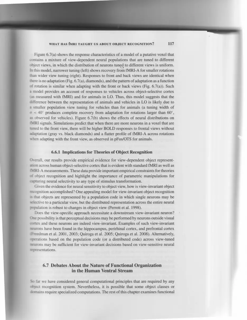
Figure 6.7. Simulations predicting fMRl responses of putative voxels containing a mixtureof view-dependent neural populations. Left, Schematic illustration of the view tuning arddistribution of neural populations tuned to different views in a voxel. For illustration purposeswe show a putative voxel with 4 neural populations. Right, Result of model simulationsillustrating the predicted fMRl-A data. In all panels, the model includes 6 Gaussians tunedto specific views around the viewing circle, separated 60' apart. Across columns, the vierrtuning width varies. Across rows, the distribution of neural populations preferring specificviews varies. Diamond, Responses without adaptation (black for back view, and gray for frontview). Lines, Response after adaptation with a front view (dashed gray line) or back view (solidblack line). (a) Mixture of view-dependent neural populations that are equally distributed ina voxel. Narrower tuning (left) shows recovery from fMRl-A for smaller rotations than widerview tuning (right). This model predicts the same pattern of recovery from adaptation whenadapting with the front or back view. (b) Mixture of view-dependent neural populations in avoxel with a higher proportion of neurons that prefer the front view The number on the rightindicates the ratio between the percentages neurons tuned to the front versus the back view.Top row, ratio - 1.2. Bottom row, ratio : 1.4. Because there are more neurons tuned to thefront view in this model, it predicts higher BOLD responses to frontal views without adaptation(gray vs. black diamonds) and a flatter profile of fMRl-A across rotations when adapting withthe front view. Adapted from Andreson, Vinberg, and Crill-Spector (in press).
(Fig. 6.7 and Andresen et al. 2008, in press). fMRI responses were modeled to beproportional to the sum of responses across all neural populations in a voxel. We sim-
ulated the fMRI responses in fMRI-A experiments for a set of putative voxels thatvaried in the view-tuning width of neural populations, the preferred view of differentneural populations, the number of different neural populations, and the distribution ofpopulations tuned to different views within a voxel. Results of the simulations indicate
that two main parameters affected the pattern of fMRI data: (l) the view-tuning width
of the neural population, and (2) the proportion of neurons in a voxel that prefer a
specific object view.
WHAT HAS fMRI TAUGHT US ABOUT OBJECT RECOGNITION? Lt7
Figure 6.7(a) shows the response characteristics of a model of a putative voxel thatcontains a mixture of view-dependent neural populations that are tuned to differentobject views, in which the distribution of neurons tunEd to different views is uniform.ln this model, niurower tuning (left) shows recovery f";fi fMRI-A for smaller rotationsthan wider view tuning (right). Responses to front and back views are identical whenthere is no adaptation (Fig. 6.7(a), diamonds), and the pattern of adaptation as a functionof rotation is similar when adapting with the front or back views (Fig. 6.7(a)). Suche model provides an account of responses to vehicles across object-selective cortexr.s measured with fMRI) and for animals in LO. Thus, this model suggests that thedifference between the representation of animals and vehicles in LO is likely due toe smaller population view tuning for vehicles than for animals (a tuning width ofo < 40o produces complete recovery from adaptation for rotations larger than 60o,rs observed for vehicles). Figure 6.7(b) shows the effects of neural distributions onfllRl signals. Simulations predict that when there are more neurons in a voxel that areuned to the front view, there will be higher BOLD responses to frontal views withoutdaptation (gray vs. black diamonds) and a flatter profile of fMRI-A across rotationsrben adapting with the front view, as observed in pFus/OTS for animals.
6.6.1Implications for Theories of Object Recognition
Orerall, our results provide empirical evidence for view-dependent object represent-son across human object-selective cortex that is evident with standard fMRI as well asfIIRI-A measurements. These data provide important empirical constraints for theoriesd object recognition and highlight the importance of parametric manipulations forqturing neural selectivity to any type of stimulus transformation.
Given the evidence for neural sensitivity to object view, how is view-invariant objectreognition accomplished? One appealing model for view-invariant object recognitionrr that objects are represented by a population code in which single neurons may ber{cctive to a particular view, but the distributed representation across the entire neuralgopulation is robust to changes in object view (Perrett et al. 1998).
Does the view-specific approach necessitate a downstream view-invariant neuron?ftc possibility is that perceptual decisions may be performed by neurons outside visualcutter and these neurons are indeed view-invariant. Examples of such view-invariant-urons have been found in the hippocampus, perirhinal cortex, and prefrontal cortextfi,aedman et al. 200I,2003; Quiroga et al. 2005; Quiroga et al. 2008). Alternatively,ogalations based on the population code (or a distributed code) across view-tuned:urons may be sufficient for view-invariant decisions based on view-sensitive neuralIpresentations.
6.7 Debates About the Nature of Functional Organizationin the Human Ventral Stream
So far we have considered general computational principles that are required by anySycct recognition system. Nevertheless, it is possible that some object classes or&srains require specialized computations. The rest of this chapter examines functional

118 KALANIT GRILL-SPECTOR
specialization in the ventral stream that may be linked to these putative "domain-specific" computations.
As illustrated in Figure 6.1, several regions in the ventral stream exhibit higher re-sponses to particular object categories,.such as places, faces, and body parts, comparedto other object categories. The discovery of category-selective regions initiated a fiercedebate about the principles of functional organization in the ventral stream. Are thereregions in the cortex that are specialized for any object category? Is there somethingspecial about computations relevant to specific categories that generates specializedcortical regions for these computations? In other words, perhaps some general pro-cessing is applied to all objects, but some computations may be specific to certaindomains and may require additional brain resources. We may also ask about how thesecategory-selective regions come about: Are they innate, or do they require experienceto develop?
Four prominent views have emerged to explaining the pattern of functional selectiv-ity in the ventral stream. The main debate centers on the question of whether regionsthat elicit maximal response for a category are a module for the representation of thatcategory, or whether they are part of a more general object recognition system.
6.7.1 Limited Category-Specific Modules and a General Areafor All Other Objects
Kanwisher and coworkers (Kanwisher 2000; Op de Beeck et al. 2008) suggested thatventral temporal cortex contains a limited number of modules specialized for the recog-nition of special object categories such as faces (in the FFA), places (in the PPA), andbody parts (in the EBA and FBA). The remaining object-selective cortex (LOC), whichshows little selectivity for particular object categories, is a general-purpose mecha-nism for perceiving any kind of visually presented object or shape. The underlyinghypothesis is that there are few domain-specific modules that perform computationsthat are specific to these classes of stimuli beyond what would be required from ageneral object recognition system. For example, faces, like other objects, need to berecognized across variations in their appearance (a domain-general process). However,given the importance of face processing for social interactions, there are aspects of faceprocessing that are unique. Specializedface processing may include identifying facesat the individual level (e.9., John vs. Harry), extracting gender information, evaluat-ing gaze and expression, and so on. These unique, face-related computations may beimplemented in face-selective regions.
6.7.2 Process Maps
Tarr and Gauthier (2000) proposed that object representations are clustered accordingto the type of processing that is required, rather than according to their visual attributes.It is possible that different levels of processing may require dedicated computationsthat are performed in localized cortical regions. For example, faces are usually rec-ognized at the individual level (e.9., "That is Bob Jacobs"), but many objects aretypically recognized at the category level (e.9., "That is a horse"). Following this rea-soning, and evidence that objects of expertise activate the FFA more than other objects
(Gauthisuggestordinat,et al. l(
Haxblcipitott
attributacrossHaxblwere n
distritr
that it
regionexcludreprev
On'
binato
Given(Biedt
a largr
consitas mu
Malanizatt
COTTC
and t
it ies
housfove
to fc
et al
biasanalrecc
tion
alscfielrwi t
I
fun

r.e "domain-
rit higher re-t-s. comparediated a fiercem. Are theree something; specializedgeneral pro-ic to ceftainut how thesee experience
rnal selectiv-:ther regionsation of thatstem.
nea
ggested that)r the recog-re PPA), and.OC), whichose mecha-underlying
rmputations
ired from aneed to be
). However,ects of facefying facesrn. evaluat-rns may be
accordingattributes.
nputations;ually rec-rbjects are
_e this rea-rer objects
wHAT HAs fMRr TAUGHT us ABour oBJEcr REcocNlrrox? 119
(Gauthier et al. 1999; Gauthier et al. 2000), Gauthier, Tarr, and their colleagues havesuggested that the FFA is not a region for face recognition, but rather a region for sub-ordinate identification of any object category that is automated by expertise (Gauthieret al. 1999: Gauthier et al. 2000: Tarr ahtl Gauthier 2000).
6.7.3 Distributed Object-Form Topography
Haxby and colleagues (2001) posited an "object-form topography," in which the oc-cipitotemporal cortex contains a topographically organized representation of shapeattributes. The representation of an object is reflected by a distinct pattern of responseacross all ventral cortex, and this distributed activation produces the visual perception.Haxby and colleagues showed that the activation patterns for eight object categorieswere replicable and that the response to a given category could be determined by thedistributed pattern of activation across all ventrotemporal cortex. Further, they showedthat it is possible to predict what object category the subjects viewed, even whenregions that show maximal activation to a particular category (e.9., the FFA) wereexcluded (Haxby et al. 2001). Thus, this model suggests that the ventrotemporal cortexrepresents object category information in an overlapping and distributed fashion.
One of the reasons that this view is appealing is that a distributed code is a com-binatorial code that allows representation of a large number of object categories.Given Biederman's rough estimate that humans can recognize about 30,000 categories(Biederman 1987), this provides a neural substrate that has a capacity to represent sucha large number of categories. Second, this model posited a provocative view that whenconsidering information in the ventral stream, one needs to consider the weak signalsas much as the strong signals, because both convey useful information.
6.7.4 Topographic Representation
Malach and colleagues (2002) suggested that eccentricity biases underlie the orga-nization of ventral and dorsal stream object-selective regions because they found acorrelation between category preference (higher response to one category over others)and eccentricity bias (higher response to a specific eccentricity than to other eccentric-ities (Levy et al. 200I; Hasson et al. 2002; Hasson et al. 2003). Regions that preferhouses to other objects also respond more strongly to peripheral stimulation than tofoveal stimulation. In contrast, regions that prefer faces or letters respond more stronglyto foveal stimulation than to peripheral stimulation. Malach and colleagues (Malachet al. 2002) proposed that the correlation between category selectivity and eccentricitybias is driven by spatial-resolution needs. Thus, objects whose recognition depends onanalysis of fine details are associated with foveal representations, and objects whoserecognition requires large-scale integration are associated with peripheral representa-tions. To date, however, there is no clear evidence that eccentricity biases in the FFA arealso coupled with better representation of high spatial frequency or smaller receptivefields or, conversely, that the PPA prefers low spatial frequencies or contains neuronswith larger receptive fields.
Presently, there is no consensus in the field about which account best explains thefunctional organization of the ventral stream. Much of the debate centers on the degree

r20 KALANIT GRILL.SPECTOR
to which object processing is constrained to discrete modules or involves distributedcomputations across large stretches of the ventral stream (Op de Beeck et al. 2008).The debate is about the spatial scale on which computations for object recognitionoccur as well as the fundamenthl principles that underlie specialization in the ventralstream.
On the one hand, domain-specific theories need to address findings of multiple focithat show selectivity. For example, multiple foci in the ventral stream respond morestrongly to faces than to objects; thus, a strong modular account of a single "facemodule" for face recognition is unlikely. Also, the spatial extent of these putativemodules is undetermined, and it is unclear whether each of these category-selectiveregions corresponds to a visual area. Further, high-resolution fMRI (1-2 mm on a side)shows that the spatial extent of category-selective regions is smaller than that estimatedwith standard fMRI (34 mm on a side) and that these regions appear more patchy(Schwarzlose et aL.2005; Grill-Spector et al. 2006b).
On the other hand, a potential problem with very distributed and overlapping accountof object representation in the ventral stream is that, in order to resolve categoryinformation, the brain may need to read out information present across the entireventral stream (which is inefficient). Further, the fact that there is information in thedistributed response does not mean that the brain uses the information in the same waythat an independent classifier does. It is possible that activation in localized regionsis more informative for perceptual decisions than the information available across theentire ventral stream (Grill-Spector et al. 2004; Williams et al. 2007). For example,FFA responses predict when subjects recognize faces and birds but do not predict whensubjects recognize houses, guitars, or flowers (Grill-Spector et aI.2004).
6.8 Differential Development of Category Selectivityfrom Childhood to Adulthood
One research direction that can shed light on these debates is an examination of thedevelopment of ventral stream functional organization. What is the role of experiencein shaping category selectivity in the ventral stream?
6.8.L fMRI Measurements of the Development of the Ventral Stream
To address these questions, our lab (Golarai et al. 2007) identified face-, place-, andobject-selective regions within individual children (7-11years old), adolescents (12-14years old), and adult subjects (18-35 years old) while subjects fixated and reportedinfrequent events when two consecutive images were identical (one-back task). Wefound a prolonged development of the right FFA (rFFA) and left PPA (IPPA) thatmanifested as an expansion of the spatial extent of these regions across developmentfrom age 7 to adulthood (Fig. 6.8). The rFFA and IPPA were significantly larger inadults than in children, with an intermediate size of these regions in adolescents.Notably, the rFFA of children was about a third of the adult size, but it was stillevident in 85Vo of children. These developmental changes could not be explained bysmaller anatomical cortical volumes of the fusiform gyrus or the parahippocampal

f'olves distributed:eck et al. 200gt.bject recognitionion in the ventral
; of multiple focim respond more,f a single ..face,f these putativefiegory-selective? mm on a side)n rhat estimatedar more patchy
apping accountsolve categoryross the entire)rmation in thet the same way'alized regionstble across theFor example,
t predict when
ty
narion of therf experience
itream
place-, andenB ( 12_14nd reportedc task). WeIIPPA) that:r'elopmenty' larger inlolescents.t was sdllllained bypocampal
f o ,ooo1 . ,
g;.qffi
2,5002,0001,5001,000
5000
2,500
€ ,,*€ r,sooE r,ooo€ soo
0
I Ll Match€d
I
ffi
; . r .600
3 ' -! 8oo
: 4 m
SHAT H^4s funr TAUGHT us ABour oBJEcr REcocNlrrox? L2l
RightPPA size
Left Right
LOC sizeLeft Right
ffi z-t t vear-old
I to1-J,9t"*-o'o
6.OOO
4,500
3,000
1,500
0
Figure 6.8. Volume of the rFFA, IPPA, STS, and LOC across children, adolescents, and adults.Fiiied bars indicate average volume across all subjects, which include 20 children, 10 adoles-cents, and 15 adults. Open bars indicate the average volumes for the subset of subjects thatnere matched for BOlD-related confounds and inilude 10 children, g adolescents, and 13adults. Error bars indicate SEM across subjects. Asterisks indicate significantly different thanadult, P < 0.05. Note that different panels have different scales on the y-axis. Adapted fromColarai et al. 2007.
gyrus (Golarai et al. 2007), which were similar across children and adults, or higher
BOlD-related confounds in children (i.e., larger fMRl-related noise or larger subject
motion; see Golarai et al. Z}}7;Grill-Spector et al. 2008) because results remained the
sarne for a subset of subjects that were matched for fMRl-related confounds across
ages. These developmental changes were specific to the rFFA and IPPA, because we
found no differences across ages in the size of the LOC or the size of the pSTS face-
selective region. Finally, within the functionally defined FFA, PPA, and LOC, therewere no differences across ages in the level of response amplitudes to faces, objects,
and places.We also measured recognition memory outside the scanner and found that that face-
and place-recognition memory increased from childhood to adulthood (Golarai et al.
2007). Further, face-recognition memory was significantly correlated with rFFA size( but not the size of other regions) in children and adolescents (but not adults), and place-
recognition memory was significantly correlated with IPPA size (but not the size of other
regions) in each of the age groups (Fig. 6.9). These data suggest that improvements in
face- and place-recognition memory during childhood and adolescence are correlated*'ith increases in the size of the rFFA and IPPA, respectively.
Another recent study (Scherf et al. 2007) examined the development of the ven-
rral stream in children (5-8 years old), adolescents (ll-14 years old), and adults
using movie clips that contained faces, objects, buildings, and navigation. Using group
analysis methods, they reported the absence of face-selective activations (vs. objects,
buildings, and navigation) in 5- to 8-year-old children in both the fusiform gyrus and
rhe pSTS. The lack of FFA in young children in the group analysis may be due to the
smaller and more variable FFA location in children, which would affect its detection
n i'iJ-'d'"#t$Ll Matched .

122
(a) Face recognitionmemory vs. rFFA size
0 1000 3000 5000
Right FFA Size[mmst
O t-tl years oldn = 2 0
KALANIT GRILL.SPECTOR
(b) Place recognitionmemory vs. IPPA size
(c) Object recognitionmemory vs. LOC size
0 2000 6000 10000
Right LOC size[mmgt
# 18-35 years oldn = 1 5
It
: cg ton : . Th t '
r t c s i ng t ' l l - r
:hc l i r : t h t , '' r r J t t hn : r r l l ;.c inrplcrt lcl :. lJ \C\ 1g$ t r
j \ [Cn \ l \ C r ' \ f
Thc rc l " '
\n \ r \ \ n . l r r rp
l f t r l t ; ' t l l l l l lCJ
r ' ' th urc' l lkc
:CpfL ' \C I l tJ l I \ '
3 \ [ ' r . - f l r " '0sC l r
: ' lJ tUrC . ! l i l
l l a : t l r - t t r r a t
[ : I i . \ rc- [ r 'n '
] r x x t r . [ : t , u f i
i r f fc ' r J I ' l r \ ' ' ! l :
z"oEo
c.9?_co)oooE.
1 . 0
0.8
0.7
0.6
0.4
0.2
0.0
4.2
0.8
0.7
0.6
0.4
0.3
0.2
0.1
1 .2
1 .0
0.8
0.7
0.6
0.4
o.2
0.0
Left PPA Size[mmst
I lz-t6 years oldn = 1 0
Figure 6.9. Recognition memory versus size of rFFA, iPPA, and LOC. (a) Recognition memoryaccuracy for different categories across age groups. Recognition memory for faces was signifi-can t lybe t te r inadu l ts thanch i ld ren(*P <0.0001)orado lescents ( * *P <0.03) .Ado lescents 'memory for faces was better than children's (**P < 0.03). Recognition memory for places wasbetter in adults than in children (t P . 0.0001). Adolescents' memory for places was better thanchildren's (+ P < 0.01). Recognition accuracy for objects was not different across age groups.Error bars indicate SEM. (b) Recognition memory for faces versus FFA size. Correlations aresignificant within children and adolescents (r > O.49, P < 0.03), but not adults. (c) Recog-nition memory of places versus PPA size. Correlations are significant within each age group(r > 0.59, P < 0.03). (d) Recognition memory for objects versus LOC size. No correlationswere significant (P > O.4). Adapted from Colarai et al. 2OO7.
in a group analysis. Indeed when Scherf and colleagues performed individual subject
analysis, they found face-selective activations in 807o of their child subjects, but the
extent of activations was smaller and more variable in location compared to those in
adults. Like Golarai and colleagues, they found no difference in the spatial extent orlevel of response amplitudes to objects in the LOC. Unlike Golorai and colleagues,
they reported no developmental changes in the PPA. The variant results may be due to
differences in stimuli (pictures vs. movies), task (one-back task vs. passive viewing),
and analysis methods (single subject vs. group analysis) across the two studies. For
example, Golarai and colleagues instructed subjects to fixate and perforrn a one-back
task. In this task, children had the same accuracy as adults but were overall slower
in their responses, with no differences across categories. In the Scherf study, subjectswatched movies, and perfonnance was not measured during the scan. It is possible that
the children had differential different eye movements or levels of attention than the
adults, and this affected their findings.
6.8.2 Implications of Differential Development of Visual Cortex
Overall, fMRI findings suggest differential developmental trajectories across the humanventral visual stream. Surprisingly, these data suggest that more than a decade isnecessary for the development of an adult-like rFFA and IPPA. This suggests thatexperience over a prolonged time may be necessary for the normal development of these

ognitionOC size
wHAT HAS fMRI TAUGHT us ABour oBJEcr REcocNrrrox? L23
regions. This result is surprising, especially because there is evidence for preferentialviewing of face-like in newborn babies and evidence for face-selective ERPs withinthe first 6 to 12 months (for a review, see Johnson 2001). One possibility suggestedby Johnson and colleagues is that face processing has an innate component that maybe implemented in subcortical pathways (e.g., through the superior colliculus), whichbiases newborns to look at faces. However, cortical processing of faces may requireextensive experience (Gauthier and Nelson 2001; Nelson 2001) and may develop later.
The reasons for differential development across ventral stream regions are un-known. Importantly, it is difficult to disentangle maturational components (geneticallyprogrammed developmental changes) from experience-related components, becauseboth are likely to play a role during development. One possibility is that the type ofrepresentations and computations in the rFFA and IPPA may require more time andexperience to mature than those in the LOC. Second, different cortical regions maymature at different rates owing to genetic factors. Third, the FFA may retain moreplasticity (even in adulthood) than the LOC, as suggested by studies that show thatFFA responses are modulated by expertise (Gauthier et al. 1999; Tarr and Gauthier2000). Fourth, the neural mechanisms underlying experience-dependent changes maydiffer among LOC, FFA, and PPA.
6.9 Conclusion
In sum, neuroimaging research in the past decade has advanced our understanding ofobject representations in the human brain. These studies have identified the functionalorganization of the human ventral stream, shown the involvement of ventral streamregions in object recognition, and laid fundamental stepping stones in understandingthe neural mechanisms that underlie invariant object recognition.
Many questions remain, however. First, what is the relationship between neural sen-sitivity to object transformations and behavioral sensitivity to object transformations?Do biases in neural representations produce biases in performance? For example, em-pirical evidence shows overrepresentation of the lower visual field in LO. Does thislead to better recognition in the lower visual field than in the upper visual field? Asecond question is related to the development of the ventral stream: To what extentis experience (vs. genes) necessary for shaping functional selectivity in the ventralstream? Third, do object representations remain plastic in adulthood? If so, what is thetemporal scale of plasticity, and are experience-induced changes long-lasting? Fourth,what computations are implemented in the distinct cortical regions that are involved inobject recognition? Does the "aha" moment in recognition involve a speciflc response ina particular brain region, or does it involve a distributed response across a large corticalexpanse? Combining experimental methods such as fMRI and MEG will provide bothhigh spatial and temporal resolution, which is critical to addressing this question. Fifth,what is the pattern of connectivity between ventral stream visual regions? Althoughthe connectivity in monkey visual cortex has been explored extensively (Van Essenet al. 1990; Moeller et al. 2008), we know little about the connectivity between corticalvisual areas in the human ventral stream. This knowledge is necessary for build-ing a model of hierarchical processing in humans and any neural network model of
stze
old
I memoryas signi f i -l lescents'laces wasetter thane groups.r t ions arel, Recog-8e group'relat ions
subject
but thethose inllent oreagues.: due toeu'ing).
es. Forte-back
slower
ubjectsrle thatran the
lumanade ists thatf these
'0@0

lu KALANIT GRILL-SPECTOR
object recognition. Approaches that combine methodologies, such as psychophysicswith fMRI, MEG with fMRI, or DTI with fMRI, will be instrumental in addressingthese fundamental questions.
Acknowledgments
I thank David Andresen, Golijeh Golarai, Rory Sayres, and Joakim Vinberg for theircontributions to the research summarized in this chapter. David Andresen, for his con-tributions to understanding viewpoint-sensitive representations in the ventral stream.Golijeh Golarai, for spearheading the research on the development of the ventral study.Rory Sayres, for his contributions to research on position invariance and distributedanalyses of the ventral stream. Joakim Vinberg, for his dedication to researching cue-invariant and figure-ground processing in the ventral stream. I thank David Remus,Kevin Weiner, Nathan Witthotft, and two anonymous reviewers for comments on thismanuscript. This work was supported by National Eye Institute Grants 5 R21 EY-016199-02 and Whitehall Foundation Grant 2005-05-I II-RES.
Bibliography
Andresen DR, Vinberg J, Grill-Spector K. 20[19. The representation of object viewpoint in the humanvisual cortex. Neuroimage 45(2):522-536. E pub Nov, 25, 2008.
Appelbaum LG, Wade AR, Vildavski VY, Pettet MW, Norcia AM. 200f, Cue-invariant networks forfigure and background processing in human visual cortex. J Neurosci 26:11,695-1 1,708.
Bar M, Tootell RB, Schacter DL, Greve DN, Fischl B, Mendola JD, Rosen BR, Dale AM. 2001.
Cortical mechanisms specific to explicit visual object recognition. Neuron29:529-535.
Berl MM, Vaidya CJ, Gaillard WD.2006. Functional imaging of developmental and adaptive changesin neurocognitton. N e uroitnage 30:67 949 l.
Biederman I. 1987. Recognition-by-components: a theory of human image understanding. PsycholRev 94:115-147.
Biederman I, Cooper EE. 1991. Evidence for complete translational and reflectional invariance invisual object priming. Pe rc e ption 20 : 5 85-593.
Booth MC, Rolls ET. 1998. View-invariant representations of familiar objects by neurons in theinferior temporal visual cortex. Cereb Cortex 8:510-523.
Bulthoff HH, Edelman S. 1992. Psychophysical support for a two-dimensional view interpolationtheory of object recognition. Proc Natl Acad Sci USA 89:60{4.
Bulthoff HH, Edelman SY, Tarr MJ. 1995. How are three-dimensional objects represented in thebrain? Cereb Conex 5:247-2ffi.
Chao LL, Haxby JV, Martin A. 1999. Attribute-based neural substrates in temporal cortex for per-
ceiving and knowing about objects. Nat Neurosci2:913-919.
Cohen L, Dehaene S, Naccache L, lrhericy S, Dehaene-Lambertz G, Henaff MA, Michel F. 2000.The visual word form area: spatial and temporal characteization of an initial stage of reading innormal subjects and posterior split-brain patients. Brain 123 (Pt 2):291-307.
Culham JC, Danckert SL, DeSouzaJF, Gati JS, Menon RS, Goodale MA. 2003. Visually guidedgrasping produces fMRI activation in dorsal but not ventral stream brain areas. Exp Brain Res153:18f189.
Desimone R, Albright TD, Gross CG, Bruce C. 1984. Stimulus-selective properties of inferiortemporal neurons in the macaque. J Neurosci 4:2051-2M2.
II
I
WHAT HAS fMRI TAUGHT US ABOUT OBJECT RECOGNITION? L25
DiCarlo JJ, Cox DD.2007. Untangling invariant object recognition. Trends Cogn Sci ll:333-341.
DiCarlo JJ, Maunsell JH. 2003. Anterior inferotemporal neurons of monkeys engaged in objectrecognition can be highly sensitive to object retinal position. J Neurophysiol S9:3264- 3278.
Dill M, Edelman S. 2001. Imperfect invariance to object translati6ti in the discrimination of complex
shapes. Perception 3O:7 O7 -7 24.Downing PE, Jiang Y, Shuman M, Kanwisher N. 2001. A cortical area selective for visual processing
of the human body. Science 293:247V2473.Fielman S, Bulthoff HH. 1992. Orientation dependence in the recognition of familiar and novel
views of three-dimensional objects. Vision Res 32:2385-24ffi.Edelman S, Intrator N. 2000. (Coarse coding of shape fragments) * (retinotopy) approximately :
representation of structure. Spat Vis 13:255-264.Eger E, Ashburner J, Haynes JD, Dolan RI, Rees G. 2008. fMRI activity patterns in human LOC
carry information about object exemplars within category. J Cogn Neurosci 20:35G370.
Epstein R, Kanwisher N. 1998. A cortical representation of the local visual environment. Nature
392:598-601.
Epstein RA, Parker WE, Feiler AM. 2008. Two kinds of fMRI repetition suppression? Evidence for
d i s sociable neural mechanism s. J N e u ro p hy s io I 99 (6) :287 7 -2886.
Fang F, He S. 2005. Cortical responses to invisible objects in the human dorsal and ventral pathways.
Nat Neurosci 8: 1 380-1 385.Fang F, Murray SO, He S. 2007. Duration-dependent FMRI adaptation and distributed viewer-centered
face representation in human visual cortex. Cereb Cortex 17:14O2-1411.
Farah MJ. 1995. Visual agnosia. Cambridge: MIT Press.
Freedman DJ, Riesenhuber M, Poggio T, Miller EK. 2001 . Categorical representation of visual stimuli
in the primate prefrontal cortex. Science 291312-316.
Freedman DJ, Riesenhuber M, Poggio T, Miller EK. 2003. A comparison of primate prefrontal andinferior temporal cortices during visual categoization. J Neurosci 23:5235-5246.
Fujita I, Tanaka K, Ito M, Cheng K. 1992. Columns for visual features of objects in monkey
i nferotemporal cortex . N ature 360:343-346.Gauthier I, Hayward WG, Tarr MJ, Anderson AW, Skudlarski P, Gore JC.2002. BOLD activity
during mental rotation and viewpoint-dependent object recognition . Neuron 34:16l-17l.
Gauthier I, Nelson CA. 2001. The development of face expertise. Curr Opin Neurobiol lI:219-224-
Gauthier I, Skudlarski P, Gore JC, Anderson AW. 2000. Expertise for cars and birds recruits brain
areas involved in face recognition. Nat Neurosci 3:l9l-197 .
Gauthier I, Tarr MJ, Anderson AW, Skudlarski P, Gore JC. 1999. Activation of the middle fusiform'face area' increases with expertise in recognizing novel objects. Nat Neurosci 2:568-573.
Gilaie-Dotan S, Ullman S, Kushnir T, Malach R. 2002. Shape-selective stereo processing in human
object-related visual areas. Hum Brain Mapp 15:67--79.
Golarai G, Ghahremani DG, Whitfield-Gabrieli S, Reiss A, Eberhardt JL, Gabrieli JD, Grill-Spector
K. 2001. Differential development of high-level visual cortex correlates with category-specific
recognition memory. Nat Neurosci lO:512-522.Grill-Spector K. 2003. The neural basis of object perception. Curr Opin Neurobiol l3:159-166.
Grill-Spector K, Golarai G, Gabrieli J. 2008. Developmental neuroimaging of the human ventral
visual cortex. Trends Cogn Sci 12:152-162.
Grill-Spector K, Henson R, Martin A. 2006a. Repetition and the brain: neural models of stimulus-
specific effects. Trends Cogn Sci 10:14-23.
Grill-Spector K, Knouf N, Kanwisher N. 2ffi4. The fusiform face area subserves face perception, not
generic within-category identifi cation. N at N euros c i 7 :555-562.
Grill-Spector K, Kushnir I Edelman S, Avidan G, Itzchak Y, Malach R. 1999. Differential processing
of objects under various viewing conditions in the human lateral occipital complex. Neuron24:187-
203.
126 KALANIT GRILL.SPECTOR
Grill-Spector K, Kushnir T, Edelman S, Itzchak X Malach R. 1998a. Cue-invariant activation in
object-related areas of the human occipital lobe. Neuron 2l:l9l-202.
Grill-Spector K, Kushnir T, Hendler T, Edelman S, Itzchak Y Malach R. 1998b. A sequence of object-
processing stages revealed by fMRI'iri the human occipital lobe. Hum Brain Mapp 6:316-328.
Grill-Spector K, Kushnir T, Hendler T, Malach R. 2000. The dynamics of object-selective activation
correlate with recognition performance in humans. Nat Neurosci 3:837-843.
Grill-Spector K, Malach R. 2001. fMR-adaptation: a tool for studying the functional properties of
human cortical neurons. Acta Psychol (Amst) 107:293-321.
Grill-Spector K, Malach R. 2fiX. The human visual cortex. Annu Rev Neurosci 27:649477.
Grill-Spector K, Sayres R, Ress D. 2006b. High-resolution imaging reveals highly selective nonface
clusters in the fusiform face area. Nat Neurosci 9:1177-1185.
Hasson U, Harel M, Levy I, Malach R. 2003. Large-scale mirror-symmetry organization of human
occipito-temporal object areas. N e uron 3l :1027 -1041.
Hasson U, Levy I, Behrmann M, Hendler T, Malach R. 2002. Eccentricity bias as an organizing
principle for human high-order object areas. Neuron 34:479490.
Haxby JV Gobbini MI, Furey ML, Ishai A, Schouten JL, Pietrini P. 2001. Distributed and overlapping
representations of faces and objects in ventral temporal cortex. Science 293:2425-2430.
Haxby JV Hoffman EA, Gobbini MI. 2000. The distributed human neural system for face perception.
Trends Cogn Sci 4:223-233.
Hemond CC, Kanwisher NG, Op de Beeck HP. 2007. A preference for contralateral stimuli in human
object- and face-selective cortex. PlnS ONE 2:e574.
Hoffman EA, Haxby JV. 2000. Distinct representations of eye gaze and identity in the distributed
human neural system for face perception. Nat Neurosci 3:8G-84.
James TW, Humphrey GK, Gati JS, Menon RS, Goodale MA. 2002. Differential effects of viewpoint
on object-driven activation in dorsal and ventral streams. Neuron 35:793-801.
Johnson MH. 2001. Functional brain development in humans. Nat Rev Neurosci 2:475483.
Kanwisher N. 2000. Domain specificity in face perception. Nat Neurosci 3:759-763.
Kanwisher N, McDermott J, Chun MM. 1997. The fusiform face area: a module in human extrastriate
cortex specialized for face perception. J Neurosci 17:43024311.
Kastner S, De Weerd P, Ungerleider LG. 2000. Texture segregation in the human visual cortex: a
functional MRI study . J N europhy s iol 83 :2453-2457 .
Kobatake E, Tanaka K. 1994. Neuronal selectivities to complex object features in the venffal visual
pathway of the macaque cerebral cortex. J NeurophysiolTl:85G867.
Kourtzi Z, Kanwisher N. 2000. Cortical regions involved in perceiving object shape. J Neurosci
20:3310-3318.
Kourtzi Z, Kanwisher N. 200 1 . Representation of perceived object shape by the human lateral occipital
complex. S c ie nc e 293 : I 506-1 509.
Kourtzi Z, Tolias AS, Altmann CF, Augath M, Logothetis NK. 2W3. Integration of local features
into global shapes: monkey and human FMRI studies. Neuron3T:333-346.
Lerner I Epshtein B, Ullman S, Malach R. 2008. Class information predicts activation by object
fragments in human object areas. J Cogn Neurosci 20:1189-1206.
Lerner Y Hendler T, Ben-Bashat D, Harel M, Malach R. 2001. A hierarchical axis of object processing
stages in the human visual cortex. Cereb Cortex II:287-297.
Lerner Y, Hendler T, Malach R. 2002. Object-completion effects in the human lateral occipital
complex. Cereb Cortex 12:163-177.
Levy I, Hasson U, Avidan G, Hendler I Malach R. 2001. Center-periphery organization of human
object areas. Nat Neurosci 4:533-539.
Logothetis NK, Pauls J, Poggio T. 1995. Shape representation in the inferior temporal cortex of
monkeys. Curc Biol 5:552-563.

nant activation in
cquence ofobjecftpp 6:316-328.elective activation
onal properties of
' :619477.
selective nonface
lrzation of human
as an organizing
d and overlapping-i-t-t30.rr lace perception.
stimuli in human
rn rhe distributed
iects of viewpoint
{7-5-.183.
u*un extrastriate
r r isual cortex: a
:he \ entral visual
tape. ./ Neurosci
n lateral occipital
of local features
ration by object
,bject processing
lateral occipital
.ation of human
rporal cortex of
WHAT HAS fMRI TAUGHT US ABOUT OBJECT RECOGNITION? 127
Lueschow A, Miller EK, Desimone R. 1994. Inferior temporal mechanisms for invariant objectrecognition . Cereb Cortex 4:523-53I.
Malach R, Levy I, Hasson U.2002. The topography of high-order human object areas. Trends CognSci 6: l7Gl84.
Malach R, Reppas JB, Benson RR, Kwong KK, Jiang H, Kennedy WA, Ledden PJ, Brady TJ, RosenBR, Tootell RB. 1995. Object-related activity revealed by functional magnetic resonance imaging
in human occipital cortex. Proc Natl Acad Sci USA 92:8135-8139.
Man D. 1980. Visual information processing: the structure and creation of visual representations.Philos Trans R Soc Innd B Biol Sci 29O:199-218.
Martin A, Wiggs CL, Ungerleider LG, Haxby JV. 1996. Neural correlates of category-specific knowl-edge. Nature 37 9:649452.
McKyton A, Zohary 8. 2007. Beyond retinotopic mapping: the spatial representation of objects inthe human lateral occipital complex. Cereb Cortex l7:1164-1112.
Mendola JD, Dale AM, Fischl B, Liu AK, Tootell RB. 1999. The representation of illusory andreal contours in human cortical visual areas revealed by functional magnetic resonance imaging.
J Neurosci I 9:8560-8572.
Miller EK, Li L, Desimone R. 1991. A neural mechanism for working and recognition memory ininferior temporal cortex. Science 254:1377 -1379.
Moeller S, Freiwald WA, Tsao DY. 2008. Patches with links: a unified system for processing faces inthe macaque temporal lobe. Science 320:7355-1359.
Nakayama K, He ZJ, Shimojo S. 1995. Visual surface representation: a critical link between low-level
and high-level vision. ln An invitation to cognitive sciences: visual cognition, ed. SM Kosslyn andDN Osherson. Cambridge: MIT Press.
Nelson CA. 2001. The development and neural bases of face recognition. Infant Child Develop
l0 :3-18.
Op de Beeck HP, Haushofer J, Kanwisher NG. 2008. Interpreting fMRI data: maps, modules anddimensions. Nat Rev Neurosci 9:123-135.
Op De Beeck H, Vogels R. 2000. Spatial sensitivity of macaque inferior temporal neurons. J CompNeurol426:505-518.
Perrett DI. 1996. View-dependent coding in the ventral stream and its consequence for recognition.
ln Vision and movement mechanisms in the cerebral cortex, ed. R Camaniti, KP Hoffmann, andAJ Lacquaniti, 142-151. Strasbourg: HFSP.
Perrett DI, Oram MW Ashbridge E. 1998. Evidence accumulation in cell populations responsive
to faces: an account of generalisation of recognition without mental transformations. Cognition67:l l I-145.
Peterson MA, Gibson BS. 1994a. Must shape recognition follow figure-ground organization?An
assumption in peril. Psychol Sci 5:253-259.
Peterson MA, Gibson BS. 1994b. Object recognition contributions to figure-ground organization:
operations on outlines and subjective contours. Percept Psychophys 56:551-564.
Poggio T, Edelman S. 1990. A network that learns to recognize three-dimensional objects. Nature
343:263-266.
Quiroga RQ, Mukamel R, Isham EA, Malach R, Fried I. 2008. Human single-neuron responses at
the threshold of conscious recognition. Proc Natl Acad Sci USA 105:3599-3604.
Quiroga RQ, Reddy L, Kreiman G, Koch C, Fried I. 2005. Invariant visual representation by single
neurons in the human brain. Nature 435:1102-1107.
Riesenhuber M, Poggio T. 1999. Hierarchical models of object recognition in cortex. Nat Neurosci
2:1019-1025.
Rolls ET. 2000. Functions of the primate temporal lobe cortical visual areas in invariant visual object
and face recognition . Neuron 27:205-218.
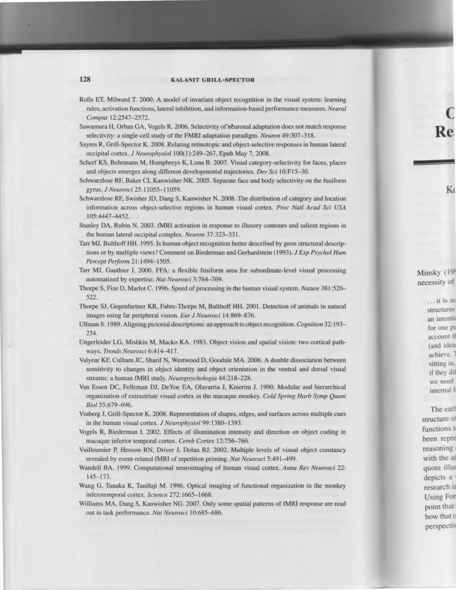
KALANIT GRILL.SPECTOR
Rolls ET, Milward T. 2000. A model of invariant object recognition in the visual system: learning
rules, activation functions, lateral inhibition, and information-based performance measures. Neural
Comput 12:2547-2572.
Sawamura H, Orban GA, Vogels R. 2006. Selectivity ofhburonal adaptation does not match response
selectivity: a single-cell study of the FMRI adaptation paradigm. Neuron 49:307-318.
Sayres R, Grill-Spector K. 2008. Relating retinotopic and object-selective responses in human lateral
occipital cortex. J Neurophysiol 100(1):249-267, Epub May 7,2008.
Scherf KS, Behrmann M, Humphreys K, Luna 8.2N7. Visual category-selectivity for faces, places
and objects emerges along different developmental trajectories. Dev Sci l0:F15-30.
Schwarzlose RF, Baker CI, Kanwisher NK. 2005. Separate face and body selectivity on the fusiformgyrus. J Neurosci 25:11055-11059.
Schwarzlose RF, Swisher JD, Dang S, Kanwisher N. 2008. The distribution of category and location
information across object-selective regions in human visual cortex. Proc Natl Acad Sci USAlO5:4447 4452.
Stanley DA, Rubin N. 2003. fMRI activation in response to illusory contours and salient regions in
the human lateral occipital complex. Neuron3T:323-331.
Tarr MJ, BulthoffHH. 1995. Is human object recognition better described by geon structural descrip-
tions orby multiple views? Comment on Biederman and Gerhardstein (1993). J Exp Psychol Hum
Pe rc e pt Pe rfo rm 2l : | 49Ll 505 .
Tarr MJ, Gauthier I. 2000. FFA: a flexible fusiform area for subordinate-level visual processing
automatized by expertise. Nat Neurosci 3:764-769.
Thorpe S, Fize D, Marlot C. 1996. Speed of processing in the human visual system. Nature 381:520-
522.
Thorpe SJ, Gegenfurtner KR, Fabre-Thorpe M, Bulthoff HH. 2001. Detection of animals in natural
images using far peripheral vision. Eur J Neurosci 14:869-876.
Ullman S. 1989. Aligning pictorial descriptions: an approach to object recognition . Cognition32:193-
254.
Ungerleider LG, Mishkin M, Macko KA. 1983. Object vision and spatial vision: two cortical path-
ways. Trends Neurosci 6:414417.
Valyear KF, Culham JC, Sharif N, Westwood D, Goodale MA. 2006. A double dissociation between
sensitivity to changes in object identity and object orientation in the ventral and dorsal visual
streams: a human fMRI study. Neuropsychologia 44:218-228.
Van Essen DC, Felleman DJ, DeYoe EA, Olavarria J, Knierim J. 1990. Modular and hierarchical
organization of extrastriate visual cortex in the macaque monkey. Cold Spring Harb Symp QuantBiol55:679496.
Vinberg J, Grill-Spector K. 2008. Representation of shapes, edges, and surfaces across multiple cues
in the human visual cortex. J Neurophysiol99:1380-1393.
Vogels R, Biederman l. 2ffi2. Effects of illumination intensity and direction on object coding in
macaque inferior temporal cortex. Cereb Cortex l2:75G766.
Vuilleumier P, Henson RN, Driver J, Dolan RI. 2002. Multiple levels of visual object constancy
revealed by event-related fMRI of repetition priming. Nat Neurosci 5:491499.
Wandell BA. 1999. Computational neuroimaging of human visual cortex. Annu Rev Neurosci 22;
145-173.
Wang G, Tanaka K, Tanifuji M. 1996. Optical imaging
inferotemporal cortex . S cienc e 27 2: 1665-1668.
Williams MA, Dang S, Kanwisher NG. 2m7. Only some
out in task performance. Nat Neurosci 10:685-686.
of functional organization in the monkey
spatial patterns of fMRI response are read
Re,
J-
MinskY t I $
necessit \ of
. . . i t i : n t '
structurc'
an intcntt '
for onc Puaccount tt
(and idca'
achier c. I
si t t ing rn-
if ther .hi
we nc'cd
internal i'
The earl
structurt ' ()1
functions t '
been rePre
reasonins ;
with thc' ai
quote i l lu :
depicts a '
research ir
Using Ft'rrpoint that I
how that o
persp€cti\
Kt
t.,.*
I

(b)E t oJE o
T*Eg o
I,10m
Prtrntrllon Dundm {mr}
Plate 6.1. Object-, face-, and place-selective cortex. (a) Data of one representive subjectshown on her part ial ly inf lated r ight hemisphere: lateral view (/eft); ventral view (r ight). Darkgray indicates sulci. Light gray indicates gyri. Black /ines delineate retinotopic regions. Blueregions del ineate object-selective regions (objects > scrambled objects), including LO andpFus/OTS ventral ly as well as dorsal foci along the intraparietal sulcus ( lPS). Red regionsdelineate face-selective regions (faces > non-faces objects), including the FFA, a region inLOS and a region in posterior STS. Magenta regions delineate overlap between face- andobjects-selective regions. Creen regions del ineate place-selective regions (places > objects),including the PPA and a dorsal region lateral to the lPS. Dark green regions del ineate overlapbetween place- and object-selective regions. Al l maps thresholded at P < 0.001, voxel level.(b) LO and pFus/OTS (but not Vl) responses are correlated with recognit ion performance (Cri l l -Spector et al. 2000). To superimpose recognit ion performance and fMRl signals on the sameplot, al l values were normalized relat ive to the maximum response for the 500-ms durations t imu lus .
For fMRl signals (blue, red, and oran %sien*#!t i : l l .
For recognit ion perfor-ge l tnes) : -o/orignal(Soo ms)
mance (black) : "h correct(condition)
"/"correct(S00 ms)
Pfr
0

Plate 6.2. Selective responses to objects across multiple visual cues across the LOC. Statisticalmaps of selective response to object from luminance, stereo and motion information in arepresentative subject. All maps were thresholded at P < 0.005, voxel level, and are shown onthe inflated right hemisphere of a representative subject. (a) Luminance objects > scrambledobjects. (b) Objects generated from random dot stereograms versus structureless random dotstreograms (perceived as a cloud of dots). (c) Objects generated from dot motion versus thesame dots moving randomly. Visual meridians are represented by red (upper), blue (horizontal),and green (lower) lines. White contour indicates a motion-selective region, or MT. Adaptedfrom Vinberg and Cril l-Spector 2008.
9o Left 4'Lefr Fovea 4o Right 4o up 4o Down
;ubject,. Darks. BlueO andegions
; ion ine- andrjects),rverlapI level., C r i l l -l samel ra t ion
rerlor-
rltltltrllllllffllllll
- 2 - 1 0 1 2Amplitude versus mean rosponse
% Signal Change
Plate 6.4. LO distributed response patterns to different object categories and stimulus po-sitions. Data are shown on the lateral aspect of the right hemisphere cortical surface for arepresentative subject. Each panel shows the distributed LO fMRl amplitudes after subtractingfrom each voxel its mean response. Red and yellow, Responses that are higher than the voxel'smean response. Blue and cyan, Responses that are lower than the voxel's mean response.lnset,Inflated right cortical hemisphere, with red square indicating the zoomed region. Notethat pattern changes significantly across columns (positions) and to a lesser extent across rows(categories). Adapted from Sayres and Grill-Spector 2008.


