Embed Size (px)
Citation preview
Vol. /No. Winter 2008
Member Publication
American Association
of Dental Editors
Copyright © 2008 Massachusetts DentalSociety ISSN: 0025-4800
The JOURNAL OF THE MASSACHUSETTS
DENTAL SOCIETY [USPS 284-680] is owned and published quarterly by theMassachusetts Dental Society, Two WillowStreet, Suite 200, Southborough, MA01745-1027. Subscription for nonmembersis $15 a year in the United States.Periodicals postage paid at Southborough,MA, and additional mailing offices.
Postmaster: Send address changes to:JOURNAL OF THE MASSACHUSETTS DENTAL
SOCIETY, Two Willow Street, Suite 200,Southborough, MA 01745.
Contributions: Please see page 53, contactthe Communications Department, or visitwww.massdental.org for author’s guidelines.
Display ad closing dates: February 1, May 1, August 1, November 1. For moreinformation, contact Rachel Marks,Exhibits Coordinator, at (508) 480-9797,ext. 259, or email [email protected].
JOURNAL OF THE
MASSACHUSETTS DENTAL
SOCIETY
EDITOR
Dr. David B. Becker
ASSISTANT EDITOR
Dr. Arthur I. Schwartz
EDITOR EMERITUS
Dr. Norman Becker
MANAGING EDITOR OF
PUBLICATIONS AND WEB SITE
Melissa Carman
MANAGER, GRAPHIC DESIGN
Jeanne M. Burdette
GRAPHIC DESIGNER
Shelley Padgett
EDITORIAL BOARD
Bruce Donoff, DMD, MD
Robert Faiella, DMD
Russell Giordano, DMD
Shepard Goldstein, DMD
Stephen McKenna, DMD
John McManama, DDS
Noshir Mehta, DMD
Charles Millstein, DMD
Philip Millstein, DMD
Maria Papageorge, DMD
Michael Sheff, DMD
Steven Tonelli, DMD
Editorial
AS DENTISTS, WE WORK IN OUR OWN CONTROLLED ENVIRON-ment, insulated from public opinion, and often do nothave a clue as to what the real world is thinking about
the services we provide. We develop our own relationships withour patients and, even in our isolation, believe that our patientsknow how beneficial our treatment is and that they respect usand what we do. Every practice has its own “personality” andattracts patients who fit that personality. If we are lucky, we arecontent with the knowledge that those we treat are comfortableand satisfied with us and our care.
In reality, we often do not know what the general publicreally thinks. A case in point is an article published in the New
York Times on October 11, 2007, which does not paint a verypretty picture of dentists or our profession. The article begins:
“For American dentists, times have never been better. Thesame cannot be said for Americans’ teeth.
With dentists’ fees rising far faster than inflation and morethan 100 million people lacking dental insurance, the per-centage of Americans with untreated cavities began risingthis decade, reversing a half-century trend of improvementin dental health.
Previously unreleased figures from the Centers for DiseaseControl and Prevention show that in 2003 and 2004, themost recent years with data available, 27 percent of chil-dren and 29 percent of adults had cavities going untreated.The level of untreated decay was the highest since the late1980s and significantly higher than that found in a surveyfrom 1999 to 2002.
Despite the rise in dental problems, state boards of dentistsand the American Dental Association, the main lobbyinggroup for dentists, have fought efforts to use dental hygienistsand other non-dentists to provide basic care to people whodo not have access to dentists.”
Journal of the Massachusetts Dental Society
The article also states:
“But many poor and lower-middle-class families do notreceive adequate care, in part because most dentists wantcustomers who can pay cash or have private insurance, andthey do not accept Medicaid patients. As a result, publiclysupported dental clinics have months-long waiting lists evenfor people who need major surgery for decayed teeth.”
Here is our problem: We know how much our profession isdoing to improve access to care, but obviously we are not get-ting the message out to the public, thus allowing an article likethis to have a much larger impact than it should. What the New
York Times did not take into account is that:
• The Massachusetts Dental Society (MDS) operates aMobile Access to Care (MAC) Van, which goes to areaswith documented unmet pediatric dental needs. The chil-dren are triaged and treated, but most importantly, they areput in a system that provides follow-up care for the chil-dren and health education for the parents or guardians.Dentists in each dental district provide free care and are set-ting up systems to teach parents how to enroll in theMassHealth (Medicaid) dental program. There is also anongoing process to get more dentists to enroll asMassHealth providers.
• The MDS and the American Dental Association (ADA),through our governmental affairs programs, have priori-tized our focus to improve access to care. At the ADA’sWashington Leadership Conference and the recent Houseof Delegates Session in San Francisco, a substantial portionof the proceedings was dedicated to strategizing aboutefforts for getting dental care to those who are most inneed. Dentists from all over the country and from all polit-ical persuasions were unified in trying to get Congress andthe Bush administration to listen and to increase the neededbudget allocations.
• The MDS and ADA are working with the Massachusettscongressional delegation on the access-to-care issue. Theirefforts include reauthorizing the State Children’s HealthInsurance Program (SCHIP) legislation.
Additionally, the New York Times article states that den-tistry is holding back the ability of hygienists to provide careto those in need. In reality, the MDS has backed pending leg-islation that allows auxiliaries to provide care, in dentist-supervised settings. A very small but organized group ofhygienists has been actively seeking the right to provideunsupervised dental procedures to those in need. They areplanning to set up private clinics. This would result in a two-tiered system of care, which would not provide those in needwith equal quality of services.
Hygienists can be licensed after only two years of educationfollowing high school. Considering the severity and complexityof emergency situations that dental offices and clinics routinelydeal with, we are quite concerned about the level of diagnosisand subsequent quality of care that would be provided tounderserved patients by less-qualified practitioners in theseunsupervised situations. Everyone deserves equal care in a safesetting.
Part of the problem with access to care is that we are noteducating enough dentists. The New York Times article misrep-resents a position that the opening of new schools is not desir-able. It states: “. . . the ADA does not support opening new den-tal schools or otherwise increasing the number of dentists.”Actually, the opposite is true. Organized dentistry feels that theopening of new schools would go a long way in helping to solvethe provider shortage problem. The ADA is spearheading aneducational foundation (led by Dr. Arthur Dugoni, ADA pastpresident and dean of the University of the Pacific) that isattempting to raise $1 billion specifically for dental education.
Dental school education is the most expensive of thelearned professions. Government subsidies have diminished tothe point that many dental schools have been forced to close. Itis common for recent graduates to start their professional liveswith educational debt as high as $250,000. Efforts to get thefederal government involved have fallen on deaf ears. Forexample, loan forgiveness programs in exchange for working inthe National Health Services or the Indian Health Services havefaced major roadblocks and lack of funding over the past fewyears. A second example is the funding for general practice res-idencies. Every year, the Bush administration cuts out this fund-ing and we have to fight to get it reinstated. The sad fact is thatthis year it was only in the $30 million range—a drop in thebucket. What makes this fact more appalling is that these clinicsor hospital-based residencies are often the only access to carefor the underserved or elderly in a given area.
We know that we are preaching to the choir, but it isextremely frustrating to read an article with the bias this promotes. There is no denial that our profession provides uswith satisfaction and rewarding experiences. We know thatmost of our colleagues care about giving back to society andhelping others. Individually and collectively, we have acteddirectly and through our elected officials to provide superiordental care to all socioeconomic groups. The New York Times
would better serve its readers and our society by joining theeffort to improve access to quality dental care rather thanattacking the people who are working to do just that. n
CORRECTING, NOT CRITIQUING,
THE ACCESS-TO-CARE ISSUE
FINANCIAL SERVICES CORNER
Editor’s Note: The following is intended to be informational. You should consult with your financial advisor before investing.
This article is brought to you by Eastern Dental Financial Services. Printed with permission from Liberty Publishing, Inc.
IF YOU’RE A MEMBER OF AMERICA’S LARGEST GENERATION,the baby boomers, you’ll be entering retire-ment in the coming years. With this in
mind, now may be a good time to pre-view some of the retirement planningchoices you’re bound to encounterin the years ahead.
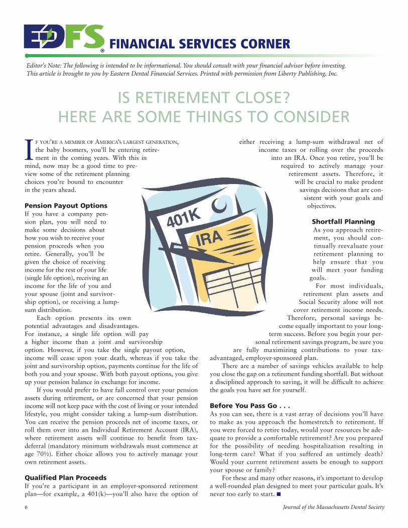
Pension Payout Options
If you have a company pen-sion plan, you will need tomake some decisions abouthow you wish to receive yourpension proceeds when youretire. Generally, you’ll begiven the choice of receivingincome for the rest of your life(single life option), receiving anincome for the life of you andyour spouse (joint and survivor-ship option), or receiving a lump-sum distribution.
Each option presents its ownpotential advantages and disadvantages.For instance, a single life option will pay a higher income than a joint and survivorshipoption. However, if you take the single payout option,income will cease upon your death, whereas if you take thejoint and survivorship option, payments continue for the life ofboth you and your spouse. With both payout options, you giveup your pension balance in exchange for income.
If you would prefer to have full control over your pensionassets during retirement, or are concerned that your pensionincome will not keep pace with the cost of living or your intendedlifestyle, you might consider taking a lump-sum distribution.You can receive the pension proceeds net of income taxes, orroll them over into an Individual Retirement Account (IRA),where retirement assets will continue to benefit from tax-deferral (mandatory minimum withdrawals must commence atage 70½). Either choice allows you to actively manage yourown retirement assets.
Qualified Plan Proceeds
If you’re a participant in an employer-sponsored retirementplan—for example, a 401(k)—you’ll also have the option of
either receiving a lump-sum withdrawal net ofincome taxes or rolling over the proceeds
into an IRA. Once you retire, you’ll berequired to actively manage your
retirement assets. Therefore, itwill be crucial to make prudent
savings decisions that are con-sistent with your goals andobjectives.
Shortfall Planning
As you approach retire-ment, you should con-tinually reevaluate yourretirement planning tohelp ensure that youwill meet your funding
goals. For most individuals,
retirement plan assets andSocial Security alone will not
cover retirement income needs.Therefore, personal savings be-
come equally important to your long-term success. Before you begin your per-
sonal retirement savings program, be sure youare fully maximizing contributions to your tax-
advantaged, employer-sponsored plan.There are a number of savings vehicles available to help
you close the gap on a retirement funding shortfall. But withouta disciplined approach to saving, it will be difficult to achievethe goals you have set for yourself.
Before You Pass Go . . .
As you can see, there is a vast array of decisions you’ll haveto make as you approach the homestretch to retirement. Ifyou were forced to retire today, would your resources be ade-quate to provide a comfortable retirement? Are you preparedfor the possibility of needing hospitalization resulting inlong-term care? What if you suffered an untimely death?Would your current retirement assets be enough to supportyour spouse or family?
For these and many other reasons, it’s important to developa well-rounded plan designed to meet your particular goals. It’snever too early to start. n
Journal of the Massachusetts Dental Society
IS RETIREMENT CLOSE?
HERE ARE SOME THINGS TO CONSIDER
BRACING FOR THE “BOOM”
GEORGE GONSER, MBA
Mr. Gonser is the managing director of MDSIS.
MDS INSURANCE SERVICES
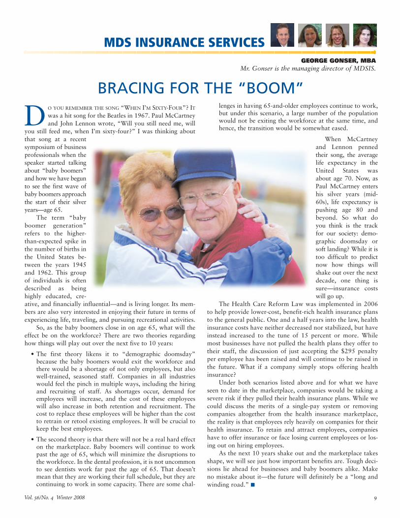
DO YOU REMEMBER THE SONG “WHEN I’M SIXTY-FOUR”? ITwas a hit song for the Beatles in 1967. Paul McCartneyand John Lennon wrote, “Will you still need me, will
you still feed me, when I’m sixty-four?” I was thinking aboutthat song at a recentsymposium of businessprofessionals when thespeaker started talkingabout “baby boomers”and how we have begunto see the first wave ofbaby boomers approachthe start of their silveryears—age 65.
The term “babyboomer generation”refers to the higher-than-expected spike inthe number of births inthe United States be-tween the years 1945and 1962. This groupof individuals is oftendescribed as beinghighly educated, cre-ative, and financially influential—and is living longer. Its mem-bers are also very interested in enjoying their future in terms ofexperiencing life, traveling, and pursuing recreational activities.
So, as the baby boomers close in on age 65, what will theeffect be on the workforce? There are two theories regardinghow things will play out over the next five to 10 years:
• The first theory likens it to “demographic doomsday”because the baby boomers would exit the workforce andthere would be a shortage of not only employees, but alsowell-trained, seasoned staff. Companies in all industrieswould feel the pinch in multiple ways, including the hiringand recruiting of staff. As shortages occur, demand foremployees will increase, and the cost of these employeeswill also increase in both retention and recruitment. Thecost to replace these employees will be higher than the costto retrain or retool existing employees. It will be crucial tokeep the best employees.
• The second theory is that there will not be a real hard effecton the marketplace. Baby boomers will continue to workpast the age of 65, which will minimize the disruptions tothe workforce. In the dental profession, it is not uncommonto see dentists work far past the age of 65. That doesn’tmean that they are working their full schedule, but they arecontinuing to work in some capacity. There are some chal-
lenges in having 65-and-older employees continue to work,but under this scenario, a large number of the populationwould not be exiting the workforce at the same time, andhence, the transition would be somewhat eased.
When McCartneyand Lennon pennedtheir song, the averagelife expectancy in theUnited States wasabout age 70. Now, asPaul McCartney entershis silver years (mid-60s), life expectancy ispushing age 80 andbeyond. So what doyou think is the trackfor our society: demo-graphic doomsday orsoft landing? While it istoo difficult to predictnow how things willshake out over the nextdecade, one thing issure—insurance costswill go up.
The Health Care Reform Law was implemented in 2006to help provide lower-cost, benefit-rich health insurance plansto the general public. One and a half years into the law, healthinsurance costs have neither decreased nor stabilized, but haveinstead increased to the tune of 15 percent or more. Whilemost businesses have not pulled the health plans they offer totheir staff, the discussion of just accepting the $295 penaltyper employee has been raised and will continue to be raised inthe future. What if a company simply stops offering healthinsurance?
Under both scenarios listed above and for what we haveseen to date in the marketplace, companies would be taking asevere risk if they pulled their health insurance plans. While wecould discuss the merits of a single-pay system or removingcompanies altogether from the health insurance marketplace,the reality is that employees rely heavily on companies for theirhealth insurance. To retain and attract employees, companieshave to offer insurance or face losing current employees or los-ing out on hiring employees.
As the next 10 years shake out and the marketplace takesshape, we will see just how important benefits are. Tough deci-sions lie ahead for businesses and baby boomers alike. Makeno mistake about it—the future will definitely be a “long andwinding road.” n
Vol. /No. Winter 2008
Vol. /No. Winter 2008 Journal of the Massachusetts Dental Society
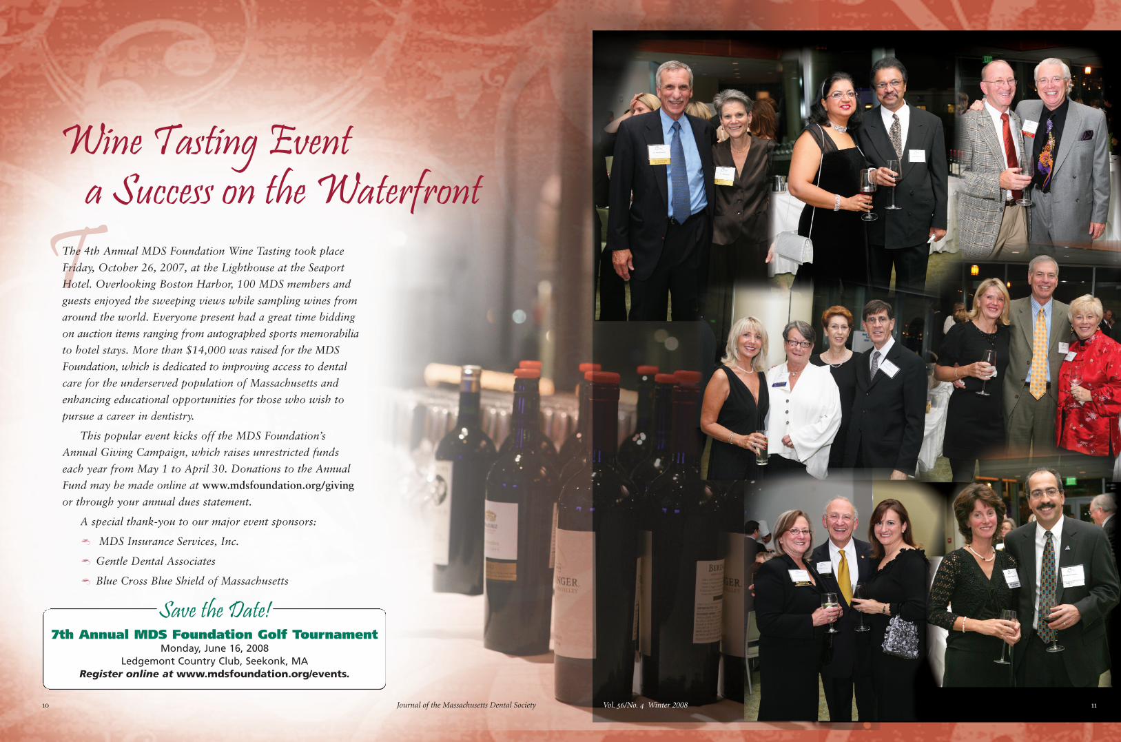
Wine Tasting Eventa Success on the Waterfront
TThe 4th Annual MDS Foundation Wine Tasting took place
Friday, October 26, 2007, at the Lighthouse at the Seaport
Hotel. Overlooking Boston Harbor, 100 MDS members and
guests enjoyed the sweeping views while sampling wines from
around the world. Everyone present had a great time bidding
on auction items ranging from autographed sports memorabilia
to hotel stays. More than $14,000 was raised for the MDS
Foundation, which is dedicated to improving access to dental
care for the underserved population of Massachusetts and
enhancing educational opportunities for those who wish to
pursue a career in dentistry.
This popular event kicks off the MDS Foundation’s
Annual Giving Campaign, which raises unrestricted funds
each year from May 1 to April 30. Donations to the Annual
Fund may be made online at www.mdsfoundation.org/giving
or through your annual dues statement.
A special thank-you to our major event sponsors:
d MDS Insurance Services, Inc.
d Gentle Dental Associates
d Blue Cross Blue Shield of Massachusetts
7th Annual MDS Foundation Golf Tournament
Monday, June 16, 2008
Ledgemont Country Club, Seekonk, MA
Register online at www.mdsfoundation.org/events.
Save the Date!
Vol. /No. Winter 2008 Journal of the Massachusetts Dental Society
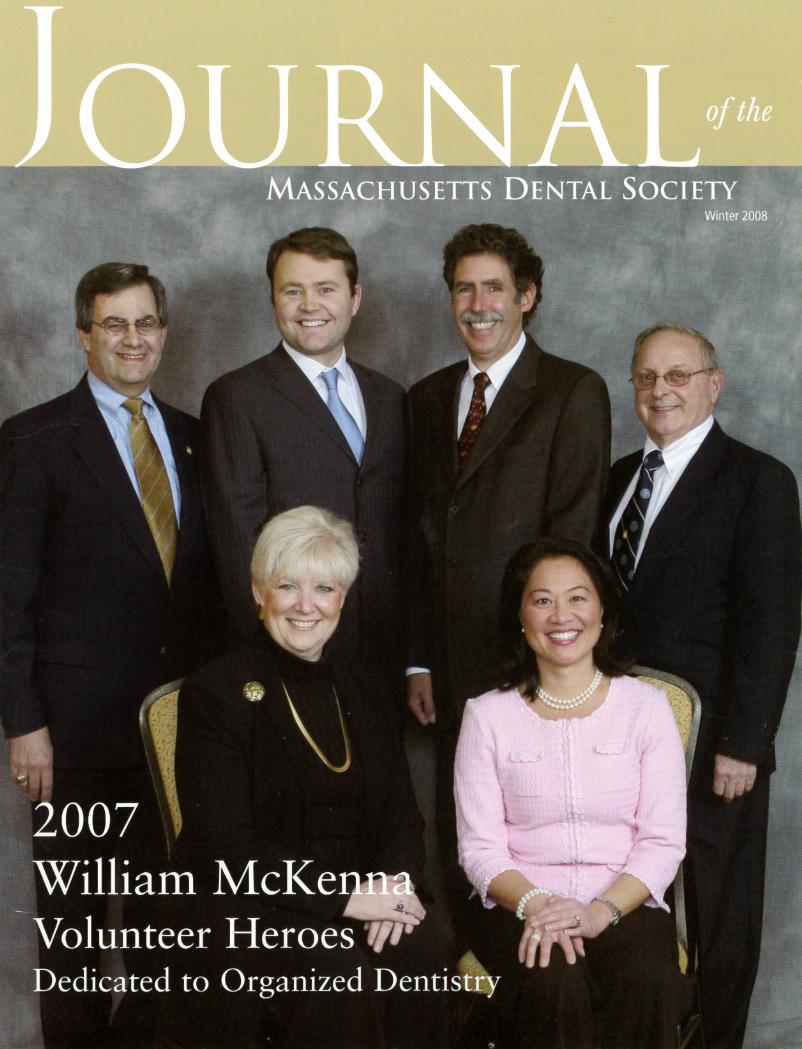
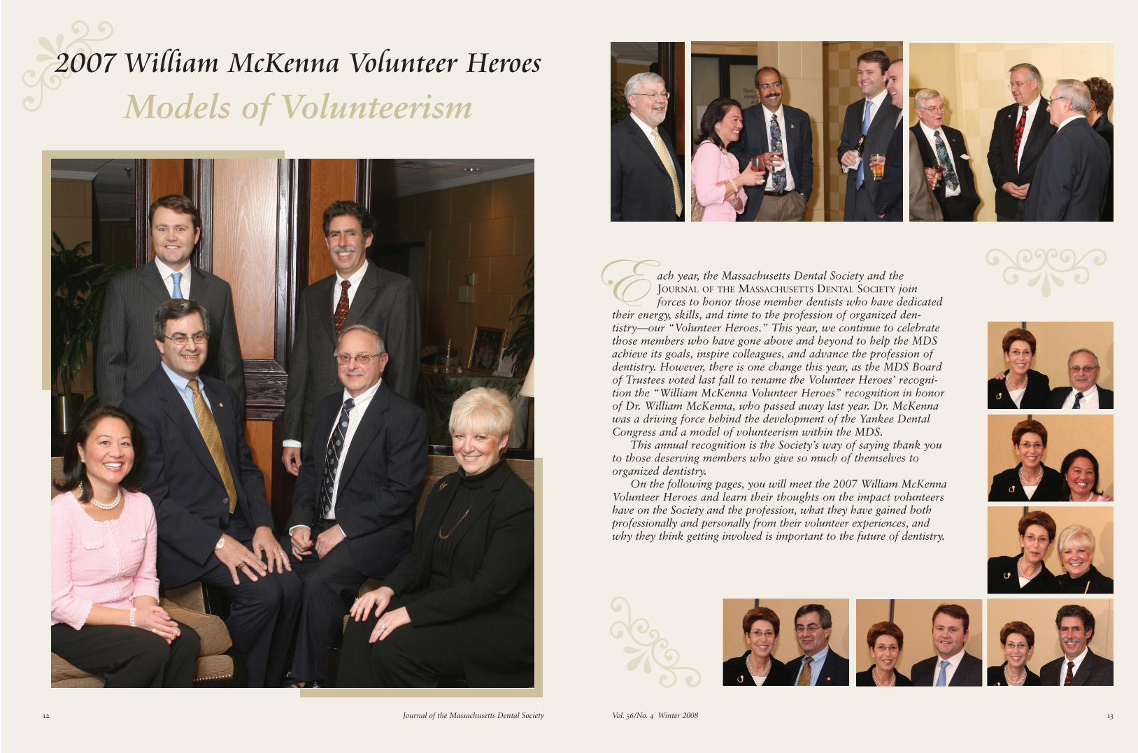
ach year, the Massachusetts Dental Society and theJOURNAL OF THE MASSACHUSETTS DENTAL SOCIETY joinforces to honor those member dentists who have dedicated
their energy, skills, and time to the profession of organized den-tistry—our “Volunteer Heroes.” This year, we continue to celebratethose members who have gone above and beyond to help the MDSachieve its goals, inspire colleagues, and advance the profession ofdentistry. However, there is one change this year, as the MDS Boardof Trustees voted last fall to rename the Volunteer Heroes’ recogni-tion the “William McKenna Volunteer Heroes” recognition in honorof Dr. William McKenna, who passed away last year. Dr. McKennawas a driving force behind the development of the Yankee DentalCongress and a model of volunteerism within the MDS.
This annual recognition is the Society’s way of saying thank youto those deserving members who give so much of themselves toorganized dentistry.
On the following pages, you will meet the 2007 William McKennaVolunteer Heroes and learn their thoughts on the impact volunteershave on the Society and the profession, what they have gained bothprofessionally and personally from their volunteer experiences, andwhy they think getting involved is important to the future of dentistry.
EN
N
N2007 William McKenna Volunteer Heroes
Models of Volunteerism
Vol. /No. Winter 2008 Journal of the Massachusetts Dental Society
Why did you choose to join the MDS?A mentor invited me to a South Shore District Dental Society meeting. I was warmly wel-comed, and it gave me the feeling that I was not alone in the practice of dentistry. It alsogave me a chance to meet dentists in the area of my practice. At that time, welfare anddental insurance were major issues facing organized dentistry; the issues still have notchanged. I was thankful for the chance to hear both sides of the issues, which spiked myinterest in joining the Massachusetts Dental Society.
Why is involvement in organized dentistry important to you?If you are not involved, you do not know what the Society has to offer. You may not knowwhat the current issues being proposed in the legislature are, and which issues ultimatelywill affect the way you practice dentistry in the future. We have strength in numbers; with-out numbers, you will be one voice in the wilderness.
Please describe the extent of your volunteer experience in dentistry. Starting as a paid practitioner in the Head Start Program for the Worcester Public Schoolsystem, I saw the need for the treatment of underprivileged children. My military experi-ence during the Vietnam War opened my eyes to the dental needs of underprivilegedrecruits. During my practice years, I volunteered to give free, in-house dental treatment toresidents at four nursing homes; this showed me the oral health needs of the elderly. I wasa consultant for the Massachusetts Hospital School, which exposed me to the needs of thehandicapped. I was a volunteer for the Sharewood Clinic, which left me with the under-standing of the needs of the immigrant population. I have lost count of the number ofChild Identification Programs (CHIP) I have volunteered for over the years. I volunteeredfor the MDS’s old Council on Education and Practice for five years, and I am in my thirdyear on the Council on Education. No one wanted to become editor for the South ShoreDistrict, so I volunteered. I have served on the MDS Peer Review Committee, ExecutiveCommittee, and Budget Committee, and all the committees of the South Shore District. Iam also a past chair of the South Shore District. Last but not least, I have been a volun-teer at the Yankee Dental Congress for many years.
How has your volunteer experience impacted you professionally and personally? I have grown as a dentist, but more importantly as a person. I have seen the ravagingaffects of dental disease on children, the young, the disabled, the old, and the infirmedpopulation. I have been able to see beyond the issues and have been able to look at thewhole picture. In many cases, these volunteer experiences have forced me to make a careerchange by going into education. The combined total of my life experiences directed me toeducate future dentists to the need of dentistry for the underprivileged and underserved,and to teach dentistry not only as a profession but also as a way of life.
What do you feel are the most important issues facing organized dentistry today? The manner in which state and federal legislature intervenes in the way we practice den-tistry in the future is important, but what’s more important is the way we educate thefuture generations of dentists.
Where do you see the future of organized dentistry in five years? 10 years? The future of organized dentistry can only grow, but it needs everyone to get involved. We,as an organization, will be only as strong as our membership.
What would you say to a recent dental school graduate to convince him/her to get moreinvolved in organized dentistry? Get involved—you can make a difference. Your reward will be knowing that you havemade a difference for the future practice of dentistry. Your reward also will be that youhave served mankind. To paraphrase President John F. Kennedy’s words: “Don’t ask whatthe Massachusetts Dental Society can do for me, but what can I do for the MassachusettsDental Society.” n
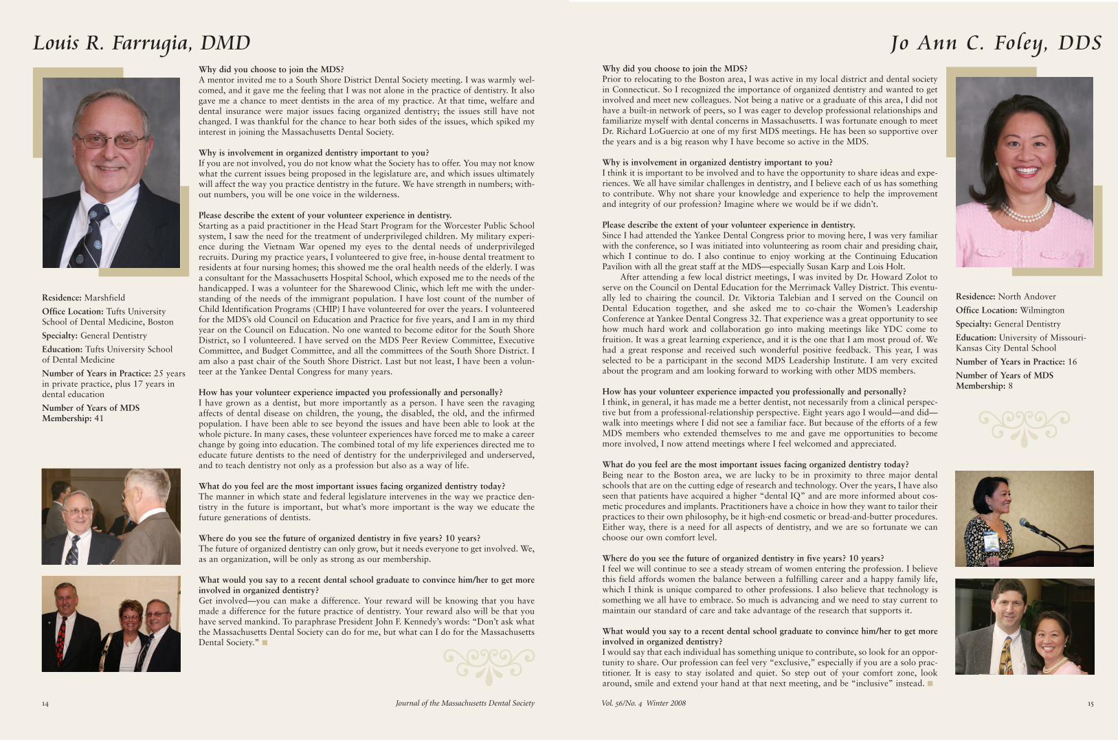
Residence: Marshfield
Office Location: Tufts UniversitySchool of Dental Medicine, Boston
Specialty: General Dentistry
Education: Tufts University School of Dental Medicine
Number of Years in Practice: 25 yearsin private practice, plus 17 years indental education
Number of Years of MDSMembership: 41
Why did you choose to join the MDS? Prior to relocating to the Boston area, I was active in my local district and dental societyin Connecticut. So I recognized the importance of organized dentistry and wanted to getinvolved and meet new colleagues. Not being a native or a graduate of this area, I did nothave a built-in network of peers, so I was eager to develop professional relationships andfamiliarize myself with dental concerns in Massachusetts. I was fortunate enough to meetDr. Richard LoGuercio at one of my first MDS meetings. He has been so supportive overthe years and is a big reason why I have become so active in the MDS.
Why is involvement in organized dentistry important to you?I think it is important to be involved and to have the opportunity to share ideas and expe-riences. We all have similar challenges in dentistry, and I believe each of us has somethingto contribute. Why not share your knowledge and experience to help the improvementand integrity of our profession? Imagine where we would be if we didn’t.
Please describe the extent of your volunteer experience in dentistry.Since I had attended the Yankee Dental Congress prior to moving here, I was very familiarwith the conference, so I was initiated into volunteering as room chair and presiding chair,which I continue to do. I also continue to enjoy working at the Continuing EducationPavilion with all the great staff at the MDS—especially Susan Karp and Lois Holt.
After attending a few local district meetings, I was invited by Dr. Howard Zolot toserve on the Council on Dental Education for the Merrimack Valley District. This eventu-ally led to chairing the council. Dr. Viktoria Talebian and I served on the Council onDental Education together, and she asked me to co-chair the Women’s LeadershipConference at Yankee Dental Congress 32. That experience was a great opportunity to seehow much hard work and collaboration go into making meetings like YDC come tofruition. It was a great learning experience, and it is the one that I am most proud of. Wehad a great response and received such wonderful positive feedback. This year, I wasselected to be a participant in the second MDS Leadership Institute. I am very excitedabout the program and am looking forward to working with other MDS members.
How has your volunteer experience impacted you professionally and personally?I think, in general, it has made me a better dentist, not necessarily from a clinical perspec-tive but from a professional-relationship perspective. Eight years ago I would—and did—walk into meetings where I did not see a familiar face. But because of the efforts of a fewMDS members who extended themselves to me and gave me opportunities to becomemore involved, I now attend meetings where I feel welcomed and appreciated.
What do you feel are the most important issues facing organized dentistry today?Being near to the Boston area, we are lucky to be in proximity to three major dentalschools that are on the cutting edge of research and technology. Over the years, I have alsoseen that patients have acquired a higher “dental IQ” and are more informed about cos-metic procedures and implants. Practitioners have a choice in how they want to tailor theirpractices to their own philosophy, be it high-end cosmetic or bread-and-butter procedures.Either way, there is a need for all aspects of dentistry, and we are so fortunate we canchoose our own comfort level.
Where do you see the future of organized dentistry in five years? 10 years?I feel we will continue to see a steady stream of women entering the profession. I believethis field affords women the balance between a fulfilling career and a happy family life,which I think is unique compared to other professions. I also believe that technology issomething we all have to embrace. So much is advancing and we need to stay current tomaintain our standard of care and take advantage of the research that supports it.
What would you say to a recent dental school graduate to convince him/her to get moreinvolved in organized dentistry?I would say that each individual has something unique to contribute, so look for an oppor-tunity to share. Our profession can feel very “exclusive,” especially if you are a solo prac-titioner. It is easy to stay isolated and quiet. So step out of your comfort zone, lookaround, smile and extend your hand at that next meeting, and be “inclusive” instead. n
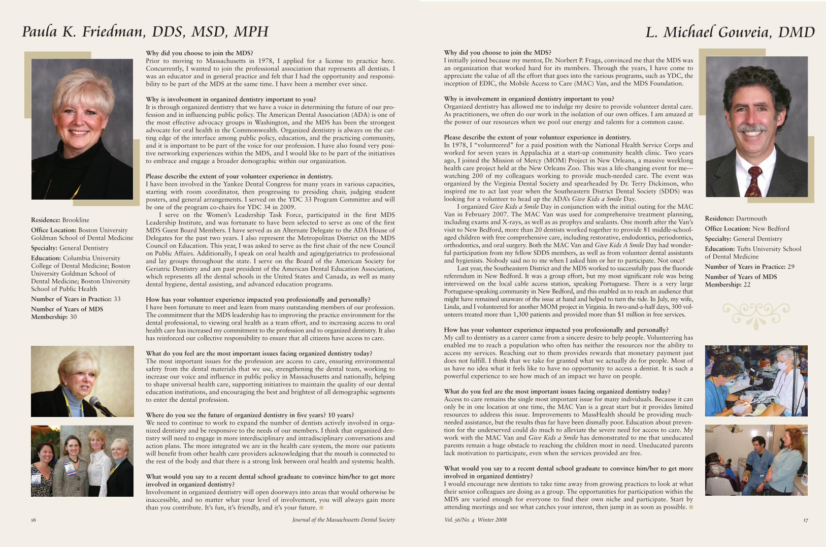
Residence: North Andover
Office Location: Wilmington
Specialty: General Dentistry
Education: University of Missouri-Kansas City Dental School
Number of Years in Practice: 16
Number of Years of MDSMembership: 8
Louis R. Farrugia, DMD Jo Ann C. Foley, DDS
N
N
Vol. /No. Winter 2008 Journal of the Massachusetts Dental Society
Why did you choose to join the MDS? Prior to moving to Massachusetts in 1978, I applied for a license to practice here.Concurrently, I wanted to join the professional association that represents all dentists. Iwas an educator and in general practice and felt that I had the opportunity and responsi-bility to be part of the MDS at the same time. I have been a member ever since.
Why is involvement in organized dentistry important to you?It is through organized dentistry that we have a voice in determining the future of our pro-fession and in influencing public policy. The American Dental Association (ADA) is one ofthe most effective advocacy groups in Washington, and the MDS has been the strongestadvocate for oral health in the Commonwealth. Organized dentistry is always on the cut-ting edge of the interface among public policy, education, and the practicing community,and it is important to be part of the voice for our profession. I have also found very posi-tive networking experiences within the MDS, and I would like to be part of the initiativesto embrace and engage a broader demographic within our organization.
Please describe the extent of your volunteer experience in dentistry. I have been involved in the Yankee Dental Congress for many years in various capacities,starting with room coordinator, then progressing to presiding chair, judging studentposters, and general arrangements. I served on the YDC 33 Program Committee and willbe one of the program co-chairs for YDC 34 in 2009.
I serve on the Women’s Leadership Task Force, participated in the first MDSLeadership Institute, and was fortunate to have been selected to serve as one of the firstMDS Guest Board Members. I have served as an Alternate Delegate to the ADA House ofDelegates for the past two years. I also represent the Metropolitan District on the MDSCouncil on Education. This year, I was asked to serve as the first chair of the new Councilon Public Affairs. Additionally, I speak on oral health and aging/geriatrics to professionaland lay groups throughout the state. I serve on the Board of the American Society forGeriatric Dentistry and am past president of the American Dental Education Association,which represents all the dental schools in the United States and Canada, as well as manydental hygiene, dental assisting, and advanced education programs.
How has your volunteer experience impacted you professionally and personally? I have been fortunate to meet and learn from many outstanding members of our profession.The commitment that the MDS leadership has to improving the practice environment for thedental professional, to viewing oral health as a team effort, and to increasing access to oralhealth care has increased my commitment to the profession and to organized dentistry. It alsohas reinforced our collective responsibility to ensure that all citizens have access to care.
What do you feel are the most important issues facing organized dentistry today? The most important issues for the profession are access to care, ensuring environmentalsafety from the dental materials that we use, strengthening the dental team, working toincrease our voice and influence in public policy in Massachusetts and nationally, helpingto shape universal health care, supporting initiatives to maintain the quality of our dentaleducation institutions, and encouraging the best and brightest of all demographic segmentsto enter the dental profession.
Where do you see the future of organized dentistry in five years? 10 years? We need to continue to work to expand the number of dentists actively involved in orga-nized dentistry and be responsive to the needs of our members. I think that organized den-tistry will need to engage in more interdisciplinary and intradisciplinary conversations andaction plans. The more integrated we are in the health care system, the more our patientswill benefit from other health care providers acknowledging that the mouth is connected tothe rest of the body and that there is a strong link between oral health and systemic health.
What would you say to a recent dental school graduate to convince him/her to get moreinvolved in organized dentistry?Involvement in organized dentistry will open doorways into areas that would otherwise beinaccessible, and no matter what your level of involvement, you will always gain morethan you contribute. It’s fun, it’s friendly, and it’s your future. n
Residence: Brookline
Office Location: Boston UniversityGoldman School of Dental Medicine
Specialty: General Dentistry
Education: Columbia UniversityCollege of Dental Medicine; BostonUniversity Goldman School ofDental Medicine; Boston UniversitySchool of Public Health
Number of Years in Practice: 33
Number of Years of MDSMembership: 30
Paula K. Friedman, DDS, MSD, MPHWhy did you choose to join the MDS? I initially joined because my mentor, Dr. Norbert P. Fraga, convinced me that the MDS wasan organization that worked hard for its members. Through the years, I have come toappreciate the value of all the effort that goes into the various programs, such as YDC, theinception of EDIC, the Mobile Access to Care (MAC) Van, and the MDS Foundation.
Why is involvement in organized dentistry important to you? Organized dentistry has allowed me to indulge my desire to provide volunteer dental care.As practitioners, we often do our work in the isolation of our own offices. I am amazed atthe power of our resources when we pool our energy and talents for a common cause.
Please describe the extent of your volunteer experience in dentistry. In 1978, I “volunteered” for a paid position with the National Health Service Corps andworked for seven years in Appalachia at a start-up community health clinic. Two yearsago, I joined the Mission of Mercy (MOM) Project in New Orleans, a massive weeklonghealth care project held at the New Orleans Zoo. This was a life-changing event for me—watching 200 of my colleagues working to provide much-needed care. The event wasorganized by the Virginia Dental Society and spearheaded by Dr. Terry Dickinson, whoinspired me to act last year when the Southeastern District Dental Society (SDDS) waslooking for a volunteer to head up the ADA’s Give Kids a Smile Day.
I organized Give Kids a Smile Day in conjunction with the initial outing for the MACVan in February 2007. The MAC Van was used for comprehensive treatment planning,including exams and X-rays, as well as as prophys and sealants. One month after the Van’svisit to New Bedford, more than 20 dentists worked together to provide 81 middle-school-aged children with free comprehensive care, including restorative, endodontics, periodontics,orthodontics, and oral surgery. Both the MAC Van and Give Kids A Smile Day had wonder-ful participation from my fellow SDDS members, as well as from volunteer dental assistantsand hygienists. Nobody said no to me when I asked him or her to participate. Not once!
Last year, the Southeastern District and the MDS worked to successfully pass the fluoridereferendum in New Bedford. It was a group effort, but my most significant role was beinginterviewed on the local cable access station, speaking Portuguese. There is a very largePortuguese-speaking community in New Bedford, and this enabled us to reach an audience thatmight have remained unaware of the issue at hand and helped to turn the tide. In July, my wife,Linda, and I volunteered for another MOM project in Virginia. In two-and-a-half days, 300 vol-unteers treated more than 1,300 patients and provided more than $1 million in free services.
How has your volunteer experience impacted you professionally and personally? My call to dentistry as a career came from a sincere desire to help people. Volunteering hasenabled me to reach a population who often has neither the resources nor the ability toaccess my services. Reaching out to them provides rewards that monetary payment justdoes not fulfill. I think that we take for granted what we actually do for people. Most ofus have no idea what it feels like to have no opportunity to access a dentist. It is such apowerful experience to see how much of an impact we have on people.
What do you feel are the most important issues facing organized dentistry today? Access to care remains the single most important issue for many individuals. Because it canonly be in one location at one time, the MAC Van is a great start but it provides limitedresources to address this issue. Improvements to MassHealth should be providing much-needed assistance, but the results thus far have been dismally poor. Education about preven-tion for the underserved could do much to alleviate the severe need for access to care. Mywork with the MAC Van and Give Kids a Smile has demonstrated to me that uneducatedparents remain a huge obstacle to reaching the children most in need. Uneducated parentslack motivation to participate, even when the services provided are free.
What would you say to a recent dental school graduate to convince him/her to get moreinvolved in organized dentistry? I would encourage new dentists to take time away from growing practices to look at whattheir senior colleagues are doing as a group. The opportunities for participation within theMDS are varied enough for everyone to find their own niche and participate. Start byattending meetings and see what catches your interest, then jump in as soon as possible. n
Residence: Dartmouth
Office Location: New Bedford
Specialty: General Dentistry
Education: Tufts University Schoolof Dental Medicine
Number of Years in Practice: 29
Number of Years of MDSMembership: 22
L. Michael Gouveia, DMD
N
Vol. /No. Winter 2008 Journal of the Massachusetts Dental Society
Why did you choose to join the MDS? I loved being involved in organized dentistry as a student and leader within the AmericanStudent Dental Association. The late Dr. Richard Forcucci also turned me on to involve-ment more specifically with the MDS. He’d bring me to local meetings in the South ShoreDistrict and encourage my involvement with the MDS as a student and later when I grad-uated. It seemed like a “no-brainer” to join and get involved, not only with my district butwith other groups within the MDS, as well.
Why is involvement in organized dentistry important to you? Organized dentistry is indeed the voice of our profession, so it seems that in order to par-ticipate and become involved in dentistry, you get involved with the organization that isthe voice. I’m still “old school” in some respects because I see dentistry as a profession,not just a job. I take pride in being a dental professional and part of that pride means givingback to the profession. I have chosen to do that through participation and involvement inorganized dentistry.
Please describe the extent of your volunteer experience in dentistry.I’ve been on the Council on Membership since my second year of dental school. I havebeen chair of the Council since 2003, which also extends to acting as the chair of subcom-mittees and task forces that are related to membership, such as the Standing Committeeon the New Dentist. Last year, I served as a Guest Board Member on the MDS Board ofTrustees and participated in the first MDS Leadership Institute program. I am currentlychair of the South Shore District, and I have participated in other activities in the district,as well as local CHIP programs. At the Yankee Dental Congress, I have volunteered in var-ious capacities, from presiding chair and room chair to coordinator of Team DevelopmentDay to general arrangements. Most recently, I was appointed to the American DentalAssociation Committee for the New Dentist.
How has your volunteer experience impacted you professionally and personally?It has been a wonderful way to meet other dentists from my district, as well as fromaround the state and nationwide. It’s nice to get out of the office and have the ability todiscuss cases. Dentistry can be isolating at times, and it’s great to be able to interact withother dental professionals on a regular basis.
Being involved in organized dentistry also has allowed me to feel more a part of theprofession and of what happens in the profession outside the walls of my office.
What do you feel are the most important issues facing organized dentistry today?As the profession continues to evolve, the biggest issues are catering membership to anever-changing world/demographic of the dental professional, as well as maintaining thedental profession as a profession governed and shaped by dentists and dental professionalsthemselves, not outside management or uninformed legislators.
Where do you see the future of organized dentistry in five years? 10 years?It’s tough to imagine that organized dentistry will be the same in five and especially in 10years. The face of dentistry itself is changing, and I’m confident that organized dentistry canand should keep up. I think that involvement in organized dentistry in political action willincrease, as nonprofessionals attempt to tell dentists how to practice. I think that dental soci-eties themselves will change to the extent that meetings and the “nuts and bolts” parts of thedental societies will change. This change may mean less face-to-face contact, perhaps, butactually include a more efficient exchange of information and ideas. Basically, advances intechnology will allow us to conduct “virtual meetings” where we can interact without phys-ically being in the same room. This will help accommodate the changing lifestyles of presentand future dentists and significantly change the way that dental societies do business.
What would you say to a recent dental school graduate to convince him/her to get moreinvolved in organized dentistry? I would tell them to just offer any time and energy that they can spare for organized den-tistry. Our profession should be shaped and molded by dentists, and the only way toensure that dentistry can thrive in the future is to play a part in the way the professionevolves. n
Residence: DorchesterOffice Location: HanoverSpecialty: General DentistryEducation: Tufts University School of Dental MedicineNumber of Years in Practice: 6Number of Years of MDSMembership: 10 (4 as student member)
Robert S. Leland, DMDWhy did you choose to join the MDS?I joined to be part of an organization that seeks to advance and protect the ethics andrights of dentistry. I further joined to get down in the trenches and work toward theseends.
Why is involvement in organized dentistry important to you?Too many members of our profession sit idly back and complain. This is human nature.There are “movers and shakers” who are willing to do the “dirty work,” and I am of thatilk. The status quo may be comfortable to many, but if organized dentistry is to keep upwith the present and advance into the future, there must be men and women willing to giveof themselves toward this end.
Please describe the extent of your volunteer experience in dentistry.I started off as a member of the Council on Dental Health, representing the MetropolitanDistrict. I next became Budgetary Chair of the Metropolitan District Dental Society whilestill maintaining my Council position. I moved through the “chairs” and eventuallybecame chair of the Metropolitan District. I then got involved with the MDS-PAC andgovernmental affairs.
Throughout the years, which have flown by all too quickly, I have lectured extensivelyon the importance of oral health to elementary school children and senior citizens. I enjoypublic speaking and have appeared on Boston Access TV. I have even presented twice atthe Yankee Dental Congress on public speaking. I have worked just about every year atYDC as a presiding chair or room coordinator. In 2006, I was honored to receive theMetropolitan District Dental Society’s John Burke Volunteer Award.
How has your volunteer experience impacted you professionally and personally?Over the years, I have made many valued friends through my involvement with variouscouncils, committees, and YDC. Involvement has given me a “hunger” to continuallyimprove myself and share new knowledge with others.
What do you feel are the most important issues facing organized dentistry today?Keeping the dental health team together and not having separate practices for dentists andhygienists is very important, as is protecting the rights of dentists to train their personneland still offer them the opportunity for advancement through more formal education. It isalso vital that the access to fluoride be available to all. Above all this is the need to makedentistry available to all who seek it.
Where do you see the future of organized dentistry in five years? 10 years?Unfortunately, I am not Nostradamus. I would like to think that we would still be fight-ing the little and big brush fires that pop up. I see dentistry in Massachusetts reaching outto all corners of the state, allowing for true “access for all.”
What would you say to a recent dental school graduate to convince him/her to get moreinvolved in organized dentistry?I would say, “This is the profession that you have chosen and if you want to succeed in itand see the profession grow, you must get involved and protect dentistry’s interests.” n
Residence: Natick
Office Locations: Brookline andNatick
Specialty: General Dentistry
Education: Tufts University Schoolof Dental Medicine
Number of Years in Practice: 35
Number of Years of MDSMembership: 35
Edward M. Salamoff, DMD
N
N
Introduction
Odontomas are the most commonly occurring tumors ofodontogenic origin.7 Many consider these lesions to be hamar-tomas, rather than true benign neoplasms.1,7 In most cases,odontomas arise within the jawbones, rather than within theoverlying soft tissues.1 The following is a case of an odontomathat presented itself clinically as an extraosseous mass within asac of soft tissue located external to the alveolar process.
Case Report
A 33-month-old female presented to the emergency departmentof the Connecticut Children’s Medical Center for diagnosis andmanagement of a soft-tissue mass in the maxillary right posteriorquadrant. According to the mother, the child had manipulated themass, which then ruptured and bled. The mother noticed bleed-ing from the site before she actually observed the mass itself. Themedical history was noncontributory. Oral examination revealeda translucent, pedunculated sac-like mass attached to and sus-pended from the crest of the maxillary alveolar ridge in the region
Peripheral Complex
Odontoma in a
Pediatric Dental Patient:
A Case ReportMELINA AMY ILIEF-ALA, DMD
ELLEN EISENBERG, DMD
GREGORY MATHIEU, DDS
Dr. Ilief-Ala is an associate pediatric dentist based in Billerica. Dr. Eisenberg is a professor in the
department of oral health and diagnostic sciences, and Dr. Mathieu is an associate professor in the
department of craniofacial sciences at the University of Connecticut Health Center in Farmington, CT.
Abstract
Odontomas are the most commonly occurring
benign odontogenic tumors of the jaws.7
Although a majority of odontomas are intra-
osseous,6 there are case reports of odontomas that
erupted into the oral cavity.2 Even less common are
peripheral or soft-tissue odontomas, only a few of which
have been reported to date.4,7,8,11 We report a peripheral
odontoma that arose in the alveolar mucosa of the pos-
terior maxilla in a young child. The diagnosis, complica-
tions, treatment, and prognosis of this entity will be
discussed.
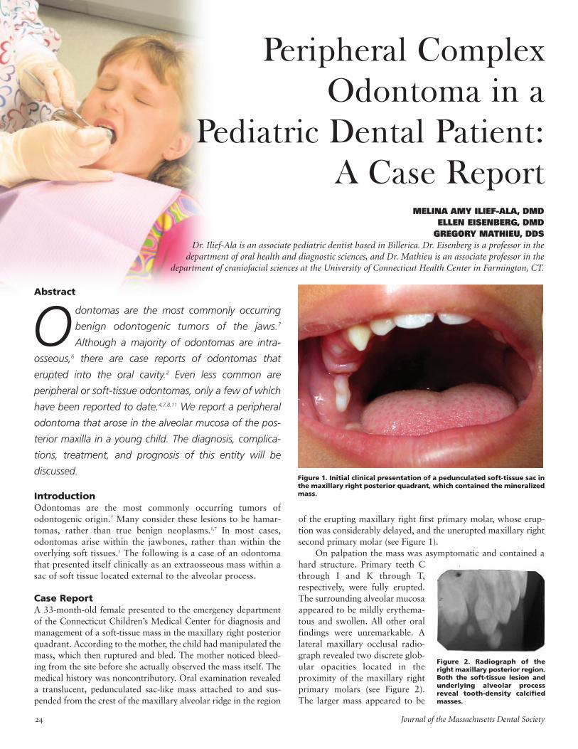
of the erupting maxillary right first primary molar, whose erup-tion was considerably delayed, and the unerupted maxillary rightsecond primary molar (see Figure 1).
On palpation the mass was asymptomatic and contained ahard structure. Primary teeth Cthrough I and K through T,respectively, were fully erupted.The surrounding alveolar mucosaappeared to be mildly erythema-tous and swollen. All other oralfindings were unremarkable. Alateral maxillary occlusal radio-graph revealed two discrete glob-ular opacities located in theproximity of the maxillary rightprimary molars (see Figure 2).The larger mass appeared to be
Figure 1. Initial clinical presentation of a pedunculated soft-tissue sac in
the maxillary right posterior quadrant, which contained the mineralized
mass.
Figure 2. Radiograph of the
right maxillary posterior region.
Both the soft-tissue lesion and
underlying alveolar process
reveal tooth-density calcified
masses.
Journal of the Massachusetts Dental Society
Vol. /No. Winter
positioned occlusal to the maxillary rightprimary first molar; this was thought tobe attributable to the displacement of thesac during film placement.
The smaller mass appeared to belocated in the attached tissue in the areadistal to the maxillary right first primarymolar. The opacifications were similar indensity to dentin. In addition to thedelayed eruption of the affected molars,there was distal displacement of the sec-ond primary molar.
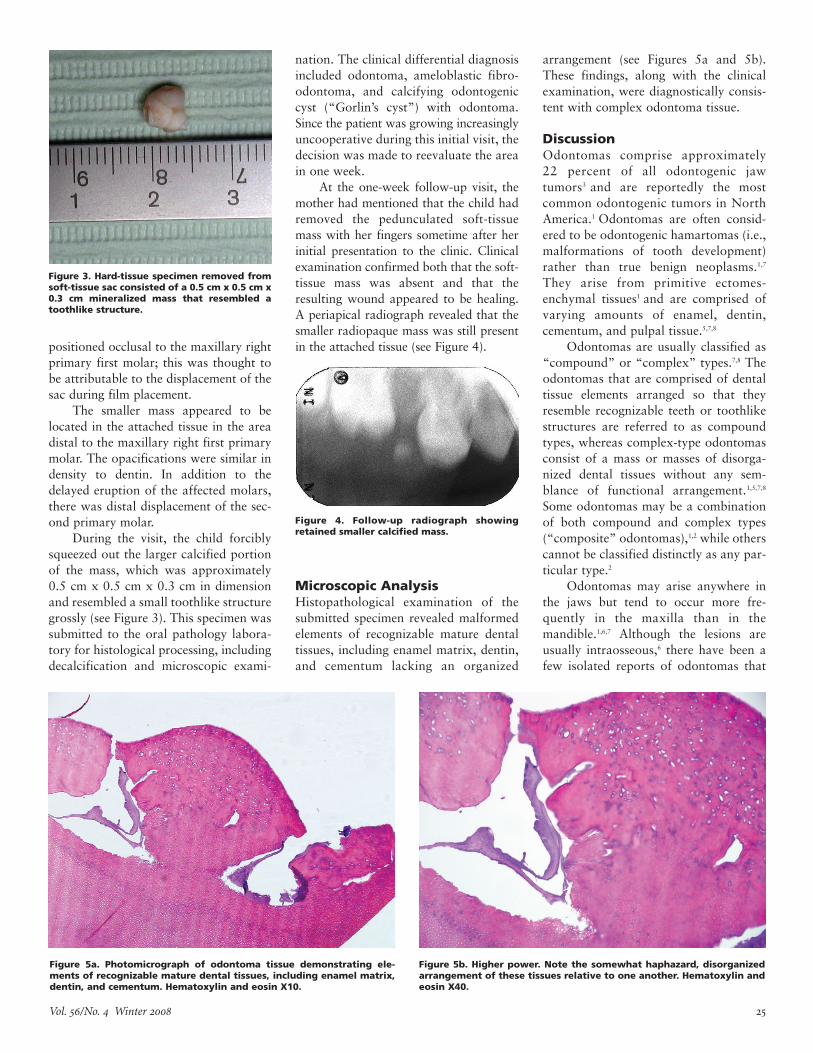
During the visit, the child forciblysqueezed out the larger calcified portionof the mass, which was approximately0.5 cm x 0.5 cm x 0.3 cm in dimensionand resembled a small toothlike structuregrossly (see Figure 3). This specimen wassubmitted to the oral pathology labora-tory for histological processing, includingdecalcification and microscopic exami-
nation. The clinical differential diagnosisincluded odontoma, ameloblastic fibro-odontoma, and calcifying odontogeniccyst (“Gorlin’s cyst”) with odontoma.Since the patient was growing increasinglyuncooperative during this initial visit, thedecision was made to reevaluate the areain one week.
At the one-week follow-up visit, themother had mentioned that the child hadremoved the pedunculated soft-tissuemass with her fingers sometime after herinitial presentation to the clinic. Clinicalexamination confirmed both that the soft-tissue mass was absent and that theresulting wound appeared to be healing.A periapical radiograph revealed that thesmaller radiopaque mass was still presentin the attached tissue (see Figure 4).
Microscopic Analysis
Histopathological examination of thesubmitted specimen revealed malformedelements of recognizable mature dentaltissues, including enamel matrix, dentin,and cementum lacking an organized
arrangement (see Figures 5a and 5b).These findings, along with the clinicalexamination, were diagnostically consis-tent with complex odontoma tissue.
Discussion
Odontomas comprise approximately22 percent of all odontogenic jawtumors3 and are reportedly the mostcommon odontogenic tumors in NorthAmerica.1 Odontomas are often consid-ered to be odontogenic hamartomas (i.e.,malformations of tooth development)rather than true benign neoplasms.1,7
They arise from primitive ectomes-enchymal tissues1 and are comprised ofvarying amounts of enamel, dentin,cementum, and pulpal tissue.5,7,8
Odontomas are usually classified as“compound” or “complex” types.7,8 Theodontomas that are comprised of dentaltissue elements arranged so that theyresemble recognizable teeth or toothlikestructures are referred to as compoundtypes, whereas complex-type odontomasconsist of a mass or masses of disorga-nized dental tissues without any sem-blance of functional arrangement.1,5,7,8
Some odontomas may be a combinationof both compound and complex types(“composite” odontomas),1,2 while otherscannot be classified distinctly as any par-ticular type.2
Odontomas may arise anywhere inthe jaws but tend to occur more fre-quently in the maxilla than in themandible.1,6,7 Although the lesions areusually intraosseous,6 there have been afew isolated reports of odontomas that
Figure 3. Hard-tissue specimen removed from
soft-tissue sac consisted of a 0.5 cm x 0.5 cm x
0.3 cm mineralized mass that resembled a
toothlike structure.
Figure 4. Follow-up radiograph showing
retained smaller calcified mass.
Figure 5a. Photomicrograph of odontoma tissue demonstrating ele-
ments of recognizable mature dental tissues, including enamel matrix,
dentin, and cementum. Hematoxylin and eosin X10.
Figure 5b. Higher power. Note the somewhat haphazard, disorganized
arrangement of these tissues relative to one another. Hematoxylin and
eosin X40.
Journal of the Massachusetts Dental Society
have erupted into the oral cavity.2 Stillrarer are “peripheral” or “soft-tissueodontomas,” of which only a few havebeen reported to date.4,7,8,11 Odontomasare often discovered as incidental radi-ographic findings1,2,4,6,7 and occur withequal frequency in males and females.2,4
They are generally asymptomatic1,2,4,6 andslow-growing.6 Large lesions (e.g., >6 cm)can cause expansion and swelling of cor-tical bone.6,7
Odontomas associated with expan-sion of the bone are more often diag-nosed in children and adolescents,2,4 sincethe first two decades of life represent theperiod during which the formation of thedentitions is in its most active phases.The most common sequela associated withan odontoma is the failure of a primary orpermanent tooth to erupt.4,5,7 The associa-tion with dentigerous cyst formation isalso common, since like any other develop-ing tooth, an odontoma is surrounded bydental follicular tissue that may becomecystic.4 Odontomas also can arise in asso-ciation with a calcifying and keratinizingodontogenic cyst (Gorlin’s cyst).4,8,9
Treatment of odontomas is conserva-tive and generally involves surgicalremoval of the lesion along with any as-sociated investing soft tissue, with little or no chance of recurrence.2,4,6 Onceremoved, both radiographic and clinicalfollow-up are essential to monitor erup-tion and location of the permanent teeth.Early detection and treatment of odon-tomas is essential to prevent complica-
tions (e.g., delayed eruption of primary orpermanent teeth, occlusal disharmonies,resorption of adjacent teeth, and swelling)and to ensure an optimal prognosis.6
This patient’s case is particularlyinteresting because the follicular sac ofthe odontoma was positioned entirelyexternal to the alveolar bone, along witha component of the lesion within theattached gingival tissue. It is likely thatthe peripheral odontoma caused delayederuption of the maxillary right primaryfirst and second molars.
Ide et al. have suggested that thegradual maturation of a peripheral odon-toma may lead to its unaided eruptioninto the oral cavity.10 In our patient’scase, given the history of forced expul-sion of the larger mass, the histopatho-logic findings, the age, and the progres-sively uncooperative behavior of thechild, the decision was made to continueto monitor the smaller calcified mass forspontaneous exfoliation for a short periodof time (~2-3 months). The need for sur-gical intervention was to be reconsideredat subsequent recall visits. However,despite repeated efforts to make contact,the patient failed to return to the clinicand was lost to follow-up. n
Acknowledgements
The authors would like to acknowledge Dr. Chelle Kucera for technical assistancewith the clinical photographs and Dr. Easwar Natarajan for providing thephotomicrographs.
References1. Sheehy EC, Odell EW, Al-Jaddir G.
Odontomas in the primary dentition: literature
review and case report. J Dent Child.
2004;71:73-76.
2. Lopez-Areal L, Silvestre Donat F, Gil Lozano J.
Compound odontoma erupting in the mouth:
4-year follow-up of a clinical case. J Oral
Pathol Med. 1992;21:285-288.
3. Shulman ER, Corio RL. Delayed eruption
associated with an odontoma. J Dent Child.
1987 May-Jun;205-207.
4. Giunta JL, Kaplan MA. Peripheral, soft tissue
odontomas. Oral Surg Oral Med Oral Path.
1990;69:406-411.
5. Oliver RG, Hodges CGL. Delayed eruption of
a maxillary central incisor associated with an
odontoma: report of case. J Dent Child.
1988 Sep-Oct;368-371.
6. Oliveira B, Campos V, Marcal S. Compound
odontoma—diagnosis and treatment: three
case reports. Pediatr Dent. 2001;23:151-157.
7. Neville BW, Damm DD, Allen CM, Bouquot
JE. Oral and maxillofacial pathology. 2nd ed.
Philadelphia (PA): W.B. Saunders Company;
c2002. p. 631-632.
8. Neville BW, Damm DD, White DK. Color atlas
of clinical oral pathology. 2nd ed. Baltimore
(MD): Lippincott Williams and Wilkins; c1999.
p. 414-415.
9. Kramer IRH, Pindborg JJ, Shear M. WHO
international histological classification of
tumors. Histological typing of odontogenic
tumors. 2nd ed. Berlin: Springer-Verlag;
c1992.
10. Ide F, Shimoyama T, Horie N. Gingival periph-
eral odontoma in an adult: case report.
J Periodontol. 2000;71:830-832.
11. Ledesma-Montes C, Perez-Bache A, Garces-
Ortiz M. Gingival compound odontoma.
Int J Oral Maxillofac Surg. 1996;25:296-297.
12. Delbem ACB, Cunha RF, Bianco KG, Afonso
RL, Goncalves TC. Odontomas in pediatric
dentistry: report of two cases. J Clin Pediatr
Dent. 2005;30(2):157-160.
CDADCDADDentist Health and Wellness Committee
Dentists in recovery helping dentists with chemical dependency
• Confidential support group meetings each month throughout the state
• Private consultations available upon request
• Confidentiality and anonymity guaranteed
Contact: P.O. Box 716, Andover, MA 01810
24-hour Hotline: (800) 468-2004 • Visit: www.cdad.org
Vol. /No. Winter Journal of the Massachusetts Dental Society
Recently, someone asked me what kind of occlusal
indicator I used to determine occlusal contacts.
I thought about it, but I couldn’t give a definitive
answer. Then I did a library search, but didn’t get very far
since there are few papers on this subject. There are also
no American Dental Association standards on the sub-
ject. So I figured out a way to see for myself. This is not
a scientific paper and there are no statistics. But I can
visually see a difference. I am working within the “least-
damaging principle,” which relates to the adage of “do
no harm.”
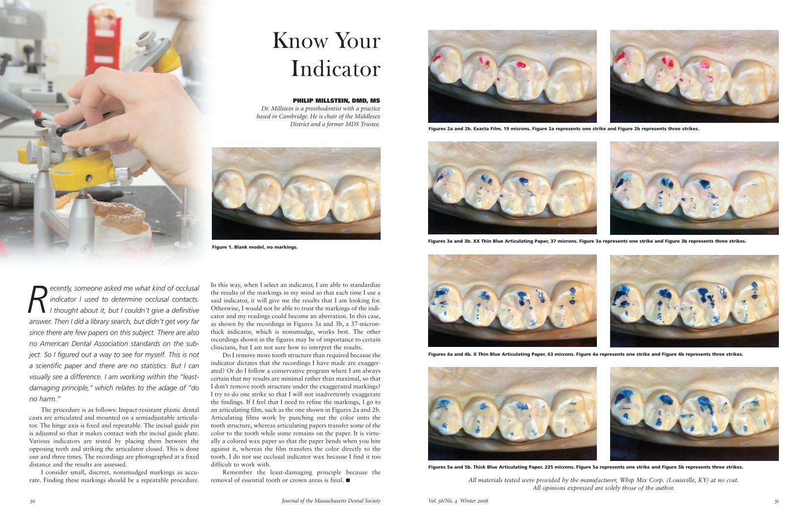
The procedure is as follows: Impact-resistant plastic dentalcasts are articulated and mounted on a semiadjustable articula-tor. The hinge axis is fixed and repeatable. The incisal guide pinis adjusted so that it makes contact with the incisal guide plate.Various indicators are tested by placing them between theopposing teeth and striking the articulator closed. This is doneone and three times. The recordings are photographed at a fixeddistance and the results are assessed.
I consider small, discreet, nonsmudged markings as accu-rate. Finding these markings should be a repeatable procedure.
Know Your
Indicator
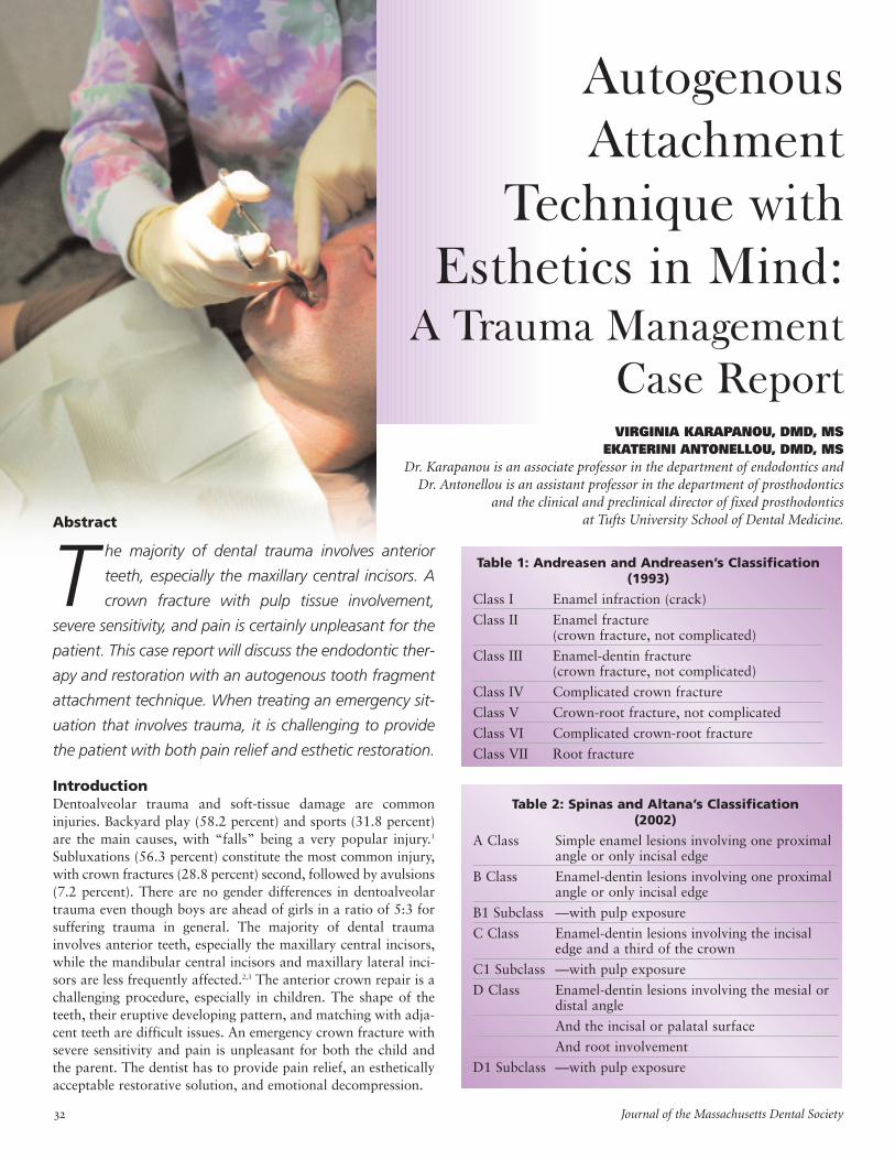
In this way, when I select an indicator, I am able to standardizethe results of the markings in my mind so that each time I use asaid indicator, it will give me the results that I am looking for.Otherwise, I would not be able to trust the markings of the indi-cator and my readings could become an aberration. In this case,as shown by the recordings in Figures 3a and 3b, a 37-micron-thick indicator, which is nonsmudge, works best. The otherrecordings shown in the figures may be of importance to certainclinicians, but I am not sure how to interpret the results.
Do I remove more tooth structure than required because theindicator dictates that the recordings I have made are exagger-ated? Or do I follow a conservative program where I am alwayscertain that my results are minimal rather than maximal, so thatI don’t remove tooth structure under the exaggerated markings?I try to do one strike so that I will not inadvertently exaggeratethe findings. If I feel that I need to refine the markings, I go toan articulating film, such as the one shown in Figures 2a and 2b.Articulating films work by punching out the color onto thetooth structure, whereas articulating papers transfer some of thecolor to the tooth while some remains on the paper. It is virtu-ally a colored wax paper so that the paper bends when you biteagainst it, whereas the film transfers the color directly to thetooth. I do not use occlusal indicator wax because I find it toodifficult to work with.
Remember the least-damaging principle because theremoval of essential tooth or crown areas is final. n
PHILIP MILLSTEIN, DMD, MS
Dr. Millstein is a prosthodontist with a practice
based in Cambridge. He is chair of the Middlesex
District and a former MDS Trustee.
All materials tested were provided by the manufacturer, Whip Mix Corp. (Louisville, KY) at no cost.
All opinions expressed are solely those of the author.
Figure 1. Blank model, no markings.
Figures 2a and 2b. Exacta Film, 19 microns. Figure 2a represents one strike and Figure 2b represents three strikes.
Figures 3a and 3b. XX Thin Blue Articulating Paper, 37 microns. Figure 3a represents one strike and Figure 3b represents three strikes.
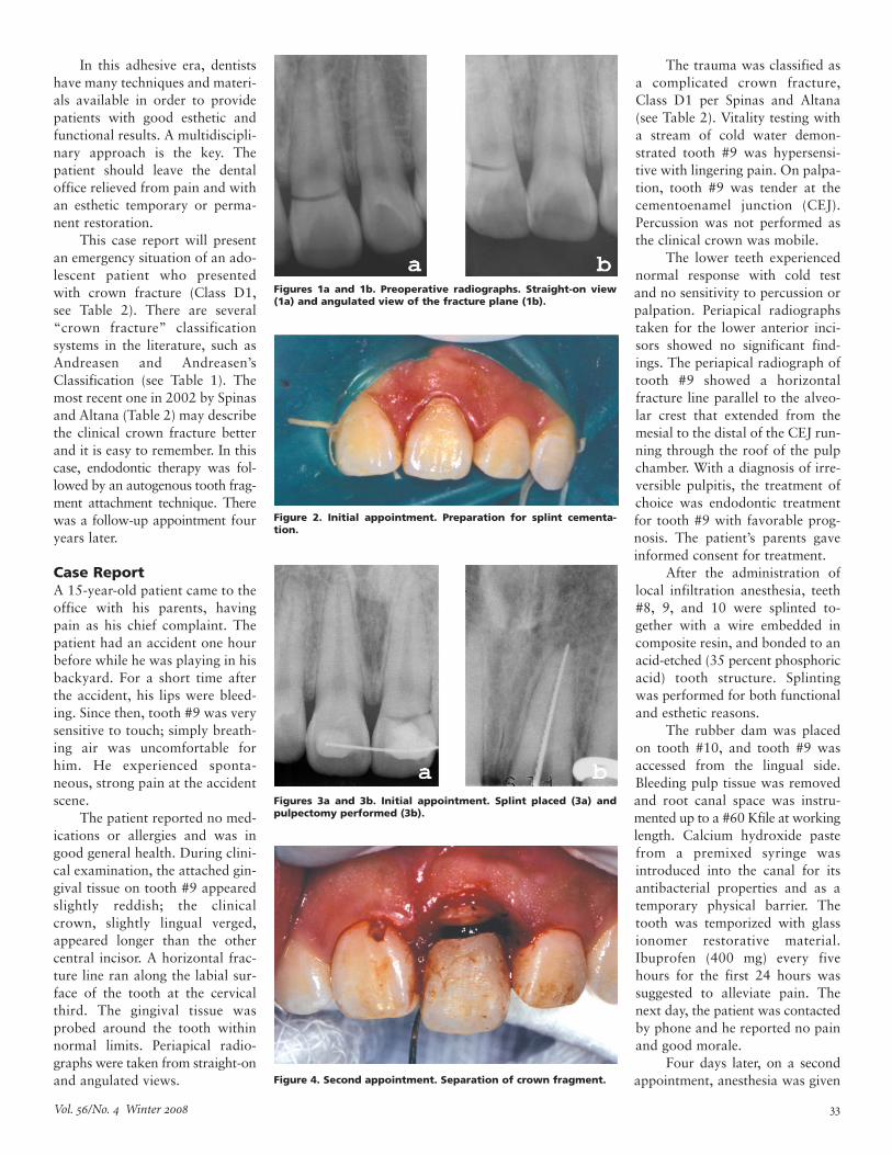
Figures 4a and 4b. X Thin Blue Articulating Paper, 63 microns. Figure 4a represents one strike and Figure 4b represents three strikes.
Figures 5a and 5b. Thick Blue Articulating Paper, 225 microns. Figure 5a represents one strike and Figure 5b represents three strikes.
Journal of the Massachusetts Dental Society
Abstract
The majority of dental trauma involves anterior
teeth, especially the maxillary central incisors. A
crown fracture with pulp tissue involvement,
severe sensitivity, and pain is certainly unpleasant for the
patient. This case report will discuss the endodontic ther-
apy and restoration with an autogenous tooth fragment
attachment technique. When treating an emergency sit-
uation that involves trauma, it is challenging to provide
the patient with both pain relief and esthetic restoration.
Introduction
Dentoalveolar trauma and soft-tissue damage are commoninjuries. Backyard play (58.2 percent) and sports (31.8 percent)are the main causes, with “falls” being a very popular injury.1
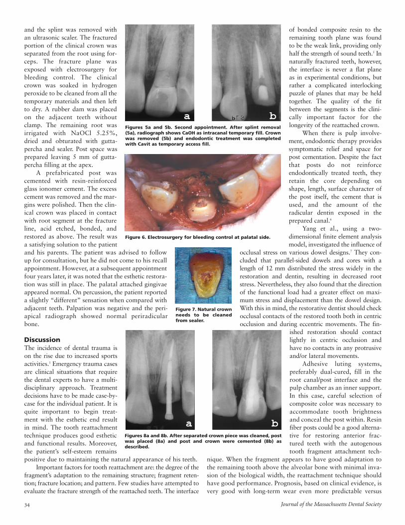
Subluxations (56.3 percent) constitute the most common injury,with crown fractures (28.8 percent) second, followed by avulsions(7.2 percent). There are no gender differences in dentoalveolartrauma even though boys are ahead of girls in a ratio of 5:3 forsuffering trauma in general. The majority of dental traumainvolves anterior teeth, especially the maxillary central incisors,while the mandibular central incisors and maxillary lateral inci-sors are less frequently affected.2,3 The anterior crown repair is achallenging procedure, especially in children. The shape of theteeth, their eruptive developing pattern, and matching with adja-cent teeth are difficult issues. An emergency crown fracture withsevere sensitivity and pain is unpleasant for both the child andthe parent. The dentist has to provide pain relief, an estheticallyacceptable restorative solution, and emotional decompression.
VIRGINIA KARAPANOU, DMD, MS
EKATERINI ANTONELLOU, DMD, MS
Dr. Karapanou is an associate professor in the department of endodontics and
Dr. Antonellou is an assistant professor in the department of prosthodontics
and the clinical and preclinical director of fixed prosthodontics
at Tufts University School of Dental Medicine.
Table 1: Andreasen and Andreasen’s Classification
(1993)
Class I Enamel infraction (crack)
Class II Enamel fracture (crown fracture, not complicated)
Class III Enamel-dentin fracture (crown fracture, not complicated)
Class IV Complicated crown fracture
Class V Crown-root fracture, not complicated
Class VI Complicated crown-root fracture
Class VII Root fracture
Table 2: Spinas and Altana’s Classification
(2002)
A Class Simple enamel lesions involving one proximal angle or only incisal edge
B Class Enamel-dentin lesions involving one proximal angle or only incisal edge
B1 Subclass —with pulp exposure
C Class Enamel-dentin lesions involving the incisal edge and a third of the crown
C1 Subclass —with pulp exposure
D Class Enamel-dentin lesions involving the mesial or distal angle
And the incisal or palatal surface
And root involvement
D1 Subclass —with pulp exposure
Autogenous
Attachment
Technique with
Esthetics in Mind: A Trauma Management
Case Report
Vol. /No. Winter
In this adhesive era, dentistshave many techniques and materi-als available in order to providepatients with good esthetic andfunctional results. A multidiscipli-nary approach is the key. Thepatient should leave the dentaloffice relieved from pain and withan esthetic temporary or perma-nent restoration.
This case report will presentan emergency situation of an ado-lescent patient who presentedwith crown fracture (Class D1,see Table 2). There are several“crown fracture” classificationsystems in the literature, such asAndreasen and Andreasen’sClassification (see Table 1). Themost recent one in 2002 by Spinasand Altana (Table 2) may describethe clinical crown fracture betterand it is easy to remember. In thiscase, endodontic therapy was fol-lowed by an autogenous tooth frag-ment attachment technique. Therewas a follow-up appointment fouryears later.
Case Report
A 15-year-old patient came to theoffice with his parents, havingpain as his chief complaint. Thepatient had an accident one hourbefore while he was playing in hisbackyard. For a short time afterthe accident, his lips were bleed-ing. Since then, tooth #9 was verysensitive to touch; simply breath-ing air was uncomfortable forhim. He experienced sponta-neous, strong pain at the accidentscene.
The patient reported no med-ications or allergies and was ingood general health. During clini-cal examination, the attached gin-gival tissue on tooth #9 appearedslightly reddish; the clinicalcrown, slightly lingual verged,appeared longer than the othercentral incisor. A horizontal frac-ture line ran along the labial sur-face of the tooth at the cervicalthird. The gingival tissue wasprobed around the tooth withinnormal limits. Periapical radio-graphs were taken from straight-onand angulated views.
The trauma was classified asa complicated crown fracture,Class D1 per Spinas and Altana(see Table 2). Vitality testing witha stream of cold water demon-strated tooth #9 was hypersensi-tive with lingering pain. On palpa-tion, tooth #9 was tender at thecementoenamel junction (CEJ).Percussion was not performed asthe clinical crown was mobile.
The lower teeth experiencednormal response with cold testand no sensitivity to percussion orpalpation. Periapical radiographstaken for the lower anterior inci-sors showed no significant find-ings. The periapical radiograph oftooth #9 showed a horizontalfracture line parallel to the alveo-lar crest that extended from themesial to the distal of the CEJ run-ning through the roof of the pulpchamber. With a diagnosis of irre-versible pulpitis, the treatment ofchoice was endodontic treatmentfor tooth #9 with favorable prog-nosis. The patient’s parents gaveinformed consent for treatment.
After the administration oflocal infiltration anesthesia, teeth#8, 9, and 10 were splinted to-gether with a wire embedded incomposite resin, and bonded to anacid-etched (35 percent phosphoricacid) tooth structure. Splintingwas performed for both functionaland esthetic reasons.
The rubber dam was placedon tooth #10, and tooth #9 wasaccessed from the lingual side.Bleeding pulp tissue was removedand root canal space was instru-mented up to a #60 Kfile at workinglength. Calcium hydroxide pastefrom a premixed syringe wasintroduced into the canal for itsantibacterial properties and as atemporary physical barrier. Thetooth was temporized with glassionomer restorative material.Ibuprofen (400 mg) every fivehours for the first 24 hours wassuggested to alleviate pain. Thenext day, the patient was contactedby phone and he reported no painand good morale.
Four days later, on a secondappointment, anesthesia was given
Figures 1a and 1b. Preoperative radiographs. Straight-on view
(1a) and angulated view of the fracture plane (1b).
Figure 2. Initial appointment. Preparation for splint cementa-
tion.
Figures 3a and 3b. Initial appointment. Splint placed (3a) and
pulpectomy performed (3b).
Figure 4. Second appointment. Separation of crown fragment.
and the splint was removed withan ultrasonic scaler. The fracturedportion of the clinical crown wasseparated from the root using for-ceps. The fracture plane wasexposed with electrosurgery forbleeding control. The clinicalcrown was soaked in hydrogenperoxide to be cleaned from all thetemporary materials and then leftto dry. A rubber dam was placedon the adjacent teeth withoutclamp. The remaining root wasirrigated with NaOCl 5.25%,dried and obturated with gutta-percha and sealer. Post space wasprepared leaving 5 mm of gutta-percha filling at the apex.
A prefabricated post wascemented with resin-reinforcedglass ionomer cement. The excesscement was removed and the mar-gins were polished. Then the clin-ical crown was placed in contactwith root segment at the fractureline, acid etched, bonded, andrestored as above. The result wasa satisfying solution to the patientand his parents. The patient was advised to followup for consultation, but he did not come to his recallappointment. However, at a subsequent appointmentfour years later, it was noted that the esthetic restora-tion was still in place. The palatal attached gingivaeappeared normal. On percussion, the patient reporteda slightly “different” sensation when compared withadjacent teeth. Palpation was negative and the peri-apical radiograph showed normal periradicularbone.
Discussion
The incidence of dental trauma ison the rise due to increased sportsactivities.1 Emergency trauma casesare clinical situations that requirethe dental experts to have a multi-disciplinary approach. Treatmentdecisions have to be made case-by-case for the individual patient. It isquite important to begin treat-ment with the esthetic end resultin mind. The tooth reattachmenttechnique produces good estheticand functional results. Moreover,the patient’s self-esteem remainspositive due to maintaining the natural appearance of his teeth.
Important factors for tooth reattachment are: the degree of thefragment’s adaptation to the remaining structure; fragment reten-tion; fracture location; and pattern. Few studies have attempted toevaluate the fracture strength of the reattached teeth. The interface
of bonded composite resin to theremaining tooth plane was foundto be the weak link, providing onlyhalf the strength of sound teeth.5 Innaturally fractured teeth, however,the interface is never a flat plane as in experimental conditions, butrather a complicated interlockingpuzzle of planes that may be heldtogether. The quality of the fitbetween the segments is the clini-cally important factor for thelongevity of the reattached crown.
When there is pulp involve-ment, endodontic therapy providessymptomatic relief and space forpost cementation. Despite the factthat posts do not reinforceendodontically treated teeth, theyretain the core depending onshape, length, surface character ofthe post itself, the cement that isused, and the amount of theradicular dentin exposed in theprepared canal.6
Yang et al., using a two-dimensional finite element analysismodel, investigated the influence of
occlusal stress on various dowel designs.7 They con-cluded that parallel-sided dowels and cores with alength of 12 mm distributed the stress widely in therestoration and dentin, resulting in decreased rootstress. Nevertheless, they also found that the directionof the functional load had a greater effect on maxi-mum stress and displacement than the dowel design.With this in mind, the restorative dentist should checkocclusal contacts of the restored tooth both in centricocclusion and during eccentric movements. The fin-
ished restoration should contactlightly in centric occlusion andhave no contacts in any protrusiveand/or lateral movements.
Adhesive luting systems,preferably dual-cured, fill in theroot canal/post interface and thepulp chamber as an inner support.In this case, careful selection ofcomposite color was necessary toaccommodate tooth brightnessand conceal the post within. Resinfiber posts could be a good alterna-tive for restoring anterior frac-tured teeth with the autogenoustooth fragment attachment tech-
nique. When the fragment appears to have good adaptation tothe remaining tooth above the alveolar bone with minimal inva-sion of the biological width, the reattachment technique shouldhave good performance. Prognosis, based on clinical evidence, isvery good with long-term wear even more predictable versus
Journal of the Massachusetts Dental Society
Figures 5a and 5b. Second appointment. After splint removal
(5a), radiograph shows CaOH as intracanal temporary fill. Crown
was removed (5b) and endodontic treatment was completed
with Cavit as temporary access fill.
Figure 6. Electrosurgery for bleeding control at palatal side.
Figure 7. Natural crown
needs to be cleaned
from sealer.
Figures 8a and 8b. After separated crown piece was cleaned, post
was placed (8a) and post and crown were cemented (8b) as
described.
Vol. /No. Winter
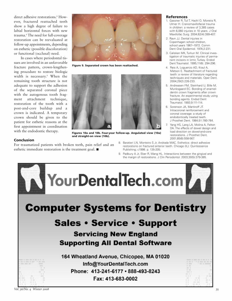
direct adhesive restorations.8 How-ever, fractured reattached teethshow a high degree of failure tolabial horizontal forces with newtrauma.4 The need for full-coveragerestoration can be reevaluated atfollow-up appointments, dependingon esthetic (possible discoloration)or functional (occlusal) need.
In cases where periodontal tis-sues are involved in an unfavorablefracture pattern, crown-lengthen-ing procedure to restore biologicwidth is necessary.9 When theremaining tooth structure is notadequate to support the adhesionof the separated coronal piecewith the autogenous tooth frag-ment attachment technique,restoration of the tooth with apost-and-core buildup and acrown is indicated. A temporarycrown should be given to thepatient for esthetic reasons at thefirst appointment in coordinationwith the endodontic therapy.
Conclusion
For traumatized patients with broken teeth, pain relief and anesthetic immediate restoration is the treatment goal. n
References1. Gassner R, Tuli T, Hachl O, Moreira R,
Ulmer H. Craniomaxillofacial trauma
in children: a review of 3,385 cases
with 6,060 injuries in 10 years. J Oral
Maxillofac Surg. 2004;62(4):399-407.
2. Ravn JJ. Dental injuries in
Copenhagen school children,
school years 1967–1972. Comm
Dent Oral Epidemiol. 1974;2:231.
3. Caliskan MK, Turkun M. Clinical inves-
tigation of traumatic injuries of perma-
nent incisors in Izmir, Turkey. Endod
Dent Traumatol. 1995;11(6): 294-296.
4. Reis A, Loguercio AD, Kraul A,
Matson E. Reattachment of fractured
teeth: a review of literature regarding
techniques and materials. Oper Dent.
2004;29(2):226-233.
5. Andreasen FM, Steinhard U, Bille M,
Munksgaard EC. Bonding of enamel-
dentin crown fragments after crown
fracture. An experimental study using
bonding agents. Endod Dent
Traumatol. 1993;9:111-114.
6. Sorenson JA, Martinoff JT.
Intracoronal reinforcement and
coronal coverage: a study of
endodontically treated teeth.
J Prosthet Dent. 1984;51:780-784.
7. Yang HS, Lang LA, Molina A, Felton
DA. The effects of dowel design and
load direction on dowel-and-core
restorations. J Prosthet Dent.
2001;85(6):558-567.
8. Baratieri LN, Monteiro S Jr, Andrada MAC. Esthetics: direct adhesive
restorations on fractured anterior teeth. Chicago (IL): Quintessence
Publishing; c1998. p. 135-205.
9. Padbury A Jr, Eber R, Wang HL. Interactions between the gingival and
the margin of restorations. J Clin Periodontol. 2003;30(5):379-385.
Figure 9. Separated crown has been reattached.
Figures 10a and 10b. Four-year follow-up. Angulated view (10a)
and straight-on view (10b).
Journal of the Massachusetts Dental Society
A Clinico-Pathologic Correlation
History
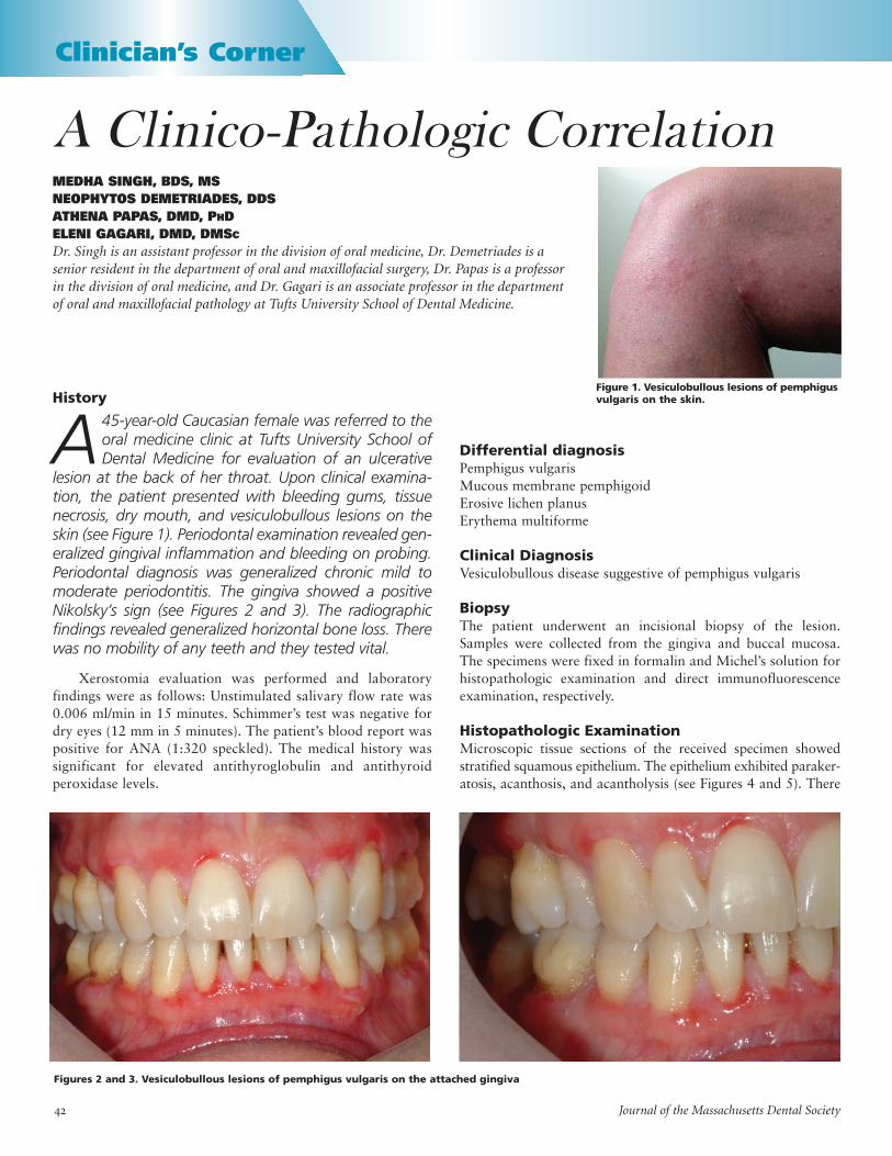
A45-year-old Caucasian female was referred to theoral medicine clinic at Tufts University School ofDental Medicine for evaluation of an ulcerative
lesion at the back of her throat. Upon clinical examina-tion, the patient presented with bleeding gums, tissuenecrosis, dry mouth, and vesiculobullous lesions on theskin (see Figure 1). Periodontal examination revealed gen-eralized gingival inflammation and bleeding on probing.Periodontal diagnosis was generalized chronic mild tomoderate periodontitis. The gingiva showed a positiveNikolsky’s sign (see Figures 2 and 3). The radiographicfindings revealed generalized horizontal bone loss. Therewas no mobility of any teeth and they tested vital.
Xerostomia evaluation was performed and laboratoryfindings were as follows: Unstimulated salivary flow rate was0.006 ml/min in 15 minutes. Schimmer’s test was negative fordry eyes (12 mm in 5 minutes). The patient’s blood report waspositive for ANA (1:320 speckled). The medical history wassignificant for elevated antithyroglobulin and antithyroidperoxidase levels.
MEDHA SINGH, BDS, MS
NEOPHYTOS DEMETRIADES, DDS
ATHENA PAPAS, DMD, PHD
ELENI GAGARI, DMD, DMSC
Dr. Singh is an assistant professor in the division of oral medicine, Dr. Demetriades is a
senior resident in the department of oral and maxillofacial surgery, Dr. Papas is a professor
in the division of oral medicine, and Dr. Gagari is an associate professor in the department
of oral and maxillofacial pathology at Tufts University School of Dental Medicine.
Clinician’s Corner
Figure 1. Vesiculobullous lesions of pemphigus
vulgaris on the skin.
Differential diagnosis
Pemphigus vulgarisMucous membrane pemphigoid Erosive lichen planusErythema multiforme
Clinical Diagnosis
Vesiculobullous disease suggestive of pemphigus vulgaris
Biopsy
The patient underwent an incisional biopsy of the lesion.Samples were collected from the gingiva and buccal mucosa.The specimens were fixed in formalin and Michel’s solution forhistopathologic examination and direct immunofluorescenceexamination, respectively.
Histopathologic Examination
Microscopic tissue sections of the received specimen showedstratified squamous epithelium. The epithelium exhibited paraker-atosis, acanthosis, and acantholysis (see Figures 4 and 5). There
Figures 2 and 3. Vesiculobullous lesions of pemphigus vulgaris on the attached gingiva
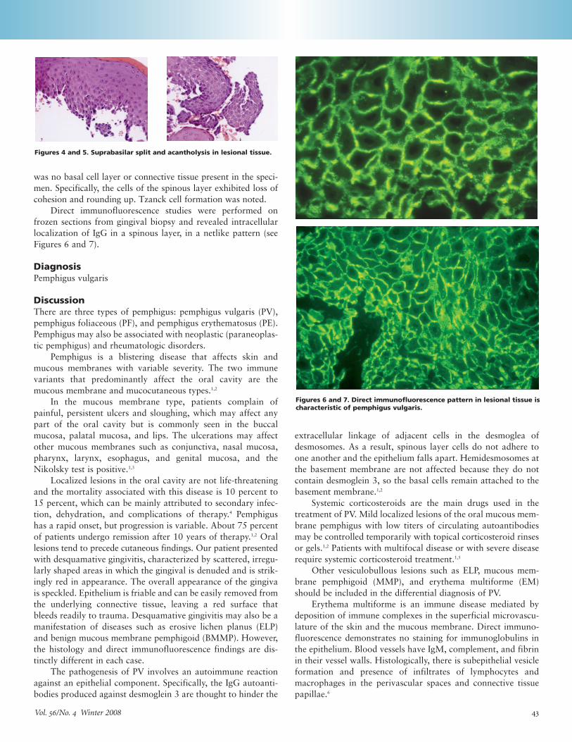
Vol. /No. Winter 2008
was no basal cell layer or connective tissue present in the speci-men. Specifically, the cells of the spinous layer exhibited loss ofcohesion and rounding up. Tzanck cell formation was noted.
Direct immunofluorescence studies were performed onfrozen sections from gingival biopsy and revealed intracellularlocalization of IgG in a spinous layer, in a netlike pattern (seeFigures 6 and 7).
Diagnosis
Pemphigus vulgaris
Discussion
There are three types of pemphigus: pemphigus vulgaris (PV),pemphigus foliaceous (PF), and pemphigus erythematosus (PE).Pemphigus may also be associated with neoplastic (paraneoplas-tic pemphigus) and rheumatologic disorders.
Pemphigus is a blistering disease that affects skin andmucous membranes with variable severity. The two immunevariants that predominantly affect the oral cavity are themucous membrane and mucocutaneous types.1,2
In the mucous membrane type, patients complain ofpainful, persistent ulcers and sloughing, which may affect anypart of the oral cavity but is commonly seen in the buccalmucosa, palatal mucosa, and lips. The ulcerations may affectother mucous membranes such as conjunctiva, nasal mucosa,pharynx, larynx, esophagus, and genital mucosa, and theNikolsky test is positive.1,3
Localized lesions in the oral cavity are not life-threateningand the mortality associated with this disease is 10 percent to15 percent, which can be mainly attributed to secondary infec-tion, dehydration, and complications of therapy.4 Pemphigushas a rapid onset, but progression is variable. About 75 percentof patients undergo remission after 10 years of therapy.1,2 Orallesions tend to precede cutaneous findings. Our patient presentedwith desquamative gingivitis, characterized by scattered, irregu-larly shaped areas in which the gingival is denuded and is strik-ingly red in appearance. The overall appearance of the gingivais speckled. Epithelium is friable and can be easily removed fromthe underlying connective tissue, leaving a red surface thatbleeds readily to trauma. Desquamative gingivitis may also be amanifestation of diseases such as erosive lichen planus (ELP)and benign mucous membrane pemphigoid (BMMP). However,the histology and direct immunofluorescence findings are dis-tinctly different in each case.
The pathogenesis of PV involves an autoimmune reactionagainst an epithelial component. Specifically, the IgG autoanti-bodies produced against desmoglein 3 are thought to hinder the
extracellular linkage of adjacent cells in the desmoglea ofdesmosomes. As a result, spinous layer cells do not adhere toone another and the epithelium falls apart. Hemidesmosomes atthe basement membrane are not affected because they do notcontain desmoglein 3, so the basal cells remain attached to thebasement membrane.1,2
Systemic corticosteroids are the main drugs used in thetreatment of PV. Mild localized lesions of the oral mucous mem-brane pemphigus with low titers of circulating autoantibodiesmay be controlled temporarily with topical corticosteroid rinsesor gels.1,2 Patients with multifocal disease or with severe diseaserequire systemic corticosteroid treatment.1,5
Other vesiculobullous lesions such as ELP, mucous mem-brane pemphigoid (MMP), and erythema multiforme (EM)should be included in the differential diagnosis of PV.
Erythema multiforme is an immune disease mediated bydeposition of immune complexes in the superficial microvascu-lature of the skin and the mucous membrane. Direct immuno-fluorescence demonstrates no staining for immunoglobulins inthe epithelium. Blood vessels have IgM, complement, and fibrinin their vessel walls. Histologically, there is subepithelial vesicleformation and presence of infiltrates of lymphocytes andmacrophages in the perivascular spaces and connective tissuepapillae.6
Figures 4 and 5. Suprabasilar split and acantholysis in lesional tissue.
Figures 6 and 7. Direct immunofluorescence pattern in lesional tissue is
characteristic of pemphigus vulgaris.
Journal of the Massachusetts Dental Society
In BMMP, there is presence of IgG, IgA, and complementalong the basement membrane. The molecular targets arelaminin-5 and bullous pemphigoid antigen 180. Histologically,there is subepithelial clefting and presence of lymphocytes in thelamina propria. There is no evidence of acantholysis.6
In ELP, direct immunofluoresence demonstrates the presenceof fibrinogen in the basement membrane. Histologically, there ishyperkeratosis, basal layer degeneration with apoptotic ker-atinocytes, and a lymphophagocytic infiltrate at the epithelialconnective tissue interface. Epithelium undergoes remodeling toform a saw-tooth rete ridge pattern and increased number ofLangerhans cells.6 Direct immunofluorescence is not pathogno-monic in cases of EM or ELP but can be helpful in excludingthese possibilities.
The diagnosis of the vesiculobullous disease is based on clin-ical and histopathologic evaluation as well as the performance ofspecific laboratory tests such as direct and indirect immunofluo-rescence.
Conclusion
In summary, pemphigus vulgaris is a serious, potentially life-threatening disease that may initially appear in the oral cavity.The oral health care practitioner should be aware of the clinicalmanifestations and laboratory procedures involved in the diag-nosis of pemphigus vulgaris. n
References1. Darling MR, Daley T. Blistering mucocutaneous diseases of the oral
mucosa—a review: part 2. Pemphigus vulgaris. J Can Dent Assoc.
2006;72(1):63-66.
2. Scully C, Challacombe SJ. Pemphigus vulgaris: update on etiopatho-
genesis, oral manifestations, and management. Crit Rev Oral Bio Med.
2002;13(5):397-408.
3. Mignogna MD, Lo Muzio LL, Mignogna RE, Bucci E. Oral pemphigus:
long-term behavior and clinical response to treatment with deflazacort in
sixteen cases. J Oral Pathol Med. 2000;29(4):145-152.
4. Ahlers KM, Senpal RA. Pemphigus vulgaris. Annals of emergency medi-
cine. J Amer Coll Emerg Physicians. 2006 Apr;47(4):395-402.
5. Toth GC, Jonkman MF. Therapy of pemphigus. Clin Dermatol.
2001;19(6):761-767.
6. Regezi JA, Sciubba JJ, Jordan RCK. Oral pathology: clinical pathologic cor-
relations. 3rd ed. Philadelphia (PA): W.B. Saunders Company; c1999: p. 18-22,
54-55, 101-104.
Take Advantage
of the MDS DiscountBased on the combined buying power of its membership,
the MDS has secured a variety of business discounts for its members. A full list of MDS business services
is available at www.massdental.org.
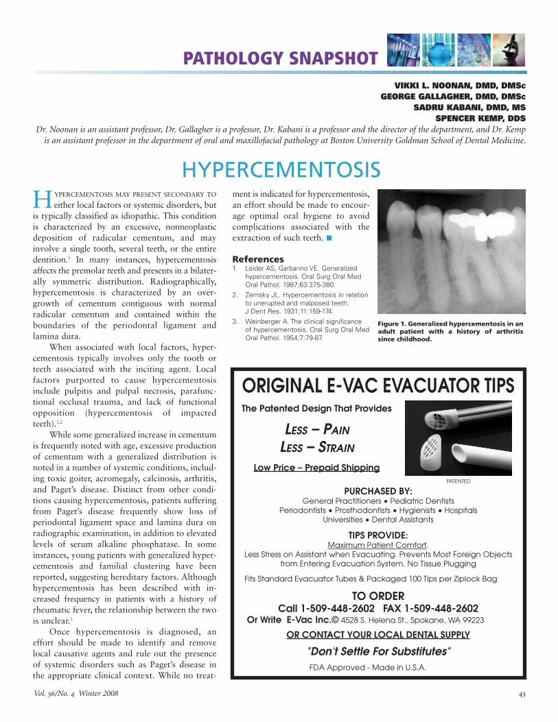
Vol. /No. Winter 2008
PATHOLOGY SNAPSHOT
VIKKI L. NOONAN, DMD, DMSC
GEORGE GALLAGHER, DMD, DMSC
SADRU KABANI, DMD, MS
SPENCER KEMP, DDS
Dr. Noonan is an assistant professor, Dr. Gallagher is a professor, Dr. Kabani is a professor and the director of the department, and Dr. Kemp
is an assistant professor in the department of oral and maxillofacial pathology at Boston University Goldman School of Dental Medicine.
HYPERCEMENTOSIS MAY PRESENT SECONDARY TO
either local factors or systemic disorders, butis typically classified as idiopathic. This conditionis characterized by an excessive, nonneoplasticdeposition of radicular cementum, and mayinvolve a single tooth, several teeth, or the entiredentition.1 In many instances, hypercementosisaffects the premolar teeth and presents in a bilater-ally symmetric distribution. Radiographically,hypercementosis is characterized by an over-growth of cementum contiguous with normalradicular cementum and contained within theboundaries of the periodontal ligament andlamina dura.
When associated with local factors, hyper-cementosis typically involves only the tooth orteeth associated with the inciting agent. Localfactors purported to cause hypercementosisinclude pulpitis and pulpal necrosis, parafunc-tional occlusal trauma, and lack of functionalopposition (hypercementosis of impactedteeth).1,2
While some generalized increase in cementumis frequently noted with age, excessive productionof cementum with a generalized distribution isnoted in a number of systemic conditions, includ-ing toxic goiter, acromegaly, calcinosis, arthritis,and Paget’s disease. Distinct from other condi-tions causing hypercementosis, patients sufferingfrom Paget’s disease frequently show loss ofperiodontal ligament space and lamina dura onradiographic examination, in addition to elevatedlevels of serum alkaline phosphatase. In someinstances, young patients with generalized hyper-cementosis and familial clustering have beenreported, suggesting hereditary factors. Althoughhypercementosis has been described with in-creased frequency in patients with a history ofrheumatic fever, the relationship between the twois unclear.3
Once hypercementosis is diagnosed, aneffort should be made to identify and removelocal causative agents and rule out the presenceof systemic disorders such as Paget’s disease inthe appropriate clinical context. While no treat-
HYPERCEMENTOSIS
Figure 1. Generalized hypercementosis in an
adult patient with a history of arthritis
since childhood.
PATENTED
ORIGINAL E-VAC EVACUATOR TIPSThe Patented Design That Provides
LESS – PAIN
LESS – STRAIN
Low Price – Prepaid Shipping
PURCHASED BY:
TIPS PROVIDE:Maximum Patient Comfort.
TO ORDERCall 1-509-448-2602 FAX 1-509-448-2602
Or Write E-Vac Inc.©
OR CONTACT YOUR LOCAL DENTAL SUPPLY
"Don't Settle For Substitutes"
ment is indicated for hypercementosis,an effort should be made to encour-age optimal oral hygiene to avoidcomplications associated with theextraction of such teeth. n
References1. Leider AS, Garbarino VE. Generalized
hypercementosis. Oral Surg Oral Med
Oral Pathol. 1987;63:375-380.
2. Zemsky JL. Hypercementosis in relation
to unerupted and malposed teeth.
J Dent Res. 1931;11:159-174.
3. Weinberger A. The clinical significance
of hypercementosis. Oral Surg Oral Med
Oral Pathol. 1954;7:79-87.
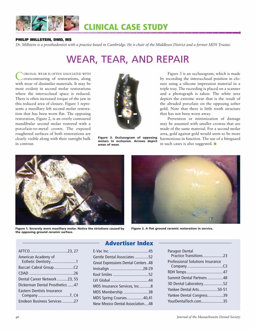
CORONAL WEAR IS OFTEN ASSOCIATED WITH
overcontouring of restorations, alongwith wear of dissimilar materials. It may bemost evident in second molar restorationswhere the interocclusal space is reduced.There is often increased torque of the jaw inthis reduced area of closure. Figure 1 repre-sents a maxillary left second molar restora-tion that has been worn flat. The opposingrestoration, Figure 2, is an overly contouredmandibular second molar restored with aporcelain-to-metal crown. The exposedroughened surfaces of both restorations areclearly visible along with their outright bulkin contour.
PHILIP MILLSTEIN, DMD, MS
Dr. Millstein is a prosthodontist with a practice based in Cambridge. He is chair of the Middlesex District and a former MDS Trustee.
WEAR, TEAR, AND REPAIR
CLINICAL CASE STUDY
Figure 1. Severely worn maxillary molar. Notice the striations caused by
the opposing ground ceramic surface.
Figure 2. A flat ground ceramic restoration in service.
Figure 3. Occlusogram of opposing
molars in occlusion. Arrows depict
areas of wear.
Figure 3 is an occlusogram, which is madeby recording the interocclusal position in clo-sure using a silicone impression material in atriple tray. The recording is placed on a scannerand a photograph is taken. The white areadepicts the extreme wear that is the result ofthe abraded porcelain on the opposing softergold. Note that there is little tooth structurethat has not been worn away.
Prevention or minimization of damagemay be assumed with smaller crowns that aremade of the same material. For a second molararea, gold against gold would seem to be moreharmonious in function. The use of a biteguardin such cases is also suggested. n
á
á
á
Advertiser Index
AFTCO....................................23, 27
American Academy of Esthetic Dentistry........................1
Baccari Cabral Group...................C2
CDAD ...........................................26
Dental Career Network ..........23, 55
Dickerman Dental Prosthetics ......47
Eastern Dentists Insurance Company ..............................7, C4
Emdeon Business Services ...........27
E-Vac Inc. .....................................45
Gentle Dental Associates .............52
Great Expressions Dental Centers ..48
Invisalign ................................28-29
Kool Smiles ..................................52
LVI Global ....................................44
MDS Insurance Services, Inc...........8
MDS Membership ........................38
MDS Spring Courses................40,41
New Mexico Dental Association....48
Paragon Dental Practice Transitions ...................23
Professional Solutions Insurance Company ..................................C3
RDH Temps...................................47
Summit Dental Partners ...............48
3D Dental Laboratory...................52
Yankee Dental Arts..................50-51
Yankee Dental Congress...............39
YourDentalTech.com.....................35
Journal of the Massachusetts Dental Society
Vol. /No. Winter 2008
BOOK REVIEWS
MIRACLE MAN OF THE
WESTERN FRONTH. MARTIN DERANIAN, DDS
Chandler House Press
Our war hero is a dentist, Dr. VaraztadH. Kazanjian. In this biography of the
“world’s most famous plastic surgeon,” Dr. H.Martin Deranian (a member of the MDS History & LibraryInformation Services Committee) demonstrates his love of tradi-tion as well as his love for his profession.
Dr. Deranian spent years with medical archives and in inter-viewing Dr. Kazanjian’s relatives, friends, and colleagues whilecompiling the material for this book. The biography traces thehistory of Dr. Kazanjian, a young man who fled from the mas-sacres in Ottoman Armenia and immigrated to Worcester,Massachusetts. Dr. Kazanjian’s desire for knowledge caused himto move to Boston, enroll at English High School (“I was a manand at first felt embarrassed when I saw the children who weremy classmates, but I soon said ‘What of it?’ The main reason isto improve my English.”) This attitude and hard work gainedhim admission to the dental school of Harvard University onSeptember 24, 1902.
In addition to the history of Dr. Kazanjian, the history ofdentistry of that era is well documented through the author’sdiligence for accuracy. Dr. Deranian’s knowledge as a historianof dentistry earned him the Hayden-Harris Award presented bythe American Academy of the History of Dentistry. We knew ofhis talent when he wrote the History of the Massachusetts
Dental Society during our Centennial celebration, and he hastaken this skill and applied it to the life of the man credited withforever changing the face of plastic surgery. Dr. Kazanjian’sinteresting life as told through the author’s easy writing styleresults in a book that reads like a novel and not a history book.
Lexi-Comp Dentistry SolutionsVARIOUS AUTHORS
Lexi-Comp
“Founded in 1978 as a clinical reference book publisher,Lexi-Comp has evolved into an industry-leading
provider of comprehensive clinical content and advanced infor-mation technology that provides healthcare professionals withthe resources they need to improve point-of-care decisions.”
This opening paragraph in a Lexi-Comp press kit intro-duced your reviewer to one of the MDS-sponsored business
partners. Of more significance, however, were samples of severalof their publications. Ohio-based Lexi-Comp provides clinicianswith databases on drug information, drug interactions, andclinical reference content.
The Little Dental Drug Booklet (PeterL. Jacobsen, author) is a pocket-sized refer-ence to the drugs most commonly used indental practice. The 82-page booklet containsinformation about prescription writing, anxiety/sedation, pain control, and bacterial, fungal,and viral infections. It includes miscellaneousinformation about such subjects as dentinsensitivity, anticaries agents, tooth whitening, periodontal prob-lems, and saliva dysfunction. The fact that the prophylacticantibiotic coverage was updated as of May 2007 indicates howcurrent the publishers are with their material.
Drug Information Handbook for Dentistry
(13th ed., Richard l. Wynn, Timothy F. Meiller,and Harold L. Crossley, authors) has informationabout commonly prescribed medications. Thisreference lists, in the form of monographs,dental-specific information for thousandsof medications and the most common herbs anddietary supplements. It includes local anesthetic/vasoconstrictor precautions and effects on dentaltreatments as well as dental comments.With Oral Soft Tissue Diseases (3rd ed.),
authors J. Robert Newland, Timothy F.Meiller, Richard L. Wynn, and Harold L.Crossley have designed a quick reference man-ual to help clinicians recognize and manageoral soft tissue disease.
The manual is divided into seven specificdiagnostic categories. Each lesion is illustratedwith one or more color photographs to helprecognize the clinical features. The authors haveincluded a section of drug monographs and sample prescrip-tions, as well as a section related to special topics such as thetreatment of oral mucosal lesions associated with chemotherapyand chronic dry mouth. Each monograph lists the etiology, visualclues, useful clinical information, and differential diagnosis, aswell as treatment recommendations and follow-up care.
With Oral Hard Tissue Diseases (2nd ed.), J. Robert Newland designed this textbook topresent radiographic features of intraosseouslesions discovered on dental radiographs.Newland divided the manual into 11 sections,each devoted to a specific diagnosis category.Visually cued with high-quality radiographs,each lesion is presented with its etiology, typ-
ical radiographic features, differential diagnosis, andclinical significance. n
NORMAN BECKER, DDS, EDITOR EMERITUS
Take Advantage of the MDS Discount
Based on the combined buying power of its membership, the MDS hassecured a variety of business discounts for its members. A full list of MDS
business services is available at www.massdental.org.
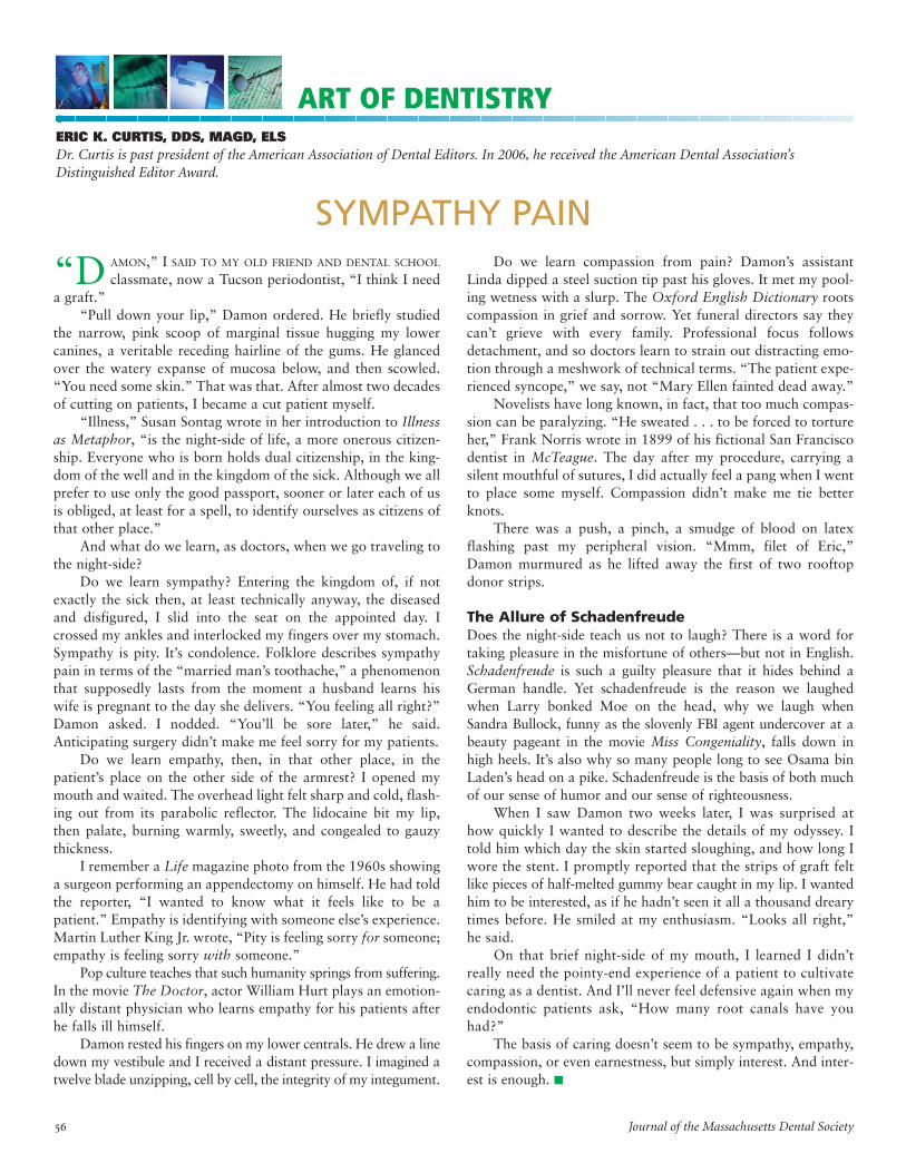
Journal of the Massachusetts Dental Society
ERIC K. CURTIS, DDS, MAGD, ELS
Dr. Curtis is past president of the American Association of Dental Editors. In 2006, he received the American Dental Association’s
Distinguished Editor Award.
ART OF DENTISTRY
SYMPATHY PAIN
“D AMON,” I SAID TO MY OLD FRIEND AND DENTAL SCHOOL
classmate, now a Tucson periodontist, “I think I needa graft.”
“Pull down your lip,” Damon ordered. He briefly studiedthe narrow, pink scoop of marginal tissue hugging my lowercanines, a veritable receding hairline of the gums. He glancedover the watery expanse of mucosa below, and then scowled.“You need some skin.” That was that. After almost two decadesof cutting on patients, I became a cut patient myself.
“Illness,” Susan Sontag wrote in her introduction to Illness
as Metaphor, “is the night-side of life, a more onerous citizen-ship. Everyone who is born holds dual citizenship, in the king-dom of the well and in the kingdom of the sick. Although we allprefer to use only the good passport, sooner or later each of usis obliged, at least for a spell, to identify ourselves as citizens ofthat other place.”
And what do we learn, as doctors, when we go traveling tothe night-side?
Do we learn sympathy? Entering the kingdom of, if notexactly the sick then, at least technically anyway, the diseasedand disfigured, I slid into the seat on the appointed day. Icrossed my ankles and interlocked my fingers over my stomach.Sympathy is pity. It’s condolence. Folklore describes sympathypain in terms of the “married man’s toothache,” a phenomenonthat supposedly lasts from the moment a husband learns hiswife is pregnant to the day she delivers. “You feeling all right?”Damon asked. I nodded. “You’ll be sore later,” he said.Anticipating surgery didn’t make me feel sorry for my patients.
Do we learn empathy, then, in that other place, in thepatient’s place on the other side of the armrest? I opened mymouth and waited. The overhead light felt sharp and cold, flash-ing out from its parabolic reflector. The lidocaine bit my lip,then palate, burning warmly, sweetly, and congealed to gauzythickness.
I remember a Life magazine photo from the 1960s showinga surgeon performing an appendectomy on himself. He had toldthe reporter, “I wanted to know what it feels like to be apatient.” Empathy is identifying with someone else’s experience.Martin Luther King Jr. wrote, “Pity is feeling sorry for someone;empathy is feeling sorry with someone.”
Pop culture teaches that such humanity springs from suffering.In the movie The Doctor, actor William Hurt plays an emotion-ally distant physician who learns empathy for his patients afterhe falls ill himself.
Damon rested his fingers on my lower centrals. He drew a linedown my vestibule and I received a distant pressure. I imagined atwelve blade unzipping, cell by cell, the integrity of my integument.
Do we learn compassion from pain? Damon’s assistantLinda dipped a steel suction tip past his gloves. It met my pool-ing wetness with a slurp. The Oxford English Dictionary rootscompassion in grief and sorrow. Yet funeral directors say theycan’t grieve with every family. Professional focus followsdetachment, and so doctors learn to strain out distracting emo-tion through a meshwork of technical terms. “The patient expe-rienced syncope,” we say, not “Mary Ellen fainted dead away.”
Novelists have long known, in fact, that too much compas-sion can be paralyzing. “He sweated . . . to be forced to tortureher,” Frank Norris wrote in 1899 of his fictional San Franciscodentist in McTeague. The day after my procedure, carrying asilent mouthful of sutures, I did actually feel a pang when I wentto place some myself. Compassion didn’t make me tie betterknots.
There was a push, a pinch, a smudge of blood on latexflashing past my peripheral vision. “Mmm, filet of Eric,”Damon murmured as he lifted away the first of two rooftopdonor strips.
The Allure of Schadenfreude
Does the night-side teach us not to laugh? There is a word fortaking pleasure in the misfortune of others—but not in English.Schadenfreude is such a guilty pleasure that it hides behind aGerman handle. Yet schadenfreude is the reason we laughedwhen Larry bonked Moe on the head, why we laugh whenSandra Bullock, funny as the slovenly FBI agent undercover at abeauty pageant in the movie Miss Congeniality, falls down inhigh heels. It’s also why so many people long to see Osama binLaden’s head on a pike. Schadenfreude is the basis of both muchof our sense of humor and our sense of righteousness.
When I saw Damon two weeks later, I was surprised athow quickly I wanted to describe the details of my odyssey. Itold him which day the skin started sloughing, and how long Iwore the stent. I promptly reported that the strips of graft feltlike pieces of half-melted gummy bear caught in my lip. I wantedhim to be interested, as if he hadn’t seen it all a thousand drearytimes before. He smiled at my enthusiasm. “Looks all right,”he said.
On that brief night-side of my mouth, I learned I didn’treally need the pointy-end experience of a patient to cultivatecaring as a dentist. And I’ll never feel defensive again when myendodontic patients ask, “How many root canals have youhad?”
The basis of caring doesn’t seem to be sympathy, empathy,compassion, or even earnestness, but simply interest. And inter-est is enough. n


