Embed Size (px)
Citation preview

World Journal of CardiologyWorld J Cardiol 2014 October 26; 6(10): 1049-1134
Published by Baishideng Publishing Group Inc
ISSN 1949-8462 (online)
EDITORS-IN-CHIEFJian-Jun Li, BeijingGiuseppe De Luca, NovaraNathan D Wong, Irvine
GUEST EDITORIAL BOARD MEMBERSShih-Tai Chang, PutzMien-Cheng Chen, KaohsiungJuei-Tang Cheng, TainanWoei-Jer Chuang, TainanShih-Hung Hsiao, KaohsiungWei-Chun Huang, KaohsiungTsung-Ming Lee, TainanTzong-Shyuan Lee, TaipeiJiun-Yi Li, TaipeiGen-Min Lin, HualienPing-Yen Liu, TainanKou-Gi Shyu, TaipeiChin-Hsiao Tseng, Taipei
MEMBERS OF THE EDITORIAL BOARD
Argentina
Mariano Falconi, Buenos AiresRicardo R Forastiero, Buenos AiresGaston A Rodriguez-Granillo, Buenos Aires
Australia
Christoph E Hagemeyer, MelbourneChristian Hamilton-Craig, BrisbaneKwok Ming Ho, PerthTin Kyaw, MelboruneKazuko Masuo, MelbourneHamish C Prosser, Sydney
Zhonghua Sun, Perth
AustriaAlexander Binder, GrazMariann Gyongyosi, ViennaRudolf Kirchmair, InnsbruckDeddo Moertl, ViennaGert Reiter, GrazIoannis Tentzeris, Vienna
BelgiumBSN Alzand, RonsePaul Vermeersch, Antwerpen
Brazil
Edimar A Bocchi, Sao PauloAntonio CC de Carvalho, Rio de JaneiroGuilherme V Guimaraes, Sao PauloRonaldo Lima, Rio de JaneiroChristiane Malfitano, Sao PauloAntonio P Mansur, Sao PauloGilberto De Nucci, CampinasAndre Talvani, Ouro Preto
Canada
Rodrigo Bagur, QuebecJagdish Butany, TorontoMohamed Chahine, QuébecPaul Farand, SherbrookeMichael E Farkouh, TorontoRobert Gros, LondonJoseph F Ndisang, SaskatoonSimon W Rabkin, Vancouver
Jacqueline WL Saw, VancouverCaroline Sirois, LevisSara S Nunes Vasconcelos, Toronto
China
Feng Cao, Xi'anXiao-Shu Cheng, NanchangJie Du, BeijingJun-Bao Du, BeijingDeng-Feng Gao, Xi'anChang-Qing Gao, Kai-Zheng Gong, YangzhouKai Huang, WuhanBin Jiang, BeijingZhi-Yong Li, NanjingTong Liu, TianjinJing-Ping Sun, Hong KongJun Tao, GuangzhouMalcolm J Underwood, Hong KongSong Wan, Hong KongYi Wan, Xi'anChi-Ming Wong, Hong KongJian-Bo Wu, LuzhouHai-Wei Wu, NanjingYong Xu, NanjingChen-Jiang Ying, WuhanHong-Kun Zhang, HangzhouJiu-Chang Zhong, Shanghai
Croatia
Viktor Culic, Split
Cuba
Fidel M Caceres-Loriga, Havana
Editorial Board2014-2017
The World Journal of Cardiology Editorial Board consists of 410 members, representing a team of worldwide experts in cardiology. They are from 46 countries, including Argentina (3), Australia (7), Austria (6), Belgium (2), Brazil (8), Canada (11), China (37), Croatia (1), Cuba (1), Cyprus (1), Czech Repoublic (2), Denmark (3), Egypt (1), Finland (3), France (3), Germany (31), Greece (10), Hungary (5), India (4), Iran (2), Ireland (1), Israel (4), Italy (61), Japan (32), Kosovo (1), Malaysia (1), Mexico (1), Morocco (1), Netherlands (9), New Zealand (1), Nigeria (2), Norway (2), Poland (8), Portugal (2), Saudi Arabia (2), Singapore (3), Slovenia (1), South Korea (9), Spain (14), Switzerland (2), Thailand (3), Turkey (13), United Arab Emirates (1), United Kingdom (20), United States (72), Uruguay (2), and Venezuela (1).
World Journal of CardiologyW J C
March 26, 2014IWJC|www.wjgnet.com
Cyprus
Christos Eftychiou, Nicosia
Czech RepoublicPavel Osmancik, PragueJan Sochman, Prague
Denmark
Louise L Schierbeck, Copenhagen NVJacob Tfelt-Hansen, CopenhagenBo G Winkel, Copenhagen
Egypt
Mohamed E Fawzy, Cairo
Finland
Fausto Biancari, OuluKjell Nikus, TampereJani T Tikkanen, Oulu
France
Dominique Charron , ParisJoao C Das-Neves-Pereira, ParisGuillaume Leurent, Rennes
Germany
Helmut Acker, EssenRalf A Benndorf, Halle (Saale)Niyazi Cebi, StadeEmmanuel Chorianopoulos, HeidelbergUlrich H Frey, EssenAlexander Ghanem, BonnMichael Gotzmann, BochumTakahiro Higuchi, WürzburgThomas W Jax, NeussChristoph J Jensen, EssenBeate E Kehrel, MuensterKlaus Kettering, FrankfurtKorff Krause, HamburgArnt V Kristen, HeidelbergPhilipp C Lurz, LeipzigThomas Muenzel, MainzUlrich Nellessen, Stendal Peter E Ong, StuttgartGuenter Pilz, HaushamTienush Rassaf, DüsseldorfBernhard Rauch, Ludwigshafen am RheinSonja Schrepfer, HamburgAndreas Schuster, GoettingenGuiscard Seebohm, MuensterHans-Jürgen Seyfarth, LeipzigErik Skobel, AachenDirk Skowasch, BonnGustav Steinhoff, RostockMichael Steinmetz, GoettingenTheodor Tirilomis, GoettingenRainer Wessely, Cologne
Greece
Dimitrios Farmakis, AthensIgnatios Ikonomidis, AthensTheofilos M Kolettis, IoanninaAntigone Lazou, ThessalonikiKonstantinos Letsas, AthensKosmas I Paraskevas, LarissaElias Rentoukas, AthensGeorgios Tagarakis, ThessalonikiTheodoros Xanthos, AthensMichael Zairis, Piraeus
Hungary
Gergely Feher, PecsAndrás Komócsi, PécsBéla Merkely, BudapestAttila Nemes, SzegedAlbert Varga, Szeged
IndiaAmitesh Aggarwal, DelhiDebasis Das, KolkataYatin Mehta, GurgaonNikhil Sikri, Bangalore
IranFarid Najafi, KermanshahMahdi Najafi, Tehran
Ireland Timothy M McGloughlin, Abu Dhabi
IsraelRobert Dragu, HaifaEhud Goldhammer, HaifaAviv Mager, Petah TikvaDavid Rott, Tel Hashomer
ItalyRomualdo Belardinelli, AnconaMatteo Bertini, FerraraRiccardo Bigi, MilanCarlo Bonanno, VicenzaGiuseppe Boriani, BolognaNatale D Brunetti, FoggiaGiuseppe Bruschi, MilanAlida LP Caforio, PadovaCorrado Carbucicchio, MilanOronzo Catalano, PaviaMassimo Chello, RomeQuirino Ciampi, BeneventoAntonio Cittadini, NaplesAnca I Corciu, PisaMichele Correale, FoggiaMichele D'Alto, NaplesFabrizio D'Ascenzo, TurinGiuseppe De Luca, NovaraRoberto De Ponti, Varese
Fabio Esposito, MilanPompilio Faggiano, BresciaKhalil Fattouch, PalermoAmalia Forte, NaplesChiara Fraccaro, RovigoMario Gaudino, RomeSandro Gelsomino, FlorenceMassimo Iacoviello, BariMassimo Imbriaco, NapoliCiro Indolfi, CatanzaroMaurizio E Landolina, PaviaChiara Lazzeri, FlorenceJacopo M Legramante, RomeAntonio Loforte, BolognaRosalinda Madonna , ChietiOlivia Manfrini, BolognaGiancarlo Marenzi, MilanRaffaele Marfella, NaplesGiovanni Mariscalco, VareseFranca Di Meglio, NaplesPietro A Modesti, FlorenceMassimo Napodano, PaduaDaria Nurzynska, NaplesClaudio Passino, PisaSalvatore Patanè, Taormina Francesco Perticone, CatanzaroNunzia R Petix, EmpoliFrancesco Petrella, MilanMario Petretta, NaplesCarmine Pizzi, BolognaMarco Pocar, MilanRoberto Pola, RomeFrancesco Prati, RomeFabio M Pulcinelli, RomeAndrea Rossi, VeronaAndrea Rubboli, BolognaGiovanni Di Salvo, NaplesGiuseppe M Sangiorgi, RomeCarlo Setacci, SienaImad Sheiban, VeronaGiuseppe Stabile, NapoliLuca Testa, Milan
Japan
Eisuke Amiya, TokyoRyuichiro Anan, MiyakonojoXian Wu Cheng, NagoyaIkuo Fukuda, AomoriShin-ichiro Hayashi, SuitaAtsushi Hirohata, OkayamaToru Hosoda, IseharaKazuhiro P Izawa, KawasakiTakatoshi Kasai, TokyoHajime Kataoka, OitaMasaya Kato, HiroshimaTomoko S Kato, TokyoAtsuhiko Kawamoto, KobeZhong-Fang Lai, KumamotoSeiichiro Matsuo, TokyoShin-ichiro Miura, FukuokaSachio Morimoto, FukuokaToshiya Muramatsu , YokohamaKoichi Sakabe, TokyoHiroyuki Sakurai, Chuo-kuAkira Sato, TsukubaShinji Satoh, FukuokaHiroshi Satoh, HamamatsuAkira Sugawara, SendaiIsao Taguchi, Tochigi
March 26, 2014IIWJC|www.wjgnet.com
Masamichi Takano, InzaiHiroki Teragawa, HiroshimaHiroyasu Ueda, OsakaTadayuki Uetani, NagoyaSho-ichi Yamagishi, KurumeHideya Yamamoto, Hiroshima Hiroshi Yoshida, Kashiwa
Kosovo
Gani Bajraktari, Prishtina
Malaysia
Harris A Ngow, Kuantan
Mexico
Erick Alexanderson, Mexico City
Morocco
Abdenasser Drighil, Casablanca
Netherlands
Pierfrancesco Agostoni, UtrechtChristos V Bourantas, RotterdamJasper J Brugts, RotterdamFilippo Cademartiri, RotterdamHenricus J Duckers, UtrechtGuido Krenning, GroningenFrans L Moll, UtrechtMartijn C Post, NieuwegeinSalah AM Said, Hengelo
New Zealand
Barry Palmer, Christchurch
Nigeria
Rufus A Adedoyin, Ile-IfeOkechukwu S Ogah, Ibadan
Norway
Jonas Hallen, OsloSerena Tonstad, Oslo
Poland
Maciej Banach, LodzIwona Cicha, ErlangenGrzegorz Gajos, KrakowPiotr Jankowski, KrakówMaciej K Kurpisz, PoznanKatarzyna M Mizia-Stec, KatowiceJerzy Sacha, Opole
Sebastian Szmit, Warsaw
Portugal
Rui A Providência, CoimbraFernando Ribeiro, Aveiro
Saudi Arabia
T Albacker, RiyadhMouaz H Al-Mallah, Riyadh
Singapore
Koon-Hou Mak, SingaporeKian Keong Poh, SingaporeSamuel SW Tay, Singapore
Slovenia
Mitja Lainscak, Golnik
South Korea
Kyung-Mook Choi, SeoulYoung-Hoon Jeong, Jinju-siHyo-Soo Kim, SeoulCheorl-Ho Kim, SuwonSeong Hwan Kim, AnsanYoung-Guk Ko, SeoulGi-Byoung Nam, SeoulJong-Min Song, SeoulDarren R Williams, Gwangju
Spain
Ezequiel Alvarez, Santiago de CompostelaMiguel A Arias, ToledoAlberto B Berenguer, ValenciaAlberto Dominguez-Rodriguez, TenerifeJulio J Ferrer-Hita, La LagunaJoaquin De Haro, MadridRaul Moreno, MadridIvan J Nunez-Gil, MadridJesus Millan Nuuez-Cortes, MadridJesus Peteiro, A CorunaAurelio Quesada, ValenciaManel Sabate, BarcelonaRocio Toro, CadizJose M Valdivielso, Lleida
Switzerland
Paul Erne, ZurichRichard Kobza, Luzern
Thailand
Nipon Chattipakorn, Chiang MaiRungroj Krittayaphong, BangkokYaowapa Maneerat, Bangkok
Turkey
Bahri Akdeniz, Izmir Ismail Biyik, UsakMurat Can, ZonguldakTurgay Celik, AnkaraYengi U Celikyurt, KocaeliOmer F Dogan, AdanaDursun Duman, IstanbulNihan Erdogan, IstanbulTevfik F Ilgenli, KonyaFehmi Kacmaz, SanliurfaKaan Kirali, IstanbulMehmet Ozaydin, IspartaMurat Ozeren, Mersin
United Arab Emirates
Nicolas Christoforou, Abu Dhabi
United Kingdom
Suneil K Aggarwal, LondonAbdallah Al-Mohammad, Sheffield Umberto Benedetto, PapworthChristopher J Boos, Poole Geoffrey Burnstock, LondonHalina Dobrzynski, ManchesterLyndon M Evans, CardiffMatthew Ginks, OxfordCathy M Holt, ManchesterJamie Y Jeremy, BristolMuhammed Z Khawaja, LondonBabu Kunadian, LiverpoolNajma Latif, HarefieldSaagar Mahida, leedsMamas Mamas, ManchesterPankaj K Mishra, WolverhamptonShahzad G Raja, LondonSudhir Rathore, CamberleyGanesh N Shivu, RavensheadNeil A Turner, Leeds
United States
Ola Akinboboye, New YorkArshad Ali, North PlattePiero Anversa, BostonEhrin J Armstrong, DenverWilbert S Aronow, ValhallaBasem Azab, Staten IslandAlison E Baird, BrooklynSaravanan Balamuthusamy, TucsonHendrick B Barner, Saint Louis Marion A Hofmann Bowman, ChicagoDanny Chu, PittsburghUndurti N Das, Federal WayJose M Dizon, New YorkKhalid M Elased, DaytonSammy Elmariah, BostonJames D Fett, LaceyDon A Gabriel, Chapel HillNisha J Garg, GalvestonCynthia J Girman, North WalesMardi Gomberg-Maitland, Chicago
March 26, 2014IIIWJC|www.wjgnet.com

March 26, 2014IVWJC|www.wjgnet.com
Robert G Gourdie, RoanokeAbdul Hakeem, Little RockM Brennan Harris, WilliamsburgRobert C Hendel, MiamiGang Hu Baton, RougeAntony Innasimuthu, PittsburghSabzali Javadov, San JuanShahrokh Javaheri, MasonKai Jiao, BirminghamPaul Kurlansky, New YorkYulong Li, OmahaJi Li, BuffaloZhongmin Li, SacramentoJoseph R Libonati, PhiladelphiaSteven E Lipshultz, DetroitYi-Hwa Liu, New HavenSuvitesh Luthra, BostonAnastasios Lymperopoulos, Fort LauderdaleShingo Maeda, PhiladelphiaJawahar L Mehta, Little RockJeffrey W Moses, New York
Jamal S Mustafa, MorgantownHiroshi Nakagawa, Oklahoma CityNavin C Nanda, BirminghamSurya Nauli, ToledoSiyamek Neragi-Miandoab, New YorkTien MH Ng, Los AngelesChee Yuan Ng, Loma LindaGustavo S Oderich, RochesterJin O-Uchi, PhiladelphiaMohammed S Razzaque, BostonJun Ren, LaramieRahman Shah, MemphisNian-Qing Shi, MadisonBoris Z Simkhovich Los, AngelesPhilippe Sucosky, Notre DameJunhui Sun, BethesdaTahir Tak, RochesterGeorge W Vetrovec, RichmondJiang W, DurhamMingyi Wang, BaltimoreLu Wang, Boston
Howard S Weber, HersheyGiora Weisz, New YorkMonte S Willis, Chapel HillMichael S Wolin, ValhallaNathan D Wong, IrvineLai-Hua Xie, NewarkMeifeng Xu, CincinnatiZequan Yang, CharlottesvilleMidori A Yenari, San FranciscoLi Zhang, Wynnewood
Uruguay
Victor Dayan, MontevideoJuan C Grignola, Montevideo
Venezuela
Diego F Davila, Merida
World Journal of CardiologyW J C
1049 High-densitylipoproteinandatherosclerosis:Rolesoflipidtransporters
Uehara Y, Saku K
1060 Contributionofcardiovascularmagneticresonanceintheevaluationof
coronaryarteries
Mavrogeni S, Markousis-Mavrogenis G, Kolovou G
1067 ST-segmentelevation:DistinguishingSTelevationmyocardialinfarctionfrom
STelevationsecondarytononischemicetiologies
Deshpande A, Birnbaum Y
1080 Non-interventionalmanagementofresistanthypertension
Doumas M, Tsioufis C, Faselis C, Lazaridis A, Grassos H, Papademetriou V
1091 Mitochondria-targetedagents:Futureperspectivesofmitochondrial
pharmaceuticsincardiovasculardiseases
Ajith TA, Jayakumar TG
1100 Perioperativeclinicalvariablesandlong-termsurvivalfollowingvascular
surgery
Garcia S, McFalls EO
1108 Quantitativeassessmentofmyocardialblushgradeinpatientswithcoronary
arterydiseaseandincardiactransplantrecipients
Hofmann NP, Dickhaus H, Katus HA, Korosoglou G
1113 NeuroticismpersonalitytraitisassociatedwithQualityofLifeinpatientswith
ChronicHeartFailure
Samartzis L, Dimopoulos S, Manetos C, Agapitou V, Tasoulis A, Tseliou E, Pozios I,
Kaldara E, Terrovitis J, Nanas S
1122 Aorto-rightatrialfistula:Latecomplicationoftricuspidvalveinfective
endocarditis
Villablanca PA, Sukhal S, Maitas O, Onuegbu A, Muñoz-Peña JM, Joseph A, Requena C,
Mohananey D
Contents MonthlyVolume6Number10October26,2014
IWJC|www.wjgnet.com October 26, 2014|Volume 6|Issue 10|
TOPIC HIGHLIGHT
REVIEW
MINIREVIEWS
OBSERVATIONAL
STUDY
CASE REPORT
ContentsWorld Journal of Cardiology
Volume6Number10October26,2014
IIWJC|www.wjgnet.com October 26, 2014|Volume 6|Issue 10|
1127 Electricalstorminsystemicsclerosis:Insidetheelectroanatomicsubstrate
Casella M, Carbucicchio C, Russo E, Pizzamiglio F, Golia P, Conti S, Costa F, Dello
Russo A, Tondo C
1131 Anaphylacticcardiovascularcollapseduringhemodialysis:Kounissyndromein
thedialysisroom
Mazarakis A, Bardousis K, Almpanis G, Mazaraki I, Ouzounis A, Kounis NG
ContentsWorld Journal of Cardiology
Volume6Number10October26,2014
EDITORS FOR THIS ISSUE
Responsible Assistant Editor: Xiang Li Responsible Science Editor: Fang-Fang Ji Responsible Electronic Editor: Huan-Liang Wu Proofing Editor-in-Chief: Lian-Sheng Ma
sion of Cardiology, Department of Medicine, Univer-sity of California, Irvine, CA 92629, United States
EDITORIALOFFICEJin-Lei Wang, DirectorXiu-Xia Song, Vice DirectorWorld Journal of CardiologyRoom 903, Building D, Ocean International Center,No. 62 Dongsihuan Zhonglu, Chaoyang District, Beijing 100025, ChinaTelephone: +86-10-85381891Fax: +86-10-85381893E-mail: [email protected] Desk: http://www.wjgnet.com/esps/helpdesk.aspxhttp://www.wjgnet.com
PUBLISHERBaishideng Publishing Group Inc8226 Regency Drive, Pleasanton, CA 94588, USATelephone: +1-925-223-8242Fax: +1-925-223-8243E-mail: [email protected] Desk: http://www.wjgnet.com/esps/helpdesk.aspxhttp://www.wjgnet.com
PUBLICATIONDATEOctober 26, 2014
COPYRIGHT© 2014 Baishideng Publishing Group Inc. Articles published by this Open-Access journal are distributed under the terms of the Creative Commons Attribution Non-commercial License, which permits use, distribution, and reproduction in any medium, provided the original work is properly cited, the use is non commercial and is otherwise in compliance with the license.
SPECIALSTATEMENTAll articles published in journals owned by the Baishideng Publishing Group (BPG) represent the views and opinions of their authors, and not the views, opinions or policies of the BPG, except where otherwise explicitly indicated.
INSTRUCTIONSTOAUTHORSFull instructions are available online at http://www.wjgnet.com/1949-8462/g_info_20100316161927.htm.
ONLINESUBMISSIONhttp://www.wjgnet.com/esps/
ⅢWJC|www.wjgnet.com
APPENDIX
ABOUT COVER
AIM AND SCOPE
FLYLEAF
October 26, 2014|Volume 6|Issue 10|
NAMEOFJOURNALWorld Journal of Cardiology
ISSNISSN 1949-8462 (online)
LAUNCHDATEDecember 31, 2009
FREQUENCYMonthly
EDITORS-IN-CHIEFJian-Jun Li, MD, PhD, Professor, Center for Coro-nary Artery Disease, Fu Wai Cardiovascular Hospital, Chinese Academy of Medical Science, Beijing 100037, China
Giuseppe De Luca, PhD, Assistant Professor, De-partment of Cardiology, Piedmont University, Novara 28100, Italy
Nathan D Wong, FACC, FAHA, PhD, Director, Professor, Heart Disease Prevention Program, Divi-
I-V Instructionstoauthors
EditorialBoardMemberofWorldJournalofCardiology ,AncaICorciu,MD,PhD,Doctor,CardioThoracicDepartment,UniversityofPisa,Pisa56124,Toscany,Italy
World Journal of Cardiology (World J Cardiol, WJC, online ISSN 1949-8462, DOI: 10.4330) is a peer-reviewed open access journal that aims to guide clinical practice and improve diagnostic and therapeutic skills of clinicians. WJC covers topics concerning arrhythmia, heart failure, vascular disease, stroke, hypertension, prevention and epidemiology, dyslipidemia and metabolic disorders, cardiac imaging, pediatrics, nursing, and health promotion. Priority publication will be given to articles concerning diagnosis and treatment of cardiology diseases. The following aspects are covered: Clinical diagnosis, laboratory diagnosis, differential diagnosis, imaging tests, pathological diagnosis, molecular biological diagnosis, immunological diagnosis, genetic diagnosis, functional diagnostics, and physical diagnosis; and comprehensive therapy, drug therapy, surgical therapy, interventional treatment, minimally invasive therapy, and robot-assisted therapy. We encourage authors to submit their manuscripts to WJC. We will give priority to manuscripts that are supported by major national and international foundations and those that are of great basic and clinical significance.
World Journal of Cardiology is now indexed in PubMed Central, PubMed, Digital Object Identifier, and Directory of Open Access Journals.
I-IV EditorialBoard
INDEXING/ABSTRACTING
Proofing Editorial Office Director: Xiu-Xia Song
High-density lipoprotein and atherosclerosis: Roles of lipid transporters
Yoshinari Uehara, Keijiro Saku
Yoshinari Uehara, Keijiro Saku, Department of Cardiology, Fu-kuoka University School of Medicine, Fukuoka 814-0180, JapanAuthor contributions: Uehara Y designed and wrote the manu-script; Saku K was involved in editing the manuscript. Correspondence to: Yoshinari Uehara, MD, PhD, Department of Cardiology, Fukuoka University School of Medicine, 7-45-1 Nanakuma, Jonan-ku, Fukuoka 814-0180,Japan. [email protected]: +81-92-8011011 Fax: +81-92-8652692 Received: January 1, 2014 Revised: February 10, 2014Accepted: August 27, 2014Published online: October 26, 2014
AbstractVarious previous studies have found a negative cor-relation between the risk of cardiovascular events and serum high-density lipoprotein (HDL) cholesterol levels. The reverse cholesterol transport, a pathway of choles-terol from peripheral tissue to liver which has several potent antiatherogenic properties. For instance, the particles of HDL mediate to transport cholesterol from cells in arterial tissues, particularly from atherosclerotic plaques, to the liver. Both ATP-binding cassette trans-porters (ABC) A1 and ABCG1 are membrane cholesterol transporters and have been implicated in mediating cholesterol effluxes from cells in the presence of HDL and apolipoprotein A-I, a major protein constituent of HDL. Previous studies demonstrated that ABCA1 and ABCG1 or the interaction between ABCA1 and ABCG1 exerted antiatherosclerotic effects. As a therapeutic approach for increasing HDL cholesterol levels, much focus has been placed on increasing HDL cholesterol levels as well as enhancing HDL biochemical functions. HDL therapies that use injections of reconstituted HDL, apoA-I mimetics, or full-length apoA-I have shown dramatic effectiveness. In particular, a novel apoA-I mi-metic peptide, Fukuoka University ApoA-I Mimetic Pep-tide, effectively removes cholesterol via specific ABCA1 and other transporters, such as ABCG1, and has an an-
tiatherosclerotic effect by enhancing the biological func-tions of HDL without changing circulating HDL choles-terol levels. Thus, HDL-targeting therapy has significant atheroprotective potential, as it uses lipid transporter-targeting agents, and may prove to be a therapeutic tool for atherosclerotic cardiovascular diseases.
© 2014 Baishideng Publishing Group Inc. All rights reserved.
Key words: ATP-binding cassette transporter; ATP-bind-ing cassette A1; ATP-binding cassette G1; Apolipopro-tein A-I; High-density lipoprotein; High-density lipopro-tein therapy; apoA-I mimetic peptide; Reconstitutedf high-density lipoprotein
Core tip: The reverse cholesterol transport pathway played with high-density lipoprotein (HDL) has several potential antiatherogenic properties. Both ATP-binding cassette (ABC) A1 and ABCG1 are lipid transporters and have been involved in mediating cholesterol effluxes from cells in the presence of HDL or apoA-I, and they exerted antiatherosclerotic effects. As a therapeutic approach for increasing HDL cholesterol levels, much focus has been placed on increasing not only HDL cho-lesterol levels, but also HDL-biological functions. Re-constituted HDL and apoA-I mimetics have significant atheroprotective potential, as it uses lipid transporter-targeting agents, and may prove to be a novel thera-peutic tool for atherosclerotic cardiovascular diseases.
Uehara Y, Saku K. High-density lipoprotein and atherosclerosis: Roles of lipid transporters. World J Cardiol 2014; 6(10): 1049-1059 Available from: URL: http://www.wjgnet.com/1949-8462/full/v6/i10/1049.htm DOI: http://dx.doi.org/10.4330/wjc.v6.i10.1049
INTRODUCTIONHigh-density lipoprotein (HDL) cholesterol is widely
TOPIC HIGHLIGHT
Submit a Manuscript: http://www.wjgnet.com/esps/Help Desk: http://www.wjgnet.com/esps/helpdesk.aspxDOI: 10.4330/wjc.v6.i10.1049
October 26, 2014|Volume 6|Issue 10|WJC|www.wjgnet.com
World Journal of CardiologyW J C
World J Cardiol 2014 October 26; 6(10): 1049-1059ISSN 1949-8462 (online)
© 2014 Baishideng Publishing Group Inc. All rights reserved.
1049
WJC 6th Anniversary Special Issues (1): Hypertension
known as “good cholesterol”, because various previous studies have found a negative correlation between the risk of cardiovascular events and serum HDL cholesterol levels[1]. However, this is still controversial whether the association is the cause or just only an ensuing symptom of a general atherosclerotic damage. HDL has several potential for antiatherogenic properties, for instance, cholesterol is transported from peripheral tissues such as the cells in the arterial walls to the liver by HDL par-ticles, where it is used for a composition of lipoproteins and a synthesis of bile acids, steroid hormones, or fat-soluble vitamins[1]. Whereas, low-HDL cholesterolemia is often observed as a characterized component of meta-bolic syndrome, such as in people who are overweight or obese, those with glucose intolerance or have obvious diabetes, those with hypertriglyceridemia, and those with high blood pressure, each of which conditions contribute to the cause of atherosclerosis[2].
METABOLISM AND THE FUNCTIONS OF HDLAlthough HDL is a lipoprotein when isolated by ultra-centrifugation has a density in the range of 1.063-1.21 g/mL (HDL2, 1.063 < d < 1.125 g/mL; HDL3, 1.125 < d < 1.21 g/mL), HDL composes a heterogeneous group of particles that differ in density, size, composition of apoli-poprotein (apo) or lipid, and electrophoretic mobility[3]. It is possible to separate HDL into two major subfractions on the basis of electro-mobility by electrophoresis; the major subfraction has the same mobility as alpha HDL,
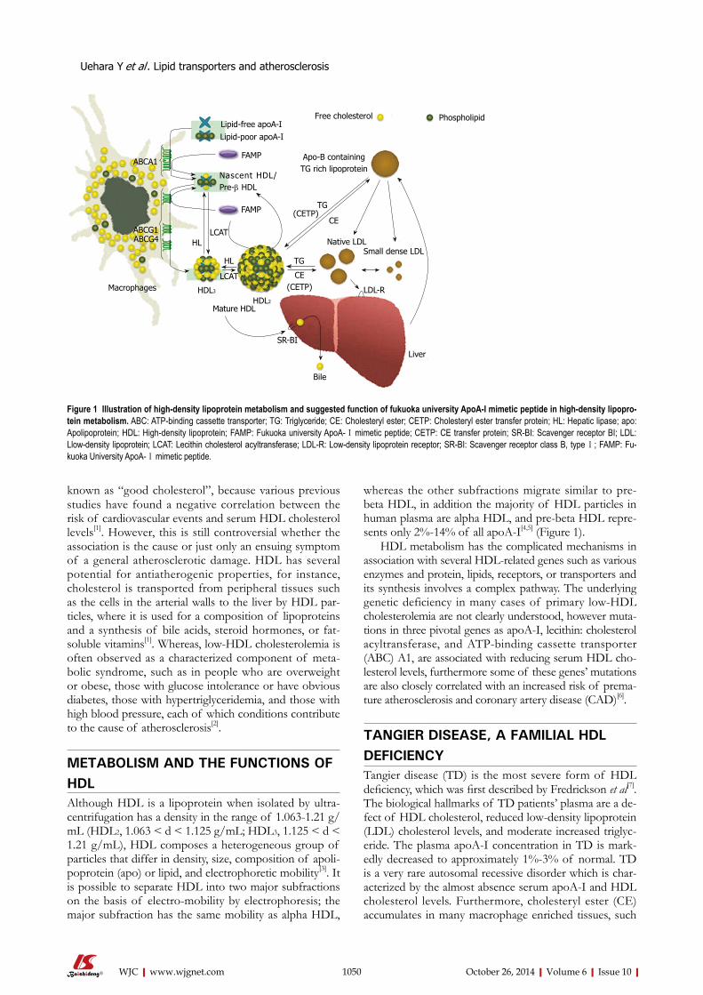
whereas the other subfractions migrate similar to pre-beta HDL, in addition the majority of HDL particles in human plasma are alpha HDL, and pre-beta HDL repre-sents only 2%-14% of all apoA-I[4,5] (Figure 1).
HDL metabolism has the complicated mechanisms in association with several HDL-related genes such as various enzymes and protein, lipids, receptors, or transporters and its synthesis involves a complex pathway. The underlying genetic deficiency in many cases of primary low-HDL cholesterolemia are not clearly understood, however muta-tions in three pivotal genes as apoA-I, lecithin: cholesterol acyltransferase, and ATP-binding cassette transporter (ABC) A1, are associated with reducing serum HDL cho-lesterol levels, furthermore some of these genes’ mutations are also closely correlated with an increased risk of prema-ture atherosclerosis and coronary artery disease (CAD)[6].
TANGIER DISEASE, A FAMILIAL HDL DEFICIENCYTangier disease (TD) is the most severe form of HDL deficiency, which was first described by Fredrickson et al[7]. The biological hallmarks of TD patients’ plasma are a de-fect of HDL cholesterol, reduced low-density lipoprotein (LDL) cholesterol levels, and moderate increased triglyc-eride. The plasma apoA-I concentration in TD is mark-edly decreased to approximately 1%-3% of normal. TD is a very rare autosomal recessive disorder which is char-acterized by the almost absence serum apoA-I and HDL cholesterol levels. Furthermore, cholesteryl ester (CE) accumulates in many macrophage enriched tissues, such
October 26, 2014|Volume 6|Issue 10|WJC|www.wjgnet.com
Uehara Y et al . Lipid transporters and atherosclerosis
1050
ABCA1
ABCG1ABCG4
Macrophages
Lipid-free apoA-ILipid-poor apoA-I
FAMP
Nascent HDL/Pre-β HDL
FAMP
LCATHL
HDL3
HL
LCAT
Free cholesterol Phospholipid
Apo-B containingTG rich lipoprotein
TG(CETP)
CE
Native LDLSmall dense LDL
LDL-R
TG
CE(CETP)
HDL2
Mature HDL
SR-BI
Bile
Liver
Figure 1 Illustration of high-density lipoprotein metabolism and suggested function of fukuoka university ApoA-I mimetic peptide in high-density lipopro-tein metabolism. ABC: ATP-binding cassette transporter; TG: Triglyceride; CE: Cholesteryl ester; CETP: Cholesteryl ester transfer protein; HL: Hepatic lipase; apo: Apolipoprotein; HDL: High-density lipoprotein; FAMP: Fukuoka university ApoA-Ⅰ mimetic peptide; CETP: CE transfer protein; SR-BI: Scavenger receptor BI; LDL: Llow-density lipoprotein; LCAT: Lecithin cholesterol acyltransferase; LDL-R: Low-density lipoprotein receptor; SR-BI: Scavenger receptor class B, type Ⅰ; FAMP: Fu-kuoka University ApoA-Ⅰ mimetic peptide.
as tonsils, spleen, liver, lymph nodes, peripheral nerves, thymus, and also arterial walls. Clinical symptoms among homozygotes patients include hepatosplenomegaly, hy-perplastic orange-yellow tonsils, corneal opacification, and premature CAD and atheosclerosis in a half of cases as well as relapsing peripheral neuropathy due to CE de-position in macrophages and Schwann cells[7-9].
In 1999, a cause of TD was found in a defect of the ABCA1 (formerly ABC1) gene[1,10,11] that is located on chromosome 9q31. This gene comprises 50 exons that span a region of approximately 149 kb[12,13]. ABCA1 has been identified as an important gene for regulating cel-lular cholesterol homeostasis and serum HDL choles-terol levels, which is defect in patients with TD. ABCA1 gene mutations cause gene dose-dependent decreases in serum HDL cholesterol levels and a decreased capacity of skin fibroblasts and monocyte-derived macrophages releasing cholesterol in the presence of extracellular apo-lipoproteins in TD patients and their heterozygous rela-tives[1,10,11,14,15].
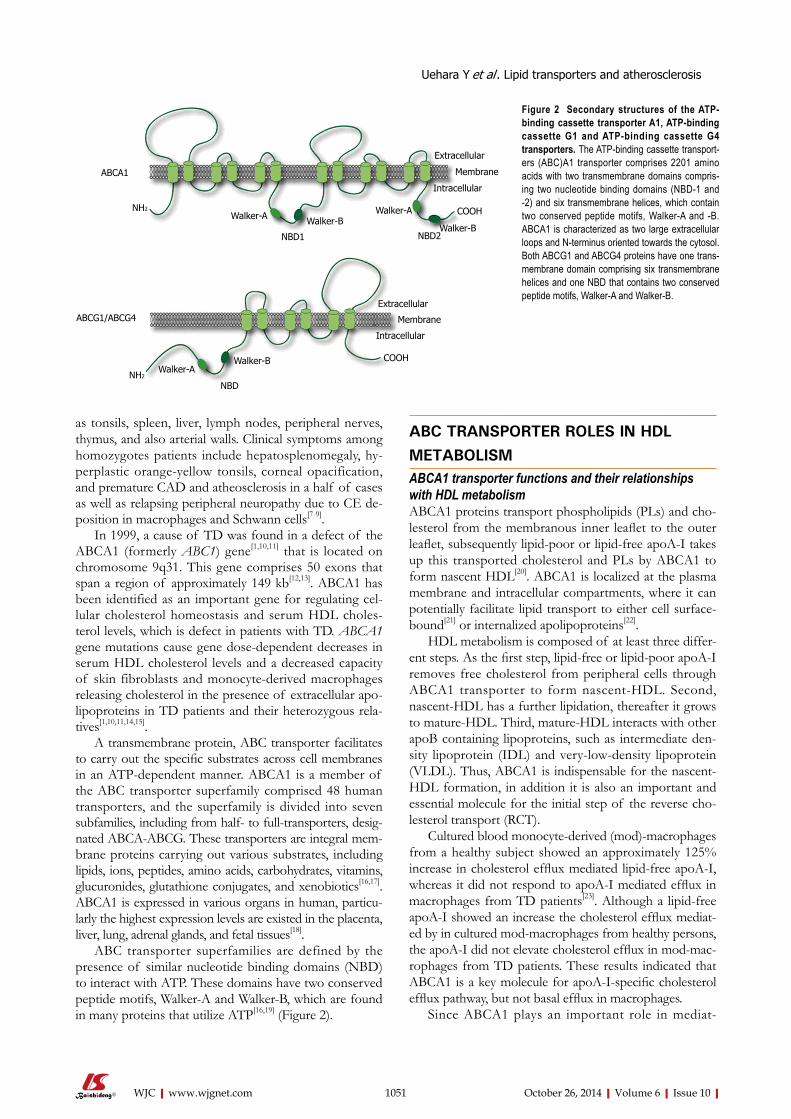
A transmembrane protein, ABC transporter facilitates to carry out the specific substrates across cell membranes in an ATP-dependent manner. ABCA1 is a member of the ABC transporter superfamily comprised 48 human transporters, and the superfamily is divided into seven subfamilies, including from half- to full-transporters, desig-nated ABCA-ABCG. These transporters are integral mem-brane proteins carrying out various substrates, including lipids, ions, peptides, amino acids, carbohydrates, vitamins, glucuronides, glutathione conjugates, and xenobiotics[16,17]. ABCA1 is expressed in various organs in human, particu-larly the highest expression levels are existed in the placenta, liver, lung, adrenal glands, and fetal tissues[18].
ABC transporter superfamilies are defined by the presence of similar nucleotide binding domains (NBD) to interact with ATP. These domains have two conserved peptide motifs, Walker-A and Walker-B, which are found in many proteins that utilize ATP[16,19] (Figure 2).
ABC TRANSPORTER ROLES IN HDL METABOLISM ABCA1 transporter functions and their relationships with HDL metabolismABCA1 proteins transport phospholipids (PLs) and cho-lesterol from the membranous inner leaflet to the outer leaflet, subsequently lipid-poor or lipid-free apoA-I takes up this transported cholesterol and PLs by ABCA1 to form nascent HDL[20]. ABCA1 is localized at the plasma membrane and intracellular compartments, where it can potentially facilitate lipid transport to either cell surface-bound[21] or internalized apolipoproteins[22].
HDL metabolism is composed of at least three differ-ent steps. As the first step, lipid-free or lipid-poor apoA-I removes free cholesterol from peripheral cells through ABCA1 transporter to form nascent-HDL. Second, nascent-HDL has a further lipidation, thereafter it grows to mature-HDL. Third, mature-HDL interacts with other apoB containing lipoproteins, such as intermediate den-sity lipoprotein (IDL) and very-low-density lipoprotein (VLDL). Thus, ABCA1 is indispensable for the nascent-HDL formation, in addition it is also an important and essential molecule for the initial step of the reverse cho-lesterol transport (RCT).
Cultured blood monocyte-derived (mod)-macrophages from a healthy subject showed an approximately 125% increase in cholesterol efflux mediated lipid-free apoA-I, whereas it did not respond to apoA-I mediated efflux in macrophages from TD patients[23]. Although a lipid-free apoA-I showed an increase the cholesterol efflux mediat-ed by in cultured mod-macrophages from healthy persons, the apoA-I did not elevate cholesterol efflux in mod-mac-rophages from TD patients. These results indicated that ABCA1 is a key molecule for apoA-I-specific cholesterol efflux pathway, but not basal efflux in macrophages.
Since ABCA1 plays an important role in mediat-
October 26, 2014|Volume 6|Issue 10|WJC|www.wjgnet.com 1051
ABCA1
ABCG1/ABCG4
NH2
NH2
Walker-A Walker-B
NBD1
Extracellular
Membrane
Intracellular
Walker-B
Walker-A
NBD2
COOH
Walker-AWalker-B
NBD
Extracellular
Membrane
Intracellular
COOH
Figure 2 Secondary structures of the ATP-binding cassette transporter A1, ATP-binding cassette G1 and ATP-binding cassette G4 transporters. The ATP-binding cassette transport-ers (ABC)A1 transporter comprises 2201 amino acids with two transmembrane domains compris-ing two nucleotide binding domains (NBD-1 and -2) and six transmembrane helices, which contain two conserved peptide motifs, Walker-A and -B. ABCA1 is characterized as two large extracellular loops and N-terminus oriented towards the cytosol. Both ABCG1 and ABCG4 proteins have one trans-membrane domain comprising six transmembrane helices and one NBD that contains two conserved peptide motifs, Walker-A and Walker-B.
Uehara Y et al . Lipid transporters and atherosclerosis
October 26, 2014|Volume 6|Issue 10|WJC|www.wjgnet.com
ABCG1 that function together as a heterodimer, and mu-tations in either of these genes can cause sitosterolemia which is a rare autosomal, recessively inherited disorder, characterized by premature atherosclerosis and xantho-mas[44-47]. These transporters mediate the sterols efflux including cholesterol and plant sterols from enterocytes return into the intestinal lumen and their excretion into the bile[44,48]. Accordingly, they protect the lipid accumu-lation in the body and augment RCT system. In animal model, ABCG5 and ABCG8 deficient mice have been shown to reduce a secretion of cholesterol in the bile and elevate sterol absorption[49], on the other hand ABCG5 and ABCG8 genes-overexpressed mice promotes choles-terol secretion in the bile, decreases cholesterol absorp-tion from diet, and increases neutral sterol excretion in the feces[50]. Liver X receptor (LXR) agonists promote the cholesterol efflux by the upregulation of ABCA1 and ABCG1, and also stimulate ABCG5 and ABCG8 which accelerate direct HDL transport of intestine into the lu-men, thus these genes also play an important role in the RCT system and their enhancement by LXR agonists prevent an atherosclerotic development[51].
MECHANISMS OF ABCA1 AND ABCG1 GENE REGULATIONABCA1 gene expression and cellular efflux of cholesterol are enhanced by cholesterol[15,18], oxysterols[52], retinoids[53], and cAMP analogs[15,54]. The ABCA1 gene promoter has been analyzed[13,52]. Both oxysterols and retinoids are li-gands for the nuclear transcription factor, LXRa/β and retinoid X receptor-alpha (RXRa), respectively, which have been identified as an enhancer of ABCA1 gene expression[52,53,55,56]. It is present in dimeric form of LXR and RXR as active transcriptional heterodimers that preferentially bind to responsive elements in the ABCA1 gene promoter[13,57]. LXRa/β and RXRa bind to the specific responsive element, called direct repeat 4 (DR4) element within the ABCA1 promoter, which is character-ized by two direct hexameric repeats separated by four nucleotides, thereafter they are activated by oxysterols and retinoids[58,59]. ABCA1 transcription are activated to bind either one or both ligands. Treatment with either a ligand of LXRa/β or RXRa enhances cellular ABCA1 expression, furthermore their combination treatment has a marked synergistic effect[60].
Since peroxisome proliferator activating receptor (PPAR)-a and -γ agonists such as fibrates and thiazolidine derivative (TZD) upregulate LXR mRNA expression, the activation of PPARs indirectly enhances a transcription activity of ABCA1 via LXR in cultured cells. In contrast, it is already known the zinc finger protein ZNF202 transcrip-tion factor as a major transcriptional repressor for ABCA1. In addition to the factor ZNF202, unsaturated fatty acids, but not saturated one, drastically suppress ABCA1-mediated cholesterol effluxes from macrophages by which they antag-onize the binding of specific agonist, oxysterol to LXR[61,62]. Moreover, various transcription factors, such as upstream stimulatory factor (USF)1, USF2, Fra2, and Sp3, also have
ing cholesterol and PL effluxes by lipid-free apoA-I, it is involved in a formation of discoidal HDL precursor, furthermore ABCA1 poorly interacts with HDL2 and HDL3. Patients with TD have extremely low levels of HDL cholesterol and they cannot compose nascent HDL particles due to a genetic defect in ABCA1 gene.
Disrupting the ABCA1 in mice resulted in HDL deficiency and impaired cholesterol transport similar to TD[24,25]. ABCA1 overexpression resulted in increased apoA-I-mediated cholesterol efflux in transgenic mice[26,27]. These results indicate that ABCA1 is an important gene in regulating circulating HDL cholesterol levels and cellular cholesterol homeostasis.
ABCG1 transporter functions and their relationships to HDL metabolismABCG1, formerly ABC8 is also a member of the ABC transporter family which has been mapped on chromo-some 21q22.3[19,28-32]. ABCG1 is one of half-transporter that contains only one NBD and a transmembrane do-main, in contrast to ABCA1[19,31] (Figure 2). Thus, ABCG1 may require a dimeric partner to become active with ABCG1 or ABCG4.
Although ABCA1 promotes cholesterol efflux to lipid-poor or lipid-free apoA-I, it only modestly induces lipid efflux of smaller particles, such as HDL3, and does not promote a cholesterol efflux of the larger HDL2 fraction[33,34]. It has been also shown by Wang et al[35] that ABCG1 and ABCG4 contributed to HDL2- and HDL3-mediated cholesterol effluxes and had an important func-tion related to HDL lipidation[35-37].
Administering a high-cholesterol, high-fat diet to ABCG1 knock-out mice resulted in a large amount of lipid accumulation in macrophages, whereas overexpres-sion of human ABCG1 gene was able to protect a di-etary fat-induced lipid accumulation in murine model[38]. Moreover, It was shown by Mauldin et al[39] that reduced function of ABCG1 facilitated foam cell formation in diabetes mice[39]. Transplanting bone marrow from ABCG1-deficient (ABCG1-/-) mice into LDL receptor-deficient mice, a model of familial hypercholesterolemia, produced contrasting effects on the formation of athero-sclerotic lesion[40-42]. In contrast to these report, decreased lesion size and formation were observed in the absence of macrophages from ABCG1-deficient mice[41,42], and whole body ABCG1 expression protected against the development of early atherosclerotic plaque[43]. However, it remains unclear that the physiological roles of ABCG1 and its contribution to atherosclerotic progression in hu-mans. In addition, ABC transporters such as ABCG1 and ABCG4, but not ABCA1, are not only responsible for passive and nonspecific efflux pathway but also mature HDL-mediated cholesterol efflux, which are spherical and transport almost all HDL cholesterol[35,37].
ROLES OF ABCG5 AND ABCG8 TRANSPORTERABCG5 and ABCG8 are half-transporters as well as
1052
Uehara Y et al . Lipid transporters and atherosclerosis
October 26, 2014|Volume 6|Issue 10|WJC|www.wjgnet.com
the potential to repress the ABCA1 transcription[63]. The ABCG1 gene has a promoter upstream of exon
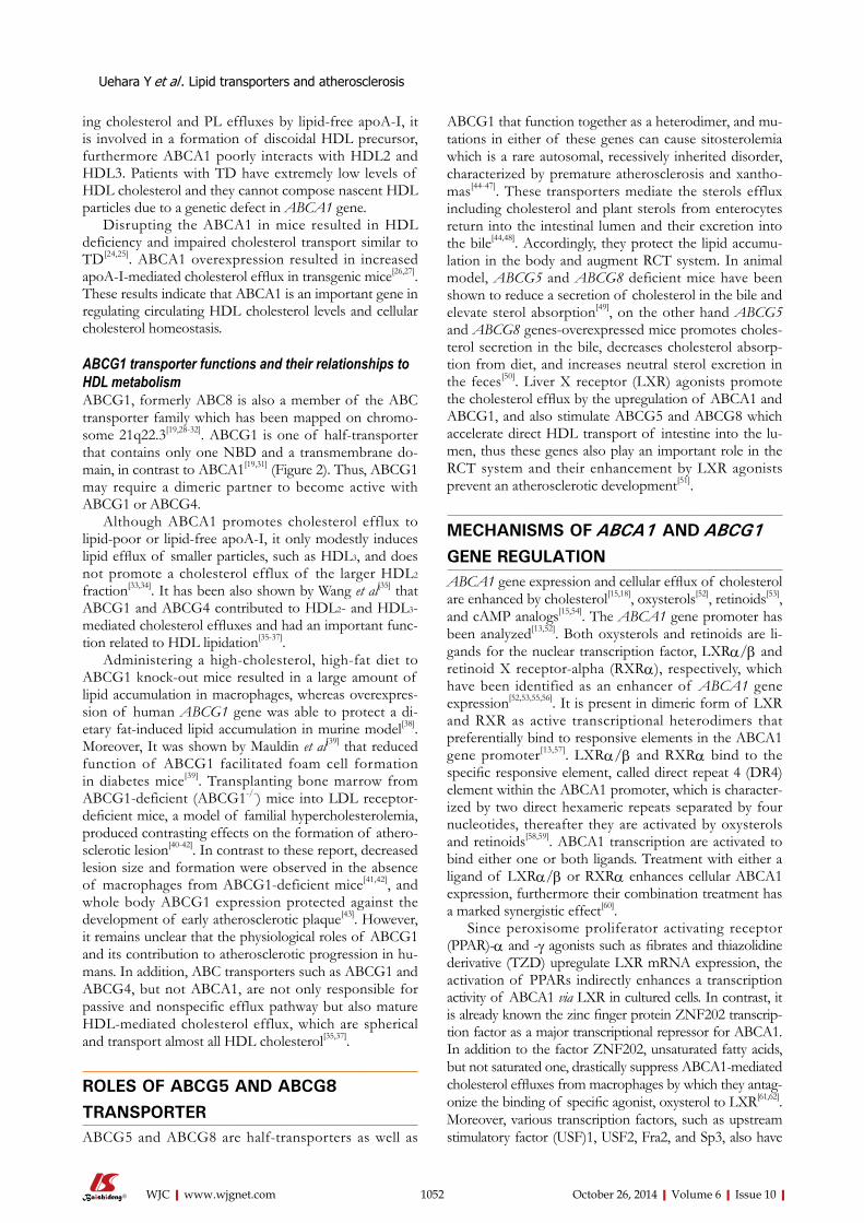
1 and another intron promoter, which encodes several transcripts[64-66]. Our previous study demonstrated that LXR activation drastically increased the ABCG1 pro-moter activity (Promoter-A) located upstream of exon 1 as well as the ABCA1 gene (Figure 3A). On the other hand, the activity of ABCG1 promoter-B located within intron 4 was not changed by an activation of LXR (Figure 3B)[62]. These results indicate that the gene transcription of exon 5 and subsequent exons might be also regulated, at least in part, by the ABCG1 promoter-A.
Electrophoretic mobility shift assay was done to confirm these findings, and it showed the existence of DNA-binding nuclear receptors on extracted ABCG1 promoter-A having DR4 element. As would be expected from these finding, only the ABCG1 promoter-A contained a DR4 element, but not promoter-B, which is required for binding to LXRa/RXR. In fact, a promoter response to ligands of LXR/RXR was totally abolished in the mutated ABCG1 promoter lacked an active DR4 element[62] (Figure 3C).
ABCG1 SINGLE NUCLEOTIDE POLYMORPHISMSIt remains unclear whether ABCG1 itself contributes
to circulating lipid levels, such as HDL cholesterol and arterial plaque regression in humans. There have been only five reports on ABCG1 polymorphisms. Our previ-ous study was the first regarding an ABCG1 polymor-phism, which appeared to be a potent functional ABCG1 polymorphism located in the promoter region[67-71]. The ABCG1 promoter -257T>G polymorphism, rs1378577, -394 T/G from the transcription start site (NM_207627.1:c. -394T>G), -134 T/G from exon 1 (NM_207627.1) is a single nucleotide mutation (SNP) on the ABCG1 promot-er region upstream of exon 1, which was reported to be a functional promoter with an LXR-responsive element[62,67].
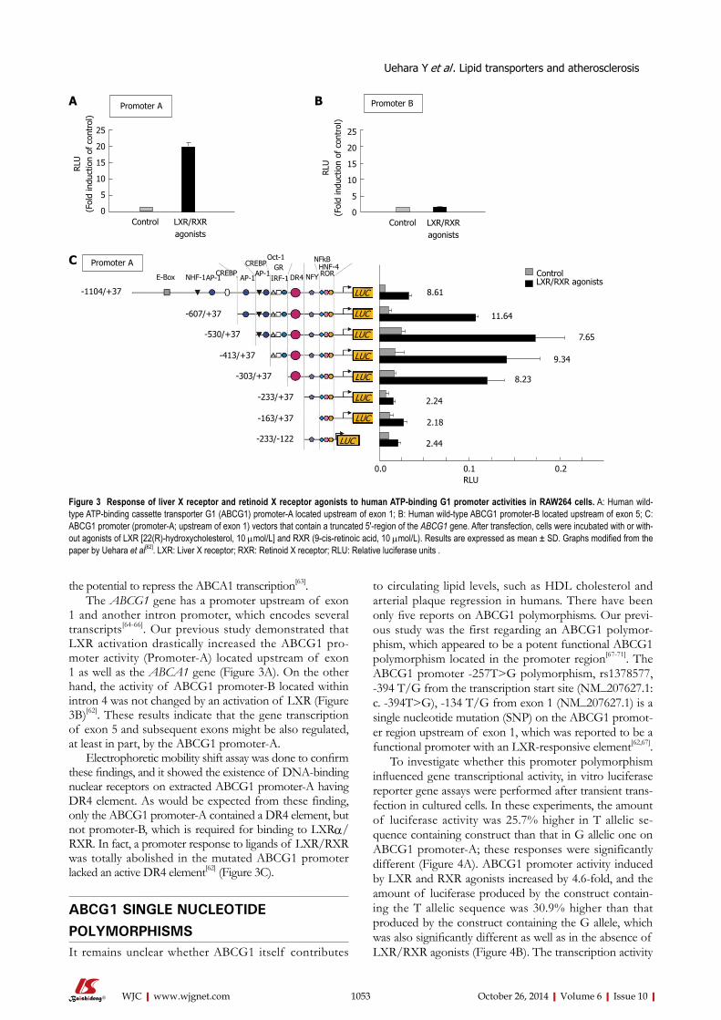
To investigate whether this promoter polymorphism influenced gene transcriptional activity, in vitro luciferase reporter gene assays were performed after transient trans-fection in cultured cells. In these experiments, the amount of luciferase activity was 25.7% higher in T allelic se-quence containing construct than that in G allelic one on ABCG1 promoter-A; these responses were significantly different (Figure 4A). ABCG1 promoter activity induced by LXR and RXR agonists increased by 4.6-fold, and the amount of luciferase produced by the construct contain-ing the T allelic sequence was 30.9% higher than that produced by the construct containing the G allele, which was also significantly different as well as in the absence of LXR/RXR agonists (Figure 4B). The transcription activity
1053
Promoter A
25
20
15
10
5
0
RLU
(Fol
d in
duct
ion
of c
ontr
ol)
25
20
15
10
5
0
RLU
(Fol
d in
duct
ion
of c
ontr
ol)
Control Control
Promoter BA B
Figure 3 Response of liver X receptor and retinoid X receptor agonists to human ATP-binding G1 promoter activities in RAW264 cells. A: Human wild-type ATP-binding cassette transporter G1 (ABCG1) promoter-A located upstream of exon 1; B: Human wild-type ABCG1 promoter-B located upstream of exon 5; C: ABCG1 promoter (promoter-A; upstream of exon 1) vectors that contain a truncated 5′-region of the ABCG1 gene. After transfection, cells were incubated with or with-out agonists of LXR [22(R)-hydroxycholesterol, 10 µmol/L] and RXR (9-cis-retinoic acid, 10 µmol/L). Results are expressed as mean ± SD. Graphs modified from the paper by Uehara et al[62]. LXR: Liver X receptor; RXR: Retinoid X receptor; RLU: Relative luciferase units .
Uehara Y et al . Lipid transporters and atherosclerosis
-1104/+37
-607/+37
-530/+37
-413/+37
-303/+37
-233/+37
-163/+37
-233/-122
LXR/RXR agonistsControl
8.61
11.64
7.65
9.34
8.23
2.24
2.18
2.44
LUC
LUC
LUC
LUC
LUC
LUC
LUC
LUC
E-Box NHF-1AP-1CREBP
AP-1
CREBPAP-1
Oct-1GR
IRF-1 DR4 NFY
NFkBHNF-4ROR
0.0 0.1 0.2RLU
Promoter AC
LXR/RXRagonists
LXR/RXRagonists
October 26, 2014|Volume 6|Issue 10|WJC|www.wjgnet.com
in the T allelic sequence was significantly higher than that in the G allelic sequence on ABCG1 promoter-A.
Furthermore, the ABCG1 promoter showed in-creased activity via stimulation by LXR and RXR, and a similar genotype-dependent effect on ABCG1 gene tran-scription under these conditions was identified. These results suggest that the ABCG1 promoter polymorphism might be an isolated regulating factor for ABCG1 gene transcription activity, independent of LXR and RXR.
We genotyped 109 Japanese male CAD patients for the ABCG1 promoter SNP. This polymorphism was as-sociated with CAD severity in Japanese men, but not with changes in lipid levels under fasting conditions in a case control study. Logistic regression analysis showed that there was an interaction between the ABCG1 promoter genotype and CAD severity.
Genotype frequencies were grouped on the basis of whether patients had multi- or single-vessel CAD. The adjusted relative risk associated with the G allele (assuming an additive effect) in a matched-pair analysis was 2.1 for multi-vessel CAD compared with single-vessel CAD and 3.5 for the G/G and T/G genotypes compared with T/T (assuming a dominant effect of the G allele)[67]. These results were consistent with the proposition that the variations for ABCG1 gene might make a contribution to interindividual variability in susceptibility or severity of atherosclerotic changes.
ABCG1 expression levels in atherosclerotic tissues might be lower among those with the G allele and may be associated with a mechanism for an increased incidence of atherosclerosis in these individuals. These results were similar to a previous study by Baldán et al[72] of transgenic mice in whom the ABCG1 gene was deleted[73]. Further-more, a recent study regarding ABCG1 as a candidate gene with possible important antiatherogenic properties also illustrates the current interest in this transporter.
HDL-TARGETING THERAPY FOR ATHEROSCLEROSISInhibiting scavenger receptor BI (SR-BI), CE transfer
protein (CETP) or PL transfer protein, and an activating ABCA1 or apoA-I elevate HDL cholesterol levels. How-ever, it is uncertain whether the effects of these interven-tions on atherosclerosis are consistence with the results of studies with animal models and inborn human HDL metabolism errors. Although it has not found a such small molecule which strongly promotes apoA-I produc-tion, one possible candidate molecule is LXR agonist which increase HDL cholesterol levels via upregulation of ABCA1 and ABCG1 expressions. Unfortunately, pre-vious study has shown that concurrent with an activation of RCT, the agonist induces hypertriglycemia consequent on increasing hepatic VLDL production.
As a therapeutic approach for increasing HDL lev-els, much research has focused both increasing HDL cholesterol levels and on enhancing HDL biochemical functions. HDL therapies that used injections of recon-stituted HDL, apoA-I mimetics, or full-length apoA-I are remarkably effective[74,75]. Nissen et al[75] showed that in humans, intravenous administation of ETC-216, an apoA-I-Milano complexed with phospholipids, produced a significant regression of coronary atherosclerotic plaques as determined by intravascular ultrasound (IVUS). After infusing ETC-216, regression of coronary athero-sclerosis was accompanied by reverse remodeling of the external elastic membrane and with no changes in lumi-nal dimensions as assessed by IVUS analyses[76].
Reconstituted HDL (rHDL), a complex of apoA-I or apoA-I mimetics with PL, must be shaped as disc, and it may be a suitable administration in patients with athero-sclerotic plaque and TD. ABCA1 plays an important role for apoA-I-mediated cholesterol efflux in macrophages, and thereby is involved in discoidal HDL precursor for-mation. Mature HDL particles shaped spherical induce cholesterol effluxes by other transporters such as ABCG1 and ABCG4, rather than ABCA1[35]. We previously estab-lished a discoidal rHDL, which was a complex of human serum-derived full length of apoA-I with PL, 1-palmito-yl-2-oleoylphosphatidylcholine (POPC)[77]. Interestingly, the apoA-I complex with a PL, a POPC/apoA-I disc, could take up cholesterol from macrophages in both nor-
1054
Rela
tive
luci
fera
se a
ctiv
ity, R
LU
3.0
2.5
2.0
1.5
0.5
0.0
3.0
2.5
2.0
1.5
0.5
0.0
Rela
tive
luci
fera
se a
ctiv
ity, R
LU
P < 0.01
P < 0.01
-30.9%
T allele G allele T allele G allele
LXR/RXR agonists
-25.7%
Figure 4 In vitro promoter activity assay for ATP-binding G1 promoter-A. ATP-binding cassette transporter G1 (ABCG1) promoter construct with a −257T/G mutation, −394 T/G from the transcription start site (NM_207627.1: c.-394T > G) on ABCG1 promoter-A, which is reported to be a functional promoter with an LXR-responsive element. A: ABCG1 transcription activity on a construct that contains the T or G allelic sequence; B: ABCG1 transcription activity induced by 5 µmol/L of T0901317 (T0) and 9-cis-retinoic acid (9cisRA) on constructs that contain the T or G allelic sequence. Results are expressed as mean ± SD. Graphs are modified from the paper by Furuyama et al[67]. ABC: ATP-binding cassette; RXR: Retinoid X receptor; LXR: Liver X receptor.
Uehara Y et al . Lipid transporters and atherosclerosis
A B
October 26, 2014|Volume 6|Issue 10|WJC|www.wjgnet.com
mal subjects and TD patients. Although studies on the use of apoA-I mimetic pep-
tides (e.g., 4F and L37pA) are underway[78-80], none of these agents are currently available for clinical use. To develop a physiological HDL-generating apoA-I mimetic peptide that functions with ABCA1 transporter, differ-ent candidate peptides were synthesized by focusing on the amino acid sequence alignments of human apoA-I interactions with ABCA1. We recently established a novel short apoA-I mimetic peptide that comprised 24 amino acids and without phospholipids Fukuoka University ApoA-I Mimetic Peptide (FAMP), which retained the amphipathic helical structure of the 243-amino acid apoA-I and the ability to associate with lipids[81]. This was shown to enhance HDL function and suppress aortic plaque formation in apoE-knockout mice that were fed a high-fat diet. FAMP markedly increased pre-beta HDL formation as well as increased the overall cholesterol ef-fluxes from peripheral tissues[82].
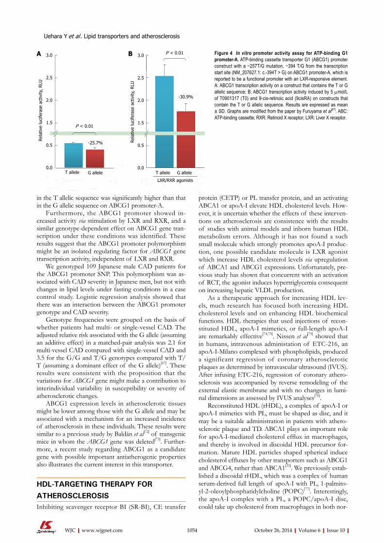
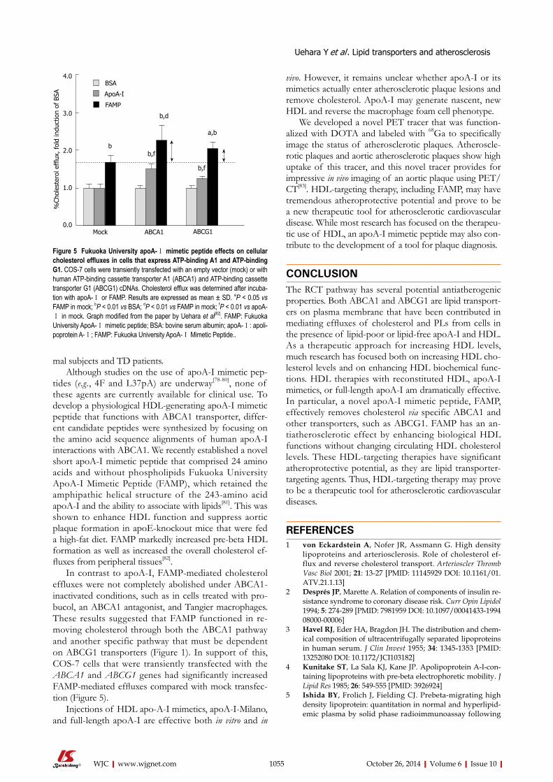
In contrast to apoA-I, FAMP-mediated cholesterol effluxes were not completely abolished under ABCA1-inactivated conditions, such as in cells treated with pro-bucol, an ABCA1 antagonist, and Tangier macrophages. These results suggested that FAMP functioned in re-moving cholesterol through both the ABCA1 pathway and another specific pathway that must be dependent on ABCG1 transporters (Figure 1). In support of this, COS-7 cells that were transiently transfected with the ABCA1 and ABCG1 genes had significantly increased FAMP-mediated effluxes compared with mock transfec-tion (Figure 5).
Injections of HDL apo-A-I mimetics, apoA-I-Milano, and full-length apoA-I are effective both in vitro and in
vivo. However, it remains unclear whether apoA-I or its mimetics actually enter atherosclerotic plaque lesions and remove cholesterol. ApoA-I may generate nascent, new HDL and reverse the macrophage foam cell phenotype.
We developed a novel PET tracer that was function-alized with DOTA and labeled with 68Ga to specifically image the status of atherosclerotic plaques. Atheroscle-rotic plaques and aortic atherosclerotic plaques show high uptake of this tracer, and this novel tracer provides for impressive in vivo imaging of an aortic plaque using PET/CT[83]. HDL-targeting therapy, including FAMP, may have tremendous atheroprotective potential and prove to be a new therapeutic tool for atherosclerotic cardiovascular disease. While most research has focused on the therapeu-tic use of HDL, an apoA-I mimetic peptide may also con-tribute to the development of a tool for plaque diagnosis.
CONCLUSIONThe RCT pathway has several potential antiatherogenic properties. Both ABCA1 and ABCG1 are lipid transport-ers on plasma membrane that have been contributed in mediating effluxes of cholesterol and PLs from cells in the presence of lipid-poor or lipid-free apoA-I and HDL. As a therapeutic approach for increasing HDL levels, much research has focused both on increasing HDL cho-lesterol levels and on enhancing HDL biochemical func-tions. HDL therapies with reconstituted HDL, apoA-I mimetics, or full-length apoA-I am dramatically effective. In particular, a novel apoA-I mimetic peptide, FAMP, effectively removes cholesterol via specific ABCA1 and other transporters, such as ABCG1. FAMP has an an-tiatherosclerotic effect by enhancing biological HDL functions without changing circulating HDL cholesterol levels. These HDL-targeting therapies have significant atheroprotective potential, as they are lipid transporter-targeting agents. Thus, HDL-targeting therapy may prove to be a therapeutic tool for atherosclerotic cardiovascular diseases.
REFERENCES1 von Eckardstein A, Nofer JR, Assmann G. High density
lipoproteins and arteriosclerosis. Role of cholesterol ef-flux and reverse cholesterol transport. Arterioscler Thromb Vasc Biol 2001; 21: 13-27 [PMID: 11145929 DOI: 10.1161/01.ATV.21.1.13]
2 Després JP, Marette A. Relation of components of insulin re-sistance syndrome to coronary disease risk. Curr Opin Lipidol 1994; 5: 274-289 [PMID: 7981959 DOI: 10.1097/00041433-199408000-00006]
3 Havel RJ, Eder HA, Bragdon JH. The distribution and chem-ical composition of ultracentrifugally separated lipoproteins in human serum. J Clin Invest 1955; 34: 1345-1353 [PMID: 13252080 DOI: 10.1172/JCI103182]
4 Kunitake ST, La Sala KJ, Kane JP. Apolipoprotein A-I-con-taining lipoproteins with pre-beta electrophoretic mobility. J Lipid Res 1985; 26: 549-555 [PMID: 3926924]
5 Ishida BY, Frolich J, Fielding CJ. Prebeta-migrating high density lipoprotein: quantitation in normal and hyperlipid-emic plasma by solid phase radioimmunoassay following
1055
bb,f
b,d
b,f
a,b
BSA
ApoA-I
FAMP
%Ch
oles
tero
l effl
ux, f
old
indu
ctio
n of
BSA
4.0
3.0
2.0
1.0
0.0Mock ABCA1 ABCG1
Figure 5 Fukuoka University apoA-Ⅰ mimetic peptide effects on cellular cholesterol effluxes in cells that express ATP-binding A1 and ATP-binding G1. COS-7 cells were transiently transfected with an empty vector (mock) or with human ATP-binding cassette transporter A1 (ABCA1) and ATP-binding cassette transporter G1 (ABCG1) cDNAs. Cholesterol efflux was determined after incuba-tion with apoA-Ⅰ or FAMP. Results are expressed as mean ± SD. aP < 0.05 vs FAMP in mock; bP < 0.01 vs BSA; dP < 0.01 vs FAMP in mock; fP < 0.01 vs apoA-Ⅰ in mock. Graph modified from the paper by Uehara et al[82]. FAMP: Fukuoka University ApoA-Ⅰ mimetic peptide; BSA: bovine serum albumin; apoA-Ⅰ: apoli-poprotein A-Ⅰ; FAMP: Fukuoka University ApoA-Ⅰ Mimetic Peptide..
Uehara Y et al . Lipid transporters and atherosclerosis
October 26, 2014|Volume 6|Issue 10|WJC|www.wjgnet.com
electrophoretic transfer. J Lipid Res 1987; 28: 778-786 [PMID: 3114402]
6 Miller M, Rhyne J, Hamlette S, Birnbaum J, Rodriguez A. Genetics of HDL regulation in humans. Curr Opin Li-pidol 2003; 14: 273-279 [PMID: 12840658 DOI: 10.1097/01.mol.0000073506.41685.d2]
7 Fredrickson DS, Altrocchi PH, Avioli LV, Goodman DS and Goodman HC. Tangier disease combined clinical staff con-ference at the National Institutes of Health. Ann Intern Med 1961; 55: 1016-1031 [DOI: 10.7326/0003-4819-55-6-1016]
8 Assman G, von Eckardstein A and Brewer HBJ. Familial high density lipoprotein deficiency: Tangier disease. The metabolic and Molecular Basis of Inherited Disease.7th ed. C. R. Scriver, A. L. Beaudet, W. S. Sly and D. Valle, editor. New York: McGraw-Hill, 1995: 2053-2072
9 Hobbs HH, Rader DJ. ABC1: connecting yellow tonsils, neu-ropathy, and very low HDL. J Clin Invest 1999; 104: 1015-1017 [PMID: 10525038 DOI: 10.1172/JCI8509]
10 Brooks-Wilson A, Marcil M, Clee SM, Zhang LH, Roomp K, van Dam M, Yu L, Brewer C, Collins JA, Molhuizen HO, Loubser O, Ouelette BF, Fichter K, Ashbourne-Excoffon KJ, Sensen CW, Scherer S, Mott S, Denis M, Martindale D, Frohlich J, Morgan K, Koop B, Pimstone S, Kastelein JJ, Hayden MR and et al. Mutations in ABC1 in Tangier disease and familial high-density lipoprotein deficiency. Nat Genet 1999; 22: 336-345 [DOI: 10.1038/11905]
11 Rust S, Rosier M, Funke H, Real J, Amoura Z, Piette JC, Deleuze JF, Brewer HB, Duverger N, Denèfle P, Assmann G. Tangier disease is caused by mutations in the gene encod-ing ATP-binding cassette transporter 1. Nat Genet 1999; 22: 352-355 [PMID: 10431238 DOI: 10.1038/11921]
12 Remaley AT, Rust S, Rosier M, Knapper C, Naudin L, Broc-cardo C, Peterson KM, Koch C, Arnould I, Prades C, Du-verger N, Funke H, Assman G, Dinger M, Dean M, Chimini G, Santamarina Fojo S, Fredrickson DS, Denefle P and Brew-er HB, Jr. Human ATP-binding cassette transporter 1 (ABC1): genomic organization and identification of the genetic defect in the original Tangier disease kindred. Proc Natl Acad Sci USA 1999; 96: 12685-12690 [DOI: 10.1073/pnas.96.22.12685]
13 Santamarina-Fojo S, Peterson K, Knapper C, Qiu Y, Freeman L, Cheng JF, Osorio J, Remaley A, Yang XP, Haudenschild C, Prades C, Chimini G, Blackmon E, Francois T, Duverger N, Rubin EM, Rosier M, Denèfle P, Fredrickson DS, Brewer HB. Complete genomic sequence of the human ABCA1 gene: analysis of the human and mouse ATP-binding cassette A promoter. Proc Natl Acad Sci USA 2000; 97: 7987-7992 [PMID: 10884428 DOI: 10.1073/pnas.97.14.7987]
14 Bodzioch M, Orsó E, Klucken J, Langmann T, Böttcher A, Diederich W, Drobnik W, Barlage S, Büchler C, Porsch-Ozcürümez M, Kaminski WE, Hahmann HW, Oette K, Rothe G, Aslanidis C, Lackner KJ, Schmitz G. The gene encoding ATP-binding cassette transporter 1 is mutated in Tangier disease. Nat Genet 1999; 22: 347-351 [PMID: 10431237 DOI: 10.1038/11914]
15 Lawn RM, Wade DP, Garvin MR, Wang X, Schwartz K, Porter JG, Seilhamer JJ, Vaughan AM, Oram JF. The Tangier disease gene product ABC1 controls the cellular apolipopro-tein-mediated lipid removal pathway. J Clin Invest 1999; 104: R25-R31 [PMID: 10525055 DOI: 10.1172/JCI8119]
16 Klein I, Sarkadi B, Váradi A. An inventory of the human ABC proteins. Biochim Biophys Acta 1999; 1461: 237-262 [PMID: 10581359 DOI: 10.1016/S0005-2736(99)00161-3]
17 Dean M, Annilo T. Evolution of the ATP-binding cassette (ABC) transporter superfamily in vertebrates. Annu Rev Genomics Hum Genet 2005; 6: 123-142 [PMID: 16124856 DOI: 10.1146/annurev.genom.6.080604.162122]
18 Langmann T, Klucken J, Reil M, Liebisch G, Luciani MF, Chimini G, Kaminski WE and Schmitz G. Molecular cloning
of the human ATP-binding cassette transporter 1 (hABC1): evidence for sterol-dependent regulation in macrophages. Biochem-Biophys-Res-Commun 1999; 257: 29-33 [DOI: 10.1006/bbrc.1999.0406]
19 Walker JE, Saraste M, Runswick MJ, Gay NJ. Distantly related sequences in the alpha- and beta-subunits of ATP synthase, myosin, kinases and other ATP-requiring enzymes and a common nucleotide binding fold. EMBO J 1982; 1: 945-951 [PMID: 6329717]
20 Oram JF, Lawn RM. ABCA1. The gatekeeper for eliminat-ing excess tissue cholesterol. J Lipid Res 2001; 42: 1173-1179 [PMID: 11483617]
21 Neufeld EB, Remaley AT, Demosky SJ, Stonik JA, Cooney AM, Comly M, Dwyer NK, Zhang M, Blanchette-Mackie J, Santamarina-Fojo S, Brewer HB. Cellular localization and trafficking of the human ABCA1 transporter. J Biol Chem 2001; 276: 27584-27590 [PMID: 11349133 DOI: 10.1074/jbc.M103264200]
22 von Eckardstein A, Rohrer L. Transendothelial lipoprotein transport and regulation of endothelial permeability and integrity by lipoproteins. Curr Opin Lipidol 2009; 20: 197-205 [PMID: 19395962 DOI: 10.1097/MOL.0b013e32832afd63]
23 Uehara Y, Tsuboi Y, Zhang B, Miura S, Baba Y, Higuchi MA, Yamada T, Rye KA, Saku K. POPC/apoA-I discs as a potent lipoprotein modulator in Tangier disease. Atherosclerosis 2008; 197: 283-289 [PMID: 17560579 DOI: 10.1016/j.atherosclerosis.2007.04.025]
24 Orsó E, Broccardo C, Kaminski WE, Böttcher A, Liebisch G, Drobnik W, Götz A, Chambenoit O, Diederich W, Lang-mann T, Spruss T, Luciani MF, Rothe G, Lackner KJ, Chi-mini G, Schmitz G. Transport of lipids from golgi to plasma membrane is defective in tangier disease patients and Abc1-deficient mice. Nat Genet 2000; 24: 192-196 [PMID: 10655069 DOI: 10.1038/72869]
25 McNeish J, Aiello RJ, Guyot D, Turi T, Gabel C, Aldinger C, Hoppe KL, Roach ML, Royer LJ, de Wet J, Broccardo C, Chi-mini G, Francone OL. High density lipoprotein deficiency and foam cell accumulation in mice with targeted disruption of ATP-binding cassette transporter-1. Proc Natl Acad Sci USA 2000; 97: 4245-4250 [PMID: 10760292 DOI: 10.1073/pnas.97.8.4245]
26 Vaisman BL, Lambert G, Amar M, Joyce C, Ito T, Shambu-rek RD, Cain WJ, Fruchart-Najib J, Neufeld ED, Remaley AT, Brewer HB, Santamarina-Fojo S. ABCA1 overexpression leads to hyperalphalipoproteinemia and increased biliary cholesterol excretion in transgenic mice. J Clin Invest 2001; 108: 303-309 [PMID: 11457883 DOI: 10.1172/JCI200112517]
27 Singaraja RR, Bocher V, James ER, Clee SM, Zhang LH, Leavitt BR, Tan B, Brooks-Wilson A, Kwok A, Bissada N, Yang YZ, Liu G, Tafuri SR, Fievet C, Wellington CL, Staels B, Hayden MR. Human ABCA1 BAC transgenic mice show increased high density lipoprotein cholesterol and ApoAI-dependent efflux stimulated by an internal promoter con-taining liver X receptor response elements in intron 1. J Biol Chem 2001; 276: 33969-33979 [PMID: 11423537 DOI: 10.1074/jbc.M102503200]
28 Croop JM, Tiller GE, Fletcher JA, Lux ML, Raab E, Golden-son D, Son D, Arciniegas S, Wu RL. Isolation and character-ization of a mammalian homolog of the Drosophila white gene. Gene 1997; 185: 77-85 [PMID: 9034316 DOI: 10.1016/S0378-1119(96)00633-6]
29 Chen H, Rossier C, Lalioti MD, Lynn A, Chakravarti A, Per-rin G, Antonarakis SE. Cloning of the cDNA for a human homologue of the Drosophila white gene and mapping to chromosome 21q22.3. Am J Hum Genet 1996; 59: 66-75 [PMID: 8659545]
30 Savary S, Denizot F, Luciani M, Mattei M, Chimini G. Molec-ular cloning of a mammalian ABC transporter homologous
1056
Uehara Y et al . Lipid transporters and atherosclerosis
October 26, 2014|Volume 6|Issue 10|WJC|www.wjgnet.com
to Drosophila white gene. Mamm Genome 1996; 7: 673-676 [PMID: 8703120 DOI: 10.1007/s003359900203]
31 Dean M, Rzhetsky A, Allikmets R. The human ATP-binding cassette (ABC) transporter superfamily. Genome Res 2001; 11: 1156-1166 [PMID: 11435397 DOI: 10.1101/gr.GR-1649R]
32 Klucken J, Buchler C, Orso E, Kaminski WE, Porsch-Ozcu-rumez M, Liebisch G, Kapinsky M, Diederich W, Drobnik W, Dean M, Allikmets R and Schmitz G. ABCG1 (ABC8), the human homolog of the Drosophila white gene, is a regula-tor of macrophage cholesterol and phospholipid transport. Proc Natl Acad Sci USA 2000; 97: 817-822 [DOI: 10.1073/pnas.97.2.817]
33 Francis GA, Knopp RH, Oram JF. Defective removal of cel-lular cholesterol and phospholipids by apolipoprotein A-I in Tangier Disease. J Clin Invest 1995; 96: 78-87 [PMID: 7615839 DOI: 10.1172/JCI118082]
34 Wang N, Silver DL, Costet P, Tall AR. Specific binding of ApoA-I, enhanced cholesterol efflux, and altered plasma membrane morphology in cells expressing ABC1. J Biol Chem 2000; 275: 33053-33058 [PMID: 0010918065 DOI: 10.1074/jbc.M005438200]
35 Wang N, Lan D, Chen W, Matsuura F, Tall AR. ATP-binding cassette transporters G1 and G4 mediate cellular choles-terol efflux to high-density lipoproteins. Proc Natl Acad Sci USA 2004; 101: 9774-9779 [PMID: 15210959 DOI: 10.1073/pnas.0403506101]
36 Smith JD. Insight into ABCG1-mediated cholesterol efflux. Arterioscler Thromb Vasc Biol 2006; 26: 1198-1200 [PMID: 16709952 DOI: 10.1161/01.ATV.0000221217.86465.66]
37 Uehara Y, Yamada T, Baba Y, Miura S, Abe S, Kitajima K, Higuchi MA, Iwamoto T, Saku K. ATP-binding cassette transporter G4 is highly expressed in microglia in Alzheim-er’s brain. Brain Res 2008; 1217: 239-246 [PMID: 18508037]
38 Kennedy MA, Barrera GC, Nakamura K, Baldán A, Tarr P, Fishbein MC, Frank J, Francone OL, Edwards PA. ABCG1 has a critical role in mediating cholesterol efflux to HDL and preventing cellular lipid accumulation. Cell Metab 2005; 1: 121-131 [PMID: 16054053 DOI: 10.1016/j.cmet.2005.01.002]
39 Mauldin JP, Srinivasan S, Mulya A, Gebre A, Parks JS, Daugherty A, Hedrick CC. Reduction in ABCG1 in Type 2 diabetic mice increases macrophage foam cell formation. J Biol Chem 2006; 281: 21216-21224 [PMID: 16723355 DOI: 10.1074/jbc.M510952200]
40 Out R, Hoekstra M, Hildebrand RB, Kruit JK, Meurs I, Li Z, Kuipers F, Van Berkel TJ, Van Eck M. Macrophage ABCG1 deletion disrupts lipid homeostasis in alveolar macrophages and moderately influences atherosclerotic lesion develop-ment in LDL receptor-deficient mice. Arterioscler Thromb Vasc Biol 2006; 26: 2295-2300 [PMID: 16857950 DOI: 10.1161/01.ATV.0000237629.29842.4c]
41 Baldán A, Pei L, Lee R, Tarr P, Tangirala RK, Weinstein MM, Frank J, Li AC, Tontonoz P, Edwards PA. Impaired development of atherosclerosis in hyperlipidemic Ldlr-/- and ApoE-/- mice transplanted with Abcg1-/- bone mar-row. Arterioscler Thromb Vasc Biol 2006; 26: 2301-2307 [PMID: 16888235 DOI: 10.1161/01.ATV.0000240051.22944.dc]
42 Ranalletta M, Wang N, Han S, Yvan-Charvet L, Welch C, Tall AR. Decreased atherosclerosis in low-density lipoprotein receptor knockout mice transplanted with Abcg1-/- bone marrow. Arterioscler Thromb Vasc Biol 2006; 26: 2308-2315 [PMID: 16917103 DOI: 10.1161/01.ATV.0000242275.92915.43]
43 Out R, Hoekstra M, Meurs I, de Vos P, Kuiper J, Van Eck M, Van Berkel TJ. Total body ABCG1 expression protects against early atherosclerotic lesion development in mice. Ar-terioscler Thromb Vasc Biol 2007; 27: 594-599 [PMID: 17204665 DOI: 10.1161/01.ATV.0000257136.24308.0c]
44 Graf GA, Yu L, Li WP, Gerard R, Tuma PL, Cohen JC, Hobbs HH. ABCG5 and ABCG8 are obligate heterodimers for protein trafficking and biliary cholesterol excretion. J Biol Chem 2003; 278: 48275-48282 [PMID: 14504269 DOI: 10.1074/
jbc.M310223200]45 Patel SB, Salen G, Hidaka H, Kwiterovich PO, Stalenhoef
AF, Miettinen TA, Grundy SM, Lee MH, Rubenstein JS, Poly-meropoulos MH, Brownstein MJ. Mapping a gene involved in regulating dietary cholesterol absorption. The sitosterol-emia locus is found at chromosome 2p21. J Clin Invest 1998; 102: 1041-1044 [PMID: 9727073 DOI: 10.1172/JCI3963]
46 Berge KE, Tian H, Graf GA, Yu L, Grishin NV, Schultz J, Kwiterovich P, Shan B, Barnes R, Hobbs HH. Accumulation of dietary cholesterol in sitosterolemia caused by mutations in adjacent ABC transporters. Science 2000; 290: 1771-1775 [PMID: 11099417 DOI: 10.1126/science.290.5497.1771]
47 Lee MH, Lu K, Hazard S, Yu H, Shulenin S, Hidaka H, Kojima H, Allikmets R, Sakuma N, Pegoraro R, Srivastava AK, Salen G, Dean M, Patel SB. Identification of a gene, ABCG5, important in the regulation of dietary cholesterol absorption. Nat Genet 2001; 27: 79-83 [PMID: 11138003 DOI: 10.1038/83799]
48 Oram JF, Vaughan AM. ATP-Binding cassette cholesterol transporters and cardiovascular disease. Circ Res 2006; 99: 1031-1043 [PMID: 17095732]
49 Yu L, Hammer RE, Li-Hawkins J, Von Bergmann K, Lut-johann D, Cohen JC, Hobbs HH. Disruption of Abcg5 and Abcg8 in mice reveals their crucial role in biliary cholesterol secretion. Proc Natl Acad Sci USA 2002; 99: 16237-16242 [PMID: 12444248 DOI: 10.1073/pnas.252582399]
50 Yu L, Li-Hawkins J, Hammer RE, Berge KE, Horton JD, Co-hen JC, Hobbs HH. Overexpression of ABCG5 and ABCG8 promotes biliary cholesterol secretion and reduces fractional absorption of dietary cholesterol. J Clin Invest 2002; 110: 671-680 [PMID: 12208868 DOI: 10.1172/JCI16001]
51 Calpe-Berdiel L, Rotllan N, Fiévet C, Roig R, Blanco-Vaca F, Escolà-Gil JC. Liver X receptor-mediated activation of reverse cholesterol transport from macrophages to feces in vivo requires ABCG5/G8. J Lipid Res 2008; 49: 1904-1911 [PMID: 18509196 DOI: 10.1194/jlr.M700470-JLR200]
52 Costet P, Luo Y, Wang N, Tall AR. Sterol-dependent trans-activation of the ABC1 promoter by the liver X receptor/reti-noid X receptor. J Biol Chem 2000; 275: 28240-28245 [PMID: 10858438]
53 Repa JJ, Turley SD, Lobaccaro JA, Medina J, Li L, Lustig K, Shan B, Heyman RA, Dietschy JM, Mangelsdorf DJ. Regula-tion of absorption and ABC1-mediated efflux of cholesterol by RXR heterodimers. Science 2000; 289: 1524-1529 [PMID: 10968783 DOI: 10.1126/science.289.5484.1524]
54 Bortnick AE, Rothblat GH, Stoudt G, Hoppe KL, Royer LJ, McNeish J, Francone OL. The correlation of ATP-binding cassette 1 mRNA levels with cholesterol efflux from various cell lines. J Biol Chem 2000; 275: 28634-28640 [PMID: 10893411 DOI: 10.1074/jbc.M003407200]
55 Oram JF, Lawn RM, Garvin MR, Wade DP. ABCA1 is the cAMP-inducible apolipoprotein receptor that mediates cho-lesterol secretion from macrophages. J Biol Chem 2000; 275: 34508-34511 [PMID: 10918070 DOI: 10.1074/jbc.M006738200]
56 Venkateswaran A, Laffitte BA, Joseph SB, Mak PA, Wilpitz DC, Edwards PA, Tontonoz P. Control of cellular cholesterol efflux by the nuclear oxysterol receptor LXR alpha. Proc Natl Acad Sci USA 2000; 97: 12097-12102 [PMID: 11035776 DOI: 10.1073/pnas.200367697]
57 Wang N, Silver DL, Thiele C, Tall AR. ATP-binding cassette transporter A1 (ABCA1) functions as a cholesterol efflux regulatory protein. J Biol Chem 2001; 276: 23742-23747 [PMID: 11309399 DOI: 10.1074/jbc.M102348200]
58 Willy PJ, Umesono K, Ong ES, Evans RM, Heyman RA, Mangelsdorf DJ. LXR, a nuclear receptor that defines a dis-tinct retinoid response pathway. Genes Dev 1995; 9: 1033-1045 [PMID: 7744246 DOI: 10.1101/gad.9.9.1033]
59 Bungert S, Molday LL, Molday RS. Membrane topology of the ATP binding cassette transporter ABCR and its relation-ship to ABC1 and related ABCA transporters: identifica-
1057
Uehara Y et al . Lipid transporters and atherosclerosis
October 26, 2014|Volume 6|Issue 10|WJC|www.wjgnet.com
tion of N-linked glycosylation sites. J Biol Chem 2001; 276: 23539-23546 [PMID: 11320094 DOI: 10.1074/jbc.M101902200]
60 Schwartz K, Lawn RM, Wade DP. ABC1 gene expression and ApoA-I-mediated cholesterol efflux are regulated by LXR. Biochem Biophys Res Commun 2000; 274: 794-802 [PMID: 10924356 DOI: 10.1006/bbrc.2000.3243]
61 Uehara Y, Engel T, Li Z, Goepfert C, Rust S, Zhou X, Langer C, Schachtrup C, Wiekowski J, Lorkowski S, Assmann G, von Eckardstein A. Polyunsaturated fatty acids and aceto-acetate downregulate the expression of the ATP-binding cassette transporter A1. Diabetes 2002; 51: 2922-2928 [PMID: 12351428 DOI: 10.2337/diabetes.51.10.2922]
62 Uehara Y, Miura S, von Eckardstein A, Abe S, Fujii A, Matsuo Y, Rust S, Lorkowski S, Assmann G, Yamada T, Saku K. Unsatu-rated fatty acids suppress the expression of the ATP-binding cassette transporter G1 (ABCG1) and ABCA1 genes via an LXR/RXR responsive element. Atherosclerosis 2007; 191: 11-21 [PMID: 16730733 DOI: 10.1016/j.atherosclerosis.2006.04.018]
63 Yang XP, Freeman LA, Knapper CL, Amar MJ, Remaley A, Brewer HB, Santamarina-Fojo S. The E-box motif in the proxi-mal ABCA1 promoter mediates transcriptional repression of the ABCA1 gene. J Lipid Res 2002; 43: 297-306 [PMID: 11861672]
64 Lorkowski S, Rust S, Engel T, Jung E, Tegelkamp K, Galin-ski EA, Assmann G, Cullen P. Genomic sequence and struc-ture of the human ABCG1 (ABC8) gene. Biochem Biophys Res Commun 2001; 280: 121-131 [PMID: 11162488 DOI: 10.1006/bbrc.2000.4089]
65 Langmann T, Porsch-Ozcürümez M, Unkelbach U, Klucken J, Schmitz G. Genomic organization and characterization of the promoter of the human ATP-binding cassette transport-er-G1 (ABCG1) gene. Biochim Biophys Acta 2000; 1494: 175-180 [PMID: 11072082 DOI: 10.1016/S0167-4781(00)00215-3]
66 Kennedy MA, Venkateswaran A, Tarr PT, Xenarios I, Kudoh J, Shimizu N, Edwards PA. Characterization of the human ABCG1 gene: liver X receptor activates an internal promoter that produces a novel transcript encoding an alternative form of the protein. J Biol Chem 2001; 276: 39438-39447 [PMID: 11500512 DOI: 10.1074/jbc.M105863200]
67 Furuyama S, Uehara Y, Zhang B, Baba Y, Abe S, Iwamoto T, Miura S, Saku K. Genotypic Effect of ABCG1 gene promoter -257T& gt; G polymorphism on coronary artery disease se-verity in Japanese men. J Atheroscler Thromb 2009; 16: 194-200 [PMID: 19556716]
68 Xu Y, Wang W, Zhang L, Qi LP, Li LY, Chen LF, Fang Q, Dang AM, Yan XW. A polymorphism in the ABCG1 pro-moter is functionally associated with coronary artery dis-ease in a Chinese Han population. Atherosclerosis 2011; 219: 648-654 [PMID: 21722899 DOI: 10.1016/j.atherosclerosis.2011.05.043]
69 Di Martino MT, Arbitrio M, Leone E, Guzzi PH, Rotundo MS, Ciliberto D, Tomaino V, Fabiani F, Talarico D, Sper-longano P, Doldo P, Cannataro M, Caraglia M, Tassone P, Tagliaferri P. Single nucleotide polymorphisms of ABCC5 and ABCG1 transporter genes correlate to irinotecan-associ-ated gastrointestinal toxicity in colorectal cancer patients: a DMET microarray profiling study. Cancer Biol Ther 2011; 12: 780-787 [PMID: 21892003 DOI: 10.4161/cbt.12.9.17781]
70 Abellán R, Mansego ML, Martínez-Hervás S, Morcillo S, Pineda-Alonso M, Carmena R, Real JT, Redon J, Rojo-Mar-tínez G, Martín-Escudero JC, Chaves FJ. Dietary polyunsatu-rated fatty acids may increase plasma LDL-cholesterol and plasma cholesterol concentrations in carriers of an ABCG1 gene single nucleotide polymorphism: study in two Span-ish populations. Atherosclerosis 2011; 219: 900-906 [PMID: 21978921 DOI: 10.1016/j.atherosclerosis.2011.09.018]
71 Olivier M, Tanck MW, Out R, Villard EF, Lammers B, Bouchareychas L, Frisdal E, Superville A, Van Berkel T, Kastelein JJ, Eck MV, Jukema JW, Chapman MJ, Dallinga-
Thie GM, Guerin M, Le Goff W. Human ATP-binding cas-sette G1 controls macrophage lipoprotein lipase bioavail-ability and promotes foam cell formation. Arterioscler Thromb Vasc Biol 2012; 32: 2223-2231 [PMID: 22772754]
72 Baldán A, Tarr P, Vales CS, Frank J, Shimotake TK, Haw-good S, Edwards PA. Deletion of the transmembrane trans-porter ABCG1 results in progressive pulmonary lipidosis. J Biol Chem 2006; 281: 29401-29410 [PMID: 16887795 DOI: 10.1074/jbc.M606597200]
73 Rohrer L, Ohnsorg PM, Lehner M, Landolt F, Rinninger F, von Eckardstein A. High-density lipoprotein transport through aortic endothelial cells involves scavenger receptor BI and ATP-binding cassette transporter G1. Circ Res 2009; 104: 1142-1150 [PMID: 19372466 DOI: 10.1161/CIRCRE-SAHA.108.190587]
74 Iwata A, Miura S, Zhang B, Imaizumi S, Uehara Y, Shiomi M, Saku K. Antiatherogenic effects of newly developed apo-lipoprotein A-I mimetic peptide/phospholipid complexes against aortic plaque burden in Watanabe-heritable hyper-lipidemic rabbits. Atherosclerosis 2011; 218: 300-307 [PMID: 21696737 DOI: 10.1016/j.atherosclerosis.2011.05.029]
75 Nissen SE, Tsunoda T, Tuzcu EM, Schoenhagen P, Cooper CJ, Yasin M, Eaton GM, Lauer MA, Sheldon WS, Grines CL, Halpern S, Crowe T, Blankenship JC, Kerensky R. Effect of recombinant ApoA-I Milano on coronary atherosclerosis in patients with acute coronary syndromes: a randomized controlled trial. JAMA 2003; 290: 2292-2300 [PMID: 14600188 DOI: 10.1001/jama.290.17.2292]
76 Nicholls SJ, Tuzcu EM, Sipahi I, Schoenhagen P, Crowe T, Kapadia S, Nissen SE. Relationship between atheroma re-gression and change in lumen size after infusion of apolipo-protein A-I Milano. J Am Coll Cardiol 2006; 47: 992-997 [PMID: 16516083 DOI: 10.1016/j.jacc.2005.11.040]
77 Rye KA, Hime NJ, Barter PJ. Evidence that cholesteryl es-ter transfer protein-mediated reductions in reconstituted high density lipoprotein size involve particle fusion. J Biol Chem 1997; 272: 3953-3960 [PMID: 9020099 DOI: 10.1074/jbc.272.7.3953]
78 Navab M, Anantharamaiah GM, Hama S, Garber DW, Chaddha M, Hough G, Lallone R, Fogelman AM. Oral ad-ministration of an Apo A-I mimetic Peptide synthesized from D-amino acids dramatically reduces atherosclerosis in mice independent of plasma cholesterol. Circulation 2002; 105: 290-292 [PMID: 11804981 DOI: 10.1161/hc0302.103711]
79 Li X, Chyu KY, Faria Neto JR, Yano J, Nathwani N, Ferreira C, Dimayuga PC, Cercek B, Kaul S, Shah PK. Differential effects of apolipoprotein A-I-mimetic peptide on evolving and established atherosclerosis in apolipoprotein E-null mice. Circulation 2004; 110: 1701-1705 [PMID: 15353488 DOI: 10.1161/01.CIR.0000142857.79401.69]
80 Remaley AT, Thomas F, Stonik JA, Demosky SJ, Bark SE, Neufeld EB, Bocharov AV, Vishnyakova TG, Patterson AP, Eggerman TL, Santamarina-Fojo S, Brewer HB. Synthetic amphipathic helical peptides promote lipid efflux from cells by an ABCA1-dependent and an ABCA1-independent pathway. J Lipid Res 2003; 44: 828-836 [PMID: 12562845 DOI: 10.1194/jlr.M200475-JLR200]
81 Uehara Y, Ando S, Oniki K, Abe S, Yahiro E, Tanigawa H, Miura SI and Saku K. FAMP, a novel apoA-I mimetic pep-tide promotes HDL via ABCA1-dependent cholesterol ef-flux. Atheroscl Suppl 2010; 11: 3-3
82 Uehara Y, Ando S, Yahiro E, Oniki K, Ayaori M, Abe S, Kawachi E, Zhang B, Shioi S, Tanigawa H, Imaizumi S, Miura S, Saku K. FAMP, a novel apoA-I mimetic peptide, suppresses aortic plaque formation through promotion of biological HDL function in ApoE-deficient mice. J Am Heart Assoc 2013; 2: e000048 [PMID: 23709562 DOI: 10.1161/JAHA.113.000048]
1058
Uehara Y et al . Lipid transporters and atherosclerosis

October 26, 2014|Volume 6|Issue 10|WJC|www.wjgnet.com
83 Kawachi E, Uehara Y, Hasegawa K, Yahiro E, Ando S, Wada Y, Yano T, Nishikawa H, Shiomi M, Miura S, Watanabe Y, Saku K. Novel molecular imaging of atherosclerosis with
gallium-68-labeled apolipoprotein A-I mimetic peptide and positron emission tomography. Circ J 2013; 77: 1482-1489 [PMID: 23459406 DOI: 10.1253/circj.CJ-12-0736]
P- Reviewer: Albacker T, Biyik I, Corciu AI, Can M, Kato M, Ko-bza R, Latif N, Prella F S- Editor: Qi Y
L- Editor: A E- Editor: Wu HL
1059
Uehara Y et al . Lipid transporters and atherosclerosis
Contribution of cardiovascular magnetic resonance in the evaluation of coronary arteries
Sophie Mavrogeni, George Markousis-Mavrogenis, Genovefa Kolovou
Sophie Mavrogeni, George Markousis-Mavrogenis, Geno-vefa Kolovou, Onassis Cardiac Surgery Center, 17561 Athens, GreeceAuthor contributions: All the authors both contributed to this paper.Correspondence to: Sophie Mavrogeni, MD, FESC, Onas-sis Cardiac Surgery Center, 50 Esperou Street, 17561 Athens, Greece. [email protected] Telephone: +30-210-9882797 Fax: +30-210-9882797Received: February 22, 2014 Revised: August 11, 2014Accepted: September 4, 2014Published online: October 26, 2014
AbstractCardiovascular magnetic resonance (CMR) allows the nonradiating assessment of coronary arteries; to achieve better image quality cardiorespiratory artefacts should be corrected. Coronary MRA (CMRA) at the mo-ment is indicated only for the detection of abnormal coronary origin, coronary artery ectasia and/or aneu-rysms (class Ⅰ indication) and coronary bypass grafts (class Ⅱ indication). CMRA utilisation for coronary ar-tery disease is not yet part of clinical routine. However, the lack of radiation is of special value for the coronary artery evaluation in children and women. CMRA can assess the proximal part of coronary arteries in almost all cases. The best results have been observed in the evaluation of the left anterior descending and the right coronary artery, while the left circumflex, which is lo-cated far away from the coil elements, is frequently im-aged with reduced quality, compared to the other two. Different studies detected an increase in wall thickness of the coronaries in patients with type Ⅰ diabetes and abnormal renal function. Additionally, the non-contrast enhanced T1-weighed images detected the presence of thrombus in acute myocardial infarction. New tech-niques using delayed gadolinium enhanced imaging promise the direct visualization of inflamed plaques in the coronary arteries. The major advantage of CMR
is the potential of an integrated protocol offering as-sessment of coronary artery anatomy, cardiac function, inflammation and stress perfusion-fibrosis in the same study, providing an individualized clinical profile of pa-tients with heart disease.
© 2014 Baishideng Publishing Group Inc. All rights reserved.
Key words: Coronary angiography; Coronary venous system; Gadolinium; Magnetic resonance imaging
Core tip: Cardiovascular magnetic resonance (CMR) allows the non-radiating assessment of coronary arter-ies. At the moment it is indicated only to detection of abnormal coronary artery origin, ectasia and/or aneu-rysms (class Ⅰ indication) and coronary artery bypass grafts (class Ⅱ indication). The utilisation of coronary MRA (CMRA) for coronary artery disease diagnosis is not at the moment part of clinical routine. However, due to lack of radiation is particularly useful for chil-dren and women. A combined CMR protocol, including CMRA and stress perfusion-fibrosis evaluation may offer a non-invasive assessment of cardiovascular profile in high risk patients.
Mavrogeni S, Markousis-Mavrogenis G, Kolovou G. Contri-bution of cardiovascular magnetic resonance in the evaluation of coronary arteries. World J Cardiol 2014; 6(10): 1060-1066 Available from: URL: http://www.wjgnet.com/1949-8462/full/v6/i10/1060.htm DOI: http://dx.doi.org/10.4330/wjc.v6.i10.1060
INTRODUCTIONCoronary artery disease (CAD) with its sequelae includ-ing myocardial infarction and heart failure, is the main cause of increased mortality in our days[1,2]. The usual way for CAD assessment is the use of invasive coronary angiography; however, the high incidence of CAD and
TOPIC HIGHLIGHT
Submit a Manuscript: http://www.wjgnet.com/esps/Help Desk: http://www.wjgnet.com/esps/helpdesk.aspxDOI: 10.4330/wjc.v6.i10.1060
October 26, 2014|Volume 6|Issue 10|WJC|www.wjgnet.com
World Journal of CardiologyW J C
World J Cardiol 2014 October 26; 6(10): 1060-1066ISSN 1949-8462 (online)
© 2014 Baishideng Publishing Group Inc. All rights reserved.
1060
WJC 6th Anniversary Special Issues (2): Coronary artery disease
the queries of invasive assessment necessitate the use of a noninvasive evaluation of coronaries[3,4].
Cardiovascular magnetic resonance (CMR) can pro-vide a combined approach including coronary arteries, cardiac function and stress myocardial perfusion-fibrosis evaluation. Coronary magnetic resonance angiography (CMRA) has been already used for assessment of coro-nary anatomy and vessels’ wall, providing useful informa-tion in CAD[5-7].
In this review we provide an update of clinical appli-cations of CMRA, discussing the current limitations and the challenges for future applications.
INDICATIONS FOR CMRAThe clinical indications of CMRA are at the moment limited only to the detection of abnormal origin of coronary arteries, coronary ectasia and/or aneurysms (class Ⅰ indication) and coronary bypass grafts (CABG) evaluation (class Ⅱ indication). The routine application of CMRA for diagnosis of CAD is not at the moment part of clinical practice[8,9].
CORONARY VESSELS ABNORMALITIES AND ANEURYSMS (CLASS I INDICATION)CMRA assesses precisely the abnormal coronary arteries and the location and dimensions of coronary aneurysms. The larger caliber and the proximal location of the coro-nary artery aneurysms (CAA) facilitate their imaging. The most important benefit of CMRA is the absence of ionizing radiation, which is of special clinical value for children and women[8,10]. Clinical entities, characterized by ectatic or aneurysmatic coronaries, include Kawasaki disease, autoimmune vasculitis and coronary artery ecta-sia[11,12].
KAWASAKI DISEASE AND OTHER AUTOIMMUNE VASCULITISIn Kawasaki disease, CMR can diagnose lesions both in acute and chronic phase. During the acute phase, a com-plete evaluation of the coronary anatomy, left and right ventricular function, myocardial inflammation and myo-cardial fibrosis either due to inflammatory process or due to myocardial infarction is essential.
The presence of CAA needs serial evaluation for pa-tients’ risk stratification. Although transthoracic echocar-diography is usually sufficient in young children, the visu-alization of the coronary arteries becomes progressively more difficult as children grow up. According to previous publications, coronary magnetic resonance, using naviga-tor techniques, has an excellent correlation with X-ray coronary angiography using both Pearson coefficient and Bland-Altman analysis and can be used as a reliable alter-native for KD patients[13,14]. Recently, the application of free-breathing techniques in children with KD using the whole-heart approach detected successfully not only the
abnormalities of coronary lumen, but also the abnormally thickened vessel wall and improved risk stratification and monitoring of therapy[15]. In parallel with coronary as-sessment, during the same examination, an evaluation of function and wall motion of both ventricles can be also performed using the standard SSFP sequence[16]. Howev-er, only anatomic evaluation is not sufficient to success-fully risk stratify KD patients. Previous studies in patients with atherosclerotic coronary artery disease proved that maybe a severe anatomic lesion could not provoke severe myocardial ischemia and in contrary, a marginal coro-nary lesion can induce significant myocardial ischemia[17]. Magnetic resonance (MR) first-pass myocardial perfusion imaging during hyperaemia, due to the vasodilating agent adenosine, demonstrates a high diagnostic performance of MR perfusion imaging for the detection of anatomi-cally defined coronary artery stenoses[18].
Other autoimmune vasculitis that can potentially de-velop coronary aneurysms include polyarteritis nodosa, microscopic polyangiitis and Wegener granulomatosis[19]. In these diseases the application of coronary MRA with simultaneous assessment of myocardial oedema-fibrosis may reveal disease activity and pathophysiology of heart lesion noninvasively and without radiation[20].
CORONARY ARTERY ECTASIACoronary artery ectasia (CAE) represents a form of ath-erosclerosis, detected in 3%-8% of subjects during X-ray coronary angiography. Sluggish blood flow is produced within the ectatic segments, leading to chest pain in effort and myocardial infarction, independently of the signifi-cance of coexisting stenosis. CAE is the dilatation of an artery 1.5 times greater than the normal coronary artery and is assessed in 5% of angiographic and in 0.22%-1.4% of autopsy cases[21-24]. It may involve the entire vessel or be localized in a specific part of the vessel. If it in-volves the entire vessel, it is called “ectasia”. It is due to atherosclerosis in > 50% of cases. Ectasia coexists with coronary artery disease in the majority of patients. Only 10%-20% of CAE coexist with systemic diseases[25,26], such as scleroderma[27,28], Ehlers-Danlos syndrome[29], different types of antineutrophil cytoplasmic antibody (ANCA)-related vasculitis[19] (Figure 1A), syphilitic aor-titis[30] and Kawasaki disease[14] (Figure 1B). In some pa-tients, CAE has a congenital origin[31]. The differentiation between congenital and acquired coronary aneurysms is rather difficult. Acquired CAE should also be differenti-ated from aneurysms due to different coronary proce-dures.
The correct follow up of ectatic vessels demands repeated angiograms and CMRA offers an excellent alternative for the evaluation of the initial part of left main, left anterior descending and right coronary arter-ies[32]. CMRA has been already proved a valuable clinical tool for diagnosis of abnormal coronary origin, and is in some cases superior to X-ray coronary angiography; however, it is still under investigation for the assessment of the CAD[32]. Our group proved that CMRA is equal
October 26, 2014|Volume 6|Issue 10|WJC|www.wjgnet.com
Mavrogeni S et al . CMR of the coronaries
1061
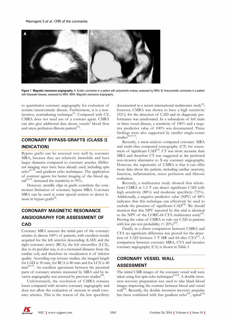
to quantitative coronary angiography for evaluation of ecstatic/aneurysmatic disease. Furthermore, it is a non-invasive, nonradiating technique[4]. Compared with CT, CMRA does not need use of a contrast agent. CMRA can also give additional data about, vessels’ blood flow and stress perfusion-fibrosis pattern[33].
CORONARY BYPASS-GRAFTS (CLASS II INDICATION)Bypass grafts can be assessed very well by coronary MRA, because they are relatively immobile and have larger diameter compared to coronary arteries. Differ-ent imaging ways have been already used, including spin echo[34-37] and gradient echo techniques. The application of contrast agents for better imaging of the blood sig-nal[38,39]. increased the sensitivity to 95%.
However, metallic clips in grafts constitute the com-monest limitation of coronary bypass MRA. Coronary MRA can be used at some special centers to detect le-sions in bypass grafts[8].
CORONARY MAGNETIC RESONANCE ANGIOGRAPHY FOR ASSESSMENT OF CADCoronary MRA assesses the initial part of the coronary arteries in almost 100% of patients, with excellent results acquired for the left anterior descending (LAD) and the right coronary artery (RCA); the left circumflex (LCX), due to its peculiar way, is at a increased distance from the cardiac coil, and therefore its visualization is of inferior quality. According top revious studies, the imaged length for LAD is 50 mm, for RCA is 80 mm and for LCX is 40 mm[40-47]. An excellent agreement between the proximal parts of coronary arteries measured by MRA and by in-vasive angiography was assessed by previous studies[48].
Unfortunately, the resolution of CMRA remains lower compared with invasive coronary angiography and does not allow the evaluation of stenosis in small coro-nary arteries. This is the reason of the low specificity
documented in a recent international multicenter study[4]; however, CMRA was shown to have a high sensitivity (92%) for the detection of CAD and its diagnostic per-formance was ameliorated. In a subanalysis of left main or three vessel disease, a sensitivity of 100% and a nega-tive predictive value of 100% was documented. These findings were also supported by smaller single-center studies[40,49-57].
Recently, a meta-analysis compared coronary MRA and multi-slice computed tomography (CT) for assess-ment of significant CAD[34]. CT was more accurate than MRA and therefore CT was suggested as the preferred non-invasive alternative to X-ray coronary angiography. However, the superiority of CMRA is that it can offer more data about the patient, including cardiac anatomy, function, inflammation, stress perfusion and fibrosis evaluation.
Recently, a multicenter study showed that whole-heart CMRA at 1.5 T can detect significant CAD with high sensitivity (88%) and moderate specificity (72%). Additionally, a negative predictive value (NPV) of 88% indicates that this technique can effectively be used to exclude the presence of significant CAD[58]. We should mention that this NPV reported by this trial is identical to the NPV of the CORE-64 CTA multicenter study[59]. Proving the value of CMRA to rule out CAD in patients with low pre-test probability (< 20%)[60].
Finally, in a direct comparison between CMRA and CTA no significant difference was proved for the detec-tion of CAD between 3 T MR and 64-slice CTA[61]. A comparison between coronary MRA, CTA and invasive coronary angiography (CA) is shown in Table 1.
CORONARY VESSEL WALL ASSESSMENTThe initial CMR images of the coronary vessel wall were taken using fast spin echo techniques[62,63]. A double inver-sion recovery preparation was used to take black-blood images improving the contrast between blood and vessel wall[64]. Recently, the double inversion recovery prepulse has been combined with fast gradient echo[65], spiral[66]
October 26, 2014|Volume 6|Issue 10|WJC|www.wjgnet.com 1062
A B
Figure 1 Magnetic resonance angiography. A: Ectatic coronaries in a patient with polyarteritis nodosa, assessed by MRA; B: Aneurysmatic coronaries in a patient with Kawasaki disease, assessed by MRA. MRA: Magnetic resonance angiography.
Mavrogeni S et al . CMR of the coronaries
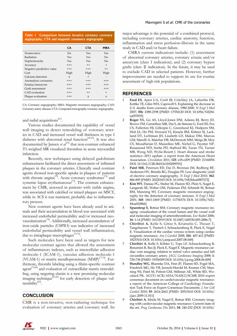
October 26, 2014|Volume 6|Issue 10|WJC|www.wjgnet.com
major advantage is the potential of a combined protocol, including coronary arteries, cardiac anatomy, function, inflammation and stress perfusion-fibrosis in the same study in CAD and/or heart failure.
CMRA current indications include: (1) assessment of abnormal coronary arteries, coronary ectasia and/or aneurysm (class Ⅰ indication); and (2) coronary bypass grafts (class Ⅱ indication). In the future, it may be used to exclude CAD in selected patients. However, further improvements are needed to support its use for routine assessment of high risk populations.
REFERENCES1 Ford ES, Ajani UA, Croft JB, Critchley JA, Labarthe DR,
Kottke TE, Giles WH, Capewell S. Explaining the decrease in U.S. deaths from coronary disease, 1980-2000. N Engl J Med 2007; 356: 2388-2398 [PMID: 17554120 DOI: 10.1056/NEJM-sa053935]
2 Roger VL, Go AS, Lloyd-Jones DM, Adams RJ, Berry JD, Brown TM, Carnethon MR, Dai S, de Simone G, Ford ES, Fox CS, Fullerton HJ, Gillespie C, Greenlund KJ, Hailpern SM, Heit JA, Ho PM, Howard VJ, Kissela BM, Kittner SJ, Lack-land DT, Lichtman JH, Lisabeth LD, Makuc DM, Marcus GM, Marelli A, Matchar DB, McDermott MM, Meigs JB, Moy CS, Mozaffarian D, Mussolino ME, Nichol G, Paynter NP, Rosamond WD, Sorlie PD, Stafford RS, Turan TN, Turner MB, Wong ND, Wylie-Rosett J. Heart disease and stroke statistics--2011 update: a report from the American Heart Association. Circulation 2011; 123: e18-e209 [PMID: 21160056 DOI: 10.1161/CIR.0b013e3182009701]
3 Patel MR, Peterson ED, Dai D, Brennan JM, Redberg RF, Anderson HV, Brindis RG, Douglas PS. Low diagnostic yield of elective coronary angiography. N Engl J Med 2010; 362: 886-895 [PMID: 20220183 DOI: 10.1056/NEJMoa0907272]
4 Kim WY, Danias PG, Stuber M, Flamm SD, Plein S, Nagel E, Langerak SE, Weber OM, Pedersen EM, Schmidt M, Botnar RM, Manning WJ. Coronary magnetic resonance angiog-raphy for the detection of coronary stenoses. N Engl J Med 2001; 345: 1863-1869 [PMID: 11756576 DOI: 10.1056/NEJ-Moa010866]
5 Spuentrup E, Botnar RM. Coronary magnetic resonance im-aging: visualization of the vessel lumen and the vessel wall and molecular imaging of arteriothrombosis. Eur Radiol 2006; 16: 1-14 [PMID: 16132919 DOI: 10.1007/s00330-005-2886-7]
6 Chiribiri A, Kelle S, Götze S, Kriatselis C, Thouet T, Tangcharoen T, Paetsch I, Schnackenburg B, Fleck E, Nagel E. Visualization of the cardiac venous system using cardiac magnetic resonance. Am J Cardiol 2008; 101: 407-412 [PMID: 18237610 DOI: 10.1016/j.amjcard.2007.08.049]
7 Chiribiri A, Kelle S, Köhler U, Tops LF, Schnackenburg B, Bonamini R, Bax JJ, Fleck E, Nagel E. Magnetic resonance car-diac vein imaging: relation to mitral valve annulus and left circumflex coronary artery. JACC Cardiovasc Imaging 2008; 1: 729-738 [PMID: 19356509 DOI: 10.1016/j.jcmg.2008.06.009]
8 Hundley WG, Bluemke DA, Finn JP, Flamm SD, Fogel MA, Friedrich MG, Ho VB, Jerosch-Herold M, Kramer CM, Man-ning WJ, Patel M, Pohost GM, Stillman AE, White RD, Wo-odard PK. ACCF/ACR/AHA/NASCI/SCMR 2010 expert consensus document on cardiovascular magnetic resonance: a report of the American College of Cardiology Founda-tion Task Force on Expert Consensus Documents. J Am Coll Cardiol 2010; 55: 2614-2662 [PMID: 20513610 DOI: 10.1016/j.jacc.2009.11.011]
9 Chiribiri A, Ishida M, Nagel E, Botnar RM. Coronary imag-ing with cardiovascular magnetic resonance: Current state of the art. Prog Cardiovasc Dis 2011; 54: 240-252 [DOI: 10.1016/
and radial acquisitions[67].Various studies documented the capability of vessel
wall imaging to detect remodeling of coronary arter-ies in CAD and increased vessel wall thickness in type I diabetes with abnormal renal function[68,69]. It was also documented by Jansen et al[70] that non-contrast enhanced T1-weighed MR visualized thrombus in acute myocardial infarction.
Recently, new techniques using delayed gadolinium enhancement facilitated the direct assessment of inflamed plaques in the coronary arteries. Clinically used contrast agents showed non-specific uptake in plaques of patients with chronic angina[71]. Acute coronary syndromes[72] and systemic lupus erythematosus[73]. The contrast enhance-ment by CMR, assessed in patients with stable angina, was associated with calcified or mixed plaques on MSCT, while in ACS it was transient, probably due to inflamma-tory process.
New contrast agents have been already used in ani-mals and their accumulation in blood was associated with increased endothelial permeability and/or increased neo-vascularization[74]. Additionally, increased accumulation of iron-oxide particles (USPIO) was indicative of increased endothelial permeability and vessel wall inflammation, due to intraplaque macrophages[75,76].
Such molecules have been used as targets for new molecular contrast agents that allowed the assessment of inflammatory indexes, such as intercellular adhesion molecule-1 (ICAM-1), vascular adhesion molecule-1 (VCAM-1) or matrix metalloproteinase (MMP)[77,78]. Fur-thrmore, thrombi labeling using a fibrin-specific contrast agent[79,80] and evaluation of extracellular matrix remodel-ling, using targeting elastin is a new promising molecular imaging technique[81,82] for early detection of plaque vul-nerability[83].
CONCLUSIONCMR is a non-invasive, non-radiating technique for evaluation of coronary arteries and coronary wall. Its
1063
CA CTA MRA
Noninvasive No Yes Yes Radiation Yes Yes No Nephrotoxicity Yes Yes No Accuracy +++ ++ + Negative predictive value +++ +++ ++ Cost High High High Calcium detection ± + - Anomalous coronaries +++ +++ +++ Ectasia/aneurysm +++ +++ +++ Graft assessment +++ +++ +++ CAD evaluation +++ ++ + Plaque evaluation +++ ± ±
Table 1 Comparison between invasive coronary coronary angiography, CTA and magnetic resonance angiography
CA: Coronary angiography; MRA: Magnetic resonance angiography; CAD: Coronary artery disease; CTA: Computed tomography coronary angiography.
Mavrogeni S et al . CMR of the coronaries
October 26, 2014|Volume 6|Issue 10|WJC|www.wjgnet.com
j.pcad.2011.09.002]10 Chiribiri A, Ishida M, Nagel E, Botnar RM. Coronary imag-
ing with cardiovascular magnetic resonance: current state of the art. Prog Cardiovasc Dis 2011; 54: 240-252 [PMID: 22014491 DOI: 10.1016/j.ijcard.2008.05.075]
11 Mavrogeni S, Papadopoulos G, Hussain T, Chiribiri A, Bot-nar R, Greil GF. The emerging role of cardiovascular mag-netic resonance in the evaluation of Kawasaki disease. Int J Cardiovasc Imaging 2013; 29: 1787-1798 [PMID: 23949280 DOI: 10.1007/s10554-013-0276-9]
12 Mavrogeni S. Coronary artery ectasia: from diagnosis to treatment. Hellenic J Cardiol 2010; 51: 158-163 [PMID: 20378518]
13 Mavrogeni S, Papadopoulos G, Douskou M, Kaklis S, Sei-menis I, Baras P, Nikolaidou P, Bakoula C, Karanasios E, Manginas A, Cokkinos DV. Magnetic resonance angiog-raphy is equivalent to X-ray coronary angiography for the evaluation of coronary arteries in Kawasaki disease. J Am Coll Cardiol 2004; 43: 649-652 [PMID: 14975477 DOI: 10.1016/j.jacc.2003.08.052]
14 Greil GF, Stuber M, Botnar RM, Kissinger KV, Geva T, Newburger JW, Manning WJ, Powell AJ. Coronary magnetic resonance angiography in adolescents and young adults with kawasaki disease. Circulation 2002; 105: 908-911 [PMID: 11864916 DOI: 10.1161/hc0802.105563]
15 Greil GF, Seeger A, Miller S, Claussen CD, Hofbeck M, Bot-nar RM, Sieverding L. Coronary magnetic resonance angi-ography and vessel wall imaging in children with Kawasaki disease. Pediatr Radiol 2007; 37: 666-673 [PMID: 17541574 DOI: 10.1007/s00247-007-0498-x]
16 Mavrogeni S, Papadopoulos G, Douskou M, Kaklis S, Sei-menis I, Varlamis G, Karanasios E, Krikos X, Giannoulia A, Cokkinos DV. Magnetic resonance angiography, function and viability evaluation in patients with Kawasaki disease. J Cardiovasc Magn Reson 2006; 8: 493-498 [PMID: 16758550 DOI: 10.1080/10976640600604773]
17 Doesch C, Seeger A, Doering J, Herdeg C, Burgstahler C, Claussen CD, Gawaz M, Miller S, May AE. Risk stratification by adenosine stress cardiac magnetic resonance in patients with coronary artery stenoses of intermediate angiographic severity. JACC Cardiovasc Imaging 2009; 2: 424-433 [PMID: 19580724 DOI: 10.1016/j.jcmg.2008.11.017]
18 Giang TH, Nanz D, Coulden R, Friedrich M, Graves M, Al-Saadi N, Lüscher TF, von Schulthess GK, Schwitter J. Detection of coronary artery disease by magnetic resonance myocardial perfusion imaging with various contrast me-dium doses: first European multi-centre experience. Eur Heart J 2004; 25: 1657-1665 [PMID: 15351166 DOI: 10.1016/j.ehj.2004.06.037]
19 Mavrogeni S, Manoussakis MN, Karagiorga TC, Douskou M, Panagiotakos D, Bournia V, Cokkinos DV, Moutsopoulos HM. Detection of coronary artery lesions and myocardial ne-crosis by magnetic resonance in systemic necrotizing vascu-litides. Arthritis Rheum 2009; 61: 1121-1129 [PMID: 19644909 DOI: 10.1002/art.24695]
20 Mavrogeni S, Sfikakis PP, Gialafos E, Bratis K, Karabela G, Stavropoulos E, Spiliotis G, Sfendouraki E, Panopoulos S, Bournia V, Kolovou G, Kitas GD. Cardiac tissue character-ization and the diagnostic value of cardiovascular magnetic resonance in systemic connective tissue diseases. Arthritis Care Res (Hoboken) 2014; 66: 104-112 [PMID: 24106233 DOI: 10.1002/acr.22181]
21 Hartnell GG, Parnell BM, Pridie RB. Coronary artery ecta-sia. Its prevalence and clinical significance in 4993 patients. Br Heart J 1985; 54: 392-395 [PMID: 4052280 DOI: 10.1136/hrt.54.4.392]
22 Markis JE, Joffe CD, Cohn PF, Feen DJ, Herman MV, Gorlin R. Clinical significance of coronary arterial ectasia. Am J Car-diol 1976; 37: 217-222 [PMID: 1108631 DOI: 10.1016/0002-9149(76)90315-5]
23 Oliveros RA, Falsetti HL, Carroll RJ, Heinle RA, Ryan GF. Atherosclerotic coronary artery aneurysm. Report of five cases and review of literature. Arch Intern Med 1974; 134: 1072-1076 [PMID: 4215378 DOI: 10.1001/archinte.1974.00320240106014]
24 Swaye PS, Fisher LD, Litwin P, Vignola PA, Judkins MP, Kemp HG, Mudd JG, Gosselin AJ. Aneurysmal coronary artery disease. Circulation 1983; 67: 134-138 [PMID: 6847792 DOI: 10.1161/01.CIR.67.1.134]
25 Falsetti HL, Carrol RJ. Coronary artery aneurysm. A review of the literature with a report of 11 new cases. Chest 1976; 69: 630-636 [PMID: 1083790 DOI: 10.1378/chest.69.5.630]
26 Befeler B, Aranda MJ, Embi A, Mullin FL, El-Sherif N, Laz-zara R. Coronary artery aneurysms: study of the etiology, clinical course and effect on left ventricular function and prognosis. Am J Med 1977; 62: 597-607 [PMID: 300567 DOI: 10.1016/0002-9343(77)90423-5]
27 Chaithiraphan S, Goldberg E, O’Reilly M, Jootar P. Multiple aneurysms of coronary artery in sclerodermal heart disease. Angiology 1973; 24: 86-93 [PMID: 4693051 DOI: 10.1177/000331977302400204]
28 Tarek el-G, Yasser AE, Gheita T. Coronary angiographic findings in asymptomatic systemic sclerosis. Clin Rheu-matol 2006; 25: 487-490 [PMID: 16440131 DOI: 10.1007/s10067-005-0073-5]
29 Imahori S, Bannerman RM, Graf CJ, Brennan JC. Ehlers-Danlos syndrome with multiple arterial lesions. Am J Med 1969; 47: 967-977 [PMID: 5362873 DOI: 10.1016/0002-9343(69)90210-1]
30 Davidson A, Eshaghpour E, Young N, Mintz GS. Late thrombosis of a coronary artery mycotic aneurysm. Am Heart J 1991; 121: 1549-1550 [PMID: 2017990 DOI: 10.1016/0002-8703(91)90168-H]
31 Cohen P, O’Gara PT. Coronary artery aneurysms: a review of the natural history, pathophysiology, and management. Cardiol Rev 2008; 16: 301-304 [PMID: 18923233 DOI: 10.1097/CRD.0b013e3181852659]
32 Bluemke DA, Achenbach S, Budoff M, Gerber TC, Gersh B, Hillis LD, Hundley WG, Manning WJ, Printz BF, Stuber M, Woodard PK. Noninvasive coronary artery imaging: mag-netic resonance angiography and multidetector computed tomography angiography: a scientific statement from the american heart association committee on cardiovascular imaging and intervention of the council on cardiovascular radiology and intervention, and the councils on clinical car-diology and cardiovascular disease in the young. Circulation 2008; 118: 586-606 [PMID: 18586979 DOI: 10.1161/CIRCU-LATIONAHA.108.189695]
33 Mavrogeni SI, Manginas A, Papadakis E, Foussas S, Douskou M, Baras P, Seimenis I, Cokkinos DV. Correlation between magnetic resonance angiography (MRA) and quan-titative coronary angiography (QCA) in ectatic coronary ves-sels. J Cardiovasc Magn Reson 2004; 6: 17-23 [PMID: 15054925 DOI: 10.1081/JCMR-120027801]
34 Galjee MA, van Rossum AC, Doesburg T, van Eenige MJ, Visser CA. Value of magnetic resonance imaging in assess-ing patency and function of coronary artery bypass grafts. An angiographically controlled study. Circulation 1996; 93: 660-666 [PMID: 8640993 DOI: 10.1161/01.CIR.93.4.660]
35 Jenkins JP, Love HG, Foster CJ, Isherwood I, Rowlands DJ. Detection of coronary artery bypass graft patency as as-sessed by magnetic resonance imaging. Br J Radiol 1988; 61: 2-4 [PMID: 3258170 DOI: 10.1259/0007-1285-61-721-2]
36 Rubinstein RI, Askenase AD, Thickman D, Feldman MS, Agarwal JB, Helfant RH. Magnetic resonance imaging to evaluate patency of aortocoronary bypass grafts. Circula-tion 1987; 76: 786-791 [PMID: 3498558 DOI: 10.1161/01.CIR.76.4.786]
37 White RD, Caputo GR, Mark AS, Modin GW, Higgins CB. Coronary artery bypass graft patency: noninvasive evalua-
1064
Mavrogeni S et al . CMR of the coronaries
October 26, 2014|Volume 6|Issue 10|WJC|www.wjgnet.com
tion with MR imaging. Radiology 1987; 164: 681-686 [PMID: 3497409 DOI: 10.1148/radiology.164.3.3497409]
38 Vrachliotis TG, Bis KG, Aliabadi D, Shetty AN, Safian R, Simonetti O. Contrast-enhanced breath-hold MR angiogra-phy for evaluating patency of coronary artery bypass grafts. AJR Am J Roentgenol 1997; 168: 1073-1080 [PMID: 9124118 DOI: 10.2214/ajr.168.4.9124118]
39 Wintersperger BJ, von Smekal A, Engelmann MG, Knez A, Penzkofer HV, Laub G, Reiser M. [Contrast media enhanced magnetic resonance angiography for determining patency of a coronary bypass. A comparison with coronary angiogra-phy]. Rofo 1997; 167: 572-578 [PMID: 9465951 DOI: 10.1055/s-2007-1015585]
40 Botnar RM, Stuber M, Danias PG, Kissinger KV, Manning WJ. Improved coronary artery definition with T2-weighted, free-breathing, three-dimensional coronary MRA. Circula-tion 1999; 99: 3139-3148 [PMID: 10377077 DOI: 10.1161/01.CIR.99.24.3139]
41 Hofman MB, Paschal CB, Li D, Haacke EM, van Rossum AC, Sprenger M. MRI of coronary arteries: 2D breath-hold vs 3D respiratory-gated acquisition. J Comput Assist Tomogr 1995; 19: 56-62 [PMID: 7822549 DOI: 10.1097/00004728-199501000-00011]
42 Lobbes MB, Miserus RJ, Heeneman S, Passos VL, Mutsaers PH, Debernardi N, Misselwitz B, Post M, Daemen MJ, van Engelshoven JM, Leiner T, Kooi ME. Atherosclerosis: con-trast-enhanced MR imaging of vessel wall in rabbit model--comparison of gadofosveset and gadopentetate dimeglu-mine. Radiology 2009; 250: 682-691 [PMID: 19244042 DOI: 10.1148/radiol.2503080875]
43 Manning WJ, Li W, Boyle NG, Edelman RR. Fat-suppressed breath-hold magnetic resonance coronary angiography. Circulation 1993; 87: 94-104 [PMID: 8419029 DOI: 10.1161/01.CIR.87.1.94]
44 Paschal CB, Haacke EM, Adler LP. Three-dimensional MR imaging of the coronary arteries: preliminary clinical experi-ence. J Magn Reson Imaging 1993; 3: 491-500 [PMID: 8324308 DOI: 10.1002/jmri.1880030311]
45 Post JC, van Rossum AC, Hofman MB, Valk J, Visser CA. Three-dimensional respiratory-gated MR angiography of coronary arteries: comparison with conventional coronary angiography. AJR Am J Roentgenol 1996; 166: 1399-1404 [PMID: 8633453 DOI: 10.2214/ajr.166.6.8633453]
46 Stuber M, Botnar RM, Danias PG, Sodickson DK, Kissinger KV, Van Cauteren M, De Becker J, Manning WJ. Double-oblique free-breathing high resolution three-dimensional coronary magnetic resonance angiography. J Am Coll Cardiol 1999; 34: 524-531 [PMID: 10440168 DOI: 10.1016/S0735-1097(99)00223-5]
47 Scheidegger MB, Müller R, Boesiger P. Magnetic resonance angiography: methods and its applications to the coronary arteries. Technol Health Care 1994; 2: 255-265 [PMID: 7842310]
48 Bogaert J, Kuzo R, Dymarkowski S, Beckers R, Piessens J, Rademakers FE. Coronary artery imaging with real-time navigator three-dimensional turbo-field-echo MR coronary angiography: initial experience. Radiology 2003; 226: 707-716 [PMID: 12601209 DOI: 10.1148/radiol.2263011750]
49 Dewey M, Teige F, Schnapauff D, Laule M, Borges AC, Rutsch W, Hamm B, Taupitz M. Combination of free-breath-ing and breathhold steady-state free precession magnetic resonance angiography for detection of coronary artery stenoses. J Magn Reson Imaging 2006; 23: 674-681 [PMID: 16568418 DOI: 10.1002/jmri.20568]
50 Jahnke C, Paetsch I, Nehrke K, Schnackenburg B, Gebker R, Fleck E, Nagel E. Rapid and complete coronary arterial tree visualization with magnetic resonance imaging: feasibility and diagnostic performance. Eur Heart J 2005; 26: 2313-2319 [PMID: 15987709 DOI: 10.1093/eurheartj/ehi391]
51 Jahnke C, Paetsch I, Schnackenburg B, Bornstedt A, Gebker R, Fleck E, Nagel E. Coronary MR angiography with steady-
state free precession: individually adapted breath-hold tech-nique versus free-breathing technique. Radiology 2004; 232: 669-676 [PMID: 15284430 DOI: 10.1148/radiol.2323031225]
52 Maintz D, Aepfelbacher FC, Kissinger KV, Botnar RM, Danias PG, Heindel W, Manning WJ, Stuber M. Coronary MR angiography: comparison of quantitative and qualitative data from four techniques. AJR Am J Roentgenol 2004; 182: 515-521 [PMID: 14736693 DOI: 10.2214/ajr.182.2.1820515]
53 Manning WJ, Li W, Edelman RR. A preliminary report comparing magnetic resonance coronary angiography with conventional angiography. N Engl J Med 1993; 328: 828-832 [PMID: 8285929 DOI: 10.1056/NEJM199303253281202]
54 Ozgun M, Hoffmeier A, Kouwenhoven M, Botnar RM, Stuber M, Scheld HH, Manning WJ, Heindel W, Maintz D. Comparison of 3D segmented gradient-echo and steady-state free precession coronary MRI sequences in patients with coronary artery disease. AJR Am J Roentgenol 2005; 185: 103-109 [PMID: 15972408 DOI: 10.2214/ajr.185.1.01850103]
55 Sakuma H, Ichikawa Y, Chino S, Hirano T, Makino K, Takeda K. Detection of coronary artery stenosis with whole-heart coronary magnetic resonance angiography. J Am Coll Cardiol 2006; 48: 1946-1950 [PMID: 17112982 DOI: 10.1016/j.jacc.2006.07.055]
56 Sakuma H, Ichikawa Y, Suzawa N, Hirano T, Makino K, Koyama N, Van Cauteren M, Takeda K. Assessment of coro-nary arteries with total study time of less than 30 minutes by using whole-heart coronary MR angiography. Radiol-ogy 2005; 237: 316-321 [PMID: 16126921 DOI: 10.1148/ra-diol.2371040830]
57 Schuetz GM, Zacharopoulou NM, Schlattmann P, Dewey M. Meta-analysis: noninvasive coronary angiography using computed tomography versus magnetic resonance imaging. Ann Intern Med 2010; 152: 167-177 [PMID: 20124233 DOI: 10.7326/0003-4819-152-3-201002020-00008]
58 Kato S, Kitagawa K, Ishida N, Ishida M, Nagata M, Ichikawa Y, Katahira K, Matsumoto Y, Seo K, Ochiai R, Kobayashi Y, Sakuma H. Assessment of coronary artery disease us-ing magnetic resonance coronary angiography: a national multicenter trial. J Am Coll Cardiol 2010; 56: 983-991 [PMID: 20828652 DOI: 10.1016/j.jacc.2010.01.071]
59 Miller JM, Rochitte CE, Dewey M, Arbab-Zadeh A, Niinu-ma H, Gottlieb I, Paul N, Clouse ME, Shapiro EP, Hoe J, Lardo AC, Bush DE, de Roos A, Cox C, Brinker J, Lima JA. Diagnostic performance of coronary angiography by 64-row CT. N Engl J Med 2008; 359: 2324-2336 [PMID: 19038879 DOI: 10.1056/NEJMoa0806576]
60 Nagel E. Magnetic resonance coronary angiography: the condemned live longer. J Am Coll Cardiol 2010; 56: 992-994 [PMID: 20828653 DOI: 10.1016/j.jacc.2010.02.069]
61 Hamdan A, Asbach P, Wellnhofer E, Klein C, Gebker R, Kelle S, Kilian H, Huppertz A, Fleck E. A prospective study for comparison of MR and CT imaging for detection of coro-nary artery stenosis. JACC Cardiovasc Imaging 2011; 4: 50-61 [PMID: 21232704 DOI: 10.1016/j.jcmg.2010.10.007]
62 Botnar RM, Stuber M, Kissinger KV, Kim WY, Spuentrup E, Manning WJ. Noninvasive coronary vessel wall and plaque imaging with magnetic resonance imaging. Circula-tion 2000; 102: 2582-2587 [PMID: 11085960 DOI: 10.1161/01.CIR.102.21.2582]
63 Fayad ZA, Fuster V, Fallon JT, Jayasundera T, Worthley SG, Helft G, Aguinaldo JG, Badimon JJ, Sharma SK. Noninvasive in vivo human coronary artery lumen and wall imaging using black-blood magnetic resonance imaging. Circulation 2000; 102: 506-510 [PMID: 10920061 DOI: 10.1161/01.CIR.102.5.506]
64 Edelman RR, Chien D, Kim D. Fast selective black blood MR imaging. Radiology 1991; 181: 655-660 [PMID: 1947077 DOI: 10.1148/radiology.181.3.1947077]
65 Botnar RM, Stuber M, Lamerichs R, Smink J, Fischer SE, Harvey P, Manning WJ. Initial experiences with in vivo right coronary artery human MR vessel wall imaging at 3 tesla.
1065
Mavrogeni S et al . CMR of the coronaries
October 26, 2014|Volume 6|Issue 10|WJC|www.wjgnet.com
J Cardiovasc Magn Reson 2003; 5: 589-594 [PMID: 14664136 DOI: 10.1081/JCMR-120025232]
66 Botnar RM, Kim WY, Börnert P, Stuber M, Spuentrup E, Manning WJ. 3D coronary vessel wall imaging utilizing a lo-cal inversion technique with spiral image acquisition. Magn Reson Med 2001; 46: 848-854 [PMID: 11675634 DOI: 10.1002/mrm.1268]
67 Katoh M, Spuentrup E, Buecker A, Schaeffter T, Stuber M, Günther RW, Botnar RM. MRI of coronary vessel walls us-ing radial k-space sampling and steady-state free precession imaging. AJR Am J Roentgenol 2006; 186: S401-S406 [PMID: 16714616 DOI: 10.2214/AJR.04.1864]
68 Kim WY, Stuber M, Börnert P, Kissinger KV, Manning WJ, Botnar RM. Three-dimensional black-blood cardiac magnetic resonance coronary vessel wall imaging detects positive arte-rial remodeling in patients with nonsignificant coronary ar-tery disease. Circulation 2002; 106: 296-299 [PMID: 12119242 DOI: 10.1161/01.CIR.0000025629.85631.1E]
69 Kim WY, Astrup AS, Stuber M, Tarnow L, Falk E, Botnar RM, Simonsen C, Pietraszek L, Hansen PR, Manning WJ, Andersen NT, Parving HH. Subclinical coronary and aortic atherosclerosis detected by magnetic resonance imaging in type 1 diabetes with and without diabetic nephropathy. Circulation 2007; 115: 228-235 [PMID: 17190865 DOI: 10.1161/CIRCULATIONAHA.106.633339]
70 Jansen CH, Perera D, Makowski MR, Wiethoff AJ, Phinikari-dou A, Razavi RM, Marber MS, Greil GF, Nagel E, Maintz D, Redwood S, Botnar RM. Detection of intracoronary throm-bus by magnetic resonance imaging in patients with acute myocardial infarction. Circulation 2011; 124: 416-424 [PMID: 21747055 DOI: 10.1161/CIRCULATIONAHA.110.965442]
71 Yeon SB, Sabir A, Clouse M, Martinezclark PO, Peters DC, Hauser TH, Gibson CM, Nezafat R, Maintz D, Manning WJ, Botnar RM. Delayed-enhancement cardiovascular magnetic resonance coronary artery wall imaging: comparison with multislice computed tomography and quantitative coronary angiography. J Am Coll Cardiol 2007; 50: 441-447 [PMID: 17662397 DOI: 10.1016/j.jacc.2007.03.052]
72 Ibrahim T, Makowski MR, Jankauskas A, Maintz D, Karch M, Schachoff S, Manning WJ, Schömig A, Schwaiger M, Botnar RM. Serial contrast-enhanced cardiac magnetic reso-nance imaging demonstrates regression of hyperenhance-ment within the coronary artery wall in patients after acute myocardial infarction. JACC Cardiovasc Imaging 2009; 2: 580-588 [PMID: 19442944 DOI: 10.1016/j.jcmg.2008.12.029]
73 Puntmann VO, D’Cruz D, Taylor PC, Hussain T, Inder-muhle A, Butzbach B, Botnar R, Nagel E. Contrast enhance-ment imaging in coronary arteries in SLE. JACC Cardiovasc Imaging 2012; 5: 962-964 [PMID: 22974810 DOI: 10.1016/j.jcmg.2012.03.017]
74 Phinikaridou A, Andia ME, Protti A, Indermuehle A, Shah A, Smith A, Warley A, Botnar RM. Noninvasive magnetic resonance imaging evaluation of endothelial permeability in murine atherosclerosis using an albumin-binding contrast agent. Circulation 2012; 126: 707-719 [PMID: 22753191 DOI: 10.1161/CIRCULATIONAHA.112.092098]
75 Kooi ME, Cappendijk VC, Cleutjens KB, Kessels AG, Kitslaar PJ, Borgers M, Frederik PM, Daemen MJ, van Engelshoven JM. Accumulation of ultrasmall superparamagnetic par-ticles of iron oxide in human atherosclerotic plaques can be detected by in vivo magnetic resonance imaging. Circula-tion 2003; 107: 2453-2458 [PMID: 12719280 DOI: 10.1161/01.CIR.0000068315.98705.CC]
76 Tang TY, Howarth SP, Miller SR, Graves MJ, Patterson AJ, U-King-Im JM, Li ZY, Walsh SR, Brown AP, Kirkpatrick PJ, Warburton EA, Hayes PD, Varty K, Boyle JR, Gaunt ME, Za-lewski A, Gillard JH. The ATHEROMA (Atorvastatin Ther-apy: Effects on Reduction of Macrophage Activity) Study. Evaluation using ultrasmall superparamagnetic iron oxide-enhanced magnetic resonance imaging in carotid disease. J Am Coll Cardiol 2009; 53: 2039-2050 [PMID: 19477353 DOI: 10.1016/j.jacc.2009.03.018]
77 Nahrendorf M, Jaffer FA, Kelly KA, Sosnovik DE, Aikawa E, Libby P, Weissleder R. Noninvasive vascular cell adhesion molecule-1 imaging identifies inflammatory activation of cells in atherosclerosis. Circulation 2006; 114: 1504-1511 [PMID: 17000904 DOI: 10.1161/CIRCULATIONAHA.106.646380]
78 Nahrendorf M, Keliher E, Panizzi P, Zhang H, Hembrador S, Figueiredo JL, Aikawa E, Kelly K, Libby P, Weissleder R. 18F-4V for PET-CT imaging of VCAM-1 expression in atherosclerosis. JACC Cardiovasc Imaging 2009; 2: 1213-1222 [PMID: 19833312 DOI: 10.1016/j.jcmg.2009.04.016]
79 Botnar RM, Buecker A, Wiethoff AJ, Parsons EC, Katoh M, Katsimaglis G, Weisskoff RM, Lauffer RB, Graham PB, Gunther RW, Manning WJ, Spuentrup E. In vivo magnetic resonance imaging of coronary thrombosis using a fibrin-binding molecular magnetic resonance contrast agent. Circu-lation 2004; 110: 1463-1466 [PMID: 15238457 DOI: 10.1161/01.CIR.0000134960.31304.87]
80 Botnar RM, Perez AS, Witte S, Wiethoff AJ, Laredo J, Ham-ilton J, Quist W, Parsons EC, Vaidya A, Kolodziej A, Barrett JA, Graham PB, Weisskoff RM, Manning WJ, Johnstone MT. In vivo molecular imaging of acute and subacute thrombosis using a fibrin-binding magnetic resonance imaging contrast agent. Circulation 2004; 109: 2023-2029 [PMID: 15066940 DOI: 10.1161/01.CIR.0000127034.50006.C0]
81 Makowski MR, Wiethoff AJ, Blume U, Cuello F, Warley A, Jansen CH, Nagel E, Razavi R, Onthank DC, Cesati RR, Marber MS, Schaeffter T, Smith A, Robinson SP, Botnar RM. Assessment of atherosclerotic plaque burden with an elastin-specific magnetic resonance contrast agent. Nat Med 2011; 17: 383-388 [PMID: 21336283 DOI: 10.1038/nm.2310]
82 von Bary C, Makowski M, Preissel A, Keithahn A, Warley A, Spuentrup E, Buecker A, Lazewatsky J, Cesati R, Onthank D, Schickl N, Schachoff S, Hausleiter J, Schömig A, Schwaiger M, Robinson S, Botnar R. MRI of coronary wall remodeling in a swine model of coronary injury using an elastin-binding contrast agent. Circ Cardiovasc Imaging 2011; 4: 147-155 [PMID: 21378029 DOI: 10.1161/CIRCIMAGING.109.895607]
83 Chiribiri A, Botnar RM, Nagel E. Magnetic resonance coro-nary angiography: where are we today? Curr Cardiol Rep 2013; 15: 328 [PMID: 23307168 DOI: 10.1007/s11886-012-0328-0]
P- Reviewer: Ueda H S- Editor: Song XX L- Editor: A E- Editor: Wu HL
1066
Mavrogeni S et al . CMR of the coronaries
ST-segment elevation: Distinguishing ST elevation myocardial infarction from ST elevation secondary to nonischemic etiologies
Alok Deshpande, Yochai Birnbaum
Alok Deshpande, Yochai Birnbaum, The Section of Cardiol-ogy, The Department of Medicine, Baylor College of Medicine, Houston, TX 77030, United StatesAuthor contributions: Both authors reviewed the literature and participated in writing the manuscript.Supported by John S Dunn Chair in Cardiology Research and EducationCorrespondence to: Yochai Birnbaum, MD, John S Dunn Chair in Cardiology Research and Education, The Section of Car-diology, The Department of Medicine, Baylor College of Medi-cine, One Baylor Plaza, MS: BCM 620, Houston, TX 77030, United States. [email protected]: +1-713-7982735 Fax: +1-713-7980270 Received: December 29, 2013 Revised: May 20, 2014 Accepted: July 27, 2014Published online: October 26, 2014
AbstractThe benefits of early perfusion in ST elevation myo-cardial infarctions (STEMI) are established; however, early perfusion of non-ST elevation myocardial infarc-tions has not been shown to be beneficial. In addition, ST elevation (STE) caused by conditions other than acute ischemia is common. Non-ischemic STE may be confused as STEMI, but can also mask STEMI on elec-trocardiogram (ECG). As a result, activating the primary percutaneous coronary intervention (pPCI) protocol often depends on determining which ST elevation pat-terns reflect transmural infarction due to acute coro-nary artery thrombosis. Coordination of interpreting the ECG in its clinical context and appropriately activating the pPCI protocol has proved a difficult task in border-line cases. But its importance cannot be ignored, as reflected in the 2013 American College of Cardiology Foundation/American Heart Association guidelines con-cerning the treatment of ST elevation myocardial infarc-tion. Multiples strategies have been tested and studied, and are currently being further perfected. No matter
the strategy, at the heart of delivering the best care lies rapid and accurate interpretation of the ECG. Here, we present the different patterns of non-ischemic STE and methods of distinguishing between them. In writing this paper, we hope for quicker and better stratification of patients with STE on ECG, which will lead to be bet-ter outcomes.
© 2014 Baishideng Publishing Group Inc. All rights reserved.
Key words: Diagnosis; Electrocardiogram; Reperfusion therapy; ST segment elevation; Myocardial infarction
Core tip: At times, distinguishing between myocardial infarction with ST elevation (STEMI) from non-ischemic causes of elevation of the ST segment is difficult, es-pecially in patients with atypical presenting symptoms. Understanding common patterns of ST elevation that are not caused by ischemia is crucial for rapid and ac-curate diagnosis. However, patients with baseline non-ischemic ST elevation (for example, early repolarization or repolarization changes caused by hypertrophy of the left ventricule) may develop acute myocardial infarction (true STEMI or non-ST elevation myocardial infarction with baseline ST elevation). Here we describe common patterns of non-ischemic ST elevation.
Deshpande A, Birnbaum Y. ST-segment elevation: Distinguishing ST elevation myocardial infarction from ST elevation secondary to nonischemic etiologies. World J Cardiol 2014; 6(10): 1067-1079 Available from: URL: http://www.wjgnet.com/1949-8462/full/v6/i10/1067.htm DOI: http://dx.doi.org/10.4330/wjc.v6.i10.1067
INTRODUCTIONToday, the electrocardiogram (ECG) is the most com-
TOPIC HIGHLIGHT
Submit a Manuscript: http://www.wjgnet.com/esps/Help Desk: http://www.wjgnet.com/esps/helpdesk.aspxDOI: 10.4330/wjc.v6.i10.1067
October 26, 2014|Volume 6|Issue 10|WJC|www.wjgnet.com
World Journal of CardiologyW J C
World J Cardiol 2014 October 26; 6(10): 1067-1079ISSN 1949-8462 (online)
© 2014 Baishideng Publishing Group Inc. All rights reserved.
1067
WJC 6th Anniversary Special Issues (5): Myocardial infarction
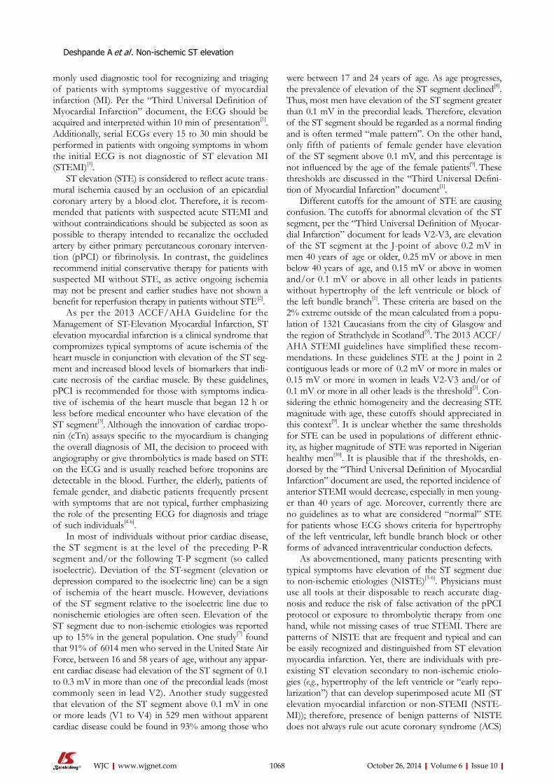
monly used diagnostic tool for recognizing and triaging of patients with symptoms suggestive of myocardial infarction (MI). Per the “Third Universal Definition of Myocardial Infarction” document, the ECG should be acquired and interpreted within 10 min of presentation[1]. Additionally, serial ECGs every 15 to 30 min should be performed in patients with ongoing symptoms in whom the initial ECG is not diagnostic of ST elevation MI (STEMI)[1].
ST elevation (STE) is considered to reflect acute trans-mural ischemia caused by an occlusion of an epicardial coronary artery by a blood clot. Therefore, it is recom-mended that patients with suspected acute STEMI and without contraindications should be subjected as soon as possible to therapy intended to recanalize the occluded artery by either primary percutaneous coronary interven-tion (pPCI) or fibrinolysis. In contrast, the guidelines recommend initial conservative therapy for patients with suspected MI without STE, as active ongoing ischemia may not be present and earlier studies have not shown a benefit for reperfusion therapy in patients without STE[2].
As per the 2013 ACCF/AHA Guideline for the Management of ST-Elevation Myocardial Infarction, ST elevation myocardial infarction is a clinical syndrome that compromizes typical symptoms of acute ischemia of the heart muscle in conjunction with elevation of the ST seg-ment and increased blood levels of biomarkers that indi-cate necrosis of the cardiac muscle. By these guidelines, pPCI is recommended for those with symptoms indica-tive of ischemia of the heart muscle that began 12 h or less before medical encounter who have elevation of the ST segment[3]. Although the innovation of cardiac tropo-nin (cTn) assays specific to the myocardium is changing the overall diagnosis of MI, the decision to proceed with angiography or give thrombolytics is made based on STE on the ECG and is usually reached before troponins are detectable in the blood. Further, the elderly, patients of female gender, and diabetic patients frequently present with symptoms that are not typical, further emphasizing the role of the presenting ECG for diagnosis and triage of such individuals[4-6].
In most of individuals without prior cardiac disease, the ST segment is at the level of the preceding P-R segment and/or the following T-P segment (so called isoelectric). Deviation of the ST-segment (elevation or depression compared to the isoelectric line) can be a sign of ischemia of the heart muscle. However, deviations of the ST segment relative to the isoelectric line due to nonischemic etiologies are often seen. Elevation of the ST segment due to non-ischemic etiologies was reported up to 15% in the general population. One study[7] found that 91% of 6014 men who served in the United State Air Force, between 16 and 58 years of age, without any appar-ent cardiac disease had elevation of the ST segment of 0.1 to 0.3 mV in more than one of the precordial leads (most commonly seen in lead V2). Another study suggested that elevation of the ST segment above 0.1 mV in one or more leads (V1 to V4) in 529 men without apparent cardiac disease could be found in 93% among those who
were between 17 and 24 years of age. As age progresses, the prevalence of elevation of the ST segment declined[8]. Thus, most men have elevation of the ST segment greater than 0.1 mV in the precordial leads. Therefore, elevation of the ST segment should be regarded as a normal finding and is often termed “male pattern”. On the other hand, only fifth of patients of female gender have elevation of the ST segment above 0.1 mV, and this percentage is not influenced by the age of the female patients[9].
These
thresholds are discussed in the “Third Universal Defini-tion of Myocardial Infarction” document[1].
Different cutoffs for the amount of STE are causing confusion. The cutoffs for abnormal elevation of the ST segment, per the “Third Universal Definition of Myocar-dial Infarction” document for leads V2-V3, are elevation of the ST segment at the J-point of above 0.2 mV in men 40 years of age or older, 0.25 mV or above in men below 40 years of age, and 0.15 mV or above in women and/or 0.1 mV or above in all other leads in patients without hypertrophy of the left ventricule or block of the left bundle branch[1]. These criteria are based on the 2% extreme outside of the mean calculated from a popu-lation of 1321 Caucasians from the city of Glasgow and the region of Strathclyde in Scotland[9]. The 2013 ACCF/AHA STEMI guidelines have simplified these recom-mendations. In these guidelines STE at the J point in 2 contiguous leads or more of 0.2 mV or more in males or 0.15 mV or more in women in leads V2-V3 and/or of 0.1 mV or more in all other leads is the threshold[3]. Con-sidering the ethnic homogeneity and the decreasing STE magnitude with age, these cutoffs should appreciated in this context[9]. It is unclear whether the same thresholds for STE can be used in populations of different ethnic-ity, as higher magnitude of STE was reported in Nigerian healthy men[10]. It is plausible that if the thresholds, en-dorsed by the “Third Universal Definition of Myocardial Infarction” document are used, the reported incidence of anterior STEMI would decrease, especially in men young-er than 40 years of age. Moreover, currently there are no guidelines as to what are considered “normal” STE for patients whose ECG shows criteria for hypertrophy of the left ventricular, left bundle branch block or other forms of advanced intraventricular conduction defects.
As abovementioned, many patients presenting with typical symptoms have elevation of the ST segment due to non-ischemic etiologies (NISTE)[3-6]. Physicians must use all tools at their disposable to reach accurate diag-nosis and reduce the risk of false activation of the pPCI protocol or exposure to thrombolytic therapy from one hand, while not missing cases of true STEMI. There are patterns of NISTE that are frequent and typical and can be easily recognized and distinguished from ST elevation myocardia infarction. Yet, there are individuals with pre-existing ST elevation secondary to non-ischemic etiolo-gies (e.g., hypertrophy of the left ventricle or “early repo-larization”) that can develop superimposed acute MI (ST elevation myocardial infarction or non-STEMI (NSTE-MI)); therefore, presence of benign patterns of NISTE does not always rule out acute coronary syndrome (ACS)
October 26, 2014|Volume 6|Issue 10|WJC|www.wjgnet.com
Deshpande A et al . Non-ischemic ST elevation
1068
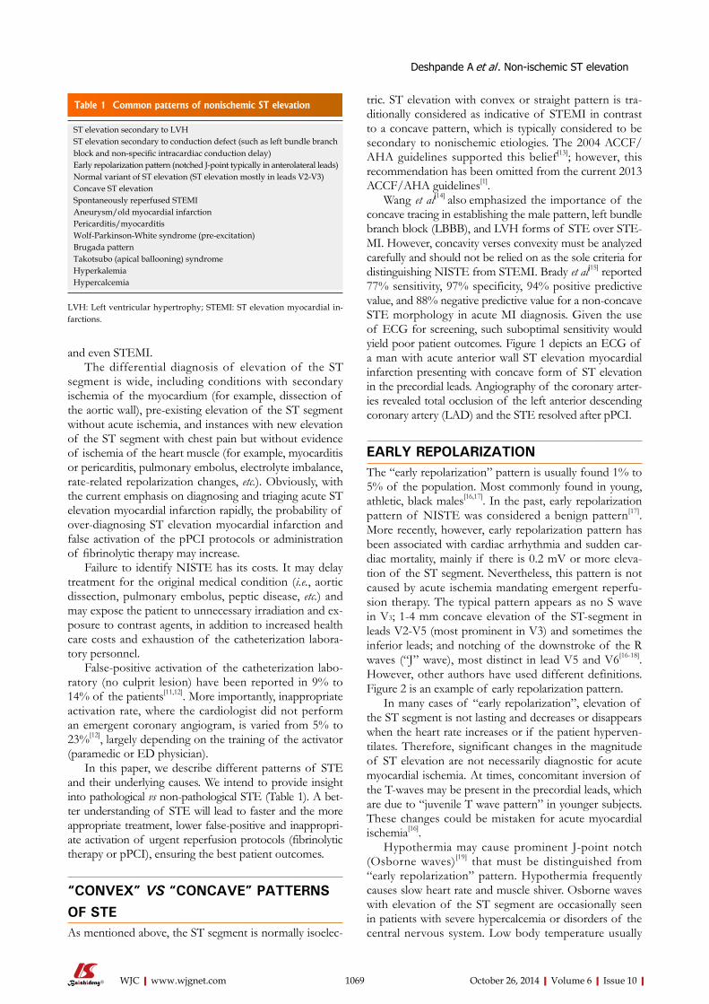
and even STEMI.The differential diagnosis of elevation of the ST
segment is wide, including conditions with secondary ischemia of the myocardium (for example, dissection of the aortic wall), pre-existing elevation of the ST segment without acute ischemia, and instances with new elevation of the ST segment with chest pain but without evidence of ischemia of the heart muscle (for example, myocarditis or pericarditis, pulmonary embolus, electrolyte imbalance, rate-related repolarization changes, etc.). Obviously, with the current emphasis on diagnosing and triaging acute ST elevation myocardial infarction rapidly, the probability of over-diagnosing ST elevation myocardial infarction and false activation of the pPCI protocols or administration of fibrinolytic therapy may increase.
Failure to identify NISTE has its costs. It may delay treatment for the original medical condition (i.e., aortic dissection, pulmonary embolus, peptic disease, etc.) and may expose the patient to unnecessary irradiation and ex-posure to contrast agents, in addition to increased health care costs and exhaustion of the catheterization labora-tory personnel.
False-positive activation of the catheterization labo-ratory (no culprit lesion) have been reported in 9% to 14% of the patients[11,12]. More importantly, inappropriate activation rate, where the cardiologist did not perform an emergent coronary angiogram, is varied from 5% to 23%[12], largely depending on the training of the activator (paramedic or ED physician).
In this paper, we describe different patterns of STE and their underlying causes. We intend to provide insight into pathological vs non-pathological STE (Table 1). A bet-ter understanding of STE will lead to faster and the more appropriate treatment, lower false-positive and inappropri-ate activation of urgent reperfusion protocols (fibrinolytic therapy or pPCI), ensuring the best patient outcomes.
“CONVEX” VS “CONCAVE” PATTERNS OF STEAs mentioned above, the ST segment is normally isoelec-
tric. ST elevation with convex or straight pattern is tra-ditionally considered as indicative of STEMI in contrast to a concave pattern, which is typically considered to be secondary to nonischemic etiologies. The 2004 ACCF/AHA guidelines supported this belief[13]; however, this recommendation has been omitted from the current 2013 ACCF/AHA guidelines[1].
Wang et al[14] also emphasized the importance of the concave tracing in establishing the male pattern, left bundle branch block (LBBB), and LVH forms of STE over STE-MI. However, concavity verses convexity must be analyzed carefully and should not be relied on as the sole criteria for distinguishing NISTE from STEMI. Brady et al[15] reported 77% sensitivity, 97% specificity, 94% positive predictive value, and 88% negative predictive value for a non-concave STE morphology in acute MI diagnosis. Given the use of ECG for screening, such suboptimal sensitivity would yield poor patient outcomes. Figure 1 depicts an ECG of a man with acute anterior wall ST elevation myocardial infarction presenting with concave form of ST elevation in the precordial leads. Angiography of the coronary arter-ies revealed total occlusion of the left anterior descending coronary artery (LAD) and the STE resolved after pPCI.
EARLY REPOLARIZATIONThe “early repolarization” pattern is usually found 1% to 5% of the population. Most commonly found in young, athletic, black males[16,17]. In the past, early repolarization pattern of NISTE was considered a benign pattern[17]. More recently, however, early repolarization pattern has been associated with cardiac arrhythmia and sudden car-diac mortality, mainly if there is 0.2 mV or more eleva-tion of the ST segment. Nevertheless, this pattern is not caused by acute ischemia mandating emergent reperfu-sion therapy. The typical pattern appears as no S wave in V3; 1-4 mm concave elevation of the ST-segment in leads V2-V5 (most prominent in V3) and sometimes the inferior leads; and notching of the downstroke of the R waves (“J” wave), most distinct in lead V5 and V6[16-18]. However, other authors have used different definitions. Figure 2 is an example of early repolarization pattern.
In many cases of “early repolarization”, elevation of the ST segment is not lasting and decreases or disappears when the heart rate increases or if the patient hyperven-tilates. Therefore, significant changes in the magnitude of ST elevation are not necessarily diagnostic for acute myocardial ischemia. At times, concomitant inversion of the T-waves may be present in the precordial leads, which are due to “juvenile T wave pattern” in younger subjects. These changes could be mistaken for acute myocardial ischemia[16].
Hypothermia may cause prominent J-point notch (Osborne waves)[19] that must be distinguished from “early repolarization” pattern. Hypothermia frequently causes slow heart rate and muscle shiver. Osborne waves with elevation of the ST segment are occasionally seen in patients with severe hypercalcemia or disorders of the central nervous system. Low body temperature usually
October 26, 2014|Volume 6|Issue 10|WJC|www.wjgnet.com 1069
ST elevation secondary to LVH ST elevation secondary to conduction defect (such as left bundle branch block and non-specific intracardiac conduction delay) Early repolarization pattern (notched J-point typically in anterolateral leads) Normal variant of ST elevation (ST elevation mostly in leads V2-V3) Concave ST elevation Spontaneously reperfused STEMI Aneurysm/old myocardial infarction Pericarditis/myocarditis Wolf-Parkinson-White syndrome (pre-excitation) Brugada pattern Takotsubo (apical ballooning) syndrome Hyperkalemia Hypercalcemia
Table 1 Common patterns of nonischemic ST elevation
LVH: Left ventricular hypertrophy; STEMI: ST elevation myocardial in-farctions.
Deshpande A et al . Non-ischemic ST elevation
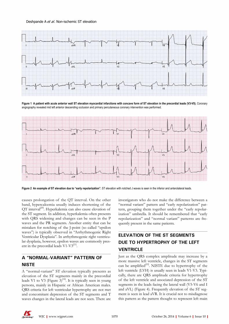
October 26, 2014|Volume 6|Issue 10|WJC|www.wjgnet.com
investigators who do not make the difference between a “normal variant” pattern and “early repolarization” pat-tern, grouping them together under the “early repolar-ization” umbrella. It should be remembered that “early repolarization” and “normal variant” patterns are fre-quently present in the same patients.
ELEVATION OF THE ST SEGMENTS
DUE TO HYPERTROPHY OF THE LEFT
VENTRICLEJust as the QRS complex amplitude may increase by a more massive left ventricle, changes in the ST segments can be amplified[22]. NISTE due to hypertrophy of the left ventricle (LVH) is usually seen in leads V1-V3. Typi-cally, there are QRS amplitude criteria for hypertrophy of the left ventricle and associated depression of the ST segments in the leads facing the lateral wall (V5-V6 and I and aVL) (Figure 4). Frequently elevation of the ST seg-ment is seen in lead aVR. It is crucial not to misdiagnose this pattern as the pattern thought to represent left main
causes prolongation of the QT interval. On the other hand, hypercalcemia usually induces shortening of the QT interval[20]. Hyperkalemia can also cause elevation of the ST segment. In addition, hyperkalemia often presents with QRS widening and changes can be seen in the P waves and the PR segments. Another entity that can be mistaken for notching of the J-point (so called “epsilon waves”) is typically observed in “Arrhythmogenic Right Ventricular Dysplasia”. In arrhythmogenic right ventricu-lar dysplasia, however, epsilon waves are commonly pres-ent in the precordial leads V1-V3[21].
A “NORMAL-VARIANT” PATTERN OF NISTEA “normal-variant” ST elevation typically presents as elevation of the ST segments mainly in the precordial leads V1 to V3 (Figure 3)[14]. It is typically seen in young persons, mainly in Hispanic or African American males. QRS criteria for left ventricular hypertrophy are not met and concomitant depression of the ST segments and T waves changes in the lateral leads are not seen. There are
1070
Figure 1 A patient with acute anterior wall ST elevation myocardial infarctions with concave form of ST elevation in the precordial leads (V3-V5). Coronary angiography revealed mid left anterior descending occlusion and primary percutaneous coronary intervention was performed.
Figure 2 An example of ST elevation due to “early repolarization”. ST elevation with notched J waves is seen in the inferior and anterolateral leads.
Deshpande A et al . Non-ischemic ST elevation
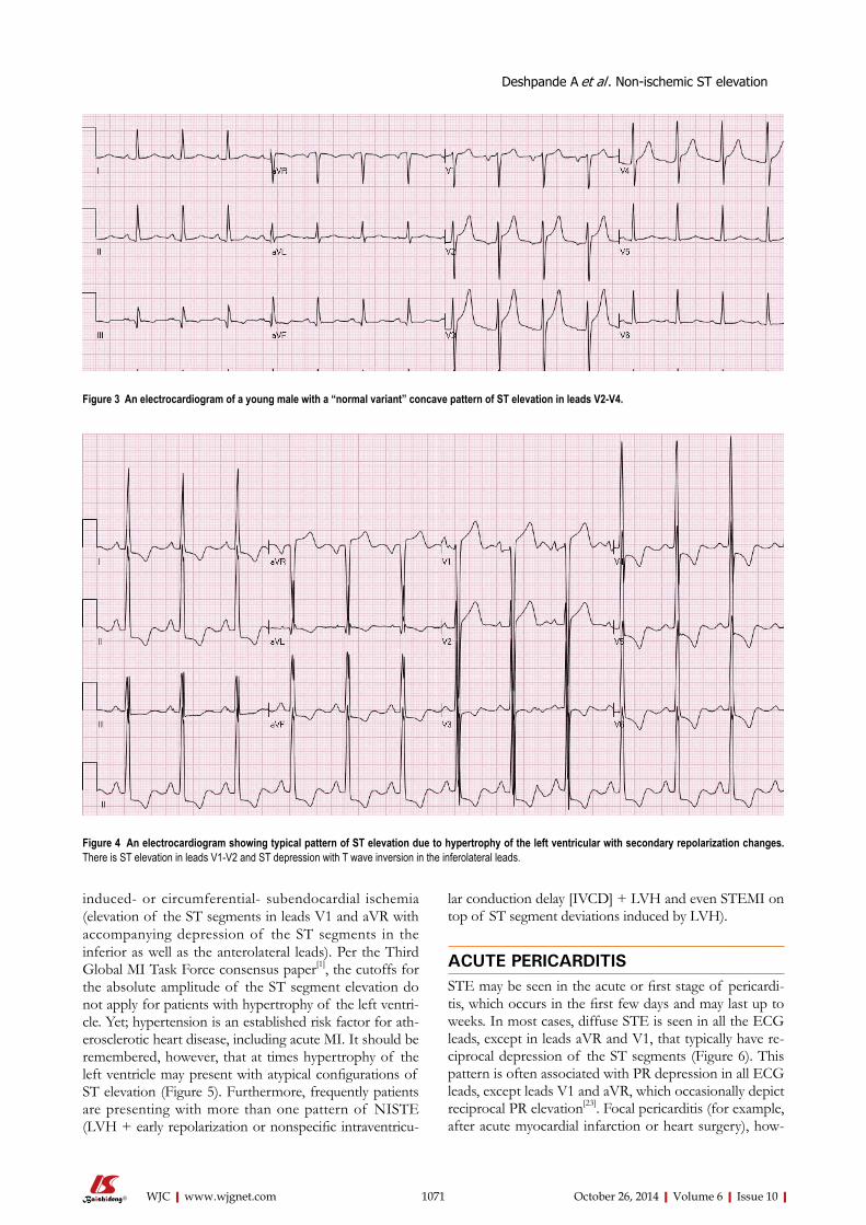
October 26, 2014|Volume 6|Issue 10|WJC|www.wjgnet.com
induced- or circumferential- subendocardial ischemia (elevation of the ST segments in leads V1 and aVR with accompanying depression of the ST segments in the inferior as well as the anterolateral leads). Per the Third Global MI Task Force consensus paper[1], the cutoffs for the absolute amplitude of the ST segment elevation do not apply for patients with hypertrophy of the left ventri-cle. Yet; hypertension is an established risk factor for ath-erosclerotic heart disease, including acute MI. It should be remembered, however, that at times hypertrophy of the left ventricle may present with atypical configurations of ST elevation (Figure 5). Furthermore, frequently patients are presenting with more than one pattern of NISTE (LVH + early repolarization or nonspecific intraventricu-
lar conduction delay [IVCD] + LVH and even STEMI on top of ST segment deviations induced by LVH).
ACUTE PERICARDITISSTE may be seen in the acute or first stage of pericardi-tis, which occurs in the first few days and may last up to weeks. In most cases, diffuse STE is seen in all the ECG leads, except in leads aVR and V1, that typically have re-ciprocal depression of the ST segments (Figure 6). This pattern is often associated with PR depression in all ECG leads, except leads V1 and aVR, which occasionally depict reciprocal PR elevation[23]. Focal pericarditis (for example, after acute myocardial infarction or heart surgery), how-
1071
Figure 3 An electrocardiogram of a young male with a “normal variant” concave pattern of ST elevation in leads V2-V4.
Figure 4 An electrocardiogram showing typical pattern of ST elevation due to hypertrophy of the left ventricular with secondary repolarization changes. There is ST elevation in leads V1-V2 and ST depression with T wave inversion in the inferolateral leads.
Deshpande A et al . Non-ischemic ST elevation
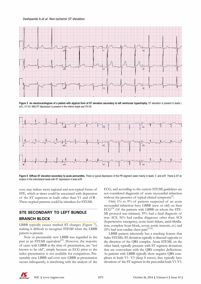
October 26, 2014|Volume 6|Issue 10|WJC|www.wjgnet.com
ever, may induce more regional and non-typical forms of STE, which at times could be associated with depression of the ST segments in leads other than V1 and aVR . These atypical patterns could be mistaken for STEMI.
STE SECONDARY TO LEFT BUNDLE
BRANCH BLOCK LBBB typically causes marked ST changes (Figure 7), making it difficult to recognize STEMI when the LBBB pattern is present.
New or presumably new LBBB was regarded in the past as an STEMI equivalent[13]. However, the majority of cases with LBBB at the time of presentation, are “not known to be old”, simply because an ECG prior to the index presentation is not available for comparison. Pre-sumably new LBBB and even new LBBB at presentation occurs infrequently, is interfering with the analysis of the
ECG, and according to the current STEMI guidelines are not considered diagnostic of acute myocardial infarction without the presence of typical clinical symptoms[3].
Only 1% to 9% of patients suspected of an acute myocardial infarction have LBBB (new or old) on their ECG[24]. Of the patients with LBBB on whom the STE-MI protocol was initiated, 39% had a final diagnosis of true ACS, 36% had cardiac diagnoses other than ACS (hypertensive emergency, acute heart failure, atrial fibrilla-tion, complete heart block, severe aortic stenosis, etc.) and 25% had non-cardiac chest pain[25,26].
LBBB pattern inherently has a masking feature that hides STEMIs. ST deviation typically is directed opposite to the direction of the QRS complex. Acute STEMI, on the other hand, typically presents with ST segment deviations that are concordant with the QRS complex deflections. As patients with LBBB typically show negative QRS com-plexes in leads V1- V3 (deep S waves), they typically have elevations of the ST segment in the precordial leads V1-V3.
1072
Figure 5 An electrocardiogram of a patient with atypical form of ST elevation secondary to left ventricular hypertrophy. ST elevation is present in leads I, aVL, V1-V2. Mild ST depression is present in the inferior leads and V5-V6.
Figure 6 Diffuse ST elevation secondary to acute pericarditis. There is typical depression of the PR segment (seen mainly in leads Ⅱ and aVF. There is ST el-evation in the inferolateral leads with ST depression in lead aVR.
Deshpande A et al . Non-ischemic ST elevation
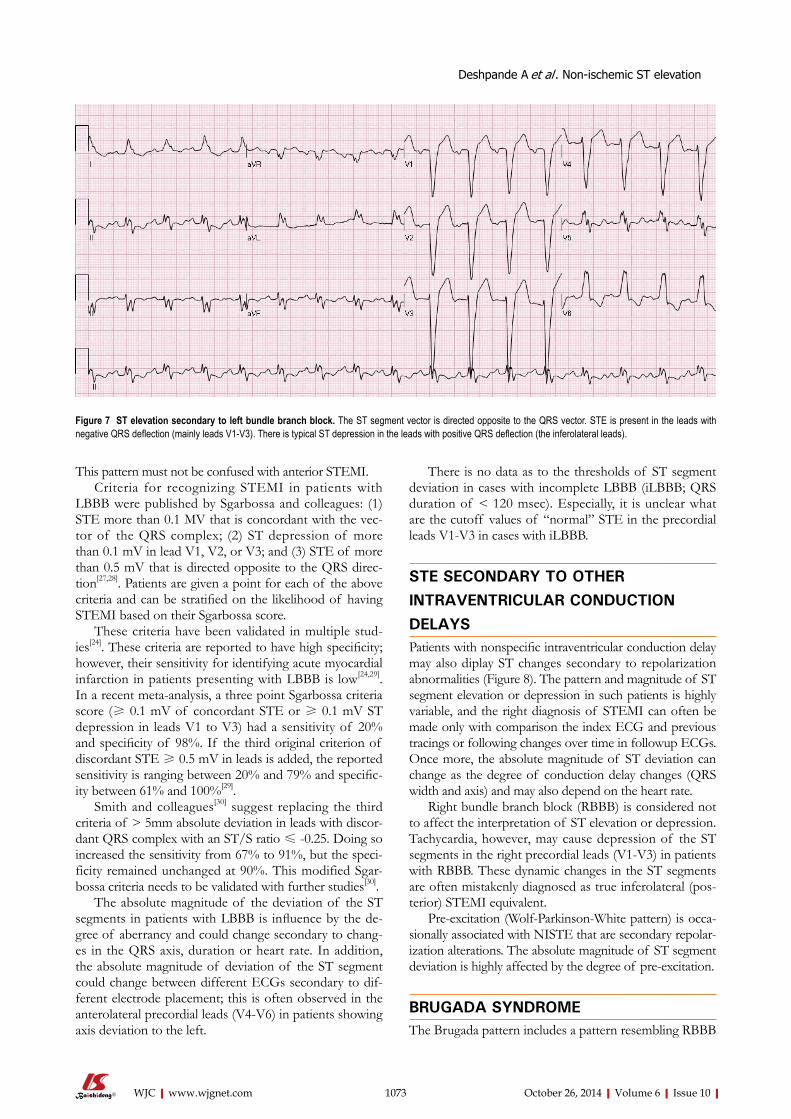
October 26, 2014|Volume 6|Issue 10|WJC|www.wjgnet.com
This pattern must not be confused with anterior STEMI.Criteria for recognizing STEMI in patients with
LBBB were published by Sgarbossa and colleagues: (1) STE more than 0.1 MV that is concordant with the vec-tor of the QRS complex; (2) ST depression of more than 0.1 mV in lead V1, V2, or V3; and (3) STE of more than 0.5 mV that is directed opposite to the QRS direc-tion[27,28]. Patients are given a point for each of the above criteria and can be stratified on the likelihood of having STEMI based on their Sgarbossa score.
These criteria have been validated in multiple stud-ies[24]. These criteria are reported to have high specificity; however, their sensitivity for identifying acute myocardial infarction in patients presenting with LBBB is low[24,29].
In a recent meta-analysis, a three point Sgarbossa criteria score (≥ 0.1 mV of concordant STE or ≥ 0.1 mV ST depression in leads V1 to V3) had a sensitivity of 20% and specificity of 98%. If the third original criterion of discordant STE ≥ 0.5 mV in leads is added, the reported sensitivity is ranging between 20% and 79% and specific-ity between 61% and 100%[29].
Smith and colleagues[30] suggest replacing the third criteria of > 5mm absolute deviation in leads with discor-dant QRS complex with an ST/S ratio ≤ -0.25. Doing so increased the sensitivity from 67% to 91%, but the speci-ficity remained unchanged at 90%. This modified Sgar-bossa criteria needs to be validated with further studies[30].
The absolute magnitude of the deviation of the ST segments in patients with LBBB is influence by the de-gree of aberrancy and could change secondary to chang-es in the QRS axis, duration or heart rate. In addition, the absolute magnitude of deviation of the ST segment could change between different ECGs secondary to dif-ferent electrode placement; this is often observed in the anterolateral precordial leads (V4-V6) in patients showing axis deviation to the left.
There is no data as to the thresholds of ST segment deviation in cases with incomplete LBBB (iLBBB; QRS duration of < 120 msec). Especially, it is unclear what are the cutoff values of “normal” STE in the precordial leads V1-V3 in cases with iLBBB.
STE SECONDARY TO OTHER INTRAVENTRICULAR CONDUCTION DELAYS Patients with nonspecific intraventricular conduction delay may also diplay ST changes secondary to repolarization abnormalities (Figure 8). The pattern and magnitude of ST segment elevation or depression in such patients is highly variable, and the right diagnosis of STEMI can often be made only with comparison the index ECG and previous tracings or following changes over time in followup ECGs. Once more, the absolute magnitude of ST deviation can change as the degree of conduction delay changes (QRS width and axis) and may also depend on the heart rate.
Right bundle branch block (RBBB) is considered not to affect the interpretation of ST elevation or depression. Tachycardia, however, may cause depression of the ST segments in the right precordial leads (V1-V3) in patients with RBBB. These dynamic changes in the ST segments are often mistakenly diagnosed as true inferolateral (pos-terior) STEMI equivalent.
Pre-excitation (Wolf-Parkinson-White pattern) is occa-sionally associated with NISTE that are secondary repolar-ization alterations. The absolute magnitude of ST segment deviation is highly affected by the degree of pre-excitation.
BRUGADA SYNDROMEThe Brugada pattern includes a pattern resembling RBBB
1073
Figure 7 ST elevation secondary to left bundle branch block. The ST segment vector is directed opposite to the QRS vector. STE is present in the leads with negative QRS deflection (mainly leads V1-V3). There is typical ST depression in the leads with positive QRS deflection (the inferolateral leads).
Deshpande A et al . Non-ischemic ST elevation
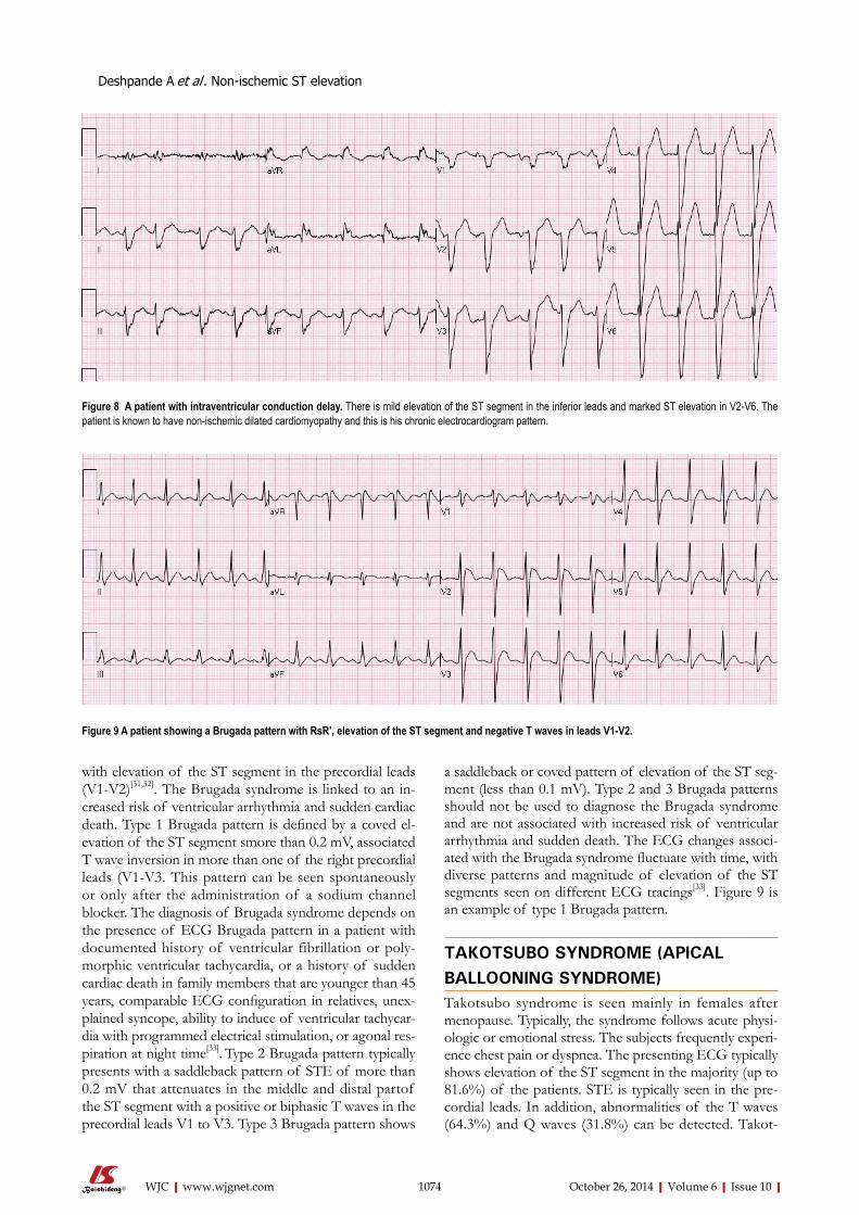
October 26, 2014|Volume 6|Issue 10|WJC|www.wjgnet.com
with elevation of the ST segment in the precordial leads (V1-V2)[31,32]. The Brugada syndrome is linked to an in-creased risk of ventricular arrhythmia and sudden cardiac death. Type 1 Brugada pattern is defined by a coved el-evation of the ST segment smore than 0.2 mV, associated T wave inversion in more than one of the right precordial leads (V1-V3. This pattern can be seen spontaneously or only after the administration of a sodium channel blocker. The diagnosis of Brugada syndrome depends on the presence of ECG Brugada pattern in a patient with documented history of ventricular fibrillation or poly-morphic ventricular tachycardia, or a history of sudden cardiac death in family members that are younger than 45 years, comparable ECG configuration in relatives, unex-plained syncope, ability to induce of ventricular tachycar-dia with programmed electrical stimulation, or agonal res-piration at night time[33]. Type 2 Brugada pattern typically presents with a saddleback pattern of STE of more than 0.2 mV that attenuates in the middle and distal partof the ST segment with a positive or biphasic T waves in the precordial leads V1 to V3. Type 3 Brugada pattern shows
a saddleback or coved pattern of elevation of the ST seg-ment (less than 0.1 mV). Type 2 and 3 Brugada patterns should not be used to diagnose the Brugada syndrome and are not associated with increased risk of ventricular arrhythmia and sudden death. The ECG changes associ-ated with the Brugada syndrome fluctuate with time, with diverse patterns and magnitude of elevation of the ST segments seen on different ECG tracings[33]. Figure 9 is an example of type 1 Brugada pattern.
TAKOTSUBO SYNDROME (APICAL BALLOONING SYNDROME)Takotsubo syndrome is seen mainly in females after menopause. Typically, the syndrome follows acute physi-ologic or emotional stress. The subjects frequently experi-ence chest pain or dyspnea. The presenting ECG typically shows elevation of the ST segment in the majority (up to 81.6%) of the patients. STE is typically seen in the pre-cordial leads. In addition, abnormalities of the T waves (64.3%) and Q waves (31.8%) can be detected. Takot-
1074
Figure 8 A patient with intraventricular conduction delay. There is mild elevation of the ST segment in the inferior leads and marked ST elevation in V2-V6. The patient is known to have non-ischemic dilated cardiomyopathy and this is his chronic electrocardiogram pattern.
Figure 9 A patient showing a Brugada pattern with RsR’, elevation of the ST segment and negative T waves in leads V1-V2.
Deshpande A et al . Non-ischemic ST elevation
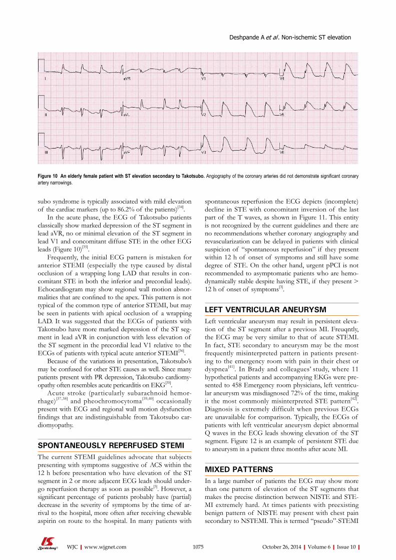
October 26, 2014|Volume 6|Issue 10|WJC|www.wjgnet.com
subo syndrome is typically associated with mild elevation of the cardiac markers (up to 86.2% of the patients)[34].
In the acute phase, the ECG of Takotsubo patients classically show marked depression of the ST segment in lead aVR, no or minimal elevation of the ST segment in lead V1 and concomitant diffuse STE in the other ECG leads (Figure 10)[35].
Frequently, the initial ECG pattern is mistaken for anterior STEMI (especially the type caused by distal occlusion of a wrapping long LAD that results in con-comitant STE in both the inferior and precordial leads). Echocardiogram may show regional wall motion abnor-malities that are confined to the apex. This pattern is not typical of the common type of anterior STEMI, but may be seen in patients with apical occlusion of a wrapping LAD. It was suggested that the ECGs of patients with Takotsubo have more marked depression of the ST seg-ment in lead aVR in conjunction with less elevation of the ST segment in the precordial lead V1 relative to the ECGs of patients with typical acute anterior STEMI[36].
Because of the variations in presentation, Takotsubo’s may be confused for other STE causes as well. Since many patients present with PR depression, Takotsubo cardiomy-opathy often resembles acute pericarditis on EKG[35].
Acute stroke (particularly subarachnoid hemor-rhage)[37,38] and pheochromocytoma[39,40] occasionally present with ECG and regional wall motion dysfunction findings that are indistinguishable from Takotsubo car-diomyopathy.
SPONTANEOUSLY REPERFUSED STEMIThe current STEMI guidelines advocate that subjects presenting with symptoms suggestive of ACS within the 12 h before presentation who have elevation of the ST segment in 2 or more adjacent ECG leads should under-go reperfusion therapy as soon as possible[3]. However, a significant percentage of patients probably have (partial) decrease in the severity of symptoms by the time of ar-rival to the hospital, more often after receiving chewable aspirin on route to the hospital. In many patients with
spontaneous reperfusion the ECG depicts (incomplete) decline in STE with concomitant inversion of the last part of the T waves, as shown in Figure 11. This entity is not recognized by the current guidelines and there are no recommendations whether coronary angiography and revascularization can be delayed in patients with clinical suspicion of “spontaneous reperfusion” if they present within 12 h of onset of symptoms and still have some degree of STE. On the other hand, urgent pPCI is not recommended to asymptomatic patients who are hemo-dynamically stable despite having STE, if they present > 12 h of onset of symptoms[3].
LEFT VENTRICULAR ANEURYSMLeft ventricular aneurysm may result in persistent eleva-tion of the ST segment after a previous MI. Freuqntly, the ECG may be very similar to that of acute STEMI. In fact, STE secondary to aneurysm may be the most frequently misinterpreted pattern in patients present-ing to the emergency room with pain in their chest or dyspnea[41]. In Brady and colleagues’ study, where 11 hypothetical patients and accompanying EKGs were pre-sented to 458 Emergency room physicians, left ventricu-lar aneurysm was misdiagnosed 72% of the time, making it the most commonly misinterpreted STE pattern[42]. Diagnosis is extremely difficult when previous ECGs are unavailable for comparison. Typically, the ECGs of patients with left ventricular aneurysm depict abnormal Q waves in the ECG leads showing elevation of the ST segment. Figure 12 is an example of persistent STE due to aneurysm in a patient three months after acute MI.
MIXED PATTERNSIn a large number of patients the ECG may show more than one pattern of elevation of the ST segments that makes the precise distinction between NISTE and STE-MI extremely hard. At times patients with preexisting benign pattern of NISTE may present with chest pain secondary to NSTEMI. This is termed “pseudo”-STEMI
1075
Figure 10 An elderly female patient with ST elevation secondary to Takotsubo. Angiography of the coronary arteries did not demonstrate significant coronary artery narrowings.
Deshpande A et al . Non-ischemic ST elevation
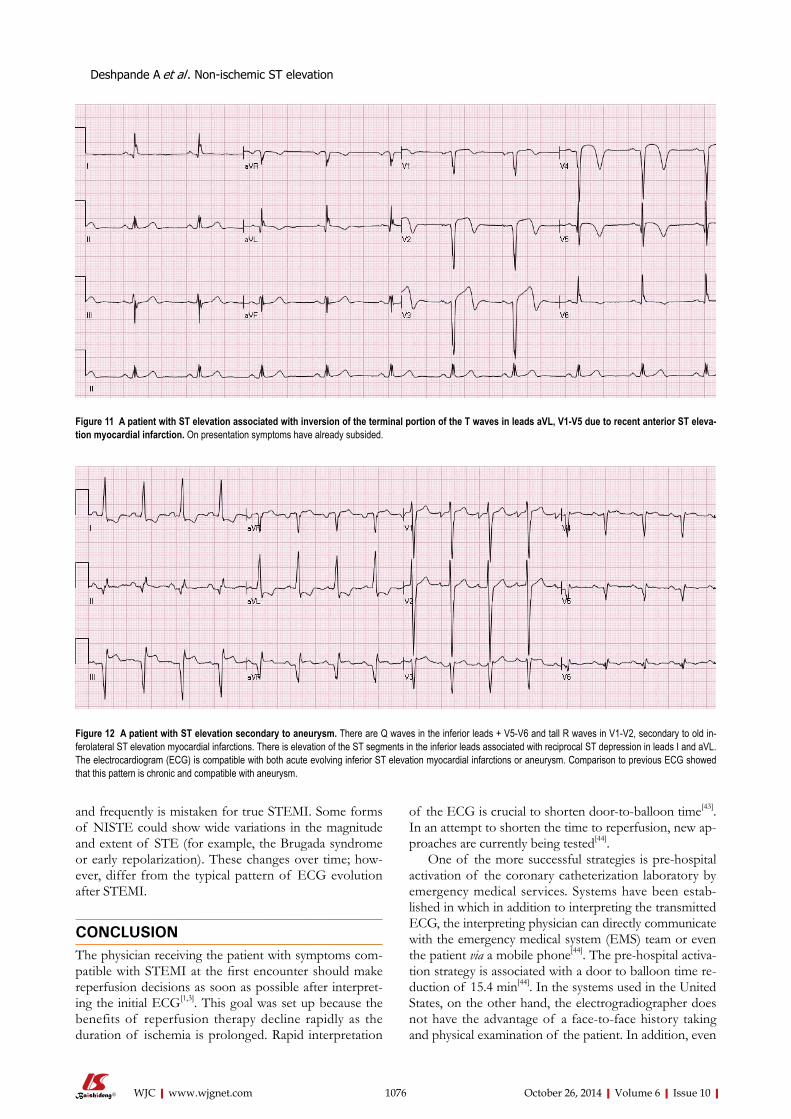
October 26, 2014|Volume 6|Issue 10|WJC|www.wjgnet.com
and frequently is mistaken for true STEMI. Some forms of NISTE could show wide variations in the magnitude and extent of STE (for example, the Brugada syndrome or early repolarization). These changes over time; how-ever, differ from the typical pattern of ECG evolution after STEMI.
CONCLUSIONThe physician receiving the patient with symptoms com-patible with STEMI at the first encounter should make reperfusion decisions as soon as possible after interpret-ing the initial ECG[1,3]. This goal was set up because the benefits of reperfusion therapy decline rapidly as the duration of ischemia is prolonged. Rapid interpretation
of the ECG is crucial to shorten door-to-balloon time[43]. In an attempt to shorten the time to reperfusion, new ap-proaches are currently being tested[44].
One of the more successful strategies is pre-hospital activation of the coronary catheterization laboratory by emergency medical services. Systems have been estab-lished in which in addition to interpreting the transmitted ECG, the interpreting physician can directly communicate with the emergency medical system (EMS) team or even the patient via a mobile phone[44]. The pre-hospital activa-tion strategy is associated with a door to balloon time re-duction of 15.4 min[44]. In the systems used in the United States, on the other hand, the electrogradiographer does not have the advantage of a face-to-face history taking and physical examination of the patient. In addition, even
1076
Figure 11 A patient with ST elevation associated with inversion of the terminal portion of the T waves in leads aVL, V1-V5 due to recent anterior ST eleva-tion myocardial infarction. On presentation symptoms have already subsided.
Figure 12 A patient with ST elevation secondary to aneurysm. There are Q waves in the inferior leads + V5-V6 and tall R waves in V1-V2, secondary to old in-ferolateral ST elevation myocardial infarctions. There is elevation of the ST segments in the inferior leads associated with reciprocal ST depression in leads I and aVL. The electrocardiogram (ECG) is compatible with both acute evolving inferior ST elevation myocardial infarctions or aneurysm. Comparison to previous ECG showed that this pattern is chronic and compatible with aneurysm.
Deshpande A et al . Non-ischemic ST elevation
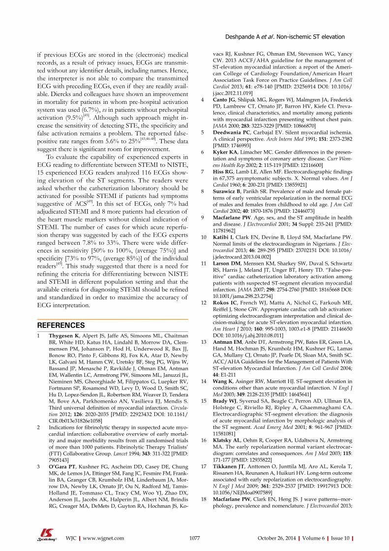
October 26, 2014|Volume 6|Issue 10|WJC|www.wjgnet.com
if previous ECGs are stored in the (electronic) medical records, as a result of privacy issues, ECGs are transmit-ted without any identifier details, including names. Hence, the interpreter is not able to compare the transmitted ECG with preceding ECGs, even if they are readily avail-able. Diercks and colleagues have shown an improvement in mortality for patients in whom pre-hospital activation system was used (6.7%), vs in patients without prehospital activation (9.5%)[45]. Although such approach might in-crease the sensitivity of detecting STE, the specificity and false activation remains a problem. The reported false-positive rate ranges from 5.6% to 25%[43,46-48]. These data suggest there is significant room for improvement.
To evaluate the capability of experienced experts in ECG reading to differentiate between STEMI to NISTE, 15 experienced ECG readers analyzed 116 ECGs show-ing elevation of the ST segments. The readers were asked whether the catheterization laboratory should be activated for possible STEMI if patients had symptoms suggestive of ACS[49]. In this set of ECGs, only 7% had adjudicated STEMI and 8 more patients had elevation of the heart muscle markers without clinical indication of STEMI. The number of cases for which acute reperfu-sion therapy was suggested by each of the ECG experts ranged between 7.8% to 33%. There were wide differ-ences in sensitivity [50% to 100%, (average 75%)] and specificity [73% to 97%, (average 85%)] of the individual readers[49]. This study suggested that there is a need for refining the criteria for differentiating between NISTE and STEMI in different population setting and that the available criteria for diagnosing STEMI should be refined and standardized in order to maximize the accuracy of ECG interpretation.
REFERENCES1 Thygesen K, Alpert JS, Jaffe AS, Simoons ML, Chaitman
BR, White HD, Katus HA, Lindahl B, Morrow DA, Clem-mensen PM, Johanson P, Hod H, Underwood R, Bax JJ, Bonow RO, Pinto F, Gibbons RJ, Fox KA, Atar D, Newby LK, Galvani M, Hamm CW, Uretsky BF, Steg PG, Wijns W, Bassand JP, Menasché P, Ravkilde J, Ohman EM, Antman EM, Wallentin LC, Armstrong PW, Simoons ML, Januzzi JL, Nieminen MS, Gheorghiade M, Filippatos G, Luepker RV, Fortmann SP, Rosamond WD, Levy D, Wood D, Smith SC, Hu D, Lopez-Sendon JL, Robertson RM, Weaver D, Tendera M, Bove AA, Parkhomenko AN, Vasilieva EJ, Mendis S. Third universal definition of myocardial infarction. Circula-tion 2012; 126: 2020-2035 [PMID: 22923432 DOI: 10.1161/CIR.0b013e31826e1058]
2 Indications for fibrinolytic therapy in suspected acute myo-cardial infarction: collaborative overview of early mortal-ity and major morbidity results from all randomised trials of more than 1000 patients. Fibrinolytic Therapy Trialists’ (FTT) Collaborative Group. Lancet 1994; 343: 311-322 [PMID: 7905143]
3 O’Gara PT, Kushner FG, Ascheim DD, Casey DE, Chung MK, de Lemos JA, Ettinger SM, Fang JC, Fesmire FM, Frank-lin BA, Granger CB, Krumholz HM, Linderbaum JA, Mor-row DA, Newby LK, Ornato JP, Ou N, Radford MJ, Tamis-Holland JE, Tommaso CL, Tracy CM, Woo YJ, Zhao DX, Anderson JL, Jacobs AK, Halperin JL, Albert NM, Brindis RG, Creager MA, DeMets D, Guyton RA, Hochman JS, Ko-
vacs RJ, Kushner FG, Ohman EM, Stevenson WG, Yancy CW. 2013 ACCF/AHA guideline for the management of ST-elevation myocardial infarction: a report of the Ameri-can College of Cardiology Foundation/American Heart Association Task Force on Practice Guidelines. J Am Coll Cardiol 2013; 61: e78-140 [PMID: 23256914 DOI: 10.1016/j.jacc.2012.11.019]
4 Canto JG, Shlipak MG, Rogers WJ, Malmgren JA, Frederick PD, Lambrew CT, Ornato JP, Barron HV, Kiefe CI. Preva-lence, clinical characteristics, and mortality among patients with myocardial infarction presenting without chest pain. JAMA 2000; 283: 3223-3229 [PMID: 10866870]
5 Deedwania PC, Carbajal EV. Silent myocardial ischemia. A clinical perspective. Arch Intern Med 1991; 151: 2373-2382 [PMID: 1746993]
6 Kyker KA, Limacher MC. Gender differences in the presen-tation and symptoms of coronary artery disease. Curr Wom-ens Health Rep 2002; 2: 115-119 [PMID: 12116600]
7 Hiss RG, Lamb LE, Allen MF. Electrocardiographic findings in 67,375 asymptomatic subjects. X. Normal values. Am J Cardiol 1960; 6: 200-231 [PMID: 13855921]
8 Surawicz B, Parikh SR. Prevalence of male and female pat-terns of early ventricular repolarization in the normal ECG of males and females from childhood to old age. J Am Coll Cardiol 2002; 40: 1870-1876 [PMID: 12446073]
9 Macfarlane PW. Age, sex, and the ST amplitude in health and disease. J Electrocardiol 2001; 34 Suppl: 235-241 [PMID: 11781962]
10 Katibi I, Clark EN, Devine B, Lloyd SM, Macfarlane PW. Normal limits of the electrocardiogram in Nigerians. J Elec-trocardiol 2013; 46: 289-295 [PMID: 23702151 DOI: 10.1016/j.jelectrocard.2013.04.002]
11 Larson DM, Menssen KM, Sharkey SW, Duval S, Schwartz RS, Harris J, Meland JT, Unger BT, Henry TD. “False-pos-itive” cardiac catheterization laboratory activation among patients with suspected ST-segment elevation myocardial infarction. JAMA 2007; 298: 2754-2760 [PMID: 18165668 DOI: 10.1001/jama.298.23.2754]
12 Rokos IC, French WJ, Mattu A, Nichol G, Farkouh ME, Reiffel J, Stone GW. Appropriate cardiac cath lab activation: optimizing electrocardiogram interpretation and clinical de-cision-making for acute ST-elevation myocardial infarction. Am Heart J 2010; 160: 995-1003, 1003.e1-8 [PMID: 21146650 DOI: 10.1016/j.ahj.2010.08.011]
13 Antman EM, Anbe DT, Armstrong PW, Bates ER, Green LA, Hand M, Hochman JS, Krumholz HM, Kushner FG, Lamas GA, Mullany CJ, Ornato JP, Pearle DI, Sloan MA, Smith SC. ACC/AHA Guidelines for the Management of Patients With ST-elevation Myocardial Infarction. J Am Coll Cardiol 2004; 44: E1-211
14 Wang K, Asinger RW, Marriott HJ. ST-segment elevation in conditions other than acute myocardial infarction. N Engl J Med 2003; 349: 2128-2135 [PMID: 14645641]
15 Brady WJ, Syverud SA, Beagle C, Perron AD, Ullman EA, Holstege C, Riviello RJ, Ripley A, Ghaemmaghami CA. Electrocardiographic ST-segment elevation: the diagnosis of acute myocardial infarction by morphologic analysis of the ST segment. Acad Emerg Med 2001; 8: 961-967 [PMID: 11581081]
16 Klatsky AL, Oehm R, Cooper RA, Udaltsova N, Armstrong MA. The early repolarization normal variant electrocar-diogram: correlates and consequences. Am J Med 2003; 115: 171-177 [PMID: 12935822]
17 Tikkanen JT, Anttonen O, Junttila MJ, Aro AL, Kerola T, Rissanen HA, Reunanen A, Huikuri HV. Long-term outcome associated with early repolarization on electrocardiography. N Engl J Med 2009; 361: 2529-2537 [PMID: 19917913 DOI: 10.1056/NEJMoa0907589]
18 Macfarlane PW, Clark EN, Heng JS. J wave patterns--mor-phology, prevalence and nomenclature. J Electrocardiol 2013;
1077
Deshpande A et al . Non-ischemic ST elevation
October 26, 2014|Volume 6|Issue 10|WJC|www.wjgnet.com
46: 505-509 [PMID: 24075127 DOI: 10.1016/j.jelectrocard.2013.08.013]
19 Spodick DH. Hypothermia with J (Osborne) waves. Am Heart Hosp J 2006; 4: 156 [PMID: 16687964]
20 Nishi SP, Barbagelata NA, Atar S, Birnbaum Y, Tuero E. Hypercalcemia-induced ST-segment elevation mimicking acute myocardial infarction. J Electrocardiol 2006; 39: 298-300 [PMID: 16777515]
21 Nasir K, Bomma C, Tandri H, Roguin A, Dalal D, Prakasa K, Tichnell C, James C, Spevak PJ, Marcus F, Calkins H. Electro-cardiographic features of arrhythmogenic right ventricular dysplasia/cardiomyopathy according to disease severity: a need to broaden diagnostic criteria. Circulation 2004; 110: 1527-1534 [PMID: 15381658]
22 Estes EH, Jackson KP. The electrocardiogram in left ventric-ular hypertrophy: past and future. J Electrocardiol 2009; 42: 589-592 [PMID: 19643433 DOI: 10.1016/j.jelectrocard.2009.06.016]
23 Punja M, Mark DG, McCoy JV, Javan R, Pines JM, Brady W. Electrocardiographic manifestations of cardiac infectious-inflammatory disorders. Am J Emerg Med 2010; 28: 364-377 [PMID: 20223398 DOI: 10.1016/j.ajem.2008.12.017]
24 Cai Q, Mehta N, Sgarbossa EB, Pinski SL, Wagner GS, Califf RM, Barbagelata A. The left bundle-branch block puzzle in the 2013 ST-elevation myocardial infarction guideline: from falsely declaring emergency to denying reperfusion in a high-risk population. Are the Sgarbossa Criteria ready for prime time? Am Heart J 2013; 166: 409-413 [PMID: 24016487 DOI: 10.1016/j.ahj.2013.03.032]
25 Jain S, Ting HT, Bell M, Bjerke CM, Lennon RJ, Gersh BJ, Rihal CS, Prasad A. Utility of left bundle branch block as a diagnostic criterion for acute myocardial infarction. Am J Cardiol 2011; 107: 1111-1116 [PMID: 21296327 DOI: 10.1016/j.amjcard.2010.12.007]
26 Mehta N, Huang HD, Bandeali S, Wilson JM, Birnbaum Y. Prevalence of acute myocardial infarction in patients with presumably new left bundle-branch block. J Electrocardiol 2012; 45: 361-367 [PMID: 22575807 DOI: 10.1016/j.jelectrocard.2012.04.006]
27 Sgarbossa EB. Value of the ECG in suspected acute myocar-dial infarction with left bundle branch block. J Electrocardiol 2000; 33 Suppl: 87-92 [PMID: 11265742]
28 Sgarbossa EB, Pinski SL, Barbagelata A, Underwood DA, Gates KB, Topol EJ, Califf RM, Wagner GS. Electrocardio-graphic diagnosis of evolving acute myocardial infarction in the presence of left bundle-branch block. GUSTO-1 (Global Utilization of Streptokinase and Tissue Plasminogen Activa-tor for Occluded Coronary Arteries) Investigators. N Engl J Med 1996; 334: 481-487 [PMID: 8559200]
29 Tabas JA, Rodriguez RM, Seligman HK, Goldschlager NF. Electrocardiographic criteria for detecting acute myocar-dial infarction in patients with left bundle branch block: a meta-analysis. Ann Emerg Med 2008; 52: 329-336.e1 [PMID: 18342992]
30 Smith SW, Dodd KW, Henry TD, Dvorak DM, Pearce LA. Diagnosis of ST-elevation myocardial infarction in the pres-ence of left bundle branch block with the ST-elevation to S-wave ratio in a modified Sgarbossa rule. Ann Emerg Med 2012; 60: 766-776 [PMID: 22939607 DOI: 10.1016/j.annemerg-med.2012.07.119]
31 Antzelevitch C, Nof E. Brugada syndrome: recent advances and controversies. Curr Cardiol Rep 2008; 10: 376-383 [PMID: 18715534]
32 Brugada P, Brugada R, Brugada J. The Brugada syndrome. Curr Cardiol Rep 2000; 2: 507-514 [PMID: 11060577]
33 Antzelevitch C, Brugada P, Borggrefe M, Brugada J, Bruga-da R, Corrado D, Gussak I, LeMarec H, Nademanee K, Perez Riera AR, Shimizu W, Schulze-Bahr E, Tan H, Wilde A. Bru-gada syndrome: report of the second consensus conference: endorsed by the Heart Rhythm Society and the European
Heart Rhythm Association. Circulation 2005; 111: 659-670 [PMID: 15655131]
34 Gianni M, Dentali F, Grandi AM, Sumner G, Hiralal R, Lonn E. Apical ballooning syndrome or takotsubo cardiomy-opathy: a systematic review. Eur Heart J 2006; 27: 1523-1529 [PMID: 16720686]
35 Zhong-qun Z, Chong-quan W, Sclarovsky S, Nikus KC, Chao-rong H, Shan M. ST-segment deviation pattern of takotsubo cardiomyopathy similar to acute pericarditis: diffuse ST-segment elevation. J Electrocardiol 2012; 46: 84-89 [PMID: 23276390 DOI: 10.1016/j.jelectrocard.2012.11.013]
36 Kosuge M, Ebina T, Hibi K, Morita S, Okuda J, Iwahashi N, Tsukahara K, Nakachi T, Kiyokuni M, Ishikawa T, Umemura S, Kimura K. Simple and accurate electrocardiographic cri-teria to differentiate takotsubo cardiomyopathy from ante-rior acute myocardial infarction. J Am Coll Cardiol 2010; 55: 2514-2516 [PMID: 20510222 DOI: 10.1016/j.jacc.2009.12.059]
37 Rahimi AR, Katayama M, Mills J. Cerebral hemorrhage: precipitating event for a tako-tsubo-like cardiomyopathy? Clin Cardiol 2008; 31: 275-280 [PMID: 18431739 DOI: 10.1002/clc.20165]
38 Santana-Cabrera L, Rodríguez Escot C, Medina Gil JM, Pérez Ortiz C. Takotsubo cardiomyopathy associated with acute subarachnoid hemorrhage. J Emerg Med 2012; 42: 586-587 [PMID: 20933359 DOI: 10.1016/j.jemermed.2010.06.032]
39 Lassnig E, Weber T, Auer J, Nömeyer R, Eber B. Pheochro-mocytoma crisis presenting with shock and tako-tsubo-like cardiomyopathy. Int J Cardiol 2009; 134: e138-e140 [PMID: 18579235 DOI: 10.1016/j.ijcard.2008.03.012]
40 Takizawa M, Kobayakawa N, Uozumi H, Yonemura S, Kodama T, Fukusima K, Takeuchi H, Kaneko Y, Kaneko T, Fujita K, Honma Y, Aoyagi T. A case of transient left ven-tricular ballooning with pheochromocytoma, supporting pathogenetic role of catecholamines in stress-induced cardio-myopathy or takotsubo cardiomyopathy. Int J Cardiol 2007; 114: e15-e17 [PMID: 17052786]
41 Engel J, Brady WJ, Mattu A, Perron AD. Electrocardio-graphic ST segment elevation: left ventricular aneurysm. Am J Emerg Med 2002; 20: 238-242 [PMID: 11992347]
42 Brady WJ, Perron AD, Chan T. Electrocardiographic ST-seg-ment elevation: correct identification of acute myocardial in-farction (AMI) and non-AMI syndromes by emergency phy-sicians. Acad Emerg Med 2001; 8: 349-360 [PMID: 11282670]
43 Camp-Rogers T, Kurz MC, Brady WJ. Hospital-based strate-gies contributing to percutaneous coronary intervention time reduction in the patient with ST-segment elevation myocar-diaI infarction: a review of the “system-of-care” approach. Am J Emerg Med 2012; 30: 491-498 [PMID: 21514087 DOI: 10.1016/j.ajem.2011.02.011]
44 Bradley EH, Herrin J, Wang Y, Barton BA, Webster TR, Mat-tera JA, Roumanis SA, Curtis JP, Nallamothu BK, Magid DJ, McNamara RL, Parkosewich J, Loeb JM, Krumholz HM. Strategies for reducing the door-to-balloon time in acute myocardial infarction. N Engl J Med 2006; 355: 2308-2320 [PMID: 17101617]
45 Diercks DB, Kontos MC, Chen AY, Pollack CV, Wiviott SD, Rumsfeld JS, Magid DJ, Gibler WB, Cannon CP, Peterson ED, Roe MT. Utilization and impact of pre-hospital electro-cardiograms for patients with acute ST-segment elevation myocardial infarction: data from the NCDR (National Car-diovascular Data Registry) ACTION (Acute Coronary Treat-ment and Intervention Outcomes Network) Registry. J Am Coll Cardiol 2009; 53: 161-166 [PMID: 19130984 DOI: 10.1016/j.jacc.2008.09.030]
46 Adams GL, Campbell PT, Adams JM, Strauss DG, Wall K, Patterson J, Shuping KB, Maynard C, Young D, Corey C, Thompson A, Lee BA, Wagner GS. Effectiveness of prehospi-tal wireless transmission of electrocardiograms to a cardiolo-gist via hand-held device for patients with acute myocardial infarction (from the Timely Intervention in Myocardial
1078
Deshpande A et al . Non-ischemic ST elevation
October 26, 2014|Volume 6|Issue 10|WJC|www.wjgnet.com
Emergency, NorthEast Experience [TIME-NE]). Am J Cardiol 2006; 98: 1160-1164 [PMID: 17056318]
47 Campbell PT, Patterson J, Cromer D, Wall K, Adams GL, Albano A, Corey C, Fox P, Gardner J, Hawthorne B, Lipton J, Sejersten M, Thompson A, Thompson A, Wilfong S, May-nard C, Wagner G. Prehospital triage of acute myocardial infarction: wireless transmission of electrocardiograms to the on-call cardiologist via a handheld computer. J Electrocar-diol 2005; 38: 300-309 [PMID: 16216601]
48 Sadeghi HM, Grines CL, Chandra HR, Mehran R, Fahy M, Cox DA, Garcia E, Tcheng JE, Griffin JJ, Stuckey TD, Lansky
AJ, O’Neill WW, Stone GW. Magnitude and impact of treat-ment delays on weeknights and weekends in patients under-going primary angioplasty for acute myocardial infarction (the cadillac trial). Am J Cardiol 2004; 94: 637-640, A9 [PMID: 15342297]
49 Jayroe JB, Spodick DH, Nikus K, Madias J, Fiol M, De Luna AB, Goldwasser D, Clemmensen P, Fu Y, Gorgels AP, Sclarovsky S, Kligfield PD, Wagner GS, Maynard C, Birn-baum Y. Differentiating ST elevation myocardial infarction and nonischemic causes of ST elevation by analyzing the presenting electrocardiogram. Am J Cardiol 2009; 103: 301-306 [PMID: 19166679 DOI: 10.1016/j.amjcard.2008.09.082]
P- Reviewer: Vermeersch P S- Editor: Ji FF L- Editor: A E- Editor: Wu HL
1079
Deshpande A et al . Non-ischemic ST elevation
Non-interventional management of resistant hypertension
Michael Doumas, Costas Tsioufis, Charles Faselis, Antonios Lazaridis, Haris Grassos, Vasilios Papademetriou
Michael Doumas, Antonios Lazaridis, 2nd Propedeutic Depart-ment of Internal Medicine, Aristotle University, 54643 Thessalo-niki, GreeceCostas Tsioufis, 1st Cardiology Department, Kapodestrian Uni-versity, 11527 Athens, GreeceCharles Faselis, Department of Internal Medicine, Veterans Af-fairs Medical Center and George Washington University, Wash-ington, DC 20422, United StatesHaris Grassos, Department of Cardiology, KAT Hospital, 14561 Athens, Greece Vasilios Papademetriou, Department of Cardiology, Veterans Affairs Medical Center and Georgetown University, Washington, DC 20422, United StatesAuthor contributions: Doumas M, Tsioufis C and Papademe-triou V conceived the manuscript; all the authors drafted parts of the manuscript and revised the whole manuscript.Correspondence to: Michael Doumas, MD, PhD, 2nd Prope-deutic Department of Internal Medicine, Aristotle University, 49 Konstantinoupoleos Street, 54643 Thessaloniki, Greece. [email protected]: +30-2310-992836 Fax: +30-2310-992834Received: December 28, 2013 Revised: April 12, 2014Accepted: August 27, 2014Published online: October 26, 2014
AbstractHypertension is one of the most popular fields of re-search in modern medicine due to its high prevalence and its major impact on cardiovascular risk and con-sequently on global health. Indeed, about one third of individuals worldwide has hypertension and is under increased long-term risk of myocardial infarction, stroke or cardiovascular death. On the other hand, resistant hypertension, the “uncontrollable” part of arterial hy-pertension despite appropriate therapy, comprises a much greater menace since long-standing, high levels of blood pressure along with concomitant debilitating entities such as chronic kidney disease and diabetes mellitus create a prominent high cardiovascular risk milieu. However, despite the alarming consequences, resistant hypertension and its effective management still have not received proper scientific attention. As-pects like the exact prevalence and prognosis are yet to
be clarified. In an effort to manage patients with resis-tant hypertension appropriately, clinical doctors are still racking their brains in order to find the best therapeutic algorithm and surmount the substantial difficulties in controlling this clinical entity. This review aims to shed light on the effective management of resistant hyper-tension and provide practical recommendations for cli-nicians dealing with such patients.
© 2014 Baishideng Publishing Group Inc. All rights reserved.
Key words: Resistant hypertension; Antihypertensive drugs; Adherence; White coat hypertension; Secondary hypertension
Core tip: Patients with resistant hypertension are ex-posed to high cardiovascular risk and proper medical management continues to puzzle clinicians. The appro-priate management of resistant hypertension is still elu-sive. This review provides practical recommendations for the management of resistant hypertension, aiming to help primary care physicians. It also highlights that the therapeutic scheme should always match the pa-tient’s profile in terms of safety, tolerability and effec-tiveness.
Doumas M, Tsioufis C, Faselis C, Lazaridis A, Grassos H, Papa-demetriou V. Non-interventional management of resistant hyper-tension. World J Cardiol 2014; 6(10): 1080-1090 Available from: URL: http://www.wjgnet.com/1949-8462/full/v6/i10/1080.htm DOI: http://dx.doi.org/10.4330/wjc.v6.i10.1080
INTRODUCTIONDespite impressive advances in the area of therapeutics, cardiovascular disease (CVD) continues to be the leading cause of death, even in the 21st century[1,2]. Among caus-ative factors, hypertension carries the greatest risk for cardiovascular (CV) mortality and morbidity. With a prev-alence of around 30% worldwide, individuals with high blood pressure have a five times greater risk of suffering
REVIEW
Submit a Manuscript: http://www.wjgnet.com/esps/Help Desk: http://www.wjgnet.com/esps/helpdesk.aspxDOI: 10.4330/wjc.v6.i10.1080
October 26, 2014|Volume 6|Issue 10|WJC|www.wjgnet.com
World Journal of CardiologyW J C
World J Cardiol 2014 October 26; 6(10): 1080-1090ISSN 1949-8462 (online)
© 2014 Baishideng Publishing Group Inc. All rights reserved.
1080
a debilitating stroke, whereas 50% of hypertensives will suffer from ischemic heart disease and around 7.0 million people will die each year[1,2]. Surprisingly, a considerable number of individuals with arterial hypertension remain undertreated or uncontrolled despite a combination of at least three antihypertensive drugs (including a diuretic), thus meeting the classical criteria of resistant hyperten-sion (RH). Furthermore, since hypertension begets hy-pertension and hypertension worsens vascular disease and vice versa, it is reasonable to consider RH a vascular emergency. In fact, prior to the advent of pharmacologi-cal therapy, these are the patients that would progress to an accelerated and malignant hypertension phase with dire consequences.
DEFINITION-PREVALENCEAccording to the seventh report of the Joint National Committee 7 (JNC7), RH is defined as the lack of con-trol of blood pressure (BP) or BP above the therapeutic goal despite the use of three antihypertensive drugs, including a diuretic, at optimal doses[3]. BP controlled with more than three antihypertensive medications is also included in the most recent definition of the American Heart Association (AHA)[4]. A more recent definition comes from the latest guidelines for the management of arterial hypertension of the European Society of Hy-pertension (ESH) and European Society of Cardiology (ESC). According to the authors, RH is defined as arterial hypertension above the therapeutic goal of systolic blood pressure (SBP) (140 mmHg) and diastolic blood pressure (DBP) (90 mmHg), and resistant to treatment despite the implementation of appropriate lifestyle measures and a combination therapy of three antihypertensive drugs, including a diuretic, at adequate doses[5]. With recent pub-licity surrounding RH, more and more studies indicate increasing frequency, but true prevalence remains largely unknown. Data from relatively small studies published so far indicate ranging prevalence from 5% in the general population to 50% in nephrology clinics[6-8]. However, more recent data from the United States and Spain sug-gest a prevalence of resistant hypertension of approxi-mately 10%[9-10]. Yet, even these data are questionable due to methodological limitations[11].
PROGNOSIS Similarly to prevalence, the prognosis of patients with RH remains an area widely understudied. It is well estab-lished that arterial hypertension and CV risk are a very tight dual complex and that CV morbidity and mortality is directly related to BP levels[12]. It thus seems rational to assume that patients with RH presenting with long-standing, uncontrolled, high BP might be at a much higher CV risk[13]. This assumption is further supported by the fact that most patients with RH have many other CV risk factors, such as chronic kidney disease (CKD), obstructive sleep apnea (OSA), diabetes or left ventricu-
lar hypertrophy (LVH)[4,8]. Although rationally sound, only small clinical studies and observational cohorts have tried to give a more concrete element to this relationship, demonstrating up to a six fold higher CV risk for patients with RH[14-20]. Therefore, ESH and ESC guidelines incor-porated RH as a condition associated with a high risk of CV and renal events[5].
The most significant information in this field comes from two recent studies. In a large retrospective observa-tional study of more than 200000 patients and a median follow-up of 3.8 years, it was found that CV event rates were almost 50% higher in patients with RH compared to those without RH[21]. Although very important, this large study suffers from the inherent drawbacks of retrospective analysis of data stored in large databases. A more accurate estimation of RH-associated CV morbidity comes from a meticulous study of almost 2000 hypertensive patients with a mean follow-up of 3.9 years[22]. It was found that RH was associated with a 2.2-fold increased risk of CV morbidity compared to control patients without RH. However, the accurate risk while being uncontrolled and the exact benefit from efficiently controlling RH are yet to be found.
MANAGEMENTGiven the relatively high prevalence of RH and the pre-sumably high CV risk of this condition, proper manage-ment of the affected individuals should be promptly es-tablished. In general, the ideal approach of a patient with RH should focus on two goals, with the primary being identification, careful evaluation and, if possible, reversal of contributing factors, followed by an effective individu-alized drug regimen.
After ensuring that treatment resistance is not due to improper office BP measurement, especially in elderly pa-tients, the astute physician has to exclude other causes of “pseudo-resistance”. The possibility of secondary hyper-tension should be examined, probably evaluating target-organ damage (TOD). Practical recommendations for a step-by-step approach are presented in detail.
WHITE COAT HYPERTENSIONWhite coat hypertension is a commonly encountered fac-tor that must always be ruled out. Several small studies pointed towards an increased prevalence of white coat hypertension among patients with RH[15,16,23] and a large study of more than 8000 patients with apparent RH un-veiled the magnitude of the white coat effect[10]. Using ambulatory BP monitoring, it was found that only 62.5% of patients with office RH actually had true RH, while the remaining 37.5% had white coat hypertension[10]. Apart from ambulatory BP monitoring, white coat hy-pertension may be excluded with the use of home BP measurements as well. In a large 20 year study of more than 2300 patients with office RH, white coat hyperten-sion was identified in approximately 30% of study par-ticipants, mainly through home BP monitoring[24].
October 26, 2014|Volume 6|Issue 10|WJC|www.wjgnet.com
Doumas M et al . Resistant hypertension
1081
Adherence to therapyPoor adherence to prescribed medication is a major problem in the cardiovascular field. A population study of about half a million patients in Italy revealed that 33% discontinued antihypertensive drugs within 6 mo of treat-ment initiation and the discontinuation rate reached 50% at 5 years post-treatment[25], with obvious detrimental consequences. Indeed, continuation of antihypertensive drugs is associated with a 37% reduction of CV events in hypertensive patients[26]. Moreover, the CV risk is 25% lower in patients with high compliance compared to those with low compliance with antihypertensive agents[26]. The problem of persistence with antihypertensive therapy in patients with RH was recently highlighted in a small study from Germany. Among 108 patients with true resistant hypertension, it was found that more than half of them were non-adherent to therapy; more impressively, among non-adherent patients, 30% were completely non-adher-ent and 56% were taking less than half of the prescribed drugs[27]. Although the study was small, it was well-de-signed and used state-of-the-art toxicological methods to assess antihypertensive drug levels, indicating that poor treatment adherence is actually exaggerated in patients with apparent RH and is a major problem. Another just published study of 339 patients with RH assessing serum levels of antihypertensive drugs confirmed the findings of the previous study since 47% of patients were non-compliant to therapy (either completely or partially)[28].
Clinical inertiaClinical or physician inertia in the hypertension field can be defined as the failure of treating physicians to initiate, intensify or change therapy when BP values are above the therapeutic goal. It has long been recognized that physicians are often reluctant to appropriately manage high blood pressure levels and do not start, intensify or switch antihypertensive therapy in about one third of occasions[29,30], reaching 50% in patients with comorbidi-ties[31,32]. Clinical inertia seems to play a major role in RH. In a recent study of more than 3500 patients with diagnosed RH, treatment intensification (dose increase or drug addition) occurred in only 21.6% of visits with elevated BP[33].
The observation that treatment intensification occurs in only one of five clinical visits is shocking and deserves to be examined in-depth, to be highlighted and appropri-ately addressed. First, it seems to reflect everyday clinical practice since the vast majority (99.5%) of clinical visits in the latter study was performed in primary care (family practice, internal medicine and obstetrics/gynecology). However, the big surprise comes from another finding of this study and regards diuretic use: instead of intensify-ing diuretic use by dose increment, the study reported that diuretic use was actually reduced by 15% at one year after the diagnosis of RH. Another finding of this study confirms the importance of increasing treatment inten-sity: treatment intensification was associated with a 64% increase in BP control at 1 year post-RH diagnosis.
Drugs inducing hypertensionA long list of drugs (either prescribed or over-the-counter) and exogenous agents result in BP elevation and consequently either induce hypertension or contribute to resistance in drug therapy. Drug-induced hypertension is common and among the main causes of treatment resistance[34]. The most frequent agents associated with drug-induced BP elevation are without any doubt non-steroidal anti-inflammatory drugs which are widely pre-scribed for a variety of conditions and are also available over-the-counter[35-37]. Other common causes include oral contraceptives, hormone replacement therapy, and sym-pathomimetics[38-40]. Special attention needs to be drawn to drugs that are not commonly used but are essential for the treatment of specific conditions: erythropoietin for the treatment of CKD-associated anemia and myelodys-plastic syndromes, cyclosporine and tacrolimus for organ transplantation, mineralocorticoids for adrenal insuf-ficiency, glucocorticoids for a wide variety of conditions, and some newer anti-neoplastic drugs (VEGF-inhibitors and tyrosine-kinase inhibitors)[41-43]. Finally, illicit drugs and herbal supplements must not be forgotten as causes of treatment resistance in hypertensive patients[44,45].
Some points regarding drug-induced BP elevation need to be highlighted. First is the heterogeneity of BP response to the above mentioned agents. Some patients experience excessive BP elevation while other patients exhibit little if any BP elevation. Then, the necessity of the administered drugs inducing BP elevation dictates management: (1) when the drug is not essential it can be withdrawn; (2) when the drug is essential and replace-ment with another less susceptible drug or dose reduc-tion seems possible it can be tried; and (3) when the drug is essential and cannot be replaced or down-titrated then the best solution seems to be to treat the elevated BP with the more appropriate antihypertensive drugs for each condition. Last, but most important, is the identi-fication of drug-induced BP elevation. Despite its high frequency and the easiness of its recognition, treating physicians often miss the opportunity of recognizing iatrogenic hypertension, a common identifiable cause of treatment resistance.
Secondary hypertensionSecondary forms of hypertension are not rare and are frequently associated with treatment resistance unless the etiological factor is removed. The list for secondary hypertension is long and includes a wide variety of condi-tions[46]; however, a detailed presentation of these causes is outside the scope of the current review. Special atten-tion should be given to the most common causes of sec-ondary hypertension: primary hyperaldosteronism, renal parenchymal disease, renovascular disease and obstructive sleep apnea[34]. For example, in a large study of more than 2000 patients with RH, primary hyperaldosteronism was identified in approximately 11% of study participants[24].
The astute physician, however, needs to know all the forms of secondary hypertension, recognize their pre-
October 26, 2014|Volume 6|Issue 10|WJC|www.wjgnet.com 1082
Doumas M et al . Resistant hypertension
October 26, 2014|Volume 6|Issue 10|WJC|www.wjgnet.com
these recommendations is not always strong and often relies on potential benefits and the lack of harm.
Several lines of evidence from epidemiological longi-tudinal studies and randomized clinical trials indicate that hypertension is more difficult to control in obese patients and requires more antihypertensive drugs[52-55]. In a re-cent report from NHANES, obesity was identified as a strong and independent predictor of apparent treatment-resistant hypertension[56]. Obesity-induced treatment resistance might be mediated by sympathetic activation, volume expansion, aldosterone excess and obstructive sleep apnea[57-59]. Although the benefits of weight loss on BP are not questioned, the impact of weight reduction (through lifestyle modification, pharmacological agents or bariatric surgery) on BP in patients with RH is poorly studied and needs to be confirmed by properly designed studies. Likewise, smoking cessation and alcohol modera-tion have not been adequately studied in RH, despite the undisputable benefits of these changes in lifestyle factors.
The paramount importance of salt restriction in patients with RH was recently highlighted. A small, ran-domized, cross-over study of 12 patients with RH evalu-ated the effects of a low and high sodium diet on BP[60]. It was found that a low sodium diet was associated with a substantial reduction of office systolic and diastolic BP by 22.7 and 9.1 mmHg, respectively. Of major impor-tance, a similar reduction in ambulatory BP was observed as well (20.1/9.8 mmHg), both during the day and night, despite the fact that ambulatory BP reduction tends to be significantly lower than office BP reduction[61].
Fitness and increased exercise capacity are associ-ated with significant morbidity and mortality benefits in patients with hypertension, prehypertension and high normal blood pressure, even in elderly patients[62-66]. The significance of regular exercise in patients with RH was recently demonstrated. A randomized study of 50 pa-tients with RH assessed the effects of a treadmill exer-cise program for 8 to 12 wk[67]. It was found that regular aerobic exercise is associated with a significant reduction in ambulatory BP by 6/3 mmHg. Another small study of 16 patients with RH points towards significant benefits of heated water-based exercise[68] but further studies are needed to confirm these preliminary findings.
DRUG TREATMENTAfter all contributing factors have been carefully assessed and effectively managed, treatment of true RH, whether pharmacological or not, relies on inhibiting the patho-physiological pathways resulting in BP elevation. Activa-tion of the renin-angiotensin system (RAS), sympathetic nervous system (SNS) overactivity and intravascular volume expansion are the three cardinal pharmacological targets in the therapeutic algorithm of an individual with RH[4,8,13,34]. The means to achieve these targets cover a broad spectrum of agents, including diuretics, angioten-sin converting enzyme inhibitors (ACEi), angiotensin re-ceptor blockers (ARBs), calcium channel blockers (CCBs), beta blockers (BBs), alpha blockers, centrally acting drugs
senting symptoms, be familiar with the tests required to establish or rule out their diagnosis, and effectively treat these conditions. It has to be noted that a lot of experi-ence is required to raise suspicion and unveil secondary forms of hypertension because there is a two-edged sword: either miss the diagnosis of a secondary form of resistant hypertension or perform several unnecessary tests without an obvious reason and with a tremendous cost. It therefore seems rational to recommend referral to a specialized center when the suspicion of a secondary form is raised by primary care physicians[5].
Target organ damageThe recent 2013 guidelines for the management of arte-rial hypertension recommend the recognition of TOD in patients with arterial hypertension[5]. The reason for this recommendation lies mainly in a more complete and accurate estimation of CV risk and the subsequent re-classification of patients with low or intermediate risk to a higher risk level, as well as the specific treatment of the various forms of TOD with appropriate antihypertensive drugs. TOD is common in patients with RH and more frequently recognized compared to patients without RH. Indeed, left ventricular hypertrophy, arterial stiffness, mi-croalbuminuria, diastolic dysfunction and chronic kidney disease are more common in patients with RH than in control patients[47-51]. The association between RH and TOD represents a “chicken-egg” question: is it the RH that results in TOD or is hypertension more difficult to control in patients with TOD? Although available data does not allow for definite conclusions, it seems that this association is bi-directional and both types of association occur in patients with RH.
Although we do not wish to dispute the importance of identifying TOD, we believe that it is of marginal clinical significance in patients with RH. Our belief is mainly for two reasons: (1) patients with RH are already at very high CV risk, due not only to hypertension but to the frequent existence of comorbidities as well; and (2) more importantly, patients with RH are already being ag-gressively treated with the majority of available means of the antihypertensive therapeutic armamentarium and the recognition of TOD is not likely to alter the therapeutic regime. Therefore, the quest for TOD in patients with RH seems to be currently of little if any clinical signifi-cance.
NON-PHARMACOLOGICAL APPROACHLifestyle factors (obesity, excessive salt intake, physical inactivity, smoking, increased alcohol consumption) con-tribute significantly to the multifactorial etiology of treat-ment resistance and are prominent therapeutic targets during assessment of patients with RH[4,8]. Thus, com-mon lifestyle modifications such as dietary weight loss, salt restriction, increased physical activity, smoking cessa-tion and moderation of alcohol intake are recommended and should be always incorporated in the therapeutic plan of individuals with RH[4,8]. However, the evidence behind
1083
Doumas M et al . Resistant hypertension
October 26, 2014|Volume 6|Issue 10|WJC|www.wjgnet.com
and other potent vasodilating agents. The most pre-scribed drug categories among patients with RH are RAS inhibitors (ACEi and ARBs), diuretics, CCBs and BBs. A recent report of more than 140000 patients with RH in-cluded in the large Medstat database revealed that 96.2% of patients were on RAS inhibitors, 93.2% on diuretics, 83.6% on CCBs, and 80% on BBs[69]. Chronotherapeu-tics might also play a role and bedtime administration of one drug or one dose might be beneficial in terms of both BP control and outcomes[70,71]; however, more solid evidence is needed before the wide generalization of this approach.
We propose a therapeutic approach of a step-by-step addition of antihypertensive drug classes in patients with RH. This approach is based on the pathophysiology of RH, the properties of antihypertensive drugs, the safety profile and the efficacy of each class of agents in RH. It has to be noted, however, that available data in RH is scarce and limited with the vast majority of antihyperten-sive drugs. Therefore, the proposed approach is more sci-entifically sound than evidence-based. Prospective studies are needed to evaluate the efficacy, safety and utility of this approach.
Triple therapyIn general terms, combining available agents is the cor-nerstone of treatment of RH. The challenge, however, rests upon constructing a regime that will be both effec-tive, in terms of blocking the majority of the implicated pathophysiological pathways, and individualized, accord-ing to the patient’s profile, lifestyle, comorbidities or even financial limitations. Moreover, the optimal combination should be well tolerated by the patient, with minimal ad-verse events to ensure long-term adherence to therapy.
Taking into account the above considerations, a triple combination of an ACEI or ARB along with a diuretic and a CCB seems to be a reasonable regime when ap-proaching a patient with RH for the first time. This com-bination is scientifically sound, widely used in everyday clinical practice, and should be applied in high doses as the first therapeutic step in patients with true RH after all forms of “pseudo-resistance” have been excluded. This combination might be applied in terms of switching previous therapy or of treatment intensification in pa-tients already using this combination in lower doses. The proposed triple combination has several advantages in terms of efficacy, safety profile, adherence to therapy and financial costs.
In terms of efficacy, this combination seems very attractive. Inhibition of the RAS system with an ACEI or an ARB is a very useful tool to subdue high BP, espe-cially in patients with concomitant CKD, heart failure, myocardial infarction, diabetes mellitus and most forms of TOD[5]. Combining a RAS inhibitor with a CCB or a diuretic is a very popular and scientifically sound choice, especially for black people and the elderly in whom CCBs and diuretics are of particular value[3]. Several studies have demonstrated the CV benefit of prescribing these
two combinations and, as a matter of fact, many fixed-dose combinations have been on the market for several years[72].
In terms of safety, RAS inhibitors are known to at-tenuate the most common adverse events of the other two classes: peripheral edema induced by calcium antago-nists and hypokalemia induced by diuretics[5,72]. In terms of persistence in therapy and cost, dual fixed combina-tions (RAS inhibitors plus thiazides or calcium antago-nists) have been available for many years on the market and physicians are already familiar with their use. Using fixed combinations increases adherence to antihyperten-sive therapy[73]. Moreover, fixed combinations are usually cheaper than the administration of each drug separately and since drugs comprising these combinations are al-ready off-patent, fixed combinations are preferred from the financial point of view. Triple fixed combinations were recently introduced in the market and are likely to improve patients’ adherence to therapy with obvious health and financial benefits[74].
In clinical practice, it is not unusual to see patients referred for RH who are actually receiving inappropriate combinations or low doses of appropriate combinations. This clinical observation provides the basis for the first step of the proposed therapeutic approach. The com-bination of ACEi with ARBs provides a very good ex-ample of inappropriate or non-preferred combinations. This combination was very popular during the last decade despite its moderate efficacy on BP reduction compared to other combinations, mainly due to expectations for potential benefits on target organ protection, especially cardioprotection and nephroprotection. The dual inhibi-tion of the renin-angiotensin system suffered a lethal kick by the ONTARGET study, in which the combina-tion did not confer any additional benefits compared to RAS monotherapy and was associated with more adverse events[75]. More recently, two other studies seem to have put the final nail in the coffin. The NEPHRON-D study found no benefit in patients with CKD[76] and the ALTI-TUDE study reported similar results for the combination with direct renin inhibitors[77]. Therefore, guidelines for the management of arterial hypertension strongly recom-mend avoiding dual RAS inhibition. Everyday clinical practice, however, is cruel. Among 140000 patients with RH included in a large database, 15.6% were treated with ACEi plus ARBs[69].
Overall, we believe that a triple combination of RAS-inhibitors, CCBs and diuretics in high doses should be tried in all patients with true resistant hypertension before other drugs are added. Certainly, exceptions apply for this combination as well, such as patients that are intoler-ant of one or more drugs included in this combination, especially in high doses, since it is known that high doses of CCBs and diuretics are associated with an increased prevalence of peripheral edema and hypokalemia, respec-tively. Furthermore, CCBs are relatively contraindicated in patients with chronic heart failure and it is better to substitute with beta blockers. Similarly, BB should be pre-
1084
Doumas M et al . Resistant hypertension
October 26, 2014|Volume 6|Issue 10|WJC|www.wjgnet.com
ferred in patients with RH and symptomatic CAD.
Thiazide diureticsAmong the antihypertensive agents available in our quiv-er, emphasis should be given to diuretics. This is due to the fact that volume expansion seems to be the most im-plicating pathophysiological cause of RH. In fact, several lines of evidence have demonstrated that over 60% of patients could gain better BP control with proper diuretic therapy. Thus, adding a diuretic, increasing the dosage of the existing one or even changing the prescribed di-uretic should be the mainstay of any treatment modifica-tion[4,8,13,34].
More specifically, hydrochlorothiazide should be used at adequate doses of up to 50 mg/d, assuming a satisfac-tory renal function with an estimated glomerular filtration rate (eGFR) > 40-50 mL/min per 1.73 m2. Chlorthali-done has proved to be similarly or more effective; howev-er, it is not widely prescribed due to its limited availability in fixed dose combinations. Whenever renal insufficiency is present, as defined by levels of eGFR < 40 mL/min per 1.73 m2, loop diuretics should take their place in the therapeutic regime. Due to their relatively short duration of action, furosemide or bumetanide should be given twice or even thrice daily, whereas torsemide with its lon-ger half-life can be given only once per day.
During the last five years, a vivid discussion has taken place regarding the comparison of hydrochlorothia-zide with chlorthalidone[78,79]. Chlorthalidone is long-acting, almost twice as potent as hydrochlorothiazide at the same dose, and has a better 24 h antihypertensive profile[80,81]. In addition, chlorthalidone was used in the ALLHAT study and proved to be equal to other anti-hypertensive drugs[82], while hydrochlorothiazide and bendrofluazide were used in the ACCOMPLISH and the ASCOT trials respectively and proved to be inferior to comparison therapy[83,84]. Moreover, an indirect com-parison of chlorthalidone with hydrochlorothiazide in the MRFIT study pointed towards the superiority of chlorthalidone[85]. However, further studies are needed in this field and specifically in patients with RH before defi-nite conclusions can be drawn.
Mineralocorticoid inhibitorsActivation of the RAS and consequently aldosterone production is a very common phenomenon in RH and a principal therapeutic target[86,87]. Aldosterone excess can be efficiently blocked by mineralocorticoid receptor an-tagonists (spironolactone and eplerenone). Several small clinical studies during the last decade proved the efficacy of spironolactone in reducing BP in patients with RH by approximately 20/10 mmHg for systolic and diastolic BP respectively[88-93]. This unprecedented BP reduction in patients with RH seems to be independent of base-line aldosterone levels and more pronounced in specific populations such as obese people and those with ob-structive sleep apnea. The beneficial effects of spirono-lactone were confirmed in the ASCOT study, in which spironolactone was used as fourth line therapy in 1411
patients of both treatment arms (diuretic + BBs vs ACEi + CCBs) following the addition of doxazosin (an alpha-blocker). Indeed, BP was reduced by 21.9/9.5 mmHg with spironolactone in this study[94]. Of note, patients in the first arm of the study were by definition RH as BP remained uncontrolled despite the use of 3 antihyperten-sive drugs, including a diuretic.
The enthusiasm for spironolactone use was somehow dampened by the findings of two recent studies. The ASPIRANT study, a double-blind, randomized, placebo-controlled study evaluated the effects of spironolactone in 117 patients with RH[95]. It was found that daytime ambulatory BP reduction with spironolactone was only 5.4/1.0 mmHg. In another, randomized, double-blind, placebo-controlled study of 119 diabetic patients with RH, the average ambulatory daytime BP reduction was 8.9/3.7 mmHg[96].
Another significant concern regards the risk of hy-perkalemia and renal function deterioration. Patients with RH are already on RAS inhibition and CKD is frequently encountered in such patients, thus increasing the risk of hyperkalemia. Therefore, extreme caution is required, es-pecially at treatment initiation, on renal function and po-tassium levels. Although a specific algorithm for RH has not been yet proposed, the recommendations of AHA regarding spironolactone use in patients with heart fail-ure seem prudent and might apply for patients with RH as well[97]. In case of gynecomastia with spironolactone, usually seen at doses above 25 mg/d, eplerenone, a more selective agent, is well tolerated and effective[98]. It has to be noted, however, that larger doses of eplerenone are usually required for the same antihypertensive effect and the significantly higher cost of eplerenone limits its use in RH.
Other antihypertensive drugsTreatment guidelines recommend maximizing diuretic therapy, either by using chlorthalidone or by adding min-eralocorticoid antagonists or both as needed. Are these recommendations implemented in primary care? The truth in everyday clinical practice is once again cruel. Among more than 5 million hypertensive patients in-cluded in the Medstat database, 140000 were using four or more antihypertensive drugs, fulfilling the criteria of RH. The rates of chlorthalidone and mineralocorticoid antagonist use were disappointingly low: 3% for chlortha-lidone and 5.9% for aldosterone antagonists[69].
However, even in cases where chlorthalidone or spironolactone are used, a considerable proportion of individuals with RH still have uncontrolled BP. These patients will need a fifth medication with the rationale of implementing an agent with a different mechanism of action compared to the already used regime. Blockade of SNS hyperactivity could be a solution to this therapeutic dilemma. BBs are particularly effective when concomitant coronary artery disease or congestive heart failure exists. Another reasonable approach would be to combine a BB along with an alpha blocker such as doxazosin, as data has shown that it is possible to achieve a more potent an-
1085
Doumas M et al . Resistant hypertension
October 26, 2014|Volume 6|Issue 10|WJC|www.wjgnet.com
tihypertensive effect.Even then, a handful of patients will still resist antihy-
pertensive treatment, thus rendering the evaluation of the role of centrally acting antihypertensive agents (clonidine, moxonidine, methyldopa) or potent vasodilators (hydrala-zine, minoxidil) as the next step. Although significantly effective in lowering BP, the increased incidence of side effects, their poor tolerability and the lack of concrete data make the implementation of these agents always with caution[3]. Finally, limited data support the antihy-pertensive action of a non-dihydropyridine CCB comple-mentary to a dihydropyridine one[99]; however, data is limited and requires confirmation in patients with RH.
Failure of drug therapyAfter all pathophysiological pathways have been blocked and most appropriate pharmaceutical efforts and com-binations have been made, it is evident that a reasonable number of patients will still retain remarkably high levels of BP. Rendering our whole medical armamentarium ineffective or poorly tolerated, this group of patients are undoubtedly the permanent headache of clinicians work-ing in primary care. Advanced help should be sought and these patients should be referred to a hypertension spe-cialist as new and more efficacious treatments, mostly in the interventional sector, come into sight[100-110].
CONCLUSIONWhereas arterial hypertension comprises one of the most extensively studied entities in the medical literature and while a huge collection of antihypertensive agents is available in our therapeutic armamentarium, surprisingly, a considerable number of patients do not achieve optimal BP control. In fact, individuals with RH continue to be exposed to high CV risk and proper medical management continues to puzzle clinicians. In any case, a proper initial approach should include detailed evaluation, exclusion and correction of other contributing factors, along with confirmation of true resistant hypertension. Consequent-ly, an appropriate drug regime should be sought, based on blocking the mechanisms involved in the pathophysi-ology of RH. At the same time, the therapeutic scheme should always match the patient’s profile in terms of safety, tolerability and effectiveness. Until new drug regi-mens are available, newer techniques of interventional management will keep the promise to radically transform our therapeutic approach towards RH.
REFERENCES1 Kearney PM, Whelton M, Reynolds K, Muntner P, Whelton
PK, He J. Global burden of hypertension: analysis of world-wide data. Lancet 2005; 365: 217-223 [PMID: 15652604 DOI: 10.1016/S0140-6736(05)17741-1]
2 Go AS, Mozaffarian D, Roger VL, Benjamin EJ, Berry JD, Blaha MJ, Dai S, Ford ES, Fox CS, Franco S, Fullerton HJ, Gil-lespie C, Hailpern SM, Heit JA, Howard VJ, Huffman MD, Judd SE, Kissela BM, Kittner SJ, Lackland DT, Lichtman JH, Lisabeth LD, Mackey RH, Magid DJ, Marcus GM, Marelli
A, Matchar DB, McGuire DK, Mohler ER, Moy CS, Mus-solino ME, Neumar RW, Nichol G, Pandey DK, Paynter NP, Reeves MJ, Sorlie PD, Stein J, Towfighi A, Turan TN, Virani SS, Wong ND, Woo D, Turner MB. Heart disease and stroke statistics--2014 update: a report from the American Heart Association. Circulation 2014; 129: e28-e292 [PMID: 24352519 DOI: 10.1161/01.cir.0000441139.02102.80]
3 Chobanian AV, Bakris GL, Black HR, Cushman WC, Green LA, Izzo JL, Jones DW, Materson BJ, Oparil S, Wright JT, Roccella EJ. The Seventh Report of the Joint National Com-mittee on Prevention, Detection, Evaluation, and Treatment of High Blood Pressure: the JNC 7 report. JAMA 2003; 289: 2560-2572 [PMID: 12748199 DOI: 10.1001/jama.289.19.2560]
4 Calhoun DA, Jones D, Textor S, Goff DC, Murphy TP, Toto RD, White A, Cushman WC, White W, Sica D, Ferdinand K, Giles TD, Falkner B, Carey RM. Resistant hypertension: diagnosis, evaluation, and treatment: a scientific statement from the American Heart Association Professional Educa-tion Committee of the Council for High Blood Pressure Research. Circulation 2008; 117: e510-e526 [PMID: 18574054 DOI: 10.1161/CIRCULATIONAHA.108.189141]
5 ESH/ESC Task Force for the Management of Arterial Hy-pertension. 2013 Practice guidelines for the management of arterial hypertension of the European Society of Hyperten-sion (ESH) and the European Society of Cardiology (ESC): ESH/ESC Task Force for the Management of Arterial Hy-pertension. J Hypertens 2013; 31: 1925-1938 [PMID: 24107724 DOI: 10.1097/HJH.0b013e328364ca4c]
6 Alderman MH, Budner N, Cohen H, Lamport B, Ooi WL. Prevalence of drug resistanpertension. Hypertension 1988; 11: II71-II75 [PMID: 3350596 DOI: 10.1161/01.HYP.11.3_Pt_2.II71]
7 Kaplan NM. Resistant hypertension. J Hypertens 2005; 23: 1441-1444 [PMID: 16003165]
8 Sarafidis PA, Bakris GL. Resistant hypertension: an over-view of evaluation and treatment. J Am Coll Cardiol 2008; 52: 1749-1757 [PMID: 19022154 DOI: 10.1016/j.jacc.2008.08.036]
9 Persell SD. Prevalence of resistant hypertension in the Unit-ed States, 2003-2008. Hypertension 2011; 57: 1076-1080 [PMID: 21502568 DOI: 10.1161/HYPERTENSIONAHA.111.170308]
10 de la Sierra A, Segura J, Banegas JR, Gorostidi M, de la Cruz JJ, Armario P, Oliveras A, Ruilope LM. Clinical features of 8295 patients with resistant hypertension classified on the basis of ambulatory blood pressure monitoring. Hypertension 2011; 57: 898-902 [PMID: 21444835 DOI: 10.1161/HYPER-TENSIONAHA.110.168948]
11 Anyfanti P, Gavriilaki E, Douma S. Evaluating the true prevalence of resistant hypertension. Hypertension 2011; 58: e23; author reply e24-e25 [PMID: 21825223 DOI: 10.1161/HYPERTENSIONAHA.111.178111]
12 Lewington S, Clarke R, Qizilbash N, Peto R, Collins R. Age-specific relevance of usual blood pressure to vascular mortal-ity: a meta-analysis of individual data for one million adults in 61 prospective studies. Lancet 2002; 360: 1903-1913 [PMID: 12493255 DOI: 10.1016/S0140-6736(02)11911-8]
13 Doumas M, Papademetriou V, Douma S, Faselis C, Tsioufis K, Gkaliagkousi E, Petidis K, Zamboulis C. Benefits from treatment and control of patients with resistant hyperten-sion. Int J Hypertens 2010; 2011: 318549 [PMID: 21234402 DOI: 10.4061/2011/318549]
14 Salles GF, Cardoso CR, Muxfeldt ES. Prognostic influence of office and ambulatory blood pressures in resistant hyperten-sion. Arch Intern Med 2008; 168: 2340-2346 [PMID: 19029499 DOI: 10.1001/archinte.168.21.2340]
15 Pierdomenico SD, Lapenna D, Bucci A, Di Tommaso R, Di Mascio R, Manente BM, Caldarella MP, Neri M, Cuccurullo F, Mezzetti A. Cardiovascular outcome in treated hypertensive patients with responder, masked, false resistant, and true resistant hypertension. Am J Hypertens 2005; 18: 1422-1428 [PMID: 16280275 DOI: 10.1016/j.amjhyper.2005.05.014]
1086
Doumas M et al . Resistant hypertension
October 26, 2014|Volume 6|Issue 10|WJC|www.wjgnet.com
16 Redon J, Campos C, Narciso ML, Rodicio JL, Pascual JM, Ruilope LM. Prognostic value of ambulatory blood pres-sure monitoring in refractory hypertension: a prospective study. Hypertension 1998; 31: 712-718 [PMID: 9461245 DOI: 10.1161/01.HYP.31.2.712]
17 Isaksson H, Ostergren J. Prognosis in therapy-resistant hy-pertension. J Intern Med 1994; 236: 643-649 [PMID: 7989899 DOI: 10.1111/j.1365-2796.1994.tb00857.x]
18 Magnanini MM, Nogueira Ada R, Carvalho MS, Bloch KV. Ambulatory blood pressure monitoring and cardiovascular risk in resistant hypertensive women. Arq Bras Cardiol 2009; 92: 448-53, 467-472, 484-489 [PMID: 19629313]
19 Perry HM, Miller JP, Fornoff JR, Baty JD, Sambhi MP, Rutan G, Moskowitz DW, Carmody SE. Early predictors of 15-year end-stage renal disease in hypertensive patients. Hyperten-sion 1995; 25: 587-594 [PMID: 7721402 DOI: 10.1161/01.HYP.25.4.587]
20 Benetos A, Thomas F, Bean K, Gautier S, Smulyan H, Guize L. Prognostic value of systolic and diastolic blood pressure in treated hypertensive men: clarification. Arch Intern Med 2003; 163: 121 [PMID: 12523931 DOI: 10.1001/archinte.163.1.121]
21 Daugherty SL, Powers JD, Magid DJ, Tavel HM, Masoudi FA, Margolis KL, O’Connor PJ, Selby JV, Ho PM. Incidence and prognosis of resistant hypertension in hypertensive pa-tients. Circulation 2012; 125: 1635-1642 [PMID: 22379110]
22 Tsioufis C, Kasiakogias A, Kordalis A, Dimitriadis K, Thomopoulos C, Tsiachris D, Vasileiou P, Doumas M, Makris T, Papademetriou V, Kallikazaros I, Bakris G, Stefa-nadis C. Dynamic resistant hypertension patterns as predic-tors of cardiovascular morbidity: a 4-year prospective study. J Hypertens 2014; 32: 415-422 [PMID: 24241057 DOI: 10.1097/HJH.0000000000000023]
23 Brown MA, Buddle ML, Martin A. Is resistant hypertension really resistant? Am J Hypertens 2001; 14: 1263-1269 [PMID: 11775136 DOI: 10.1016/S0895-7061(01)02193-8]
24 Douma S, Petidis K, Doumas M, Papaefthimiou P, Triantaf-yllou A, Kartali N, Papadopoulos N, Vogiatzis K, Zamboulis C. Prevalence of primary hyperaldosteronism in resistant hypertension: a retrospective observational study. Lancet 2008; 371: 1921-1926 [PMID: 18539224 DOI: 10.1016/S0140-6736(08)60834-X]
25 Corrao G, Zambon A, Parodi A, Poluzzi E, Baldi I, Merlino L, Cesana G, Mancia G. Discontinuation of and changes in drug therapy for hypertension among newly-treated patients: a population-based study in Italy. J Hypertens 2008; 26: 819-824 [PMID: 18327094 DOI: 10.1097/HJH.0b013e3282f4edd7]
26 Corrao G, Parodi A, Nicotra F, Zambon A, Merlino L, Cesana G, Mancia G. Better compliance to antihypertensive medica-tions reduces cardiovascular risk. J Hypertens 2011; 29: 610-618 [PMID: 21157368 DOI: 10.1097/HJH.0b013e328342ca97]
27 Jung O, Gechter JL, Wunder C, Paulke A, Bartel C, Geiger H, Toennes SW. Resistant hypertension? Assessment of adherence by toxicological urine analysis. J Hypertens 2013; 31: 766-774 [PMID: 23337469 DOI: 10.1097/HJH.0b013e32835e2286]
28 Strauch B, Petrák O, Zelinka T, Rosa J, Somlóová Z, Indra T, Chytil L, Marešová V, Kurcová I, Holaj R, Wichterle D, Widimský J. Precise assessment of noncompliance with the antihypertensive therapy in patients with resistant hypertension using toxicological serum analysis. J Hyper-tens 2013; 31: 2455-2461 [PMID: 24220593 DOI: 10.1097/HJH.0b013e3283652c61]
29 Berlowitz DR, Ash AS, Hickey EC, Friedman RH, Glick-man M, Kader B, Moskowitz MA. Inadequate management of blood pressure in a hypertensive population. N Engl J Med 1998; 339: 1957-1963 [PMID: 9869666 DOI: 10.1056/NEJM199812313392701]
30 Moser M, Franklin SS. Hypertension management: results of a new national survey for the hypertension education foundation: Harris interactive. J Clin Hypertens (Green-wich) 2007; 9: 316-323 [PMID: 17485966 DOI: 10.1111/
j.1524-6175.2007.07152.x]31 Kerr EA, Zikmund-Fisher BJ, Klamerus ML, Subramanian
U, Hogan MM, Hofer TP. The role of clinical uncertainty in treatment decisions for diabetic patients with uncontrolled blood pressure. Ann Intern Med 2008; 148: 717-727 [PMID: 18490685 DOI: 10.7326/0003-4819-148-10-200805200-00004]
32 Turner BJ, Hollenbeak CS, Weiner M, Ten Have T, Tang SS. Effect of unrelated comorbid conditions on hyperten-sion management. Ann Intern Med 2008; 148: 578-586 [PMID: 18413619 DOI: 10.7326/0003-4819-148-8-200804150-00002]
33 Daugherty SL, Powers JD, Magid DJ, Masoudi FA, Margolis KL, O’Connor PJ, Schmittdiel JA, Ho PM. The association between medication adherence and treatment intensifica-tion with blood pressure control in resistant hypertension. Hypertension 2012; 60: 303-309 [DOI: 10.1161/CIRCULA-TIONAHA.111.068064]
34 Faselis C, Doumas M, Papademetriou V. Common second-ary causes of resistant hypertension and rational for treat-ment. Int J Hypertens 2011; 2011: 236239 [PMID: 21423678 DOI: 10.4061/2011/236239]
35 Kurth T, Hennekens CH, Stürmer T, Sesso HD, Glynn RJ, Buring JE, Gaziano JM. Analgesic use and risk of subse-quent hypertension in apparently healthy men. Arch Intern Med 2005; 165: 1903-1909 [PMID: 16157836 DOI: 10.1001/archinte.165.16.1903]
36 Aw TJ, Haas SJ, Liew D, Krum H. Meta-analysis of cyclooxy-genase-2 inhibitors and their effects on blood pressure. Arch Intern Med 2005; 165: 490-496 [PMID: 15710786 DOI: 10.1001/archinte.165.5.ioi50013]
37 Johnson AG, Nguyen TV, Day RO. Do nonsteroidal anti-inflammatory drugs affect blood pressure? A meta-analysis. Ann Intern Med 1994; 121: 289-300 [PMID: 8037411 DOI: 10.7326/0003-4819-121-4-199408150-00011]
38 Lubianca JN, Faccin CS, Fuchs FD. Oral contraceptives: a risk factor for uncontrolled blood pressure among hyperten-sive women. Contraception 2003; 67: 19-24 [PMID: 12521653]
39 Rosenthal T, Oparil S. Oral contraceptives, hormones re-placement therapy, and hypertension. In: Lip G, Hall J eds. Comprehensive hypertension. New York: Elsevier/Mosby, 2007
40 Salerno SM, Jackson JL, Berbano EP. Effect of oral pseudo-ephedrine on blood pressure and heart rate: a meta-analysis. Arch Intern Med 2005; 165: 1686-1694 [PMID: 16087815 DOI: 10.1001/archinte.165.15.1686]
41 Sica DA. Angiogenesis inhibitors and hypertension: an emerging issue. J Clin Oncol 2006; 24: 1329-1331 [PMID: 16446321]
42 Zhu X, Stergiopoulos K, Wu S. Risk of hypertension and renal dysfunction with an angiogenesis inhibitor sunitinib: systematic review and meta-analysis. Acta Oncol 2009; 48: 9-17 [PMID: 18752081 DOI: 10.1080/02841860802314720]
43 Gampenrieder SP, Romeder F, Muß C, Pircher M, Ressler S, Rinnerthaler G, Bartsch R, Sattlberger C, Mlineritsch B, Greil R. Hypertension as a predictive marker for bevaci-zumab in metastatic breast cancer: results from a retrospec-tive matched-pair analysis. Anticancer Res 2014; 34: 227-233 [PMID: 24403467]
44 Akkina SK, Ricardo AC, Patel A, Das A, Bazzano LA, Brecklin C, Fischer MJ, Lash JP. Illicit drug use, hyperten-sion, and chronic kidney disease in the US adult population. Transl Res 2012; 160: 391-398 [PMID: 22735028 DOI: 10.1016/j.trsl.2012.05.008]
45 Ernst E. The risk-benefit profile of commonly used herbal therapies: Ginkgo, St. John’s Wort, Ginseng, Echinacea, Saw Palmetto, and Kava. Ann Intern Med 2002; 136: 42-53 [PMID: 11777363 DOI: 10.7326/0003-4819-136-1-200201010-00010]
46 Rimoldi SF, Scherrer U, Messerli FH. Secondary arterial hy-pertension: when, who, and how to screen? Eur Heart J 2014; 35: 1245-1254 [PMID: 24366917 DOI: 10.1093/eurheartj/eht534]
1087
Doumas M et al . Resistant hypertension
October 26, 2014|Volume 6|Issue 10|WJC|www.wjgnet.com
47 Cuspidi C, Vaccarella A, Negri F, Sala C. Resistant hyperten-sion and left ventricular hypertrophy: an overview. J Am Soc Hypertens 2010; 4: 319-324 [PMID: 21130978 DOI: 10.1016/j.jash.2010.10.003]
48 Pabuccu T, Baris N, Ozpelit E, Akdeniz B, Guneri S. The relationship between resistant hypertension and arterial stiffness. Clin Exp Hypertens 2012; 34: 57-62 [PMID: 21967027 DOI: 10.3109/10641963.2011.618203]
49 Quinaglia T, Martins LC, Figueiredo VN, Santos RC, Yugar-Toledo JC, Martin JF, Demacq C, Pimenta E, Calhoun DA, Moreno H. Non-dipping pattern relates to endothelial dys-function in patients with uncontrolled resistant hyperten-sion. J Hum Hypertens 2011; 25: 656-664 [PMID: 21544090 DOI: 10.1038/jhh.2011.43]
50 Oliveras A, Armario P, Martell-Clarós N, Ruilope LM, de la Sierra A. Urinary albumin excretion is associated with noc-turnal systolic blood pressure in resistant hypertensives. Hy-pertension 2011; 57: 556-560 [PMID: 21220713 DOI: 10.1161/HYPERTENSIONAHA.110.165563]
51 Raff U, Schmidt BM, Schwab J, Schwarz TK, Achenbach S, Bär I, Schmieder RE. Renal resistive index in addition to low-grade albuminuria complements screening for target organ damage in therapy-resistant hypertension. J Hypertens 2010; 28: 608-614 [PMID: 20090556 DOI: 10.1097/HJH.0b013e32833487b8]
52 Molenaar EA, Hwang SJ, Vasan RS, Grobbee DE, Meigs JB, D’Agostino RB, Levy D, Fox CS. Burden and rates of treat-ment and control of cardiovascular disease risk factors in obesity: the Framingham Heart Study. Diabetes Care 2008; 31: 1367-1372 [PMID: 18375414 DOI: 10.2337/dc07-2413]
53 Bhan V, Yan RT, Leiter LA, Fitchett DH, Langer A, Lonn E, Tan M, Silagy S, Goodman SG, Yan AT. Relation between obesity and the attainment of optimal blood pressure and lipid targets in high vascular risk outpatients. Am J Car-diol 2010; 106: 1270-1276 [PMID: 21029823 DOI: 10.1016/j.amjcard.2010.06.055]
54 Lloyd-Jones DM, Evans JC, Larson MG, O’Donnell CJ, Roc-cella EJ, Levy D. Differential control of systolic and diastolic blood pressure : factors associated with lack of blood pres-sure control in the community. Hypertension 2000; 36: 594-599 [PMID: 11040241 DOI: 10.1161/01.HYP.36.4.594]
55 Cushman WC, Ford CE, Cutler JA, Margolis KL, Davis BR, Grimm RH, Black HR, Hamilton BP, Holland J, Nwachuku C, Papademetriou V, Probstfield J, Wright JT, Alderman MH, Weiss RJ, Piller L, Bettencourt J, Walsh SM. Success and pre-dictors of blood pressure control in diverse North American settings: the antihypertensive and lipid-lowering treatment to prevent heart attack trial (ALLHAT). J Clin Hypertens (Greenwich) 2002; 4: 393-404 [PMID: 12461301 DOI: 10.1111/j.1524-6175.2002.02045.x]
56 Egan BM, Zhao Y, Axon RN, Brzezinski WA, Ferdinand KC. Uncontrolled and apparent treatment resistant hyper-tension in the United States, 1988 to 2008. Circulation 2011; 124: 1046-1058 [PMID: 21824920 DOI: 10.1161/CIRCULA-TIONAHA.111.030189]
57 Lambert GW, Straznicky NE, Lambert EA, Dixon JB, Schla-ich MP. Sympathetic nervous activation in obesity and the metabolic syndrome--causes, consequences and therapeu-tic implications. Pharmacol Ther 2010; 126: 159-172 [PMID: 20171982 DOI: 10.1016/j.pharmthera.2010.02.002]
58 Logan AG, Perlikowski SM, Mente A, Tisler A, Tkacova R, Niroumand M, Leung RS, Bradley TD. High prevalence of unrecognized sleep apnoea in drug-resistant hypertension. J Hypertens 2001; 19: 2271-2277 [PMID: 11725173 DOI: 10.1097/00004872-200112000-00022]
59 Goodfriend TL, Calhoun DA. Resistant hypertension, obesi-ty, sleep apnea, and aldosterone: theory and therapy. Hyper-tension 2004; 43: 518-524 [PMID: 14732721 DOI: 10.1161/01.HYP.0000116223.97436.e5]
60 Pimenta E, Gaddam KK, Oparil S, Aban I, Husain S, Dell’
Italia LJ, Calhoun DA. Effects of dietary sodium reduction on blood pressure in subjects with resistant hypertension: results from a randomized trial. Hypertension 2009; 54: 475-481 [PMID: 19620517 DOI: 10.1161/HYPERTENSIO-NAHA.109.131235]
61 Doumas M, Anyfanti P, Bakris G. Should ambulatory blood pressure monitoring be mandatory for future studies in resis-tant hypertension: a perspective. J Hypertens 2012; 30: 874-876 [PMID: 22495128 DOI: 10.1097/HJH.0b013e328352c3c7]
62 Kokkinos P, Manolis A, Pittaras A, Doumas M, Giannelou A, Panagiotakos DB, Faselis C, Narayan P, Singh S, Myers J. Exercise capacity and mortality in hypertensive men with and without additional risk factors. Hypertension 2009; 53: 494-499 [PMID: 19171789 DOI: 10.1161/HYPERTENSIO-NAHA.108.127027]
63 Faselis C, Doumas M, Kokkinos JP, Panagiotakos D, Kheir-bek R, Sheriff HM, Hare K, Papademetriou V, Fletcher R, Kokkinos P. Exercise capacity and progression from prehypertension to hypertension. Hypertension 2012; 60: 333-338 [PMID: 22753224 DOI: 10.1161/HYPERTENSIO-NAHA.112.196493]
64 Kokkinos P, Doumas M, Myers J, Faselis C, Manolis A, Pit-taras A, Kokkinos JP, Papademetriou V, Singh S, Fletcher RD. A graded association of exercise capacity and all-cause mortality in males with high-normal blood pressure. Blood Press 2009; 18: 261-267 [PMID: 19919397 DOI: 10.3109/08037050903272859]
65 Kokkinos P, Myers J, Doumas M, Faselis C, Manolis A, Pittaras A, Kokkinos JP, Singh S, Fletcher RD. Exercise capacity and all-cause mortality in prehypertensive men. Am J Hypertens 2009; 22: 735-741 [DOI: 10.1161/CIRCULA-TIONAHA.110.938852]
66 Kokkinos P, Myers J, Faselis C, Panagiotakos DB, Doumas M, Pittaras A, Manolis A, Kokkinos JP, Karasik P, Greenberg M, Papademetriou V, Fletcher R. Exercise capacity and mortal-ity in older men: a 20-year follow-up study. Circulation 2010; 122: 790-797
67 Dimeo F, Pagonas N, Seibert F, Arndt R, Zidek W, Westhoff TH. Aerobic exercise reduces blood pressure in resistant hy-pertension. Hypertension 2012; 60: 653-658 [PMID: 22802220 DOI: 10.1161/HYPERTENSIONAHA.112.197780]
68 Guimarães GV, Cruz LG, Tavares AC, Dorea EL, Fer-nandes-Silva MM, Bocchi EA. Effects of short-term heated water-based exercise training on systemic blood pressure in patients with resistant hypertension: a pilot study. Blood Press Monit 2013; 18: 342-345 [PMID: 24192849 DOI: 10.1097/MBP.0000000000000000]
69 Hanselin MR, Saseen JJ, Allen RR, Marrs JC, Nair KV. De-scription of antihypertensive use in patients with resistant hypertension prescribed four or more agents. Hypertension 2011; 58: 1008-1013 [PMID: 22042809 DOI: 10.1161/HYPER-TENSIONAHA.111.180497]
70 Hermida RC, Ayala DE, Calvo C, López JE, Mojón A, Fontao MJ, Soler R, Fernández JR. Effects of time of day of treatment on ambulatory blood pressure pattern of patients with resis-tant hypertension. Hypertension 2005; 46: 1053-1059 [PMID: 16087787 DOI: 10.1161/01.HYP.0000172757.96281.bf]
71 Hermida RC, Ayala DE, Fernández JR, Calvo C. Chrono-therapy improves blood pressure control and reverts the nondipper pattern in patients with resistant hypertension. Hypertension 2008; 51: 69-76 [PMID: 17968001 DOI: 10.1161/HYPERTENSIONAHA.107.096933]
72 Gradman AH, Basile JN, Carter BL, Bakris GL. Combina-tion therapy in hypertension. J Clin Hypertens (Green-wich) 2011; 13: 146-154 [PMID: 21366845 DOI: 10.1111/j.1751-7176.2010.00397.x]
73 Bangalore S, Kamalakkannan G, Parkar S, Messerli FH. Fixed-dose combinations improve medication compliance: a meta-analysis. Am J Med 2007; 120: 713-719 [PMID: 17679131 DOI: 10.1016/j.amjmed.2006.08.033]
1088
Doumas M et al . Resistant hypertension
October 26, 2014|Volume 6|Issue 10|WJC|www.wjgnet.com
74 Calhoun DA, Lacourcière Y, Chiang YT, Glazer RD. Triple antihypertensive therapy with amlodipine, valsartan, and hydrochlorothiazide: a randomized clinical trial. Hyperten-sion 2009; 54: 32-39 [PMID: 19470877 DOI: 10.1161/HYPER-TENSIONAHA.109.131300]
75 Dunkler D, Dehghan M, Teo KK, Heinze G, Gao P, Kohl M, Clase CM, Mann JF, Yusuf S, Oberbauer R. Diet and kidney disease in high-risk individuals with type 2 diabetes melli-tus. JAMA Intern Med 2013; 173: 1682-1692 [PMID: 23939297]
76 Fried LF, Emanuele N, Zhang JH, Brophy M, Conner TA, Duckworth W, Leehey DJ, McCullough PA, O’Connor T, Pa-levsky PM, Reilly RF, Seliger SL, Warren SR, Watnick S, Pe-duzzi P, Guarino P. Combined angiotensin inhibition for the treatment of diabetic nephropathy. N Engl J Med 2013; 369: 1892-1903 [PMID: 24206457 DOI: 10.1056/NEJMoa1303154]
77 Parving HH, Brenner BM, McMurray JJ, de Zeeuw D, Haff-ner SM, Solomon SD, Chaturvedi N, Persson F, Desai AS, Nicolaides M, Richard A, Xiang Z, Brunel P, Pfeffer MA. Cardiorenal end points in a trial of aliskiren for type 2 diabe-tes. N Engl J Med 2012; 367: 2204-2213 [PMID: 23121378]
78 Khosla N, Chua DY, Elliott WJ, Bakris GL. Are chlorthalidone and hydrochlorothiazide equivalent blood-pressure-lowering medications? J Clin Hypertens (Greenwich) 2005; 7: 354-356 [PMID: 16088299 DOI: 10.1111/j.1524-6175.2005.04451.x]
79 Psaty BM, Lumley T, Furberg CD. Meta-analysis of health outcomes of chlorthalidone-based vs nonchlorthalidone-based low-dose diuretic therapies. JAMA 2004; 292: 43-44 [PMID: 15238589 DOI: 10.1001/jama.292.1.43-c]
80 Ernst ME, Carter BL, Goerdt CJ, Steffensmeier JJ, Phillips BB, Zimmerman MB, Bergus GR. Comparative antihy-pertensive effects of hydrochlorothiazide and chlorthali-done on ambulatory and office blood pressure. Hyperten-sion 2006; 47: 352-358 [PMID: 16432050 DOI: 10.1161/01.HYP.0000203309.07140.d3]
81 Ernst ME, Carter BL, Zheng S, Grimm RH. Meta-analysis of dose-response characteristics of hydrochlorothiazide and chlorthalidone: effects on systolic blood pressure and potas-sium. Am J Hypertens 2010; 23: 440-446 [PMID: 20111008 DOI: 10.1038/ajh.2010.1]
82 ALLHAT Officers and Coordinators for the ALLHAT Col-laborative Research Group, The Antihypertensive and Lip-id-Lowering Treatment to Prevent Heart Attack Trial. Major outcomes in high-risk hypertensive patients randomized to angiotensin-converting enzyme inhibitor or calcium channel blocker vs diuretic: The Antihypertensive and Lipid-Low-ering Treatment to Prevent Heart Attack Trial (ALLHAT). JAMA 2002; 288: 2981-2997 [PMID: 12479763 DOI: 10.1001/jama.288.23.2981]
83 Jamerson K, Weber MA, Bakris GL, Dahlöf B, Pitt B, Shi V, Hester A, Gupte J, Gatlin M, Velazquez EJ. Benazepril plus amlodipine or hydrochlorothiazide for hypertension in high-risk patients. N Engl J Med 2008; 359: 2417-2428 [PMID: 19052124 DOI: 10.1056/NEJMoa0806182]
84 Dahlöf B, Sever PS, Poulter NR, Wedel H, Beevers DG, Caulfield M, Collins R, Kjeldsen SE, Kristinsson A, McInnes GT, Mehlsen J, Nieminen M, O’Brien E, Ostergren J. Preven-tion of cardiovascular events with an antihypertensive regi-men of amlodipine adding perindopril as required versus atenolol adding bendroflumethiazide as required, in the Anglo-Scandinavian Cardiac Outcomes Trial-Blood Pressure Lowering Arm (ASCOT-BPLA): a multicentre randomised controlled trial. Lancet 2005; 366: 895-906 [PMID: 16154016 DOI: 10.1016/S0140-6736(05)67185-1]
85 Dorsch MP, Gillespie BW, Erickson SR, Bleske BE, Weder AB. Chlorthalidone reduces cardiovascular events compared with hydrochlorothiazide: a retrospective cohort analy-sis. Hypertension 2011; 57: 689-694 [PMID: 21383313 DOI: 10.1161/HYPERTENSIONAHA.110.161505]
86 Nishizaka MK, Calhoun DA. The role of aldosterone an-tagonists in the management of resistant hypertension.
Curr Hypertens Rep 2005; 7: 343-347 [PMID: 16157075 DOI: 10.1007/s11906-005-0067-3]
87 George J, Struthers AD. Evaluation of the aldosterone-blocking agent eplerenone in hypertension and heart failure. Expert Opin Pharmacother 2007; 8: 3053-3059 [PMID: 18001264 DOI: 10.1517/14656566.8.17.3053]
88 Nishizaka MK, Zaman MA, Calhoun DA. Efficacy of low-dose spironolactone in subjects with resistant hypertension. Am J Hypertens 2003; 16: 925-930 [PMID: 14573330 DOI: 10.1016/S0895-7061(03)01032-X]
89 Sharabi Y, Adler E, Shamis A, Nussinovitch N, Mar-kovitz A, Grossman E. Efficacy of add-on aldosterone receptor blocker in uncontrolled hypertension. Am J Hy-pertens 2006; 19: 750-755 [PMID: 16814132 DOI: 10.1016/j.amjhyper.2005.11.016]
90 Mahmud A, Mahgoub M, Hall M, Feely J. Does aldoste-rone-to-renin ratio predict the antihypertensive effect of the aldosterone antagonist spironolactone? Am J Hyper-tens 2005; 18: 1631-1635 [PMID: 16364838 DOI: 10.1016/j.amjhyper.2005.06.010]
91 Lane DA, Shah S, Beevers DG. Low-dose spironolactone in the management of resistant hypertension: a surveillance study. J Hypertens 2007; 25: 891-894 [PMID: 17351384 DOI: 10.1097/HJH.0b013e328014954d]
92 Ouzan J, Pérault C, Lincoff AM, Carré E, Mertes M. The role of spironolactone in the treatment of patients with refrac-tory hypertension. Am J Hypertens 2002; 15: 333-339 [PMID: 11991219 DOI: 10.1016/S0895-7061(01)02342-1]
93 de Souza F, Muxfeldt E, Fiszman R, Salles G. Efficacy of spi-ronolactone therapy in patients with true resistant hyperten-sion. Hypertension 2010; 55: 147-152 [PMID: 19858405]
94 Chapman N, Dobson J, Wilson S, Dahlöf B, Sever PS, Wedel H, Poulter NR; Anglo-Scandinavian Cardiac Outcomes Trial Investigators. Effect of spironolactone on BP in subjects with resistant hypertension. Hypertension 2007; 49: 839–845 [DOI: 10.1161/01.HYP.0000259805.18468.8c]
95 Václavík J, Sedlák R, Plachy M, Navrátil K, Plásek J, Jar-kovsky J, Václavík T, Husár R, Kociánová E, Táborsky M. Addition of spironolactone in patients with resistant arterial hypertension (ASPIRANT): a randomized, double-blind, pla-cebo-controlled trial. Hypertension 2011; 57: 1069-1075 [PMID: 21536989 DOI: 10.1161/HYPERTENSIONAHA.111.169961]
96 Oxlund CS, Henriksen JE, Tarnow L, Schousboe K, Gram J, Jacobsen IA. Low dose spironolactone reduces blood pressure in patients with resistant hypertension and type 2 diabetes mellitus: a double blind randomized clinical trial. J Hypertens 2013; 31: 2094-2102 [PMID: 24107738 DOI: 10.1097/HJH.0b013e3283638b1a]
97 Hunt SA, Abraham WT, Chin MH, Feldman AM, Francis GS, Ganiats TG, Jessup M, Konstam MA, Mancini DM, Michl K, Oates JA, Rahko PS, Silver MA, Stevenson LW, Yancy CW, Antman EM, Smith SC, Adams CD, Anderson JL, Faxon DP, Fuster V, Halperin JL, Hiratzka LF, Jacobs AK, Nishimura R, Ornato JP, Page RL, Riegel B. ACC/AHA 2005 Guideline Update for the Diagnosis and Management of Chronic Heart Failure in the Adult: a report of the American College of Car-diology/American Heart Association Task Force on Practice Guidelines (Writing Committee to Update the 2001 Guide-lines for the Evaluation and Management of Heart Failure): developed in collaboration with the American College of Chest Physicians and the International Society for Heart and Lung Transplantation: endorsed by the Heart Rhythm Soci-ety. Circulation 2005; 112: e154-e235 [PMID: 16160202]
98 Calhoun DA, White WB. Effectiveness of the selective aldo-sterone blocker, eplerenone, in patients with resistant hyper-tension. J Am Soc Hypertens 2008; 2: 462-468 [PMID: 20409927 DOI: 10.1016/j.jash.2008.05.005]
99 Saseen JJ, Carter BL, Brown TE, Elliott WJ, Black HR. Com-parison of nifedipine alone and with diltiazem or verapamil in hypertension. Hypertension 1996; 28: 109-114 [PMID:
1089
Doumas M et al . Resistant hypertension
October 26, 2014|Volume 6|Issue 10|WJC|www.wjgnet.com
8675249]100 Krum H, Schlaich M, Whitbourn R, Sobotka PA, Sadowski J,
Bartus K, Kapelak B, Walton A, Sievert H, Thambar S, Abra-ham WT, Esler M. Catheter-based renal sympathetic dener-vation for resistant hypertension: a multicentre safety and proof-of-principle cohort study. Lancet 2009; 373: 1275-1281 [PMID: 19332353 DOI: 10.1016/S0140-6736(09)60566-3]
101 Esler MD, Krum H, Sobotka PA, Schlaich MP, Schmieder RE, Böhm M. Renal sympathetic denervation in patients with treatment-resistant hypertension (The Symplicity HTN-2 Tri-al): a randomised controlled trial. Lancet 2010; 376: 1903-1909 [PMID: 21093036 DOI: 10.1016/S0140-6736(10)62039-9]
102 Doumas M, Faselis C, Papademetriou V. Renal sym-pathetic denervation and systemic hypertension. Am J Cardiol 2010; 105: 570-576 [PMID: 20152255 DOI: 10.1016/j.amjcard.2009.10.027]
103 Doumas M, Douma S. Renal sympathetic denervation: the jury is still out. Lancet 2010; 376: 1878-1880 [PMID: 21093037 DOI: 10.1016/S0140-6736(10)62111-3]
104 Worthley SG, Tsioufis CP, Worthley MI, Sinhal A, Chew DP, Meredith IT, Malaiapan Y, Papademetriou V. Safety and efficacy of a multi-electrode renal sympathetic denervation system in resistant hypertension: the EnligHTN I trial. Eur Heart J 2013; 34: 2132-2140 [PMID: 23782649]
105 Doumas M, Faselis C, Papademetriou V. Renal sympa-
thetic denervation in hypertension. Curr Opin Nephrol Hypertens 2011; 20: 647-653 [PMID: 21885968 DOI: 10.1097/MNH.0b013e32834b620c]
106 Doumas M, Douma S. Interventional management of re-sistant hypertension. Lancet 2009; 373: 1228-1230 [PMID: 19332354 DOI: 10.1016/S0140-6736(09)60624-3]
107 Papademetriou V, Doumas M, Anyfanti P, Faselis C, Kok-kinos P, Tsioufis K. Renal nerve ablation for hypertensive patients with chronic kidney disease. Curr Vasc Pharmacol 2013; 11: In press
108 Scheffers IJ, Kroon AA, Schmidli J, Jordan J, Tordoir JJ, Mohaupt MG, Luft FC, Haller H, Menne J, Engeli S, Ceral J, Eckert S, Erglis A, Narkiewicz K, Philipp T, de Leeuw PW. Novel baroreflex activation therapy in resistant hyperten-sion: results of a European multi-center feasibility study. J Am Coll Cardiol 2010; 56: 1254-1258 [PMID: 20883933 DOI: 10.1016/j.jacc.2010.03.089]
109 Doumas M, Guo D, Papademetriou V. Carotid baroreceptor stimulation as a therapeutic target in hypertension and other cardiovascular conditions. Expert Opin Ther Targets 2009; 13: 413-425 [PMID: 19335064 DOI: 10.1517/14728220902780185]
110 Doumas M, Faselis C, Tsioufis C, Papademetriou V. Ca-rotid baroreceptor activation for the treatment of resistant hypertension and heart failure. Curr Hypertens Rep 2012; 14: 238-246 [PMID: 22457242 DOI: 10.1007/s11906-012-0258-7]
P- Reviewer: Sugawara A S- Editor: Ji FF L- Editor: Roemmele A E- Editor: Wu HL
1090
Doumas M et al . Resistant hypertension
Mitochondria-targeted agents: Future perspectives of mitochondrial pharmaceutics in cardiovascular diseases
Thekkuttuparambil Ananthanarayanan Ajith, Thankamani Gopinathan Jayakumar
Thekkuttuparambil Ananthanarayanan Ajith, Department of Biochemistry, Amala Institute of Medical Sciences, Thrissur 680 555, Kerala, IndiaThankamani Gopinathan Jayakumar, Department of Inter-ventional Cardiology, Amala Cardiac Centre, Thrissur 680 555, Kerala, IndiaAuthor contributions: Ajith TA contributed the text and was involved in designing, drafting, editing and revising the article; Jayakumar TG gave substantial contributions to the conception and revision of the article critically for important intellectual con-tent; both authors approved the final version of the manuscript to be published.Correspondence to: Thekkuttuparambil Ananthanarayanan Ajith, PhD, Professor, Department of Biochemistry, Amala Insti-tute of Medical Sciences, Amala Nagar, Thrissur 680 555, Kerala, India. [email protected]: +91-487-2304116 Fax: +91-487-2307969Received: May 15, 2014 Revised: July 7, 2014Accepted: August 27, 2014Published online: October 26, 2014
AbstractMitochondria are one of the major sites for the genera-tion of reactive oxygen species (ROS) as an undesirable side product of oxidative energy metabolism. Damaged mitochondria can augment the generation of ROS. Dys-function of mitochondria increase the risk for a large number of human diseases, including cardiovascular diseases (CVDs). Heart failure (HF) following ischemic heart disease, infantile cardiomyopathy and cardiac hypertrophy associated with left ventricular dilations are some of the CVDs in which the role of mitochon-drial oxidative stress has been reported. Advances in mitochondrial research during the last decade focused on the preservation of its function in the myocardium, which is vital for the cellular energy production. Expe-rimental and clinical trials have been conducted using mitochondria-targeted molecules like: MnSOD mimetics, such as EUK-8, EUK-134 and MitoSOD; choline esters of glutathione and N-acetyl-L-cysteine; triphenylphospho-nium ligated vitamin E, lipoic acid, plastoquinone and
mitoCoQ10; and Szeto-Schiller (SS)- peptides (SS-02 and SS-31). Although many results are inconclusive, some of the findings, especially on CoQ10, are worthwhile. This review summarizes the role of mitochondria-tar-geted delivery of agents and their consequences in the control of HF.
© 2014 Baishideng Publishing Group Inc. All rights reserved.
Key words: Cardiovascular diseases; Oxidative stress; Antioxidant; Electron transport chain; Mitochondrial medicine; Heart failure
Core tip: Dysfunction of mitochondria increases the risk for a large number of human diseases, including car-diovascular diseases. Heart failure (HF) following ische-mic heart disease, infantile cardiomyopathy and cardiac hypertrophy associated with left ventricular dilations are some of the cardiovascular diseases in which the role of mitochondrial oxidative stress has been repor-ted. Recent reports on chronic HF followed by ischemic heart disease suggested a reduced supply of energy necessary for the contractile function of cardiomyo-cytes. Since mitochondrial damages are central to the pathophysiology of HF, various approaches are used to target compounds at mitochondria alone or adjunct to standard therapies.
Ajith TA, Jayakumar TG. Mitochondria-targeted agents: Future perspectives of mitochondrial pharmaceutics in cardiovascular diseases. World J Cardiol 2014; 6(10): 1091-1099 Available from: URL: http://www.wjgnet.com/1949-8462/full/v6/i10/1091.htm DOI: http://dx.doi.org/10.4330/wjc.v6.i10.1091
INTRODUCTIONAlthough substantial improvements were made in the treatment of cardiovascular events during the last decade, cardiovascular disease (CVD), such as atherosclerosis,
REVIEW
Submit a Manuscript: http://www.wjgnet.com/esps/Help Desk: http://www.wjgnet.com/esps/helpdesk.aspxDOI: 10.4330/wjc.v6.i10.1091
October 26, 2014|Volume 6|Issue 10|WJC|www.wjgnet.com
World Journal of CardiologyW J C
World J Cardiol 2014 October 26; 6(10): 1091-1099ISSN 1949-8462 (online)
© 2014 Baishideng Publishing Group Inc. All rights reserved.
1091
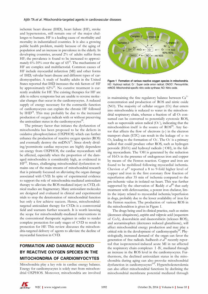
ischemic heart disease (IHD), heart failure (HF), stroke and hypertension, still remain one of the major chal-lenges to humans. HF is a leading cause of morbidity and mortality in industrialized countries. It is also a growing public health problem, mainly because of the aging of population and an increase in prevalence in the elderly. In developing countries, around 2% of adults suffer from HF; the prevalence is found to be increased to approxi-mately 6%-10% over the age of 65[1]. The mechanisms of HF are complex and multifactorial. Common causes of HF include myocardial infarction (MI) and other forms of IHD, valvular heart disease and different types of car-diomyopathies. A study of healthy adults in the United States reported that IHD increases the risk factors of HF by approximately 62%[2]. No curative treatment is cur-rently available for HF. The existing therapies for HF are able to relieve symptoms but are unable to reverse molec-ular changes that occur in the cardiomyocytes. A reduced supply of energy necessary for the contractile function of cardiomyocytes can explain the chronic HF followed by IHD[3]. This may probably be due to the increased production of oxygen radicals with or without preserving the antioxidant status in the cardiomyocytes[4].
The primary factor that initiates the dysfunction of mitochondria has been proposed to be the defects in oxidative phosphorylation (OXPHOS) which can further enhance the production of reactive oxygen species (ROS) and eventually destroy the mtDNA[5]. Since slowly divid-ing/postmitotic cardiac myocytes are highly dependent on energy from OXPHOS, the cardiac myocardium will be affected, especially when the proportion of the dam-aged mitochondria is considerably high, as evidenced in HF[3]. Hence, challenging mitochondrial dysfunction re-mains one of the main streams of mitochondrial research that is primarily focussed on alleviating the organ damage associated with CVD. In spite of experimental evidence to support the role of mitochondria-mediated antioxidant therapy to alleviate the ROS-mediated injury in CVD, cli-nical studies are fragmentary. Many antioxidant molecules are designed and evaluated in clinical and experimental trials to stop the deterioration of mitochondrial function but only a few achieve success. Hence, mitochondrial-targeted antioxidant therapy for CVDs is a controversial field and warrants further research. It is worth knowing the scope for mitochondrially-mediated interventions in the conventional therapeutic regimen in order to render complete protection for early stages of CVD to result in protection for HF. This review discusses the mitochon-dria-targeted delivery of agents to alleviate the decline of myocardial function in CVD.
FORMATION AND DAMAGE INDUCED BY REACTIVE OXYGEN SPECIES IN THE MITOCHONDRIA OF CARDIOMYOCYTESMitochondria play a key role in cardiac energy balance. Energy for cardiomyocytes is solely met from mitochon-drial OXPHOS. Moreover, mitochondria are involved
in maintaining the fine regulatory balance between Ca2+ concentration and production of ROS and nitric oxide (NO). The majority of cellular oxygen (O2) that enters into mitochondria is reduced to water in the mitochon-drial respiratory chain, whereas a fraction of all O2 con-sumed can be converted to potentially cytotoxic ROS, such as superoxide anion radical (O2
-.), indicating that the mitochondrion itself is the source of ROS[6]. Any fac-tor that affects the flow of electrons (e-) in the electron transport chain (ETC) can result in the leakage of e- to O2, leading to the formation of O2
-. The O2- is a primary
radical that could produce other ROS, such as hydrogen peroxide (H2O2) and hydroxyl radicals (·OH), in the fail-ing myocardium. The .OH is generated by the reduction of H2O2 in the presence of endogenous iron and copper by means of the Fenton reaction. Copper and iron are found to be mobilized following myocardial ischemia. Chevion et al[7] reported a 8 to 9-fold higher level of copper and iron in the first coronary flow fraction of reperfusion after 35 min of ischemia compared to the pre-ischemic value in isolated rat heart. This was further supported by the obervation of Reddy et al[8] that early treatment with deferoxamine, a potent iron chelator, lim-its the injury related to myocardial ischemia/reperfusion in dogs, probably due to the lesser availability of iron for the Fenton reaction. The production of various ROS in the mitochondrion is given in Figure 1.
The drugs being used in clinical practice, such as statins (decreases ubiquinone), aspirin and valproic acid (sequesters of CoA), doxorubicin and daunorubicin (releases ROS), and acetaminophen (decreases reduced glutathione), will affect mitochondrial energy production and may play a critical role in the development of cardiomyopathy[6]. Phy-siologically, increased demand of the organ can favor the generation of free radicals. Sudheesh et al[9] recently repor-ted that isoproterenol-induced acute MI in rat affected the respiratory chain complexes Ⅰ-Ⅳ, mediated through an increase in the ROS level in the cardiomyocytes. Fur-thermore, the declined antioxidant status in the mito-chondria during aging can also provoke mitochondrial dysfunction in cardiomyocytes[10]. Hypercholesterolemia can also affect mitochondrial functions by declining the mitochondrial membrane potential mediated through
October 26, 2014|Volume 6|Issue 10|WJC|www.wjgnet.com
Ajith TA et al . Mitochondria-targeted agents in cardiovascular diseases
1092
Complex Ⅱ Complex Ⅲ
Complex I
e- .OHFe2+
H2O2 O2-.
NO mt NOSONOO-
O2
90%-95%
4e-
H2O
Figure 1 Formation of various reactive oxygen species in mitochondria. HO.: Hydroxyl radical; O2
.-: Super oxide anion radical; ONOO-: Peroxynitrite; mtNOS: Mitochondrial-specific nitric oxide synthase; NO: Nitric oxide.
Complex Ⅳ
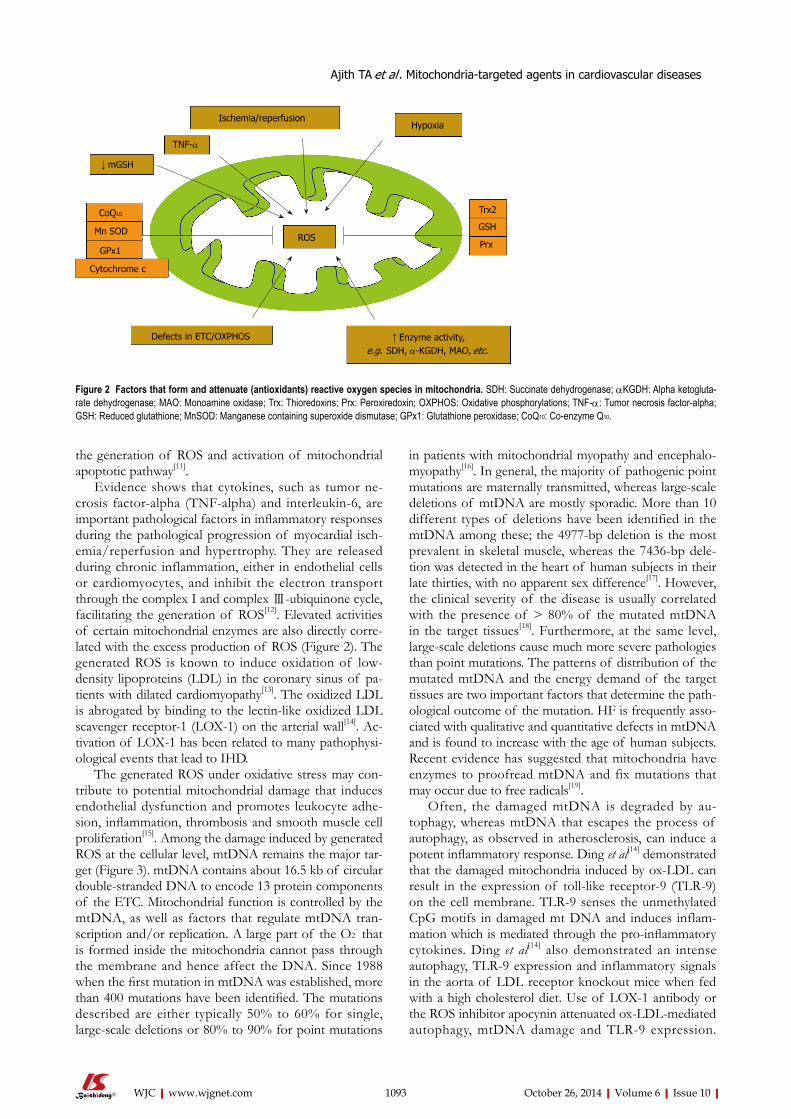
the generation of ROS and activation of mitochondrial apoptotic pathway[11].
Evidence shows that cytokines, such as tumor ne-crosis factor-alpha (TNF-alpha) and interleukin-6, are important pathological factors in inflammatory responses during the pathological progression of myocardial isch-emia/reperfusion and hypertrophy. They are released during chronic inflammation, either in endothelial cells or cardiomyocytes, and inhibit the electron transport through the complex I and complex Ⅲ-ubiquinone cycle, facilitating the generation of ROS[12]. Elevated activities of certain mitochondrial enzymes are also directly corre-lated with the excess production of ROS (Figure 2). The generated ROS is known to induce oxidation of low-density lipoproteins (LDL) in the coronary sinus of pa-tients with dilated cardiomyopathy[13]. The oxidized LDL is abrogated by binding to the lectin-like oxidized LDL scavenger receptor-1 (LOX-1) on the arterial wall[14]. Ac-tivation of LOX-1 has been related to many pathophysi-ological events that lead to IHD.
The generated ROS under oxidative stress may con-tribute to potential mitochondrial damage that induces endothelial dysfunction and promotes leukocyte adhe-sion, inflammation, thrombosis and smooth muscle cell proliferation[15]. Among the damage induced by generated ROS at the cellular level, mtDNA remains the major tar-get (Figure 3). mtDNA contains about 16.5 kb of circular double-stranded DNA to encode 13 protein components of the ETC. Mitochondrial function is controlled by the mtDNA, as well as factors that regulate mtDNA tran-scription and/or replication. A large part of the O2
- that is formed inside the mitochondria cannot pass through the membrane and hence affect the DNA. Since 1988 when the first mutation in mtDNA was established, more than 400 mutations have been identified. The mutations described are either typically 50% to 60% for single, large-scale deletions or 80% to 90% for point mutations
in patients with mitochondrial myopathy and encephalo-myopathy[16]. In general, the majority of pathogenic point mutations are maternally transmitted, whereas large-scale deletions of mtDNA are mostly sporadic. More than 10 different types of deletions have been identified in the mtDNA among these; the 4977-bp deletion is the most prevalent in skeletal muscle, whereas the 7436-bp dele-tion was detected in the heart of human subjects in their late thirties, with no apparent sex difference[17]. However, the clinical severity of the disease is usually correlated with the presence of > 80% of the mutated mtDNA in the target tissues[18]. Furthermore, at the same level, large-scale deletions cause much more severe pathologies than point mutations. The patterns of distribution of the mutated mtDNA and the energy demand of the target tissues are two important factors that determine the path-ological outcome of the mutation. HF is frequently asso-ciated with qualitative and quantitative defects in mtDNA and is found to increase with the age of human subjects. Recent evidence has suggested that mitochondria have enzymes to proofread mtDNA and fix mutations that may occur due to free radicals[19].
Often, the damaged mtDNA is degraded by au-tophagy, whereas mtDNA that escapes the process of autophagy, as observed in atherosclerosis, can induce a potent inflammatory response. Ding et al[14] demonstrated that the damaged mitochondria induced by ox-LDL can result in the expression of toll-like receptor-9 (TLR-9) on the cell membrane. TLR-9 senses the unmethylated CpG motifs in damaged mt DNA and induces inflam-mation which is mediated through the pro-inflammatory cytokines. Ding et al[14] also demonstrated an intense autophagy, TLR-9 expression and inflammatory signals in the aorta of LDL receptor knockout mice when fed with a high cholesterol diet. Use of LOX-1 antibody or the ROS inhibitor apocynin attenuated ox-LDL-mediated autophagy, mtDNA damage and TLR-9 expression.
October 26, 2014|Volume 6|Issue 10|WJC|www.wjgnet.com 1093
Ischemia/reperfusion
TNF-α
↓ mGSH
CoQ10
Mn SOD
GPx1
Cytochrome c
ROS
Defects in ETC/OXPHOS ↑ Enzyme activity,e.g. SDH, α-KGDH, MAO, etc.
Hypoxia
Trx2
GSH
Prx
Figure 2 Factors that form and attenuate (antioxidants) reactive oxygen species in mitochondria. SDH: Succinate dehydrogenase; αKGDH: Alpha ketogluta-rate dehydrogenase; MAO: Monoamine oxidase; Trx: Thioredoxins; Prx: Peroxiredoxin; OXPHOS: Oxidative phosphorylations; TNF-α: Tumor necrosis factor-alpha; GSH: Reduced glutathione; MnSOD: Manganese containing superoxide dismutase; GPx1: Glutathione peroxidase; CoQ10: Co-enzyme Q10.
Ajith TA et al . Mitochondria-targeted agents in cardiovascular diseases
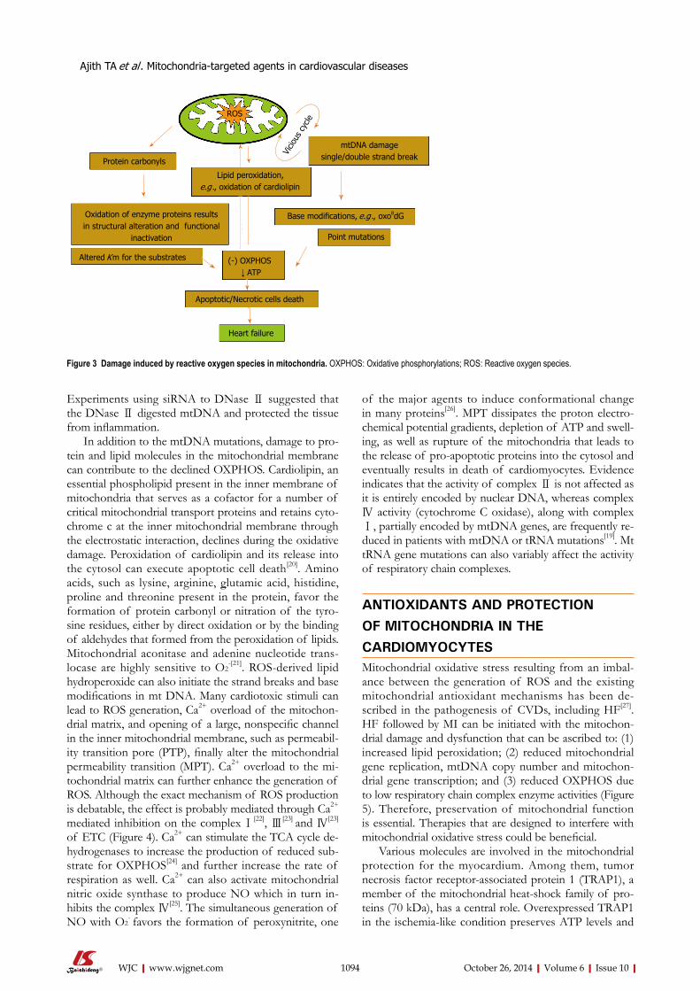
October 26, 2014|Volume 6|Issue 10|WJC|www.wjgnet.com
of the major agents to induce conformational change in many proteins[26]. MPT dissipates the proton electro-chemical potential gradients, depletion of ATP and swell-ing, as well as rupture of the mitochondria that leads to the release of pro-apoptotic proteins into the cytosol and eventually results in death of cardiomyocytes. Evidence indicates that the activity of complex Ⅱ is not affected as it is entirely encoded by nuclear DNA, whereas complex Ⅳ activity (cytochrome C oxidase), along with complex Ⅰ, partially encoded by mtDNA genes, are frequently re-duced in patients with mtDNA or tRNA mutations[19]. Mt tRNA gene mutations can also variably affect the activity of respiratory chain complexes.
ANTIOXIDANTS AND PROTECTION OF MITOCHONDRIA IN THE CARDIOMYOCYTESMitochondrial oxidative stress resulting from an imbal-ance between the generation of ROS and the existing mitochondrial antioxidant mechanisms has been de-scribed in the pathogenesis of CVDs, including HF[27]. HF followed by MI can be initiated with the mitochon-drial damage and dysfunction that can be ascribed to: (1) increased lipid peroxidation; (2) reduced mitochondrial gene replication, mtDNA copy number and mitochon-drial gene transcription; and (3) reduced OXPHOS due to low respiratory chain complex enzyme activities (Figure 5). Therefore, preservation of mitochondrial function is essential. Therapies that are designed to interfere with mitochondrial oxidative stress could be beneficial.
Various molecules are involved in the mitochondrial protection for the myocardium. Among them, tumor necrosis factor receptor-associated protein 1 (TRAP1), a member of the mitochondrial heat-shock family of pro-teins (70 kDa), has a central role. Overexpressed TRAP1 in the ischemia-like condition preserves ATP levels and
Experiments using siRNA to DNase Ⅱ suggested that the DNase Ⅱ digested mtDNA and protected the tissue from inflammation.
In addition to the mtDNA mutations, damage to pro-tein and lipid molecules in the mitochondrial membrane can contribute to the declined OXPHOS. Cardiolipin, an essential phospholipid present in the inner membrane of mitochondria that serves as a cofactor for a number of critical mitochondrial transport proteins and retains cyto-chrome c at the inner mitochondrial membrane through the electrostatic interaction, declines during the oxidative damage. Peroxidation of cardiolipin and its release into the cytosol can execute apoptotic cell death[20]. Amino acids, such as lysine, arginine, glutamic acid, histidine, proline and threonine present in the protein, favor the formation of protein carbonyl or nitration of the tyro-sine residues, either by direct oxidation or by the binding of aldehydes that formed from the peroxidation of lipids. Mitochondrial aconitase and adenine nucleotide trans-locase are highly sensitive to O2
-[21]. ROS-derived lipid hydroperoxide can also initiate the strand breaks and base modifications in mt DNA. Many cardiotoxic stimuli can lead to ROS generation, Ca2+ overload of the mitochon-drial matrix, and opening of a large, nonspecific channel in the inner mitochondrial membrane, such as permeabil-ity transition pore (PTP), finally alter the mitochondrial permeability transition (MPT). Ca2+ overload to the mi-tochondrial matrix can further enhance the generation of ROS. Although the exact mechanism of ROS production is debatable, the effect is probably mediated through Ca2+ mediated inhibition on the complex Ⅰ[22], Ⅲ[23] and Ⅳ[23] of ETC (Figure 4). Ca2+ can stimulate the TCA cycle de-hydrogenases to increase the production of reduced sub-strate for OXPHOS[24] and further increase the rate of respiration as well. Ca2+ can also activate mitochondrial nitric oxide synthase to produce NO which in turn in-hibits the complex Ⅳ[25]. The simultaneous generation of NO with O2
- favors the formation of peroxynitrite, one
1094
ROS
Protein carbonyls
Lipid peroxidation,e.g ., oxidation of cardiolipin
Vicio
us c
ycle
mtDNA damagesingle/double strand break
Base modifications, e.g ., oxo8dG
Point mutations
Oxidation of enzyme proteins results in structural alteration and functional
inactivation
Altered Km for the substrates (-) OXPHOS ↓ ATP
Apoptotic/Necrotic cells death
Heart failure
Figure 3 Damage induced by reactive oxygen species in mitochondria. OXPHOS: Oxidative phosphorylations; ROS: Reactive oxygen species.
Ajith TA et al . Mitochondria-targeted agents in cardiovascular diseases
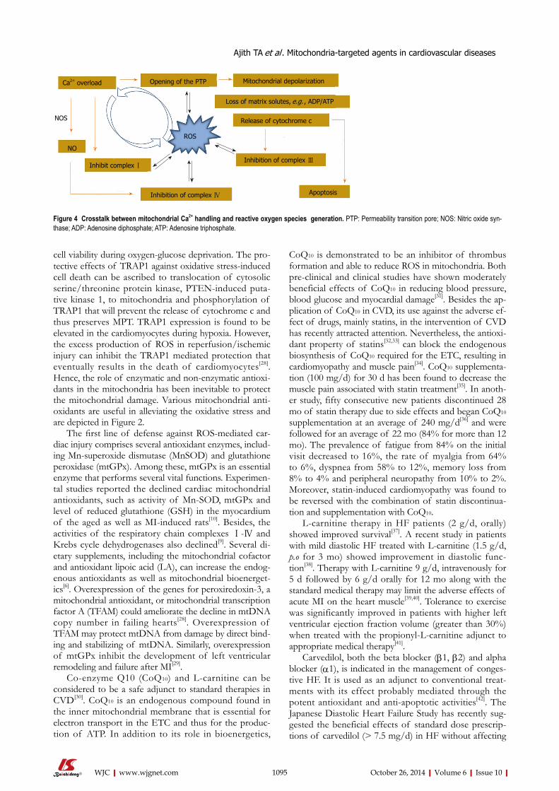
October 26, 2014|Volume 6|Issue 10|WJC|www.wjgnet.com
cell viability during oxygen-glucose deprivation. The pro-tective effects of TRAP1 against oxidative stress-induced cell death can be ascribed to translocation of cytosolic serine/threonine protein kinase, PTEN-induced puta-tive kinase 1, to mitochondria and phosphorylation of TRAP1 that will prevent the release of cytochrome c and thus preserves MPT. TRAP1 expression is found to be elevated in the cardiomyocytes during hypoxia. However, the excess production of ROS in reperfusion/ischemic injury can inhibit the TRAP1 mediated protection that eventually results in the death of cardiomyocytes[28]. Hence, the role of enzymatic and non-enzymatic antioxi-dants in the mitochondria has been inevitable to protect the mitochondrial damage. Various mitochondrial anti-oxidants are useful in alleviating the oxidative stress and are depicted in Figure 2.
The first line of defense against ROS-mediated car-diac injury comprises several antioxidant enzymes, includ-ing Mn-superoxide dismutase (MnSOD) and glutathione peroxidase (mtGPx). Among these, mtGPx is an essential enzyme that performs several vital functions. Experimen-tal studies reported the declined cardiac mitochondrial antioxidants, such as activity of Mn-SOD, mtGPx and level of reduced glutathione (GSH) in the myocardium of the aged as well as MI-induced rats[10]. Besides, the activities of the respiratory chain complexes Ⅰ-Ⅳ and Krebs cycle dehydrogenases also declined[9]. Several di-etary supplements, including the mitochondrial cofactor and antioxidant lipoic acid (LA), can increase the endog-enous antioxidants as well as mitochondrial bioenerget-ics[6]. Overexpression of the genes for peroxiredoxin-3, a mitochondrial antioxidant, or mitochondrial transcription factor A (TFAM) could ameliorate the decline in mtDNA copy number in failing hearts[28]. Overexpression of TFAM may protect mtDNA from damage by direct bind-ing and stabilizing of mtDNA. Similarly, overexpression of mtGPx inhibit the development of left ventricular remodeling and failure after MI[29].
Co-enzyme Q10 (CoQ10) and L-carnitine can be considered to be a safe adjunct to standard therapies in CVD[30]. CoQ10 is an endogenous compound found in the inner mitochondrial membrane that is essential for electron transport in the ETC and thus for the produc-tion of ATP. In addition to its role in bioenergetics,
CoQ10 is demonstrated to be an inhibitor of thrombus formation and able to reduce ROS in mitochondria. Both pre-clinical and clinical studies have shown moderately beneficial effects of CoQ10 in reducing blood pressure, blood glucose and myocardial damage[31]. Besides the ap-plication of CoQ10 in CVD, its use against the adverse ef-fect of drugs, mainly statins, in the intervention of CVD has recently attracted attention. Nevertheless, the antioxi-dant property of statins[32,33] can block the endogenous biosynthesis of CoQ10 required for the ETC, resulting in cardiomyopathy and muscle pain[34]. CoQ10 supplementa-tion (100 mg/d) for 30 d has been found to decrease the muscle pain associated with statin treatment[35]. In anoth-er study, fifty consecutive new patients discontinued 28 mo of statin therapy due to side effects and began CoQ10 supplementation at an average of 240 mg/d[36] and were followed for an average of 22 mo (84% for more than 12 mo). The prevalence of fatigue from 84% on the initial visit decreased to 16%, the rate of myalgia from 64% to 6%, dyspnea from 58% to 12%, memory loss from 8% to 4% and peripheral neuropathy from 10% to 2%. Moreover, statin-induced cardiomyopathy was found to be reversed with the combination of statin discontinua-tion and supplementation with CoQ10.
L-carnitine therapy in HF patients (2 g/d, orally) showed improved survival[37]. A recent study in patients with mild diastolic HF treated with L-carnitine (1.5 g/d, p.o for 3 mo) showed improvement in diastolic func-tion[38]. Therapy with L-carnitine 9 g/d, intravenously for 5 d followed by 6 g/d orally for 12 mo along with the standard medical therapy may limit the adverse effects of acute MI on the heart muscle[39,40]. Tolerance to exercise was significantly improved in patients with higher left ventricular ejection fraction volume (greater than 30%) when treated with the propionyl-L-carnitine adjunct to appropriate medical therapy[41].
Carvedilol, both the beta blocker (β1, β2) and alpha blocker (α1), is indicated in the management of conges-tive HF. It is used as an adjunct to conventional treat-ments with its effect probably mediated through the potent antioxidant and anti-apoptotic activities[42]. The Japanese Diastolic Heart Failure Study has recently sug-gested the beneficial effects of standard dose prescrip-tions of carvedilol (> 7.5 mg/d) in HF without affecting
1095
Ca2+ overload Opening of the PTP Mitochondrial depolarization
Loss of matrix solutes, e.g. , ADP/ATP
Release of cytochrome c
Inhibition of complex Ⅲ
Apoptosis Inhibition of complex Ⅳ
NOS
NO
Inhibit complex Ⅰ
Figure 4 Crosstalk between mitochondrial Ca2+ handling and reactive oxygen species generation. PTP: Permeability transition pore; NOS: Nitric oxide syn-thase; ADP: Adenosine diphosphate; ATP: Adenosine triphosphate.
ROS
Ajith TA et al . Mitochondria-targeted agents in cardiovascular diseases
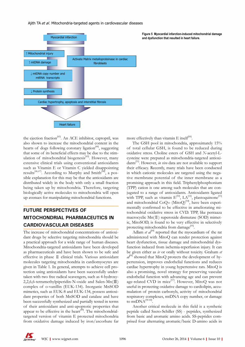
October 26, 2014|Volume 6|Issue 10|WJC|www.wjgnet.com
the ejection fraction[43]. An ACE inhibitor, captopril, was also shown to increase the mitochondrial content in the hearts of dogs following coronary ligation[44], suggesting that some of its beneficial effects may be due to the stim-ulation of mitochondrial biogenesis[45]. However, many extensive clinical trials using conventional antioxidants such as Vitamin E or Vitamin C yielded disappointing results[46,47]. According to Murphy and Smith[48], a pos-sible explanation for this may be that the antioxidants are distributed widely in the body with only a small fraction being taken up by mitochondria. Therefore, targeting biologically active molecules to mitochondria will open up avenues for manipulating mitochondrial functions.
FUTURE PERSPECTIVES OF MITOCHONDRIAL PHARMACEUTICS IN CARDIOVASCULAR DISEASESThe increase of mitochondrial concentrations of antioxi-dant drugs by selective targeting mitochondria should be a practical approach for a wide range of human diseases. Mitochondria-targeted antioxidants have been developed as pharmaceuticals and have been shown to be safe and effective in phase Ⅱ clinical trials. Various antioxidant molecules targeting mitochondria in cardiomyocytes are given in Table 1. In general, attempts to achieve cell pro-tection using antioxidants have been successfully under-taken with two free radical scavengers, such as 4-hydroxy-2,2,6,6-tetramethylpiperidin-N-oxide and Salen-Mn(Ⅲ) complex of o-vanillin (EUK-134). Inorganic MnSOD mimetics, such as EUK-8 and EUK-134, possess antioxi-dant properties of both MnSOD and catalase and have been successfully synthesized and partially tested in terms of their antioxidant and anti-apoptotic properties that appear to be effective in the heart[49]. The mitochondrial-targeted version of vitamin E protected mitochondria from oxidative damage induced by iron/ascorbate far
more effectively than vitamin E itself[50]. The GSH pool in mitochondria, approximately 15%
of total cellular GSH, is found to be reduced during oxidative stress. Choline esters of GSH and N-acetyl-L-cysteine were prepared as mitochondria-targeted antioxi-dants[51]. However, in vivo data are not available to support their efficacy. Recently, many trials have been conducted in which cationic molecules are targeted using the nega-tive membrane potential of the inner membrane as a promising approach in this field. Triphenylphosphonium (TPP) cation is one among such molecules that are con-jugated to a range of antioxidants. Antioxidants ligated with TPP, such as vitamin E[50], LA[52], plastoquinone[53]
and mitochondrial CoQ10 (MitoQ)[54], have been experi-mentally confirmed to be effective in ameliorating mi-tochondrial oxidative stress in CVD. TPP, like pentaaza macrocyclic Mn(Ⅱ) superoxide dismutase (SOD) mimet-ic, MitoSOD, is found to be very effective in selectively protecting mitochondria from damage[55].
Adlam et al[54] reported that the myocardium of the rat administered with MitoQ can render protection against heart dysfunction, tissue damage and mitochondrial dys-function induced from ischemia-reperfusion injury. It can be given either as iv or orally without toxicity. Graham et al[56] showed that MitoQ protects the development of hy-pertension, improves endothelial functions and reduces cardiac hypertrophy in young hypertensive rats. MitoQ is also a promising, novel strategy for preserving vascular endothelial function with advancing age and can prevent age-related CVD in mice[57]. However, MitoQ was not useful in protecting oxidative damage to cardiolipin, accu-mulation of protein carbonyls, activity of mitochondrial respiratory complexes, mtDNA copy number, or damage to mtDNA[58-60].
Another critical molecule in this field is a synthetic peptide called Szeto-Schiller (SS) - peptides, synthesized from basic and aromatic amino acids. SS-peptides com-prised four alternating aromatic/basic D-amino acids in
1096
Myocardial infarction
↑ Mitochondrial injury
Activate Matrix metalloproteinase in cardiac fibroblasts ↑ mtDNA damage
↓ mtDNA copy number and mtRNA transcripts
↓ Protein synthesis
Cardiac hypertrophy, apoptosis and interstitial fibrosis
Heart failure
Figure 5 Myocardial infarction-induced mitochondrial damage and dysfunction that resulted in heart failure.
Ajith TA et al . Mitochondria-targeted agents in cardiovascular diseases
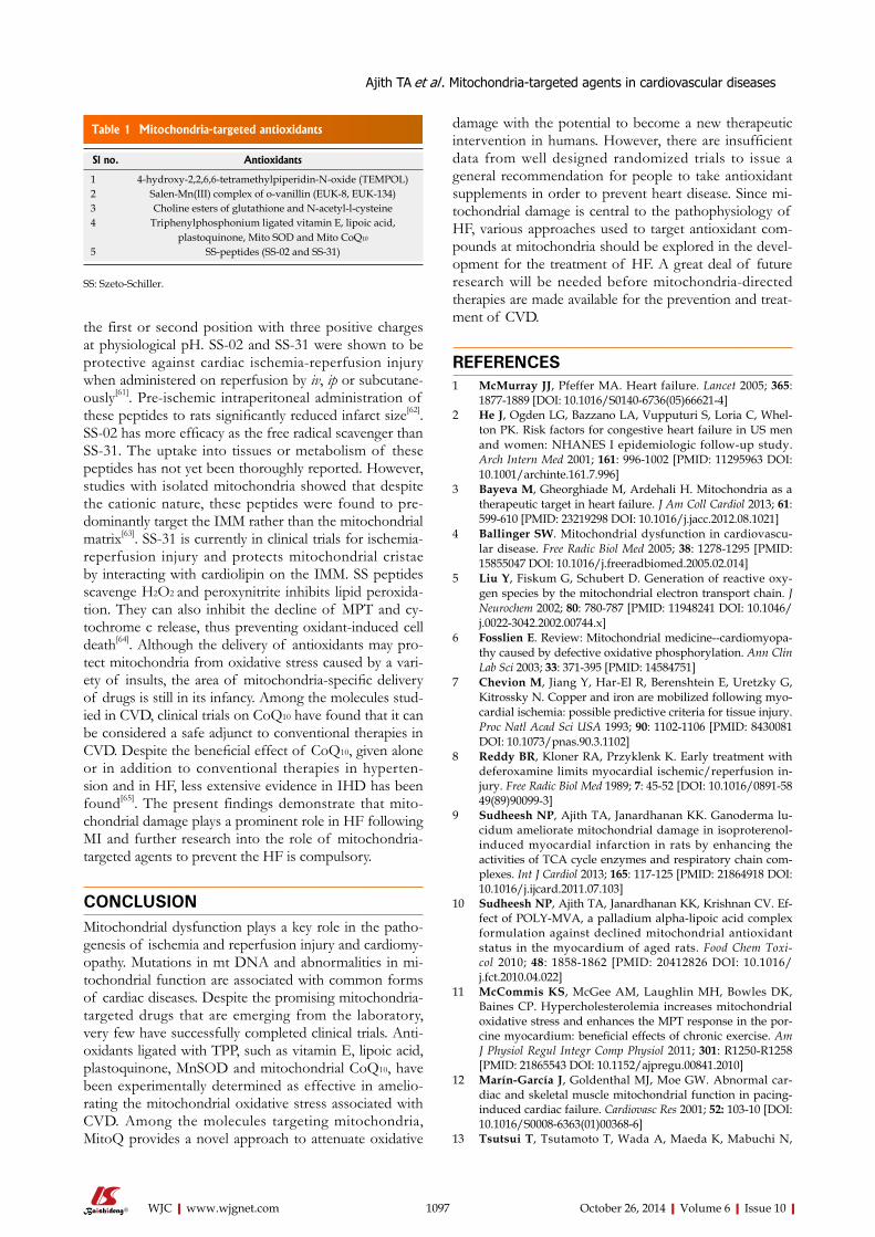
October 26, 2014|Volume 6|Issue 10|WJC|www.wjgnet.com
the first or second position with three positive charges at physiological pH. SS-02 and SS-31 were shown to be protective against cardiac ischemia-reperfusion injury when administered on reperfusion by iv, ip or subcutane-ously[61]. Pre-ischemic intraperitoneal administration of these peptides to rats significantly reduced infarct size[62]. SS-02 has more efficacy as the free radical scavenger than SS-31. The uptake into tissues or metabolism of these peptides has not yet been thoroughly reported. However, studies with isolated mitochondria showed that despite the cationic nature, these peptides were found to pre-dominantly target the IMM rather than the mitochondrial matrix[63]. SS-31 is currently in clinical trials for ischemia-reperfusion injury and protects mitochondrial cristae by interacting with cardiolipin on the IMM. SS peptides scavenge H2O2 and peroxynitrite inhibits lipid peroxida-tion. They can also inhibit the decline of MPT and cy-tochrome c release, thus preventing oxidant-induced cell death[64]. Although the delivery of antioxidants may pro-tect mitochondria from oxidative stress caused by a vari-ety of insults, the area of mitochondria-specific delivery of drugs is still in its infancy. Among the molecules stud-ied in CVD, clinical trials on CoQ10 have found that it can be considered a safe adjunct to conventional therapies in CVD. Despite the beneficial effect of CoQ10, given alone or in addition to conventional therapies in hyperten-sion and in HF, less extensive evidence in IHD has been found[65]. The present findings demonstrate that mito-chondrial damage plays a prominent role in HF following MI and further research into the role of mitochondria-targeted agents to prevent the HF is compulsory.
CONCLUSIONMitochondrial dysfunction plays a key role in the patho-genesis of ischemia and reperfusion injury and cardiomy-opathy. Mutations in mt DNA and abnormalities in mi-tochondrial function are associated with common forms of cardiac diseases. Despite the promising mitochondria-targeted drugs that are emerging from the laboratory, very few have successfully completed clinical trials. Anti-oxidants ligated with TPP, such as vitamin E, lipoic acid, plastoquinone, MnSOD and mitochondrial CoQ10, have been experimentally determined as effective in amelio-rating the mitochondrial oxidative stress associated with CVD. Among the molecules targeting mitochondria, MitoQ provides a novel approach to attenuate oxidative
damage with the potential to become a new therapeutic intervention in humans. However, there are insufficient data from well designed randomized trials to issue a general recommendation for people to take antioxidant supplements in order to prevent heart disease. Since mi-tochondrial damage is central to the pathophysiology of HF, various approaches used to target antioxidant com-pounds at mitochondria should be explored in the devel-opment for the treatment of HF. A great deal of future research will be needed before mitochondria-directed therapies are made available for the prevention and treat-ment of CVD.
REFERENCES1 McMurray JJ, Pfeffer MA. Heart failure. Lancet 2005; 365:
1877-1889 [DOI: 10.1016/S0140-6736(05)66621-4]2 He J, Ogden LG, Bazzano LA, Vupputuri S, Loria C, Whel-
ton PK. Risk factors for congestive heart failure in US men and women: NHANES I epidemiologic follow-up study. Arch Intern Med 2001; 161: 996-1002 [PMID: 11295963 DOI: 10.1001/archinte.161.7.996]
3 Bayeva M, Gheorghiade M, Ardehali H. Mitochondria as a therapeutic target in heart failure. J Am Coll Cardiol 2013; 61: 599-610 [PMID: 23219298 DOI: 10.1016/j.jacc.2012.08.1021]
4 Ballinger SW. Mitochondrial dysfunction in cardiovascu-lar disease. Free Radic Biol Med 2005; 38: 1278-1295 [PMID: 15855047 DOI: 10.1016/j.freeradbiomed.2005.02.014]
5 Liu Y, Fiskum G, Schubert D. Generation of reactive oxy-gen species by the mitochondrial electron transport chain. J Neurochem 2002; 80: 780-787 [PMID: 11948241 DOI: 10.1046/j.0022-3042.2002.00744.x]
6 Fosslien E. Review: Mitochondrial medicine--cardiomyopa-thy caused by defective oxidative phosphorylation. Ann Clin Lab Sci 2003; 33: 371-395 [PMID: 14584751]
7 Chevion M, Jiang Y, Har-El R, Berenshtein E, Uretzky G, Kitrossky N. Copper and iron are mobilized following myo-cardial ischemia: possible predictive criteria for tissue injury. Proc Natl Acad Sci USA 1993; 90: 1102-1106 [PMID: 8430081 DOI: 10.1073/pnas.90.3.1102]
8 Reddy BR, Kloner RA, Przyklenk K. Early treatment with deferoxamine limits myocardial ischemic/reperfusion in-jury. Free Radic Biol Med 1989; 7: 45-52 [DOI: 10.1016/0891-5849(89)90099-3]
9 Sudheesh NP, Ajith TA, Janardhanan KK. Ganoderma lu-cidum ameliorate mitochondrial damage in isoproterenol-induced myocardial infarction in rats by enhancing the activities of TCA cycle enzymes and respiratory chain com-plexes. Int J Cardiol 2013; 165: 117-125 [PMID: 21864918 DOI: 10.1016/j.ijcard.2011.07.103]
10 Sudheesh NP, Ajith TA, Janardhanan KK, Krishnan CV. Ef-fect of POLY-MVA, a palladium alpha-lipoic acid complex formulation against declined mitochondrial antioxidant status in the myocardium of aged rats. Food Chem Toxi-col 2010; 48: 1858-1862 [PMID: 20412826 DOI: 10.1016/j.fct.2010.04.022]
11 McCommis KS, McGee AM, Laughlin MH, Bowles DK, Baines CP. Hypercholesterolemia increases mitochondrial oxidative stress and enhances the MPT response in the por-cine myocardium: beneficial effects of chronic exercise. Am J Physiol Regul Integr Comp Physiol 2011; 301: R1250-R1258 [PMID: 21865543 DOI: 10.1152/ajpregu.00841.2010]
12 Marín-García J, Goldenthal MJ, Moe GW. Abnormal car-diac and skeletal muscle mitochondrial function in pacing-induced cardiac failure. Cardiovasc Res 2001; 52: 103-10 [DOI: 10.1016/S0008-6363(01)00368-6]
13 Tsutsui T, Tsutamoto T, Wada A, Maeda K, Mabuchi N,
1097
Sl no. Antioxidants
1 4-hydroxy-2,2,6,6-tetramethylpiperidin-N-oxide (TEMPOL) 2 Salen-Mn(III) complex of o-vanillin (EUK-8, EUK-134) 3 Choline esters of glutathione and N-acetyl-l-cysteine 4 Triphenylphosphonium ligated vitamin E, lipoic acid,
plastoquinone, Mito SOD and Mito CoQ10
5 SS-peptides (SS-02 and SS-31)
Table 1 Mitochondria-targeted antioxidants
SS: Szeto-Schiller.
Ajith TA et al . Mitochondria-targeted agents in cardiovascular diseases
October 26, 2014|Volume 6|Issue 10|WJC|www.wjgnet.com
Hayashi M, Ohnishi M, Kinoshita M. Plasma oxidized low-density lipoprotein as a prognostic predictor in patients with chronic congestive heart failure. J Am Coll Cardiol 2002; 39: 957-62 [DOI: 10.1016/S0735-1097(02)01721-7]
14 Ding Z, Liu S, Wang X, Khaidakov M, Dai Y, Mehta JL. Oxi-dant stress in mitochondrial DNA damage, autophagy and inflammation in atherosclerosis. Sci Rep 2013; 3: 1077 [PMID: 23326634 DOI: 10.1038/srep01077]
15 Davidson SM. Endothelial mitochondria and heart disease. Cardiovasc Res 2010; 88: 58-66 [PMID: 20558442 DOI: 10.1093/cvr/cvq195]
16 Holt IJ, Harding AE, Morgan-Hughes JA. Deletions of muscle mitochondrial DNA in patients with mitochondrial myopathies. Nature 1988; 331: 717-719 [PMID: 2830540 DOI: 10.1038/331717a0]
17 Wei YH. Mitochondrial DNA alterations as ageing-asso-ciated molecular events. Mutat Res 1992; 275: 145-55 [DOI: 10.1016/0921-8734(92)90019-L]
18 Wei YH. Mitochondrial DNA mutations and oxidative dam-age in aging and diseases: an emerging paradigm of geron-tology and medicine. Proc Natl Sci Counc Repub China B 1998; 22: 55-67 [PMID: 9615468]
19 Arbustini E, Diegoli M, Fasani R, Grasso M, Morbini P, Banchieri N, Bellini O, Dal Bello B, Pilotto A, Magrini G, Campana C, Fortina P, Gavazzi A, Narula J, Viganò M. Mito-chondrial DNA mutations and mitochondrial abnormalities in dilated cardiomyopathy. Am J Pathol 1998; 153: 1501-10 [DOI: 10.1016/S0002-9440(10)65738-0]
20 Ott M, Robertson JD, Gogvadze V, Zhivotovsky B, Orrenius S. Cytochrome c release from mitochondria proceeds by a two-step process. Proc Natl Acad Sci USA 2002; 99: 1259-1263 [PMID: 11818574 DOI: 10.1073/pnas.241655498]
21 Vasquez-Vivar J, Kalyanaraman B, Kennedy MC. Mito-chondrial aconitase is a source of hydroxyl radical. An electron spin resonance investigation. J Biol Chem 2000; 275: 14064-14069 [PMID: 10799480]
22 Batandier C, Leverve X, Fontaine E. Opening of the mi-tochondrial permeability transition pore induces reactive oxygen species production at the level of the respiratory chain complex I. J Biol Chem 2004; 279: 17197-17204 [PMID: 14963044 DOI: 10.1074/jbc.M310329200]
23 Grijalba MT, Vercesi AE, Schreier S. Ca2 -induced increased lipid packing and domain formation in submitochondrial particles. A possible early step in the mechanism of Ca2 -stimulated generation of reactive oxygen species by the respiratory chain. Biochemistry 1999; 38: 13279-13287 [DOI: 10.1021/bi9828674]
24 Wan B, LaNoue KF, Cheung JY, Scaduto RC. Regula-tion of citric acid cycle by calcium. J Biol Chem 1989; 264: 13430-13439 [PMID: 2503501]
25 Brookes P, Darley-Usmar VM. Hypothesis: the mitochon-drial NO () signaling pathway, and the transduction of ni-trosative to oxidative cell signals: an alternative function for cytochrome C oxidase. Free Radic Biol Med 2002; 32: 370–74 [DOI: 10.1016/S0891-5849(01)00805-X]
26 Brookes PS, Darley-Usmar VM. Role of calcium and super-oxide dismutase in sensitizing mitochondria to peroxyni-trite-induced permeability transition. Am J Physiol Heart Circ Physiol 2004; 286: H39-H46 [PMID: 12933349 DOI: 10.1152/ajpheart.00742.2003]
27 Subramanian S, Kalyanaraman B, Migrino RQ. Mitochon-drially targeted antioxidants for the treatment of cardio-vascular diseases. Recent Pat Cardiovasc Drug Discov 2010; 5: 54-65 [DOI: 10.2174/157489010790192601]
28 Xiang F, Huang YS, Shi XH, Zhang Q. Mitochondrial chap-erone tumour necrosis factor receptor-associated protein 1 protects cardiomyocytes from hypoxic injury by regulat-ing mitochondrial permeability transition pore opening. FEBS J 2010; 277: 1929-1938 [PMID: 20236315 DOI: 10.1111/j.1742-4658.2010.07615.x]
29 Tsutsui H, Kinugawa S, Matsushima S. Mitochondrial oxidative stress and dysfunction in myocardial remodel-ling. Cardiovasc Res 2009; 81: 449-456 [PMID: 18854381 DOI: 10.1093/cvr/cvn280]
30 Hagen TM, Moreau R, Suh JH, Visioli F. Mitochondrial de-cay in the aging rat heart: evidence for improvement by di-etary supplementation with acetyl-L-carnitine and/or lipoic acid. Ann N Y Acad Sci 2002; 959: 491-507 [PMID: 11976222 DOI: 10.1111/j.1749-6632.2002.tb02119.x]
31 Garrido-Maraver J, Cordero MD, Oropesa-Avila M, Vega AF, de la Mata M, Pavon AD, Alcocer-Gomez E, Calero CP, Paz MV, Alanis M, de Lavera I, Cotan D, Sanchez-Alcazar JA. Clinical applications of coenzyme Q10. Front Biosci 2014; 19: 619-633 [DOI: 10.2741/4231]
32 Ajith TA, Divya KR. An in vitro comparative study on the antibacterial and antioxidant activities of atorvastatin and simvastatin. Pharmaceutical Biol 2007; 45: 1-5 [DOI: 10.1080/13880200701574992]
33 Ajith TA, Riji T, Anu V. In vitro antioxidant and DNA protective effects of a novel HMG.CoA reductase inhibitor, rosuvastatin. Clin Exp Physiol Pharmacol 2008; 35: 625-29 [DOI: 10.1111/j.1440-1681.2007.04853.x]
34 Littarru GP, Langsjoen P. Coenzyme Q10 and statins: bio-chemical and clinical implications. Mitochondrion 2007; 7 Suppl: S168-S174 [PMID: 17482884 DOI: 10.1016/j.mito.2007.03.002]
35 Caso G, Kelly P, McNurlan MA, Lawson WE. Effect of coen-zyme q10 on myopathic symptoms in patients treated with statins. Am J Cardiol 2007; 99: 1409-1412 [PMID: 17493470 DOI: 10.1016/j.amjcard.2006.12.063]
36 Langsjoen PH, Langsjoen JO, Langsjoen AM, Lucas LA. Treatment of statin adverse effects with supplemental Coen-zyme Q10 and statin drug discontinuation. Biofactors 2005; 25: 147-152 [DOI: 10.1002/biof.5520250]
37 Rizos I. Three-year survival of patients with heart failure caused by dilated cardiomyopathy and L-carnitine admin-istration. Am Heart J 2000; 139: S120-S123 [PMID: 10650325 DOI: 10.1067/mhj.2000.103917]
38 Serati AR, Motamedi MR, Emami S, Varedi P, Movahed MR. L-carnitine treatment in patients with mild diastolic heart failure is associated with improvement in diastolic function and symptoms. Cardiology 2010; 116: 178-182 [PMID: 20639632 DOI: 10.1159/000318810]
39 Colonna P, Iliceto S. Myocardial infarction and left ven-tricular remodeling: results of the CEDIM trial. Carnitine Ecocardiografia Digitalizzata Infarto Miocardico. Am Heart J 2000; 139: S124-S130 [PMID: 10650326 DOI: 10.1067/mhj.2000.103918]
40 Tarantini G, Scrutinio D, Bruzzi P, Boni L, Rizzon P, Iliceto S. Metabolic treatment with L-Carnitine in acute anterior ST segment elevation myocardial infarction. A randomized con-trolled trial. Cardiology 2006; 106: 215-223 [PMID: 16685128 DOI: 10.1159/000093131]
41 Study on propionyl-L-carnitine in chronic heart failure. Eur Heart J 1999; 20: 70-76 [DOI: 10.1053/euhj.1998.1271]
42 Cheng J, Kamiya K, Kodama I. Carvedilol: molecular and cellular basis for its multifaceted therapeutic potential. Car-diovasc Drug Rev 2001; 19: 152-171 [PMID: 11484068]
43 Yamamoto K, Origasa H, Suzuki Y, Takahashi T, Shinozaki T, Watanabe T, Sakata Y, Izumi C, Taira K, Hori M. Relation of risk factors with response to carvedilol in heart failure with preserved ejection fraction - A report from the Japa-nese Diastolic Heart Failure Study (J-DHF). J Cardiol 2013; S0914-5087: 326-332
44 Yanagishita T, Tomita M, Itoh S, Mukae S, Arata H, Ishioka H, Geshi E, Konno N, Katagiri T. Protective effect of cap-topril on ischemic myocardium. Jpn Circ J 1997; 61: 161-169 [DOI: 10.1253/jcj.61.161]
45 Sanbe A, Tanonaka K, Kobayasi R, Takeo S. Effects of long-term therapy with ACE inhibitors, captopril, enala-pril and trandolapril, on myocardial energy metabolism
1098
Ajith TA et al . Mitochondria-targeted agents in cardiovascular diseases
October 26, 2014|Volume 6|Issue 10|WJC|www.wjgnet.com
in rats with heart failure following myocardial infarc-tion. J Mol Cell Cardiol 1995; 27: 2209–2222 [DOI: 10.1016/S0022-2828(95)91551-6]
46 Bjelakovic G, Nikolova D, Gluud LL, Simonetti RG, Gluud C. Antioxidant supplements for prevention of mortality in healthy participants and patients with various diseases. Cochrane Database Syst Rev 2008; (2): CD007176 [PMID: 18425980]
47 Cochemé HM, Murphy MP. Can antioxidants be effective therapeutics? Curr Opin Investig Drugs 2010; 11: 426-431 [PMID: 20336590]
48 Murphy MP, Smith RA. Targeting antioxidants to mitochon-dria by conjugation to lipophilic cations. Annu Rev Pharmacol Toxicol 2007; 47: 629-656 [PMID: 17014364 DOI: 10.1146/an-nurev.pharmtox.47.120505.105110]
49 Pucheu S, Boucher F, Sulpice T, Tresallet N, Bonhomme Y, Malfroy B, de Leiris J. EUK-8 a synthetic catalytic scavenger of reactive oxygen species protects isolated iron-overloaded rat heart from functional and structural damage induced by ischemia/reperfusion. Cardiovasc Drugs Ther 1996; 10: 331-339 [PMID: 8877076]
50 Smith RA, Porteous CM, Coulter CV, Murphy MP. Selec-tive targeting of an antioxidant to mitochondria. Eur J Biochem 1999; 263: 709-716 [PMID: 10469134 DOI: 10.1046/j.1432-1327.1999.00543.x]
51 Sheu SS, Nauduri D, Anders MW. Targeting antioxidants to mitochondria: a new therapeutic direction. Biochim Biophys Acta 2006; 1762: 256-265 [PMID: 16352423 DOI: 10.1016/j.bbadis.2005.10.007]
52 Brown SE, Ross MF, Sanjuan-Pla A, Manas AR, Smith RA, Murphy MP. Targeting lipoic acid to mitochondria: synthesis and characterization of a triphenylphosphonium-conjugated alpha-lipoyl derivative. Free Radic Biol Med 2007; 42: 1766-1780 [PMID: 17512456 DOI: 10.1016/j.freeradbiomed.2007.02.033]
53 Skulachev VP, Anisimov VN, Antonenko YN, Bakeeva LE, Chernyak BV, Erichev VP, Filenko OF, Kalinina NI, Kapelko VI, Kolosova NG, Kopnin BP, Korshunova GA, Lichinitser MR, Obukhova LA, Pasyukova EG, Pisarenko OI, Roginsky VA, Ruuge EK, Senin II, Severina II, Skulachev MV, Spivak IM, Tashlitsky VN, Tkachuk VA, Vyssokikh MY, Yaguzhin-sky LS, Zorov DB. An attempt to prevent senescence: a mito-chondrial approach. Biochim Biophys Acta 2009; 1787: 437-461 [PMID: 19159610 DOI: 10.1016/j.bbabio.2008.12.008]
54 Adlam VJ, Harrison JC, Porteous CM, James AM, Smith RA, Murphy MP, Sammut IA. Targeting an antioxidant to mitochondria decreases cardiac ischemia-reperfusion injury. FASEB J 2005; 19: 1088-1095 [PMID: 15985532]
55 Kelso GF, Maroz A, Cochemé HM, Logan A, Prime TA, Pe-skin AV, Winterbourn CC, James AM, Ross MF, Brooker S,
Porteous CM, Anderson RF, Murphy MP, Smith RA. A mito-chondria-targeted macrocyclic Mn(II) superoxide dismutase mimetic. Chem Biol 2012; 19: 1237-1246 [PMID: 23102218 DOI: 10.1016/j.chembiol.2012.08.005]
56 Graham D, Huynh NN, Hamilton CA, Beattie E, Smith RA, Cochemé HM, Murphy MP, Dominiczak AF. Mitochondria-targeted antioxidant MitoQ10 improves endothelial function and attenuates cardiac hypertrophy. Hypertension 2009; 54: 322-328 [PMID: 19581509]
57 Gioscia-Ryan RA, Larocca TJ, Sindler AL, Zigler MC, Mur-phy MP, Seals DR. Mitochondria-targeted antioxidant (Mi-toQ) ameliorates age-related arterial endothelial dysfunction in mice. J Physiol 2014; (Accepted Article) [DOI: 10.1113/jphysiol.2013.268680]
58 Paradies G, Petrosillo G, Paradies V, Ruggiero FM. Role of cardiolipin peroxidation and Ca2+ in mitochondrial dys-function and disease. Cell Calcium 2009; 45: 643-650 [PMID: 19368971 DOI: 10.1016/j.ceca.2009.03.012]
59 Davies SM, Poljak A, Duncan MW, Smythe GA, Murphy MP. Measurements of protein carbonyls, ortho- and meta-tyrosine and oxidative phosphorylation complex activity in mitochondria from young and old rats. Free Radic Biol Med 2001; 31: 181-190 [DOI: 10.1016/S0891-5849(01)00576-7]
60 Santos JH, Meyer JN, Mandavilli BS, Van Houten B. Quantitative PCR-based measurement of nuclear and mi-tochondrial DNA damage and repair in mammalian cells. Methods Mol Biol 2006; 314: 183-199 [PMID: 16673882 DOI: 10.1385/1-59259-973-7]
61 Szeto HH. Mitochondria-targeted cytoprotective peptides for ischemia-reperfusion injury. Antioxid Redox Signal 2008; 10: 601-619 [PMID: 17999629 DOI: 10.1089/ars.2007.1892]
62 Cho J, Won K, Wu D, Soong Y, Liu S, Szeto HH, Hong MK. Potent mitochondria-targeted peptides reduce myocardial infarction in rats. Coron Artery Dis 2007; 18: 215-220 [PMID: 17429296 DOI: 10.1097/01.mca.0000236285.71683.b6]
63 Zhao K, Zhao G-M, Wu D, Soong Y, Birk AV, Schiller PW, Szeto HH. Cell-permeable Peptide Antioxidants Targeted to Inner Mitochondrial Membrane inhibit Mitochondrial Swell-ing, Oxidative Cell Death, and Reperfusion Injury. J Biol Chem 2004; 279: 34682-34690 [PMID: 15178689 DOI: 10.1074/jbc.M402999200]
64 Rocha M, Hernandez-Mijares A, Garcia-Malpartida K, Bañuls C, Bellod L, Victor VM. Mitochondria-targeted an-tioxidant peptides. Curr Pharm Des 2010; 16: 3124-31 [DOI: 10.2174/138161210793292519]
65 Pepe S, Marasco SF, Haas SJ, Sheeran FL, Krum H, Rosen-feldt FL. Coenzyme Q10 in cardiovascular disease. Mito-chondrion 2007; 7 Suppl: S154-S167 [PMID: 17485243 DOI: 10.1016/j.mito.2007.02.005]
P- Reviewer: Alexeyev M, Fujiwara N, Julie NL, Song GB, Zeng LF S- Editor: Song XX L- Editor: Roemmele A
E- Editor: Wu HL
1099
Ajith TA et al . Mitochondria-targeted agents in cardiovascular diseases
Perioperative clinical variables and long-term survival following vascular surgery
Santiago Garcia, Edward O McFalls
Santiago Garcia, Minneapolis VA Healthcare System, Univer-sity of Minnesota, MN 55417, United StatesEdward O McFalls, Division of Cardiology (111C), VA Medical Center, Minneapolis VA Healthcare System, Professor of Medi-cine, University of Minnesota, MN 55417, United StatesAuthor contributions: Garcia S and McFalls EO jointly wrote this manuscript; McFalls EO was the Principal Investigator of the Coronary Artery Revascularization Prophylaxis trial. Supported by A career development award from the VA Office of Research and Development, No. 1IK2CX000699-01Correspondence to: Edward O McFalls, MD, PhD, Division of Cardiology (111C), VA Medical Center, Minneapolis VA Health-care System, Professor of Medicine, University of Minnesota, 1 Veterans Drive, Minneapolis, MN 55417, United States. [email protected]: +1-612-4673664 Fax: +1-612-7275668Received: December 21, 2013 Revised: March 6, 2014 Accepted: September 16, 2014 Published online: October 26, 2014
AbstractCardiovascular disease is the leading cause of death in patients with peripheral arterial disease (PAD). Coro-nary artery disease (CAD) is highly prevalent, and often times coexist, in patients with PAD. The management of patients with PAD that requires a high-risk vascular surgical procedure for intermittent claudication, critical limb ischemia or expanding abdominal aortic aneurysm requires risk stratification with the revised cardiac risk index, optimization of medical therapies, and limited use of cardiac imaging prior to surgery. Preventive re-vascularization in patients with stable CAD, with the sole intention to mitigate the risk of cardiac complica-tions in the peri-operative period, is not effective and may be associated with significant bleeding and throm-botic risks, in particular if stents are used. A strategy of universal use of cardiac troponins in the perioperative period for active surveillance of myocardial ischemia may be more reasonable and cost-effective than the current standard of care of widespread use of cardiac imaging prior to high-risk surgery. An elevated cardiac
troponin after vascular surgery is predictive of long-term mortality risk. Medical therapies such as aspirin and statins are recommended for patients with post-operative myocardial ischemia. Ongoing trials are as-sessing the role of novel anticoagulants. Additional research is needed to define the role of cardiac imaging and invasive angiography in this population.
© 2014 Baishideng Publishing Group Inc. All rights reserved.
Key words: Peripheral arterial disease; Myocardial in-farction; Coronary artery disease; Prognosis; Coronary revascularization
Core tip: Patients with advanced peripheral arterial disease who need vascular surgery have a high preva-lence of coronary atherosclerosis and are at increased risk of perioperative myocardial infarction. Coronary revascularization prior to the vascular operation is not an effective intervention to mitigate this risk. A strategy of widespread use of cardiac troponins in the periop-erative period is recommended to detect perioperative ischemic events associated with a long-term mortality risk. The selective use of medical interventions, cardiac imaging and coronary angiography in this population deserves further study.
Garcia S, McFalls EO. Perioperative clinical variables and long-term survival following vascular surgery. World J Cardiol 2014; 6(10): 1100-1107 Available from: URL: http://www.wjgnet.com/1949-8462/full/v6/i10/1100.htm DOI: http://dx.doi.org/10.4330/wjc.v6.i10.1100
INTRODUCTIONThe approach to patients with peripheral arterial disease (PAD) is best appreciated in the broader context of the epidemiology of the disease, risk factors, and surgical and endovascular interventions to improve symptoms, pre-
MINIREVIEWS
Submit a Manuscript: http://www.wjgnet.com/esps/Help Desk: http://www.wjgnet.com/esps/helpdesk.aspxDOI: 10.4330/wjc.v6.i10.1100
October 26, 2014|Volume 6|Issue 10|WJC|www.wjgnet.com
World Journal of CardiologyW J C
World J Cardiol 2014 October 26; 6(10): 1100-1107ISSN 1949-8462 (online)
© 2014 Baishideng Publishing Group Inc. All rights reserved.
1100
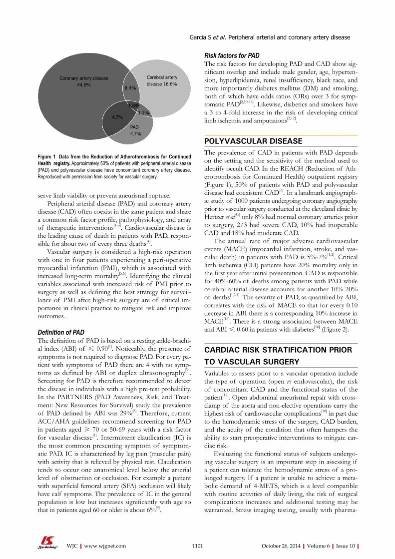
serve limb viability or prevent aneurismal rupture. Peripheral arterial disease (PAD) and coronary artery
disease (CAD) often coexist in the same patient and share a common risk factor profile, pathophysiology, and array of therapeutic interventions[1-3]. Cardiovascular disease is the leading cause of death in patients with PAD, respon-sible for about two of every three deaths[4].
Vascular surgery is considered a high-risk operation with one in four patients experiencing a peri-operative myocardial infarction (PMI), which is associated with increased long-term mortality[5,6]. Identifying the clinical variables associated with increased risk of PMI prior to surgery as well as defining the best strategy for surveil-lance of PMI after high-risk surgery are of critical im-portance in clinical practice to mitigate risk and improve outcomes.
Definition of PADThe definition of PAD is based on a resting ankle-brachi-al index (ABI) of ≤ 0.90[1]. Noticeably, the presence of symptoms is not required to diagnose PAD. For every pa-tient with symptoms of PAD there are 4 with no symp-toms as defined by ABI or duplex ultrasonography[7]. Screening for PAD is therefore recommended to detect the disease in individuals with a high pre-test probability. In the PARTNERS (PAD Awareness, Risk, and Treat-ment: New Resources for Survival) study the prevalence of PAD defined by ABI was 29%[8]. Therefore, current ACC/AHA guidelines recommend screening for PAD in patients aged ≥ 70 or 50-69 years with a risk factor for vascular disease[1]. Intermittent claudication (IC) is the most common presenting symptom of symptom-atic PAD. IC is characterized by leg pain (muscular pain) with activity that is relieved by physical rest. Claudication tends to occur one anatomical level below the arterial level of obstruction or occlusion. For example a patient with superficial femoral artery (SFA) occlusion will likely have calf symptoms. The prevalence of IC in the general population is low but increases significantly with age so that in patients aged 60 or older is about 6%[9].
Risk factors for PAD The risk factors for developing PAD and CAD show sig-nificant overlap and include male gender, age, hyperten-sion, hyperlipidemia, renal insufficiency, black race, and more importantly diabetes mellitus (DM) and smoking, both of which have odds ratios (ORs) over 3 for symp-tomatic PAD[2,10-14]. Likewise, diabetics and smokers have a 3 to 4-fold increase in the risk of developing critical limb ischemia and amputations[2,12].
POLYVASCULAR DISEASEThe prevalence of CAD in patients with PAD depends on the setting and the sensitivity of the method used to identify occult CAD. In the REACH (Reduction of Ath-erotrombosis for Continued Health) outpatient registry (Figure 1), 50% of patients with PAD and polyvascular disease had coexistent CAD[3]. In a landmark angiograph-ic study of 1000 patients undergoing coronary angiography prior to vascular surgery conducted at the cleveland clinic by Hertzer et al[15] only 8% had normal coronary arteries prior to surgery, 2/3 had severe CAD, 10% had inoperable CAD and 18% had moderate CAD.
The annual rate of major adverse cardiovascular events (MACE) (myocardial infarction, stroke, and vas-cular death) in patients with PAD is 5%-7%[1,2]. Critical limb ischemia (CLI) patients have 20% mortality only in the first year after initial presentation. CAD is responsible for 40%-60% of deaths among patients with PAD while cerebral arterial disease accounts for another 10%-20% of deaths[1,2,4]. The severity of PAD, as quantified by ABI, correlates with the risk of MACE so that for every 0.10 decrease in ABI there is a corresponding 10% increase in MACE[16]. There is a strong association between MACE and ABI ≤ 0.60 in patients with diabetes[16] (Figure 2).
CARDIAC RISK STRATIFICATION PRIOR TO VASCULAR SURGERYVariables to assess prior to a vascular operation include the type of operation (open vs endovascular), the risk of concomitant CAD and the functional status of the patient[17]. Open abdominal aneurismal repair with cross-clamp of the aorta and non-elective operations carry the highest risk of cardiovascular complications[18] in part due to the hemodynamic stress of the surgery, CAD burden, and the acuity of the condition that often hampers the ability to start preoperative interventions to mitigate car-diac risk.
Evaluating the functional status of subjects undergo-ing vascular surgery is an important step in assessing if a patient can tolerate the hemodynamic stress of a pro-longed surgery. If a patient is unable to achieve a meta-bolic demand of 4-METS, which is a level compatible with routine activities of daily living, the risk of surgical complications increases and additional testing may be warranted. Stress imaging testing, usually with pharma-
October 26, 2014|Volume 6|Issue 10|WJC|www.wjgnet.com
Garcia S et al . Peripheral arterial and coronary artery disease
1101
Figure 1 Data from the Reduction of Atherothrombosis for Continued Health registry. Approximately 50% of patients with peripheral arterial disease (PAD) and polyvascular disease have concomitant coronary artery disease. Reproduced with permission from society for vascular surgery.
Coronary artery disease44.6%
Cerebral arterydisease 16.6%
8.4%
1.6%1.2%
4.7%
PAD4.7%
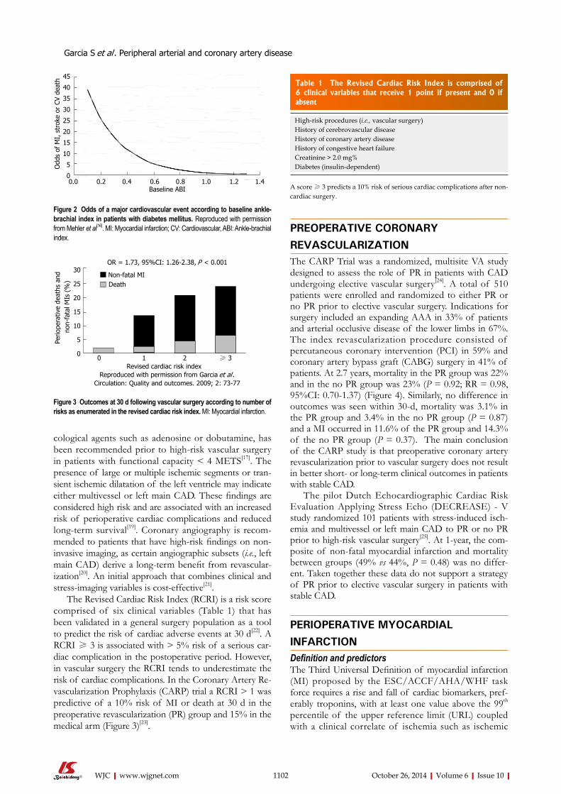
cological agents such as adenosine or dobutamine, has been recommended prior to high-risk vascular surgery in patients with functional capacity < 4 METS[17]. The presence of large or multiple ischemic segments or tran-sient ischemic dilatation of the left ventricle may indicate either multivessel or left main CAD. These findings are considered high risk and are associated with an increased risk of perioperative cardiac complications and reduced long-term survival[19]. Coronary angiography is recom-mended to patients that have high-risk findings on non-invasive imaging, as certain angiographic subsets (i.e., left main CAD) derive a long-term benefit from revascular-ization[20]. An initial approach that combines clinical and stress-imaging variables is cost-effective[21].
The Revised Cardiac Risk Index (RCRI) is a risk score comprised of six clinical variables (Table 1) that has been validated in a general surgery population as a tool to predict the risk of cardiac adverse events at 30 d[22]. A RCRI ≥ 3 is associated with > 5% risk of a serious car-diac complication in the postoperative period. However, in vascular surgery the RCRI tends to underestimate the risk of cardiac complications. In the Coronary Artery Re-vascularization Prophylaxis (CARP) trial a RCRI > 1 was predictive of a 10% risk of MI or death at 30 d in the preoperative revascularization (PR) group and 15% in the medical arm (Figure 3)[23].
PREOPERATIVE CORONARY REVASCULARIZATION The CARP Trial was a randomized, multisite VA study designed to assess the role of PR in patients with CAD undergoing elective vascular surgery[24]. A total of 510 patients were enrolled and randomized to either PR or no PR prior to elective vascular surgery. Indications for surgery included an expanding AAA in 33% of patients and arterial occlusive disease of the lower limbs in 67%. The index revascularization procedure consisted of percutaneous coronary intervention (PCI) in 59% and coronary artery bypass graft (CABG) surgery in 41% of patients. At 2.7 years, mortality in the PR group was 22% and in the no PR group was 23% (P = 0.92; RR = 0.98, 95%CI: 0.70-1.37) (Figure 4). Similarly, no difference in outcomes was seen within 30-d, mortality was 3.1% in the PR group and 3.4% in the no PR group (P = 0.87) and a MI occurred in 11.6% of the PR group and 14.3% of the no PR group (P = 0.37). The main conclusion of the CARP study is that preoperative coronary artery revascularization prior to vascular surgery does not result in better short- or long-term clinical outcomes in patients with stable CAD.
The pilot Dutch Echocardiographic Cardiac Risk Evaluation Applying Stress Echo (DECREASE) - V study randomized 101 patients with stress-induced isch-emia and multivessel or left main CAD to PR or no PR prior to high-risk vascular surgery[25]. At 1-year, the com-posite of non-fatal myocardial infarction and mortality between groups (49% vs 44%, P = 0.48) was no differ-ent. Taken together these data do not support a strategy of PR prior to elective vascular surgery in patients with stable CAD.
PERIOPERATIVE MYOCARDIAL INFARCTIONDefinition and predictorsThe Third Universal Definition of myocardial infarction (MI) proposed by the ESC/ACCF/AHA/WHF task force requires a rise and fall of cardiac biomarkers, pref-erably troponins, with at least one value above the 99th percentile of the upper reference limit (URL) coupled with a clinical correlate of ischemia such as ischemic
October 26, 2014|Volume 6|Issue 10|WJC|www.wjgnet.com 1102
High-risk procedures (i.e., vascular surgery) History of cerebrovascular disease History of coronary artery disease History of congestive heart failure Creatinine > 2.0 mg% Diabetes (insulin-dependent)
Table 1 The Revised Cardiac Risk Index is comprised of 6 clinical variables that receive 1 point if present and 0 if absent
A score ≥ 3 predicts a 10% risk of serious cardiac complications after non-cardiac surgery.
0.0 0.2 0.4 0.6 0.8 1.0 1.2 1.4
45
40
35
30
25
20
15
10
5
0
Odd
s of
MI,
str
oke
or C
V de
ath
Baseline ABI
OR = 1.73, 95%CI: 1.26-2.38, P < 0.001
Non-fatal MIDeath
Perio
pera
tive
deat
hs a
nd n
on-f
atal
MIs
(%
)
30
25
20
15
10
5
00 1 2 ≥ 3
Revised cardiac risk indexReproduced with permission from Garcia et al .
Circulation: Quality and outcomes. 2009; 2: 73-77
Figure 2 Odds of a major cardiovascular event according to baseline ankle-brachial index in patients with diabetes mellitus. Reproduced with permission from Mehler et al[16]. MI: Myocardial infarction; CV: Cardiovascular, ABI: Ankle-brachial index.
Figure 3 Outcomes at 30 d following vascular surgery according to number of risks as enumerated in the revised cardiac risk index. MI: Myocardial infarction.
Garcia S et al . Peripheral arterial and coronary artery disease
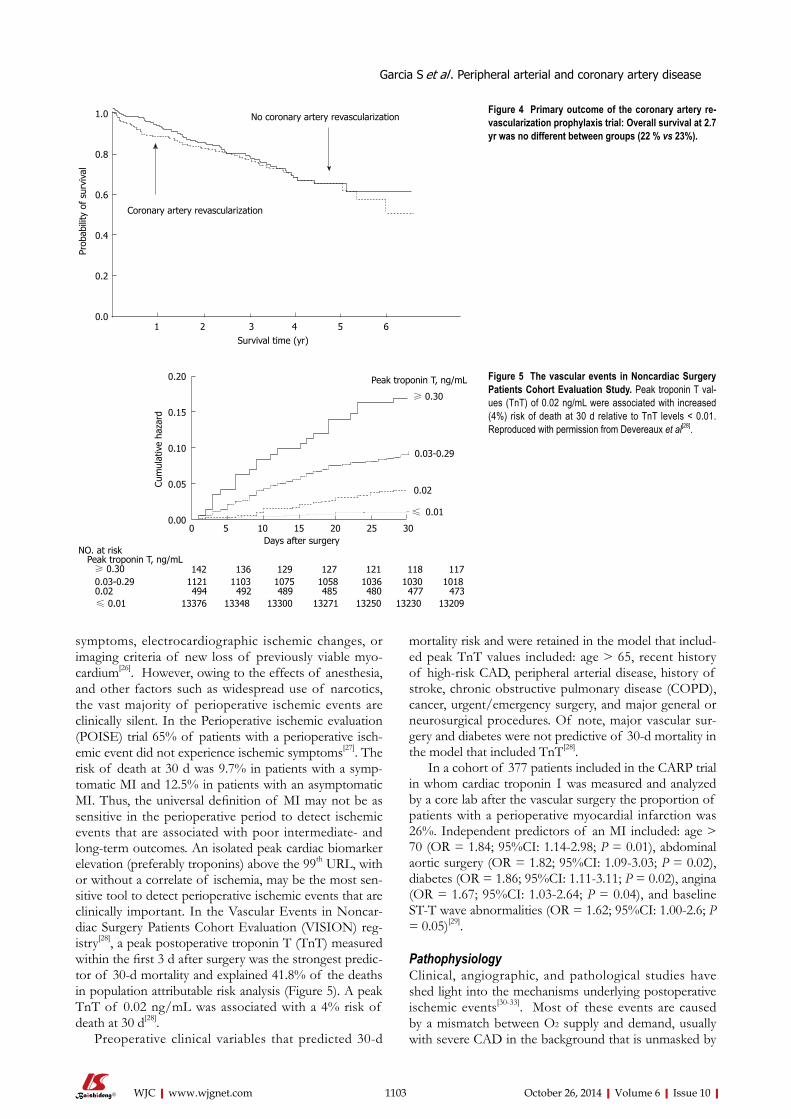
October 26, 2014|Volume 6|Issue 10|WJC|www.wjgnet.com
mortality risk and were retained in the model that includ-ed peak TnT values included: age > 65, recent history of high-risk CAD, peripheral arterial disease, history of stroke, chronic obstructive pulmonary disease (COPD), cancer, urgent/emergency surgery, and major general or neurosurgical procedures. Of note, major vascular sur-gery and diabetes were not predictive of 30-d mortality in the model that included TnT[28].
In a cohort of 377 patients included in the CARP trial in whom cardiac troponin Ⅰ was measured and analyzed by a core lab after the vascular surgery the proportion of patients with a perioperative myocardial infarction was 26%. Independent predictors of an MI included: age > 70 (OR = 1.84; 95%CI: 1.14-2.98; P = 0.01), abdominal aortic surgery (OR = 1.82; 95%CI: 1.09-3.03; P = 0.02), diabetes (OR = 1.86; 95%CI: 1.11-3.11; P = 0.02), angina (OR = 1.67; 95%CI: 1.03-2.64; P = 0.04), and baseline ST-T wave abnormalities (OR = 1.62; 95%CI: 1.00-2.6; P = 0.05)[29].
PathophysiologyClinical, angiographic, and pathological studies have shed light into the mechanisms underlying postoperative ischemic events[30-33]. Most of these events are caused by a mismatch between O2 supply and demand, usually with severe CAD in the background that is unmasked by
symptoms, electrocardiographic ischemic changes, or imaging criteria of new loss of previously viable myo-cardium[26]. However, owing to the effects of anesthesia, and other factors such as widespread use of narcotics, the vast majority of perioperative ischemic events are clinically silent. In the Perioperative ischemic evaluation (POISE) trial 65% of patients with a perioperative isch-emic event did not experience ischemic symptoms[27]. The risk of death at 30 d was 9.7% in patients with a symp-tomatic MI and 12.5% in patients with an asymptomatic MI. Thus, the universal definition of MI may not be as sensitive in the perioperative period to detect ischemic events that are associated with poor intermediate- and long-term outcomes. An isolated peak cardiac biomarker elevation (preferably troponins) above the 99th URL, with or without a correlate of ischemia, may be the most sen-sitive tool to detect perioperative ischemic events that are clinically important. In the Vascular Events in Noncar-diac Surgery Patients Cohort Evaluation (VISION) reg-istry[28], a peak postoperative troponin T (TnT) measured within the first 3 d after surgery was the strongest predic-tor of 30-d mortality and explained 41.8% of the deaths in population attributable risk analysis (Figure 5). A peak TnT of 0.02 ng/mL was associated with a 4% risk of death at 30 d[28].
Preoperative clinical variables that predicted 30-d
1103
No coronary artery revascularization
Coronary artery revascularization
1 2 3 4 5 6Survival time (yr)
Prob
abili
ty o
f su
rviv
al
1.0
0.8
0.6
0.4
0.2
0.0
Figure 4 Primary outcome of the coronary artery re-vascularization prophylaxis trial: Overall survival at 2.7 yr was no different between groups (22 % vs 23%).
0.20
0.15
0.10
0.05
0.00
Cum
ulat
ive
haza
rd
0 5 10 15 20 25 30Days after surgery
Peak troponin T, ng/mL
≥ 0.30
0.03-0.29
0.02
≤ 0.01
NO. at riskPeak troponin T, ng/mL≥ 0.30 142 136 129 127 121 118 1170.03-0.29 1121 1103 1075 1058 1036 1030 10180.02 494 492 489 485 480 477 473≤ 0.01 13376 13348 13300 13271 13250 13230 13209
Figure 5 The vascular events in Noncardiac Surgery Patients Cohort Evaluation Study. Peak troponin T val-ues (TnT) of 0.02 ng/mL were associated with increased (4%) risk of death at 30 d relative to TnT levels < 0.01. Reproduced with permission from Devereaux et al[28].
Garcia S et al . Peripheral arterial and coronary artery disease
October 26, 2014|Volume 6|Issue 10|WJC|www.wjgnet.com
the stress of the surgery. Landesberg et al[30] showed that tac ST-segment depression related to rapid heart rates is common in the perioperative period and predictive of long-term mortality. The duration of ST-segment depres-sion and peak catecholamine levels after surgery are as-sociated with infarct size. Chronic total occlusions (CTOs) are common in patients with a perioperative ischemic event or cardiac death (81%) relative to only 29% in pa-tients without ischemic complications after surgery[31]. Two pathological studies reported conflicting data on the incidence of plaque rupture after fatal postoperative MI[32-33]. Dawood et al[32] described evidence of plaque rupture in only 7% of patients (42 autopsies). Conversely, a higher incidence of plaque rupture (46%) was described by Cohen et al[33]. Differences in timing of the autopsy relative to the time of the MI may account for some of the discrepancies in the data.
Management of perioperative myocardial infarctionData from randomized clinical trials are lacking to guide therapy in the postoperative period. Small studies have shown that interventions aimed at improving oxygen delivery and minimizing myocardial oxygen consumption are beneficial in this setting[34]. The main goal of therapy is to preserve coronary perfusion pressure during dias-tole. This is best achieved with judicious utilization of beta-blockers, analgesia, and fluid administration with the intention to avoid tachycardia and hypotension. In the POISE trial for every 10-beats/min increase in heart rate there was a 31% relative increase in the odds of periop-erative MI[27]. Current guidelines recommend aggressive blood pressure control in patients with PAD, in particular in patients with diabetes and/or chronic kidney disease (goal < 130/80 mmHg)[35]. In the HOPE (Heart Out-comes Prevention Evaluation) trial ramipril 10 mg was associated with a 22% reduction in cardiovascular events and is currently recommended for high-risk patients, in-cluding those with PAD[36].
Statins contribute to plaque stabilization by decreas-ing circulating levels of inflammatory cytokines and reactive oxygen species while increasing expression of nitric oxide synthase[37]. Additionally, evidence from ran-domized clinical trials and observational studies support its use in clinical practice. In the DECREASE-Ⅲ study a 53% reduction in CV death and myocardial infarction was seen with high-dose fluvastatin in patients undergo-ing vascular surgery[38]. In another trial of 100 patients randomly assigned to 20 mg of atorvastatin or placebo prior to vascular surgery, the use of statins was associated with a significant reduction in cardiac events, from 26% to 8% at 6 mo[39]. An observational study of 164 veterans undergoing vascular surgery at our medical center dem-onstrated that utilization of statin drugs was associated with a reduction in long-term mortality[5]. Guidelines recommend the use of statins in patients with peripheral arterial disease to reduce cardiovascular events[2].
Owing to concerns for bleeding after non-cardiac surgery, the use of medical therapies and interventional
strategies commonly used to treat spontaneous MIs such as antiplatelet agents, anticoagulants and invasive coro-nary angiography are rarely used in this setting and have not been extensively studied in clinical trials. The man-agement of myocardial infarction After NoncArdiac sur-gery (MANAGE) trial (NCT01661101) will be the first study to randomize patients (n = 3200) with a PMI after noncardiac surgery to dabigatran or placebo. The primary end point is the occurrence of a major vascular complica-tion (vascular mortality, nonfatal MI, nonfatal stroke, and pulmonary embolism). The trial plans to complete enroll-ment in November 2015.
FUTURE DIRECTIONSAnother strategy for prevention of myocardial ischemia during surgery is ischemic preconditioning, which de-scribes the protection afforded by application of non-lethal episodes of myocardial ischemia prior to the index ischemic event[40,41]. The Cardiac Remote Ischemic Pre-conditioning Prior to Elective Vascular Surgery (CRIPES, NCT: 01558596) was designed to determine the feasibil-ity and safety of using remote ischemic preconditioning (RIPC) prior to vascular surgery, and to obtain prelimi-nary estimates of its effects on detectable postsurgical increases in cardiac troponin Ⅰ[42]. A similar strategy of RIPC has been evaluated prior to coronary angioplasty[43] and coronary artery bypass surgery[44] with positive initial results.
CONCLUSIONPatients with PAD in need of elective vascular surgery have a high prevalence of coronary atherosclerosis and are at increased risk of perioperative myocardial infarc-tion. Coronary revascularization prior to the vascular operation is not an effective intervention to mitigate this risk. A strategy of widespread use of cardiac troponins in the perioperative period is recommended to detect perioperative ischemic events associated with a long-term mortality risk. The selective use of medical interventions, cardiac imaging and coronary angiography in this popula-tion deserves further study.
REFERENCES1 Hirsch AT, Haskal ZJ, Hertzer NR, Bakal CW, Creager MA,
Halperin JL, Hiratzka LF, Murphy WR, Olin JW, Puschett JB, Rosenfield KA, Sacks D, Stanley JC, Taylor LM, White CJ, White J, White RA, Antman EM, Smith SC, Adams CD, Anderson JL, Faxon DP, Fuster V, Gibbons RJ, Halperin JL, Hiratzka LF, Hunt SA, Jacobs AK, Nishimura R, Ornato JP, Page RL, Riegel B. ACC/AHA 2005 guidelines for the man-agement of patients with peripheral arterial disease (lower extremity, renal, mesenteric, and abdominal aortic): execu-tive summary a collaborative report from the American As-sociation for Vascular Surgery/Society for Vascular Surgery, Society for Cardiovascular Angiography and Interventions, Society for Vascular Medicine and Biology, Society of In-terventional Radiology, and the ACC/AHA Task Force on Practice Guidelines (Writing Committee to Develop Guide-
1104
Garcia S et al . Peripheral arterial and coronary artery disease
October 26, 2014|Volume 6|Issue 10|WJC|www.wjgnet.com
lines for the Management of Patients With Peripheral Arte-rial Disease) endorsed by the American Association of Car-diovascular and Pulmonary Rehabilitation; National Heart, Lung, and Blood Institute; Society for Vascular Nursing; TransAtlantic Inter-Society Consensus; and Vascular Disease Foundation. J Am Coll Cardiol 2006; 47: 1239-1312 [PMID: 16545667 DOI: 10.1016/j.jacc.2005.10.009]
2 Norgren L, Hiatt WR, Dormandy JA, Nehler MR, Harris KA, Fowkes FG. Inter-Society Consensus for the Management of Peripheral Arterial Disease (TASC II). J Vasc Surg 2007; 45 Suppl S: S5-S67 [PMID: 17223489]
3 Bhatt DL, Steg PG, Ohman EM, Hirsch AT, Ikeda Y, Mas JL, Goto S, Liau CS, Richard AJ, Röther J, Wilson PW. Inter-national prevalence, recognition, and treatment of cardio-vascular risk factors in outpatients with atherothrombosis. JAMA 2006; 295: 180-189 [PMID: 16403930 DOI: 10.1001/jama.295.2.180]
4 Criqui MH, Langer RD, Fronek A, Feigelson HS, Klauber MR, McCann TJ, Browner D. Mortality over a period of 10 years in patients with peripheral arterial disease. N Engl J Med 1992; 326: 381-386 [PMID: 1729621 DOI: 10.1056/NEJM199202063260605]
5 Marston N, Brenes J, Garcia S, Kuskowski M, Adabag S, Santilli S, McFalls EO. Peak postoperative troponin levels outperform preoperative cardiac risk indices as predictors of long-term mortality after vascular surgery Troponins and postoperative outcomes. J Crit Care 2012; 27: 66-72 [PMID: 21798697 DOI: 10.1016/j.jcrc.2011.06.004]
6 McFalls EO, Ward HB, Moritz TE, Littooy F, Santilli S, Rapp J, Larsen G, Reda DJ. Clinical factors associated with long-term mortality following vascular surgery: outcomes from the Coronary Artery Revascularization Prophylaxis (CARP) Trial. J Vasc Surg 2007; 46: 694-700 [PMID: 17903649 DOI: 10.1016/j.jvs.2007.05.060]
7 Hiatt WR, Hoag S, Hamman RF. Effect of diagnostic criteria on the prevalence of peripheral arterial disease. The San Luis Valley Diabetes Study. Circulation 1995; 91: 1472-1479 [PMID: 7867189 DOI: 10.1161/01.CIR.91.5.1472]
8 Hirsch AT, Criqui MH, Treat-Jacobson D, Regensteiner JG, Creager MA, Olin JW, Krook SH, Hunninghake DB, Com-erota AJ, Walsh ME, McDermott MM, Hiatt WR. Peripheral arterial disease detection, awareness, and treatment in pri-mary care. JAMA 2001; 286: 1317-1324 [PMID: 11560536 DOI: 10.1001/jama.286.11.1317]
9 Fowkes FG, Housley E, Cawood EH, Macintyre CC, Ruck-ley CV, Prescott RJ. Edinburgh Artery Study: prevalence of asymptomatic and symptomatic peripheral arterial disease in the general population. Int J Epidemiol 1991; 20: 384-392 [PMID: 1917239 DOI: 10.1093/ije/20.2.384]
10 Criqui MH, Vargas V, Denenberg JO, Ho E, Allison M, Langer RD, Gamst A, Bundens WP, Fronek A. Ethnicity and peripheral arterial disease: the San Diego Population Study. Circulation 2005; 112: 2703-2707 [PMID: 16246968 DOI: 10.1161/CIRCULATIONAHA.105.546507]
11 Selvin E, Erlinger TP. Prevalence of and risk factors for peripheral arterial disease in the United States: results from the National Health and Nutrition Examination Survey, 1999-2000. Circulation 2004; 110: 738-743 [PMID: 15262830 DOI: 10.1161/01.CIR.0000137913.26087.F0]
12 Selvin E, Marinopoulos S, Berkenblit G, Rami T, Brancati FL, Powe NR, Golden SH. Meta-analysis: glycosylated he-moglobin and cardiovascular disease in diabetes mellitus. Ann Intern Med 2004; 141: 421-431 [PMID: 15381515 DOI: 10.7326/0003-4819-141-6-200409210-00007]
13 Ridker PM, Stampfer MJ, Rifai N. Novel risk factors for systemic atherosclerosis: a comparison of C-reactive protein, fibrinogen, homocysteine, lipoprotein(a), and standard cho-lesterol screening as predictors of peripheral arterial disease. JAMA 2001; 285: 2481-2485 [PMID: 11368701 DOI: 10.1001/
jama.285.19.2481]14 Muntner P, Wildman RP, Reynolds K, Desalvo KB, Chen J,
Fonseca V. Relationship between HbA1c level and periph-eral arterial disease. Diabetes Care 2005; 28: 1981-1987 [PMID: 16043742 DOI: 10.2337/diacare.28.8.1981]
15 Hertzer NR, Beven EG, Young JR, O’Hara PJ, Ruschhaupt WF, Graor RA, Dewolfe VG, Maljovec LC. Coronary artery disease in peripheral vascular patients. A classification of 1000 coronary angiograms and results of surgical manage-ment. Ann Surg 1984; 199: 223-233 [PMID: 6696538 DOI: 10.1097/00000658-198402000-00016]
16 Mehler PS, Coll JR, Estacio R, Esler A, Schrier RW, Hiatt WR. Intensive blood pressure control reduces the risk of cardiovascular events in patients with peripheral arterial dis-ease and type 2 diabetes. Circulation 2003; 107: 753-756 [PMID: 12578880 DOI: 10.1161/01.CIR.0000049640.46039.52]
17 Fleisher LA, Beckman JA, Brown KA, Calkins H, Chaikof EL, Fleischmann KE, Freeman WK, Froehlich JB, Kasper EK, Kersten JR, Riegel B, Robb JF, Smith SC, Jacobs AK, Adams CD, Anderson JL, Antman EM, Buller CE, Creager MA, Et-tinger SM, Faxon DP, Fuster V, Halperin JL, Hiratzka LF, Hunt SA, Lytle BW, Nishimura R, Ornato JP, Page RL, Riegel B, Tarkington LG, Yancy CW. ACC/AHA 2007 Guidelines on Perioperative Cardiovascular Evaluation and Care for Noncardiac Surgery: Executive Summary: A Report of the American College of Cardiology/American Heart Associa-tion Task Force on Practice Guidelines (Writing Committee to Revise the 2002 Guidelines on Perioperative Cardiovas-cular Evaluation for Noncardiac Surgery) Developed in Col-laboration With the American Society of Echocardiography, American Society of Nuclear Cardiology, Heart Rhythm Society, Society of Cardiovascular Anesthesiologists, Society for Cardiovascular Angiography and Interventions, Society for Vascular Medicine and Biology, and Society for Vascu-lar Surgery. J Am Coll Cardiol 2007; 50: 1707-1732 [PMID: 17950159 DOI: 10.1016/j.jacc.2007.09.003]
18 McFalls EO, Ward HB, Moritz TE, Apple FS, Goldman S, Pierpont G, Larsen GC, Hattler B, Shunk K, Littooy F, Santilli S, Rapp J, Thottapurathu L, Krupski W, Reda DJ, Henderson WG. Predictors and outcomes of a perioperative myocardial infarction following elective vascular surgery in patients with documented coronary artery disease: results of the CARP trial. Eur Heart J 2008; 29: 394-401 [PMID: 18245121 DOI: 10.1093/eurheartj/ehm620]
19 Eagle KA, Brundage BH, Chaitman BR, Ewy GA, Fleisher LA, Hertzer NR, Leppo JA, Ryan T, Schlant RC, Spencer WH, Spittell JA, Twiss RD. Guidelines for perioperative car-diovascular evaluation for noncardiac surgery: an abridged version of the report of the American College of Cardiol-ogy/American Heart Association Task Force on Practice Guidelines. Mayo Clin Proc 1997; 72: 524-531 [PMID: 9179136 DOI: 10.4065/72.6.524]
20 Garcia S, Moritz TE, Ward HB, Pierpont G, Goldman S, Larsen GC, Littooy F, Krupski W, Thottapurathu L, Reda DJ, McFalls EO. Usefulness of revascularization of patients with multivessel coronary artery disease before elective vascular surgery for abdominal aortic and peripheral occlusive dis-ease. Am J Cardiol 2008; 102: 809-813 [PMID: 18805102 DOI: 10.1016/j.amjcard.2008.05.022]
21 Boersma E, Poldermans D, Bax JJ, Steyerberg EW, Thomson IR, Banga JD, van De Ven LL, van Urk H, Roelandt JR. Pre-dictors of cardiac events after major vascular surgery: Role of clinical characteristics, dobutamine echocardiography, and beta-blocker therapy. JAMA 2001; 285: 1865-1873 [PMID: 11308400 DOI: 10.1001/jama.285.14.1865]
22 Lee TH, Marcantonio ER, Mangione CM, Thomas EJ, Po-lanczyk CA, Cook EF, Sugarbaker DJ, Donaldson MC, Poss R, Ho KK, Ludwig LE, Pedan A, Goldman L. Derivation and prospective validation of a simple index for predic-
1105
Garcia S et al . Peripheral arterial and coronary artery disease
October 26, 2014|Volume 6|Issue 10|WJC|www.wjgnet.com
tion of cardiac risk of major noncardiac surgery. Circulation 1999; 100: 1043-1049 [PMID: 10477528 DOI: 10.1161/01.CIR.100.10.1043]
23 Garcia S, Moritz TE, Goldman S, Littooy F, Pierpont G, Larsen GC, Reda DJ, Ward HB, McFalls EO. Perioperative complications after vascular surgery are predicted by the revised cardiac risk index but are not reduced in high-risk subsets with preoperative revascularization. Circ Cardiovasc Qual Outcomes 2009; 2: 73-77 [PMID: 20031818 DOI: 10.1161/CIRCOUTCOMES.108.827683]
24 McFalls EO, Ward HB, Moritz TE, Goldman S, Krupski WC, Littooy F, Pierpont G, Santilli S, Rapp J, Hattler B, Shunk K, Jaenicke C, Thottapurathu L, Ellis N, Reda DJ, Henderson WG. Coronary-artery revascularization before elective major vascular surgery. N Engl J Med 2004; 351: 2795-2804 [PMID: 15625331 DOI: 10.1056/NEJMoa041905]
25 Poldermans D, Schouten O, Vidakovic R, Bax JJ, Thom-son IR, Hoeks SE, Feringa HH, Dunkelgrün M, de Jaegere P, Maat A, van Sambeek MR, Kertai MD, Boersma E. A clinical randomized trial to evaluate the safety of a nonin-vasive approach in high-risk patients undergoing major vascular surgery: the DECREASE-V Pilot Study. J Am Coll Cardiol 2007; 49: 1763-1769 [PMID: 17466225 DOI: 10.1016/j.jacc.2006.11.052]
26 Thygesen K, Alpert JS, White HD, Jaffe AS, Apple FS, Gal-vani M, Katus HA, Newby LK, Ravkilde J, Chaitman B, Clemmensen PM, Dellborg M, Hod H, Porela P, Underwood R, Bax JJ, Beller GA, Bonow R, Van der Wall EE, Bassand JP, Wijns W, Ferguson TB, Steg PG, Uretsky BF, Williams DO, Armstrong PW, Antman EM, Fox KA, Hamm CW, Ohman EM, Simoons ML, Poole-Wilson PA, Gurfinkel EP, Lopez-Sendon JL, Pais P, Mendis S, Zhu JR, Wallentin LC, Fernán-dez-Avilés F, Fox KM, Parkhomenko AN, Priori SG, Tendera M, Voipio-Pulkki LM, Vahanian A, Camm AJ, De Caterina R, Dean V, Dickstein K, Filippatos G, Funck-Brentano C, Hel-lemans I, Kristensen SD, McGregor K, Sechtem U, Silber S, Tendera M, Widimsky P, Zamorano JL, Morais J, Brener S, Harrington R, Morrow D, Lim M, Martinez-Rios MA, Stei-nhubl S, Levine GN, Gibler WB, Goff D, Tubaro M, Dudek D, Al-Attar N. Universal definition of myocardial infarc-tion. Circulation 2007; 116: 2634-2653 [PMID: 17951284 DOI: 10.1161/CIRCULATIONAHA.107.187397]
27 Devereaux PJ, Xavier D, Pogue J, Guyatt G, Sigamani A, Garutti I, Leslie K, Rao-Melacini P, Chrolavicius S, Yang H, Macdonald C, Avezum A, Lanthier L, Hu W, Yusuf S. Char-acteristics and short-term prognosis of perioperative myo-cardial infarction in patients undergoing noncardiac surgery: a cohort study. Ann Intern Med 2011; 154: 523-528 [PMID: 21502650 DOI: 10.7326/0003-4819-154-8-201104190-00003]
28 Devereaux PJ, Chan MT, Alonso-Coello P, Walsh M, Berwanger O, Villar JC, Wang CY, Garutti RI, Jacka MJ, Sigamani A, Srinathan S, Biccard BM, Chow CK, Abraham V, Tiboni M, Pettit S, Szczeklik W, Lurati Buse G, Botto F, Guyatt G, Heels-Ansdell D, Sessler DI, Thorlund K, Garg AX, Mrkobrada M, Thomas S, Rodseth RN, Pearse RM, Thabane L, McQueen MJ, VanHelder T, Bhandari M, Bosch J, Kurz A, Polanczyk C, Malaga G, Nagele P, Le Manach Y, Leuwer M, Yusuf S. Association between postoperative tro-ponin levels and 30-day mortality among patients undergo-ing noncardiac surgery. JAMA 2012; 307: 2295-2304 [PMID: 22706835 DOI: 10.1001/jama.2012.5502]
29 Devereaux PJ, Yang H, Yusuf S, Guyatt G, Leslie K, Villar JC, Xavier D, Chrolavicius S, Greenspan L, Pogue J, Pais P, Liu L, Xu S, Málaga G, Avezum A, Chan M, Montori VM, Jacka M, Choi P. Effects of extended-release metoprolol succinate in patients undergoing non-cardiac surgery (POISE trial): a ran-domised controlled trial. Lancet 2008; 371: 1839-1847 [PMID: 18479744]
30 Landesberg G, Mosseri M, Zahger D, Wolf Y, Perouansky M, Anner H, Drenger B, Hasin Y, Berlatzky Y, Weissman
C. Myocardial infarction after vascular surgery: the role of prolonged stress-induced, ST depression-type ischemia. J Am Coll Cardiol 2001; 37: 1839-1845 [PMID: 11401120 DOI: 10.1016/S0735-1097(01)01265-7]
31 Ellis SG, Hertzer NR, Young JR, Brener S. Angiographic correlates of cardiac death and myocardial infarction complicating major nonthoracic vascular surgery. Am J Cardiol 1996; 77: 1126-1128 [PMID: 8644673 DOI: 10.1016/S0002-9149(96)00130-0]
32 Dawood MM, Gutpa DK, Southern J, Walia A, Atkinson JB, Eagle KA. Pathology of fatal perioperative myocardial infarction: implications regarding pathophysiology and prevention. Int J Cardiol 1996; 57: 37-44 [PMID: 8960941 DOI: 10.1016/S0167-5273(96)02769-6]
33 Cohen MC, Aretz TH. Histological analysis of coronary artery lesions in fatal postoperative myocardial infarction. Cardiovasc Pathol 1999; 8: 133-139 [PMID: 10722235 DOI: 10.1016/S1054-8807(98)00032-5]
34 Martinez E, Kim L, Rosenfeld B, Faraday N, Bass E, Perler B, Williams GN, Dorman T, Pronovost P. Early detection and real-time intervention of postoperative myocardial ischemia: the STOPMI (Study for the Treatment of Perioperative Myo-cardial Ischemia) Study. Abstract presented at: Association of University Anesthesiologists; May 16-18, 2008; Durham NC
35 European Society of Hypertension-European Society of Cardiology Guidelines Committee. 2003 European Society of Hypertension-European Society of Cardiology guidelines for the management of arterial hypertension. J Hypertens 2003; 21: 1011-1053 [PMID: 12777938 DOI: 10.1097/00004872-200306000-00001]
36 Yusuf S, Sleight P, Pogue J, Bosch J, Davies R, Dagenais G. Effects of an angiotensin-converting-enzyme inhibitor, ramipril, on cardiovascular events in high-risk patients. The Heart Outcomes Prevention Evaluation Study Investiga-tors. N Engl J Med 2000; 342: 145-153 [PMID: 10639539 DOI: 10.1056/NEJM200001203420301]
37 Davignon J. Beneficial cardiovascular pleiotropic effects of statins. Circulation 2004; 109: III39-III43 [PMID: 15198965]
38 Schouten O, Boersma E, Hoeks SE, Benner R, van Urk H, van Sambeek MR, Verhagen HJ, Khan NA, Dunkelgrun M, Bax JJ, Poldermans D. Fluvastatin and perioperative events in patients undergoing vascular surgery. N Engl J Med 2009; 361: 980-989 [PMID: 19726772 DOI: 10.1056/NEJ-Moa0808207]
39 Durazzo AE, Machado FS, Ikeoka DT, De Bernoche C, Mo-nachini MC, Puech-Leão P, Caramelli B. Reduction in car-diovascular events after vascular surgery with atorvastatin: a randomized trial. J Vasc Surg 2004; 39: 967-975; discussion 975-976 [PMID: 15111846]
40 Przyklenk K, Bauer B, Ovize M, Kloner RA, Whittaker P. Regional ischemic ‘preconditioning’ protects remote virgin myocardium from subsequent sustained coronary occlu-sion. Circulation 1993; 87: 893-899 [PMID: 7680290 DOI: 10.1161/01.CIR.87.3.893]
41 Gho BC, Schoemaker RG, van den Doel MA, Duncker DJ, Verdouw PD. Myocardial protection by brief ischemia in noncardiac tissue. Circulation 1996; 94: 2193-2200 [PMID: 8901671 DOI: 10.1161/01.CIR.94.9.2193]
42 Garcia S, Rector TS, Zakharova MY, Magras A, Sandoval Y. Cardiac Remote Ischemic Preconditioning Prior to Elective Major Vascular Surgery (CRIPES): Study Design and Ratio-nale. J Clin Trials 2013; S5: 002
43 Hoole SP, Heck PM, Sharples L, Khan SN, Duehmke R, Den-sem CG, Clarke SC, Shapiro LM, Schofield PM, O’Sullivan M, Dutka DP. Cardiac Remote Ischemic Preconditioning in Coronary Stenting (CRISP Stent) Study: a prospective, ran-domized control trial. Circulation 2009; 119: 820-827 [PMID: 19188504 DOI: 10.1161/CIRCULATIONAHA.108.809723]
44 Hausenloy DJ, Mwamure PK, Venugopal V, Harris J, Bar-
1106
Garcia S et al . Peripheral arterial and coronary artery disease

October 26, 2014|Volume 6|Issue 10|WJC|www.wjgnet.com
nard M, Grundy E, Ashley E, Vichare S, Di Salvo C, Kolvekar S, Hayward M, Keogh B, MacAllister RJ, Yellon DM. Effect of remote ischaemic preconditioning on myocardial injury in
patients undergoing coronary artery bypass graft surgery: a randomised controlled trial. Lancet 2007; 370: 575-579 [PMID: 17707752 DOI: 10.1016/S0140-6736(07)61296-3]
P- Reviewer: Al-Mohammad A, Athanasios G, Nemes A S- Editor: Ji FF L- Editor: A E- Editor: Wu HL
1107
Garcia S et al . Peripheral arterial and coronary artery disease
quantitative myocardial blush grade for the assess-ment of microvascular integrity in patients with acute coronary syndromes (ACS) and heart transplant (HT) recipients with cardiac allograft vasculopathy (CAV). Using an, in the meanwhile well-established, compu-tational algorithm, a prompt diagnosis can be made in the catheterization lab, which can identify patients with ACS and increased risk for myocardial remodelling and congestive heart failure in the long-term. In addition, this computational algorithm can identify HT recipients with increased risk for CAV and adverse cardiovascular outcomes.
Hofmann NP, Dickhaus H, Katus HA, Korosoglou G. Quantita-tive assessment of myocardial blush grade in patients with coro-nary artery disease and in cardiac transplant recipients. World J Cardiol 2014; 6(10): 1108-1112 Available from: URL: http://www.wjgnet.com/1949-8462/full/v6/i10/1108.htm DOI: http://dx.doi.org/10.4330/wjc.v6.i10.1108
INTRODUCTIONImpaired myocardial perfusion by either epicardial coro-nary artery disease (CAD) or small vessel disease is a common challenge in cardiology worldwide. Both CAD and cardiac allograft vasculopathy (CAV) in heart trans-plant (HT) recipients significantly influence mortality in such patient cohorts. Considering the vast amount of health care costs for post-rehabilitation support after myocardial infarction[1] and HT[2], preventive medical care should primarily focus on the early detection of cardiac pathology and risk stratification of such patients. Quantitative assessment of epicardial and microvascu-lar integrity can aid tailoring pharmacologic therapy of patients identified at high risk for future events, which may ultimately improve clinical outcomes. We therefore
Quantitative assessment of myocardial blush grade in patients with coronary artery disease and in cardiac transplant recipients
Nina Patricia Hofmann, Hartmut Dickhaus, Hugo A Katus, Grigorios Korosoglou
Nina Patricia Hofmann, Hugo A Katus, Grigorios Korosoglou, Department of Cardiology, University of Heidelberg, 69120 Heidel-berg, GermanyHartmut Dickhaus, Department of Medical Informatics, Univer-sity of Heidelberg, 69120 Heidelberg, GermanyAuthor contributions: All the authors together contributed to this paper.Correspondence to: Nina Patricia Hofmann, MD, Department of Cardiology, University of Heidelberg, Im Neuenheimer Feld 410, 69120 Heidelberg, Germany. [email protected]: +49-6221-5637487Received: February 20, 2014 Revised: July 25, 2014Accepted: August 27, 2014Published online: October 26, 2014
AbstractQuantitative assessment of myocardial perfusion by myocardial blush grade (MBG) is an angiographic com-puter-assisted method to assess myocardial tissue-level reperfusion in patients with acute coronary syndromes and microvascular integrity in heart transplant recipi-ents with suspected cardiac allograft vasculopathy. This review describes the ability of quantitative MBG as a simple, fast and cost effective modality for the prompt diagnosis of impaired microvascular integrity during routine cardiac catheterization. Herein, we summa-rize the existing evidence, its usefulness in the clinical routine, and compare this method to other techniques which can be used for the assessment of myocardial perfusion.
© 2014 Baishideng Publishing Group Inc. All rights reserved.
Key words: Quantitative myocardial blush grade; Prog-nosis; Heart transplantation; Coronary artery disease
Core tip: In this article, we highlight the ability of
MINIREVIEWS
Submit a Manuscript: http://www.wjgnet.com/esps/Help Desk: http://www.wjgnet.com/esps/helpdesk.aspxDOI: 10.4330/wjc.v6.i10.1108
October 26, 2014|Volume 6|Issue 10|WJC|www.wjgnet.com
World Journal of CardiologyW J C
World J Cardiol 2014 October 26; 6(10): 1108-1112ISSN 1949-8462 (online)
© 2014 Baishideng Publishing Group Inc. All rights reserved.
1108
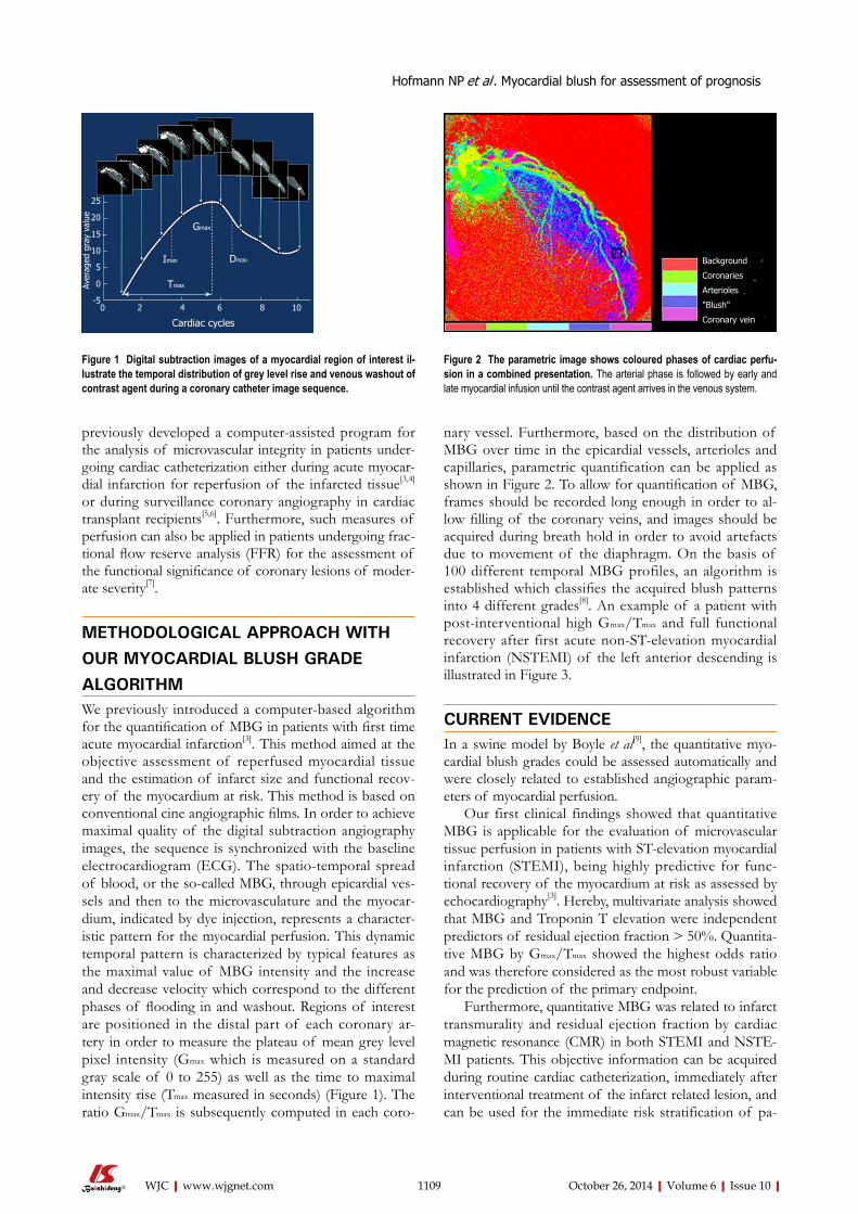
previously developed a computer-assisted program for the analysis of microvascular integrity in patients under-going cardiac catheterization either during acute myocar-dial infarction for reperfusion of the infarcted tissue[3,4] or during surveillance coronary angiography in cardiac transplant recipients[5,6]. Furthermore, such measures of perfusion can also be applied in patients undergoing frac-tional flow reserve analysis (FFR) for the assessment of the functional significance of coronary lesions of moder-ate severity[7].
METHODOLOGICAL APPROACH WITH OUR MYOCARDIAL BLUSH GRADE ALGORITHMWe previously introduced a computer-based algorithm for the quantification of MBG in patients with first time acute myocardial infarction[3]. This method aimed at the objective assessment of reperfused myocardial tissue and the estimation of infarct size and functional recov-ery of the myocardium at risk. This method is based on conventional cine angiographic films. In order to achieve maximal quality of the digital subtraction angiography images, the sequence is synchronized with the baseline electrocardiogram (ECG). The spatio-temporal spread of blood, or the so-called MBG, through epicardial ves-sels and then to the microvasculature and the myocar-dium, indicated by dye injection, represents a character-istic pattern for the myocardial perfusion. This dynamic temporal pattern is characterized by typical features as the maximal value of MBG intensity and the increase and decrease velocity which correspond to the different phases of flooding in and washout. Regions of interest are positioned in the distal part of each coronary ar-tery in order to measure the plateau of mean grey level pixel intensity (Gmax which is measured on a standard gray scale of 0 to 255) as well as the time to maximal intensity rise (Tmax measured in seconds) (Figure 1). The ratio Gmax/Tmax is subsequently computed in each coro-
nary vessel. Furthermore, based on the distribution of MBG over time in the epicardial vessels, arterioles and capillaries, parametric quantification can be applied as shown in Figure 2. To allow for quantification of MBG, frames should be recorded long enough in order to al-low filling of the coronary veins, and images should be acquired during breath hold in order to avoid artefacts due to movement of the diaphragm. On the basis of 100 different temporal MBG profiles, an algorithm is established which classifies the acquired blush patterns into 4 different grades[8]. An example of a patient with post-interventional high Gmax/Tmax and full functional recovery after first acute non-ST-elevation myocardial infarction (NSTEMI) of the left anterior descending is illustrated in Figure 3.
CURRENT EVIDENCE In a swine model by Boyle et al[9], the quantitative myo-cardial blush grades could be assessed automatically and were closely related to established angiographic param-eters of myocardial perfusion.
Our first clinical findings showed that quantitative MBG is applicable for the evaluation of microvascular tissue perfusion in patients with ST-elevation myocardial infarction (STEMI), being highly predictive for func-tional recovery of the myocardium at risk as assessed by echocardiography[3]. Hereby, multivariate analysis showed that MBG and Troponin T elevation were independent predictors of residual ejection fraction > 50%. Quantita-tive MBG by Gmax/Tmax showed the highest odds ratio and was therefore considered as the most robust variable for the prediction of the primary endpoint.
Furthermore, quantitative MBG was related to infarct transmurality and residual ejection fraction by cardiac magnetic resonance (CMR) in both STEMI and NSTE-MI patients. This objective information can be acquired during routine cardiac catheterization, immediately after interventional treatment of the infarct related lesion, and can be used for the immediate risk stratification of pa-
October 26, 2014|Volume 6|Issue 10|WJC|www.wjgnet.com
Hofmann NP et al . Myocardial blush for assessment of prognosis
1109
Figure 1 Digital subtraction images of a myocardial region of interest il-lustrate the temporal distribution of grey level rise and venous washout of contrast agent during a coronary catheter image sequence.
Figure 2 The parametric image shows coloured phases of cardiac perfu-sion in a combined presentation. The arterial phase is followed by early and late myocardial infusion until the contrast agent arrives in the venous system.
Background
Coronaries
Arterioles
"Blush"
Coronary vein
Gmax
DmaxImax
Tmax
Cardiac cycles
Aver
aged
gra
y va
lue
25
20
15
10
5
0
-50 2 4 6 8 10
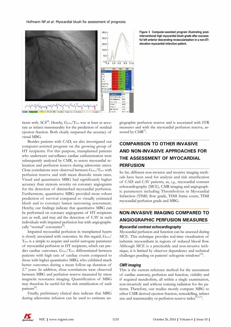
tients with ACS[4]. Hereby, Gmax/Tmax was at least as accu-rate as infarct transmurality for the prediction of residual ejection fraction. Both clearly surpassed the accuracy of visual MBG.
Besides patients with CAD, we also investigated our computer-assisted program on the growing group of HT recipients. For this purpose, transplanted patients who underwent surveillance cardiac catheterization were subsequently analyzed by CMR, to assess myocardial re-laxation and perfusion reserve during adenosine stress. Close correlations were observed between Gmax/Tmax with perfusion reserve and with mean diastolic strain rates. Visual and quantitative MBG had significantly higher accuracy than stenosis severity on coronary angiograms for the detection of diminished myocardial perfusion. Furthermore, quantitative MBG provided more robust prediction of survival compared to visually estimated blush and to coronary lumen narrowing assessment. Hereby, our findings indicate that quantitative MBG can be performed on coronary angiograms of HT recipients just as well, and may aid the detection of CAV in such individuals with impaired perfusion but with angiographi-cally “normal” coronaries[5].
Impaired myocardial perfusion in transplanted hearts is closely associated with outcomes. In this regard, Gmax/Tmax is a simple to acquire and useful surrogate parameter of myocardial perfusion in HT recipients, which can pre-dict cardiac outcomes. Gmax/Tmax differentiated between patients with high rate of cardiac events compared to those with higher quantitative MBG, who exhibited much better outcomes during a mean follow-up duration of 2.7 years. In addition, close correlations were observed between MBG and perfusion reserve measured by stress magnetic resonance imaging. Quantification of MBG may therefore be useful for the risk stratification of such patients[6].
Finally, preliminary clinical data indicate that MBG during adenosine infusion can be used to estimate an-
giographic perfusion reserve and is associated with FFR measures and with the myocardial perfusion reserve, as-sessed by CMR[7].
COMPARISON TO OTHER INVASIVE
AND NON-INVASIVE APPROACHES FOR
THE ASSESSMENT OF MYOCARDIAL
PERFUSIONSo far, different non-invasive and invasive imaging meth-ods have been used for analysis and risk stratification of CAD and CAV patients, as, e.g., myocardial contrast echocardiography (MCE), CMR imaging and angiograph-ic parameters including Thrombolysis in Myocardial Infarction (TIMI) flow grade, TIMI frame count, TIMI myocardial perfusion grade and MBG.
NON-INVASIVE IMAGING COMPARED TO
ANGIOGRAPHIC PERFUSION MEASURESMyocardial contrast echocardiographyMyocardial perfusion and function can be assessed during MCE. This technique provides real-time visualisation of ischemic myocardium in regions of reduced blood flow. Although MCE is a practicable and non-invasive tech-nique, it is limited by observer dependency and technical challenges pending on patients’ echogenic windows[10].
CMR imaging This is the current reference method for the assessment of cardiac anatomy, perfusion and function, viability and if required metabolism, all within a single examination, non-invasively and without ionizing radiation for the pa-tients. Therefore, our studies mostly compare MBG to either CMR derived ejection fraction, remodelling, infarct size and transmurality or perfusion reserve index[4-6,11].
October 26, 2014|Volume 6|Issue 10|WJC|www.wjgnet.com 1110
49.0
39.0
29.0
19.0
9.0
-1.0-1.0 2.0 5.0 8.0 11.0
Figure 3 Computer-assisted program illustrating post-interventional high myocardial blush grade after success-ful left anterior descending revascularization in a non-ST-elevation myocardial infarction patient.
Hofmann NP et al . Myocardial blush for assessment of prognosis
October 26, 2014|Volume 6|Issue 10|WJC|www.wjgnet.com
myocardial infarction in German hospitals. Health Econ 2014; 23: 653-669 [PMID: 23696223 DOI: 10.1002/hec.2941]
2 Digiorgi PL, Reel MS, Thornton B, Burton E, Naka Y, Oz MC. Heart transplant and left ventricular assist device costs. J Heart Lung Transplant 2005; 24: 200-204 [PMID: 15701438 DOI: 10.1016/j.healun.2003.11.397]
3 Korosoglou G, Haars A, Michael G, Erbacher M, Hardt S, Giannitsis E, Kurz K, Franz-Josef N, Dickhaus H, Katus HA, Kuecherer H. Quantitative evaluation of myocardial blush to assess tissue level reperfusion in patients with acute ST-elevation myocardial infarction: incremental prognostic value compared with visual assessment. Am Heart J 2007; 153: 612-620 [PMID: 17383301 DOI: 10.1016/j.ahj.2006.12.019]
4 Riedle N, Dickhaus H, Erbacher M, Steen H, Andrassy M, Lossnitzer D, Hardt S, Rottbauer W, Zugck C, Giannitsis E, Katus HA, Korosoglou G. Early assessment of infarct size and prediction of functional recovery by quantitative myocardial blush grade in patients with acute coronary syndromes treated according to current guidelines. Catheter Cardiovasc Interv 2010; 76: 502-510 [PMID: 20882653 DOI: 10.1002/ccd.22540]
5 Korosoglou G, Riedle N, Erbacher M, Dengler TJ, Zugck C, Rottbauer W, Hardt S, Bekeredjian R, Kristen A, Giannitsis E, Osman NF, Dickhaus H, Katus HA. Quantitative myocardial blush grade for the detection of cardiac allograft vasculopa-thy. Am Heart J 2010; 159: 643-651.e2 [PMID: 20362724]
6 Hofmann NP, Voss A, Dickhaus H, Erbacher M, Doesch A, Ehlermann P, Gitsioudis G, Buss SJ, Giannitsis E, Katus HA, Korosoglou G. Long-term outcome after heart transplanta-tion predicted by quantitative myocardial blush grade in coronary angiography. Am J Transplant 2013; 13: 1491-1502 [PMID: 23617734 DOI: 10.1111/ajt.12223]
7 Korosoglou G, Hofmann NP, Erbacher M, Dickhaus H, Bekeredjian R, Neumann FJ, Katus HA, Hardt SE. Quantita-tive myocardial blush grade reserve during pharmacologic hyperemia is related to fractional flow reserve measures in patients with stable coronary artery disease. Clin Res Cardiol 2013; 102 (abstract)
8 Dickhaus H, Erbacher M, Kücherer H. Quantification of myocardial perfusion for CAD diagnosis. Stud Health Technol Inform 2007; 129: 1339-1343 [PMID: 17911932]
9 Boyle AJ, Schuleri KH, Lienard J, Vaillant R, Chan MY, Zim-met JM, Mazhari R, Centola M, Feigenbaum G, Dib J, Kapur NK, Hare JM, Resar JR. Quantitative automated assessment of myocardial perfusion at cardiac catheterization. Am J Cardiol 2008; 102: 980-987 [PMID: 18929697 DOI: 10.1016/j.amjcard.2008.05.064]
10 Korosoglou G, Hansen A, Hoffend J, Gavrilovic G, Wolf D, Zehelein J, Haberkorn U, Kuecherer H. Comparison of real-time myocardial contrast echocardiography for the assess-ment of myocardial viability with fluorodeoxyglucose-18 positron emission tomography and dobutamine stress echocardiography. Am J Cardiol 2004; 94: 570-576 [PMID: 15342285 DOI: 10.1016/j.amjcard.2004.05.018]
11 Hofmann NP, Steuer C, Voss A, Erbel C, Celik S, Doesch A, Ehlermann P, Buss SJ, Giannitsis E, Katus HA, and Koroso-glou G. Comprehensive Bio-Imaging using Myocardial Per-fusion Reserve Index during Adenosine Cardiac Magnetic Resonance and the High-sensitive Troponin T for the Prog-nosis of Heart Transplant Recipients. Am J Transplant 2014 Oct 7; Epub ahead of print [DOI: 10.1111/ajt.12924]
12 Cannon CP. Importance of TIMI 3 flow. Circulation 2001; 104: 624-626 [PMID: 11489764 DOI: 10.1161/01.CIR.104.6.624]
13 Galiuto L, DeMaria AN, del Balzo U, May-Newman K, Flaim SF, Wolf PL, Kirchengast M, Iliceto S. Ischemia-reper-fusion injury at the microvascular level: treatment by endo-thelin A-selective antagonist and evaluation by myocardial contrast echocardiography. Circulation 2000; 102: 3111-3116 [PMID: 11120703]
14 Hansen A, Kumar A, Wolf D, Frankenbergerova K, Filusch A, Gross ML, Mueller S, Katus H, Kuecherer H. Evaluation of cardioprotective effects of recombinant soluble P-selectin
INVASIVE ASSESSMENT OF CARDIAC PERFUSION IN PATIENTS WITH ACS: FROM THE EPICARDIUM TO THE MYOCARDIUMTIMI flow grades and TIMI frame countReperfusion after myocardial infarction or ACS deter-mines clinical outcome[12]. However, clinical and ex-perimental data indicate that stenosis reduction during percutaneous coronary intervention (PCI) is not always associated with adequate myocardial tissue reperfusion, so that patients with TIMI 3 flow grade after PCI may still exhibit impaired microvascular integrity[3,13,14]. Thus, epicardial restoration of coronary blood flow is only pre-requisite, but not a guarantee for myocardial recovery[15], the latter being a major predictor of mortality and mor-bidity in CAD patients.
TIMI myocardial perfusion grade and visual myocardial blush gradeVisually assessed MBG represents a reasonable alterna-tive to TIMI flow grade and TIMI frame count, since it can distinguish between high and low risk constellations. The TIMI myocardial perfusion grade demonstrates a similar method that also considers the dynamic contrast agent washout. Unfortunately, the accuracy of both tech-niques is limited due to their categorical nature, which is associated with high observer variability, especially with non-expert readers.
Quantitative myocardial blush gradeThe “Quantitative Blush Evaluator” (QuBE) from the TAPAS trial, an open-source computer program for quantification of myocardial perfusion, was used on an-giograms in patients with acute STEMI[16]. The QuBE score correlated significantly with visual MBG as well as infarct size and microvascular dysfunction assessed by CMR[17]. Nevertheless, it has exclusively been used for STEMI patients so far, and further evaluation in other patient cohorts is warranted.
CONCLUSIONQuantitative assessment of MBG can be performed on coronary angiograms of either CAD or CAV patients. In this regard, Gmax/Tmax is a simple and useful surrogate pa-rameter of microvascular integrity, which can (1) estimate clinical outcome in HT recipients with impaired perfu-sion reserve but without angiographically evident ath-erosclerosis and (2) infarct transmurality and functional recovery in both STEMI and NSTEMI. These results can be used for tailoring pharmacological treatment and aid early risk stratification in both CAD and CAV patients.
REFERENCES1 Stargardt T, Schreyögg J, Kondofersky I. Measuring the rela-
tionship between costs and outcomes: the example of acute
1111
Hofmann NP et al . Myocardial blush for assessment of prognosis
October 26, 2014|Volume 6|Issue 10|WJC|www.wjgnet.com
glycoprotein ligand-immunoglobulin in myocardial isch-emia-reperfusion injury by real-time myocardial contrast echocardiography. J Am Coll Cardiol 2004; 44: 887-891 [PMID: 15312876 DOI: 10.1016/j.jacc.2004.05.052]
15 Ito H, Okamura A, Iwakura K, Masuyama T, Hori M, Takiu-chi S, Negoro S, Nakatsuchi Y, Taniyama Y, Higashino Y, Fujii K, Minamino T. Myocardial perfusion patterns related to thrombolysis in myocardial infarction perfusion grades af-ter coronary angioplasty in patients with acute anterior wall myocardial infarction. Circulation 1996; 93: 1993-1999 [PMID: 8640973 DOI: 10.1161/01.CIR.93.11.1993]
16 Vogelzang M, Vlaar PJ, Svilaas T, Amo D, Nijsten MW, Zijlstra F. Computer-assisted myocardial blush quantification after percutaneous coronary angioplasty for acute myocardial in-farction: a substudy from the TAPAS trial. Eur Heart J 2009; 30: 594-599 [PMID: 19168868 DOI: 10.1093/eurheartj/ehn542]
17 Porto I, Hamilton-Craig C, De Maria GL, Vergallo R, Cautilli G, Galiuto L, Burzotta F, Leone AM, Niccoli G, Natale L, Bonomo L, Crea F. Quantitative Blush Evaluator accurately quantifies microvascular dysfunction in patients with ST-elevation myocardial infarction: comparison with cardiovas-cular magnetic resonance. Am Heart J 2011; 162: 372-381.e2 [PMID: 21835300]
P- Reviewer: Avanzas P, Pastromas S S- Editor: Song XX L- Editor: A E- Editor: Wu HL
1112
Hofmann NP et al . Myocardial blush for assessment of prognosis
Neuroticism personality trait is associated with Quality of Life in patients with Chronic Heart Failure
Lampros Samartzis, Stavros Dimopoulos, Christos Manetos, Varvara Agapitou, Athanasios Tasoulis, Eleni Tseliou, Iraklis Pozios, Elisavet Kaldara, John Terrovitis, Serafim Nanas
Lampros Samartzis, St. George’s University of London Medical School at University of Nicosia, 2408 Nicosia, CyprusLampros Samartzis, Department of Psychiatry, Nicosia Mental Health Services, Athalassa Psychiatric Hospital, 1432 Nicosia, CyprusLampros Samartzis, Stavros Dimopoulos, Christos Manetos, Varvara Agapitou, Athanasios Tasoulis, Serafim Nanas, 1st Critical Care Medicine Department, Cardiopulmonary Exercise Testing and Rehabilitation Laboratory, “Evgenidio” Hospital, Na-tional and Kapodistrian University of Athens, 15128 Athens, GreeceEleni Tseliou, Iraklis Pozios, Elisavet Kaldara, John Terrovi-tis, 3rd Cardiology Department, “Laiko” Hospital, National and Kapodistrian University of Athens, 15128 Athens, GreeceAuthor contributions: Samartzis L, Dimopoulos S and Nanas S designed the experiments; Manetos C, Agapitou V, Tasoulis A, Tseliou E, Pozios I and Kaldara E prepared the experiments; Samartzis L and Dimopoulos S prepared the manuscript; Terro-vitis J and Nanas S revised and approved the final version of the manuscript.Correspondence to: Stavros Dimopoulos, MD, 1st Critical Care Department, Cardiopulmonary Exercise Testing and Reha-bilitation Laboratory, “Evgenidio” Hospital, National and Kapo-distrian University of Athens, 20, Papadiamantopoulou str, 15128 Athens, Greece. [email protected]: +30-697-3956974 Fax: +30-210-7242785Received: May 15, 2014 Revised: August 7, 2014 Accepted: September 16, 2014Published online: October 26, 2014
AbstractAIM: To evaluate Quality of life (QoL) in chronic heart failure (CHF) in relation to Neuroticism personality trait and CHF severity.
METHODS: Thirty six consecutive, outpatients with Chronic Heart Failure (6 females and 30 males, mean age: 54 ± 12 years), with a left ventricular ejection fraction ≤ 45% at optimal medical treatment at the time of inclusion, were asked to answer the Kansas City Cardiomyopathy Questionnaire (KCCQ) for Quality of
Life assessment and the NEO Five-Factor Personality Inventory for personality assessment. All patients un-derwent a symptom limited cardiopulmonary exercise testing on a cycle-ergometer, in order to access CHF severity. A multivariate linear regression analysis us-ing simultaneous entry of predictors was performed to examine which of the CHF variables and of the person-ality variables were correlated independently to QoL scores in the two summary scales of the KCCQ, namely the Overall Summary Scale and the Clinical Summary Scale.
RESULTS: The Neuroticism personality trait score had a significant inverse correlation with the Clinical Sum-mary Score and Overall Summary Score of the KCCQ (r = -0.621, P < 0.05 and r = -0.543, P < 0.001, respec-tively). KCCQ summary scales did not show significant correlations with the personality traits of Extraversion, Openness, Conscientiousness and Agreeableness. Mul-tivariate linear regression analysis using simultaneous entry of predictors was also conducted to determine the best linear combination of statistically significant univari-ate predictors such as Neuroticism, VE/VCO2 slope and VO2 peak, for predicting KCCQ Clinical Summary Score. The results show Neuroticism (β = -0.37, P < 0.05), VE/VCO2 slope (β = -0.31, P < 0.05) and VO2 peak (β = 0.37, P < 0.05) to be independent predictors of QoL. In multivariate regression analysis Neuroticism (b = -0.37, P < 0.05), the slope of ventilatory equivalent for carbon dioxide output during exercise, (VE/VCO2 slope) (b = -0.31, P < 0.05) and peak oxygen uptake (VO2 peak), (b = 0.37, P < 0.05) were independent predictors of QoL (adjusted R2 = 0.64; F = 18.89, P < 0.001).
CONCLUSION: Neuroticism is independently associat-ed with QoL in CHF. QoL in CHF is not only determined by disease severity but also by the Neuroticism person-ality trait.
© 2014 Baishideng Publishing Group Inc. All rights reserved.
OBSERVATIONAL STUDY
Submit a Manuscript: http://www.wjgnet.com/esps/Help Desk: http://www.wjgnet.com/esps/helpdesk.aspxDOI: 10.4330/wjc.v6.i10.1113
October 26, 2014|Volume 6|Issue 10|WJC|www.wjgnet.com
World Journal of CardiologyW J C
World J Cardiol 2014 October 26; 6(10): 1113-1121ISSN 1949-8462 (online)
© 2014 Baishideng Publishing Group Inc. All rights reserved.
1113
Key words: Chronic heart failure; Five-Factor Personal-ity Inventory; Kansas City Cardiomyopathy Question-naire; Quality of Life
Core tip: Of the patients with chronic heart failure (CHF), those who are experiencing low Quality of Life (QoL) show higher morbidity, hospitalization rates and mortal-ity. There is a link between low QoL, low adherence to pharmaceutical and non-pharmaceutical treatment as well as exercise training rehabilitation, and high anxiety and depression levels. The personality of the patient has been also found to play a role in affecting QoL and therefore prognosis. Taking into account that the per-sonality trait of Neuroticism, has been found to affect QoL in chronically ill individuals, this study explores its possible role in predicting QoL in CHF population in re-lation to disease severity.
Samartzis L, Dimopoulos S, Manetos C, Agapitou V, Tasoulis A, Tseliou E, Pozios I, Kaldara E, Terrovitis J, Nanas S. Neuroticism personality trait is associated with Quality of life in patients with Chronic Heart Failure. World J Cardiol 2014; 6(10): 1113-1121 Available from: URL: http://www.wjgnet.com/1949-8462/full/v6/i10/1113.htm DOI: http://dx.doi.org/10.4330/wjc.v6.i10.1113
INTRODUCTIONThe quantification of patients’ Quality of life (QoL) is becoming a useful endpoint in the study of chronic heart failure (CHF), not only as a marker of health care quality, but also as a tool for monitoring patients over time[1,2]. The evaluation of patients’ QoL as well as of factors that are pertinent to it are considered as important for the prognosis in CHF, as QoL has been shown to identify patients with more severe syndrome[3-6] and at greater risk for hospitalization or death[7-9]. QoL is closely related to exercise intolerance, and exercise capacity improvement by exercise training in CHF improved QoL and reduced mortality and cardiac events[10,11]. Furthermore, QoL in CHF has been associated with psychological factors such as depression, anxiety and personality. Depressed as well as anxious CHF patients were found to have poor QoL[12], with depression being a strong predictor of QoL even after adjustment with disease severity[13].
However the role of personality in the QoL of CHF has been largely overlooked, as most studies have fo-cused on the role of depression and anxiety as the main psychological determinants. The personality trait that has been most widely studied in CHF population is type-D, which was found to be independently associated with impaired QoL in CHF[14], anxiety and depression[15], and increased mortality[16]. Another well documented person-ality trait, Neuroticism, has been demonstrated to affect QoL in chronically ill individuals as well as to be corre-lated with increased levels of anxiety and depression[17-21]. However, there have been only a few reports indicating a possible role of Neuroticism to predict QoL[22] and prog-
nosis in CHF patients[23,24].Therefore, the personality trait of Neuroticism con-
stitutes an interesting but yet not well explored factor in the relationship between personality and QoL in patients with CHF. The aim of this study is to investigate the role of Neuroticism in QoL in CHF in relation to CHF severity. We hypothesized that Neuroticism affects QoL in patients with stable CHF, independently of the disease severity.
MATERIALS AND METHODSStudy populationThe study population consisted of 36 consecutive stable CHF outpatients (6 women and 30 men), with a left ven-tricular ejection fraction (LVEF) ≤ 45% at optimal medi-cal treatment at the time of inclusion, who were referred to our laboratory from the Heart Failure Clinic of the Medical School of the University of Athens, during the years 2008-2009, in order to perform a symptom-limited cardiopulmonary exercise test (CPET), as a part of heart failure evaluation.
Patients were excluded from the study if there was any contraindication for a CPET according to the Ameri-can Thoracic Society/American College of Chest Physi-cians Statement on CPET[25] and if there was a history of moderate to severe chronic obstructive pulmonary disease or cancer disease or other systemic inflammatory chronic illness. Patients enrolled in the study had no his-tory of a known psychiatric disorder or psychiatric/psy-chological treatment. Patients who were on psychotropic medication or who received any form of psychotherapy treatment were excluded of the study. None of the pa-tients had a history of psychiatric hospitalization in the past. Baseline demographic data and clinical character-istics of all patients are presented in Table 1. Informed consent was obtained from all patients, as approved by the Human Study Committee of our Institution.
Design of the studyAll patients underwent an incremental symptom-limited CPET, and the day of exercise testing they were admin-istered a questionnaire consisted of a self-rating psy-chometric tests battery to complete in their home. Data collection was obtained from patients’ questionnaire re-sponses and analyzed by an expert clinician to psychiatric disorders.
Cardiopulmonary exercise testingAll patients performed a symptom-limited CPET on an electro-magnetically braked cycle ergometer (Ergoline 800; Sensor Medics, Anaheim, California, United States). The work rate increment was estimated by using Hansen et al[26]’s equation in order to attain test duration of 8-12 min. Measurements were recorded for 2 min at rest, for 3 min of unloaded pedaling before exercise, during ex-ercise and for recovering period. Oxygen saturation was measured continuously by pulse oximetry, heart rate and
October 26, 2014|Volume 6|Issue 10|WJC|www.wjgnet.com
Samartzis L et al . Neuroticism is associated with Qol in CHF
1114
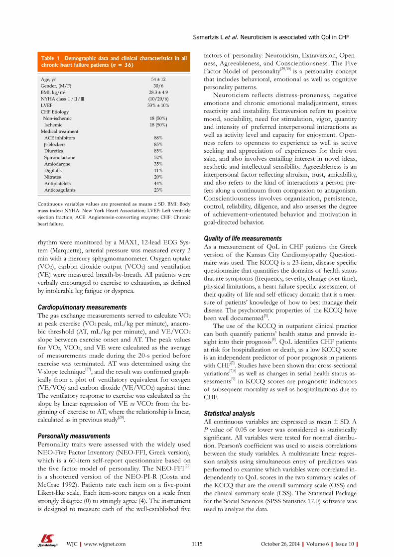
rhythm were monitored by a MAX1, 12-lead ECG Sys-tem (Marquette), arterial pressure was measured every 2 min with a mercury sphygmomanometer. Oxygen uptake (VO2), carbon dioxide output (VCO2) and ventilation (VE) were measured breath-by-breath. All patients were verbally encouraged to exercise to exhaustion, as defined by intolerable leg fatigue or dyspnea.
Cardiopulmonary measurementsThe gas exchange measurements served to calculate VO2 at peak exercise (VO2 peak, mL/kg per minute), anaero-bic threshold (AT, mL/kg per minute), and VE/VCO2 slope between exercise onset and AT. The peak values for VO2, VCO2, and VE were calculated as the average of measurements made during the 20-s period before exercise was terminated. AT was determined using the V-slope technique[27], and the result was confirmed graph-ically from a plot of ventilatory equivalent for oxygen (VE/VO2) and carbon dioxide (VE/VCO2) against time. The ventilatory response to exercise was calculated as the slope by linear regression of VE vs VCO2 from the be-ginning of exercise to AT, where the relationship is linear, calculated as in previous study[28].
Personality measurementsPersonality traits were assessed with the widely used NEO-Five Factor Inventory (NEO-FFI, Greek version), which is a 60-item self-report questionnaire based on the five factor model of personality. The NEO-FFI[29] is a shortened version of the NEO-PI-R (Costa and McCrae 1992). Patients rate each item on a five-point Likert-like scale. Each item-score ranges on a scale from strongly disagree (0) to strongly agree (4). The instrument is designed to measure each of the well-established five
factors of personality: Neuroticism, Extraversion, Open-ness, Agreeableness, and Conscientiousness. The Five Factor Model of personality[29,30] is a personality concept that includes behavioral, emotional as well as cognitive personality patterns.
Neuroticism reflects distress-proneness, negative emotions and chronic emotional maladjustment, stress reactivity and instability. Extraversion refers to positive mood, sociability, need for stimulation, vigor, quantity and intensity of preferred interpersonal interactions as well as activity level and capacity for enjoyment. Open-ness refers to openness to experience as well as active seeking and appreciation of experiences for their own sake, and also involves entailing interest in novel ideas, aesthetic and intellectual sensibility. Agreeableness is an interpersonal factor reflecting altruism, trust, amicability, and also refers to the kind of interactions a person pre-fers along a continuum from compassion to antagonism. Conscientiousness involves organization, persistence, control, reliability, diligence, and also assesses the degree of achievement-orientated behavior and motivation in goal-directed behavior.
Quality of life measurementsAs a measurement of QoL in CHF patients the Greek version of the Kansas City Cardiomyopathy Question-naire was used. The KCCQ is a 23-item, disease specific questionnaire that quantifies the domains of health status that are symptoms (frequency, severity, change over time), physical limitations, a heart failure specific assessment of their quality of life and self-efficacy domain that is a mea-sure of patients’ knowledge of how to best manage their disease. The psychometric properties of the KCCQ have been well documented[3].
The use of the KCCQ in outpatient clinical practice can both quantify patients’ health status and provide in-sight into their prognosis[8]. QoL identifies CHF patients at risk for hospitalization or death, as a low KCCQ score is an independent predictor of poor prognosis in patients with CHF[7]. Studies have been shown that cross-sectional variations[7,8] as well as changes in serial health status as-sessments[9] in KCCQ scores are prognostic indicators of subsequent mortality as well as hospitalizations due to CHF.
Statistical analysisAll continuous variables are expressed as mean ± SD. A P value of 0.05 or lower was considered as statistically significant. All variables were tested for normal distribu-tion. Pearson’s coefficient was used to assess correlations between the study variables. A multivariate linear regres-sion analysis using simultaneous entry of predictors was performed to examine which variables were correlated in-dependently to QoL scores in the two summary scales of the KCCQ that are the overall summary scale (OSS) and the clinical summary scale (CSS). The Statistical Package for the Social Sciences (SPSS Statistics 17.0) software was used to analyze the data.
October 26, 2014|Volume 6|Issue 10|WJC|www.wjgnet.com 1115
Age, yr 54 ± 12 Gender, (M/F) 30/6 BMI, kg/m² 28.3 ± 4.9 NYHA class Ⅰ/Ⅱ/Ⅲ (10/20/6) LVEF 33% ± 10% CHF Etiology Non-ischemic 18 (50%) Ischemic 18 (50%) Medical treatment ACE inhibitors 88% β-blockers 85% Diuretics 85% Spironolactone 52% Amiodarone 35% Digitalis 11% Nitrates 20% Antiplatelets 44% Anticoagulants 23%
Table 1 Demographic data and clinical characteristics in all chronic heart failure patients (n = 36)
Continuous variables values are presented as means ± SD. BMI: Body mass index; NYHA: New York Heart Association; LVEF: Left ventricle ejection fraction; ACE: Angiotensin-converting enzyme; CHF: Chronic heart failure.
Samartzis L et al . Neuroticism is associated with Qol in CHF
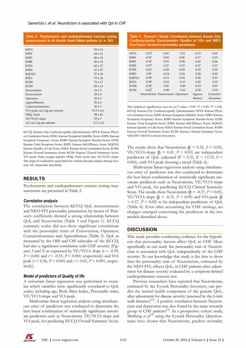
October 26, 2014|Volume 6|Issue 10|WJC|www.wjgnet.com
The results show that Neuroticism (β = 0.32, P < 0.05), VE/VCO2 slope (β = 0.41, P < 0.05) are independent predictors of QoL (adjusted R2 = 0.51, F = 13.33, P < 0.001), with VO2 peak showing a trend (Table 4).
Multivariate linear regression analysis using simultane-ous entry of predictors was also conducted to determine the best linear combination of statistically significant uni-variate predictors such as Neuroticism, VE/VCO2 slope and VO2 peak, for predicting KCCQ Clinical Summary Score. The results show Neuroticism (β = -0.37, P < 0.05), VE/VCO2 slope (β = -0.31, P < 0.05) and VO2 peak (β = 0.37, P < 0.05) to be independent predictors of QoL (Table 4). Even after accounting for CHF etiology, no changes emerged concerning the predictors in the two models described above.
DISCUSSIONThis study provides confirming evidence for the hypoth-esis that personality factors affect QoL in CHF. More specifically, in our study the personality trait of Neuroti-cism is associated with QoL independently of the CHF severity. To our knowledge this study is the first to show that the personality trait of Neuroticism, estimated by the NEO-FFI, affects QoL, in CHF patients after adjust-ment for disease severity evaluated by a symptom-limited cardiopulmonary exercise test.
Previous researchers have reported that Neuroticism, estimated by the Eysenk Personality Inventory, can pre-dict the mental health component of the generic QoL, after adjustment for disease severity (assessed by the 6-min walk distance)[22]. A positive correlation between Neuroti-cism and depression was also found by the same research group in CHF patients[24]. In a prospective cohort study, Murberg et al[23] using the Eysenk Personality Question-naire have shown that Neuroticism, predicts mortality
RESULTSPsychometric and cardiopulmonary exercise testing mea-surements are presented in Table 2.
Correlation analysisThe correlations between KCCQ QoL measurements and NEO-FFI personality parameters, by means of Pear-son’s coefficients showed a strong relationship between QoL and Neuroticism (Table 3 and Figure 1). KCCQ summary scales did not show significant correlations with the personality traits of Extraversion, Openness, Conscientiousness and Agreeableness (Table 3). QoL as measured by the OSS and CSS subscales of the KCCQ, had also a significant correlation with CHF severity (Fig-ures 2 and 3) as expressed by VE/VCO2 slope (r = -0.59, P < 0.001 and r = -0.55, P < 0.001, respectively) and VO2
peak (r = 0.56, P < 0.001 and r = 0.65, P < 0.001, respec-tively).
Model of predictors of Quality of lifeA univariate linear regression was performed to exam-ine which variables were significantly correlated to QoL scales, including age, Body Mass Index, Personality traits, VE/VCO2 slope and VO2 peak.
Multivariate linear regression analysis using simultane-ous entry of predictors was conducted to determine the best linear combination of statistically significant univari-ate predictors such as Neuroticism, VE/VCO2 slope and VO2 peak, for predicting KCCQ Overall Summary Score.
1116
KPLS 85 ± 14 KSSS 64 ± 21 KSFS 86 ± 16 KSBS 80 ± 19 KTSS 82 ± 17 KSES 67 ± 27 KQOLS 57 ± 24 KSLS 75 ± 26 KOSS 75 ± 17 KCSS 85 ± 14 Neuroticism 16 ± 5 Extraversion 29 ± 8 Openness 25 ± 5 Agreeableness 26 ± 6 Conscientiousness 30 ± 5 VO2 peak, mL/kg per minute 15.9 ± 4.4 WRp, Watt 99 ± 41 VE/VCO2 slope 35 ± 7 AT, mL/kg per minute 9.7 ± 2.4
Table 2 Psychometric and cardiopulmonary exercise testing measurements in all chronic heart failure patients (n = 36)
KCCQ: Kansas City Cardiomyopathy Questionnaire; KPLS: Kansas Physi-cal Limitation Score; KSSS: Kansas Symptom Stability Score; KSFS: Kansas Symptom Frequency Score; KSBS: Kansas Symptom Burden Score; KTSS: Kansas Total Symptom Score; KSES: Kansas Self-Efficacy Score; KQOLS: Kansas Quality of Life Score; KSLS: Kansas Social Limitation Score; KOSS: Kansas Overall Summary Score; KCSS: Kansas Clinical Summary Score; VO2 peak: Peak oxygen uptake; WRp: Peak work rate; VE/VCO2 slope: The slope of ventilatory equivalent for carbon dioxide output during exer-cise; AT: Anaerobic threshold.
KPLS -0.53b -0.01 0.31 -0.15 -0.03 KSSS -0.30 -0.02 -0.06 -0.17 -0.15 KSFS -0.54b -0.31 -0.09 -0.41 -0.24 KSBS -0.57b -0.27 -0.15 -0.27 -0.37 KTSS -0.61b -0.29 -0.09 -0.35 -0.32 KSES 0.00 -0.14 -0.24 -0.28 -0.42 KQOLS -0.38a -0.13 -0.02 -0.26 -0.31 KSLS -0.39a -0.12 0.13 0.02 0.10 KOSS -0.54b 0.05 0.08 -0.19 -0.07 KCSS -0.62b -0.08 0.10 -0.28 -0.10
Neuroticism Extraversion Openness Agreea-bleness
Conscien-tiousness
Table 3 Pearson’s Simple Correlations between Kansas City Cardiomyopathy Questionnaire Quality of Life and NEO Five-Factor Inventory personality parameters
The statistical significance was set at P value < 0.05. aP < 0.05; bP < 0.01. KCCQ: Kansas City Cardiomyopathy Questionnaire; KPLS: Kansas Physi-cal Limitation Score; KSSS: Kansas Symptom Stability Score; KSFS: Kansas Symptom Frequency Score; KSBS: Kansas Symptom Burden Score; KTSS: Kansas Total Symptom Score; KSES: Kansas Self-Efficacy Score; KQOLS: Kansas Quality of Life Score; KSLS: Kansas Social Limitation Score; KOSS: Kansas Overall Summary Score; KCSS: Kansas Clinical Summary Score; NEO-FFI: NEO Five-Factor Inventory.
Samartzis L et al . Neuroticism is associated with Qol in CHF
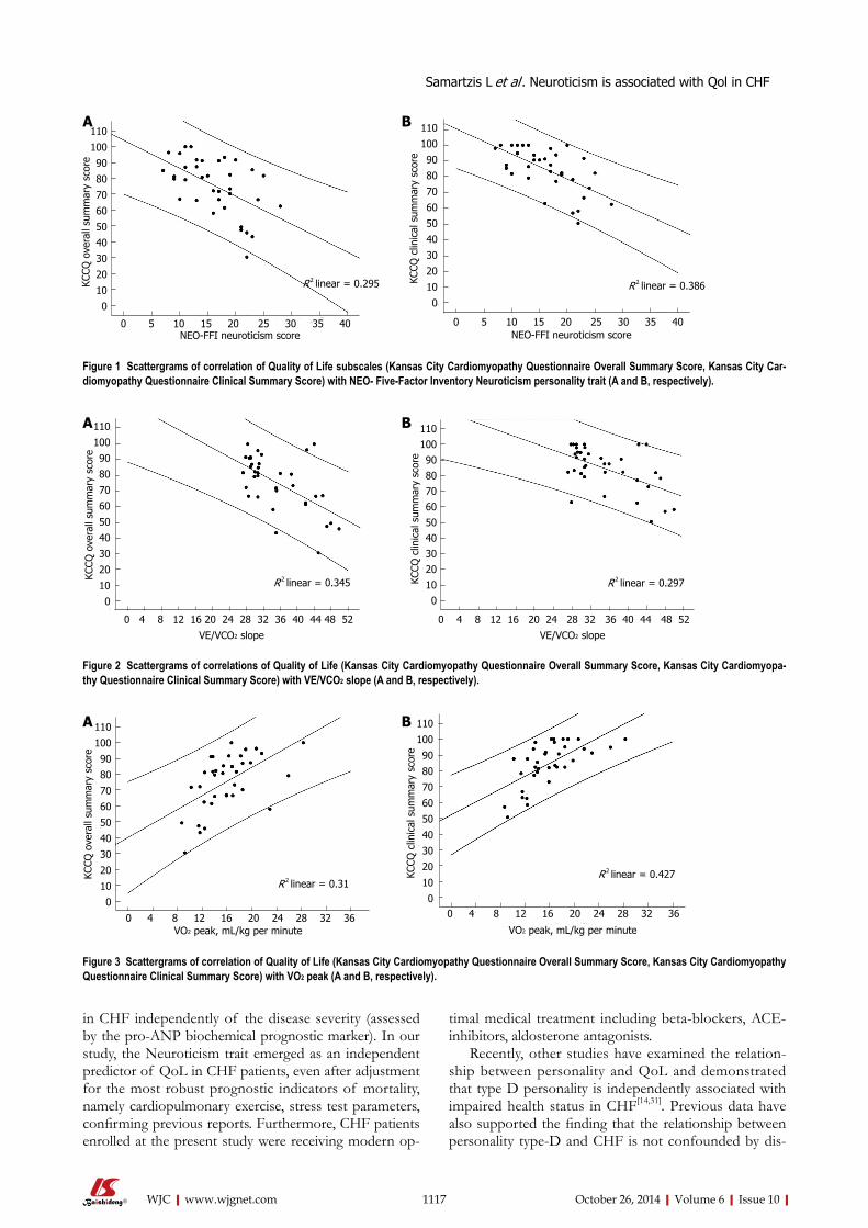
October 26, 2014|Volume 6|Issue 10|WJC|www.wjgnet.com
in CHF independently of the disease severity (assessed by the pro-ANP biochemical prognostic marker). In our study, the Neuroticism trait emerged as an independent predictor of QoL in CHF patients, even after adjustment for the most robust prognostic indicators of mortality, namely cardiopulmonary exercise, stress test parameters, confirming previous reports. Furthermore, CHF patients enrolled at the present study were receiving modern op-
timal medical treatment including beta-blockers, ACE-inhibitors, aldosterone antagonists.
Recently, other studies have examined the relation-ship between personality and QoL and demonstrated that type D personality is independently associated with impaired health status in CHF[14,31]. Previous data have also supported the finding that the relationship between personality type-D and CHF is not confounded by dis-
1117
110
100
90
80
70
60
50
40
30
20
10
0
110
100
90
80
70
60
50
40
30
20
10
0
110
100
90
80
70
60
50
40
30
20
10
0
1101009080706050403020100
110
100
90
80
70
60
50
40
30
20
10
0
110
100
90
80
70
60
50
40
30
20
10
0
0 5 10 15 20 25 30 35 40 0 5 10 15 20 25 30 35 40
0 4 8 12 16 20 24 28 32 36 40 44 48 52 0 4 8 12 16 20 24 28 32 36 40 44 48 52
0 4 8 12 16 20 24 28 32 36 0 4 8 12 16 20 24 28 32 36
KCCQ
ove
rall
sum
mar
y sc
ore
KCCQ
clin
ical
sum
mar
y sc
ore
KCCQ
ove
rall
sum
mar
y sc
ore
KCCQ
clin
ical
sum
mar
y sc
ore
KCCQ
clin
ical
sum
mar
y sc
ore
KCCQ
ove
rall
sum
mar
y sc
ore
VO2 peak, mL/kg per minute VO2 peak, mL/kg per minute
VE/VCO2 slope VE/VCO2 slope
NEO-FFI neuroticism score NEO-FFI neuroticism score
R 2 linear = 0.295 R 2 linear = 0.386
R 2 linear = 0.297R 2 linear = 0.345
R 2 linear = 0.31R 2 linear = 0.427
Figure 1 Scattergrams of correlation of Quality of Life subscales (Kansas City Cardiomyopathy Questionnaire Overall Summary Score, Kansas City Car-diomyopathy Questionnaire Clinical Summary Score) with NEO- Five-Factor Inventory Neuroticism personality trait (A and B, respectively).
Figure 2 Scattergrams of correlations of Quality of Life (Kansas City Cardiomyopathy Questionnaire Overall Summary Score, Kansas City Cardiomyopa-thy Questionnaire Clinical Summary Score) with VE/VCO2 slope (A and B, respectively).
A B
A B
A B
Figure 3 Scattergrams of correlation of Quality of Life (Kansas City Cardiomyopathy Questionnaire Overall Summary Score, Kansas City Cardiomyopathy Questionnaire Clinical Summary Score) with VO2 peak (A and B, respectively).
Samartzis L et al . Neuroticism is associated with Qol in CHF
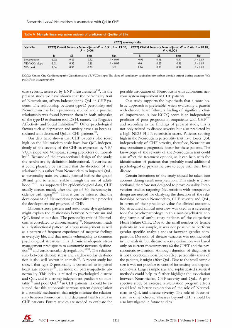
October 26, 2014|Volume 6|Issue 10|WJC|www.wjgnet.com
ease severity, assessed by BNP measurements[32]. In the present study we have shown that the personality trait of Neuroticism, affects independently QoL in CHF pa-tients. The relationship between type-D personality and Neuroticism has been previously studied and a positive relationship was found between them in both subscales of the type-D evaluation tool DS14, namely the Negative Affectivity and Social Inhibition[33]. Other psychological factors such as depression and anxiety have also been as-sociated with decreased QoL in CHF patients[12].
Our data have shown that CHF patients who score high on the Neuroticism scale have low QoL indepen-dently of the severity of the CHF as expressed by VE/VCO2 slope and VO2 peak, strong predictors of mortal-ity[28]. Because of the cross-sectional design of the study, the results are by definition bidirectional. Nevertheless it could plausibly be assumed that the direction of the relationship is rather from Neuroticism to impaired QoL, as personality traits are usually formed before the age of 30 and tend to remain stable through the rest of adult-hood[34-37]. As supported by epidemiological data, CHF usually occurs mainly after the age of 30, increasing in-cidence with aging[38,39]. Thus it can be inferred that the development of Neuroticism personality trait precedes the development and progress of CHF.
Chronic stress pattern and autonomic dysregulation might explain the relationship between Neuroticism and QoL found in our data. The personality trait of Neuroti-cism is correlated to chronic anxiety[40]. Neuroticism leads to a dysfunctional pattern of stress management as well as a pattern of frequent experience of negative feelings in everyday life, and that means vulnerability to common psychological stressors. This chronic inadequate stress management predisposes to autonomic nervous dysfunc-tion[41] and cardiovascular dysregulation[42,43]. The relation-ship between chronic stress and cardiovascular dysfunc-tion is also well known in animals[44]. A recent study has shown that type-D personality is correlated to impaired heart rate recovery[45], an index of parasympathetic ab-normality. This index is related to psychological distress and QoL and is a strong independent predictor of mor-tality[46] and poor QoL[47] in CHF patients. It could be as-sumed that this autonomic nervous system dysregulation is a possible mechanism that might mediate the relation-ship between Neuroticism and decreased health status in CHF patients. Future studies are needed to evaluate the
possible association of Neuroticism with autonomic ner-vous system impairment in CHF patients.
Our study supports the hypothesis that a more ho-listic approach is preferable, when evaluating a patient with chronic heart failure, a finding of significant clini-cal importance. A low KCCQ score is an independent predictor of poor prognosis in outpatients with CHF[7-9] and according to the findings of present study, this is not only related to disease severity but also predicted by a high NEO-FFI Neuroticism score. Patients scoring high in the Neuroticism personality trait have lower QoL independently of CHF severity, therefore, Neuroticism may constitute a prognostic factor for these patients. The knowledge of the severity of the Neuroticism trait could also affect the treatment options, as it can help with the identification of patients that probably need additional psychological or psychiatric care to cope with their heart disease.
Certain limitations of the study should be taken into account during result interpretation. This study is cross-sectional, therefore not designed to prove causality. Inter-vention studies targeting Neuroticism with prospective design are needed for clarifying the direction of the rela-tionships between Neuroticism, CHF severity and QoL, in terms of their predictive value for clinical outcome. No structured clinical interview was used as a screening tool for psychopathology in this non-psychiatric set-ting sample of ambulatory patients of the outpatient Heart Failure Clinic. Due to the small number of female patients in our sample, it was not possible to perform gender-specific analysis and/or between-gender com-parisons. Duration of disease variables was not included in the analysis, but disease severity estimation was based only on current measurements via the CPET and the psy-chometric evaluation. Although duration of diagnosis it is not theoretically possible to affect personality traits of the patients, it might affect QoL. Due to the small sample size it was not possible to control for anxiety and depres-sion levels. Larger sample size and sophisticated statistical methods could help to further highlight the association between Neuroticism, CHF severity and QoL. A pro-spective study of exercise rehabilitation program effects could lead to better exploration of the role of Neuroti-cism to QoL and disease severity. The role of Neuroti-cism in other chronic illnesses beyond CHF should be also investigated in future studies.
1118
KCCQ summary scales
Variables KCCQ Overall Summary Score adjusted R 2 = 0.51; F = 13.33, P < 0.001
KCCQ Clinical Summary Score adjusted R 2 = 0.64; F =18.89, P < 0.001
B SE beta Sig. B SE beta Sig. Neuroticism -1.02 0.43 -0.32 P < 0.05 -0.95 0.31 -0.37 P < 0.05 VE/VCO2 slope -1.01 0.32 -0.41 P < 0.05 -0.6 0.23 -0.31 P < 0.05 VO2 peak 1.04 0.55 0.26 NS 1.16 0.39 0.37 P < 0.05
Table 4 Multiple linear regression analyses of predictors of Quality of Life
KCCQ: Kansas City Cardiomyopathy Questionnaire; VE/VCO2 slope: The slope of ventilatory equivalent for carbon dioxide output during exercise; VO2 peak: Peak oxygen uptake.
Samartzis L et al . Neuroticism is associated with Qol in CHF
October 26, 2014|Volume 6|Issue 10|WJC|www.wjgnet.com
In conclusion, Neuroticism personality trait predicts independently QoL in CHF patients after adjustment for CHF severity. Notwithstanding the limitations of a small study, we propose that a more flexible approach to CHF diagnosis, which includes personality dimensions along with a description of CHF symptoms, may result in a more inclusive and useful diagnostic scheme for treating people with chronic heart failure. Taking into account the relationship between Neuroticism and QoL, personality factors could probably help to explain at least partially some of the mortality risk in CHF patients previously predicted by poor QoL. Psychiatric interventions might possibly be incorporated into the treatment of these pa-tients to improve QoL and possibly prognosis.
COMMENTSBackgroundMeasuring and exploring factors that affect Quality of life (QoL) is important for chronic heart failure (CHF) patients in order to monitor their treatment course and assessing prognosis, as well as for evaluating treatment interventions’ and health services effectiveness. Patients that are experiencing low QoL present higher morbidity, hospitalization rate and mortality. QoL in CHF is related to exer-cise capacity and adherence to rehabilitation programs and low QoL is related to exercise training intolerance. Of the psychological factors that affect QoL, anxiety and depression levels have been shown to play an important role, but the role of personality traits of the CHF patient hasn’t been well understood yet. Taking into account that the personality trait of Neuroticism, has been demonstrated to affect QoL in chronically ill individuals, there is a need for exploration of its possible role as a predictor of QoL in patients with CHF.Research frontiersOf the available generic and disease-specific tools for describing dimensions of QoL in CHF, the Kansas City Cardiomyopathy Questionnaire is a self-reported, disease specific questionnaire that is considered reliable and valid for this popu-lation. Cardiopulmonary exercise testing has emerged over the years as a very useful modality for assessing variables related to exercise capacity and with CHF severity in this population.Innovations and breakthroughsPrevious researchers have reported that Neuroticism, estimated by the Ey-senk Personality Inventory (EPI), can predict the mental health component of the generic QoL, after adjustment for disease severity assessed by the 6-min walk distance. Also a prospective cohort study using the Eysenk Personality Questionnaire (EPQ), an expanded version of EPI, has shown that Neuroticism predicts mortality in CHF independently of the disease severity assessed by the pro-ANP biochemical prognostic marker. These study provides additional evidence for the hypothesis that personality factors affect QoL in CHF. More spe-cifically, these found that the personality trait of Neuroticism is associated with QoL independently of the CHF severity. To these knowledge this study is the first to show that the personality trait of Neuroticism, estimated by the Five-Factor Personality Inventory, affects QoL, in CHF patients after adjustment for disease severity evaluated by a symptom-limited cardiopulmonary exercise test, the “gold” standard for the assessment of exercise capacity in these patients.ApplicationsThe results of this study showed that not only disease severity but also the personality characteristic of Neuroticism can affect patients’ QoL, therefore worsening prognosis of CHF. The personality of CHF patient has to be taken into account during CHF treatment and rehabilitation programs, and a tailor-made individualized psychosocial intervention by a mental health professional might help improving patients QoL and consequently CHF prognosis.TerminologyCHF population in this manuscript refers to patients with Chronic Heart Failure, that are currently been stabilized in optimal medical treatment and their left ven-tricle ejection fraction remains less than 45%. CPET refers to CardioPulmonary Exercise Testing that is used for evaluation of the patient in terms of heart func-tion and exercise capacity. This evaluation is also important for exercise training rehabilitation of the CHF patient.
Peer reviewThis is a very interesting and novel observational study of the effect of neuroti-cism on quality of life of a Greek cohort of chronic heart failure patients. The paper reads well overall and reports some novel findings.
REFERENCES1 Spertus J. Selecting end points in clinical trials: What evi-
dence do we really need to evaluate a new treatment? Am Heart J 2001; 142: 745-747 [PMID: 11685157 DOI: 10.1067/mhj.2001.119135]
2 Normand SL, Rector TS, Neaton JD, Piña IL, Lazar RM, Proestel SE, Fleischer DJ, Cohn JN, Spertus JA. Clinical and analytical considerations in the study of health status in de-vice trials for heart failure. J Card Fail 2005; 11: 396-403 [PMID: 15948091 DOI: 10.1016/j.cardfail.2005.04.002]
3 Green CP, Porter CB, Bresnahan DR, Spertus JA. Develop-ment and evaluation of the Kansas City Cardiomyopathy Questionnaire: a new health status measure for heart failure. J Am Coll Cardiol 2000; 35: 1245-1255 [PMID: 10758967 DOI: 10.1016/S0735-1097(00)00531-3]
4 Juenger J, Schellberg D, Kraemer S, Haunstetter A, Zugck C, Herzog W, Haass M. Health related quality of life in patients with congestive heart failure: comparison with other chronic diseases and relation to functional variables. Heart 2002; 87: 235-241 [PMID: 11847161 DOI: 10.1136/heart.87.3.235]
5 Athanasopoulos LV, Dritsas A, Doll HA, Cokkinos DV. Comparative value of NYHA functional class and quality-of-life questionnaire scores in assessing heart failure. J Car-diopulm Rehabil Prev 2010; 30: 101-105 [PMID: 19952769 DOI: 10.1097/HCR.0b013e3181be7e47]
6 Athanasopoulos LV, Dritsas A, Doll HA, Cokkinos DV. Explanation of the variance in quality of life and activity ca-pacity of patients with heart failure by laboratory data. Eur J Cardiovasc Prev Rehabil 2010; 17: 375-379 [PMID: 19940776 DOI: 10.1097/HJR.0b013e328333e962]
7 Heidenreich PA, Spertus JA, Jones PG, Weintraub WS, Rumsfeld JS, Rathore SS, Peterson ED, Masoudi FA, Krum-holz HM, Havranek EP, Conard MW, Williams RE. Health status identifies heart failure outpatients at risk for hospital-ization or death. J Am Coll Cardiol 2006; 47: 752-756 [PMID: 16487840 DOI: 10.1016/j.jacc.2005.11.021]
8 Soto GE, Jones P, Weintraub WS, Krumholz HM, Sper-tus JA. Prognostic value of health status in patients with heart failure after acute myocardial infarction. Circula-tion 2004; 110: 546-551 [PMID: 15262843 DOI: 10.1161/01.CIR.0000136991.85540.A9]
9 Kosiborod M, Soto GE, Jones PG, Krumholz HM, Wein-traub WS, Deedwania P, Spertus JA. Identifying heart failure patients at high risk for near-term cardiovascular events with serial health status assessments. Circulation 2007; 115: 1975-1981 [PMID: 17420346 DOI: 10.1161/CIRCULA-TIONAHA.106.670901]
10 Flynn KE, Piña IL, Whellan DJ, Lin L, Blumenthal JA, Ellis SJ, Fine LJ, Howlett JG, Keteyian SJ, Kitzman DW, Kraus WE, Miller NH, Schulman KA, Spertus JA, O’Connor CM, Weinfurt KP. Effects of exercise training on health status in patients with chronic heart failure: HF-ACTION randomized controlled trial. JAMA 2009; 301: 1451-1459 [PMID: 19351942 DOI: 10.1001/jama.2009.457]
11 O’Connor CM, Whellan DJ, Lee KL, Keteyian SJ, Cooper LS, Ellis SJ, Leifer ES, Kraus WE, Kitzman DW, Blumenthal JA, Rendall DS, Miller NH, Fleg JL, Schulman KA, McKelvie RS, Zannad F, Piña IL. Efficacy and safety of exercise training in patients with chronic heart failure: HF-ACTION randomized controlled trial. JAMA 2009; 301: 1439-1450 [PMID: 19351941 DOI: 10.1001/jama.2009.454]
12 Chung ML, Moser DK, Lennie TA, Rayens MK. The effects of depressive symptoms and anxiety on quality of life in patients with heart failure and their spouses: testing dyadic
1119
COMMENTS
Samartzis L et al . Neuroticism is associated with Qol in CHF
October 26, 2014|Volume 6|Issue 10|WJC|www.wjgnet.com
dynamics using Actor-Partner Interdependence Model. J Psychosom Res 2009; 67: 29-35 [PMID: 19539816 DOI: 10.1016/j.jpsychores.2009.01.009]
13 Faller H, Steinbüchel T, Störk S, Schowalter M, Ertl G, An-germann CE. Impact of depression on quality of life assess-ment in heart failure. Int J Cardiol 2010; 142: 133-137 [PMID: 19162345 DOI: 10.1016/j.ijcard.2008.12.093]
14 Schiffer AA, Pedersen SS, Widdershoven JW, Hendriks EH, Winter JB, Denollet J. The distressed (type D) personality is independently associated with impaired health status and increased depressive symptoms in chronic heart failure. Eur J Cardiovasc Prev Rehabil 2005; 12: 341-346 [PMID: 16079641 DOI: 10.1097/01.hjr.0000173107.76109.6c]
15 Spindler H, Kruse C, Zwisler AD, Pedersen SS. Increased anxiety and depression in Danish cardiac patients with a type D personality: cross-validation of the Type D Scale (DS14). Int J Behav Med 2009; 16: 98-107 [PMID: 19322662 DOI: 10.1007/s12529-009-9037-5]
16 Schiffer AA, Smith OR, Pedersen SS, Widdershoven JW, De-nollet J. Type D personality and cardiac mortality in patients with chronic heart failure. Int J Cardiol 2010; 142: 230-235 [PMID: 19162343 DOI: 10.1016/j.ijcard.2008.12.090]
17 Jerant A, Chapman BP, Franks P. Personality and EQ-5D scores among individuals with chronic conditions. Qual Life Res 2008; 17: 1195-1204 [PMID: 18839336 DOI: 10.1007/s11136-008-9401-y]
18 Chapman BP, Duberstein PR, Sörensen S, Lyness JM. Per-sonality and perceived health in older adults: the five factor model in primary care. J Gerontol B Psychol Sci Soc Sci 2006; 61: P362-P365 [PMID: 17114306]
19 Duberstein PR, Sörensen S, Lyness JM, King DA, Conwell Y, Seidlitz L, Caine ED. Personality is associated with per-ceived health and functional status in older primary care patients. Psychol Aging 2003; 18: 25-37 [PMID: 12641310 DOI: 10.1037/0882-7974.18.1.25]
20 Gilhooly M, Hanlon P, Cullen B, Macdonald S, Whyte B. Successful ageing in an area of deprivation: part 2--a quan-titative exploration of the role of personality and beliefs in good health in old age. Public Health 2007; 121: 814-821 [PMID: 17606277 DOI: 10.1016/j.puhe.2007.03.003]
21 Goodwin R, Engstrom G. Personality and the perception of health in the general population. Psychol Med 2002; 32: 325-332 [PMID: 11866326 DOI: 10.1017/S0033291701005104]
22 Westlake C, Dracup K, Creaser J, Livingston N, Heywood JT, Huiskes BL, Fonarow G, Hamilton M. Correlates of health-related quality of life in patients with heart failure. Heart Lung 2002; 31: 85-93 [PMID: 11910383 DOI: 10.1067/mhl.2002.122839]
23 Murberg TA, Bru E, Svebak S, Tveterås R, Aarsland T. De-pressed mood and subjective health symptoms as predictors of mortality in patients with congestive heart failure: a two-years follow-up study. Int J Psychiatry Med 1999; 29: 311-326 [PMID: 10642905 DOI: 10.1016/S0191-8869(00)00067-2]
24 Westlake C, Dracup K, Fonarow G, Hamilton M. Depres-sion in patients with heart failure. J Card Fail 2005; 11: 30-35 [PMID: 15704061 DOI: 10.1016/j.cardfail.2004.03.007]
25 American Thoracic Society; American College of Chest Physicians. ATS/ACCP Statement on cardiopulmonary exercise testing. Am J Respir Crit Care Med 2003; 167: 211-277 [PMID: 12524257 DOI: 10.1164/rccm.167.2.211]
26 Hansen JE, Sue DY, Wasserman K. Predicted values for clinical exercise testing. Am Rev Respir Dis 1984; 129: S49-S55 [PMID: 6421218]
27 Beaver WL, Wasserman K, Whipp BJ. A new method for detecting anaerobic threshold by gas exchange. J Appl Physiol (1985) 1986; 60: 2020-2027 [PMID: 3087938]
28 Nanas SN, Nanas JN, Sakellariou DCh, Dimopoulos SK, Drakos SG, Kapsimalakou SG, Mpatziou CA, Papaza-chou OG, Dalianis AS, Anastasiou-Nana MI, Roussos C. VE/VCO2 slope is associated with abnormal resting hae-
modynamics and is a predictor of long-term survival in chronic heart failure. Eur J Heart Fail 2006; 8: 420-427 [PMID: 16310408 DOI: 10.1016/j.ejheart.2005.10.003]
29 Costa PT, McCrae RR. Revised NEO Personality Inventory (NEO-PI-R) and NEO Five-Factor Inventory (NEO-FFI) professional manual. Odessa, FL: Psychological Assessment Resources, 1992
30 Digman J. Personality Structure: Emergence of the 5-Factor Model. Annu Rev Psychol 1990: 417-440 [DOI: 10.1146/an-nurev.ps.41.020190.002221]
31 Pedersen SS, Herrmann-Lingen C, de Jonge P, Scherer M. Type D personality is a predictor of poor emotional quality of life in primary care heart failure patients independent of depressive symptoms and New York Heart Association functional class. J Behav Med 2010; 33: 72-80 [PMID: 19937107 DOI: 10.1007/s10865-009-9236-1]
32 Pelle AJ, van den Broek KC, Szabó B, Kupper N. The re-lationship between type D personality and chronic heart failure is not confounded by disease severity as assessed by BNP. Int J Cardiol 2010; 145: 82-83 [PMID: 19477027 DOI: 10.1016/j.ijcard.2009.05.018]
33 De Fruyt FDJ. Type D personality: A Five-Factor Model per-spective. Psychol Health 2002; 17: 671-683 [DOI: 10.1080/08870440290025858]
34 Costa PT, McCrae RR. Personality in adulthood: a six-year longitudinal study of self-reports and spouse ratings on the NEO Personality Inventory. J Pers Soc Psychol 1988; 54: 853-863 [PMID: 3379583 DOI: 10.1037/0022-3514.54.5.85310.1037/0022-3514.54.5.853]
35 Terracciano A, Costa PT, McCrae RR. Personality plasticity after age 30. Pers Soc Psychol Bull 2006; 32: 999-1009 [PMID: 16861305 DOI: 10.1177/0146167206288599]
36 McCrae RR, Costa PT, Arenberg D. Constancy of adult per-sonality structure in males: longitudinal, cross-sectional and times-of-measurement analyses. J Gerontol 1980; 35: 877-883 [PMID: 6969272 DOI: 10.1093/geronj/35.6.877]
37 Costa PT, McCrae RR, Zonderman AB, Barbano HE, Lebow-itz B, Larson DM. Cross-sectional studies of personality in a national sample: 2. Stability in neuroticism, extraversion, and openness. Psychol Aging 1986; 1: 144-149 [PMID: 3267391 DOI: 10.1037/0882-7974.1.2.144]
38 Cowie MR, Mosterd A, Wood DA, Deckers JW, Poole-Wil-son PA, Sutton GC, Grobbee DE. The epidemiology of heart failure. Eur Heart J 1997; 18: 208-225 [PMID: 9043837 DOI: 10.1093/oxfordjournals.eurheartj.a015223]
39 Cowie MR, Wood DA, Coats AJ, Thompson SG, Poole-Wilson PA, Suresh V, Sutton GC. Incidence and aetiology of heart failure; a population-based study. Eur Heart J 1999; 20: 421-428 [PMID: 10213345 DOI: 10.1053/euhj.1998.1280]
40 Brandes M, Bienvenu OJ. Personality and anxiety disorders. Curr Psychiatry Rep 2006; 8: 263-269 [PMID: 16879789 DOI: 10.1007/s11920-006-0061-8]
41 Watkins LL, Blumenthal JA, Carney RM. Association of anx-iety with reduced baroreflex cardiac control in patients after acute myocardial infarction. Am Heart J 2002; 143: 460-466 [PMID: 11868052 DOI: 10.1067/mhj.2002.120404]
42 Grippo AJ, Johnson AK. Stress, depression and cardiovascu-lar dysregulation: a review of neurobiological mechanisms and the integration of research from preclinical disease mod-els. Stress 2009; 12: 1-21 [PMID: 19116888 DOI: 10.1080/10253890802046281]
43 Lucini D, Di Fede G, Parati G, Pagani M. Impact of chronic psychosocial stress on autonomic cardiovascu-lar regulation in otherwise healthy subjects. Hypertension 2005; 46: 1201-1206 [PMID: 16203875 DOI: 10.1161/01.HYP.0000185147.32385.4b]
44 Sgoifo A, Pozzato C, Costoli T, Manghi M, Stilli D, Ferrari PF, Ceresini G, Musso E. Cardiac autonomic responses to intermit-tent social conflict in rats. Physiol Behav 2001; 73: 343-349 [PMID: 11438360 DOI: 10.1016/S0031-9384(01)00455-3]
1120
Samartzis L et al . Neuroticism is associated with Qol in CHF

October 26, 2014|Volume 6|Issue 10|WJC|www.wjgnet.com
45 von Känel R, Barth J, Kohls S, Saner H, Znoj H, Saner G, Schmid JP. Heart rate recovery after exercise in chronic heart failure: role of vital exhaustion and type D personality. J Cardiol 2009; 53: 248-256 [PMID: 19304130 DOI: 10.1016/j.jjcc.2008.11.008]
46 Nanas S, Anastasiou-Nana M, Dimopoulos S, Sakellariou D, Alexopoulos G, Kapsimalakou S, Papazoglou P, Tsolakis E, Papazachou O, Roussos C, Nanas J. Early heart rate recov-
ery after exercise predicts mortality in patients with chronic heart failure. Int J Cardiol 2006; 110: 393-400 [PMID: 16371237 DOI: 10.1016/j.ijcard.2005.10.032]
47 von Känel R, Saner H, Kohls S, Barth J, Znoj H, Saner G, Schmid JP. Relation of heart rate recovery to psychological distress and quality of life in patients with chronic heart failure. Eur J Cardiovasc Prev Rehabil 2009; 16: 645-650 [PMID: 19801939 DOI: 10.1097/HJR.0b013e3283299542]
P- Reviewer: Brugts JJ, Lymperopoulos A, Nunez-Gil IJ S- Editor: Ji FF L- Editor: A E- Editor: Wu HL
1121
Samartzis L et al . Neuroticism is associated with Qol in CHF
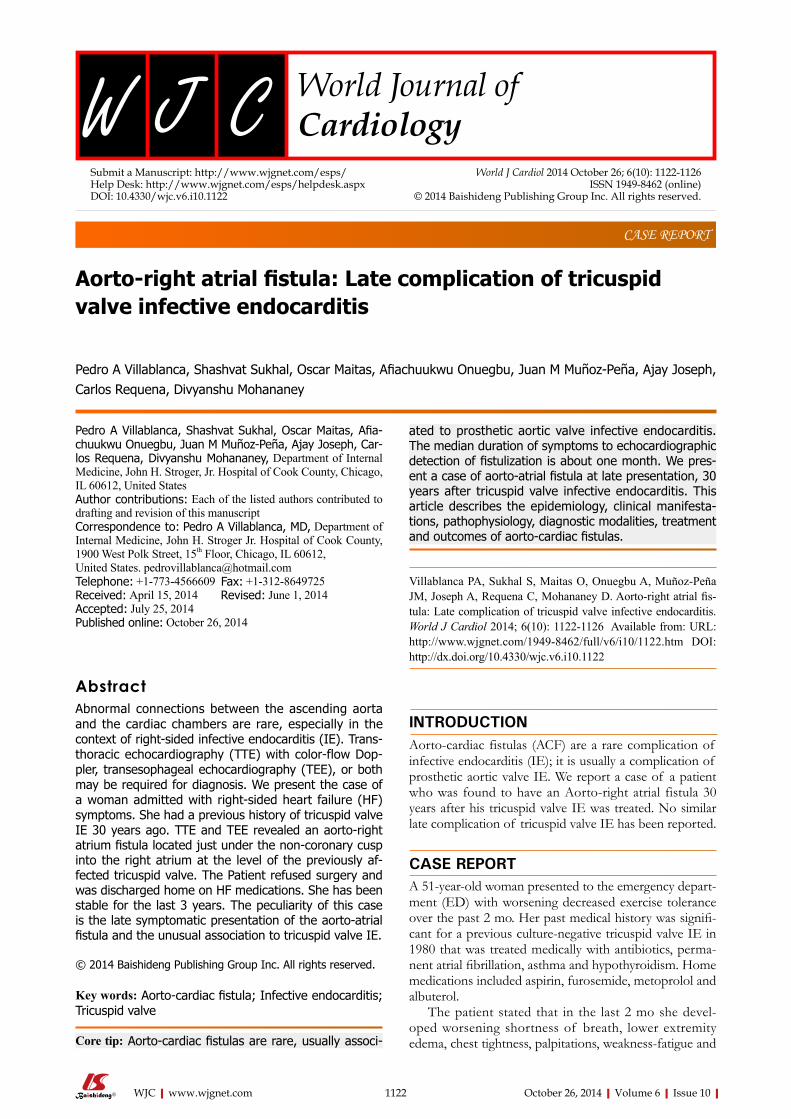
Aorto-right atrial fistula: Late complication of tricuspid valve infective endocarditis
Pedro A Villablanca, Shashvat Sukhal, Oscar Maitas, Afiachuukwu Onuegbu, Juan M Muñoz-Peña, Ajay Joseph, Carlos Requena, Divyanshu Mohananey
Pedro A Villablanca, Shashvat Sukhal, Oscar Maitas, Afia-chuukwu Onuegbu, Juan M Muñoz-Peña, Ajay Joseph, Car-los Requena, Divyanshu Mohananey, Department of Internal Medicine, John H. Stroger, Jr. Hospital of Cook County, Chicago, IL 60612, United States Author contributions: Each of the listed authors contributed to drafting and revision of this manuscriptCorrespondence to: Pedro A Villablanca, MD, Department of Internal Medicine, John H. Stroger Jr. Hospital of Cook County, 1900 West Polk Street, 15th Floor, Chicago, IL 60612, United States. [email protected]: +1-773-4566609 Fax: +1-312-8649725Received: April 15, 2014 Revised: June 1, 2014 Accepted: July 25, 2014Published online: October 26, 2014
AbstractAbnormal connections between the ascending aorta and the cardiac chambers are rare, especially in the context of right-sided infective endocarditis (IE). Trans-thoracic echocardiography (TTE) with color-flow Dop-pler, transesophageal echocardiography (TEE), or both may be required for diagnosis. We present the case of a woman admitted with right-sided heart failure (HF) symptoms. She had a previous history of tricuspid valve IE 30 years ago. TTE and TEE revealed an aorto-right atrium fistula located just under the non-coronary cusp into the right atrium at the level of the previously af-fected tricuspid valve. The Patient refused surgery and was discharged home on HF medications. She has been stable for the last 3 years. The peculiarity of this case is the late symptomatic presentation of the aorto-atrial fistula and the unusual association to tricuspid valve IE.
© 2014 Baishideng Publishing Group Inc. All rights reserved.
Key words: Aorto-cardiac fistula; Infective endocarditis; Tricuspid valve
Core tip: Aorto-cardiac fistulas are rare, usually associ-
ated to prosthetic aortic valve infective endocarditis. The median duration of symptoms to echocardiographic detection of fistulization is about one month. We pres-ent a case of aorto-atrial fistula at late presentation, 30 years after tricuspid valve infective endocarditis. This article describes the epidemiology, clinical manifesta-tions, pathophysiology, diagnostic modalities, treatment and outcomes of aorto-cardiac fistulas.
Villablanca PA, Sukhal S, Maitas O, Onuegbu A, Muñoz-Peña JM, Joseph A, Requena C, Mohananey D. Aorto-right atrial fis-tula: Late complication of tricuspid valve infective endocarditis. World J Cardiol 2014; 6(10): 1122-1126 Available from: URL: http://www.wjgnet.com/1949-8462/full/v6/i10/1122.htm DOI: http://dx.doi.org/10.4330/wjc.v6.i10.1122
INTRODUCTIONAorto-cardiac fistulas (ACF) are a rare complication of infective endocarditis (IE); it is usually a complication of prosthetic aortic valve IE. We report a case of a patient who was found to have an Aorto-right atrial fistula 30 years after his tricuspid valve IE was treated. No similar late complication of tricuspid valve IE has been reported.
CASE REPORTA 51-year-old woman presented to the emergency depart-ment (ED) with worsening decreased exercise tolerance over the past 2 mo. Her past medical history was signifi-cant for a previous culture-negative tricuspid valve IE in 1980 that was treated medically with antibiotics, perma-nent atrial fibrillation, asthma and hypothyroidism. Home medications included aspirin, furosemide, metoprolol and albuterol.
The patient stated that in the last 2 mo she devel-oped worsening shortness of breath, lower extremity edema, chest tightness, palpitations, weakness-fatigue and
CASE REPORT
Submit a Manuscript: http://www.wjgnet.com/esps/Help Desk: http://www.wjgnet.com/esps/helpdesk.aspxDOI: 10.4330/wjc.v6.i10.1122
October 26, 2014|Volume 6|Issue 10|WJC|www.wjgnet.com
World Journal of CardiologyW J C
World J Cardiol 2014 October 26; 6(10): 1122-1126ISSN 1949-8462 (online)
© 2014 Baishideng Publishing Group Inc. All rights reserved.
1122
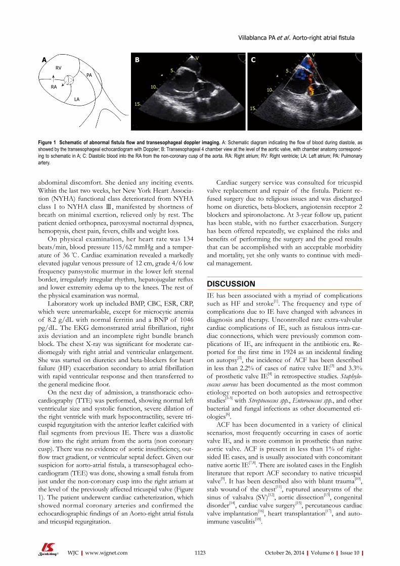
abdominal discomfort. She denied any inciting events. Within the last two weeks, her New York Heart Associa-tion (NYHA) functional class deteriorated from NYHA class Ⅰ to NYHA class Ⅲ, manifested by shortness of breath on minimal exertion, relieved only by rest. The patient denied orthopnea, paroxysmal nocturnal dyspnea, hemoptysis, chest pain, fevers, chills and weight loss.
On physical examination, her heart rate was 134 beats/min, blood pressure 115/62 mmHg and a temper-ature of 36 ℃. Cardiac examination revealed a markedly elevated jugular venous pressure of 12 cm, grade 4/6 low frequency pansystolic murmur in the lower left sternal border, irregularly irregular rhythm, hepatojugular reflux and lower extremity edema up to the knees. The rest of the physical examination was normal.
Laboratory work up included BMP, CBC, ESR, CRP, which were unremarkable, except for microcytic anemia of 8.2 g/dL with normal ferritin and a BNP of 1046 pg/dL. The EKG demonstrated atrial fibrillation, right axis deviation and an incomplete right bundle branch block. The chest X-ray was significant for moderate car-diomegaly with right atrial and ventricular enlargement. She was started on diuretics and beta-blockers for heart failure (HF) exacerbation secondary to atrial fibrillation with rapid ventricular response and then transferred to the general medicine floor.
On the next day of admission, a transthoracic echo-cardiography (TTE) was performed, showing normal left ventricular size and systolic function, severe dilation of the right ventricle with mark hypocontractility, severe tri-cuspid regurgitation with the anterior leaflet calcified with flail segments from previous IE. There was a diastolic flow into the right atrium from the aorta (non coronary cusp). There was no evidence of aortic insufficiency, out-flow tract gradient, or ventricular septal defect. Given our suspicion for aorto-atrial fistula, a transesophageal echo-cardiogram (TEE) was done, showing a small fistula from just under the non-coronary cusp into the right atrium at the level of the previously affected tricuspid valve (Figure 1). The patient underwent cardiac catheterization, which showed normal coronary arteries and confirmed the echocardiographic findings of an Aorto-right atrial fistula and tricuspid regurgitation.
Cardiac surgery service was consulted for tricuspid valve replacement and repair of the fistula. Patient re-fused surgery due to religious issues and was discharged home on diuretics, beta-blockers, angiotensin receptor 2 blockers and spironolactone. At 3-year follow up, patient has been stable, with no further exacerbation. Surgery has been offered repeatedly, we explained the risks and benefits of performing the surgery and the good results that can be accomplished with an acceptable morbidity and mortality, yet she only wants to continue with medi-cal management.
DISCUSSIONIE has been associated with a myriad of complications such as HF and stroke[1]. The frequency and type of complications due to IE have changed with advances in diagnosis and therapy. Uncontrolled rare extra-valvular cardiac complications of IE, such as fistulous intra-car-diac connections, which were previously common com-plications of IE, are infrequent in the antibiotic era. Re-ported for the first time in 1924 as an incidental finding on autopsy[2], the incidence of ACF has been described in less than 2.2% of cases of native valve IE[3] and 3.3% of prosthetic valve IE[4] in retrospective studies. Staphylo-coccus aureus has been documented as the most common etiology reported on both autopsies and retrospective studies[3-5] with Streptococcus spp., Enterococcus spp., and other bacterial and fungal infections as other documented eti-ologies[6].
ACF has been documented in a variety of clinical scenarios, most frequently occurring in cases of aortic valve IE, and is more common in prosthetic than native aortic valve. ACF is present in less than 1% of right-sided IE cases, and is usually associated with concomitant native aortic IE[7,8]. There are isolated cases in the English literature that report ACF secondary to native tricuspid valve[9]. It has been described also with blunt trauma[10], stab wound of the chest[11], ruptured aneurysms of the sinus of valsalva (SV)[12], aortic dissection[13], congenital disorder[14], cardiac valve surgery[15], percutaneous cardiac valve implantation[16], heart transplantation[17], and auto-immune vasculitis[18].
October 26, 2014|Volume 6|Issue 10|WJC|www.wjgnet.com
Villablanca PA et al . Aorto-right atrial fistula
1123
RV
RA
PA
LA15
10
5
V
15
10
5
Figure 1 Schematic of abnormal fistula flow and transesophageal doppler imaging. A: Schematic diagram indicating the flow of blood during diastole, as showed by the transesophageal echocardiogram with Doppler; B: Transesophageal 4 chamber view at the level of the aortic valve, with chamber anatomy correspond-ing to schematic in A; C: Diastolic blood into the RA from the non-coronary cusp of the aorta. RA: Right atrium; RV: Right ventricle; LA: Left atrium; PA: Pulmonary artery.
VA B C
The proposed theory to explain the fistulization mechanism between the aorta and the cardiac chamber is through the bacterial invasion and spread of the affected valve into the adjacent tissues and structures, resulting in the formation of a periannular abscess and erosion of the SV. The aortic abscesses involving the SV may rup-ture internally with erosion of the sinus and subsequent development of aorto-cavitary or aorto-pericardial fistu-las[3,5,7,19,20]. Perivalvular abscesses have been reported as the cause of 6%-9% of fistula cases[21,22]. Due to its rela-tive avascularity and infected regurgitation of jet striking subvalvular structures[23], the intervalvular fibrosa is more susceptible to infection[24]. The ACF creates a left to right shunt from any of the three aortic valve sinuses to any of the four cardiac chambers with no preponderance from any specific aortic sinus to a specific cavity, resulting in further hemodynamic deterioration[5,7]. These pathologic communications are highly morbid and lead to hemody-namic instability secondary to the shunt effect[19].
Diagnosing ACF can be challenging, and the clinical presentation will depend on the size of the shunt. Pa-tients with a small ACF may be completely asymptomatic with an associated murmur only[25,26], but the clinical pre-sentation may range from refractory HF[20] to a chest pain syndrome due to acute coronary syndrome and aortic dissection[17,27]. Cardiac auscultation may cause a continu-ous murmur[28], a thrill[29] or both[25], and can be the key to further pursue this diagnosis with appropriate imaging modalities. The median duration of symptoms to echo-cardiographic detection of fistulization is about 25 d as reported in a retrospective multi-center study[7]. There are isolated cases reported years after prosthetic valve implan-tation[15]. A high index of suspicion is required, especially in the background of recent surgery or previous IE.
Although aortography is the gold standard for diag-nosis, non-invasive methods such as contrast enhanced CT, MRI, and echocardiography are currently preferred. TTE is the initial test of choice in the routine assessment of patients presenting with HF symptoms or murmurs, and is therefore usually the first image modality that al-lows us to confirm or suspect the presence of an ACF. However, TEE is superior to TTE for better delineation of function and morphology when intra-cardiac com-plications, such as ACF, are suspected[30,31]. The high rate of echocardiographic diagnosis is likely due to the high-pressure differences between the aorta and the cardiac chambers, which enables observation of the highly tur-bulent flow that is easily detectable by color Doppler[7]. Three–dimensional echocardiography has been reported to have the potential to delineate anatomic structures, allowing a greater understanding of the pathological pro-cess and also obtaining unconventional views of cardiac structures[32]. It can delineate structures that are otherwise not visible in TEE and TTE, allowing cropping, full-volume data, and slicing in various planes[33,34]. Computed tomography, magnetic resonance imaging, and aortogra-phy can allow better description, position, dimension and anatomic conditions of the ACF, and may be required as an important adjunctive tool to confirm the diagnosis
and delineate the anatomy before closure[35-38].Surgery, which is the primary treatment of ACF, may
carry severe complications, particularly with critically un-stable patients with an increased postoperative mortality after surgical correction[4]. Factors associated with adverse outcomes include staphylococcal infection, urgent or emergency surgery, moderate to severe HF, renal failure, increased age and residual fistula[3,7,19,20,22,39]. With the high postoperative mortality with surgical closure of ACF and with the advancement of endovascular technologies, more emphasis is now placed on percutaneous closure with devices such as an Amplatzer plug[40,41], though it should be avoided in in patients with active infection.
We report a case of a patient who was found to have an Aorto-right atrial fistula 30 years after his tricuspid valve was treated for IE. To our knowledge and after a systematic review of the English literature, no similar late complication of treated IE has been reported.
COMMENTSCase characteristicsA 51-year-old female with a history of tricuspid valve infective endocarditis pre-sented with shortness of breath.Clinical diagnosisRight side heart failure symptoms. Differential diagnosisAorto-cardiac fistula vs valvular heart disease vs new infective endocarditis. Laboratory diagnosisHb of 8.2 g/dL with normal ferritin and a BNP of 1046 pg/dL; inflammatory mark-ers (erythrocyte sedimentation rate, serum C-reactive protein, blood cell count) were within normal limits. Imaging diagnosisTransthoracic and transesophageal echocardiography demonstrated Aorto-right atrial fistula.TreatmentThe patient was medically managed for her heart failure after she refused surgi-cal treatment.Related reportsEchocardiography images and explanatory figure are provided in the case re-port. Experiences and lessonsA high index of suspicion for aorto-cardiac fistula is required, especially in the background of recent surgery or previous infective endocarditis.Peer reviewThis is an interesting paper.
REFERENCES1 Mansur AJ, Grinberg M, da Luz PL, Bellotti G. The compli-
cations of infective endocarditis. A reappraisal in the 1980s. Arch Intern Med 1992; 152: 2428-2432 [PMID: 1456853]
2 Boyd L. A study of four thousand cases of aneurysm of the thoracic aorta. AM J MED SCI 1924; 168: 654-667
3 Anguera I, Miro JM, Evangelista A, Cabell CH, San Roman JA, Vilacosta I, Almirante B, Ripoll T, Fariñas MC, Anguita M, Navas E, Gonzalez-Juanatey C, Garcia-Bolao I, Muñoz P, de Alarcon A, Sarria C, Rufi G, Miralles F, Pare C, Fowler VG, Mestres CA, de Lazzari E, Guma JR, Moreno A, Corey GR. Periannular complications in infective endocarditis in-volving native aortic valves. Am J Cardiol 2006; 98: 1254-1260 [PMID: 17056342 DOI: 10.1016/j.amjcard.2006.06.016]
4 Anguera I, Miro JM, San Roman JA, de Alarcon A, Anguita M, Almirante B, Evangelista A, Cabell CH, Vilacosta I, Ripoll
October 26, 2014|Volume 6|Issue 10|WJC|www.wjgnet.com 1124
COMMENTS
Villablanca PA et al . Aorto-right atrial fistula
October 26, 2014|Volume 6|Issue 10|WJC|www.wjgnet.com
F, Mestres CA, Moreno A, Pomar JL, Mezzelani P, Sanz G. Aortocardiac fistulas complicating infective endocarditis. Am J Cardiol 2001; 87: 652-654, A10 [PMID: 11230858]
20 Archer TP, Mabee SW, Baker PB, Orsinelli DA, Leier CV. Aorto-left atrial fistula. A reversible cause of acute refractory heart failure. Chest 1997; 111: 828-831 [PMID: 9118732]
21 San Román JA, Vilacosta I, Sarriá C, de la Fuente L, Sanz O, Vega JL, Ronderos R, González Pinto A, Jesús Rollán M, Graupner C, Batlle E, Lahulla F, Stoermann W, Portis M, Fernández-Avilés F. Clinical course, microbiologic profile, and diagnosis of periannular complications in prosthetic valve endocarditis. Am J Cardiol 1999; 83: 1075-1079 [PMID: 10190523]
22 Choussat R, Thomas D, Isnard R, Michel PL, Iung B, Ha-nania G, Mathieu P, David M, du Roy de Chaumaray T, De Gevigney G, Le Breton H, Logeais Y, Pierre-Justin E, de Riberolles C, Morvan Y, Bischoff N. Perivalvular abscesses associated with endocarditis; clinical features and prognostic factors of overall survival in a series of 233 cases. Perivalvu-lar Abscesses French Multicentre Study. Eur Heart J 1999; 20: 232-241 [PMID: 10082156]
23 Chandra S, Ameta D, Kharwar RB, Goyal M, Kumar D, Dwivedi SK, Saran RK. Three-dimensional echocardiograph-ic delineation of an acquired aorto-left atrial fistula compli-cating native aortic valve endocarditis - “advantage of three dimensions”. Echocardiography 2013; 30: E326-E330 [PMID: 23931072 DOI: 10.1111/echo.12319]
24 Kim HW, Chung CH. Mitral-aortic intervalvular fibrosa pseudoaneurysm resulting in the displacement of the left main coronary artery after aortic valve replacement. J Thorac Cardiovasc Surg 2010; 139: e18-e20 [PMID: 19660298 DOI: 10.1016/j.jtcvs.2008.07.045]
25 Chow WH, Lee PK, Cheung KL, Mok CK. Two-dimensional and pulsed Doppler echocardiographic diagnosis of an ac-quired aortic right ventricular fistula. Clin Cardiol 1989; 12: 544-545 [PMID: 2791376]
26 Lorenz J, Reddy CV, Khan R, Hoover E, Hsu HK, El-Sherif N. Aortico-right ventricular shunt following aortic valve re-placement. Chest 1983; 83: 922-925 [PMID: 6851697]
27 Amabile N, Gil JM, Sarran A. Aorto-right ventricular fistula presenting 10 years after aortic surgery as an acute coronary syndrome. Arch Cardiovasc Dis 2009; 102: 153-154 [PMID: 19303583 DOI: 10.1016/j.acvd.2008.08.003]
28 Cakir C, Duygu H, Kilicaslan B, Ertas F, Ozen N, Nazli C, Ergene O. Postoperative diagnosis of aorto-right ventricular outflow tract fistula caused by stab wound: a case report. J Am Soc Echocardiogr 2007; 20: 1415.e5-1415.e7 [PMID: 17628398 DOI: 10.1016/j.echo.2007.05.005]
29 Jackson DH, Murphy GW, Stewart S, DeWeese JA, Schreiner BF. Delayed appearance of left-to-right shunt following aor-tic valvular replacement. Report of two cases. Chest 1979; 75: 184-186 [PMID: 421554]
30 Ananthasubramaniam K. Clinical and echocardiographic features of aorto-atrial fistulas. Cardiovasc Ultrasound 2005; 3: 1 [PMID: 15655075 DOI: 10.1186/1476-7120-3-1]
31 Patsouras D, Argyri O, Siminilakis S, Michalis L, Sideris D. Aortic dissection with aorto-left atrial fistula formation soon after aortic valve replacement: A lethal complication diagnosed by transthoracic and transesophageal echocar-diography. J Am Soc Echocardiogr 2002; 15: 1409-1411 [PMID: 12415238]
32 Maffè S, Zenone F, Dellavesa P, Paffoni P, Paino AM, Si-gnorotti F, Cucchi L, Pardo NF, Parravicini U. Usefulness of three-dimensional transthoracic echocardiography in particular clinical settings: a case of aorto-cavitary fistula in periprosthetic aortic valve abscess. Echocardiography 2012; 29: E141-E144 [PMID: 22329527 DOI: 10.1111/j.1540-8175.2011.01641.x]
33 Chan KL, Liu X, Ascah KJ, Beauchesne LM, Burwash IG. Comparison of real-time 3-dimensional echocardiography
T, Muñoz P, Navas E, Gonzalez-Juanatey C, Sarria C, Garcia-Bolao I, Fariñas MC, Rufi G, Miralles F, Pare C, Fowler VG, Mestres CA, de Lazzari E, Guma JR, del Río A, Corey GR. Periannular complications in infective endocarditis involv-ing prosthetic aortic valves. Am J Cardiol 2006; 98: 1261-1268 [PMID: 17056343 DOI: 10.1016/j.amjcard.2006.05.066]
5 Arnett EN, Roberts WC. Valve ring abscess in active infec-tive endocarditis. Frequency, location, and clues to clinical diagnosis from the study of 95 necropsy patients. Circulation 1976; 54: 140-145 [PMID: 1277418]
6 Williams-Phillips S. Aorto-cavitary fistula: a complication of infective endocarditis. West Indian Med J 2012; 61: 756-759 [PMID: 23620977]
7 Anguera I, Miro JM, Vilacosta I, Almirante B, Anguita M, Muñoz P, Roman JA, de Alarcon A, Ripoll T, Navas E, Gonzalez-Juanatey C, Cabell CH, Sarria C, Garcia-Bolao I, Fariñas MC, Leta R, Rufi G, Miralles F, Pare C, Evangelista A, Fowler VG, Mestres CA, de Lazzari E, Guma JR. Aorto-cavitary fistulous tract formation in infective endocarditis: clinical and echocardiographic features of 76 cases and risk factors for mortality. Eur Heart J 2005; 26: 288-297 [PMID: 15618052 DOI: 10.1093/eurheartj/ehi034]
8 Yuan SM. Right-sided infective endocarditis: recent epide-miologic changes. Int J Clin Exp Med 2014; 7: 199-218 [PMID: 24482708]
9 Akowuah EF, Casula R, Thanos A, Cooper GJ. Aorto-right atrial fistula associated with native tricuspid valve endo-carditis. J Cardiovasc Surg (Torino) 2002; 43: 841-842 [PMID: 12483176]
10 Muehlschlegel JD, Alomar-Melero E, Staples ED, Janelle GM. Acute high-output failure from an aortoventricular fistula due to a ruptured sinus of Valsalva aneurysm after blunt chest trauma. Anesth Analg 2006; 103: 1408-1409 [PMID: 17122212 DOI: 10.1213/01.ane.0000242526.04908.2e]
11 Barbosa FM, Quiroga JM, Otero AE, Girela GA. Aortic valve regurgitation with aorto-right ventricular fistula fol-lowing penetrating cardiac injury. Interact Cardiovasc Thorac Surg 2011; 13: 653-654 [PMID: 21908889 DOI: 10.1510/icvts.2011.275297]
12 Onorato E, Casilli F, Mbala-Mukendi M, Perlasca E, San-toro F, Bortone F, Arena V. Sudden heart failure due to a ruptured posterior Valsalva sinus aneurysm into the right atrium: feasibility of catheter closure using the Amplatzer duct occluder. Ital Heart J 2005; 6: 603-607 [PMID: 16274025]
13 Matsuhisa H, Obo H, Nakagiri K, Mukohara N, Shida T. Aorto-right atrial fistula caused by type A aortic dissection. Ann Thorac Surg 2004; 78: 2173-2175 [PMID: 15561067 DOI: 10.1016/j.athoracsur.2003.08.007]
14 Pinaud F, Pezard P, Merheb M, Sibileau E, Baufreton C. Congenital aorto-right ventricular fistula in an adult. Eur Heart J 2009; 30: 2116 [PMID: 19460841 DOI: 10.1093/eur-heartj/ehp206]
15 Roy D, Saba S, Grinberg I, Zughaib M, Sakwa M, Clancy P, McKendrick G. Aorto-right ventricular fistula: a late compli-cation of aortic valve replacement. Tex Heart Inst J 1999; 26: 140-142 [PMID: 10397439]
16 Muñoz-García AJ, Rodríguez-Bailón I, Briales JH, Navarro MJ, García JM, de Teresa-Galván E. Aorto-right ventricular fistula after percutaneous aortic valve implantation of a Co-reValve prosthesis. Tex Heart Inst J 2011; 38: 728-729 [PMID: 22199450]
17 Caruso A, Iarussi D, Materazzi C, Dialetto G, Covino F, Bossone E, Cotrufo M. Aortic dissection with fistula to right atrium after heart transplantation: diagnosis by transthoracic and transesophageal echocardiography. Echocardiography 2000; 17: 337-340 [PMID: 10979003]
18 Melua A, Campbell N, McCluskey D, MacGowan SW. Aorto-atrial fistula without aneurysm formation in Behçet’s disease. Heart 1998; 80: 200-201 [PMID: 9813571]
19 Anguera I, Quaglio G, Miró JM, Paré C, Azqueta M, Marco
1125
Villablanca PA et al . Aorto-right atrial fistula

October 26, 2014|Volume 6|Issue 10|WJC|www.wjgnet.com
with conventional 2-dimensional echocardiography in the assessment of structural heart disease. J Am Soc Echo-cardiogr 2004; 17: 976-980 [PMID: 15337963 DOI: 10.1016/j.echo.2004.05.005]
34 Patel V, Fountain A, Guglin M, Nanda NC. Three-dimen-sional transthoracic echocardiography in identification of Aorto-right atrial fistula and aorto-right ventricular fistulas. Echocardiography 2010; 27: E105-E108 [PMID: 20584055 DOI: 10.1111/j.1540-8175.2010.01225.x]
35 Park H, Park TH, Lee DY, Ahn J, Baek HK, Kim MH, Kim YD, Park KJ, Wu JS. A case of aortic dissection with fistula from aorta to right ventricle. Korean Circ J 2012; 42: 629-631 [PMID: 23091509 DOI: 10.4070/kcj.2012.42.9.629]
36 Rajiah P, Kanne JP. Cardiac MRI: Part 1, cardiovascular shunts. AJR Am J Roentgenol 2011; 197: W603-W620 [PMID: 21940532 DOI: 10.2214/AJR.10.7257]
37 Dwivedi SK, Vijay SK, Chandra S, Ahmad N, Saran RK, Singh SK. Giant aorto-right ventricular fistula with single
coronary artery. Circulation 2012; 125: e462-e465 [PMID: 22431888 DOI: 10.1161/CIRCULATIONAHA.111.045039]
38 Siebers C, Schramm R, Friedmann A, Weig T. Severe car-diogenic shock due to acute onset of an aorto-to-right atrial shunt in a patient with aortic valve endocarditis. Int J Surg Case Rep 2014; 5: 108-110 [PMID: 24463563 DOI: 10.1016/j.ijscr.2013.12.015]
39 Ellis SG, Goldstein J, Popp RL. Detection of endocarditis-associated perivalvular abscesses by two-dimensional echocardiography. J Am Coll Cardiol 1985; 5: 647-653 [PMID: 3973262]
40 Lu TL, Beregi JP, Rey C, Midulla M, Lions C. Percutaneous closure of an aorto-right ventricular fistula with an Am-platzer plug. J Vasc Interv Radiol 2011; 22: 100-101 [PMID: 21195903 DOI: 10.1016/j.jvir.2010.09.019]
41 Lebreiro AM, Silva JC. Transcatheter closure of an iatrogenic aorto-right ventricular fistula. Catheter Cardiovasc Interv 2012; 79: 448-452 [PMID: 21735529 DOI: 10.1002/ccd.23222]
P- Reviewer: Firstenberg MA, Oteo JA S- Editor: Ji FF L- Editor: A E- Editor: Wu HL
1126
Villablanca PA et al . Aorto-right atrial fistula
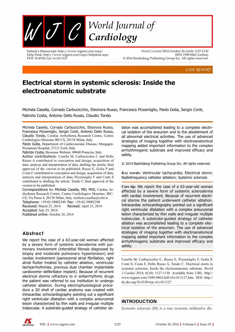
Electrical storm in systemic sclerosis: Inside the electroanatomic substrate
Michela Casella, Corrado Carbucicchio, Eleonora Russo, Francesca Pizzamiglio, Paolo Golia, Sergio Conti, Fabrizio Costa, Antonio Dello Russo, Claudio Tondo
Michela Casella, Corrado Carbucicchio, Eleonora Russo, Francesca Pizzamiglio, Sergio Conti, Antonio Dello Russo, Claudio Tondo, Cardiac Arrhythmia Research Centre, Centro Cardiologico Monzino IRCCS, 20138 Milan, ItalyPaolo Golia, Department of Cardiovascular Disease, Morgagni-Pierantoni Hospital, 27121 Forlì, Italy Fabrizio Costa, Biosense-Webster, 00040 Pomezia, ItalyAuthor contributions: Casella M, Carbucicchio C and Dello Russo A contributed to conception and design, acquisition of data, analysis and interpretation of data, drafting the article, final approval of the version to be published; Russo E, Golia P and Costa F contributed to conception and design, acquisition of data, analysis and interpretation of data; Pizzamiglio F and Conti S contributed to drafting the article; Tondo C final approval of the version to be published.Correspondence to: Michela Casella, MD, PhD, Cardiac Ar-rhythmia Research Centre, Centro Cardiologico Monzino, IRC-CS, Via Parea 4, 20138 Milan, Italy. [email protected]: +39-02-58002340 Fax: +39-02-58002398Received: March 25, 2014 Revised: April 23, 2014Accepted: July 25, 2014Published online: October 26, 2014
AbstractWe report the case of a 63-year-old woman affected by a severe form of systemic scleroderma with pul-monary involvement (interstitial fibrosis diagnosed by biopsy and moderate pulmonary hypertension) and cardiac involvement (paroxysmal atrial fibrillation, right atrial flutter treated by catheter ablation, ventricular tachyarrhythmias, previous dual chamber implantable cardioverter defibrillator implant). Because of recurrent electrical storms refractory to iv antiarrhythmic drugs the patient was referred to our institution to undergo catheter ablation. During electrophysiological proce-dure a 3D shell of cardiac anatomy was created with intracardiac echocardiography pointing out a significant right ventricular dilatation with a complex aneurysmal lesion characterized by thin walls and irregular multiple trabeculae. A substrate-guided strategy of catheter ab-
lation was accomplished leading to a complete electri-cal isolation of the aneurism and to the abolishment of all abnormal electrical activities. The use of advanced strategies of imaging together with electroanatomical mapping added important information to the complex arrhythmogenic substrate and improved efficacy and safety.
© 2014 Baishideng Publishing Group Inc. All rights reserved.
Key words: Ventricular tachycardia; Electrical storm; Radiofrequency catheter ablation; Systemic sclerosis
Core tip: We report the case of a 63-year-old woman affected by a severe form of systemic scleroderma with cardiac involvement. Because of recurrent electri-cal storms the patient underwent catheter ablation. Intracardiac echocardiography pointed out a significant right ventricular dilatation with a complex aneurysmal lesion characterized by thin walls and irregular multiple trabeculae. A substrate-guided strategy of catheter ablation was accomplished leading to a complete elec-trical isolation of the aneurism. The use of advanced strategies of imaging together with electroanatomical mapping added important information to the complex arrhythmogenic substrate and improved efficacy and safety.
Casella M, Carbucicchio C, Russo E, Pizzamiglio F, Golia P, Conti S, Costa F, Dello Russo A, Tondo C. Electrical storm in systemic sclerosis: Inside the electroanatomic substrate. World J Cardiol 2014; 6(10): 1127-1130 Available from: URL: http://www.wjgnet.com/1949-8462/full/v6/i10/1127.htm DOI: http://dx.doi.org/10.4330/wjc.v6.i10.1127
INTRODUCTIONSystemic sclerosis (SS) is a rare systemic infiltrative dis-
CASE REPORT
Submit a Manuscript: http://www.wjgnet.com/esps/Help Desk: http://www.wjgnet.com/esps/helpdesk.aspxDOI: 10.4330/wjc.v6.i10.1127
October 26, 2014|Volume 6|Issue 10|WJC|www.wjgnet.com
World Journal of CardiologyW J C
World J Cardiol 2014 October 26; 6(10): 1127-1130ISSN 1949-8462 (online)
© 2014 Baishideng Publishing Group Inc. All rights reserved.
1127
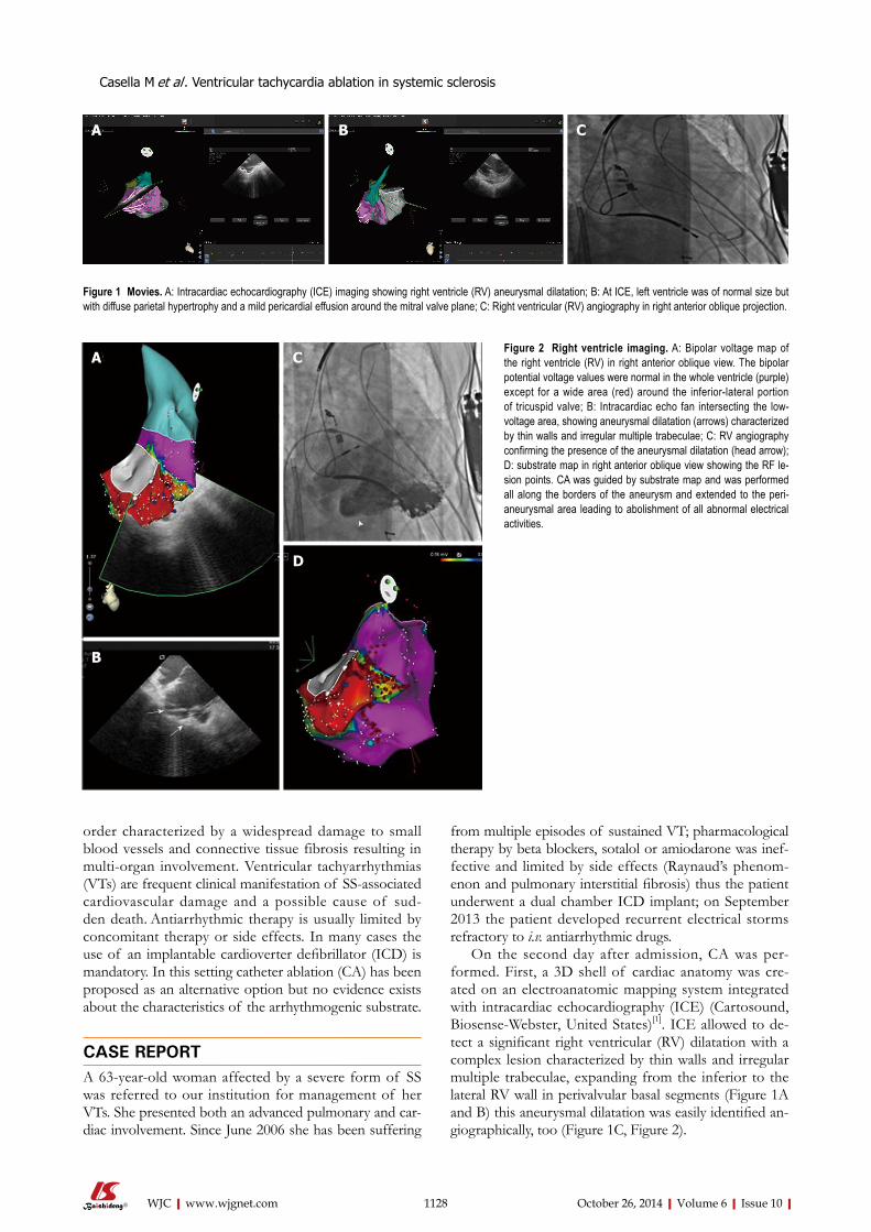
order characterized by a widespread damage to small blood vessels and connective tissue fibrosis resulting in multi-organ involvement. Ventricular tachyarrhythmias (VTs) are frequent clinical manifestation of SS-associated cardiovascular damage and a possible cause of sud-den death. Antiarrhythmic therapy is usually limited by concomitant therapy or side effects. In many cases the use of an implantable cardioverter defibrillator (ICD) is mandatory. In this setting catheter ablation (CA) has been proposed as an alternative option but no evidence exists about the characteristics of the arrhythmogenic substrate.
CASE REPORTA 63-year-old woman affected by a severe form of SS was referred to our institution for management of her VTs. She presented both an advanced pulmonary and car-diac involvement. Since June 2006 she has been suffering
from multiple episodes of sustained VT; pharmacological therapy by beta blockers, sotalol or amiodarone was inef-fective and limited by side effects (Raynaud’s phenom-enon and pulmonary interstitial fibrosis) thus the patient underwent a dual chamber ICD implant; on September 2013 the patient developed recurrent electrical storms refractory to i.v. antiarrhythmic drugs.
On the second day after admission, CA was per-formed. First, a 3D shell of cardiac anatomy was cre-ated on an electroanatomic mapping system integrated with intracardiac echocardiography (ICE) (Cartosound, Biosense-Webster, United States)[1]. ICE allowed to de-tect a significant right ventricular (RV) dilatation with a complex lesion characterized by thin walls and irregular multiple trabeculae, expanding from the inferior to the lateral RV wall in perivalvular basal segments (Figure 1A and B) this aneurysmal dilatation was easily identified an-giographically, too (Figure 1C, Figure 2).
October 26, 2014|Volume 6|Issue 10|WJC|www.wjgnet.com
Casella M et al . Ventricular tachycardia ablation in systemic sclerosis
1128
A B C
Figure 1 Movies. A: Intracardiac echocardiography (ICE) imaging showing right ventricle (RV) aneurysmal dilatation; B: At ICE, left ventricle was of normal size but with diffuse parietal hypertrophy and a mild pericardial effusion around the mitral valve plane; C: Right ventricular (RV) angiography in right anterior oblique projection.
Figure 2 Right ventricle imaging. A: Bipolar voltage map of the right ventricle (RV) in right anterior oblique view. The bipolar potential voltage values were normal in the whole ventricle (purple) except for a wide area (red) around the inferior-lateral portion of tricuspid valve; B: Intracardiac echo fan intersecting the low-voltage area, showing aneurysmal dilatation (arrows) characterized by thin walls and irregular multiple trabeculae; C: RV angiography confirming the presence of the aneurysmal dilatation (head arrow); D: substrate map in right anterior oblique view showing the RF le-sion points. CA was guided by substrate map and was performed all along the borders of the aneurysm and extended to the peri-aneurysmal area leading to abolishment of all abnormal electrical activities.
A
B
C
D
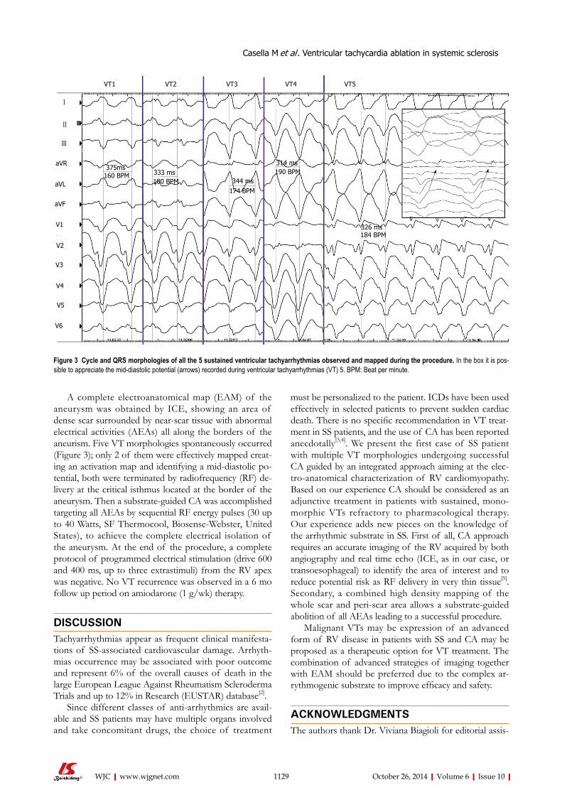
A complete electroanatomical map (EAM) of the aneurysm was obtained by ICE, showing an area of dense scar surrounded by near-scar tissue with abnormal electrical activities (AEAs) all along the borders of the aneurism. Five VT morphologies spontaneously occurred (Figure 3); only 2 of them were effectively mapped creat-ing an activation map and identifying a mid-diastolic po-tential, both were terminated by radiofrequency (RF) de-livery at the critical isthmus located at the border of the aneurysm. Then a substrate-guided CA was accomplished targeting all AEAs by sequential RF energy pulses (30 up to 40 Watts, SF Thermocool, Biosense-Webster, United States), to achieve the complete electrical isolation of the aneurysm. At the end of the procedure, a complete protocol of programmed electrical stimulation (drive 600 and 400 ms, up to three extrastimuli) from the RV apex was negative. No VT recurrence was observed in a 6 mo follow up period on amiodarone (1 g/wk) therapy.
DISCUSSIONTachyarrhythmias appear as frequent clinical manifesta-tions of SS-associated cardiovascular damage. Arrhyth-mias occurrence may be associated with poor outcome and represent 6% of the overall causes of death in the large European League Against Rheumatism Scleroderma Trials and up to 12% in Research (EUSTAR) database[2].
Since different classes of anti-arrhythmics are avail-able and SS patients may have multiple organs involved and take concomitant drugs, the choice of treatment
must be personalized to the patient. ICDs have been used effectively in selected patients to prevent sudden cardiac death. There is no specific recommendation in VT treat-ment in SS patients, and the use of CA has been reported anecdotally[3,4]. We present the first case of SS patient with multiple VT morphologies undergoing successful CA guided by an integrated approach aiming at the elec-tro-anatomical characterization of RV cardiomyopathy. Based on our experience CA should be considered as an adjunctive treatment in patients with sustained, mono-morphic VTs refractory to pharmacological therapy. Our experience adds new pieces on the knowledge of the arrhythmic substrate in SS. First of all, CA approach requires an accurate imaging of the RV acquired by both angiography and real time echo (ICE, as in our case, or transoesophageal) to identify the area of interest and to reduce potential risk as RF delivery in very thin tissue[5]. Secondary, a combined high density mapping of the whole scar and peri-scar area allows a substrate-guided abolition of all AEAs leading to a successful procedure.
Malignant VTs may be expression of an advanced form of RV disease in patients with SS and CA may be proposed as a therapeutic option for VT treatment. The combination of advanced strategies of imaging together with EAM should be preferred due to the complex ar-rythmogenic substrate to improve efficacy and safety.
ACKNOWLEDGMENTSThe authors thank Dr. Viviana Biagioli for editorial assis-
October 26, 2014|Volume 6|Issue 10|WJC|www.wjgnet.com 1129
Figure 3 Cycle and QRS morphologies of all the 5 sustained ventricular tachyarrhythmias observed and mapped during the procedure. In the box it is pos-sible to appreciate the mid-diastolic potential (arrows) recorded during ventricular tachyarrhythmias (VT) 5. BPM: Beat per minute.
VT1 VT2 VT3 VT4 VT5
375ms160 BPM 333 ms
180 BPM 344 ms174 BPM
314 ms190 BPM
326 ms184 BPM
aVR
aVL
aVF
V1
V2
V3
V4
V5
V6
Casella M et al . Ventricular tachycardia ablation in systemic sclerosis
Ⅰ
Ⅱ
Ⅲ
October 26, 2014|Volume 6|Issue 10|WJC|www.wjgnet.com
Peer reviewThis case report describes a case of SS complicated by multiple VTs. This very rare case nicely demonstrates image findings together with important electro-physiologic features in this entity.
REFERENCES1 Dello Russo A, Casella M, Pelargonio G, Bonelli F, Santan-
geli P, Fassini G, Riva S, Carbucicchio C, Giraldi F, De Iuliis P, Bartoletti S, Pintus F, Di Biase L, Pepi M, Natale A, Fio-rentini C, Tondo C. Intracardiac echocardiography in elec-trophysiology. Minerva Cardioangiol 2010; 58: 333-342 [PMID: 20485239]
2 Tyndall AJ, Bannert B, Vonk M, Airò P, Cozzi F, Carreira PE, Bancel DF, Allanore Y, Müller-Ladner U, Distler O, Ian-none F, Pellerito R, Pileckyte M, Miniati I, Ananieva L, Gur-man AB, Damjanov N, Mueller A, Valentini G, Riemekasten G, Tikly M, Hummers L, Henriques MJ, Caramaschi P, Scheja A, Rozman B, Ton E, Kumánovics G, Coleiro B, Feierl E, Szucs G, Von Mühlen CA, Riccieri V, Novak S, Chizzolini C, Kotulska A, Denton C, Coelho PC, Kötter I, Simsek I, de la Pena Lefebvre PG, Hachulla E, Seibold JR, Rednic S, Stork J, Morovic-Vergles J, Walker UA. Causes and risk factors for death in systemic sclerosis: a study from the EULAR Sclero-derma Trials and Research (EUSTAR) database. Ann Rheum Dis 2010; 69: 1809-1815 [DOI: 10.1136/ard.2009.114264]
3 Chung HH, Kim JB, Hong SH, Lee HJ, Joung B, Lee MH. Radiofrequency Catheter Ablation of Hemodynamically Unstable Ventricular Tachycardia Associated with Systemic Sclerosis. J Korean Med Sci 2012; 27: 215-221 [DOI: 10.3346/jkms.2012.27.2.215]
4 Lacroix D, Brigadeau F, Marquié C, Klug D. Electroanatomic mapping and ablation of ventricular tachycardia associated with systemic sclerosis. Europace 2004; 6: 336-342 [PMID: 15172658]
5 Khouzam RN, D’Cruz IA, Arroyo M, Minderman D. Sys-temic scleroderma with moderate to severe mitral regurgita-tion: unusual three-dimensional echocardiographic features. Can J Cardiol 2008; 24: 152 [PMID: 18273492]
P- Reviewer: Kettering K, Nam GB S- Editor: Ji FF L- Editor: A E- Editor: Wu HL
tance.
COMMENTSCase characteristicsA 63-year-old woman affected by a severe form of systemic sclerosis (SS) with previous dual chamber implantable cardioverter defibrillator implant presented with drug-refractory multiple forms of ventricular tachycardias. Clinical diagnosisIdentification of aneurismal lesion of the right ventricular (RV) with the critical isth-mus from which the five morphologies of ventricular tachyarrhythmias (VTs) arise. Differential diagnosisArrhythmogenic right ventricular dysplasia, myocarditis.Laboratory diagnosisPositive Antinuclear Antibodies; metabolic panel and liver function test were within normal limits.Imaging diagnosisIntracardiac echocardiography allowed to detect a significant RV dilatation with a complex lesion characterized by thin walls and irregular multiple trabeculae.Pathological diagnosisA complete electroanatomical map of the aneurysm showed an area of dense scar surrounded with abnormal electrical activities.TreatmentFive VTs morphologies were terminated by radiofrequency (RF) delivery at the critical isthmus located at the border of the aneurysm.Related reportsThere is no specific recommendation in VT treatment in SS patients, and the use of CA has been reported anecdotally.Term explanationCARTO system is a non fluoroscopic mapping system allowing to accurately determine the location of arrhythmia origin, define cardiac chamber geometry in 3D, delineate areas of anatomic interest.Experiences and lessonsThis is the first case of systemic sclerosis patient with multiple VT morpholo-gies undergoing successful CA guided by an integrated approach aiming at the electro-anatomical characterization of RV cardiomyopathy.
1130
COMMENTS
Casella M et al . Ventricular tachycardia ablation in systemic sclerosis
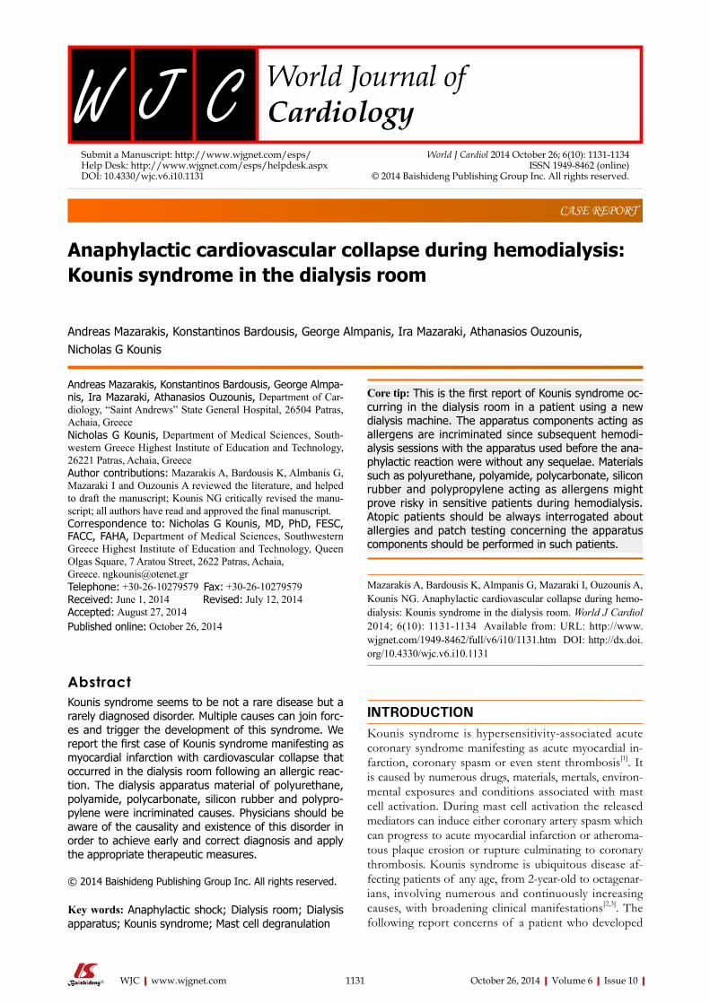
Anaphylactic cardiovascular collapse during hemodialysis: Kounis syndrome in the dialysis room
Andreas Mazarakis, Konstantinos Bardousis, George Almpanis, Ira Mazaraki, Athanasios Ouzounis, Nicholas G Kounis
Andreas Mazarakis, Konstantinos Bardousis, George Almpa-nis, Ira Mazaraki, Athanasios Ouzounis, Department of Car-diology, “Saint Andrews” State General Hospital, 26504 Patras, Achaia, Greece Nicholas G Kounis, Department of Medical Sciences, South-western Greece Highest Institute of Education and Technology, 26221 Patras, Achaia, Greece Author contributions: Mazarakis A, Bardousis K, Almbanis G, Mazaraki I and Ouzounis A reviewed the literature, and helped to draft the manuscript; Kounis NG critically revised the manu-script; all authors have read and approved the final manuscript.Correspondence to: Nicholas G Kounis, MD, PhD, FESC, FACC, FAHA, Department of Medical Sciences, Southwestern Greece Highest Institute of Education and Technology, Queen Olgas Square, 7 Aratou Street, 2622 Patras, Achaia, Greece. [email protected]: +30-26-10279579 Fax: +30-26-10279579Received: June 1, 2014 Revised: July 12, 2014 Accepted: August 27, 2014Published online: October 26, 2014
AbstractKounis syndrome seems to be not a rare disease but a rarely diagnosed disorder. Multiple causes can join forc-es and trigger the development of this syndrome. We report the first case of Kounis syndrome manifesting as myocardial infarction with cardiovascular collapse that occurred in the dialysis room following an allergic reac-tion. The dialysis apparatus material of polyurethane, polyamide, polycarbonate, silicon rubber and polypro-pylene were incriminated causes. Physicians should be aware of the causality and existence of this disorder in order to achieve early and correct diagnosis and apply the appropriate therapeutic measures.
© 2014 Baishideng Publishing Group Inc. All rights reserved.
Key words: Anaphylactic shock; Dialysis room; Dialysis apparatus; Kounis syndrome; Mast cell degranulation
Core tip: This is the first report of Kounis syndrome oc-curring in the dialysis room in a patient using a new dialysis machine. The apparatus components acting as allergens are incriminated since subsequent hemodi-alysis sessions with the apparatus used before the ana-phylactic reaction were without any sequelae. Materials such as polyurethane, polyamide, polycarbonate, silicon rubber and polypropylene acting as allergens might prove risky in sensitive patients during hemodialysis. Atopic patients should be always interrogated about allergies and patch testing concerning the apparatus components should be performed in such patients.
Mazarakis A, Bardousis K, Almpanis G, Mazaraki I, Ouzounis A, Kounis NG. Anaphylactic cardiovascular collapse during hemo-dialysis: Kounis syndrome in the dialysis room. World J Cardiol 2014; 6(10): 1131-1134 Available from: URL: http://www.wjgnet.com/1949-8462/full/v6/i10/1131.htm DOI: http://dx.doi.org/10.4330/wjc.v6.i10.1131
INTRODUCTIONKounis syndrome is hypersensitivity-associated acute coronary syndrome manifesting as acute myocardial in-farction, coronary spasm or even stent thrombosis[1]. It is caused by numerous drugs, materials, mertals, environ-mental exposures and conditions associated with mast cell activation. During mast cell activation the released mediators can induce either coronary artery spasm which can progress to acute myocardial infarction or atheroma-tous plaque erosion or rupture culminating to coronary thrombosis. Kounis syndrome is ubiquitous disease af-fecting patients of any age, from 2-year-old to octagenar-ians, involving numerous and continuously increasing causes, with broadening clinical manifestations[2,3]. The following report concerns of a patient who developed
CASE REPORT
Submit a Manuscript: http://www.wjgnet.com/esps/Help Desk: http://www.wjgnet.com/esps/helpdesk.aspxDOI: 10.4330/wjc.v6.i10.1131
October 26, 2014|Volume 6|Issue 10|WJC|www.wjgnet.com
World Journal of CardiologyW J C
World J Cardiol 2014 October 26; 6(10): 1131-1134ISSN 1949-8462 (online)
© 2014 Baishideng Publishing Group Inc. All rights reserved.
1131
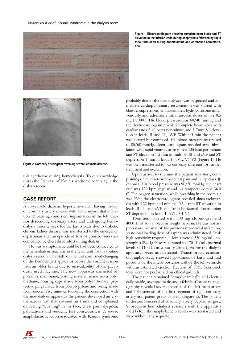
this syndrome during hemodialysis. To our knowledge this is the first case of Kounis syndrome occurring in the dialysis room.
CASE REPORTA 75-year-old diabetic, hypertensive man having history of coronary artery disease with acute myocardial infarc-tion 12 years ago and stent implantation in the left ante-rior descending coronary artery and undergoing hemo-dialysis thrice a week for the last 3 years due to diabetic chronic kidney disease, was transferred to the emergency department after an episode of loss of consciousness ac-companied by chest discomfort during dialysis.
He was asymptomatic until he had been connected to the hemodialysis machine in the renal unit for his routine dialysis session. The staff of the unit confirmed changing of the hemodialysis apparatus before the current session with an older brand due to unavailability of the previ-ously used machine. The new apparatus consisted of polyamix membrane, potting material made from poly-urethane, housing caps made from polycarbonate, pro-tective plugs made from polypropylene and o-ring made from silicon. Five minutes following the connection with the new dialysis apparatus the patient developed an ery-thematous rash that covered his trunk and complained of feeling “burning” in his face, chest pain, dyspnea, palpitations and suddenly lost consciousness. Α severe anaphylactic reaction associated with Kounis syndrome
probably due to the new dialyzer- was suspected and im-mediate cardiopulmonary resuscitation was started with chest compressions, antihistamines, hydrocortisone intra-venously and adrenaline intramuscular doses of 0.2-0.5 mg (1:1000). His blood pressure was 60/40 mmHg and the electrocardiogram revealed complete heart block with cardiac rate of 40 beats per minute and 5-7mm ST eleva-tion in leads Ⅱ and Ⅲ, AVF. Within 3 min the patient was alerted but confused. His blood pressure was raised to 85/60 mmHg, electrocardiogram revealed atrial fibril-lation with rapid ventricular response 135 beat per minute and ST elevation 1-2 mm in leads Ⅱ, Ⅲ and aVF and ST depression 1 mm in leads Ⅰ, aVL, V1-V3 (Figure 1). He was then transferred to our coronary care unit for further treatment and evaluation.
Upon arrival to the unit the patient was alert, com-plaining of mild retrosternal chest pain and Killip class Ⅱ dyspnea. His blood pressure was 90/50 mmHg, the heart rate was 120 bpm regular and his temperature was 36.0 C. The oxygen saturation, while breathing in the room air was 93%, the electrocardiogram revealed sinus tachycar-dia with 122 bpm and minimal 0.5-1 mm ST elevation in leads Ⅱ, Ⅲ and aVF and 1mm horizontal-downsloping ST depression in leads Ⅰ, aVL, V1-V6.
Treatment started with 300 mg clopidogrel and 4000IU of low molecular weight heparin. He was not as-pirin naïve because of his previous myocardial infarction, so no oral loading dose of aspirin was administered. Peak high sensitivity troponin Ⅰ levels were 0.350 ng/mL, eo-sinophils 8%, IgEs were elevated to 170 IU/mL (normal levels < 110 IU/mL) but specific IgEs for the dialysis apparatus were not detected. Transthoracic echocar-diographic study showed hypokinesis of basal and mid portions of the infero-posterior wall of the left ventricle with an estimated ejection fraction of 50%. Skin prick tests were not performed on ethical grounds.
The patient remained hemodynamically and electri-cally stable, asymptomatic and afebrile. Coronary angi-ography revealed severe stenosis of the left main artery and 70% stenosis of the first segment of right coronary artery and patent previous stent (Figure 2). The patient underwent successful coronary artery bypass surgery. Subsequent hemodialysis sessions with the apparatus used before the anaphylactic reaction were re-started and were without any sequelae.
October 26, 2014|Volume 6|Issue 10|WJC|www.wjgnet.com
Mazarakis A et al . Kounis syndrome in the dialysis room
1132
Figure 1 Electrocardiogram showing complete heart block and ST elevation in the inferior leads during anaphylaxis followed by rapid atrial fibrillation during antihistamine and adrenaline administra-tion.
Figure 2 Coronary arteriogram revealing severe left main disease.
DISCUSSIONThe described patient developed severe anaphylaxis asso-ciated with myocardial infarction and cardiovascular col-lapse in the hemodialysis room soon after changing the dialysis apparatus. He was found to have increased IgEs and eosinophil count suggesting hypersensitivity reaction and was diagnosed as type Ⅱ variant of Kounis syndrome. All 4 components of the used apparatus namely polyamix membrane, potting material made from polyurethane, housing caps made from polycarbonate, protective plugs made from polypropylene and o-ring made from silicon rubber have been incriminated to induce hypersensitivity reactions. Polyamix membrane is made from a polymer blend of polyarylethersulfone, polyvinylpyrrolidone and polyamide all of which are sensitizers[4]. Polyurethane chemicals are produced by the reaction of isocyanates and they may cause allergic contact dermatitis or precipi-tate asthma attacks[5]. Polycarbonate can induced allergic reactions especially in dental procedures[6]. Polypropylene is able to induce irritant contact dermatitis[7] and silicon rubber has induced hypersensitivity reactions known as “latex-fruit syndrome”[8]. These materials have been in-criminated to induce hypersensitivity reactions by activat-ing high and low affinity IgE receptors known as FCγRⅠ, FCγRⅡ, FCεRⅠ and FCεRⅡ receptors situated on both mast cell and platelet surface[9].
Therefore the described patient was exposed to 5 antigens. Indeed, clinical studies indicate that sensitive patients simultaneously exposed to several allergens can have more symptoms than mono-sensitized individuals[10]. This could be an explanation for the patient’s immediate cardiovascular collapse. On the other hand, immuno-globulin E antibodies with different specificities can have additive effects and small, even subthreshold numbers of them can join forces and trigger the cells to release their mediators. This can occur when the patient is si-multaneously exposed to the corresponding antigens[11]. The initiation of allergic inflammation takes place when allergens cross-bridge their corresponding, receptor-bound, immunoglobin IgE antibodies on the mast cell or basophil cell surface. These cells degranulate and release their mediators when the critical number of bridged IgE antibodies reaches the order of 2000 out of maximal number of some 500000-1000000 IgE antibodies on the cell surface[12]. A total of approximately 1000 bridges are necessary to induced mast cell degranulation.
Kounis syndrome seems to be not a rare disease but a rarely diagnosed disorder. Multiple and combined causes can trigger the development of this syndrome. Physicians should be aware of its pathophysiology and existence in order to apply predictive, preventive, diagnostic and ap-propriate therapeutic measures.
COMMENTSCase characteristicsA 75-year-old diabetic, hypertensive man suffering from coronary artery disease and renal failure developed anaphylactic cardiac collapse soon after been con-
nected with the dialysis apparatus.Clinical diagnosisThe appearance of erythematous rash that covered his trunk together with feel-ing “burning” in his face, chest pain, dyspnea, palpitations, sudden loss of con-sciousness, electrocardiophic changes, increased cardiac enzymes, increased eosinophils and IgEs were suggestive of type Ⅱ variant of hypersensitivity-associated Kounis syndrome.Differential diagnosisThe differential diagnosis included anaphylactic shock and acute myocardial infarction but their combination is classical with Kounis acute associated with hypersensitity coronary syndrome.Laboratory diagnosisSerial electrocardiographic changes of complete heart block, atrial fibrillation, ST segment elevation, increased cardiac enzymes and troponin Ⅰ, increased eosinophils, increased IgEs and hypokinetic basal and mid portions of the infero-posterior wall of the left ventricle were observed. Imaging diagnosisCoronary angiography revealed severe stenosis of the left main artery and 70% stenosis of the first segment of right coronary artery but patent previous stent that had been implanted in the left anterior descending artery 12 years previ-ously.Pathological diagnosisNeither pathological examination nor skin biopsy for the erythematous rash was thought necessary to be performed.TreatmentThe patient was treated initially with chest compressions, antihistamines, hydrocortisone intravenously and adrenaline intramuscular doses followed by clopidogrel and of low molecular weight heparin and finally underwent success-ful coronary artery bypass surgery. Subsequent hemodialysis sessions with the apparatus used before the anaphylactic reaction were re-started and were without any sequelae.Related reportsNo related reports are available and this case of Kounis syndrome is the first in the world literature.Term explanationThis case of Kounis type II variant syndrome is characterized as unique.Experience and lessonsKounis syndrome is not a rare disease but a rarely diagnosed disorder caused by multiple and combined causes therefore physicians should be aware of its pathophysiology and existence in order to apply predictive, preventive, diagnos-tic and appropriate therapeutic measures. Peer reviewThis is a very interesting report of the case of Kounis syndrome occuring in the dialysis room.
REFERENCES1 Kounis NG. Coronary hypersensitivity disorder: the Kounis
syndrome. Clin Ther 2013; 35: 563-571 [PMID: 23490289 DOI: 10.1016/j.clinthera.2013.02.022]
2 Parent B, Wearden P, Kounis NG, Chrysostomou C. Kounis syndrome or allergic coronary vasospasm in a two-year-old. Congenit Heart Dis 2011; 6: 499-503 [PMID: 21418536 DOI: 10.1111/j.1747-0803.2011.00499.x]
3 Biteker M, Duran NE, Biteker FS, Civan HA, Kaya H, Gök-deniz T, Yildiz M, Ozkan M. Allergic myocardial infarction in childhood: Kounis syndrome. Eur J Pediatr 2010; 169: 27-29 [PMID: 19277706 DOI: 10.1007/s00431-009-0965-5]
4 Situm M, Lugović-Mihić L, Bulat V, Peternel R, Vojniković B, Martinis M, Toth I. Dermatological aspects of contact derma-titis from eyeglass frames and optical materials. Coll Antropol 2013; 37 Suppl 1: 19-24 [PMID: 23837217]
5 Turan H, Saricaoğlu H, Turan A, Tunali S. Polyurethane toi-let seat contact dermatitis. Pediatr Dermatol 2011; 28: 731-732 [PMID: 21575050 DOI: 10.1111/j.1525-1470.2011.01482.x]
6 Tanoue N, Nagano K, Matsumura H. Use of a light-polymer-ized composite removable partial denture base for a patient
October 26, 2014|Volume 6|Issue 10|WJC|www.wjgnet.com 1133
COMMENTS
Mazarakis A et al . Kounis syndrome in the dialysis room

October 26, 2014|Volume 6|Issue 10|WJC|www.wjgnet.com
10.2217/fca.11.63]10 MacGlashan DW, Bochner BS, Adelman DC, Jardieu PM,
Togias A, McKenzie-White J, Sterbinsky SA, Hamilton RG, Lichtenstein LM. Down-regulation of Fc(epsilon)RI ex-pression on human basophils during in vivo treatment of atopic patients with anti-IgE antibody. J Immunol 1997; 158: 1438-1445 [PMID: 9013989]
11 Nopp A, Johansson SG, Lundberg M, Oman H. Simultane-ous exposure of several allergens has an additive effect on multisensitized basophils. Allergy 2006; 61: 1366-1368 [PMID: 17002715 DOI: 10.1111/j.1398-9995.2006.01211.x]
12 Wickman M. When allergies complicate allergies. Allergy 2005; 60 Suppl 79: 14-18 [PMID: 15842228 DOI: 10.1111/j.1398-9995.2005.00852.x]
P- Reviewer: Kounis GN, Xiong XJ S- Editor: Ji FF L- Editor: A E- Editor: Wu HL
hypersensitive to poly(methyl methacrylate), polysulfone, and polycarbonate: a clinical report. J Prosthet Dent 2005; 93: 17-20 [PMID: 15623992 DOI: 10.1016/j.prosdent.2004.09.022]
7 Patiwael JA, Wintzen M, Rustemeyer T, Bruynzeel DP. Air-borne irritant contact dermatitis due to synthetic fibres from an air-conditioning filter. Contact Dermatitis 2005; 52: 126-129 [PMID: 15811024 DOI: 10.1111/j.0105-1873.2005.00526.x]
8 Ricci G, Piccinno V, Calamelli E, Giannetti A, Pession A. La-tex-fruit syndrome in Italian children and adolescents with natural rubber latex allergy. Int J Immunopathol Pharmacol 2013; 26: 263-268 [PMID: 23527732]
9 Kounis NG, Mazarakis A, Tsigkas G, Giannopoulos S, Goudevenos J. Kounis syndrome: a new twist on an old dis-ease. Future Cardiol 2011; 7: 805-824 [PMID: 22050066 DOI:
1134
Mazarakis A et al . Kounis syndrome in the dialysis room
GENERAL INFORMATIONWorld Journal of Cardiology (World J Cardiol, WJC, online ISSN 1949-8462, DOI: 10.4330) is a peer-reviewed open access (OA) academic journal that aims to guide clinical practice and improve diagnostic and therapeutic skills of clinicians.
Aim and scopeWJC covers topics concerning arrhythmia, heart failure, vascular disease, stroke, hypertension, prevention and epidemiology, dyslipi-demia and metabolic disorders, cardiac imaging, pediatrics, nursing, and health promotion. Priority publication will be given to articles concerning diagnosis and treatment of cardiology diseases. The fol-lowing aspects are covered: Clinical diagnosis, laboratory diagnosis, differential diagnosis, imaging tests, pathological diagnosis, molecular biological diagnosis, immunological diagnosis, genetic diagnosis, functional diagnostics, and physical diagnosis; and comprehensive therapy, drug therapy, surgical therapy, interventional treatment, min-imally invasive therapy, and robot-assisted therapy. We encourage authors to submit their manuscripts to WJC. We will give priority to manuscripts that are supported by major national and international foundations and those that are of great basic and clinical significance. WJC is edited and published by Baishideng Publishing Group (BPG). BPG has a strong professional editorial team composed of science editors, language editors and electronic editors. BPG current-ly publishes 43 OA clinical medical journals, including 42 in English, has a total of 15471 editorial board members or peer reviewers, and is a world first-class publisher.
ColumnsThe columns in the issues of WJC will include: (1) Editorial: The editorial board members are invited to make comments on an im-portant topic in their field in terms of its current research status and future directions to lead the development of this discipline; (2) Frontier: The editorial board members are invited to select a highly cited cutting-edge original paper of his/her own to summarize ma-jor findings, the problems that have been resolved and remain to be resolved, and future research directions to help readers understand his/her important academic point of view and future research dir-ections in the field; (3) Diagnostic Advances: The editorial board members are invited to write high-quality diagnostic advances in their field to improve the diagnostic skills of readers. The topic covers general clinical diagnosis, differential diagnosis, pathological diagnosis, laboratory diagnosis, imaging diagnosis, endoscopic diag-nosis, biotechnological diagnosis, functional diagnosis, and physical diagnosis; (4) Therapeutics Advances: The editorial board members are invited to write high-quality therapeutic advances in their field to help improve the therapeutic skills of readers. The topic covers medication therapy, psychotherapy, physical therapy, replacement therapy, interventional therapy, minimally invasive therapy, endo-scopic therapy, transplantation therapy, and surgical therapy; (5) Field of Vision: The editorial board members are invited to write commentaries on classic articles, hot topic articles, or latest articles to keep readers at the forefront of research and increase their levels of clinical research. Classic articles refer to papers that are included in Web of Knowledge and have received a large number of citations (ranking in the top 1%) after being published for more than years, reflecting the quality and impact of papers. Hot topic articles refer
to papers that are included in Web of Knowledge and have received a large number of citations after being published for no more than 2 years, reflecting cutting-edge trends in scientific research. Latest articles refer to the latest published high-quality papers that are included in PubMed, reflecting the latest research trends. These commentary articles should focus on the status quo of research, the most important research topics, the problems that have now been resolved and remain to be resolved, and future research directions. Basic information about the article to be commented (including authors, article title, journal name, year, volume, and inclusive page numbers; (6) Minireviews: The editorial board members are invited to write short reviews on recent advances and trends in research of molecular biology, genomics, and related cutting-edge technologies to provide readers with the latest knowledge and help improve their diagnostic and therapeutic skills; (7) Review: To make a systematic review to focus on the status quo of research, the most import-ant research topics, the problems that have now been resolved and remain to be resolved, and future research directions; (8) Topic Highlight: The editorial board members are invited to write a series of articles (7-10 articles) to comment and discuss a hot topic to help improve the diagnostic and therapeutic skills of readers; (9) Medical Ethics: The editorial board members are invited to write articles about medical ethics to increase readers’ knowledge of med-ical ethics. The topic covers international ethics guidelines, animal studies, clinical trials, organ transplantation, etc.; (10) Clinical Case Conference or Clinicopathological Conference: The editorial board members are invited to contribute high-quality clinical case confer-ence; (11) Original Articles: To report innovative and original find-ings in cardiology; (12) Research Report: To briefly report the novel and innovative findings in cardiology; (13) Meta-Analysis: Covers the systematic review, mixedtreatment comparison, meta-regression, and overview of reviews, in order to summarize a given quantitative effect, e.g., the clinical effectiveness and safety of clinical treatments by combining data from two or more randomized controlled trials, thereby providing more precise and externally valid estimates than those which would stem from each individual dataset if analyzed separately from the others; (14) Case Report: To report a rare or typical case; (15) Letters to the Editor: To discuss and make reply to the contributions published in WJC, or to introduce and comment on a controversial issue of general interest; (16) Book Reviews: To introduce and comment on quality monographs of cardiology; and (17) Autobiography: The editorial board members are invited to write their autobiography to provide readers with stories of suc-cess or failure in their scientific research career. The topic covers their basic personal information and information about when they started doing research work, where and how they did research work, what they have achieved, and their lessons from success or failure.
Name of journalWorld Journal of Cardiology
ISSNISSN 1949-8462 (online)
Launch dateDecember 31, 2009
FrequencyMonthly
World Journal of CardiologyW J C
IWJC|www.wjgnet.com October 26, 2014|Volume 6|Issue 10|
World J Cardiol 2014 October 26; 6(10): I-VISSN 1949-8462 (online)
© 2014 Baishideng Publishing Group Inc. All rights reserved.
INSTRUCTIONS TO AUTHORS
Submit a Manuscript: http://www.wjgnet.com/esps/Help Desk: http://www.wjgnet.com/esps/helpdesk.aspxwww.wjgnet.com
Instructions to authors
Editors-in-ChiefJian-Jun Li, MD, PhD, Professor, Center for Coronary Artery Disease, Fu Wai Cardiovascular Hospital, Chinese Academy of Medical Science, Beijing 100037, China
Giuseppe De Luca, PhD, Assistant Professor, Department of Cardiology, Piedmont University, Novara 28100, Italy
Nathan D Wong, FACC, FAHA, PhD, Director, Professor, Heart Disease Prevention Program, Division of Cardiology, De-partment of Medicine, University of California, Irvine, CA 92629, United States
Editorial officeJin-Lei Wang, DirectorXiu-Xia Song, Vice DirectorWorld Journal of CardiologyRoom 903, Building D, Ocean International Center,No. 62 Dongsihuan Zhonglu, Chaoyang District,Beijing 100025, ChinaTelephone: +86-10-85381891Fax: +86-10-85381893E-mail: [email protected] Desk: http://www.wjgnet.com/esps/helpdesk.aspxhttp://www.wjgnet.com
PublisherBaishideng Publishing Group Inc8226 Regency Drive, Pleasanton, CA 94588, USATelephone: +1-925-223-8242Fax: +1-925-223-8243E-mail: [email protected] Desk: http://www.wjgnet.com/esps/helpdesk.aspxhttp://www.wjgnet.com
Instructions to authorsFull instructions are available online at http://www.wjgnet.com/1949-8462/g_info_20100316161927.htm.
Indexed and Abstracted inPubMed Central, PubMed, Digital Object Identifier, and Direc-tory of Open Access Journals.
SPECIAL STATEMENTAll articles published in journals owned by the BPG represent the views and opinions of their authors, and not the views, opinions or policies of the BPG, except where otherwise explicitly indicated.
Biostatistical editingStatistical review is performed after peer review. We invite an ex-pert in Biomedical Statistics to evaluate the statistical method used in the paper, including t-test (group or paired comparisons), chi-squared test, Ridit, probit, logit, regression (linear, curvilinear, or stepwise), correlation, analysis of variance, analysis of covariance, etc. The reviewing points include: (1) Statistical methods should be described when they are used to verify the results; (2) Whether the statistical techniques are suitable or correct; (3) Only homogeneous data can be averaged. Standard deviations are preferred to standard errors. Give the number of observations and subjects (n). Losses in observations, such as drop-outs from the study should be re-ported; (4) Values such as ED50, LD50, IC50 should have their 95% confidence limits calculated and compared by weighted probit analysis (Bliss and Finney); and (5) The word ‘significantly’ should be replaced by its synonyms (if it indicates extent) or the P value (if it indicates statistical significance).
Conflict-of-interest statementIn the interests of transparency and to help reviewers assess any po-tential bias, WJC requires authors of all papers to declare any compet-
ing commercial, personal, political, intellectual, or religious interests in relation to the submitted work. Referees are also asked to indi-cate any potential conflict they might have reviewing a particular paper. Before submitting, authors are suggested to read “Uniform Requirements for Manuscripts Submitted to Biomedical Journals: Ethical Considerations in the Conduct and Reporting of Research: Conflicts of Interest” from International Committee of Medical Journal Editors (ICMJE), which is available at: http://www.icmje.org/ethical_4conflicts.html.
Sample wording: [Name of individual] has received fees for serv-ing as a speaker, a consultant and an advisory board member for [names of organizations], and has received research funding from [names of organization]. [Name of individual] is an employee of [name of or-ganization]. [Name of individual] owns stocks and shares in [name of organization]. [Name of individual] owns patent [patent identification and brief description].
Statement of informed consentManuscripts should contain a statement to the effect that all human studies have been reviewed by the appropriate ethics committee or it should be stated clearly in the text that all persons gave their informed consent prior to their inclusion in the study. Details that might disclose the identity of the subjects under study should be omitted. Authors should also draw attention to the Code of Ethics of the World Med-ical Association (Declaration of Helsinki, 1964, as revised in 2004).
Statement of human and animal rightsWhen reporting the results from experiments, authors should follow the highest standards and the trial should conform to Good Clinical Practice (for example, US Food and Drug Administration Good Clinical Practice in FDA-Regulated Clinical Trials; UK Medicines Research Council Guidelines for Good Clinical Practice in Clinical Trials) and/or the World Medical Association Declaration of Hel-sinki. Generally, we suggest authors follow the lead investigator’s na-tional standard. If doubt exists whether the research was conducted in accordance with the above standards, the authors must explain the rationale for their approach and demonstrate that the institutional review body explicitly approved the doubtful aspects of the study.
Before submitting, authors should make their study approved by the relevant research ethics committee or institutional review board. If human participants were involved, manuscripts must be accom-panied by a statement that the experiments were undertaken with the understanding and appropriate informed consent of each. Any per-sonal item or information will not be published without explicit con-sents from the involved patients. If experimental animals were used, the materials and methods (experimental procedures) section must clearly indicate that appropriate measures were taken to minimize pain or discomfort, and details of animal care should be provided.
SUBMISSION OF MANUSCRIPTSManuscripts should be typed in 1.5 line spacing and 12 pt. Book Antiqua with ample margins. Number all pages consecutively, and start each of the following sections on a new page: Title Page, Abstract, Introduction, Materials and Methods, Results, Discus-sion, Acknowledgements, References, Tables, Figures, and Figure Legends. Neither the editors nor the publisher are responsible for the opinions expressed by contributors. Manuscripts formally ac-cepted for publication become the permanent property of BPG, and may not be reproduced by any means, in whole or in part, with-out the written permission of both the authors and the publisher. We reserve the right to copy-edit and put onto our website accepted manuscripts. Authors should follow the relevant guidelines for the care and use of laboratory animals of their institution or national animal welfare committee. For the sake of transparency in regard to the performance and reporting of clinical trials, we endorse the policy of the ICMJE to refuse to publish papers on clinical trial results if the trial was not recorded in a publicly-accessible registry at its outset. The only register now available, to our knowledge, is http://www.clinicaltrials.gov sponsored by the United States National Library of Medicine and we encourage all potential con-
IIWJC|www.wjgnet.com October 26, 2014|Volume 6|Issue 10|
Instructions to authors
tributors to register with it. However, in the case that other registers become available you will be duly notified. A letter of recommenda-tion from each author’s organization should be provided with the contributed article to ensure the privacy and secrecy of research is protected.
Authors should retain one copy of the text, tables, photo-graphs and illustrations because rejected manuscripts will not be returned to the author(s) and the editors will not be responsible for loss or damage to photographs and illustrations sustained dur-ing mailing.
Online submissionsManuscripts should be submitted through the Online Submis-sion System at: http://www.wjgnet.com/esps/. Authors are highly recommended to consult the ONLINE INSTRUCTIONS TO AUTHORS (http://www.wjgnet.com/1949-8462/g_info_ 20100316161927.htm) before attempting to submit online. For assist-ance, authors encountering problems with the Online Submission System may send an email describing the problem to [email protected], or by telephone: +86-10-85381892. If you submit your manuscript online, do not make a postal contribution. Repeated online submission for the same manuscript is strictly prohibited.
MANUSCRIPT PREPARATIONAll contributions should be written in English. All articles must be submitted using word-processing software. All submissions must be typed in 1.5 line spacing and 12 pt. Book Antiqua with ample mar-gins. Style should conform to our house format. Required informa-tion for each of the manuscript sections is as follows:
Title pageTitle: Title should be less than 12 words.
Running title: A short running title of less than 6 words should be provided.
Authorship: Authorship credit should be in accordance with the standard proposed by ICMJE, based on (1) substantial contribu-tions to conception and design, acquisition of data, or analysis and interpretation of data; (2) drafting the article or revising it critically for important intellectual content; and (3) final approval of the ver-sion to be published. Authors should meet conditions 1, 2, and 3.
Institution: Author names should be given first, then the complete name of institution, city, province and postcode. For example, Xu-Chen Zhang, Li-Xin Mei, Department of Pathology, Chengde Med-ical College, Chengde 067000, Hebei Province, China. One author may be represented from two institutions, for example, George Sgourakis, Department of General, Visceral, and Transplantation Surgery, Essen 45122, Germany; George Sgourakis, 2nd Surgical Department, Korgialenio-Benakio Red Cross Hospital, Athens 15451, Greece
Author contributions: The format of this section should be: Author contributions: Wang CL and Liang L contributed equally to this work; Wang CL, Liang L, Fu JF, Zou CC, Hong F and Wu XM designed the research; Wang CL, Zou CC, Hong F and Wu XM performed the research; Xue JZ and Lu JR contributed new reagents/analytic tools; Wang CL, Liang L and Fu JF analyzed the data; and Wang CL, Liang L and Fu JF wrote the paper.
Supportive foundations: The complete name and number of sup-portive foundations should be provided, e.g., Supported by National Natural Science Foundation of China, No. 30224801
Correspondence to: Only one corresponding address should be provided. Author names should be given first, then author title, af-filiation, the complete name of institution, city, postcode, province, country, and email. All the letters in the email should be in lower
case. A space interval should be inserted between country name and email address. For example, Montgomery Bissell, MD, Professor of Medicine, Chief, Liver Center, Gastroenterology Division, Uni-versity of California, Box 0538, San Francisco, CA 94143, United States. [email protected]
Telephone and fax: Telephone and fax should consist of +, coun-try number, district number and telephone or fax number, e.g., Tele-phone: +86-10-85381892 Fax: +86-10-85381893
Peer reviewers: All articles received are subject to peer review. Normally, three experts are invited for each article. Decision on acceptance is made only when at least two experts recommend publication of an article. All peer-reviewers are acknowledged on Express Submission and Peer-review System website.
AbstractThere are unstructured abstracts (no less than 200 words) and struc-tured abstracts. The specific requirements for structured abstracts are as follows:
An informative, structured abstract should accompany each manuscript. Abstracts of original contributions should be struc-tured into the following sections: AIM (no more than 20 words; Only the purpose of the study should be included. Please write the Aim in the form of “To investigate/study/…”), METHODS (no less than 140 words for Original Articles; and no less than 80 words for Brief Articles), RESULTS (no less than 150 words for Original Articles and no less than 120 words for Brief Articles; You should present P values where appropriate and must provide relevant data to illustrate how they were obtained, e.g., 6.92 ± 3.86 vs 3.61 ± 1.67, P < 0.001), and CONCLUSION (no more than 26 words).
Key wordsPlease list 5-10 key words, selected mainly from Index Medicus, which reflect the content of the study.
Core tip Please write a summary of less than 100 words to outline the most innovative and important arguments and core contents in your paper to attract readers.
TextFor articles of these sections, original articles and brief articles, the main text should be structured into the following sections: INTRO-DUCTION, MATERIALS AND METHODS, RESULTS and DISCUSSION, and should include appropriate Figures and Tables. Data should be presented in the main text or in Figures and Tables, but not in both.
IllustrationsFigures should be numbered as 1, 2, 3, etc., and mentioned clearly in the main text. Provide a brief title for each figure on a separate page. Detailed legends should not be provided under the figures. This part should be added into the text where the figures are applicable. Keep-ing all elements compiled is necessary in line-art image. Scale bars should be used rather than magnification factors, with the length of the bar defined in the legend rather than on the bar itself. File names should identify the figure and panel. Avoid layering type directly over shaded or textured areas. Please use uniform legends for the same subjects. For example: Figure 1 Pathological changes in atrophic gas-tritis after treatment. A: ...; B: ...; C: ...; D: ...; E: ...; F: ...; G: …etc. It is our principle to publish high resolution-figures for the E-versions.
TablesThree-line tables should be numbered 1, 2, 3, etc., and mentioned clearly in the main text. Provide a brief title for each table. Detailed legends should not be included under tables, but rather added into the text where applicable. The information should complement, but not duplicate the text. Use one horizontal line under the title, a second under column heads, and a third below the Table, above any
IIIWJC|www.wjgnet.com October 26, 2014|Volume 6|Issue 10|
footnotes. Vertical and italic lines should be omitted.
Notes in tables and illustrationsData that are not statistically significant should not be noted. aP < 0.05, bP < 0.01 should be noted (P > 0.05 should not be noted). If there are other series of P values, cP < 0.05 and dP < 0.01 are used. A third series of P values can be expressed as eP < 0.05 and fP < 0.01. Other notes in tables or under illustrations should be expressed as 1F, 2F, 3F; or sometimes as other symbols with a superscript (Arabic numerals) in the upper left corner. In a multi-curve illustration, each curve should be labeled with ●, ○, ■, □, ▲, △, etc., in a cer-tain sequence.
AcknowledgmentsBrief acknowledgments of persons who have made genuine con-tributions to the manuscript and who endorse the data and conclu-sions should be included. Authors are responsible for obtaining written permission to use any copyrighted text and/or illustrations.
REFERENCESCoding systemThe author should number the references in Arabic numerals ac-cording to the citation order in the text. Put reference numbers in square brackets in superscript at the end of citation content or after the cited author’s name. For citation content which is part of the narration, the coding number and square brackets should be typeset normally. For example, “Crohn’s disease (CD) is associated with increased intestinal permeability[1,2]”. If references are cited directly in the text, they should be put together within the text, for example, “From references[19,22-24], we know that...”
When the authors write the references, please ensure that the order in text is the same as in the references section, and also ensure the spelling accuracy of the first author’s name. Do not list the same citation twice.
PMID and DOIPleased provide PubMed citation numbers to the reference list, e.g., PMID and DOI, which can be found at http://www.ncbi.nlm.nih.gov/sites/entrez?db=pubmed and http://www.crossref.org/Sim-pleTextQuery/, respectively. The numbers will be used in E-version of this journal.
Style for journal referencesAuthors: the name of the first author should be typed in bold-faced letters. The family name of all authors should be typed with the in-itial letter capitalized, followed by their abbreviated first and middle initials. (For example, Lian-Sheng Ma is abbreviated as Ma LS, Bo-Rong Pan as Pan BR). The title of the cited article and italicized journal title (journal title should be in its abbreviated form as shown in PubMed), publication date, volume number (in black), start page, and end page [PMID: 11819634 DOI: 10.3748/wjg.13.5396].
Style for book referencesAuthors: the name of the first author should be typed in bold-faced letters. The surname of all authors should be typed with the initial letter capitalized, followed by their abbreviated middle and first initials. (For example, Lian-Sheng Ma is abbreviated as Ma LS, Bo-Rong Pan as Pan BR) Book title. Publication number. Publication place: Publication press, Year: start page and end page.
FormatJournals English journal article (list all authors and include the PMID where applicable)1 Jung EM, Clevert DA, Schreyer AG, Schmitt S, Rennert J,
Kubale R, Feuerbach S, Jung F. Evaluation of quantitative con-trast harmonic imaging to assess malignancy of liver tumors: A prospective controlled two-center study. World J Gastroenterol 2007; 13: 6356-6364 [PMID: 18081224 DOI: 10.3748/wjg.13. 6356]
Chinese journal article (list all authors and include the PMID where applicable)2 Lin GZ, Wang XZ, Wang P, Lin J, Yang FD. Immunologic
effect of Jianpi Yishen decoction in treatment of Pixu-diar-rhoea. Shijie Huaren Xiaohua Zazhi 1999; 7: 285-287
In press3 Tian D, Araki H, Stahl E, Bergelson J, Kreitman M. Signature
of balancing selection in Arabidopsis. Proc Natl Acad Sci USA 2006; In press
Organization as author4 Diabetes Prevention Program Research Group. Hyperten-
sion, insulin, and proinsulin in participants with impaired glu-cose tolerance. Hypertension 2002; 40: 679-686 [PMID: 12411462 PMCID:2516377 DOI:10.1161/01.HYP.0000035706.28494. 09]
Both personal authors and an organization as author 5 Vallancien G, Emberton M, Harving N, van Moorselaar RJ;
Alf-One Study Group. Sexual dysfunction in 1, 274 European men suffering from lower urinary tract symptoms. J Urol 2003; 169: 2257-2261 [PMID: 12771764 DOI:10.1097/01.ju. 0000067940.76090.73]
No author given6 21st century heart solution may have a sting in the tail. BMJ
2002; 325: 184 [PMID: 12142303 DOI:10.1136/bmj.325. 7357.184]
Volume with supplement7 Geraud G, Spierings EL, Keywood C. Tolerability and safety
of frovatriptan with short- and long-term use for treatment of migraine and in comparison with sumatriptan. Headache 2002; 42 Suppl 2: S93-99 [PMID: 12028325 DOI:10.1046/j.1526-4610.42.s2.7.x]
Issue with no volume8 Banit DM, Kaufer H, Hartford JM. Intraoperative frozen
section analysis in revision total joint arthroplasty. Clin Orthop Relat Res 2002; (401): 230-238 [PMID: 12151900 DOI:10.1097/00003086-200208000-00026]
No volume or issue9 Outreach: Bringing HIV-positive individuals into care. HRSA
Careaction 2002; 1-6 [PMID: 12154804]
BooksPersonal author(s)10 Sherlock S, Dooley J. Diseases of the liver and billiary system.
9th ed. Oxford: Blackwell Sci Pub, 1993: 258-296Chapter in a book (list all authors)11 Lam SK. Academic investigator’s perspectives of medical
treatment for peptic ulcer. In: Swabb EA, Azabo S. Ulcer disease: investigation and basis for therapy. New York: Marcel Dekker, 1991: 431-450
Author(s) and editor(s)12 Breedlove GK, Schorfheide AM. Adolescent pregnancy.
2nd ed. Wieczorek RR, editor. White Plains (NY): March of Dimes Education Services, 2001: 20-34
Conference proceedings13 Harnden P, Joffe JK, Jones WG, editors. Germ cell tumours V.
Proceedings of the 5th Germ cell tumours Conference; 2001 Sep 13-15; Leeds, UK. New York: Springer, 2002: 30-56
Conference paper14 Christensen S, Oppacher F. An analysis of Koza's computa-
tional effort statistic for genetic programming. In: Foster JA, Lutton E, Miller J, Ryan C, Tettamanzi AG, editors. Genetic programming. EuroGP 2002: Proceedings of the 5th Euro-pean Conference on Genetic Programming; 2002 Apr 3-5; Kinsdale, Ireland. Berlin: Springer, 2002: 182-191
Electronic journal (list all authors)15 Morse SS. Factors in the emergence of infectious diseases.
Emerg Infect Dis serial online, 1995-01-03, cited 1996-06-05; 1(1): 24 screens. Available from: URL: http://www.cdc.gov/ncidod/eid/index.htm
Patent (list all authors)16 Pagedas AC, inventor; Ancel Surgical R&D Inc., assignee.
IVWJC|www.wjgnet.com
Instructions to authors
October 26, 2014|Volume 6|Issue 10|
VWJC|www.wjgnet.com
Instructions to authors
October 26, 2014|Volume 6|Issue 10|
Flexible endoscopic grasping and cutting device and position-ing tool assembly. United States patent US 20020103498. 2002 Aug 1
Statistical dataWrite as mean ± SD or mean ± SE.
Statistical expressionExpress t test as t (in italics), F test as F (in italics), chi square test as χ2 (in Greek), related coefficient as r (in italics), degree of freedom as υ (in Greek), sample number as n (in italics), and probability as P (in italics).
UnitsUse SI units. For example: body mass, m (B) = 78 kg; blood pres-sure, p (B) = 16.2/12.3 kPa; incubation time, t (incubation) = 96 h, blood glucose concentration, c (glucose) 6.4 ± 2.1 mmol/L; blood CEA mass concentration, p (CEA) = 8.6 24.5 mg/L; CO2 volume fraction, 50 mL/L CO2, not 5% CO2; likewise for 40 g/L formal-dehyde, not 10% formalin; and mass fraction, 8 ng/g, etc. Arabic numerals such as 23, 243, 641 should be read 23243641.
The format for how to accurately write common units and quantums can be found at: http://www.wjgnet.com/1949-8462/g_info_20100312200347.htm.
AbbreviationsStandard abbreviations should be defined in the abstract and on first mention in the text. In general, terms should not be abbrevi-ated unless they are used repeatedly and the abbreviation is helpful to the reader. Permissible abbreviations are listed in Units, Symbols and Abbreviations: A Guide for Biological and Medical Editors and Authors (Ed. Baron DN, 1988) published by The Royal Society of Medicine, London. Certain commonly used abbreviations, such as DNA, RNA, HIV, LD50, PCR, HBV, ECG, WBC, RBC, CT, ESR, CSF, IgG, ELISA, PBS, ATP, EDTA, mAb, can be used directly without further explanation.
ItalicsQuantities: t time or temperature, c concentration, A area, l length, m mass, V volume.Genotypes: gyrA, arg 1, c myc, c fos, etc.Restriction enzymes: EcoRI, HindI, BamHI, Kbo I, Kpn I, etc.Biology: H. pylori, E coli, etc.
Examples for paper writingAll types of articles’ writing style and requirement will be found in the link: http://www.wjgnet.com/esps/NavigationInfo.aspx?id=15
RESUBMISSION OF THE REVISED MANUSCRIPTSAuthors must revise their manuscript carefully according to the
revision policies of BPG. The revised version, along with the signed copyright transfer agreement, responses to the reviewers, and English language Grade A certificate (for non-native speakers of English), should be submitted to the online system via the link contained in the e-mail sent by the editor. If you have any questions about the revision, please send e-mail to [email protected].
Language evaluation The language of a manuscript will be graded before it is sent for revision. (1) Grade A: priority publishing; (2) Grade B: minor lan-guage polishing; (3) Grade C: a great deal of language polishing needed; and (4) Grade D: rejected. Revised articles should reach Grade A.
Copyright assignment formPlease download a Copyright assignment form from http://www.wjgnet.com/1949-8462/g_info_20100312200118.htm.
Responses to reviewersPlease revise your article according to the comments/suggestions provided by the reviewers. The format for responses to the reviewers’ comments can be found at: http://www.wjgnet.com/1949-8462/g_info_20100312195923.htm.
Proof of financial supportFor papers supported by a foundation, authors should provide a copy of the approval document and serial number of the foundation.
STATEMENT ABOUT ANONYMOUS PUBLICATION OF THE PEER REVIEWERS' COMMENTSIn order to increase the quality of peer review, push authors to carefully revise their manuscripts based on the peer reviewers' com-ments, and promote academic interactions among peer reviewers, authors and readers, we decide to anonymously publish the review-ers’ comments and author’s responses at the same time the manu-script is published online.
PUBLICATION FEEWJC is an international, peer-reviewed, OA online journal. Articles published by this journal are distributed under the terms of the Creative Commons Attribution Non-commercial License, which permits use, distribution, and reproduction in any medium and format, provided the original work is properly cited. The use is non-commercial and is otherwise in compliance with the license. Authors of accepted articles must pay a publication fee. Publication fee: 698 USD per article. All invited articles are published free of charge.

© 2014 Baishideng Publishing Group Inc. All rights reserved.
Published by Baishideng Publishing Group Inc8226 Regency Drive, Pleasanton, CA 94588, USA
Telephone: +1-925-223-8242Fax: +1-925-223-8243
E-mail: [email protected] Desk: http://www.wjgnet.com/esps/helpdesk.aspx
http://www.wjgnet.com


