Embed Size (px)
Citation preview

Send Orders for Reprints to [email protected]
Recent Patents on Biomarkers 2014, 4, 133-149 133
Wound Repair - Updates in Dressing Patents and Regeneration Biomarkers
Carolina Constantin1, Georgeta Paunica-Panea
2,3, Vlad D. Constantin
2,3 and Monica Neagu
1’*
1Immunobiology Laboratory “Victor Babes” National Institute of Pathology, 99-101 Splaiul Independentei, Sect 5,
Bucharest, Postal Code 050096, Romania; 2Surgery Clinics, “Sf. Pantelimon” Emergency Hospital, Bucharest, Roma-
nia; 3“Carol Davila” University of Medicine and Pharmacy, Bucharest, Romania
Received: December 12, 2014; Accepted: January 5, 2015; Revised: January 11, 2014
Abstract: Only in the last year, over 100 reports were published focusing on innovative dressing for hard to heal wounds.
Complex technologies were developed to tackle the intricate mechanisms of wound healing and moreover to overcome
the chronic status of the wound. The essential wound management recommendations include compression and moist
wound environment maintanance. In clinical practice semi-occlusive/occlusive, antimicrobial, and advanced wound ma-
trix dressings are already implemented. In the last 10 years, several patents disclosing materials, new approaches and
technologies for biomarkers detection were published. Moreover patents that can indicate the stage of wound healing were
also disclosed. The criteria for choosing the primary dressings should be patient-oriented meeting both patient’s character-
istics and wound peculiarities, while not ignoring healthcare costs
Keywords: Biomarkers, inflammation, innovative dressings, nanotechnology, patents, proliferation, wound healing phases.
INTRODUCTION
Wound healing is a multiple process encompassing com-
plex mechanisms; the main issue in wound healing process is
the potential switch from a physiologically healing wound to
a chronic state non-healing wound. This chronic non-healing
wound is clinically demanding and the physician needs to
put in practice up-dated knowledge regarding complex mo-
lecular mechanisms and hence driving these wounds into the
normal healing cascade [1].
The wound in this case is characterized by a chronic in-
flammation that hinders the normal healing and the wound
expands its lesion(s) even months on end Fig. (1).
The wound healing process is an active and time-
dependent mechanism having three stages:
• Inflammation phase starts immediately upon wounding
and continues up to approximately 5 days. In this stage
the blood vessels contract and immediately a clot is
formed to induce haemostasis. Then the blood vessels
dilate allowing cells and important factors to access the
wounded site (white blood cells, antibodies, growth fac-
tors, enzymes and nutrients). In this stage, the known in-
flammations signs appear: erythema, heat, oedema, pain
and functional disturbance. Innate immunity cells
(neutrophils and macrophages) react immediately in this
*Address correspondence to this author at the Immunobiology Laboratory
“Victor Babes” National Institute of Pathology, 99-101 Splaiul Independen-
tei, sect 5, Bucharest, Postal Code 050096, Romania; Tel: 40 21 319 45 28; Fax: 40 21 319 45 28; E-mail: [email protected]
phase, phagocytose the microbes, dying cells and dam-
aged tissue;
• Proliferative phase can overlap a few days with the in-
flammation phase, but then it develops around 3 weeks.
In this phase, the granulation tissue appears, fibroblasts
secrete collagen, and a physical contraction is initiated
in order to reduce the tissue defect. The deposition of
collagen and extracellular matrix favours neo-
angiogenesis that nourishes fibroblasts in order to initi-
ate granulation tissue. A healthy granulation tissue has
an uneven texture and a naturally pink colour; if this tis-
sue is dark it can be an indication of reduced perfusion,
ischemia and / or infection. In the end of this phase, epi-
thelialization is induced, especially across wound’s
moist surfaces.
• Remodeling or maturation phase overlaps with the prior
phase, but it can last even 2 years; its development de-
pends on the wound type and lesion’s extension. Colla-
gen is produced to enhance the tensile strength of the
skin and thus the scar is formed. Actually, there is a re-
modeling of collagen from type III to type I. Inflamma-
tory cells are decreasing to the normal number and ac-
tivity, blood vessels regress slowly to the normal num-
ber and structure.
Wound healing is highly dependent on various intrinsic
and extrinsic factors that can drive forward a wound or can
reset phases to a chronic inflammation [2]. In chronic
wounds, this stages are completely altered and the inflamma-
.
2210-3104/14 $100.00+.00 © 2014 Bentham Science Publishers

134 Recent Patents on Biomarkers 2014, Vol. 4, No. 3 Constantin et al.
tion phase is abnormally extended, while the other phases are
insufficiently developed [3]. In chronic inflammation, the
agent that initiates the process is not eliminated, thus the
innate and adaptive immune cells are continuously triggered
enhancing the oxidative milieu. This inflammatory milieu is
sustained by reactive oxygen species, hydrolytic enzymes,
interferon- (IFN- ), various other cytokines and growth
factors. In the end, the inflammation overruns re-generation
and other deleterious processes appear: tissue destruction,
thickening and scarring of connective tissue, cells and/or
tissues death.
Chronic wounds and venous ulcers of different ethyolo-
gies can affect around 1% of Western populations. Health-
care resources can be up to a monthly cost of over 4,000$ for
an open leg ulcer. By 2016, the global market for this type
of skin pathology is estimated to a total sum of around
20 billion dollars. In Germany, it was estimated that a ve-
nous leg ulcer has an average duration of seven years, during
this time, wound dressings and nursing services costs repre-
sent the most of the health care funds [4].
The central wound management recommendations in-
clude compression and moist wound environment mainte-
nance. In clinical practice, semi-occlusive/occlusive, antimi-
crobial, and advanced wound matrix dressings are already
implemented. Clinicians agree upon the fact that choosing
the primary dressings should be patient-oriented meeting
both patient’s characteristics and wound peculiarities, while
not ignoring healthcare costs [5].
We can draw some major outlines in dressing develop-
ment, although wound care therapy and especially chronical
wounds remain an intense research area. Negative pressure
wound therapy, developed in the early 2000, involves a sub-
atmospheric pressure to the wound that is covered by a
dressing [6] and it is based on a continuous extraction of the
wound fluid increasing the local blood flow [7]. In 2011, a
novel class of products, hydroconductive dressings, was in-
troduced at the Symposium on Advanced Wound Care [1].
These new types of dressings, through a capillary technol-
ogy, draw exudates and debris from the wound. Other ex-
tremely old approach, such as hyperbaric oxygen therapy, is
still in efficacy testing phases [8]. This therapy is based on
Fig. (1). (a) Normal wound healing phases (b) and Chronic inflammation.
�������
������������� ������ ���� ������
�������������
����������������������������������������������������������������������������������������� �
������ �������� ����
������������� ������ ���� ������
�������������
�������������������������������������������������������������������������������������������� �
������������������ �� ����
�������

Wound Dressing Patents Recent Patents on Biomarkers 2014, Vol. 4, No. 3 135
the increased oxygen level in the blood, aiding the immune
cells and the vasculatory system to proceed to wound healing
[9]. New types of biocompatible dressing that have enhanced
efficacy supported by growth factors, anti-inflammatory
molecules, antibiotics and so on, have lately gained momen-
tum in wound health care.
Recently, in wound healing domain, tissue engineering
studies thrusted especially for areas such as cosmetic surgery
and replacement of diseased tissues. The main issue in the
process of tissue repairing is the complexity of the organ:
two layers, dermis and epidermis, that harbor complex inter-
related cells and factors. Dermis is composed of connective
tissue having fibroblasts implanted in a collagen matrix. The
outer layer, epidermis has mainly keratinocytes (more than
95% of the total cell population) and important immune cells
appending to the skin’s immune specific system, specialized
dendritic cells (Langerhans cells) and melanocytes. While
the epidermis does not have vascular system, the dermis
comprises dermal capillaries [10].
Innovative technologies that use stem cells are a future
solid guarantee for skin regenerative medicine. Various stem
cells can be put in use, human embryonic stem (ES) cells,
induced pluripotent stem (iPS) cells, and mesenchymal stem
cells (MSCs) [11]. MSCs are a class of pluripotent cells,
found in various tissues, and were proven to generate all skin
cell types and moreover their use does not trigger deleterious
immune reactions [12, 13].
Although, health care market in wound management
abound, the fact that we still do not have an ideal dressing or
a perfect technology for chronical wounds resides in the lack
of cellular and molecular comprehension of the chronic
process [1]. Advanced approaches in this domain will reside
in depicting intimate characteristics not only of the wound
but as well for the patient carrying it. After establishing these
criteria the most appropriate, individualized wound care
complex approach can be chosen.
INNOVATIVE DRESSING
Innovative dressing types range from materials that ab-
sorb the exudate, to scaffolds that induce skin regeneration
and matrices that harbor cells that activate regeneration. Due
to the fact that wound healing has multiple steps where com-
plex arrays of cells and factors are involved and their activity
is orchestrated in time and space, the design of a wound
healing system or dressing, should follow some important
characteristics. Hence, an innovative dressing should have an
optimal gaseous exchange, should maintain a moist wound
environment, prevent any deleterious microbial activity and
absorb the generated exudates. An ideal dressing still does
not exist although recently we faced an explosion of publica-
tions on this topic. An array of materials are published, smart
polymeric materials, combination of various natural or syn-
thetic ones that are intended to trigger /accelerate wound
healing and moreover can deliver controlled drugs are some
of the recent published materials [14].
Dressings classification can be done in several ways:
depending on their function (debridement, antibacterial, oc-
clusive, absorbent, adherence), on the type of material (e.g.
hydrocolloid, alginate, chitosan, collagen and so on), on the
physical form (ointment, film, foam, gel), on the contact
degree with the wound (primary, secondary and island dress-
ing). Besides these classifications, probably many more cri-
teria can be brought into discussion [15]. For the sake of
highlighting the recently published patents, we have chosen
to further elaborate only on the material classification dress-
ings.
Hydrocolloid Dressings are the most widely used dress-
ings because they have properties that recommend them for a
large range of wounds. Clinically wise, they are useful be-
cause they can adhere to both moist and dry wound sites.
They are actually a family of products obtained as gel form-
ing materials (colloidal agents) that can be combined with
other materials, mainly elastomers and adhesives. The most
usual gel agent includes carboxymethylcellulose (CMC),
gelatin and/or pectin. They can be combined with other ma-
terials such as alginates.
A 3D gelatin composite hydrogel was intercalated with
soluble ciprofloxacin in a nanostructure silicate, montmoril-
lonite (MMT). In vitro drug release testing showed that there
is a controlled release up to 150h. These composite hydro-
gels induced wound healing processes in in vitro cell cul-
tures additionally maintaining their antimicrobial properties
[16].
Gelatin and poly(lactic-co-glycolic acid) (PLGA) nanofi-
bers were recently combined. The idea was to encapsulate
epidermal growth factor (EGF) in PLGA nanofibers scaf-
folds. Biocompatibility properties were tested on human fi-
broblasts investigating cell’s collagen type I and type III
gene expression. Positive results were obtained in terms of
bioactivity and hemostasis, good encapsulation capacity and
controlled release, thus another skin tissue engineering scaf-
fold and future to be wound dressing [17].
Alginate Dressings are based on calcium and sodium salts
of alginic acid. This is a polysaccharide including mannuronic
and guluronic acid units. Clinically wise, alginate dressings
have high absorbency and they will form gels upon contact
with wound exudates. These dressings can be designed as
freeze-dried porous sheets and delivered as foams or designed
as flexible fibres for large cavity wounds. Several combination
were recently published using alginate based-dressing. As
such, a new porous chitosan-alginate based polyelectrolyte
was reported. The authors demonstrate that physical proper-
ties, thickness, roughness, porosity and liquid uptake are very
good. When tested against cell lines, this new biomaterials
were not cytotoxic to L929 cell line. Authors conclude that
physicochemical properties of chitosan-alginate polyelectro-
lyte complexes, can be modified according to their clinical
utility by variation of surfactant proportion (e.g. Pluronic
F68). Hence a new biodegradable and biocompatible material
in wound dressing and/or scaffolds was reported [18].

136 Recent Patents on Biomarkers 2014, Vol. 4, No. 3 Constantin et al.
Furthermore, in 2014, a novel gelatin-chitosan sponge
was published. The reported results showed that the material
has uniform porous structure with a high porosity, high water
uptake capacity (>1500%), and retention while the degrada-
tion percent in 28 days was in the range of 38 - 53.9%. Bio-
compatibility results proved that there is no cytotoxicity at
least for 21 days and in vivo evaluation showed that this ma-
terial favoured cell’s attachment in skin wound healing [19].
Alginates can be combined with a metal ion with antimi-
crobial activity. Alginate combined with copper showed
antibacterial activity against Escherichia coli, Staphylococcus
aureus, methicillin-resistant Staphylococcus aureus (MRSA),
Staphylococcus epidermidis and Streptococcus pyogenes. The
antibacterial activity of the combined material was propor-
tional to the Cu(2+
) ion concentration [20].
Alginates can incorporate drugs, like gentamicin sulphate
(GS) and can be formatted into alginate/pectin nanoparticles.
This nanoparticles had a good moisture transmission avoid-
ing hence wound dehydration or occlusion. The encapsulated
GS, tested in cell lines had a total permeation time of 3-6
days. Antimicrobial tests performed against Staphylococcus
aureus and Pseudomonas aeruginosa showed prolonged
antimicrobial effect of the nanoparticles when compared to
GS alone in both short and long time administration (up to
12 days) [21].
Sodium alginate (SA)/poly(vinyl alcohol) (PVA)/moxifl-
oxacin hydrochloride (MH) nanofibrous membranes (NFM)
combination were recently reported. This alginate-based
dressing had antibacterial effect against Pseudomonas aeru-
ginosa and Staphylococcus aureus. Testing performed in
animal models showed that 80% of the antibiotic MH was
released after 10 h. In vitro and in vivo results suggested for
this MH/PVA/SA nanofibers dressing future clinical applica-
tions [22].
Complex gels were designed from fully interpenetrating
networks (IPNs) using collagen-I and alginate. The idea be-
hind the designed gel is to develop a matrix with physiologi-
cal mechanical properties matching the biology of resident
cells in order to repair and regenerate the skin. Dermal fibro-
blasts cultures were encapsulated in this 3D matrix. When
different combinations of collagen and alginate were per-
formed, different cell morphologies were obtained. An en-
hanced stiffness of the matrix induced an up-regulation of
inflammation mediators (e.g. IL-10 and COX-2). Interesting
results are highlighted by the authors showing that matrix
mechanical properties can regulate the phases of wound
healing [23].
Chitosan and alginates are treated in the same category of
dressings. Chitosan and chitin based nanostructured materi-
als ‘flooded’ the recent published literature. Nanofibers from
chitin and chitosan have good biological properties in terms
of biodegradability, biocompatibility, antibacterial activity,
low immunogenicity and increased wound healing capacity.
Moreover, they can be used as drug delivery systems and
tissue engineering scaffolds [24]. Recently, hydrophobic
chitosan sponges were developed for further application as
drug-sustained-release, porous wound dressing [25]. Chito-
san (CTS) nanofibers with various fractions of silver
nanoparticles (AgNPs) were recently obtained. These nan-
ofibers have antibacterial activity (gram-negative Pseudo-
monas aeruginosa, and gram-positive methicillin-resistant
Staphylococcus aureus), thus bases for effective future dress-
ing in infected wounds [26]. Combination of chitosan with
agarose resulted in a hydrogel with bactericidal activity. In
experimental wounds, the lack of a reactive or a granuloma-
tous inflammatory reaction demonstrated its near future ap-
plication as a wound dressing [27]. Chitosan incorporated
with lysostaphin, an enzyme with bactericidal activity
against Staphylococcus genus, was confirmed as good anti-
bacterial activity in human and bovine skin infections [28].
Materials incorporating chitosan/sericin nanofibers were
proven as well biocompatible, promoting cell proliferation,
while hindering Gram-positive and Gram-negative bacteria
functions [29].
Biomaterials containing regenerated cellulose (RC) and
chitosan (Ch) combined with silver nanoparticles (AgNP)
and antibiotic gentamicin (G) were also reported. In animal
models, increased healing was obtained with RC-Ch-Ag and
RC-Ch-Ag-G combinations thus base for new wound dress-
ing material to be applied in humans [30].
In 2014, an interesting publication reported the use of
chitosan hydrogel for the detection of enzymes, as an infec-
tion-sensing wound dressing. Chitosan is functionalized with
a fluorogenic substrate, e.g alanyl-alanyl-phenylalanine-7-
amido-4-methylcoumarin (AAP-AMC) for the detection of
an active serine protease -chymotrypsin. After optimization,
this new hydrogel can indicate the presence of -
chymotrypsin in less than 5 min at a concentration as low as
10 nM. This sensing dressing could be adapted to specify a
potential infection of the wound [31].
Hydrogel Dressings. In wound care, hydrogel dressings
are designed to retain moisture on the wound’s surface and
hence aid immune system’s cells to eliminate damaged tissue
and start the healing proccess. Using these dressings pa-
tient’s pain is reduced and on the wound level it can provide
an anti-infectious barrier for skin. These dressings have
around 90% water, keeping the tissue well hydrated, but they
do not absorb the wound’s exudate. For these reasons, they
are not used per se in low drainage or infected wounds, un-
less they are actually combined with other types of dressings.
Hydrogel dressings are made out of synthetic polymers such
as poly(methacrylates) and polyvinylpyrrolidine (PVP).
These poly.mers are not inherently bioactive, thus PVP can
be blended with salicylic acid (SA)-based poly(anhydride-
esters) (SAPAE). This materials exhibit hydrogel properties.
In vitro studies published in 2014, demonstrated that SA was
released over 3-4 days and that the polymer blends signifi-
cantly the inflammatory cytokine, TNF- , without negative
effects [32].

Wound Dressing Patents Recent Patents on Biomarkers 2014, Vol. 4, No. 3 137
Fibers PVP-indomethacin (INDO) were developed in
order to design a controlled release system. The complete
drug release was in 45 minutes, thus a low cost fibers having
active agent / drug rapid release properties was reported [33].
A novel composite hydrogel was recently reported com-
prising poly (vinyl alcohol) (PVA) with lysine (Lys) and
vanillin (V). This composed hydrogel PVA/Lys/V has good
antibacterial activity (against gram-negative Escherichia coli
and gram-positive Staphylococcus aureus) and in in vivo
experimental wounds this material displayed good healing
results. Moreover, after one week, the wound burns dressed
with this hydrogel had already regenerating epidermis, with
capillary new vessels. These recent combined hydrogel
PVA/Lys/V is reported as effective especially in burns without
eliminating their utilization in other types of skin wounds [34].
Polymers in Wound Healing
Polymeric biomaterials were first developed for severe
hemorrhage control, recently their potential was broaden to
wound dressing agents related to the condition / type of the
wound (acute, chronic, superficial, and full thickness) and to
the phases of the wound healing process. The use of bio-
polymers depends on their biocompatibility, biodegradabil-
ity, non-immunogenicity and mechanical properties [35].
Poly(lactide-co-glycolic acid) scaffolds alone or in com-
bination are subject of intense research. Hybrid membranes
with silk fibroin (SF) and poly(lactide-co-glycolic acid)
(PLGA) have been reported as having increased attachment
and proliferation capacity in in vitro cell cultures. In vivo
experimental models showed that residual wound area
treated with the new hybrid dressing was significantly
smaller in comparison to controls [36].
New polymers were designed with negatively charged 3-
sulfopropyl methacrylate (SA) and positively charged [2-
(methacryloyloxy)ethyl] trimethylammonium (TMA) onto
expanded polytetrafluoroethylene (ePTFE) membranes.
These polymers were assesed for hydration property, resis-
tance to fibrinogen adsorption, hemocompatibility, resistance
to fibroblast attachment and bacteria colonization. In mouse
model, using this polymer, complete re-epithelialization was
observed and new tissues were generated after 14 days.
Authors are optimistic and highlighted that these mixed-
charge copolymers are the newest generation of biomaterials
for wound dressings [37].
Another recent report shows complex nanofibers made of
poly(D, L-lactide) (PDLLA) and poly(ethylene oxide) where
2,3-dihydroxybenzoic acid (DHBA) is incorporated. DHBA
was released in 2h from the nanofibers and inhibited the
growth of several bacteria strains (Pseudomonas aeruginosa,
Klebsiella pneumoniae, Escherichia coli, Salmonella typhi-
murium and Staphylococcus aureus). Probably, the rapid
diffusion of DHBA from the nanofibers was based on the
hydrogen bonds that DHBA established with the C=O
groups from PDLLA, bonds that increased the thermal sta-
bility of the nanofiber mesh [38].
Citrate-based polymers are highly biocompatible proving
also antimicrobial activity. Studying different polymers
types, poly-octamethylene citrate had the best antimicrobial
effect. It is interesting that the intrinsic antibacterial proper-
ties in citrate-based polymers enable them to inhibit bacteria
without antibiotics/silver nanoparticles, or other anti-
bacterial compounds. Thus, citrate-based polymers have a
good medical potential once more when antimicrobial action
is intended [39].
Bio-nanotextiles, an emerging domain with an evolving
technology, can be the source of new wound dressings and
new tissue scaffolds. Silk fibroin (SF) has good biocompati-
bility, permeability, biodegradability, morphologic flexibil-
ity, and proper mechanical properties. Antibacterial polyeth-
ylenimine (PEI) was introduced in SF and, besides cytotox-
icity evaluations on L929 fibroblasts, this new bio-
nanotextiles proved a strong antibacterial activity against
Staphylococcus aureus and Pseudomonas aeruginosa [40].
Chronic wounds have triggered the development of anti-
biofilm components that make the biofilm-embedded bacte-
ria sensitive to antibiotics. An antibiofilm enzyme-based
wound spray was developed combined with an antimicrobial
compound for treating chronic wounds. In experimental
mouse models, this combination performed very well in me-
thicillin-resistant Staphylococcus aureus experimental infec-
tion. The recently published in vivo model will be further
developed in future clinical trials [41].
Several metal-nanostructures were developed, such as
gold-tellurium nanostructures (Au-Te NSs), silver-tellurium
nanostructures (Ag-Te NSs), and gold/silver-tellurium
nanostructures (Au/Ag-Te NSs). This nanostructures were
developed due to their antimicrobial activity against several
strains such as Escherichia coli, Staphylococcus enteridis
and Staphylococcus aureus. The best antibacterial activity
was proven for Au/Ag-Te NSs due to a double action: Ag(+)
ions release and Te-related ions that can generate ROS with
deleterious effects on bacteria. Authors show that these
nanostructures can be inserted in wound dressing, proving
also good biocompatibility and low-cost fabrication [42].
Silver, as indicated above, is increasingly used in wound
dressings formulations. The main purpose is to control bacte-
ria infecting wounds. In vivo bacteria are likely to exist in
biofilms, this status is clinically challenging as both control
and eradication. Two agents (ethylenediaminetetraacetic acid
(EDTA) and benzethonium chloride (BC)) designed to dis-
rupt biofilms, were incorporated to an already approved
wound dressing and supplemented with silver (AAg + E). In
the presence of AAg + E, the biofilm was eradicated through
a synergistic action of EDTA and BC while silver had per-
formed its bactericidal activity [43].

138 Recent Patents on Biomarkers 2014, Vol. 4, No. 3 Constantin et al.
Comparing Efficacy
In search of the best wound dressing, recently, an exten-
sive comparison was published for the commercially avail-
able dressings. The most commonly five used materials were
tested on pig’s skin. Hence, Xeroform (fine mesh gauze
impregnated with a blend of 3% bismuth tribromophenate),
Opsite (polyurethane film), Kaltostat (calcium sodium algi-
nate), DuoDERM (hydrocolloid), Aquacel (hydrofiber), and
Mepilex (silicone foam) were tested. Out of all, the hydro-
colloid dressing elicited the greatest re-epithelialization
property (over 80%) while the silicone foam the lowest (over
30%). After 5 days, all dressings exhibited complete re-
epithelialization except the silicone foam. Neither infections,
nor inflammation was registered in all the tests. The silicone
foam was the easiest to use, whereas the hydrofiber, calcium
sodium alginate and polyurethane film were the most diffi-
cult to handle. After thorough evaluation, the gauze and the
hydrocolloid proved to be overall the most effective type of
dressings. While the hydrocolloid has the best re-
epithelialization, the gauze was the least expensive, easy to
use, and demonstrated rapid re-epithelialization. The evalua-
tion report recommended that the gauze should be used for
large areas, while the hydrocolloid for smaller, difficult to
treat areas [44].
Cells and Factors for Skin Regeneration
Besides the wound dressing types described above, there
are recent publications focusing on cells and specific factors
that initiate and further aid skin regeneration phases. In ani-
mal model, a surgically induced dermal wound in rat tested
the healing capacity of a novel gel composed of chitosan,
dextran sulfate and polyvinylpyrrolidone K30 (CDP). CDP
gels embeded with 20μg/mL EGF would induce in vivo a
superior wound healing process. This dressing significantly
reduced the wound defect and enhanced epithelization in
comparison to CDP gel without the growth factor [45]. In
humans, a gel containing beta urogastrone (rhEGF) was
tested in diabetic foot ulcers against the patients treated with
classical betadine dressing. The follow-up was done for 2-8
weeks and the results showed that the gel containing the
growth factor significantly reduced wound healing time with
an improved wound closure [46]. In venous leg ulcers, colla-
gen-gel matrix containing EGF was tested in patients that
were followed-up for 1-3 months. The average wound sur-
face was significantly reduced, the dressing was evaluated as
very easy and generally well tolerated, thus another good
clinical option for leg ulcers [47].
Other growth factors, such as recombinant human granulo-
cyte-macrophage colony-stimulating factor (rhGM-CSF) incor-
porated in alginate was tested in patients with chronic skin ul-
cers. Compared to the control group, an enhanced granulation
tissue, better re-epithelialization and reduced wound pain were
obtained in patients receiving rhGM-CSF [48].
Cells, as direct wound healing players, were in the last
years the main subject of research in skin regeneration do-
main. In animal model, the combination of dermal matrix
with autologous / allogenic cells reduced the inflammation
phase and accelerated the initiation of granulation tissue.
Using mesenchymal multipotent stromal cells, an important
medical need can be met for extensive wounds resulted from
important physical injuries [49].
Genetically-modified hair follicle stem cells (HFSCs)
were introduced in 3D Gel-C6S-HA (gelatin-chondroitin-6-
sulfate-hyaluronic acid) scaffolds and tested in animal mod-
els. Electron transmission microscopy showed that cells were
adhering and growing on the scaffold. The results showed
that HFSCs genetically modified for VEGF, actively pro-
moted angiogenesis and intensively stimulated wound heal-
ing [50]. Also a hyaluronic acid-based therapy tested in dia-
betic wound patients showed that autologous fibroblasts can
induce complete ulcer healing in over 80% of the patients,
compared to the 30% healing in controls. The healing time
was also reduced to around 35 days in comparison to the 48
days in controls. The combination of autologous fibroblast-
hyaluronic acid offers a new treatment in diabetic-related
wounds [51].
PATENTS UNDERLYING WOUND HEALING - MA-
TERIALS AND HEALING BIOMARKERS
There is an array of patents in this field spanning from
devices that diagnose the healing process, biocompatible
synthetic polymers or natural ones, through scaffolds that
harbor cells and/or factors that induce skin regeneration.
Thus, in 2012, a patent claimed a device that indicates
the wound’s healing phase based on the detection of at least
one biomarker [52]. With this device the physician can de-
termine if a wound is healing or if the healing is delayed.
The wound dressing can be a polyurethane film with incor-
porated detection compounds like aptamers or antibodies
that detect the presence of one or more biomarkers from two
groups (see Table 1). The patent detects, for example the
first biomarker as MPO (group 1) and the second biomarker
FGF-2 (group 2).
The phases of wound healing are evaluated by detecting
and further comparing in the wound sample the amount of a
first biomarker and the amount of a second biomarker. The
relation between the amount of the first biomarker to the
amount of the second one indicates the wound healing phase.
The analyzed sample can be a wound exudate, an aspirated
fluid, a tissue sample extracted from the dressing material
(upon removal of the dressing from the wound) or from non-
necrotic tissue removed during debridement.
If the first biomarker (group 1) is secreted by neutrophils
and the second biomarker (group 2) is secreted by fibro-
blasts, and if the amount of the first one is higher compared
to the second one, then the inflammatory phase is indicated.
If the case is opposite than the proliferative phase is indi-
cated. Regarding the actual difference in the detected amount

Wound Dressing Patents Recent Patents on Biomarkers 2014, Vol. 4, No. 3 139
of biomarkers indicating one phase or the other, authors
claim a significant doubling of the amount between group 1
and group 2 of biomarkers.
The actual dressings that can be used for detection of the
biomarkers comprise an array of coverings or support matri-
ces as described in Table 2.
As detection methods for biomarkers any type of classi-
cal method can be used (ELISA, immunofluorescence test,
microarray, luminescence test, radioimmunoassay, Western
blot or dot blot). If the levels are expected to be very low,
additional technology can be used such as mass spectrome-
try, Fourier transform infrared spectroscopy (FTIR), polym-
erase chain reaction (PCR), quantitative real-time PCR, or
Northern blot.
A diagram depicting the major steps in this type of patent
is presented in Fig. (2).
Another patent claimed in 2013 reports a sampling de-
vice for wounds and it comprises a biodegradable porous
scaffold that is put in contact with the sampled tissue [53].
The scaffold contains a reversibly thermo-switchable gel.
This sampling device is a diagnostic method that can be eas-
ily applied to a variety of wounds, monitoring the physio-
logical status of associated soft tissue and depicting any un-
derlying pathologies. Moreover, this device can be used for a
particular therapy delivery to the wound. It can be used for
detection of various biomarkers, for detection of microbial
product(s)/cell(s), for a substance associated with the im-
mune response to the infection and/or for detection of sub-
stances resulted from an endocrine or metabolic condition
(e.g. diabetes, hypoxia, sepsis, biofilms or fibrosis).
Like in the previous patent [52], in this one [53], detec-
tion of a marker is claimed, indicating the healing process
phase and/or how far into the healing phase the wound is.
Moreover, the patent can indicate the level when an infection
or other pathological condition might develop. Thus, the
early diagnosis can point toward a therapy for microbe eradi-
cation, creating hence a favourable tissue environment for
tissue repair. Conventional wound sampling devices are dif-
ficult to apply in case of acute and chronic wounds, thus this
new sampling method applied in wound care would offer
physicians, plastic surgeons, dermatologists, a sampling de-
vice combined with a diagnostic assay. If there is an anti-
microbial treatment, the marker would identify the level of
infection, hence therapy efficacy. The markers that can be
monitored in this sampling device are described in Table 3
and can comprise microbial product and/or microbial cells.
This patent [53] claims a biodegradable porous scaffold
that contains a fluid to be put in contact with the tissue that
will be sampled. The fluid within the scaffold provides a
medium in which components from the sampled tissue can
be infiltrated. Then, in a conventional point-of-care test
(POCT) or point-of-use diagnostic assay the extracted fluid
can be analyzed. The scaffold can comprise as fluid various
media, from mere water to hydrogels or thermo-switchable
gel materials. These later ones can switch from liquid to gel
at a given temperature or over a given temperature range.
These materials can be polymers and copolymers based on
polylactide, poly(lactide-co-glycolide), polycaprolactone,
poly(propylene oxide), poly(butylene oxide), polyvinyl
methyl ether), poly(/V-isopropylacrylamide), poly[2-(/V-
morpholino)ethyl methacrylate], and poly[2-(dimethylamino)
ethyl methacrylate], and derivatives of these materials. The
claimed mechanism shows that, this gel soakes into the
wound fluids, hence absorbs the wound markers and then
upon analysis can be turned into liquid for sample handling
or stored at 10-15°C to reverse the gelation. The patent can
be used in two modalities: the scaffold may be loaded with
the fluid and then placed on the wound, or the scaffold may
be placed in a wound and then immediately loaded with the
fluid or at various time points. Several applications of the
fluid can be done to the same scaffold and the fluid may
penetrate entirely or only partially into the scaffold. Moreo-
ver, the scaffold can be seeded with regenerative tissue cells
if the physician indicates. In this case markers that
Table 1. Biomarkers Used Individually or in Combination for Identifying the Healing Grade of a Wound - Patent [52].
Group Biomarker Cells that Secrete the Biomarker
1 Myeloperoxidase (MPO), neutrophil elastase (nElastase), Human Neutrophil Lipolin (HNL), Lac-
toferrin, Lysozyme, Neutrophil Gelatinase-Associated Lipocalin (NGAL), Human Neutrophil Elas-
tase Anti-Neutrophil Cytoplasmic Antibodies (HNE ANCA's), MMP9, Proteinase 3, Serpin Peptidase
Inhibitor Clade B and D, Reactive Oxygen Species (ROS), or Reactive Nitrogen Species (RNS)
Neutrophil
Basic fibroblast growth factor (FGF-2), Fibroblast Growth Factor-10 (FGF-10), Fibroblast-specific
protein 1 (FSP1), prolyl-4-hydroxylase (5B5), Insulin Growth Factor-1 (IGF-1), Tetranectin, Colla-
gen alpha 1, 2, and 3 chains, SERPINA1, or Complement Components
Fibroblast 2
Calgranulin A/B, Cystatin A, S100 Calcium Binding Proteins, CD163, CD204, CD206, AM-3K,
CSF-1R (colony-stimulating factor-1 receptor), a specific marker of macrophages, EMR1 (epidermal
growth factor module-containing mucin-like receptor 1), F4/80, pro-collagen, collagen, or fibronectin
M1 macrophages initiating an
inflammatory response
M2 macrophages initiating repair
and angiogenesis

140 Recent Patents on Biomarkers 2014, Vol. 4, No. 3 Constantin et al.
Table 2. Wound Dressing Types that Can Be Subjected to Detection of Healing Biomarkers [52].
Dressing type Components
Films (semipermeable or a semi-occlusive) Polyurethane copolymers, polyurethane film, acrylamides, acrylates, paraf-
fin, polysaccharides, cellophane and lanolin
Hydrocolloids (flexible foam, formulated in polyurethane, or formulated as
an adhesive mass such as polyisobutylene)
Carboxymethylcellulose protein constituents of gelatin, pectin, and complex
polysaccharides including Acacia gum, guar gum and karaya
Polymers (80% - 90% water) conventionally formulated as sheets, powders,
pastes and gels in conjunction with cross-linked polymers such as polyeth-
ylene oxide, polyvinyl pyrollidone, acrylamide, propylene glycol
Agar, starch or propylene glycol
Foams (hydrophilic open-celled contact surface and hydrophobic closed-cell
polyurethane)
Polysaccharide
Impregnates Pine mesh gauze, paraffin and lanolin-coated gauze, polyethylene glycol-
coated gauze, knitted viscose, rayon, and polyester
Cellulose-like polysaccharide formulated as non-woven composites of fi-
bers or spun into woven composites
Alginates, including calcium alginate
Fig. (2). Diagram of biomarker evaluation stages for wound healing. In the dressing, specific antibodies can be embedded. These antibodies
would detect from the biological sample the presence of the specific biomarkers (wound biomarker detection). Further, these biomarkers are
evaluated as concentration and cellular origin (biomarker evaluation). If molecules secreted by inflammatory cells like neutrophils prevail
upon those secreted by fibroblasts the inflammatory phase of wound healing is detected and vice versa, if fibroblast-related molecules are
more abundant the proliferation phase is indicated. Upon this evaluation, the therapy can be monitored and/or adapted to the detected phases (wound healing phase evaluation).
��� ������������ ������������������� ����������������
���� ������������������������������������������
���������� ���������������������������������������� �������
������������ ����������� �������� ���������� �� ����������� ��������� ��������� ���������� ��
����������� ������������ ������� ���������� �� ������������ ���������� �������� ���������� ��
���� �����������
��������������������������������������
�������������������������������
������������������������������
���������������
��������������������������������������������������
��������

Wound Dressing Patents Recent Patents on Biomarkers 2014, Vol. 4, No. 3 141
Table 3. Microbial-Related and Immune-Response-Related Biomarkers that Can Be Detected for Monitoring Wound Infection
[53].
Enzymes: oxidase, lipase, tryptophanase, beta-lactamase, beta-lactamase inhibitor, esterase, dehydrogenase, kinase, hydrolase,
protease, nuclease, phosphatase, decarboxylase, and/or carboxylase
Metabolic-related factors: adenosine triphosphate (ATP), a pyridine nucleotide such as nicotinamide adenine dinucleotide (NADH)
or a flavin such as flavin adenine dinucleotide (FADH)
Exotoxin (superantigen), enterotoxin, pore - forming toxin
Microbial product
Bacterial autoinducer signaling molecules: homoserine lactone derivatives, oxo-alkyl derivatives, autoinducer-2, and competence
stimulating peptides
Microbial cells Staphylococcus epidermidis, Staphylococcus aureus, Corynebacterium, Brevibacterium, Proprionibacterium acnes, Pityrosporum,
Candida albicans; and microorganisms such as coagulase-negative Streptococci, E. Coli, Proteus, Klebsiella pneumonia or
anaerobes like Pseudomonas aeruginosa, Acinetobacter, Stenotrophomonas maltophilia
Immune response Enzymes: lysozyme, complement and phospholipase A2, antimicrobial peptides such as cathelicidin and the -defensins, and
immunoglobulin A.
Markers for inflammatory phase, or early stage expression markers (up to day 2): PDGF, TGF- , KRT17, K6HF, TNF- , IGF-1,
CXCR4, CD68, IL1 , IL10, IFNv, CD44 and CD1 1 C
Oxidative stress markers: iron II and iron III salts
Proteases: serine proteases, like elastase, thrombin; cysteine proteases matrix metalloproteases, like collagenase; and carboxyl (acid)
proteases; and endotoxins and/or inflammatories, such as lipopolysaccharides, and histamine.
Markers for proliferative phase or late expression markers (days 4-8): cell migration and proliferation factors, such as VEGF, BFGF,
IL10, MMP2, CTGF, MMP-9, IL2, IL4, IL6, IL8, CD99, type V collagen, FGF, VEGFR, NRP-1, Ang1 and Ang2, PDGFR, MCP-1,
3, 5 and 5 , VE-cadherin, CD31, ephrin, plasminogen activators, eNOS, COX-2 AC133 and Id1/ld3
Extracellular matrix proteins: collagen, hyaluronic acid, fibronectin, laminin, elastin, proteoglycans such as agrin, perlecan, versican,
decorin and fibromodulin, and sulphated glycosaminoglycans (GAGs) (e.g. heparin sulphate, chondroitin sulphate and keratin sulphate
Continuous expression markers: HSP70, MIF, CD6, TIMP1 and TIMP2
Reduced expression markers: PIP5K28, endothelial-1, TYRP1 and KRT2A
Cells characteristic for proliferative phase: fibroblasts, keratinocytes, endothelial cells, inflammatory cells.
Markers for an endocrine or metabolic condition: islet cell cytoplasmic autoantibodies, glutamic acid decarboxylase antibodies,
insulinoma-associated-2 autoantibodies, C-peptide, insulin autoantibodies, enzyme (e.g. -glucuronidase, N-acetyl- -
glucosaminidase, acid phosphatase, amylase, alkaline phosphatase, trehalase, aldolase, arginase, lipase, cholinesterase).
Markers for cell proliferation and migration: VEGF, BFGF, IL10, MMP2, CTGF, MMP-9, IL2, IL4, IL6, IL8, CD99, type V
collagen, FGF, VEGFR, NRP-1, Ang1 and Ang2, PDGFR, MCP-1, 3, 5 and 5 , VE-cadherin, CD31, ephrin, plasminogen
activators, eNOS, COX-2 AC133 Id1/ld3
should be identified are more difficult because there are new
added cells to the wound. During regeneration, these new
cells can express different markers. When the scaffold is
used as a sampling device, and wound fluid needs to be col-
lected, the contact time between the scaffold and wound can
be from 1 minute to 48 hr.
When used as a therapy device, it can incorporate a
therapeutical agent, an antimicrobial compound (e.g. silver,
iodine or chlorhexidine), an agent that improves scar resolu-
tion and/or prevents scar formation (e.g. insulin, vitamin B,
hyaluronic acid, mitomycin C, growth factors TGFbeta, cy-
tokines, corticosteroids) and/or agents that promote re-
epithelialisation.
Another patent [54] seeks to provide an intelligent dressing
system for the treatment of leg ulcers. This dressing system
enables information collection from the wound through incor-
porated sensor technologies (temperature, pH, moisture and
cell impedance). These sensors can be monitored within a
hospital or even at home and transmitted to the health care
professional through telecommunications. This dressing is
capable to induce regeneration through electromagnetic stimu-
lation because it is equipped with an electromagnetic coil,
creating a pulsed electromagnetic wave. This incorporated coil
could also support the release of ions in solution from a reser-
voir introduced in the dressing. In fact, the dressing comprises
a sensor array printed on a flexible substrate that measures at
least two or more parameters: temperature, moisture, imped-
ance, pH, particular compounds or gases, light absorption or
light transmission. These sensors are arranged in a two dimen-
sional triangulation offering the best clinical decisions upon
wound state.

142 Recent Patents on Biomarkers 2014, Vol. 4, No. 3 Constantin et al.
This dressing for intelligent wound management is com-
prised out of several layers:
• the first one is based on nano-fibre technology and it is
in contact with the wound, absorbing and holding exu-
dates, it can also withhold drugs, e.g. a specific antibi-
otic;
• second layer can comprise a transparent material, made
as an indicative membrane for the presence of particular
exudate matter and an early warning for physician or pa-
tient;
• third encapsulation layer can support sensors for the
measurement of cell impedance, pH value and other
compounds levels; the electrical connections are de-
signed to be hosted by two additional layers.
Another set of patents [55-57] claimed by a similar group
of researchers is the enhancement of wound regeneration
through gap junctions restoration. Gap junctions facilitate
direct cell-cell communication [58] and are involved in de-
layed-healing wounds, incompletely healing wounds, and
chronic wounds. The patent claims the use of one or more
connexin inhibitors with anti-connexin peptides, gap junc-
tion closing compounds, hemichannel closing compounds,
and connexin carboxy-terminal polypeptides.
The therapeutical approach comprising one or more anti-
connexin polynucleotides can be administered within 24h
and can be used for various chronic wounds having as
pathoetiology (e.g. diabetic ulcer, diabetic foot ulcer, venous
ulcer, venous stasis ulcer, pressure ulcer, decubitus ulcer,
vasculitic ulcer, arterial ulcer, infectious ulcer, burn ulcer,
trauma-induced ulcer, ulceration associated with pyoderma
gangrenosum). The combination can be selected as shown in
Table 4 from a primary group of anti-connexin agents and
the second one selected to subtract the first type of anti-
connexin agents.
The patent claims a formulation for topical delivery as a
foam, spray or gel. The gel can be a polyoxyethylene-
polyoxypropylene copolymer-based gel or a carboxymethyl
cellulose-based gel or a pluronic gel. The recommended
doses are in the range of 0.01 - 0.050 mg/kg body weight.
Doses may be applied 3-7 days apart, or more. In the case of
a chronic wound, repeated applications can be made (e.g.
weekly, bi-weekly, monthly or other frequency) when the
physician recommends it [55].
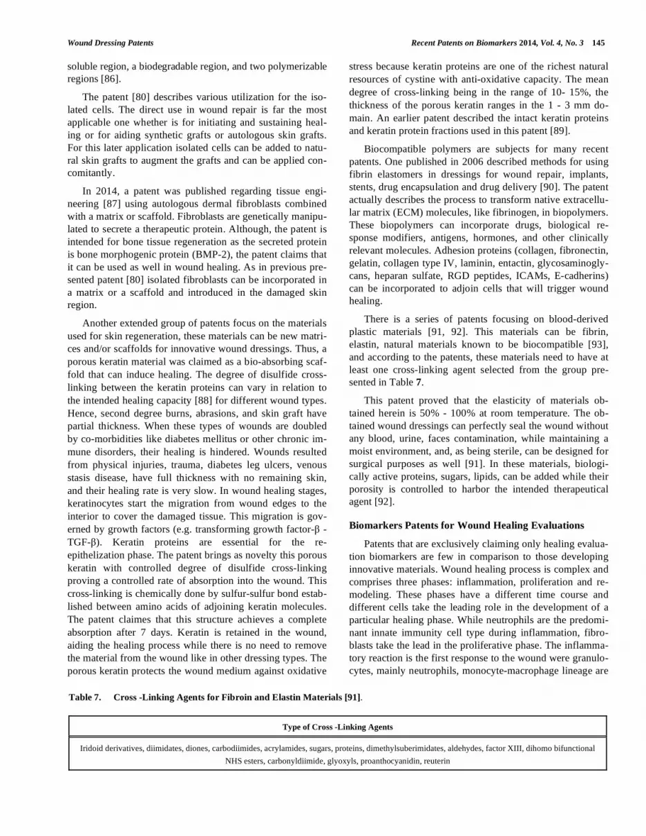
Another patent related to wound healing is the admini-
stration of a complement inhibitor to inhibit complement
activation, particularly through C3, C5 or C5a signaling [59].
The cellular process this patent is based on, is that the acti-
vated complement system is an essential effector mechanism
of the innate immune as a rapid immune response to tissue
injury. Out of the entire complement cascade, C3a and C5a
are the most potent chemoattractants upon activation of the
complement system [60]. C3a and C5a attract neutrophils,
monocytes, and macrophages to the sites where complement
cascade was activated [61]. Activated immunity cells, like
macrophages, can also produce C3 and activate phagocytosis
and/or clearance of apoptotic and necrotic cells [62]. Moreo-
ver, C5a receptor (C5aR or CD88) signaling in Toll-like re-
ceptor (TLR)-activated macrophages selectively inhibits the
transcription of genes that encode the IL-12 cytokine family,
which in turn drives the polarization and recruitment of T-
helper lymphocytes 1 (Thl) [63]. While complement activa-
tion is needed to restore tissue injury, inappropriate comple-
ment activation can cause injury and contribute to further
tissue damage [64]. In this light, the patent [59], claims that
for chronic wounds, administering therapeutically effective
complement inhibitors, that can control complement activa-
tion, a healing process prevails. Thus, one or more inhibitors
for C3, C3aR, C5a, C5aR, factor D, factor B, C4, Clq, or any
combination of these inhibitors would be efficient (Table 5).
The patent foresees a systemic administration or a lo-
cally/topically administration of these inhibitors. This ad-
ministration can be done together or sequentially with other
wound therapies and this administration can continue until
the chronic wound heals. The patent is based on the results
obtained in animal models. Genetically deficient animals for
C3, C5 and the C5a receptor have accelerated healing of
cutaneous wounds, in comparison to their wild-type counter-
parts.
When topical administration is foreseen, besides classical
dressing, the patent recommends an enhancer that induces an
increased penetration of the therapeutical agent such as al-
pha-hydroxy acids, limonene, azone (AZ), lauryl alcohol
(LA), other alcohols, isopropyl myristate (IPM), and so on.
An array of patents is recently focusing on advanced
therapy that uses cells and stem cells that can regenerate
skin’s multi-layered tissue.
Angiogenesis-regulating cells and factors can enhance
wound healing. In a patent, therapeutic platelets are claimed
Table 4. Wound Regeneration Through Gap Junctions Restoration Using a Combination of two Specific Agents [55-61].
Anti-Connexin Agent Structures
First group Anti-connexin oligonucleotides (anti-connexin 43 oligonucleotides), anti-connexin 43 peptides or peptidomimetics, gap junc-
tion closing compounds, hemichannel closing compounds, connexin carboxy-terminal polypeptides
Second group Selected from the above group as modified to subtract the sub-category of anti-connexin agents from which the first anti-
connexin agent was selected.
Wound Dressing Patents Recent Patents on Biomarkers 2014, Vol. 4, No. 3 143
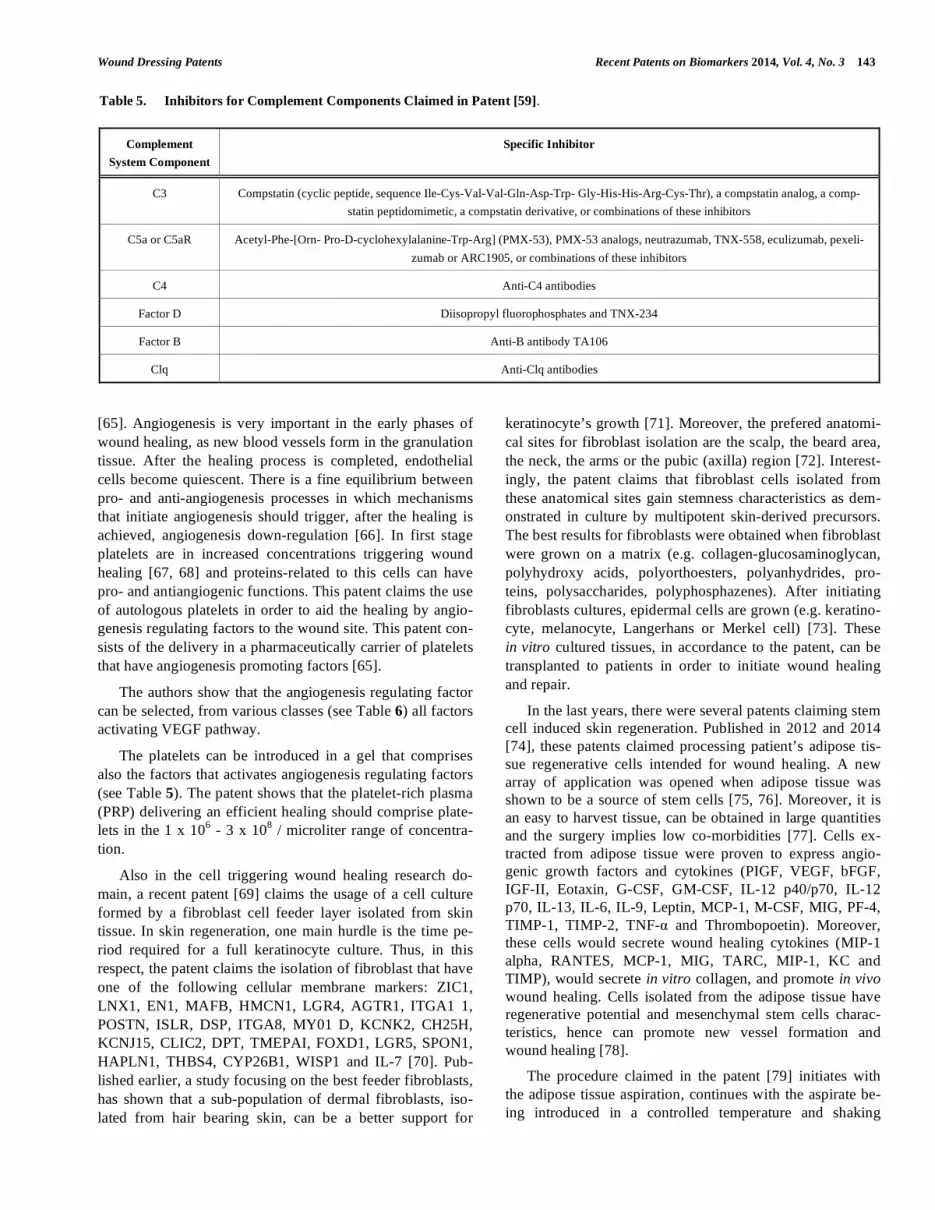
[65]. Angiogenesis is very important in the early phases of
wound healing, as new blood vessels form in the granulation
tissue. After the healing process is completed, endothelial
cells become quiescent. There is a fine equilibrium between
pro- and anti-angiogenesis processes in which mechanisms
that initiate angiogenesis should trigger, after the healing is
achieved, angiogenesis down-regulation [66]. In first stage
platelets are in increased concentrations triggering wound
healing [67, 68] and proteins-related to this cells can have
pro- and antiangiogenic functions. This patent claims the use
of autologous platelets in order to aid the healing by angio-
genesis regulating factors to the wound site. This patent con-
sists of the delivery in a pharmaceutically carrier of platelets
that have angiogenesis promoting factors [65].
The authors show that the angiogenesis regulating factor
can be selected, from various classes (see Table 6) all factors
activating VEGF pathway.
The platelets can be introduced in a gel that comprises
also the factors that activates angiogenesis regulating factors
(see Table 5). The patent shows that the platelet-rich plasma
(PRP) delivering an efficient healing should comprise plate-
lets in the 1 x 10⁶ - 3 x 10
8 / microliter range of concentra-
tion.
Also in the cell triggering wound healing research do-
main, a recent patent [69] claims the usage of a cell culture
formed by a fibroblast cell feeder layer isolated from skin
tissue. In skin regeneration, one main hurdle is the time pe-
riod required for a full keratinocyte culture. Thus, in this
respect, the patent claims the isolation of fibroblast that have
one of the following cellular membrane markers: ZIC1,
LNX1, EN1, MAFB, HMCN1, LGR4, AGTR1, ITGA1 1,
POSTN, ISLR, DSP, ITGA8, MY01 D, KCNK2, CH25H,
KCNJ15, CLIC2, DPT, TMEPAI, FOXD1, LGR5, SPON1,
HAPLN1, THBS4, CYP26B1, WISP1 and IL-7 [70]. Pub-
lished earlier, a study focusing on the best feeder fibroblasts,
has shown that a sub-population of dermal fibroblasts, iso-
lated from hair bearing skin, can be a better support for
keratinocyte’s growth [71]. Moreover, the prefered anatomi-
cal sites for fibroblast isolation are the scalp, the beard area,
the neck, the arms or the pubic (axilla) region [72]. Interest-
ingly, the patent claims that fibroblast cells isolated from
these anatomical sites gain stemness characteristics as dem-
onstrated in culture by multipotent skin-derived precursors.
The best results for fibroblasts were obtained when fibroblast
were grown on a matrix (e.g. collagen-glucosaminoglycan,
polyhydroxy acids, polyorthoesters, polyanhydrides, pro-
teins, polysaccharides, polyphosphazenes). After initiating
fibroblasts cultures, epidermal cells are grown (e.g. keratino-
cyte, melanocyte, Langerhans or Merkel cell) [73]. These
in vitro cultured tissues, in accordance to the patent, can be
transplanted to patients in order to initiate wound healing
and repair.
In the last years, there were several patents claiming stem
cell induced skin regeneration. Published in 2012 and 2014
[74], these patents claimed processing patient’s adipose tis-
sue regenerative cells intended for wound healing. A new
array of application was opened when adipose tissue was
shown to be a source of stem cells [75, 76]. Moreover, it is
an easy to harvest tissue, can be obtained in large quantities
and the surgery implies low co-morbidities [77]. Cells ex-
tracted from adipose tissue were proven to express angio-
genic growth factors and cytokines (PIGF, VEGF, bFGF,
IGF-II, Eotaxin, G-CSF, GM-CSF, IL-12 p40/p70, IL-12
p70, IL-13, IL-6, IL-9, Leptin, MCP-1, M-CSF, MIG, PF-4,
TIMP-1, TIMP-2, TNF- and Thrombopoetin). Moreover,
these cells would secrete wound healing cytokines (MIP-1
alpha, RANTES, MCP-1, MIG, TARC, MIP-1, KC and
TIMP), would secrete in vitro collagen, and promote in vivo
wound healing. Cells isolated from the adipose tissue have
regenerative potential and mesenchymal stem cells charac-
teristics, hence can promote new vessel formation and
wound healing [78].
The procedure claimed in the patent [79] initiates with
the adipose tissue aspiration, continues with the aspirate be-
ing introduced in a controlled temperature and shaking
Table 5. Inhibitors for Complement Components Claimed in Patent [59].
Complement
System Component
Specific Inhibitor
C3 Compstatin (cyclic peptide, sequence Ile-Cys-Val-Val-Gln-Asp-Trp- Gly-His-His-Arg-Cys-Thr), a compstatin analog, a comp-
statin peptidomimetic, a compstatin derivative, or combinations of these inhibitors
C5a or C5aR Acetyl-Phe-[Orn- Pro-D-cyclohexylalanine-Trp-Arg] (PMX-53), PMX-53 analogs, neutrazumab, TNX-558, eculizumab, pexeli-
zumab or ARC1905, or combinations of these inhibitors
C4 Anti-C4 antibodies
Factor D Diisopropyl fluorophosphates and TNX-234
Factor B Anti-B antibody TA106
Clq Anti-Clq antibodies

144 Recent Patents on Biomarkers 2014, Vol. 4, No. 3 Constantin et al.
chamber in order to start the disintegration procedures and
further isolate regenerative cells. Various disintegration
compounds can be used (neutral proteases, collagenase, tryp-
sin, lipase, hyaluronidase, deoxyribonuclease, Liberase H1,
pepsin, collagenase), or other known physical procedures
like ultrasonic, lasers, microwaves, or other mechanical de-
vices. Buoyant and non-buoyant components are left to settle
and the buoyant layer comprises the regenerative cells that
will be further washed and concentrated. The non-buoyant
layer is to be removed from the disintegration chambers as it
comprises blood, collagen, lipids and other non-regenerative
cells. The cells are washed in sterile condition and the patent
claims that an indicator of red blood removal can be a regis-
tered OD 540nm in the range 0.546 - 0.842, readings that
shows a low contamination with red blood cells. The cell
fraction can contain different types of cells: stem cells, pro-
genitor cells, endothelial precursor cells, adipocytes and
other regenerative cells, this fraction can also contain con-
taminants, such as collagen and other connective tissue pro-
teins. Further filtration is needed to separate only stem cells
or endothelial progenitors cells. The filtration system (e.g.
polysulfone, polyethersulfone or a mixed ester materials)
gradually removes collagen fibers and then larger fibers until
cell suspensions are purified.
Using this method, cell suspensions can be obtained in
the range of 1 105
- 1 107
cells/mL. The automated system
can be re-configurated if the isolated cells need to be sub-
jected to additional manipulation. Thus cells can be further
cultivated and tested for their viability/cell growth, or tran-
siently transfected for further gene therapy applications, or
for generation of cells lines or any other assay development
for cell/tissue engineering applications. When the intended
cells are ready to be used they can be placed into a syringe,
and inoculated subcutaneously, intramuscularly. As stated in
the patent, a portion of the separated batch needs to be cryo-
preserved [79].
From our point of view, we would have expected a stage
were the patent identifyes, in terms of cellular markers, at
least partially, the cell types isolated by this procedure.
Also dealing with mesenchymal stem cells, but this time
derived from skin [80] a patent published in 2013 shows the
methods for isolating, purifying, culturing, storing of three
types of skin-derived cells. The patent focuses on mesen-
chymal stem cells with the differentiating phenotype
CD146+, CD271+ and regeneration-associated cells with the
phenotype SSEA3+ (stage-specific embryonic antigen 3) and
CD105+. From human skin, mesenchymal stem cells
(MSCs) have the phenotype CD146+ and CD271+. CD271
(LNGFR) cells are isolated and these cells can be involved in
the development, survival, and differentiation of regenerative
cells [81]. Primary cells were obtained from a 4mm diameter
adult skin punch biopsy using a previously published method
[82]. From these cells, upon cultivation, cells with specific
markers were sorted using a FACS sorter method. The patent
defines SERA cells as cells expressing SSEA3 and CD105+,
isolated from human dermal skin cells and that can produce
iPSCs. After isolation, SERA cells can be maintained around
8 passages, hence can be cultivated around 6 weeks until
application. These cells can be grown on a scaffold that can
incorporate additional molecules. The scaffold can be biode-
gradable polymeric fibers, collagen fibers, synthetic or natu-
ral extracellular matrix (ECM). Additional molecules can be
active agents such as growth factors, anti-inflammatory
compounds, antibiotics, antivirals, or any intended therapeu-
tical combination.
Like in the previous described patents, in this one cells
can be incorporated in a matrix or in a scaffold in order to
enhance their survival and/or growth. Matrix can be a fibrin
scaffold [83] that can support MSC in a concentration of at
least 1 x 10⁶ cells/mL. This scaffold can have anti-
fibrinolytic agents (tranexamic acid, arginine, lysine) and
anti-coagulation compounds (factor VIII, fibronectin, von
Willebrand factor, vitronectin) [84]. Cells can be mixed with
a hydrogel where cells can adhere and grow, and this combi-
nation can be applied directly [85]. The hydrogel can be de-
signed to fill a cavity that needs to be regenerated. Besides,
the termosensitive hydrogels described above, the patent can
use hydrogels that can be solid in visible or ultraviolet light.
These hydrogels are composed of macromers with a water
Table 6. Angiogenesis Regulating Factors for Wound Healing Mediated by Platelets [65].
Activators For: Compounds
VEGF pathways Agents that activate the neuropilin 1 & 2 pathways, VEGF-A and C, FGF, HGF, angiopoietin-1, insulin-like growth factor-1,
epidermal growth factor, platelet derived growth factor, platelet factor 4, thrombospondin-1, TGF-beta-1, plasminogen activator
inhibitor type-1 (PAI-I), alpha2-antiplasmin and alpha2-macroglobulin VEGFRl (flt-1), VEGFR2 (flk-2), VEGFR3 (flt-4), hepa-
rin sulfate proteoglycan, VEGF121, VEGF145, VEGF165, VEGF168, VEGF189, VEGF -B and -D, PLGF 1, PLGF2, HIV-I
TAT, Sema-E, Sema-III, Sema-IV, bFGF, PDGFR, EGFR, and IGFR
Platelet Thrombin, collagen, serotonin, ADP, acetylcholine and combinations
Angiogenesis PAR-I agonists: TFLLR-NH2; TFLLRNPNDK-NH2; SFLLRNPNDKYEPF-NH2; and SFLLRN-NH2
PAR-4 antagonist: transcinnamoyl- YPGKF-NH2 (tcY-NH2; Ma et al.) and YD-3, a non-peptide PAR4 antagonist nonpeptide
PAR-4 antagonist, YD-3 (ethyl 4-(l- benzyl-lH-indazol-3-yl)benzoate)
Wound Dressing Patents Recent Patents on Biomarkers 2014, Vol. 4, No. 3 145
soluble region, a biodegradable region, and two polymerizable
regions [86].
The patent [80] describes various utilization for the iso-
lated cells. The direct use in wound repair is far the most
applicable one whether is for initiating and sustaining heal-
ing or for aiding synthetic grafts or autologous skin grafts.
For this later application isolated cells can be added to natu-
ral skin grafts to augment the grafts and can be applied con-
comitantly.
In 2014, a patent was published regarding tissue engi-
neering [87] using autologous dermal fibroblasts combined
with a matrix or scaffold. Fibroblasts are genetically manipu-
lated to secrete a therapeutic protein. Although, the patent is
intended for bone tissue regeneration as the secreted protein
is bone morphogenic protein (BMP-2), the patent claims that
it can be used as well in wound healing. As in previous pre-
sented patent [80] isolated fibroblasts can be incorporated in
a matrix or a scaffold and introduced in the damaged skin
region.
Another extended group of patents focus on the materials
used for skin regeneration, these materials can be new matri-
ces and/or scaffolds for innovative wound dressings. Thus, a
porous keratin material was claimed as a bio-absorbing scaf-
fold that can induce healing. The degree of disulfide cross-
linking between the keratin proteins can vary in relation to
the intended healing capacity [88] for different wound types.
Hence, second degree burns, abrasions, and skin graft have
partial thickness. When these types of wounds are doubled
by co-morbidities like diabetes mellitus or other chronic im-
mune disorders, their healing is hindered. Wounds resulted
from physical injuries, trauma, diabetes leg ulcers, venous
stasis disease, have full thickness with no remaining skin,
and their healing rate is very slow. In wound healing stages,
keratinocytes start the migration from wound edges to the
interior to cover the damaged tissue. This migration is gov-
erned by growth factors (e.g. transforming growth factor- -
TGF- ). Keratin proteins are essential for the re-
epithelization phase. The patent brings as novelty this porous
keratin with controlled degree of disulfide cross-linking
proving a controlled rate of absorption into the wound. This
cross-linking is chemically done by sulfur-sulfur bond estab-
lished between amino acids of adjoining keratin molecules.
The patent claimes that this structure achieves a complete
absorption after 7 days. Keratin is retained in the wound,
aiding the healing process while there is no need to remove
the material from the wound like in other dressing types. The
porous keratin protects the wound medium against oxidative
stress because keratin proteins are one of the richest natural
resources of cystine with anti-oxidative capacity. The mean
degree of cross-linking being in the range of 10- 15%, the
thickness of the porous keratin ranges in the 1 - 3 mm do-
main. An earlier patent described the intact keratin proteins
and keratin protein fractions used in this patent [89].
Biocompatible polymers are subjects for many recent
patents. One published in 2006 described methods for using
fibrin elastomers in dressings for wound repair, implants,
stents, drug encapsulation and drug delivery [90]. The patent
actually describes the process to transform native extracellu-
lar matrix (ECM) molecules, like fibrinogen, in biopolymers.
These biopolymers can incorporate drugs, biological re-
sponse modifiers, antigens, hormones, and other clinically
relevant molecules. Adhesion proteins (collagen, fibronectin,
gelatin, collagen type IV, laminin, entactin, glycosaminogly-
cans, heparan sulfate, RGD peptides, ICAMs, E-cadherins)
can be incorporated to adjoin cells that will trigger wound
healing.
There is a series of patents focusing on blood-derived
plastic materials [91, 92]. This materials can be fibrin,
elastin, natural materials known to be biocompatible [93],
and according to the patents, these materials need to have at
least one cross-linking agent selected from the group pre-
sented in Table 7.
This patent proved that the elasticity of materials ob-
tained herein is 50% - 100% at room temperature. The ob-
tained wound dressings can perfectly seal the wound without
any blood, urine, faces contamination, while maintaining a
moist environment, and, as being sterile, can be designed for
surgical purposes as well [91]. In these materials, biologi-
cally active proteins, sugars, lipids, can be added while their
porosity is controlled to harbor the intended therapeutical
agent [92].
Biomarkers Patents for Wound Healing Evaluations
Patents that are exclusively claiming only healing evalua-
tion biomarkers are few in comparison to those developing
innovative materials. Wound healing process is complex and
comprises three phases: inflammation, proliferation and re-
modeling. These phases have a different time course and
different cells take the leading role in the development of a
particular healing phase. While neutrophils are the predomi-
nant innate immunity cell type during inflammation, fibro-
blasts take the lead in the proliferative phase. The inflamma-
tory reaction is the first response to the wound were granulo-
cytes, mainly neutrophils, monocyte-macrophage lineage are
Table 7. Cross -Linking Agents for Fibroin and Elastin Materials [91].
Type of Cross -Linking Agents
Iridoid derivatives, diimidates, diones, carbodiimides, acrylamides, sugars, proteins, dimethylsuberimidates, aldehydes, factor XIII, dihomo bifunctional
NHS esters, carbonyldiimide, glyoxyls, proanthocyanidin, reuterin

146 Recent Patents on Biomarkers 2014, Vol. 4, No. 3 Constantin et al.
recruited. If this recruitment is normal in the first acute
phases, the uninterrupted presence of neutrophils is associ-
ated with delayed wound healing and chronical status. The
proliferative phase (epithelialization, angiogenesis and ma-
trix formation) starts when neutrophils have been cleared out
by activated macrophages. Hence, in the proliferative phase,
fibroblasts are the main cell-type in a normal healing wound.
Detection of secreted molecules by these types of cells can
be specific biomarkers indicating healing phases and can
indicate wound healing progression. Locally secreted bio-
markers can be used for point-of-care diagnostic determina-
tions [52] (see also Table 1).
In 2014, a report showed that serum C-reactive protein
(CRP) and interleukin-6 (IL-6) evaluated in wound fluid can
depict a local inflammation state of chronic wounds. In this
study the clinical wound improvement was statistically re-
lated to the decrease in wound fluid of these two tested bio-
markers, namely CRP and IL-6 [94].
Patents for Biomarkers. Besides the publications on the
topic of wound healing biomarkers, a patent claimed the anti-
oxidant capacity of a sample to indicate the level of wound
infection. The system comprises the diagnostic device in a
wound dressing that can have also an antimicrobial agent [95].
Symptoms such as local swelling, heat, pain and redness,
is recognized by the physician as infection signs but an ear-
lier detection would be beneficial for patient. Definitive di-
agnosis is routinely achieved by using a swab that extracts
wound fluid and then it is subjected to microbiological tests
that can take at least 48 - 72 hours. This invention claims the
total antioxidant capacity of an infected wound fluid to be
significantly higher compared to a non-infected wound. The
total antioxidant capacity reflects the microbial burden of the
wound. Actually, the wound fluid can inactivate reactive
oxygen species (ROS) such as hydroxyl radicals (OH), sin-
glet oxygen (1O2), hydroperoxyl radicals (
1OOH), superoxide
radical anions (O2 .) and hydrogen peroxide (H2O2). The in-
dicator molecule can be oxidized or reduced by ROS with a
registered modification in absorbance or fluorescence, rou-
tinely a clear colour change. Table 8 describes the redox
indicators that can be used herein.
The antioxidant capacity can be an assayed as the rate of
color change of e.g. cytochrome C due to the superoxide
generation. This method was described more than 50 years
ago [96] and still it is in laboratory use due to its easy to per-
form and robust results. The patent [95] describes the super-
oxide anions generation by the reaction of hypoxanthine with
xanthine oxidase, the generated superoxide anion reacts with
a reference amount of cytochrome C and with the antioxi-
dants present in the wound fluid sample. When evaluating
the activity, the inventors advise that a rise to 130-150% of
the total antioxidant capacity compared to the control can
indicate an infected wound fluid. The wound fluid should be
harvested in the time interval of 1 - 24 hours. Besides the
activity registered, the change in this activity in time should
be as well recorded, evaluating the rate of change. When an
increase in total antioxidant capacity is detected antimicro-
bial dressing is applied.
In 2014, a report showed the healing ability of chronic
ulcers as assessed by evaluating hydroxyproline, total protein
and enzymatic antioxidants (glutathione peroxidase - GPx,
glutathione S-transferase - GST) in the granulation tissue
[97].
Other patent evaluated the stages of wound healing by
quantifing the expression levels of certain genes, biomarkers
for wound healing phases. A chronic wound has as charac-
teristics a reduced level of angiotensin II receptor, IL-1R
receptor antagonist or inositol triphosphate receptor 3, while
the expression of interleukins, growth factors and collagens
is increased. According to the invention, angiotensin II re-
ceptor, IL-1R antagonist, inositol triphosphate receptor 3,
certain interleukins, growth factors and collagens are mark-
ers for wound status. Quantifying by hybridization assay the
RNA extracted from wound tissue and evaluating these ge-
netic markers, the stage of a wound and/or the evolution of
one can be identified [98].
CURRENT & FUTURE DEVELOPMENTS
Wound healing is a wide-spread medical problem and the
history of dressing development has shown that there is no
general all-purpose dressing. Each year, we are witnessing
new materials and procedures whether published and/or pat-
ented. Clinicians have more than 50 different classes of
dressings while 3,000 products aid wound care domain. All
of these products have properties that aim to induce a normal
Table 8. Antioxidant Indicators Used in Patent [95].
Indicator Molecule Target Molecule
Cytochrome C Superoxide
1,10-Phenanthrolene Chelates iron, zinc and other divalent metals
Diphenylamine sulphonic acid Redox indicator
Triphenylmethane dyes Redox indicator
Starch Iodine

Wound Dressing Patents Recent Patents on Biomarkers 2014, Vol. 4, No. 3 147
wound healing process. Already approved wound dressing
materials are constantly improved, mainly by adding
drugs/growth factors or by down-regulating factors that can
damage the tissue [99]. Although, health care market in
wound management abound, the fact that we still do not
have the best thrapeutical approach for chronic wounds
resides in the lack of cellular and molecular comprehension
of the chronic process.
Advanced approaches in this domain will reside in de-
picting intimate characteristics not only of the wound but as
well for the patient. After establishing these particularities
one can choose the most appropriate, individualized wound
care complex approach. The mechanisms of action for the
wound dressings are very diverse, and while some are effi-
cient in one stage of the wound development, others are
more efficacious in other stage, hence the nurse and the phy-
sician has to develop a specific wound management program
tailored to both wound and the patient carrying it [100, 101].
The future development of patents in wound dressing
field and in the biomarkers domain that enables the assess-
ment of the healing efficiency will move toward the best
physiologically-friendly materials. Innovative scaffolds will
enlarge the incorporated factors and cells that will aid tissue
regeneration. Always keeping in mind that the fate of a
wound resides on both local and systemic approaches, pa-
tient nutrition counseling, education and prevention, should
assist the complex clinical wound management.
CONFLICT OF INTEREST
The authors confirm that this article content has no con-
flict of interest.
ACKNOWLEDGEMENTS
Partially supported by PN-II-PT-PCCA-2013-4-1386
(Project No. 185), PN-II-PT-PCCA-2013-4-1407 (Project
No. 190), PN 09.33-01.01/2009. Author Georgeta Paunica-
Panea has a doctoral grant financed through Project POS-
DRU/159/1.5/S/135760.
REFERENCES
[1] Sood A, Granick MS, Tomaselli NL. Wound dressings and com-
parative effectiveness data. Adv Wound Care (New Rochelle)
2014; 3(8): 511-29.
[2] Leaper DJ, Harding KG. Wounds: Biology and Management. Ox-
ford University Press, USA 1998.
[3] Jankovic A, Binic I, Stanojevic M. Port wine stain, soft tissue bone
hypertrophy and leg ulcer. BMJ Case Rep 2009; 2009. pii:
bcr01.2009.1452. doi: 10.1136/bcr.01.2009.1452.
[4] Wound Care Market by Type (Traditional (Wound Closure, Anti
infective), Basic (Films, Cleansing), Advanced (Hydrogels, Hydro-
colloids, Alginate, Collagen), Active (Artificial Skin & Skin Sub-
stitutes), Pressure Relief Devices, NPWT) - Global Forecast to
2019. Available at: http://www.marketsandmarkets.com/Market-
Reports/wound-care-market-371.html. (Accessed on: December, 1,
2014)
[5] Meissner MH. Venous ulcer care: Which dressings are cost effec-
tive? Phlebology 2014; 29 (1 Suppl): 174-80.
[6] Lillis K. Effective wound care requires look at total patient picture.
(Infection Connection). Healthcare Purchasing News. High Beam
Res 2003; 27(1): 23.
[7] Baxter H, Ballard K. Vacuum-assisted closure. Nurs Times 2001;
97(35): 51-2.
[8] O'Reilly D, Linden R, Fedorko L, Tarride JE, Jones WG, Bowen
JM, et al. A prospective, double-blind, randomized, controlled
clinical trial comparing standard wound care with adjunctive hy-
perbaric oxygen therapy (HBOT) to standard wound care only for
the treatment of chronic, non-healing ulcers of the lower limb in
patients with diabetes mellitus: A study protocol. Trials 2011; 12:
69.
[9] Grim PS, Gottlieb LJ, Boddie A, Batson E. Hyperbaric oxygen
therapy. JAMA 1990; 263(16): 2216-20.
[10] Neagu M. The immune system a hidden treasure for biomarker
discovery in cutaneous melanoma. Adv Clin Chem 2012; 58: 89-
140.
[11] Badylak SF, Nerem RM. Progress in tissue engineering and regen-
erative medicine. Proc Natl Acad Sci USA 2010; 107(8): 3285-86.
[12] Neagu M, Constantin C. Stem Cell standing at the Immune Proc-
esses Cross Roads -Tumorigenesis versus Regeneration. In: Tanase
C, Neagu M. Eds. Stem Cells Between Regeneration And Tumori-
genesis. Bentham eBooks; 2014; in press.
[13] Trounson A. New perspectives in human stem cell therapeutic
research. BMC Med 2009; 7(1): 29.
[14] Mayet N, Choonara YE, Kumar P, Tomar LK, Tyagi C, Du Toit
LC, et al. A comprehensive review of advanced biopolymeric
wound healing systems. J Pharm Sci 2014; 103(8): 2211-30.
[15] Boateng JS, Matthews KH, Stevens HN, Eccleston GM. Wound
healing dressings and drug delivery systems: A Review. J Pharm
Sci 2008; 97(8): 2892-23.
[16] Kevadiya BD, Rajkumar S, Bajaj HC, Chettiar SS, Gosai K,
Brahmbhatt H, et al. Biodegradable gelatin-ciprofloxacin-
montmorillonite composite hydrogels for controlled drug release
and wounddressing application. Colloids Surf B Biointerfaces
2014; 122: 175-83.
[17] Norouzi M, Shabani I, Ahvaz HH, Soleimani M. PLGA/gelatin
hybrid nanofibrous scaffolds encapsulating EGF for skin regenera-
tion. J Biomed Mater Res A 2014; doi: 10.1002/jbm.a.35355.
[18] Bueno CZ, Dias AM, de Sousa HJ, Braga ME, Moraes AM. Con-
trol of the properties of porous chitosan-alginate membranes
through the addition of different proportions of Pluronic F68. Mater
Sci Eng C Mater Biol Appl 2014; 44: 117-25.
[19] Han F, Dong Y, Su Z, Yin R, Song A, Li S. Preparation, character-
istics and assessment of a novel gelatin-chitosan sponge scaffold as
skin tissue engineering material. Int J Pharm 2014; 476(1-2): 124-
33.
[20] Klinkajon W, Supaphol P. Novel copper (II) alginate hydrogels and
their potential for use as anti-bacterial wound dressings. Biomed
Mater 2014; 9(4): 045008.
[21] De Cicco F, Porta A, Sansone F, Aquino RP, Del Gaudio P.
Nanospray technology for an in situ gelling nanoparticulate powder
as a wound dressing. Int J Pharm 2014; 473(1-2): 30-7.
[22] Fu R, Li C, Yu C, Xie H, Shi S, Li Z, et al. A novel electrospun
membrane based on moxifloxacin hydrochloride/poly(vinyl alco-
hol)/sodium alginate for antibacterial wound dressings in practical
application. Drug Deliv 2014; 28: 1-12.
[23] Branco da Cunha C, Klumpers DD, Li WA, Koshy ST, Weaver JC,
Chaudhuri O, et al. Influence of the stiffness of three-dimensional
alginate/collagen-I interpenetrating networks on fibroblast biology.
Biomaterials 2014; 35(32): 8927-36.
[24] Ding F, Deng H, Du Y, Shi X, Wang Q. Emerging chitin and chito-
san nanofibrous materials for biomedical applications. Nanoscale
2014; 6(16): 9477-93.
[25] Yodkhum K, Phaechamud T. Hydrophobic chitosan sponges modi-
fied by aluminum monostearate and dehydrothermal treatment as
sustained drug delivery system. Mater Sci Eng C Mater Biol Appl
2014; 42: 715-25.
[26] Lee SJ, Heo DN, Moon JH, Ko WK, Lee JB, Bae MS, et al. Elec-
trospun chitosan nanofibers with controlled levels of silver
nanoparticles. Preparation, characterization and antibacterial activ-
ity. Carbohydr Polym 2014; 111: 530-37.

148 Recent Patents on Biomarkers 2014, Vol. 4, No. 3 Constantin et al.
[27] Miguel SP, Ribeiro MP, Brancal H, Coutinho P, Correia IJ. Ther-
moresponsive chitosan-agarose hydrogel for skin regeneration.
Carbohydr Polym 2014; 111: 366-73.
[28] Szweda P, Gorczyca G, Tylingo R, Kurlenda J, Kwiecinski J,
Milewski S. Chitosan-protein scaffolds loaded with lysostaphin as
potential antistaphylococcal wound dressing materials. J Appl Mi-
crobiol 2014; 117(3): 634-42.
[29] Zhao R, Li X, Sun B, Zhang Y, Zhang D, Tang Z, et al. Electros-
pun chitosan/sericin composite nanofibers with antibacterial prop-
erty as potential wound dressings. Int J Biol Macromol 2014; 68:
92-7.
[30] Ahamed MI, Sankar S, Kashif PM, Basha SK, Sastry TP. Evalua-
tion of biomaterial containing regenerated cellulose and chitosan
incorporated with silver nanoparticles. Int J Biol Macromol 2014;
72C: 680-86.
[31] Sadat EMM, Schönherr H. Enzyme-sensing chitosan hydrogels.
Langmuir 2014; 30(26): 7842-50.
[32] Ouimet MA1, Fogaça R, Snyder SS, Sathaye S, Catalani LH,
Pochan DJ, et al. Poly(anhydride-ester) and Poly(N-vinyl-2-
pyrrolidone) Blends: Salicylic acid-releasing blends with hydrogel-
like properties that reduce inflammation. Macromol Biosci 2014;
doi: 10.1002/mabi.201400238.
[33] Rasekh M, Karavasili C, Soong YL, Bouropoulos N, Morris M,
Armitage D, et al. Electrospun PVP-indomethacin constituents for
transdermal dressings and drug delivery devices. Int J Pharm 2014;
473(1-2): 95-104.
[34] Zhou G, Ge H, Wang L, Liu M, Wang B, Su H, et al. Research on
a novel poly (vinyl alcohol)/lysine/vanillin wound dressing: Bio-
compatibility, bioactivity and antimicrobial activity. Burns 2014;
pii:S0305-4179(14)00128-4.
[35] Agrawal P, Soni S, Mittal G, Bhatnagar A. Role of polymeric bio-
materials as wound healing agents. Int J Low Extrem Wounds
2014; 13(3): 180-90.
[36] Shahverdi S, Hajimiri M, Esfandiari MA, Larijani B, Atyabi F,
Rajabiani A, et al. Fabrication and structure analysis of
poly(lactide-co-glycolic acid)/silk fibroin hybrid scaffold for
wound dressingapplications. Int J Pharm 2014; 473(1-2): 345-55.
[37] Jhong JF, Venault A, Liu L, Zheng J, Chen SH, Higuchi A, et al.
Introducing mixed-charge copolymers as wound dressing biomate-
rials. ACS Appl Mater Interfaces 2014; 6(12): 9858-70.
[38] Ahire JJ, Neppalli R, Heunis TD, van Reenen AJ, Dicks LM.
2,3-dihydroxybenzoic acid electrospun into poly(D,L-lactide)
(PDLLA)/poly(ethylene oxide) (PEO) nanofibers inhibited the
growth of gram-positive and gram-negative bacteria. Curr Micro-
biol 2014; 69(5): 587-93.
[39] Su LC, Xie Z, Zhang Y, Nguyen KT, Yang J. Study on the antimi-
crobial properties of citrate-based biodegradable polymers. Front
Bioeng Biotechnol 2014; 2: 23.
[40] Calamak S, Erdo du C, Ozalp M, Ulubayram K. Silk fibroin based
antibacterial bionanotextiles as wound dressing materials. Mater
Sci Eng C Mater Biol Appl 2014; 43: 11-20.
[41] Gawande PV, Clinton AP, LoVetri K, Yakandawala N, Rumbaugh
KP, Madhyastha S. Antibiofilm efficacy of dispersinB(®) wound
spray used in combination with a silver wound dressing. Microbiol
Insights 2014; 7: 9-13.
[42] Chang HY, Cang J, Roy P, Chang HT, Huang YC, Huang CC.
Synthesis and antimicrobial activity of gold/silver-tellurium nanos-
tructures. ACS Appl Mater Interfaces 2014; 6(11): 8305-12.
[43] Said J, Walker M, Parsons D, Stapleton P, Beezer AE, Gaisford S.
An in vitro test of the efficacy of an anti-biofilm wound dressing.
Int J Pharm 2014; 474(1-2): 177-81.
[44] Masella PC, Balent EM, Carlson TL, Lee KW, Pierce LM. Evalua-
tion of six split-thickness skin graft donor-site dressing materials in
a swine Model. Plast Reconstr Surg Glob Open 2014; 1(9): e84.
[45] Khanbanha N, Atyabi F, Taheri A, Talaie F, Mahbod M, Dinarvand
R. Healing efficacy of an EGF impregnated triple gel based wound
dressing: In vitro and in vivo studies. Biomed Res Int 2014;
http://dx.doi.org/10.1155/2014/493732.
[46] Singla S, Garg R, Kumar A, Gill C. Efficacy of topical application
of beta urogastrone (recombinant human epidermal growth factor)
in Wagner's Grade 1 and 2 diabetic foot ulcers: Comparative analy-
sis of 50 patients. J Nat Sci Biol Med 2014; 5(2): 273-77.
[47] Doerler M, Eming S, Dissemond J, Wolter A, Stoffels-Weindorf
M, Reich-Schupke S, et al. A novel epidermal growth factor con-
taining wound dressing for the treatment of hard-to-heal venous leg
ulcers. Adv Skin Wound Care 2014; 27(10): 456-60.
[48] Huang G, Sun T, Zhang L, Wu Q, Zhang K, Tian Q, et al. Com-
bined application of alginate dressing and human granulocyte-
macrophage colony stimulating factor promotes healing in refrac-
tory chronic skin ulcers. Exp Ther Med 2014; 7(6): 1772-76.
[49] Pokhitonov DY, Borovkova NV, Filippov OP, Klyukvin IY, Khva-
tov VB, Ponomaryov IN, et al. Experimental substantiation and
clinical use of a combination of dermal matrix with allogenic or
autologous cells for the treatment of extensive traumatic wounds.
Bull Exp Biol Med 2014; 157(5): 705-10.
[50] Quan R, Zheng X, Xu S, Zhang L, Yang D. Gelatin-chondroitin-6-
sulfate-hyaluronic acid scaffold seeded with vascular endothelial
growth factor 165 modified hair follicle stem cells as a three-
dimensional skin substitute. Stem Cell Res Ther 2014; 5(5): 118.
[51] You HJ, Han SK, Rhie JW. Randomised controlled clinical trial for
autologous fibroblast-hyaluronic acid complex in treating diabetic
foot ulcers. J Wound Care 2014; 23(11): 521-30.
[52] Garcia, S., McNulty, A., Villarrea, K. Devices and methods for the
diagnosis and treatment of wounds using biomarkers.
US20120129186 (2012).
[53] Iddon, P., Raxworthy, M., Pio, S.L. Sampling device.
WO2013050429 (2013).
[54] Elder, I. A wound management system. WO2013026999 (2013).
[55] Duft, B.J. Impaired wound healing compositions and treatments.
US20100279921 (2010).
[56] Becker, D.L., Green, C.R., Duft, B.J. Impaired wound healing
compositions and treatments. WO2009075881 (2009),
US20110300130 (2011) & EP2628747 (2013).
[57] Duft, B.J. Impaired wound healing compositions and treatments.
WO2009075882 (2009).
[58] Brandner JM, Houdek P, Hüsing B, Kaiser C, Moll I. Connexins
26, 30, and 43: Differences among spontaneous, chronic, and ac-
celerated human wound healing. J Invest Dermatol 2004;122:1310-
20.
[59] Lambris, J.D., Rafail, S. Wound healing using complement inhibi-
tors. WO2012174055 (2012).
[60] Cazander G, Jukema GN, Nibbering PH. Complement activation
and inhibition in wound healing. Clin Dev Immunol 2012;
doi:10.1155/2012/534291.
[61] Ricklin D, Hajishengallis G, Yang K, Lambris JD. Complement: A
key system for immune surveillance and homeostasis. Nat Immu-
nol 2010; l l(9): 785-97.
[62] Gullstrand B, Martensson U, Sturfelt G, Bengtsson AA, Truedsson
L. Complement classical pathway components are all important in
clearance of apoptotic and secondary necrotic cells. Clin Exp Im-
munol 2009; 156(2): 303-11.
[63] Zhang X, Kimura Y, Fang C, Zhou L, Sfyroera G, Lambris JD,
et al. Regulation of Toll-like receptor-mediated inflammatory
response by complement in vivo. Blood 2007; 110(l): 228-36.
[64] Markiewski M, Lambris JD. The role of complement in inflamma-
tory diseases from behind the scenes into the spotlight. Am J Pathol
2007; 171(3): 715-27.
[65] Orgill, D., Pietramaggiori, G., Klement, G., Cervi, D., Folkman, J.
Therapeutic platelet compositions and methods. WO2009002811
(2009).
[66] Singer AJ, Clark RA. Cutaneous wound healing. N Engl J Med
1999; 341: 738-46.
[67] Jurk K, Kehrel BE. Platelets: Physiology and biochemistry. Semin
Thromb Hemost 2005; 31: 381-92.
[68] Capla JM, Ceradini DJ, Tepper OM, Callaghan MJ, Bhatt KA,
Galiano RD, et al. Skin graft vascularization involves precisely
regulated regression and replacement of endothelial cells through
both angiogenesis and vasculogenesis. Plast Reconstr Surg 2006;
117: 836-44.
[69] Albert, C., Jahoda, B. Cell support comprising dermal fibroblasts.
WO2011015862 (2011).
[70] Reynolds AJ, Chaponnier C, Jahoda CA, Gabbiani G. A quantita-
tive study of the differential expression of alpha-smooth muscle ac-

Wound Dressing Patents Recent Patents on Biomarkers 2014, Vol. 4, No. 3 149
tin in cell populations of follicular and non-follicular origin. J In-
vest Dermatol 1993; 101(4): 577-83.
[71] Nolte SV, Xu W, Rennekampff HO, Rodemann HP. Diversity of
fibroblasts a review on implications for skin tissue engineering.
Cells Tissues Organs 2008; 187(3): 165-76.
[72] Chipev CC, Simon M. Phenotypic differences between dermal
fibroblasts from different body sites determine their responses to
tension and TGF[beta]1. BMC Dermatol 2002; 2:13.
[73] Waelti ER, Inaebnit SP, Rast HP, Hunziker T, Limat A, Braathen
LR, et al. Co-culture of human keratinocytes on post-mitotic hu-
man dermal fibroblast feeder cells: Production of large amounts of
interleukin-6. J Invest Dermatol 1992; 98(5): 805-08.
[74] Fraser, J.K., Hedrick, M.H., Daniels, E. Methods of using adipose
derived stem cells to promote wound healing. US8105580 (2012)
& US8691216 (2014).
[75] Zuk PA, Zhu M, Mizuno H, Huang J, Futrell JW, Katz AJ, et al.
Multilineage cells from human adipose tissue: Implications for cell-
based therapies. Tissue Eng 2001; 7(2): 211-28.
[76] Zuk PA, Zhu M, Ashjian P, De Ugarte DA, Huang JI, Mizuno H,
et al. Human adipose tissue is a source of multipotent stem cells.
Mol Biol Cell 2002; 13(12): 4279-95.
[77] Commons GW, Halperin B, Chang CC. Large-volume liposuction:
A review of 631 consecutive cases over 12 years. Plast Reconstr
Surg 2001; 108(6): 1753-63.
[78] Alhadlaq A, Tang M, Mao JJ. Engineered adipose tissue from
human mesenchymal stem cells maintains predefined shape and
dimension: Implications in soft tissue augmentation and reconstruc-
tion. Tissue Eng 2005; 11: 556-66.
[79] Fraser, J.K., Hedrick, M.H. Preservation of non embryonic cells
from non hematopoietic tissues. WO2003024215 (2003).
[80] Byrne, J.A. Regenerative sera cells and mesenchymal stem cells.
WO2013151725 (2013).
[81] Rogers ML, Beare A, Zola H, Rush RA. CD 271 (P75 Neurotro-
phin Receptor). J Biol Regulators & Homeostatic Agents 2008;
22(1): 1-6.
[82] Byrne JA, Nguyen HN, Reijo Pera RA. Enhanced generation of
induced pluripotent stem cells from a subpopulation of human fi-
broblasts. PLoS One 2009; 4(9): e7118.
[83] Atlas, R., Nur, I., Meidler, R., Bar, L., Buensuceso, C., Kihm, A.J.,
Dhanaraj, S. Fibrin based scaffold, preparation and use thereof.
US20120039855 (2012).
[84] Nur, I., Bar, L., Lieberman, O. Stabilized mixture comprising fi-
brinogen. US6121232 (2000).
[85] Vacanti, C.A. Generation of new tissues on surfaces using hydro-
gels for cells. US5944754 (1999).
[86] Hubbell, J.A., Pathak, C.P., Sawhney, A.S., Desai, N.P., Hill, J.L.
A curable additional polymerizable macromolecular monomers
comprising at least one water soluble region, at least one degrad-
able, hydrolyzable region and free radical polymerizable end
groups; drug delivery. US5410016 (1995).
[87] Byrne, J.A., Wang, J.C. Cellular compositions for tissue engineer-
ing. WO2014105913 (2014).
[88] Kelly, R.J., Marsh, C., Ali, M.A., Sigurjonsson, G.F. Porous kera-
tin construct and method of making the same. US8124735 (2012).
[89] Kelly, R.J., Roddick-Lanzilotta, A.D., Ali, M.A. Comprising S-
sulfonated keratin associated with a water soluble polymer through
hydrogen bonding and further comprising a protein cross-linking
agent. US20080038327 (2008).
[90] Campbell, P., Fisher, G.W., Kumta, P., Sipe, D.M., Smith, J.,
Weiss, L.E. Biocompatible polymers and methods of use.
EP2093256 (2009).
[91] Campbell, P.G., Burgess, J.E., Weiss, L.E., Smith, J. Methods and
apparatus for manufacturing plasma based plastics and bioplastics
produced therefrom. US8529961 (2013).
[92] Campbell, P., Fisher, G.W., Kumta, P., Sipe, D.M., Smith, J.,
Weiss, L.E. Biocompatible polymers and methods of use.
WO2007126411 (2008).
[93] Cronkite EP, Lozner EL, Deaver JM. Use of thrombin and fibrino-
gen in skin grafting. JAMA 1994; 124: 976-78.
[94] Liu T, Yang F, Li Z, Yi C, Bai X. A prospective pilot study to
evaluate wound outcomes and levels of serum C-reactive protein
and interleukin-6 in the wound fluid of patients with trauma-related
chronic wounds. Ostomy Wound Manag 2014; 60(6): 30-7.
[95] Cullen, B.M., Clark, R. Marker of wound infection.
WO2006123091 (2006).
[96] van Gelder BF, Slater EC. The extinction coefficient of cytochrome
c. Biochim Biophvs Acta 1962; 58: 593-95.
[97] Honnegowda TM, Kumar P, Udupa P, Rao P, Bhandary S, Mahato
KK, et al. Effect of limited access dressing on hydroxyproline and
enzymatic antioxidant status in nonhealing chronic ulcers. Indian J
Plast Surg 2014; 47(2): 216-20.
[98] McGrath, K.P. Biomarkers for wound healing. US8034553 (2011).
[99] Erfurt-Berge C, Renner R. Recent developments in topical wound
therapy: Impact of antimicrobiological changes and rebalancing
thewound milieu. Biomed Res Int 2014;
http://dx.doi.org/10.1155/2014/819525.
[100] Broussard C. Dressing decisions. In: Krasner D, Rodeheaver G,
Sibbald R, Eds. Chronic Wound Care. 4th ed. Malvern, Pa: HMP
Communications, LLC 2007; 249-262.
[101] Couch KS. Discovering hydroconductive dressings. Ostomy
Wound Manag 2012; 58(4): 2-3.