Embed Size (px)
Citation preview
WRAP53 Is Essential for Cajal Body Formation and forTargeting the Survival of Motor Neuron Complex to CajalBodiesSalah Mahmoudi1, Sofia Henriksson1, Irene Weibrecht2, Stephen Smith3, Ola Soderberg2, Staffan
Stromblad3, Klas G. Wiman1, Marianne Farnebo1*
1 Department of Oncology-Pathology, Cancer Centrum Karolinska, Karolinska Institutet, Stockholm, Sweden, 2 Department of Genetics and Pathology, Rudbeck
Laboratory, University of Uppsala, Uppsala, Sweden, 3 Center for Biosciences, Department of Biosciences and Nutrition, Novum, Karolinska Institutet, Huddinge, Sweden
Abstract
The WRAP53 gene gives rise to a p53 antisense transcript that regulates p53. This gene also encodes a protein that directssmall Cajal body–specific RNAs to Cajal bodies. Cajal bodies are nuclear organelles involved in diverse functions such asprocessing ribonucleoproteins important for splicing. Here we identify the WRAP53 protein as an essential factor for Cajalbody maintenance and for directing the survival of motor neuron (SMN) complex to Cajal bodies. By RNA interference andimmunofluorescence we show that Cajal bodies collapse without WRAP53 and that new Cajal bodies cannot be formed. Byimmunoprecipitation we find that WRAP53 associates with the Cajal body marker coilin, the splicing regulatory proteinSMN, and the nuclear import receptor importinb, and that WRAP53 is essential for complex formation between SMN–coilinand SMN–importinb. Furthermore, depletion of WRAP53 leads to accumulation of SMN in the cytoplasm and prevents theSMN complex from reaching Cajal bodies. Thus, WRAP53 mediates the interaction between SMN and associated proteins,which is important for nuclear targeting of SMN and the subsequent localization of the SMN complex to Cajal bodies.Moreover, we detect reduced WRAP53–SMN binding in patients with spinal muscular atrophy, which is the leading geneticcause of infant mortality worldwide, caused by mutations in SMN1. This suggests that loss of WRAP53-mediated SMNtrafficking contributes to spinal muscular atrophy.
Citation: Mahmoudi S, Henriksson S, Weibrecht I, Smith S, Soderberg O, et al. (2010) WRAP53 Is Essential for Cajal Body Formation and for Targeting the Survivalof Motor Neuron Complex to Cajal Bodies. PLoS Biol 8(11): e1000521. doi:10.1371/journal.pbio.1000521
Academic Editor: Tom Misteli, National Cancer Institute, United States of America
Received April 7, 2010; Accepted September 3, 2010; Published November 2, 2010
Copyright: � 2010 Mahmoudi et al. This is an open-access article distributed under the terms of the Creative Commons Attribution License, which permitsunrestricted use, distribution, and reproduction in any medium, provided the original author and source are credited.
Funding: This work was supported by grants from the Swedish Cancer Society (Cancerfonden), the Swedish Childhood Cancer Society (Barncancerfonden),Konung Gustaf V Jubilee Fund, Ake Wiberg Fund, Mary Beve Fund, Ake Olsson Fund, Beijerstiftelsen, Karolinska Institutet, and the Center for Biosciences. Thefunders had no role in study design, data collection and analysis, decision to publish, or preparation of the manuscript.
Competing Interests: The authors have declared that no competing interests exist.
Abbreviations: aa, amino acids; EGFP, enhanced green fluorescent protein; GFP, green fluorescent protein; IF, immunoflourescence; in situ PLA, in situ proximityligation assay; IP, immunopreciptation; PML, promyelocytic leukemia; RNP, ribonucleoprotein; scaRNA, small Cajal body–specific RNA; siRNA, small interferingRNA; SMA, spinal muscular atrophy; SMN, survival of motor neuron; snRNP, small nuclear ribonucleoprotein WB, Western blotting.
* E-mail: [email protected]
Introduction
We previously discovered WRAP53 as an antisense gene to the
p53 tumor suppressor gene [1]. WRAP53 gives rise to a regulatory
antisense transcript with a critical role for p53 function [1] and was
recently approved as the official name of this gene (for ‘‘WD40
encoding RNA antisense to p53’’; also denoted TCAB1 or
WDR79). This gene also encodes a protein that directs small Cajal
body–specific RNAs (scaRNAs), including the telomerase RNA, to
Cajal bodies [2,3]. Cajal bodies are nuclear organelles containing
factors involved in ribonucleoprotein (RNP) maturation, spliceo-
some formation, histone mRNA processing, RNA polymerase
assembly, telomerase biogenesis, and histone gene transcription
[4–6]. The Cajal body was discovered more than 100 years ago by
Santiago Ramon y Cajal, as a spherical structure often located in
close proximity to the nucleolus (formerly called ‘‘nucleolar
accessory body’’ or ‘‘coiled body’’). Cajal bodies are dynamic
structures that move within the nucleoplasm, move to and from
nucleoli, join each other to form larger structures, and separate
from larger into smaller bodies [7]. Nuclei contain 0–10 Cajal
bodies, depending on cell cycle stage and cell type. Although Cajal
bodies per se are not essential for cell survival, defects in Cajal
body formation have been linked to impaired cell proliferation and
splicing rates [8–10]. The reason why cells survive without Cajal
bodies even though many processes in this organelle are essential
for survival is probably that these processes can also occur in the
nucleoplasm in the absence of Cajal bodies [11]. Thus, collecting
enzymes and substrates in Cajal bodies may rather be a way to
increase the efficiency of these processes by concentrating all
factors at one site.
Cajal bodies are molecularly defined by the presence of the
marker protein coilin. Coilin is essential for Cajal body integrity
and function, and loss of coilin disrupts Cajal bodies. It has been
proposed that coilin, upon oligomerization, provides a scaffold for
the assembly of the different types of Cajal body components
[12,13] and that interaction with coilin mediates recruitment of
proteins to Cajal bodies [14]. Formation of Cajal bodies also
depends on spliceosomal small nuclear RNPs (snRNPs) that are
rate-limiting factors for the assembly of additional Cajal bodies
[10,15]. Proteins involved in snRNP biogenesis, such as the
PLoS Biology | www.plosbiology.org 1 November 2010 | Volume 8 | Issue 11 | e1000521
survival of motor neuron (SMN) protein, are also important but
not essential for Cajal body structure [10].
The SMN protein is part of a large complex essential for the
assembly of snRNPs in the cytoplasm [16]. The SMN complex
enables nuclear import of the snRNPs by binding to the nuclear
import receptor importinb [17,18] and further transports the
snRNPs to Cajal bodies for additional modification and matura-
tion. Interaction between SMN and importinb is required for
SMN nuclear import, while SMN–coilin interaction is believed to
mediate SMN complex localization to Cajal bodies [14]. Reduced
levels of SMN due to mutations or deletions of the SMN1 gene
cause the common neurodegenerative disorder spinal muscular
atrophy (SMA), the leading genetic cause of infant mortality
worldwide, which affects approximately one in 6,000 infants. A
second copy of the SMN1 gene, SMN2, partially compensates for
SMN1 loss. However, because of a single nucleotide change, most
SMN2 transcripts lack exon 7, resulting in the production of the C-
terminally truncated and unstable protein SMNDC15 [19,20].
The reason why the motor neurons in the spinal cord are
selectively degenerated in SMN deficiency is still unknown. The
clinical severity of this disease is correlated with low copy number
of SMN2 and reduced number of nuclear structures containing the
SMN protein (encoded by SMN2) [21–23]. The latter suggests that
targeting defects of SMN to nuclear structures contribute to SMA
type I. In the present study, we have identified and characterized
WRAP53 as a new critical player in Cajal body formation and for
recruiting the SMN complex to Cajal bodies by mediating
interactions between SMN, importinb, and coilin. Moreover,
WRAP53 and SMN association is disrupted in SMA patients,
suggesting a role of WRAP53 in SMA pathogenesis.
Results
WRAP53 Is an Essential Component for Cajal BodyFormation
The WRAP53 protein has been found highly enriched in
nuclear Cajal bodies in HeLa cells [2,3]. To further examine the
presence of WRAP53 in Cajal bodies, a panel of cancer cell lines
and primary cells including U2OS, H1299, HCT116, HEK293,
MCF-7, HeLa-PV, and HDF were stained using a polyclonal
antibody against WRAP53 and a monoclonal antibody against the
Cajal body marker coilin. WRAP53 localized to Cajal bodies in all
cell types analyzed (Figure 1A). Importantly, complete overlap
between WRAP53 and coilin was observed in 100% of Cajal
bodies in all cells (n.300), clearly indicating that WRAP53 is a
constitutive component of Cajal bodies (Figure 1A).
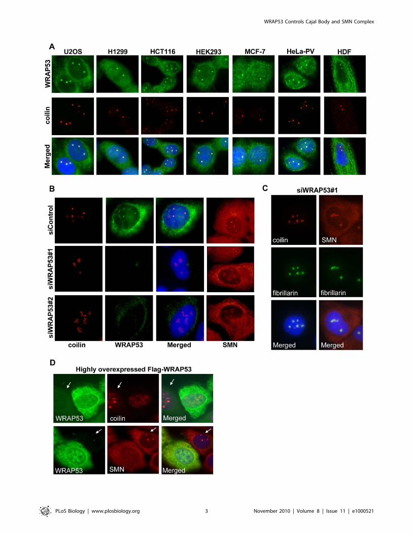
To investigate whether WRAP53 plays a role in the formation
or maintenance of Cajal bodies, WRAP53 was depleted in U2OS
and HeLa cells, and the effects on Cajal bodies, i.e., coilin, was
analyzed by immunoflourescence (IF) microscopy and Western
blotting (WB). Two different small interfering RNA (siRNA) oligos
targeting WRAP53 were used (siWRAP53#1 and siWRAP53#2),
both knocking down WRAP53 mRNA with 90% efficiency (Figure
S1A and S1B). In control cells, treated with a scramble siRNA
with no homology to any gene (siControl), coilin displayed the
characteristic Cajal body localization and co-localized with
WRAP53 in all Cajal bodies (Figures 1B and S1C). A weak
staining of coilin was also seen in nucleoli, consistent with previous
findings that coilin transits through the nucleolus during the
normal life cycle of the protein [13]. Strikingly, no Cajal bodies
were found in WRAP53-depleted cells (Figures 1B and S1C).
Instead, coilin accumulated in the nucleoli. Other Cajal body
proteins, such as SMN, also showed absence of Cajal body
accumulation and increased nucleolar staining in WRAP53-
depleted cells (Figures 1B and S1C). Staining with the nucleolar
marker fibrillarin confirmed nucleolar accumulation of coilin and
SMN upon WRAP53 depletion (Figure 1C). Thus, WRAP53 is
required for Cajal body maintenance. WRAP53-depleted cells
were also analyzed for changes in other nuclear structures, such as
nucleoli (fibrillarin), gems (SMN), and promyelocytic leukemia
(PML) bodies. No effects on these structures were observed (Figure
S1D), demonstrating that WRAP53 is an essential component for
Cajal bodies but is not essential for other nuclear structures.
Loss of Cajal bodies was strictly associated with the degree of
WRAP53 knockdown, where complete knockdown of WRAP53
led to the disappearance of all Cajal bodies, and cells still
expressing low levels of nuclear WRAP53 showed Cajal body
staining (Figure S1C). We also knocked down coilin and SMN in
U2OS and HeLa cells. Depletion of coilin resulted in the
disappearance of all Cajal bodies, leaving WRAP53 and SMN
dispersed throughout the nucleoplasm (Figure S1E). Depletion of
SMN significantly reduced the number and size of Cajal bodies,
but some cells still had Cajal bodies left. Both WRAP53 and coilin
were present in the remaining Cajal bodies and accumulated in
nucleoli (Figure S1F). Thus, WRAP53 and coilin are essential for
Cajal body structure, whereas SMN is not.
We next examined the effects on Cajal bodies in cells
overexpressing WRAP53. Flag-tagged WRAP53 expressed at
lower levels showed Cajal body accumulation (Figure S2A). In
contrast, Flag-WRAP53 expressed at higher levels gave rise to a
different nuclear expression pattern, with a more even distribution
throughout the nucleoplasm (Figures 1D and S2A). Interestingly,
no Cajal bodies were detected in these cells, and coilin and SMN
were, like WRAP53, distributed throughout the nucleoplasm.
Similar phenomena were observed using enhanced green
fluorescent protein (EGFP)–tagged WRAP53 (Figure S2B). WB
analysis of WRAP53 knockdown and WRAP53-overexpressing
cells showed no difference in coilin or SMN protein levels (Figures
S1B and S2C), and immunostaining of WRAP53-overexpressing
cells showed no change in other nuclear structures, including PML
bodies (Figure S2D). Thus, aberrant overexpression of WRAP53
Author Summary
Cajal bodies, discovered more than 100 years ago bySantiago Ramon y Cajal, are sub-organelles found in thenucleus of proliferative cells and neurons. They have beenimplicated in a variety of nuclear functions includingribonucleoprotein maturation, spliceosome formation,histone mRNA processing, RNA polymerase assembly,telomerase biogenesis, and histone gene transcription.Concentrating relevant molecules within Cajal bodies mayserve to increase the efficiency of specific nuclearfunctions. Here we identify the WRAP53 protein as anessential factor for Cajal body maintenance and fordirecting the splicing regulatory protein ‘‘survival of motorneuron’’ (SMN) complex to Cajal bodies. We show thatWRAP53 is a constitutive component of Cajal bodies, andthat knockdown of WRAP53 disrupts existing Cajal bodiesand prevents formation of new Cajal bodies. Mechanisti-cally, we find that WRAP53 recruits the SMN complex fromthe cytoplasm to Cajal bodies by mediating interactionsbetween SMN, importinb, and coilin. Finally, we reportdeficient WRAP53–SMN binding in patients with spinalmuscular atrophy, suggesting a role in this pathology. Thisstudy not only reveals new functions of the WRAP53protein, but also increases our understanding of themolecular mechanism behind Cajal body formation andrecruitment of factors to Cajal bodies.
WRAP53 Controls Cajal Body and SMN Complex
PLoS Biology | www.plosbiology.org 2 November 2010 | Volume 8 | Issue 11 | e1000521
WRAP53 Controls Cajal Body and SMN Complex
PLoS Biology | www.plosbiology.org 3 November 2010 | Volume 8 | Issue 11 | e1000521
prevents Cajal body formation and causes significant mislocaliza-
tion of the Cajal body proteins coilin and SMN to the
nucleoplasm. This finding confirms the notion that WRAP53 is
an essential component of Cajal body structure and that proper
localization of WRAP53 is required for its role in Cajal body
formation.
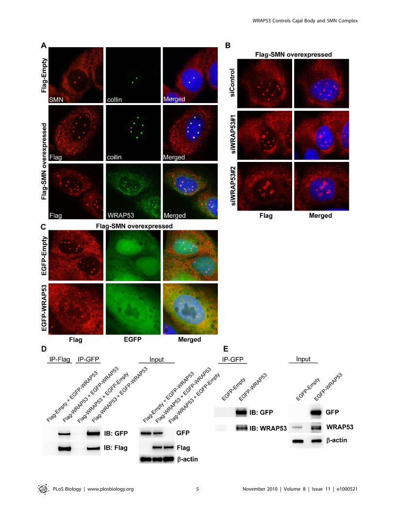
WRAP53 Is Required for De Novo Formation of CajalBodies Induced by SMN Overexpression
snRNPs are known to be rate-limiting for Cajal body formation
[8,24]. The SMN complex transports snRNPs into the nucleus,
and overexpressing the SMN protein induces formation of
additional Cajal bodies. In light of this knowledge, we examined
the influence of WRAP53 on de novo formation of Cajal bodies.
Flag-tagged SMN was transiently transfected into U2OS cells,
which increased the number of Cajal bodies per cell from 2–3 in
control cells up to ten in Flag-SMN cells (Figure 2A). All Cajal
bodies were positive for WRAP53 and coilin (Figure 2A). Cytosolic
accumulations of SMN were observed in Flag-SMN cells;
however, neither coilin nor WRAP53 were present in these
structures (Figure 2A). Interestingly, both knockdown and
aberrant overexpression of WRAP53 repressed generation of
Cajal bodies induced by SMN overexpression (Figure 2B and 2C).
Instead, Flag-SMN mislocalized to the nucleoli in WRAP53-
depleted cells and to the nucleoplasm in WRAP53-overexpressing
cells. These results show that WRAP53 is required for formation of
new Cajal bodies induced by SMN overexpression, which further
supports the idea that WRAP53 is essential for Cajal body
assembly.
The finding that exogenous WRAP53 alters the localization of
endogenous WRAP53, SMN, and coilin suggests a dominant
negative effect of overexpressed WRAP53 that could be caused by
WRAP53 self-interaction. Previous reports demonstrate such
phenomena for coilin, where overexpressed coilin mislocalizes to
nucleoli and disrupts Cajal bodies through dominant negative
interference between exogenous and endogenous coilin [13]. To
investigate if WRAP53 self-associates, U2OS cells were co-
transfected with Flag-WRAP53 and EGFP-WRAP53 constructs.
Immunoprecipitation (IP) with anti–green fluorescent protein
(GFP) or anti-Flag antibodies showed that Flag-WRAP53 protein
co-precipitated EGFP-WRAP53 and vice versa (Figure 2D). This
indicates that exogenous WRAP53 self-associates in vivo.
Furthermore, IP of EGFP-WRAP53 in U2OS cells co-precipitated
endogenous WRAP53, whereas IP of EGFP alone did not
(Figure 2E). This suggests that overexpressed WRAP53 interacts
with endogenous WRAP53 in vivo, which also strengthens our
hypothesis that overexpressed EGFP-WRAP53 or Flag-WRAP53
can cause mislocalization of endogenous WRAP53 by self-
association.
The WD40 Domain and C-Terminal Region of WRAP53Mediates Interaction with Coilin and SMN and TargetsWRAP53 to Cajal Bodies
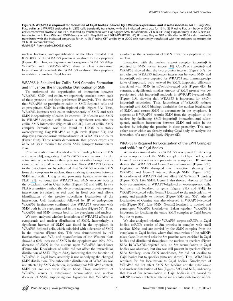
IP of endogenous WRAP53 furthermore revealed that
WRAP53 associates with coilin and SMN (Figure 3A). Reciprocal
IP of coilin and SMN verified the interactions with WRAP53. To
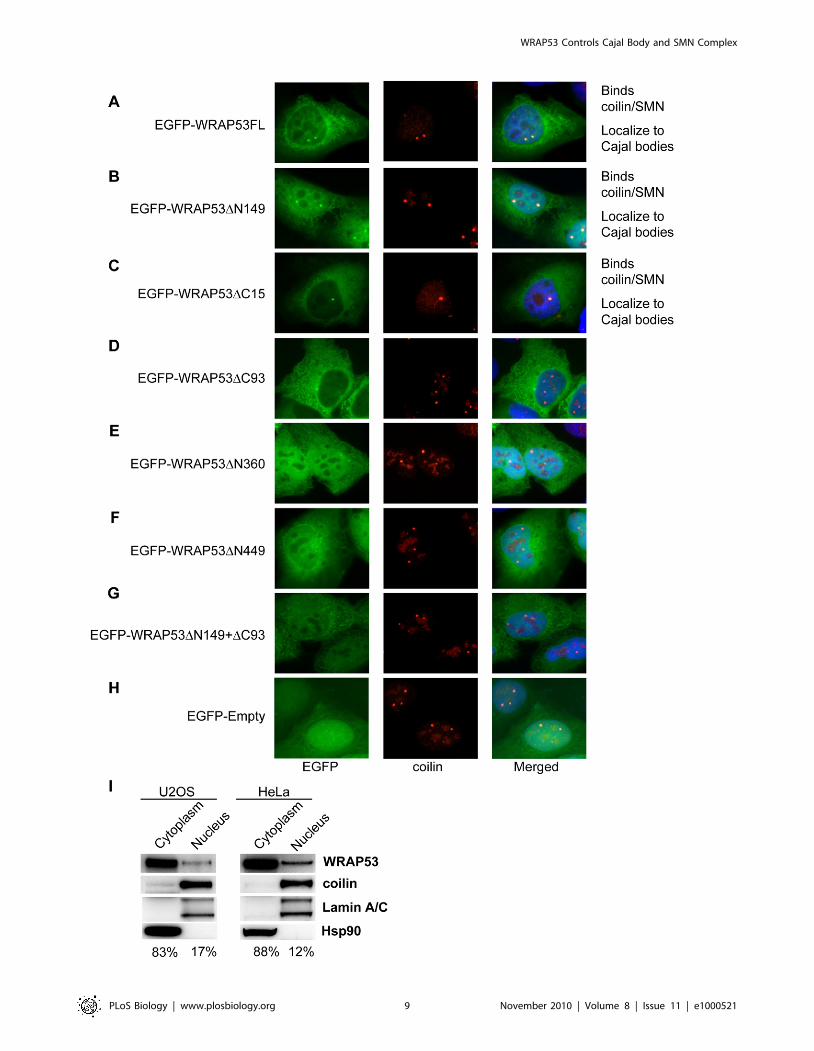
assess which region of WRAP53 interacts with coilin and SMN,
we generated and transiently overexpressed a series of EGFP-
tagged WRAP53 deletion constructs in U2OS cells (Figure 3B).
Each construct expressed a protein of the expected size, as
demonstrated by immunoblotting using both GFP and WRAP53
antibodies (Figure 3C and data not shown). IP of EGFP-WRAP53
using GFP antibody showed that WRAP53 constructs containing
the WD40 domain plus the C-terminal region containing amino
acids (aa) 456–533 associated with coilin and SMN. Constructs
lacking these two domains or only expressing one of them co-
precipitated neither coilin nor SMN (Figure 3C). Hence, WRAP53
associates with both coilin and SMN, and the same sequence in
WRAP53 is important for interaction with both these proteins.
To investigate which region of WRAP53 mediates its localiza-
tion to Cajal bodies, the panel of EGFP-WRAP53 deletion
constructs was transiently transfected into U2OS cells, and protein
localization was analyzed by IF. The cells were also stained for
coilin to visualize Cajal bodies. Interestingly, only the WRAP53
constructs that bind coilin and SMN (EGFP-WRAP53FL, EGFP-
WRAP53DN149, and EGFP-WRAP53DC15) accumulated in
Cajal bodies (Figure 4A–4C). In contrast, the constructs unable
to bind coilin or SMN failed to localize to Cajal bodies (Figure 4D–
4H). This suggests that interaction with coilin and/or SMN is
necessary for WRAP53 localization to Cajal bodies. No change in
Cajal body number was observed in cells overexpressing the
different WRAP53 constructs (data not shown). These observa-
tions were made in cells expressing low to moderate levels of
WRAP53. In cells with high WRAP53 expression, nuclear
mislocalization of WRAP53, SMN, and coilin was observed. Most
likely this is due to sequestering of coilin and SMN in the
nucleoplasm by EGFP-WRAP53, since high expression of
WRAP53 deletion mutants unable to bind coilin did not
mislocalize coilin/SMN and had no effects on Cajal body
appearance (data not shown). Thus, the WD40 domain and the
C-terminal region of WRAP53 target WRAP53 to Cajal bodies.
We also observed that all WRAP53 constructs showed
cytoplasmic localization (Figure 4A–4G) and that WRAP53
constructs lacking the C-terminal region (EGFP-WRAP53DC93
and EGFP-WRAP53DC15) demonstrated a more pronounced
cytoplasmic staining (Figure 4C and 4D). This was most apparent
with the EGFP-WRAP53DC93 construct. In contrast, N-termi-
nally deleted constructs exhibited the opposite distribution, i.e., a
stronger nuclear staining (Figure 4B, 4E, and 4F), which was most
apparent with the EGFP-WRAP53DN149 construct. These results
indicate that the C- and N-terminal regions of WRAP53 contain
elements important for the subcellular distribution of WRAP53.
Previous studies failed to detect any WRAP53 protein in the
cytoplasm [2,3]. To investigate this further we performed IF
staining of endogenous WRAP53 with three different WRAP53
antibodies. Interestingly, all three antibodies show cytoplasmic
localization of WRAP53, in addition to accumulation in Cajal
bodies, using both methanol and paraformaldehyde fixation
(Figures 1A, S3B, and S3C). A clear reduction in cytoplasmic
and nuclear WRAP53 staining was observed after WRAP53
depletion, confirming the specificity of the WRAP53 staining in
both compartments (Figure 1B). We also performed cell
fractionation followed by WB of the WRAP53 protein. This
confirmed that WRAP53 is present both in cytoplasmic and
Figure 1. Aberrant expression of WRAP53 disrupts Cajal bodies and mislocalizes coilin and SMN. (A) IF staining of endogenous WRAP53and the Cajal body marker coilin in different cell types. Nuclei were stained with DAPI in all IF experiments. (B) IF staining of coilin, WRAP53, and SMNin U2OS cells treated with the indicated siRNA oligos for 48 h. (C) U2OS cells treated with siWRAP53#1 for 48 h stained for either coilin or SMNtogether with the nucleolar marker fibrillarin. (D) U2OS cells transiently transfected with Flag-tagged full-length WRAP53 for 16 h and stained forWRAP53, coilin, and SMN. Arrows indicate untransfected cells used as controls.doi:10.1371/journal.pbio.1000521.g001
WRAP53 Controls Cajal Body and SMN Complex
PLoS Biology | www.plosbiology.org 4 November 2010 | Volume 8 | Issue 11 | e1000521
WRAP53 Controls Cajal Body and SMN Complex
PLoS Biology | www.plosbiology.org 5 November 2010 | Volume 8 | Issue 11 | e1000521
nuclear fractions, and quantification of the blots revealed that
83%–88% of the WRAP53 protein is localized to the cytoplasm
(Figure 4I). Thus, endogenous and exogenous WRAP53 (Flag-
WRAP53 and EGFP-WRAP53) show a clear cytoplasmic
localization. We conclude that WRAP53 localizes to the cytoplasm
in addition to nuclear Cajal bodies.
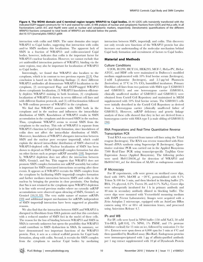
WRAP53 Is Required for Coilin–SMN Complex Formationand Influences the Intracellular Distribution of SMN
To understand the organization of interaction between
WRAP53, SMN, and coilin, we separately knocked down each
of these proteins, and then performed IP analysis. This showed
that WRAP53 co-precipitates coilin in SMN-depleted cells and
co-precipitates SMN in coilin-depleted cells (Figure 5A). Thus,
WRAP53 interacts with coilin independently of SMN and with
SMN independently of coilin. In contrast, IP of coilin and SMN
in WRAP53-depleted cells showed a significant reduction in
coilin–SMN interaction (Figure 5B and 5C). Loss of coilin–SMN
complex formation was also found in U2OS cells stably
overexpressing Flag-WRAP53 at high levels (Figure 5D) and
displaying nucleoplasmic mislocalization of WRAP53 and coilin
(Figure S4A). These results demonstrate that proper expression
of WRAP53 is required for coilin–SMN complex formation in
vivo.
Previous studies have described a direct binding between SMN
and coilin [14], suggesting that WRAP53 is not required for the
actual interaction between these proteins but rather brings them in
close proximity to allow their interaction. Since WRAP53 localizes
to the cytoplasm, we hypothesized that WRAP53 recruits SMN
from the cytoplasm to nucleus, thus enabling interaction between
SMN and coilin. Using in situ proximity ligation assay (in situ
PLA) [25], we found that WRAP53 and SMN associate both in
the cytoplasm and in Cajal bodies (Figures 5E and S4B). In situ
PLA is a sensitive method that detects endogenous protein–protein
interactions (visualized as red dots) in fixed cells and allows
identification of the precise subcellular localization of the
interaction. Cell fractionation followed by IP of endogenous
WRAP53 furthermore confirmed that WRAP53 associates with
SMN both in the cytoplasm and in the nucleus (Figure 5F). Thus,
WRAP53 and SMN interact both in the cytoplasm and nucleus.
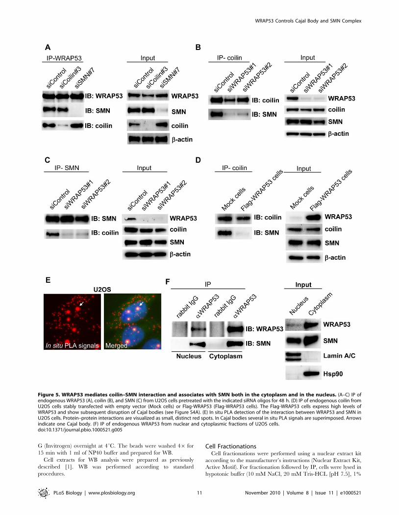
We next analyzed whether knockdown of WRAP53 affects the
cytoplasmic and nuclear distribution of SMN. Interestingly, a
significant increase of SMN was found in the cytoplasm in
WRAP53-depleted cells, which coincided with a decrease of SMN
in the nucleus (Figure 6A). This was demonstrated by cell
fractionation and WB, and quantification of the Western blots
showed a 60% increase of SMN in the cytoplasm and 40%–50%
decrease of SMN in the nucleus upon WRAP53 knockdown
(Figure 6B). Knockdown of coilin did not affect the intracellular
distribution of SMN (Figure S5A), indicating that the function of
WRAP53 in Cajal body assembly is not underlying the changed
SMN distribution. The subcellular distribution of WRAP53 was
not affected by SMN depletion, indicating that WRAP53 controls
SMN but not vice versa (Figure S5A). Thus, knockdown of
WRAP53 results in cytoplasmic accumulation and nuclear
decrease of SMN, supporting our hypothesis that WRAP53 is
involved in the recruitment of SMN from the cytoplasm to the
nucleus.
Interaction with the nuclear import receptor importinb is
required for SMN nuclear import [18]. Co-IPs of importinb and
WRAP53 showed that the two proteins interact (Figure 6C). To
test whether WRAP53 influences interaction between SMN and
importinb, cells were depleted for WRAP53 and immunoprecip-
itates of importinb were assayed for SMN. Importinb efficiently
associated with SMN in siControl-treated cells (Figure 6D). In
contrast, a significantly smaller amount of SMN protein was co-
precipitated with importinb antibody in siWRAP53-treated cells
(Figure 6D), showing that WRAP53 is important for SMN–
importinb association. Thus, knockdown of WRAP53 reduces
importinb and SMN binding, diminishes the nuclear localization
of SMN, and causes SMN to accumulate in the cytoplasm. It
appears as if WRAP53 recruits SMN from the cytoplasm to the
nucleus by facilitating SMN–importinb interaction and subse-
quently mediates interaction between SMN and coilin in the
nucleus by bringing the proteins in close proximity. This may
either occur within an already existing Cajal body or catalyze the
formation of a new Cajal body (Figure 6E).
WRAP53 Is Required for Localization of the SMN Complexand snRNP to Cajal Bodies
We next examined whether WRAP53 is required for directing
other components of the SMN complex to Cajal bodies, and
Gemin3 was chosen as a representative component. IP analysis
showed that WRAP53 and Gemin3 indeed associate (Figure S5B).
Depletion of SMN disrupted this interaction, showing that
WRAP53 and Gemin3 interact through SMN (Figure S5B).
Knockdown of WRAP53 did not affect SMN–Gemin3 binding
(Figure S5C). Like SMN, Gemin3 also showed absence of Cajal
body accumulation in WRAP53-depleted or -overexpressed cells,
but were still localized in gems (Figure S5D and S5E). In
WRAP53-depleted cells, Gemin3 localized to the nucleoplasm, to
gems, and partially to nucleoli (Figure S5D and S5F). Altered
localization of Gemin2 was also observed in WRAP53-depleted
cells (Figure S5F). Like SMN, Gemin2 localized to nucleoli and
gems upon WRAP53 knockdown. Taken together, WRAP53 is
important for localizing the entire SMN complex to Cajal bodies
but not to gems.
We also analyzed whether WRAP53 targets snRNPs to Cajal
bodies. snRNPs consist of Sm proteins in complex with small
nuclear RNAs and are carried by the SMN complex from the
cytoplasm to Cajal bodies, where final maturation of the snRNPs
takes place. In control cells the Sm proteins were enriched in Cajal
bodies and distributed throughout the nucleus in speckles (Figure
S6A). In WRAP53-depleted cells, no Sm accumulation in Cajal
bodies was observed, but Sm was still present in speckles (Figure
S6A). Similary, upon SMN knockdown, Sm did not localize to
Cajal bodies but to speckles (data not shown). Thus, WRAP53 is
required for Sm localization to Cajal bodies. Knockdown of
WRAP53 did not affect SMN–Sm binding nor the cytoplasmic
and nuclear distribution of Sm (Figures S5C and S6B), indicating
that loss of Sm accumulation in Cajal bodies is not caused by
snRNP assembly defects or altered intracellular distribution of Sm.
Figure 2. WRAP53 is required for formation of Cajal bodies induced by SMN overexpression, and it self-associates. (A) IF using SMN,Flag, coilin, and WRAP53 antibodies in U2OS cells transiently transfected with the indicated constructs for 16 h. (B) IF using Flag antibody in U2OScells treated with siWRAP53 for 24 h, followed by transfection with Flag-tagged SMN for additional 24 h. (C) IF using Flag antibody in U2OS cells co-transfected with Flag-SMN and EGFP-Empty or with Flag-SMN and EGFP-WRAP53FL. (D) IP using Flag or GFP antibodies in U2OS cells transientlytransfected with the indicated constructs for 24 h. (E) IP using GFP antibody in U2OS cells transiently transfected with the indicated constructs for24 h. IB, immunoblotting.doi:10.1371/journal.pbio.1000521.g002
WRAP53 Controls Cajal Body and SMN Complex
PLoS Biology | www.plosbiology.org 6 November 2010 | Volume 8 | Issue 11 | e1000521
WRAP53 Controls Cajal Body and SMN Complex
PLoS Biology | www.plosbiology.org 7 November 2010 | Volume 8 | Issue 11 | e1000521
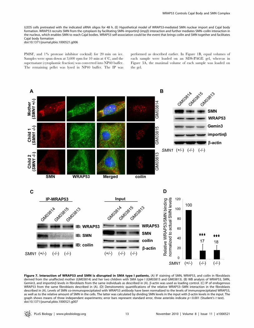
Interaction of WRAP53 and SMN Is Disrupted in SMAType I Patients
The most severe form SMA, SMA type I, correlates with a
reduced number of SMN-containing nuclear bodies [21–23]. The
role of WRAP53 in Cajal body formation and nuclear localization
of SMN encouraged us to investigate the interplay of WRAP53
and SMN in vivo in SMA disease. We first analyzed SMN
localization in nuclear bodies in fibroblasts derived from an
unaffected mother (GM03814, serving as control) and her two
children with SMA type I (GM03815 and GM03813). Co-staining
of SMN and coilin showed that SMN accumulated in nuclear
bodies (gems and Cajal bodies) in 67% of control fibroblasts
(GM03814), compared to only 13% (GM03815) and 16%
(GM03813) of SMA fibroblasts. Cajal bodies (coilin accumulation)
were detected in 43% of control fibroblasts (GM03814), but in
only 25% (GM03815) and 15% (GM03813) of the SMA
fibroblasts. WRAP53 was present in all Cajal bodies. The absence
of SMN in nuclear bodies coincides with lack of Cajal bodies in the
same cells (Figure 7A). Thus, both gem and Cajal body number
are decreased in SMA fibroblasts. This observation could not be
explained by the difference in WRAP53 levels in SMA fibroblasts
compared to control (Figure 7B). However, WRAP53 showed
reduced binding to SMN protein in the SMA fibroblasts
(Figure 7C). To investigate whether the lack of binding between
WRAP53 and SMN was just a reflection of decreased levels of
SMN in the SMA fibroblasts, or whether WRAP53 binding to the
SMN protein derived from the SMN2 allele is in fact weaker, we
quantified the relative amount of SMN that interacts with
WRAP53 in SMA patients and in normal cells. This revealed a
lack of binding between SMN and WRAP53 in cells from SMA
patients that cannot be explained by the lower amounts of SMN,
and that the relative binding between WRAP53 and SMN in these
cells was reduced by 83% (Figure 7D). We did not observe altered
binding between WRAP53 and coilin in the SMA cells (Figure 7C),
demonstrating that the lost interaction between WRAP53 and
SMN is specific and not a secondary effect of disrupted WRAP53–
coilin interaction. We thus conclude that the interaction between
WRAP53 and SMN is disrupted in SMA type I patients, which
further relates to a failure of SMN accumulation in nuclear bodies.
Discussion
Here we identify WRAP53 as an essential factor for Cajal body
maintenance and for directing the SMN complex to Cajal bodies.
We show that WRAP53 is a constitutive component of Cajal
bodies that overlaps coilin in 100% of Cajal bodies in a variety of
cell lines. Knockdown of WRAP53 disrupts Cajal bodies, prevents
formation of new Cajal bodies, and relocates Cajal body proteins
coilin and SMN from Cajal bodies to nucleoli. WRAP53 seems
specifically important for Cajal body integrity, since depletion of
WRAP53 does not affect gems or other nuclear structures,
including nucleoli and PML bodies. We show that WRAP53
separately associates with coilin and SMN and is required for their
complex formation. Previous studies demonstrated a direct
interaction between SMN and coilin, suggesting that WRAP53
is not important for their binding but rather mediates interaction
by bringing the proteins in close proximity. This may either occur
within an already existing Cajal body or result in the formation of
a new Cajal body. Importantly, WRAP53’s role in Cajal body
formation goes beyond bringing SMN and coilin together, since
knockdown of SMN does not abolish all Cajal body structures.
Residual Cajal bodies containing WRAP53 and coilin still remain.
Moreover, in HeLa-PV cells, WRAP53 and coilin localize to all
Cajal bodies and SMN to only 40% of them (n = 100) (data not
shown). These observations, together with the fact that knockdown
of WRAP53 or coilin disrupts all Cajal body structures, point to a
more general function of WRAP53 in assisting coilin as a scaffold
protein in Cajal body formation.
Cajal bodies have been suggested to have separate compart-
ments containing snRNP, snoRNP/scaRNP, or basal transcription
factors [10]. Depletion of proteins involved in snRNP maturation,
such as SMN, TGS1, and PHAX, disrupts canonical Cajal bodies
containing snRNP, whereas residual Cajal bodies lacking snRNPs
but containing coilin and snoRNP/scaRNP components still
remain. Without WRAP53, both canonical and residual Cajal
bodies collapse, suggesting that WRAP53 is important for
processes in addition to snRNP maturation. WRAP53 has been
shown to be essential for scaRNA, including telomerase RNA,
localization to Cajal bodies [2,3], which could account for some of
the observed defects in Cajal body formation upon WRAP53
perturbation.
We also observe that high overexpression of WRAP53
disassembles Cajal bodies and results in nucleoplasmic mislocali-
zation of WRAP53, coilin, and SMN. This indicates that
overexpressed WRAP53 has a dominant negative effect on
WRAP53 function and that exogenous and endogenous WRAP53
may compete for factors important for WRAP53 localization to
Cajal bodies and Cajal body formation. The fact that endogenous
WRAP53 co-precipitates with exogenous WRAP53 indicates that
WRAP53 can self-associate, which can also explain the observed
effect. Indeed, self-oligomerization appears to be a general feature
of nuclear body marker proteins including coilin, SMN, and PML
[26–28], which is consistent with our findings that WRAP53 is a
signature protein for Cajal bodies. In line with this notion,
overexpression of coilin also disrupts Cajal bodies and results in
coilin mislocalization [13]. Hypothetically, WRAP53 self-associa-
tion could be the event that brings coilin and SMN together and
facilitates Cajal body formation.
The effects on Cajal bodies of depletion and overexpression of
WRAP53 are highly similar to those of loss or overexpression of
coilin. Coilin mutants have been described in human, mouse [8],
Arabidopsis [29], and Drosophila [30]. In all of these species, loss of
coilin produces defects in Cajal body formation. Overexpression of
coilin, on the other hand, produces slightly different effects in the
different organisms. In Drosophila and Arabidopsis, enhanced coilin
levels result in normal Cajal body formation or formation of larger
Cajal bodies, whereas overexpression of coilin in mice and human
cells disrupts Cajal bodies, as previously described. It would be
interesting to investigate the effects of WRAP53 depletion and
overexpression in other organisms as well.
Deletion mapping of the WRAP53 protein demonstrates that
the WD40 domain plus C-terminal aa 456–533 are required for
Figure 3. WRAP53 binds coilin and SMN via its WD40 domain and C-terminal region. (A) IP of endogenous WRAP53, coilin, and SMN fromU2OS cells followed by immunoblotting (IB) with the indicated antibodies. Rabbit and mouse IgG were used as negative controls. (B) Schematicillustration of EGFP-tagged WRAP53 deletion constructs: WRAP53FL (full-length), WRAP53DN149+DC93 (contains aa 150–455), WRAP53DN149(contains aa 150–584), WRAP53DC93 (contains aa 1–455), WRAP53DC15 (contains aa 1–533), WRAP53DN360 (contains aa 361–548), andWRAP53DN449 (contains aa 450–548). The EGFP protein has a molecular weight of approximately 27 kDa. (C) U2OS cells transfected with theindicated WRAP53 constructs for 16 h, followed by IP with GFP antibody. Asterisk indicates the heavy chain. The WRAP53DN360 product is 50 kDa insize and is thus covered by the heavy chain.doi:10.1371/journal.pbio.1000521.g003
WRAP53 Controls Cajal Body and SMN Complex
PLoS Biology | www.plosbiology.org 8 November 2010 | Volume 8 | Issue 11 | e1000521
WRAP53 Controls Cajal Body and SMN Complex
PLoS Biology | www.plosbiology.org 9 November 2010 | Volume 8 | Issue 11 | e1000521
interaction with coilin and SMN. The same domains also target
WRAP53 to Cajal bodies, suggesting that interaction with coilin
and/or SMN mediates this localization. The apparent lack of
SMN in a fraction of WRAP53- and coilin-associated Cajal
bodies, however, indicates that coilin is the important factor for
WRAP53’s nuclear localization. However, we cannot exclude that
yet unidentified interaction partners of WRAP53, binding via the
same regions, may also be important for localization of WRAP53
to Cajal bodies.
Interestingly, we found that WRAP53 also localizes to the
cytoplasm, which is in contrast to two previous reports [2,3]. Our
conclusion is based on the following findings: (1) three different
WRAP53 antibodies all demonstrate WRAP53 localization to the
cytoplasm, (2) overexpressed Flag- and EGFP-tagged WRAP53
shows cytoplasmic localization, (3) WRAP53 knockdown efficient-
ly depletes WRAP53 staining in both locations, (4) cytoplasmic
localization of WRAP53 is shown in seven different cell lines and
with different fixation protocols, and (5) cell fractionation followed
by WB confirms presence of WRAP53 in the cytoplasm.
We find that WRAP53 associates with SMN both in the
cytoplasm and in the nucleus and influences the intracellular
distribution of SMN. Knockdown of WRAP53 results in SMN
accumulation in the cytoplasm and decreased SMN in the nucleus.
Thus, cytoplasmic WRAP53 seems to recruit SMN from the
cytoplasm to the nucleus. This role of WRAP53 is separate from
WRAP53’s function in Cajal body formation, since knockdown of
coilin does not affect the intracellular distribution of SMN.
Moreover, knockdown of WRAP53 abrogates interaction between
SMN and the nuclear pore receptor importinb, which could
explain the skewed intracellular distribution of SMN observed in
WRAP53-depleted cells. Nuclear localization of SMN has been
shown to depend on SMN–importinb complex formation but also
on other factors such as proper snRNP assembly [18]. Important-
ly, WRAP53 depletion does not affect the interaction between
SMN, Gemin3, and Sm. This suggests that WRAP53 does not
promote SMN complex formation nor snRNP assembly but rather
is important for SMN-associated interactions occurring after these
events. It appears as if WRAP53 recruits the SMN complex from
the cytoplasm by facilitating SMN–importinb complex formation
and further mediates interaction between SMN and coilin in the
nucleus by bringing the proteins in close proximity. Our finding
that Sm is not retained in the cytoplasm upon WRAP53 depletion
is in line with several previous studies where no cytosolic snRNP
accumulations were observed upon silencing of SMN, Gemin3, or
Gemin4 [10,31,32]. The very stable nature of mature snRNPs
[33] and additional import mechanisms for snRNPs independent
of SMN–importinb interaction have been suggested as plausible
reasons.
We also find that the interaction between SMN and WRAP53 is
disrupted in fibroblasts from SMA patients and that this correlates
with a reduced number of SMN foci in the nuclei of these cells.
The reason for the loss of binding between WRAP53 and SMN in
SMA is currently unknown but opens the possibility that WRAP53
could contribute to SMN dysfunction in SMA. In summary, we
have demonstrated two important functions of the WRAP53
protein. First, it acts as a critical scaffold protein for Cajal body
formation, along with coilin. Second, it recruits the SMN complex
from the cytoplasm to nuclear Cajal bodies by mediating
interaction between SMN, importinb, and coilin. This discovery
not only reveals new functions of the WRAP53 protein but also
increases our understanding of the molecular mechanism behind
Cajal body formation and recruitment of factors to Cajal bodies.
Material and Methods
Culture ConditionsU2OS, H1299, HCT116, HEK293, MCF-7, HeLa-PV, HeLa-
ATCC, and HDF cells were maintained in Dulbecco’s modified
medium supplemented with 10% fetal bovine serum (Invitrogen),
2 mM L-glutamine (Invitrogen), and 2.5 mg/ml Plasmocin
(InvivoGen) at 37uC in 5% CO2 humidified incubators. Primary
fibroblast cell lines from two patients with SMA type I (GM03813
and GM03815) and one heterozygous carrier (GM03814,
clinically unaffected mother of GM03813 and GM03815) were
obtained from Coriell Cell Repository and maintained in MEM
supplemented with 10% fetal bovine serum. The GM03815 cells
were initially described in the Coriell Cell Repository as derived
from a heterozygous carrier (clinically unaffected father of
GM03813). However, snRNP deficiency and genetic linkage
analysis of these cells showed that they in fact are derived from a
homozygous carrier with SMA type I (a male sibling of GM03813)
[34].
RNA Preparations and Real-Time Quantitative ReverseTranscription PCR
Total RNA was extracted from tumor cell lines using the Trizol
reagent (Invitrogen). The RNA was reverse transcribed with First-
Strand cDNA synthesis using Superscript II (Invitrogen). Quan-
titative real-time PCR was carried out in the Applied Biosystems
7500 Real-Time PCR using transcript-specific TaqMan Gene
Expression Assays (Applied Biosystems). The following probes
were used: Hs01126636_g1 for detection of WRAP53 and
Hs00167441_m1 for detection of ALAS1 as endogenous control.
IF MicroscopyFor IF experiments, cells were grown on sterilized cover slips,
fixed with 100% MeOH at 220uC, permeabilized with 0.1%
Triton X-100 for 5 min, and then blocked in blocking buffer (2%
BSA, 5% glycerol, 0.2% Tween 20, and 0.1% NaN3). Cover slips
were subsequently incubated for 1 h in primary antibody and
40 min in secondary antibody diluted in blocking buffer. The
cover slips were mounted with Vectorshield mounting medium
with DAPI (Vector Laboratories). Images were acquired with a
Zeiss Axioplan 2 microscope, equipped with an AxioCam HRm
camera using 436 or 606 oil immersion lenses, and processed
using Axiovision Release 4.7.
IPs and WBFor IP, cells were lysed in NP40 buffer (150 mM NaCl, 50 mM
Tris-HCL [pH 8.0], 1% NP40, 1% PMSF, and 1% protease
inhibitor cocktail) for 15 min on ice, followed by sonication 26 for
10 s. Extracts were spun down at 6,000 rpm for 5 min at 4uC and
then quantified by Bradford assay (Bio-Rad). Endogenous proteins
were immunoprecipitated with 1 mg of affinity-purified antibody
per 1 mg extract supplemented with 10 ml of Dynabeads Protein
Figure 4. The WD40 domain and C-terminal region targets WRAP53 to Cajal bodies. (A–H) U2OS cells transiently transfected with theindicated EGFP-tagged constructs for 16 h and stained for coilin. (I) WB analysis of nuclear and cytoplasmic fractions from U2OS and HeLa cells. In allfractionations Lamin A/C and Hsp90 were used as nuclear and cytoplasmic markers, respectively. Densitometric quantifications of the differentWRAP53 fractions compared to total levels of WRAP53 are indicated below the panels.doi:10.1371/journal.pbio.1000521.g004
WRAP53 Controls Cajal Body and SMN Complex
PLoS Biology | www.plosbiology.org 10 November 2010 | Volume 8 | Issue 11 | e1000521
G (Invitrogen) overnight at 4uC. The beads were washed 46 for
15 min with 1 ml of NP40 buffer and prepared for WB.
Cell extracts for WB analysis were prepared as previously
described [1]. WB was performed according to standard
procedures.
Cell FractionationsCell fractionations were performed using a nuclear extract kit
according to the manufacturer’s instructions (Nuclear Extract Kit,
Active Motif). For fractionation followed by IP, cells were lysed in
hypotonic buffer (10 mM NaCl, 20 mM Tris-HCL [pH 7.5], 1%
Figure 5. WRAP53 mediates coilin–SMN interaction and associates with SMN both in the cytoplasm and in the nucleus. (A–C) IP ofendogenous WRAP53 (A), coilin (B), and SMN (C) from U2OS cells pretreated with the indicated siRNA oligos for 48 h. (D) IP of endogenous coilin fromU2OS cells stably transfected with empty vector (Mock cells) or Flag-WRAP53 (Flag-WRAP53 cells). The Flag-WRAP53 cells express high levels ofWRAP53 and show subsequent disruption of Cajal bodies (see Figure S4A). (E) In situ PLA detection of the interaction between WRAP53 and SMN inU2OS cells. Protein–protein interactions are visualized as small, distinct red spots. In Cajal bodies several in situ PLA signals are superimposed. Arrowsindicate one Cajal body. (F) IP of endogenous WRAP53 from nuclear and cytoplasmic fractions of U2OS cells.doi:10.1371/journal.pbio.1000521.g005
WRAP53 Controls Cajal Body and SMN Complex
PLoS Biology | www.plosbiology.org 11 November 2010 | Volume 8 | Issue 11 | e1000521
Figure 6. WRAP53 regulates SMN distribution and mediates SMN–importinb interaction. (A) WB analysis of SMN levels in fractionatedU2OS cells treated with the indicated siRNA oligos. (B) Densitometric quantifications of SMN levels in fractionated U2OS cells treated with the siRNAsshown in (A). Levels of SMN have been normalized against the internal fractionation control. The graph shows means of five independentexperiments; error bars represent standard error; one asterisk indicates p,0.05, two asterisks indicate p,0.01, according to the Student’s t test. (C) IPof endogenous WRAP53 and importinb from U2OS cells. Rabbit and mouse IgG were used as negative controls. (D) IP of endogenous importinb from
WRAP53 Controls Cajal Body and SMN Complex
PLoS Biology | www.plosbiology.org 12 November 2010 | Volume 8 | Issue 11 | e1000521
PMSF, and 1% protease inhibitor cocktail) for 20 min on ice.
Samples were spun down at 3,600 rpm for 10 min at 4uC, and the
supernatant (cytoplasmic fraction) was converted into NP40 buffer.
The remaining pellet was lysed in NP40 buffer. The IP was
performed as described earlier. In Figure 1B, equal volumes of
each sample were loaded on an SDS-PAGE gel, whereas in
Figure 3A, the maximal volume of each sample was loaded on
the gel.
Figure 7. Interaction of WRAP53 and SMN is disrupted in SMA type I patients. (A) IF staining of SMN, WRAP53, and coilin in fibroblastsderived from the unaffected mother (GM03814) and her two children with SMA type I (GM03815 and GM03813). (B) WB analysis of WRAP53, SMN,Gemin3, and importinb levels in fibroblasts from the same individuals as described in (A). b-actin was used as loading control. (C) IP of endogenousWRAP53 from the same fibroblasts described in (A). (D) Densitometric quantifications of the relative WRAP53–SMN interaction in the fibroblastsdescribed in (A). Levels of SMN co-immunoprecipitated with WRAP53 antibody have been normalized to the levels of immunoprecipitated WRAP53,as well as to the relative amount of SMN in the cells. The latter was calculated by dividing SMN levels in the input with b-actin levels in the input. Thegraph shows means of three independent experiments; error bars represent standard error, three asterisks indicate p,0.001 (Student’s t test).doi:10.1371/journal.pbio.1000521.g007
U2OS cells pretreated with the indicated siRNA oligos for 48 h. (E) Hypothetical model of WRAP53-mediated SMN nuclear import and Cajal bodyformation. WRAP53 recruits SMN from the cytoplasm by facilitating SMN–importinb (impb) interaction and further mediates SMN–coilin interaction inthe nucleus, which enables SMN to reach Cajal bodies. WRAP53 self-association could be the event that brings coilin and SMN together and facilitatesCajal body formationdoi:10.1371/journal.pbio.1000521.g006
WRAP53 Controls Cajal Body and SMN Complex
PLoS Biology | www.plosbiology.org 13 November 2010 | Volume 8 | Issue 11 | e1000521
AntibodiesFour different WRAP53 antibodies were used: rabbit a-
WRAP53-C1 [1] (used for IP and WB), rabbit a-WRAP53-C2
(used for WB, IP, and IF), rabbit a-WRAP53 (Wdr79, A301-
442A-1, Bethyl Laboratories; used for WB, IP, IF, and in situ
PLA), and mouse polyclonal a-WRAP53 full-length (H00055135-
B01, Abnova; used only in Figure S3A and S3B). To generate a-
WRAP53-C2, rabbits were immunized with a KLH-conjugated
WRAP53 peptide that maps to a region between aa 498–548 of
full-length WRAP53 protein (Innovagen AB). The following
antibodies were used in IF, IP, and WB: mouse a-coilin
(ab11822, Abcam), rabbit a-coilin (sc-32860, Santa Cruz Biotech-
nology), mouse a-SMN (610647, BD Biosciences), mouse a-SMN
(sc-32313, Santa Cruz Biotechnology), rabbit a-SMN (sc-15320,
Santa Cruz Biotechnology), mouse a-Gemin3 (ab10305, Abcam),
mouse a-Gemin3 (sc-57007, Santa Cruz Biotechnology), mouse a-
Gemin2 (sc-57006, Santa Cruz Biotechnology), mouse a-impor-
tinb (035K4852, Sigma), mouse a-importinb (sc-137016, Santa
Cruz Biotechnology), rabbit a-fibrillarin (ab5821, Abcam), mouse
a-Sm (ab3138, Abcam), mouse a-SmB/B9 (sc-271094, Santa Cruz
Biotechnology), mouse a-Hsp90a/b (sc-13119, Santa Cruz
Biotechnology), rabbit a-Lamin A/C (sc-20681, Santa Cruz
Biotechnology), rabbit IgG (sc-2027, Santa Cruz Biotechnology),
mouse IgG (sc-2025, Santa Cruz Biotechnology), mouse a-Flag
M2 (200472-21, Stratagene), rabbit a-GFP (ab290, Abcam), and
mouse a-b-actin (Sigma). The following secondary antibodies were
used: sheep a-mouse HRP (NA931V, GE Healthcare), donkey a-
rabbit HRP (NA934V, GE Healthcare), swine a-rabbit FITC
(F0054, Dako Cytomation), and horse a-mouse Texas Red (TI-
2000, Vector).
siRNA Oligonucleotides, Plasmids, and TransfectionThe following siRNAs from Qiagen were used: siWRAP53#1
(SI00388941), siWRAP53#2 (SI00388948), siSMN1#7
(SI03108084), siSMN1#11 (SI04950932), siSMN1#12
(SI04950939), siCoilin#3 (SI00350343), siCoilin#7
(SI04330830), and a control siRNA (1027280). siRNA (10–
20 nM) was transfected into cells using either Oligofectamine
(Invitrogen) or HiPerfect (Qiagen) transfection reagents, in
accordance with the supplier’s recommendations. Flag-WRAP53
was cloned as described in Mahmoudi et al. [1]. To generate
EGFP-tagged WRAP53 constructs, full-length or deletion mutants
were amplified by PCR (Advantage-HF 2 polymerase, Clontech)
and subcloned into pEGFP-C1 vector (Clontech). Flag-SMN was
cloned by PCR (Advantage-HF 2 polymerase, Clontech) from
SMN cDNA and subcloned into pCMV-Tag2 vector (Invitrogen).
All primers used for PCR amplifications are listed in Table S1.
Plasmid transfections were performed using Lipofectamine 2000
Reagent (Invitrogen).
For the generation of cells with stable overexpression of
WRAP53, Flag-tagged full-length WRAP53 cDNA was cloned
into the pLenti6/V5-D-TOPO vector (Invitrogen). The pLenti6-
Flag-WRAP53 plasmid along with the pMDLg/RRE, pCMV-
VSVG, and pRSV-Rev plasmids required for viral production
were transfected into HEK293FT cells using Lipofectamine 2000
(Invitrogen). U20S cells were infected with viruses containing
either pLenti6-Flag-WRAP53 or empty pLenti6/V5-D-TOPO
vector, and positive cells were selected 48 h after infection using
10 mg/ml Blasticidin (Invitrogen).
Statistical AnalysisAll analyses were performed using Microsoft Office Excel 2003.
Two-tailed Student’s t test was used to determine statistical
significance.
In Situ PLAIn situ PLA experiments were performed as described
previously [35]. Incubation with primary antibodies (0.4 ng/ml
rabbit a-WRAP53 and 1 ng/ml mouse a-SMN in blocking
solution) was performed at room temperature for 1 h. Cells were
washed 36 for 5 min in PBS plus 0.1% Tween 20, with the first
wash at 37uC. Secondary proximity probes (Rabbit-PLUS and
Mouse-MINUS, Duolink kit, Olink Biosciences AB) were
incubated for 2 h at 37uC. Cells were washed 16 for 5 min in
10 mM Tris-HCl (pH 7.5) plus 0.1% Tween 20 at 37uC, then 26for 5 min in PBS plus 0.1% Tween 20. All subsequent steps were
done according to the Duolink kit protocol (Olink Biosciences AB).
FITC-labeled donkey a-rabbit F(ab9)2 fragment (Jackson Immu-
noResearch) was added in order to counterstain for WRAP53.
Images were acquired using an epifluorescent microscope
(Axioplan 2, Zeiss) equipped with a 100-W mercury lamp, a
CCD camera (C4742-95, Hamamatsu), emission filters for
visualization of DAPI, FITC, and Cy3.5, and a 636 objective
(plan-neofluar). WRAP53 staining was used to select image
position.
Supporting Information
Figure S1 WRAP53 and coilin, but not SMN, areessential for Cajal body integrity. (A) Quantitative real-time
PCR analysis of U2OS cells treated with the indicated siRNAs for
48 h. (B) WB analysis of WRAP53, coilin, and SMN levels in
U2OS cells treated with the indicated siRNA oligos for 48 h. b-
actin was used as loading control. (C) IF staining of SMN, coilin,
and WRAP53 in HeLa cells treated with siControl and
siWRAP53#1 oligos for 48 h. Nuclei were stained with DAPI in
all IF experiments. (D) Immunostainings of U2OS cells treated
with the indicated siRNA oligos for 48 h, followed by staining with
fibrillarin, SMN, or PML. Arrows indicate nucleoli in the
fibrillarin staining and a gem in the SMN staining. (E) IF staining
of WRAP53, SMN, and coilin in U2OS and HeLa cells treated
with siControl and siCoilin#7 oligos for 48 h. (F) IF staining of
WRAP53, coilin, and SMN in U2OS and HeLa cells treated with
siControl, siSMN1#7, or siSMN1#11 oligos for 48 h.
Found at: doi:10.1371/journal.pbio.1000521.s001 (3.15 MB TIF)
Figure S2 High exogenous expression of WRAP53disrupts Cajal body structure. (A) Immunostaining of
U2OS cells transiently transfected with Flag-WRAP53 for 16 h,
followed by staining with Flag- and WRAP53-specific antibodies.
(B) U2OS cells transiently transfected with EGFP-tagged full-
length WRAP53 for 16 h and stained for coilin or SMN. (C) WB
analysis of WRAP53, coilin, and SMN levels in U2OS cells
overexpressing Flag-tagged WRAP53. b-actin was used as loading
control. (D) IF staining of Flag and PML in U2OS cells transiently
transfected with Flag-tagged WRAP53 for 16 h. Arrows indicate
an untransfected cell.
Found at: doi:10.1371/journal.pbio.1000521.s002 (1.84 MB TIF)
Figure S3 WRAP53 is expressed in the cytoplasm and inCajal bodies. (A) WB analysis of the four WRAP53 antibodies
mentioned in the paper. Full-length filters are shown to
demonstrate the specificity of the WRAP53 antibodies. The rabbit
a-WRAP53 (Wdr79, A301-442A-1, Bethyl Laboratories) antibody
was used in all IF stains shown in the main figures. This is also the
same antibody employed by Tycowski et al. [2,3]. The mouse
polyclonal a-WRAP53 full-length antibody (H00055135-B01,
Abnova) corresponds to the anti-TCAB1 antibody used by
Venteicher et al. [2,3]. (B) IF staining of endogenous WRAP53
in U2OS cells using a-WRAP53-C2 (top) and a-WRAP53 full-
WRAP53 Controls Cajal Body and SMN Complex
PLoS Biology | www.plosbiology.org 14 November 2010 | Volume 8 | Issue 11 | e1000521
length (bottom) antibodies. (C) Immunostaining of endogenous
WRAP53 using the a-WRAP53 (Wdr79) antibody and FA fixation
protocol in U2OS cells. Shortly, cells were grown on sterilized
cover slips and fixed with 4% FA for 10 min at room temperature.
The cells were then permeabilized with 0.1% Triton X-100 for
3 min at room temperature, followed by 30 min of blocking in
blocking buffer (2% BSA and 5% glycerol). Cover slips were
subsequently incubated for 1 h in primary antibody and 40 min in
secondary antibody diluted in blocking buffer. The cover slips
were mounted with Vectorshield mounting medium with DAPI
(Vector Laboratories).
Found at: doi:10.1371/journal.pbio.1000521.s003 (1.30 MB TIF)
Figure S4 Stable Flag-WRAP53 cells with high WRAP53expression lack Cajal bodies. (A) IF analysis of U2OS cells
stably transfected with either Flag–Empty vector (Mock cells) or
Flag-WRAP53 (Flag-WRAP53 cells) stained for WRAP53 and
coilin. (B) WRAP53 interacts with SMN both in the cytoplasm and
in the nucleus. In situ PLA of WRAP53–SMN interaction in
WRAP53-depleted U2OS cells as negative control. A clear
reduction in in situ PLA signals was observed in siWRAP53
compared to siControl, confirming the specificity of detection in
Figure 5E. Co-IF with WRAP53 is shown in green, and the
nuclear staining in blue.
Found at: doi:10.1371/journal.pbio.1000521.s004 (1.34 MB TIF)
Figure S5 Aberrant expression of WRAP53 leads tomislocalization of Gemin3 and Gemin2. (A) WB analysis
of WRAP53 and SMN levels in fractionated U2OS cells treated
with the indicated siRNA oligos for 48 h. Lamin A/C and Hsp90
were used as nuclear and cytoplasmic markers, respectively. (B) IP
of endogenous WRAP53 from U2OS cells pretreated with the
indicated siRNA oligos for 48 h. (C) IP of endogenous SMN from
U2OS cells pretreated with the indicated siRNA oligos for 48 h.
(D) IF of U2OS cells treated with the indicated siRNA oligos for
48 h, followed by staining with Gemin3- (ab10305, Abcam) and
WRAP53-specific antibodies. Arrows indicate a gem. (E) IF
staining of WRAP53 and Gemin3 (ab10305, Abcam) in U2OS
cells transiently transfected with Flag-tagged WRAP53 for 16 h.
Arrows indicate an untransfected cell. (F) IF of U2OS cells treated
with the indicated siRNA oligos for 48 h, followed by staining with
SMN-, Gemin2-, Gemin3- (sc-57007, Santa Cruz Biotechnology),
and WRAP53-specific antibodies.
Found at: doi:10.1371/journal.pbio.1000521.s005 (3.60 MB TIF)
Figure S6 WRAP53-depleted cells show altered localiza-tion of Sm but no change in intracellular distribution ofSm. (A) IF of U2OS cells treated with the indicated siRNA oligos
for 48 h, followed by staining with Sm-, coilin-, and WRAP53-
specific antibodies. (B) WB analysis of Sm levels in fractionated
U2OS cells treated with the indicated siRNA oligos.
Found at: doi:10.1371/journal.pbio.1000521.s006 (1.07 MB TIF)
Table S1 PCR primers for cloning of EGFP-WRAP53and Flag-SMN constructs.Found at: doi:10.1371/journal.pbio.1000521.s007 (0.71 MB TIF)
Author Contributions
The author(s) have made the following declarations about their
contributions: Conceived and designed the experiments: SM SH IW SS
OS MF. Performed the experiments: SM SH IW SS MF. Analyzed the
data: SM SH IW SS OS MF. Contributed reagents/materials/analysis
tools: OS SS KGW MF. Wrote the paper: SM SH IW SS OS SS KGW
MF.
References
1. Mahmoudi S, Henriksson S, Corcoran M, Mendez-Vidal C, Wiman KG, et al.
(2009) Wrap53, a natural p53 antisense transcript required for p53 induction
upon DNA damage. Mol Cell 33: 462–471.
2. Tycowski KT, Shu MD, Kukoyi A, Steitz JA (2009) A conserved WD40 protein
binds the Cajal body localization signal of scaRNP particles. Mol Cell 34: 47–57.
3. Venteicher AS, Abreu EB, Meng Z, McCann KE, Terns RM, et al. (2009) A
human telomerase holoenzyme protein required for Cajal body localization and
telomere synthesis. Science 323: 644–648.
4. Cioce M, Lamond AI (2005) Cajal bodies: a long history of discovery. Annu Rev
Cell Dev Biol 21: 105–131.
5. Gall JG (2000) Cajal bodies: the first 100 years. Annu Rev Cell Dev Biol 16:
273–300.
6. Cristofari G, Adolf E, Reichenbach P, Sikora K, Terns RM, et al. (2007) Human
telomerase RNA accumulation in Cajal bodies facilitates telomerase recruitment
to telomeres and telomere elongation. Mol Cell 27: 882–889.
7. Platani M, Goldberg I, Swedlow JR, Lamond AI (2000) In vivo analysis of Cajal
body movement, separation, and joining in live human cells. J Cell Biol 151:
1561–1574.
8. Tucker KE, Berciano MT, Jacobs EY, LePage DF, Shpargel KB, et al. (2001)
Residual Cajal bodies in coilin knockout mice fail to recruit Sm snRNPs and
SMN, the spinal muscular atrophy gene product. J Cell Biol 154: 293–307.
9. Whittom AA, Xu H, Hebert MD (2008) Coilin levels and modifications
influence artificial reporter splicing. Cell Mol Life Sci 65: 1256–1271.
10. Lemm I, Girard C, Kuhn AN, Watkins NJ, Schneider M, et al. (2006) Ongoing
U snRNP biogenesis is required for the integrity of Cajal bodies. Mol Biol Cell
17: 3221–3231.
11. Deryusheva S, Gall JG (2009) Small Cajal body-specific RNAs of Drosophila
function in the absence of Cajal bodies. Mol Biol Cell 20: 5250–5259.
12. Bauer DW, Gall JG (1997) Coiled bodies without coilin. Mol Biol Cell 8: 73–82.
13. Hebert MD, Matera AG (2000) Self-association of coilin reveals a common
theme in nuclear body localization. Mol Biol Cell 11: 4159–4171.
14. Hebert MD, Szymczyk PW, Shpargel KB, Matera AG (2001) Coilin forms the
bridge between Cajal bodies and SMN, the spinal muscular atrophy protein.
Genes Dev 15: 2720–2729.
15. Sleeman JE, Ajuh P, Lamond AI (2001) snRNP protein expression enhances the
formation of Cajal bodies containing p80-coilin and SMN. J Cell Sci 114:
4407–4419.
16. Yong J, Golembe TJ, Battle DJ, Pellizzoni L, Dreyfuss G (2004) snRNAs contain
specific SMN-binding domains that are essential for snRNP assembly. Mol Cell
Biol 24: 2747–2756.
17. Mouaikel J, Narayanan U, Verheggen C, Matera AG, Bertrand E, et al. (2003)
Interaction between the small-nuclear-RNA cap hypermethylase and the spinal
muscular atrophy protein, survival of motor neuron. EMBO Rep 4: 616–622.
18. Narayanan U, Achsel T, Luhrmann R, Matera AG (2004) Coupled in vitro
import of U snRNPs and SMN, the spinal muscular atrophy protein. Mol Cell
16: 223–234.
19. Monani UR, Sendtner M, Coovert DD, Parsons DW, Andreassi C, et al. (2000)
The human centromeric survival motor neuron gene (SMN2) rescues embryonic
lethality in Smn(-/-) mice and results in a mouse with spinal muscular atrophy.
Hum Mol Genet 9: 333–339.
20. Rochette CF, Gilbert N, Simard LR (2001) SMN gene duplication and the
emergence of the SMN2 gene occurred in distinct hominids: SMN2 is unique to
Homo sapiens. Hum Genet 108: 255–266.
21. Coovert DD, Le TT, McAndrew PE, Strasswimmer J, Crawford TO, et al.
(1997) The survival motor neuron protein in spinal muscular atrophy. Hum Mol
Genet 6: 1205–1214.
22. Patrizi AL, Tiziano F, Zappata S, Donati MA, Neri G, et al. (1999) SMN protein
analysis in fibroblast, amniocyte and CVS cultures from spinal muscular atrophy
patients and its relevance for diagnosis. Eur J Hum Genet 7: 301–309.
23. Lefebvre S, Burlet P, Liu Q, Bertrandy S, Clermont O, et al. (1997) Correlation
between severity and SMN protein level in spinal muscular atrophy. Nat Genet
16: 265–269.
24. Kaiser TE, Intine RV, Dundr M (2008) De novo formation of a subnuclear
body. Science 322: 1713–1717.
25. Soderberg O, Gullberg M, Jarvius M, Ridderstrale K, Leuchowius KJ, et al.
(2006) Direct observation of individual endogenous protein complexes in situ by
proximity ligation. Nat Methods 3: 995–1000.
26. Lorson CL, Strasswimmer J, Yao JM, Baleja JD, Hahnen E, et al. (1998) SMN
oligomerization defect correlates with spinal muscular atrophy severity. Nat
Genet 19: 63–66.
27. Ishov AM, Sotnikov AG, Negorev D, Vladimirova OV, Neff N, et al. (1999)
PML is critical for ND10 formation and recruits the PML-interacting protein
daxx to this nuclear structure when modified by SUMO-1. J Cell Biol 147:
221–234.
WRAP53 Controls Cajal Body and SMN Complex
PLoS Biology | www.plosbiology.org 15 November 2010 | Volume 8 | Issue 11 | e1000521
28. Fagioli M, Alcalay M, Tomassoni L, Ferrucci PF, Mencarelli A, et al. (1998)
Cooperation between the RING + B1-B2 and coiled-coil domains of PML is
necessary for its effects on cell survival. Oncogene 16: 2905–2913.
29. Collier S, Pendle A, Boudonck K, van Rij T, Dolan L, et al. (2006) A distant
coilin homologue is required for the formation of cajal bodies in Arabidopsis.
Mol Biol Cell 17: 2942–2951.
30. Liu JL, Wu Z, Nizami Z, Deryusheva S, Rajendra TK, et al. (2009) Coilin is
essential for Cajal body organization in Drosophila melanogaster. Mol Biol Cell
20: 1661–1670.
31. Shpargel KB, Matera AG (2005) Gemin proteins are required for efficient
assembly of Sm-class ribonucleoproteins. Proc Natl Acad Sci U S A 102:
17372–17377.
32. Girard C, Neel H, Bertrand E, Bordonne R (2006) Depletion of SMN by RNA
interference in HeLa cells induces defects in Cajal body formation. NucleicAcids Res 34: 2925–2932.
33. Fury MG, Zieve GW (1996) U6 snRNA maturation and stability. Exp Cell Res
228: 160–163.34. Wan L, Battle DJ, Yong J, Gubitz AK, Kolb SJ, et al. (2005) The survival of
motor neurons protein determines the capacity for snRNP assembly:biochemical deficiency in spinal muscular atrophy. Mol Cell Biol 25:
5543–5551.
35. Jarvius M, Paulsson J, Weibrecht I, Leuchowius KJ, Andersson AC, et al. (2007)In situ detection of phosphorylated platelet-derived growth factor receptor beta
using a generalized proximity ligation method. Mol Cell Proteomics 6:1500–1509.
WRAP53 Controls Cajal Body and SMN Complex
PLoS Biology | www.plosbiology.org 16 November 2010 | Volume 8 | Issue 11 | e1000521


