Embed Size (px)
Citation preview
Neurochemical Research, Vol. 24, No. 4, 1999, pp. 521-535
X-Linked Adrenoleukodystrophy: Genes, Mutations, andPhenotypes*
Kirby D. Smith,1,2,5,6 Stephan Kemp,1,2 Lelita T. Braiterman,1,2 Jyh-Feng Lu,1,2,5 He-Ming Wei,1,2
Michael Geraghty,2,5 Gail Stetten,2,4,5 James S. Bergin,1 Jonathan Pevsner,1,2 and Paul A. Watkins1,2
(Accepted September 28, 1998)
X-linked adrenoleukodystrophy (X-ALD) is a complex and perplexing neurodegenerative disorder.The metabolic abnormality, elevated levels of very long-chain fatty acids in tissues and plasma,and the biochemical defect, reduced peroxisomal very long-chain acyl-CoA synthetase (VLCS)activity, are ubiquitous features of the disease. However, clinical manifestations are highly variablewith regard to time of onset, site of initial pathology and rate of progression. In addition, theabnormal gene in X-ALD is not the gene for VLCS. Rather, it encodes a peroxisomal membraneprotein with homology to the ATP-binding cassette (ABC) transmembrane transporter superfamilyof proteins. The X-ALD protein (ALDP) is closely related to three other peroxisomal membraneABC proteins. In this report we summarize all known X-ALD mutations and establish the lack ofan X-ALD genotype/phenotype correlation. We compare the evolutionary relationships amongperoxisomal ABC proteins, demonstrate that ALDP forms homodimers with itself and heterodimerswith other peroxisomal ABC proteins and present cDNA complementation studies suggesting thatthe peroxisomal ABC proteins have overlapping functions. We also establish that there are at leasttwo peroxisomal VLCS activities, one that is ALDP dependent and one that is ALDP independent.Finally, we discuss variable expression of the peroxisomal ABC proteins and ALDP independentVLCS in relation to the variable clinical presentations of X-ALD.
KEY WORDS: Adrenoleukodystrophy; peroxisomes; ABC transporters; mutations; genotype/phenotype;VLCS.
INTRODUCTION
The most common peroxisomal disorder is X-linked adrenoleukodystrophy (X-ALD; McKusick No.300100). It is a post-natal progressive neurodegenerative
1 The Kennedy Krieger Institute and Departments of 2 Pediatrics, 3
Neurology, and 4 Gynecology and Obstetrics and the 5 Institute ofGenetic Medicine, The Johns Hopkins University School of Medi-cine, Baltimore, Maryland
6 Address reprint requests to Kirby D. Smith, Room 400A, The Ken-nedy Krieger Institute, 707 North Broadway, Baltimore, MD 21205.Telephone: 410-502-9124; Fax: 410-502-9839; E-Mail:[email protected]
* Special issue dedicated to Drs. Ann and Hugo Moser.
5210364-3190/99/0400-0521$16.00/0 © 1999 Plenum Publishing Corporation
disorder that is characterized biochemically by increasedtissue and plasma concentrations of very long-chain fattyacids (VLCFA; C > 22:0) (1). Clinical manifestationsof X-ALD are highly variable with respect to age ofonset, rate of progression, and site of initial neurologicalinvolvement. The major forms of X-ALD are childhoodcerebral (CCER), adult adrenomyeloneuropathy (AMN)and Addison-only (AD) (1). It is generally agreed thatthe primary cause of elevated VLCFAs is decreased per-oxisomal (3-oxidation associated with reduced activity ofthe peroxisomal enzyme, VLCFA acyl-CoA synthetase(VLCS), that activates VLCFAs to their CoA thioesters(2-7). Surprisingly, following positional cloning of theX-ALD gene (8), the deduced amino acid sequence of

522 Smith et al.
its product, ALDP, is related to a family of ATP-bindingcassette (ABC) transmembrane transporter proteins andnot VLCS (9,10). The existence of ALDP was unknownand not hypothesized prior to its cloning and its functionin VLCFA p-oxidation and its role in X-ALD patho-genesis have yet to be determined.
The X-ALD Protein
ALDP is a member of the ATP Binding Cassette(ABC) transmembrane transporter super family of pro-karyotic and eukaryotic proteins (11). These proteinstransport a variety of ligands ranging from ions to pro-teins. The typical eukaryotic ABC transporter is tran-scribed from a single gene and consists of 2 hydrophobictransmembrane domains and 2 hydrophilic domains thateach contain a nucleotide binding fold. In contrast,ALDP has only one hydrophobic and one hydrophilicdomain and is designated a half-transporter (8). ALDPhas been localized to the peroxisomal membrane in hu-man fibroblasts, with the hydrophilic carboxyl-terminaldomain oriented toward the cytoplasm (12,13). Comple-mentation studies have shown that expression of ALDPcDNA in fibroblasts from X-ALD patients will restoreVLCFA p-oxidation (14,15). To date, three additionalmammalian peroxisomal ABC half-transporters havebeen identified: PMP70 (16,17), ALDR (18,75) andP70R (PMP69) (19,76).
X-ALD Gene Mutations
The X-ALD gene occupies approximately 26 kilo-bases of genomic DNA (20) and encodes a mRNA of4.3 Kb and a protein of 745 amino acids (8). In part toverify that the gene encoding ALDP is defective in X-ALD, several laboratories have searched for X-ALDgene mutations in X-ALD patients. X-ALD gene muta-tions have been identified in all X-ALD patients wherethe entire gene has been examined. A total of 200 mu-tations have been reported by various groups (8,13,21-40,72-75) and are listed in Table I. New mutationsreported here for the first time are indicated in the tablewith an asterisk (*). Of these 200 mutations, 107 aremissense (53.5%), 48 are frame shift (24%), 17 are non-sense (8.5%), 13 are large deletions of one or more ex-ons (6.5%), 10 are in-frame deletions or insertions (5%),and 5 are splicing defects (2.5%). The majority of X-ALD kindreds have private mutations; 68.5% (137) arenon-recurrent.
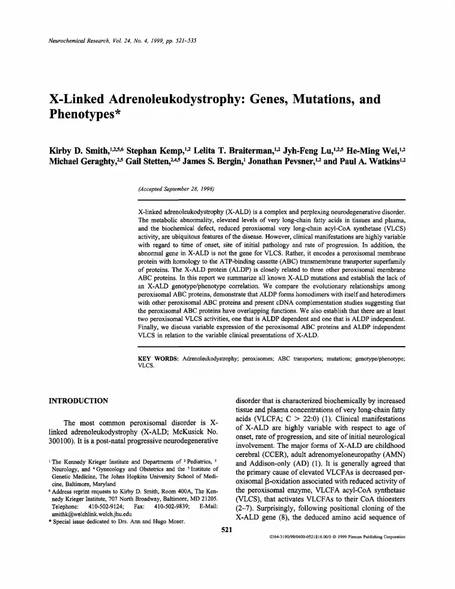
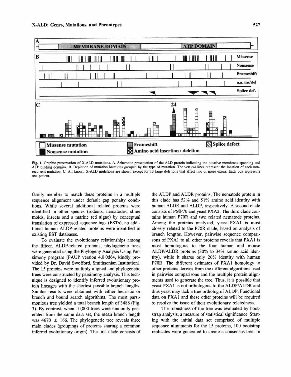
As depicted in Fig. 1, mutations are spread throughthe entire gene but are not evenly distributed. There isa clustering of mutations in the putative membrane span-
ning region (38%), the putative nucleotide binding do-main (29%) and in exon 5 (16%). The remainingmutations (17%) occur throughout the rest of the codingregion. Interestingly, only 2 mutations have been de-scribed in exon 10 (40,41) which encodes the carboxy-terminal 81 amino acids (10.9%) of ALDP.
Two nucleotide polymorphisms have been reportedin the X-ALD gene. A silent G/A nucleotide polymor-phism at position 1548 (L516L) in the sixth exon hasbeen identified with the same frequency in two indepen-dent studies (24,25); an adenosine residue was presentin 15% of patients (combined 14 of 95). A C/G nucle-otide polymorphism has been observed 8 nucleotidesdownstream of the stop codon (25). A guanine residuewas present at position 2246 in 8 out of 33 (24%) pa-tients investigated. It should be noted that the originalreport of the X-ALD cDNA sequence (8) contained asequencing error C368T resulting in A123V.
Mutational Hot Spot. A mutational hot spot hasbeen identified in exon 5 (25,31). A recurrent dinucleo-tide deletion (AG 1415/1416) has been reported in 12%(24/200) of all reported kindreds. Haplotype analysis in12 of these kindreds established that the "hot spot" kin-dreds were unrelated (25,31). The AG deletion results ina frame shift at amino acid residue E471 and a prematurestop codon at position 554. The predicted protein wouldlack the nucleotide-binding fold and would likely be in-active. Two adjacent mutations, an A insertion at posi-tion 1411 and a dinucleotide AA deletion at position1412/1413 have been reported (31). Motifs known to beassociated with short deletion/insertion mutations (42)have not been identified in the region flanking the re-curring mutations in exon 5.
Distribution of Missense Mutations. Among X-ALD patients, functionally important regions in a proteinshould contain more missense mutations than regionsthat are of less functional importance. In order to iden-tify such functionally important regions in ALDP, wecalculated the frequency of missense mutations in 50amino acid intervals from the amino to the carboxyl ter-mini. If missense mutations were distributed randomlythroughout the protein, 6.7% of all missense mutationswould be expected in each 50 amino acid interval. Thisdistribution is displayed in Fig. 2A and compared to the6.7% level expected for a random distribution of muta-tions. When compared to the diagram of the proteinshown at the bottom of the figure, it is clear that thereare three regions of ALDP where there are higher thanexpected numbers of missense mutations. The first tworegions are located within the putative transmembranedomain. These regions might function in the homo- orheterodimerization of ALDP, the binding of ALDP to
X-ALD: Genes, Mutations, and Phenotypes 523
VLCS or in substrate recognition by ALDP. The secondregion is between the predicted transmembrane spans 4and 5 and contains the EAA-like motif (43) which re-sembles the 15 amino acid core of an EAA motif thatis characteristic of prokaryotic ABC transporters (44,45).The third region contains the putative nucleotide bindingfold between the signature domains Walker A and B.
The open reading frame of the X-ALD gene has ahigh G/C content (65%). CpG sites are known to bemutational hot spots because the C residues of this din-ucleotide is a target site for DNA methylase, forming 5-methylcytosine which is readily converted to thymine bydeamination (46). Indeed, 60 out of 107 ALDP missensemutations (56%) are found in CpG sites. Furthermore,in 78% of all missense mutations either a T was substi-tuted for a C or a G was converted to an A (a C to Ttransition on the antisense strand). To ensure that ahigher incidence of missense mutations in any intervalof ALDP is not caused by a higher occurrence of CpGsites in that interval, we corrected the distribution ofmissense mutations for redundant CpG site mutations.When identical missense mutations were found in agiven CpG site, only one was counted. All identical mis-sense mutations not occurring in CpG sties were in-cluded in the distribution. When corrected for redundantCpG site mutations the same three regions with higherthan expected mutations were observed (Figure 2B).
Functional Significance of Exon 10. If the distri-bution of mutations over the length of the X-ALD genewas constant, 10.9% of the mutations would be expectedin exon 10, which encodes the 81 carboxyl-terminalamino acids. However, only 2 X-ALD mutations and nopolymorphisms have been reported in exon 10 from se-quence analysis of 200 X-ALD patients and more than300 normal X-chromosomes. This discrepancy suggeststhat this segment of ALDP maybe of functional signif-icance. To gain insight into this possibility, we con-structed an X-ALD mutant protein in which the 52carboxyl-terminal amino acids (ALDP d693-745) werereplaced by a myc epitope (pSK693myc). The 2 knownexon 10 mutations occur in the 29 amino acids that areup stream of the deletion in pSK693myc. This constructwas used to transfect a transformed fibroblast cell linederived from an X-ALD patient with an A626T mutationin ALDP. Before transfection no ALDP could be de-tected in this cell line (13). To investigate if the mutantALDPd693-745 is functionally active, we determinedVLCFA p-oxidation in X-ALD fibroblasts after trans-fection with pSK693myc and compared this result withthose obtained after transfection with a plasmid contain-ing wild type ALDP (pLB741) or a plasmid without aninsert (pcDNA3). Fibroblasts from an X-ALD patient
(A626T) have a marked reduction in C24:0 P-oxidationwhen compared to normal cells. As shown in Table II,C24:0 p-oxidation remained low after transfection withpcDNA3. After correction for the fraction of cells trans-fected, plasmids containing either wildtype ALDP(pLB741) or the carboxyl-terminal deletion ALDP(pSK639myc) restored VLCFA p-oxidation to the sameextent. This experiment indicates that the 52 carboxyl-terminal amino acids of ALDP are not necessary for thestability of ALDP or for normal VLCFA P-oxidation. Itremains unclear why mutations are so infrequent in thisregion.
Promoter Mutations. Interestingly, no promotermutations or complete gene deletions have been re-ported. All large chromosomal deletions of the X-ALDgene are located in the carboxyl-half of the gene. A pos-sible explanation for this phenomenon was suggested byMosser et al. (47). They suggested that exon 1 of the X-ALD gene might contain promoter sequences belongingto the ubiquitously expressed gene, CDM, that is located456 bp 5' to the X-ALD gene and is transcribed in theopposite direction. While there is no direct evidence forthis hypothesis and the function of the COM gene un-known it is possible the disruption of the CDM genemight be lethal.
Mutant ALDP Stability. Analyses of fibroblast celllines from X-ALD patients with a variety of mutationsincluding insertions, deletions, nonsense and missensemutations, either by immunofluorescence or by immu-noblotting, has established that ALDP is not detectablein 67% of all X-ALD patients examined (80 total)(13,36,48). Only 23% of X-ALD kindreds expressALDP at wildtype levels. However, all X-ALD patientsexamined have ALDP mRNA, independent of the typeor location of the mutation (32). Thus, the absence ofALDP is likely to be caused by protein instability ratherthan RNA instability. All mutations examined (19 total)other than missense mutations result in nondetectablelevels of ALDP. No mobility shifts were observed byPAGE for frame shift or nonsense mutations leading totruncated ALDP that would have retained the antibodyepitope (36,48). Of 57 non-recurrent mutations exam-ined for ALDP stability, 38 were missense mutations.Approximately 66% (25 total) of missense mutations re-sulted in nondetectable levels of ALDP. Nine of the 13missense mutations that did not affect ALDP stabilitywere located within the putative transmembrane domainof ALDP.
Genotype Versus Phenotype. X-ALD includes atleast 6 distinct clinical phenotypes that are categorizedon the basis of age of onset, rate of progression, and thesite of initial pathology (1). As suggested by the obser-

524 Smith et al.
Table I. X-linked Adrenoleukodystrophy World Wide Mutations (9/98)
Exon
111111111111111
1111111112223333456666666667777777888
Allele
S98LR104CR104HT105IL107PS108WG116RA141TN148SS149NR152CR152PR163HY174DY174SQ178EY181CR182PD194HD200ND200VL211PN214DL220PD221GT254MP263LG266RE271KK276EG277RG277WE291KE291DA294TY296CE302KL322PS342PR389GR389HR401WR401QR418WP484RG507VG512SS515FR518WR518QG522WP534LF540SQ544RS552PR554HP560LP560RM566KR591WR591QS606PS606LE609K
Mutation
293C>T310C>T311G>A3 HOT320T>C323C>G346G>A421G>A443A>G446G>A454OT455G>C488G>A520T>G521A>C5320G542A>G545G>C580G>C598OA599A>T632T>C640A>G659T>C662A>G761OT788OT796G>A811G>A826A>G829G>A829G>T871G>A873OC880G>A887A>G904G>A965T>C1024T>C11650G1166G>A1201OT1202G>A1252OT1452OG1520G>T1534G>A1544OT1552OT1553G>A1564G>A1601OT1619T>C1631A>G1654T>C1661G>A16790T1679OG1697T>A1771OT1772G>A1816T>C1817OT1825G>A
Missense mutaATG(387)
679OT696OT697G>A700OT706T>C709OG732G>A807G>A829A>G832G>A840OT841G>C874G>A906T>G907A>C918OG928A>G931G>C966G>C984G>A985A>T1018T>C1026A>G1045T>C1048A>G1147C>T1174C>T1182C>A1197G>A1212A>G1215G>A1215G>T1257G>A1259G>C1266G>A1273A>G1290G>A1351T>C1410T>C1551C>G1552G>A1587OT1588G>A1638OT1838OG1906G>T1920G>A1930OT1938OT1939G>A1950G>A1987OT2005T>C2017A>G2040T>C2047G>A20650T20650C>G2083T>A2157C>T2158G>A2202T>C2203OT2211G>A
ionsN
22111111312112111I1111111113111111111111213311313211121161111132
ALDP
++/-nd-nd
+/--nd+++ndndndndndndnd+ndndndnd
+/--nd
+/-ndnd+ndndnd-ndndndnd+nd+nd+-ndnd-nd
+/----nd+--
+/---nd+-+—
Reference
3631,3229363736363124,36,733231,36323224,377229363132733637733236373624,32,7373133731141336733841133731,327324,37,7324,37,*287331,35,362423,3639,7336357339,73*
*
29,36,73363173313623,31,7332,37
Total
111111111111111111111111111111111
111111111111
11
1111
111111
11111
11111111
1
11111

525X-ALD: Genes, Mutations, and Phenotypes
Table I. Continued.
Exon
8888999
10
Exon
111111122445789
10
Exon
11115679
Exon
36779
Exon
11111111111113
Allele
E609GR617CR617GR617HA626TD629HR660WW679R
Allele
W10XQ133XW137XQ157XY181XY212XW242XQ311XW326XR464XQ466XE477XQ590XW595XQ645XQ672X
Allele
80-81insLRL138-141del277-278insNE291del491-500insVG528del587-590delI657del
Allele
Fs E408Fs R545Fs R545Fs G593Fs R622
Allele
Fs A19FsP34FsL46FsT91FsG92Fs A99Fs Q133Fs Y180Fs L197Fs S207FsR231Fs F261Fs G266Fs V378
Mutation
1826A>G1849C>T1849C>G1850G>A1876G>A1885G>C1978C>T2035T>C
Mutation
30G>A397C>T411G>A469C>T543C>A636C>G726G>A931C>T977G>A1390C>T1396C>T1429G>T1768C>T1785G>A1933C>T2014C>T
In frame
871delGAG
1582delGGT1759-70del1969delATC
Mutation
1224A>Givs1634+1G>Aivs1635-2A>Givs1780+1G>Aivs1866-10G>A
Mutation
56delC102C>AT138insT274del34277delC298delG401TGCTG>AGCATT541delTA591insT618dell3693delGG785del7796delG1135insC
Missense mutationsATG(387)
2212A>G2235C>T2235C>G2236G>A2262G>A2271G>C2364C>T2421T>C
Nonsense mutations
416G>A783C>T797G>A855C>T929C>A1022C>G1112G>A1317C>T1363G>A1776C>T1782C>T1815G>T2154C>T2171G>A2319C>T2400C>T
amino acid insertions & deletions
1257delGAG
1968delGGT2145-56del2355detATC
Splice defects
1610A>Givs2020+1G>Aivs2021-2A>Givs2166+1G>Aivs2252-10G>A
Frame shifts
442delC488C>AT524insT660del34663delC684delG787TGCTG>AGCATT927delTA977insT1004del131079delGG1171del71182delG1521insC
N
13121161
N
1111111111121111
N
3
1
N
11111
N
11111111112111
ALDP
_ndnd--+-nd
ALDP
-----ndndndndndndndndnd--
ALDP
ndndnd-—ndndnd
ALDP
ndnd-ndnd
ALDP
-ndndndndndnd—ndnd-ndnd—
Reference
3223,32,373731,33131331,32,35,36,7340
Reference
363232323229372222233124,37277313*
Reference
*373713,37,7336297332
Reference
3123303630
Reference
3273362932*
3436733731,36323736
Total
11111111
Total
11
Total
11111111
Total
11111
Total
111
1
111
111
111111
1111111
111111111
11

526 Smith et al.
Table I. Continued.
Exon
4555666889
start
1223336777889
Exon
610
Allele
Fs A417Fs V470Fs V470Fs E471Fs F517Fs G529Fs P534Fs A597Fs S606Fs D649
Allele
0.5 kb in exon 1exon2delexon2-7delexon3-5delexon3-10delexon3-10delexon6-10delexon7-9delexon7-10delexon7-10delexon8-9delexon8-10delmultiple exons
Allele
L516L
Mutation
1250delC1411insA1412delAA1415delAG1551delC1585delGalt1603-19911791delTA1818delG1949delGC
7 kb deletion
Nucl. change
1548G/A2246C/G (3' UTR)
Missense mutationsATG(387)
1636delC1797insA1798delAAISOldelAG1937delC1971delGalt 1989-23 772177delTA2204delG2335delGC
Multiple exons
Polymorphisms
1934G/A2632C/G (3' UTR)
N
111
24111111
N
1
15%24%
ALDP
-ndnd-ndnd-ndnd-
ALDP
ndndndndndndndndnd—ndndnd
Reference
36313124,25,31,36,37,72,73233832232336
Reference
268883126267383183135
Reference
23-2525
Total
1111111111
Total
11
Total
11
Two different nucleotide numbering systems have been used for reporting X-ALD mutations. The first uses the first nucleotide of the cDNA asnumber + 1 . The second uses the A of the initiator Methionine codon as + 1 . In this report, all mutation designations are standardized to the second,more common, numbering system. All mutation designations conform to the nomenclature described in Human Mutation 8:197-202 (1996). Forframe shift mutations the last unchanged amino acid is indicated. The mutations marked with an * are reported for the first time in this report. Thecolumn headed ALDP indicates the presence or absence of ALDP in fibroblasts as indicated by cellular immunofluorescence and/or immunoblotting:(+) = normal levels, (— ) = undetectable levels, and (+/-) = reduced levels.
analyzed sequence relationships of ALDP and other pro-teins by comparison of protein and DNA sequences inGenBank and other databases using the BLAST pro-grams. Proteins were multiply aligned using the Pileupprogram of the GCG software package (Madison, WL).Fifteen proteins were identified including: four humanproteins (ALDP, ALDR, PMP70, and P70R), two yeastproteins (PXA1 and PXA2), several closely related ro-dent orthologues and five C. elegans orthologues. Theassignment of the two ALDP-related yeast proteins isconsistent with the phylogenetic analysis of the 29-30known ABC proteins encoded in the S. cerevisiae ge-nome (49,50). While there are hundreds of known ABCtransporter proteins, the 15 identified in this search aredefined as a group by their relatively high amino acididentity and the failure of any other ABC transporter
vation that males in the same family can have differentforms of X-ALD (1), mutation analysis has confirmedthat phenotypic expression cannot be predicted on thebasis of the genotype of the X-ALD gene. Indeed, allphenorypes are observed in patients that lack ALDP in-cluding those with identical dinucleotide deletions inexon 5 (25,31,36). This is illustrated in Table III wherethe phenotypes associated with the exon 5 dinucleotidedeletion, which lacks detectable ALDP, in 15 separatekindreds are listed.
Evolutionary Relationship Among PeroxisomalABC Half-Transporters. By sequence comparison, theperoxisomal ABC half-transporters are a closely relatedsubgroup of the ABC superfamily that presumably aroseby recent gene duplication. However, they are locatedon different chromosomes as indicated in Table IV. We
111111111111
11111111111
X-ALD: Genes, Mutations, and Phenotypes 527
Fig. 1. Graphic presentation of X-ALD mutations. A. Schematic presentation of the ALD protein indicating the putative membrane spanning andATP binding domains. B. Depiction of mutation locations grouped by the type of mutation. The vertical lines represent the location of each non-recurrent mutation. C. All known X-ALD mutations are shown except for 13 large deletions that affect two or more exons. Each box representsone patient.
family member to match these proteins in a multiplesequence alignment under default gap penalty condi-tions. While several additional related proteins wereidentified in other species (rodents, nematodes, slimemolds, insects and a marine red algae) by conceptualtranslation of expressed sequence tags (ESTs), no addi-tional human ALDP-related proteins were identified inexisting EST databases.
To evaluate the evolutionary relationships amongthe fifteen ALDP-related proteins, phylogenetic treeswere generated using the Phylogeny Analysis Using Par-simony program (PAUP version 4.0.0d64, kindly pro-vided by Dr. David Swofford, Smithsonian Institution).The 15 proteins were multiply aligned and phylogenetictrees were constructed by parsimony analysis. This tech-nique is designed to identify inferred evolutionary pro-tein lineages with the shortest possible branch lengths.Similar results were obtained with either heuristic orbranch and bound search algorithms. The most parsi-monious tree yielded a total branch length of 3488 (Fig.3). By contrast, when 10,000 trees were randomly gen-erated from the same data set, the mean branch lengthwas 4670 + 166. The phylogenetic tree reveals threemain clades (groupings of proteins sharing a commoninferred evolutionary origin). The first clade consists of
the ALDP and ALDR proteins. The nematode protein inthis clade has 52% and 51% amino acid identity withhuman ALDR and ALDP, respectively. A second cladeconsists of PMP70 and yeast PXA2. The third clade con-tains human P70R and two related nematode proteins.Among the proteins analyzed, yeast PXA1 is mostclosely related to the P70R clade, based on analysis ofbranch lengths. However, pairwise sequence compari-sons of PXA1 to all other proteins reveals that PXA1 ismost homologous to the four human and mouseALDP/ALDR proteins (30% to 34% amino acid iden-tity), while it shares only 26% identity with humanP70R. The different estimates of PXA1 homology toother proteins derives from the different algorithms usedin pairwise comparisons and the multiple protein align-ments used to generate the tree. Thus, it is possible thatyeast PXA1 is not orthologous to the ALDP/ALDR andthus yeast may lack a true ortholog of ALDP. Functionaldata on PXA1 and these other proteins will be requiredto resolve the issue of their evolutionary relatedness.
The robustness of the tree was evaluated by boot-strap analysis, a measure of statistical significance. Start-ing with the initial data set comprised of multiplesequence alignments for the 15 proteins, 100 bootstrapreplicates were generated to create a consensus tree. In

528 Smith et al.
Fig. 2. Regional frequency of missense mutations in ALDP. Missensemutations should occur most frequently in regions of functional im-portance. A total of 107 missense mutations have been reported in theX-ALD gene. The frequency of missense mutations found per 50amino acid intervals is plotted in the upper panel (A) for all missensemutations and in the lower panel (B) after correction for redundantCpG mutations. The dashed line indicates the frequency of mutationsexpected in each 50 amino acid interval if they were random. TheALD protein is represented schematically at the bottom of the figure.The transmembrane and ATP binding domains are indicated. Thehatched box within the transmembrane domain indicates the locationof the EAA motif.
vergent from its putative orthologue but the overall to-pology of the tree is supported by bootstrap analysis. Aprediction based on this tree is that additional mamma-lian PMP70 and P70R genes may exist that correspondto the multiple nematode sequences identified in thesegroups. Searches of human and murine EST databaseshave so far failed to identify such homologues.
Non-Processed, Nontranscribed Autosomal X-ALDPseudogenes. In addition to the functional peroxisomalABC half-transporters, there are several autosomal pseu-dogenes on different chromosomes. These are comprisedof approximately 9.7 kb of the X-ALD gene that encom-passes exons 7-10 (31,51). Sequence comparison shows92-96% nucleotide identity among the X-ALD autoso-mal pseudogenes. PCR amplification of genomic DNAwith PCR primers specific for exon 7 yields a singleDNA fragment when assayed by agarose gel electropho-resis (Fig. 4A). Analysis of the same genomic PCR frag-ment by single strand conformation polymorphismanalysis (SSCP) (31) reveals several distinct bands in-dicating that more than one sequence is contained in theexon 7 PCR amplicon. SSCP analysis of a mouse/humansomatic cell hybrid in which the only human chromo-some is the X allows discrimination of the X-chromo-
100% of cases, the P70R group was intact as was theALDP/ALDR clade (exclusive of yeast PXA1). In 78%of the cases, the PMP70 protein clade included yeastPXA2. Thus, each of the yeast proteins is relatively di-
Table III. Genotype Versus Phenotype
X-ALD Kindred
123456789
101112131415
Phenotypes
CCERAMNAMNCCERAMNCCER, AMN, AddisonCCER, AMN, AddisonAMNCCER, AMN, AddisonCCERCCER, AMNAMNCCERAMNCCER
Reference
252525252531313131313136363636
Listed are 15 independent X-ALD kindreds with the common X-ALD exon 5 dinucleotide deletion. This mutation results in frame shift,a premature termination signal and a lack of detectable ALDP.
Table IV. Chromosomal Location of Human Peroxisomal ABCHalf-transporters
cDNA
ALDALDRPMP70P70R
Chromosomal Location
Xq2812qll1p21
14q24
Table II. Correction of C24:0 B-oxidation by ALDP Exon 10Deletion Mutant
Cell Line
NormalA626TA626TA626T
; Construct
nonepcDNA3pSK693mycpLB741
C24:0nmol/hr/mg protein
0.870.100.170.18
TransfectionEfficiency
NAND37%35%
%Correction
5359
Fibroblasts from an X-ALD patient (A626T) were transfected withthe indicated plasmids by electroporation. The frequency of transfec-tion was determined by indirect immunofluorescence using either anti-myc or anti-ALDP antibodies and C24:0 3-oxidation determined asdescribed previously (15).

X-ALD: Genes, Mutations, and Phenotypes 529
Fig. 3. Phylogenetic tree. Evolutionary relationships among the 15 proteins in this tree were inferred from multiple sequences alignments using thePhylogeny Analysis Using Parsimony program (PAUP version 4.0.0d64). The most parsimonious path for the evolution of this protein family(shortest total branch length) is shown.
some and autosomal conformers. No prominent PCRfragment is amplified from mouse genomic DNA. PCRanalysis of a human monochromosomal mapping panelwith exon 9/10 PCR primers identifies chromosomes 1,2, 20, 22, and possibly 16 as containing X-ALD pseu-dogenes (Fig. 4B). Fluorescence In situ Hybridization(FISH) analysis using cloned genomic fragments of theautosomal pseudogenes identified homologous se-quences at 2p11, 10p11, 16p11, 20ptel, and 22q11 (Ta-ble V). Previous studies by Eichler, et. al. (51) localizedX-ALD pseudogenes to 2p1 1, 10p1 1, 16p1 1 and 22ql 1.Even though strong PCR reactions were supported bychromosomes 1 and 20, FISH analysis detected X-ALDhomologs rarely on chromosome 20 and never on chro-mosome 1. Thus, these locations may have more diver-gent homologs representing an earlier duplication.Southern blot analysis of mouse and primate genomicDNA with a partial cDNA probe (exons 2-10) indicatedthat there may have been two expansions of this X-ALDgene segment in higher primates (Fig. 4C). The singlerestriction fragment detected in mouse genomic DNAhas been confirmed to be on the X-chromosome by seg-
regation analysis (52) and by FISH analysis (Fig. 5). TheX-ALD signal indicated by the arrow localizes to bandB of the mouse X-chromosome which is homologous tohuman Xq28. The single restriction fragment observedin old world monkeys (Rhesus) is presumably also onthe X-chromosome. An initial expansion appears to haveoccurred on the evolutionary line leading to Orangutansand a subsequent or independent expansion in the greatapes (Gorilla, Chimpanzee and Human). As pointed outearlier this rare example of non-processed pseudogeneson several different chromosomes complicates mutationanalysis (31) and illustrates pericentromeric plasticity ofnon-homologous interchromosomal exchange (51).
Interactions Among Peroxisomal ABC Half-Trans-porters. By comparison to known ABC half-transporterproteins, it has been suggested that ALDP may functionas a homodimer and/or as a heterodimer with one ormore of the 3 other peroxisomal ABC transporters(53,54). Other ABC half-transporters, including the his-tocompatibility complex proteins, TAP1 and TAP 2, andthe yeast peroxisomal ABC half-transporters, PXA1 andPXA2 (PAT1 and PAT2), form heterodimers to generate

Smith et al.530
Fig. 4. X-ALD Autosomal Pseudogenes. A. X-ALD exon 7 was amplified by PCR from total genomic DNA isolated from human, mouse or themouse/human somatic hybrid cell, AHAlla, and analyzed by SSCP as previously described(31). h = human, m = mouse. B. Genomic DNA froma rodent/human somatic hybrid cell human monochromosomal mapping panel was amplified by PCR using PCR primers derived from exons 9 and10 of the X-ALD gene. PCR primers and PCR conditions were as previously described (31). C. Southern blot analysis of mouse and primategenomic DNAs digested with Hind III and probed with a partial X-ALD cDNA clone (exons 2-10).
a functional transporter (55,56). In order to test this no-tion, an in vitro co-immunoprecipitation assay was de-veloped. In this assay, radiolabeled ALDP was generatedfrom its cDNA by in vitro transcription and translation.Unlabeled peroxisomal ABC half-transporter proteins,ALDP, ALDRP and PMP70, with a c-myc epitope attheir C-termini were produced by in vitro transcriptionand translation in separate reactions. Equal amounts ofradiolabeled ALDP without a c-myc epitope and unla-beled protein containing a c-myc epitope were mixed inthe presence of ATP and then immunoprecipitated withanti-c-myc antibody agarose conjugate. Co-immunopre-cipitation was deduced from the presence of radiolabeledALDP with the unlabeled c-myc tagged protein immu-noprecipitate. As seen in Fig. 6, ALDP homodimers andheterodimers of ALDP with both ALDR and PMP70 aredemonstrated by this assay. Although it is clear thatALDP has some role in the VLCFA (3-oxidation path-way, the function of ALDP and the other peroxisomalABC half-transporters is as yet unknown. That theseproteins may have related functions is indicated by theobservation that overexpression of ALDP, PMP70 and
ALDR can restore VLCFA (3-oxidation in fibroblastsfrom ALD patients (Table VI). We know that ALDP isrequired for normal peroxisomal VLCFA activation and(3-oxidation and that expression of ALDP cDNA restoresVLCFA (3-oxidation in cells which lack the protein. Al-though ALDP is a peroxisomal membrane protein, it isnot required for peroxisome assembly. ALDP is differ-
Table V. FISH Localization of X-ALD Autosomal Pseudogenes
Chromosomal location
2plllOpl l16pll20ptel22qll
number of signals in45 metaphase spreads
842140
426
DNA isolated from 6 autosomal genomic clones (12-15-kb) wasprepared for FISH analysis and hybridized as described in the legendto figure 5, except that human Cot-1 DNA was used for prehybridi-zation. Metaphase spreads were prepared from peripheral blood lym-phocytes (70). Hybridization signals were scored from 45 metaphasespreads and the data were pooled.

X-ALD: Genes, Mutations, and Phenotypes 531
Fig. 6. Co-immunoprecipitation of peroxisomal ABC half-transporters.Unlabeled peroxisomal ABC half-transporter proteins with a c-mycepitope at their carboxyl-termini were generated by coupled in vitrotranscription/translation (TNT, Promega) from their respective cDNAs.Radiolabeled ALDP ([35S]-methionine) without a c-myc epitope wasgenerated independently by in vitro transcription/translation of itscDNA. Equal volumes of labeled ALDP and unlabeled c-myc taggedproteins were incubated together with protease inhibitors and ATP.The mixture was added to a c-myc agarose conjugate (Santa Cruz) toimmunoprecipitate the unlabeled, epitope tagged ABC half-transporterprotein. The agarose conjugate was washed and the immunoprecipi-tated proteins were fractionated by SDS-PAGE, transferred to nitro-cellulose membranes and protein-protein interactions judged by thepresence of radiolabeled ABC half-transporter bands following phos-phorimagery. Non-specific aggregation of the radiolabeled protein withthe agarose conjugate was determined by substitution of a non-c-myctagged unlabeled protein for the c-myc tagged protein. Phosphorima-ger analysis was performed with a Fuji BAS2500 Phosphorimager us-ing a high resolution screen.
Fig. 5. Localization of ALD gene to the mouse X-chromosome. Flu-orescence in situ hybridization (FISH) to metaphase spreads from amale mouse embryonic stem cell line, ES-J1, was performed as de-scribed(70). Chromosome spreads containing 40 chromosomes wereexamined after hybridization with a biotin labeled 6.2kb Hind III frag-ment of genomic DNA from the cloned mouse ALD gene. The probewas labeled with biotin-14-dATP by nick translation (BRL) and 200ngwere preannealed with 2ug of mouse Cot-1 DNA for 1 hour at 37°C.The probe and slides were denatured separately in 70% formamide,2X SSC, pH 7.0 at 75°C. Hybridization was at 37°C for 17 hr. andprobe was detected with avidin conjugated fluorescein. Chromosomeswere counterstained with propidium iodide in antifade (Vector) andviewed using a Zeiss epifluorescene microscope equipped with a broadband-pass FITC filter cube. Clear hybridization was seen on the X-chromosome and no consistent signals were seen on any other chro-mosome in 20 spreads analyzed. The arrow points to the localizationof signal at band b of the mouse X-chromosome which is homologousto human Xq28, the human site of the X-ALD gene.
SV40 T antigen-transformed fibroblasts from an X-ALD patientwere transfected with vector alone (pCDNA3) or with vector contain-ing cDNA for PMP70, ALDP or ALDRP. Transfection was performedas previously described (15). The rates of C24:0 B-oxidation observedin the transfected cells were corrected for the fraction of the cellsexpressing the transgene, as determined by IMF staining. The adjustedrates are compared to the rate determined in SV 40 T antigen trans-formed normal fibroblasts. The indicated values are the mean and stan-dard deviation for pCDNA3 (n=6), PMP70 (n=l), ALDP (n=5),ALDRP (n=4), and normal (n = 5).
entially expressed at the cellular level (57) so that notall normal peroxisomes have ALDP and peroxisomesfrom X-ALD cells are normal except for VLCFA (3-oxidation. While the precise role of ALDP in peroxi-some metabolism is unknown, its identification as amember of the ABC transporter superfamily suggeststhat it might transport VLCFAs (the substrate for (3-ox-idation), VLCFA-CoA, CoA, ATP or other required me-tabolites into the peroxisome; Mosser and coworkersspeculated that ALDP might translocate VLCS itself intothe peroxisome (8). However, by analogy to other ABC
transporter proteins (58-63), the relationship betweenALDP and VLCFA metabolism may be indirect. ALDPcould function as an anchor or platform for VLCS in theperoxisomal membrane or interact with other proteins ortransporters in a regulatory role. Thus, it is possible thatassociation and dissociation of ALDP as a homodimeror heterodimer in a cell type specific manner might re-flect different or changing metabolic states.
Very Long Chain Acyl-CoA Synthetase. As notedabove, all X-ALD patients examined in detail have mu-tations in the X-ALD gene, which encodes a peroxi-
Table VI. Peroxisomal ABC Half-Transporter Complementation ofSV-40 Transformed X-ALD Fibroblasts
cDNA
Vector alonePMP70ALDP
ALDRP
Transformed Fibroblasts
C24:0 p-oxidationnmol/hr/mg protein
0.052 + 0.0040.180.22 ± 0.040.23 ± 0.06
0.26 ± 0.02

532 Smith et al.
action that requires VLCS activity. Fibroblasts from theX-ALD mouse, recently generated in our laboratory(52), have a marked reduction in the level of VLCFA(3-oxidation accompanied by an increase in VLCFAabundance (Table VII). Thus, there appears to be anALDP dependent VLCS activity in mouse fibroblaststhat is not due to the protein identified as mouse VLCS.In order to initiate examination of the relationship ofmouse VLCS and ALDP, mouse VLCS cDNA was ex-pressed in fibroblasts from the X-ALD mouse whichlack ALDP. Surprisingly, mouse VLCS restoredVLCFA (3-oxidation in these cells to the same extent asdid expression of human ALDP cDNA (Table VIII).Thus, the activity of this cloned mouse VLCS appearsto be independent of ALDP and it may contribute totissue variability of VLCFA (3-oxidation activity andVLCFA levels in different cell types depending on itsdifferential expression in specific cell types.
Genetic Modification of X-ALD Clinical Presenta-tion. As cited above, several studies have establishedthat there is no correlation of X-ALD mutations and themarked variability of X-ALD clinical presentation. In-deed, null mutations are associated with all forms of X-ALD from the most severe to the mildest (Table II). Thesource of variable X-ALD phenotypes could be environ-mental, genetic or both. Genetic segregation analyses(65-67) provide support for the hypothesis that at leastone autosomal gene plays a role in the manifestation ofcharacteristics that derive in the first instance from mu-tations in the X-ALD gene. The genes for the proteinsdescribed in this report are candidates for such modifiereffects. In addition to ALDP, at least two of the perox-isomal half-transporters, ALDR and PMP70, as well asVLCS, are able to correct the metabolic defect in cellsthat lack ALDP.
Most surprising is the demonstration of at least twoVLCS activities, one of which appears to be independentof the presence of ALDP. While it has been clearly dem-onstrated that there is no correlation between the plasmalevel of VLCFAs and the clinical phenotype of X-ALD(1), relevant measurements in X-ALD target tissues, thecerebral cortex and the adrenal gland, are not available.It is clear from studies of VLCFA levels in various tis-sues of the X-ALD mouse model that there are markeddifferences in the accumulation of VLCFA (52). Datadiscussed in this report demonstrate that the peroxisomalhalf-transporters and VLCS are expressed in a tissuespecific manner. Higher expression of one or more ofthese genes could lead to variable levels of VLCFAs inthe affected cell types in X-ALD leading to variable phe-notypic outcomes. Studies to examine such correlationsare currently in progress.
somal membrane protein. Paradoxically, the biochemicaldefect in X-ALD is the failure of peroxisomes to activateVLCFAs to their CoA derivatives, a reaction catalyzedby VLCS and not ALDP. Experimental evidence sug-gests that ALDP and VLCS interact functionally andperhaps physically. Recently, VLCS protein was purifiedfrom rat liver peroxisomes (9) and VLCS cDNA cloned(10). Subsequently, we cloned yeast VLCS (64) as wellas human (S. Steinberg and P. A. Watkins, unpublishedobservations) and mouse (S. Kemp and A. Heinzer, un-published observations) VLCS cDNA. Both mouse andhuman VLCS are expressed primarily in liver and kid-ney with low levels of expression detected in brain, heartand adrenal gland (S. Steinberg, S. Kemp, J-F. Lu, A.Heinzer and P.A. Watkins, unpublished observations).The observation that expression of this VLCS is not de-tected in all cells that degrade VLCFAs by (3-oxidationsuggests that there must be additional VLCS genes. Forexample, mouse fibroblasts have no detectable VLCSprotein or mRNA yet actively degrade VLCFAs, a re-
Table VII. VLCFA Metabolism in Mouse Fibroblasts
A. 3-Oxidation
Normal (n=3)X-ALD (n=4)
B. Concentration
Normal (n=3)X-ALD (n=5)
C24:0nmol/hr/mg protein
0.99 ± 0.150.37 ± 0.03
C26:0mg/mg protein
0.062 ± 0.0130.27 ±0 .11
C24:0/C16:0Ratio
0.26 ± 0.040.09 ± 0.02
C26:0/C22:0Ratio
0.064 ± 0.0260.35 ± 0.18
A. C24:0 B-oxidation. Normal and X-ALD mouse fibroblasts weregrown, harvested and assayed as described previously (52).
B. VLCFA concentrations. Total lipids were extracted from culturedfibroblasts and fatty acids were separated according to chain lengthand quantitated by Capillary Gas Chromatography as described pre-viously (52).
Table VIII. Complementation of C24:0 p-Oxidation in Fibroblastsfrom X-ALD Mice
cDNA Construct
nonepcDNA3human ALDPmouse VLCS
n
5355
P-oxidationnmol/hr/mg protein
0.13 ± 0.010.13 ± 0.060.89 ±0.180.81 ± 0.16
Fibroblasts from an X-ALD mouse were transfected with the indi-cated cDNA constructs as previously described (15). C24:0 (3-oxida-tion specific activity (SA) was determined (71) and corrected for thefraction of cells expressing the transgene, as determined by IMF stain-ing (15). The indicated values are mean and standard deviation.

X-ALD: Genes, Mutations, and Phenotypes 533
ACKNOWLEDGMENTS
The authors would like to thank Ann and Hugo Moser for theirpioneering studies of peroxisomes and X-linked adrenoleukodystrophythat have made the studies presented in this manuscript both possibleand relevant and for their continuing interest and support. We thankAnn Heinzer for a careful reading of the manuscript. This work wassupported by grants from the National Insituties of Health, HD 10981,HD24061, and DK51149, the Myelin Project and the United Leuko-dystrophy Foundation.
REFERENCES
1. Moser, H.W., Smith, K.D., and Moser, A.B. 1994. X-linked Ad-renoleukodystrophy. Pages 2325-49, in Scriver, C.R., Beaudet,A.L., Sly, W.S., and Valle, D. (eds). The Metabolic and MolecularBasis of Inherited Disease, Seventh Edition. New York: McGrawHill.
2. Poulos, A., Singh, H., Paton, B., Sharp, P., and Derwas, N. 1986.Accumulation and defective (3-oxidation of very long chain fattyacids in Zellweger's syndrome, adrenoleukodystrophy and Ref-sum's disease variants. Clin. Genet. 29:397-408.
3. Singh, I., Moser, A.B., Goldfischer, S., and Moser, H.W. 1984.Lignoceric acid is oxidized in the peroxisomes: Implications forthe Zellweger cerebro-hepato-renal syndrome and adrenoleuko-dystrophy. Proc. Natl. Acad. Sci. USA 81:4203-7.
4. Wanders, R.J.A, van Roermond, C.W.T, van Wijland, M.J.A, Ni-jenhuis, A.A, Tromp, A., Schutgens, R.B.H., Brouwer-Kelder,E.M., Schram, A.W., Tager, J.M., van den Bosch, H., and Schalk-wijk, C. 1987. X-linked adrenoleukodystrophy: Defective perox-isomal oxidation of very long chain fatty acids but not of verylong chain fatty acyl-CoA esters. Clin. Chim. Acta. 165:321-9.
5. Singh, I., Moser, A.B., Moser, H.W., and Kishimoto, Y. 1984.Adrenoleukodystrophy: Impaired oxidation of very long chainfatty acids in white blood cells, cultured skin fibroblasts and am-niocytes. Pediatr. Res. 18(3):286-90.
6. Lazo, O., Contreras, M., Bhushan, A., Stanley, W., and Singh, I.1989. Adrenoleukodystrophy: Impaired oxidation of fatty acidsdue to peroxisomal lignoceroyl-CoA ligase deficiency. Arch.Biochem. Biophys. 270(2):722-8.
7. Wanders, R.J.A., van Roermund, C.W.T., van Wijland, M.J.A.,Schutgens, R.B.H., van deb Bosch, H., Schram, A.W., and Tager,J.M. 1988. Direct evidence that the deficient oxidation of verylong chain fatty acids in X-linked adrenoleukodystrophy is due toan impaired ability of peroxisomes to activate very long chainfatty acids. Biochem. Biophys. Res. Commun, 153:618—24.
8. Mosser, J., Douar, A.-M., Sarde, C.-O., Kioschis, P., Feil, R.,Moser, H., Poustka, A.-M., Mandel, J.-M., and Aubourg, P. 1993.Putative X-linked adrenoleukodystrophy gene shares unexpectedhomology with ABC transporters. Nature 361:726-30.
9. Uchida, Y., Kondo, N., Orii, T., and Hashimoto, T. 1996. Purifi-cation and properties of rat liver peroxisomal very long chainAcyl-CoA synthetase. J. Biochem. 119:565-71.
10. Uchiyama, A., Aoyama, T., Kamijo, K., Uchida, Y., Kondo, N.,Orii, T., and Hashimoto, T. 1996. Molecular cloning of cDNAencoding rat very long chain acyl-CoA synthetase. J. Biol. Chem.271(48):30360-5.
11. Higgins, C.F. 1992. ABC transporters: From microorganisms toman. Annu. Rev. Cell. Biol. 8:67-113.
12. Mosser, J., Lutz, Y., Stoeckel, M.E., Sarde, C.-O., Kretz, C.,Douar, A.M., Lopez, J., Aubourg, P., and Mandel, J.L. 1994. Thegene responsible for adrenoleukodystrophy encodes a peroxisomalmembrane protein. Hum. Mol. Genet. 3(2):265-71.
13. Watkins, P.A., Gould, S.J., Smith, M.A., Braiterman, L.T., Wei,H.M., Kok, F., Moser, A.B., Moser, H.W., and Smith, K.D. 1995.
Altered expression of ALDP in X-linked adrenoleukodystrophy.Am. J. Hum. Genet. 57:292-301.
14. Carrier, N., Lopez, J., Moullier, P., Rocchiccioli, F., Rolland, M.-O., Jorge, P., Mosser, J., Mandel, J.-M., Bougneres, P.-F., Danos,O., and Aubourg, P. 1995. Retroviral-mediated gene transfer cor-rects very-long-chain fatty acid metabolism in adrenoleukodystro-phy fibroblasts. Proc. Natl. Acad. Aci. USA 92:1674-8.
15. Braiterman, L.T., Zheng, S., Watkins, P.A., Geraghty, M.T., John-son, G., McGuinness, M.C., Moser, A.B., and Smith, K.D. 1998.Suppression of peroxisomal membrane protein defects by perox-isomal ATP binding cassette (ABC) proteins. Hum. Molec. Genet.7(2):239-47.
16. Kamijo, K., Taketani, S., Yokota, S., Osumi, T., and Hashimoto,T. 1990. The 70-kDa peroxisomal membrane protein is a memberof the Mdr (P-glycoprotein)-related ATP-binding protein super-family. J. Biol. Chem. 265(8):4534-40.
17. Gartner, J., Moser, H., and Valle, D. 1992. Mutations in the 70Kperoxisomal membrane protein gene in Zellweger syndrome. Na-ture. Genet. 1:16-23.
18. Lombard-Platet, G., Savary, S., Sarde, C.-O., Mandel, J.-L., andChimini, G. 1996. A close relative of the adrenoleukodystrophy(ALD) gene codes for a peroxisomal protein with a specific ex-pression pattern. Proc. Natl. Acad. Sci. USA 93:1265-9.
19. Shani, N., Jimenez-Sanchez, G., Steel, G., Dean, M., and Valle,D. 1997. Identification of a fourth half ABC transporter in thehuman peroxisomal membrane. Hum. Mol. Genet. 6(11):1925-31.
20. Sarde, C.-O., Mosser, J., Kioschis, P., Kretz, C., Vicaire, S., Au-bourg, P., Poustka, A., and Mandel, J.L. 1994. Genomic organi-zation of the adrenoleukodystrophy gene. Genomics. 22:13-20.
21. Cartier, N., Sarde, C.O., Douar, A.M., Mosser, J., Mandel, J.L.,and Aubourg, P. 1993. Abnormal messenger RNA expression anda missense mutation in patients with X-linked adrenoleukodystro-phy. Hum. Mol. Genet. 2:1949-51.
22. Barcelo, A., Giros, M., Albiach, V.J., Vaquerizo, J., Pampols, T.,and Estivill, X. 1996. Identification of two new nonsense muta-tions (Q31IX and W326X) in exon 2 of the adrenoleukodystrophy(ALD) gene. Hum. Mutat. 8:286-7.
23. Fanen, P., Guidoux, S., Sarde, C.O., Mandel, J.L., Goossens, M.,and Aubourg, P. 1994. Identification of mutations in the putativeATP-binding domain of the adrenoleukodystrophy gene. J. Clin.Invest. 94:516-20.
24. Fuchs, S., Sarde, C.O., Wedemann, H., Schwinger, E., Mandel, J.L., and Gal, A. 1994. Missense mutations are frequent in the genefor X-chromosomal adrenoleukodystrophy (ALD). Hum. Mol. Ge-net. 3(10):1903-5.
25. Kemp, S., Ligtenberg, J.L., van Geel, B.M., Barth, P.O., Wolter-man, R.A., Schoute, F., Sarde, C.-O., Mandel, J.-L., van Oost,B.A., and Bolhuis, P.A. 1994. Identification of a two base pairdeletion in five unrelated families with adrenoleukodystrophy: Apossible hot spot for mutations. Biochem. Biophys. Res. Commun.202(2):647-53.
26. Koike, R., Onodera, O., Tabe, H., Kaneko, K., Miyatake, T., Iwa-saki, S., Nakano, M., Shizuma, N., Ikeguchi, K., Nishizawa, M.,Mosser, J., Sarde, C.O., and Tsuji, S. 1995. Partial deletions ofputative adrenoleukodystrophy (ALD) gene in Japanese ALD pa-tients. Hum. Mutat. 6:263-7.
27. Uchiyama, A., Suzuki, Y., Song, X.Q., Fukao, T., Imamura, A.,Tomatsu, S., Shimozawa, N., and et al. 1994. Identification of anonsense mutation in ALD protein cDNA from a patient withadrenoleukodystrophy. Biochem. Biophys. Res. Commun. 198:632-6.
28. Berger, J., Molzer, B., Fae, I., and Bernheimer, H. 1994. X-linkedadrenoleukodystrophy (ALD): A novel mutation of the ALD genein 6 members of a family presenting with 5 different phenotypes.Biochem. Biophys. Res. Commun. 205:1638-43.
29. Braun, A., Ambach, H., Kammerer, S., Rolinski, B., Stockier, S.,Rabl, W., Gartner, J., Zierz, S., and Roscher, A.A. 1995. Muta-tions in the gene for X-linked adrenoleukodystrophy in patients

534 Smith et al.
with different clinical phenotypes. Am. J. Hum. Genet. 56:854—61.
30. Kemp, S., Ligtenberg, M.J.L., van Geel, B.M., Barth, P.G., Sarde,C.O., van Oost, B.A., and Bolhuis, P.A. 1995. Two intronic mu-tations in the adrenoleukodystrophy gene. Hum. Mutat. 6:272-3.
31. Kok, F., Neumann, S., Sarde, C.-O., Zheng, S., Wu, K..-H., Wei,H.-M., Bergin, J., Watkins, P.A., Gould, S., Sack, G., Moser, H.,Mandel, J.-L., and Smith, K.D. 1995. Mutational analysis of pa-tients with X-linked adrenoleukodystrophy. Hum. Mutat. 6:104-15.
32. Ligtenberg, M.J.L., Kemp, S., Sarde, C.O., van Geel, B., Kleijer,W.J., Barth, P.J., Mandel, J.L., van Oost, A., and Bolhuis, P.A.1995. Spectrum of mutations in the gene encoding the adrenoleu-kodystrophy protein. Am. J. Hum. Genet. 56:44-50.
33. Matsumoto, T., Kondoh, T., Masuzaki, H., Harada, N., Matsusaka,T., Kinochita, E., Takeo, G., Tsujihata, M., Suzuki, Y., and Tsuji,Y. 1994. A point mutation at the ATP-binding region of the ALDgene in a family with X-linked adrenoleukodystrophy. Jpn. J.Hum. Genet. 39:345-51.
34. Song, X.Q., Fukao, T., Suzuki, Y., Imamura, A., Uchiyama, A.,Shimozawa, N., Kondo, N., and Orii, T. 1995. Identification of anovel frameshift mutation in a Japanese adrenoleukodystrophy pa-tient. Hum. Mol. Genet. 4:1093-4.
35. Yasutake, T., Tamada, T., Furuya, H., Shinnoh, N., Goto, I., andKobayashi, T. 1995. Molecular analysis of X-linked adrenoleu-kodystrophy patients. J. Neurol. Sci. 131:58-64.
36. Feigenbaum, V., Lombard-Platet, G., Guidoux, S., Sarde, C.,Mandel, J.L., and Aubourg, P. 1996. Mutational and protein anal-ysis of patients and heterozygous women with X-lirlked adreno-leukodystrophy. Am. J. Hum. Genet. 58:1135-44.
37. Krasemann, E.W., Meier, V., Korenke, G.C., Hunneman, D.H.,and Hanefeld, F. 1996. Identification of mutations in the ALD-gene of 20 families with adrenoleukodystro-phy/adrenomyeloneuropathy. Hum. Genet. 97:194-7.
38. Ueyama, H., Yamano, T., Shimada, M., and Ohkubo, I. 1996.Novel missense and frameshift mutations in the adrenoleukodys-trophy gene. Jpn. J. Hum. Genet. 41:407-11.
39. Imamura, A., Suzuki, Y., Song, X.Q., Fukao, T., Uchiyama, A.,Shimozawa, N., Kamijo, K., Hashimoto, T., Orii, T., and Kondo,N. 1997. Two novel missense mutations in the ATP-binding do-main of the adrenoleukodystrophy gene: Immunoblotting and im-munocytological study of two patients. Clin. Genet. 51:322-5.
40. Korenke, G.C., Krasemann, E., Meier, V., Beuche, W., Hunne-man, D.H., and Hanefeld, F. 1998. First missense mutation(W679R) in exon 10 of the adrenoleukodystrophy gene in siblingswith adrenomyeloneuropathy. Hum Mutat 1 (Suppl.):S204-S206.
41. Holzinger, A., Maier, E., Stockler-Ipsiroglu, S., Braun, A., andRoscher, A.A. 1998. Characterization of a novel mutation in exon10 of the adrenoleukodystrophy gene. Clin. Genet. 53:482-7.
42. Cooper, D.N., Krawczak, M., and Antonarakis, S.E. 1995. Thenature and mechanisms of human gene mutations. Pages 259-91,in Scriver, C.R., Beaudet, A.L., Sly, W.S., Valle, D., and (eds).The Metabolic and Molecular Basis of Inherited Diseases, SeventhEdition. New York: McGraw-Hill.
43. Shani, N., Sapag, A., and Valle, D. 1996. Characterization andanalysis of conserved motifs in a peroxisomal ATP-binding cas-sette transporter. J. Biol. Chem. 271:8725-30.
44. Kerppola, R.E., and Ames, G.F. 1992. Topology of the hydro-phobic membrane-bound components of the histidine periplasmicpermease. Comparison with other members of the family. J. Biol.Chem. 267(4):2329-36.
45. Saurin, W., Koster, W., and Dassa, E. 1994. Bacterial bindingprotein-dependent permeases: Characterization of distinctive sig-natures for functionally related integral cytoplasmic membraneproteins. Mol. Microbiol. 12(6):993-1004.
46. Barker, D., Schafer, M., and White, R. 1984. Restriction sitescontaining CpG show a higher frequency of polymorphism in hu-man DNA. Cell 36:131-8.
47. Mosser, J., Sarde, C.O., Vicaire, S., Yates, J.R., and Mandel, J.L.1994. A new human gene (DXS1357E) with ubiquitous expres-sion, located in Xq28 adjacent to the adrenoleukodystrophy gene.Genomics 22:469-71.
48. Kemp, S., Mooyer, P.A.W., Bolhuis, P.A., van Geel, B.M., Man-del, J.L., Barth, P.G., Aubourg, P., and Wanders, R.J.A. 1996.ALDP expression in fibroblasts of patients with X-linked adren-oleukodystrophy. J. Inher. Metab. Dis. 19:667-74.
49. Decottignies, A. and Goffeau, A. 1997. Complete inventory of theyeast ABC proteins. Nature Genet. 15:137-45.
50. Taglicht, D. and Michaelis, S. 1998. Saccharomyces cerevisiaeABC proteins and their relevance to human health and disease.Meth. Enzymol. 292:130-162.
51. Eichler, E.E., Budarf, M.L., Rocchi, M., Deaven, L.L., Doggett,N.A., Baldini, A., Nelson, D.L., and Mohrenweiser, H.W. 1997.Interchromosomal duplications of the adrenoleukodystrophy lo-cus: A Phenomenon of pericentromeric plasticity. Hum. Molec.Genet. 6(7):991-1002.
52. Lu, J.-F., Lawler, A.M., Watkins, P.A., Powers, J.M., Moser,A.B., Moser, H.W., and Smith, K.D. 1997. A mouse model forX-linked adrenoleukodystrophy. Proc. Natl. Acad. Sci. USA 94:9366-71.
53. Valle, D. and Gartner, J. 1993. Penetrating the peroxisome. Nature361:682-3.
54. Shani, N., Steel, G., Dean, M., and Valle, D. 1996. Four half ABCtransporters may heterodimerize in the peroxisome membrane.Am. J. Hum. Genet. 59(4 (Suppl)):42A.
55. Spies, T. and DeMars, R. 1991. Restored expression of majorhistocompatibility class I molecules by gene transfer of a putativepeptide transporter. Nature 351:323-4.
56. Shani, N. and Valle, D. 1996. A Saccharomyces cerevisiae hom-olog of the human adrenoleukodystrophy transporter is a hetero-dimer of two half ATP-binding cassette transporters. Proc. Natl.Acad. Sci. USA 93:11901-6.
57. Fouquet, F., Zhou, J.M., Ralston, E., Murray, K., Troalen, F., Ma-gal, E., Robain, O., Dubois-Dalcq, M., and Aubourg, P. 1997.Expression of the adrenoleukodystrophy protein in the human andmouse central nervous system. Neurobiol. Dis. 3:271-85.
58. Philipson, L.H. and Steiner, D.F. 1995. Pas de deux or more: thesulfonylurea receptor and K+ channels. Science 268:372-3.
59. Al-Awqati, Q. 1995. Regulation of ion channels by ABC trans-porters that secrete ATP. Science 269:805-6.
60. Aguilar-Bryan, L., Nichols, C.G., Wechsler, S.W., Clement IV,J.P., Boyd III, A.E., Gonzalez, G., Herrera-Sosa, H., Nguy, K.,Bryan, J., and Nelson, D.A. 1995. Cloning of the beta cell high-affinity sulfonylurea receptor: A regulator of insulin secretion. Sci-ence 268:423-5.
61. Thomas, P.M., Cote, G.J., Wohilk, N., Haddad, B., Mathew, P.M.,Rabi, W., Aguilar-Bryan, L., Gagel, R.F., and Bryan, J. 1995.Mutations in the sulfonylurea receptor gene in familial persistenthyperinsulinemic hypoglycemia of infancy. Science 268:426-9.
62. Schwiebert, E.M., Egan, M.E., Hwang, T.-O., Fulmer, S.B., Allen,S.S., Cutting, G.R., and Guggino, W.B. 1995. CFTR regulatesoutwardly rectifying chloride channels through an autocrine mech-anism involving ATP. Cell 81:1063-73.
63. Starts, M.J., Canessa, C.M., Olsen, J.C., Hamrik, M., Cohn, J.A.,Rossier, B.C., and Boucher, R.C. 1995. CFTR as a cAMP-depen-dent regulator of sodium channels. Science 269:847-50.
64. Watkins, P.A., Lu, J.-F., Steinberg, S.J., Gould, S.J., Smith, K.D.,and Braiterman, L.T. 1998. Disruption of the Saccharomyces cer-evisiae FAT1 gene decreases very long chain fatty acyl-CoA syth-etase activity and elevates intracellular very long chain fatty acidconcentrations. J. Biol. Chem. 273:18210-18219.
65. Smith, K.D., Sack, G., Beaty, T., Bergin, A., Naidu, S., Moser,A., and Moser, H.W. 1991. A genetic basis for the multiple phe-notypes of X-linked adrenoleukodystrophy. Am. J. Hum. Genet.49:165.

X-ALD: Genes, Mutations, and Phenotypes 535
66. Moser, H.W., Moser, A.B., Smith, K.D., Bergin, A., Borel, J.,Shankroff, J., Stine, O.C., Merette, C., Ott, J., Krivit, W., andShapiro, E. 1992. Adrenoleukodystrophy: Phenotypic variability:Implications for therapy. J. Inher. Metab. Dis. 15:645-64.
67. Maestri, N.E. and Beaty, T.H. 1992. Predictions of a 2-locusmodel for disease heterogeneity: Applications to adrenoleukodys-trophy. Am. J. Hum. Genet. 44:576-82.
68. Migeon, B.R., Moser, H.W., Moser, A.B., Axelman, J., Sillence,D., and Norum, R.A. 1981. Adrenoleukodystrophy: Evidence forX-linkage, inactivation and selection favoring the mutant allele inheterozygous cells. Proc. Natl. Acad. Sci. USA 78:5066-70.
69. Gartner, J., Jimenez-Sanchez, G., Roerig, P., and Valle, D. 1998.Genomic organization of the 70-kDa peroxisomal membrane pro-tein gene (PXMP1). Genomics 48(2):203-8.
70. Pinkel, D., Straume, T., and Gray, J.W. 1986. Cytogenetic analysisusing quantitative, high sensitivity, fluorescence hybridization.Proc. Natl. Acad. Sci. 83:2934-8.
71. Watkins, P.A., Ferrell, E.V., Pedersen, J.I., and Hoefler, G. 1991.Peroxisomal fatty acid beta-oxidation in HepG2 cells. Arch.Biochem. Biophys. 289(2):329-36.
72. Barcelo, A., Giros, M., Sarde, C.O., Martinez-Bermejo, X., Man-del, J.L., Pampols, T., and Estivill, X. 1994. Identification of anew framshift mutation (ISOldelAG) in the ALD gene. Hum.Mol. Genet. 3:1889-90.
73. Takano, H., Koika, R., Onodera, O., Sasaki, R., and Tsuji, S.1998. Mutational analysis and genotype-phenotype correlation of29 unrelated Japanese patients with X-linked adrenoleukodystro-phy (ALD). Arch. Neurol.: in press.
74. Osaka, H., Sekiguchi, H., Inoue, K., Ikuta, K.., Sakakihara, Y.,Oka, A., Onishi, T., Miyakawa, T., Suzuki, K., Kimura, S., Ko-saka, K., and Matsuyama, S. 1998. A novel mutation found in anadrenoleukodystrophy patient who underwent bone marrow trans-plantation. J. Inher. Metab. Dis. 21:162-6.
75. Holzinger, A., Kammerer, S., Berger, J., and Roscher, A.A. 1997.cDNA cloning and mRNA expression of the human adrenoleu-kodystrophy related protein (ALDRP), a peroxisomal ABC trans-porter. Biochem. Biophys. Res. Commun. 239(1):261-4.
76. Holzinger, A., Kammerer, S., and Roscher, A.A. 1997. Primarystructure of human PMP69, a putative peroxisomal ABC-trans-porter. Biochem. Biophys. Res. Commun. 237(l):152-7.


