Embed Size (px)
Citation preview

Xanthan Induces Plant Susceptibility by SuppressingCallose Deposition1[OA]
Maximina H. Yun, Pablo S. Torres, Mohamed El Oirdi, Luciano A. Rigano, Rocio Gonzalez-Lamothe,Marıa Rosa Marano, Atilio P. Castagnaro, Marcelo A. Dankert, Kamal Bouarab2, and Adrian A. Vojnov2*
Fundacion Instituto Leloir, Instituto de Investigaciones Bioquımicas de Buenos Aires, Consejo Nacional deInvestigaciones Cientıficas y Tecnicas and Facultad de Ciencias Exactas y Naturales, Universidad de BuenosAires, Buenos Aires C1405BWE, Argentina (M.H.Y., P.S.T., L.A.R., M.A.D., A.A.V.); Centre en AmeliorationVegetale, Centre de Recherche sur les Mecanismes du Fonctionnement Cellulaire, Departement de Biologie,Facultes des Sciences, Universite de Sherbrooke, Sherbrooke, Quebec, Canada J1K2R1 (M.E.O., K.B.);Department of Biochemistry, Universite de Montreal, Montreal, Quebec, Canada H3C 3J7 (R.G.L.); Institutode Biologia Molecular de Rosario-Departmento Microbiologıa, Facultad de Ciencias, Bioquımicas yFarmaceuticas, Universidad National de Rosario, Rosario S2002LRK, Argentina (M.R.M.); and EstacionExperimental Agroindustrial Obispo Colombres, Las Talitas, 4101-Tucuman, Argentina (A.P.C.)
Xanthan is the major exopolysaccharide secreted by Xanthomonas spp. Despite its diverse roles in bacterial pathogenesis ofplants, little is known about the real implication of this molecule in Xanthomonas pathogenesis. In this study we show that incontrast to Xanthomonas campestris pv campestris strain 8004 (wild type), the xanthan minus mutant (strain 8397) and the mutantstrain 8396, which is producing truncated xanthan, fail to cause disease in both Nicotiana benthamiana and Arabidopsis(Arabidopsis thaliana) plants. In contrast to wild type, 8397 and 8396 strains induce callose deposition in N. benthamiana andArabidopsis plants. Interestingly, treatment with xanthan but not truncated xanthan, suppresses the accumulation of callose andenhances the susceptibility of both N. benthamiana and Arabidopsis plants to 8397 and 8396 mutant strains. Finally, inconcordance, we also show that treatment with an inhibitor of callose deposition previous to infection induces susceptibility to8397 and 8396 strains. Thus, xanthan suppression effect on callose deposition seems to be important for Xanthomonas infectivity.
The phytopathogenic bacterium Xanthomonas cam-pestris pv campestris (Xcc) is the causal agent of blackrot disease of cruciferous plants.Xccproduces a range ofextracellular enzymes (including proteases, pectinases,and endoglucanase) and an extracellular polysaccharide(xanthan), both being essential for pathogenesis (Dowand Daniels, 2000). The expression cascade of these viru-lence factors is likely to occur in the determined place, atthe appropriate stage of infection, and at a proper level toachieve successful infection by the pathogen (Dow andDaniels, 2000). In Xcc, the production of extracellular
enzymes and xanthan is subjected to coordinate positiveregulation by the regulation of pathogenicity factors genecluster (Tang et al., 1991; Slater et al., 2000). Xanthanbiosynthesis is regulated (at least in part) at a transcrip-tional level (Vojnov et al., 2001). The production of xan-than seems to be required at early stages ofXcc infectionin leaf mesophyll tissue (Newman et al., 1994), butcopious production of this polysaccharide is observed atlater stages of pathogenesis in tissue undergoing necro-sis (Vojnov et al., 2001). Similarly, the operon that con-trols the synthesis of exopolysaccharide in Ralstoniasolanacearum is mainly activated at later stages of infec-tion of tomato (Solanum lycopersicum; Kang et al., 1999).
The chemical structure of xanthan has been studiedby several laboratories. It consists of a cellulosic back-bone with (1/4)-b-D-Glc linkages and a trisaccharideside chain, b-D-Man-(1/4)-b-D-GlcUA-(1/2)-a-D-Man,(1/3) linked to every two Glcs (Fig. 1A). About half ofthe terminal Mans are substituted with ketal-pyruvateresidues and the other half with acetyl residues, andall of the internal ones with acetyl residues (Janssonet al., 1975; Melton et al., 1976).
Most of the biochemical details of the biosynthesis ofxanthan (Ielpi et al., 1993) and the genes that encode forthe enzymes involved in the transfer of the sugars and ofthe nonglycosidic substituents have been described(Vanderslice et al., 1990). Xanthan-deficient mutants ofXcchave been derived from wild-type strain 8004 by Tn5mutagenesis (Barrere et al., 1986). The structural analysis
1 This work was supported by the Agencia de Promocion Cien-tıficas y Tecnologica (PICT–02 no. 08–10740) and Concejo Nacionalde Investigaciones Cientıficas y Tecnicas (to A.A.V.), and by theConseil de Recherche en Science Naturelles et Genie du Canada, theFondation Canadienne pour l’Innovation, and the Universite deSherbrooke (to K.B.).
2 These authors contributed equally to the paper.* Corresponding author; e-mail [email protected]; fax
011541152387501.The author responsible for distribution of materials integral to the
findings presented in this article in accordance with the policydescribed in the Instructions for Authors (www.plantphysiol.org) is:Adrian A. Vojnov ([email protected]).
[OA] Open Access articles can be viewed online without a sub-scription.
Article, publication date, and citation information can be found atwww.plantphysiol.org/cgi/doi/10.1104/pp.105.074542.
178 Plant Physiology, May 2006, Vol. 141, pp. 178–187, www.plantphysiol.org � 2006 American Society of Plant Biologists www.plant.org on January 16, 2016 - Published by www.plantphysiol.orgDownloaded from
Copyright © 2006 American Society of Plant Biologists. All rights reserved.

of the polysaccharide produced by one of such Tn5mutants, the 8396 strain, has been described. The newpolymer contains Glc and Man in a molar ratio of 2.0:1.0instead of Glc, Man, and GlcUA (2.0:2.0:1.0) as in xanthan(Fig. 1B; Heyraud et al., 1998; Vojnov et al., 2002). Geneticand biochemical analysis of another mutant, the 8397strain, provided evidence for a role forgumBandgumC inthe formation of high-molecular-mass xanthan from itspentasaccharide repeating unit (Vojnov et al., 1998). Thismutant does not produce xanthan and became unable togrow on turnip (Brassica campestris; Newman et al., 1994).Both 8397 and 8396 strains were complemented by re-combinant plasmid pIZD15-261 that contains the wholegum cluster (Vojnov et al., 1998, 2002).
The production of xanthan polymers by phyto-pathogenic bacteria has been implicated in severalsymptoms, including the wilting induced by vascularpathogens and the water soaking associated withfoliar pathogens (Denny, 1995). In addition, the viru-lence of numerous phytopathogenic bacteria, includ-ing R. (Pseudomonas) solanacearum, Erwinia amilovora,Erwinia stewartii, and X. campestris, have been corre-lated with their ability to produce exopolysaccharidepolymers in planta (Dolph et al., 1988; Kao et al., 1992;Geier and Geider, 1993; Saile et al., 1997; Yu et al., 1999;Newman et al., 1994; Vojnov et al., 2001).
Suppression of host defenses is emerging as a keypathogenesis-related mechanism. A Pseudomonas syrin-gae type III effector has been implicated in suppressionof cell wall-based extracellular defense in Arabidopsis(Arabidopsis thaliana) plants (Hauck et al., 2003). In
addition to type III effectors, the tomato and Arabi-dopsis pathogen P. syringae pv tomato (DC3000 strain)also produces the phytotoxin coronatine, which isrequired for full virulence in Arabidopsis (Mittal andDavis, 1995; Bender et al., 1999). In their study of X.campestris pv Vesicatoria, Keshavarzi et al. (2004) showthat wild-type bacteria suppresses cell wall alterations,including callose deposition, that constitute a basalform of resistance to bacterial colonization.
Despite the extensive data available on exopolysac-charides of Xanthomonas, little is known of its role inhost-pathogen interaction. In this report, we evaluatethe role of xanthan in the pathogenicity of Xcc inArabidopsis and Nicotiana benthamiana, the latter beingmore amenable to in planta studies. The involvementof xanthan and its structural features in the plantdefense response were analyzed by comparing thepopulation dynamics and symptom development ofwild-type and xanthan-deficient strains. Our resultssuggest that xanthan specifically suppresses localplant defense by the inhibition of callose deposition,and that the biological function of this exopolysac-charide depends on its chemical composition.
RESULTS
Xanthan Is Required for Xcc Virulence and NecrosisDevelopment in N. benthamiana and Arabidopsis
Two X. campestris mutants defective in xanthan pro-duction (strains 8397 and 8396) were used for testing
Figure 1. Structures of the exopolysaccharides produced by Xcc. A, Xanthan produced by 8004 strain. B, Truncated xanthanproduced by 8396 strain.
Suppression of Plant Defense Responses
Plant Physiol. Vol. 141, 2006 179 www.plant.org on January 16, 2016 - Published by www.plantphysiol.orgDownloaded from
Copyright © 2006 American Society of Plant Biologists. All rights reserved.

the importance of xanthan in plant-Xanthomonas in-teraction. Both 8397 and 8396 were shown to growsimilarly in PYM (peptone, yeast, and malt extracts)liquid media, indicating that lack of xanthan productiondid not impair their in vitro growth (data not shown).Symptoms and growth inN. benthamiana and Arabidop-sis plants were checked after inoculating the leaves withthe wild-type strain and the 8397 and 8396 mutants(Vojnov et al., 1998, 2002). To assess bacterial develop-ment in N. benthamiana, leaf discs were bored from theinfiltrated area, ground in 10 mM MgCl2, and seriallydiluted to measure bacterial numbers (Bouarab et al.,2002). Arabidopsis plants were submerged upside downin the bacterial solution for 30 s and then covered with a
transparent lid for 2 d to allow bacteria to penetrate theleaf and the first measure of bacterial population wascarried out (Tornero and Dangl, 2001). In contrast to thewild type, both mutant strains produced almost nosymptoms in N. benthamiana and Arabidopsis plants(Fig. 2,A and B). Growth of the mutants was reducedin N. benthamiana and Arabidopsis. For the wild-typestrain, populations increased more than three orders ofmagnitude over the 4 d monitoring period. In contrast,the strain 8397 showed a 25- to 30-fold lower finalconcentration, after the same infection period, in bothN. benthamiana (Fig. 2C) and Arabidopsis (Fig. 2D). Asimilar growth profile has been shown for this mutant inturnip plants (Newman et al., 1994). Finally the strain
Figure 2. Infection of N. benthamiana and Arabidopsis (Arabidopsis thaliana) with Xcc strains. Symptoms in N. benthamianaleaves (A) and Arabidopsis plants (B) 4 days after inoculation with either wild-type Xcc strain 8004 and Xcc xanthan mutantstrains 8397 or 8396 (107 cfu/mL). Photos of disease symptoms were taken 8 dpi. Bacterial population of Xcc strains in N.benthamiana leaves (C) and Arabidopsis plants (D). The mean and standard deviation of three independent experiments ofbacterial numbers are given.
Yun et al.
180 Plant Physiol. Vol. 141, 2006 www.plant.org on January 16, 2016 - Published by www.plantphysiol.orgDownloaded from
Copyright © 2006 American Society of Plant Biologists. All rights reserved.

8396 showed an intermediate but significant lessergrowth in bothN. benthamiana (Fig. 2C) and Arabidopsis(Fig. 2D) compared with the wild type.
Xanthan Induces Susceptibility to Xcc inN. benthamiana and Arabidopsis
To investigate the effect of xanthan in Xanthomonas-plant interactions, N. benthamiana and Arabidopsisleaves were preinfiltrated with purified xanthan 24 hbefore inoculation with Xcc strains (Fig. 3). Interest-ingly, leaves of both N. benthamiana and Arabidopsispreinfiltrated with xanthan showed disease symptomsin response to Xcc strains 8397 and 8396 (Fig. 3, A andB), whereas control leaves that had been preinfiltratedwith water showed no symptoms of disease restora-tion (data not shown). Bacterial populations wereassessed 4 d after inoculation. For the strain 8397,the number of bacteria recovered from leaves of N.benthamiana and Arabidopsis plants pretreated withxanthan was approximately 30- or 25-fold higher thanone of the plants pretreated with water, and it reachedthe same levels of infection as the wild-type strain (Fig.3, C and D). Similarly, the bacteria number of the strain8396 was approximately 15-fold higher than the onefrom water-treated plants (Fig. 3, C and D).
The xanthan effect was dose dependent. The mini-mal concentration of xanthan required for restoringdisease symptoms in 8397 strain was 50 mg/mL (Fig.4A). Surprisingly, coinoculation of xanthan with thisstrain is sufficient to permit the mutant to grow inN. benthamiana leaves (Fig. 4B). Thus, in 8397 mutantstrain, the restoration of infection by xanthan does notrequire an induction period to be effective. The sameresults were obtained in the Arabidopsis experiment(data not shown).
Truncated Xanthan Is Compromised in Its Ability toInduce Susceptibility to Xcc 8396 and 8397 Strains
We extended our experiments to understandwhether the structure of xanthan is essential for thesusceptibility recovery of Xcc mutants shown in N.benthamiana and Arabidopsis. Truncated xanthan pro-duced by the 8396 strain lacks the negatively chargedGlcUA and ketal-pyruvate residues (Fig. 1B). We thentested whether this truncated xanthan is able to restorethe disease toXcc as pure xanthan did. Pretreatment ofN.benthamiana and Arabidopsis leaves with the truncatedexopolysaccharide failed to induce susceptibility to Xcc8396 or 8397 strains (Fig. 5, A and B). Bacterial popula-tions in the zone of infiltration were assessed 4 d after
Figure 3. Xanthan induces susceptibility to 8397 and 8396 strains in N. benthamiana and Arabidopsis. Symptoms inN. benthamiana leaves (A) and Arabidopsis plants (B) treated with xanthan (150 mg/mL) and then inoculated with 8004, 8397,and 8396 strains (107 cfu/mL as in Fig. 2) 24 h after treatment. Photos of disease symptoms were taken 8 dpi. Bacterialpopulations were accounted at 0 and 4 dpi of N. benthamiana (C) and Arabidopsis (D). The mean and SD of three separatemeasurements of bacterial numbers are given. Data sets marked with an asterisk are significantly different from control (water-pretreated leaves) as assessed by the Student’s t test: *P , 0.001.
Suppression of Plant Defense Responses
Plant Physiol. Vol. 141, 2006 181 www.plant.org on January 16, 2016 - Published by www.plantphysiol.orgDownloaded from
Copyright © 2006 American Society of Plant Biologists. All rights reserved.

inoculation. The number of bacteria population recov-ered from leaves that had been pretreated with truncatedxanthan was not affected by this pretreatment. Truncatedxanthan could not recover bacterial population numberof 8396 or 8397 strains at wild-type levels as xanthanpretreatment did (Fig. 5, C and D). These results suggestthat the negatively charged GlcUA and ketal-pyruvateresidues of xanthan are required for the biological ac-tivity of the polymer, and the modification of its struc-ture abolishes the effect on the defense response of N.benthamiana and Arabidopsis.
Xanthan Suppresses Callose Deposition
Callose is a b-1,3 glucan with (1,6) modificationsthat are usually localized in pollen grains and tubes,dead elements of the phloem, plasmodesmatas, andtracheids. Callose synthesis can be induced by mechan-ical wound, physiological stress, and phytopathogeninfection (Stone and Clarke, 1992).
To determine whether the callose deposition is oneof the mechanisms involved in reducing the growth of8397 and 8396 mutants in planta, N. benthamiana leaves
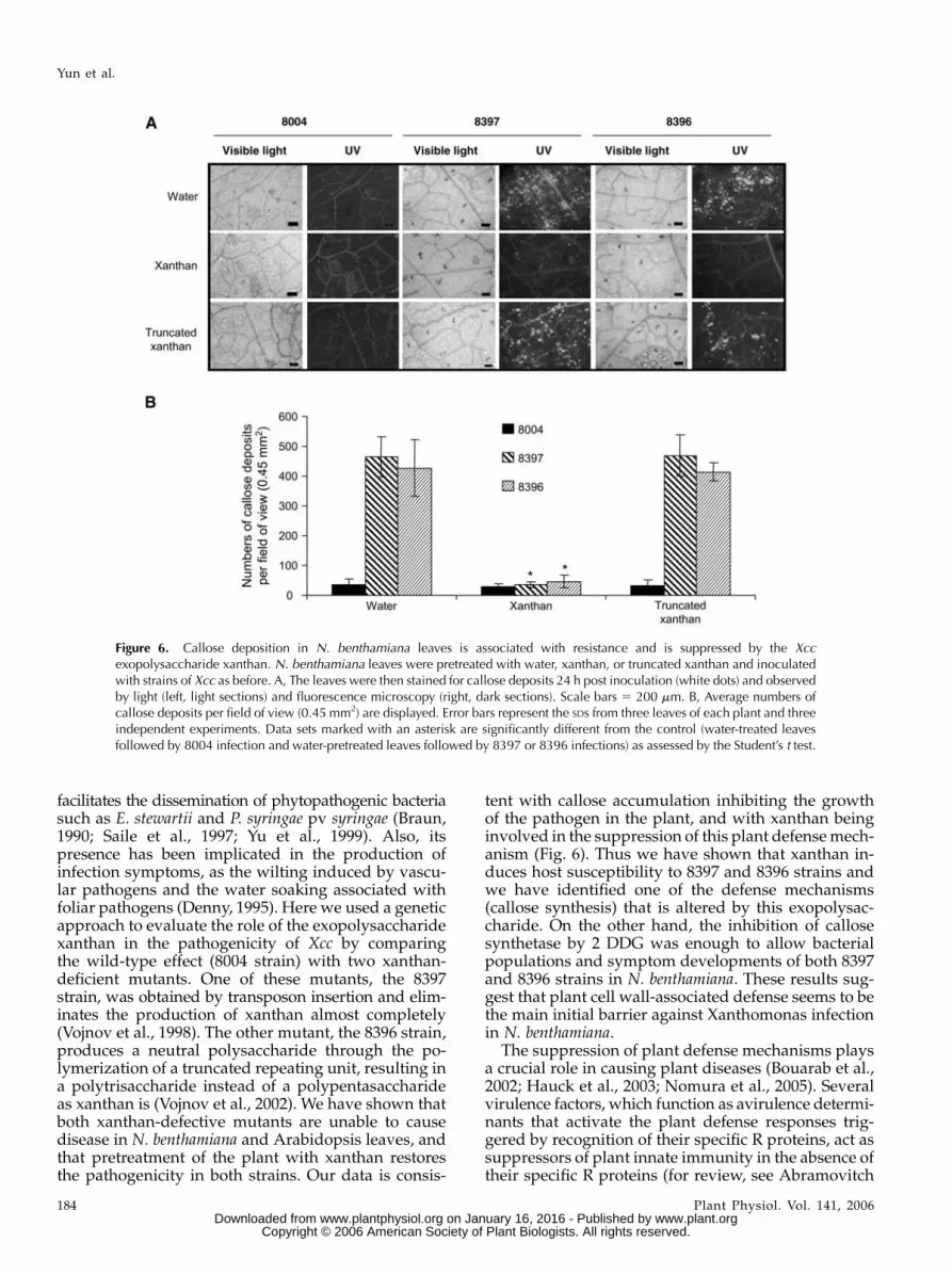
were inoculated with Xcc strains 8004, 8397, and 8396and callose deposition was monitored. Twenty-fourhours after infection, the inoculated leaves were stainedfor callose with aniline blue and cytological observa-tions were performed at the sites of infection withUV-fluorescence microscopy. Callose depositions canbe identified as bright-green points in leaves or veins(Fig. 6). The callose depositions were quantified withIMAGE PRO PLUS software (Media Cybernetics). Morethan 10 adjacent fields of view along the length of theleaf were analyzed and averaged. The values providedare the average and SDs of more than four independentleaves for each replicate. The leaves inoculated witheither 8397 or 8396 strains had a higher level (approx-imately 10-fold) of defense-associated callose deposi-tion in the host cell wall than the leaves inoculatedwith Xcc 8004 strain (Fig. 6, A and B). The same resultswere obtained in Arabidopsis (data not shown). Thus,reduced virulence associated with the lack of xanthanor the modification of its structure correlates with asevere diminution of the callose deposition.
To investigate if xanthan is the molecule responsibleof the inhibition of callose accumulation after infection,
Figure 4. The susceptibility to Xcc induced byxanthan is dose dependent. A, Leaves of 4-week-oldplants were preinfiltrated with either different con-centrations of Xcc xanthan or water and 24 h laterpretreated leaves were inoculated with the Xcc mu-tant strain 8397 (107 cfu/mL). B, Immediate estab-lishment of susceptibility to Xcc 8397 strain by thexanthan suppressor. Leaves were preinfiltrated withXcc xanthan (150 mg/mL) 0 and 24 h before inocu-lation (0 and 24), or water 24 h before inoculation(water) and then pretreated leaves were infected withthe Xcc mutant strain 8397 as before. Numbers ofbacteria were assessed immediately upon infectionand 4 d later. The mean and SD of three separatemeasurements of bacterial numbers are given. Datasets marked with an asterisk are significantly differentfrom control (water-pretreated leaves) as assessed bythe Student’s t test: *P , 0.001.
Yun et al.
182 Plant Physiol. Vol. 141, 2006 www.plant.org on January 16, 2016 - Published by www.plantphysiol.orgDownloaded from
Copyright © 2006 American Society of Plant Biologists. All rights reserved.

leaves of N. benthamiana were pretreated with xanthanor water (control) 24 h before inoculation with the Xcc8397 or 8396 strains. Control leaves showed an accu-mulation of callose deposition while the leaves pre-treated with xanthan failed to accumulate callose inresponse to both mutant strains (Fig. 6, A and B).Remarkably the truncated xanthan was not able tosuppress the callose deposition (Fig. 6, A and B). Thesame results were obtained in Arabidopsis (data notshown). This suggested that xanthan induces suscep-tibility of N. benthamiana to Xcc by suppressing theaccumulation of callose deposition. This suppressioneffect is dependent on the structure of the xanthan.
Inhibition of Callose Deposition by 2-Deoxy-D-Glc
Allows the Xanthan-Deficient Mutants toGrow in N. benthamiana
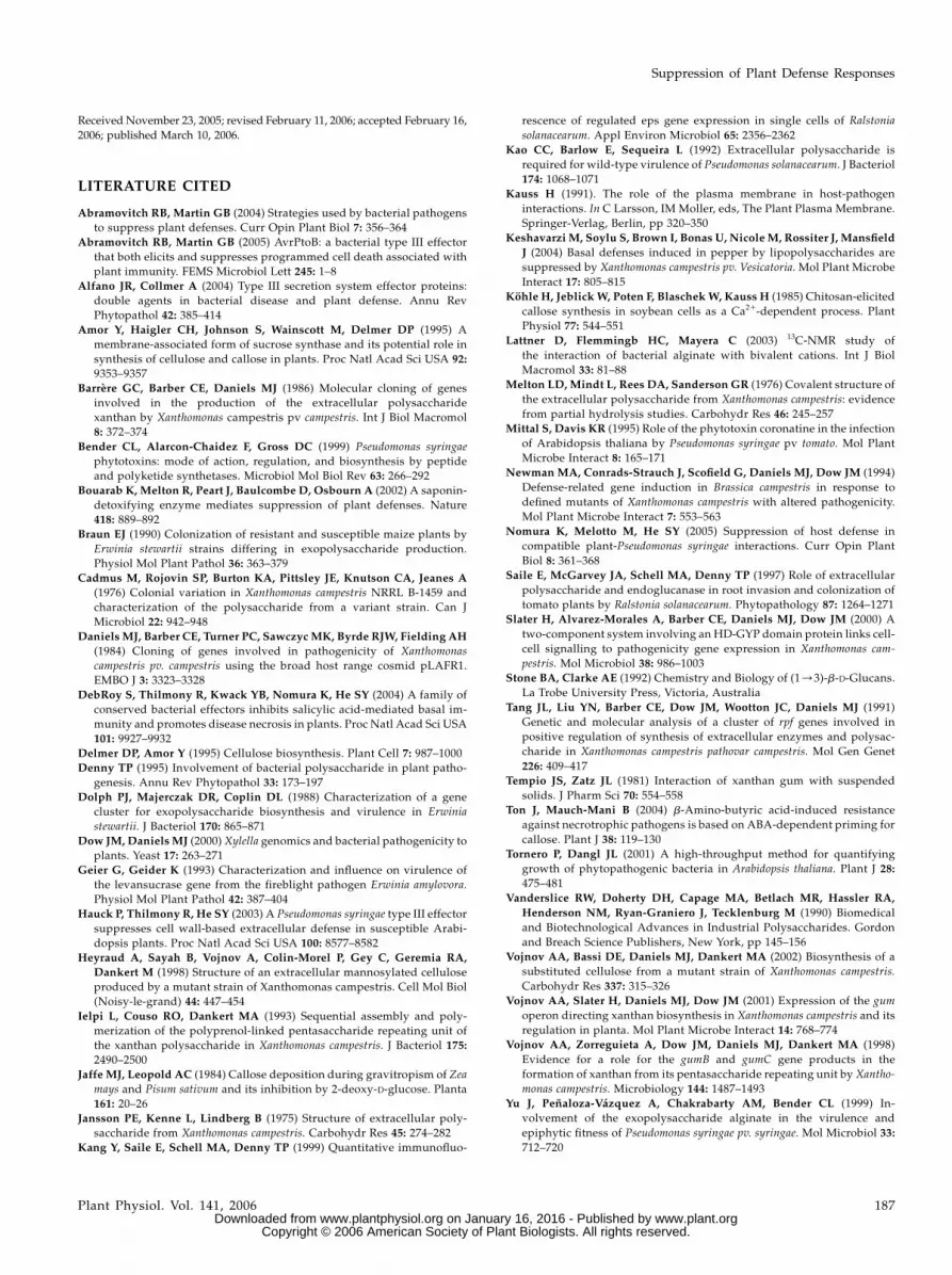
To examine whether the callose defense responseobserved with the nonvirulent Xcc strains was relevantto the plant resistance against the pathogen, N. ben-thamiana infection experiments were performed 24 hafter the administration of 2-deoxy-D-Glc (2 DDG), acallose synthesis inhibitor (Jaffe and Leopold, 1984).Callose deposition was evaluated 48 h postinoculationas before. As expected, no callose deposition was ob-served after 2 DDG treated leaves (Fig. 7, B and C).Interestingly, when leaves were pretreated with 250 mM,
but not 25 mM, of this inhibitor, both Xcc mutant strains8397 and 8396 were unable to induce the callosedeposition response and recovered the ability to causeinfection, represented by the appearance of foliarsymptoms as well as by the increase in bacterialpopulations of both strains (Fig. 7, A and D). The 2DDG inhibitor solution did not produce any symp-toms or foliar changes when infiltrated alone (data notshown). The experiment has also been done using Glc(25 and 250 mM) as a control, and the same result as forwater pretreated leaves was obtained for both concen-trations (data not shown). The controls with the Xcc8004 strain (wild type) were done for each experimentdescribed in Figure 7. As shown in Figure 6, the wild-type bacteria did not induce callose deposition and noeffect was observed when pretreatment was done with2 DGG (data not shown). The data presented hereshows direct correlation between the absence of callosedeposition and the presence of infection symptoms,suggesting a major role of this mechanism in thedefense response to Xcc.
DISCUSSION
Exopolysaccharides have been implicated in plant-pathogen interaction as important virulence factors. Sev-eral results reported that exopolysaccharides production
Figure 5. The structure of xanthan is essential for its effect in N. benthamiana and Arabidopsis. Symptoms in N. benthamianaleaves (A) and Arabidopsis plants (B) treated with the truncated xanthan (150 mg/mL) produced by 8396 mutant strain, andinoculated with 8004, 8397, or 8396 strains 24 h after treatment as before. Photos of disease symptoms were taken 8 dpi.Bacterial populations were accounted at 0 and 4 dpi ofN. benthamiana (C) or Arabidopsis (D). The mean and SD of three separatemeasurements of bacterial numbers are given.
Suppression of Plant Defense Responses
Plant Physiol. Vol. 141, 2006 183 www.plant.org on January 16, 2016 - Published by www.plantphysiol.orgDownloaded from
Copyright © 2006 American Society of Plant Biologists. All rights reserved.
facilitates the dissemination of phytopathogenic bacteriasuch as E. stewartii and P. syringae pv syringae (Braun,1990; Saile et al., 1997; Yu et al., 1999). Also, itspresence has been implicated in the production ofinfection symptoms, as the wilting induced by vascu-lar pathogens and the water soaking associated withfoliar pathogens (Denny, 1995). Here we used a geneticapproach to evaluate the role of the exopolysaccharidexanthan in the pathogenicity of Xcc by comparingthe wild-type effect (8004 strain) with two xanthan-deficient mutants. One of these mutants, the 8397strain, was obtained by transposon insertion and elim-inates the production of xanthan almost completely(Vojnov et al., 1998). The other mutant, the 8396 strain,produces a neutral polysaccharide through the po-lymerization of a truncated repeating unit, resulting ina polytrisaccharide instead of a polypentasaccharideas xanthan is (Vojnov et al., 2002). We have shown thatboth xanthan-defective mutants are unable to causedisease in N. benthamiana and Arabidopsis leaves, andthat pretreatment of the plant with xanthan restoresthe pathogenicity in both strains. Our data is consis-
tent with callose accumulation inhibiting the growthof the pathogen in the plant, and with xanthan beinginvolved in the suppression of this plant defense mech-anism (Fig. 6). Thus we have shown that xanthan in-duces host susceptibility to 8397 and 8396 strains andwe have identified one of the defense mechanisms(callose synthesis) that is altered by this exopolysac-charide. On the other hand, the inhibition of callosesynthetase by 2 DDG was enough to allow bacterialpopulations and symptom developments of both 8397and 8396 strains in N. benthamiana. These results sug-gest that plant cell wall-associated defense seems to bethe main initial barrier against Xanthomonas infectionin N. benthamiana.
The suppression of plant defense mechanisms playsa crucial role in causing plant diseases (Bouarab et al.,2002; Hauck et al., 2003; Nomura et al., 2005). Severalvirulence factors, which function as avirulence determi-nants that activate the plant defense responses trig-gered by recognition of their specific R proteins, act assuppressors of plant innate immunity in the absence oftheir specific R proteins (for review, see Abramovitch
Figure 6. Callose deposition in N. benthamiana leaves is associated with resistance and is suppressed by the Xccexopolysaccharide xanthan. N. benthamiana leaves were pretreated with water, xanthan, or truncated xanthan and inoculatedwith strains of Xcc as before. A, The leaves were then stained for callose deposits 24 h post inoculation (white dots) and observedby light (left, light sections) and fluorescence microscopy (right, dark sections). Scale bars 5 200 mm. B, Average numbers ofcallose deposits per field of view (0.45 mm2) are displayed. Error bars represent the SDs from three leaves of each plant and threeindependent experiments. Data sets marked with an asterisk are significantly different from the control (water-treated leavesfollowed by 8004 infection and water-pretreated leaves followed by 8397 or 8396 infections) as assessed by the Student’s t test.
Yun et al.
184 Plant Physiol. Vol. 141, 2006 www.plant.org on January 16, 2016 - Published by www.plantphysiol.orgDownloaded from
Copyright © 2006 American Society of Plant Biologists. All rights reserved.

Figure 7. Inhibition of callose synthetase by 2DDG restores mutant strain symptoms and bac-terial populations. A, Disease symptoms on N.benthamiana leaves preinfiltrated with water, 25or 250 mM of 2 DDG, and subsequently infectedwithXccmutant strains 8397 or 8396 (107 cfu/mL).Photos of disease symptoms were taken 8 dpi. B,Microscopy pictures of callose deposition fromleaves treated with 2 DDG followed by infectionwith 8397 and 8396 strains as in A. C, Averagenumbers of callose deposits per field of view (0.45mm2) are displayed and data with asterisk aresignificantly different from the control (water-pretreated leaves followed by 8397 or 8396 in-fections) as assessed by the Student’s t test. D, Forevery treatment bacterial populations were cal-culated at days 0 and 4, after inoculation asbefore. Error bars represent the SDs and data setsmarked with an asterisk are significantly differentfrom the control (water-pretreated leaves fol-lowed by 8397 or 8396 infections) as assessedby the Student’s t test.
Suppression of Plant Defense Responses
Plant Physiol. Vol. 141, 2006 185 www.plant.org on January 16, 2016 - Published by www.plantphysiol.orgDownloaded from
Copyright © 2006 American Society of Plant Biologists. All rights reserved.

and Martin, 2004, 2005; Alfano and Collmer, 2004;Nomura et al., 2005). It has recently been shown thatthe P. syringae type III secretion system down-regulatesthe expression of a set of Arabidopsis genes encodingputatively secreted cell wall and defense proteins in asalicylic acid-independent manner (Hauck et al., 2003).Transgenic expression of the avirulence gene AvrPtorepresses a similar set of host genes in susceptible Arabi-dopsis plants, compromises defense-related callosedeposition in the host cell wall, and permits substan-tial multiplication of a bacterial hrp (hypersensitiveresponse and pathogenicity) mutant (Hauck et al.,2003). In addition, DebRoy and collaborators (2004)identified a key group of conserved type III effectors inplant-pathogenic bacteria that target salicylic acid-dependent basal immunity, including callose deposi-tion, and promote disease necrosis in plants. In thisstudy, we have shown that xanthan production in-duces plant disease by suppressing callose deposition.We have also demonstrated that the truncated xanthanfails to suppress this defense mechanism and thereforethe resistance of N. benthamiana to 8397 and 8396strains. This result suggests that the presence of thenegatively charged GlcUA and ketal-pyruvate res-idues in the xanthan might be essential for its biolog-ical function during bacterial-plant interaction.
The polysaccharide pair mannuronate-guluronatewas previously shown to be the preferred binding sitefor bivalent calcium ions in the bacterial alginate(Lattner et al., 2003). Moreover, xanthan, through itsnegatively charged GlcUA and ketal-pyruvate residues,is adsorbed significantly by magnesium carbonate, alu-minum hydroxide, zinc oxide, and calcium carbonate,demonstrating its capacity to interact with suspendedsolids (Tempio and Zatz, 1981). Callose deposition hasbeen considered to reflect local changes of the plant cellmembrane, which leads to an increased Ca21 concen-tration, activating membrane-bound callose synthase(Kauss, 1991; Amor et al., 1995; Delmer and Amor, 1995).In addition, local increase in Ca21 ions could directlyactivate the callose synthase enzyme and initiate calloseformation (Kohle et al., 1985). This suggests that onemechanism by which xanthan could act to suppress cellwall-based plant defense would be the binding of extra-cellular calcium ions, through its negative charge, whichconsequently interferes with signal transduction linkedto callose synthetase activation.
In conclusion, we show in this article that callose isrequired for resistance to Xcc and xanthan inducessusceptibility to Xcc in N. benthamiana and Arabidopsisby suppressing the callose deposition. This suppres-sion effect depends on the chemical structure of theexopolysaccharide. Our data present an importantconceptual stride forward in understanding the roleof exopolysaccharides produced by pathogens in plantdisease establishment. An exciting future challengewill be the biochemical and genetic elucidation of thissuppression effect, which may have implications forour understanding of Xcc pathogenesis and to developinnovative disease control methods.
MATERIALS AND METHODS
Bacterial Strains and Culture Conditions
Xcc strains 8004 (wild type), 8397 (gum::Tn5lac; EPS minus), and 8396
(gum::Tn5lac; truncated EPS ) were described previously (Cadmus et al., 1976;
Daniels et al., 1984; Tang et al., 1991; Slater et al., 2000) and characterized
(Vojnov et al., 1998, 2002). Xcc strains were cultured in a 28�C shaker in PYM
medium as reported by Cadmus and collaborators (1976). Bacterial popula-
tions were measured in a U-3200 spectrophotometer (Hitachi Instruments) or
an Ultrospec 1000 Pharmacia Biotech UV/visible spectrophotometer, at 600 nm.
The antibiotics kanamycin (50 mg/mL) and rifampicin (25 mg/mL) were
added when appropriate.
Xanthan Preparations
Xanthan or truncated xanthan were purified from culture of Xcc as pre-
viously described (Vojnov et al., 1998).
Plant Material
Nicotiana benthamiana and Arabidopsis (Arabidopsis thaliana; Columbia
ecotype) seeds were germinated on 0.8% agar. Two-leaved seedlings were
then grown on soil in growth chamber at 22�C with 70% relative humidity and
a 12-h light/12-h dark cycle.
N. benthamiana Inoculations
All plant inoculations involved a minimum of three leaves from each of
three plants, and each experiment was carried out at least three times. Leaves of
30-d-old plants were inoculated, by infiltration, with Xcc strains (107 cfu/mL in
water) or water only as previously described (Newman et al., 1994). For the
infiltration experiments leaves were preinfiltrated with either water, purified
xanthan, or truncated xanthan (150 mg/mL), and then inoculated with Xcc
8004, 8397, or 8396 bacterial suspensions 24 h later. Inoculation was performed
according to published methods (Newman et al., 1994). Bacteria were hand
infiltrated into plant leaves at the abaxial surface by using a 1-mL syringe
without needle. After inoculation, observations were performed every 6 h,
assessing the foliar symptoms and taking samples for different assays. When
necessary, treatment with 2-deoxy-D-Glc (Sigma-Aldrich) was carried out 24 h
before inoculations, in concentrations of 25 or 250 mM as used previously (Ton
and Mauch-Mani, 2004). To assess bacterial development in planta, six 0.6 cm2
discs from each leaf were taken at 0, 1, 2, 3, and 4 d postinoculation (dpi) and
bacterial populations monitored as described (Newman et al., 1994).
Arabidopsis Inoculations
Plant infections were performed as previously described (Tornero and
Dangl, 2001) but adaptations were made for the infection with Xcc. Bacterial
suspensions (OD600 5 0.05) were prepared from 24 h/28�C PYM-liquid Xcc
cultures (Cadmus et al., 1976) with 10 mM MgCl2, and Silwet L-77 was added
(200 mL/L). Plant pots (nine seedlings each) were submerged upside down in
the bacterial solution for 30 s and then covered with a transparent lid. Forty-
eight hours after inoculation the lid was removed and samples were taken
(days 2, 3, and 4). Bacterial populations were measured as previously
described (Tornero and Dangl, 2001).
Callose Deposition Assay
Callose staining was performed 24 h after bacterial inoculation as described
previously (Hauck et al., 2003). Leaves were examined with a Zeiss Axiophot
D-7082 photomicroscope with an A3 fluorescence cube. The callose depositions
were quantified with IMAGE PRO PLUS software (Media Cybernetics).
ACKNOWLEDGMENTS
We thank our lab colleagues for fruitful discussions and for critical
reading of the manuscript. Atilio Castagnaro, Maria Rosa Marano, Marcelo
Dankert, and Adrian Vojnov are members of the Career Investigator of the
Consejo Nacional de Investigaciones Cientıficas y Tecnicas.
Yun et al.
186 Plant Physiol. Vol. 141, 2006 www.plant.org on January 16, 2016 - Published by www.plantphysiol.orgDownloaded from
Copyright © 2006 American Society of Plant Biologists. All rights reserved.
Received November 23, 2005; revised February 11, 2006; accepted February 16,
2006; published March 10, 2006.
LITERATURE CITED
Abramovitch RB, Martin GB (2004) Strategies used by bacterial pathogens
to suppress plant defenses. Curr Opin Plant Biol 7: 356–364
Abramovitch RB, Martin GB (2005) AvrPtoB: a bacterial type III effector
that both elicits and suppresses programmed cell death associated with
plant immunity. FEMS Microbiol Lett 245: 1–8
Alfano JR, Collmer A (2004) Type III secretion system effector proteins:
double agents in bacterial disease and plant defense. Annu Rev
Phytopathol 42: 385–414
Amor Y, Haigler CH, Johnson S, Wainscott M, Delmer DP (1995) A
membrane-associated form of sucrose synthase and its potential role in
synthesis of cellulose and callose in plants. Proc Natl Acad Sci USA 92:
9353–9357
Barrere GC, Barber CE, Daniels MJ (1986) Molecular cloning of genes
involved in the production of the extracellular polysaccharide
xanthan by Xanthomonas campestris pv campestris. Int J Biol Macromol
8: 372–374
Bender CL, Alarcon-Chaidez F, Gross DC (1999) Pseudomonas syringae
phytotoxins: mode of action, regulation, and biosynthesis by peptide
and polyketide synthetases. Microbiol Mol Biol Rev 63: 266–292
Bouarab K, Melton R, Peart J, Baulcombe D, Osbourn A (2002) A saponin-
detoxifying enzyme mediates suppression of plant defenses. Nature
418: 889–892
Braun EJ (1990) Colonization of resistant and susceptible maize plants by
Erwinia stewartii strains differing in exopolysaccharide production.
Physiol Mol Plant Pathol 36: 363–379
Cadmus M, Rojovin SP, Burton KA, Pittsley JE, Knutson CA, Jeanes A
(1976) Colonial variation in Xanthomonas campestris NRRL B-1459 and
characterization of the polysaccharide from a variant strain. Can J
Microbiol 22: 942–948
Daniels MJ, Barber CE, Turner PC, Sawczyc MK, Byrde RJW, Fielding AH
(1984) Cloning of genes involved in pathogenicity of Xanthomonas
campestris pv. campestris using the broad host range cosmid pLAFR1.
EMBO J 3: 3323–3328
DebRoy S, Thilmony R, Kwack YB, Nomura K, He SY (2004) A family of
conserved bacterial effectors inhibits salicylic acid-mediated basal im-
munity and promotes disease necrosis in plants. Proc Natl Acad Sci USA
101: 9927–9932
Delmer DP, Amor Y (1995) Cellulose biosynthesis. Plant Cell 7: 987–1000
Denny TP (1995) Involvement of bacterial polysaccharide in plant patho-
genesis. Annu Rev Phytopathol 33: 173–197
Dolph PJ, Majerczak DR, Coplin DL (1988) Characterization of a gene
cluster for exopolysaccharide biosynthesis and virulence in Erwinia
stewartii. J Bacteriol 170: 865–871
Dow JM, Daniels MJ (2000) Xylella genomics and bacterial pathogenicity to
plants. Yeast 17: 263–271
Geier G, Geider K (1993) Characterization and influence on virulence of
the levansucrase gene from the fireblight pathogen Erwinia amylovora.
Physiol Mol Plant Pathol 42: 387–404
Hauck P, Thilmony R, He SY (2003) A Pseudomonas syringae type III effector
suppresses cell wall-based extracellular defense in susceptible Arabi-
dopsis plants. Proc Natl Acad Sci USA 100: 8577–8582
Heyraud A, Sayah B, Vojnov A, Colin-Morel P, Gey C, Geremia RA,
Dankert M (1998) Structure of an extracellular mannosylated cellulose
produced by a mutant strain of Xanthomonas campestris. Cell Mol Biol
(Noisy-le-grand) 44: 447–454
Ielpi L, Couso RO, Dankert MA (1993) Sequential assembly and poly-
merization of the polyprenol-linked pentasaccharide repeating unit of
the xanthan polysaccharide in Xanthomonas campestris. J Bacteriol 175:
2490–2500
Jaffe MJ, Leopold AC (1984) Callose deposition during gravitropism of Zea
mays and Pisum sativum and its inhibition by 2-deoxy-D-glucose. Planta
161: 20–26
Jansson PE, Kenne L, Lindberg B (1975) Structure of extracellular poly-
saccharide from Xanthomonas campestris. Carbohydr Res 45: 274–282
Kang Y, Saile E, Schell MA, Denny TP (1999) Quantitative immunofluo-
rescence of regulated eps gene expression in single cells of Ralstonia
solanacearum. Appl Environ Microbiol 65: 2356–2362
Kao CC, Barlow E, Sequeira L (1992) Extracellular polysaccharide is
required for wild-type virulence of Pseudomonas solanacearum. J Bacteriol
174: 1068–1071
Kauss H (1991). The role of the plasma membrane in host-pathogen
interactions. In C Larsson, IM Moller, eds, The Plant Plasma Membrane.
Springer-Verlag, Berlin, pp 320–350
Keshavarzi M, Soylu S, Brown I, Bonas U, Nicole M, Rossiter J, Mansfield
J (2004) Basal defenses induced in pepper by lipopolysaccharides are
suppressed by Xanthomonas campestris pv. Vesicatoria. Mol Plant Microbe
Interact 17: 805–815
Kohle H, Jeblick W, Poten F, BlaschekW, Kauss H (1985) Chitosan-elicited
callose synthesis in soybean cells as a Ca21-dependent process. Plant
Physiol 77: 544–551
Lattner D, Flemmingb HC, Mayera C (2003) 13C-NMR study of
the interaction of bacterial alginate with bivalent cations. Int J Biol
Macromol 33: 81–88
Melton LD, Mindt L, Rees DA, Sanderson GR (1976) Covalent structure of
the extracellular polysaccharide from Xanthomonas campestris: evidence
from partial hydrolysis studies. Carbohydr Res 46: 245–257
Mittal S, Davis KR (1995) Role of the phytotoxin coronatine in the infection
of Arabidopsis thaliana by Pseudomonas syringae pv tomato. Mol Plant
Microbe Interact 8: 165–171
Newman MA, Conrads-Strauch J, Scofield G, Daniels MJ, Dow JM (1994)
Defense-related gene induction in Brassica campestris in response to
defined mutants of Xanthomonas campestris with altered pathogenicity.
Mol Plant Microbe Interact 7: 553–563
Nomura K, Melotto M, He SY (2005) Suppression of host defense in
compatible plant-Pseudomonas syringae interactions. Curr Opin Plant
Biol 8: 361–368
Saile E, McGarvey JA, Schell MA, Denny TP (1997) Role of extracellular
polysaccharide and endoglucanase in root invasion and colonization of
tomato plants by Ralstonia solanacearum. Phytopathology 87: 1264–1271
Slater H, Alvarez-Morales A, Barber CE, Daniels MJ, Dow JM (2000) A
two-component system involving an HD-GYP domain protein links cell-
cell signalling to pathogenicity gene expression in Xanthomonas cam-
pestris. Mol Microbiol 38: 986–1003
Stone BA, Clarke AE (1992) Chemistry and Biology of (1/3)-b-D-Glucans.
La Trobe University Press, Victoria, Australia
Tang JL, Liu YN, Barber CE, Dow JM, Wootton JC, Daniels MJ (1991)
Genetic and molecular analysis of a cluster of rpf genes involved in
positive regulation of synthesis of extracellular enzymes and polysac-
charide in Xanthomonas campestris pathovar campestris. Mol Gen Genet
226: 409–417
Tempio JS, Zatz JL (1981) Interaction of xanthan gum with suspended
solids. J Pharm Sci 70: 554–558
Ton J, Mauch-Mani B (2004) b-Amino-butyric acid-induced resistance
against necrotrophic pathogens is based on ABA-dependent priming for
callose. Plant J 38: 119–130
Tornero P, Dangl JL (2001) A high-throughput method for quantifying
growth of phytopathogenic bacteria in Arabidopsis thaliana. Plant J 28:
475–481
Vanderslice RW, Doherty DH, Capage MA, Betlach MR, Hassler RA,
Henderson NM, Ryan-Graniero J, Tecklenburg M (1990) Biomedical
and Biotechnological Advances in Industrial Polysaccharides. Gordon
and Breach Science Publishers, New York, pp 145–156
Vojnov AA, Bassi DE, Daniels MJ, Dankert MA (2002) Biosynthesis of a
substituted cellulose from a mutant strain of Xanthomonas campestris.
Carbohydr Res 337: 315–326
Vojnov AA, Slater H, Daniels MJ, Dow JM (2001) Expression of the gum
operon directing xanthan biosynthesis in Xanthomonas campestris and its
regulation in planta. Mol Plant Microbe Interact 14: 768–774
Vojnov AA, Zorreguieta A, Dow JM, Daniels MJ, Dankert MA (1998)
Evidence for a role for the gumB and gumC gene products in the
formation of xanthan from its pentasaccharide repeating unit by Xantho-
monas campestris. Microbiology 144: 1487–1493
Yu J, Penaloza-Vazquez A, Chakrabarty AM, Bender CL (1999) In-
volvement of the exopolysaccharide alginate in the virulence and
epiphytic fitness of Pseudomonas syringae pv. syringae. Mol Microbiol 33:
712–720
Suppression of Plant Defense Responses
Plant Physiol. Vol. 141, 2006 187 www.plant.org on January 16, 2016 - Published by www.plantphysiol.orgDownloaded from
Copyright © 2006 American Society of Plant Biologists. All rights reserved.


