Embed Size (px)
Citation preview

Introduction Numerous studies have shown that anti-neoplastic drug effects are subject to significant inter-individual variability. Based on an abun-dance of accumulated literature from twin stud-ies, pre-clinical experimental data, clinical trials, biomarker-driven and epidemiologic studies, it is widely accepted that this variability is largely due to inherited genetic variation [1-3]. The aim of pharmacogenetics/genomics is to identify genetic predictors of treatment outcomes that would allow individualization of therapy for
cancer patients. Identification of pharmacogenomic determi-nants is especially needed for cancers with poor treatment outcomes. Acute myeloid leukemia (AML) in adult populations is associated with poor (approximately 20%) overall five-year rela-tive survival (OS) rates. Despite continuing ef-forts to stratify AML patients according to prog-nostic host and tumor characteristics, including crucial cytogenetic and molecular markers, and despite new pharmaceutical agents, outcome remains suboptimal [4]. Overall prognosis for
Int J Mol Epidemiol Genet 2010;1(4):278-294 www.ijmeg.org /IJMEG1005001
Original Article XPD DNA nucleotide excision repair gene polymorphisms associated with DNA repair deficiency predict better treat-ment outcomes in secondary acute myeloid leukemia Nataliya Kuptsova-Clarkson1,2, Christine B. Ambrosone1, Joli Weiss1, Maria R. Baer1,3,5 , Lara E. Sucheston1, Gary Zirpoli1, Kenneth J. Kopecky4, Laurie Ford5, Javier Blanco6, Meir Wetzler5, Kirsten B. Moysich1 1Department of Cancer Prevention and Control and 5Medicine, Roswell Park Cancer Institute, Buffalo, NY, USA; 2Pre-sently at Children’s Memorial Research Center, Children’s Memorial Hospital, Chicago, IL, USA; 3presently at Greene-baum Cancer Center, University of Maryland School of Medicine, Baltimore, MD, USA; 4Southwest Oncology Group Statistical Center, Fred Hutchinson Cancer Research Center, Seattle, WA, USA; 6Department of Pharmaceutical Sci-ences, University at Buffalo, Buffalo, NY, USA. Received May 15, 2010, accepted August 1, 2010, available online August 10, 2010 Abstract: Pharmacogenetic studies in DNA repair pathway have consistently demonstrated correlations between the XRCC1 Arg399Gln, XPD Lys751Gln and XPD Asp312Gln genotypes, previously associated with suboptimal DNA re-pair, and differential cancer treatment outcomes. We evaluated these polymorphisms and XPD haplotypes in adult de novo (n=214) and secondary (n=79) acute myeloid leukemia (AML) patients treated with cytarabine and anthracy-cline chemotherapy. Genotyping was performed by MALDI-TOF mass spectrometry. Logistic and proportional hazards regression models were used to evaluate relationships. Differential responses were observed in secondary, but not de novo, AML. Among secondary AML patients, the odds of achieving complete remission (CR) were higher for the XPD 312Asn/Asn (OR= 11.23; 95% CI, 2.23-56.63) and XPD 751Gln/Gln (OR= 7.07; 95% CI, 1.42-35.18) genotypes. The XPD diplotypes were coded as the combination of two of the following haplotypes: haplotype A=(Lys)751A/(Asp)312G; B=(Gln)751C/(Asn)312A; C=(Lys)751A/(Asn)312A; and D=(Gln)751C/(Asp)312G. The BB diplotype was asso-ciated with CR attainment [OR=18.31; 95% CI: 2.08-283.57] and longer survival [HR=0.31; 95% CI: 0.14-0.73] com-pared to the referent AA diplotype. The XPD 751 CC, 312GA, 312AA genotypes and the XPD DC diplotype were also associated with longer overall survival (OS).Thus, XPD codon 312 and 751 variant genotypes and haplotypes contain-ing at least one variant allele may predict better treatment responses. If validated, these findings could support stratification of chemotherapy in secondary AML. Keywords: Acute Myeloid Leukemia (AML), secondary AML, pharmacogenetics/pharmacogenomics, DNA repair gene polymorphisms

DNA repair genetic variation and AML outcomes
279 Int J Mol Epidemiol Genet 2010;1(4):278-294
patients with secondary AML (sAML), including both AML following an antecedent hematologic disorder (AHD) and therapy-related acute mye-loid leukemia (t-AML), is even worse, with an average life expectancy of eight to ten months from diagnosis [5,6]. Cytosine arabinoside (ARA-C), an antimetabolite, anthracyclines (daunorubicin, idarubicin) and etoposide are the most commonly used drugs in AML treatment. They have different mechanisms of action, in-cluding inhibition of DNA synthesis, formation of irreversible complexes with topoisomerases, generation of free radicals and intracellular oxi-dative damage, leading to accumulation of sin-gle- and double-srand DNA breaks. Among the major DNA repair pathways, base excision repair (BER) is used for minor base alterations and non-bulky DNA adducts, while nucleotide excision repair (NER) is involved in fixing more complex DNA lesions such as bulky DNA adducts. The double-strand break repair (DSB) pathway repairs broken DNA strands by either homologous recombination (HR) or non-homologous end joining (NHEJ) [7,8]. It has been noted previously that acquired resistance to alkylating agents, anthracyclines and epipo-dophyllotoxins may develop as a result of en-hanced DNA repair, NER repair in particular [9]. Overall, more than 130 genes have been asso-ciated with human DNA repair, and numerous studies have investigated associations between single nucleotide polymorphisms (SNPs) in nearly all DNA repair pathways and clinical out-comes [10]. Variant alleles associated with less proficient DNA repair activity may lead to de-creased repair of DNA damage in malignant cells, and thereby improve response to chemo-therapy. However, compromised repair activity may also lead to accumulation of DNA damage in normal cells, leading to more profound treat-ment-related toxicities in normal tissues, and may predispose towards secondary cancers. Germline variations in DNA repair activity of genes including those encoding the X-ray cross-complementing group 1 (XRCC1) protein in the BER pathway and the XPD protein in the NER pathway, may have a significant impact on AML patients’ response to treatment, including their induction chemotherapy outcomes, toxicity pro-files and survival [10-13]. The XRCC1 codon 399Gln homozygous variant genotype has been found to decrease risk of both de novo and
t-AML, and has been associated with both poor and favorable survival outcomes for other can-cers [14-17]. The variant XPD allele Lys751Gln SNP was associated with increased risk of ther-apy-related AML (t-AML) as well as shorter dis-ease-free survival (DFS) and OS for AML pa-tients [18]. Additionally, AML chemotherapy is associated with a wide range of toxicities, the most serious of which are hematologic (myelo-suppression), hematologic (myelosuppression), gastrointestinal, infectious, dermatologic [9,19], and there are few data on impact of DNA repair gene polymorphisms on treatment-related tox-icities [20-23]. We previously reported on associations between AML and variant XPD genotypes in a cohort of elderly AML patients in a Southwest Oncology Group (SWOG) clinical trial. Those with the XPD 751Gln/312Asp haplotype had significantly better odds of complete remission (CR), as well as significantly reduced risk of resistant disease [20]. Here we evaluated relationships between functional SNPs in XRCC1 Arg399Gln, ERCC2/(XPD) Lys751Gln and ERCC2/ (XPD) Asp312Asn and patients’ responses to induction chemo-therapy, chemotherapy-induced toxicities and OS in adult AML patients treated at Roswell Park Cancer Institute (RPCI). The latter patient population was larger and included a larger number of sAML patients, and thus therapeutic outcomes in sAML were investigated in this work. Materials and methods Patients The study population included 293 adult pa-tients with de novo AML or sAML with all French-American-British subtypes (FAB) other than M3 or acute promyelocytic leukemia who were treated with ARA-C and anthracycline-based chemotherapy at RPCI between 1994 and 2006 and had cryopreserved vials of pre-treatment marrow or peripheral blood cells available in the RPCI Leukemia Tissue Bank. Among 79 (27%) patients with sAML, 57 had an AHD, 32 had t-AML, and 10 had both. Patients were predomi-nantly Caucasian (90%). For remission induc-tion, 45% received standard-dose ARA-C, daun-orubicin and etoposide, 23% received high-dose ARA-C and idarubicin, and 32% were enrolled in clinical trials and received experimental agents, including BCL-2 inhibitor, P-glycoprotein (P-gp)
DNA repair genetic variation and AML outcomes
280 Int J Mol Epidemiol Genet 2010;1(4):278-294
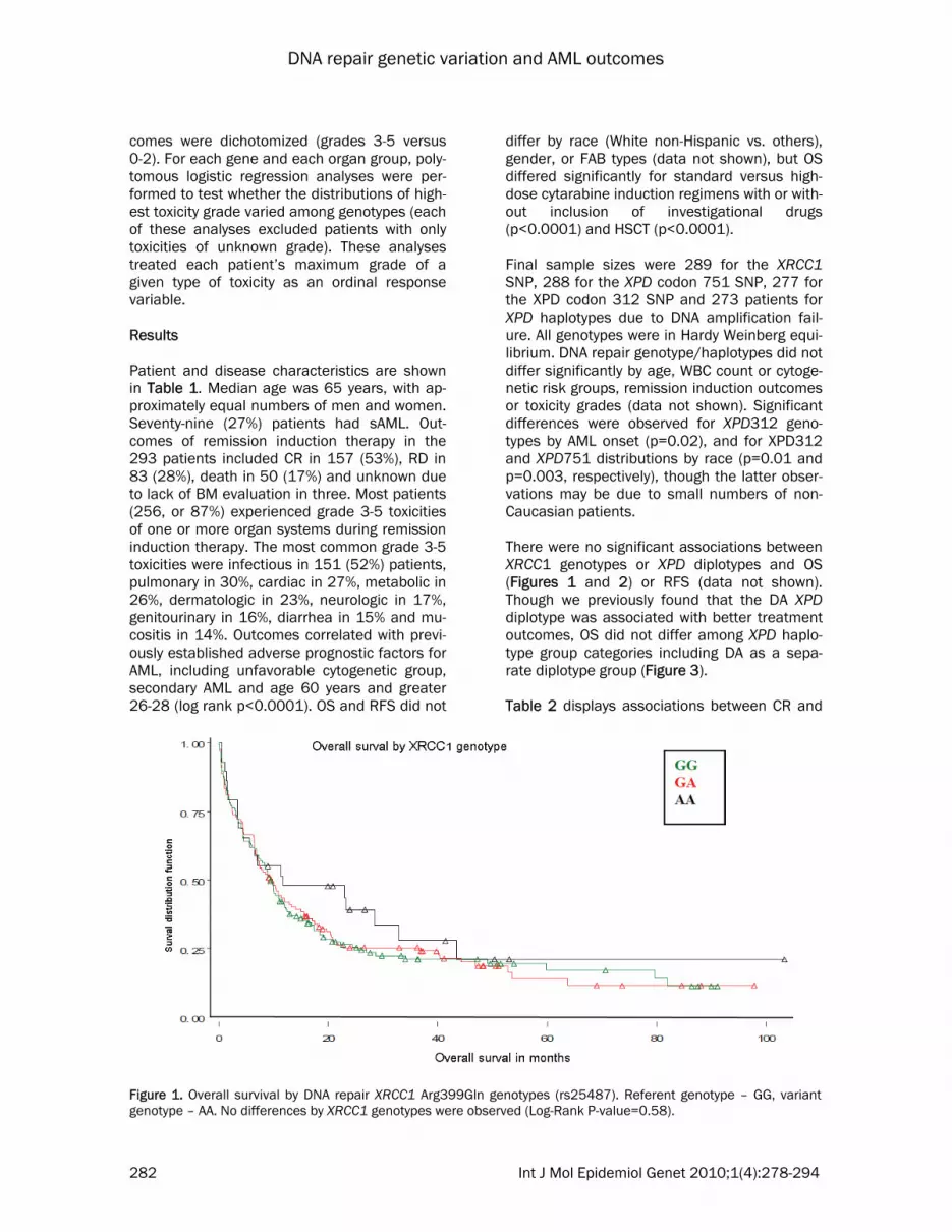
modulators or arsenic trioxide in addition to ARA-C and an anthracycline. Sixty-six patients (23%) underwent hematopoietic stem cell transplanta-tion (HSCT), either allogeneic (n=41) or autolo-gous (n=25), as part of their AML therapy. Clini-cal information was obtained from the RPCI Leu-kemia Section Database and from patients’ medical records. The study was approved by the RPCI Institutional Review Board. Data collection and analysis Data on patient and disease characteristics, toxicities of induction chemotherapy, and treat-ment outcomes were collected and evaluated according to standard procedures. De novo AML and sAML patient subgroups were defined by absence or presence of AHD prior to AML diag-nosis or t-AML. Complete remission (CR) was defined by normal peripheral blood cell counts, including absolute neutrophil count of 1x10⁹/L or more, platelet count of 100x109/L or more, and less than 5% blasts in the peripheral blood or bone marrow (BM). Relapse was defined as reappearance of more than 5 % leukemic blasts in the bone marrow not attributable to any other cause. Overall Survival (OS) was measured from the date of treatment until death from any cause, with observation censored on the date the patient was last known to be alive. Relapse-free survival (RFS) was measured from the date CR was achieved until the date of the first re-lapse of AML or death from any cause, with ob-servation censored on the date of last contact for patients without report of relapse. Clinical toxicity data during remission induction chemo-therapy were abstracted from medical records. Severity of toxicities was graded 1 (mild) through 5 (fatal) according to the NCI Common Toxicity Criteria, version 3.0 (http://ctep.cancer.gov/protocolDevelopment/electro-nic_applications/docs/ctcaev3.pdf). For the analyses described here, induction chemother-apy toxicities were combined into the following categories: genitourinary, gastrointestinal (including but not limited to: nausea/vomiting, diarrhea, oral mucositis, esophagitis), hepatic, pulmonary, cardiovascular, hemorrhage, derma-tologic, neurologic, metabolic, hypersensitivity reactions and infectious complications. For each patient, the grade of toxicity assigned to an organ group was the maximum grade of all the specific toxicities within that group. Genotyping
DNA was extracted from cryopreserved bone marrow samples using Gentra PureGene DNA extraction kits (Minneapolis, MN) and geno-typed for SNPs in XRCC1 (rs25487) and XPD/ERCC2 (codon 751, rs13181 and codon 312, rs1799793) by matrix-assisted laser desorp-tion/ionization time of flight mass spectrometry (MALDI-TOF-MS) [24]. Polymerase chain reac-tion amplification was performed using SNP-specific primers (XPD312: F: 5’- ACGTTGGAT-GACGACGC CCACCTGGCCAA - 3’ and R: 5’ – CGTTGGATGGG AGGCGGGAAAGGGACT – 3’; for XPD751: F: 5’- ACGTTGGATGAGCAGCTAGAATCA-GAGGAG - 3’ and R: 5’- ACGTTGGATGCACCAG-GAACCGTTTATG GC -3’ and for XRCC1: F: 5’- ACGTTGGAT GCAGGATAAGGAGCAGGGTTG - 3’ and R: 5’- ACG TTGGATGTAAGAGTGGGTGCTGG ACT -3’). Duplicate aliquots for approximately 10% of the samples were randomly distributed throughout the plates for quality control pur-poses. Controls for genotype and two ‘no tem-plate’ controls were also included on each plate. All genotyping results were reviewed manually for quality control. Statistical analyses Chi-Square test statistics with 1 degree of free-dom were used to test for deviation from Hardy-Weinberg equilibrium for each polymorphism. The Estimation Haplotype (EH) genetic linkage utility program was used to evaluate possible linkage disequilibrium (LD) for the XPD SNPs located on chromosome 19q13.2-13.3. XPD haplotypes were coded as previously reported 20: haplotype A=(Lys)751A/(Asp)312G; B=(Gln)751C/(Asn)312A; C=(Lys)751A/(Asn)312A; and D=(Gln)751C/(Asp)312G. Data analysis was performed using SAS 9.1 software (SAS Insti-tute, Cary, NC). Categorical distributions of pa-tient, disease and outcome characteristics (age, sex, race, de novo vs. secondary AML onset, FAB type, WBC (white blood cell) count, treat-ment outcomes, toxicity grades) were compared among genotypes and haplotypes using Chi-square analysis, and Fisher’s exact tests when appropriate, with calculation of Monte-Carlo estimates for the exact p-vlaues across nine haplotype groups. Distributions of OS and RFS were estimated by Kaplan-Meier (KM) method and compared be-tween genotypes/haplotypes by log-rank tests. Crude and adjusted unconditional logistic re-gression (LR) models were run for the assess-ment of relationships between CR, resistant

DNA repair genetic variation and AML outcomes
281 Int J Mol Epidemiol Genet 2010;1(4):278-294
disease (RD) and genotype/haplotypes. Odds ratios (OR) and their 95% confidence intervals (CI) for the associations between DNA repair genotypes/haplotypes and CR were adjusted for age and WBC count (continuous), as well as cytogenetic group and HSCT treatment. The prognostic cytogenetic groups were determined as favorable, intermediate, unfavorable as de-scribed by Byrd J. et al. from Cancer and Leuke-mia Group B results [25]; a few patients with not known karyotype belonged to the ‘unknown’ cytogenetic prognostic group. The OR and their 95% CI for the associations between DNA re-pair genoypes/haplotypes and CR were addi-tionally adjusted for the type of induction che-motherapy when RD outcome was modeled. Hazard ratios (HR) and their 95% confidence intervals (CI) for the associations between DNA repair genotypes/haplotypes and OS were ad-justed for age and WBC count (continuous), as well as cytogenetic group and HSCT in the Cox
proportional hazards regression analysis. Analy-ses of treatment outcomes (CR, RD, OS) were stratified by de novo vs. secondary AML, except that statistical analysis between ungrouped XPD diplotypes and treatment outcomes was not stratified by AML onset due to small numbers. In order to make our current findings compara-ble to the ones from the previous study, in the logistic regression and Cox proportional hazards models the grouping of XPD haplotypes into diplotype groups was performed in the same manner [20]. We also grouped the haplotypes by the number of variant alleles in order to ob-serve any variant allele dose response relation-ship and stratified the analysis by AML onset. Patients who underwent HSCT (n=41) were cen-sored at the time of HSCT for all RFS analyses. Unconditional and polytomous logistic regres-sion analysis of toxicities in relation to geno-type/haplotype group was performed. For the unconditional regression analysis, toxicity out-
Table 1. Summary of selected patient and disease characteristics
Characteristic Description No. % Sex Female 135 46 Male 158 54 Race/Ethnicity Asian 3 1 Black 15 5 Hispanic, NOS 6 2 White, non-Hispanic 264 90
Other/unknown 5 2 AML Onset de novo 214 73 Secondary 79 27 Cytogenetic Favorable 20 7 Risk Group Intermediate 149 51 Unfavorable 96 33 Unknown 28 10 FAB Type M0 17 6 M1 65 22 M2 125 43 M4 51 17 M5 13 4 M6 12 4 M7 2 1 Other AML 8 3 Median Min – Max Age, yrs 65 20 - 85 Marrow Blasts (%) 62 0 – 98 WBC (X1000/μL) 19.03 0.43 – 555
DNA repair genetic variation and AML outcomes
282 Int J Mol Epidemiol Genet 2010;1(4):278-294
comes were dichotomized (grades 3-5 versus 0-2). For each gene and each organ group, poly-tomous logistic regression analyses were per-formed to test whether the distributions of high-est toxicity grade varied among genotypes (each of these analyses excluded patients with only toxicities of unknown grade). These analyses treated each patient’s maximum grade of a given type of toxicity as an ordinal response variable. Results Patient and disease characteristics are shown in Table 1. Median age was 65 years, with ap-proximately equal numbers of men and women. Seventy-nine (27%) patients had sAML. Out-comes of remission induction therapy in the 293 patients included CR in 157 (53%), RD in 83 (28%), death in 50 (17%) and unknown due to lack of BM evaluation in three. Most patients (256, or 87%) experienced grade 3-5 toxicities of one or more organ systems during remission induction therapy. The most common grade 3-5 toxicities were infectious in 151 (52%) patients, pulmonary in 30%, cardiac in 27%, metabolic in 26%, dermatologic in 23%, neurologic in 17%, genitourinary in 16%, diarrhea in 15% and mu-cositis in 14%. Outcomes correlated with previ-ously established adverse prognostic factors for AML, including unfavorable cytogenetic group, secondary AML and age 60 years and greater 26-28 (log rank p<0.0001). OS and RFS did not
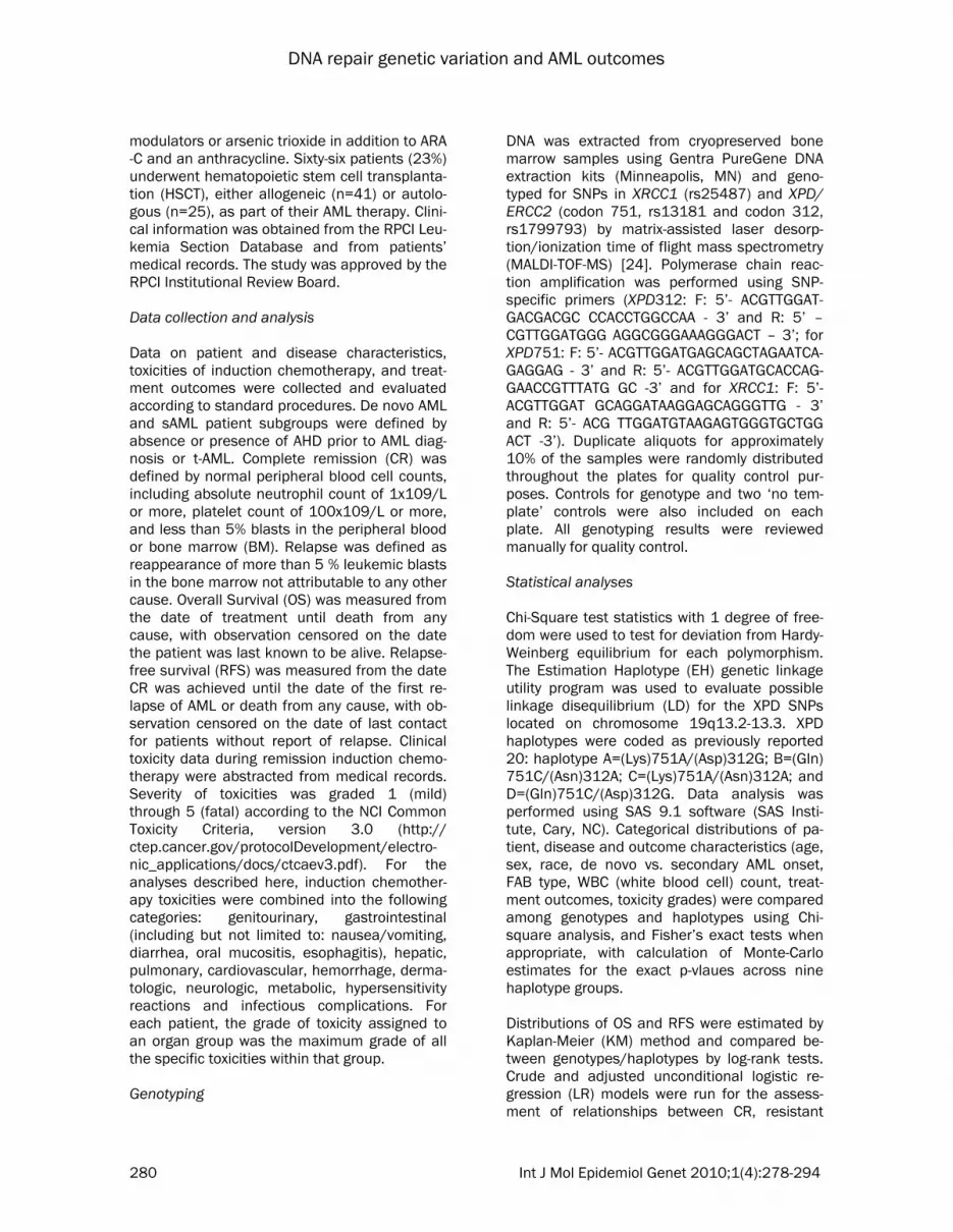
differ by race (White non-Hispanic vs. others), gender, or FAB types (data not shown), but OS differed significantly for standard versus high-dose cytarabine induction regimens with or with-out inclusion of investigational drugs (p<0.0001) and HSCT (p<0.0001). Final sample sizes were 289 for the XRCC1 SNP, 288 for the XPD codon 751 SNP, 277 for the XPD codon 312 SNP and 273 patients for XPD haplotypes due to DNA amplification fail-ure. All genotypes were in Hardy Weinberg equi-librium. DNA repair genotype/haplotypes did not differ significantly by age, WBC count or cytoge-netic risk groups, remission induction outcomes or toxicity grades (data not shown). Significant differences were observed for XPD312 geno-types by AML onset (p=0.02), and for XPD312 and XPD751 distributions by race (p=0.01 and p=0.003, respectively), though the latter obser-vations may be due to small numbers of non-Caucasian patients. There were no significant associations between XRCC1 genotypes or XPD diplotypes and OS (Figures 1 and 2) or RFS (data not shown). Though we previously found that the DA XPD diplotype was associated with better treatment outcomes, OS did not differ among XPD haplo-type group categories including DA as a sepa-rate diplotype group (Figure 3). Table 2 displays associations between CR and
Figure 1. Overall survival by DNA repair XRCC1 Arg399Gln genotypes (rs25487). Referent genotype – GG, variant genotype – AA. No differences by XRCC1 genotypes were observed (Log-Rank P-value=0.58).

DNA repair genetic variation and AML outcomes
283 Int J Mol Epidemiol Genet 2010;1(4):278-294
Figure 2. Overall survival by ERCC2 Lys751Gln genotypes (rs13181). Referent genotype – AA, variant genotype – CC. No differences by XPD, codon 751, genotypes were observed (Log-Rank P-value=0.93).
Figure 3. Overall survival by ERCC2 Asp312Asn genotypes (rs1799793). Referent genotype – GG, variant genotype – AA. No differences by XPD, codon 312, genotypes were observed (Log-Rank P-value=0.74).

DNA repair genetic variation and AML outcomes
284 Int J Mol Epidemiol Genet 2010;1(4):278-294
OS and XRCC1 and XPD genotypes. Significantly increased odds of CR were associated with one or both variant XPD751 genotypes (OR 2.05; 95% CI, 1.20-3.52 for AC+CC). Patients with sAML and any of the variant genotypes had highly significant greater odds of achieving CR (OR=8.42 [2.08-34.01] for AC+CC). Similar trends were observed for the XPD312 geno-types: in the overall AML RPCI cohort the odds of achieving CR in homozygous variant AA geno-type carriers were 4 times higher than in those with the common GG genotype (OR 4.34, [1.80-10.48], and within the sAML subgroup, odds were 11-fold higher (OR 11.23, 95% CI, 2.23-56.63). Variant XPD312 (GA, AA, GA+AA) and XPD751 (CC, AA+CC) genotypes predicted sig-nificantly lower hazards of death among pa-tients with sAML, with OR 0.39 [0.20-0.79] and OR 0.46 [0.23-0.88] for the XPD312 Asn/Asn (or AA) genotype and XPD751 Gln/Gln (or CC) genotypes, respectively. In the sAML subgroup there were inverse associations between risk of RD and the AC XPD751 gene polymorphism (OR=0.28 [95% CI, 0.08-0.88]), but no other significant associations with RD were seen (data not shown). Associations between CR, RD, OS and XPD hap-lotypes are presented in Table 3. Because of the limited number of CRs in each of the seven ungrouped XPD diplotypes (and CC and DD were excluded due to their small numbers), these analyses were not stratified by AML onset. The AA diplotype (with all common genotype alleles for both XPD polymorphisms) was used as the referent category. The BB diplotype (containing all variant alleles of both XPD genotypes) was the best predictor of CR achievement (OR=4.53, 95% CI, 1.60-12.87). When a joint analysis was performed grouping haplotypes by number of variant alleles, no clear effect was observed. However, the DC diplotype with two variant al-leles was associated with a 2-fold increase in CR odds (OR=2.00, 95% CI 1.04-3.83), and pa-tients with the BB diplotype (with four variant alleles) had the highest CR odds, as mentioned above. Signficantly higher CR odds were not observed in patients with the DA diplotype and/or the D haplotype-containing diplotype group (DA, DB, DC, DD), and XPD haplotypes were not associated with RD. Table 4 presents results for XPD diplotype groups, stratified by AML onset. In these analy-ses, the DC and BB diplotypes emerged as pre-dictors of better CR and OS: The AA group
served as the referent and consisted of all com-mon XPD alleles. As mentioned above, there was a 4.56-fold increase in odds of CR in the BB group, likely associated with deficient DNA re-pair. The associations were not significant in de novo AML, but among sAML patients an 18.31-fold higher CR rate [OR=18.31, 95% CI, 2.08-283.57] was observed in the BB group; due to small numbers (7 patients with CR in BB diplo-type category) the exact conditional probabilities were calculated in the logistic regression model for these estimates. Overall Survival in the BB group was also longer among sAML patients only [HR=0.31, 95% CI, 0.14-0.73]. Compared to the AA diplotype, a significantly higher CR rate was also observed in the DC//CC/DD group, but results were only significant in sAML (OR=6.35, 95% CI, 1.14-47.16). Since there were only one patient with CC diplotype (who had de novo AML and achieved CR) and three patients with DD diplotype (two with sAML who failed to achieve CR and one with de novo onset who achieved CR), the DC diplotype most likely contributes to these findings. OS in the DC/CC/DD group was also longer among sAML patients only [HR=0.44, 95% CI, 0.23-0.87]. No evident associations were noted for RD (results not shown). Tables 5 and 6 present polytomous regression results of toxicity categories in relation to XRCC1 and XPD genotypes/haplotypes. Analy-ses of toxicities by haplotypes were limited to combined haplotype categories due to small numbers. As shown in Table 5, the homozygote variant XPD genotypes C751C and A312A were both associated with significantly reduced risks of nausea/vomiting [OR=0.47 (.23-0.94) and OR=38 (0.17-0.81), respectively]. The BB diplo-type was associated with a three-fold reduction in risk of nausea/vomiting (OR 0.31; 95% CI, 0.11-0.79), and the heterozygote XPD geno-types A751C and G312A were both associated with increased risk of infectious complications [OR=1.71 (1.05-2.78) and OR=1.68 (1.03-2.76), respectively]. The DC diplotype group was also associated with increased risk of infectious complications (OR=1.77, 95%CI: 1.02-3.11). The XRCC1 variant genotypes were associated with metabolic and pulmonary toxicities (Table 6). Discussion In this study, we evaluated the role of XPD and XRCC1 gene polymorphic variation in response

DNA repair genetic variation and AML outcomes
285 Int J Mol Epidemiol Genet 2010;1(4):278-294
* CR and OS estimates are adjusted for the following covariates: age, cytogenetic group, white blood cell count, HSCT ** Referent genotype
Polymorphism
Complete Remission (CR) *
Total CR, Overall
CR in de novo AML CR in Secondary AML N OR CI N OR CI N OR CI
XRCC1 Arg399Gln: GG** 137 73
1.00
--- 106/64 1.00 --- 31/9
1.00
---
GA 123 66 1.05 0.60 -- 1.83 84/53 1.01 0.53 --1.95 39/13 1.10 0.35 -- 3.46 AA 29 15 1.03 0.42 -- 2.50 20/10 0.65 0.23 -- 1.86 9/5 2.56 0.44 -- 14.79
GA or AA 152 81 1.05 0.62 -- 1.77 104/63 0.92 0.50 --1.70 48/18 1.29 0.43 -- 3.86
Total 289 154 XPD Lys751Gln: AA** 125 57
1.00
--- 93/52
1.00
--- 32/5 1.00 ---
AC 118 68 2.00 1.12 -- 3.56 90/55 1.39 0.72 -- 2.67 28/13 9.52 2.10 -- 43.12 CC 45 27 2.22 1.00 -- 4.91 27/19 1.68 0.61 -- 4.60 18/8 7.07 1.42 -- 35.18
AC or CC 163 95 2.05 1.20 -- 3.52 117/74 1.44 0.78 -- 2.68 46/21 8.42 2.08 -- 34.01
Total 288 152 XPD Asp312Asn: GG** 127 60
1.00
--- 93/53
1.00 --- 34/7 1.00 ---
GA 113 64 1.54 0.86 -- 2.75 90/54 1.24 0.64 -- 2.40 23/10 3.39 0.89 -- 12.85 AA 37 26 4.34 1.80 --10.48 21/16 2.97 0.95 --9.23 16/10 11.23 2.23 -- 56.63
GA or AA 150 90 1.98 1.15 -- 3.40 111/70 1.47 0.79 -- 2.75 39/20 5.19 1.54 -- 17.51
Total 277 150
Overall Survival (OS)
Polymorphism Total
N
OS OS in de novo AML OS in Secondary AML
Deaths HR CI Deaths HR CI Deaths HR CI XRCC1 Arg399Gln: GG 137 107
1.00
--- 78
1.00
--- 29
1.00
---
GA 123 97 0.96 0.72 – 1.27 62 1.05 0.75 – 1.48 35 0.84 0.48 – 1.46
AA 29 20 0.77 0.48 –1.25 12 0.89 0.48 – 1.64 8 0.56 0.23 – 1.35
GA or AA 152 117 0.92 0.70 – 1.20 74 1.02 0.74 – 1.40 43 0.78 0.46 – 1.33
Total 289 224 152 72
XPD Lys751Gln: AA 125 99
1.00
--- 69
1.00
--- 30
1.00
--- AC 118 91 0.86 0.65 – 1.15 65 1.00 0.71 – 1.41 26 0.59 0.34 – 1.04 CC 45 35 0.81 0.54 – 1.21 20 1.15 0.69 – 1.92 15 0.46 0.24 – 0.88 AC or CC 163 126 0.85 0.65 – 1.11 85 1.03 0.75 – 1.42 41 0.53 0.32 – 0.88
Total 288 225 154 71 XPD Asp312Asn: GG 127 94 1.00 --- 62
1.00 --- 32 1.00 ---
GA 113 91 1.01 0.76 – 1.36 71 1.32 0.93 – 1.86 20 0.48 0.26 – 0.86
AA 37 29 0.77 0.50 – 1.19 15 1.09 0.61 – 1.93 14 0.39 0.20 – 0.79
GA or AA 150 120 0.95 0.72 – 1.25 86 1.27 0.91 – 1.77 34 0.44 0.26 – 0.75
Total 277 214 148 66
Table 2. Associations between Complete Remission, Overall Survival and DNA Repair Gene Polymorphisms

DNA repair genetic variation and AML outcomes
286 Int J Mol Epidemiol Genet 2010;1(4):278-294
Table 3. Associations between Treatment Outcomes and XPD Haplotypes*
* Haplotypes: A=Lys751A/Asp312G; B=Gln751C/Asn312A; C=Lys751A/Asn312A; D=Gln751C/Asp312G; Haplotype frequencies: A=0.424; B=0.121; C=0.211; D=0.242 CR and OS estimates are adjusted for the following covariates: age (continuous), AML onset (de novo vs. secondary), cytogenetic group (favorable, intermediate, unfavorable, unknown), peripheral white cell count (continuous), type of HSCT (allogenic, autologous, none); RD estimates are adjusted for: age, AML onset, cytogenetic group, induction therapy, HSCT ** Referent haplotype or haplotype category OR = estimated odds ratio, HR = estimated hazard ratio, CI = 95% confidence interval † Haplotypes CC (n=1) and DD (n=3) were excluded from this analysis because of small numbers OR = estimated odds ratio, HR = estimated hazard ratio, CI = 95% confidence interval.
XPD Haplotype Total N
Complete Response Resistant Disease Overall Survival
N OR CI N OR CI N of Deaths HR CI
AA** 105 48 1.00 --- 31 1.00 --- 80 1.00 ---
AC 11 4 0.84 0.19 -- 3.63 6 2.35 0.61 -- 9.09 10 1.21 0.62 -- 2.36
BB 24 17 4.53 1.60 --12.87 6 0.66 0.23 -- 1.88 19 0.71 0.42 -- 1.19
BC 10 6 3.58 0.81 -- 15.81 2 0.40 0.08 -- 2.09 8 0.92 0.44 -- 1.92
DA 18 10 2.05 0.64 -- 6.54 4 0.63 0.19 -- 2.15 12 0.74 0.40 -- 1.36
DB 16 9 0.82 0.21 -- 3.17 4 1.14 0.31 -- 4.14 12 1.15 0.62 -- 2.14
DC† 85 50 2.00 1.04 -- 3.83 22 0.73 0.37 -- 1.45 68 0.90 0.65 -- 1.25
AA (‘0’ variant alleles) 105 48 1.00 --- 31 1.00 --- 80 1.00 ---
AC/DA (1 variant allele) 29 14 1.45 0.57 -- 3.70 10 1.09 0.43 – 2.73 22 0.90 0.56 -- 1.45
CC/DD/DC (2 variant alleles) 89 52 2.00 1.05 -- 3.79 24 0.75 0.38 -- 1.48 71 0.89 0.65 -- 1.24
BC/DB (3 variant alleles) 26 15 1.62 0.57 -- 4.59 6 0.71 0.25 -- 2.05 20 1.04 0.63 -- 1.72
BB (4 variant alleles) 24 17 4.56 1.60 --12.95 6 0.65 0.23 -- 1.86 19 0.71 0.42 -- 1.18
AA** 105 48 1.00 --- 31 1.00 --- 80 1.00 ---
DA 18 10 2.05 0.64 -- 6.53 4 0.63 0.19 -- 2.16 12 0.74 0.40 -- 1.36
DC 85 50 2.00 1.04 -- 3.83 22 0.72 0.36 -- 1.44 68 0.90 0.65 -- 1.25
Other 65 38 2.20 1.08 -- 4.48 20 0.91 0.45 -- 1.86 52 0.90 0.63 -- 1.30
AA/AC/BB/BC/CC 151 76 1.00 --- 45 1.00 --- 118 1.00 ---
DA/DB/DC/DD 122 70 1.27 0.74 -- 2.16 32 0.84 0.48 -- 1.49 94 0.95 0.72 -- 1.25
Total 273 146 77 212

DNA repair genetic variation and AML outcomes
287 Int J Mol Epidemiol Genet 2010;1(4):278-294
Table 4. Associations between treatment outcomes and XPD haplotypes, effect of AML onset
* N1=number of CR/deaths with certain haplotype ** models adjusted for age, cytogenetic group, AML onset (if not stratified), HSCT, WBC count † Exact OR and CR are calculated; ††p-value=0.02 (DC), 0.05 (Other), 0.03 (CC/DD/DC), 0.004 (BB) accordingly
Haplotype group Total N CR in de novo AML CR in secondary AML
N1* OR/ HR CI N1*
OR/ HR CI
AA/AC/BB/BC/CC 151 62 1.00 --- 14 1.00 ---
DA/DB/DC/DD 122 58 1.08 0.58 -- 2.02 12 1.92 0.64 -- 5.75
Total 273 120 26
AA** 105 43 1.00 --- 5† 1.00 ---
DA 18 8 1.25 0.34 – 4.64 2† 10.55 0.45 – 287.48
DC 85 41 1.42 0.68 – 2.45 9†† 7.73 1.36 – 59.24
Other 65 28 1.79 0.75 – 4.26 10†† 5.05 0.98 – 34.50
AA (‘0’ variant alleles) 105 43 1.00 --- 5† 1.00 ---
AC/DA (1 variant allele) 29 12 1.13 0.39 – 3.29 2† 4.32 0.24 – 72.29
CC/DD/DC (2 variant alleles) 89 43 1.52 0.73 – 3.13 9†† 6.35 1.14 – 47.16
BC/DB (3 variant alleles) 26 12 1.28 0.34 – 4.78 3† 2.99 0.31 – 28.12
BB (4 variant alleles) 24 10 2.54 0.70 – 9.21 7†† 18.31 2.08 – 283.57
OS in de novo AML OS in secondary AML
AA/AC/BB/BC/CC 151 77 1.00 --- 41 1.00 ---
DA/DB/DC/DD 122 70 1.06 0.77 – 1.47 24 0.70 0.41 – 1.21
Total 273 147 65
AA** 105 55 1.00 --- 25 1.00 ---
DA 18 7 0.69 0.31 -- 1.52 5 0.62 0.21-- 1.83
DC 85 54 1.15 0.79 -- 1.68 14 0.41 0.20 -- 0.83
Other 65 31 1.33 0.79 -- 2.23 21 0.61 0.30 -- 1.23
AA (‘0’ variant alleles) 105 55 1.00 --- 25 1.00 ---
AC/DA (1 variant allele) 29 14 0.94 0.52 – 1.69 8 0.60 0.25 – 1.45
CC/DD/DC (2 variant alleles) 89 55 1.13 0.78 – 1.65 16 0.44 0.23 – 0.87
BC/DB (3 variant alleles) 26 12 1.40 0.73 – 2.68 8 0.58 0.25 -- 1.35
BB (4 variant alleles) 24 11 1.09 0.56 -- 2.11 8 0.31 0.14 -- 0.73

DNA repair genetic variation and AML outcomes
288 Int J Mol Epidemiol Genet 2010;1(4):278-294
Table 5. Results of polytomous logistic regression (LR) analysis on DNA repair genotypes/haplotypes and selected toxicity categories†
† ORs and 95% CI from polytomous logistic regression models are adjusted for induction treatment regimen and age
* GI toxicity includes: nausea/vomiting, oral mucositis, diarrhea, esophagitis/dysphagia, other ** referent genotype
Gene Genotype Patients Nausea Vomiting Oral Mucositis Diarrhea GI toxicity* Skin Toxicity Infections No % OR 95% CI OR 95% CI OR 95% CI OR 95% CI OR 95% CI OR 95% CI
XRCC1 GG 137 46.8 1 --- 1 --- 1 --- 1 --- 1 --- 1 --- GA 123 41.9 0.90 0.56 – 1.44 0.82 0.52 – 1.27 0.88 0.56 – 1.38 0.88 0.56 – 1.37 0.90 0.57 – 1.43 0.91 0.58 – 1.44
AA 29 9.9 0.97 0.45 – 2.04 1.18 0.57 – 2.44 0.85 0.41 – 1.76 0.96 0.47 – 1.93 0.83 0.39 – 1.71 0.76 0.35 – 1.62
GA or AA 152 0.91 0.58 – 1.43 0.89 0.58 – 1.35 0.87 0.57 – 1.34 0.89 0.59 – 1.36 0.89 0.58 – 1.37 0.88 0.57 – 1.36
XPD AA** 125 42.7 1 --- 1 --- 1 --- 1 --- 1 --- 1 --- Lys751Gln AC 118 40.3 0.92 0.56 – 1.49 0.77 0.48 – 1.22 0.85 0.53 – 1.35 0.77 0.49 – 1.23 0.98 0.60 – 1.58 1.71 1.05 – 2.78
CC 45 15.4 0.47 0.23 – 0.94 1.20 0.64 – 2.24 0.73 0.37 – 1.41 0.94 0.50 – 1.77 1.33 0.70 – 2.50 1.22 0.64 – 2.30 AC or CC 163 0.77 0.49 – 1.21 0.87 0.57 – 1.34 0.82 0.53 – 1.26 0.82 0.53 – 1.25 1.07 0.69 – 1.67 1.54 0.99 – 2.43
XPD GG** 127 43.3 1 --- 1 --- 1 --- 1 --- 1 --- 1 --- Asp312Asn GA 113 38.6 1.04 0.63 – 1.72 1.09 0.68 – 1.75 1.01 0.63 – 1.62 1.11 0.70 – 1.78 0.91 0.56 – 1.49 1.68 1.03 – 2.76 AA 37 12.6 0.38 0.17 – 0.81 0.99 0.51 – 1.90 0.64 0.31 – 1.33 0.83 0.42 – 1.63 1.57 0.79 – 3.12 1.39 0.70 – 2.76
GA or AA 150 0.81 0.51 – 1.29 1.06 0.69 – 1.64 0.91 0.58 – 1.41 1.03 0.67 – 1.59 1.05 0.67 – 1.65 1.60 1.02 – 2.53
XPD All other 151 55.3 1 --- 1 ---
haplotype D-group 122 44.7 1.23 0.76-1.97 0.87 0.56-1.76 0.97 0.61—1.52 0.92 0.59—1.43 0.86 0.54—1.35 1.26 0.80—2.00
AA 105 38.5 1 --- 1 ---
DA 18 6.6 0.87 0.33-2.26 0.86 0.34-2.12 0.79 0.31—1.93 0.86 0.36—2.11 0.59 0.22—1.48 0.71 0.26—1.86 DC 85 31.1 1.08 0.61-1.89 0.86 0.50-1.46 0.92 0.54—1.57 0.91 0.54—1.54 0.99 0.57—1.72 1.77 1.02—3.11 Other 65 23.8 0.49 0.26-0.91 1.26 0.72--2.21 0.79 0.43—1.42 1.00 0.56—1.77 1.20 0.67—2.15 1.38 0.77--2.47
AA 105 38.5 1 --- 1 --- DA/AC 29 10.6 0.85 0.39-1.86 1.24 0.60—2.59 0.99 0.46—2.08 1.21 0.58—2.53 0.64 0.29—1.39 0.94 0.42—2.04
DD/CC/DC 89 32.6 1.06 0.61-1.85 0.82 0.48—1.40 0.91 0.54—1.55 0.85 0.50—1.43 1.08 0.63—1.86 1.73 1.00—3.01
DB/BC 26 9.5 0.57 0.24-1.31 1.19 0.52—2.72 0.90 0.39—2.05 0.97 0.42—2.24 0.82 0.36—1.83 1.50 0.66—3.39 BB 24 8.8 0.31 0.11-0.79 1.21 0.56—2.61 0.53 0.21—1.27 0.92 0.42—2.07 1.60 0.69—3.68 1.30 0.57—2.93

DNA repair genetic variation and AML outcomes
289 Int J Mol Epidemiol Genet 2010;1(4):278-294
Table 6. Results of polytomous LR analysis on DNA repair genotypes/haplotypes and selected toxicity categories (cont.) † .
† ORs and 95% CI from polytomous logistic regression models are adjusted for induction treatment regimen and age
** referent genot
Gene Genotype Patients Cardiac Pulmonary Genitourinary Neurologic Metabolic Overall Toxicity No % OR 95% CI OR 95% CI OR 95% CI OR 95% CI OR 95% CI
XRCC1 GG 137 46.8 1 --- 1 --- 1 --- 1 --- 1 --- 1 ---
GA 123 41.9 1.42 0.88 – 2.28 1.78 1.09 – 2.94 1.20 0.67 – 2.14 1.13 0.67 – 1.89 1.11 0.71 – 1.73 1.24 0.79 – 1.97 AA 29 9.9 1.47 0.69 – 3.05 1.71 0.78 – 3.69 0.41 0.09 – 1.31 1.02 0.42 – 2.30 2.38 1.15 – 4.96 1.00 0.46 – 2.15
GA or AA 152 1.43 0.92 – 2.24 1.77 1.11 – 2.85 1.03 0.59 – 1.81 1.10 0.68 – 1.80 1.28 0.84 – 1.95 1.19 0.77 – 1.85
XPD AA** 125 42.7 1 --- 1 --- 1 --- 1 --- 1 --- 1 ---
Lys751Gln AC 118 40.3 1.24 0.76 – 2.02 1.05 0.64 – 1.75 0.94 0.51 – 1.73 1.20 0.70 – 2.07 0.93 0.58 – 1.48 1.03 0.63 – 1.67 CC 45 15.4 1.04 0.53 – 1.99 1.01 0.49 – 2.02 0.76 0.30 – 1.77 1.21 0.58 – 2.47 0.69 0.37 – 1.27 0.56 0.29 – 1.09 AC or CC 163 1.18 0.75 – 1.85 1.04 0.65 – 1.67 0.89 0.50 – 1.58 1.20 0.73 – 1.99 0.85 0.55 – 1.31 0.87 0.56 – 1.37
XPD GG** 127 43.3 1 --- 1 --- 1 --- 1 --- 1 --- 1 --- Asp312Asn GA 113 38.6 1.08 0.65 – 1.78 0.91 0.55 – 1.52 0.73 0.37 – 1.40 1.02 0.59 – 1.76 0.88 0.55 – 1.42 1.05 0.64 – 1.71
AA 37 12.6 0.95 0.46 – 1.90 0.79 0.36 – 1.68 1.06 0.43 – 2.43 1.25 0.58 – 2.62 0.86 0.45 – 1.65 0.70 0.35 – 1.39 GA or AA 150 1.04 0.66 – 1.65 0.88 0.55 – 1.42 0.81 0.45 – 1.47 1.07 0.65 – 1.78 0.88 0.57 – 1.35 0.94 0.60 – 1.49
XPD All other 151 55.3 haplotype D-group 122 44.7 1.08 0.68—1.73 1.01 0.62—1.63 0.76 0.41—1.39 0.94 0.56—1.57 0.79 0.50—1.24 0.95 0.60—1.51
AA 105 38.5 DA 18 6.6 1.12 0.44—2.77 0.68 0.24—1.78 1.56 0.50—4.44 1.08 0.37—2.93 1.26 0.50—3.15 0.68 0.26—1.78 DC 85 31.1 1.20 0.68—2.12 0.97 0.55—1.71 0.64 0.30—1.36 1.09 0.59—2.01 0.83 0.48—1.42 1.01 0.59—1.77
Other 65 23.8 0.99 0.54—1.80 0.82 0.43—1.55 1.13 0.52—2.37 1.06 0.54—2.03 0.90 0.52—1.58 0.73 0.40—1.32
AA 105 38.5
DA/AC 29 10.6 1.22 0.57—2.59 0.62 0.26—1.39 1.50 0.58—3.68 1.02 0.41—2.37 1.55 0.74—3.27 0.88 0.40—1.94 DD/CC/DC 89 32.6 1.23 0.70—2.15 0.96 0.55—1.69 0.67 0.31—1.40 1.06 0.58—1.95 0.79 0.46—1.34 0.96 0.55—1.66 DB/BC 26 9.5 0.58 0.21—1.43 0.95 0.35—2.37 1.75 0.61—4.74 0.69 0.23—1.78 0.90 0.42—1.94 0.76 0.32—1.77
BB 24 8.8 1.20 0.51—2.72 0.88 0.34—2.37 0.64 0.17—1.92 1.71 0.70—4.09 0.74 0.33—1.63 0.64 0.28—1.47

DNA repair genetic variation and AML outcomes
290 Int J Mol Epidemiol Genet 2010;1(4):278-294
to induction chemotherapy, toxicities and sur-vival in a population of 293 predominantly Cau-casian adult patients treated for AML. We found that the strongest predictors of CR were Asn/Asn codon 312 XPD genotypes and BB diplotype (751Gln/312Asn-751Gln/312Asn). Variant DNA sequence in ERCC2/XPD gene codons 312 and 751 is associated with im-paired DNA repair activity 29-31. In accordance, Morvan et al.32 showed that ERCC2 expression in the NCI-60 tumor cell line panel was associ-ated with reduced DNA NER activity, and en-hanced drug cytotoxicity. If variant alleles of NER-related polymorphisms are associated with less proficient DNA repair activity, they may lead to decreased repair of DNA damage in malig-nant cells, facilitating their apoptosis. Hence, less resistance to chemotherapy and better odds of achieving CR can be anticipated, as well as longer survival of patients. Furthermore, compromised repair activity may lead to accu-mulation of more DNA damage, leading to more profound treatment-related toxicities in normal tissues, and may predispose towards secondary cancers. Our findings at least partially confirm the above hypothesis. First, we observed significantly higher CR rates after induction chemotherapy for AML in association with variant codon 312 and codon 751 XPD gene polymorphisms, as well as diplotypes containing variant alleles (BB, DC). Highly significant increased CR odds were observed among sAML patients with variant allele genotypes/haplotypes in the two studied XPD polymorphisms. In particular, in the BB dip-lotype group, patients had 18.31 times higher chance of achieving CR (95% CI, 2.0-283.57) compared to the AA diplotype category. In re-gard to OS, lower hazards of dying were associ-ated with variant XPD genotypes or XPD haplo-type group, containing variant alleles, in the sAML subgroup. The reasons for the association of variant XPD genotypes and haplotypes and better treatment outcomes in sAML, but not de novo AML, are not fully understood. These results could reflect germ-line XPD alleles that predispose toward development of MDS or t-AML by compromised DNA repair activity and resultant accumulation of mutations. The same alleles may then predict better reponse to chemotherapy by virtue of less proficient repair of cytotoxic damage. It is
unclear whether the association described is direct or indirect. In a direct association, the SNPs studied here would directly govern the responses to agents in the AML cells. In a direct association, the SNPs should play a similar role in de novo and secondary AML cells, but this was not the case, suggesting that other modifier elements likley had an impact. Zijno et al. [33] demonstrated that healthy volunteers with XPD 751 Gln/Gln genotype had a 4.55-fold higher rate of sister chromatid exhanges per cell than individuals with Lys/Lys genotype. In addition, Allan et al. [18], had previously reported that the Gln/Gln genotype XPD751 polymorphism was associated with a 2.22-fold increased risk of t-AML development among patients treated with chemotherapy only [18]. In that study, how-ever, Lys/Lys de novo patients had a better sur-vival, which we cannot confirm. In another UK study by Seedhouse et al. [14], sAML was not associated with XPD genotypes, but carrying at least one variant XRCC1 399Gln allele pre-dicted a lower risk of t-AML. Investigators from the Children’s Oncology Group, however, did not observe any associations of XPD with outcome in children with AML [34]. In our previous analy-sis of the same XRCC1 and XPD genotypes in relation to clinical outcomes of a smaller-sized AML patient population from SWOG clinical tri-als (n=200), we did not observe significant as-sociations with XPD variant genotypes. How-ever, the ‘D’ (751Gln/312Asp) haplotype was associated with 4-fold higher odds of CR. In that study, we analysed patients with de novo and secondary AML together and did not stratify analyses by AML onset. Similarly to those obser-vations, in the present study we did not observe any significant associations between XRCC1 genotypes and any treatment outcome [20], but we were unable to replicate the previous results with respect to significantly higher CR rates for the DA diplotype and D-haplotype overall. Our findings suggest that in AML, variation in the XPD gene may be associated with subopti-mal DNA repair activity and may thus predis-pose to sAML development. However at the same time, suboptimal DNA repair capacity may contribute to better sensitivity to chemotherapy and better treatment outcomes in AML. Since the multiplicative interaction between AML on-set and genotypes/haplotypes was not ob-served and the numbers for the sAML analysis were limited, leading to wide confidence inter-vals, it cannot be clearly concluded from our

DNA repair genetic variation and AML outcomes
291 Int J Mol Epidemiol Genet 2010;1(4):278-294
data whether the genotype effect on treatment outcomes is limited to the patients with sAML. The impact of DNA repair pathway polymor-phisms has also been studied in other malig-nancies. A 1.3-fold longer time to treatment failure was associated with ERCC2 (XPD) codon 751 SNP, compared to the Lys/Lys common homozygote genotype in patients with multiple myeloma treated with autologous stem cell transplantation [35]. The authors attributed better reponse to suboptimal DNA repair associ-ated with the Gln allele, which is in accordance with our hypothesis and observations. Numer-ous studies were also previously conducted to investigate relationships between the XRCC1 Arg399Gln, XPD codon 751 and 312 SNPs and therapy outcome of solid tumors treated with platinum analogs. Quintela et al. [15] observed significantly longer median OS in carriers of the XPD 751Gln, XPD 312Asn and XRCC1 399Gln variant alleles receiving cisplatin for head and neck squamous cell carcinoma in a small Euro-pean population (n=103). In contrast to our findings and others mentioned above, variant alleles of either of these polymorphisms were strong predictors of treatment failure in patients with advanced colorectal cancer [36-38], eso-phageal cancer [39] and lung cancer [40]. Con-sidering the amount of literature on BER and NER pharmacogenetics, it becomes clearer that modest genetic effects can be only partly, if at all, explained by single polymorphisms, and that effects are tissue- and treatment-specific. It is not understood how DNA repair, and NER and BER mechanisms in particular, could play a role in our finding that decreased nausea/vomiting toxicity and increased risk of infectious complications are significantly associated with genetic variation in XPD. These associations could be due to LD with other true causative alleles or may be due to chance because of small numbers of events. Even though we previ-ously reported associations between XRCC3 (double-strand break repair pathway) 241Met allele and codon 751 XPD and liver toxicities, XPD and genitourinary and gastrointestinal tox-icities, and ERCC1 (NER) and lung, metabolic toxicities in AML patients on SWOG trials [20], these associations might be random due to small numbers of events. Previously, several other studies also investigated the role of DNA repair gene polymorphisms in toxicity outcomes, but only a few were conducted in AML patient
populations. In a recent Chinese report [41] on treatment outcomes in adult AML, the risks of neutropenia, nausea and vomiting, and alopecia were significantly higher among patients with variant XRCC3 Thr241Met genotypes. The risk of hematuria was also significantly higher in patients with the variant, compared to the wild-type, genotype. More than two-fold increased risk of grade 3-4 gastrointestinal toxicity was reported in cisplatin-treated lung cancer pa-tients with variant ERCC1 C8092A and XRCC1 Arg399Gln alleles in North American and Chi-nese studies respectively [21,22]. A recent ex-ploratory study [23] investigated associations between toxicities and treatment outcomes in 107 Caucasian advanced colorectal patients treated with irinotecan-based regimens, and XRCC1 haplotypes. Although no genetic variants were associated with diarrhea or grade 3/4 he-matologic toxicity, none of the patients homozy-gous for XRCC1 GGCC-G haplotype experienced severe neutropenia. Additional data are needed to interpret the associations between NER ge-netic variation and toxicity outcomes. A limitation of this study is that application of the candidate-gene approach with a small num-ber of candidate genes and SNPs does not ac-count for genomic multi-genetic effects. This study was initiated several years ago, before the availability of current opportunities for genome-wide association studies. However, the signifi-cance of XRCC1 Arg399Gln, XPD Lys751Gln and Asp312Asn SNPs in modifying DNA repair functional capacity and cancer outcomes is well established [42]. As recently noted, cancer ge-netic lesions tend to cluster around certain pathways after exposure to therapeutic agents, rather than representing activation of single oncogenes [43,44]. Multiple gene variants within pathways more than likely interact with each other during repair processes [45,46]. Thus, the biological pathway approach may pro-vide more in-depth analysis of genetic variation responsible for clinical outcomes, as well as provide clues for the follow-up functional stud-ies. Due to explorative nature of this analysis and small number of selected genotypes the adjustment for multiple comparisons has not been performed. This could increase the odds of false positive significant findings. However, the consistency of our results across both XPD genotypes and diplotypes suggest that XPD variant alleles are associated with suboptimal repair, with relevant biological plausibility for

DNA repair genetic variation and AML outcomes
292 Int J Mol Epidemiol Genet 2010;1(4):278-294
differential clinical outcomes. To our knowledge, this is the first report of asso-ciations between XPD haplotypes and therapy outcomes in sAML, as well as chemotherapy-induced toxicities. If confirmed in larger studies, genotyping for XPD SNPs could potentially lead to modifications of treatment strategies in sAML by incorporating stratification based on pharma-cogenetic data. Acknowledgements The study was funded by an NIH grant R03 -CA108353. Address correspondence to: Kirsten B. Moysich, PhD, Roswell Park Cancer Institute, Elm & Carl-ton Sts., Buffalo, NY 14263 Tel.: (716) 845 8004, Fax: (716) 845 8125, E-mail: [email protected] References [1] Evans WE, Johnson JA. Pharmacogenomics: the
inherited basis for interindividual differences in drug response. Annu Rev Genomics Hum Genet. 2001;2:9-39.
[2] Evans WE, McLeod HL. Pharmacogenomics--drug disposition, drug targets, and side effects. N Engl J Med. 2003;348:538-549.
[3] McLeod HL, Evans WE. Pharmacogenomics: unlocking the human genome for better drug therapy. Annu Rev Pharmacol Toxicol. 2001;41:101-121.
[4] emal A, Siegel R, Ward E, Hao Y, Xu J, Murray T, Thun MJ. Cancer statistics, 2008. CA Cancer J Clin. 2008;58:71-96.
[5] Smith SM, Le Beau MM, Huo D, Karrison T, Sobecks RM, Anastasi J, Vardiman JW, Rowley JD, Larson RA. Clinical-cytogenetic associations in 306 patients with therapy-related myelodys-plasia and myeloid leukemia: the University of Chicago series. Blood. 2003;102:43-52.
[6] Schoch C, Kern W, Kohlmann A, Hiddemann W, Schnittger S, Haferlach T. Acute myeloid leuke-mia with a complex aberrant karyotype is a dis-tinct biological entity characterized by genomic imbalances and a specific gene expression pro-file. Genes Chromosomes Cancer. 2005;43:227-238.
[7] Friedberg EC. DNA damage and repair. Nature. 2003;421:436-440.
[8] Aplan PD. "You break it, you fix it." Blood. 2005;105:1843-1844.
[9] Chabner Bruce A, Amrein Philip C, Druker Brian J, Michaelson MD, Mitsiades Constantine S, Goss Paul E, Ryan David P, Ramachandra Su-mant, Richardson Paul G, Supko Jeffrey G,
"Chapter 51. Antineoplastic Agents" (Chapter). Brunton LL, Lazo JS, Parker KL: Goodman & Gilman's The Pharmacological Basis of Thera-peutics, 11e: http://www.accessmedicine.com/content.aspx?aID=957513.
[10] Gossage L, Madhusudan S. Cancer pharmacoge-nomics: role of DNA repair genetic polymor-phisms in individualizing cancer therapy. Mol Diagn Ther. 2007;11:361-380.
[11] Ansari M, Krajinovic M. Pharmacogenomics of acute leukemia. Pharmacogenomics. 2007;8:817-834.
[12] Kelly KM, Perentesis JP. Polymorphisms of drug metabolizing enzymes and markers of genotoxic-ity to identify patients with Hodgkin's lymphoma at risk of treatment-related complications. Ann Oncol. 2002;13 Suppl 1:34-39.
[13] Seedhouse C, Russell N. Advances in the under-standing of susceptibility to treatment-related acute myeloid leukaemia. Br J Haematol. 2007;137:513-529.
[14] Seedhouse C, Bainton R, Lewis M, Harding A, Russell N, Das-Gupta E. The genotype distribu-tion of the XRCC1 gene indicates a role for base excision repair in the development of therapy-related acute myeloblastic leukemia. Blood. 2002;100:3761-3766.
[15] Quintela-Fandino M, Hitt R, Medina PP, Gamarra S, Manso L, Cortes-Funes H, Sanchez-Cespedes, M. DNA-repair gene polymorphisms predict fa-vorable clinical outcome among patients with advanced squamous cell carcinoma of the head and neck treated with cisplatin-based induction chemotherapy. J Clin Oncol. 2006;24:4333-4339.
[16] Giachino DF, Ghio P, Regazzoni S, Mandrile G, Novello S, Selvaggi G, Gregori D, DeMarchi M, Scagliotti GV. Prospective assessment of XPD Lys751Gln and XRCC1 Arg399Gln single nucleo-tide polymorphisms in lung cancer. Clin Cancer Res. 2007;13:2876-2881.
[17] Bewick MA, Conlon MS, Lafrenie RM. Polymor-phisms in XRCC1, XRCC3, and CCND1 and sur-vival after treatment for metastatic breast can-cer. J Clin Oncol. 2006;24:5645-5651.
[18] Allan JM, Smith AG, Wheatley K, Hills RK, Travis LB, Hill DA, Swirsky DM, Morgan GJ, Wild CP. Genetic variation in XPD predicts treatment out-come and risk of acute myeloid leukemia follow-ing chemotherapy. Blood. 2004;104:3872-3877.
[19] Whitlock JA, Wells RJ, Hord JD, Janco RL, Greer JP, Gay JC, Edwards JR, McCurley TL, Lukens JN. High-dose cytosine arabinoside and etoposide: an effective regimen without anthracyclines for refractory childhood acute non-lymphocytic leu-kemia. Leukemia. 1997;11:185-189.
[20] [20] Kuptsova N, Kopecky KJ, Godwin J, Ander-son J, Hoque A, Willman CL, Slovak ML,Ambrosone CB. Polymorphisms in DNA repair genes and therapeutic outcomes of AML pa-

DNA repair genetic variation and AML outcomes
293 Int J Mol Epidemiol Genet 2010;1(4):278-294
tients from SWOG clinical trials. Blood. 2007;109:3936-3944.
[21] Suk R, Gurubhagavatula S, Park S, Zhou W, Su L, Lynch TJ, Wain JC, Neuberg D, Liu G, Christiani DC. Polymorphisms in ERCC1 and grade 3 or 4 toxicity in non-small cell lung cancer patients. Clin Cancer Res. 2005;11:1534-1538.
[22] Wang Z, Xu B, Lin D, Tan W, Leaw S, Hong X, Hu X. XRCC1 polymorphisms and severe toxicity in lung cancer patients treated with cisplatin-based chemotherapy in Chinese population. Lung Can-cer. 2008.
[23] Hoskins JM, Marcuello E, Altes A, Marsh S, Max-well T, Van Booven DJ, Pare L, Culverhouse R, McLeod HL, Baiget M. Irinotecan pharmacoge-netics: influence of pharmacodynamic genes. Clin Cancer Res. 2008;14:1788-1796.
[24] Fannon W. Single nucleotide polymorphism (SNP) analysis of a variety of non-ideal DNA sam-ple types by SEQUENOM MassARRAY matrix assisted laser desoption/ionization time of flight (MALDI-TOF). 2002;43.
[25] Byrd JC, Mrozek K, Dodge RK, Carroll AJ, Ed-wards, CG, Arthur DC, Pettenati MJ, Patil SR, Rao KW, Watson MS, Koduru PR, Moore JO, Stone RM, Mayer RJ, Feldman EJ, Davey FR, Schiffer CA, Larson RA, Bloomfield CD. Pretreatment cytogenetic abnormalities are predictive of in-duction success, cumulative incidence of re-lapse, and overall survival in adult patients with de novo acute myeloid leukemia: results from Cancer and Leukemia Group B (CALGB 8461). Blood. 2002;100:4325-4336.
[26] Leith CP, Kopecky KJ, Godwin J, McConnell T, Slovak ML, Chen IM, Head DR, Appelbaum FR, Willman CL. Acute myeloid leukemia in the eld-erly: assessment of multidrug resistance (MDR1) and cytogenetics distinguishes biologic sub-groups with remarkably distinct responses to standard chemotherapy. A Southwest Oncology Group study. Blood. 1997;89:3323-3329.
[27] Godwin JE, Kopecky KJ, Head DR, Willman, CL, Leith CP, Hynes HE, Balcerzak SP, Appelbaum FR. A double-blind placebo-controlled trial of granulocyte colony-stimulating factor in elderly patients with previously untreated acute myeloid leukemia: a Southwest oncology group study (9031). Blood. 1998;91:3607-3615.
[28] Anderson JE, Kopecky KJ, Willman CL, Head D, O'Donnell MR, Luthardt FW, Norwood TH, Chen IM, Balcerzak SP, Johnson DB, Appelbaum FR. Outcome after induction chemotherapy for older patients with acute myeloid leukemia is not im-proved with mitoxantrone and etoposide com-pared to cytarabine and daunorubicin: a South-west Oncology Group study. Blood. 2002;100:3869-3876.
[29] Spitz MR, Wu X, Wang Y, Wang LE, Shete S, Amos CI, Guo Z, Lei L, Mohrenweiser H, Wei Q. Modulation of nucleotide excision repair capacity by XPD polymorphisms in lung cancer patients. Cancer Res. 2001;61:1354-1357.
[30] Robert J, Morvan VL, Smith D, Pourquier P, Bon-net J. Predicting drug response and toxicity based on gene polymorphisms. Crit Rev Oncol Hematol. 2005;54:171-196.
[31] Shen MR, Jones IM, Mohrenweiser H. Noncon-servative amino acid substitution variants exist at polymorphic frequency in DNA repair genes in healthy humans. Cancer Res. 1998;58:604-608.
[32] Le Morvan V, Bellott R, Moisan F, Mathoulin-Pelissier S, Bonnet J, Robert J. Relationships between genetic polymorphisms and anticancer drug cytotoxicity vis-a-vis the NCI-60 panel. Phar-macogenomics. 2006;7:843-852.
[33] Zijno A, Verdina A, Galati R, Leopardi P, Marcon F, Andreoli C, Rossi S, Crebelli R. Influence of DNA repair polymorphisms on biomarkers of genotoxic damage in peripheral lymphocytes of healthy subjects. Mutat Res. 2006;600:184-192.
[34] Mehta PA, Alonzo TA, Gerbing RB, Elliott JS, Wilke TA, Kennedy RJ, Ross JA, Perentesis JP, Lange BJ, Davies SM. XPD Lys751Gln polymor-phism in the etiology and outcome of childhood acute myeloid leukemia: a Children's Oncology Group report. Blood. 2006;107:39-45.
[35] Vangsted A, Gimsing P, Klausen TW, Nexo BA, Wallin H, Andersen P, Hokland P, Lillevang ST, Vogel U. Polymorphisms in the genes ERCC2, XRCC3 and CD3EAP influence treatment out-come in multiple myeloma patients undergoing autologous bone marrow transplantation. Int J Cancer. 2007;120:1036-1045.
[36] Stoehlmacher J, Ghaderi V, Iobal S, Groshen S, Tsao-Wei D, Park D, Lenz HJ. A polymorphism of the XRCC1 gene predicts for response to plati-num based treatment in advanced colorectal cancer. Anticancer Res. 2001;21:3075-3079.
[37] Le Morvan V, Smith D, Laurand A, Brouste V, Bellott R, Soubeyran I, Mathoulin-Pelissier S, Robert J. Determination of ERCC2 Lys751Gln and GSTP1 Ile105Val gene polymorphisms in colorectal cancer patients: relationships with treatment outcome. Pharmacogenomics. 2007;8:1693-1703.
[38] Park DJ, Stoehlmacher J, Zhang W, Tsao-Wei DD, Groshen S, Lenz HJ. A Xeroderma pigmentosum group D gene polymorphism predicts clinical outcome to platinum-based chemotherapy in patients with advanced colorectal cancer. Can-cer Res. 2001;61:8654-8658.
[39] Wu X, Gu J, Wu TT, Swisher SG, Liao Z, Correa AM, Liu J, Etzel CJ, Amos CI, Huang M, Chiang SS, Milas L, Hittelman WN, Ajani JA. Genetic variations in radiation and chemotherapy drug action pathways predict clinical outcomes in esophageal cancer. J Clin Oncol. 2006;24:3789-3798.
[40] Gurubhagavatula S, Liu G, Park S, Zhou W, Su L, Wain JC, Lynch TJ, Neuberg DS, Christiani DC. XPD and XRCC1 genetic polymorphisms are prognostic factors in advanced non-small-cell

DNA repair genetic variation and AML outcomes
294 Int J Mol Epidemiol Genet 2010;1(4):278-294
lung cancer patients treated with platinum che-motherapy. J Clin Oncol. 2004;22:2594-2601.
[41] Liu L, Yang L, Zhang Y, Xu ZF, Yu MH, Wang JX, Xiao ZJ. [Polymorphisms of RAD51(G135C) and XRCC3(C241T) genes and correlations thereof with prognosis and clinical outcomes of acute myeloid leukemia]. Zhonghua Yi Xue Za Zhi. 2008;88:378-382.
[42] Xu Z, Chen ZP, Malapetsa A, Alaoui-Jamali, M, Bergeron J, Monks A, Myers TG, Mohr G, Saus-ville EA, Scudiero DA, Aloyz R, Panasci LC. DNA repair protein levels vis-a-vis anticancer drug resistance in the human tumor cell lines of the National Cancer Institute drug screening pro-gram. Anticancer Drugs. 2002;13:511-519.
[43] Ulrich CM, Robien K, McLeod HL. Cancer phar-macogenetics: polymorphisms, pathways and beyond. Nat Rev Cancer. 2003;3:912-920.
[44] Tonon G. From oncogene to network addiction: the new frontier of cancer genomics and thera-peutics. Future Oncol. 2008;4:569-577.
[45] Cistulli C, Lavrik OI, Prasad R, Hou E, Wilson SH. AP endonuclease and poly(ADP-ribose) poly-merase-1 interact with the same base excision repair intermediate. DNA Repair (Amst). 2004;3:581-591.
[46] Sossou M, Flohr-Beckhaus C, Schulz I, Daboussi F, Epe B, Radicella JP. APE1 overexpression in XRCC1-deficient cells complements the defec-tive repair of oxidative single strand breaks but increases genomic instability. Nucleic Acids Res. 2005;33:298-306.


