Embed Size (px)
Citation preview
ARTICLE
Zika virus infection in pregnant rhesus macaquescauses placental dysfunction andimmunopathologyAlec J. Hirsch1,2, Victoria H.J. Roberts3, Peta L. Grigsby3,4, Nicole Haese1,2, Matthias C. Schabel5,6,
Xiaojie Wang5, Jamie O. Lo4, Zheng Liu5, Christopher D. Kroenke5, Jessica L. Smith1,2, Meredith Kelleher3,
Rebecca Broeckel1,2, Craig N. Kreklywich1,2, Christopher J. Parkins1, Michael Denton1, Patricia Smith1,
Victor DeFilippis1,2, William Messer7,8, Jay A. Nelson1,2, Jon D. Hennebold3,4, Marjorie Grafe9, Lois Colgin10,
Anne Lewis10, Rebecca Ducore10, Tonya Swanson2, Alfred W. Legasse2, Michael K. Axthelm 2,
Rhonda MacAllister11, Ashlee V. Moses1,2, Terry K. Morgan4,12, Antonio E. Frias3,4 & Daniel N. Streblow1,2
Zika virus (ZIKV) infection during pregnancy leads to an increased risk of fetal growth
restriction and fetal central nervous system malformations, which are outcomes broadly
referred to as the Congenital Zika Syndrome (CZS). Here we infect pregnant rhesus maca-
ques and investigate the impact of persistent ZIKV infection on uteroplacental pathology,
blood flow, and fetal growth and development. Despite seemingly normal fetal growth and
persistent fetal-placenta-maternal infection, advanced non-invasive in vivo imaging studies
reveal dramatic effects on placental oxygen reserve accompanied by significantly decreased
oxygen permeability of the placental villi. The observation of abnormal oxygen transport
within the placenta appears to be a consequence of uterine vasculitis and placental villous
damage in ZIKV cases. In addition, we demonstrate a robust maternal-placental-fetal
inflammatory response following ZIKV infection. This animal model reveals a potential
relationship between ZIKV infection and uteroplacental pathology that appears to affect
oxygen delivery to the fetus during development.
DOI: 10.1038/s41467-017-02499-9 OPEN
1 The Vaccine & Gene Institute, Oregon Health and Science University (OHSU), 505 NW 185th Ave, Beaverton 97006, USA. 2Division of Pathobiology &Immunology, Oregon National Primate Research Center (ONPRC), 505 NW 185th Ave, Beaverton 97006, USA. 3 Division of Reproductive & DevelopmentalSciences, ONPRC, 505 NW 185th Ave, Beaverton 97006, USA. 4Department of Obstetrics & Gynecology, OHSU, 3181 Sam Jackson Park Road, PortlandOR 97239, USA. 5 Advanced Imaging Research Center, OHSU, 3181 Sam Jackson Park Road, Portland, OR 97239, USA. 6 Utah Center for Advanced ImagingResearch, Department of Radiology, University of Utah, 201 President’s Circle, Salt Lake City, UT 84112, USA. 7Department of Molecular Microbiology &Immunology, OHSU, 3181 Sam Jackson Park Road, Portland, OR 97239, USA. 8Department of Medicine, Division of Infectious Diseases, OHSU, 3181 SamJackson Park Road, Portland, OR 97239, USA. 9 Neuropathology, OHSU, 3181 Sam Jackson Park Road, Portland, OR 97239, USA. 10 Pathology Services Unit,Division of Comparative Medicine, ONPRC, 505 NW 185th Ave, Beaverton 97006, USA. 11 Clinical Services Unit, Division of Comparative Medicine, ONPRC,505 NW 185th Ave, Beaverton 97006, USA. 12 Department of Pathology, OHSU, 3181 Sam Jackson Park Road, Portland, OR 97239, USA. Alec J. Hirsch,Victoria H.J. Roberts, and Peta L. Grigsby contributed equally to this work. Correspondence and requests for materials should be addressed toA.J.H. (email: [email protected]) or to A.E.F. (email: [email protected]) or to D.N.S. (email: [email protected])
NATURE COMMUNICATIONS | (2018) 9:263 |DOI: 10.1038/s41467-017-02499-9 |www.nature.com/naturecommunications 1
1234
5678
90
In general, both transplacental passage of maternal viralinfections and subsequent fetal infection are rare1. Whenmaternal viral infection does occur, it leads to productive viral
replication in the placenta and a fetal inflammatory response thatcan have deleterious outcomes, even though the virus is notalways detected in the fetus1. Zika virus (ZIKV), a mosquito-borne flavivirus closely related to yellow fever and dengue viruses,recently caused an epidemic in the Americas2. Historically, ZIKVinfections have been sporadic and associated with relatively milddisease. However, clinical observations of the recent epidemicindicate that ZIKV may cause neurological sequelae in adults,such as Guillain–Barré Syndrome as well as a spectrum of neu-rological birth defects in infected fetuses, including microcephaly,eye defects and hearing loss3,4. While initial reports from theWHO and CDC highlighted microcephaly as the major concernwith vertical transmission of ZIKV infection in pregnancy, newerstudies refer to Congenital Zika Syndrome (CZS), of whichmicrocephaly is one severe manifestation of infection. The datafrom a cohort of >1000 pregnant women with possible ZIKVinfection indicated that ~5% of the resultant neonates had evi-dence of birth defects, and this proportion was ~10% if restrictedto cases with confirmed evidence of ZIKV infection5. Within thespectrum of anomalies of CZS, there are a range of adverseobstetric outcomes, which occur in the absence of severe neonatalCNS deficits6,7. Viral infections during pregnancy have beendemonstrated to cause spontaneous abortions, stillbirth, fetalinfection, intrauterine growth restriction (IUGR), oligohy-dramnios, preterm premature rupture of membranes and pretermdelivery; such outcomes have been strongly associated with ZIKVinfection during pregnancy8,9. However, infection with otherflaviviruses such as dengue virus are rarely associated with fetalinfection or severe birth defects10, suggesting that ZIKV is quiteunique in this regard. Furthermore, these observationsstrongly suggest a component of placental dysfunction in ZIKVcases, which has not previously been extensively investigatedin vivo.
Prompted by the ZIKV epidemic, recent efforts to understandthe detrimental effects of this virus on the developing fetus andpregnancy outcomes have expanded exponentially. The presenceof ZIKV RNA in the mother, fetal brain and amniotic fluid (AF)during human pregnancy has been reported11–13. Much of theattention to date has been focused on the biology of ZIKV14, theAedes spp. mosquito vector control15, birth defects includingmicrocephaly16–18, and sexual transmission of the virus19,20.However, because of the immunologic complexity of pregnancy,the maternal-feto-placental immune responses and neonatalneurodevelopmental consequences from ZIKV infection in uterohave yet to be fully explored. Recent studies by our group andothers21–26 identified the presence of ZIKV RNA in the plasma ofexperimentally infected rhesus monkeys, as well as a robustimmune response, including ZIKV-specific antibodies and T-cellsin non-pregnant adult animals. ZIKV infection of a single preg-nant pigtail macaque (Macaca nemestrina) resulted in severalsequelae in the fetus reminiscent of CZS in humans, includingrestricted fetal brain growth and the presence of viral RNA in thefetal brain27. Additionally, infection of pregnant rhesus macaquesusing the French Polynesian ZIKV strain has similarly demon-strated some evidence of disrupted fetal growth, prolongedmaternal viremia, and inflammation at the maternal-fetal inter-face, including mild decidual perivascular inflammation (notunusual in human decidua) and placental acute chorioamnionitis,which is almost always associated with a bacterial intra-amnioticinfection28. However, the impact of ZIKV infection on in vivouteroplacental blood flow and oxygen transport has not pre-viously been examined or correlated with in vitro placentalhistology.
As the placenta has a critical role in fetal development andpregnancy success, attention has been focused on the mechanismsby which ZIKV transmission occurs, the placental cell typesimpacted and the adverse effects of maternal ZIKV infection29–35.However, placenta studies are impeded by the inability toexamine placental function in vivo, with most data generatedfrom post-delivery in vitro studies. Recently, we have madeadvancements to overcome this hurdle through the developmentof placenta-specific magnetic resonance imaging (MRI) protocolsthat provide quantitative assessment of placental perfusion andoxygenation in vivo. These protocols have been developed andvalidated in nonhuman primate models of perturbation duringpregnancy36,37. Like humans, the nonhuman primate has a longgestation and a hemochorial placenta, making this a well-suitedtranslational model to understand human disease mechanisms.The available data from nonhuman primate studies and humancohorts suggest some underlying inflammation contributing tothe placental dysfunction in ZIKV-infected pregnancies, but priorto now, such assessments have not been linked to an in vivofunctional correlate.
In order to investigate the effect of maternal ZIKV infectionduring pregnancy, five pregnant rhesus macaques were infectedwith ZIKV at different time points across gestation. We hypo-thesized that ZIKV infection would induce both placental andfetal inflammation and perturb placental function. Maternal viralloads as well as innate and adaptive immune responses weremeasured longitudinally following ZIKV infection. Chronologicalevaluation of fetal growth, uteroplacental blood flow and amnioticfluid index were assessed via non-invasive ultrasound (US)throughout pregnancy. Moreover, we employed our in vivoimaging capabilities to quantify placental perfusion and oxyge-nation following maternal ZIKV infection. Uteroplacentalpathology was reviewed to test for correlations between tissuestructure and functional assay results. Comprehensive immuno-phenotyping and quantification of viral RNA in maternal, pla-cental and fetal tissue samples was performed post-delivery.
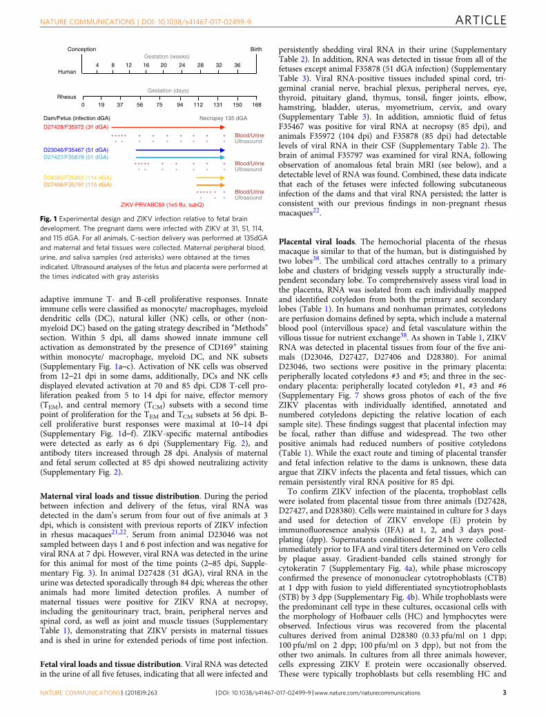
ResultsNonhuman primate model of ZIKV during pregnancy. Fivepregnant rhesus macaques (time-mated breeding) were infectedwith ZIKV (strain PRVABC59) at 31 days of gestation (dGA)(animal ID: D27428), 51 dGA (animals: D23046 and D27427),114 dGA (animal: D28380) and 115 dGA (animal: D27406).(Throughout this manuscript, animal identification numbers willbe preceded by “D”, indicating dams or “F” indicating fetuses).Full term in rhesus macaques is 168 days. The dams receivedZIKV inoculum (1 × 10⁵ ffu) divided over 10 subcutaneousinjections in the hand, wrist and upper arm (1 × 10⁴ ffu perinjection site)22. Each pregnancy was monitored followinginfection by serial physical exams, blood counts, a total chemistrypanel and ultrasound. Cesarean section delivery was performedfor all animals at 135dGA, at which time maternal and fetal tis-sues were collected and processed (Fig. 1). Six uninfected controlrhesus macaques from two ongoing studies were used as acomparative data set for all placental parameters unless otherwisenoted. These animals underwent advanced imaging, were simi-larly delivered at 135dGA, and experienced no treatments ormanipulations during pregnancy.
Innate and adaptive immune responses after ZIKV infection.We monitored the development of the maternal immuneresponse post-ZIKV infection by utilizing PBMCs isolated fromblood samples at various days post infection (dpi). PBMCs werephenotypically analyzed by flow cytometry using antibody panelsdesigned to identify innate immune cell activation as well as
ARTICLE NATURE COMMUNICATIONS | DOI: 10.1038/s41467-017-02499-9
2 NATURE COMMUNICATIONS | (2018) 9:263 |DOI: 10.1038/s41467-017-02499-9 |www.nature.com/naturecommunications
adaptive immune T- and B-cell proliferative responses. Innateimmune cells were classified as monocyte/ macrophages, myeloiddendritic cells (DC), natural killer (NK) cells, or other (non-myeloid DC) based on the gating strategy described in “Methods”section. Within 5 dpi, all dams showed innate immune cellactivation as demonstrated by the presence of CD169+ stainingwithin monocyte/ macrophage, myeloid DC, and NK subsets(Supplementary Fig. 1a–c). Activation of NK cells was observedfrom 12–21 dpi in some dams, additionally, DCs and NK cellsdisplayed elevated activation at 70 and 85 dpi. CD8 T-cell pro-liferation peaked from 5 to 14 dpi for naive, effector memory(TEM), and central memory (TCM) subsets with a second timepoint of proliferation for the TEM and TCM subsets at 56 dpi. B-cell proliferative burst responses were maximal at 10–14 dpi(Supplementary Fig. 1d–f). ZIKV-specific maternal antibodieswere detected as early as 6 dpi (Supplementary Fig. 2), andantibody titers increased through 28 dpi. Analysis of maternaland fetal serum collected at 85 dpi showed neutralizing activity(Supplementary Fig. 2).
Maternal viral loads and tissue distribution. During the periodbetween infection and delivery of the fetus, viral RNA wasdetected in the dam’s serum from four out of five animals at 3dpi, which is consistent with previous reports of ZIKV infectionin rhesus macaques21,22. Serum from animal D23046 was notsampled between days 1 and 6 post infection and was negative forviral RNA at 7 dpi. However, viral RNA was detected in the urinefor this animal for most of the time points (2–85 dpi, Supple-mentary Fig. 3). In animal D27428 (31 dGA), viral RNA in theurine was detected sporadically through 84 dpi; whereas the otheranimals had more limited detection profiles. A number ofmaternal tissues were positive for ZIKV RNA at necropsy,including the genitourinary tract, brain, peripheral nerves andspinal cord, as well as joint and muscle tissues (SupplementaryTable 1), demonstrating that ZIKV persists in maternal tissuesand is shed in urine for extended periods of time post infection.
Fetal viral loads and tissue distribution. Viral RNA was detectedin the urine of all five fetuses, indicating that all were infected and
persistently shedding viral RNA in their urine (SupplementaryTable 2). In addition, RNA was detected in tissue from all of thefetuses except animal F35878 (51 dGA infection) (SupplementaryTable 3). Viral RNA-positive tissues included spinal cord, tri-geminal cranial nerve, brachial plexus, peripheral nerves, eye,thyroid, pituitary gland, thymus, tonsil, finger joints, elbow,hamstring, bladder, uterus, myometrium, cervix, and ovary(Supplementary Table 3). In addition, amniotic fluid of fetusF35467 was positive for viral RNA at necropsy (85 dpi), andanimals F35972 (104 dpi) and F35878 (85 dpi) had detectablelevels of viral RNA in their CSF (Supplementary Table 2). Thebrain of animal F35797 was examined for viral RNA, followingobservation of anomalous fetal brain MRI (see below), and adetectable level of RNA was found. Combined, these data indicatethat each of the fetuses were infected following subcutaneousinfection of the dams and that viral RNA persisted; the latter isconsistent with our previous findings in non-pregnant rhesusmacaques22.
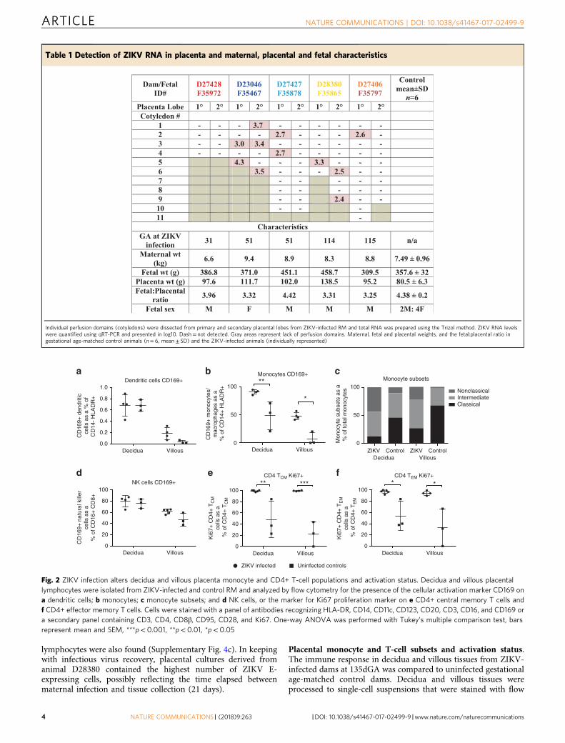
Placental viral loads. The hemochorial placenta of the rhesusmacaque is similar to that of the human, but is distinguished bytwo lobes38. The umbilical cord attaches centrally to a primarylobe and clusters of bridging vessels supply a structurally inde-pendent secondary lobe. To comprehensively assess viral load inthe placenta, RNA was isolated from each individually mappedand identified cotyledon from both the primary and secondarylobes (Table 1). In humans and nonhuman primates, cotyledonsare perfusion domains defined by septa, which include a maternalblood pool (intervillous space) and fetal vasculature within thevillous tissue for nutrient exchange38. As shown in Table 1, ZIKVRNA was detected in placental tissues from four of the five ani-mals (D23046, D27427, D27406 and D28380). For animalD23046, two sections were positive in the primary placenta:peripherally located cotyledons #3 and #5; and three in the sec-ondary placenta: peripherally located cotyledon #1, #3 and #6(Supplementary Fig. 7 shows gross photos of each of the fiveZIKV placentas with individually identified, annotated andnumbered cotyledons depicting the relative location of eachsample site). These findings suggest that placental infection maybe focal, rather than diffuse and widespread. The two otherpositive animals had reduced numbers of positive cotyledons(Table 1). While the exact route and timing of placental transferand fetal infection relative to the dams is unknown, these dataargue that ZIKV infects the placenta and fetal tissues, which canremain persistently viral RNA positive for 85 dpi.
To confirm ZIKV infection of the placenta, trophoblast cellswere isolated from placental tissue from three animals (D27428,D27427, and D28380). Cells were maintained in culture for 3 daysand used for detection of ZIKV envelope (E) protein byimmunofluoresence analysis (IFA) at 1, 2, and 3 days post-plating (dpp). Supernatants conditioned for 24 h were collectedimmediately prior to IFA and viral titers determined on Vero cellsby plaque assay. Gradient-banded cells stained strongly forcytokeratin 7 (Supplementary Fig. 4a), while phase microscopyconfirmed the presence of mononuclear cytotrophoblasts (CTB)at 1 dpp with fusion to yield differentiated syncytiotrophoblasts(STB) by 3 dpp (Supplementary Fig. 4b). While trophoblasts werethe predominant cell type in these cultures, occasional cells withthe morphology of Hofbauer cells (HC) and lymphocytes wereobserved. Infectious virus was recovered from the placentalcultures derived from animal D28380 (0.33 pfu/ml on 1 dpp;100 pfu/ml on 2 dpp; 100 pfu/ml on 3 dpp), but not from theother two animals. In cultures from all three animals however,cells expressing ZIKV E protein were occasionally observed.These were typically trophoblasts but cells resembling HC and
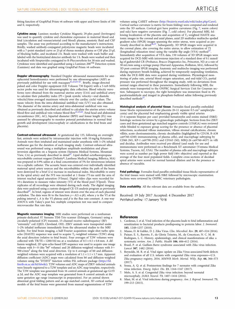
Conception Birth
ZIKV-PRVABC59 (1e5 ffu; subQ)
940 168
Human
Rhesus
D27428/F35972 (31 dGA)
Dam/Fetus (infection dGA)
D23046/F35467 (51 dGA)D27427/F35878 (51 dGA)
D27406/F35797 (115 dGA)D28380/F35865 (114 dGA)
20 24 28 324 8 12 16
112 131 15019 37 56 75
Gestation (weeks)
Gestation (days)
* * * * * * * Ultrasound* * * * * * *
* ** * * * * Blood/Urine
* * * * * * * Ultrasound* * * * * * * * * * Blood/Urine
* * * Ultrasound* * * * * * * Blood/Urine
Necropsy 135 dGA
36
Fig. 1 Experimental design and ZIKV infection relative to fetal braindevelopment. The pregnant dams were infected with ZIKV at 31, 51, 114,and 115 dGA. For all animals, C-section delivery was performed at 135dGAand maternal and fetal tissues were collected. Maternal peripheral blood,urine, and saliva samples (red asterisks) were obtained at the timesindicated. Ultrasound analyses of the fetus and placenta were performed atthe times indicated with gray asterisks
NATURE COMMUNICATIONS | DOI: 10.1038/s41467-017-02499-9 ARTICLE
NATURE COMMUNICATIONS | (2018) 9:263 |DOI: 10.1038/s41467-017-02499-9 |www.nature.com/naturecommunications 3
lymphocytes were also found (Supplementary Fig. 4c). In keepingwith infectious virus recovery, placental cultures derived fromanimal D28380 contained the highest number of ZIKV E-expressing cells, possibly reflecting the time elapsed betweenmaternal infection and tissue collection (21 days).
Placental monocyte and T-cell subsets and activation status.The immune response in decidua and villous tissues from ZIKV-infected dams at 135dGA was compared to uninfected gestationalage-matched control dams. Decidua and villous tissues wereprocessed to single-cell suspensions that were stained with flow
0
50
100
CD
169+
mon
ocyt
es/
mac
roph
ages
as
a %
of C
D14
+ H
LAD
R+
Monocytes CD169+**
*
0
20
40
60
80
100
Ki6
7+ C
D4+
TE
Mce
lls a
s a
% o
f CD
4+ T
EM
CD4 TEM Ki67+
* *
0
20
40
60
80
100
CD
169+
nat
ural
kill
erce
lls a
s a
% o
f CD
16+
CD
8+
NK cells CD169+
0
20
40
60
80
100
Ki6
7+ C
D4+
TC
Mce
lls a
s a
% o
f CD
4+ T
CM
CD4 TCM Ki67+
** ***
0
50
100
Mon
ocyt
e su
bset
s as
a%
of t
otal
mon
ocyt
es
Monocyte subsets
ClassicalIntermediateNonclassical
0.0
0.2
0.4
0.6
0.8
1.0
CD
169+
den
driti
cce
lls a
s a
% o
fC
D14
- H
LAD
R+
Dendritic cells CD169+
ZIKV infected Uninfected controls
Decidua Villous
Decidua Villous Decidua Villous Decidua Villous
Decidua VillousDecidua
ZIKV Control ZIKV ControlVillous
a b c
d e f
Fig. 2 ZIKV infection alters decidua and villous placenta monocyte and CD4+ T-cell populations and activation status. Decidua and villous placentallymphocytes were isolated from ZIKV-infected and control RM and analyzed by flow cytometry for the presence of the cellular activation marker CD169 ona dendritic cells; b monocytes; c monocyte subsets; and d NK cells, or the marker for Ki67 proliferation marker on e CD4+ central memory T cells andf CD4+ effector memory T cells. Cells were stained with a panel of antibodies recognizing HLA-DR, CD14, CD11c, CD123, CD20, CD3, CD16, and CD169 ora secondary panel containing CD3, CD4, CD8β, CD95, CD28, and Ki67. One-way ANOVA was performed with Tukey’s multiple comparison test, barsrepresent mean and SEM, ***p< 0.001, **p< 0.01, *p< 0.05
Table 1 Detection of ZIKV RNA in placenta and maternal, placental and fetal characteristics
Dam/FetalID#
D27428F35972
D23046F35467
D27427F35878
D28380F35865
D27406F35797
Controlmean±SDn=6
Placenta Lobe 1° 2° 1° 2° 1° 2° 1° 2° 1° 2°Cotyledon #
1 - - - 3.7 - - - - - -2 - - - - 2.7 - - - 2.6 -3 - - 3.0 3.4 - - - - - -4 - - - - 2.7 - - - - -5 4.3 - - - 3.3 - - -6 3.5 - - - 2.5 - -7 - - - - -8 - - - - -9 - - 2.4 - -
10 - - -11 -
CharacteristicsGA at ZIKV
infection 31 51 51 114 115 n/a
Maternal wt (kg) 6.6 9.4 8.9 8.3 8.8 7.49 ± 0.96
Fetal wt (g) 386.8 371.0 451.1 458.7 309.5 357.6 ± 32Placenta wt (g) 97.6 111.7 102.0 138.5 95.2 80.5 ± 6.3Fetal:Placental
ratio 3.96 3.32 4.42 3.31 3.25 4.38 ± 0.2
Fetal sex M F M M M 2M: 4F
Individual perfusion domains (cotyledons) were dissected from primary and secondary placental lobes from ZIKV-infected RM and total RNA was prepared using the Trizol method. ZIKV RNA levelswere quantified using qRT-PCR and presented in log10. Dash= not detected. Gray areas represent lack of perfusion domains. Maternal, fetal and placental weights, and the fetal:placental ratio ingestational age-matched control animals (n= 6, mean ± SD) and the ZIKV-infected animals (individually represented)
ARTICLE NATURE COMMUNICATIONS | DOI: 10.1038/s41467-017-02499-9
4 NATURE COMMUNICATIONS | (2018) 9:263 |DOI: 10.1038/s41467-017-02499-9 |www.nature.com/naturecommunications
cytometric antibody panels specific for innate and T-cell subsets.The frequency of activated innate immune cells (monocytes/macrophages and DCs) was higher in placental tissues fromZIKV-infected dams vs. control dams (Fig. 2a, b, d). There was asignificant increase in the percent of activated (CD169+) mono-cyte/macrophages from ZIKV-infected placenta tissues comparedto uninfected controls in both the decidua (p< 0.01, one-wayANOVA) and villous (p< 0.05, one-way ANOVA). The mono-cyte population was further divided into three functionally dif-ferent subsets based on CD14 and CD16 expression: classical(CD14hiCD16−), intermediate (CD14hiCD16+), and non-classical(CD14loCD16+) monocytes. Surprisingly, the ratio of classical tonon-classical monocytes was decreased in both decidua and vil-lous from ZIKV-infected dams compared to uninfected controls(Fig. 2c). Classical monocytes are involved in wound healing,antigen presentation, and typically secrete IL-10, while non-classical monocytes actively patrol tissues following damage andsecrete inflammatory cytokines (IFN-γ, TNF-α, IL-1β, and IL-12)39. Staining of placenta tissues for macrophage subtypes viaCD68 (M1) and CD163 (M2) showed both populations werepresent, with an increase in M1 macrophages in the villousstroma during ZIKV infection (Supplementary Fig. 5). Pro-liferation of CD4 T cells was also increased in placenta tissuesfrom ZIKV-infected dams compared to uninfected controls.
These differences were most evident and significant in the CD4TEM (p< 0.05, one-way ANOVA) and CD4 TCM (p< 0.01, one-way ANOVA) cell subsets (Fig. 2e, f). These results suggest anactive inflammatory immune response is maintained in the pla-centa following ZIKV infection.
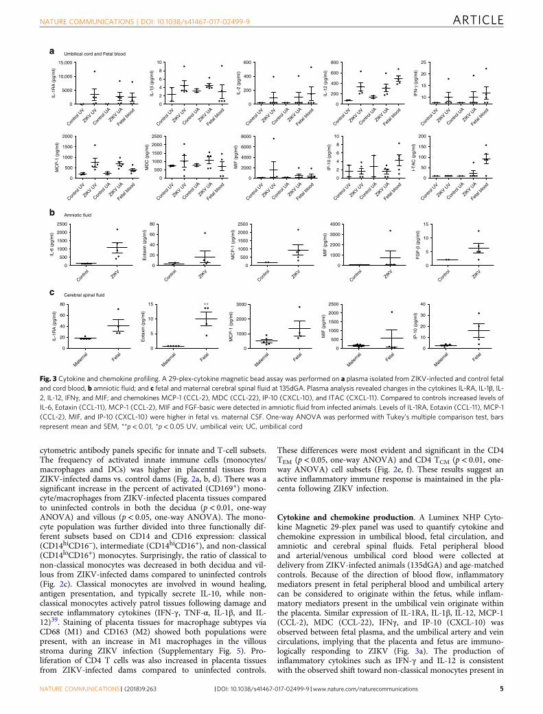
Cytokine and chemokine production. A Luminex NHP Cyto-kine Magnetic 29-plex panel was used to quantify cytokine andchemokine expression in umbilical blood, fetal circulation, andamniotic and cerebral spinal fluids. Fetal peripheral bloodand arterial/venous umbilical cord blood were collected atdelivery from ZIKV-infected animals (135dGA) and age-matchedcontrols. Because of the direction of blood flow, inflammatorymediators present in fetal peripheral blood and umbilical arterycan be considered to originate within the fetus, while inflam-matory mediators present in the umbilical vein originate withinthe placenta. Similar expression of IL-1RA, IL-1β, IL-12, MCP-1(CCL-2), MDC (CCL-22), IFNγ, and IP-10 (CXCL-10) wasobserved between fetal plasma, and the umbilical artery and veincirculations, implying that the placenta and fetus are immuno-logically responding to ZIKV (Fig. 3a). The production ofinflammatory cytokines such as IFN-γ and IL-12 is consistentwith the observed shift toward non-classical monocytes present in
0
200
400
600
800
IL-1
2 (p
g/m
l)
10
15
20
25
IFN
-γ (
pg/m
l)
0
50
100
150
200
I-T
AC
(pg
/ml)
0
500
1000
1500
2000
MC
P-1
(pg
/ml)
0
500
1000
1500
2000
2500
MD
C (
pg/m
l)
0
2000
4000
6000
8000
MIF
(pg
/ml)
0
5000
10,000
15,000
IL-1
RA
(pg
/ml)
0
2
4
6
8
10
IP-1
0 (p
g/m
l)
0
200
400
600
IL-2
(pg
/ml)
0
2
4
6
8
10
IL-1
β (p
g/m
l)
0
500
1000
1500
2000
2500
IL-6
(pg
/ml)
0
500
1000
1500
2000
2500
MC
P-1
(pg
/ml)
0
5
10
15
FG
F-β
(pg
/ml)
0
20
40
60
80
Eot
axin
(pg
/ml)
0
1000
2000
3000
4000
MIF
(pg
/ml)
0
5
10
15
Eot
axin
(pg
/ml)
0
500
1000
1500
2000
2500
MIF
(pg
/ml)
0
10
20
30
40
IP-1
0 (p
g/m
l)
0
1000
2000
3000
MC
P-1
(pg
/ml)
0
20
40
60
80
IL-1
RA
(pg
/ml)
Umbilical cord and Fetal blood
Amniotic fluid
Cerebral spinal fluid
**
Contro
l UV
ZIKV U
V
Contro
l UA
ZIKV U
A
Fetal
blood
Contro
l UV
ZIKV U
V
Contro
l UA
ZIKV U
A
Fetal
blood
Contro
l UV
ZIKV U
V
Contro
l UA
ZIKV U
A
Fetal
blood
Contro
l UV
ZIKV U
V
Contro
l UA
ZIKV U
A
Fetal
blood
Contro
l UV
ZIKV U
V
Contro
l UA
ZIKV U
A
Fetal
blood
Contro
l UV
ZIKV U
V
Contro
l UA
ZIKV U
A
Fetal
blood
Contro
l UV
ZIKV U
V
Contro
l UA
ZIKV U
A
Fetal
blood
Contro
l UV
ZIKV U
V
Contro
l UA
ZIKV U
A
Fetal
blood
Contro
l UV
ZIKV U
V
Contro
l UA
ZIKV U
A
Fetal
blood
Contro
l UV
ZIKV U
V
Contro
l UA
ZIKV U
A
Fetal
blood
Contro
lZIK
V
Contro
lZIK
V
Contro
l
ZIKV
Contro
lZIK
V
Contro
lZIK
V
Mat
erna
lFet
al
Mat
erna
lFet
al
Mat
erna
lFet
al
Mat
erna
lFet
al
Mat
erna
lFet
al
a
b
c
Fig. 3 Cytokine and chemokine profiling. A 29-plex-cytokine magnetic bead assay was performed on a plasma isolated from ZIKV-infected and control fetaland cord blood, b amniotic fluid; and c fetal and maternal cerebral spinal fluid at 135dGA. Plasma analysis revealed changes in the cytokines IL-RA, IL-1β, IL-2, IL-12, IFNγ, and MIF; and chemokines MCP-1 (CCL-2), MDC (CCL-22), IP-10 (CXCL-10), and ITAC (CXCL-11). Compared to controls increased levels ofIL-6, Eotaxin (CCL-11), MCP-1 (CCL-2), MIF and FGF-basic were detected in amniotic fluid from infected animals. Levels of IL-1RA, Eotaxin (CCL-11), MCP-1(CCL-2), MIF, and IP-10 (CXCL-10) were higher in fetal vs. maternal CSF. One-way ANOVA was performed with Tukey’s multiple comparison test, barsrepresent mean and SEM, **p< 0.01, *p< 0.05 UV, umbilical vein; UC, umbilical cord
NATURE COMMUNICATIONS | DOI: 10.1038/s41467-017-02499-9 ARTICLE
NATURE COMMUNICATIONS | (2018) 9:263 |DOI: 10.1038/s41467-017-02499-9 |www.nature.com/naturecommunications 5
the placenta villous and decidua. However, the expression of I-TAC (CXCL-11) was predominately higher in fetal plasma.Comparatively, the expression of all these cytokines was minimalin the maternal circulation. Cytokine profiling of amniotic fluidrevealed increased levels of IL-6, Eotaxin (CCL-11), MCP-1(CCL-2), and FGF-β (Fig. 3b). Together these finding are quitedramatic and suggest that the fetus is undergoing a significantinflammatory immune response. Similarly, the CSF of the fetushad significantly elevated levels of IL-1RA, Eotaxin (CCL-11),MCP-1 (CCL-2) and IP-10 (CXCL-10) compared to the maternalCSF (p< 0.05, one-way ANOVA), which could be indicative of anongoing viral infection (Fig. 3c).
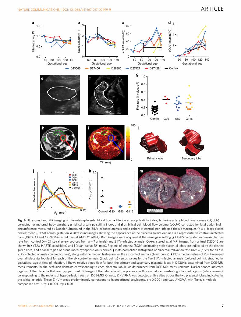
Assessment of uteroplacental blood flow. Longitudinal assess-ments of uteroplacental hemodynamics and fetal growth weremade by ultrasound examinations prior to infection and acrossgestation until delivery. Neither the uterine artery pulsatilityindex (PI), nor the umbilical artery PI, deviated from control inany of the ZIKV animals (Fig. 4a, b). Control animal data wereobtained at two gestational ages: 85dGA and 135dGA (Fig. 4a–d).Quantitative estimation of volumetric blood flow in the uterineartery with ultrasound has been used by our group and others toassess perfusion on the maternal side of the placenta40–43. Thecalculated blood flow volume in the uterine artery (cQuta) cor-rected for maternal body weight demonstrated some variabilitybetween ZIKV animals, but revealed an overall trend toincreasing with advancing gestational age, as would be anticipated(Fig. 5c). Similarly, the quantitative estimation of blood flow onthe fetal side of the placenta41,44,45, the calculated blood flow inthe umbilical vein (cQuv) also increased across gestation, withvalues comparable to uninfected control animals, (Fig. 4d). WhileDoppler ultrasound provides a semi-quantitative estimation ofblood flow, it is limited to measurements in major vessels bothproximal and distal to the placental circulation contributing to alack of sensitivity to detect placental dysfunction. Significantly, weobserved an increase in echogenicity of the placenta in all three ofthe ZIKV animals infected early in gestation. This observationwas consistently observed and all ultrasound exams were per-formed by one ultrasonographer (AEF) with similar imageacquisition parameters. This finding was apparent at ~40 dpi(mid-gestation, Fig. 4f) and is suggestive of inflammation and/orischemic injury. Given the increased echogenicity of the placentaand the limitations of Doppler ultrasound to assess placentalfunction in vivo, we employed advanced non-invasive imagingmodalities prior to delivery at 135dGA to further evaluate pla-cental vascular function in vivo36,37,46,47.
Contrast-enhanced ultrasound (CEUS) with maternal infusionof Definity® contrast agent was used to locate and visualize spiralartery sources supplying individual placental cotyledons from theZIKV-infected animals and uninfected controls at 135dGA. Theflux rate constant (β) provides a measure of microvascularresistance and gives an indirect measure of blood flow in theplacental intervillous space47. In comparing microvascular fluxrates from ZIKV-infected animals to data from uninfected controlanimals at 135dGA, we observed a significant increase in flux rateregardless of gestational age at the time of infection (Fig. 4g, p<0.0001 one-way ANOVA). While our CEUS data analysisvisualizes the spiral artery sources that supply individual placentalcotyledons, it does not allow for absolute quantification of bloodflow volumes, instead reflecting the change in vascular impe-dance. The increase in flux rate following in utero infection withZIKV is indicative of increased velocity of maternal blood beingdelivered to the intervillous space from the spiral arteries, whichmay result in shear stress-induced placental injury.
Placental perfusion and oxygenation by MRI. As CEUS may notreflect the global regional perfusion differences amongst cotyle-dons, we also employed novel MRI techniques for placentalimaging at 135dGA36,37. Our group has recently developed MRImethods to quantify maternal perfusion of the placenta usingboth Dynamic Contrast-Enhanced MRI (DCE-MRI) to inter-rogate delivery and transport of maternal blood in the pla-centa36,47, and Blood Oxygen Level-Dependent (BOLD) MRI tocharacterize placental oxygenation, (via T2 * mapping)37. Thistechnique enables us to separately quantify placental perfusionand placental oxygenation while accounting for the complexvascular network of the intervillous space. When comparingDCE-MRI results for the five ZIKV-infected animals to a group ofsix pregnant rhesus macaque controls matched at 135dGA, wefound modest, but not statistically significant, decreases in bothtotal placental blood flow (139 ml/min vs. 161 ml/min, respec-tively) and normalized placental blood flow (1.14 ml/ml/min vs.1.25 ml/ml/min, respectively). Histograms of R2 * (= 1/T2*, nor-malized to placental volume) reveal that four of the five ZIKV-infected animals (excepting D27406) demonstrated a higher levelof oxygenated maternal blood within the placenta as compared tothe control group (Fig. 4j). We modeled the spatial distribution ofT2* values within the placenta to estimate the ratio of oxygenpermeability-surface area product to blood flow37. Combiningthese values with flow estimates from DCE-MRI, revealed largeand statistically significant decreases (PSvi = 1.22 × 10⁶ vs. PSvi =8.69 × 10⁶, p = 0.018) in oxygen permeability-surface area productin the ZIKV-infected dams relative to control animals. PSvi is aparameter representing the efficiency of oxygen transport fromintervillous space to the fetal villi and is quantitated as the pro-duct of fetal villous permeability, fetal villous surface area andintervillous space37. P values were calculated using a non-parametric two-sample Kolmogorov–Smirnov test. D27406 wasalso an outlier in these data, with oxygen permeability values inthe low range of the control animals. Combined with ourobservation of nearly normal maternal blood flow and increasedlevels of oxygenated blood within the placenta, these resultsstrongly suggest that ZIKV infection impairs transplacentaltransport of oxygen, potentially resulting in long-term fetaloxygen deprivation during gestation, likely secondary to placentaldamage occurring at the level of the fetal villi. Furthermore,high-resolution post-contrast imaging reveals both gross andscattered punctate regions of placental infarction with no sig-nificant contrast uptake (Fig. 4), indicating abnormalperfusion in the placenta from the ZIKV-infected dams. Lastly,comparison of the three-dimensional isosurfaces of the primaryand secondary lobes generated by MRI analysis with the grossplacenta post-delivery of one animal (D23046) showed closecorrespondence between areas of low perfusion and theinfarcted regions (Fig. 4l, m). Of note, regions of low perfusioncorrespond to positive sites of ZIKV RNA detection (Fig. 4m,white asterisks).
Placental and fetal growth after ZIKV exposure. Serial fetalbiometry measurements made by ultrasound enabled fetal growthto be tracked across gestation. Abdominal circumference, headcircumference, biparietal diameter, and femur length did notdeviate from the reference data across gestation (SupplementaryFig. 6). Amniotic fluid levels were on the lower end of the normalcontrol range at 135dGA for all ZIKV animals (SupplementaryFig. 6e). Fetal weight at 135dGA is reported in Table 1. Of note,there is variability across the five ZIKV-infected animals, butmean fetal weight did not differ from the control fetal cohort andwe did not find evidence of fetal growth restriction followingZIKV infection. Importantly, cesarean section delivery of the
ARTICLE NATURE COMMUNICATIONS | DOI: 10.1038/s41467-017-02499-9
6 NATURE COMMUNICATIONS | (2018) 9:263 |DOI: 10.1038/s41467-017-02499-9 |www.nature.com/naturecommunications
Primary lobe Secondary lobe
**
* *
*
60 80 100 120 140Gestational age
60 80 100 120 1400
1
2
3
Gestational age
Um
bilic
al a
rter
y P
I
60 80 100 120 1400
20
40
60
80
Gestational age
cQU
tA (
ml/m
in/k
g)
60 80 100 120 1400
2
4
6
Gestational age
cQU
V (
ml/m
in/A
C)
D23046 D27406 D28380 D27427 D27428 Control
a b c d
e
T2* (ms)0
100h i
j k m
l
Rel
ativ
e fr
eque
ncy
Control G30 G50 G115
Control
G30G50G50G115G115
0 0.12R
2* (ms–1)
0
2.5
PS
υ i (
×10
6 /cm
3 )
f
Control G30 G50 G115
g
1.5
0.0
1.0
0.5
Ute
rine
arte
ry P
I
Control ZIKV
1.0
0.8
0.6
Flu
x ra
te (�
valu
e, s
–1)
0.4
0.2
0.0
Fig. 4 Ultrasound and MR imaging of utero-feto-placental blood flow. a Uterine artery pulsatility index, b uterine artery blood flow volume (cQUtA)corrected for maternal body weight, c umbilical artery pulsatility index, and d umbilical vein blood flow volume (cQUV) corrected for fetal abdominalcircumference measured by Doppler ultrasound in the ZIKV-exposed animals and a cohort of control, non-infected rhesus macaques (n= 6, black closedcircles; mean± SEM) across gestation. e Ultrasound images showing the appearance of the placenta (white outline) in a representative control uninfecteddam (102dGA) and f a ZIKV-infected dam at 61dpi (112dGA). Both images were acquired at the same gain setting. g CE-US calculated microvascular fluxrate from control (n= 27 spiral artery sources from n= 7 animals) and ZIKV-infected animals. Co-registered axial MRI images from animal D23046 areshown in h (T2w HASTE acquisition) and i (quantitative T2* map). Regions of interest (ROIs) delineating both placental lobes are indicated by the dashedgreen lines, and a focal region of pronounced hypoperfusion is circled. j Plots normalized histograms of placental relaxation rate (R2*= 1/T2*) for all fiveZIKV-infected animals (colored curves), along with the median histogram for the six control animals (black curve). k Plots median values of PSvi (averagedover all placental lobules) for each of the six control animals (black points) versus values for the five ZIKV-infected animals (colored points), stratified bygestational age at time of infection. l Shows relative blood flow for both the primary and secondary placental lobes in D23046 determined from DCE-MRImeasurements for the perfusion domains corresponding to each placental lobule, as determined from DCE-MRI measurements. Darker shades indicatedregions of the placenta that are hypoperfused. m Image of the fetal side of the placenta in this animal, demonstrating infarcted regions (white arrows)corresponding to the regions of hypoperfusion seen on DCE-MRI. Of note, ZIKV RNA was detected at five sites across the two placental lobes, indicated bythe white asterisk. These ZIKV + areas predominantly correspond to hypoperfused cotyledons. p< 0.0001 one-way ANOVA with Tukey’s multiplecomparison test, ***p< 0.001, **p< 0.01
NATURE COMMUNICATIONS | DOI: 10.1038/s41467-017-02499-9 ARTICLE
NATURE COMMUNICATIONS | (2018) 9:263 |DOI: 10.1038/s41467-017-02499-9 |www.nature.com/naturecommunications 7
fetus was performed at 135dGA, which corresponds to the earlythird trimester and the start of the exponential fetal growthperiod. Deviation from the normal growth trajectory may havemanifested if the pregnancy had been continued closer to term of168dGA. Placental weight was greater in the ZIKV-infected ani-mals compared to controls; importantly, calculated fetal:placentalweight ratio was decreased in 4 out of 5 animals. The fetal:pla-cental ratio is often used as a proxy for placental efficiency48 andthe decrease in this ratio may reflect aberrant placental functionfollowing in utero ZIKV infection, which is consistent with bothour functional imaging data (Fig. 4) and the placenta pathologydescribed below.
Uteroplacental pathology. All five ZIKV-infected cases showedat least microscopic placental infarctions (Fig. 5a). Larger grossinfarctions were visible in the cases infected earlier in gestation(Supplementary Fig. 7). Additionally, there was a subtle pattern ofvillous stromal calcifications compared with the gestational age-matched uninfected controls (Fig. 5b), which we concluded ismost likely a feature of stromal fibroblast cell necrosis (Fig. 5c).Villous maturation was similar between cases and controls.Chorionic villi were negative for chronic villitis (feature some-times seen in viral infections like CMV, or herpes), but ZIKV-infected cases did have diagnostic chronic decidualitis withplasma cells, which are required for the diagnosis. Two of the fivecases had a pronounced leukocytoclastic vasculitis with a mixtureof lymphocytes, plasma cells, and a few eosinophils infiltrating thespiral artery muscular wall (Fig. 5c, d). This type of vasculitis is
usually related to a hypersensitivity reaction, but viral infectionsare also a well-recognized etiology49. In the three of the ZIKVcases infected at days 31–51, some of the spiral arteries also hadless active inflammation, but clearly showed evidence of moreremote lymphocytic vasculitis with medial damage and abnormalfibrin deposition (not part of the normal physiologic changes thatcharacterize pregnancy-induced vascular remodeling (Fig. 5e, f)50.Spiral artery vasculitis was absent in the two ZIKV cases infectedat 115dGA (these two cases also had fewer plasma cells in thedecidua, fewer placental villous calcifications, and only micro-scopic placental infarctions), which likely reflects the shorter timebetween infection and tissue collection.
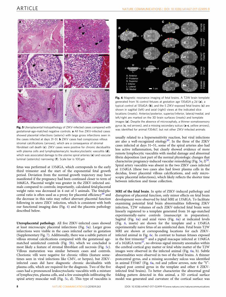
MRI of the fetal brain. In spite of ZIKV-induced pathology anddisruption of placental function, only minor effects on fetal braindevelopment were observed by fetal MRI at 135dGA. To facilitateexamining potential fetal brain abnormalities following ZIKVinfection, T2W volumes of each ZIKV-infected fetal brain werelinearly registered to a template generated from 16 age-matchedexperimentally-naive controls (manuscript in preparation).Sagittal (Fig. 6a) and axial views (Fig. 6a) at indicated levels(Fig. 6, insets) are shown for the template and a 135dGAexperimentally naive fetus of an uninfected dam. Fetal brain T2WMRI are shown at corresponding locations for each ZIKV-infected animal in Fig. 6c. In contrast to human fetuses exposedin the first trimester51 and a pigtail macaque infected at 119dGAof a 162dGA term27, no obvious signal intensity anomalies withinthe cerebral cortical gray matter or fetal white matter of the T2Wimages were observed in the infected animal (Fig. 6a, b). Subtleabnormalities were observed in two of the fetal brains. A thinnerpostcentral gyrus, and a missing secondary sulcus was identifiedin animal F35467 (Fig. 6a, red and yellow arrows, note the “Y”-shaped post central gyrus in the template and other 4 ZIKV-infected fetal brains). To better characterize the abnormal gyralfolding pattern detected in this animal, a 3D cortical surfacemodel was generated and curvature of the cortical surface was
D23
046
F35
467
D27
406
F35
797
D27
427
F35
878
D28
380
F35
865
a
b
D27
428
F35
972
AP
I
S
1 cm
c
AP
L
M
AP
L
R
AP
I
S
A: AnteriorP: PosteriorS: SuperiorI: InferiorL: LateralM: Medial
Fig. 6 Magnetic resonance imaging of fetal brains. A T2W brain templategenerated from 16 control fetuses at gestation age 135dGA± 2d (a), atypical control at 135dGA (b), and the 5 ZIKV-exposed fetal brains (c) areshown in sagittal (left) and axial (right) views at the indicated slicelocations (insets). Anterior/posterior, superior/inferior, lateral/medial, andleft/right are marked on the 3D brain surfaces (insets) and templateimages (a). Despite the absence of microcephaly, a thinner somatosensorygyrus (c, red arrows), and a missing secondary sulcus (a–c, yellow arrows),was identified for animal F35467, but not other ZIKV-infected animals
a b
e
dc
f
*
*
Fig. 5 Uteroplacental histopathology of ZIKV-infected cases compared withgestational-age-matched negative controls. a All five ZIKV-infected casesshowed placental infarctions (asterix) with large gross infarctions seen inthe cases infected at days 31–51. b ZIKV cases had conspicuous villousstromal calcifications (arrows), which are a consequence of stromalfibroblast cell death (c). ZIKV cases were positive for chronic decidualitiswith plasma cells and lymphoplasmacytic leuokocytoclastic vasculitis (d),which was associated damage to the uterine spiral arteries (e) and vascularluminal (astericks) narrowing (f). Scale bar is 100 µm
ARTICLE NATURE COMMUNICATIONS | DOI: 10.1038/s41467-017-02499-9
8 NATURE COMMUNICATIONS | (2018) 9:263 |DOI: 10.1038/s41467-017-02499-9 |www.nature.com/naturecommunications
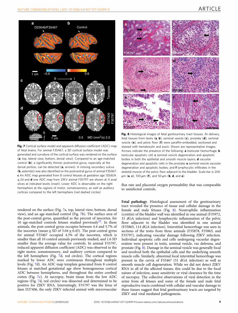
rendered on the surface (Fig. 7a, top, lateral view; bottom, dorsalview), and an age-matched control (Fig. 7b). The surface area ofthe post-central gyrus, quantified as the percent of isocortex, for10 age-matched controls fetuses studied previously52. In theseanimals, the post-central gyrus occupies between 4.4 and 5.7% ofthe isocortex (mean± SD of 5.04± 0.45). The post-central gyrusfor animal F35467 occupied 4.3% of the isocortex, which issmaller than all 10 control animals previously studied, and 1.6 SDsmaller than the average value for controls. In animal F35797,reduced apparent diffusion coefficient (ADC) was observed in theright motor, somatosensory, and auditory cortices compared tothe left hemisphere (Fig. 7d, red circles). The cortical regionsmarked by lower ADC were continuous throughout multiplelevels (Fig. 7d). An ADC map template generated from 8 controlfetuses at matched gestational age show homogeneous corticalADC between hemispheres, and throughout the entire cerebralcortex (Fig. 7c). At necropsy, brain tissue from the low ADCregions (Fig. 7d, red circles) was dissected and determined to bepositive for ZIKV RNA. Interestingly, F35797 was the fetus ofdam D27406, the only ZIKV-infected animal with microvascular
flux rate and placental oxygen permeability that was comparableto uninfected controls.
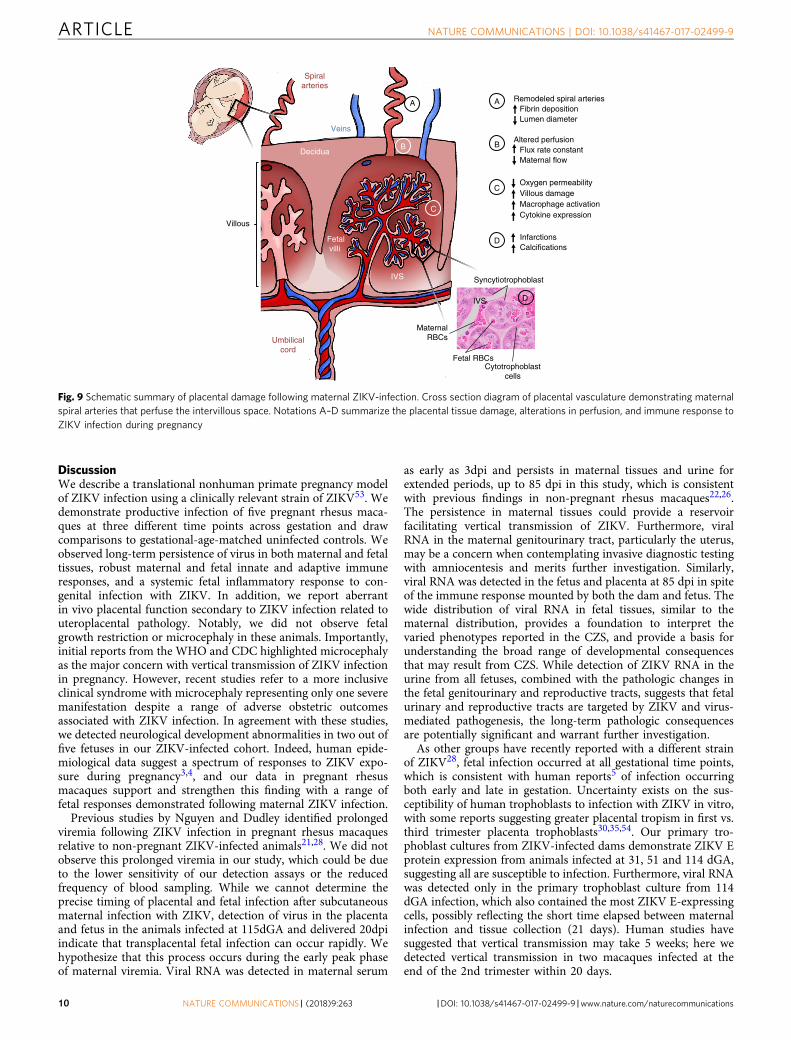
Fetal pathology. Histological assessment of the genitourinarytract revealed the presence of tissue and cellular damage in thefemale and male fetuses (Fig. 8). Neutrophilic inflammation(cystitis) of the bladder wall was identified in one animal (F35972,31 dGA infection) and lymphocytic inflammation of the pelvicfloor adjacent to the bladder was identified in one animal(F35865, 114 dGA infection). Interstitial hemorrhage was seen insections of the testis from three animals (F35878, F35865, andF35797), indicating vascular damage following ZIKV infection.Individual apoptotic cells and cells undergoing vacuolar degen-eration were present in testis, seminal vesicle, vas deferens, andprostate (Fig. 8). Damage in the seminal vesicle was generally focaland involved both the epithelial cells and the underlying smoothmuscle cells. Similarly, abnormal focal interstitial hemorrhage waspresent in the cervix of F35467 (51 dGA infection) as well assmooth muscle cell degeneration. While we did not detect ZIKVRNA in all of the affected tissues, this could be due to the focalnature of infection, assay sensitivity or viral clearance by the timeof necropsy. The collective observations of viral detection in theurine from all fetuses and some of the female and male fetalreproductive tracts combined with cellular and vascular damage tothese tissues suggest that fetal genitourinary tracts are targeted byZIKV and viral mediated pathogenesis.
0 0.6
a bD23046/F35467 Control
1 cm
c
dT
empl
ate
D27
406
F35
797
S I
S
I 1 cm
–0.6
0.5 MD (mm2/s) 2.0
Fig. 7 Cortical surface model and apparent diffusion coefficient (ADC) mapof fetal brains. For animal F35467, a 3D cortical surface model wasgenerated and curvature of the cortical surface was rendered on the surface(a, top, lateral view; bottom, dorsal view). Compared to an age-matchedcontrol (b), a significantly thinner postcentral gyrus, especially at thedorsal portion, can be detected (a, arrows). A missing secondary sulcus(b, asterisks) was also identified on the postcentral gyrus of animal F35467.c An ADC map generated from 8 control fetuses at gestation age 135dGA± 2d and d one ADC map from ZIKV animal F35797 are shown at 4 axialslices at indicated levels (inset). Lower ADC is observable on the righthemisphere at the regions of motor, somatosensory, as well as auditorycortices compared to the left hemisphere (red dashed circles)
e
F35797 F35865
c
F35865
a
F35797
F35865
F35797
f
d
b
Fig. 8 Histological images of fetal genitourinary tract tissues. At delivery,fetal tissues from testis (a, b), seminal vesicle (c), prostate (d), seminalvesicle (e), and pelvic floor (f) were paraffin-embedded, sectioned andstained with hematoxylin and eosin. Shown are representative images.Arrows indicate the presence of the following: a testicular hemorrhage; btesticular apoptotic cell; c seminal vesicle degeneration and apoptoticbodies in both the epithelial and smooth muscle layers; d vacuolardegeneration and apoptotic cells in the prostate; e seminal vesicle vacuolardegeneration and apoptotic bodies; and f lymphocytic infiltrates in theskeletal muscle of the pelvic floor adjacent to the bladder. Scale bar is 200µm (a, c), 100 µm (f), and 50 µm (b, d, and e)
NATURE COMMUNICATIONS | DOI: 10.1038/s41467-017-02499-9 ARTICLE
NATURE COMMUNICATIONS | (2018) 9:263 |DOI: 10.1038/s41467-017-02499-9 |www.nature.com/naturecommunications 9
DiscussionWe describe a translational nonhuman primate pregnancy modelof ZIKV infection using a clinically relevant strain of ZIKV53. Wedemonstrate productive infection of five pregnant rhesus maca-ques at three different time points across gestation and drawcomparisons to gestational-age-matched uninfected controls. Weobserved long-term persistence of virus in both maternal and fetaltissues, robust maternal and fetal innate and adaptive immuneresponses, and a systemic fetal inflammatory response to con-genital infection with ZIKV. In addition, we report aberrantin vivo placental function secondary to ZIKV infection related touteroplacental pathology. Notably, we did not observe fetalgrowth restriction or microcephaly in these animals. Importantly,initial reports from the WHO and CDC highlighted microcephalyas the major concern with vertical transmission of ZIKV infectionin pregnancy. However, recent studies refer to a more inclusiveclinical syndrome with microcephaly representing only one severemanifestation despite a range of adverse obstetric outcomesassociated with ZIKV infection. In agreement with these studies,we detected neurological development abnormalities in two out offive fetuses in our ZIKV-infected cohort. Indeed, human epide-miological data suggest a spectrum of responses to ZIKV expo-sure during pregnancy3,4, and our data in pregnant rhesusmacaques support and strengthen this finding with a range offetal responses demonstrated following maternal ZIKV infection.
Previous studies by Nguyen and Dudley identified prolongedviremia following ZIKV infection in pregnant rhesus macaquesrelative to non-pregnant ZIKV-infected animals21,28. We did notobserve this prolonged viremia in our study, which could be dueto the lower sensitivity of our detection assays or the reducedfrequency of blood sampling. While we cannot determine theprecise timing of placental and fetal infection after subcutaneousmaternal infection with ZIKV, detection of virus in the placentaand fetus in the animals infected at 115dGA and delivered 20dpiindicate that transplacental fetal infection can occur rapidly. Wehypothesize that this process occurs during the early peak phaseof maternal viremia. Viral RNA was detected in maternal serum
as early as 3dpi and persists in maternal tissues and urine forextended periods, up to 85 dpi in this study, which is consistentwith previous findings in non-pregnant rhesus macaques22,26.The persistence in maternal tissues could provide a reservoirfacilitating vertical transmission of ZIKV. Furthermore, viralRNA in the maternal genitourinary tract, particularly the uterus,may be a concern when contemplating invasive diagnostic testingwith amniocentesis and merits further investigation. Similarly,viral RNA was detected in the fetus and placenta at 85 dpi in spiteof the immune response mounted by both the dam and fetus. Thewide distribution of viral RNA in fetal tissues, similar to thematernal distribution, provides a foundation to interpret thevaried phenotypes reported in the CZS, and provide a basis forunderstanding the broad range of developmental consequencesthat may result from CZS. While detection of ZIKV RNA in theurine from all fetuses, combined with the pathologic changes inthe fetal genitourinary and reproductive tracts, suggests that fetalurinary and reproductive tracts are targeted by ZIKV and virus-mediated pathogenesis, the long-term pathologic consequencesare potentially significant and warrant further investigation.
As other groups have recently reported with a different strainof ZIKV28, fetal infection occurred at all gestational time points,which is consistent with human reports5 of infection occurringboth early and late in gestation. Uncertainty exists on the sus-ceptibility of human trophoblasts to infection with ZIKV in vitro,with some reports suggesting greater placental tropism in first vs.third trimester placenta trophoblasts30,35,54. Our primary tro-phoblast cultures from ZIKV-infected dams demonstrate ZIKV Eprotein expression from animals infected at 31, 51 and 114 dGA,suggesting all are susceptible to infection. Furthermore, viral RNAwas detected only in the primary trophoblast culture from 114dGA infection, which also contained the most ZIKV E-expressingcells, possibly reflecting the short time elapsed between maternalinfection and tissue collection (21 days). Human studies havesuggested that vertical transmission may take 5 weeks; here wedetected vertical transmission in two macaques infected at theend of the 2nd trimester within 20 days.
Fetalvilli
Umbilicalcord
IVS
Spiralarteries
Veins
Decidua
Villous
A
B
C
IVS
MaternalRBCs
Fetal RBCs
Syncytiotrophoblast
Cytotrophoblastcells
D
InfarctionsCalcifications
D
COxygen permeability Villous damageMacrophage activationCytokine expression
BAltered perfusion
Flux rate constantMaternal flow
A Remodeled spiral arteriesFibrin depositionLumen diameter
Fig. 9 Schematic summary of placental damage following maternal ZIKV-infection. Cross section diagram of placental vasculature demonstrating maternalspiral arteries that perfuse the intervillous space. Notations A–D summarize the placental tissue damage, alterations in perfusion, and immune response toZIKV infection during pregnancy
ARTICLE NATURE COMMUNICATIONS | DOI: 10.1038/s41467-017-02499-9
10 NATURE COMMUNICATIONS | (2018) 9:263 |DOI: 10.1038/s41467-017-02499-9 |www.nature.com/naturecommunications
We comprehensively evaluated the interplay between thematernal-fetal and placental inflammatory milieu in response toZIKV infection during pregnancy. Our data suggests that ZIKVexposure in utero produces a robust proinflammatory fetalimmune response characterized by alterations in classical vs. non-classical monocyte phenotype and presence of activated CD4 T-cells as well as elevated cytokines (IL-12, IL-2 TNFα and IFNγ)and the chemokine IP-10 (CXCL-10). IP-10 is inducible byproinflammatory mediators such as interferon-γ (IFNγ), TNFα,and several viruses55–58. This chemokine is involved in therecruitment and potentiation of Th1 type inflammatory respon-ses. The fetus is a semi-allograft, and active maternal tolerancemechanisms (Th2 and Treg recruitment) are critical to preventfetal rejection. Inflammation of the placenta and decidua due toviral infections not only alters fetal organ development but alsoalters the fetal immune system, and can result in aberrant post-natal immune responses to infections8,59. The implication is thatin utero ZIKV infection may cause vulnerability in the neonatalinnate and adaptive immune responses and thus increase thesusceptibility to subsequent sepsis, additional infections andautoimmune diseases.
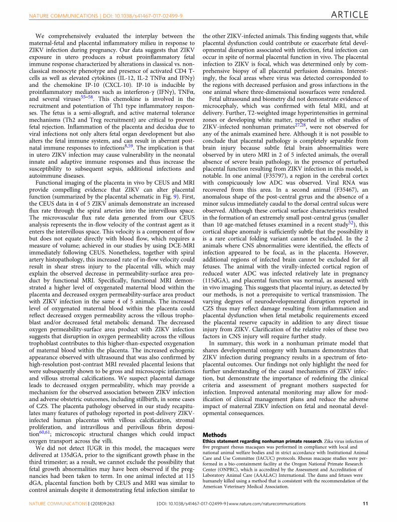
Functional imaging of the placenta in vivo by CEUS and MRIprovide compelling evidence that ZIKV can alter placentalfunction (summarized by the placental schematic in Fig. 9). First,the CEUS data in 4 of 5 ZIKV animals demonstrate an increasedflux rate through the spiral arteries into the intervillous space.The microvascular flux rate data generated from our CEUSanalysis represents the in-flow velocity of the contrast agent as itenters the intervillous space. This velocity is a component of flowbut does not equate directly with blood flow, which requires ameasure of volume; achieved in our studies by using DCE-MRIimmediately following CEUS. Nonetheless, together with spiralartery histopathology, this increased rate of in-flow velocity couldresult in shear stress injury to the placental villi, which mayexplain the observed decrease in permeability-surface area pro-duct by functional MRI. Specifically, functional MRI demon-strated a higher level of oxygenated maternal blood within theplacenta and decreased oxygen permeability-surface area productwith ZIKV infection in the same 4 of 5 animals. The increasedlevel of oxygenated maternal blood within the placenta couldreflect decreased oxygen permeability across the villous tropho-blast and/or decreased fetal metabolic demand. The decreasedoxygen permeability-surface area product with ZIKV infectionsuggests that disruption in oxygen permeability across the villoustrophoblast contributes to this higher-than-expected oxygenationof maternal blood within the placenta. The increased echogenicappearance observed with ultrasound that was also confirmed byhigh-resolution post-contrast MRI revealed placental lesions thatwere subsequently shown to be gross and microscopic infarctionsand villous stromal calcifications. We suspect placental damageleads to decreased oxygen permeability, which may provide amechanism for the observed association between ZIKV infectionand adverse obstetric outcomes, including stillbirth, in some casesof CZS. The placenta pathology observed in our study recapitu-lates many features of pathology reported in post-delivery ZIKV-infected human placentas with villous calcification, stromalproliferation, and intravillous and perivillous fibrin deposi-tion60,61; microscopic structural changes which could impactoxygen transport across the villi.
We did not detect IUGR in this model, the macaques weredelivered at 135dGA, prior to the significant growth phase in thethird trimester; as a result, we cannot exclude the possibility thatfetal growth abnormalities may have been observed if the preg-nancies had been taken to term. In one animal infected at 115dGA, placental function both by CEUS and MRI was similar tocontrol animals despite it demonstrating fetal infection similar to
the other ZIKV-infected animals. This finding suggests that, whileplacental dysfunction could contribute or exacerbate fetal devel-opmental disruption associated with infection, fetal infection canoccur in spite of normal placental function in vivo. The placentalinfection to ZIKV is focal, which was determined only by com-prehensive biopsy of all placental perfusion domains. Interest-ingly, the focal areas where virus was detected corresponded tothe regions with decreased perfusion and gross infarctions in theone animal where three-dimensional isosurfaces were rendered.
Fetal ultrasound and biometry did not demonstrate evidence ofmicrocephaly, which was confirmed with fetal MRI, and atdelivery. Further, T2-weighted image hyperintensities in germinalzones or developing white matter, reported in other studies ofZIKV-infected nonhuman primates27,28, were not observed forany of the animals examined here. Although it is not possible toconclude that placental pathology is completely separable frombrain injury because subtle fetal brain abnormalities wereobserved by in utero MRI in 2 of 5 infected animals, the overallabsence of severe brain pathology, in the presence of perturbedplacental function resulting from ZIKV infection in this model, isnotable. In one animal (F35797), a region in the cerebral cortexwith conspicuously low ADC was observed. Viral RNA wasrecovered from this area. In a second animal (F35467), ananomalous shape of the post-central gyrus and the absence of aminor sulcus immediately caudal to the dorsal central sulcus wereobserved. Although these cortical surface characteristics resultedin the formation of an extremely small post-central gyrus (smallerthan 10 age-matched fetuses examined in a recent study52), thiscortical shape anomaly is sufficiently subtle that the possibility itis a rare cortical folding variant cannot be excluded. In the 2animals where CNS abnormalities were identified, the effects ofinfection appeared to be focal, as in the placenta. However,additional regions of infected brain cannot be excluded for allfetuses. The animal with the virally-infected cortical region ofreduced water ADC was infected relatively late in pregnancy(115dGA), and placental function was normal, as assessed within vivo imaging. This suggests that placental injury, as detected byour methods, is not a prerequisite to vertical transmission. Thevarying degrees of neurodevelopmental disruption reported inCZS thus may reflect damage resulting from inflammation andplacental dysfunction when fetal metabolic requirements exceedthe placental reserve capacity in addition to any direct tissueinjury from ZIKV. Clarification of the relative roles of these twofactors in CNS injury will require further study.
In summary, this work in a nonhuman primate model thatshares developmental ontogeny with humans demonstrates thatZIKV infection during pregnancy results in a spectrum of feto-placental outcomes. Our findings not only highlight the need forfurther understanding of the causal mechanisms of ZIKV infec-tion, but demonstrate the importance of redefining the clinicalcriteria and assessment of pregnant mothers suspected forinfection. Improved antenatal monitoring may allow for mod-ification of clinical management plans and reduce the adverseimpact of maternal ZIKV infection on fetal and neonatal devel-opmental consequences.
MethodsEthics statement regarding nonhuman primate research. Zika virus infection offive pregnant rhesus macaques was performed in compliance with local andnational animal welfare bodies and in strict accordance with Institutional AnimalCare and Use Committee (IACUC) protocols. Rhesus macaque studies were per-formed in a bio-containment facility at the Oregon National Primate ResearchCenter (ONPRC), which is accredited by the Assessment and Accreditation ofLaboratory Animal Care (AAALAC) International. The dams and fetuses werehumanely killed using a method that is consistent with the recommendation of theAmerican Veterinary Medical Association.
NATURE COMMUNICATIONS | DOI: 10.1038/s41467-017-02499-9 ARTICLE
NATURE COMMUNICATIONS | (2018) 9:263 |DOI: 10.1038/s41467-017-02499-9 |www.nature.com/naturecommunications 11
Zika virus PRVABC59. Zika virus isolate PRVABC5922,53 was generously pro-vided by the Centers for Disease Control (CDC). PRVABC59 was passed twice onC6/36 cells (ATCC CRL-1660), and a working stock was concentrated by ultra-centrifugation through a 20% sorbitol cushion and titered in Vero cells (ATCCCRL-1586). The working stock was sequenced and described previously22. All cellswere cultured in Dulbecco’s modified eagle medium (DMEM) containingpenicillin-streptomycin-glutamine and 5–10% fetal calf serum (FCS).
Study design. Gestational age was determined by monitoring daily blood samplesfor the mid-cycle peak in estradiol, which immediately precedes the surge ofluteinizing hormone responsible for ovulation. Females are paired with malesseveral days prior to the estradiol surge and are then separated the day after peaklevels are observed. Day 1 of gestation is considered 48 h post-estradiol surge. Thistimed-mating scheme allows for gestational age to be determined within 24–48 h ofconception. At 31, 51, 114, and 115 days of gestation (dGA; term = 168 days)timed-pregnant rhesus macaques (Macaca mulatta) were infected subcutaneouslywith a total of 1 × 10⁵ focus-forming units (ffu) of ZIKV PRABC59 diluted in 1 mlof PBS that was distributed over both hands, wrists and upper arms as 10–100 µlinjections (to mimic the typical pattern of mosquito bites)22. The dams wereevaluated daily for clinical signs of disease. Body temperature along with peripheralblood and urine samples were obtained, and fetal/placental ultrasound analyseswere performed at indicated times post infection (Fig. 1). Blood was collected on 0,3, 7, 14, 21, 28, 35, 43, 49, 57, 63, 71, 78, 85, 91, 98, and 105 dpi with the followingexceptions: (1) Animals D28380 and D27406 was taken to necropsy at 21 dpi; (2)Animals D23046 and D27427 were taken to necropsy at 85 dpi; and (3) day 3 wasnot collected for D23046. Collection of blood samples and ultrasound analysis wereperformed under ketamine sedation (10 mg/kg, IM). Peripheral blood mono-nuclear cells (PBMCs) and plasma were isolated by centrifugation over lymphocyteseparation medium and then analyzed for lymphocyte phenotype and frequency byflow cytometry. Plasma was assessed for viral load by qRT-PCR and the levels ofcytokines by a multiplex-bead based assay. Urine and saliva were assessed for viralRNA by qRT-PCR and for infectious virus by co-culture and focus-forming assaysusing Vero cells. An in utero MRI scan of the fetal brain and placenta was per-formed just prior to delivery. Necropsy was performed on the all of the dams andfetuses at 135dGA. Samples of maternal and fetal tissues (joints, muscles, organs,brain, spinal cord, eyes, glands, lymph nodes and bone marrow) and biologicalfluids (cerebral spinal fluid, blood, amniotic fluid, and urine) were collected andstored in RNAlater, Trizol (RNA isolation), medium (virus isolation) as well asformalin fixed and embedded in paraffin.
ZIKV detection by one-step qRT-PCR analysis. RNA from maternal, fetal andplacental tissue samples, blood, urine, CSF, and amniotic fluid was isolated usingTrizol (Invitrogen) according to manufacturer’s protocol. ZIKV RNA levels weremeasured by a one-step quantitative real-time reverse transcription polymerasechain reaction assay (qRT-PCR) using TaqMan® One-Step RT-PCR Master Mix(Applied Biosystems). Primers and probes were as previously described62 with aone base change in the probe to match the PRVABC59 sequence (Genbankaccession #: KU501215.1). Forward primer: 5′- CCGCTGCCCAACACAAG-3′(ZIKV PRVABC59 genome sequence nucleotides 1192–1208); reverse: 5′-CCACTAACGTTCTTTTGCAGACAT-3′ (complement of nucleotides1245–1268); and TaqMan probe: 5′ Fam-5′-AGCCTACCTTGACAAGCAATCA-GACACTCAA-3′ -MGB (nucleotides 1213–1243). Forward and reverse primerswere used at 250 nM in the reaction, and the probe at 200 nM. The limit ofdetection for our One-step RT-PCR quantification assay was approximately 100genomes making the limit of detection in bodily fluids 1e4 for 100 µl of plasma orurine and 1/10th of the RNA recovered used per reaction. The limit of detection intissues was ~400 genomes per µg total RNA using 250 ng total RNA per reaction.
Isolation of placental immune cells and trophoblasts. At the time of necropsy,~0.5–1 g of decidua and villous tissue were collected and stored in 5 mL of HBSSsupplemented with 2% FBS and 10 mM Hepes (HBSS + ), for flow cytometryanalysis. Tissues were dissected into a fine slurry using forceps and scissors prior tobeing digested with collagenase, 50 mg collagenase in 35 mL HBSS + , for 30 min at37 °C, with rocking. Digested tissues were washed with HBSS + and run through a70 μM filter. The cells present in the flow through were pelleted by centrifugation at500 × g. Red blood cells were lysed in the cell pellet with BD Pharm Lyse lysingbuffer (BD Biosciences). After a final wash in HBSS + , cells were resuspended in5 mL HBSS + and counted using a hemocytometer. Cells were analyzed by flowcytometry as described below.
To evaluate placental infection by ZIKV, villous trophoblasts were isolated fromthe placentas of three animals (D27428, D27427 and D28380) essentially accordingto the method of Kliman et al.63. Briefly, villous tissue (30–35 g) that had beendissected free of membranes was washed, minced, filtered through sterile gauze anddigested with three rounds of incubation in a trypsin-DNase solution (0.25%trypsin [Sigma-Aldrich, St. Louis MO] in PBS with 0.5 mg/ml DNase 1 [Sigma;2000 Kunitz units/mg]). Digested tissue was collected, layered over newborn calfserum (NCS; Sigma) at a 5:1 v/v ratio and centrifuged at 1000 × g for 5 min. Cellpellets were resuspended in DMEM and the mononuclear cytotrophoblast
population was isolated by centrifugation on a preformed Percoll gradient(10–70%) with collection of cells banding between 35–55%. Recovered cells werewashed and plated on 35 mm Corning Primaria tissue culture dishes (Thermo-Fisher Scientific; Waltham, MA) in 1:1 DMEM/Hams F12 (Thermo-FisherScientific) supplemented with 10% FBS, 2 mM L-glutamine and antibiotics(Penicillin-streptomycin-neomycin). Cells were cultured for 3 days post plating(pp) with medium exchange every 24 h. Supernatants were harvested at days 1, 2,and 3 pp and evaluated for the presence of infectious ZIKV by titration on Verocells. Monolayers were used immediately after each supernatant collection on days1–3pp for immunofluorescence detection of ZIKV antigen. Briefly, cells werewashed, fixed and permeabilized in 4% pFormaldehye followed by 0.2% Triton X-100, blocked with 20% normal goat serum and sequentially incubated with an anti-Flavivirus group antigen mAb (Clone D1–4G2) and a goat-anti-Mouse IgG Alexa-Fluor 488 (Thermo-Fisher) secondary antibody.
Phenotypic analysis of peripheral blood and tissue mononuclear cells. Flowcytometry was used to quantify the immune cell phenotype and level of cellularproliferation and activation for maternal PBMCs isolated at the time points definedabove as well as the immune cells obtained from placental tissues. The panel ofantibodies used for the analysis of innate immune cells consisted of HLA-DR (G46–610 µg/mL Beckman Coulter), CD14 (HCD14 10 µg/mL Biolegend), CD11c (3.9 10µg/mL Biolegend), CD123 (SSDCLY107D2 10 µg/mL Beckman Coulter), CD20(B9E9 10 µg/mL Beckman Coulter), CD3 (SP34-2 2 µg/mL Biolegend), CD8 (SK-110 µg/mL BD Bioscience), CD16 (3G8 10 µg/mL ThermoFisher), and CD169 (7–3292 µg/mL Biolegend). To differentiate between monocyte/macrophages, dendritic cells(DC), and natural killer (NK) cells the following gating strategy was utilized:monocyte/macrophages (CD3−CD20−CD14+HLA-DR+), classical monocytes (CD3-
CD20- HLA-DR+ CD14+CD16−), intermediate monocytes (CD3−CD20− HLA-DR+
CD14+CD16+), non-classical monocytes CD3−CD20− HLA-DR+ CD14−CD16+),DC (CD3−CD20−CD14−HLA-DR+), myeloid DCs (CD3−CD20−CD14−HLA-DR+
CD123−), CD11c+ plasmacytoid DCs (CD3−CD20−CD14−HLA-DR+CD123+CD11c−), other DCs (CD3−CD20−CD14−HLA-DR+CD123−CD11c−), and NK cells (CD3−CD20−CD8+CD16+). The percentage of activated cells (CD169+) within each subsetwas calculated as a representation of the cellular activation profile. T cells wereanalyzed with the following panel of antibodies directed against CD3 (SP34-2 2 µg/mL Biolegend), CD4 (L200 5 µg/mL Thermofisher), CD8β (SK-1 10 µg/mL BDBioscience), CD95 (DX2 2 µg/mL BD Biosciences), CD28 (CD28.2 2 µg/ml Biole-gend) and the intracellular proliferation marker Ki67 (20Raj1 2 µg/ml, Thermo-fisher). CD3+ T cells were first identified as CD4+ or CD8+; within the CD4+ andCD8+ T-cell subsets the naive (CD28+CD95−), central memory (CD28+CD95+), andeffector memory (CD28−CD95+) subsets were analyzed. B cells were stained usingthe following antibodies: CD20 (B9E9 5 µg/ml Beckman Coulter), CD27 O323 5 µg/mL Biolegend), and IgD (Goat polyclonal 2 µg/mL Southern Biotech) to delineatenaive (CD20+CD27−IgD+), memory (CD20+CD27−IgD−) and marginal-zone like Bcells (CD20+CD27+IgD+) as well as Ki67 to identify recent proliferating cells. Thepercentage of proliferating (Ki67+) B and T cells within each subset was calculated.The gating strategies were performed as previously described64. Phenotyping wasperformed using an LSRII instrument (BD Bioscience) and the data were analyzedwith FlowJo Software (TreeStar).
ZIKV enzyme-linked immunosorbent assay (ELISA). Anti-ZIKV antibodieswere measured by ELISA in the maternal and fetal plasma as well as in the cordblood (venous and arterial) and amniotic fluid. High-binding polystyrene 96-wellplates (Corning) were coated overnight with 100 µl of PBS containing a 1:100dilution of purified ZIKV particle preparations. The plates were blocked with PBScontaining 2% milk and 0.05% Tween (ELISA-Block) for 1 h at room temperature,washed with 0.05% Tween-PBS (ELISA-Wash), and incubated with two-folddilutions of rhesus plasma in ELISA-Block starting at a dilution of 1:50. The platewas incubated at room temperature for 2 h. Plates were washed several times withELISA-Wash and then incubated with secondary anti-monkey IgM/A/G (Rock-land, Inc.) conjugated with horseradish peroxidase for 30 mins. Plates were washedwith ELISA-Wash and bound secondary antibody was detected using the OPDsubstrate (Life Technologies) with an HCl stop. The plates were read within 10 minusing a Synergy HTX Microplate Reader (BioTeck) at 490 nm. Dilution titers ofZIKV-binding antibodies were determined using a Log-to-Log transformationmethod and the results were graphed using GraphPad Prism v6 software.
Anti-ZIKV neutralization assay. Plaque Reduction Neutralization Test (PRNT50)was used to measure the concentration of serum that neutralizes 50% of a fixednumber of ZIKV-PRABC59. NHP serum from the dam and fetus, cord blood, andamniotic fluid was serially diluted 2-fold from starting dilutions of 1:10 and mixedwith an equal volume of a fixed number of plaque forming units (50 PFU) of ZIKVfor final serum dilutions ranging from 1:40 to 1:81,920. Sera and virus wereincubated for 1 h at 37 °C. The mixtures were added to individual wells of 12-wellplates seeded with Vero cells at 90% confluence for 1 h at 37 °C on a rocker andthen overlaid with DMEM containing methylcellulose. Plates were incubated for3 days and then fixed and counterstained with methyl blue. Plaques were countedand PRNT50 values were calculated utilizing the sigmoid dose-response curve
ARTICLE NATURE COMMUNICATIONS | DOI: 10.1038/s41467-017-02499-9
12 NATURE COMMUNICATIONS | (2018) 9:263 |DOI: 10.1038/s41467-017-02499-9 |www.nature.com/naturecommunications
fitting function of GraphPad Prism v6 software with upper and lower limits of 100and 0, respectively.
Cytokine assay. Luminex monkey Cytokine Magnetic 29-plex panel (Invitrogen)was used to quantify cytokine and chemokine expression in maternal blood andfetal (circulation and venous/arterial cord blood) plasma, amniotic fluid and CSFsamples. The assay was performed according to the manufacturer’s instructions.Briefly, washed antibody-conjugated polystyrene magnetic beads were incubatedwith a 7-point standard curve or 25 µl of rhesus monkey plasma or CSF plus 25 µLof blocking buffer, and incubated 2 h. Beads were washed with wash buffer andlabeled with the biotinylated detector antibody for 1 h. Beads were washed and thenincubated with Streptavidin conjugated to R-Phycoerythrin for 30 min and washed.Cytokines were identified and quantified using a Luminex 200TM Detection system(Luminex) and data was graphed using GraphPad Prism v6 software.
Doppler ultrasonography. Standard Doppler ultrasound measurements for uter-oplacental hemodynamics were performed by one ultrasonographer (AEF) aspreviously published for use with macaques41,43,65–67. Briefly, image-directedpulsed and color Doppler equipment (GE Voluson 730 Expert) with a 5-to9-MHzsector probe was used for ultrasonographic data collection. Blood velocity wave-forms were obtained from the maternal uterine artery (UtA) and umbilical arteryto calculate their pulsatility index (PI = [peak systolic velocity—end diastolicvelocity]/time-averaged maximum velocity over the cardiac cycle) values. Themean velocity from the intra-abdominal umbilical vein (UV) was also obtained.The diameter of the uterine artery and intra-abdominal umbilical vein wasobtained as previously described and utilized to calculate the uterine artery volumeblood flow and umbilical vein volume blood flow41,43,65. Fetal head and abdominalcircumference (HC, AC), bipartiel diameter (BPD) and femur length (FL) wasassessed by ultrasonography to monitor potenial pertubationas in normal fetalgrowth and development (microcephaly or calcifications in the fetal brain andplacenta).
Contrast-enhanced ultrasound. At gestational day 135, following an overnightfast, animals were sedated by intramuscular injection with 10 mg/kg Ketamine.Animals were then intubated and maintained under anesthesia with 1–2% inhaledisoflurane gas for the duration of each imaging study. Contrast-enhanced ultra-sound was performed using a multiphase amplitude-modulation and phase-inversion algorithm on a Sequoia system (Siemens Medical Systems, MountainView, CA) as previously described in detail47. Lipid-shelled octofluoropropanemicrobubble contrast reagent (Definity®, Lantheus Medical Imaging, Billerica, MA)was prepared in 0.9% saline at a final concentration of 5% for intravenous infusionvia a cephalic catheter. The acoustic beam was centered over individually identifiedmaternal spiral artery sources and the microbubbles within the path of the beamwere destroyed by a brief (2 s) increase in mechanical index. Microbubble re-entryin the spiral artery and the IVS was recorded at 1 frame /75 ms until the area ofinterest reached signal saturation (VImax). Digital video clips were recorded for10 s durations to measure video intensity (VI) of the blood pool (VIpool). Threereplicates of all recordings were obtained during each study. The digital imagingdata were analyzed using a custom-designed CE-US analysis program as previouslydescribed47. In brief, regions of interest were drawn over the area of each placentalcotyledon. The data were fit to the function y = A(1-e-βt), where y is the VI at thepulsing interval t, A is the VI plateau and β is the flux rate constant. A one-wayANOVA with Tukey’s post hoc multiple comparison test was used to comparemicrovascular flux rate data.
Magnetic resonance imaging. MRI studies were performed on a nonhumanprimate-dedicated 3T Siemens TIM-Trio scanner (Erlangen, Germany) using acircularly-polarized (CP) transmit, 15-channel receive radiofrequency (RF)“extremity” coil (QED, Cleveland, OH). ZIKV animals were transported under1–2% inhaled isoflurane immediately from the ultrasound studies to the MRIfacility. For fetal brain imaging, a half-Fourier acquisition single-shot turbo spin-echo (HASTE) sequence was used to acquire T2-weighted volumes (T2W) alongthe axial direction (relative to fetal brain). Four averages of T2W volumes werecollected with TR/TE = 1200/102 ms at a resolution of 0.5 × 0.5 × 0.8 mm. A dif-fusion-weighted, 2D spin-echo-based EPI sequence was used to acquire one imagevolume with b = 0 (the “b0” volume) and 20 diffusion-weighted volumes with b =500 s/mm2 along the fetal axial direction. Up to 6 averages of b0 and diffusion-weighted volumes were acquired with a resolution of 1.1 × 1.1 × 1.7 mm. Apparentdiffusion coefficient (ADC) maps were calculated from b0 and diffusion-weightedvolumes using the “DTIFIT” function within FSL software package (https://fsl.fmrib.ox.ac.uk/fsl/fslwiki). T2W volumes and ADC maps of ZIKV-infected animalswere linearly registered to a T2W template and an ADC map template, respectively.The T2W template was generated from 16 control animals at gestational age G135± 2d, and the ADC map template was generated from 8 control animals at thesame gestation age range (manuscript in preparation). For an animal shownabnormal gyral folding pattern and an age-matched control, 3D cortical surfacemodels of the fetal brains were generated from manual segmentations of T2W
volumes using CARET software (http://brainvis.wustl.edu/wiki/index.php/Caret).Cortical surface curvature (a metric for brain folding) were computed and renderedonto the 3D surfaces. Cortical gyri have positive curvatures (Fig. 7, warm colors)and sulci have negative curvatures (Fig. 7, cold colors). For placental MRI, fol-lowing localization of the placenta and acquisition of T2-weighted HASTE ana-tomic images in the coronal and axial planes, axial 2D multislice multiecho spoiledgradient echo (SPGR) images spanning the entire uterus, were acquired as pre-viously described in detail36,37. Subsequently, 3D SPGR images were acquired inthe coronal plane, also covering the entire uterus, to allow estimation of T1(longitudinal relaxation time) using the variable flip angle (VFA) method37.Immediately after acquisition of VFA data, 150 volumes of 3D SPGR images wereacquired for DCE-MRI with intravenous injection of a standard dose of 0.1 mmol/kg of gadoteridol CR (Prohance, Bracco Diagnostics Inc, Princeton, NJ) at a rate of30 mL/min using a syringe pump (Harvard Apparatus, Holliston, MA), followed by3D post-contrast SPGR imaging. Anatomic and multiecho imaging was performedduring expiratory breath-holding, achieved by temporarily suspending ventilation,while the DCE-MRI data were acquired during ventilation. Physiological mon-itoring of pulse rate, arterial blood oxygen saturation, and end-tidal CO2 partialpressure was performed throughout the imaging study, with no deviations fromnormal ranges observed in these parameters. Immediately following MRI, theanimals were transported to the ONPRC Surgical Services Unit for Cesarean sec-tion. Subsequent to necropsy, the right hemisphere was immersion fixed in 4%paraformaldehyde and imaged in phosphate-buffered saline following previouslydescribed methods37.
Histological analysis of placental tissue. Formalin-fixed paraffin-embeddedtissue blocks representative of the placenta (8–11 separate 0.5 cm3 samples/pla-centa) and the placental bed emphasizing grossly visible spiral arteries(3–4 separate biopsies per case) provided hematoxylin and eosins stained (H&E)histologic sections for review by a gynecologic pathologist. Sections from five ZIKVcases and three gestational age-matched negative controls (135dGA) were reviewedwhile blinded to exposure group scoring for the presence or absence of placentalinfarctions, accelerated villous maturation, villous stromal calcifications, chronicvillitis, acute chorioamnionitis, chronic decidualitis (highlighted by CD138, B-A38cat#760-4248) immunostaining of plasma cells), and macrophage subtyping byCD68 (M1, KP-1 cat#790-2931) and CD163 (M2, MRQ-26 cat#760-4437) in villiand decidua. Antibodies were received pre-diluted (and ready for use andimmunostains were performed on a Benchmark XT autostainer (Ventana MedicalSystems, Tucson, AZ, USA). The number of plasma cells and macrophage subtypeswere scored as number per high power field (40× objective) and reported as theaverage of the four most populated fields. Complete cross-sections of decidualspiral arteries were scored for normal luminal dilation and for the presence orabsence of vasculitis.
Fetal pathology. Formalin-fixed paraffin-embedded tissue blocks representative ofthe fetal tissues were stained with H&E followed by microscopic examination.Sections were reviewed by ONPRC veterinary pathologists.
Data availability. All the relevant data are available from the authors.
Received: 19 July 2017 Accepted: 4 December 2017
References1. Cardenas, I. et al. Viral infection of the placenta leads to fetal inflammation and
sensitization to bacterial products predisposing to preterm labor. J. Immunol.185, 1248–1257 (2010).
2. Musso, D. & Gubler, D. J. Zika Virus. Clin. Microbiol. Rev. 29, 487–524 (2016).3. Paixao, E. S., Barreto, F., da Gloria Teixeira, M., da Conceicao, N. C. M. &
Rodrigues, L. C. History, epidemiology, and clinical manifestations of zika: asystematic review. Am. J. Public. Health 106, 606–612 (2016).
4. Brasil, P. et al. Guillain-Barre syndrome associated with Zika virus infection.Lancet 387, 1482 (2016).
5. Reynolds, M. R. et al. Vital signs: update on Zika Virus-associated birth defectsand evaluation of all U.S. infants with congenital Zika virus exposure—U.S.Zika pregnancy registry, 2016. MMWR Morb. Mortal. Wkly. Rep. 66, 366–373(2017).
6. Sousa, A. Q. et al. Postmortem findings for 7 neonates with congenital Zikavirus infection. Emerg. Infect. Dis. 23, 1164–1167 (2017).
7. Melo, A. S. et al. Congenital Zika virus infection: beyond neonatalmicrocephaly. JAMA Neurol. 73, 1407–1416 (2016).
8. Silasi, M. et al. Viral infections during pregnancy. Am. J. Reprod. Immunol. 73,199–213 (2015).
NATURE COMMUNICATIONS | DOI: 10.1038/s41467-017-02499-9 ARTICLE
NATURE COMMUNICATIONS | (2018) 9:263 |DOI: 10.1038/s41467-017-02499-9 |www.nature.com/naturecommunications 13
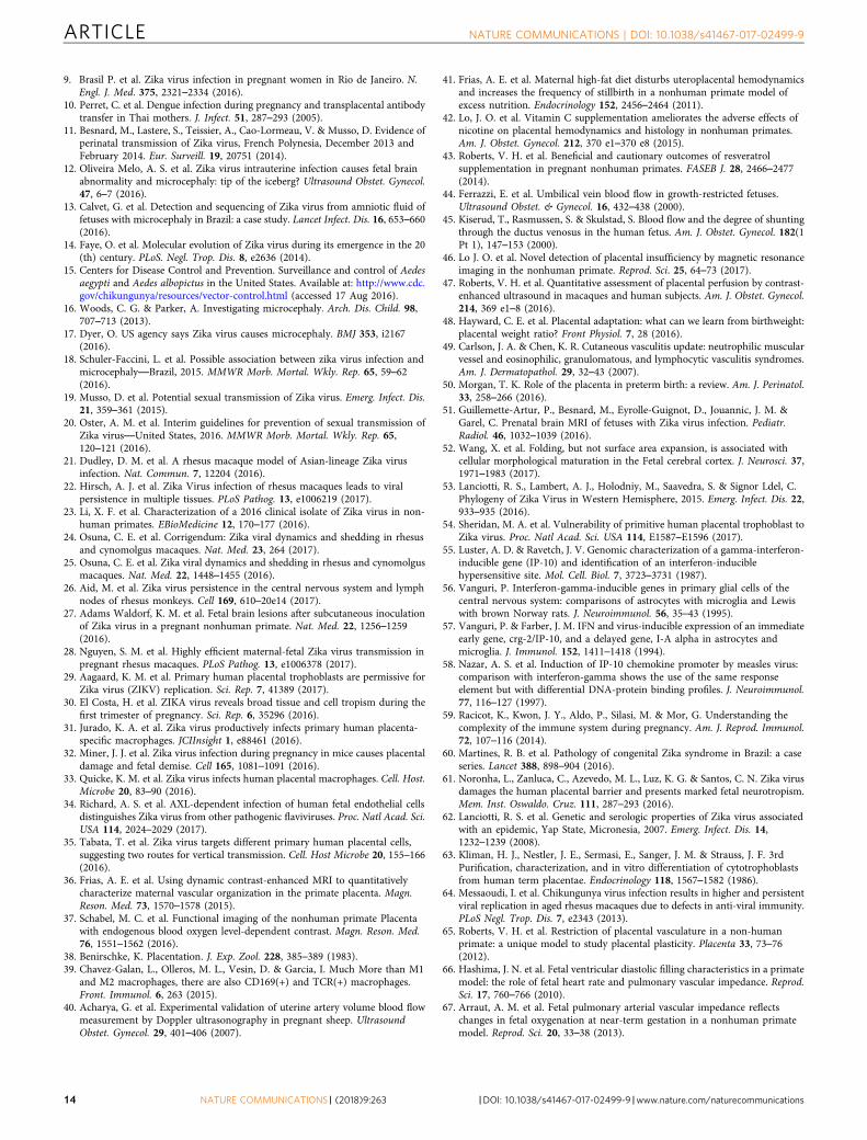
9. Brasil P. et al. Zika virus infection in pregnant women in Rio de Janeiro. N.Engl. J. Med. 375, 2321–2334 (2016).
10. Perret, C. et al. Dengue infection during pregnancy and transplacental antibodytransfer in Thai mothers. J. Infect. 51, 287–293 (2005).
11. Besnard, M., Lastere, S., Teissier, A., Cao-Lormeau, V. & Musso, D. Evidence ofperinatal transmission of Zika virus, French Polynesia, December 2013 andFebruary 2014. Eur. Surveill. 19, 20751 (2014).
12. Oliveira Melo, A. S. et al. Zika virus intrauterine infection causes fetal brainabnormality and microcephaly: tip of the iceberg? Ultrasound Obstet. Gynecol.47, 6–7 (2016).
13. Calvet, G. et al. Detection and sequencing of Zika virus from amniotic fluid offetuses with microcephaly in Brazil: a case study. Lancet Infect. Dis. 16, 653–660(2016).
14. Faye, O. et al. Molecular evolution of Zika virus during its emergence in the 20(th) century. PLoS. Negl. Trop. Dis. 8, e2636 (2014).
15. Centers for Disease Control and Prevention. Surveillance and control of Aedesaegypti and Aedes albopictus in the United States. Available at: http://www.cdc.gov/chikungunya/resources/vector-control.html (accessed 17 Aug 2016).
16. Woods, C. G. & Parker, A. Investigating microcephaly. Arch. Dis. Child. 98,707–713 (2013).
17. Dyer, O. US agency says Zika virus causes microcephaly. BMJ 353, i2167(2016).
18. Schuler-Faccini, L. et al. Possible association between zika virus infection andmicrocephaly—Brazil, 2015. MMWR Morb. Mortal. Wkly. Rep. 65, 59–62(2016).
19. Musso, D. et al. Potential sexual transmission of Zika virus. Emerg. Infect. Dis.21, 359–361 (2015).
20. Oster, A. M. et al. Interim guidelines for prevention of sexual transmission ofZika virus—United States, 2016. MMWR Morb. Mortal. Wkly. Rep. 65,120–121 (2016).
21. Dudley, D. M. et al. A rhesus macaque model of Asian-lineage Zika virusinfection. Nat. Commun. 7, 12204 (2016).
22. Hirsch, A. J. et al. Zika Virus infection of rhesus macaques leads to viralpersistence in multiple tissues. PLoS Pathog. 13, e1006219 (2017).
23. Li, X. F. et al. Characterization of a 2016 clinical isolate of Zika virus in non-human primates. EBioMedicine 12, 170–177 (2016).
24. Osuna, C. E. et al. Corrigendum: Zika viral dynamics and shedding in rhesusand cynomolgus macaques. Nat. Med. 23, 264 (2017).
25. Osuna, C. E. et al. Zika viral dynamics and shedding in rhesus and cynomolgusmacaques. Nat. Med. 22, 1448–1455 (2016).
26. Aid, M. et al. Zika virus persistence in the central nervous system and lymphnodes of rhesus monkeys. Cell 169, 610–20e14 (2017).
27. Adams Waldorf, K. M. et al. Fetal brain lesions after subcutaneous inoculationof Zika virus in a pregnant nonhuman primate. Nat. Med. 22, 1256–1259(2016).
28. Nguyen, S. M. et al. Highly efficient maternal-fetal Zika virus transmission inpregnant rhesus macaques. PLoS Pathog. 13, e1006378 (2017).
29. Aagaard, K. M. et al. Primary human placental trophoblasts are permissive forZika virus (ZIKV) replication. Sci. Rep. 7, 41389 (2017).
30. El Costa, H. et al. ZIKA virus reveals broad tissue and cell tropism during thefirst trimester of pregnancy. Sci. Rep. 6, 35296 (2016).
31. Jurado, K. A. et al. Zika virus productively infects primary human placenta-specific macrophages. JCIInsight 1, e88461 (2016).
32. Miner, J. J. et al. Zika virus infection during pregnancy in mice causes placentaldamage and fetal demise. Cell 165, 1081–1091 (2016).
33. Quicke, K. M. et al. Zika virus infects human placental macrophages. Cell. Host.Microbe 20, 83–90 (2016).
34. Richard, A. S. et al. AXL-dependent infection of human fetal endothelial cellsdistinguishes Zika virus from other pathogenic flaviviruses. Proc. Natl Acad. Sci.USA 114, 2024–2029 (2017).
35. Tabata, T. et al. Zika virus targets different primary human placental cells,suggesting two routes for vertical transmission. Cell. Host Microbe 20, 155–166(2016).
36. Frias, A. E. et al. Using dynamic contrast-enhanced MRI to quantitativelycharacterize maternal vascular organization in the primate placenta. Magn.Reson. Med. 73, 1570–1578 (2015).
37. Schabel, M. C. et al. Functional imaging of the nonhuman primate Placentawith endogenous blood oxygen level-dependent contrast. Magn. Reson. Med.76, 1551–1562 (2016).
38. Benirschke, K. Placentation. J. Exp. Zool. 228, 385–389 (1983).39. Chavez-Galan, L., Olleros, M. L., Vesin, D. & Garcia, I. Much More than M1
and M2 macrophages, there are also CD169(+) and TCR(+) macrophages.Front. Immunol. 6, 263 (2015).
40. Acharya, G. et al. Experimental validation of uterine artery volume blood flowmeasurement by Doppler ultrasonography in pregnant sheep. UltrasoundObstet. Gynecol. 29, 401–406 (2007).
41. Frias, A. E. et al. Maternal high-fat diet disturbs uteroplacental hemodynamicsand increases the frequency of stillbirth in a nonhuman primate model ofexcess nutrition. Endocrinology 152, 2456–2464 (2011).
42. Lo, J. O. et al. Vitamin C supplementation ameliorates the adverse effects ofnicotine on placental hemodynamics and histology in nonhuman primates.Am. J. Obstet. Gynecol. 212, 370 e1–370 e8 (2015).
43. Roberts, V. H. et al. Beneficial and cautionary outcomes of resveratrolsupplementation in pregnant nonhuman primates. FASEB J. 28, 2466–2477(2014).
44. Ferrazzi, E. et al. Umbilical vein blood flow in growth-restricted fetuses.Ultrasound Obstet. & Gynecol. 16, 432–438 (2000).
45. Kiserud, T., Rasmussen, S. & Skulstad, S. Blood flow and the degree of shuntingthrough the ductus venosus in the human fetus. Am. J. Obstet. Gynecol. 182(1Pt 1), 147–153 (2000).
46. Lo J. O. et al. Novel detection of placental insufficiency by magnetic resonanceimaging in the nonhuman primate. Reprod. Sci. 25, 64–73 (2017).
47. Roberts, V. H. et al. Quantitative assessment of placental perfusion by contrast-enhanced ultrasound in macaques and human subjects. Am. J. Obstet. Gynecol.214, 369 e1–8 (2016).
48. Hayward, C. E. et al. Placental adaptation: what can we learn from birthweight:placental weight ratio? Front Physiol. 7, 28 (2016).
49. Carlson, J. A. & Chen, K. R. Cutaneous vasculitis update: neutrophilic muscularvessel and eosinophilic, granulomatous, and lymphocytic vasculitis syndromes.Am. J. Dermatopathol. 29, 32–43 (2007).
50. Morgan, T. K. Role of the placenta in preterm birth: a review. Am. J. Perinatol.33, 258–266 (2016).
51. Guillemette-Artur, P., Besnard, M., Eyrolle-Guignot, D., Jouannic, J. M. &Garel, C. Prenatal brain MRI of fetuses with Zika virus infection. Pediatr.Radiol. 46, 1032–1039 (2016).
52. Wang, X. et al. Folding, but not surface area expansion, is associated withcellular morphological maturation in the Fetal cerebral cortex. J. Neurosci. 37,1971–1983 (2017).
53. Lanciotti, R. S., Lambert, A. J., Holodniy, M., Saavedra, S. & Signor Ldel, C.Phylogeny of Zika Virus in Western Hemisphere, 2015. Emerg. Infect. Dis. 22,933–935 (2016).
54. Sheridan, M. A. et al. Vulnerability of primitive human placental trophoblast toZika virus. Proc. Natl Acad. Sci. USA 114, E1587–E1596 (2017).
55. Luster, A. D. & Ravetch, J. V. Genomic characterization of a gamma-interferon-inducible gene (IP-10) and identification of an interferon-induciblehypersensitive site. Mol. Cell. Biol. 7, 3723–3731 (1987).
56. Vanguri, P. Interferon-gamma-inducible genes in primary glial cells of thecentral nervous system: comparisons of astrocytes with microglia and Lewiswith brown Norway rats. J. Neuroimmunol. 56, 35–43 (1995).
57. Vanguri, P. & Farber, J. M. IFN and virus-inducible expression of an immediateearly gene, crg-2/IP-10, and a delayed gene, I-A alpha in astrocytes andmicroglia. J. Immunol. 152, 1411–1418 (1994).
58. Nazar, A. S. et al. Induction of IP-10 chemokine promoter by measles virus:comparison with interferon-gamma shows the use of the same responseelement but with differential DNA-protein binding profiles. J. Neuroimmunol.77, 116–127 (1997).
59. Racicot, K., Kwon, J. Y., Aldo, P., Silasi, M. & Mor, G. Understanding thecomplexity of the immune system during pregnancy. Am. J. Reprod. Immunol.72, 107–116 (2014).
60. Martines, R. B. et al. Pathology of congenital Zika syndrome in Brazil: a caseseries. Lancet 388, 898–904 (2016).
61. Noronha, L., Zanluca, C., Azevedo, M. L., Luz, K. G. & Santos, C. N. Zika virusdamages the human placental barrier and presents marked fetal neurotropism.Mem. Inst. Oswaldo. Cruz. 111, 287–293 (2016).
62. Lanciotti, R. S. et al. Genetic and serologic properties of Zika virus associatedwith an epidemic, Yap State, Micronesia, 2007. Emerg. Infect. Dis. 14,1232–1239 (2008).
63. Kliman, H. J., Nestler, J. E., Sermasi, E., Sanger, J. M. & Strauss, J. F. 3rdPurification, characterization, and in vitro differentiation of cytotrophoblastsfrom human term placentae. Endocrinology 118, 1567–1582 (1986).
64. Messaoudi, I. et al. Chikungunya virus infection results in higher and persistentviral replication in aged rhesus macaques due to defects in anti-viral immunity.PLoS Negl. Trop. Dis. 7, e2343 (2013).
65. Roberts, V. H. et al. Restriction of placental vasculature in a non-humanprimate: a unique model to study placental plasticity. Placenta 33, 73–76(2012).
66. Hashima, J. N. et al. Fetal ventricular diastolic filling characteristics in a primatemodel: the role of fetal heart rate and pulmonary vascular impedance. Reprod.Sci. 17, 760–766 (2010).
67. Arraut, A. M. et al. Fetal pulmonary arterial vascular impedance reflectschanges in fetal oxygenation at near-term gestation in a nonhuman primatemodel. Reprod. Sci. 20, 33–38 (2013).
ARTICLE NATURE COMMUNICATIONS | DOI: 10.1038/s41467-017-02499-9
14 NATURE COMMUNICATIONS | (2018) 9:263 |DOI: 10.1038/s41467-017-02499-9 |www.nature.com/naturecommunications
68. Nyland, T. G. et al. Ultrasonic assessment of fetal growth in the nonhumanprimate (Macaca mulatta). J. Clin. Ultrasound 12, 387–395 (1984).
69. Tarantal, A. F. in The Laboratory Primate (ed Wolfe-Coote, S.) 317–352(Elsevier, London, 2005).
AcknowledgementsThe work presented in this manuscript was supported by the Oregon National PrimateResearch Center (ONPRC) P51 OD011092 NIH Core Grant as well as R21-HD091032(D.N.S.) and R01-HD086331 (A.E.F.). Veterinarian and husbandry staff at the ONPRCprovided excellent care for the animals used in this study. We thank Jessica Gaffney fortechnical assistance on this project.
Author contributionsA.J.H., D.N.S., P.L.G., R.M.: Designed, coordinated and implemented all NHP experiments.D.N.S., A.J.H., V.H.J.R., A.E.F., T.K.M., C.D.K., N.H., L.C., M.C.S., J.D.H., J.A.N., W.M.,P.L.G., V.D.F., M.K.A.: Reviewed the data, aided in experimental design, drafted and editedthe manuscript. A.J.H., J.L.S.: Provided and prepared viral stocks and performed plaqueassays. A.J.H., J.L.S., C.J.P.: Developed and performed viral load assays. N.H., R.B., D.N.S.,M.D.: Performed immunophenotyping and cytokine assays. D.N.S., V.H.J.R., N.H., R.B., P.S.,C.N.K., M.K., T.S., A.W.L.: Coordinated and processed macaque samples for distribution.A.E.F.: Directed the placental studies and performed all ultrasounds with the assistance ofV.H.J.R., D.N.S. R.M. A.E.F., V.H.J.R.: Performed analysis of the ultrasound data. M.C.S.:Performed MR imaging of placenta. M.C.S., J.O.L.: Performed analysis of placental MRI data.C.D.K., X.W., Z.L.: Performed MR imaging of fetal brain and analysis of the data. T.K.M.,L.C., A.L., R.D., M.G.: Performed pathology of maternal decidua and fetal tissues includingplacenta. A.V.M.: Isolated placental cells and performed the in vitro ZIKV experiments.
Additional informationSupplementary Information accompanies this paper at https://doi.org/10.1038/s41467-017-02499-9.
Competing interests: The authors declare no competing financial interests.
Reprints and permission information is available online at http://npg.nature.com/reprintsandpermissions/
Publisher's note: Springer Nature remains neutral with regard to jurisdictional claims inpublished maps and institutional affiliations.
Open Access This article is licensed under a Creative CommonsAttribution 4.0 International License, which permits use, sharing,
adaptation, distribution and reproduction in any medium or format, as long as you giveappropriate credit to the original author(s) and the source, provide a link to the CreativeCommons license, and indicate if changes were made. The images or other third partymaterial in this article are included in the article’s Creative Commons license, unlessindicated otherwise in a credit line to the material. If material is not included in thearticle’s Creative Commons license and your intended use is not permitted by statutoryregulation or exceeds the permitted use, you will need to obtain permission directly fromthe copyright holder. To view a copy of this license, visit http://creativecommons.org/licenses/by/4.0/.
© The Author(s) 2018
NATURE COMMUNICATIONS | DOI: 10.1038/s41467-017-02499-9 ARTICLE
NATURE COMMUNICATIONS | (2018) 9:263 |DOI: 10.1038/s41467-017-02499-9 |www.nature.com/naturecommunications 15


