Embed Size (px)
Citation preview

© 2013 Pearson Education, Inc.
Outline18.1 An Introduction to Biochemistry18.2 Protein Structure and Function: An Overview18.3 Amino Acids18.4 Acid–Base Properties of Amino Acids18.5 Handedness18.6 Molecular Handedness and Amino Acids18.7 Primary Protein Structure18.8 Shape-Determining Interactions in Proteins18.9 Secondary Protein Structure18.10 Tertiary Protein Structure18.11 Quaternary Protein Structure18.12 Chemical Properties of Proteins

© 2013 Pearson Education, Inc.
Goals
1. What are the structural features of amino acids? Be able to describe and recognize amino acid structures and illustrate how they are connected in proteins.
2. What are the properties of amino acids? Be able to describe how the properties of amino acids depend on their side chains and how their ionic charges vary with pH.
3. Why do amino acids have “handedness?” Be able to explain what is responsible for handedness and recognize simple molecules that display this property.
4. What is the primary structure of a protein and what conventions are used for drawing and naming primary structures?
Be able to define protein primary structure, explain how primary structures are represented, and draw and name a simple protein structure, given its amino acid sequence

© 2013 Pearson Education, Inc.
Goals, Continued5. What types of interactions determine the overall shapes
of proteins?
Be able to describe and recognize disulfide bonds, hydrogen bonding along the protein backbone, and noncovalent interactions between amino acid side chains in proteins.
6. What are the secondary and tertiary structures of proteins?
Be able to define these structures and the attractive forces that determine their nature, describe the α-helix and β-sheet and distinguish between fibrous and globular proteins.
7. What is quaternary protein structure?
Be able to define quaternary structure, identify the forces responsible for quaternary structure, and give examples of proteins with quaternary structure.

© 2013 Pearson Education, Inc.
18.1 An Introduction to Biochemistry
• Biochemistry is the study of molecules and their reactions in living organisms.
• Physicians are faced with biochemistry every day; all diseases are associated with abnormalities in biochemistry.
• Nutritionists evaluate dietary needs based on biochemistry.
• The pharmaceutical industry designs molecules that mimic or alter the action of biomolecules.
• The goal of biochemistry is to understand the structures of biomolecules and the relationship between their structures and functions.

© 2013 Pearson Education, Inc.
18.1 An Introduction to Biochemistry
• Biochemistry is the common ground for the life sciences, where answers to fundamental questions are being found at the molecular level.
• The principal classes of biomolecules are proteins, carbohydrates, lipids, and nucleic acids.
• Biochemical reactions must continuously break down food molecules, generate and store energy, build up new biomolecules, and eliminate waste.
• Despite the huge size and complexity of some biomolecules, their functional groups and chemical reactions are no different from those of simpler organic molecules.
• All the principles of chemistry introduced thus far apply to biochemistry.

© 2013 Pearson Education, Inc.
18.1 An Introduction to Biochemistry

© 2013 Pearson Education, Inc.
18.2 Protein Structure and Function: An Overview
• Proteins are polymers of amino acids.
• Every amino acid contains an amine group (NH2), a carboxyl group (COOH), and an R group called a side chain, bonded to a central carbon atom.
• The central carbon is the alpha carbon. • Amino acids in proteins are alpha-
amino (α-amino) acids because the amine group in each is connected to the alpha carbon.
• Each α-amino acid has a different R group. This is what distinguishes them from one another.
• R groups may be hydrocarbons, or may contain a functional group.
© 2013 Pearson Education, Inc.
18.2 Protein Structure and Function: An Overview
• Two or more amino acids can link together by forming amide bonds, which are known as peptide bonds when they occur in proteins.

© 2013 Pearson Education, Inc.
18.2 Protein Structure and Function: An Overview
• Two amino acids form a dipeptide.
• Three amino acids form a tripeptide.
• Many amino acids together form a polypeptide.
• Proteins have four levels of structure.– Primary structure is the sequence of amino acids in a
protein chain.
– Secondary structure is the regular and repeating spatial organization of neighboring segments of single protein chains.
– Tertiary structure is the overall shape of a protein molecule produced by regions of secondary structure combined with the overall bending and folding of the protein chain.
– Quaternary structure refers to the overall structure of proteins composed of more than one polypeptide.

© 2013 Pearson Education, Inc.
18.2 Protein Structure and Function: An Overview

© 2013 Pearson Education, Inc.
18.3 Amino Acids
• All of the proteins in living organisms are built from 20 amino acids.
• Each amino acid has a three-letter shorthand code.
• For 19 of these amino acids, only the identity of the side chain attached to the carbon differs.
• The remaining amino acid (proline) is a secondary amine whose nitrogen and carbon atoms are joined in a five-membered ring.

© 2013 Pearson Education, Inc.
18.3 Amino Acids

© 2013 Pearson Education, Inc.
18.3 Amino Acids

© 2013 Pearson Education, Inc.
18.3 Amino Acids

© 2013 Pearson Education, Inc.
18.3 Amino Acids
• The 20 α-amino acids that make up proteins are classified as neutral, acidic, or basic, depending on the nature of their side chains.
• The 15 neutral amino acids are further divided into those with nonpolar side chains and those with polar functional groups such as amide or hydroxyl groups in their side chains.
• The sequence of amino acids in a protein and the chemical nature of their side chains enables proteins to perform their functions.

© 2013 Pearson Education, Inc.
18.3 Amino Acids
• Noncovalent forces act between different molecules or between different parts of the same large molecule.
• The nonpolar side chains are described as hydrophobic (water-fearing). To avoid aqueous fluids, nonpolar side chains gather into clusters to create a water-free environment.
• The polar, acidic, and basic side chains are hydrophilic (water-loving). Attractions between water molecules and hydrophilic groups on the surface of folded proteins impart water solubility to the proteins.

© 2013 Pearson Education, Inc.
18.4 Acid-Base Properties of Amino Acids
• Amino acids contain both an acidic group, —COOH and a basic group, NH2.
• These two groups can undergo an intramolecular acid–base reaction to form a zwitterion, a neutral ion with one positive charge and one negative charge and is thus, electrically neutral.
• This gives amino acids many of the physical properties of salts: crystals, high melting points, and water solubility.

© 2013 Pearson Education, Inc.
18.4 Acid-Base Properties of Amino Acids
• In acidic solution, amino acid zwitterions accept protons on their basic —COO– groups to leave only the positively charged —NH3
+ groups.
• In basic solution, amino acid zwitterions lose protons from their acidic —NH3
+ groups to leave only the negatively charged —COO– groups.

© 2013 Pearson Education, Inc.
18.4 Acid-Base Properties of Amino Acids
• The isoelectric point (pI) describes the pH at which a sample of an amino acid has equal numbers of + and – charges.
• At this point, the net charge of all the molecules of that amino acid in a pure sample is zero.
• The pI for each amino acid is different, due to the influence of the side chain.
• A few amino acids have isoelectric points that are not near neutrality (pH 7). Since the side-chain groups of these compounds are substantially ionized at physiological pH of 7.4, these amino acids are usually referred to by the names of the ions formed when the groups in the side chains are ionized.

© 2013 Pearson Education, Inc.
18.4 Acid-Base Properties of Amino Acids
• Side chain interactions are important in stabilizing protein structure thus, it is important to be aware of their charges at physiological pH.
• Isoelectric points influence protein solubility and determine which amino acids in an enzyme participate directly in enzymatic reactions.
• Acidic and basic side chains are particularly important because at physiological pH these groups are fully charged and can participate not only in ionic bonds within a protein chain, but also transfer from one molecule to another during reactions.

© 2013 Pearson Education, Inc.
18.5 Handedness
• The mirror images of your hand cannot be superimposed on each other; one does not completely fit on top of the other.
• Objects that have handedness in this manner are said to be chiral (pronounced ky-ral, from the Greek cheir, meaning “hand”)

© 2013 Pearson Education, Inc.
18.5 Handedness
• Not all objects are chiral. There is no such thing as a right-handed tennis ball or a left-handed coffee mug.
• Objects that lack handedness are said to be nonchiral, or achiral. Their mirror images are superimposable because they have a plane of symmetry.

© 2013 Pearson Education, Inc.
18.6 Molecular Handedness and Amino Acids
• Alanine is a chiral molecule. Its mirror images cannot be superimposed. As a result, alanine exists in two forms that are mirror images of each other: a “right-handed” form known as D-alanine and a “left-handed” form known as L-alanine.
• Propane is an achiral molecule. The molecule and its mirror image are identical and it has no left- and right-handed isomers.

© 2013 Pearson Education, Inc.
18.6 Molecular Handedness and Amino Acids
• Carbon forms four bonds oriented to the four corners of an imaginary tetrahedron.
• In alanine, the central carbon atom is connected to four different groups: a COO– group, an H atom, an NH3
+ group, and a CH3 group.
• Such a carbon is referred to as a chiral carbon atom, or a chiral center.
© 2013 Pearson Education, Inc.
18.6 Molecular Handedness and Amino Acids
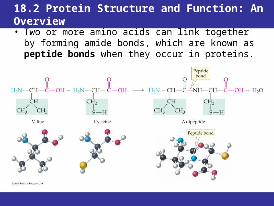
• The two mirror-image forms of a chiral molecule like alanine are called enantiomers or optical isomers (“optical” because of their effect on polarized light).
• Enantiomers are one kind of stereoisomer, compounds that have the same formula and atoms with the same connections but different spatial arrangements.
• Pairs of enantiomers have many of the same physical properties: the same melting point, solubility in water, isoelectric point, and density.

© 2013 Pearson Education, Inc.
18.6 Molecular Handedness and Amino Acids
• Pairs of enantiomers differ in their effect on polarized light and in how they react with other molecules that are also chiral.
• Pairs of enantiomers often differ in their biological activity, odors, tastes, or activity as drugs.
• 19 of the 20 common amino acids are chiral. Only the L-enantiomers are found in proteins.

© 2013 Pearson Education, Inc.
18.7 Protein Primary Structure
• The primary structure of a protein is the sequence in which its amino acids are lined up and connected by peptide bonds.
• Along the backbone of a protein is a chain of alternating peptide bonds and α-carbon atoms.
• The amino acid side chains are substituents along the backbone, where they are bonded to the carbon atoms.

© 2013 Pearson Education, Inc.
18.7 Protein Primary Structure
• The carbon and nitrogen atoms along the backbone lie in a zigzag arrangement, with tetrahedral bonding around the α-carbon atoms.
• The carbonyl-group double bond electrons are shared with the adjacent C—N bond. This sharing makes the C—N bond sufficiently like a double bond that there is no rotation around it.
• The carbonyl group, the N—H group bonded to it, and the two adjacent α-carbons form a rigid, planar unit.

© 2013 Pearson Education, Inc.
18.7 Protein Primary Structure
• Two amino acids can form two different dipeptides, X—Y and Y—X.
• Peptides and proteins are always written with the amino terminal amino acid (also called N-terminal) on the left and carboxyl-terminal amino acid (C-terminal) on the right.
• The individual amino acids joined in the chain are referred to as residues.
• A peptide is named by citing the amino acid residues in order, starting at the N-terminus and ending with the C-terminus.

© 2013 Pearson Education, Inc.
18.7 Protein Primary Structure
• The primary structure of a protein is the result of amino acids being lined in precisely the correct order.
• So crucial is primary structure to function that the change of only one amino acid can drastically alter a protein’s biological properties.
• Sickle-cell anemia is the result of a single amino acid substitution that replaces one amino acid (glutamate, Glu) with another (valine, Val) in the hemoglobin molecule.
• A hydrophobic pocket is exposed on the surface of the hemoglobin and hydrophobic valine on another hemoglobin molecule is drawn into this pocket.
• Insoluble fibrous chains are formed, forcing the cell into a sickle shape.

© 2013 Pearson Education, Inc.
18.7 Protein Primary StructureProteins in the Diet
• Proteins are a necessary part of the daily diet because our bodies do not store proteins like they do carbohydrates and fats.
• 9 of the 20 amino must be obtained in the diet. These are known as the essential amino acids.
• A complete protein source provides each of the nine essential amino acids. Most meat and dairy products meet this requirement.
• Traditional vegetarian food combinations provide complementary proteins. – Grains are low in lysine and threonine, but contain methionine and tryptophan. – Legumes (lentils, beans, and peas) supply lysine and threonine, but are low in
methionine and tryptophan. The two sources of protein complement each other.
• Disorders caused by inadequate protein intake are known as protein-energy malnutrition (PEM).
– In kwashiorkor, protein is deficient although caloric intake may be adequate. Children with kwashiorkor have an enlarged liver, and are underdeveloped.
– Marasmus is the result of starvation. It is identified with severe muscle wasting, below-normal stature, and poor response to treatment.
• A reassessment of diet in developed countries is underway, with a focus on disease prevention.
© 2013 Pearson Education, Inc.
18.8 Shape-Determining Interactions in Proteins
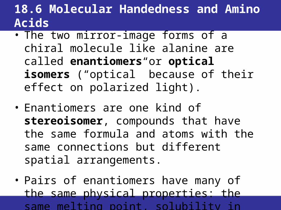
HYDROGEN BONDS ALONG THE BACKBONE Hydrogen bonds form between the hydrogen atoms
in the N—H groups and the oxygen atoms in the C=O groups along protein backbones.
HYDROGEN BONDS OF R GROUPS WITH EACH OTHER OR WITH BACKBONE ATOMS
• Some amino acid side chains contain atoms that can form hydrogen bonds. These can connect different parts of a protein molecule
• Often hydrogen-bonding side chains are present on the surface of a folded protein, where they can form hydrogen-bonds with surrounding water molecules.

© 2013 Pearson Education, Inc.
18.8 Shape-Determining Interactions in Proteins

© 2013 Pearson Education, Inc.
18.8 Shape-Determining Interactions in Proteins
IONIC ATTRACTIONS BETWEEN R GROUPS (SALT BRIDGES)
• Ionized acidic and basic side chains will create salt bridges.
HYDROPHOBIC INTERACTIONS BETWEEN R GROUPS
• Hydrocarbon side chains are attracted to each other by dispersion forces to create a water-free pocket. Although individual attractions are weak, their large number in proteins plays a major role in stabilizing the folded structures.
COVALENT SULFUR–SULFUR BONDS • Cysteine amino acid residues have side chains containing thiol
functional groups S-OH that can react to form sulfur–sulfur bonds.

© 2013 Pearson Education, Inc.
18.8 Shape-Determining Interactions in Proteins
Protein Analysis by Electrophoresis• Protein molecules in solution can be separated from each other by
taking advantage of their net charges.• In the electric field between two electrodes, a positively charged
particle moves toward the negative electrode and a negatively charged particle moves toward the positive electrode. This movement is electrophoresis.
• The net charge on a protein is determined by how many acidic or basic side-chains are ionized, and this depends on the pH.
• By varying the nature of the buffer, proteins can be separated in a variety of ways, including by their molecular weight.
• Once the separation is complete, the proteins are made visible by the addition of a dye.
• Electrophoresis is routinely used in the clinical laboratory for determining which proteins are present, and in what amounts in a blood sample.

© 2013 Pearson Education, Inc.
18.8 Shape-Determining Interactions in Proteins
Protein Analysis by Electrophoresis• Normal adult hemoglobin (HbA) and hemoglobin showing the
inherited sickle-cell trait (HbS) differ in their net charges.
• HbA and HbS move different distances during electrophoresis.
• A normal individual has only HbA.
• An individual with sickle-cell anemia has no HbA.
• An individual with sickle-cell trait has roughly equal amounts of HbA and HbS.
• HbA and HbS have negative charges of different magnitudes because HbS has two fewer Glu residues than HbA.
© 2013 Pearson Education, Inc.
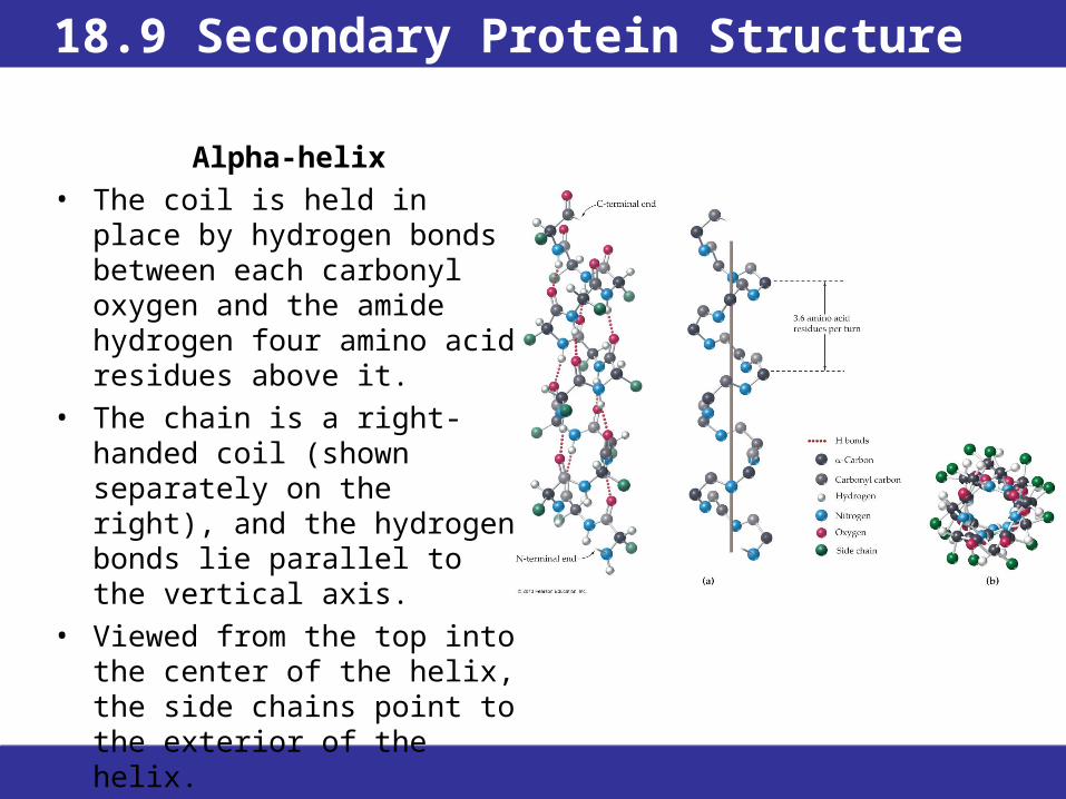
18.9 Secondary Protein Structure
Alpha-helix• The coil is held in place by
hydrogen bonds between each carbonyl oxygen and the amide hydrogen four amino acid residues above it.
• The chain is a right-handed coil (shown separately on the right), and the hydrogen bonds lie parallel to the vertical axis.
• Viewed from the top into the center of the helix, the side chains point to the exterior of the helix.

© 2013 Pearson Education, Inc.
18.9 Secondary Protein Structure
Beta-sheet• The hydrogen bonds stabilize
interactions between neighboring protein chains.
• The protein chains usually lie side by side so that alternating chains run from the N-terminal end to the C-terminal end and from the C-terminal end to the N-terminal end (known as the antiparallel arrangement).
• A pair of stacked pleated sheets illustrate how the R groups point above and below the sheets.

© 2013 Pearson Education, Inc.
18.9 Secondary Protein Structure
• Fibrous protein is a tough, insoluble protein whose protein chains form fibers or sheets.– Wool, hair, and fingernails are made of fibrous
proteins known as keratins which are composed of α-helices twisted together into small fibrils that are in turn twisted into larger and larger bundles.
– Natural silk and spider webs are made of fibroin, composed of stacks of β-sheets. The R groups must be relatively small, so fibroin contains regions of alternating glycine and alanine. The sheets stack so that sides with the smaller glycine hydrogens face each other and sides with the larger alanine methyl groups face each other.

© 2013 Pearson Education, Inc.
18.9 Secondary Protein Structure
• Globular protein is water-soluble protein whose chain is folded in a compact shape with hydrophilic groups on the outside.
• Their structures, which vary widely with their functions, are not regular.
• Sections of α-helix and β-sheet are usually present.
• Hydrophilic side chains on the outer surfaces of globular proteins account for their water solubility, allowing them to travel through body fluids.
• Many globular proteins are enzymes.
• The overall shapes of globular proteins represent another level of structure, tertiary structure.

© 2013 Pearson Education, Inc.
18.10 Tertiary Protein Structure
• The way in which an entire protein chain is coiled and folded into its specific three-dimensional shape is the protein’s tertiary protein structure.
• Each protein molecule folds in a distinctive manner that is determined by its primary structure and results in its maximum stability.
• A protein with the shape in which it functions in living systems is known as a native protein.
• A protein composed only of amino acid residues is a simple protein.
• Tertiary structure is drawn in a style that shows the combination of α-helix and β-sheet regions, the loops connecting them, and disulfide bonds.

© 2013 Pearson Education, Inc.
18.10 Tertiary Protein Structure
© 2013 Pearson Education, Inc.
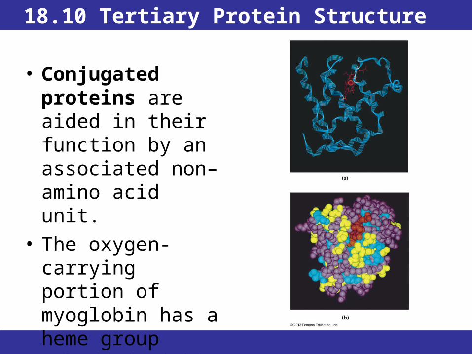
18.10 Tertiary Protein Structure
• Conjugated proteins are aided in their function by an associated non–amino acid unit.
• The oxygen-carrying portion of myoglobin has a heme group embedded within the polypeptide chain.

© 2013 Pearson Education, Inc.
18.11 Quaternary Protein Structure
• Quaternary protein structure is the way in which two or more protein chains aggregate to form large, ordered structures.
• Polypeptides are primarily held together by noncovalent forces, but covalent bonds and non–amino acid portions may also be incorporated.

© 2013 Pearson Education, Inc.
18.11 Quaternary Protein Structure
HEMOGLOBIN• Hemoglobin is a conjugated quaternary protein
composed of four polypeptide chains (two each of two polypeptides called α-chain and β-chain) held together by hydrophobic interactions and four heme groups.
• The α-chains have 141 amino acids, and the β-chains have 146 amino acids.
• The heme groups each contain an iron. Hemoglobin is the oxygen carrier in red blood cells. In the lungs, O2 binds to Fe2+ so that each hemoglobin can carry a maximum of four O2 molecules.
• In tissues in need of oxygen, O2 is released, and CO2 is picked up and carried back to the lungs.

© 2013 Pearson Education, Inc.
18.11 Quaternary Protein Structure
Figure 18.8 Heme and hemoglobin, a protein with quaternary structure. (a) The polypeptides are shown in purple, green, blue, and yellow, with their heme units in red. Each polypeptide resembles myoglobin in structure. (b) A heme unit is present in each of the four polypeptides in hemoglobin.

© 2013 Pearson Education, Inc.
18.11 Quaternary Protein Structure
COLLAGEN• Collagen is the major constituent
of connective tissues. • The basic structural unit of
collagen (tropocollagen) is three intertwined chains of about 1000 amino acids each. Each chain is loosely coiled in a left-handed (counterclockwise) direction.
• Three of these coiled chains wrap around one another (in a clockwise direction) to form a stiff, triple helix in which the chains are held together by hydrogen bonds.
• All collagens have a glycine every three residues, and prolines are hydroxylated in a reaction that requires vitamin C.

© 2013 Pearson Education, Inc.
18.11 Quaternary Protein StructureProtein Structure Summary
• Primary structure is the sequence of amino acids connected by peptide bonds in the polypeptide chain; for example, Asp-Arg-Val-Tyr.
• Secondary structure is the arrangement in space of the polypeptide chain, which includes the regular patterns of the α-helices and the β-sheet motifs (held together by hydrogen bonds between backbone carbonyl and amino groups in amino acid residues) plus the loops and coils that connect these segments.
• Tertiary structure is the folding of a protein molecule into a specific three- dimensional shape held together by noncovalent interactions primarily between amino acid side chains that can be quite far apart along the backbone and, in some cases, by disulfide bonds between side-chain thiol groups.
• Quaternary structure is two or more protein chains assembled in a larger three-dimensional structure held together by noncovalent interactions.
Classes of Proteins Summary
• Fibrous proteins are tough, insoluble, and composed of fibers and sheets.
• Globular proteins are water-soluble and have chains folded into compact shapes.
• Simple proteins contain only amino acid residues.
• Conjugated proteins include one or more non–amino acid units.

© 2013 Pearson Education, Inc.
18.11 Quaternary Protein StructureCollagen: A Tale of Two Diseases
Scurvy
• Scurvy is experienced whenever fresh fruits and vegetables are not available for long periods of time. Armies, navies, and medieval people in northern regions in late winter were particularly susceptible to scurvy.
• Symptoms include joint pain and swelling, blackened bruises on the skin, and swollen, bleeding gums accompanied by tooth loss.
• Lack of vitamin C leads to the formation of weak collagen. Since tropo collagen is part of capillary walls, weak collagen leads to the spontaneous bruising, bleeding, and soft tissue swelling that are characteristic of scurvy.
• In 1747, James Lind established that citrus prevented and cured scurvy on long sea voyages, leading to British sailors being known as “Limeys.”
Osteogenesis Imperfecta
• The primary symptom of this disease is spontaneous broken bones.
• Incorrectly synthesized collagen leads to weak bone structures. There is no cure; current research is directed at understanding the underlying biochemical defect in hopes of designing better treatment.
• A definitive diagnosis requires genetic testing.
© 2013 Pearson Education, Inc.
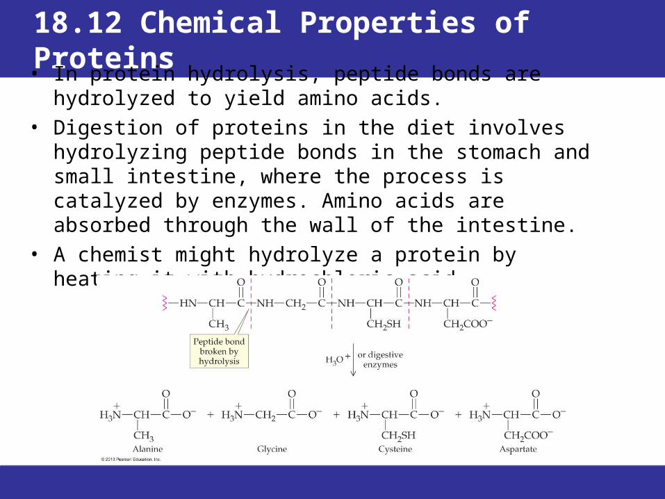
18.12 Chemical Properties of Proteins
• In protein hydrolysis, peptide bonds are hydrolyzed to yield amino acids.
• Digestion of proteins in the diet involves hydrolyzing peptide bonds in the stomach and small intestine, where the process is catalyzed by enzymes. Amino acids are absorbed through the wall of the intestine.
• A chemist might hydrolyze a protein by heating it with hydrochloric acid.
© 2013 Pearson Education, Inc.
18.12 Chemical Properties of Proteins
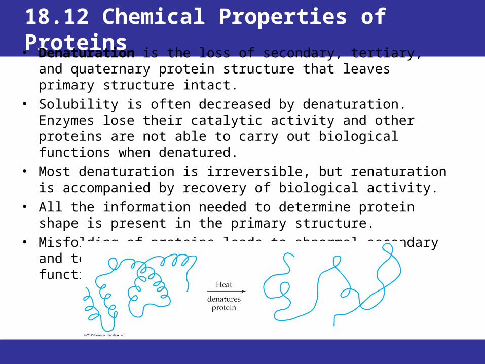
• Denaturation is the loss of secondary, tertiary, and quaternary protein structure that leaves primary structure intact.
• Solubility is often decreased by denaturation. Enzymes lose their catalytic activity and other proteins are not able to carry out biological functions when denatured.
• Most denaturation is irreversible, but renaturation is accompanied by recovery of biological activity.
• All the information needed to determine protein shape is present in the primary structure.
• Misfolding of proteins leads to abnormal secondary and tertiary structures that compromise the function of the protein.

© 2013 Pearson Education, Inc.
18.12 Chemical Properties of Proteins
• Heat—The weak side-chain attractions in globular proteins are easily disrupted by heating.
• Mechanical agitation—The most familiar example of denaturation by agitation is the foam produced by beating egg whites. Denaturation of proteins at the surface of the air bubbles stiffens the protein and causes the bubbles to be held in place.
• Detergents—Even very low concentrations of detergents can disrupt the association of hydrophobic side chains.
• Organic compounds—Polar solvents interfere with hydrogen bonding by competing for bonding sites.
• pH change—Excess H+ or OH– ions react with basic or acidic side chains in amino acid residues and disrupt salt bridges
• Inorganic salts—High concentrations of ions can disturb salt bridges.
© 2013 Pearson Education, Inc.
18.12 Chemical Properties of ProteinsPrions: Proteins that Cause Disease
• Prions (pronounced pree-ons) are “proteinaceous infectious particles.”
• They are associated with Creutzfeldt–Jakob disease (CJD), bovine spongiform encephalopathy (BSE), scrapie in sheep; a chronic wasting disease in elk and mule deer; and in humans kuru, and fatal familial insomnia (FFI).
• Some well-supported facts about prions include:– Humans and all animals tested thus far have a gene for making a normal
prion protein that resides in the brain.
– Alpha helices in normal prions are replaced by β-sheets, resulting in a disease-causing prion.
– A misfolded prion can induce a normal prion to flip from the normal shape to the disease-causing shape.
– The infectious nature of prions is not affected by heat or UV-radiation treatment.
– Synthetic prions cause neurological disease in mice similar to mad cow disease or CJD.

© 2013 Pearson Education, Inc.
Chapter Summary1. What are the structural features of amino acids? • Amino acids in body fluids have an ionized carboxylic acid group,
—COO–, an ionized amino group, NH3+, and a side-chain R group
bonded to a central carbon atom (the α-carbon). • Twenty different amino acids occur in proteins, connected by
peptide bonds (amide bonds) formed between the carboxyl group of one amino acid and the amino group of the next.
2. What are the properties of amino acids? • Amino acid side chains have acidic or basic functional groups or
neutral groups that are either polar or nonpolar. In glycine, the “side chain” is a hydrogen atom.
• The dipolar ion in which the amino and carboxylic acid groups are both ionized is known as a zwitterion.
• For each amino acid, there is a distinctive isoelectric point—the pH at which the numbers of positive and negative charges are equal. At more acidic pH, some carboxylic acid groups are not ionized; at more basic pH, some amino groups are not ionized.

© 2013 Pearson Education, Inc.
Chapter Summary, Continued3. Why do amino acids have “handedness?”
• An object, including a molecule, has “handedness” —is chiral—when it has no plane of symmetry and thus, has mirror images that cannot be superimposed on each other.
• A simple molecule can be identified as chiral if it contains a carbon atom bonded to four different groups. All α-amino acids, except glycine, meet this condition by having four different groups bonded to the α carbon.
4. What is the primary structure of a protein and what conventions are used for drawing and naming primary structures?
• Proteins are polymers of amino acids (polypeptides).
• Their primary structure is the linear sequence in which the amino acids are connected by peptide bonds. Using formulas or amino acid abbreviations, the primary structures are written with the amino-terminal end on the left and the carboxyl-terminal end on the right.
• To name a peptide, the names of the amino acids are combined, starting at the amino-terminal end, with the endings of all but the carboxyl-terminal amino acid changed to -yl.
• Primary structures are often represented by combining three-letter abbreviations for the amino acids.

© 2013 Pearson Education, Inc.
Chapter Summary, Continued
5. What types of interactions determine the overall shapes of proteins?
• Protein chains are drawn into their distinctive and biochemically active shapes by attractions between atoms along their backbones and between atoms in side-chain groups.
• Hydrogen bonding can occur between the backbone carbonyl groups and amide hydrogens of adjacent protein chains.
• Noncovalent interactions between side chains include ionic bonding between acidic and basic groups (salt bridges), and hydrophobic interactions among nonpolar groups.
• Covalent sulfur–sulfur bonds (disulfide bonds) can form bridges between the side chains in cysteine.

© 2013 Pearson Education, Inc.
Chapter Summary, Continued6. What are the secondary and tertiary structures of proteins? • Secondary structures include the regular, repeating three-dimensional
structures held in place by hydrogen bonding between backbone atoms within a chain or in adjacent chains.
– The α-helix is a coil with hydrogen bonding between carbonyl oxygen atoms and amide hydrogen atoms four amino acid residues farther along the same chain.
– The β-sheet is a pleated sheet with adjacent protein-chain segments connected by hydrogen bonding between peptide groups. The adjacent chains in the β-sheet may be parts of the same protein chain or different protein chains.
• Secondary structure mainly determines the properties of fibrous proteins, which are tough and insoluble.
• Tertiary structure is the overall three-dimensional shape of a folded protein chain.
• Tertiary structure determines the properties of globular proteins, which are water-soluble, with hydrophilic groups on the outside and hydrophobic groups on the inside. Globular proteins often contain regions of α-helix and/or β-sheet secondary structures.

© 2013 Pearson Education, Inc.
Chapter Summary, Continued
7. What is quaternary protein structure? • Proteins that incorporate more than one peptide chain are said to have
quaternary structure.
• In a quaternary structure, two or more folded protein subunits are united in a single structure by noncovalent interactions.
• Hemoglobin, for example, consists of two pairs of subunits, with a nonprotein heme molecule in each of the four subunits. Collagen is a fibrous protein composed of protein chains twisted together in triple helixes.
8. What chemical properties do proteins have? • The peptide bonds are broken by hydrolysis, which may occur in acidic
solution or during enzyme-catalyzed digestion of proteins in food. The end result of hydrolysis is production of the individual amino acids from the protein.
• Denaturation is the loss of overall structure by a protein while retaining its primary structure. Among the agents that cause denaturation are heat, mechanical agitation, pH change, and exposure to a variety of chemical agents, including detergents.