Embed Size (px)
Citation preview
© 2014 Pearson Education, Inc.
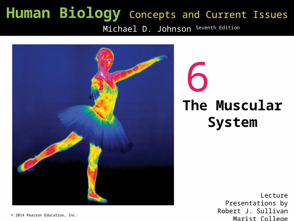
Human Biology Concepts and Current Issues Seventh Edition
Michael D. Johnson
Lecture Presentations byRobert J. Sullivan
Marist College
6The Muscular
System
© 2014 Pearson Education, Inc.
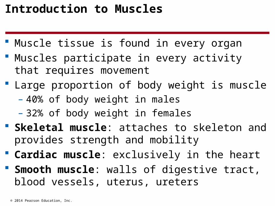
Introduction to Muscles
Muscle tissue is found in every organ Muscles participate in every activity that requires
movement Large proportion of body weight is muscle
– 40% of body weight in males
– 32% of body weight in females Skeletal muscle: attaches to skeleton and provides
strength and mobility Cardiac muscle: exclusively in the heart Smooth muscle: walls of digestive tract, blood
vessels, uterus, ureters
© 2014 Pearson Education, Inc.
Muscles Produce Movement or Generate Tension
Muscles may produce movement– Voluntary: conscious control over movement (picking
up a pen)
– Involuntary: unconscious control over movement (beating of heart)
Many muscles resist movement– Maintenance of posture
– Maintenance of blood pressure Muscles generate heat

© 2014 Pearson Education, Inc.
The Fundamental Activity of Muscle Is Contraction
Excitable: contract in response to electrical or chemical stimuli
All muscle cells have one mechanism of action:– They contract (shorten), then relax (lengthen)
© 2014 Pearson Education, Inc.
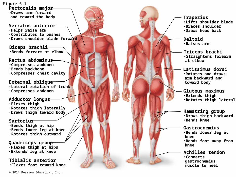
Figure 6.1Pectoralis major•Draws arm forwardand toward the body
Serratus anterior•Helps raise arm•Contributes to pushes•Draws shoulder blade forward
Biceps brachii•Bends forearm at elbow
Rectus abdominus•Compresses abdomen•Bends backbone•Compresses chest cavity
External oblique•Lateral rotation of trunk•Compresses abdomen
Adductor longus•Flexes thigh•Rotates thigh laterally•Draws thigh toward body
Sartorius•Bends thigh at hip•Bends lower leg at knee•Rotates thigh outward
Quadriceps group•Flexes thigh at hips•Extends leg at knee
Tibialis anterior•Flexes foot toward knee
Trapezius•Lifts shoulder blade•Braces shoulder•Draws head back
Deltoid•Raises arm
Triceps brachi•Straightens forearmat elbow
Latissimus dorsi•Rotates and drawsarm backward andtoward body
Gluteus maximus•Extends thigh•Rotates thigh laterally
Hamstring group•Draws thigh backward•Bends knee
Gastrocnemius•Bends lower leg atknee
•Bends foot away fromknee
Achilles tendon•Connectsgastrocnemiusmuscle to heel

© 2014 Pearson Education, Inc.
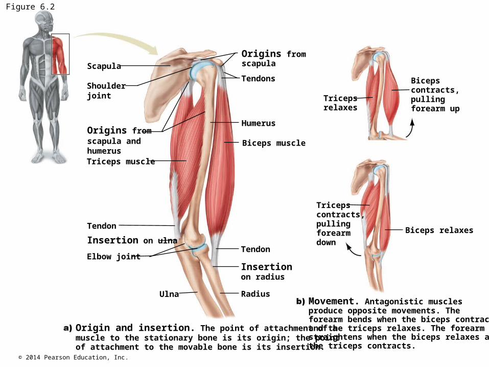
Skeletal Muscles Cause Bones to Move
600 skeletal muscles Synergistic muscles: work together to created the
same movement Antagonistic muscles: muscles that oppose each
other Many muscles attach to bones via tendons Origin: end of muscle that attaches to relatively
stationary bone Insertion: end of muscle attached to another bone
across a joint, action pulls insertion toward origin
© 2014 Pearson Education, Inc.
Figure 6.2
Scapula
Shoulderjoint
Origins fromscapula andhumerusTriceps muscle
Tendon
Elbow joint
Ulna Radius
Tendon
Biceps muscle
Humerus
Tendons
Tricepsrelaxes
Tricepscontracts, pulling forearm down
Biceps relaxes
Biceps contracts, pulling forearm up
Origins fromscapula
Insertion on ulna
Insertion on radius
Origin and insertion. The point of attachment of a muscle to the stationary bone is its origin; the point of attachment to the movable bone is its insertion.
Movement. Antagonistic muscles produce opposite movements. The forearm bends when the biceps contracts and the triceps relaxes. The forearm straightens when the biceps relaxes and the triceps contracts.
© 2014 Pearson Education, Inc.
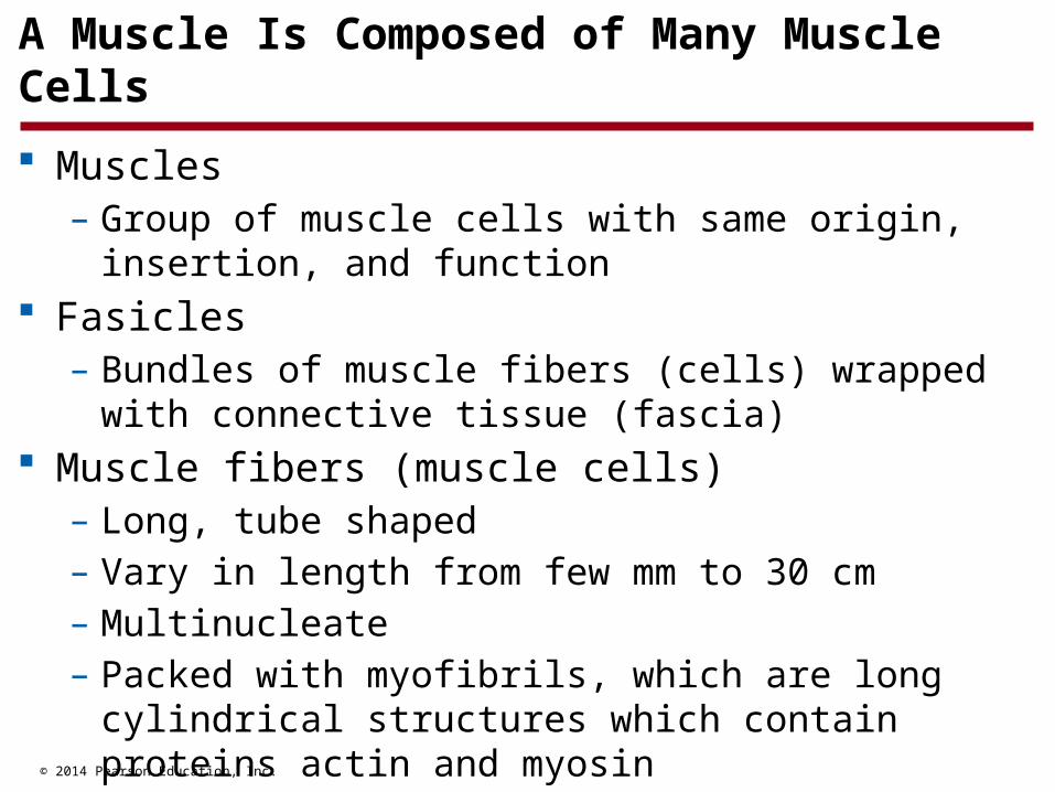
A Muscle Is Composed of Many Muscle Cells
Muscles– Group of muscle cells with same origin, insertion, and
function Fasicles
– Bundles of muscle fibers (cells) wrapped with connective tissue (fascia)
Muscle fibers (muscle cells)– Long, tube shaped– Vary in length from few mm to 30 cm– Multinucleate– Packed with myofibrils, which are long cylindrical
structures which contain proteins actin and myosin
© 2014 Pearson Education, Inc.
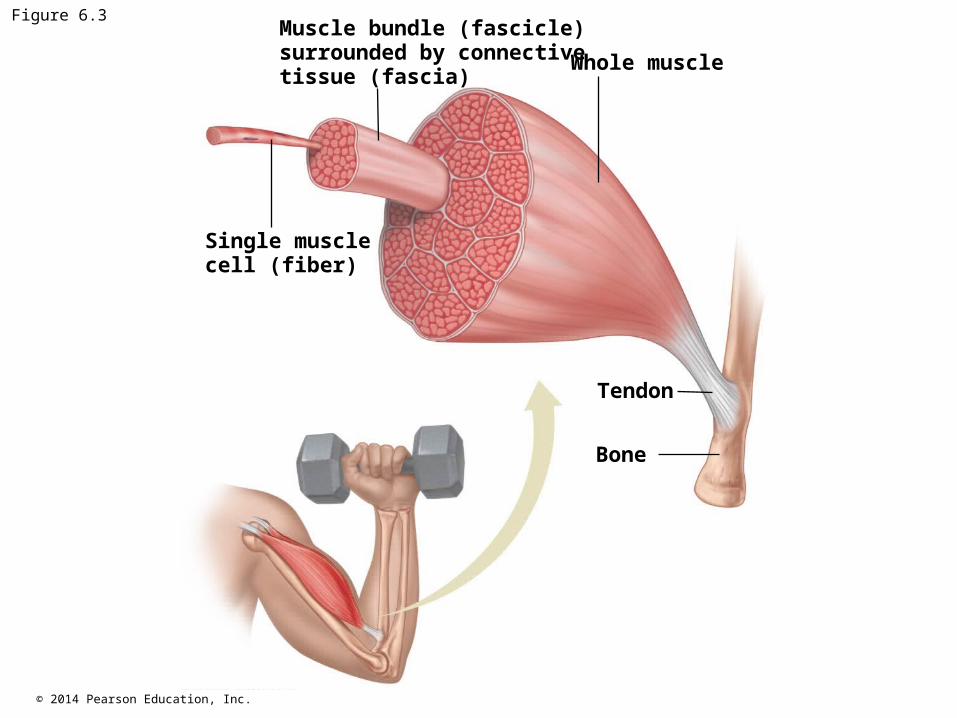
Figure 6.3Muscle bundle (fascicle)surrounded by connectivetissue (fascia)
Whole muscle
Single musclecell (fiber)
Tendon
Bone
© 2014 Pearson Education, Inc.
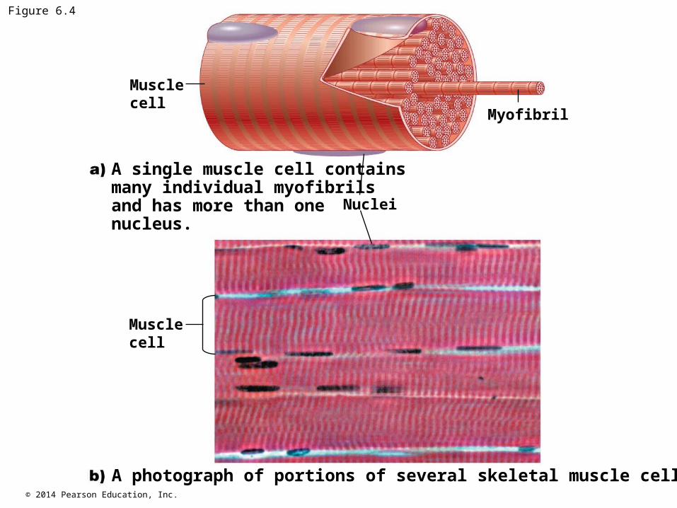
Figure 6.4
Musclecell
Myofibril
Nuclei
Musclecell
A single muscle cell contains many individual myofibrils and has more than one nucleus.
A photograph of portions of several skeletal muscle cells.

© 2014 Pearson Education, Inc. Animation: Muscle Structure and Function Right-click and select Play
© 2014 Pearson Education, Inc.
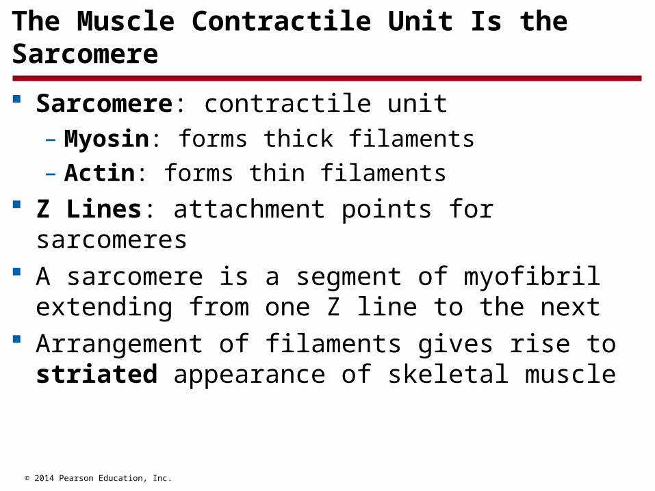
The Muscle Contractile Unit Is the Sarcomere
Sarcomere: contractile unit– Myosin: forms thick filaments
– Actin: forms thin filaments Z Lines: attachment points for sarcomeres A sarcomere is a segment of myofibril extending
from one Z line to the next Arrangement of filaments gives rise to striated
appearance of skeletal muscle
© 2014 Pearson Education, Inc.
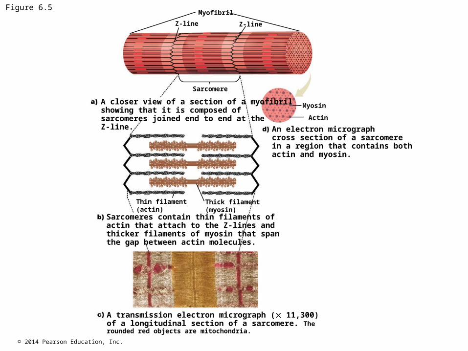
Figure 6.5Myofibril
Z-lineZ-line
Sarcomere
Myosin
Actin
Thin filament(actin)
Thick filament(myosin)
A closer view of a section of a myofibril showing that it is composed of sarcomeres joined end to end at theZ-line. An electron micrograph
cross section of a sarcomerein a region that contains bothactin and myosin.
Sarcomeres contain thin filaments of actin that attach to the Z-lines and thicker filaments of myosin that span the gap between actin molecules.
A transmission electron micrograph ( 11,300) of a longitudinal section of a sarcomere. The rounded red objects are mitochondria.

© 2014 Pearson Education, Inc.
Individual Muscle Cells Contract and Relax
Muscle contraction: each sarcomere shortens a little Basic process of contraction:
1. Skeletal muscle must be activated by a nerve
2. Nerve activation increases the concentration of calcium ions in the vicinity of the contractile proteins
3. Presence of calcium permits contractions
4. When nerve stimulation stops, contraction stops
© 2014 Pearson Education, Inc.
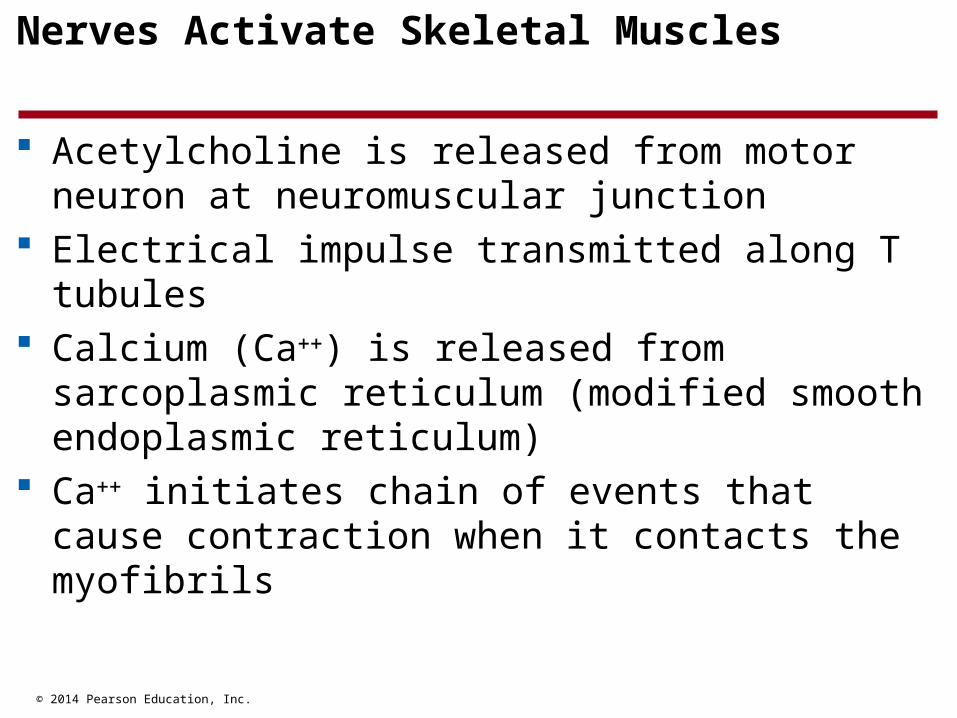
Nerves Activate Skeletal Muscles
Acetylcholine is released from motor neuron at neuromuscular junction
Electrical impulse transmitted along T tubules Calcium (Ca) is released from sarcoplasmic
reticulum (modified smooth endoplasmic reticulum) Ca initiates chain of events that cause contraction
when it contacts the myofibrils
© 2014 Pearson Education, Inc.
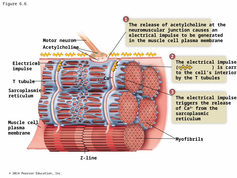
Figure 6.6
Motor neuron
Electricalimpulse
T tubule
Sarcoplasmicreticulum
Muscle cellplasmamembrane
Z-line
Myofibrils
Ca2
Acetylcholine
The release of acetylcholine at the neuromuscular junction causes anelectrical impulse to be generated in the muscle cell plasma membrane
The electrical impulse triggers the release of Ca2 from the sarcoplasmic reticulum
The electrical impulse ( ) is carriedto the cell’s interior by the T tubules
1
2
3
© 2014 Pearson Education, Inc.
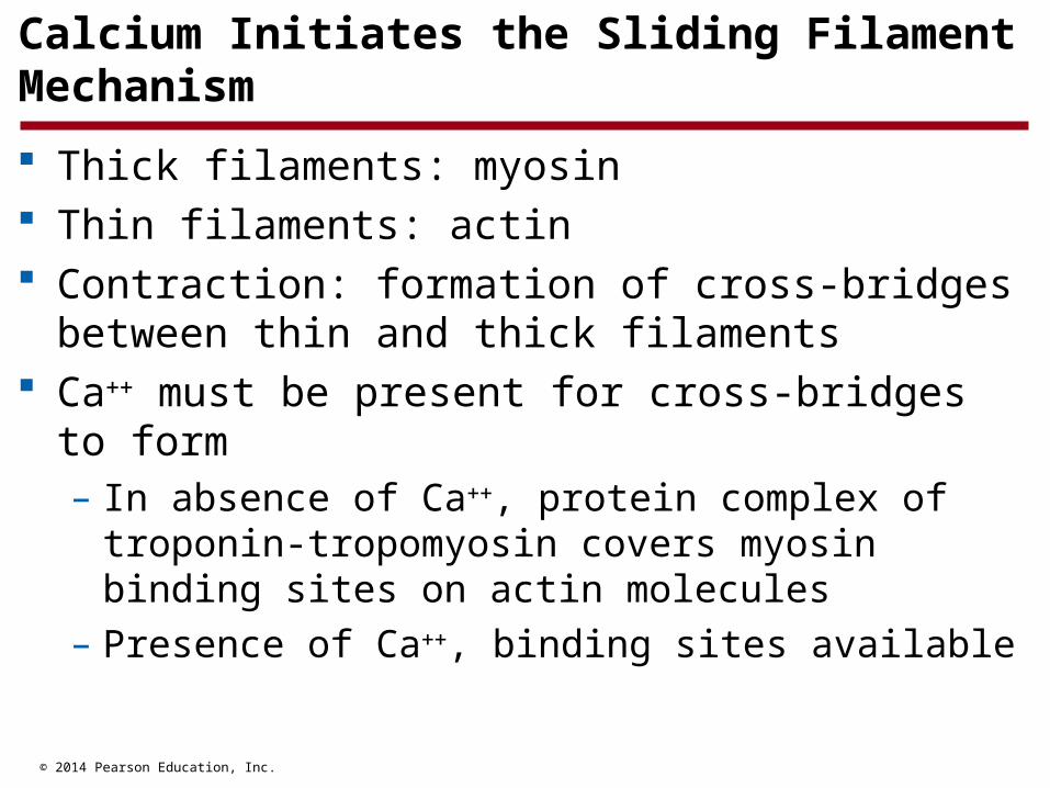
Calcium Initiates the Sliding Filament Mechanism
Thick filaments: myosin Thin filaments: actin Contraction: formation of cross-bridges between thin
and thick filaments Ca must be present for cross-bridges to form
– In absence of Ca, protein complex of troponin-tropomyosin covers myosin binding sites on actin molecules
– Presence of Ca, binding sites available

© 2014 Pearson Education, Inc.
Figure 6.7
Myofibril
Thickfilament
Thinfilament
Myosin molecule head
Myosinmolecule
ActinmoleculeRelaxed state. The myosin heads do
not make contact with actin.
Contraction. The myosin heads form cross-bridges with actin andthen bend, pulling the actin filaments toward the center of the sarcomere.
© 2014 Pearson Education, Inc.
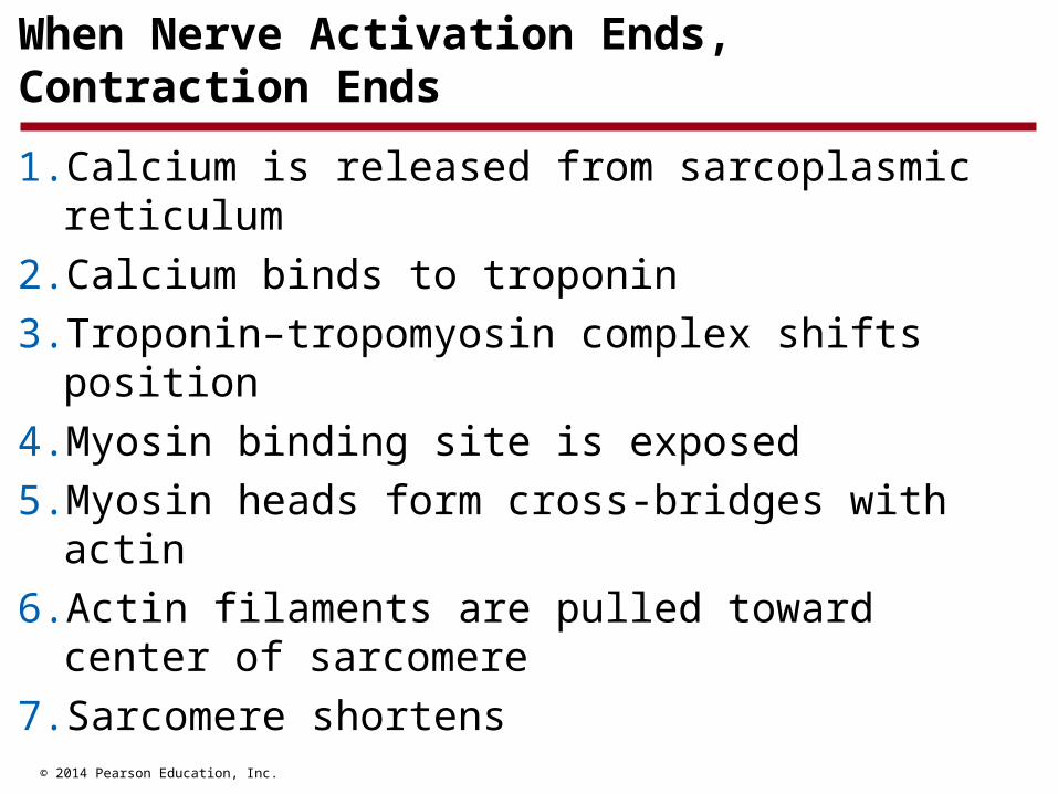
When Nerve Activation Ends, Contraction Ends
1. Calcium is released from sarcoplasmic reticulum
2. Calcium binds to troponin
3. Troponin–tropomyosin complex shifts position
4. Myosin binding site is exposed
5. Myosin heads form cross-bridges with actin
6. Actin filaments are pulled toward center of sarcomere
7. Sarcomere shortens

© 2014 Pearson Education, Inc.
Figure 6.8
Myofibril
Sarcoplasmicreticulum
Thickand thinfilaments
Tropomyosin
Actin filament
Troponin
Myosin head
Myosinfilament
Sarcoplasmicreticulum
Electricalimpulse
Calcium release
Myosinbindingsites
Cross-bridge
Ca2 Ca2
Resting sarcomere. In the absence of calcium the muscle is relaxed because the myosin heads cannot form cross-bridges with actin.
Cross-bridge attachment. The binding of calcium to troponin causes a shift in the troponin-tropomyosin complex, allowing cross-bridges to form.

© 2014 Pearson Education, Inc.
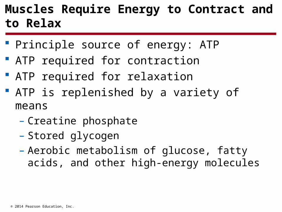
Muscles Require Energy to Contract and to Relax
Nerve activation ends, contraction ends Ca pumped back into sarcoplasmic reticulum
(requires ATP) Ca no longer bound to troponin Myosin binding site covered ATP must bind to myosin before myosin heads can
detach from actin No calcium no cross-bridges Muscle relaxes
© 2014 Pearson Education, Inc.
Muscles Require Energy to Contract and to Relax
Principle source of energy: ATP ATP required for contraction ATP required for relaxation ATP is replenished by a variety of means
– Creatine phosphate
– Stored glycogen
– Aerobic metabolism of glucose, fatty acids, and other high-energy molecules

© 2014 Pearson Education, Inc.
Table 6.1
© 2014 Pearson Education, Inc.
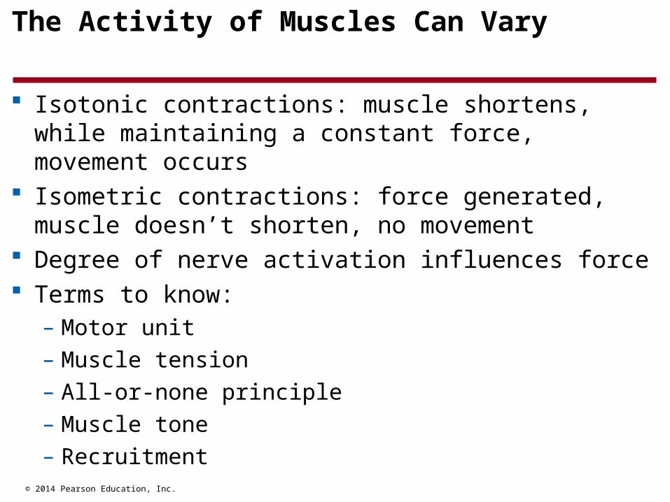
The Activity of Muscles Can Vary
Isotonic contractions: muscle shortens, while maintaining a constant force, movement occurs
Isometric contractions: force generated, muscle doesn’t shorten, no movement
Degree of nerve activation influences force Terms to know:
– Motor unit
– Muscle tension
– All-or-none principle
– Muscle tone
– Recruitment
© 2014 Pearson Education, Inc.
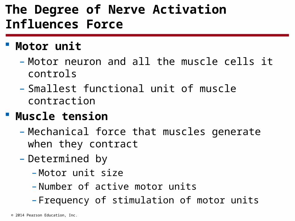
The Degree of Nerve Activation Influences Force
Motor unit – Motor neuron and all the muscle cells it controls
– Smallest functional unit of muscle contraction Muscle tension
– Mechanical force that muscles generate when they contract
– Determined by– Motor unit size– Number of active motor units– Frequency of stimulation of motor units
© 2014 Pearson Education, Inc.
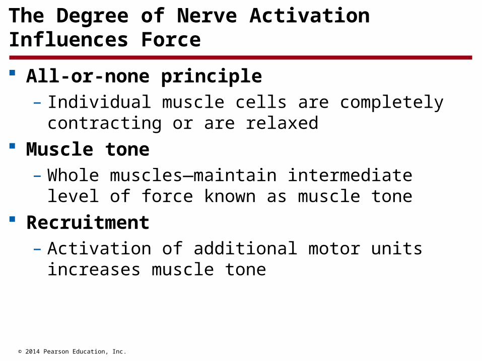
The Degree of Nerve Activation Influences Force
All-or-none principle – Individual muscle cells are completely contracting or
are relaxed Muscle tone
– Whole muscles—maintain intermediate level of force known as muscle tone
Recruitment– Activation of additional motor units increases muscle
tone
© 2014 Pearson Education, Inc.
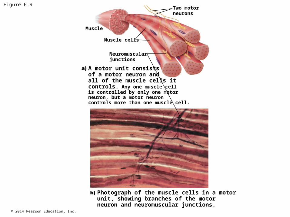
Figure 6.9
Muscle
Muscle cells
Neuromuscular junctions
Two motorneurons
A motor unit consists of a motor neuron and all of the muscle cells it controls. Any one muscle cell is controlled by only one motor neuron, but a motor neuron controls more than one muscle cell.
Photograph of the muscle cells in a motor unit, showing branches of the motor neuron and neuromuscular junctions.
© 2014 Pearson Education, Inc.
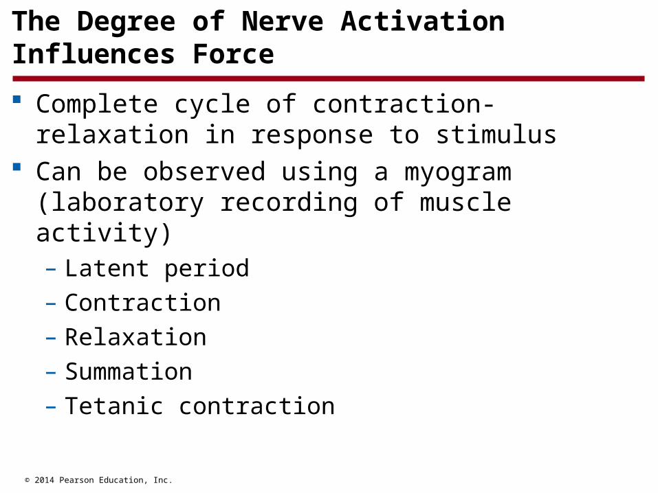
The Degree of Nerve Activation Influences Force
Complete cycle of contraction-relaxation in response to stimulus
Can be observed using a myogram (laboratory recording of muscle activity)– Latent period
– Contraction
– Relaxation
– Summation
– Tetanic contraction
© 2014 Pearson Education, Inc.
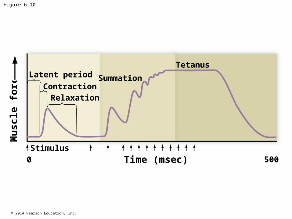
Figure 6.10
Tetanus
SummationLatent period
ContractionRelaxation
500
Stimulus
0 Time (msec)
Mu
scle
fo
rce
© 2014 Pearson Education, Inc.
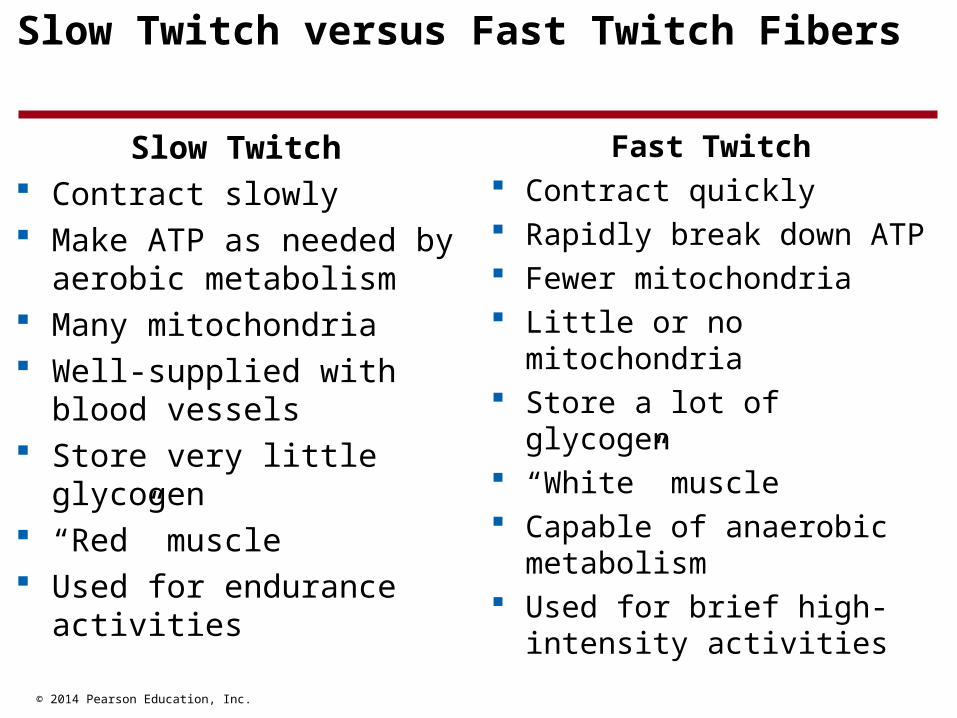
Slow Twitch versus Fast Twitch Fibers
Slow Twitch Contract slowly Make ATP as needed by
aerobic metabolism Many mitochondria Well-supplied with blood
vessels Store very little glycogen “Red” muscle Used for endurance
activities
Fast Twitch Contract quickly Rapidly break down ATP Fewer mitochondria Little or no mitochondria Store a lot of glycogen “White” muscle Capable of anaerobic
metabolism Used for brief high-intensity
activities
© 2014 Pearson Education, Inc.
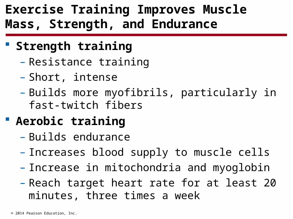
Exercise Training Improves Muscle Mass, Strength, and Endurance
Strength training– Resistance training
– Short, intense
– Builds more myofibrils, particularly in fast-twitch fibers Aerobic training
– Builds endurance
– Increases blood supply to muscle cells
– Increase in mitochondria and myoglobin
– Reach target heart rate for at least 20 minutes, three times a week
© 2014 Pearson Education, Inc.
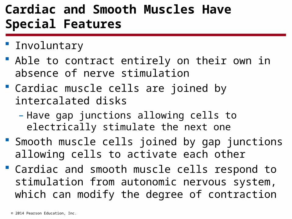
Cardiac and Smooth Muscles Have Special Features
Involuntary Able to contract entirely on their own in absence of
nerve stimulation Cardiac muscle cells are joined by intercalated disks
– Have gap junctions allowing cells to electrically stimulate the next one
Smooth muscle cells joined by gap junctions allowing cells to activate each other
Cardiac and smooth muscle cells respond to stimulation from autonomic nervous system, which can modify the degree of contraction

© 2014 Pearson Education, Inc.
Figure 6.12
Intercalated discCardiac muscle cell
Adhesion junction
Protein channel
Gap junction
Cell membranes of adjacent cells
A closer view showing that intercalated discs arebridged by gap junctions that permit direct electrical connections between cells.
A view of several adjacent cardiac muscle cells showing their blunt shape and the intercalated discs that join them together.
© 2014 Pearson Education, Inc.
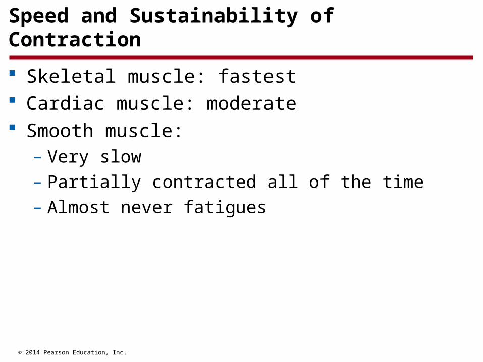
Speed and Sustainability of Contraction
Skeletal muscle: fastest Cardiac muscle: moderate Smooth muscle:
– Very slow
– Partially contracted all of the time
– Almost never fatigues

© 2014 Pearson Education, Inc.
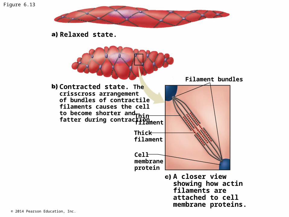
Arrangement of Myosin and Actin Filaments
Cardiac muscle– Sarcomere arrangement of thick and thin filaments
– Striated appearance Smooth muscle
– Filaments arranged in criss-crossed bundles, not sarcomeres
– No striations
© 2014 Pearson Education, Inc.
Figure 6.13
Filament bundles
Thinfilament
Thickfilament
Cellmembraneprotein
Relaxed state.
Contracted state. Thecrisscross arrangementof bundles of contractilefilaments causes the cellto become shorter andfatter during contraction.
A closer view showing how actin filaments are attached to cell membrane proteins.
© 2014 Pearson Education, Inc.
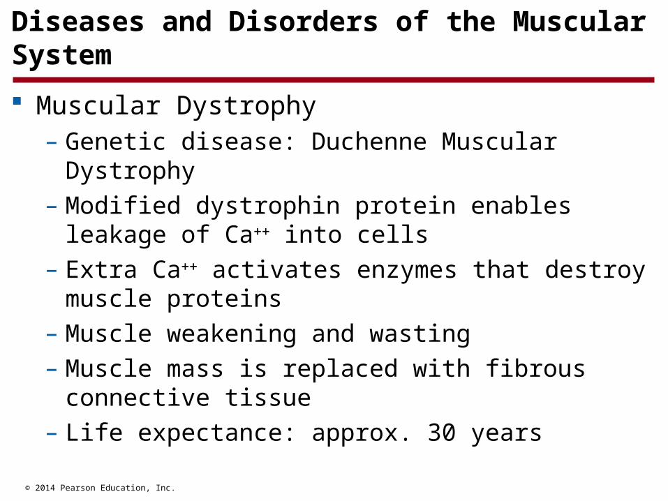
Diseases and Disorders of the Muscular System
Muscular Dystrophy– Genetic disease: Duchenne Muscular Dystrophy
– Modified dystrophin protein enables leakage of Ca into cells
– Extra Ca activates enzymes that destroy muscle proteins
– Muscle weakening and wasting
– Muscle mass is replaced with fibrous connective tissue
– Life expectance: approx. 30 years
© 2014 Pearson Education, Inc.
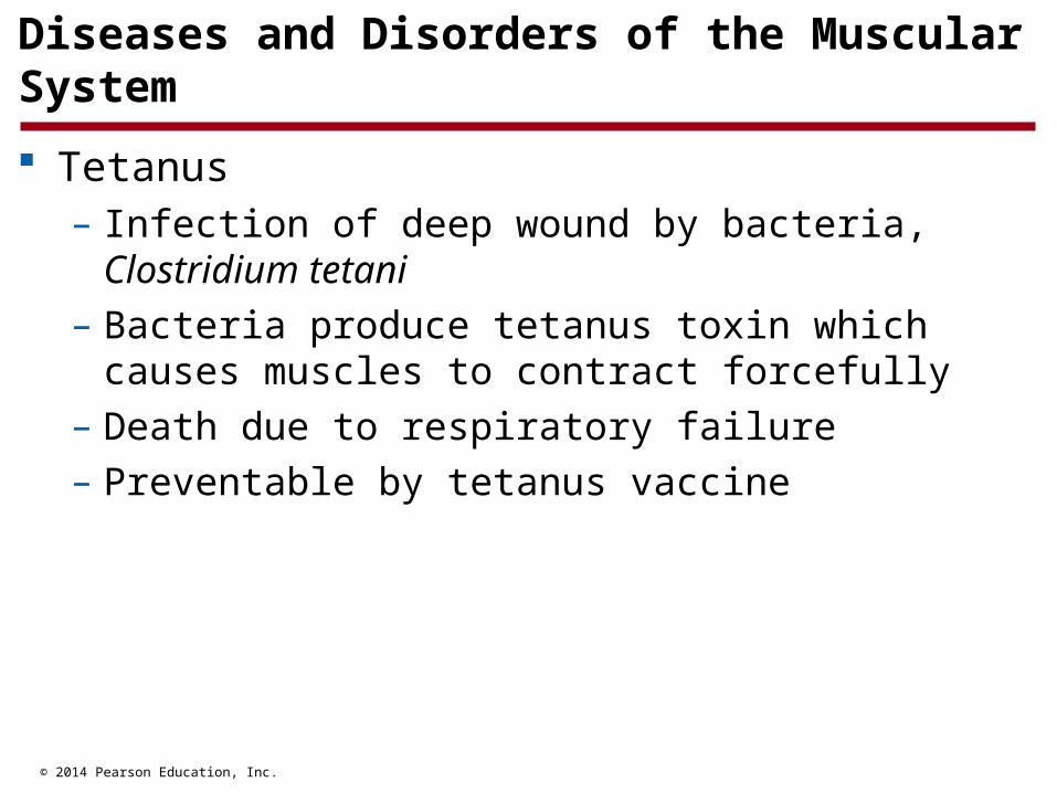
Diseases and Disorders of the Muscular System
Tetanus– Infection of deep wound by bacteria, Clostridium tetani
– Bacteria produce tetanus toxin which causes muscles to contract forcefully
– Death due to respiratory failure
– Preventable by tetanus vaccine
© 2014 Pearson Education, Inc.
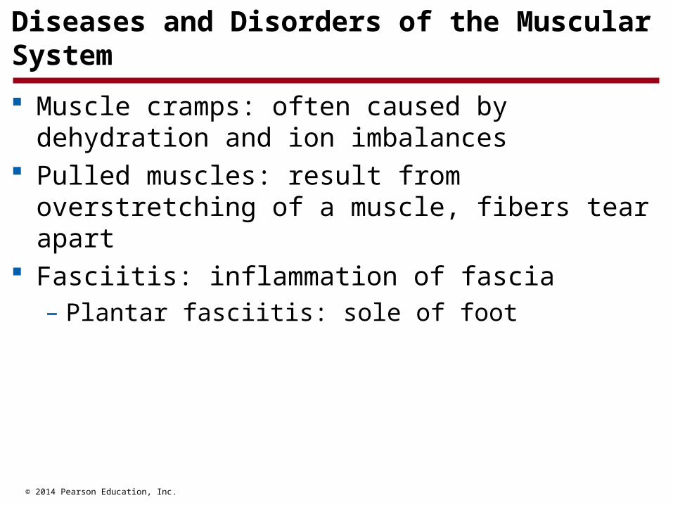
Diseases and Disorders of the Muscular System
Muscle cramps: often caused by dehydration and ion imbalances
Pulled muscles: result from overstretching of a muscle, fibers tear apart
Fasciitis: inflammation of fascia– Plantar fasciitis: sole of foot