Embed Size (px)
Citation preview
生理学报 Acta Physiologica Sinica, December 25, 2014, 66(6): 709–717DOI: 10.13294/j.aps.2014.0084 http://www.actaps.com.cn
709
小鼠心室肌脱细胞化细胞外基质薄片的制备和评价
姜煜东,李文思,余 翀,王 璐,孙小夕,席姣娅*
华中科技大学同济医学院基础医学院生理学系,中德干细胞中心,湖北省药物靶向研究评价重点实验室,武汉 430030
摘 要:心肌细胞外基质(extracellular matrix, ECM)可由心肌经脱细胞处理制得,被广泛认为是一种理想的制备工程心肌的生
物支架材料。然而目前的脱细胞方法尚存在不足,本研究拟联合使用经典去垢剂改良脱细胞方法,制备性能更为优良的心
肌ECM薄片,以用于构建工程心肌片。用振荡切片机将包埋于低熔点琼脂糖中的成年昆明小白鼠心室肌组织沿心脏横轴切
成300 μm厚的薄片,随机分为正常对照组、SDS脱细胞组(0.1% SDS处理)和改良脱细胞组(0.1% SDS和0.5% Triton X-100联合
处理)。通过总RNA和总蛋白质含量分析、HE染色和免疫荧光染色等方法评估各组的脱细胞程度和ECM成分保留状态;将
改良脱细胞组ECM与小鼠胚胎干细胞源心肌细胞(murine embryonic stem cell-derived cardiomyocytes, mES-CMs)和小鼠胚胎成
纤维细胞(murine embryonic fibroblasts, MEFs)共培养以检测其生物相容性。结果显示:SDS脱细胞组和改良脱细胞组ECM中
残留的总RNA及蛋白质含量均低于对照组。HE染色结果显示改良脱细胞组核质去除较SDS脱细胞组更彻底。改良脱细胞组
可见胶原蛋白IV和层粘连蛋白两种ECM关键成分表达量和分布接近正常心肌组织,而SDS脱细胞组中这两种蛋白明显减少
且分布紊乱。mES-CMs和MEFs能存活于改良脱细胞组ECM表面12天以上并向内迁移。综上,SDS和Triton X-100联合脱细
胞法制备ECM薄片效果明显,能更好地保留天然ECM成分和结构,具有良好的生物相容性。
关键词:心肌片;组织工程;脱细胞化;细胞外基质
中图分类号:R329.3;Q813.1
Acquirement and evaluation of murine ventricular extracellular matrix
JIANG Yu-Dong, LI Wen-Si, YU Chong, WANG Lu, SUN Xiao-Xi, XI Jiao-Ya*
Department of Physiology and Chinese-German Stem Cell Center, School of Basic Medicine, Tongji Medical College, Huazhong University of Science and Technology, the Key Laboratory for Drug Target Researches and Pharmacodynamic Evaluation of Hubei Province, Wuhan 430030, China
Abstract: Cardiac extracellular matrix (ECM), generated from the process of decellularization, has been widely considered as an ideal source of biological scaffolds. However, current ECM preparations are generally difficult to be applied to generate cardiac tissue. Our research was aimed to improve decellularization protocols to prepare cardiac ECM slices. Adult murine ventricular tissues were embedded in low melting agarose and cut into 300 μm slices, and then were divided randomly into three groups: normal cardiac tissue, SDS treated group (0.1% SDS) and SDS+Triton X-100 treated group (0.1% SDS+0.5% Triton X-100). Total RNA content and protein content quantification, HE staining and immunostaining were used to evaluate the removal of cell components and preserva-tion of vital ECM components. Furthermore, murine embryonic stem cell-derived cardiomyocytes (mES-CMs) and mouse embryonic fibroblasts (MEFs) were co-cultured with ECM slices to evaluate biocompatibility. The relative residual RNA and protein contents of
Received 2014-05-11 Accepted 2014-07-09This work was supported by the National Natural Science Foundation of China (No. 31100828), the Natural Science Foundation
of Hubei Province, China (No. 2011CDB363), the Scientific Research Foundation for the Returned Overseas Chinese Scholars, State Education Ministry, China, the Fundamental Research Funds (No. 2013TS145)/the Fundamental Research Funds for the Undergrad-uates (No. HUST: 2011B142, 2012B366) in Huazhong University of Science and Technology, the National Undergraduate Training Programs for Innovation and Entrepreneurship (No. HUST: 2012175), and the Science and Technology Innovation Funds for the Uni-versity or College Students, China (No. 9).
*Corresponding author. Tel: +86-27-83692622; Fax: +86-27-83692608; E-mail: [email protected]
研究论文
生理学报 Acta Physiologica Sinica, December 25, 2014, 66(6): 709–717 710
ECM slices significantly decreased after decellularization. HE staining showed that SDS+Triton X-100 treatment better destroyed cellular structure and removed nuclei of ECM slices, compared with SDS treatment. Immunostaining showed that collagen IV and laminin were better preserved and presented better similarity to original cardiac tissue in ECM slices acquired by SDS+Triton X-100 treatment. However, collagen IV and laminin were significantly decreased and arranged disorderly in SDS treated group. We observed effective survival (≥ 12 days) of MEFs and mES-CMs on ECM slices acquired by SDS+Triton X-100 treatment, and signs of integra-tion, whereas those signs were not found in SDS treated group. We concluded that, compared with traditional SDS method, new combined protocol (SDS+Triton X-100) generated ECM slices with better component and structural preservation, as well as better biocompatibility.
Key words: myocardium slice; tissue engineering; decellularization; extracellular matrix
心肌梗死引发心肌细胞衰竭和死亡,造成心脏
泵血功能下降,心肌代偿性增生肥大乃至最后的心
力衰竭。心脏移植被认为是拯救晚期心力衰竭的
最佳方法,但供体来源缺乏和排斥反应导致只有少
数患者可以受益 [1]。近年来研究表明,用组织工程
技术在体外构建工程心肌 (engineered cardiac tissue, ECT) 并移植给患者,有望明显修复心泵功能 [2]。
该技术需要将合适的种子细胞种植在合适的支架材
料上,离体构建具有收缩功能的 ECT。尽管如此,
ECT 在移植后仅能紧紧地附着于宿主心肌表面,而
不是有效地从结构上整合于宿主心肌,梗死区内可
见 ECT 和宿主心肌的明显界限 [3],这将大大限制
ECT 对梗死区微环境的调节作用,明显制约移植
ECT 对宿主心功能的修复效果和对患者的治疗效
果。ECT 难以和宿主心肌达到结构和功能高效整合
的原因之一是:构建三维 ECT 所用支架的理化特性、
超微结构和三维构架与天然心肌差异较大 [4]。人们
一直致力于制备各种接近细胞外基质 (extracellular matrix, ECM) 结构的仿生可降解多孔支架用于心肌
组织工程,如人工合成的多孔聚合物支架 [4,5]、纳
米材料模拟支架 [5,6] 或各种水凝胶 ( 如胶原蛋白、
纤维蛋白等 ) 等 [7]。然而这些人工合成的支架,无
论是超微结构还是功能,仍和天然的心肌 ECM 相
差甚远,基于这些支架构建的 ECT 和天然心肌都
有不同程度的差别。
将心肌脱细胞处理可制备出保留天然成分、超
微结构和三维结构的心肌 ECM,能有效支持细胞
的存活和生长,并影响其迁移和分化,是一种构
建 ECT 的理想支架 [8]。脱细胞化获取心肌 ECM组织片的方法多种多样,最常用到的脱细胞试剂
是去垢剂和酶类 [9] ;物理方法例如超声波和射线
等已被尝试用于进一步提升脱细胞化效率 [10]。理
想的脱细胞化方法是在保留 ECM 超微结构和生物
活性的基础上,尽可能去除组织中的所有细胞成
分,从而基本排除免疫原性 [11]。不同脱细胞试剂
或方法对各种细胞成分的去除不尽相同,所制得
的 ECM 中各种成分的含量也存在明显差异 [12]。越
来越多的研究表明,采用单一脱细胞化试剂的脱
细胞效率有限,单纯提高试剂浓度和反应时间不
仅不能显著提升脱细胞化效率,反而可能过度消
化 ECM 中关键成分 [13],对支架结构造成进一步
损伤,影响种子细胞的粘附生长和重组心肌力学
功能的发挥,不利于仿生 ECT 的构建 [14]。如何改
进脱细胞方法以尽可能去除细胞成分,同时使制
备的 ECM 能最大程度地保留原有基质成分和结
构,是目前亟待解决的关键问题。
本研究拟联合使用经典脱细胞试剂 SDS 和 Triton X-100,改良脱细胞方法,制备性能更为优良的心
肌 ECM 薄片,为构建仿生 ECT 片提供理想的生物
支架。
1 材料和方法
1.1 伦理学声明 所有动物实验均遵守国际实验
动物学伦理规范,并获得湖北省科学技术院批准
(2005-50)。1.2 小鼠心肌薄片的制备 [15,16] 取 6 周龄成年昆
明小白鼠 ( 华中科技大学实验动物中心 ) 麻醉后立
即取出心脏,剪除心房及大血管,将余下心室肌组
织包埋于 37 ℃、4% 低熔点琼脂糖中,立即置于冰
上冷却成型。用振荡切片机 (Leica VT1000S,德国
Leica 公司 ) 沿横断面切成 300 μm 心室肌薄片,置
于 4 ℃无钙台氏液中保存。
1.3 脱细胞化制备心肌 ECM 薄片 将心室肌薄
片随机分为正常对照组、SDS 脱细胞组和改良脱
细胞组。改良脱细胞组心肌片置于 0.1% SDS 中,
37 ℃振荡反应 11.5 h,去除 SDS,加入 PBS 漂洗
姜煜东等:脱细胞ECM制备改良 711
三次,再加入 0.5% Triton X-100,37 ℃振荡反
应 0.5 h。SDS 脱细胞组心室肌薄片按相同反应条
件用 0.1% SDS 处理 12 h。两组反应结束后,均加
入 PBS 振荡漂洗三次,时间分别为 10 min、30 min和 1 h,以去除残余脱细胞试剂,避免对 ECM 的进
一步损伤 [17]。正常对照组心室肌薄片置于含双抗的
PBS 中,37 ℃振荡反应 12 h。1.4 总 RNA 定量和总蛋白定量分析 随机选取
正常对照组、SDS 脱细胞组和改良脱细胞组处理后
组织片,分别称重后,使用 Trizol 提取总 RNA,经
氯仿和异丙醇萃取得到 RNA 纯水溶液,并在 260 nm 波长处测定吸光度,计算总 RNA 相对含量 ( 单位 μg/g) ;用 RIPA 试剂抽提得到总蛋白质溶液。取
部分蛋白样品,与 BCA 蛋白定量试剂盒反应,在
562 nm波长处测定吸光度,计算蛋白质相对含量 (单位 μg/mg)。1.5 HE 染色 随机选取正常对照组、SDS 脱细
胞组和改良脱细胞组处理后组织片,用 4% 多聚甲
醛 (paraformaldehyde, PFA) 室温下固定 2 h,OCT包埋,用冰冻切片机 ( 德国 Leica 公司 ) 切成 8 μm厚度冰冻切片。冰冻切片经水洗、苏木素染色、盐
酸酒精脱色、伊红染色、梯度酒精及二甲苯脱水处
理,制得 HE 染色切片。在光镜 (TE2000-S,日本
Nikon 公司 ) 下观察。
1.6 免疫荧光染色 [16] 用 0.5% Trion X-100 在室
温下对 8 μm 冰冻切片破膜,漂洗后用 5% 的牛血
清白蛋白 4 ℃封闭 1 h。用抗小鼠胶原蛋白Ⅳ (1:800, Abcam) 或抗小鼠层粘连蛋白 (1:200, Abcam) 作为
一抗 4 ℃孵育过夜。漂洗后加入抗鼠 IgG-Alexa Flour 647 或 IgM-Alexa Flour 647荧光抗体 (Molecular Probes) 作为二抗,室温避光孵育 1 h。Hoechst 33342室温下染核后,用无荧光甘油封片。在荧光显微镜
(TE2000-S,日本 Nikon 公司 ) 下观察胶原蛋白Ⅳ和
层粘连蛋白表达状态。
1.7 小鼠胚胎干细胞源心肌细胞 (murine embryonic stem cell-derived cardiomyocytes, mES-CMs) 和小
鼠胚胎成纤维细胞 (murine embryonic fibroblasts, MEFs) 的制备 如本研究组前期文献报道 [16,18,19]
诱导小鼠转基因 ES 细胞 (D3 系,α-PIG44 克隆 )[20]
向心肌分化。概括地说,用含 20% 胎牛血清的
IMDM 培养基悬浮 mES 细胞制成细胞密度为 106 个 /mL 的细胞悬液,置于摇床上 37 ℃、5% CO2 培养
箱中培养 2 天形成胚体,按 103 胚体 / 皿稀释后继
续置于摇床上 37 ℃、5% CO2 培养箱中培养至第 9天出现跳动胚体。加入 10 μg/mL 的嘌呤霉素筛选
纯化 mES-CMs[20],第 14 天可获得纯化的表达 GFP的 mES-CMs,用于后续实验。
如本研究组前期文献报道 [16] 制备 MEFs。概括
地说,断颈处死怀孕 14.5 天的母鼠 ( 昆明小白鼠,
华中科技大学实验动物中心 ),无菌条件下取出胚
胎,去除头尾、四肢及内脏,保留躯干部分,剪碎
并用胰酶消化成单细胞悬液。在含 15% 胎牛血清的
IMDM培养基中进行原代培养 (培养条件37.5 ℃,5% CO2),每 2 天更换培养基,培养至第 4 代时,可用
于后续实验。
1.8 ECM 重细胞化 如本研究组前期文献报
道 [16],将 SDS 脱细胞组和改良脱细胞组 1 片 ECM薄片分别置于特制底部带漏斗形凹槽的共培养皿 [15]
中,微干燥后加入 10 μL 含 1 × 10⁵ MEFs 的细胞悬
液,混匀后静置 10 min,再加入 1 mL 含 20% 胎牛血清的 IMEM 培养基 37 ℃培养 30 min 后,加入 (10 ± 1)个纯化的 mES-CMs 团块,小心移至 37 ℃、5% CO2 培养箱中静置培养。共培养过程中每 24 h 在荧
光显微镜 (TE2000-S,日本 Nikon 公司 ) 下观察生
长状况,每 2 天更换培养基。共培养后在荧光显微
镜下观察GFP阳性的mES-CMs的数量和分布变化。
共培养第 12 天时,将组织固定并制成 8 μm 冰冻切
片,进行 HE 染色观察。
1.9 统计学分析 采用 SPSS13.0 软件对数据进行
分析,结果以 mean ± SD 表示。采用非配对 t 检验
分析,P < 0.05 时认为差异有统计学显著性意义。
2 结果
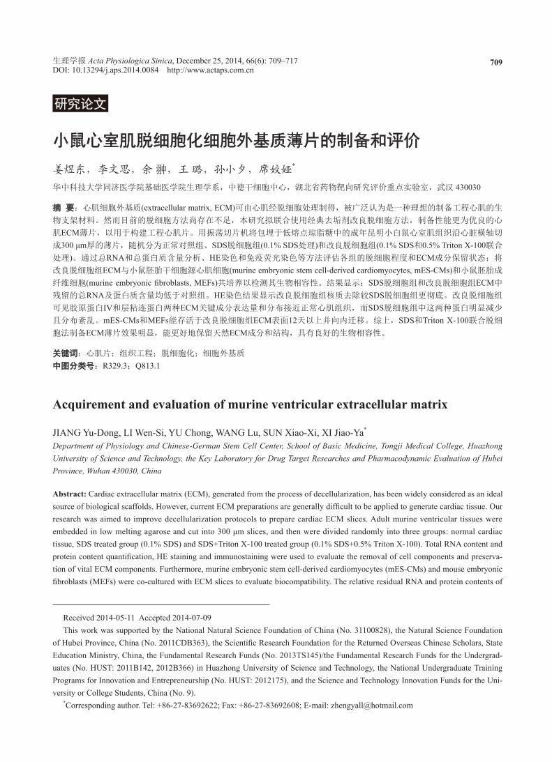
2.1 脱细胞处理能有效去除ECM中残留细胞成分
总 RNA 定量显示,正常对照组心肌片总 RNA相对含量为 (1 026.20 ± 212.12) μg/g,SDS 脱细胞
组和改良脱细胞组 ECM 薄片中总 RNA 相对含量
均明显低于正常对照组心肌片 ( 图 1A),分别为
(420.19 ± 58.66) μg/g (n = 6, P < 0.001) 和 (417.68 ± 80.90) μg/g (n = 6, P < 0.001),两个脱细胞组间无明
显差异 (P = 0.96)。总蛋白质定量检测显示相似结果,正常对照组
心肌片总蛋白质相对含量为 (195.58 ± 23.21) μg/mg,SDS 脱细胞组和改良脱细胞组 ECM 薄片总蛋白
质相对含量均明显低于正常对照组心肌片 ( 图 1B),分别为 (42.04 ± 13.49) μg/mg (n = 6, P < 0.001) 和 (44.22 ±
生理学报 Acta Physiologica Sinica, December 25, 2014, 66(6): 709–717 712
24.39) μg/mg (n = 6, P < 0.001),两个脱细胞组间无
明显差异 (P = 0.88)。以上结果提示两种脱细胞方法均能有效去除
ECM 中残留细胞成分,从理论上降低免疫原性。
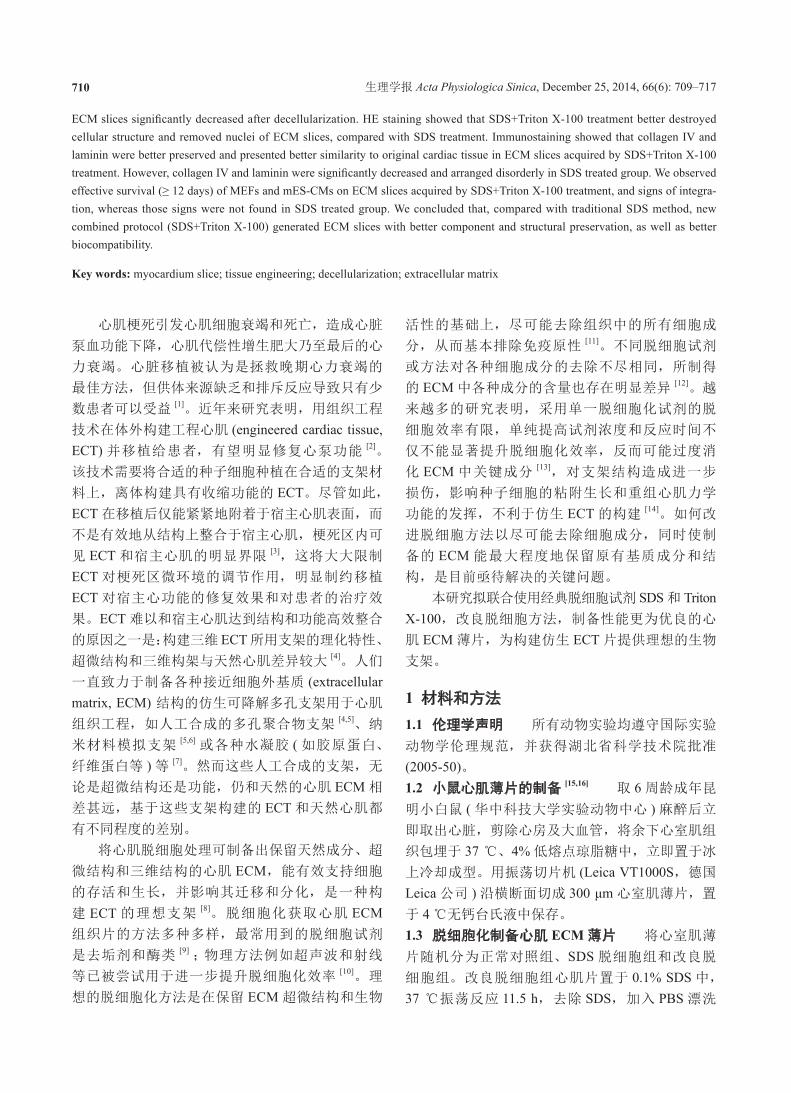
2.2 SDS+Triton X-100改良脱细胞法能有效去除心
肌细胞核,并更好地保留ECM原有结构
如图 2 所示,HE 染色后正常对照组心肌薄片
肌纤维清晰可见,可见清晰的核仁、心肌纤维及闰
图 1. 脱细胞处理能有效去除ECM中残留细胞成分
Fig. 1. Decellularization can effectively reduce residual cell components in ECM. The relative residual total RNA (A) and total protein (B) contents in SDS treated group and SDS+Triton X-100 treated group were significantly lower than those in normal ventricular slices. Data were presented as mean ± SD. *P < 0.001. n.s, no significant difference. n = 6.
图 2. SDS+Triton X-100改良脱细胞法能有效去除心肌细胞核,并更好地保留ECM原有结构 Fig. 2. SDS+Triton X-100 decellularization can effectively remove nuclei and better preserve original structure of ECM. Representa-tive images of light microscopy (upper) and HE staining (lower) of normal ventricular tissue, SDS treated group and SDS+Triton X-100 treated group. A few granular nuclei can be observed in SDS treated group, while no sign of nuclei was observed in SDS+Triton X-100 treated group. Scale bar, 100 μm.
姜煜东等:脱细胞ECM制备改良 713
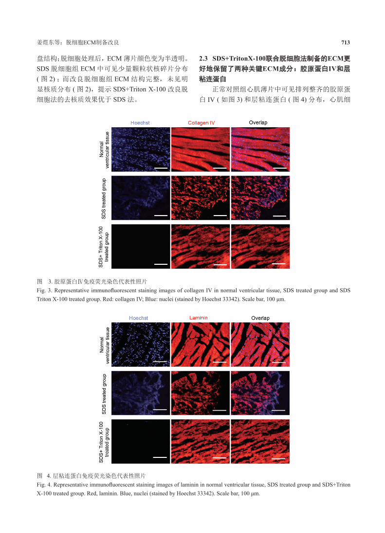
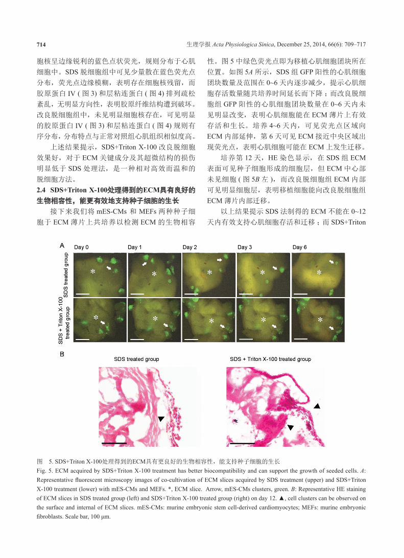
盘结构;脱细胞处理后,ECM 薄片颜色变为半透明。
SDS 脱细胞组 ECM 中可见少量颗粒状核碎片分布
( 图 2) ;而改良脱细胞组 ECM 结构完整,未见明
显核质分布 ( 图 2),提示 SDS+Triton X-100 改良脱
细胞法的去核质效果优于 SDS 法。
2.3 SDS+TritonX-100联合脱细胞法制备的ECM更
好地保留了两种关键ECM成分:胶原蛋白IV和层
粘连蛋白
正常对照组心肌薄片中可见排列整齐的胶原蛋
白 IV ( 如图 3) 和层粘连蛋白 ( 图 4) 分布,心肌细
图 3. 胶原蛋白Ⅳ免疫荧光染色代表性照片
Fig. 3. Representative immunofluorescent staining images of collagen IV in normal ventricular tissue, SDS treated group and SDS Triton X-100 treated group. Red: collagen IV; Blue: nuclei (stained by Hoechst 33342). Scale bar, 100 μm.
图 4. 层粘连蛋白免疫荧光染色代表性照片
Fig. 4. Representative immunofluorescent staining images of laminin in normal ventricular tissue, SDS treated group and SDS+Triton X-100 treated group. Red, laminin. Blue, nuclei (stained by Hoechst 33342). Scale bar, 100 μm.
生理学报 Acta Physiologica Sinica, December 25, 2014, 66(6): 709–717 714
胞核呈边缘锐利的蓝色点状荧光,规则分布于心肌
细胞中。SDS 脱细胞组中可见少量散在蓝色荧光点
分布,荧光点边缘模糊,表明存在细胞核残留,而
胶原蛋白 IV ( 图 3) 和层粘连蛋白 ( 图 4) 排列疏松
紊乱,无明显方向性,表明胶原纤维结构遭到破坏。
改良脱细胞组中,未见明显细胞核存在,可见明显
的胶原蛋白 IV ( 图 3) 和层粘连蛋白 ( 图 4) 规则有
序分布,分布特点与正常对照组心肌组织相似度高。
上述结果提示,SDS+Triton X-100 改良脱细胞
效果好,对于 ECM 关键成分及其超微结构的损伤
明显低于 SDS 处理法,是一种相对高效而温和的
脱细胞方法。
2.4 SDS+Triton X-100处理得到的ECM具有良好的
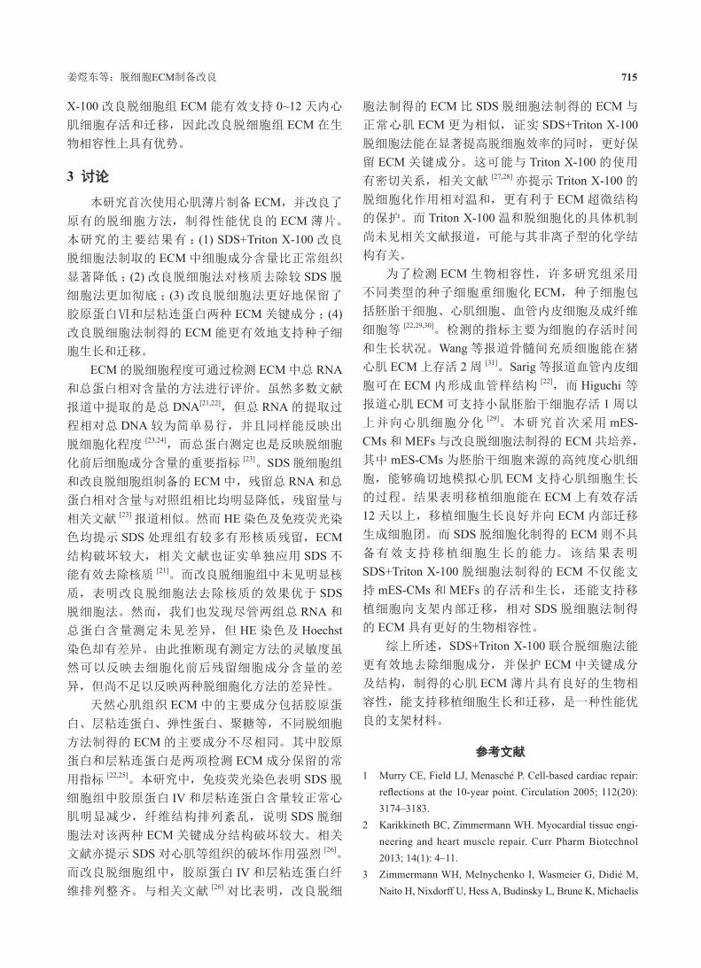
生物相容性,能更有效地支持种子细胞的生长
接下来我们将 mES-CMs 和 MEFs 两种种子细
胞于 ECM 薄片上共培养以检测 ECM 的生物相容
性。图 5 中绿色荧光点即为移植心肌细胞团块所在
位置。如图 5A 所示,SDS 组 GFP 阳性的心肌细胞
团块数量及范围在 0~6 天内逐步减少,提示心肌细
胞存活数量随共培养时间延长而下降;而改良脱细
胞组 GFP 阳性的心肌细胞团块数量在 0~6 天内未
见明显改变,表明心肌细胞能在 ECM 薄片上有效
存活和生长。培养 4~6 天内,可见荧光点区域向
ECM 内部延伸,第 6 天可见 ECM 接近中央区域出
现荧光点,表明心肌细胞可能在 ECM 上发生迁移。
培养第 12 天,HE 染色显示,在 SDS 组 ECM表面可见种子细胞形成的细胞层,但 ECM 中心部
未见细胞 ( 图 5B 左 ),而改良脱细胞组 ECM 内部
可见明显细胞层,表明移植细胞能向改良脱细胞组
ECM 薄片内部迁移。
以上结果提示 SDS 法制得的 ECM 不能在 0~12天内有效支持心肌细胞存活和迁移;而 SDS+Triton
图 5. SDS+Triton X-100处理得到的ECM具有更良好的生物相容性,能支持种子细胞的生长
Fig. 5. ECM acquired by SDS+Triton X-100 treatment has better biocompatibility and can support the growth of seeded cells. A: Representative fluorescent microscopy images of co-cultivation of ECM slices acquired by SDS treatment (upper) and SDS+Triton X-100 treatment (lower) with mES-CMs and MEFs. *, ECM slice. Arrow, mES-CMs clusters, green. B: Representative HE staining of ECM slices in SDS treated group (left) and SDS+Triton X-100 treated group (right) on day 12. ▲, cell clusters can be observed on the surface and internal of ECM slices. mES-CMs: murine embryonic stem cell-derived cardiomyocytes; MEFs: murine embryonic fibroblasts. Scale bar, 100 μm.
姜煜东等:脱细胞ECM制备改良 715
X-100 改良脱细胞组 ECM 能有效支持 0~12 天内心
肌细胞存活和迁移,因此改良脱细胞组 ECM 在生
物相容性上具有优势。
3 讨论
本研究首次使用心肌薄片制备 ECM,并改良了
原有的脱细胞方法,制得性能优良的 ECM 薄片。
本研究的主要结果有:(1) SDS+Triton X-100 改良
脱细胞法制取的 ECM 中细胞成分含量比正常组织
显著降低;(2) 改良脱细胞法对核质去除较 SDS 脱
细胞法更加彻底;(3) 改良脱细胞法更好地保留了
胶原蛋白Ⅵ和层粘连蛋白两种 ECM 关键成分;(4)改良脱细胞法制得的 ECM 能更有效地支持种子细
胞生长和迁移。
ECM 的脱细胞程度可通过检测 ECM 中总 RNA和总蛋白相对含量的方法进行评价。虽然多数文献
报道中提取的是总 DNA[21,22],但总 RNA 的提取过
程相对总 DNA 较为简单易行,并且同样能反映出
脱细胞化程度 [23,24],而总蛋白测定也是反映脱细胞
化前后细胞成分含量的重要指标 [23]。SDS 脱细胞组
和改良脱细胞组制备的 ECM 中,残留总 RNA 和总
蛋白相对含量与对照组相比均明显降低,残留量与
相关文献 [23] 报道相似。然而 HE 染色及免疫荧光染
色均提示 SDS 处理组有较多有形核质残留,ECM结构破坏较大,相关文献也证实单独应用 SDS 不
能有效去除核质 [21]。而改良脱细胞组中未见明显核
质,表明改良脱细胞法去除核质的效果优于 SDS脱细胞法。然而,我们也发现尽管两组总 RNA 和
总蛋白含量测定未见差异,但 HE 染色及 Hoechst染色却有差异。由此推断现有测定方法的灵敏度虽
然可以反映去细胞化前后残留细胞成分含量的差
异,但尚不足以反映两种脱细胞化方法的差异性。
天然心肌组织 ECM 中的主要成分包括胶原蛋
白、层粘连蛋白、弹性蛋白、聚糖等,不同脱细胞
方法制得的 ECM 的主要成分不尽相同。其中胶原
蛋白和层粘连蛋白是两项检测 ECM 成分保留的常
用指标 [22,25]。本研究中,免疫荧光染色表明 SDS 脱
细胞组中胶原蛋白 IV 和层粘连蛋白含量较正常心
肌明显减少,纤维结构排列紊乱,说明 SDS 脱细
胞法对该两种 ECM 关键成分结构破坏较大。相关
文献亦提示 SDS 对心肌等组织的破坏作用强烈 [26]。
而改良脱细胞组中,胶原蛋白 IV 和层粘连蛋白纤
维排列整齐。与相关文献 [26] 对比表明,改良脱细
胞法制得的 ECM 比 SDS 脱细胞法制得的 ECM 与
正常心肌 ECM 更为相似,证实 SDS+Triton X-100脱细胞法能在显著提高脱细胞效率的同时,更好保
留 ECM 关键成分。这可能与 Triton X-100 的使用
有密切关系,相关文献 [27,28] 亦提示 Triton X-100 的
脱细胞化作用相对温和,更有利于 ECM 超微结构
的保护。而 Triton X-100 温和脱细胞化的具体机制
尚未见相关文献报道,可能与其非离子型的化学结
构有关。
为了检测 ECM 生物相容性,许多研究组采用
不同类型的种子细胞重细胞化 ECM,种子细胞包
括胚胎干细胞、心肌细胞、血管内皮细胞及成纤维
细胞等 [22,29,30]。检测的指标主要为细胞的存活时间
和生长状况。Wang 等报道骨髓间充质细胞能在猪
心肌 ECM 上存活 2 周 [31]。Sarig 等报道血管内皮细
胞可在 ECM 内形成血管样结构 [22],而 Higuchi 等报道心肌 ECM 可支持小鼠胚胎干细胞存活 1 周以
上并向心肌细胞分化 [29]。本研究首次采用 mES-CMs 和 MEFs 与改良脱细胞法制得的 ECM 共培养,
其中 mES-CMs 为胚胎干细胞来源的高纯度心肌细
胞,能够确切地模拟心肌 ECM 支持心肌细胞生长
的过程。结果表明移植细胞能在 ECM 上有效存活
12 天以上,移植细胞生长良好并向 ECM 内部迁移
生成细胞团。而 SDS 脱细胞化制得的 ECM 则不具
备有效支持移植细胞生长的能力。该结果表明
SDS+Triton X-100 脱细胞法制得的 ECM 不仅能支
持 mES-CMs 和 MEFs 的存活和生长,还能支持移
植细胞向支架内部迁移,相对 SDS 脱细胞法制得
的 ECM 具有更好的生物相容性。
综上所述,SDS+Triton X-100 联合脱细胞法能
更有效地去除细胞成分,并保护 ECM 中关键成分
及结构,制得的心肌 ECM 薄片具有良好的生物相
容性,能支持移植细胞生长和迁移,是一种性能优
良的支架材料。
参考文献
1 Murry CE, Field LJ, Menasché P. Cell-based cardiac repair: reflections at the 10-year point. Circulation 2005; 112(20): 3174–3183.
2 Karikkineth BC, Zimmermann WH. Myocardial tissue engi-neering and heart muscle repair. Curr Pharm Biotechnol 2013; 14(1): 4–11.
3 Zimmermann WH, Melnychenko I, Wasmeier G, Didié M, Naito H, Nixdorff U, Hess A, Budinsky L, Brune K, Michaelis
生理学报 Acta Physiologica Sinica, December 25, 2014, 66(6): 709–717 716
B, Dhein S, Schwoerer A, Ehmke H, Eschenhagen T. Engi-neered heart tissue grafts improve systolic and diastolic function in infarcted rat hearts. Nat Med 2006; 12(4): 452–458.
4 Vu DT, Martinez EC, Kofidis T. Myocardial restoration: is it the cell or the architecture or both? Cardiol Res Pract 2012; 2012: 240497.
5 Caspi O, Lesman A, Basevitch Y, Gepstein A, Arbel G, Habib IH, Gepstein L, Levenberg S. Tissue engineering of vascularized cardiac muscle from human embryonic stem cells. Circ Res 2007; 100(2): 263–272.
6 Dvir T, Timko BP, Brigham MD, Naik SR, Karajanagi SS, Levy O, Jin H, Parker KK, Langer R, Kohane DS. Nanow-ired three-dimensional cardiac patches. Nat Nanotechnol 2011; 6(11): 720–725.
7 Zhou J, Chen J, Sun H, Qiu X, Mou Y, Liu Z, Zhao Y, Li X, Han Y, Duan C, Tang R, Wang C, Zhong W, Liu J, Luo Y, Mengqiu Xing M, Wang C. Engineering the heart: evalua-tion of conductive nanomaterials for improving implant inte-gration and cardiac function. Sci Rep 2014; 4: 3733.
8 Badylak SF, Taylor D, Uygun K. Whole-organ tissue en-gineering: decellularization and recellularization of three- dimensional matrix scaffolds. Annu Rev Biomed Eng 2011; 13: 27–53.
9 Hrebikova H, Diaz D, Mokry J. Chemical decellularization: a promising approach for preparation of extracellular matrix. Biomed Pap Med Fac Univ Palacky Olomouc Czech Repub 2013; doi: 10.5507/bp.2013.076.
10 Ota T, Taketani S, Iwai S, Miyagawa S, Furuta M, Hara M, Uchimura E, Okita Y, Sawa Y. Novel method of decellular-ization of porcine valves using polyethylene glycol and gamma irradiation. Ann Thorac Surg 2007; 83(4): 1501–1507.
11 Jiang YD (姜煜东), Li WS, Yin MM, Yu C, Hu XW, Xi JY. Progress on cardiac extracellular matrix (ECM): acquire-ment, evaluation and modification of cardiac ECM. J Physiol Stud (生理学研究) 2013; 1(1): 1–6 (Chinese, English abstract).
12 Mendoza-Novelo B, Avila EE, Cauich-Rodríguez JV, Jorge-Herrero E, Rojo FJ, Guinea GV, Mata-Mata JL. Decel-lularization of pericardial tissue and its impact on tensile viscoelasticity and glycosaminoglycan content. Acta Biomater 2011; 7(3): 1241–1248.
13 Elder BD, Eleswarapu SV, Athanasiou KA. Extraction tech-niques for the decellularization of tissue engineered articular cartilage constructs. Biomaterials 2009; 30(22): 3749–3756.
14 Pagoulatou E, Triantaphyllidou IE, Vynios DH, Papachristou DJ, Koletsis E, Deligianni D, Mavrilas D. Biomechanical and structural changes following the decellularization of
bovine pericardial tissues for use as a tissue engineering scaffold. J Mater Sci Mater Med 2012; 6(23): 1387–1396.
15 Pillekamp F, Reppel M, Dinkelacker V, Duan Y, Jazmati N, Bloch W, Brockmeier K, Hescheler J, Fleischmann BK, Koehling R. Establishment and characterization of a mouse embryonic heart slice preparation. Cell Physiol Biochem 2005; 16(1–3): 127–132.
16 Xi J, Khalil M, Spitkovsky D, Hannes T, Pfannkuche K, Bloch W, Sarić T, Brockmeier K, Hescheler J, Pillekamp F. Fibroblasts support functional integration of purified embry-onic stem cell-derived cardiomyocytes into avital myocardial tissue. Stem Cells Dev 2011; 20(5): 821–830.
17 Cebotari S, Tudorache I, Jaekel T, Hilfiker A, Dorfman S, Ternes W, Haverich A, Lichtenberg A. Detergent decellular-ization of heart valves for tissue engineering: toxicological effects of residual detergents on human endothelial cells. Artif Organs 2010; 34(3): 206–210.
18 Tang M, Yin M, Tang M, Liang H, Yu C, Hu X, Luo H, Baudis B, Haustein M, Khalil M, Sarić T, Hescheler J, Xi J. Baicalin maintains late-stage functional cardiomyocytes in embryoid bodies derived from murine embryonic stem cells. Cell Physiol Biochem 2013; 32(1): 86–99.
19 Cheng Y, Wang L, Tang M, Yin M, Cui Y, Liang H, Song Y, Hu X, Luo H, Gao Y, Wang J, Hescheler J, Xi J. Effects of puerarin on cardiac differentiation and ventricular special-ization of murine embryonic stem cells. Cell Physiol Biochem 2013; 32(4): 789–800.
20 Kolossov E, Bostani T, Roell W, Breitbach M, Pillekamp F, Nygren JM, Sasse P, Rubenchik O, Fries JW, Wenzel D, Geisen C, Xia Y, Lu Z, Duan Y, Kettenhofen R, Jovinge S, Bloch W, Bohlen H, Welz A, Hescheler J, Jacobsen SE, Fleischmann BK. Engraftment of engineered ES cell-derived cardiomyocytes but not BM cells restores contractile func-tion to the infarcted myocardium. J Exp Med 2006; 203(10): 2315–2327.
21 Gui L, Chan SA, Breuer CK, Niklason LE. Novel utilization of serum in tissue decellularization. Tissue Eng Part C Methods 2010; 16(2): 173–184.
22 Sarig U, Au-Yeung GC, Wang Y, Bronshtein T, Dahan N, Boey FY, Venkatraman SS, Machluf M. Thick acellular heart extracellular matrix with inherent vasculature: a potential platform for myocardial tissue regeneration. Tissue Eng Part A 2012; 18(19–20): 2125–2137.
23 Hecker L, Khait L, Radnoti D, Birla R. Development of a microperfusion system for the culture of bioengineered heart muscle. ASAIO J 2008; 54(3): 284–294.
24 Khait L, Hecker L, Radnoti D, Birla RK. Micro-perfusion for cardiac tissue engineering: development of a bench-top
姜煜东等:脱细胞ECM制备改良 717
system for the culture of primary cardiac cells. Ann Biomed Eng 2008; 36(5): 713–725.
25 Akhyari P, Aubin H, Gwanmesia P, Barth M, Hoffmann S, Huelsmann J, Preuss K, Lichtenberg A. The quest for an optimized protocol for whole-heart decellularization: a comparison of three popular and a novel decellularization technique and their diverse effects on crucial extracellular matrix qualities. Tissue Eng Part C Methods 2011; 17(9): 915–926.
26 Kasimir MT, Rieder E, Seebacher G, Silberhumer G, Wolner E, Weigel G, Simon P. Comparison of different decellular-ization procedures of porcine heart valves. Int J Artif Organs 2003; 26(5): 421–427.
27 Wang KX (王克学), Zhang JF, Zhan QP, Jian XH. Effect of trypsin and Triton-X 100 for decellularization of porcine aortic heart valves. J First Mil Med Univ (第一军医大学学
报) 2005; 25(1): 22–25 (Chinese, English abstract).28 Vavken P, Joshi S, Murray MM. TRITON-X is most effec-
tive among three decellularization agents for ACL tissue engineering. J Orthop Res 2009; 27(12): 1612–1618.
29 Higuchi S, Lin Q, Wang J, Lim TK, Joshi SB, Anand GS, Chung MC, Sheetz MP, Fujita H. Heart extracellular matrix supports cardiomyocyte differentiation of mouse embryonic stem cells. J Biosci Bioeng 2013; 115(3): 320–325.
30 Fan D, Takawale A, Lee J, Kassiri Z. Cardiac fibroblasts, fibrosis and extracellular matrix remodeling in heart disease. Fibrogenesis Tissue Repair 2012; 5(1): 15.
31 Wang B, Borazjani A, Tahai M, Curry AL, Simionescu DT, Guan J, To F, Elder SH, Liao J. Fabrication of cardiac patch with decellularized porcine myocardial scaffold and bone marrow mononuclear cells. J Biomed Mater Res A 2010; 94(4): 1100–1110.