Embed Size (px)
Citation preview
1
Isolation of T cell receptors specifically reactive with mutated tumor associated antigens from tumor
infiltrating lymphocytes based on CD137 expression
Maria Parkhurst1, Alena Gros1, Anna Pasetto1, Todd Prickett1, Jessica S. Crystal1, Paul Robbins1, and
Steven A. Rosenberg1
1NIH/NCI Surgery Branch, Bethesda, MD Conflict of Interest Statement: The authors have no conflicts of interest to disclose. Financial Support: This work was supported through the National Institutes of Health Intramural
research program.
Running Title: Isolation of tumor associated mutation reactive TCRs Keywords: Cancer immunotherapy, T cell receptors, Tumor associated mutations, CD137, Melanoma Word count for Abstract: 249 Figure count: 5 Table count: 2 Supplementary table documents: 2 Supplementary figure documents: 1 Reference count: 66 Corresponding author: Maria Parkhurst, Ph.D. NIH/NCI Surgery Branch Staff Scientist Bldg. CRC, Room 4-5744 9000 Rockville Pike Bethesda, MD 20892 Phone: 301-435-3026 Email: [email protected]
Research. on January 23, 2020. © 2016 American Association for Cancerclincancerres.aacrjournals.org Downloaded from
Author manuscripts have been peer reviewed and accepted for publication but have not yet been edited. Author Manuscript Published OnlineFirst on November 8, 2016; DOI: 10.1158/1078-0432.CCR-16-2680

2
Statement of Translational Relevance
Somatic mutations in tumor cells can be recognized by tumor infiltrating lymphocytes (TIL), and these
appear to be the target antigens that result in cancer regression following adoptive cell transfer (ACT)
with TIL. We are currently screening patients in our ACT protocols for the presence of mutation reactive
T cells and are selecting T cell populations for treatment based on this information. However, many
patients who have received large numbers of mutation reactive T cells have not responded to therapy,
perhaps because the adoptively transferred cells are highly differentiated with little proliferative
potential. To overcome this problem, we would like to treat patients with autologous lymphocytes with
less differentiated phenotypes that have been genetically modified to express mutation reactive T cell
receptors (TCRs). Here we report a strategy for isolating such TCRs from patients with melanoma that
can readily be adapted to patients with other more common epithelial cancers.
Research. on January 23, 2020. © 2016 American Association for Cancerclincancerres.aacrjournals.org Downloaded from
Author manuscripts have been peer reviewed and accepted for publication but have not yet been edited. Author Manuscript Published OnlineFirst on November 8, 2016; DOI: 10.1158/1078-0432.CCR-16-2680

3
Abstract
Purpose: The adoptive transfer of lymphocytes genetically modified to express tumor reactive T cell
receptors (TCRs) can mediate tumor regression. Some tumor infiltrating lymphocytes (TIL) recognize
somatic mutations expressed only in the patient’s tumors, and evidence suggests that clinically effective
TIL target tumor specific neoantigens. Here we attempted to isolate neoantigen reactive TCRs as a
prelude to the treatment of patients with autologous T cells genetically modified to express such TCRs.
Experimental Design: Mutations expressed by tumors were identified using whole exome and RNA
sequencing. Tandem minigene (TMG) constructs encoding 12-24 mutated gene products were
synthesized, each encoding the mutated amino acid flanked by 12 amino acids of the normal protein
sequence. TIL were cultured with autologous dendritic cells (DCs) transfected with in vitro transcribed
(IVT) mRNAs encoding TMGs and were evaluated for IFNγ secretion and CD137 expression. Neoantigen-
reactive T cells were enriched from TIL by sorting for CD137+ CD8+ T cells and expanded in vitro.
Dominant TCR α and β chains were identified in the enriched populations using a combination of 5’
rapid amplification of cDNA ends, deep sequencing of genomic DNA, PairSeq™ analysis, and single cell
RT-PCR analysis. Human PBL retrovirally transduced to express the TCRs were evaluated for recognition
of relevant neoantigens.
Results: We identified 27 TCRs from 6 patients that recognized 14 neoantigens expressed by autologous
tumor cells.
Conclusions: This strategy provides the means to generate T cells expressing neoantigen reactive TCRs
for use in future adoptive cell transfer immunotherapy trials for patients with cancer.
Research. on January 23, 2020. © 2016 American Association for Cancerclincancerres.aacrjournals.org Downloaded from
Author manuscripts have been peer reviewed and accepted for publication but have not yet been edited. Author Manuscript Published OnlineFirst on November 8, 2016; DOI: 10.1158/1078-0432.CCR-16-2680
4
Introduction
The primary goal of cancer therapy is to eliminate tumor cells without inducing toxicities in normal
tissues. The adoptive transfer of normal peripheral blood lymphocytes (PBL) genetically modified by the
insertion of tumor reactive T cell receptors (TCRs) or chimeric antigen receptors (CARs) can mediate
tumor regression in multiple histologies (1-7). However choosing a tumor specific antigenic target is
critical because adoptively transferred T cells reactive with epitopes presented on normal tissues, even
at very low levels, can induce severe toxicities (5,8,9). Since cancer cells contain unique somatic genetic
mutations that are not present in normal tissues, it seems likely that therapies targeting such mutations
might be clinically beneficial while eliminating toxicities associated with normal tissue expression. The
adoptive transfer of tumor infiltrating lymphocytes (TIL) can mediate regression of metastatic
melanoma, and accumulating evidence suggests that clinically effective therapeutic TIL target tumor
specific mutations (10-12). In addition, adoptively transferred, neoantigen-reactive T cells mediated an
objective partial clinical response in a patient with metastatic cholangiocarcinoma that is ongoing more
than two years following treatment (13). To develop personalized, patient-specific gene therapy
reagents, we attempted to isolate mutation reactive TCRs that we could genetically introduce into
autologous PBL.
CD137 (41BB) is a member of the TNFR-family (14,15) that functions as a costimulatory molecule to
promote proliferation and survival of activated T cells (16-19). Expression of CD137 on T cells is transient
and is limited to T cells that have recently been activated by TCR engagement and signaling (20).
Upregulation of CD137 on recently activated T cells has been used to identify and isolate virus- and
tumor-reactive T cells from peripheral blood and TIL (20-25). Here, we attempted to use CD137
upregulation on in vitro stimulated TIL to isolate mutation reactive T cells and subsequently isolate TCRs
that mediated recognition of neoepitopes. In particular, we first screened TIL for the presence of T cells
Research. on January 23, 2020. © 2016 American Association for Cancerclincancerres.aacrjournals.org Downloaded from
Author manuscripts have been peer reviewed and accepted for publication but have not yet been edited. Author Manuscript Published OnlineFirst on November 8, 2016; DOI: 10.1158/1078-0432.CCR-16-2680

5
reactive with mutations identified by whole exome and RNA sequencing of the autologous patient’s
tumors as previously described (11,12). We then attempted to isolate the mutation reactive T cells by
stimulating them with autologous antigen presenting cells transfected with RNA encoding the mutations
and subsequently FACS sorting CD8+ CD137+ T cells. After reevaluating the reactivity of the expanded T
cells, the dominant TCR α and β chains were isolated from the enriched populations and used to
generate recombinant retroviruses. When these TCRs were introduced into open-repertoire PBL, many
of them mediated recognition of the relevant neoepitopes. This strategy provides the means to
generate tumor-reactive T cells for use in future adoptive immunotherapies.
Research. on January 23, 2020. © 2016 American Association for Cancerclincancerres.aacrjournals.org Downloaded from
Author manuscripts have been peer reviewed and accepted for publication but have not yet been edited. Author Manuscript Published OnlineFirst on November 8, 2016; DOI: 10.1158/1078-0432.CCR-16-2680
6
Materials and Methods
Patients
Tumor biopsies and leukapheresis products were obtained from individuals with stage IV melanoma
enrolled on a clinical protocol (03-C-0277) approved by the institutional-review board (IRB) of the
National Cancer Institute (NCI). All subjects had progressive disease when their samples were collected.
The 7 patients evaluated in these studies (Patients 3466, 3713, 3784, 3903, 3678, 3716, and 3926) were
either treatment-naïve, or had undergone prior therapies including surgery, chemotherapy, and
immunotherapy (IL-2, IFNα, or adoptive transfer of TCR transduced cells). Cells from leukaphereses were
prepared over a Ficoll-Hypaque gradient (LSM; ICN Biomedicals Inc.) and cryopreserved until further
use. For two of the patients from whom we prospectively screened TIL for mutation reactive T cells, we
were able to establish melanoma cell lines in vitro (3784 and 3903). Melanoma cell lines were
established from enzymatically separated tumor cells cultured in RPMI 1640 medium supplemented
with 10% FBS (HyClone Defined, Logan, UT) at 37 °C and 5% CO2. Melanoma cell lines were mycoplasma
negative and were authenticated based on the identification of patient-specific somatic mutations and
HLA molecules. For the other two patients (3716 and 3678), we were unable to establish cultured cell
lines.
Whole exome sequencing and RNA sequencing
Genomic DNA purification, library construction, exome capture of approximately 20,000 coding genes,
and next-generation sequencing of fresh tumors (FrTu), early passage cell lines, and matched normal
pheresis samples were performed at Personal Genome Diagnostics (Baltimore, MD) as previously
described (26) or in the Surgery Branch as previously described (27). RNA-seq libraries were also
prepared from some tumor samples as previously described (28). Whole-exome sequencing and RNA-
Research. on January 23, 2020. © 2016 American Association for Cancerclincancerres.aacrjournals.org Downloaded from
Author manuscripts have been peer reviewed and accepted for publication but have not yet been edited. Author Manuscript Published OnlineFirst on November 8, 2016; DOI: 10.1158/1078-0432.CCR-16-2680

7
seq analysis of FrTu or early passage tumor cell lines were used to identify the number of putative non-
synonymous somatic variants using previously described filters (27).
Antibodies, and phenotypic characterization.
The following anti-human antibodies were used for cell surface staining: CD3-AF700 (clone: UCHT1),
CD4-APC-Cy7 (clone: SK3), CD8-PE-Cy7 (clone: SK1), CD137-APC (clone: 4B4-1). Antibodies were from
purchased from BioLegend, Miltenyi Biotech, and BD Bioscience. Anti-PD-1 antibody was kindly provided
by Linda Liu from Amplimmune (AMP-514, 1/300, PD-1 Alexa Fluor 647). Fluorochrome conjugated anti-
mouse TCRβ constant region antibodies (clone: H57-597, eBioscience) were used to assess TCR
transduction efficiencies.
Isolation of tumor infiltrating lymphocyte (TIL) populations
Tumor infiltrating lymphocytes (TIL) were isolated using two different methods. TIL from tumor biopsy
fragments were generated as previously described (29). Alternatively, CD3+ CD8+ PD1+ T cells from FrTu
digests were isolated and expanded as previously described (25).
Generation of autologous dendritic cells
Immature dendritic cells were generated from leukaphereses by in vitro differentiation of monocytes
using IL4 and GM-CSF using slight modifications of a previously described method (13). Briefly, cells
were thawed, resuspended in AIMV (GIbco) at a density of ~1e6 cells/cm2, and incubated for 90 min at
37°C and 5% CO2. Non-adherent cells were then depleted, and the remaining adherent cells were
incubated with DC medium (RPMI-1640, 5% human serum, 100 U/ml penicillin and 100 µg/ml
streptomycin, 2 mM L-glutamine, 800 IU/ml GM-CSF, and 200 U/ml IL4). Alternatively, monocytes were
isolated from leukaphereses products using anti-CD14 coated magnetic beads (Miltenyi Biotech)
Research. on January 23, 2020. © 2016 American Association for Cancerclincancerres.aacrjournals.org Downloaded from
Author manuscripts have been peer reviewed and accepted for publication but have not yet been edited. Author Manuscript Published OnlineFirst on November 8, 2016; DOI: 10.1158/1078-0432.CCR-16-2680
8
according to the manufacturer’s instructions. CD14+ cells were incubated in DC media containing GM-
CSF and IL4 as described above. DCs were harvested between days 4 and 7 for use in experiments.
Construction of tandem minigene constructs(TMGs), and in vitro transcription (IVT) of TMG RNA
Tandem minigenes (TMGs) encoding tumor associated mutations were constructed as previously
described (12,13). Briefly, a minigene was constructed for each non-synonymous variant identified,
consisting of the mutated amino acid flanked by 12 amino acids of the wild-type protein sequence. In
the case of frame-shift insertions or deletions, the frame-shifted amino acid sequence was used until the
first stop codon. 12 to 24 minigenes were strung together to generate a TMG construct. These TMG
constructs were codon optimized and cloned in frame into pcRNA2SL. Linearized DNA was used for the
in vitro transcription (IVT) of RNA using the mmessage mmachine T7 Ultra kit (Life Technologies). The
full-length amino acid sequences of cancer germline antigens NY-ESO-1, MAGEA3, SSX2, and melanoma
antigens gp100 and MART-1 were cloned individually into pcRNA2SL, and these constructs were used to
generate IVT RNA as described above.
Transfection of DCs with IVT RNA
DCs were transfected with IVT RNA via electroporation as previously described (13). Briefly, DCs were
resuspended in Opti-MEM media (Life Technologies) at 1-4e7 cells/ml. 2-8 μg of IVT RNA were mixed
with 50-100 µl of DCs and were electroporated with 150 V, 10 ms, and 1 pulse, using a BTX-830 square
wave electroporator (Harvard Apparatus) in a 2mm gap cuvette. Electroporated DCs were rested
overnight prior to coculture.
Peptide prediction and pulsing
Research. on January 23, 2020. © 2016 American Association for Cancerclincancerres.aacrjournals.org Downloaded from
Author manuscripts have been peer reviewed and accepted for publication but have not yet been edited. Author Manuscript Published OnlineFirst on November 8, 2016; DOI: 10.1158/1078-0432.CCR-16-2680

9
Candidate 8-11mers containing mutated residues that were predicted to bind to the patients’ HLA-I
molecules were identified using the immune epitope database (IEDB) (www.iedb.org). The MHC binding
predictions were made using the IEDB analysis resource Consensus tool (30) which combines predictions
from ANN aka NetMHC (31,32), SMM (33), and Comblib (34). Crude and HPLC-purified peptides were
synthesized by GenScript or BioSynthesis.
For experiments requiring peptide pulsing, DCs were resuspended in DC media at ~1e6 cells/ml. DCs
were incubated overnight at 37°C and 5% CO2 with wild-type or mutated 25mers at a concentration of
10 µg/ml. Alternatively, DCs were pulsed with 1µg/ml or with 10-fold serial dilutions starting at 10µg/ml
of minimal epitopes for ~1.5 h at 37°C and 5% CO2. Peptide-pulsed DCs were centrifuged and
resuspended in 50/50 media (50% AIMV, 50% RPMI, 5% in-house human serum) prior to co-incubation
with T cells in co-culture assays.
Initial screening of TIL for recognition of mutated antigens
Both IFNγ enzyme-linked immunospot (ELISPOT) assay and CD137 upregulation at 20-24 hours were
used to measure target cell recognition by TIL populations as previously described (35). ~2e4 T cells
were cocultured with ~3-7e4 transfected DCs in 50/50 media without exogenously added cytokines. For
ELISPOT assays, raw data were plotted without subtracting background, and recognition was considered
positive if more than 60 spots were observed and the number of spots exceeded twice background.
Prior to processing ELISPOT assays, cells were harvested for flow-cytometry detection of CD137
expression. Cells were stained with anti-CD3, anti-CD8, and anti-CD137 at 4°C, and flow cytometry
acquisition was performed on Canto I or Canto II flow cytometers (BD Biosciences). Data were analyzed
using FloJo software (Treestar Inc) after gating on live cells (PI negative), single cells.
CD137+ T cell sorting and in vitro expansion
Research. on January 23, 2020. © 2016 American Association for Cancerclincancerres.aacrjournals.org Downloaded from
Author manuscripts have been peer reviewed and accepted for publication but have not yet been edited. Author Manuscript Published OnlineFirst on November 8, 2016; DOI: 10.1158/1078-0432.CCR-16-2680

10
For TIL populations containing mutation reactive T cells, we attempted to isolate those T cells by FACS
sorting CD8+ CD137+ cells after stimulation with autologous DCs electroporated with relevant TMG
RNAs. ~1-5e6 TIL were coincubated overnight with ~1e6 electroporated DCs in 24 well plate wells (2
ml/well). The cocultures were then stained with anti-CD3, anti- CD8, and anti-CD137 for at 4°C, and cells
were washed once prior to acquisition. Live (PI-negative) CD3+ CD8+ CD137+ cells were sorted using
either a BD FACSAria or BD FACSJazz. Sorted T cells were expanded using excess irradiated (4000 rad)
allogeneic feeder cells (pool of three different donor leukapheresis samples) in 50/50 media containing
30 ng/ml anti-CD3 (OKT3) and 3000 IU/ml IL-2. Cells were typically reevaluated for recognition of
relevant TMGs and peptides 2-3 weeks after the initial stimulation using IFNγ ELISPOT and CD137
upregulation assays as described above.
T cell receptor sequencing and analysis
TCRs present in enriched TIL populations were identified using one or a combination of 4 different
methods: 5’ rapid amplification of cDNA ends (5’ RACE), deep sequencing of genomic DNA, PairSeq™
analysis of cDNA from FrTu digests, and single cell RT-PCR. 5’ RACE was performed as previously
described using degenerate constant region primers (35), and TCR PCR products were sequenced
(Macrogen). TCRα and TCRβ deep sequencing were carried out from genomic DNA by Adaptive
Biotechnologies (Seattle, WA). Only productive TCR rearrangements were used in the calculations of TCR
frequencies. PairSeq™ analysis of cDNA from FrTu digests was done by Adaptive Biotechnologies
(Seattle, WA) as previously described (36,37). For one patient, 3678, the PairSeq™ analysis was not
robust, and therefore, we used a single cell RT-PCR strategy to identify productive TCR α/β pairs as
previously described (37-39). Briefly, single-cell sorting for CD8+ cells from one of the CD137-enriched
populations from patient 3678 was performed using a modified FACSAria instrument (BD Biosciences).
TCR sequences from the sorted single cells were obtained by a series of two nested PCR reactions. PCR
Research. on January 23, 2020. © 2016 American Association for Cancerclincancerres.aacrjournals.org Downloaded from
Author manuscripts have been peer reviewed and accepted for publication but have not yet been edited. Author Manuscript Published OnlineFirst on November 8, 2016; DOI: 10.1158/1078-0432.CCR-16-2680

11
products were purified and sequenced by Sanger method with internally nested Cα and Cβ region
primers by Beckmann Coulter.
Retroviral vector construction and transduction of T cells
Construction of retroviral vectors encoding TCRs identified using the methods described above was
done as previously described (35). Briefly, TCRα V-J regions were linked to the mouse TCRα
constant chain, and TCRβ-V-D-J regions were linked to the mouse TCRβ constant (CB2) chain. Use of
the mouse TCR constant regions promotes pairing of the introduced TCR (40) and also facilitates
identification of positively transduced T cells by flow cytometry using an antibody specific for the
mouse TCRβ constant chain (eBioscience). For TCRs from patients 3784, 3903, 3678, and 3716, the
mouse constant regions were also modified to introduce additional disulfide bonds (41), and to
enhance the expression of the TCR α chain as previously described (42). The full-length TCRα and
TCRβ chains were cloned into pMSGV1 retroviral vectors separated by a furin SGSG P2A linker
(GeneOracle or GensScript).
Transient retroviral supernatants were generated by co-transfecting the retroviral construct encoding
the TCR of interest and the envelope protein encoding plasmid RD114 into 293 GP cells (ATCC) using
Lipofectamine™ 2000 (Invitrogen). After 48 h, viral supernatants were harvested and diluted 1:1 with
DMEM supplemented with 10% FBS. The supernatants were centrifuged at 2,000 g for 2 h at 32°C onto
non-tissue culture treated plates previously pre-coated overnight with 10 µg/ml of retronectin (Takara).
Activated PBMC (incubated for 48h in 50/50 media supplemented with 300 IU/ml IL2 and 50 ng/ml
OKT3) were centrifuged onto the virally coated plates for 10 min at 300 g. Transduced T cells were used
2-3 weeks after the initial stimulation or were cryopreserved for later use. GFP and mock transduced T
cells were used as controls in all transduction experiments.
Research. on January 23, 2020. © 2016 American Association for Cancerclincancerres.aacrjournals.org Downloaded from
Author manuscripts have been peer reviewed and accepted for publication but have not yet been edited. Author Manuscript Published OnlineFirst on November 8, 2016; DOI: 10.1158/1078-0432.CCR-16-2680
12
Evaluation of mutated antigen recognition by transduced T cells
Recognition of autologous or HLA-matched DCs electroporated with relevant TMG RNAs or pulsed with
relevant peptides by TCR transduced PBL was evaluated by IFNγ secretion in coculture supernatants.
When available, we also evaluated recognition of autologous melanoma cell lines, and in some cases,
these cell lines were treated with IFNγ (10 ng/ml; Peprotech) for 24 hours prior to coculture. Briefly,
responder T cells (~1e5) were coincubated with stimulator cells (0.5-1e5; 200 μl per 96 well plate well)
~20 h at 37° C and 5% CO2, and the concentration of human IFNγ in coculture supernatants was
measured by ELISA using commercially available reagents (Thermo Scientific). In many previous
investigations, we and others have demonstrated that PBL genetically modified to express tumor
antigen reactive TCRs by means of retroviral transduction consistently secrete IFNγ, lyse relevant target
cells, and bind tetramer in vitro (43-45). Although we evaluated recognition of mutated antigens by TIL
using both IFNγ ELISPOT and CD137 upregulation, given all the previous studies demonstrating the
consistent multi-functionality of TCR transduced PBL, we believed that measuring reactivity by IFNγ
secretion alone was sufficient for identifying mutation reactive TCRs.
Research. on January 23, 2020. © 2016 American Association for Cancerclincancerres.aacrjournals.org Downloaded from
Author manuscripts have been peer reviewed and accepted for publication but have not yet been edited. Author Manuscript Published OnlineFirst on November 8, 2016; DOI: 10.1158/1078-0432.CCR-16-2680
13
Results
Validation of methods by retrospective analysis of TIL used for treatment
To evaluate the feasibility of isolating mutation reactive TCRs from TIL based on expression of CD137
after stimulation with mutated antigens, we conducted preliminary experiments using TIL from 2
patients with melanoma, 3466 and 3713. These TIL had previously been used to treat patients and had
been screened for the presence of mutation reactive T cells (28). For patient 3466, an HLA-A*0201
restricted mutated epitope in COL18A1 was identified, and for patient 3713, an HLA-A*0201 restricted
mutated epitope in SRPX was found (28). Treatment TIL from patient 3466 were stimulated with
autologous DCs electroporated with IVT RNA encoding TMG1 which contained the mutated COL18A1
minigene, and approximately 3% of CD8+ T cells upregulated expression of CD137 (Fig. 1A). Treatment
TIL from patient 3713 were stimulated with autologous DCs electroporated with IVT RNA encoding
TMG2 which contained the mutated SRPX minigene, and approximately 2% of CD8+ T cells upregulated
expression of CD137 (Fig. 1B). For both of these patients, the highest 0.5% of CD3+ CD8+ CD137+ cells
were sorted by FACS and expanded in vitro. The resulting T cell populations were reevaluted for TMG
recognition by overnight coculture with autologous electroporated DCs. For the enriched T cell
population from patient 3466, approximately 87% of the CD8+ T cells upregulated expression of CD137
in response to TMG1 (COL18A1) (Fig. 1C), and for the enriched T cell population from patient 3713,
approximately 89% of the CD8+ T cells upregulated expression of CD137 in response to TMG2 (SRPX)
(Fig. 1D).
From these highly enriched populations, we identified dominant TCR α and β chains using 5’ RACE. From
both of these enriched populations we identified one dominant α chain and one dominant β chain (Fig.
1E). The TCRs were cloned into MSGV1 retroviral vectors and used to transduce open-repertoire
Research. on January 23, 2020. © 2016 American Association for Cancerclincancerres.aacrjournals.org Downloaded from
Author manuscripts have been peer reviewed and accepted for publication but have not yet been edited. Author Manuscript Published OnlineFirst on November 8, 2016; DOI: 10.1158/1078-0432.CCR-16-2680
14
autologous PBL. The resulting T cell populations were cocultured overnight with relevant target cells,
and IFNγ in coculuture supernatants was evaluated by ELISA. The TCR from the CD137-enriched TIL from
patient 3466 mediated specific recognition of the HLA-A*0201 restricted mutated COL18A1 peptide (Fig.
1F), and the TCR from the CD137-enriched TIL from patient 3713 mediated specific recognition of the
HLA-A*0201 restricted mutated SRPX peptide (Fig. 1G). These results demonstrated it was feasible to
isolate mutation reactive TCRs from TIL that had been enriched for mutation-reactive T cells based on
upregulation of CD137.
Prospective screening of TIL from patients for mutation reactive T cells
TIL that have been used to treat patients with melanoma have usually been derived from multiple
biopsy fragments that have been combined and have undergone a round of in vitro stimulation with
anti-CD3 and IL2. The TCR clonotypic repertoire in these expanded TIL is often different than that in the
original tumor specimen. In addition, we recently observed that CD8+ PD1+ T cells from FrTu digests are
highly enriched for the presence of tumor reactive T cells (25). Therefore, to isolate multiple mutation
reactive TCRs that we might eventually be able to use in individualized gene therapy protocols, we
speculated we might want to use earlier TIL cultures from individual tumor biopsy fragments or CD8+
PD1+ T cells from FrTu digests as a source of tumor reactive TCRs.
To evaluate the feasibility of isolating mutation reactive TCRs from TIL without prior knowledge of any
immunogenic mutations, we first screened TIL from 5 patients (3784, 3678, 3716, 3903, and 3926) for
recognition of mutations identified based on exome sequencing of autologous tumor cells by
transducing autologous DCs with tandem minigenes (TMGs), concatenated constructs encoding mutated
residues with the 12 normal flanking amino acids , as previously described (12). For 3 patients (3784,
3903, and 3926), we also used CD8+ PD1+ FACS sorted populations from FrTu digests as TIL sources. As
Research. on January 23, 2020. © 2016 American Association for Cancerclincancerres.aacrjournals.org Downloaded from
Author manuscripts have been peer reviewed and accepted for publication but have not yet been edited. Author Manuscript Published OnlineFirst on November 8, 2016; DOI: 10.1158/1078-0432.CCR-16-2680
15
one example, TIL screening results from patient 3784 are presented in Table 1. From 3784 TIL
populations, we observed some degree of recognition of TMGs 3, 4, 5, 6, and 8; however, the
frequencies of reactive T cells were generally low. We also evaluated recognition of several non-
mutated shared antigens and observed specific recognition of the melanoma antigen gp100 in several
TIL populations from patient 3784.
From 4 of the 5 patients, 3784, 3678, 3716, and 3903, we observed recognition of multiple TMGs by
several TIL sources. Lists of the individual mutated minigenes, TMG constructs, and screening results
(other than those presented in Table 1) can be found in Supplementary tables 1-9. For patient 3926, we
did not identify any mutation reactive T cells from TIL fragments or FrTu CD8+ PD1+ T cells.
Isolation of mutation reactive TCRs from TIL based on CD137 expression after in vitro stimulation, FACS
sorting, and expansion
To isolate mutation reactive T cells from individual TIL fragments and/or FrTu CD8+ PD1+ T cells from
patients 3784, 3678, 3716, and 3903, we FACS sorted CD8+ T cells with the highest expression of CD137
after overnight stimulation with immunogenic mutated TMG RNAs as previously described (27). We
expanded the sorted populations in vitro and reevaluated recognition of mutated TMG RNAs. For
populations that we successfully enriched for the presence of TMG reactive T cells, we attempted to
identify the specific mutation being recognized by evaluating recognition of synthetic peptides encoding
the mutation. In addition, we attempted to identify minimal epitopes by evaluating recognition of
candidate 8-11mers predicted to bind to the patients’ HLA class I molecules using the immune epitope
database (IEDB) (www.iedb.org). A complete list of the predicted minimal epitopes for each patient is
presented in Supplementary Table 5. TCR α and β chains in the enriched T cell populations were
identified by genomic DNA deep sequencing of TCR α and β chains (Adaptive Biotechnologies). In
Research. on January 23, 2020. © 2016 American Association for Cancerclincancerres.aacrjournals.org Downloaded from
Author manuscripts have been peer reviewed and accepted for publication but have not yet been edited. Author Manuscript Published OnlineFirst on November 8, 2016; DOI: 10.1158/1078-0432.CCR-16-2680

16
addition, for all of these patients, PairSeq ™ analysis of cDNA derived from T cells from FrTu digests had
previously been performed, and when available, this data was used to guide the construction of TCRs
(36,37).
For some T cell populations, one single dominant α and one single dominant β chain were identified. An
example of this is presented in Fig. 2. In our original screening assay, we identified a small population of
T cells in TIL fragment 6 (F6) from patient 3784 that recognized TMG5 (Table 1; Fig. 2A). From F6, we
FACS sorted 756 CD8+ T cells that expressed high levels of CD137 (~0.1% of the CD3+ CD8+ T cells) after
overnight stimulation with autologous DCs electroporated with RNA encoding TMG5 and expanded
those cells in vitro. After 2 weeks, we reevaluated recognition of TMG5 by the sorted and expanded
population (Fig. 2B). 89% of CD8+ T cells expressed CD137 after overnight coculture indicating successful
enrichment of the reactive population. We also evaluated recognition of each 25 amino acid peptide
encoded by TMG5 and determined the enriched population recognized a mutated KIF16B peptide (Fig.
2C). Using the IEDB database, we narrowed the epitope to an 11mer with high binding affinity to HLA-
B*0702 as previously described (27) (Supplementary Table 5). From this enriched population, one single
dominant TCR α and one single dominant TCR β chain were identified by genomic DNA deep sequencing
(Fig. 2D). This TCR α/β chain pair was also identified as being present in T cells from FrTu using PairSeq™
analysis (Adaptive Biotechnologies), albeit at very low frequency (37). This TCR was cloned into a
retroviral vector and used to transduce open-repertoire PBL, and the resulting T cell populations were
evaluated for recognition of relevant target cells. The TCR from the CD137-enriched F6 TIL from patient
3784 mediated specific recognition of the mutated KIF16B 25- and 11- amino acid peptides but not their
wild-type counterparts (Fig. 2E). In addition, this TCR mediated specific recognition of the autologous
tumor cell line compared to an allogeneic HLA-mismatched melanoma cell line (Fig. 2E). Additional
Research. on January 23, 2020. © 2016 American Association for Cancerclincancerres.aacrjournals.org Downloaded from
Author manuscripts have been peer reviewed and accepted for publication but have not yet been edited. Author Manuscript Published OnlineFirst on November 8, 2016; DOI: 10.1158/1078-0432.CCR-16-2680
17
examples of cases in which a dominant TCR α/β chain pair mediated recognition of mutated antigens
are presented in Supplementary Figures 1-10.
For other enriched T cell populations, genomic DNA deep sequencing of TCR α and β chains indicated
the presence of one dominant TCR α and one dominant TCR β chain, but when we introduced that pair
into PBL, it did not mediate recognition of the expected mutation. In these situations, we employed
additional techniques for pairing correct α and β chains. For most cases, the PairSeq™ analysis of T cells
from FrTu was sufficient to guide the construction of TCRs. One example of this is presented in Fig. 3. In
our original screening assay, we identified a population of T cells in TIL fragment 3 (F3) from patient
3716 that recognized TMG3 (Fig. 3A). From F3, we FACS sorted CD8+ T cells that expressed high levels of
CD137 after overnight stimulation with autologous DCs electroporated with RNA encoding TMG3 and
expanded those cells in vitro. After 2 weeks, we reevaluated recognition of TMG3 by the sorted and
expanded population (Fig. 3B). 80% of CD8+ T cells expressed CD137 after overnight coculture indicating
successful enrichment of the reactive population. We also evaluated recognition of each 25 amino acid
peptide encoded by TMG3 and determined the enriched population recognized a mutated TFDP2
peptide. From this enriched population, one dominant α and one dominant β chain were identified by
genomic DNA deep sequencing (Fig. 3C), but when we transduced this TCR into PBL, it did not mediate
recognition of TMG3 or the mutated TFDP2 peptide (Fig. 3D). We then used the PairSeq™ analysis of T
cells from 3716 FrTu and identified 3 α/β pairs that were present at very low frequencies in the fresh
tumor but were represented in the top 4 most frequent TCRs in the CD137 enriched population (Fig. 3C).
The results of the PairSeq™ analysis indicated the #1 α chain was paired with the #2 β chain, the #4 α
chain was paired with the #1 β chain, and the #3 α chain was paired with the #3 β chain. When we
retrovirally transduced PBL with each of these 3 different TCRs, all three mediated specific recognition of
the mutated TFDP2 25mer but not its wild-type counterpart (Fig. 3D). In addition, we identified an HLA-
Research. on January 23, 2020. © 2016 American Association for Cancerclincancerres.aacrjournals.org Downloaded from
Author manuscripts have been peer reviewed and accepted for publication but have not yet been edited. Author Manuscript Published OnlineFirst on November 8, 2016; DOI: 10.1158/1078-0432.CCR-16-2680

18
B*1501 restricted 9mer (Supplementary Table 5), and all three TCRs mediated specific recognition of
this mutated minimal epitope (Fig. 3D). Additional examples of cases in which we employed PairSeq ™
analysis to identify mutation reactive TCR α/β chain pairs are presented in Supplementary Figures 11-13.
For one enriched T cell population from patient 3903 reactive with TMG9, genomic DNA deep
sequencing of TCR α and β chains indicated the presence of one dominant TCR α and one dominant TCR
β chain, and when we introduced that pair into PBL, it mediated recognition of the TMG9 (Fig. 4A-D).
However, this pair was not identified in the PairSeq™ analysis of FrTu from this patient. Instead, two
other pairs were identified, namely the #2α chain was found to be paired with both the #2 and #3 β
chains (Fig. 4C). PBL retrovirally transduced to express the 2α/3β pair, but not the 2α/2β pair, also
mediated recognition of TMG9 (Fig. 4D). Interestingly, the 2α/1β pair, which was not found using
PairSeq™ analysis and would not have been predicted by pairing α/β chains by frequency, also mediated
recognition of TMG9 (Fig. 4D). We previously identified the minimal epitope recognized in TMG9 to be
an HLA-B*3801 restricted peptide derived from a mutation in KIAA1279 (27) (Supplementary Table 5).
To determine if there were differences in the functional avidities of these TCRs, we conducted a peptide
titration experiment (Fig. 4E). We observed the 2α/3β pair mediated recognition of lower
concentrations of peptide when pulsed onto autologous DCs than the other two TCRs.
Finally, for one enriched T cell population from patient 3678 reactive with TMG9, genomic DNA deep
sequencing of TCR α and β chains indicated the presence of multiple TCR α and β chains present at less
than 20% (Fig. 5A-C). In addition, when we screened the enriched population for recognition of
individual peptides encoded by TMG9, it appeared as though two mutated peptides might be
recognized, namely UGGT2 and XPNPEP1. These results suggested the enriched TMG9 reactive
population might contain multiple TCR clonotypes. For patient 3678, the PairSeq™ data was not robust,
so for this enriched population, single cell RT-PCR of cDNA from individual CD8+ cells was conducted as
Research. on January 23, 2020. © 2016 American Association for Cancerclincancerres.aacrjournals.org Downloaded from
Author manuscripts have been peer reviewed and accepted for publication but have not yet been edited. Author Manuscript Published OnlineFirst on November 8, 2016; DOI: 10.1158/1078-0432.CCR-16-2680

19
previously described to identify TCR α and β chain pairs (37-39). This analysis identified 4 dominant pairs
present in the enriched population at frequencies of 32%, 26%, 19% and 11% (Fig. 5C). Interestingly,
these α and β chains were also found among the 6 most frequent α and β chains identified in the
genomic DNA deep sequencing analysis, but it would have been difficult to identify these pairs based on
that data alone. When we retrovirally transduced PBL with each of these 4 different TCRs, all four
mediated specific recognition of TMG9. Two of them recognized the mutated UGGT2 peptide, and the
other two recognized the mutated XPNPEP1 peptide (Fig. 5D). In addition, we identified an HLA-A*0301
restricted 11mer from XPNPEP1 and an HLA-A*0201 restricted 9mer from UGGT2 (Supplementary Table
5). The two XPNPEP1 reactive TCRs mediated peptide recognition with comparable avidity as did the
two UGGT2 reactive TCRs (Fig. 5D).
From the four patients on whom we performed prospective screening to identify mutation reactive TIL,
namely 3784, 3678, 3716, and 3903, we attempted to enrich mutation reactive T cells from 31 individual
TIL samples (Table 1 and Supplementary Tables 6-9). For each of these samples, we sorted at least 50
CD137+ CD8+ T cells after stimulation with mutated TMG constructs. After in vitro expansion with anti-
CD3 and IL2, 29 of these 31 cell populations contained more TMG reactive cells than prior to sorting.
Two populations from patient 3716, namely F3 vs. TMG4 and F4 vs. TMG3, were not enriched for TMG
reactive cells. As such, our success rate for enriching TMG reactive T cells was 94%. We performed TCR
sequencing on 24 of the enriched populations and identified mutation reactive TCRs from 23 of them.
One TMG3 reactive population from patient 3784 appeared to recognize mutated FLNA (data not
shown), and we isolated one dominant TCR α and one dominant TCR β chain from this population.
However, when we retrovirally introduced this TCR into PBMC, recognition of TMG3 and mutated FLNA
was weak and inconsistent. As such, our success rate for identifying mutation reactive TCRs from
enriched populations was 96%. Overall, we isolated 27 mutation reactive TCRs from TIL from 6 patients
Research. on January 23, 2020. © 2016 American Association for Cancerclincancerres.aacrjournals.org Downloaded from
Author manuscripts have been peer reviewed and accepted for publication but have not yet been edited. Author Manuscript Published OnlineFirst on November 8, 2016; DOI: 10.1158/1078-0432.CCR-16-2680

20
that mediated recognition of 14 neoepitopes (Table 2).
Research. on January 23, 2020. © 2016 American Association for Cancerclincancerres.aacrjournals.org Downloaded from
Author manuscripts have been peer reviewed and accepted for publication but have not yet been edited. Author Manuscript Published OnlineFirst on November 8, 2016; DOI: 10.1158/1078-0432.CCR-16-2680

21
Discussion
The adoptive transfer of tumor infiltrating lymphocytes (TIL) can mediate regression of metastatic
melanoma, and accumulating evidence suggests that therapeutic TIL target tumor specific mutations
(10-12). In addition, adoptively transferred, mutation-reactive T cells appear able to mediate rejection of
metastatic epithelial cancers (13). Despite these observations, some patients treated with adoptively
transferred, mutation-reactive TIL do not respond to therapy. For example, patient 3716 did not
respond to adoptive cell therapy despite the presence of mutation reactive T cells in the TIL used for his
treatment (Supplementary Table 9). One potential contributing factor to the lack of efficacy is that TIL
used for treatments undergo extensive in vitro expansion and are usually highly differentiated cells with
limited proliferative potential (46). Preclinical studies strongly suggest that less-differentiated T cells
with more naïve phenotypes including naive, stem cell memory, and central memory T cell subsets are
significantly more effective for treating mice with rapidly growing tumors (47-49). In the study reported
here, we isolated mutation reactive TCRs that we could genetically introduce into any autologous PBL
subset to develop personalized, patient-specific gene therapy reagents.
CD137 is transiently expressed on T cells that have recently been activated by TCR engagement (20), and
expression of CD137 on T cells has been used to identify and isolate virus- and tumor-reactive T cells
from peripheral blood and TIL (20-24). We are currently conducting a clinical trial in which patients with
melanoma are being treated with adoptively transferred T cells from TIL that have been FACS sorted for
CD137 prior to in vitro expansion (NCT02111863). Since CD137 is an activation marker, it is likely that
these cells have undergone multiple rounds of proliferation in vivo prior to their selection. We then
expand them further in vitro to obtain sufficient numbers of cells for treatments, so that when they are
adoptively transferred back to the patient, they are usually highly differentiated cells with limited
proliferative potential. Therefore, in the work presented here, we used CD137 upregulation after
Research. on January 23, 2020. © 2016 American Association for Cancerclincancerres.aacrjournals.org Downloaded from
Author manuscripts have been peer reviewed and accepted for publication but have not yet been edited. Author Manuscript Published OnlineFirst on November 8, 2016; DOI: 10.1158/1078-0432.CCR-16-2680

22
antigen specific stimulation to enrich mutation reactive T cells and isolate TCRs that we could introduce
into less differentiated PBL as a treatment platform.
By enriching TIL for mutation reactive T cells based on CD137 upregulation after in vitro stimulation, we
successfully isolated 27 mutation reactive TCRs from 6 patients (Table 2). For some patients we
identified TCRs reactive with multiple mutations identified through whole exome sequencing and/or
RNA sequencing of the autologous patient’s tumors. In addition, for some mutations, we isolated
multiple reactive TCR clonotypes, and we could select the most functionally avid TCR clonotype using
peptide titration experiments (Fig. 4). Although most TCR clonotypes that recognized the same
neoantigen consisted of diverse TCR α and β chains, we noted 3 of the 4 TCRs from patient 3784 that
recognized a neoepitope in the SON protein used TRBV28*01 and had CDR3 regions of similar length,
but each was paired with a unique α chain (Supplementary Figures 3, 12, and 13). In addition, for
patient 3903, a single α chain could be paired with two different β chains, and a very similar α chain
could be paired with a third β chain, all of which mediated recognition of a mutated KIAA1279 peptide,
albeit with different avidities (Fig. 4). These observations suggest that for some neoepitopes, there may
be dominant TCR α or β chain usage by reactive T cells, a phenomenon that has previously been
observed for non-mutated self antigens and viral epitopes (50-52). For patient treatment, it seems likely
we would want to transfer a diverse population of T cells encompassing as many mutation reactive TCRs
as possible. Using the methods described here, we will likely be able to identify multiple mutation
reactive TCR clonotypes that we could introduce into the autologous patient’s PBL for adoptive transfer.
For every TCR we isolated, we evaluated and observed recognition of synthetic peptides containing
mutations in addition to transfected constructs. For all of the TCRs, we observed specific recognition of
25-mers containing the mutations, and for all but two of the neoantigens described here (SON and
CORO7), we were able to identify 8-11 amino acid minimal epitopes. For the mutated SON antigen, we
Research. on January 23, 2020. © 2016 American Association for Cancerclincancerres.aacrjournals.org Downloaded from
Author manuscripts have been peer reviewed and accepted for publication but have not yet been edited. Author Manuscript Published OnlineFirst on November 8, 2016; DOI: 10.1158/1078-0432.CCR-16-2680
23
determined the restriction element to be HLA-B*0702 by evaluating recognition of COS-7 cells
transfected with TMG8 and each of the patient’s class I MHC molecules as previously described (27). We
identified three potential minimal epitopes based on HLA-B*0702 binding algorithms, but none were
recognized by our TCR transduced T cells. We also made every overlapping 8-, 9-, 10-, and 11- amino
acid peptide, but none were recognized by our TCR transduced T cells. In addition, we reverted every
mutated minigene within TMG8 to its wild type counterpart, and when we electroporated DCs with
RNAs encoding these TMGs, reactivity was only diminished when the wild type SON was present
indicating this was the correct neoantigen. It seems possible that the SON epitope presented on the
surfaces of antigen presenting cells or tumor cells may be modified during processing in some
unpredictable fashion. Nonetheless, based on the afore-mentioned data and on the observation that the
mutated 25 amino acid peptide was whereas the wild type counterpart was not (Supplementary Figures
3, 12, and 13), it is clear that mutated SON was the correct neoantigen recognized by several TCRs from
patient 3784. For the mutated CORO7 antigen, we determined the restriction element to be HLA-
B*5101 by evaluating recognition of COS-7 cells transfected with TMG2 and each of the patient’s class I
MHC molecules as previously described (27). We identified three potential minimal epitopes based on
HLA-B*5101 binding algorithms, but none were recognized by our TCR transduced T cells. We did not
pursue the identification of the minimal epitope further. However, based on the observation that the
mutated 25 amino acid peptide was recognized whereas the wild type counterpart was not
(Supplementary Figure 6), it is clear that mutated CORO7 was the correct neoantigen recognized by a
TCR from Patient 3678.
The functional avidities of the TCRs we isolated varied significantly. Several TCRs mediated recognition
of sub-nanomolar concentrations of their respective 8-11 amino acid minimal epitopes when pulsed
onto autologous or HLA-matched DCs including one of the KIAA1279 TCRs (Fig. 4), both of the UGGT2
Research. on January 23, 2020. © 2016 American Association for Cancerclincancerres.aacrjournals.org Downloaded from
Author manuscripts have been peer reviewed and accepted for publication but have not yet been edited. Author Manuscript Published OnlineFirst on November 8, 2016; DOI: 10.1158/1078-0432.CCR-16-2680

24
TCRs (Fig. 5), the GNB5 TCR (Supplementary Fig. 4), and the FBXO21 TCR (Supplementary Fig. 6). The
other TCRs required higher peptide concentrations for recognition. TCRs specifically reactive with MART-
1, gp100, and NY-ESO-1 that have previously been reported to mediate tumor regressions in patients
have generally mediated recognition of sub-nanomolar concentrations of peptides pulsed onto T2 cells
(1,2,53). Since T2 cells are TAP-deficient, they can not process and present peptides from endogenous
proteins on their cell surfaces. Therefore, in peptide-pulsing experiments using T2 cells as APCs, there is
little or no competition for the binding of exogenously loaded peptides to HLA molecules. Since DCs can
process and present peptides from endogenous proteins, it is not clear how T2 cells and DCs compare to
each other as APCs in peptide-pulsing experiments with exogenously loaded short peptides. As such,
based on the data presented here, it is not possible to compare the functional avidities of the
neoantigen reactive TCRs we isolated with TCRs recognizing non-mutated peptides that have previously
been reported to mediate tumor regressions.
One significant problem we encountered in identifying mutation reactive TCRs was pairing the correct
TCR α and β chains based on genomic deep sequencing frequencies from CD137 enriched T cell
populations. Many times, one dominant α chain and one dominant β chain were identified, and the pair
constituted a mutation reactive TCR. However, in some cases, this strategy did not work (Figs. 3 and 5),
and we had to rely on data from PairSeq™ analysis that had previously been performed on FrTu digests
or single cell RT-PCR of the CD137 sorted cells to match reactive TCR α/β pairs. For future studies, we
will likely avoid the use of individual TCR α and β chain deep sequencing and simply use single cell RT-
PCR. In particular, we will sort single CD137+ cells and perform RT-PCR directly. In a few rare cases, we
attempted to sort for mutation reactive CD137+ T cells, but the cells that proliferated in vitro were not
appreciably enriched. For example, in the original screening assays, F3 from patient 3713 appeared to
contain a significant number of TMG4 reactive T cells (Supplementary Table 9). However, when we
Research. on January 23, 2020. © 2016 American Association for Cancerclincancerres.aacrjournals.org Downloaded from
Author manuscripts have been peer reviewed and accepted for publication but have not yet been edited. Author Manuscript Published OnlineFirst on November 8, 2016; DOI: 10.1158/1078-0432.CCR-16-2680

25
sorted CD137+ CD8+ T cells after stimulation with TMG4, the cells that expanded in vitro contained a
mixture of TMG3 and TMG4 reactive T cells, and the TCRs in that population overlapped with the TMG3
enriched population presented in Figure 3. Performing single cell RT-PCR directly on sorted CD137+ T
cells should allow for the rapid identification of correct TCR α/β pairs from multiple clones, including
those that may not expand efficiently in vitro.
One issue that should be addressed in terms of treating patients with individualized neoantigen reactive
TCRs is the time required to isolate them. Once a tumor has been resected from a patient, whole exome
sequencing, RNA sequencing, and data analysis to identify potential neoantigens take approximately 1
week. The synthesis of RNAs encoding TMGs containing the identified mutations, and the synthesis of
mutated 25 amino acid peptides take approximately 3 weeks. Screening TIL for the presence of
mutation reactive T cells requires approximately 1 week. Single cell sorting of CD137+ T cells after
coculture with relevant mutations and subsequent TCR analysis by PCR and sequencing take
approximately 1 week. Construction of retroviral vectors encoding potential neoantigen reactive TCRs
requires 2-3 weeks. Retroviral supernatant production, transduction of PBL, expansion of the TCR
transduced cells, and functional evaluation take approximately 2 weeks. Collectively, using current
technologies, the entire process requires 2-3 months. Individual patient characteristics will dictate
whether or not this time frame is reasonable, and we are constantly considering new methods for
streamlining the process.
We have previously described two different approaches for identifying tumor and/or mutation reactive
TCRs from TIL (28,37). In one study, HLA-peptide tetramers were used to sort neoantigen reactive T cells
(28). This technique requires the use of HLA binding prediction algorithms to guide the synthesis of HLA-
peptide tetramers. For many class I HLA molecules, peptide binding prediction algorithms are
reasonably accurate, and tetramers can readily be synthesized (54). However, for some class I HLA
Research. on January 23, 2020. © 2016 American Association for Cancerclincancerres.aacrjournals.org Downloaded from
Author manuscripts have been peer reviewed and accepted for publication but have not yet been edited. Author Manuscript Published OnlineFirst on November 8, 2016; DOI: 10.1158/1078-0432.CCR-16-2680

26
molecules and for many class II HLA molecules, peptide binding prediction algorithms are not robust,
and reagents from which tetramers can be synthesized are not available. In the work presented here,
we isolated mutation reactive T cells by sorting for CD137+ TIL after stimulation with autologous APCs
transfected with neoantigens. This method has the significant advantage of bypassing the need to use
HLA binding prediction algorithms to guide the synthesis of HLA-peptide tetramers. In a second study,
we identified and evaluated the function of the most frequent TCR clonotypes in fresh tumor digests
(37). Of the 78 TCRs evaluated, 36 mediated recognition of autologous tumor cell lines, 11 were
identified as recognizing tumor-associated mutations, 2 mediated recognition of non-mutated shared
antigens, and we were unable to identify antigens recognized by the rest. Although this method allows
for the rapid identification of TCRs from TIL, since we did not identify the specific antigens recognized by
many of these TCRs, it is not clear whether we should pursue this method clinically due to concerns
about potential on-target, off-tumor toxicities. The primary objective of the work presented here was to
determine if we could consistently isolate mutation reactive TCRs from TIL. For each TCR, we identified
the specific tumor associated mutation being recognized, thus decreasing the safety concerns associated
with using such TCRs in personalized gene therapy trials.
Another potential technique to enrich mutation reactive T cells is through the use of IFNγ capture
assays. IFNγ secreted by previously activated T cells is retained on the cell surface, allowing for their
specific isolation and expansion (55). This assay could be used to isolate mutation reactive T cells and
TCRs after stimulation with autologous antigen presenting cells electroporated with IVT RNAs as
described here. However, we have previously identified T cells that upregulate CD137 that do not
secrete IFNγ (56). Therefore, the use of IFNγ capture might miss the selection of some T cells from which
mutation reactive TCRs could be isolated.
Research. on January 23, 2020. © 2016 American Association for Cancerclincancerres.aacrjournals.org Downloaded from
Author manuscripts have been peer reviewed and accepted for publication but have not yet been edited. Author Manuscript Published OnlineFirst on November 8, 2016; DOI: 10.1158/1078-0432.CCR-16-2680
27
Here we focused on the identification of class I HLA restricted TCRs from mutation reactive CD8+ T cells.
However, tumor-reactive CD4+ T cells have been shown to play a significant role in mediating tumor
regression in both animal models and patients (13,57-59). Therefore, in the future we will also attempt
to isolate class II HLA reactive TCRs for use in gene therapy protocols. CD137 is expressed on activated
CD4+ T cells and has previously been used to isolate Aspergillus fumigatus-reactive T-helper cells for
adoptive transfer (60). However, several studies have suggested that CD134 (OX40) is more robustly
expressed on activated CD4+ T cells than CD137 (61,62). CD134 is transiently expressed on CD4+ T cells
upon antigen stimulation and can be used as a marker to sort mutation-reactive T cells (35). After
stimulation of TIL with DCs electroporated with IVT RNAs or pulsed with long peptides containing
mutations, it seems likely we will be able to isolate mutation reactive class II restricted TCRs from
CD137- or CD134-sorted CD4+ T cells.
Recently we have described the isolation of mutation reactive T cells from peripheral blood (27,28).
Although their frequencies are generally lower than in TIL, the use of peripheral blood as a source of
mutation reactive TCRs would be beneficial for patients from whom TIL can not be isolated and/or
expanded in vitro. In addition, recent advances have made whole exome sequencing possible from
paraffin-fixed sections of the original tumor (63), from tumor needle biopsies (64), and from tumor-
derived DNA isolated from peripheral blood (65,66). Collectively, these findings suggest it may be
possible to develop adoptive cell transfer therapies using non-invasive techniques to identify tumor
specific mutations and mutation reactive TCRs.
Research. on January 23, 2020. © 2016 American Association for Cancerclincancerres.aacrjournals.org Downloaded from
Author manuscripts have been peer reviewed and accepted for publication but have not yet been edited. Author Manuscript Published OnlineFirst on November 8, 2016; DOI: 10.1158/1078-0432.CCR-16-2680
28
References 1. Morgan RA, Dudley ME, Wunderlich JR, Hughes MS, Yang JC, Sherry RM, et al. Cancer regression
in patients after transfer of genetically engineered lymphocytes. Science 2006;314:126-29. 2. Johnson LA, Morgan RA, Dudley ME, Cassard L, Yang JC, Hughes MS, et al. Gene therapy with
human and mouse T-cell receptors mediates cancer regression and targets normal tissues expressing cognate antigen. Blood 2009;114:535-46.
3. Kochenderfer JN, Yu Z, Frasheri D, Restifo NP, Rosenberg SA. Adoptive transfer of syngeneic T cells transduced with a chimeric antigen receptor that recognizes murine CD19 can eradicate lymphoma and normal B cells. Blood 2010;116:3875-86.
4. Jensen MC, Popplewell L, Cooper LJ, DiGiusto D, Kalos M, Ostberg JR, et al. Antitransgene rejection responses contribute to attenuated persistence of adoptively transferred CD20/CD19-specific chimeric antigen receptor redirected T cells in humans. Biol Blood Marrow Transplant 2010;16:1245-56.
5. Parkhurst MR, Yang JC, Langan RC, Dudley ME, Nathan DA, Feldman SA, et al. T cells targeting carcinoembryonic antigen can mediate regression of metastatic colorectal cancer but induce severe transient colitis. Mol Ther 2011;19:620-26.
6. Brentjens RJ, Riviere I, Park JH, Davila ML, Wang X, Stefanski J, et al. Safety and persistence of adoptively transferred autologous CD19-targeted T cells in patients with relapsed or chemotherapy refractory B-cell leukemias. Blood 2011;118:4817-28.
7. Riddell SR, Sommermeyer D, Berger C, Liu LS, Balakrishnan A, Salter A, et al. Adoptive therapy with chimeric antigen receptor-modified T cells of defined subset composition. Cancer J 2014;20:141-44.
8. Morgan RA, Yang JC, Kitano M, Dudley ME, Laurencot CM, Rosenberg SA. Case report of a serious adverse event following the administration of T cells transduced with a chimeric antigen receptor recognizing ERBB2. Mol Ther 2010;18:843-51.
9. Morgan RA, Chinnasamy N, Abate-Daga D, Gros A, Robbins PF, Zheng Z, et al. Cancer regression and neurological toxicity following anti-MAGE-A3 TCR gene therapy. J Immunother 2013;36:133-51.
10. Corbiere V, Chapiro J, Stroobant V, Ma W, Lurquin C, Lethe B, et al. Antigen spreading contributes to MAGE vaccination-induced regression of melanoma metastases. Cancer Res 2011;71:1253-62.
11. Robbins PF, Lu YC, El-Gamil M, Li YF, Gross C, Gartner J, et al. Mining exomic sequencing data to identify mutated antigens recognized by adoptively transferred tumor-reactive T cells. Nat Med 2013;19:747-52.
12. Lu YC, Yao X, Crystal JS, Li YF, El-Gamil M, Gross C, et al. Efficient identification of mutated cancer antigens recognized by T cells associated with durable tumor regressions. Clin Cancer Res 2014;20:3401-10.
13. Tran E, Turcotte S, Gros A, Robbins PF, Lu YC, Dudley ME, et al. Cancer immunotherapy based on mutation-specific CD4+ T cells in a patient with epithelial cancer. Science 2014;344:641-45.
14. Kwon BS, Weissman SM. cDNA sequences of two inducible T-cell genes. Proc Natl Acad Sci U S A 1989;86:1963-67.
15. Watts TH. TNF/TNFR family members in costimulation of T cell responses. Annu Rev Immunol 2005;23:23-68.
16. Wen T, Bukczynski J, Watts TH. 4-1BB ligand-mediated costimulation of human T cells induces CD4 and CD8 T cell expansion, cytokine production, and the development of cytolytic effector function. J Immunol 2002;168:4897-906.
Research. on January 23, 2020. © 2016 American Association for Cancerclincancerres.aacrjournals.org Downloaded from
Author manuscripts have been peer reviewed and accepted for publication but have not yet been edited. Author Manuscript Published OnlineFirst on November 8, 2016; DOI: 10.1158/1078-0432.CCR-16-2680

29
17. Lee HW, Park SJ, Choi BK, Kim HH, Nam KO, Kwon BS. 4-1BB promotes the survival of CD8+ T lymphocytes by increasing expression of Bcl-xL and Bfl-1. J Immunol 2002;169:4882-88.
18. Halstead ES, Mueller YM, Altman JD, Katsikis PD. In vivo stimulation of CD137 broadens primary antiviral CD8+ T cell responses. Nat Immunol 2002;3:536-41.
19. Dawicki W, Watts TH. Expression and function of 4-1BB during CD4 versus CD8 T cell responses in vivo. Eur J Immunol 2004;34:743-51.
20. Wolfl M, Kuball J, Ho WY, Nguyen H, Manley TJ, Bleakley M, et al. Activation-induced expression of CD137 permits detection, isolation, and expansion of the full repertoire of CD8+ T cells responding to antigen without requiring knowledge of epitope specificities. Blood 2007;110:201-10.
21. Ye Q, Song DG, Poussin M, Yamamoto T, Best A, Li C, et al. CD137 accurately identifies and enriches for naturally occurring tumor-reactive T cells in tumor. Clin Cancer Res 2014;20:44-55.
22. Watanabe K, Suzuki S, Kamei M, Toji S, Kawase T, Takahashi T, et al. CD137-guided isolation and expansion of antigen-specific CD8 cells for potential use in adoptive immunotherapy. Int J Hematol 2008;88:311-20.
23. Zandvliet ML, van LE, Jedema I, Kruithof S, Kester MG, Guchelaar HJ, et al. Simultaneous isolation of CD8(+) and CD4(+) T cells specific for multiple viruses for broad antiviral immune reconstitution after allogeneic stem cell transplantation. J Immunother 2011;34:307-19.
24. Wolfl M, Kuball J, Eyrich M, Schlegel PG, Greenberg PD. Use of CD137 to study the full repertoire of CD8+ T cells without the need to know epitope specificities. Cytometry A 2008;73:1043-49.
25. Gros A, Robbins PF, Yao X, Li YF, Turcotte S, Tran E, et al. PD-1 identifies the patient-specific CD8(+) tumor-reactive repertoire infiltrating human tumors. J Clin Invest 2014;124:2246-59.
26. Jones S, Wang TL, Shih I, Mao TL, Nakayama K, Roden R, et al. Frequent mutations of chromatin remodeling gene ARID1A in ovarian clear cell carcinoma. Science 2010;330:228-31.
27. Gros A, Parkhurst MR, Tran E, Pasetto A, Robbins PF, Ilyas S, et al. Prospective identification of neoantigen-specific lymphocytes in the peripheral blood of melanoma patients. Nat Med 2016;22:433-8.
28. Cohen CJ, Gartner JJ, Horovitz-Fried M, Shamalov K, Trebska-McGowan K, Bliskovsky VV, et al. Isolation of neoantigen-specific T cells from tumor and peripheral lymphocytes. J Clin Invest 2015;125:3981-91.
29. Jin J, Sabatino M, Somerville R, Wilson JR, Dudley ME, Stroncek DF, et al. Simplified method of the growth of human tumor infiltrating lymphocytes in gas-permeable flasks to numbers needed for patient treatment. J Immunother 2012;35:283-92.
30. Kim Y, Ponomarenko J, Zhu Z, Tamang D, Wang P, Greenbaum J, et al. Immune epitope database analysis resource. Nucleic Acids Res 2012;40:W525-W30.
31. Nielsen M, Lundegaard C, Worning P, Hvid CS, Lamberth K, Buus S, et al. Improved prediction of MHC class I and class II epitopes using a novel Gibbs sampling approach. Bioinformatics 2004;20:1388-97.
32. Lundegaard C, Lamberth K, Harndahl M, Buus S, Lund O, Nielsen M. NetMHC-3.0: accurate web accessible predictions of human, mouse and monkey MHC class I affinities for peptides of length 8-11. Nucleic Acids Res 2008;36:W509-W12.
33. Peters B, Sette A. Generating quantitative models describing the sequence specificity of biological processes with the stabilized matrix method. BMC Bioinformatics 2005;6:132.
34. Sidney J, Assarsson E, Moore C, Ngo S, Pinilla C, Sette A, et al. Quantitative peptide binding motifs for 19 human and mouse MHC class I molecules derived using positional scanning combinatorial peptide libraries. Immunome Res 2008;4:2.
Research. on January 23, 2020. © 2016 American Association for Cancerclincancerres.aacrjournals.org Downloaded from
Author manuscripts have been peer reviewed and accepted for publication but have not yet been edited. Author Manuscript Published OnlineFirst on November 8, 2016; DOI: 10.1158/1078-0432.CCR-16-2680
30
35. Tran E, Ahmadzadeh M, Lu YC, Gros A, Turcotte S, Robbins PF, et al. Immunogenicity of somatic mutations in human gastrointestinal cancers. Science 2015;350:1387-90.
36. Howie B, Sherwood AM, Berkebile AD, Berka J, Emerson RO, Williamson DW, et al. High-throughput pairing of T cell receptor alpha and beta sequences. Sci Transl Med 2015;7:301ra131.
37. Pasetto A, Alena G, Robbins PF, Deniger DC, Prickett TD, Matus-Nicodemos R, et al. Tumor- and neoantigen-reactive T-cell receptors can be identified based on their frequency in fresh tumor. Cancer Immunol Res 2016.
38. Dossinger G, Bunse M, Bet J, Albrecht J, Paszkiewicz PJ, Weissbrich B, et al. MHC multimer-guided and cell culture-independent isolation of functional T cell receptors from single cells facilitates TCR identification for immunotherapy. PLoS One 2013;8:e61384.
39. Kobayashi E, Mizukoshi E, Kishi H, Ozawa T, Hamana H, Nagai T, et al. A new cloning and expression system yields and validates TCRs from blood lymphocytes of patients with cancer within 10 days. Nat Med 2013;19:1542-46.
40. Cohen CJ, Zhao Y, Zheng Z, Rosenberg SA, Morgan RA. Enhanced antitumor activity of murine-human hybrid T-cell receptor (TCR) in human lymphocytes is associated with improved pairing and TCR/CD3 stability. Cancer Res 2006;66:8878-86.
41. Cohen CJ, Li YF, El-Gamil M, Robbins PF, Rosenberg SA, Morgan RA. Enhanced antitumor activity of T cells engineered to express T-cell receptors with a second disulfide bond. Cancer Res 2007;67:3898-903.
42. Haga-Friedman A, Horovitz-Fried M, Cohen CJ. Incorporation of transmembrane hydrophobic mutations in the TCR enhance its surface expression and T cell functional avidity. J Immunol 2012;188:5538-46.
43. Chinnasamy N, Wargo JA, Yu Z, Rao M, Frankel TL, Riley JP, et al. A TCR targeting the HLA-A*0201-restricted epitope of MAGE-A3 recognizes multiple epitopes of the MAGE-A antigen superfamily in several types of cancer. J Immunol 2011;186:685-96.
44. Johnson LA, Heemskerk B, Powell DJ, Jr., Cohen CJ, Morgan RA, Dudley ME, et al. Gene transfer of tumor-reactive TCR confers both high avidity and tumor reactivity to nonreactive peripheral blood mononuclear cells and tumor-infiltrating lymphocytes. J Immunol 2006;177:6548-59.
45. Morgan RA, Dudley ME, Yu YY, Zheng Z, Robbins PF, Theoret MR, et al. High efficiency TCR gene transfer into primary human lymphocytes affords avid recognition of melanoma tumor antigen glycoprotein 100 and does not alter the recognition of autologous melanoma antigens. J Immunol 2003;171:3287-95.
46. Gattinoni L, Klebanoff CA, Restifo NP. Paths to stemness: building the ultimate antitumour T cell. Nat Rev Cancer 2012;12:671-84.
47. Hinrichs CS, Borman ZA, Cassard L, Gattinoni L, Spolski R, Yu Z, et al. Adoptively transferred effector cells derived from naive rather than central memory CD8+ T cells mediate superior antitumor immunity. Proc Natl Acad Sci U S A 2009;106:17469-74.
48. Klebanoff CA, Gattinoni L, Palmer DC, Muranski P, Ji Y, Hinrichs CS, et al. Determinants of successful CD8+ T-cell adoptive immunotherapy for large established tumors in mice. Clin Cancer Res 2011;17:5343-52.
49. Hinrichs CS, Borman ZA, Gattinoni L, Yu Z, Burns WR, Huang J, et al. Human effector CD8+ T cells derived from naive rather than memory subsets possess superior traits for adoptive immunotherapy. Blood 2011;117:808-14.
50. Trautmann L, Labarriere N, Jotereau F, Karanikas V, Gervois N, Connerotte T, et al. Dominant TCR V alpha usage by virus and tumor-reactive T cells with wide affinity ranges for their specific antigens. Eur J Immunol 2002;32:3181-90.
Research. on January 23, 2020. © 2016 American Association for Cancerclincancerres.aacrjournals.org Downloaded from
Author manuscripts have been peer reviewed and accepted for publication but have not yet been edited. Author Manuscript Published OnlineFirst on November 8, 2016; DOI: 10.1158/1078-0432.CCR-16-2680

31
51. Mantovani S, Palermo B, Garbelli S, Campanelli R, Robustelli Della Cuna G, Gennari R, et al. Dominant TCR-alpha requirements for a self antigen recognition in humans. J Immunol 2002;169:6253-60.
52. Li D, Gao G, Li Z, Sun W, Li X, Chen N, et al. Profiling the T-cell receptor repertoire of patient with pleural tuberculosis by high-throughput sequencing. Immunol Lett 2014;162:170-80.
53. Robbins PF, Kassim SH, Tran TL, Crystal JS, Morgan RA, Feldman SA, et al. A pilot trial using lymphocytes genetically engineered with an NY-ESO-1-reactive T-cell receptor: long-term follow-up and correlates with response. Clin Cancer Res 2015;21:1019-27.
54. Toebes M, Coccoris M, Bins A, Rodenko B, Gomez R, Nieuwkoop NJ, et al. Design and use of conditional MHC class I ligands. Nat Med 2006;12:246-51.
55. Becker C, Pohla H, Frankenberger B, Schuler T, Assenmacher M, Schendel DJ, et al. Adoptive tumor therapy with T lymphocytes enriched through an IFN-gamma capture assay. Nat Med 2001;7:1159-62.
56. Turcotte S, Gros A, Tran E, Lee CC, Wunderlich JR, Robbins PF, et al. Tumor-reactive CD8+ T cells in metastatic gastrointestinal cancer refractory to chemotherapy. Clin Cancer Res 2014;20:331-43.
57. Muranski P, Boni A, Antony PA, Cassard L, Irvine KR, Kaiser A, et al. Tumor-specific Th17-polarized cells eradicate large established melanoma. Blood 2008;112:362-73.
58. Hunder NN, Wallen H, Cao J, Hendricks DW, Reilly JZ, Rodmyre R, et al. Treatment of metastatic melanoma with autologous CD4+ T cells against NY-ESO-1. N Engl J Med 2008;358:2698-703.
59. Kreiter S, Vormehr M, van de Roemer N, Diken M, Lower M, Diekmann J, et al. Mutant MHC class II epitopes drive therapeutic immune responses to cancer. Nature 2015;520:692-96.
60. Bacher P, Jochheim-Richter A, Mockel-Tenbrink N, Kniemeyer O, Wingenfeld E, Alex R, et al. Clinical-scale isolation of the total Aspergillus fumigatus-reactive T-helper cell repertoire for adoptive transfer. Cytotherapy 2015;17:1396-405.
61. Dawicki W, Bertram EM, Sharpe AH, Watts TH. 4-1BB and OX40 act independently to facilitate robust CD8 and CD4 recall responses. J Immunol 2004;173:5944-51.
62. Taraban VY, Rowley TF, O'Brien L, Chan HT, Haswell LE, Green MH, et al. Expression and costimulatory effects of the TNF receptor superfamily members CD134 (OX40) and CD137 (4-1BB), and their role in the generation of anti-tumor immune responses. Eur J Immunol 2002;32:3617-27.
63. Van Allen EM, Wagle N, Stojanov P, Perrin DL, Cibulskis K, Marlow S, et al. Whole-exome sequencing and clinical interpretation of formalin-fixed, paraffin-embedded tumor samples to guide precision cancer medicine. Nat Med 2014;20:682-88.
64. Lee HB, Joung JG, Kim J, Lee KM, Ryu HS, Lee HO, et al. The use of FNA samples for whole-exome sequencing and detection of somatic mutations in breast cancer surgical specimens. Cancer Cytopathol 2015;123:669-77.
65. Murtaza M, Dawson SJ, Tsui DW, Gale D, Forshew T, Piskorz AM, et al. Non-invasive analysis of acquired resistance to cancer therapy by sequencing of plasma DNA. Nature 2013;497:108-12.
66. Lohr JG, Adalsteinsson VA, Cibulskis K, Choudhury AD, Rosenberg M, Cruz-Gordillo P, et al. Whole-exome sequencing of circulating tumor cells provides a window into metastatic prostate cancer. Nat Biotechnol 2014;32:479-84.
Research. on January 23, 2020. © 2016 American Association for Cancerclincancerres.aacrjournals.org Downloaded from
Author manuscripts have been peer reviewed and accepted for publication but have not yet been edited. Author Manuscript Published OnlineFirst on November 8, 2016; DOI: 10.1158/1078-0432.CCR-16-2680
32
Figure Legends
Figure 1: Isolation of mutation reactive TCRs from TIL based on CD137 expression after in vitro
stimulation. (A and B) Treatment TIL from patients 3466 (A) and 3713 (B) were cocultured overnight
with autologous DCs electroporated with IVT RNAs encoding TMG constructs previously identified as
being recognized: TMG1 (containing mutated COL18A1) for patient 3466 and TMG2 (containing mutated
SRPX) for patient 3713. CD3+ CD8+ CD137+ cells were sorted by FACS and expanded in vitro. (C and D)
The resulting T cell populations were cocultured overnight with autologous DCs electroporated with IVT
RNAs encoding the TMGs, and CD137 expression on CD3+CD8+ T cells was evaluated by FACS. (E) TCR
sequences in cDNA from the enriched populations were determined by 5’ RACE. (F and G) TCRs were
cloned into MSGV1 retroviral vectors and used to transduce autologous PBL. Transduction efficiencies
were measured by staining cells with an anti-murine TCRβ constant region antibody. Since both the
mutated COL18A1 and SRPX epitopes were previously identified as being HLA-A*0201 restricted, the
transduced T cell populations were evaluated for recognition of peptide-pulsed T2 cells based on IFNγ
secretion.
Figure 2: Isolation of a single mutated KIF16B reactive TCR from a tumor biopsy fragment from patient
3784. (A) TIL fragment F6 from patient 3784 was cocultured overnight with autologous DCs
electroporated with IVT RNA encoding TMG5, and recognition was evaluated on the basis of IFNγ
ELISPOT ( ) and CD137 expression by FACS ( ). (B) CD3+ CD8+ CD137+ cells were sorted by FACS and
expanded in vitro, and the resulting T cell population was again cocultured overnight with autologous
DCs electroporated with IVT RNA encoding TMG5. Recognition was again evaluated based of IFNγ
ELISPOT and CD137 expression. (C) Recognition of individual 25 amino acid peptides encoded by TMG5
by the enriched T cell population was evaluated based on IFNγ ELISPOT and CD137 expression after
overnight coculture with autologous peptide-pulsed DCs (10 μg/ml pulsed for ~20 hours prior to
Research. on January 23, 2020. © 2016 American Association for Cancerclincancerres.aacrjournals.org Downloaded from
Author manuscripts have been peer reviewed and accepted for publication but have not yet been edited. Author Manuscript Published OnlineFirst on November 8, 2016; DOI: 10.1158/1078-0432.CCR-16-2680

33
coculture). (D) TCR α and β chain sequences from the enriched population were determined by genomic
DNA deep sequencing (Adaptive Biotechnologies), and frequencies (%) of productively rearranged
sequences were calculated. (E) The dominant TCR was cloned into an MSGV1 retroviral vector and used
to transduce PBL. Transduction efficiency was measured by staining cells with an anti-murine TCRβ
constant region antibody. Recognition of the mutated KIF16B 25mer as well as a shorter 11mer
predicted to bind to HLA-B*0702 was evaluated by TCR transduced T cells based on IFNγ secretion after
overnight coculture with peptide pulsed autologous or HLA-matched allogeneic DCs (10 μg/ml pulsed
for ~20 hours prior to coculture for the 25mer; 10-6 - 10 μg/ml pulsed for ~1.5 hours prior to coculture
for the 11mer). Recognition of IFNγ treated (10 ng/ml 24 hours prior to coculture) autologous and
allogeneic melanoma cells lines was also evaluated on the basis of IFNγ secretion after overnight
coculture.
Figure 3: Isolation of multiple mutated TFDP2 reactive TCRs from a tumor biopsy fragment from patient
3716. (A) TIL fragment F3 from patient 3716 was cocultured overnight with autologous DCs
electroporated with IVT RNA encoding TMG3, and recognition was evaluated on the basis of IFNγ
ELISPOT and CD137 expression. (B) CD3+ CD8+ CD137+ cells were sorted by FACS and expanded in vitro,
and the resulting T cell population was again cocultured overnight with autologous DCs electroporated
with IVT RNA encoding TMG3. Recognition was again evaluated based of IFNγ ELISPOT and CD137
expression. (C) TCR α and β chain sequences in genomic DNA from the enriched populations were
determined by deep sequencing, and frequencies (%) of productively rearranged sequences were
calculated. These were then compared to TCR α/β pair sequences identified in cDNA from FrTu digests
using PairSeq™ analysis. The dominant TCR α/β chain pair identified by deep sequencing (TCR 1) was
cloned into an MSGV1 retroviral vector as were the three TCR α/β chain pairs identified via PairSeq™
analysis (indicated by *). (D) Retroviral vectors were used to transduce PBL, and transduction efficiencies
Research. on January 23, 2020. © 2016 American Association for Cancerclincancerres.aacrjournals.org Downloaded from
Author manuscripts have been peer reviewed and accepted for publication but have not yet been edited. Author Manuscript Published OnlineFirst on November 8, 2016; DOI: 10.1158/1078-0432.CCR-16-2680
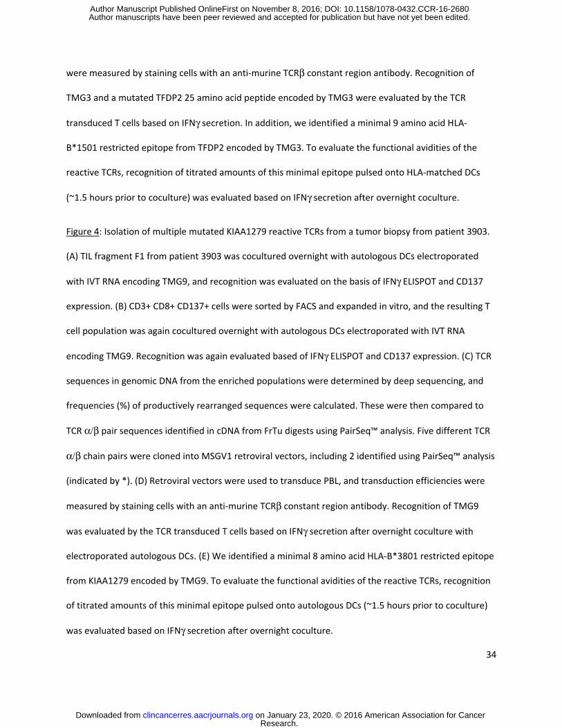
34
were measured by staining cells with an anti-murine TCRβ constant region antibody. Recognition of
TMG3 and a mutated TFDP2 25 amino acid peptide encoded by TMG3 were evaluated by the TCR
transduced T cells based on IFNγ secretion. In addition, we identified a minimal 9 amino acid HLA-
B*1501 restricted epitope from TFDP2 encoded by TMG3. To evaluate the functional avidities of the
reactive TCRs, recognition of titrated amounts of this minimal epitope pulsed onto HLA-matched DCs
(~1.5 hours prior to coculture) was evaluated based on IFNγ secretion after overnight coculture.
Figure 4: Isolation of multiple mutated KIAA1279 reactive TCRs from a tumor biopsy from patient 3903.
(A) TIL fragment F1 from patient 3903 was cocultured overnight with autologous DCs electroporated
with IVT RNA encoding TMG9, and recognition was evaluated on the basis of IFNγ ELISPOT and CD137
expression. (B) CD3+ CD8+ CD137+ cells were sorted by FACS and expanded in vitro, and the resulting T
cell population was again cocultured overnight with autologous DCs electroporated with IVT RNA
encoding TMG9. Recognition was again evaluated based of IFNγ ELISPOT and CD137 expression. (C) TCR
sequences in genomic DNA from the enriched populations were determined by deep sequencing, and
frequencies (%) of productively rearranged sequences were calculated. These were then compared to
TCR α/β pair sequences identified in cDNA from FrTu digests using PairSeq™ analysis. Five different TCR
α/β chain pairs were cloned into MSGV1 retroviral vectors, including 2 identified using PairSeq™ analysis
(indicated by *). (D) Retroviral vectors were used to transduce PBL, and transduction efficiencies were
measured by staining cells with an anti-murine TCRβ constant region antibody. Recognition of TMG9
was evaluated by the TCR transduced T cells based on IFNγ secretion after overnight coculture with
electroporated autologous DCs. (E) We identified a minimal 8 amino acid HLA-B*3801 restricted epitope
from KIAA1279 encoded by TMG9. To evaluate the functional avidities of the reactive TCRs, recognition
of titrated amounts of this minimal epitope pulsed onto autologous DCs (~1.5 hours prior to coculture)
was evaluated based on IFNγ secretion after overnight coculture.
Research. on January 23, 2020. © 2016 American Association for Cancerclincancerres.aacrjournals.org Downloaded from
Author manuscripts have been peer reviewed and accepted for publication but have not yet been edited. Author Manuscript Published OnlineFirst on November 8, 2016; DOI: 10.1158/1078-0432.CCR-16-2680

35
Figure 5: Isolation of multiple mutated XPNPEP1 and UGGT2 reactive TCRs from a tumor biopsy
fragment from patient 3678. (A) TIL fragment F4 from patient 3678 was cocultured overnight with
autologous DCs electroporated with IVT RNA encoding TMG9, and recognition was evaluated on the
basis of IFNγ ELISPOT and CD137 expression. (B) CD3+ CD8+ CD137+ cells were sorted by FACS and
expanded in vitro, and the resulting T cell population was again cocultured overnight with autologous
DCs electroporated with IVT RNA encoding TMG9. Recognition was again evaluated based of IFNγ
ELISPOT and CD137 expression. (C) TCR sequences in genomic DNA from the enriched populations were
determined by deep sequencing, and frequencies (%) of productively rearranged sequences were
calculated. In addition, single CD8+ T cells from the enriched population were sorted, and RT-PCR was
conducted to identify TCR α and β chain pairs. Four different TCR α/β chain pairs were cloned into
MSGV1 retroviral vectors based on single cell RT-PCR analysis (indicated by *). (D) These retroviruses
were used to transduce PBL, and transduction efficiencies were measured by staining cells with an anti-
murine TCRβ constant region antibody. Recognition of TMG9 and two mutated 25 amino acid peptides
encoded by TMG9, UGGT2 and XPNPEP1, were evaluated by the TCR transduced T cells based on IFNγ
secretion after overnight coculture with peptide pulsed autologous DCs. In addition, we identified an
HLA-A*0301 restricted 11mer from XPNPEP1 and an HLA-A*0201 restricted 9mer from UGGT2, both of
which were encoded by TMG9. To evaluate the functional avidities of the reactive TCRs, recognition of
titrated amounts of these minimal epitopes pulsed onto HLA-matched DCs (~1.5 hours prior to
coculture) was evaluated based on IFNγ secretion after overnight coculture.
Research. on January 23, 2020. © 2016 American Association for Cancerclincancerres.aacrjournals.org Downloaded from
Author manuscripts have been peer reviewed and accepted for publication but have not yet been edited. Author Manuscript Published OnlineFirst on November 8, 2016; DOI: 10.1158/1078-0432.CCR-16-2680
126 134 183 0
2000
4000
6000
8000
10000
a C
D1
37
a CD8
Sort highest ~0.5% CD3+ CD8+ CD137+ cells Expand with a-CD3/IL2
31 34 34 0
2000
4000
6000
8000
10000
3466 Rx TIL vs. TMG1 Pre-sort
3713 Rx TIL vs. TMG2 Pre-sort
a C
D1
37
a CD8
TCR sequencing and cloning
PBL transduction
IFNg
(pg/
ml)
>
a CD8 a CD8
3466 Rx TIL vs. TMG1 Post-sort and expansion
3713 Rx TIL vs. TMG2 Post-sort and expansion
A B
C D
E
F
3466 TCR 3713 TCR
TCRAV CDR3 Rank (%) TCRAV CDR3 Rank (%) 2-1*01 CAVEGDTGFQKLVF 1 (78.0) 29*01 CAASLSGGGADGLTF 1 (36.6) TCRBV CDR3 Rank (%) TCRBV CDR3 Rank (%)
13-1*01 CASSSPRSSRDTDTQYF 1 (81.6) 5-6*01 CASSLDRKAFF 1 (59.4)
G
3466 TCR 46.9% mTCRb+ (of CD3+ cells)
3713 TCR 71.1% mTCRb+ (of CD3+ cells)
Figure 1
Research. on January 23, 2020. © 2016 American Association for Cancerclincancerres.aacrjournals.org Downloaded from
Author manuscripts have been peer reviewed and accepted for publication but have not yet been edited. Author Manuscript Published OnlineFirst on November 8, 2016; DOI: 10.1158/1078-0432.CCR-16-2680

Patient 3784 F6 vs. TMG5 Pre-sort
TCR sequencing and cloning
PBL transduction
IFNg
(pg/
ml)
IF
Ng
ELIS
PO
Ts
%C
D1
37
+
IFNg
ELIS
PO
Ts
%C
D1
37
+
>
TCRAV CDR3 Rank (%) TCRBV CDR3 Rank (%)
34*01 CGADFLMNRDDKIIF 1 (96.8) 12-3*01 CASRAVINTDTQYF 1 (89.6)
>
Patient 3784 F6 vs. TMG5 Post-sort and in vitro expansion
Screening of enriched population for recognition of 25mers encoded by TMG5
0
0.1
0.2
0.3
0.4
0.5
0.6
0.7
0.8
0
20
40
60
80
100
120
140
160
180
control TMG TMG5
6 0.8 0
20
40
60
80
100
0
200
400
600
800
1000
control TMG TMG5
0
10
20
30
40
50
60
70
80
90
0100200300400500600700800900
1000
96.4% mTCRb+ (gated on CD3+ cells)
A B
C
>
D
E
Figure 2
0
4000
8000
12000
16000
20000>
Peptide concentration (g/ml)
mut. KIF16B 11 mer
32 38 0
4000
8000
12000
16000
20000
0
100
200
300
400
500
Research. on January 23, 2020. © 2016 American Association for Cancerclincancerres.aacrjournals.org Downloaded from
Author manuscripts have been peer reviewed and accepted for publication but have not yet been edited. Author Manuscript Published OnlineFirst on November 8, 2016; DOI: 10.1158/1078-0432.CCR-16-2680

TCR sequencing and cloning
IFN
g EL
ISP
OTs
%C
D1
37
+
30 0.4
0
2
4
6
8
10
12
14
0
200
400
600
800
1000
control TMG TMG3
8 0.3 0
20
40
60
80
100
0
200
400
600
800
1000
control TMG TMG3
TCR TCRAV CDR3 Rank (%) TCRBV CDR3 Rank (%)
1 19*01 CALSDSGGSNYKLTF 1 (76.4) 10-3*01 CAISEVGTGSSGNTIYF 1 (43.8)
2* 19*01 CALSDSGGSNYKLTF 1 (76.4) 5-5*01 CASRFVAPSDGYNEQFF 2 (17.6)
3* 9-2*01 CALSDPQDSGYSTLTF 3 (3.6) 5-4*01 CASSLGQGRVEQYF 3 (8.5)
4* 1-2*01 CAVSPGAGGSYIPTF 4 (3.6) 10-3*01 CAISEVGTGSSGNTIYF 1 (43.8)
* These pairs were identified using Pair-Seq™ analysis of FrTu digest.
PBL transduction
> >
IFNg secretion by transduced PBL (pg/ml)
TCR 1 TCR 2 TCR 3 TCR 4
% mTCRb (of CD3+ cells) 87.7 92.9 81.2 92.7
media 5 12 3 5
DC + control TMG 47 94 92 47
DC + TMG3 48 >20000 >20000 >20000
DC + DMSO 6 21 21 8
DC + wt TFDP2 25 mer 6 22 17 5
DC + mut TFDP2 25 mer 6 13549 9708 6799
Patient 3716 F3 vs. TMG3 Pre-sort
A B Patient 3716 F3 vs. TMG3
Post-sort and in vitro expansion
C
D
Figure 3
0
4000
8000
12000
16000
20000
1.E-12 1.E-10 1.E-08 1.E-06
TCR 2
TCR 3
TCR 4
Peptide Concentration (g/ml)
IFN
g (p
g/m
l)
> mut. TFDP2 9 mer
Research. on January 23, 2020. © 2016 American Association for Cancerclincancerres.aacrjournals.org Downloaded from
Author manuscripts have been peer reviewed and accepted for publication but have not yet been edited. Author Manuscript Published OnlineFirst on November 8, 2016; DOI: 10.1158/1078-0432.CCR-16-2680
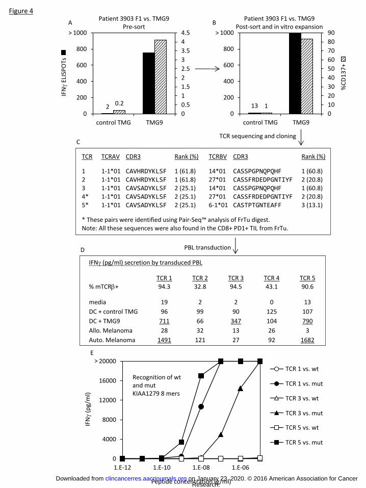
Patient 3903 F1 vs. TMG9 Pre-sort
TCR sequencing and cloning
IFN
g EL
ISP
OTs
%C
D1
37
+
TCR TCRAV CDR3 Rank (%) TCRBV CDR3 Rank (%)
1 1-1*01 CAVHRDYKLSF 1 (61.8) 14*01 CASSPGPNQPQHF 1 (60.8) 2 1-1*01 CAVHRDYKLSF 1 (61.8) 27*01 CASSFRDEDPGNTIYF 2 (20.8) 3 1-1*01 CAVSADYKLSF 2 (25.1) 14*01 CASSPGPNQPQHF 1 (60.8) 4* 1-1*01 CAVSADYKLSF 2 (25.1) 27*01 CASSFRDEDPGNTIYF 2 (20.8) 5* 1-1*01 CAVSADYKLSF 2 (25.1) 6-1*01 CASTPTGNTEAFF 3 (13.1)
* These pairs were identified using Pair-Seq™ analysis of FrTu digest. Note: All these sequences were also found in the CD8+ PD1+ TIL from FrTu.
PBL transduction
> >
Patient 3903 F1 vs. TMG9 Post-sort and in vitro expansion
2 0.2
0
0.5
1
1.5
2
2.5
3
3.5
4
4.5
0
200
400
600
800
1000
control TMG TMG9
13 1 0
10
20
30
40
50
60
70
80
90
0
200
400
600
800
1000
control TMG TMG9
IFNg (pg/ml) secretion by transduced PBL
TCR 1 TCR 2 TCR 3 TCR 4 TCR 5
% mTCRb+ 94.3 32.8 94.5 43.1 90.6
media 19 2 2 0 13
DC + control TMG 96 99 90 125 107
DC + TMG9 711 66 347 104 790
Allo. Melanoma 28 32 13 26 3
Auto. Melanoma 1491 121 27 92 1682
0
4000
8000
12000
16000
20000
1.E-12 1.E-10 1.E-08 1.E-06
1a/1b (KIAA1279 8merwt)1a/1b (KIAA1279 8mermut)2a/1b (KIAA1279 8merwt)2a/1b (KIAA1279 8mermut)2a/3b (KIAA1279 8merwt)2a/3b (KIAA1279 8mermut)
IFN
g (p
g/m
l)
Peptide concentration (g/ml)
TCR 1 vs. wt
TCR 1 vs. mut
TCR 3 vs. wt
TCR 3 vs. mut
TCR 5 vs. wt
TCR 5 vs. mut
>
Recognition of wt and mut KIAA1279 8 mers
A B
C
D
E
Figure 4
Research. on January 23, 2020. © 2016 American Association for Cancerclincancerres.aacrjournals.org Downloaded from
Author manuscripts have been peer reviewed and accepted for publication but have not yet been edited. Author Manuscript Published OnlineFirst on November 8, 2016; DOI: 10.1158/1078-0432.CCR-16-2680

Patient 3678 F4 vs. TMG9 Pre-sort
IFNg
ELIS
PO
Ts
%C
D1
37
+
> >
Patient 3678 F4 vs. TMG9 Post-sort and in vitro expansion
13 0.1
0
0.5
1
1.5
2
2.5
3
0
200
400
600
800
1000
control TMG TMG9
0
10
20
30
40
50
0
200
400
600
800
1000
control TMG TMG9
TCR TCRAV CDR3 Deep seq rank (%)
SC PCR rank (%) TCRBV CDR3
Deep seq rank (%)
SC PCR Rank (%)
1* 1-2*01 CAVPSGSARQLTF 1 (18.9) 1 (32%) 7-2*01 CASSLDFSRQETQYF 3 (16.1) 1 (32%)
2* 21*01 CAVIKGYSTLTF 4 (13.5) 2 (26%) 27-1*01 CASSLYLASNTGELFF 6 (5.2) 2 (26%)
3* 20*01 CAVQAWGFGNVLHC 3 (14.6) 3 (19%) 6-1*01 CASSDRNTNYGYTF 2 (16.3) 3 (19%)
4* 20*01 CAVKSWSGPGWGNQAGTALIF 6 (8.9) 4 (11%) 19*01 CASSMRVQSNTGELFF 1 (17.4) 4 (11%)
* These pairs were identified using single cell RT-PCR (SC PCR).
IFNg (pg/ml) secretion by transduced PBL
TCR 1 TCR 2 TCR 3 TCR 4
% mTCRb+ (of CD3+ cells) 92.3 90.5 92.4 93.6
media 5 2 1 2
DC + control TMG 22 33 47 21
DC + TMG9 9543 5154 1496 3743
DC + DMSO 1 4 236 2
DC + wt UGGT2 25 mer 1 6 221 1
DC + mut UGGT2 25 mer 2 4 >20000 >20000
DC + wt XPNPEP1 25 mer 61 4 220 0
DC + mut XPNPEP1 25 mer >20000 18327 473 2
A B
D
C
Figure 5
TCR sequencing and cloning
PBL transduction
0
4000
8000
12000
16000
20000
1.E-12 1.E-10 1.E-08 1.E-06
TCR 3
TCR4
mut. UGGT2 9 mer
0
4000
8000
12000
16000
20000
1.E-12 1.E-10 1.E-08 1.E-06
TCR 1
TCR 2
mut. XPNPEP1 11 mer
IFNg
(pg/
ml)
Peptide Concentration (g/ml) Peptide Concentration (g/ml)
> >
Research. on January 23, 2020. © 2016 American Association for Cancerclincancerres.aacrjournals.org Downloaded from
Author manuscripts have been peer reviewed and accepted for publication but have not yet been edited. Author Manuscript Published OnlineFirst on November 8, 2016; DOI: 10.1158/1078-0432.CCR-16-2680

Table 1: Screening of Patient 3784 sorted populations from fresh tumor digest and TIL fragments for recognition of mutated tandem minigenes (TMGs)
FrTu 3784
TIL 3784
CD8+ CD8+ PD1- CD8+ PD1+ Rx1 F2 F4 F5 F6
ELISPOTSa 41BB+b ELISPOTS 41BB+ ELISPOTS 41BB+ ELISPOTS 41BB+ ELISPOTS 41BB+ ELISPOTS 41BB+ ELISPOTS 41BB+ ELISPOTS 41BB+
No target 0 0.0 0 0.0 0 0.1 0 0.1 9 1.1 3 0.5 0 0.2 2 0.2
DC + control TMG 1 0.1 4 0.2 2 0.4 39 0.1 5 1.7 6 1.9 6 0.4 25 0.4
DC + TMG1 0 0.1 1 0.2 0 0.5 1 0.0 5 1.8 35 1.6 4 0.3 14 0.3
DC + TMG2 26 0.2 1 0.2 4 0.3 14 0.1 5 2.1 100 1.6 1 0.2 11 0.4
DC + TMG3 6 0.1 0 0.0 72c 0.7 108 0.2 131 2.8 213 0.9 52 0.5 268 0.4
DC + TMG4 2 0.1 1 0.2 9 0.5 53 0.2 10 1.2 43 0.9 123 0.7 46 0.2
DC + TMG5 4 0.1 3 0.2 125 0.6 162 0.4 27 2.3 21 1.1 36 0.2 159 0.7
DC + TMG6 0 0.1 2 0.1 2 0.5 128 0.1 31 2.0 76 0.8 22 0.4 113 0.6
DC + TMG7 1 0.0 0 0.2 3 0.2 13 0.0 4 1.4 15 0.8 7 0.3 26 0.0
DC + TMG8 7 0.1 2 0.1 210 d, e 1.9 404 2.4 265 2.8 291 3.1 250 1.7 478 7.6
DC + TMG9 0 0.1 1 0.0 3 0.5 24 0.1 31 1.7 229 1.1 51 0.4 58 0.4
DC + MART 4 0.2 3 0.0 30 0.6 8 0.2 10 0.8 52 0.9 6 0.2 52 0.3
DC + gp100 12 0.1 3 0.1 171 1.4 235 0.7 287 1.5 407 2.5 233 1.5 56 0.6
DC + tyrosinase 4 0.1 4 0.1 0 0.2 16 0.0 7 1.0 18 1.0 2 0.2 9 0.3
DC + MAGE-A3 0 0.1 1 0.1 3 0.5 29 0.1 1 1.2 8 0.8 7 0.4 37 0.7
DC + NY-ESO-1 1 0.1 2 0.2 3 0.1 34 0.2 3 1.3 7 0.9 2 0.2 6 0.5
DC + SSX2 1 0.0 1 0.1 3 0.3 30 0.1 3 0.6 22 1.0 3 0.2 5 0.2
OKT3 497 70.4 478 68.3 466 35.7 558 87.2 550 60.3 561 64.4 535 43.5 535 63.7
a IFNg ELISPOTS (per 2e4 T cells) b % 41BB + cells (gated on CD3+ CD8+ cells) c Underlined values indicate >60 ELISPOTS or >0.5% 41BB+ and >2X background with control TMG and no target. d Highlighted cells indicate positive results in both ELISPOT and 41BB assays. e Circled cells indicate populations from which we sorted and expanded T cells for further analysis.
Research. on January 23, 2020. © 2016 American Association for Cancerclincancerres.aacrjournals.org Downloaded from
Author manuscripts have been peer reviewed and accepted for publication but have not yet been edited. Author Manuscript Published OnlineFirst on November 8, 2016; DOI: 10.1158/1078-0432.CCR-16-2680

Table 2: Tumor associated mutation reactive T cell receptors identified from patients with melanoma Patient TMGa Mutated
antigen TIL Sourcesb # of independent TCRs and
methods used to identify them Data presentation
3466 1 COL18A1 Rx TIL 1 (5’ RACE) Fig. 1 3713 2 SRPX Rx TIL 1 (5’ RACE) Fig. 1 3903 9 KIAA1279 FrTu CD8+ PD1+/
F1 3 (TCR deep seq and PairSeq™) Fig. 4
3903 3 KIAA1967 FrTu CD8+ PD1+ 1 (TCR deep seq) Supp. Fig. 10 3903 8 PHKA1 FrTu CD8+ PD1+ 1 (TCR deep seq and PairSeq™) Supp. Fig. 1 3784 5 KIF16B FrTu CD8+ PD1+ 1 (TCR deep seq and PairSeq™) Supp. Fig. 11 3784 5 KIF16B F5 1 (TCR deep seq and PairSeq™) Supp. Fig. 2 3784 5 KIF16B F6 1 (TCR deep seq and PairSeq™) Fig. 2 3784 8 SON FrTu CD8+ PD1+ 2 (TCR deep seq and PairSeq™) Supp. Fig. 12 3784 8 SON FrTu CD8+ PD1+/
F2/F6/F5 1 (TCR deep seq and PairSeq™) Supp. Fig. 3
3784 8 SON F5 1 (TCR deep seq and PairSeq™) Supp. Fig. 13 3784 4 GNB5 F5 1 (TCR deep seq and PairSeq™) Supp. Fig. 4 3678 3 FBXO21 F1/F3/F4 1 (TCR deep seq) Supp. Fig. 5 3678 2 CORO7 F1/F2 1 (TCR deep seq) Supp. Fig. 6 3678 7 RECQL5 F3 1 (TCR deep seq) Supp. Fig. 7 3678 7 RECQL5 F6 1 (TCR deep seq) Supp. Fig. 8 3678 9 UGGT2 F4 2 (TCR deep seq and SC RT-PCR) Fig. 5 3678 9 XPNPEP1 F4/F6 2 (TCR deep seq and SC RT-PCR) Fig. 5 3716 3 TFDP2 F3 3 (TCR deep seq and PairSeq™) Fig. 3 3716 3 TFDP2 PF2 1 (TCR deep seq and PairSeq™) Supp. Fig. 9 a TMG: Tandem minigene construct against which reactivity was identified b Three different types of TIL products were used as sources for the identification of mutation reactive
TCRs: Rx TIL refers to in vitro expanded TIL that had been used to treat the patient; TIL denoted with F or PF refers to TIL from fragments from tumor biopsies that had been expanded in vitro with IL2; and FrTu CD8+ PD1+ refers to TIL from fresh tumor digests that had been FACS sorted for CD8+ PD1+ T cells and expanded in vitro in the presence of irradiated feeders, OKT3, and IL2.
Research. on January 23, 2020. © 2016 American Association for Cancerclincancerres.aacrjournals.org Downloaded from
Author manuscripts have been peer reviewed and accepted for publication but have not yet been edited. Author Manuscript Published OnlineFirst on November 8, 2016; DOI: 10.1158/1078-0432.CCR-16-2680
Published OnlineFirst November 8, 2016.Clin Cancer Res Maria R. Parkhurst, Alena Gros, Anna Pasetto, et al. based on CD137 expressiontumor associated antigens from tumor infiltrating lymphocytes Isolation of T cell receptors specifically reactive with mutated
Updated version
10.1158/1078-0432.CCR-16-2680doi:
Access the most recent version of this article at:
Material
Supplementary
http://clincancerres.aacrjournals.org/content/suppl/2016/11/08/1078-0432.CCR-16-2680.DC1
Access the most recent supplemental material at:
Manuscript
Authoredited. Author manuscripts have been peer reviewed and accepted for publication but have not yet been
E-mail alerts related to this article or journal.Sign up to receive free email-alerts
Subscriptions
Reprints and
To order reprints of this article or to subscribe to the journal, contact the AACR Publications
Permissions
Rightslink site. Click on "Request Permissions" which will take you to the Copyright Clearance Center's (CCC)
.http://clincancerres.aacrjournals.org/content/early/2016/11/08/1078-0432.CCR-16-2680To request permission to re-use all or part of this article, use this link
Research. on January 23, 2020. © 2016 American Association for Cancerclincancerres.aacrjournals.org Downloaded from
Author manuscripts have been peer reviewed and accepted for publication but have not yet been edited. Author Manuscript Published OnlineFirst on November 8, 2016; DOI: 10.1158/1078-0432.CCR-16-2680