Embed Size (px)
Citation preview
⌒
⌒
THOMAS JEFFERSON UNIVERSITY HOSPITALDEPARTMENT OF RADIOLOGY
TECIMCALDJAGNOSTrc AND PEDJATRIC
PROTOCOLS AND PROCEDURES
皿 Ⅵ EWED and APPROVED BY:
⌒
⌒
⌒
The "rorrLine views,, are intended as a guide- The technologist is expected toconsider each patient's general physical condition and clinical history. If thetechnologist feels the routines should be compromised, he/she must consultwith a ratiologist or technical supervisor. The reason for the compromise' theradiolosisfs o; teclutical supervisorrs name must be recorded in the CommentSection of Imaqecast.
Revised 7L / 09 | 10
⌒
Ⅲ TRODUCT10N TO RAD10GRAPmC&FLuoROSCOPIC PROCED…fPOSIT10NS AND ROVTINESl
⌒
⌒
ROUTINE RADIOGRAPIilC POSITIONS
AOI = Area of InterestDocumentation of patient complaint spect$r on request(i.e. X-Ray: LT hand; Ttaurrra: 2 wks ago;Pain: 2nd Meta.carpal; Area: Distal MP joint space)
*ALL SKTILL EXITMS SIIOULD BE APPRO\IED TIIROUGI{ A BONE RADIOLOGIST
L Headwork
A. Skutl1. PA2. AP Towne3. Base4. Rt. lateral5. Lt. lateral
B. Mandible1. PA,2. BotJl obliques3. 'Towne4. Lateral of affected side
C. T.M. Joints1. RL lateral - open a:ed close mouth2. LL lateral - open and close mout]r3. Base and Towaes views (include only for fracture or dislocation)
D-.Sinuses1. PA2. Waters wittr mouth open {modified Waters)3. Base4. Lateral
E- Facial Bones1. PA2. Waters3. Dragerated Waters4. Base5. Latera-l face
5
⌒
⌒
⌒
F. Mastoidsl. AP2. Base3. Transorbital4. Laws5. Stcnvers
G.Optic Foramcnl. Rt.and lt.Rhcese
H.Orbitsl. Scc facial bones2. Rheeseヽ電ew
I. NaSal Bonesl.Right and Left La“ral
2. Waters
」. Zygomatic Archesl. See facial bones2.Mays v■ew
K Foreign Body ofEvclMH一Clean cassettesマにd■ AGFA scrcen clcaner p4or to cxposure
l.PA Caldwel1230 caudal anntion2. Waters3. Lateral
Ⅱ. Spine
A_ G洒甍d10x12 po― it lnevcr dO―n and utension mewsllnilatcral is chedL`● by radiO10gilsl
l. AP
2. RPO3. LPO4. Lateral5. In cases of traupaa, cioss table laterat is done first and shown to a
radiologist. Ttren proceed with what he/she orders' ViewspossiblY needed for trauma:a. Odontoidb. Flexion and extension (not done until lateral is shown to radiologist)
c. Trauma obliques-requested by ER physicians. as needed
Revised 1l109/20rO
6
⌒
ヘ
⌒
B. Thoracic 14x17 Portrait1. AP2. LateraJ
C. Lumbar1. AP (14 x 17 - full abdomen)2. RPO 14x17 Portrait3. LPO L4xl7 Portrait4. Lerte,ral 14x17 Pottrait5. La.teral I5-S1 spot 1Ox12 portrait
D. Sacru,m. 1Ox12 Portrait1. AP2. LateraJ
E. Cocgnr lOxL2 Portrait1. AP2- Ip..te.ral
F. S.I. Joints1. AP pelvis 14x17 landscaPe2. RPO 10x12 Portrait3. LPO 1Ox12 Portrait
G. Pelvis 14x17 landscaPe1. AP
H. HiP1. AP pelvis 14x17 LandscaPe2. AP hiP loxl2Portrait3. Late;r hip loilz portrait_in cases of trauma or possible fracture
do shoot through lateral
Upoer D<tr,emities - Despite what views are requested, it is departmental
@ be done or all trau"ma extremitibs (Le. hand,
iog.i, *ii"t, elbow, ioes, k'''ees, feet and ankles)' Ttren on AP arr.d lateral
is obtained.
A. Finger (identiff alfected finger wittr arrow)1. PA hand2. Oblique (both obliques if traurna)3. Lateral ofaffected digit
7
⌒
⌒
⌒
B. Hand1. PA2. Oblique (bofh obliques if trauma)3. Lateral
C. Wrist1. PA with hand flat on cassette2. Oblique (botJr obliques if trauma)3. I,grteral4. Navicular vielr (if indicated on request or ordered by radiologist)
D. Forearm1. AP2. Oblique - only if requested3. Lateral
E. Elbow1. AP2. Lateral3. Obliques (if trauma)4. Radial head view (if trauma)
F. Humerus 14x17 lengttrwise1. AP - interaal and external rotation2. For traur::a:
a- AP - neutralb. Ttanstlroracic lateral - LOxL2lengthwise
G. Shoulder1- ftrauma)
a- AP internal rotation (15o caudad)b. AP extemal rotation (I5o caudad)
. c. Ardllaryd. Yview
2. (Non-Thaurra)a. AP internal rotatioa (l5o caudad)b. AP external rotation (15o caudad)c. Y view
8
⌒
IV. Lower Extremities
"-"es1. AP forefoot (identi$ affected toe)2. Oblique toe fbot]r obliques if trauma)3. Lateral toe
B. Foot1. AP2. Obliques (both obliques if trauma)3. Ldteral
C. Calcaneus1. Axial2. Lateral
D. Ankle1. AP'2. 45o medial obligue (both obliques if trauma)3. Lateral4. 15o medial oblique'(mortise view for trauma)
\
E. Krrees (AP erect of affected krree only unless ordered as bilateral, L4xLZlands-cape - if patient can bear weight
Table Bucky:1. Iateral 1Ox12 portrait2. Tbnnel 1Ox12 portrait''For Ttaurna:3. Both obliques lAxL2 portrait4. Axial - 1Ox12 portrait if possible when indicated for patella .
ATIN: Wlen long bones ond botf-Joittts are rcqttlrred, thcJoitrt aiearcslloirl,td be don'e sepato;telg (att aieuts|
f. nUia 14x17 portrait1. AP2. Lateral
G. Femur (include botl joints)1. AP l4xLZ portrait2. Lateral 14x17 portrait
9
Re宙scd l1/21/11,11/16/201l lceSI
ヘ
⌒́ 、
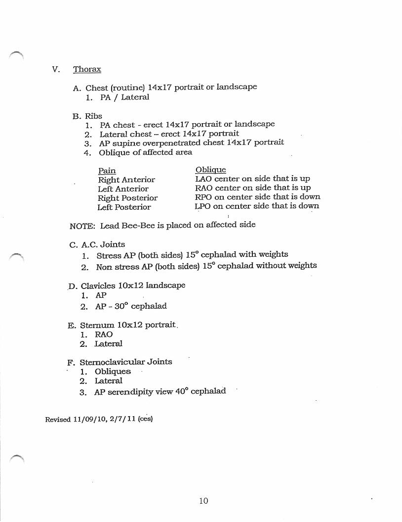
V. Thorax
A. Chest (routine) L4xLT portrait or landscape1. PA / Lateral
B. Ribs1. PA chest - erect L4xLZ portrait or landscape2. Latetal ctrest - erect 74x17 portrait3. AP supine overpenetrated chest L4xl7 portrait4. Obligtre of alfected are,a
Pain ObligueRight Anterior LAO center on side that is upLeft Anterior RAO center on side tJ'at is upRight Posterior RPO on center side that is downIeit posterior L]>O on center side that is down
;
NOTE: I,ead Bee-Bee is placed on allected side
C. A.C. Joints1. Stness AP (both sides) 15" cephalad with weights
2. Non stress AP (both sides) 15" cephalad ruithout weights
D. Clavicles 1Ox12 landscaPe1. AP
2. AP - 30" cePhalad
E. Ster:eum LOxI2 Portrait.1. RAO2. J,ateral
F. Sternoclavicular Joints' 1. Obliques
2. Lateral3. AP serendipity view 4Oo cephalad
Revised LL l@ I 70, 2/7 / Ll (esl
ハ 、
10
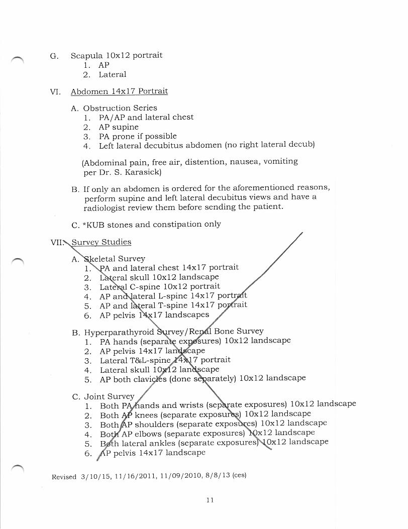
G. Scapula lOxl2 portrait1. AP2. Lateral
Abdomen 14x17 Portrait
A. Obstruction Series1. PA/AP and iateral chest2. AP supine3. PA prone if possible4. Left lateral decubitus abdomen (no right laterai decub)
(Abdominal pain, free air, distention, nausea, vomitingper Dr. S. Karasick)
B. If only an abdomen is ordered for the aforementioned reasons,perform supine and left lateral decubitus views and have aradiologist review them before sending the patient.
C. *KUB stones and constipation only
Studies
letal SurveyA and lateral chest l4xl7 Portrait
.eral skull 10x12 landscaPeLatls{ C-spine 1Ox12 PortraitAP an\ateral L-sPine l4xl7 PortAP and T-spine l4xl7AP pelvis 1 17 landscapes
B. Hyperparathyroid \rveY/ Re Bone Survey1. PA hands (separa\q ex res) 10x12 landscape2. AP pelvis l4xl73. Lateral T&L-spine. 7 portrait4. Lateral skull 1 21a5。 AP both clav s (done tely) 10x12 landscaPe
C. Joint Survey1.Both P, and wrists ( ate exposures) lOxl2 landscaPe
VI.
2.Both3.Both
knees (separate exposu lOxl2landscapeP shoulders (separate exPos ) 10x12 landscape
4.Bo AP elbows (separate exPosures) 12 landscapeh lateral ankles (separate exposures
P pelvis l4xl7 landscaPe
Revised 3/lOlt5, rll16/2O11, 11 l09l2OlO,8l8l13 (ces)
Oxl2landscape
⌒
⌒
1
2
3
4
5
6
⌒
⌒
TE LPEDmTRIC PRoToCOLS d%PROCEDyws
⌒
_ Re宙 scd I1/09/10
34
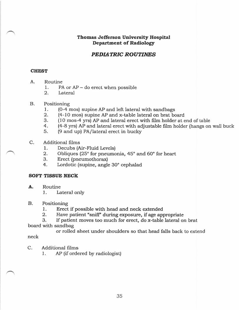
Thomas Jefferson Universit5r HospitalDepartaeat of Radiologr
PDDIATRIC ROUTINES
CHEST
A. Routine1. PA or AP - do erect when possible2. lateral
B. Positioning1. (0-4 mos) supine AP and left lateral witJ: sandbags2. (4-lO mos) supine AP and x-table lateral on brat board3. (10 mos-4 yrs) AP and lateral erect with film holder at end of table4. (a-B yrs) AP and lateral erect with adjustable film holder (hangs on wall buck5. (9 and up) PA/ lateral erect in bucky
C. Additional films1. Decubs (Air-Fluid l,evels)2. Obliques (25' for pneumonia, 45' and 60" for heart3. Erect (pneumothorax)4. Lordotic (supine, angle 3O' cephalad
SOFT TISSI'E ITECIK
A. Routine1. Lateral onJy
B. Positioning1 . Erect if possible with head and neck extended2. Have patient "snifP during e:eosure, if age appropriate3. lf patient moves too much for erect, do x-table lateral on brat
board with sandbagor rolled sheet under shoulders so t].at head falls back to extend
neck
C. Additional fllmsl. AP(r ordcred by radiolo」 Stl
⌒
35
⌒
1.2.3.
4.
Constipation - Flate plate onlYAbdominal Pain - Flate plate and erectObstruction - PA/AP erect chest x-ray, supine and erect -Abdomen (decubitus if erect cannot be obtained).AP and Prone Abdomen - nausea/vomiting
A.
C.
B
Routine1. AP and lateral (non-trauma)2. Open mouth (trauma and torticollis)
Positioning1. Sitting or standing for older children2. Supine and cross-table lateral on brat board for toddiers
Additional views1. Obliques2. Flexion and extension - after neutral lateral is cleared by radiologist
A.
B。
Routine1. AP and lateral
Positioning1. Same immobilization techniques as with CXR, but try to collimate
A.
B.
Routine1. AP and lateral1. Obliques on all patients with history of spondylolisthesis
Positioning1. AP - center just above crest, keep cones open, shield as possible2. Lateral - center just above crest, collimate3. Use same immobilization techniques as a CXR
Revised 2l 16l 15, ll l2l I ll
36
⌒
⌒
C. Additional views (only if requested by radiologist)1. Obliques2. LS-SI spot3. Flexion / extension
RoutinePA erect
t erect
Positioning1. Use2-3 FA cassettes, ID rag in 14x36 bucky; include
B.
from tip of earcephalad 7z ofYou may shield fromAsk patient if he/she in his/her shoes; if so films, mustbe done without shoes
6.
4.
5
For PA erect: first to adjust shield to height of ASISthen turn patient PAFor lateral; turn to left arms in front of patient、、■dl elbows chin up and shield ASIS downCassettes mu placed in same being processed
be done if patient is "new" or hasn scoliotic films
S;attach flltet to collimator so that it covers
C. Additionall.Lateral
1.AP2. AP3. AP bilateral
Positioning1. Tape lead ruler
hip joins on 74x17on 14x17on 14x17
ter of
fOr years (or if ordered by ortho)2. Su bending views: place patient supine; have bend
, keeping both shoulders on table so that righttoward right hip then left shoulder leans toward left
include from sternal notch to ASIS; usually lits on 14xI7cassette
SCANOGRAM
A. Roltine
2. Assist pa to table, patient carefui not to dislodge ruler
SCOLIOTICS
⌒
B.
3.Be is under patient's hips, & ankle arrd along patient's midline
■Buc対- Bucky.
4. Tape feet together5. IMPORTANT: Don't move FFD, ruler or patient between exposures;
tube and bucky may be siid longitudinally to reach from hips toankles but don't iet patient move himself/herself
6. Always shoot bilaterally in each exposure
SHOI'LDER
A. Routinel. AP - internal/ external
B. Positioning1. Hands and legs sandbagged down or, if necessary, use brat board
with affected arm unrestrained; t).en use compression band torestrain affected arm
C. Additional views1. Lordotic - angle 3O" cephalad , center 1" below shoulder2. Y - view
CLAVICLE
A. Routine1. AP - 0" angulation. Axial - 30" cePhalad
B. Positioning1.. Place small ro11 under shoulders to lift chin out of way2. Sandbag or brat board with arms at baby's sides
3. Newboms may be wrapped in a blanket with arms down; for infants,use head clamps to keep mandible away from clavicle
4. Do tabletop when Possible
EXTREMITIPS
A. General Information1. AP and lateral (include both obliques for trauma)
ITUMERUS
A. Routine1. AP2. LatetaJ
38
B. Posilioning1. AP arm extended - sandbag forearm or use compression band2. Laleral - flex elbow, put arm across body
ELBOW
A. Routine1. AP2. I.aLeral3. Both obliques for trauma
B. Positioningl. AP - arm extended - sandbag forearm or use compression band2. lateral - flex elbow 9O" - keep wrist and hand lateral
C. Additional Views1. Radial Head - modified AP elbow; rotate humerus laterally and wrist intemally2. Axial - elbow flexed, true lateral, arrd angle 45o toward shoulder
E1OREABM
A. Routine1. AP2. I-atera)
B. Positioning1. AP - extend elbow - use compression band as needed2. l,ateral - Ilex elbow 9O" - keep hand/wrist lateral; if being done
supine on an infant or toddler under compression band, bring handabove head but still keep hand lateral by pointing thumb toward table
TIAITDA$D;futsf
A. Routine1. PA/AP2. l,ateral3. Oblique for trauma
B. Positioning1. PA - use compression band over hand/wrist as needed; keep fingers
extended (even on newborns); sandbag forearm as needed2. t aLeral - use sponges under compression and to keep baby's hand
true lateral
C. Additional宙ews
39
~ 1.Na宙 cular― wHstin uhar icxion,anglc 20° toward clbow
BONE AGD
A. Roudnel. 12 mos old alld up― left hand/w亘 st― PA only2. Under 12 mos old― leFt upper extrenuty(shoulder― >fmgcrdp)left
lower extremity(hip― >toc)
B. Posidoningl. Keep rlngers extended(eVen On newborns)
Ю 韓
… …PDLV・IS
A. Routinel. AP
B. Pos,tioningl. Remove diapcr
⌒ 2. Sandbag arms3. compression band over pelvis4.Invert Fcet and sandbag fect
HIPS
A. Routincl. AP pel宙 s
2. Frog Lateral3. For above always do bilatcral,cvcn if ott one sidc is ordered
B、 Positioningl. AP― see“pelvis'above2. Lateral― ncx lcnecs sc that soles Offeet touch,comprcssion band over
hccs
C. Additional viewsl. Roll latcraユ ーobuque patient 45° ,ccntcr over affccted hip
2. Bridgeman lateral― use grld on oldcr paticnts
D. Shieldhgl. When doing patient's frst exarn of hip/pClVis,one view should be
⌒ completcly
40
unshielded; all subsequent/follow-up hip Iilms should be shielded forall views
KNEE
A. RoutineI. AP and lateral2. Obliques for tramma
B. Positioning1.. AP- extend knee, compression band over knee2. Lateral - patient \ring on affected side, flex knee 15o
C. Additional viewsl. Tunnel2. Tangenntial patella - patient prone, flex knee 90", tube angled 20o
cephalad
E'EIEUR ORTIB/FIB
A. RoutineI . AP and lateral
B. Positioning1. AP- extend knee, use compression band, sandbags and brat board as
needed2. Lateral - turn patient on alfected side, bring unaffected leg up in
front, collimate and shield as much as possible
C. Addidonal viewsl. Standing legs - for Blount's disease or rickets
AI{KIjE
A. RoutineI . AP arrd lateral2. Obliques for trauma
B. PositioningL AP - extended knee, dorsi-flex foot, compression band over knee as
necessary, have parent hold foot dorsi-flexed2. LaLeraJ- turn patient on affected side, use compression band as
needed
F'OOT
41
A. RoutineI . AP ald lateral2. Internal oblique for trauma
B. Positioning1. AP - flex knees, place feet under compression band2. l-ateraf - sarne as above but with foot lateral
42
⌒
⌒
C. Addidonal viewsl. Calcancus― Point toes up― angle 45° caphalad2. Harrls vie、v or calcaneus― havc patient stand on cassette;nex knecs,
angic 25° toward the hecl
esmey
AP bilatera-l lower extremities (hip- >toes single if infamtl1
2
3
AP ofDone
3. AP of bilateral upper4.AP of bilaterallowerl
RIickcts survcy
l. AP bilateral
2. PA b」ateral
teral upper extremities (shoulder - ?syphilis, rickets, anemia,
- separate exposures)
B. Skeletal survey1. AP and la2.AP and lateral tire spine (T- pine may be combined for
each view but C- must be te exposures); don't collimatefor AP T- or Lspine includc and abdomen
(separate exposures)(single exposure if infant)
5. AP of bilateral feet (if on AP of lower extremities)6. Done for suspected or congenif al abnormalities
D.
C.
Lcad
2.AP1.AP a fdomen (to R/O lead chips)
bilateral knees―bilateral■vrists
SKqDL
A. Routine1. APoTPA2. Towne's - 3O" caudad3. Both laterals
B. Positioning1. AP and Towne's - murrmy wrap child, strap onto brat board; place
head clamps on each side ofhead; place compression band overface and head clamps.0t looks fike torture but if patient is able to cry he can breathe!)⌒
43
2. LaLerals - remove head clamps; rotate brat board by placing a sandbagunder the board in the area of the patient's arm or chest; thishelps the patient to be able to tum his/her head true lateral; placesponge under head; hold head latera-l while tightening compressionband
3. Don't attempt to turn head true lateral while body is true AP!
SIIIUSES
A. Routine1. APIPA2. Water's / reverse Water's3. l,ateral
B. Positioning1. AP/PA - see "skull" for immobilization method, center at nasion2. Water's /reverse Water's - place sandbag behind child's shoulders to
extend neck and raise chin; angle cephalad as needed if neck is notextended enough by sandbag; center at acanthion
3. Lateral - see "skull" for immobilization method; center at outer canthus
NASAL BOIIES
A. RoutineI . Water's / reverse Water's2. Both laterals
B. Positioning1 . Water's / reverse Water's - see "sinuses" for immobilization method2. lateral - (non-grid, finger tectrnique) use for compression band and
sponge - see "lateral skull' for immobilization method
IIAIIIIIBLG
A. Routinet. PA - no angulation - use brat board, head clamps, and compression
band as needed2. Both lateral obliques - place patient supine with sandbag behind
shoulders to allow head/neck to extend back; turn head almostlateral; place lilm non-grid under mandible; angle cephalad as needed(depending on how far head/neck is extended) so that both halves ofmandible €rre not superimposed
44