Embed Size (px)
Citation preview

HEAD INJURY (GENERAL) TrH1 (1)
Head Injury (GENERAL)Last updated: June 25, 2019
DEFINITIONS, CLASSIFICATIONS.............................................................................................................2
EPIDEMIOLOGY........................................................................................................................................2
Incidence................................................................................................................................2
Morbidity................................................................................................................................2
Mortality.................................................................................................................................2
ETIOLOGY.................................................................................................................................................2
PATHOPHYSIOLOGY (PRIMARY VS. SECONDARY INJURY)....................................................................3
PRIMARY BRAIN INJURY.........................................................................................................................3
Mechanisms............................................................................................................................3
SECONDARY BRAIN INJURY....................................................................................................................3
PATHOPHYSIOLOGY (CEREBRAL BLOOD FLOW, EDEMA)....................................................................3
PATHOPHYSIOLOGY (BIOCHEMISTRY)...................................................................................................4
PATHOLOGY, CLINICAL FEATURES........................................................................................................4
CONCUSSION..........................................................................................................................................5
DIFFUSE AXONAL INJURY (DAI)...........................................................................................................6
CONTUSION AND LACERATION..............................................................................................................6
INTRACEREBRAL HEMORRHAGE (TICH).................................................................................................9
MISSILE (GUNSHOT) INJURY..................................................................................................................9
Self-inflicted injuries............................................................................................................12
STAB INJURY (IMPALEMENT)...............................................................................................................12
COMPRESSION INJURY..........................................................................................................................13
DEGREES OF SEVERITY..........................................................................................................................13
PREHOSPITAL MANAGEMENT................................................................................................................14
1. Oxygenation.....................................................................................................................14
2. Blood Circulation.............................................................................................................14
3. Cervical Spine Stabilization.............................................................................................15
4. Brief Neurologic Status....................................................................................................15
5. Transportation..................................................................................................................15
DIAGNOSTIC EVALUATION....................................................................................................................15
Diagnosis in Latin................................................................................................................15
HISTORY...............................................................................................................................................15
PHYSICAL EXAMINATION.....................................................................................................................15
IMAGING...............................................................................................................................................16
Imaging modalities..........................................................................................................................16
CT without contrast..............................................................................................................17
Plain skull radiographs.........................................................................................................18
Nonenhanced MRI...............................................................................................................18
Angiography.........................................................................................................................19
Ultrasonography...................................................................................................................19
Conditions.......................................................................................................................................19
Concussion...........................................................................................................................19
Diffuse axonal injury............................................................................................................19
Scalp Injuries........................................................................................................................20
Contusion.............................................................................................................................20
Penetrating injuries...............................................................................................................21
OTHER NEUROLOGIC TESTS.................................................................................................................21
NON-NEUROLOGIC TESTS....................................................................................................................22
MANAGEMENT........................................................................................................................................22
DISPO....................................................................................................................................................22
Hospitalization.....................................................................................................................22
Transfer................................................................................................................................22
ABC.....................................................................................................................................................22
Airway, Ventilation.........................................................................................................................22
Blood pressure.................................................................................................................................23
Resuscitation fluids..............................................................................................................23
GENERAL MEASURES...........................................................................................................................23
Regimen, Activity................................................................................................................23
HEAD INJURY (GENERAL) TrH1 (2)
Nutritional Support...............................................................................................................23
DVT prophylaxis..................................................................................................................23
Hemoglobin..........................................................................................................................24
NEUROLOGICAL MEASURES.................................................................................................................24
ICP management.............................................................................................................................24
Monitoring............................................................................................................................24
Treatment.............................................................................................................................25
CPP..................................................................................................................................................26
Advanced brain oxygenation / metabolic status monitoring...........................................................26
Oximeters.............................................................................................................................26
TCD......................................................................................................................................27
Microdialysis........................................................................................................................27
Sedation & Analgesia......................................................................................................................27
Seizure prophylaxis.........................................................................................................................28
Antibiotics.......................................................................................................................................28
MEDICATIONS NEEDING FURTHER TESTING.........................................................................................28
FAILED MEDICATIONS..........................................................................................................................28
NOT RECOMMENDED MEDICATIONS.....................................................................................................28
SURGERY.................................................................................................................................................28
Indications for surgery.........................................................................................................28
Method to measure hematoma volume................................................................................29
REHABILITATION....................................................................................................................................29
NON-NEUROLOGIC COMPLICATIONS....................................................................................................30
EARLY COMPLICATIONS.......................................................................................................................30
LONGER TERM COMPLICATIONS...........................................................................................................30
NEUROLOGIC COMPLICATIONS & SEQUELQE.....................................................................................31
I. VASCULAR........................................................................................................................................31
II. INFECTION........................................................................................................................................33
III. CSF & MENINGES..........................................................................................................................33
IV. POST-TRAUMATIC EPILEPSY..........................................................................................................33
V. POST-TRAUMATIC MOVEMENT DISORDERS....................................................................................33
VI. POSTCONCUSSION (S. POSTTRAUMATIC) SYNDROME.....................................................................33
VII. POSTTRAUMATIC PSYCHIATRIC DISORDERS.................................................................................34
PROGNOSIS, OUTCOME..........................................................................................................................35
5 most powerful factors predicting outcome........................................................................35
Other prognostic factors.......................................................................................................37
Penetrating TBI...............................................................................................................................37
SCALES OF OUTCOME...........................................................................................................................37
PREVENTION...........................................................................................................................................38
SPECIAL ASPECTS..................................................................................................................................38
POSTERIOR FOSSA MASS LESIONS.........................................................................................................38
ALCOHOL..............................................................................................................................................38
LONG-BONE FRACTURES (CONCURRENT WITH TBI)............................................................................39
Timing of internal fixation...................................................................................................39
SPORTS.................................................................................................................................................39
Chronic Traumatic Encephalopathy (s. Dementia Pugilistica, Punch-drunk syndrome).....39
Management of Concussion in Sports..................................................................................39
Second Impact Syndrome....................................................................................................40
PEDIATRIC HEAD INJURY → see p. TrH20 >>
TBI – traumatic brain injury.
DEFINITIONS, CLASSIFICATIONS Traumatic brain injury (TBI) - nondegenerative, noncongenital brain insult from acute external mechanical force, with associated altered state of consciousness, and temporary or permanent impairments of cognitive, physical, psychosocial functions.
“HEAD INJURY” and “BRAIN INJURY” are not identical, but cannot be easily separated.
N.B. HEAD INJURY may not be associated with neurological deficits!

HEAD INJURY (GENERAL) TrH1 (3)
Scalp injuries
CLOSED – contusion.
OPEN:
1) puncture
2) laceration
3) avulsion
Skull fractures see p. TrH5 >>
Brain injury (TBI)
Communication with outside:
A. CLOSED:
a) no scalp injury
b) no skull fracture
c) scalp injury not connected with skull fracture.
caused by blunt objects or no contact.
B. OPEN – scalp injury connected to skull fracture; Risk of infection!
caused by blunt or sharp objects.
Penetrating – with dura matter injury;
caused by missile injuries (much more common) or impalements.
Location:
A. Diffuse:
a) concussion (s. commotio) – mildest TBI with negative imaging.
b) diffuse axonal injury (DAI) – microhemorrhages on imaging
B. Focal:
a) contusions
b) lacerations
c) hematomas (extradural, subdural, subarachnoid, intracerebral)
Cerebrovascular injury
EPIDEMIOLOGY Brain injury – plague of modern society – despite technological progress (carriages → powerful cars; fist fighting → shotguns), innate man’s aggression is not curbed.
INCIDENCE
0.2% per year (in USA, head injury occurs every 7 seconds and death every 5 minutes)
High-risk populations:
1) young people (peak 15-24 yrs; second peak > 65 yrs – due to falls)
2) men (affected 2-4 times* as often as women; approaches 1:1 as age increases - increased likelihood of TBI caused by falls)
*motor vehicle accidents, contact sports, interpersonal violence, alcohol abuse
3) low-income individuals
4) unmarried individuals
5) members of ethnic minority groups (esp. African Americans, Native Americans)
6) residents of inner cities
7) individuals with history of substance / alcohol abuse
8) individuals with previous TBI
MORBIDITY
20-25% brain trauma cases need hospitalization
major cause of disability!
100% severe TBI, 66% moderate TBI → permanent disability.
5-10% patients go to long-term care facility.
HEAD INJURY (GENERAL) TrH1 (4)
MORTALITY
mortality ≈ 10% (higher in persons 15-24 yrs and > 65 yrs).
Major cause of death in young adults!
5% of all patients die at site of accident (60% of fatalities occur before patients can be admitted to hospital). also see “Prehospital Management” below
N.B. in gunshot TBI, mortality is 90-92% (73-76% are dead at scene; of remaining, 60% die at hospital)
50,000 individuals each year die from head injuries in USA
according to TBI severity :
1) mild TBI - mortality ≈ 0%
2) moderate TBI - mortality ≈ 2.5-20%
3) severe TBI - mortality ≈ 30-50%
if patient dies at hospital, average time to death is 2 days after trauma.
TRAUMAS IN GENERAL:
– TBI is major determinant of survival in most cases of blunt trauma.
– TBI contributes significantly for 50-75% of all traumatic deaths.
– in any traumatic cases, presence of TBI increases fatality rate 3-fold.
ETIOLOGY 1. Motor vehicle accidents - most common cause! (≈ 50%) (much more common in suburban/rural
areas)
– 70% MVA injuries are TBIs.
2. Falls - 2nd most common cause (20-30%) (much more common in elderly > children)
3. Personal violence (assaults, gunshot wounds, child abuse) - more common cause in large urban areas.
– firearms are 3rd leading cause of TBI (12%; incidence increasing), esp. African American men.
4. Sports (esp. football and soccer), bicycles – more common in children.
Work-related TBIs - 45-50% all TBIs, esp. military employees (57% are related to transportation).
PATHOPHYSIOLOGY ( Primary vs. Secondary Injury ) FINAL NEUROLOGIC STATUS is sum of irreversible damage acquired at time of initial injury and damage from secondary insults!
brain cells do not regenerate - once brain cell is destroyed, it cannot be replaced; gliotic scar will take its place, but not its function, which is lost forever.
PRIMARY BRAIN INJURY- occurs at time of trauma : portion of brain sustains irreversible damage, and second portion sustains lesser degree of damage (recovers over months).
microscopically - mechanical cellular disruption, microvascular injury.
I. Diffuse shearing injuries (concussion, diffuse axonal injury)
II. Contusions
III. Lacerations
IV. Tears (of cranial nerves, brainstem, pituitary stalk, etc)
MECHANISMS
Brain is protected by scalp, skull, meninges, CSF!
A. Contact, s. direct impact injury (object striking head or head striking object; rarely – head compression) → scalp injury, skull fracture, contusions & lacerations.
Injuries tend to be focal
external signs of trauma are frequently noted at site of contact.
skull initially bends inward at point of contact (if force is sufficient, skull fracture can occur) - cranium absorbs some of applied energy, while some energy is transmitted to brain by shock waves that travel and distort / disrupt intracranial contents.
injury from compression requires significant force because skull architecture provides substantial resistance to deformation; if skull ability to absorb force is overcome → multiple linear skull fractures.

HEAD INJURY (GENERAL) TrH1 (5)
Direct impact sets head in motion, resulting in simultaneous indirect injury (isolated direct impact injury is rare).
B. Acceleration-deceleration, s. indirect injury (cranial contents are set into vigorous motion by forces other than direct contact of skull with another object) → shear, tensile, compressive strains → hematoma, diffuse axonal injury, injury to cranial nerves and pituitary stalk.
Injuries tend to be diffuse
brain is most susceptible to lateral rotation, while tolerating sagittal movements best.
injury depends on direction of force :
a) TRANSLATIONAL forces (force in AP or true lateral direction - brain's center of gravity is moved in straight line) → damage to superficial structures (bridging structures, cortex); as force is increased, deep structures are also affected.
b) ROTATIONAL forces (head is rotated around long axis of body without moving center of gravity of brain – practically impossible) → high shear stress to deep structures.
c) ANGULAR forces (combinations of TRANSLATIONAL and ROTATIONAL forces - head pivots on cervical spine) - predominating force determines injury pattern (any type of TBI except skull fractures and epidural hematomas).
SECONDARY BRAIN INJURY- any insults that occur after trauma and worsen neurologic deficits:
N.B. secondary brain injury is main target of TBI treatment!
I. SYSTEMIC DISORDERS :
1. Hypotension!!! (systolic BP < 90 mmHg doubles mortality!);
early causes of hypotension - intra- or extracorporeal hemorrhage, cardiac contusion;
later causes of hypotension - sepsis, pulmonary embolism, GI bleeding.
N.B. intracranial trauma per se does not cause hypotension!; exceptions:
1) profound blood loss from scalp lacerations.
2) infants (relatively small circulating blood volumes - blood may accumulate without much evidence of increased ICP) - hemorrhage from large linear skull fracture into epidural, subperiosteal or subgaleal hematoma, intracranial bleeding (esp. in child with hydrocephalus and functioning shunt).
3) high cervical (> C4) fractures with medullary compression [hypotension with bradycardia, nonresponsive to fluid therapy] .
4) terminal stage.
2. Hypoxia!! (PaO2 < 60 mmHg doubles mortality!); causes of hypoxia - apnea caused by brain stem compression, mechanical airway obstruction (in unconscious patient), chest / pulmonary trauma, intoxication with alcohol / CNS depressants.
3. Anemia (hematocrit < 30%)
4. Fever
5. Hypoglycemia
II. INTRACRANIAL DISORDERS :
1. Expanding intracranial mass (hematoma, contusions, brain edema & engorgement) → raised ICP → decrease in cerebral perfusion pressure, herniation. Cause 50% deaths!
2. Seizure
3. Vasospasm (due to SAH) → local ischemia.
N.B. SAH is most common type of traumatic intracranial hemorrhage!
Very dangerous combination: ICP↑ + hypotension
PATHOPHYSIOLOGY ( Cerebral Blood Flow, Edema ) TBI disrupts autoregulation & BBB
N.B. injured brain is very sensitive to perfusion fluctuations – autoregulation limits become narrower (e.g. the lower limit for autoregulation may rise from 40 mmHg to 70 mmHg – here is rationale to keep CPP > 60-70 mmHg to prevent ischemia).
immediately after trauma , blood vessel lose tone and passively dilate (impaired autoregulation) → cerebral blood flow↑ (although metabolic demands and oxygen consumption are diminished) → ICP↑.
5-30 min later : postcapillary sphincters and venules constrict but arterioles remain paralyzed (maximally dilated) → cerebral blood flow↓ (typically less than half of normal values), cerebral blood volume↑ (brain engorgement) → ischemia, ICP↑↑.
– lowest CBF values occur within first 6-12 hours after injury.
– these changes may exist for several days after injury.
– it is especially common in children.
HEAD INJURY (GENERAL) TrH1 (6)
N.B. despite disrupted autoregulation, vasoreactivity to PCO2 remains (enables therapeutic hyperventilation)
Duration of cerebral autoregulation impairment significantly correlates with worse outcomes after severe TBI!
Preiksaitis et al. Association of Severe Traumatic Brain Injury Patient Outcomes With Duration of Cerebrovascular Autoregulation Impairment Events. Neurosurgery, Volume 79, Issue 1, 1 July 2016, Pages 75–82,
mechanical forces and ischemia disrupt BBB for several hours (demonstrated by contrast MRI) → vasogenic brain edema → ICP↑.↓
ischemia (produced by any mechanism) causes cytotoxic brain edema.
In TBI, both vasogenic and cytotoxic brain edema occur!
brain edema reaches maximum at 48-72 hours.
brain edema:
a) localized (associated with other lesions – hematoma, contusion, infarction)
b) diffuse
– on occasion (esp. in children), TBI causes diffuse brain edema within few hours without any focal lesions - due to microvascular disruption and greatly increased CBF.
– on occasion, malignant edema develops after evacuation of intracranial hematoma (esp. SDH), esp. if patient had hypotension /hypoxia episodes (cause generalized vasoparalysis).
PATHOPHYSIOLOGY ( Biochemistry ) immediately after trauma , bioelectrical brain activity stops → widespread neuron depolarization →
release of excitatory neurotransmitters (glutamate, aspartate):
– excess neuronal firing → K+ leaves cells, Na+ accumulates intracellularly → acute neuronal swelling.
– intracellular Ca2+↑ → generation of oxygen free radicals → membrane damage.
metabolic activity↑ to restore ionic balance → release of lactate → acidosis
excitatory neurotransmitters, lactate, oxygen free radicals are released into bruised / ischemic areas → intense inflammatory response → further brain edema.
PATHOLOGY, CLINICAL FEATURES Head injuries and their sequelae are embedded inextricably in medicolegal system - detailed history, review of systems, complete examination, and management steps are essential - documentation should be meticulous!
External signs of trauma (scalp lacerations, abrasions, hematomas, bruising, etc) at site of impact - only confirm that injury has occurred; not always present in patient who has sustained serious brain damage!
N.B. as scalp injuries occur at site of impact - carefully explore for foreign bodies or underlying skull fractures.
– in gunshot injuries, carefully document entry and exit wounds, powder burns, and foreign bodies.
scalp may be injured with or without breach in its surface.
injured scalp becomes markedly edematous.
lacerations are particularly common, as scalp is readily crushed and split against underlying bone (most scalp lacerations are linear because of skull convexity).
scalp subcutaneous layer has rich vascular supply → significant blood loss when scalp is lacerated.
galea is poorly fixated to underlying periosteum → large scalp flaps (scalping or degloving injuries), little resistance to hematoma / abscess formation in subgaleal plane.
N.B. external bleeding or subgaleal blood collections may cause shock in small infant!
Alteration of consciousness (practically the must symptom for TBI!!!) - inadequate functioning of brainstem or both cortices caused by:
a) primary injury (e.g. midbrain or diencephalic hemorrhages, diffuse axonal injury)
b) ischemia (due to ICP↑)
c) hypotension
d) hypoxia
e) hypoglycemia
f) intoxicating substance consumed before injury – may distort entire clinical picture!
level and duration (of consciousness alteration) characterize TBI degree. see below >>
LUCID INTERVALS are not unusual (in one study, 25% talked at some point between trauma onset and their deterioration into coma: 81% had focal lesion, 19% had diffuse brain swelling).
HEAD INJURY (GENERAL) TrH1 (7)
Possible immediate accompaniments of trauma
1) immediate brief generalized convulsion (“impact seizure”) - result from transient mechanical and neurochemical changes - most of these patients will not have additional seizures (?*) and do not require long-term anticonvulsants (but require anticonvulsants for first 7 days).
*other authors state - single seizure at time of injury increases risk of post-traumatic epilepsy 10-fold
2) transient apnea, flaccidity, areflexia, dilatation of pupils (esp. in children).
3) arterial hypertension, cardiac arrest (in absence of overwhelming brain damage, recovery from arrest is rule).
RESIDUAL EFFECTS (on recovering consciousness)
1. Dizziness , nausea , emesis (common in immediate posttraumatic period, regardless of TBI degree).
2. Slight blurring of vision (transient cortical blindness may follow concussion - localized edema or vasospasm in calcarine fissure; usually resolves spontaneously within 24 hours).
3. Difficulty with concentration , mental cloudiness and confusion before full consciousness is restored (this “mental” period is prolonged, roughly proportional to degree of brain injury);
some patients are combative when they regain consciousness.
behavioral changes (such as agitation) are most evident at night.
difficulties with activities of daily living may continue for months.
4. Amnesia (duration is good indicator of TBI degree):
1. Anterograde amnesia (amnesia for events after trauma) - somewhat of misnomer - severe inattention in postinjury state primarily prevents retention of new information, ("posttraumatic confusional state" is more accurate term).
2. Retrograde amnesia (amnesia for events preceding trauma) – never occurs without anterograde amnesia; may be absent in mild TBI.
during weeks, improvement occurs in orderly progression from most distant to recent memories (islands of absolute amnesia may remain in severe cases).
HYSTERICAL POSTTRAUMATIC AMNESIA - tendency to recount events that cannot be recalled on later testing, bizarre affect, forgetting one's own name, excessive anterograde deficit.
5. Headache (constant generalized or frontal; may be throbbing hemicranial like migraine)
common for days ÷ months following trauma. H: β-blockers
persistent severe headache and repeated vomiting in context of normal alertness and no focal neurologic signs are usually benign.
6. Early post-traumatic SEIZURES (2.5-7% clinically; 22% by EEG) - develop within 7 days after TBI (50-80% manifest during 1st day as immediate SEIZURES) - result from cerebral edema, hemorrhagic lesions (intracerebral, subdural > epidural hematoma), penetrating injury (42% risk of seizures), depressed skull fractures.
seizures are major threat - increase tissue energy requirements and cerebral blood flow by up to 400% → ICP↑.
STATUS EPILEPTICUS may ensue!
seizures occurring after 7 days = late post-traumatic SEIZURES (posttraumatic epilepsy)
see below
7. Focal neurologic signs – hemiplegia (9%), aphasia (6%), cranial nerve palsies, etc – depend on extent and site of damage to intracranial structures.
(almost) complete return of motor power and speech is common when cause is compression by hematoma! (vs. laceration → severe residual defects).
Injury to CRANIAL NERVES:
a) frequent complication of skull base fractures.
b) torn / stretched by brain movement within skull (esp. CN1).
c) penetrating trauma.
occasionally, cranial nerve palsies may not be evident for several days.
recovery (partial or complete) is rule, but prognosis is worse for CN1 and CN2.
CN1 (in 7% TBI cases) – anosmia and apparent loss of taste (actually loss of perception of aromatic flavors, with elementary tastes retained) occurs in ≈ 10-30 of severe TBI cases (esp. with falls on back of head or anterior fossa fractures) - results from brain displacement → shearing of olfactory filaments at cribriform plate;

HEAD INJURY (GENERAL) TrH1 (8)
Even trivial head injury (to any part of head) can result in anosmia!
– check for CSF rhinorrhea and frontoorbital contusions.
– recovery may last up to 5 years; residual hyposmia is usual.
CN2 (in 5% TBI cases) – fractures of sphenoid bone may transect CN2; closed TBI → partial injuries (blurring of vision, central or paracentral scotomas, sector defects); direct orbital injury → reversible iridoplegia (short-lived blurred vision for close objects).
– indirect optic neuropathy: observation, high-dose steroids, surgery.
– delayed onset of visual loss → surgical decompression.
– prognosis extremely variable (0-100%).
CN3 – injured in uncal herniation.
– aberrant regeneration often occurs (e.g. lid elevation on attempted adduction).
CN4 – fracture of lesser sphenoid wing or stretching near CNS exit site.
– TBI is most common cause of trochlear palsies!
– only ≈ 50-66% recover because of frequent nerve avulsion.
– bilateral CN4 lesions can occur - if dorsal midbrain and both 4th nerves are impacted in niche of tentorium cerebelli; only ≈ 25% recover.
CN51, CN52 (in facial trauma)
– hyperpathia in nerve distribution may be permanent.
CN6 – most commonly injured oculomotor nerve!
CN7 – petrous fractures (in 30-50% transverse temporal bone fractures; in 10-30% longitudinal fractures); facial palsy may be delayed 5-7 days (mechanism - progressive edema within nerve - good prognosis).
– longitudinal fractures - spontaneous recovery is usual.
– transverse fractures → nerve decompression.
CN8 – petrous fractures (nerve laceration in 80% transverse temporal bone fractures) → sensorineural hearing loss, vertigo, positional nystagmus immediately after injury.
– patients with low- or high-frequency hearing loss may have some recovery but those with low- and high-frequency loss usually do not recover.
– vertigo due to labyrinth concussion usually resolves within year.
CN9-12 - fracture of occipital condyle; COLLET-SICARD syndrome.
MIDDLE EAR trauma → tympanic perforation, hemotympanum, ossicular disruption → conductive hearing loss.
– most tympanic perforations heal spontaneously. see p. Ear38 >>
– ossicular incongruences → ossiculoplasty (if hearing loss persists > 3 months).
INNER EAR trauma:
1) cochlear concussion → sensorineural hearing loss (for high-tones).
2) perilymphatic fistula → sensorineural hearing loss.
3) otolith dislodgement →benign paroxysmal positional vertigo
CONCUSSION- biomechanically induced alteration of brain function, typically affecting memory and orientation.
It's clinical diagnosis:
1) immediate brief (< 6 hours) loss of consciousness* (dazed or "star struck"; loss of consciousness is not deep - pupillary reactions and other brainstem functions are intact; extensor plantar responses may be present briefly but not decerebrate posturing).
↓ *± temporary respiratory arrest, loss of reflexes
N.B. true loss of consciousness is not required and it occurs in < 10% of concussion cases!
2) brief disorientation
3) antegrade amnesia lasting minutes (i.e. amnesia for traumatic event).

HEAD INJURY (GENERAL) TrH1 (9)
4) dizziness-nausea (single episode of vomiting), lethargy-irritability.
5) headache
6) in severe cases - brief convulsion, autonomic signs (facial pallor, bradycardia → tachycardia, mild hypotension, sluggish pupillary reaction).
Concussion is mild head injury!
Concussion selectively disrupts attention!
wide spectrum of neurologic symptoms may be described but are quickly resolved* - most patients on presentation are neurologically normal (GCS = 15) - diagnosis is usually retrospective!
*patients become normal within few minutes; others may be slightly dazed for few minutes and complain of headaches for ≥ 12 hours.
concussions are graded :
grade I - confused temporarily but does not display any memory changes.
grade II - brief disorientation and anterograde amnesia of < 5 minutes' duration.
grade III - loss of consciousness for < 5 minutes and retrograde amnesia.
grade IV - loss of consciousness for 5-10 minutes.
grade V - loss of consciousness > 10 minutes.
According to new guidelines, concussion severity is no longer classified at time of event!
The only objective signs of concussion (to rule out malingering) – but only within first 48 hrs:
1. Assymetry of corneal reflexes
2. Horizontal nystagmus (may be due to alcohol intoxication!)
3. Abnormal vestibular reflexes (↑ or ↓, assymetry)
mechanism of loss of consciousness - functional disconnection* of brain stem from cerebral hemispheres.
*transient electrophysiologic dysfunction of RAS in upper midbrain caused by rotation of cerebral hemispheres on relatively fixed brainstem.
higher primates are particularly susceptible to concussion; in contrast, billy goats, rams, woodpeckers can tolerate impact velocity and deceleration 100 times greater than humans.
mechanism of amnesia is not known.
no immediate or delayed structural brain damage - no significant long-term sequelae (except postconcussion syndrome).
biochemical and ultrastructural changes exist - depolarization due to excitatory amino acid-mediated ionic fluxes, mitochondrial ATP depletion, local BBB disruption.
repeat concussions have cumulative effects that lead to traumatic encephalopathy.
— having ApoE-4 gene increases risk for chronic problems following concussion.
DIFFUSE AXONAL INJURY (DAI)- immediate loss of consciousness lasting > 6 hours (i.e. concussion is a mild form of DAI).
Unconsciousness in resuscitated patient despite absence of any intracranial mass lesion or history of hypoxia
N.B. DAI is almost ubiquitous to all patterns / degrees of TBI
extensive generalized shearing or stretching of axons within white matter – caused by ACCELERATION / DECELERATION injury – rotational or angular forces*, but not translational forces.
*e.g. brain ROTATION within skull
N.B. magnitude of acceleration needed to produce DAI requires head to strike object (increased likelihood that DAI will be accompanied by other intracranial lesions)
axons are either sheared at time of impact or degenerate soon after.
– stretch injury first affects nodes of Ranvier (blebbing of nodal axolemma).
– membrane channels open to admit toxic levels of calcium.
– numerous swollen and disconnected axons (axonal retraction bulbs) throughout white matter - appear within hours of injury.
same forces act on vessels → intracerebral hemorrhages (micro / macro).
Duret hemorrhages – punctate hemorrhages caused by small penetrating arteriole stretching:
– also may occur during transtentorial herniation (as secondary injury) – hemorrhages are larger than in DAI.
Subdural hematomas – caused by stretching of bridging subdural veins.
little cerebral swelling - no ICP↑ (? children may develop diffuse cerebral edema).
Coma lasting 6-24 = mild DAI (only axon stretching); 30% patients demonstrate decorticate or decerebrate posturing, but by 24 hours they are following commands.

HEAD INJURY (GENERAL) TrH1 (10)
– good prognosis (mild or no permanent disabilities, but some patients die).
Coma lasting > 24 hours = moderate or severe DAI (irreversible axon shearing, hemorrhages).
– persistent brain stem dysfunction (posturing), autonomic dysfunction (e.g. hypertension, hyperpyrexia).
– poor prognosis (up to persistent vegetative state or death).
N.B. DAI is contributing cause of death in 30-40% cases!
All patients with DAI present identically in coma - no early clinical predictor differentiates mild, moderate, or severe DAI!
Centripetal theory of A.K. OMMAYA and T.A. GENNARELLI (1982) - increases in rotation / acceleration / deceleration force involve progressively deeper (medial) areas of brain:
1) mild DAI (grade 1) – lesions only in subcortical axons (mainly in parasagittal white matter of cerebral hemispheres).
2) moderate DAI (grade 2) – plus lesions in corpus callosum.
3) severe DAI (grade 3) – plus lesions in dorsolateral quadrants of rostral brain stem (cerebral peduncle).
if patient survives → wallerian degeneration (affected areas of white matter are replaced by glial proliferation over several months) → degeneration of involved fiber tracts (delayed neurologic deterioration).
CONTUSION AND LACERATION- foci of hemorrhagic necrosis (hemorrhage mixed into tissue*) on brain surface** - result of CONTACT ( IMPACT ) injury.
*vs. HEMATOMA - focal collection of blood
**wedge shaped (in cross section) - base in gray matter, taper into white matter
CONTUSION - pia-arachnoid is intact (e.g. in blunt injuries).
LACERATION - pia-arachnoid is torn (e.g. in penetrating injuries).
blood frequently spreads under pia; if pia is lacerated → SAH.
contusion is surrounded by brain edema (cytotoxic).
surrounding edema enlarges with time* (during first 1-2 days; up to 7-10 days) → mass effect:
1) compression of adjacent tissue → ischemia → necrosis → cyst
2) ICP↑, brain herniation
contusion is nidus for delayed* hematoma formation (esp. in alcoholics, elderly patients or taking anticoagulants).
*H: routinely repeat CT + careful clinical follow-up
LOCATION of contusions:
1) coup contusion - at site of impact (direct trauma or during brain acceleration):
a) skull is sufficiently bent inward to strike underlying brain
b) moving brain abruptly strikes fixed skull
c) under depressed skull fractures (“fracture contusion”)
d) along tract of missile injuries
2) contrecoup contusion - in antipolar area, i.e. at point opposite impact (during brain deceleration).
classically, contrecoup contusion occurs when falling head strikes ground:
backward fall → contrecoup contusions at frontal and temporal poles;
fall on side → contrecoup contusions at opposite temporal lobe.
forward fall does not cause contrecoup contusions on back of brain because interior surface of skull is smooth at this point!
amount of energy dissipated at site of impact determines type of contusion:
impact from small hard object - most of energy is dissipated at impact site → coup contusion.
impact from larger object - less injury at impact site since energy is dissipated at beginning or end of head motion → contrecoup contusion.
role of skull compliance:
pediatric compliant skull is easily deformed → coup injury.
adults - brain is forced against bony protuberances opposite point of impact → countercoup injury.

HEAD INJURY (GENERAL) TrH1 (11)
N.B. whatever site of injury, contusions are most severe in orbital surface of FRONTAL LOBES and anterior & basal portions of TEMPORAL LOBES - brain glides over ridged bony surfaces – orbital roof, sphenoid wing & petrous ridge (crests of gyri are most susceptible to traumatic forces)!
There are more frontal lesions after occipital injury than vice versa!
Gliding contusions - along superior margin of cerebral hemispheres - due to sagittal angular acceleration/deceleration with abrupt stretching and tearing of parasagittal veins, arachnoid membrane, and adjacent cerebrum.
N.B. gliding contusions are result of acceleration/deceleration shear strains (as is diffuse axonal injury) - tend to be bilateral!
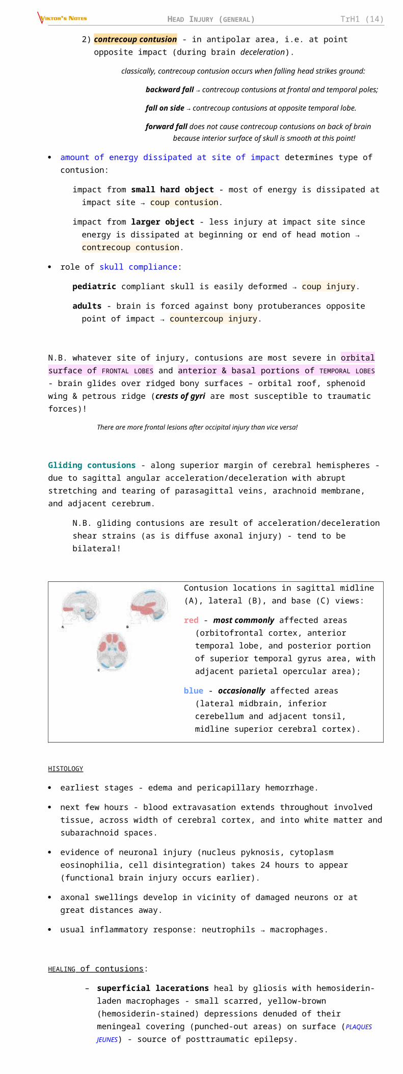
Contusion locations in sagittal midline (A), lateral (B), and base (C) views:
red - most commonly affected areas (orbitofrontal cortex, anterior temporal lobe, and posterior portion of superior temporal gyrus area, with adjacent parietal opercular area);
blue - occasionally affected areas (lateral midbrain, inferior cerebellum and adjacent tonsil, midline superior cerebral cortex).
HISTOLOGY
earliest stages - edema and pericapillary hemorrhage.
next few hours - blood extravasation extends throughout involved tissue, across width of cerebral cortex, and into white matter and subarachnoid spaces.
evidence of neuronal injury (nucleus pyknosis, cytoplasm eosinophilia, cell disintegration) takes 24 hours to appear (functional brain injury occurs earlier).
axonal swellings develop in vicinity of damaged neurons or at great distances away.
usual inflammatory response: neutrophils → macrophages.
HEALING of contusions:
– superficial lacerations heal by gliosis with hemosiderin-laden macrophages - small scarred, yellow-brown (hemosiderin-stained) depressions denuded of their meningeal covering (punched-out areas) on surface (PLAQUES JEUNES) - source of posttraumatic epilepsy.
– larger areas of necrosis that extend deep heal by formation of MENINGOCEREBRAL CICATRIX (composed of glia, fibroblasts, and meninges) or larger CAVITATED LESIONS.
CLINICAL FEATURES:
1) focal deficits (coincide with affected brain region; most often hemiparesis or gaze preference) – manifest after consciousness is regained.
2) increasing ICP – manifests as progressive neurologic deterioration.
3) late posttraumatic seizures
contusion per se is clinically silent if in non-eloquent area (e.g. anterior temporal lobes or inferior frontal lobes); but may manifest latter as expanding mass!
contusions in brainstem may be fatal.
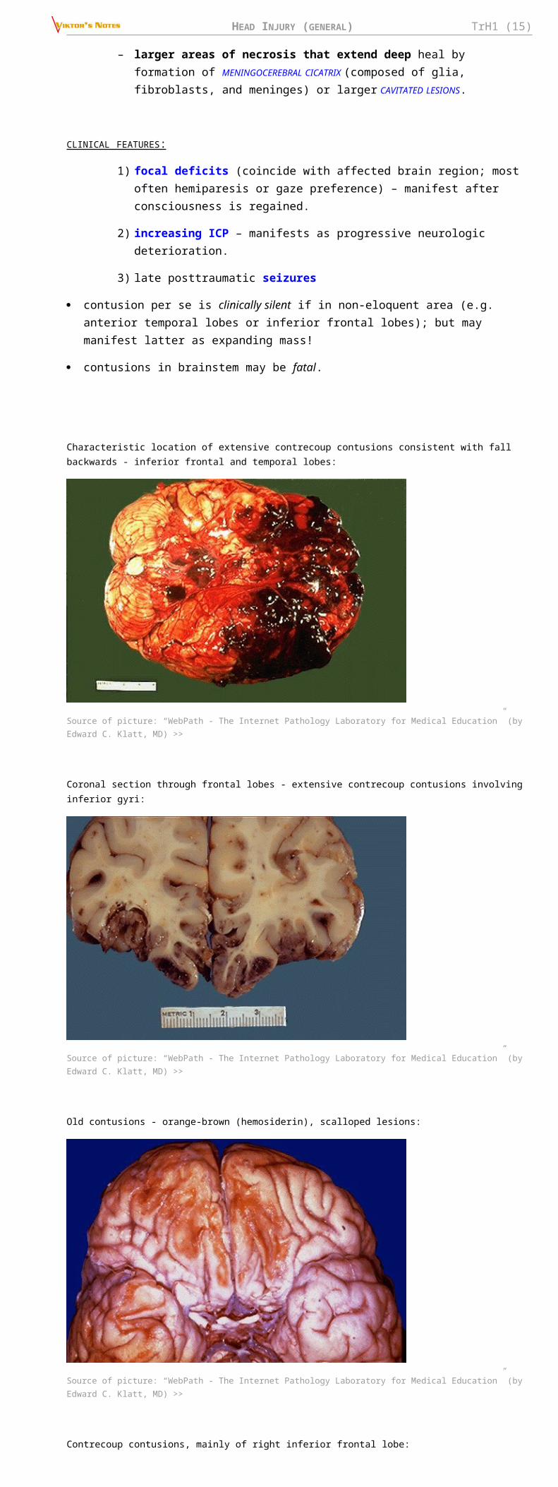
Characteristic location of extensive contrecoup contusions consistent with fall backwards - inferior frontal and temporal lobes:
Source of picture: “WebPath - The Internet Pathology Laboratory for Medical Education” (by Edward C. Klatt, MD) >>
Coronal section through frontal lobes - extensive contrecoup contusions involving inferior gyri:
HEAD INJURY (GENERAL) TrH1 (12)
Source of picture: “WebPath - The Internet Pathology Laboratory for Medical Education” (by Edward C. Klatt, MD) >>
Old contusions - orange-brown (hemosiderin), scalloped lesions:
Source of picture: “WebPath - The Internet Pathology Laboratory for Medical Education” (by Edward C. Klatt, MD) >>
Contrecoup contusions, mainly of right inferior frontal lobe:
Source of picture: “WebPath - The Internet Pathology Laboratory for Medical Education” (by Edward C. Klatt, MD) >>
Extensive blunt force trauma (vehicular accident) - contusions and lacerations:
Source of picture: “WebPath - The Internet Pathology Laboratory for Medical Education” (by Edward C. Klatt, MD) >>
Contusion of temporal poles with fresh hemorrhages:

HEAD INJURY (GENERAL) TrH1 (13)
Source of picture: James C.E. Underwood “General and Systematic Pathology” (1992); Churchill Livingstone; ISBN-13: 978-0443037122 >>
INTRACEREBRAL HEMORRHAGE (tICH)See below >>
MISSILE (GUNSHOT) INJURYWounding capacity of firearm is related to kinetic energy of its missile:
Kinetic energy = ½ × mass × velocity2
Types of injuries:
A. Tangential wounds - caused by impact at oblique angle relative to skull.
if missile has high velocity but low energy, it can travel around skull under scalp without passing through skull itself (but at site of impact depressed skull fracture can occur).
intracranial damage (primarily cortical contusions) occur at site of initial impact (pressure waves generated by impact).
B. Penetrating injury - projectile breaches cranium but does not exit;
low-velocity projectile loses energy as it penetrates skull; projectile may bounce off opposite inner table of skull and ricochet within brain.
C. Perforating injury (worst prognosis!) - projectile passes entirely through head, leaving both entrance and exit wounds.
entrance wound is smaller than exit wound.
brain damage is accompanied by extensive hemorrhage.
Bullets that penetrate skull do not travel in straight path:
a) low-velocity civilian soft bullets - tend to be deflected by intracranial structures (final track is erratic and occasionally bears no relation to exit or entrance site).
destabilizing motions include yaw (deviation of longitudinal axis of bullet from straight line), tumbling (forward rotation of bullet around its center of mass), rotation (oscillatory motion of bullet axis around its center of mass).
b) high-velocity military metal-jacket bullets - can project straight through tissues and easily fracture bones.
Bullets can damage brain parenchyma through 3 mechanisms:
A. Direct laceration & crushing (main mechanism of low-velocity bullets); destroyed tissue is either ejected out of entrance or exit wounds or compressed into walls of missile tract.
B. Cavitation (severe in high-velocity bullets) - produced by centrifugal effects of missile.C. Percussion shock waves (last 5-10 msec; severe in high-velocity bullets) - cause stretch injury
far from missile path (if shock wave reaches brain stem, cardiovascular and respiratory collapse can occur; shock waves can disrupt vessel walls → traumatic aneurysms).
all these create permanent cavity (3-4 times larger than missile diameter) and pulsating temporary cavity (as much as 30 times larger than missile diameter → diffuse damage to brain).
ICP
rapid increase (up to 100 mmHg) for several minutes → drop (depending on volume of secondary hemorrhage and edema).
CLINICAL FEATURES
- loss of function of brain that is directly injured.
penetration of frontal or parietal lobes by small missiles may not cause loss of consciousness! (good prognosis for survival)
HEAD INJURY (GENERAL) TrH1 (14)
COMPLICATIONS : hemorrhage!!!, infection!!!, post-traumatic epilepsy.
metal fragments may cause electrolysis, may migrate within intracranial or intraspinal compartments.
penetrating wounds (incl. GSW) to the head are not associated with C-spine injuries.
injury to vascular walls (contact or shearing forces) may lead to aneurysm formation (most commonly – pseudoaneurysm – very vulnerable to delayed ruptures).
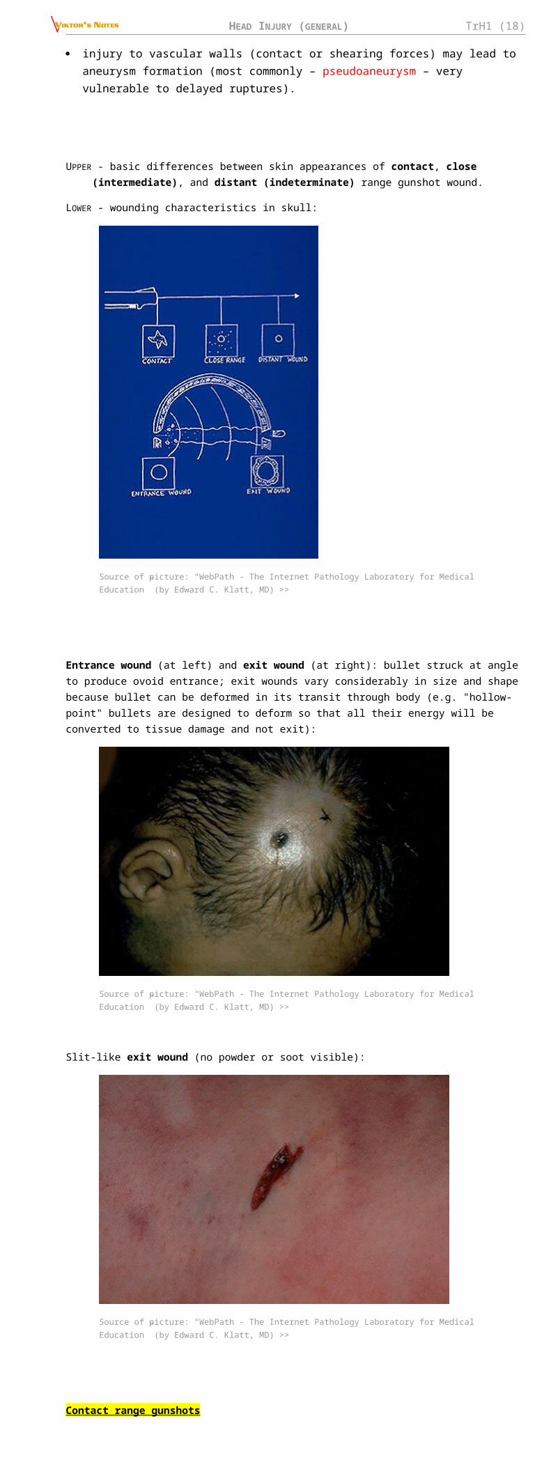
UPPER - basic differences between skin appearances of contact, close (intermediate), and distant (indeterminate) range gunshot wound.
LOWER - wounding characteristics in skull:
Source of picture: “WebPath - The Internet Pathology Laboratory for Medical Education” (by Edward C. Klatt, MD) >>
Entrance wound (at left) and exit wound (at right): bullet struck at angle to produce ovoid entrance; exit wounds vary considerably in size and shape because bullet can be deformed in its transit through body (e.g. "hollow-point" bullets are designed to deform so that all their energy will be converted to tissue damage and not exit):
Source of picture: “WebPath - The Internet Pathology Laboratory for Medical Education” (by Edward C. Klatt, MD) >>
Slit-like exit wound (no powder or soot visible):
Source of picture: “WebPath - The Internet Pathology Laboratory for Medical Education” (by Edward C. Klatt, MD) >>
Contact range gunshots
HEAD INJURY (GENERAL) TrH1 (15)
Contact gunshot entrance wound; since barrel contacts skin, gases released by fired round go into subcutaneous tissue → star-shaped laceration; note also grey-black discoloration from soot, as well as faint abrasion ring:
Source of picture: “WebPath - The Internet Pathology Laboratory for Medical Education” (by Edward C. Klatt, MD) >>
Contact range gunshot wound - abrasion ring, formed when force of gases entering below skin blow skin surface back against gun muzzle:
Source of picture: “WebPath - The Internet Pathology Laboratory for Medical Education” (by Edward C. Klatt, MD) >>
Contact range gunshot wound - abrasion ring, very clear muzzle imprint:
Source of picture: “WebPath - The Internet Pathology Laboratory for Medical Education” (by Edward C. Klatt, MD) >>
Contact range gunshot wound - grey-black discoloration from burned powder:
Source of picture: “WebPath - The Internet Pathology Laboratory for Medical Education” (by Edward C. Klatt, MD) >>
Contact range gunshot wound - skull surface demonstrates heavy soot, as well as radiating fracture lines; thus direction of fire was toward back of this picture:

HEAD INJURY (GENERAL) TrH1 (16)
Source of picture: “WebPath - The Internet Pathology Laboratory for Medical Education” (by Edward C. Klatt, MD) >>
Contact range gunshot wound (entrance wound on skin) - black gunshot residue (red arrow) and coagulative necrosis:
Source of picture: “WebPath - The Internet Pathology Laboratory for Medical Education” (by Edward C. Klatt, MD) >>
Intermediate range gunshots
Intermediate range gunshot entrance wound - powder "tattooing" around entrance site:
Source of picture: “WebPath - The Internet Pathology Laboratory for Medical Education” (by Edward C. Klatt, MD) >>
Intermediate range gunshot entrance wound - powder “tattooing”; actual entrance site is somewhat irregular, because bullet can tumble in flight:
Source of picture: “WebPath - The Internet Pathology Laboratory for Medical Education” (by Edward C. Klatt, MD) >>

HEAD INJURY (GENERAL) TrH1 (17)
SELF-INFLICTED INJURIES
injury on dominant side.
powder burns at entrance site.
large stellate scalp lacerations (dissection of subgaleal layer by exploding gases).
if entrance through mouth, injury to hard palate → upper airway compromise.
careful aim and close range → mortality ≈ 95%.
Suicide is more lethal than homicide!
STAB INJURY (IMPALEMENT) skull penetration is most common in thin bones of skull:
1) orbital surfaces
2) squamous portion of temporal bone – highest mortality (short distance to brainstem and vascular structures)
3) craniocervical junction
2/3 cases on left side!
knife leaves narrow elongated “slot” fracture (in some cases, no radiological abnormality can be identified).
cerebral damage is largely restricted to wound tract (filled with clots).
N.B. unlike missile injuries, no concentric zone of coagulative necrosis caused by dissipated energy is present; unlike motor vehicle accidents, no diffuse shearing injury to brain occurs.
major complications are vascular (main cause of mortality):
1) massive intracerebral hematoma (50%)
2) subdural hematoma (9%)
3) contusion (31%)
4) traumatic aneurysms (risk of rupture - H: early angiography & repair)
5) carotid-cavernous fistula
6) stroke (5%)
Mechanism for "defense wounds":
Source of picture: “WebPath - The Internet Pathology Laboratory for Medical Education” (by Edward C. Klatt, MD) >>
Typical "defense wounds":
Source of picture: “WebPath - The Internet Pathology Laboratory for Medical Education” (by Edward C. Klatt, MD) >>
HEAD INJURY (GENERAL) TrH1 (18)
Source of picture: “WebPath - The Internet Pathology Laboratory for Medical Education” (by Edward C. Klatt, MD) >>
COMPRESSION INJURY requires significant force - skull architecture provides substantial resistance to deformation.
multiple linear skull fractures (can be depressed if high-energy rapid compression force is applied to small area of skull).
DEGREES OF SEVERITY - determined by GCS score after initial* resuscitation**.
*within 6-48 hours of TBI
**no hypoxia, hypotension, hypothermia, intoxication, sedation
additional criteria: duration of loss of consciousness, duration of antegrade amnesia.
rarely used criteria: number of days to achieve GCS score 15, number of days to achieve GCS motor score 6, length of hospital stay, CT results.
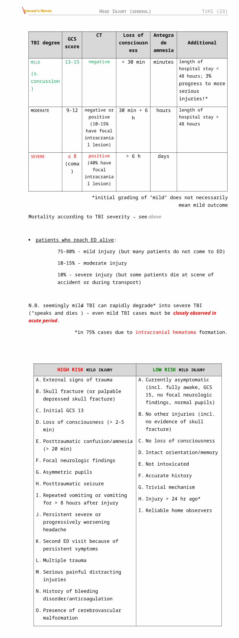
TBI degreeGCS score
CT Loss of consciousness
Antegrade amnesia
Additional
MILD
(s. concussion)
13-15 negative < 30 min minutes length of hospital stay < 48 hours; 3% progress to more serious injuries!*
MODERATE 9-12 negative or positive (10-
15% have focal intracranial
lesion)
30 min ÷ 6 h hours length of hospital stay > 48 hours
SEVERE ≤ 8 (coma)
positive (40% have focal intracranial
lesion)
> 6 h days
*initial grading of "mild" does not necessarily mean mild outcome
Mortality according to TBI severity → see above
patients who reach ED alive :
75-80% - mild injury (but many patients do not come to ED)
10-15% - moderate injury
10% - severe injury (but some patients die at scene of accident or during transport)
N.B. seemingly mild TBI can rapidly degrade* into severe TBI (“speaks and dies”) – even mild TBI cases must be closely observed in acute period.
*in 75% cases due to intracranial hematoma formation.
HIGH RISK MILD INJURY LOW RISK MILD INJURY
A. External signs of trauma
B. Skull fracture (or palpable depressed skull fracture)
C. Initial GCS 13
D. Loss of consciousness (> 2-5 min)
E. Posttraumatic confusion/amnesia (> 20 min)
A. Currently asymptomatic (incl. fully awake, GCS 15, no focal neurologic findings, normal pupils)
B. No other injuries (incl. no evidence of skull fracture)
C. No loss of consciousness
D. Intact orientation/memory

HEAD INJURY (GENERAL) TrH1 (19)
F. Focal neurologic findings
G. Asymmetric pupils
H. Posttraumatic seizure
I. Repeated vomiting or vomiting for > 8 hours after injury
J. Persistent severe or progressively worsening headache
K. Second ED visit because of persistent symptoms
L. Multiple trauma
M. Serious painful distracting injuries
N. History of bleeding disorder/anticoagulation
O. Presence of cerebrovascular malformation
P. Intoxication (→ unreliable examination)
Q. Mechanism: high-speed motor vehicle accident, fall of > 8 ft
R. Unreliable / unknown history of injury
S. Suspected child abuse
T. Age > 60 or < 2 yrs
E. Not intoxicated
F. Accurate history
G. Trivial mechanism
H. Injury > 24 hr ago*
I. Reliable home observers
*may miss chronic subdural hematoma
PREHOSPITAL MANAGEMENT FACIAL INJURIES → see p. TrH25 >>
ANTERIOR NECK INJURIES → see p. TrS21 >>
SPINAL INJURIES → see p. TrS5 >>
TRIAGE principles:
– thoracic, vascular, and abdominal injuries take precedence over head wounds!
– head injuries are more urgent than spinal injuries.
– triage of head injuries: 1. Deteriorating patients (who are not moribund); 2. Stable patients with level of consciousness↓; 3. Stable awake patients
“GOLDEN HOUR” – first hour is very important prognostically – treat hypoxia & hypotension in the field and en route to the hospital
60% fatalities occur before patients can be admitted to hospital (40% at scene and 20% in ER).
proper management in field can make difference between normal existence or lifetime spent in total paralysis.
mortality for military injuries: 4.5/100 in World War II → 2.5/100 in Korea → < 1/100 in Vietnam.
1. Rapid evacuation is given much of credit! (in civilian injuries - helicopter transport)
2. Trained teams of rescue workers (not physicians) provide intubation, shock treatment, and other emergency measures.
importance of ABC - ultimate outcome of brain injury is as much (or more) dependent on early ABC as any other organ.
even moderate hypotension can convert reversible brain injury to irreversible ischemic brain damage.
Spinal cord injury is present in as many as 10% patients!
Every patient with significant head injury has cervical spine injury until proved otherwise!!!
QUADRIPLEGIA IS FOREVER
1. OXYGENATION
- should be secured immediately.
Only means we can help injured brain in the field – supply oxygen to brain!
hypoxia – most common cause of prehospital death! see “Secondary Injury” above
AIRWAYS
1. Clear mouth (foreign bodies, vomitus, blood).
2. Do not extend neck! Use jaw thrust technique / chin lift maneuver:

HEAD INJURY (GENERAL) TrH1 (20)
3. Airway maintenance :
a) oropharyngeal tube
b) early endotracheal intubation:
– everybody in COMA (GCS < 8)!
– everybody with PENETRATING INJURY (if physician waits for coma before intubating patient, mortality approaches 100%).
– extensive FACIAL INJURIES
– COMBATIVE patient
– nasotracheal intubation is preferable (no neck manipulations), but avoid in facial / skull base fractures.
– for awake patient intubation use RSI (rapid sequence intubation).
N.B. failure to use paralytic agents, pharyngeal anesthesia, and barbiturate induction → massive ICP elevation! see below
– there is some suggestion of increased mortality with prehospital intubation in patients with moderate-to-severe TBI compared with patients intubated in ED (bag-valve-mask ventilation with good technique may be of more benefit to brain injured patients than prehospital intubation!).
c) cricothyrotomy, tracheostomy, percutaneous transtracheal ventilation.
d) stabilize mandibular fractures see p. TrH25 >>
BREATHING
a) spontaneous but insufficient breathing → 100% oxygen + assisted ventilation with demand valve.
b) no spontaneous breathing → ventilation (by positive-pressure ventilation with 100% oxygen).
– portable pulse oximetry should verify SaO2 > 96% (provide supplemental oxygen to achieve this level).
– ventilatory rate 10-12 breaths per minute (PaCO2 35-40 mmHg); in larger patients, higher ventilatory rates may be required to provide adequate minute ventilation; neurologic deterioration - high risk of intracranial mass lesion → higher ventilatory rates (20-25/min, PaCO2 < 35 mmHg).
2. BLOOD CIRCULATION
Injured brain is extremely susceptible to lowered perfusion states!
FLUID RESUSCITATION (brain injury per se rarely causes hypotension!!!)
several large-bore intravenous catheters.
isotonic (or hypertonic*) saline to aggressively restore SBP to > 100-110 mmHg. see below >>
*there are studies showing that 250 mL of hypertonic (7.5%) saline bolus in the field improves survival
avoid volume overload / hypertension! (but fluids should not be withheld in hypotensive patient for fear of increasing cerebral edema and ICP)
autohemotransfusion: leg elevation + Military Anti-Shock Trousers.
MANAGE BLEEDING, OPEN WOUNDS
scalp lacerations may bleed large volume into bulky dressing; better prehospital dressing is less bulky, but with firm constant pressure!
temporary scalp bleeding control → see MANAGEMENT
skull fractures – do not require special care in the field
– do not impede liquorrhea through fracture lines
– if brain / nerves exposed – cover with dressing with sterile saline (do not push brain back into cranium) – it is largely devitalized tissue and will be removed during scalp repair.

HEAD INJURY (GENERAL) TrH1 (21)
do not impede liquorrhea/ bleeding from nose / ear canal – will lead to intracranial hematoma and infection; H: place absorbent dressing without tamponade
– epistaxis is safe to tamponade only if no signs of anterior skull base fracture
life-threatening dural sinus bleeding can be slowed by placing patient in reverse Trendelenburg position (risk of air embolism!)
penetrating objects should be left in place (stabilized with bulky fluffy bandage) - to be removed in operating room.
3. CERVICAL SPINE STABILIZATION
→ see p. TrS5 >>
4. BRIEF NEUROLOGIC STATUS
1. Level of consciousness (ideally GCS)
2. Pupil size and light reactivity (asymmetry is most important)
3. Extremity motorics (asymmetry is most important) - 1spontaneous movements, 2following commands, 3reaction to painful stimuli.
5. TRANSPORTATION
- rapidly to trauma center with CT and definitive neurosurgical intervention*.
*level 1 trauma centers certified by American College of Surgeons (or state trauma certification systems) have trauma surgeon in house 24/7 and neurosurgeon available within 10 minutes of notification.
patient is moved en bloc - to avoid displacing spine (or other bones), so that spinal cord (and blood vessels) are not injured.
transport on left side, HOB elevated 15-30 degrees.
for transport times longer than 15-20 minutes, serial neurologic assessments should be documented every 10-15 minutes.
– most important parameter to monitor – level of consciousness (along with pulse, BP, breathing)
– all patients should be placed on cardiac monitor as they are transported (brainstem compression can cause cardiac dysrhythmias).
many severely head-injured patients are initially combative / agitated - transporting patient who is fighting against physical restraints may exacerbate physical injury, cause rise in ICP, and interfere with appropriate stabilization and management; H: prehospital sedation or paralysis (inform ED and trauma teams about it!).
Morphine and other depressants are contraindicated during initial management! (short acting opioid [fentanyl], sedative [propofol], paralytic are fine)
early contact by telephone or radio with trauma center - medications given to patient coordinated through neurosurgeons and other health care providers at trauma center.
DIAGNOSTIC EVALUATION DIAGNOSIS IN LATIN
Morbus traumaticus cerebri, periodus acutus/subacutus, forma levis/mediocris/gravis.
periodus acutus – within 3 months.
Diagnosis is constructed by naming injuries from outside to inside:
1) scalp (e.g. Vulnus contusum reg. parietalis; Haematoma subaponeuroticum reg. frontalis)
2) fractures (e.g. F-ra aperta impressa cominutiva ossis frontalis; F-ra basis cranii fossae mediae; F-ra ossis parietalis)
3) brain, including complications (e.g. Syndr. commotionale leve/mediocre/grave; Compressio cerebri. Haematoma epidurale/subdurale; Contusiones multiplices hemispherii sin.; Contusio cerebri lobi occipitalis; Haemorrhagia subarachnoidalis).
state syndrome first, then its cause (compressio → haematoma).
if there is contusion or hematoma, do not mention concussion.
Ebrietas vulgaris is mentioned at concurrent conditions.
HISTORY patient may be comatose or confused – witnesses & paramedics are of crucial importance!
1. Mechanisms, forces & circumstances of injury.

HEAD INJURY (GENERAL) TrH1 (22)
2. Loss of consciousness – duration, lucid interval, convulsions, apnea, cardiac arrest.
3. After consciousness was regained – amnesia (last thing that can be recalled), vertigo, nausea, vision blurring, headache, other pains.
4. Prior head injuries.
5. Remote or active medicament* / drug / alcohol use - risk of intracranial bleeding, cloud mental status. *esp. present anticoagulant therapy
6. Premorbid history of headaches, seizures, syncope, TIA & stroke, gait disturbance, psychiatric disease – all these can be the cause of TBI and should be treated!
PHYSICAL EXAMINATIONExamination of patient with decreased level of consciousness → see p. S30 >>
Cervical spine precautions! (10% patients with severe TBI have spinal trauma)
see p. TrS5 >>
*perform during PRIMARY SURVEY (identifying life-threatening conditions);
vs. SECONDARY SURVEY - detailed examination for identifying all traumatic injuries
1. ABC *
Evaluate for hypoxia, hypotension, anemia, and multiple injuries!
2. Monitor level of consciousness* – use Glasgow Coma Scale. see p. S30 >>
if conscious, assess attention, orientation*
GCS score is assessed in field (or by first responder), then reassessed after specific treatment interventions.
N.B. decrease of even 1-2 points in GCS score indicates significant change in neurologic status → prompt reevaluation!
3. Signs of trauma :
1) scalp injuries see above >>
2) skull fractures see p. TrH5 >>
3) facial trauma see p. TrH25 >>
4) cervical spine (swelling, pain on palpation, deformity, step-off, malalignment → prompt immobilization) see p. TrS5 >>
5) injuries outside cranium* should also be searched for at outset, because they are likely to be forgotten if not initially noted.
*most common in automobile accidents (e.g. WADDELL triad - child pedestrian hit by motor vehicle - 1chest-abdomen trauma, 2leg injury, 3countercoup head injury).
Neurologic Examination
a) awake patient → detailed neurologic examination.
b) uncooperative / comatose patient:
1) pupils* - size & light response. see below >>
2) motor* - response to painful stimuli, pathologic and deep tendon reflexes.
3) brainstem (for deeply comatose patients) - oculocephalic (± oculovestibular) tests, respiratory patterns.
*asymmetry is most important
4. Cranial nerves & brainstem
pupils!!!* (size and response to light) see p. D1eye >> , p. S30 >>
N.B. pupils are the only indicator of neurological function in chemically paralyzed patient!
Don’t miss direct orbital trauma!
Asymmetry - measurement difference of ≥ 1mm.
Dilated - pupillary size of > 4 mm.
Fixed – pupillary response < 1 mm to bright light.
pupillary asymmetry is due to intracranial injury unless proved otherwise.
unilateral dilated pupil in unconscious patient – CN3 compression (uncal herniation).
pinpoint pupils - pontine lesions.
light nonreactive pupils in mid position - midbrain tectum lesions.
eye movements
HEAD INJURY (GENERAL) TrH1 (23)
N.B. if voluntary eye movements cannot be assessed → OCULOCEPHALIC and OCULOVESTIBULAR testing see p. S30 >>
corneal reflex
facial asymmetry (e.g. when patient grimaces with noxious stimuli)
gag reflex - dysphagia (risk of aspiration)
respiratory pattern see p. 2115 (4-5) >> , p. S30 >>
5. Monitor for signs of ICP↑: see p. S50 >>
1) increasing headache, vomiting
2) declining mental status (decreasing GCS)
3) tense fontanelle
4) Cushing reflex (tachypnea > 20/min*; systolic BP increase > 15 mmHg or widening of pulse pressure, reflex bradycardia < 60/min or change > 10/min)
*also may indicate pulmonary failure or infection
6. Motor examination see p. S30 >>
N.B. if patient is not alert enough to cooperate with strength testing, motor examination is limited to assessment of motor asymmetry, pathologic & deep tendon reflexes!
N.B. occult extremity trauma can make examination painful or difficult!
7. Sensory examination is not reliable in patients who are intoxicated or comatose!
8. Bedside cognitive testing (e.g. Mini-Mental State Examination) - to distinguish damaged and spared realms of cognitive functioning; practically never done in acute phase of TBI. see p. D2 >> , p. D3 >>
ability to lay down new memories (determines duration of antegrade amnesia via serial mental status assessments).
IMAGINGPresence / absence of cervical spine instability must be determined first! (lateral cervical spine* and chest X-rays are usually obtained in resuscitation room).
*modern approach - cervical spine CT (at same time as admission head CT) in all but mildest TBI patients. see p. TrS5 >>
IMAGING MODALITIES
CT MRI ANGIOGRAPHY CRANIOGRAM
ADVANTAGES
1. Fast, no motion artifacts
2. Patient accessible for monitoring.
3. Defines acute hemorrhages, mass effects, bony injuries, hydrocephalus, edema.
Defines contusions and pericontusional edema, posttraumatic ischemic infarction, brainstem injuries
1. Helps localize acute traumatic lesions.
2. Defines vascular injuries, injuries to venous sinuses
3. Detects mass effects.
1. Readily available
2. May help screen some patients for further imaging studies.
DISADVANTAGES
1. Artifacts arise from patient movement, foreign bodies.
2. Streak artifacts may obscure brain stem or posterior fossa.
1. Slow (long scanning time), motion artifacts
2. Magnetic field precludes use of monitors and life-support equipment.
3. Does not define most acute hemorrhagic lesions
4. Not useful for bony injuries.
5. Unsuitable for people exposed to metallic objects.
Requires formal IR procedure so it is done only if CTA is positive.
Does not indicate presence or absence of intracranial injury so it is obsolete study.
American College of Radiology (ACR) Appropriateness Criteria Scales
(1 = least appropriate; 9 = most appropriate; NA = not applicable):
Clinical SituationSkull
X-ray
Cervical
X-rayCT MRI MRA
Angio-graphy
mild closed TBI (GCS ≥ 13, no neurologic deficit)
2 2 7 NA NA NA
mild closed TBI (focal neurologic deficit)
2 No consensus 9 6 4 NA
moderate ÷ severe closed TBI, stable 4 8 9 6 NA NA

HEAD INJURY (GENERAL) TrH1 (24)
mild ÷ moderate closed TBI, child < 2 yrs
4 6 9 6 NA NA
closed TBI, rule out carotid or vertebral artery dissection
NA 5 4 8* 8 6
penetrating TBI, stable, neurologically intact
8 8 8 6 3 6
penetrating TBI, likelihood of vessel injury
8 8 8 6 8 8
depressed skull fracture 8 6 9 6 NA NA
calvarial fracture 8 6 9 5 NA NA
penetrating TBI, skull-base fracture 6 6 9 6 6 4
subacute TBI, late neurologic deterioration
NA NA 8 8 NA NA
subacute / chronic TBI, stable, normal CT, cognitive and/or neurologic deficit
NA NA NA 8 4 NA
chronic TBI, neurologic dysfunction NA NA 6 8 4 NA
*5 for gadolinium-enhanced MRI
CT WITHOUT CONTRAST
- diagnostic imaging of choice! – detects skull fractures, contusions, blood, edema.Contrast is rarely required to outline subacute / chronic hematomas or intracranial abscesses
CT is indicated for all patients except LOW-RISK mild TBI;
– for HIGH-RISK mild TBI (without skull fracture), CT may be deferred if patient is hospitalized for observation (any deterioration in neurologic status or any focal signs → CT).
If CT is indicated, skull radiographs are not necessary (CT bony window shows even basilar fractures)
more severe TBI, greater urgency for CT (exception is when other life-threatening injuries take precedence); e.g. severe TBI (esp. with lateralizing signs) → CT as soon as cardiorespiratory stability is ensured.
N.B. urgency for CT is function of likelihood of surgically correctable focal lesion (vs. diffuse injuries - do not benefit from acute neurosurgical intervention - precise anatomic information is not necessary for optimal management).
deteriorating patients should be accompanied by physician;
stable but seriously injured patients should be accompanied by experienced ER or trauma nurse.
if abdominal ultrasound or diagnostic peritoneal lavage has not been performed, most comatose victims also have abdominal CT and, if thoracic injury is suspected, chest CT.
patients going to operating room for treatment of other injuries, must have head CT (because they will not be available for monitoring); if patient is taken immediately to surgery without head CT* → historically: intraoperative air ventriculogram (detects large intracranial mass lesions).
*try to obtain at least single-cut CT through level of lateral ventricles
Most important things to look in CT: mass lesions (hematoma), state of basal cisterns (incl. blood inside), midline shift, ventricular size.
Basal cisterns are evaluated at the level of the midbrain:
Basal cisterns can be: open (all limbs open), partially closed, s. “crowded” (one or two limbs obliterated), or completely closed s. “effaced” (all limbs obliterated).
Midline shift is calculated at the level of the foramen of Monro to the septum pellucidum:
Midline shift = (A/2) - B

HEAD INJURY (GENERAL) TrH1 (25)
Technique
from base of occiput to top of vertex in at max. 5-mm increments.
three data sets are obtained:
1) bone windows - bony anatomy of skull.
2) tissue windows - detailed survey of brain.
3) subdural windows - visualization of hemorrhages adjacent to brain (subdural hematomas).
seriously injured intubated patients should receive neuromuscular blockage during CT study; in severe cases, single-cut CT through level of lateral ventricles has high yield for revealing hematomas.
Indications for SELECTIVE REPEAT CT:
1. GCS↓ by > 1 point
2. New / aggravated focal signs
3. Persistent severe headache, frequent vomiting
4. Seizure
Indications for SCHEDULED REPEAT CT:
1. Nonoperated intracranial hemorrhages, contusions*
2. Coagulopathies
CT is repeated within 4-24 hours of initial scan (some contusions and intracranial hematomas are brought to operating room after repeated CT)
*studies do not support SCHEDULED REPEAT CT in patients with intracranial hemorrhage and GCS13-15 and no change in status because imagings do not change management and outcomes
AbdelFattah K, Eastman A., Aldy K., et al “A prospective evaluation of the use of routine repeat cranial CT scans in patients with intracranial hemorrhage and GCS score of 13 to 15” Journal of Trauma and Acute Care Surgery
Marshall Classification of Diffuse Brain Injury
use in prognostication – see below >>

HEAD INJURY (GENERAL) TrH1 (26)
Grade 1 = normal CT scan (9.6% mortality)
Grade 2 = cisterns present, shift < 5mm (13.5% mortality)
Grade 3 = cisterns compressed / absent, shift < 5mm (34% mortality)
Grade 4 = shift > 5mm (56.2% mortality)
Marshall LF, Bowers-Marshall S, Klauber MR et al. A new classification of head injury based on computerized tomography. J Neurosurg 75(Suppl):S14-20, 1991
PLAIN SKULL RADIOGRAPHS
– only rare indications:
N.B. many patients with skull fracture have no neurological sequelae and many with severe intracranial abnormalities have no associated skull fractures!
1) screening for CT in mild TBI with signs of skull fracture on physical examination when CT is not immediately available (if skull fracture is confirmed → CT & observation for delayed complications).
N.B. yield of craniography in LOW-RISK mild TBI is very low (even if detected, linear fractures have limited clinical significance in adults – do not change treatment course).
midline shift of calcified pineal gland or M-echo signal dislocation > 3 mm (in echoencephaloscopy) is additional indication to seek for CT.
2) depressed skull fracture – use tangential shots to measure the depth of impression.
3) penetrating injury – to detect intracranial foreign bodies (small intracranial foreign bodies may be missed unless skull X-ray is taken after minor penetrating injuries).
4) additional indications for pediatric population:
a) screening for CT in children < 5 yrs (difficult clinical examination) with minor TBI
b) suspected child abuse (skull radiograph as part of skeletal survey).
c) suspected linear skull fracture (risk of leptomeningeal cyst) - deep scalp lacerations, large scalp hematoma.
NONENHANCED MRI- most useful in subacute/chronic setting; prognostic use during subsequent care (assists in directing rehabilitation and goals of care);
MRI gives superior (than CT) depiction of nonsurgical lesions; some authors now advocate MRI use in neurologically stable patients with moderate-to-severe closed TBI, whereas CT is reserved for neurologically unstable patients.
specific sequences :
T2* (either GRE [gradient-recalled echo] or SWI) is most sensitive sequence to detect small hemorrhages, blood breakdown products.
diffusion-weighted images - useful for cerebral infarctions;
FLAIR - useful for SAH.
N.B. gadolinium enhancement gives no notable advantage!
Indications:
1. Subacute period of severe TBI - to depict full extent of injury to brain!
2. Suspected contusions not seen on CT (MRI is criterion standard for defining contusions!)
3. Persistent symptoms with postconcussive syndrome
4. Suspected posttraumatic ischemic infarction
5. Mental status abnormalities (unexplained by CT; MRI is more sensitive for diffuse axonal injury!)
6. Subacute / chronic subdural hematomas (MRI is more sensitive than CT but normally CT is just enough).
Contraindication - penetrating injuries with possible (not ruled out with X-ray) intracranial metallic fragments.
ANGIOGRAPHY
- used only when vascular injury may be present and when endovascular intervention is anticipated:
a) penetrating injury (esp. due to impalement) – absolute indication if CTA shows proximity to major vessels!
b) carotid injuries on CTA with (attributable neurologic deficits from) flow limitation
c) skull base fracture near major arteries with unexplained neurologic deficits (esp. temporal bone fractures).
performed only in patients in stable condition.
CTA / MRA can obtain similar information.
formerly (in pre-CT era), angiography was used to diagnose intracranial hematomas.
HEAD INJURY (GENERAL) TrH1 (27)
ULTRASONOGRAPHY
a) intracranial hemorrhage in infants.
b) leptomeningeal cyst (after linear skull fracture) in infants.
c) orbital soft-tissue injury.
CONDITIONS
CONCUSSION
– normal CT/MRI.
DTI is abnormal!
Diffuse increase in mean diffusivity after remote concussions. Sagittal (A and B), coronal (C and D), and axial (E and F) slices of the tract-based spatial statistics group contrast on mean diffusivity maps (controls vs concussed in blue). The contrasts are overlaid on the mean fractional anisotropy skeleton (in green) and the standard MNI152 T1 1-mm brain template. The results are thresholded at P ≥ .05, corrected for multiple comparisons:
Picture source: Tremblay S, Henry LC, Bedetti C, et al. Diffuse white matter tract abnormalities in clinically normal ageing retired athletes with a history of sports-related concussions. Brain. 2014;137:(11) 2997-3011.
DIFFUSE AXONAL INJURY
– often not visible (microscopic damage).
CT - small petechial hemorrhages in white matter (most frequently juxtacortical & periventricular, corpus callosum, internal capsule and dorsorostral brainstem) – “tear” hemorrhages;
DAI can sometimes be superimposed by generalized brain swelling.
MRI is more sensitive (may be positive even when CT is negative; e.g. nonhemorrhagic DAI) - diffuse, small, focal abnormalities limited to white matter tracts.
SWI is the most sensitive sequence (more sensitive than GRE)!
months after injury - reduced bulk of white matter with persistent hemosiderin foci.
T2-MRI - foci of hypointense signal representing small shear hemorrhages at gray/white junction (arrows):
CT - multiple white matter hemorrhages:

HEAD INJURY (GENERAL) TrH1 (28)
MRI - Duret hemorrhage in splenium of corpus callosum:
SCALP INJURIES
CT reliably depicts subgaleal hematomas, elevated and avulsed soft tissues, scalp edema.
air within scalp tissue suggests scalp laceration (gas detected several days after trauma - infection).
CONTUSION
Contusion extends to cortical surface (vs. intracerebral hematoma)
MRI is criterion standard for defining contusions!
early - inhomogeneous hyperdensity* on CT, hyperintensity on MRI (T1 and T2).
*initially (< 1-2 hours after TBI), partial volumes between dense microhemorrhages and hypodense edema can render contusions isoattenuating on CT (high initial false-negative rate!); vs. MRI demonstrate contusions from onset!
– mass effect may distort adjacent sulci and lateral ventricles.
– some degree of SAH is almost always present.
after several hours - ring of lower density (edema surrounding contusion; very early - only edema without hemorrhages).
after week (blood within contusions has begun to degrade, and MRI becomes more useful) - surrounding ringlike contrast enhancement.
N.B. contusions progress with time in size, number, and amount of hemorrhage within contusions – these changes are most evident over first 24-48 hours (in 25% cases delayed hemorrhage occurs in areas that were previously free of hemorrhage) – routinely repeat CT!!!
Noncontrast CT - hyperdense region in anterior temporal lobe:
Noncontrast CT - cerebellar contusion; right-sided soft-tissue swelling and bone injury are also obvious:
HEAD INJURY (GENERAL) TrH1 (29)
Noncontrast CT - large, right frontal contusion with hemorrhage and surrounding edema; smaller right temporal cortical contusion (short arrow); small left frontal subdural hematoma (long arrow):
Enlargement of contusion:
A) CT on day 1 - subtle area of slightly hypoattenuating right frontal lobe contusion (arrows)
B) CT on day 2 - large, right frontal contusion (arrow) and new, large, left temporal contusion (arrow); scalp hematomas have increased in size:
CT immediately after blunt trauma to left convexity - severe swelling of entire left cerebral hemisphere with small collection of subarachnoid blood (up arrow); right hemisphere shows contrecoup gliding contusions (down arrows):
PENETRATING INJURIES
CT – imaging modality of choice: focal areas of increased attenuation (represent parenchymal, intraventricular, subarachnoid, or extracerebral hemorrhage).
path of missile is often obvious from location of hemorrhage and metallic fragments (produce streak artifacts).
coronal sections may be helpful in patients with skull base or high convexity involvement.
Plain radiographs - can be helpful in assessing bullet trajectory, the presence of large foreign bodies, and the presence of intracranial air; when CT is available, plain radiographs are not essential and are not recommended as a routine.
MRI - not generally recommended in the acute management.
may help evaluate injuries from penetrating wooden or other nonmagnetic objects.
in addition to artifact and image distortion, ferromagnetic missiles can also rotate and deflect in response to magnetic torque (literature has not reported any patient suffering additional injury caused by MRI).

HEAD INJURY (GENERAL) TrH1 (30)
Angiography / CTA – when vascular injury is suspected:
a) wound’s trajectory passes through or near the Sylvian fissure, supraclinoid carotid, cavernous sinus, or a major venous sinus – facial, orbital, pterional entry sites.
b) substantial and otherwise unexplained SAH or delayed hematoma.
aneurysms may form in delayed fashion (initial negative CTA/angio does not exclude the risk).
OTHER NEUROLOGIC TESTSLumbar puncture is contraindicated (unless meningitis is suspected – but LP only after imaging excluded mass lesion = basilar cisterns must be open on CT).
LP can be therapeutic in SAH.
Ophthalmoscopy – signs of ICP↑ (papilledema, loss of venous pulsation).
EEG - not emergency test!
if taken at some convenient time after admission, it may aid in prognosis:
at time of injury - suppression of electrical cortical activity;
with recovery - activity returns to normal (often through phase of generalized slowing and increased voltage).
– common feature in all patients (may persist for many weeks) - undue susceptibility of cortical activity to OVERVENTILATION .
– areas of cortical damage may show abnormal activity (slowing and spike activity) for weeks ÷ months – risk of epilepsy!
Most useful role of EEG - diagnosis of nonconvulsive status epilepticus!*
*may be detected in ≈ 8% comatose patients
N.B. sedative medications may have confounding effect!
bilaterally absent somatosensory & auditory evoked potentials predict unfavorable outcome in ≈ 99.5% patients.
Transcranial Doppler
normal average linear blood velocity (LBV) in MCA ≈ 60 cm/s.
LBV > 100-120 cm/s:
a) hyperemia (parallel LBV increase in MCA and ICA) – esp. days 2-4 after TBI
b) vasospasm (LBV in MCA exceeds LBV in ICA ate least 3 times) – usually on days ≥ 5 post TBI
low LBV (esp. early after TBI) reflects brain hypoperfusion.
GOSLING (PULSATILITY) index = (systolic LBV - diastolic LBV) / mean LBV.
if > 1 – sign of intracranial hypertension
NON-NEUROLOGIC TESTSMonitor ECG - high incidence of supraventricular tachycardia (most common dysrhythmia after TBI), ventricular dysrhythmias, ST-T wave abnormalities, prolonged QT.
Urine toxicology screen and blood alcohol level - interpreting patient's mental status.
Glycemia
hyperglycemia correlates with poor outcome.
Serum electrolytes & osmolarity
Osmolarity = 2 (Na + K) + glycemia + urea
Sodium (alterations occur in 50% comatose patients):
HYPONATREMIA - due to:
a) syndrome of inappropriate antidiuretic hormone (SIADH)
b) cerebral salt wasting (release of natriuretic hormone → urinary Na losses↑ → volume depletion).
hyponatremia can potentiate brain edema and cause seizures.
HYPERNATREMIA - due to:
a) dehydration
b) diabetes insipidus (urine output↑, urine specific gravity↓)

HEAD INJURY (GENERAL) TrH1 (31)
N.B. MANNITOLMANNITOL can mask diabetes insipidus by producing high urine output.
Magnesium is depleted in acute phases of both minor and severe TBI.
H: MAGNESIUMMAGNESIUM SULFATESULFATE IV or PO
N.B. magnesium blocks excitotoxic response and functions as antioxidant - careful monitoring of magnesium may improve outcome!
Coagulation studies (PT, aPTT, fibrinogen, platelet counts)
CBC, Hct – monitor in case of bleeding (type and cross match are always obtained with initial orders).
Arterial-venous blood gases – monitor in comatose patients.
20-gauge Silastic catheter is inserted with tip in internal jugular bulb, usually on right - arterial jugular venous oxygen content difference can be obtained.
normal venous saturation ≈ 65%;
< 50-55% - global brain ischemia;
> 90% - luxury perfusion (hyperemia).
Maintain arterial - jugular venous O2 content difference < 7 vol%.
Volemic indicators (CVP, PCWP) – monitor during treatment of hypotension.
Renal function tests & CK - to exclude rhabdomyolysis if crush injury has occurred or marked rigidity is present.
Within 4-5 days after admission, comatose patients should have screening endocrine battery (incl. cortisol, prolactin, growth hormone, thyroid function) – screening of pituitary injury.
MANAGEMENT Physicians can do nothing to either replace lost neurons or accelerate restoration of recovering neurons!
Main goal of TBI management – prevention, swift recognition and treatment of SECONDARY INSULTS (i.e. preventing / reversing further brain injury)
N.B. in all neurotrauma cases maintain SBP > 110 mmHg (DAMAGE CONTROL RESUSCITATION)
DISPOHOSPITALIZATION
Neurologic deterioration is most common within 24 hrs after TBI
all patients (except LOW-RISK mild TBI) must be admitted!
– HIGH-RISK mild TBI patients should be observed for 8-24 hours* ("neuro checks" at 30-60 min intervals - any deterioration in neurologic status or any focal signs → CT).
– if physician decides that patient with HIGH-RISK mild TBI can be sent home, appropriate early follow-up should be arranged.
*intoxicated patients must be observed until clinical sobriety
all severe TBI and most moderate TBI patients are admitted to ICU.
if patient is sent home :
1) patient should be monitored at home* in quiet environment in acute period (12-24 hours) by responsible nonintoxicated adult who lives with patient (and has access to telephone).
*do not allow to work, drive, etc
2) provide instructions (verbal and written - "head injury check list") describing signs & symptoms of delayed complications of TBI* – patient should be awakened every 2 hours and assessed neurologically.
*e.g. severe headaches, persistent nausea and vomiting, seizures, confusion or unusual behavior, watery discharge from nose or ear
3) no analgesics other than ACETAMINOPHENACETAMINOPHEN or IBUPROFENIBUPROFEN should be used.
TRANSFER
Severe TBI patient:
1. ICP control - intubation, MANNITOLMANNITOL.
2. Seizure control – LEVETIRACETAMLEVETIRACETAM or PHENYTOINPHENYTOIN.

HEAD INJURY (GENERAL) TrH1 (32)
N.B. if patient is deteriorating, perform emergency burr holes see below
ABCsee “Secondary Injury”, “Prehospital Management” above
AIRWAY, VENTILATION
all severe TBI patients (or who have penetrating TBI) should be intubated: to prevent aspiration, to protect airway and respiratory drive, to allow PRN hyperventilation.
Rapid sequence induction [RSI] (with adequate sedation and paralysis!!!) is recommended to avoid ICP↑. see p. 3905 >>
1) SEDATION - cerebroprotective agent ETOMIDATEETOMIDATE (maintains BP, lowers ICP and brain metabolism, has rapid onset and brief duration);
THIOPENTALTHIOPENTAL 3-5 mg/kg is not recommended (lowers BP → secondary brain injury)
2) LIDOCAINELIDOCAINE IV (1-2 mg/kg)
3) PARALYSIS - SUCCINYLCHOLINESUCCINYLCHOLINE 1-2 mg/kg.
4) airway irritation (→ ICP↑) is blunted by MORPHINEMORPHINE or FENTANYLFENTANYL.
hyperventilation is recommended only as a short temporizing measure for the quick reduction of elevated ICP.
hyperventilation probably should be avoided during the first 24 hours after injury when cerebral CBF is often critically reduced (hyperoxia induces ischemia).
if hyperventilation is used, jugular venous oxygen saturation (SjO2) or brain tissue O2 partial pressure (BtpO2) measurements are recommended to monitor oxygen delivery.
pulmonary toilet.
coma for > 5-10 days → tracheostomy.
BLOOD PRESSURE
- to maintain brain perfusion (cerebral perfusion pressure). see below >>
End point is CPP > 60-70 mmHg.
Goals
1. Systolic BP > 100* mmHg (IV fluids ± vasopressor agents, if necessary)
* Level III recommendation: Maintaining SBP at ≥ 100 mm Hg (for patients 50-69 years old) or at ≥ 110 mm Hg (for patients 15-49 or > 70 years old) may be considered to decrease mortality and improve outcomes.
do not treat hypertension < 160* mmHg until intracranial hypertension is excluded (Cushing reflex is for brain perfusion)
*< 140 mmHg if ongoing risk of intracranial bleeding
N.B. albumin worsens outcomes (albumin extravasates and worsens cerebral edema)
Level II evidence – avoid SBP < 90 mm Hg
single episode of SBP < 90 mm Hg is associated with doubling of mortality in severe TBI (≥ 2 hypotension episodes increase mortality 8-fold)
2. CVP 5-15 cmH2O
3. PCWP 10-14 mmHg. further see p. S50 >>
4. SaO2 > 90%
Level III evidence – avoid PaO2 < 60 mmHg or SaO2 < 90%.
hypoxemia SaO2 ≤ 90% for 11.5-20 min is independent predictor of mortality in severe TBI (p = 0.024)
N.B. fluid restriction is contraindicated in TBI (historical tenet of TBI treatment was fluid restriction, which was believed to limit cerebral edema; but fluid restriction decreases intravascular volume → cardiac output↓ → CBF↓ → cerebral edema)
RESUSCITATION FLUIDS
NORMALNORMAL SALINESALINE is the best resuscitation fluid; HYPERTONICHYPERTONIC SALINESALINE is not better* but it is practical in battle field as takes less volume to carry in backpacks of paramedics.
*several studies of prehospital fluids in patients with TBI suggested that hypertonic solutions may restore cerebral perfusion, reduce cerebral edema, and modulate the inflammatory response that contributes to neuronal injury, and thus may benefit resuscitation of these patients.
“Out-of-Hospital Hypertonic Resuscitation Following Severe Traumatic Brain Injury: A Randomized Controlled Trial” Bulger EM, May S, Brasel KJ, et al JAMA. 2010;304:1455-1464 - largest randomized clinical trial of hypertonic fluid resuscitation following TBI - conclusion: initial fluid resuscitation of patients with severe TBI with either 7.5% saline + 6% dextran 70 or 7.5% saline alone is not superior to NS with respect to 6-month neurologic outcome or survival.

HEAD INJURY (GENERAL) TrH1 (33)
GENERAL MEASURESREGIMEN, ACTIVITY
General measures for COMATOSE PATIENTS - see p. S30 >>
neurochecks every 60 mins for 6 hrs, then every hour until stable.
mobilize after 24 hrs; avoid direct sunlight.
Do not overemphasize severity of injury - unnecessarily prolongs period of invalidism!
after severe TBI, return to active work should be deferred for 2-3 months.
NUTRITIONAL SUPPORT
early nutritional support can directly affect TBI outcome.
Level II A recommendation: “Feeding patients to attain basal caloric replacement at least by the 5th day and, at most, by the 7th day post-injury is recommended to decrease mortality”.
Level II B recommendation: “Transgastric jejunal* feeding is recommended to reduce the incidence of ventilator-associated pneumonia”
*vs. intragastric (e.g. PEG) feeding
– waiting 5 days to start enteral nutrition doubles risk of death at 2 weeks (waiting 7 days quadruples risk).
severe TBI gives 100-500-fold catecholamine surge → stress ulcers (H: PPI), MI.
DVT PROPHYLAXIS
TBI increases risk of DVT significantly and that correlates with TBI severity.
Level III recommendation: LMWH or low-dose unfractionated heparin may be used in combination with mechanical prophylaxis. However, there is an increased risk for expansion of intracranial hemorrhage. In addition to compression stockings, pharmacologic prophylaxis may be considered if the brain injury is stable and the benefit is considered to outweigh the risk of increased intracranial hemorrhage. There is insufficient evidence to support recommendations regarding the preferred agent, dose, or timing of pharmacologic prophylaxis for DVT.
Knudson et al. found that head injury with an Abbreviated Injury Score of ≥ 3, among other factors, was an independent predictor of VTE in trauma patients.
TBI has been associated with up to 54% incidence of deep venous thrombosis without prophylactic treatment and a 25% incidence in patients with isolated TBI treated with sequential compression devices.
Ekeh found that DVT occurred in 1/3 of moderate and severe TBI patients with isolated head injuries, having a lower incidence than those patients with concomitant extracranial injuries. Age, subarachnoid hemorrhage, Injury Severity Score >15, and extremity injury were predictors of DVT.
Reiff et al. demonstrated a 3-4-fold increase in the DVT risk in TBI despite use of mechanical and chemoprophylaxis; VTE risk increases with TBI severity.
early (< 24 h) initiation of VTE chemoprophylaxis in patients with traumatic intracranial hemorrhage appears to be safe.
Fabio A. Frisoli et al. Early Venous Thromboembolism Chemoprophylaxis After Traumatic Intracranial Hemorrhage. Neurosurgery, nyx164,https://doi.org/10.1093/neuros/nyx164. Published: 18 July 2017
Traumatic intracranial hemorrhage expansion occurred in 18% of patients in the early cohort (prophylaxis within 24 hrs after bleed) compared to 17% in the delayed cohort (> 48 hrs) (P = 0.83).
HEMOGLOBIN
Maintain Hb > 7 g/dL
Robertson CS “Effect of erythropoietin and transfusion threshold on neurological recovery after traumatic brain injury: a randomized clinical trial.” JAMA. 2014 Jul 2;312(1):36-47
In patients with closed head injury, neither administration of erythropoietin nor maintaining Hb > 10 g/dL resulted in improved neurological outcome at 6 months (vs. Hb > 7 g/dL). Transfusion threshold of 10 g/dL was associated with a ↑incidence of thromboembolic events.
NEUROLOGICAL MEASURES
ICP management
- central to intensive care of critically brain injured patient!
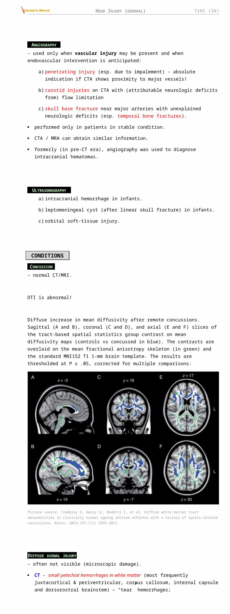
HEAD INJURY (GENERAL) TrH1 (34)
N.B. in absence of any obvious signs of increased ICP, no prophylactic treatment should be initiated!
MONITORING
Level II B recommendation: management of severe TBI patients using information from ICP monitoring is recommended to reduce in-hospital and 2-week post-injury mortality.
Options (class III) for penetrating TBI: early ICP monitoring is recommended if: unable to assess the neurologic examination accurately / the need to evacuate a mass lesion is unclear / imaging studies (e.g. CT) suggest elevated ICP.
Indications: other indications → see p. S50 >>
A. GCS > 8 + significant mass lesions on CT scan (but often such patients need to go to OR)
B. Salvageable patient with GCS 3-8 (after resuscitation) plus:
a) abnormal CT scan - mass lesions (contusions, hematomas) when patient is not taken to operating room for evacuation OR diffuse cerebral edema (esp. with obliteration of perimesencephalic cisterns – limited residual compliance).
b) normal CT scan + any two of the following (on admission): SBP < 90 mmHg, age > 40 yrs, unilateral or bilateral motor posturing.
most academic neurosurgical centers monitor ICP continuously with even moderately severe head injuries.
even patients taken to operating room frequently have ICP monitors placed at end of operation - later brain swelling may still be a problem even after successful mass evacuation (and even after craniectomy).
ICP monitor is zeroed at midbrain level.
if GCS is < 6, EVD is recommended over intraparenchymal monitor to drain CSF for first 12 hours postinjury (level III recommendation).
there are studies that do not support ICP monitoring; but from practical standpoint, the ICU staff pays much less attention to brain if ICP monitor is absent.
Qiang Yuan “Impact of intracranial pressure monitoring on mortality in patients with traumatic brain injury: a systematic review and meta-analysis” J Neurosurg 122:574–587, 2015
- systematic review and meta-analysis of ICP monitoring studies including 24,792 patients.
- current clinical evidence does not indicate that ICP monitoring overall is significantly superior to no ICP monitoring in terms of the mortality of TBI patients. However, studies published after 2012 indicated a lower mortality in patients who underwent ICP monitoring.
Chesnut RM, Temkin N, Carney N, et al. A trial of intracranial-pressure monitoring in traumatic brain injury. N Engl J Med. Dec 2012;367(26):2471-2481
- high-quality multi-center (South and Central America), Class 1 RCT (N=324)
- ICP monitor vs. close clinical assessment for ICP management
- outcomes for patients managed with information from clinical assessment do not differ from those for patients managed with information from the ICP monitor – no recommendation to use either method preferentially.
- ICP monitoring alone is not sufficient to improve outcomes.
Association between ICP monitoring and mortality in patients with TBI stratified by randomization or not (shown as log [Odds Ratio] with SE). IV = inverse variance.
Association between ICP monitoring and mortality in patients with TBI stratified by publication date (shown as log [Odds Ratio] with SE):
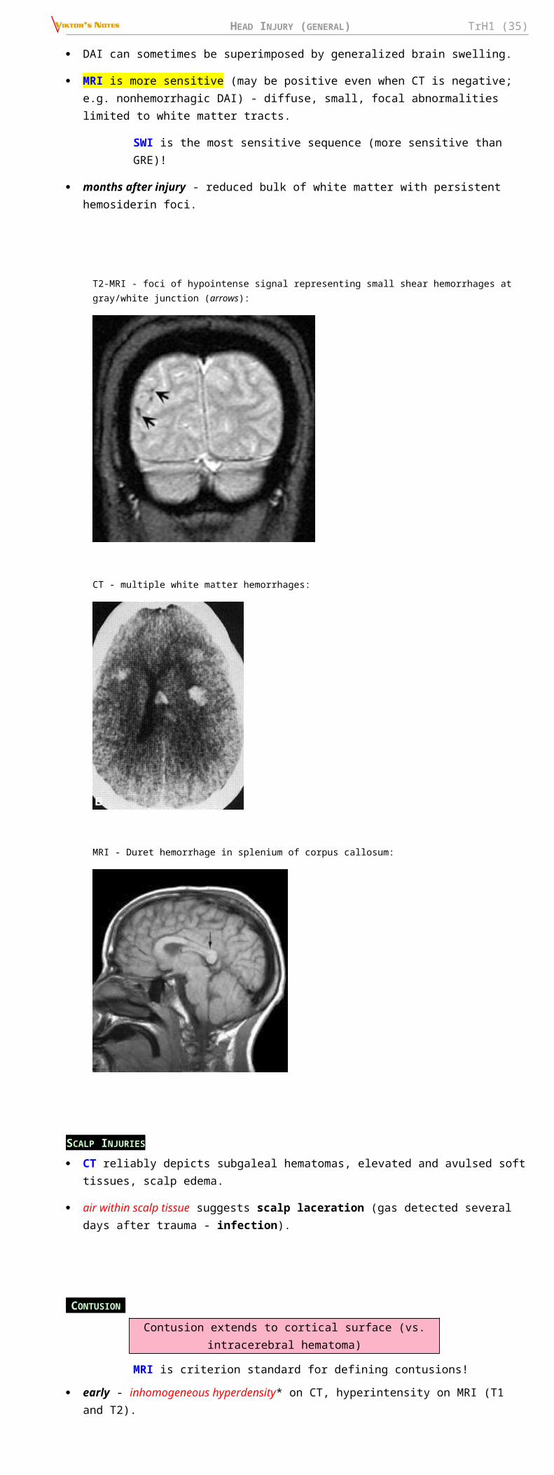
HEAD INJURY (GENERAL) TrH1 (35)
when ICP remains < 20 mmHg for 24-48 hours without treatment, ICP monitoring is discontinued.
TREATMENT
ICP > 22* mmHg for > 15 mins within 1 hour must be treated ASAP (to keep CPP > 70 mmHg) → see p. S50 >>
* > 15 mmHg after decompressive craniectomy (some would also treat > 18 mmHg for patients > 55 yrs or women of any age)
Level II B recommendation: Treating ICP above 22 mm Hg is recommended because values above this level are associated with increased mortality.
Level III recommendation: A combination of ICP values and clinical and brain CT findings may be used to make management decisions.
N.B. don’t guide management by ICP alone; correlate with clinical exam and CT findings!
Ventilation
in the absence of cerebral herniation, normal ventilation is the goal (PaCO2 goal 35-45 mm Hg).
– if hyperventilation is used, jugular venous oxygen saturation (SjO2) or brain tissue O2 partial pressure (BtpO2) measurements are recommended to monitor oxygen delivery.
Prophylactic hyperventilation vs. normoventilation in severe TBIProphylactic hyperventilation vs. normoventilation in severe TBI
In the normoventilation group, PaCO2 was kept 30–35 mmHg for 5 days.
In the hyperventilation groups, PaCO2 was kept 24–28 mmHg for 5 days.
Subgroup of hyperventilation received (bolus → sustained IV infusion for 5 days) TROMETHAMINE to examine the effect of loss of CSF buffer during hyperventilation.
There were no differences at 12 months follow-up.
Prolonged prophylactic hyperventilation is deleterious in TBI.
Deleterious effect of sustained hyperventilation could be overcome by THAM.
Muizelaar JP , Marmarou A , Ward JD , Kontos HA , Choi SC , Becker DP , Gruemer H , Young HF . Adverse effects of prolonged hyperventilation in patients with severe traumatic brain injury. J Neurosurg 1991; 75 : 731 – 739
EVD
two regimens of EVD:
a) continuously monitor ICP and only intermittently drain for ICP elevations (“20 pop down to 10” protocol).
b) continuous drainage of CSF with intermittent ICP measurements – prevalent among pediatric experts and may be more effective in lowering ICP.
Hyperosmolar fluids
restrict MANNITOLMANNITOL use prior to ICP monitoring to patients with signs of transtentorial herniation or progressive neurological deterioration not attributable to extracranial causes.
class 2 study (Mangat et al. 2014) indicated that HYPERTONICHYPERTONIC SALINESALINE may be more effective than mannitol in lowering ICP but no difference was found in short-term mortality.
Hypertonic saline is better for underresuscitated patient than mannitol!
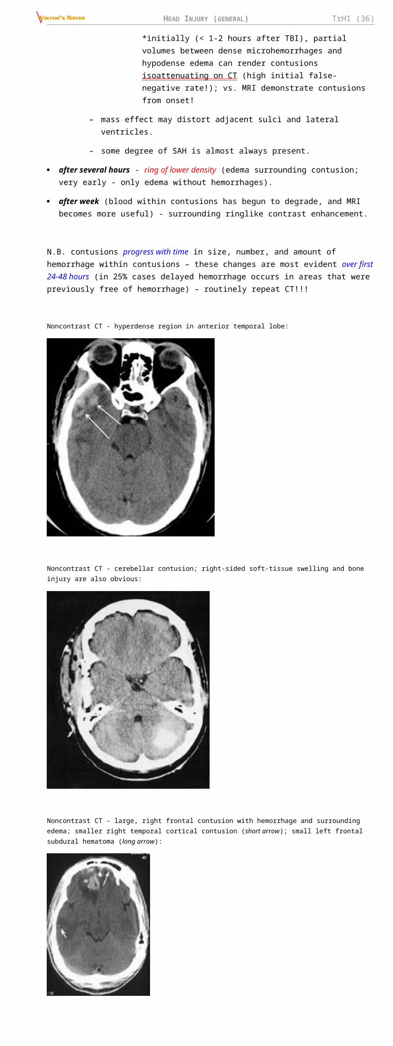
HEAD INJURY (GENERAL) TrH1 (36)
Trial: 2 mL/kg of 20 % Trial: 2 mL/kg of 20 % mannitolmannitol vs. 2 mL/kg of 7.5 % vs. 2 mL/kg of 7.5 % hypertonic salinehypertonic saline given when either ICP > 25 mmHg or CPP < 70 mmHg for > 5 min
There was no significant difference in mortality or GOS between the two treatment arms.
Vialet R et al. Isovolume hypertonic solutes (sodium chloride or mannitol) in the treatment of refractory posttraumatic intracranial hypertension: 2 mgL/kg 7.5 % saline is more effective than 2 ml/kg 20 % mannitol. Critical Care Medicine 2003; 3 1 : 1683 – 1687
N.B. mannitol dose was 0.4 g/kg – lower than we use routinely (1 g/kg)
Trial: Trial: ICP-guidedICP-guided mannitol therapy vs. mannitol therapy vs. empiricalempirical mannitol therapy. mannitol therapy.
Mean ICP was 5.5 mmHg lower in the empirically treated group compared to the ICP-guided group (p < 0.05) but mortality trended to be higher:
Smith HP et al. Comparison of mannitol regimens in patients with severe head injury undergoing intracranial pressure monitoring. J Neurosurg 1986; 6 5: 820 – 824
Sedation
PROPOFOLPROPOFOL is recommended for the control of ICP, but it is not recommended for improvement in mortality or 6-month outcomes (N.B. high-dose propofol can produce significant morbidity)
high-dose BARBITURATEBARBITURATE is recommended to control elevated ICP refractory to maximum standard medical and surgical treatment. protocol – see p. S50 >>
BCT (best conservative treatment) vs. BCT + pentobarbital – for refractory ICP.
Significant positive treatment effect of pentobarbital (p = 0.04)
Eisenberg HM, Frankowski RF, Contant CF, Marshall LF, Walker MD. High-dose barbiturates control elevated intracranial pressure in patients with severe head injury. J Neurosurg 1988; 6 9 : 15 – 23.
Cochrane 2012: “There is no evidence that barbiturate therapy in patients with acute severe head injury improves outcome. Barbiturate therapy results in a fall in blood pressure in one of four patients. The hypotensive effect will offset any ICP lowering effect on cerebral perfusion pressure.”
Barbiturates help to control ICP but outcome is unchanged (predetermined by primary injury?)
Hypothermia (prophylactic)
Level II B recommendation: early (within 2.5 hours), short-term (48 hours post-injury) prophylactic hypothermia is not recommended to improve outcomes in diffuse TBI
33 degrees of Celsius reached within 8 hrs after severe TBI:
Clifton GL et al. Lack of effect of induction of hypothermia after acute brain injury. N Engl J Med 2001 ; 3 44 : 556 – 563
– robust clinical trials have shown a trend toward worse outcomes with hypothermia!!!! - hypothermia should be used as a last resort!!!
NABIS: H II trial (National Acute Brain Injury Study Hypothermia II)NABIS: H II trial (National Acute Brain Injury Study Hypothermia II)
http://www.medscape.com/viewarticle/735302?src=mpnews&spon=26
– two high-quality pediatric trials failed to show benefit and additionally suggested harm related to PROPHYLACTIC hypothermia for TBI. see p. TrH20 >>
Hypothermia (therapeutic)
THERAPEUTIC hypothermia (to treat elevated ICP) in severe TBI is effective in decreasing elevated ICP but does not improve neurological outcome and may increase mortality!
– hypothermia bears risks of coagulopathy, immunosuppression, and cardiac dysrhythmia.

HEAD INJURY (GENERAL) TrH1 (37)
– it is suggested that gradual rewarming can mitigate the inherent risk of rebound intracranial pressure elevation.
– there has been interest in localized cerebral cooling in the hopes of obtaining the desired benefits without the systemic side effects.
CPP
MAP by convention is calibrated to the level of the right atrium of the heart.
Central venous pressure (CVP) is also calibrated to the level of the right atrium of the heart; but in TBI ICP is higher than CVP.
CPP = MAP – ICP
Level II B recommendation: recommended target CPP value for survival and favorable outcomes is between 60 and 70 mm Hg. Whether 60 or 70 mmHg is the minimum optimal CPP threshold is unclear and may depend upon the patient’s autoregulatory status.
Level III recommendation: avoid aggressive attempts to maintain CPP > 70 mm Hg with fluids and pressors because of the risk of adult respiratory failure (ARDS).
Level III recommendation: CPP below 50 should be avoided.
attention to pressure autoregulatory status : patients with intact autoregulation (PRx < 0.05) are best served by higher CPP values while pressure-passive patients with dysfunctional pressure autoregulation do better with lower CPP values.
optimal CPP value may need to be tailored to individual patients.
ADVANCED BRAIN OXYGENATION / METABOLIC STATUS MONITORING
The goal of the medical management is to ensure that nutrient delivery to the brain is optimized through the period of abnormal physiology and brain swelling - the only way to be assured that this is being achieved is to measure brain metabolites (reassurance that the needs of oxidative metabolism are being met).
OXIMETERS
– monitor brain oxygenation; rationale – sometimes even if ICP is slightly elevated, brain oxygenation remains normal so no need to treat.
1. Global – jugular venous oxygen saturation (SjO2); avoid SjO2 < 50%
2. Regional – brain tissue O2 partial pressure s. oxygenation (BtpO2 = PbrO2 = pBtO2 = PbtO2 = PtiO2) – indicated for severe TBI.
e.g. Licox Brain Tissue Oxygenation Probe (FDA approved in 2000) – measures oxygenation in mm3 around its tip

HEAD INJURY (GENERAL) TrH1 (38)
e.g. “CODMAN Neurotrend Cerebral Tissue Monitoring System” – fiberoptic sensor placed directly in brain tissue monitors local brain tissue PtiO2, PtiCO2, pH, temperature.
where to place? – into penumbra; no need to monitor noninjured brain or dead brain areas.
normal brain PtiO2 = 25-45 mmHg
First measure if brain oxygenation falls – increase FiO2; RBC transfusion and increasing CPP are secondary measures.
Eriksson EA, Barletta JF, Figueroa BE, et al. The first 72 hours of brain tissue oxygenation predicts patient survival with traumatic brain injury. J Trauma Acute Care Surg. May 2012;72(5):1345-1349. PMID: 22673264
Threshold of BtpO2 most predictive of mortality was 29 mmHg
Chang JJ, Youn TS, Benson D, et al. Physiologic and functional outcome correlates of brain tissue hypoxia in traumatic brain injury. Crit Care Med. Jan 2009;37(1):283-290. PMID: 19050612
Threshold of BtpO2 to avoid was 20 mmHg
Level III recommendation: Jugular bulb monitoring of arteriovenous oxygen content difference (AVDO2), as a source of information for management decisions, may be considered to reduce mortality and improve outcomes at 3 and 6 months post-injury.
Level III recommendation: Jugular venous saturation (SjO2) of < 50% may be a threshold to avoid in order to reduce mortality and improve outcomes
Although patients with desaturations identified with advanced cerebral monitoring have poorer outcomes, Level II evidence showed no improvement in outcomes for monitored patients.
BOOST-2 trialBOOST-2 trial – ICP monitor alone* vs. ICP monitor plus BtpO2
*brain oxygenation monitor was still placed but the monitor and numbers were hidden from treating staff
o having brain oxygenation data available reduced the duration of brain hypoxia:
o condition at discharge was better in ICP monitor plus BtpO2 group:
TCD- cerebral autoregulation monitoring.
Sanchez-Porras R, Santos E, Czosnyka M, Zheng Z, Unterberg AW, Sakowitz OW. 'Long' pressure reactivity index (L-PRx) as a measure of autoregulation correlates with outcome in traumatic brain injury patients. Acta Neurochir (Wien). Sep 2012;154(9):1575-1581. PMID: 22743796
There is an association between mortality and L-PRx > 0.2.
MICRODIALYSIS

HEAD INJURY (GENERAL) TrH1 (39)
– monitors cerebral metabolism (e.g. lactate, glutamate)
SEDATION & ANALGESIA
Sedative drugs should be avoided if possible (difficult to monitor level of consciousness + depress respiration) but useful for ICP control.
restlessness may be combated with small doses of short-acting sedatives (e.g. PROPOFOLPROPOFOL, PPRECEDEXRECEDEX!!!; historical drug - PARALDEHYDEPARALDEHYDE)
for agitation antipsychoticsantipsychotics are useful when used sparingly.
periodic withholding of sedation (“short sedation vacations”) - to allow periodic neurologic assessment.
for analgesia – PARACETAMOLPARACETAMOL, FFIORICETIORICET, , IBUPROFENIBUPROFEN.
SEIZURE PROPHYLAXIS
- it is prophylaxis of EARLY post-traumatic seizures - anticonvulsant drug is administered routinely in severe TBI and/or cortical irritation but only for 7 days see p. E9 >>
N.B. norepinephrine is seizure suppressant.
ANTIBIOTICS
- prophylactically indicated for:
a) complicated scalp lacerations
b) open skull fractures (includes penetrating head injury) – 5 days of triple antibiotics
c) CSF leak persisting > 7 days
MEDICATIONS NEEDING FURTHER TESTINGBBETAETA--BLOCKERSBLOCKERS
observational studies reveal a significant mortality advantage with β-blockers; however, quality of evidence is very low.
Aziz S. Alali et al. Beta-Blockers and Traumatic Brain InjuryA Systematic Review, Meta-Analysis, and Eastern Association for the Surgery of Trauma Guideline. Annals of Surgery. 2017;266(6):952-961.
FAILED MEDICATIONSPPROGESTERONEROGESTERONE – failed in ProTECT III trialProTECT III trial
TTIRILAZADIRILAZAD – no overall benefit on outcome was detected, except in post hoc sub group analysis: men with traumatic SAH had lower mortality.
MMAGNESIUMAGNESIUM - given within 8 h of moderate to severe TBI does not improve outcome and may even have a detrimental effect:
Temkin NR et al. Magnesium sulfate for neuroprotection after traumatic brain injury: a randomised controlled trial. Lancet Neurol 2007; 6 : 29 – 38 .
MMINOCYCLINEINOCYCLINE - a broad-spectrum antibiotic of the tetracycline family that was found to inhibit cytochrome c, thereby inhibiting caspase-mediated apoptosis.
drug is neuroprotective in different animal models of acute and chronic neurodegeneration, including TBI.
most human trials showed either small neurological improvements or deleterious neurotoxic effects.
NOT RECOMMENDED MEDICATIONS1. Steroids – increased ICP is due to vasodilation and cytotoxic edema (not due to vasogenic edema);
no human studies demonstrated benefit! (Maxwell et al. showed that glucocorticoids could reduce cerebral edema in an animal model of TBI by reducing abnormal vascular permeability)
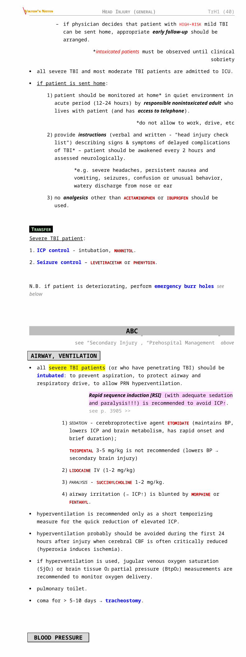
HEAD INJURY (GENERAL) TrH1 (40)
Corticosteroid Randomization After Significant Head Injury Trial (MRC CRASH) trialCorticosteroid Randomization After Significant Head Injury Trial (MRC CRASH) trial → Level I recommendation (10,800 patients, 239 centers in 49 countries worldwide):
CRASH trial collaborators. Final results of MRC CRASH, a randomised placebo-controlled trial of intravenous corticosteroid in adults with head injury - outcomes at 6 months. Lancet 2005; 3 65 : 1957 – 1959.
“Steroids are not recommended for improving outcome or reducing ICP. In
patients with severe TBI, high-dose methylprednisolone was associated with increased mortality* and is contraindicated”.
*cause unclear but not due to infections or GI bleeding; RR for death 1.15 (p=0.0001).
2. Vasodilators (aminophylline, pentoxifylline) – decrease seizure threshold, may worsen edema, ↑risk of rebleeds.
3. Hemostatic agents (VIT. C, VIT. K, CaCl2)
4. Hypotonic fluids IVI (e.g. D5W) – may worsen edema.
SURG ERY See p. Op320 >>
INDICATIONS FOR SURGERY
1. Scalp lacerations. see p. Op320 >>
2. Certain open skull fractures. see p. TrH5 >>
3. Certain depressed skull fractures. see p. TrH5 >>
4. Extra-axial (epidural / subdural) or intra-axial hematoma / hemorrhagic contusion:
see p. TrH11 >> , p. TrH13 >>
a) thickness > 15 mm (EDH) or > 10 mm (SDH)
b) volume > 30 mL (EDH, SDH) or > 50 mL (IPH)* (esp. in temporal or posterior fossa** locations - these regions do not tolerate additional mass and > 30 mL is a threshold here). * more details for IPH >>
**more details for posterior fossa mass lesions >>
c) midline shift > 5 mm (shift > 20 mm – mortality approaches 100%).
d) neurodeficits (e.g. GCS < 9, GCS decrease by ≥ 2 points, focal neurologic signs, refractory ICP > 20 mmHg, effaced / compressed basal cisterns, heterogenous clot on CT [indicates active bleeding]).
N.B. in general, any neurodeficit due to extra-axial hematoma is indication for surgery!
These hematomas must be evacuated immediately - patients go immediately to OR!
Other hematomas / contusions are managed nonoperatively with serial CT and close neurological observation in a neurosurgical ICU.
N.B. early surgery (by CT criteria) has better morbidity* and mortality outcomes vs. delayed surgery (by clinical deterioration criteria)!
*e.g. ischemic effect of prolonged cortical compression by hematoma
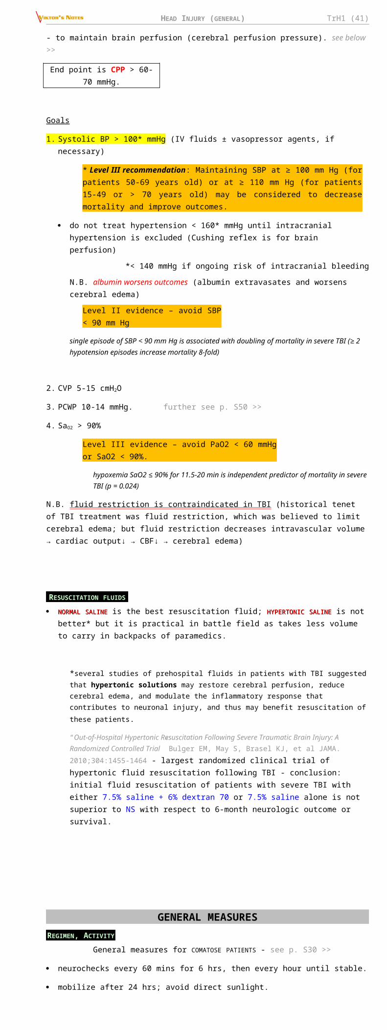
HEAD INJURY (GENERAL) TrH1 (41)
5. Symptomatic chronic subdural hematoma.see p. TrH13 >>
6. Penetrating injuries (esp. with foreign bodies). see p. Op320 >>
7. Medically refractory intracranial hypertension → decompressive craniectomy.
see p. Op320 >>
surgical intervention is not beneficial in most patients with GCS 3-5 (unless pupils are reactive and CT shows no bihemispheric/multilobar dominant hemispheric injuries).
conservatively uncontrollable ICP↑ with rapid patient deterioration in ED (consciousness↓ + asymmetric findings) → think of brain compression* → emergency blind (exploratory) BURR HOLES. see p. TrH11 >> , p. TrH13 >>
*if skull fracture is present – most commonly epidural hematoma.
Notes on hematoma size:
patients with severe cerebral atrophy (e.g. elderly) may accommodate large intracranial mass, whereas young individuals experience neurological deficits with relatively small intracranial hemorrhages.
dynamic nature: some intracranial hemorrhages may be actively bleeding during initial CT - what may appear as relatively small on initial CT scan may actually become quite significant in short period of time!!!
DTICH (delayed traumatic ICH)DTICH (delayed traumatic ICH) – see below >>
METHOD TO MEASURE HEMATOMA VOLUME
V = ABC / 2
identify the CT slice with the largest area of hemorrhage (Slice 1)
measure the largest diameter, A
measure the largest diameter 90° to A on the same slice, B.
count the number of 10-mm slices - compare each slice with slice:
if the hemorrhage is greater than 75% compared with slice 1, count the slice as 1.
if the hemorrhage is 25 to 75%, count the slice as 0.5.
if the hemorrhage is less than 25%, do not count the slice.
Add up the total C.
REHABILITATION Guidelines by Texas Head In jury Foundation :
1. Treatment should be adjusted to patient's level of function.
2. Treatment should be consistent, repetitious, and structured.
3. As patient's ability improves, difficulty of task should be increased and degree of structure should be decreased.
4. Results of performance should be consistently provided to patient.
5. Information should be presented via more than one sensory avenue.
6. Information and tasks should be relevant to needs and interests of patient.
Treatment of spasticity → see p. Mov3 >>
Aim to physical & cognitive independence! (i.e. rehabilitation is continued until optimum training has been achieved or patient has become independent)
rehabilitation is creative, cooperative effort of health-care team, patient, and family.

HEAD INJURY (GENERAL) TrH1 (42)
– family members must be kept informed of progress; they usually wish to learn rehabilitation techniques, and instruction should be provided (family will assume primary responsibility in outpatient therapy).
rehabilitation should begin as soon as patient is stable.
relearning old skills / learning methods to compensate for lost skills.
1. Physical & occupational therapy
functional activities out of bed are begun as quickly as possible!
2. Cognitive rehabilitation
cognitive enhancing medications (still experimental):
1)1) AMANTADINEAMANTADINE – efficacy proven by double-blind, placebo-controlled, randomized – efficacy proven by double-blind, placebo-controlled, randomized study – speeds up recovery but final outcome the same.study – speeds up recovery but final outcome the same.
2) METHYLPHENIDATEMETHYLPHENIDATE – improved motor outcomes and attention.
3) DONEPEZILDONEPEZIL – improved visual and verbal memory as well as attentional deployment.
4) LEVODOPALEVODOPA – alerting responses in vegetative or comatose patients.
5) CITICOLINECITICOLINE – approved in 59 countries for neuroprotection and neurorepair in patients with TBI.
Zafonte R et al “Effect of Citicoline on Functional and Cognitive Status Among Patients With Traumatic Brain Injury (Citicoline Brain Injury Treatment Trial (COBRIT)) - citicoline is not superior to placebo (no significant differences in survival or functional and cognitive outcomes in a 180-day evaluation) as acute and postacute therapy among patients with broad range of severity of TBI - worldwide use of citicoline for TBI should now be questioned
NON-NEUROLOGIC COMPLICATIONS
EARLY COMPLICATIONSI. Pulmonary about respiratory care → see p. S30 >>
1. PNEUMONIA (incl. aspiration pneumonia) occurs in 30-50% intubated patients.
2. ATELECTASIS - common in all poorly responsive patients;
H: chest physical therapy, adequate ventilator tidal volumes (incl. PEEP); pneumonia can supervene if atelectasis is not treated!
3. NEUROGENIC PULMONARY EDEMA (form of ARDS with increase in interstitial and alveolar fluid): suddenly raised ICP → medullary ischemia → increased sympathetic tone → sudden shift of intravascular volume from systemic to pulmonary circulation (via hypothalamic influence on pulmonary microvasculature) → alveoli fill with fluid as they would in congestive heart failure, but left ventricular end-diastolic pressure (measured by PCWP) is normal.
etiology : acute central nervous system injury (well documented in SAH but prevalent in ICH).
clinically : presents abruptly and progresses quickly.
radiographically , it is indistinguishable from cardiogenic pulmonary edema.
treatment : lower ICP - reverses neurogenic stimulation that causes neurogenic pulmonary edema. Intubation with mechanical ventilator support is often required. Resolution usually occurs within several days.
II. Gastrointestinal about GI care → see p. S30 >>
1. GI HEMORRHAGE from gastric stress erosions / ulcers.
– most patients with severe TBI develop gastric erosions, but only few have clinically significant hemorrhages.
– GI bleeding usually occurs in first days to 1 week after injury.
H: prophylaxis & treatment with gastric coating agents (sucralfate), H2 receptor blockers, frequent antacid administration.
2. PARALYTIC ILEUS
3. BOWEL INCONTINENCE - caused by underlying constipation, impaired communication and mobility (H: stool softeners, laxatives, rectal suppositories).
III. Cardiologic
TBI (esp. with intracranial bleeding) can cause primary cardiac dysfunction - variety of rhythm, rate, conduction abnormalities - can be life threatening!
adequate cardiac output is essential to ensure cerebral perfusion!
IV. Hematologic
large number of patients demonstrate mild coagulopathy.

HEAD INJURY (GENERAL) TrH1 (43)
N.B. multiple contusions detected by first CT scan = trauma-related coagulopathy that will correct itself within few hours.
5-10 (up to 90% patients with severe TBI) have various degrees of DIC.
– DIC can develop within hours after any injury disrupting brain tissue (release of tissue thromboplastin into systemic circulation).
– DIC increases risk of delayed intracranial hemorrhage.
any coagulation abnormality must be reversed.
V. Urinary :
1. Urinary tract infections
2. Urinary incontinence – caused by UTI, impaired communication and mobility (i.e. overflow incontinence; H: patient is taken to bathroom and given opportunity to void without instrumentation every 2 hours during day and every 4 hours overnight; if unable to void or unable to evacuate urinary bladder to completion → intermittent catheterization).
LONGER TERM COMPLICATIONS1. Deep venous thrombosis, pulmonary embolism
2. Decubitus ulcers
3. Malnutrition
4. Spastic contractures - can be prevented with aggressive physical therapy.
5. Heterotopic ossification (11-76%) - ectopic bone formation in soft tissue surrounding joints or around paralyzed muscles.
pathophysiology - inappropriate differentiation of mesenchymal cells into osteoblasts; contributing factors - autonomic dysregulation (due to increased vascularity and venous hemostasis), humoral factors, local inflammatory mediators.
risk factors - posttraumatic coma lasting > 2 weeks, limb spasticity, decreased mobility.
risk is greatest during first 3-4 months.
clinically : joint pain, decreased range of motion*, ± low-grade fever & periarticular swelling-warmth-erythema.
*although ossification does not originate in joints, ankylosis occurs in ≈ 20% cases.
location (in decreasing order of frequency): hips, knees, elbows, shoulders, hands, spine.
diagnosis :
1) serum alkaline phosphatase↑ and ESR↑ – nonspecific markers in early phases.
2) triple-phase bone scanning – earliest diagnosis!!!
3) plain radiography – lag behind triple-phase bone scan results by 2-3 weeks.
4) sonography.
prophylaxis – range-of-motion (ROM) exercises; unclear role - NSAIDs, low-dose radiation, bisphosphonates
use of forceful ROM is somewhat controversial because it is thought to be cause of heterotopic ossification, but data from human studies have not demonstrated this mechanism.
treatment :
1) bisphosphonates - prevent crystallization of calcium phosphate (but have no effect on mature ectopic bone).
2) for functional impairment - surgical excision delayed 12-18 months to allow heterotopic bone to mature (to minimize risk of recurrence).
NEUROLOGIC COMPLICATIONS & SEQUELQE
I. VASCULAR1. Cerebral fat embolism see p. Vas3 >>
2. Epidural Hematoma see p. TrH11 >>
3. Subdural Hematoma see p. TrH13 >>
4. Arterial rupture, occlusion, dissection see p. Vas11 >>
high mortality - 23-43%.
ICA most commonly injured at skull base
– manifests as pain, pharyngeal bleed, cranial nerve palsies, distal thrombemboli
VA most commonly during cervical motion that injures vessel intima.

HEAD INJURY (GENERAL) TrH1 (44)
– causes may be trivial (chiropractic manipulations, violent sports)
– unilateral VA injury may be asymptomatic (but thrombosis may extend to basilar)
5. Venous sinus injuries see p. Op320 >>
6. Carotid-cavernous fistula see p. TrH9 >>
7. SA H - most common type of traumatic intracranial hemorrhage (some blood extravasation into subarachnoid spaces is to be expected in almost any head injury!; present on CT in 26-53% of severe TBI cases):
a) tears of subarachnoid vessels (most frequently leptomeningeal vessels at vertex - greatest brain movement at impact).
b) blood from ventricles (reached subarachnoidal space with CSF flow)
in most cases, SAH per se is of little clinical importance* (except to indicate that brain has been injured) - no specific treatment is needed.
*but SAH doubles risk of death (because of associated lesions) >>
detected by CT in 26-53% cases of severe TBI; repeat CT - to exclude possibility of additional intracranial loculated blood.
– traumatic SAH is typically located in interhemispheric or sylvian fissure, cerebral sulci (vs. SAH from ruptured cerebral aneurysm – primarily in basal cisterns).
– differentiation of interhemispheric blood from relatively attenuating falx cerebri may be difficult (N.B. blood extends into paramedian sulci).
– with extensive SAH, brainstem, infundibulum, carotid branches are bathed by blood, and they may appear as filling defects.
N.B. SAH may render basal cisterns isodense with brain, simulating cisternal compression!
traumatic basal SAH - due to blow to side of chin or jaw in alcohol-induced fistfight - ruptures vertebral artery as it enters cranial cavity (degree of traumatic force required to cause basal SAH is less than reasonably expected).
complications :
1) communicating hydrocephalus.
2) vasospasm (most intense during 2nd week after TBI) → delayed ischemia, infarction. H: NIMODIPINENIMODIPINE
In the patients with symptoms of delayed ischemia, vasospasm was angiographically proven!
Taneda M, Kataoka K, Akai F, et al.: Traumatic subarachnoid hemorrhage and its treatment with Nimodipine. J Neurosurg 85:82-89, 1996
8. Chronic calcified scalp hematoma (seen chiefly in older patients prone to repeated head injuries, such as epileptics and recidivist alcoholics); underlying vault may be thickened.
9. Intraventricular hemorrhage (1.5-10%) – ruptured subependymal veins.
intraventricular blood is indicator of severe TBI
unclotted blood may layer in most dependent part of ventricles.
hydrocephaly is a risk (N.B. ventricles may remain small due to compression by brain edema!)
10. Cerebral thrombosis
ARTERIAL THROMBOSIS - caused by injury to artery wall.
thrombosis of contralateral PCA is rare complication of brain distortion by epidural or subdural hematoma.
thrombosis occasionally develops several days after TBI in elderly patients with cerebral arteriosclerosis (it is difficult to assess role of cerebral trauma in production of these strokes).
VENOUS SINUS THROMBOSIS → severe ICP elevations and venous infarction.
treatment – anticoagulation (significant risk in those with acute head injuries!); if thrombosis progresses → direct intracranial intravenous thrombolysis.
11. Traumatic aneurysms (< 1%)
etiology :
a) penetrating vessel injuries (stab wounds > gunshot wounds) → pseudoaneurysms at distal sites in intracranial circulation (can appear weeks to months after injury).
see p. Vas25 >>
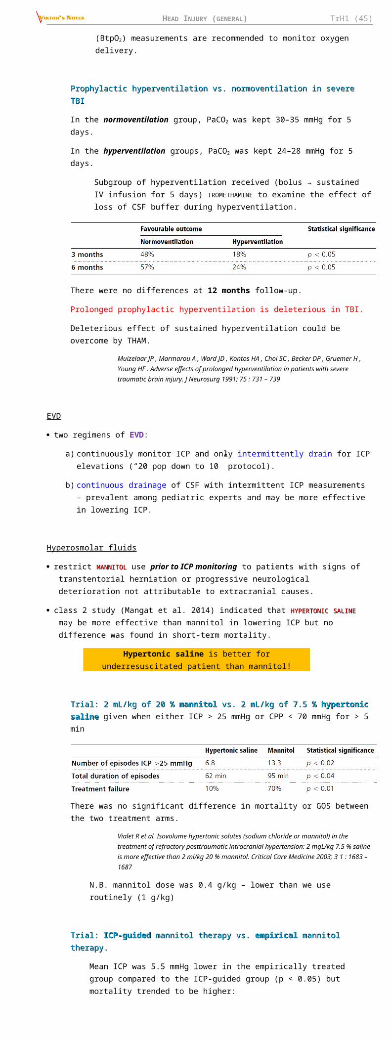
HEAD INJURY (GENERAL) TrH1 (45)
b) blunt vessel injuries → true aneurysms (intact adventitia); examples:
1) head hyperextension and rotation → aneurysm on ICA at skull base;
2) closed head injury with skull fracture → aneurysm on peripheral intracranial vessel (esp. distal ACA, e.g. pericallosal artery - traumatized at falcine edge).
present with delayed SAH or intracerebral hematoma.
diagnosis - angiography - absolutely indicated ASAP in stab injuries! (intracerebral hematoma may harbor and simultaneously obscure traumatic aneurysm)
– in CT / MRI era, angiography is rarely employed in evaluating head trauma - discovery of traumatic aneurysms is usually delayed until unexpected neurological event occurs.
– traumatic aneurysms are unrelated to arterial bifurcations.
treatment (best results if still unruptured) - surgical or endovascular.
– anticoagulation for pseudoaneurysms may minimize thrombus propagation and embolization (controversial).
Acute traumatic arterial aneurysm (carotid angiogram, lateral projection): metallic fragments are seen posteriorly; vessels around them are stretched and narrowed, and rounded collection of contrast medium, representing traumatic aneurysm, is seen (arrow), unrelated to arterial bifurcation:
12. Intracerebral hemorrhage (tICH) (4-23%); see also p. Vas20 >>
Differences between spontaneous ICH (sICH) and traumatic ICH (tICH):
– tICHs are more likely to be lobar
– tICHs are more likely to be superficial
– tICHs are more likely to have a medium-sized volume (25–65 cc).
caused by:
a) shearing forces that mechanically stretch and tear deep small-caliber arterioles – resulting small petechial hemorrhages subsequently coalesce to form ICH.
– 85% in frontal and temporal lobes; multiple in 20% cases.
– on occasion, ICH (ipsi- or contralateral) develops after other intracranial hematoma evacuation
b) penetrating injury - occur wherever vessels are interrupted along tract of missile.
occur with all degrees of TBI; linear fracture is found in 40-80% cases.
most prominent in white matter (vs. contusions).
reach maximum size by 2-3 days following head injury.
clinical features:
– 50% of patients sustain loss of consciousness at time of impact; some regain consciousness for lucid interval, and 1/3 have no loss of consciousness at all.
– focal brain injury (similar to contusion or extradural collections).
– brain compression → herniation.
diagnosis - CT (hemorrhagic contusion ÷ large blood collection) - focal area of increased attenuation*, surrounded by low attenuation (edema or contusion); irregular and may have mass effect; over time becomes well demarcated, isoattenuating and then hypoattenuating.
*ICH is made of > 66% clots – appears more homogenous than contusion!
DELAYED HEMORRHAGE (DTICH – DELAYED TRAUMATIC ICH) S. BOLLINGER'S SPÄT-APOPLEXIE (occurs in 1-7% TBI patients) – ICH that develops in the part of the brain that on the initial CT looked “normal”; may manifest as apoplectic event even after 10 days of asymptomatic interval.
Yamaki et al. (1990): only 84% of all ICHs reached the maximum size within 12 hours from initial CT scan!
Noncontrast CT - occipital and temporal intracerebral hematomas, surrounded by mild edema and hemorrhagic contusion; small interhemispheric subdural hematoma in posterior interhemispheric fissure; obvious midline shift:
HEAD INJURY (GENERAL) TrH1 (46)
treatment :
a) conservative management
b) surgical evacuation (for indications – see above); see p. Vas20 >>
Pathophysiological pros for surgical evacuation:
– contused brain does not recover but appears as encephalomalacic brain tissue loss on convalescent phase imaging - removing TICH does not increase tissue loss.
– extravasated blood is believed to be neurotoxic, leading to secondary injury that may be avoided by surgical removal.
– larger TICHs may be associated with an ischaemic penumbra of brain tissue that could be salvaged.
– some TICHs expand to the point at which they cause mass effect with high ICP, resulting in secondary brain injury.
Surgical Trial In Traumatic intraCerebral Haemorrhage (STITCH)Surgical Trial In Traumatic intraCerebral Haemorrhage (STITCH)
- Early Surgery (within 12 hrs) vs. Initial Conservative Treatment (delayed evacuation if it became clinically appropriate); tICH > 10 mL.
Gregson BA et al. Surgical Trial In Traumatic intraCerebral Haemorrhage (STITCH): a randomised controlled trial of Early Surgery compared with Initial Conservative Treatment. Health Technol Assess 2015;19(70).
cf. SITCH trialSITCH trial – nontraumatic ICH – see p. Vas20 >>
Early Surgery may be a valuable tool in the treatment of tICH, especially if the GCS is 9-12 – analogy with SDH / EDH management; those with GCS of 13–15 can probably be watched carefully for any deterioration; if GCS has dropped below 8, surgical intervention appears to be less effective.
At 6 months:
– Early Surgery patients were 10.5% more likely to have a favorable outcome (absolute benefit), but this difference did not quite reach statistical significance because of the reduced sample size (63% vs. 53%, odds ratio 0.65; 95% confidence interval (CI) 0.35 to 1.21; p = 0.17].
– Mortality was significantly lower in the Early Surgery group (15% vs. 33%; absolute difference 18.3%; 95% CI 5.7% to 30.9%; p = 0.006).
Contusions, intracerebral hematomas may show remarkable resolution, but more common end results are focal atrophy or encephalomalacia (extensive softening and cyst formation)
– MRI may show evidence of past hemorrhage (prolongation of T1 and T2).
– injury early in life (vascular damage rather than to direct trauma) → traumatic hemiatrophy - affects both brain and overlying skull.
Cranial hemiatrophy - left hemicranium is smaller (arrows indicate groove for superior sagittal sinus); left petrous bone is elevated, and paranasal sinuses are more extensive on affected left side.

HEAD INJURY (GENERAL) TrH1 (47)
II. INFECTION- etiology / risk factors:
1) penetrating injuries.
2) skull fractures - open depressed fractures, basilar fractures with CSF leak, linear fractures that extend into paranasal sinuses or middle ear.
3) iatrogenic - ICP monitoring, surgical interventions.
1. Cranial (osteomyelitis) see p. Inf7 >>
2. Epidural (abscess) see p. Inf5 >>
3. Subdural (empyema) see p. Inf5 >>
4. Subarachnoid (meningitis) see p. Inf3 >>
5. Intracerebral (abscess) see p. Inf5 >>
III. CSF & MENINGES1. CSF leaks (3%, but 5-10% in basal skull fractures) see p. S64 >>
2. Post-traumatic hydrocephalus (COMMUNICATING > NONCOMMUNICATING) – due to SAH, intraventricular hemorrhage. see p. S60 >>
unexplained hydrocephalus after acute TBI → prompt thorough examination of posterior fossa (epidural or subdural hematomas may be missed on routine axial CT scans).
normal pressure hydrocephalus may also occur – diagnosis is difficult: see p. S62 >>
– 86% severe TBI patients demonstrate some degree of ventriculomegaly on follow-up CT (secondary to diffuse brain atrophy).
– TBI patients often have memory difficulties and gait abnormalities secondary to head injury.
N.B. normal pressure hydrocephalus presents as neurologic deterioration weeks to months following TBI; high-volume lumbar puncture improves neurologic condition.
3. Subdural hygroma see p. TrH15 >>
4. Leptomeningeal cyst (s. growing fracture ) see p. TrH5 >>
5. Pneumocele (s. pneumocephalus ) - air in cranial cavity after missile wounds penetrate sinuses:
a) frontal pneumocele (after fracture of frontal sinus)
b) occipital pneumocele (after fracture through mastoid).
free air can also be sucked into penetration cavity behind projectile.
clinically :
a) asymptomatic
b) headaches, mental symptoms.
N.B. signs of ICP↑ do not develop (unless pneumocele becomes infected or filled with CSF).
diagnosis – X-ray, CT: air may not appear for several days after injury (and then only after patients sneeze or blow nose).
treatment (rarely indicated per se; if spontaneous air absorption does not occur): transfrontal craniotomy → opening in frontal sinus is covered with strip of fascia lata or pericranial flap.
IV. POST-TRAUMATIC EPILEPSYsee p. E9 >>
V. POST-TRAUMATIC MOVEMENT DISORDERSsee p. Mov22 >>
VI. POSTCONCUSSION (S. POSTTRAUMATIC) SYNDROME- frequent (30-50%) sequela of MILD head injury.
rarer in children (but symptoms tend to be more severe).
diagnosed in DSM-IV as cognitive disorder not otherwise specified.
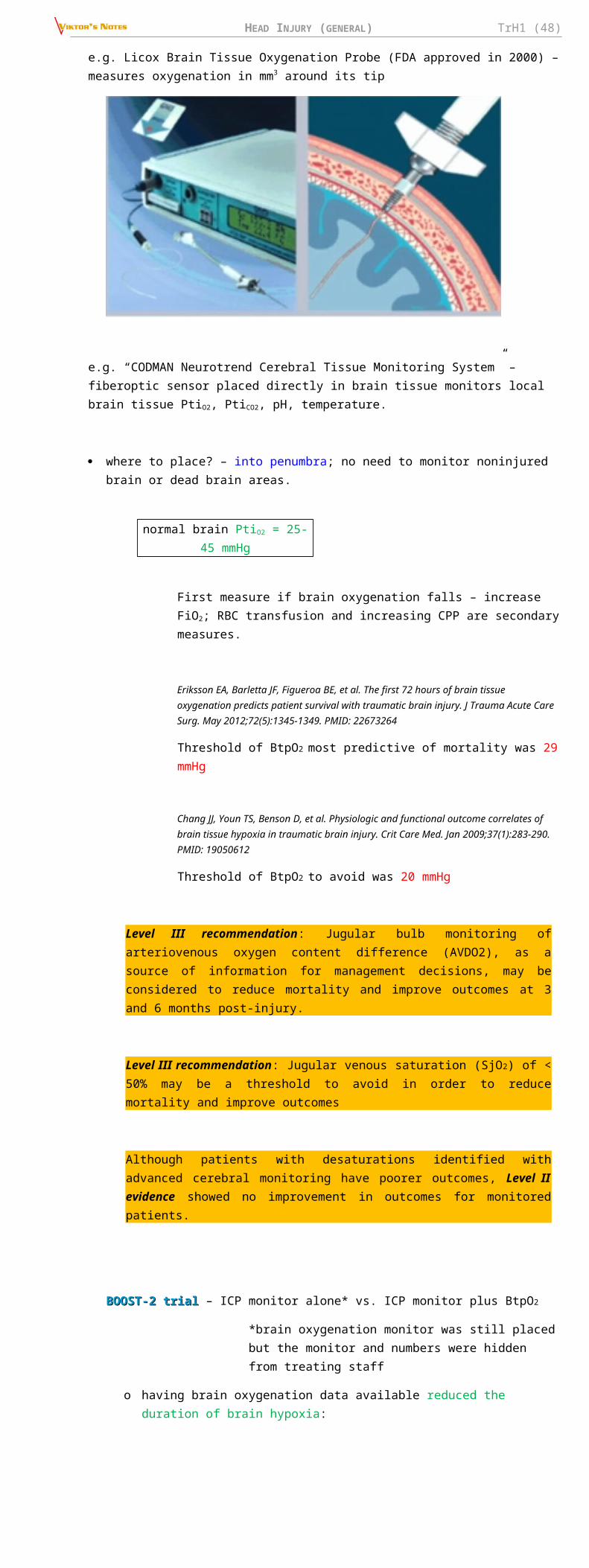
HEAD INJURY (GENERAL) TrH1 (48)
PATHOPHYSIOLOGY
1. Alterations in excitatory amino acids, catecholamines, cations (≈ similar to migraine)
2. Subtle axonal shearing lesions
3. Microscopic cortical contusions.
Even mild trauma can cause neuronal damage!
CLINICAL FEATURES
- large array of symptoms and signs (of at least 3 months duration); most common:
Syndrome can be extremely debilitating!1) headaches (30-90%):
– headaches are more frequent and with longer duration in mild rather than severe head injury.
– triptans can abort chronic post-traumatic headaches (PTHs) attributable to mild head trauma.
– TOPIRAMATETOPIRAMATE appears to be an effective headache prophylactic therapy in chronic PTH.
– low doses of tricyclic antidepressants appear to have little efficacy.
a) tension-type headaches (85% of all post-traumatic headaches); one cause may be temporomandibular joint injury.
b) migraine-like (also very common) can develop in hours to weeks after mild TBI.
– immediately after mild TBI in sports children ÷ young adults may have first-time migraine with aura; this may be triggered multiple times after additional mild head injury - termed FOOTBALLER'S MIGRAINE.
c) referred headaches from neck injuries (myofascial, intervertebral disc, facet joint injury).
d) greater occipital neuralgia from direct blow to nerve or due to suboccipital muscle spasm (superior trapezius, semispinalis capitis). H: local anesthetic nerve blocks (can be combined with injectable corticosteroid).
2) dizziness (34-50%) – caused by: labyrinthine concussion, benign positional vertigo, brain stem injury.
3) nonspecific psychological symptoms (> 50%) – fatigue (43%), irritability, anxiety, depression, disruption of sleep patterns, loss of concentration and memory (20-40%), light / noise sensitivity (10%), post-traumatic stress disorder.
– neuropsychological testing has documented cognitive impairments (reduction in information processing speed, attention, reaction time, and memory for new information).
4) blurred vision (14%) usually caused by convergence insufficiency.
5) hyposmia (≈ 5%) caused by damage to olfactory filaments.
no direct correlation between TBI severity and development of PCS - symptoms may develop in patients who were only dazed (did not lose consciousness) by injury.
on objective examination - minimal or no neurologic abnormalities (fixed neurologic deficits are not part of PCS).
DIAGNOSIS
- neuropsychological testing!!!
MRI role is not yet defined - symptoms may appear in absence of any imaging evidence of structural abnormality!
N.B. CT / MRI / may be warranted to rule out delayed complications of TBI! (e.g. chronic subdural hematoma may mimic PCS)
EEG, evoked potentials are also normal.
when patients have unusual / persistent complaints, consider possible contributions of personality disorders, psychosocial problems, or secondary gain* (“COMPENSATION NEUROSIS” or malingering).
*N.B. patients with claims in compensation case, however, have similar symptoms that improve with time and similar cognitive test results as those without claims (for many claimants, end of litigation does not mean end of symptoms or return to work - they are not cured by verdict).
TREATMENT
Simple reassurance is often major treatment – inform patient that cognitive-emotional dysfunction, headaches and other somatic symptoms are not uncommon and that most patients improve after 3-6 months!
1. Supportive psychotherapy
2. Cognitive retraining see below (REHABILITATION)
3. Physiotherapy
4. Antidepressant and antianxiety-type medications.

HEAD INJURY (GENERAL) TrH1 (49)
5. Headaches often respond to:
a) PROPRANOLOLPROPRANOLOL (30-60 mg/d in three divided doses), or occasionally to calcium channel blockers.
b) NSAIDs
c) tricyclic antidepressants
PROGNOSIS
symptoms usually last 2-6 months (typically, peak 4-6 weeks following injury), but symptoms may persist for years (some are disabled permanently); risk factors for persistent symptoms:
1) women
2) age > 40 years
3) prior history of head trauma
4) neurotic symptoms before injury
5) domestic or financial difficulties.
2 years after injury, 20% patients still complain of headaches.
cognitive deficits usually resolve within 3 months after injury.
VII. POSTTRAUMATIC PSYCHIATRIC DISORDERSSerious residual mental problems are found only after severe TBI.
almost every patient with severe TBI shows mental changes immediately after recovery of consciousness (frequently steps to complete recovery are semistupor, bewilderment, Korsakoff-like phase, euphoria).
1. Posttraumatic agitation / aggression is common (≈ 25% patients are found to be aggressive during follow-up period of 5 years).
associated with frontal injuries, esp. severe TBI
consistently associated with depression, young age.
treatment steps :
1) first treat / eliminate agitating factors:
– impaired recognition and inability to communicate are often agitating factors.
– pain
– infection
– electrolyte imbalance
– adverse effects of drugs (esp. avoid centrally acting drugs).
– psychosis, insomnia.
2) environmental modifications - tools for orientation, minimizing unnecessary stimuli (noisy rooms, bright lights, frequent visitors).
3) drugs: high-dose β-blockers (particularly PROPRANOLOLPROPRANOLOL), anticonvulsants (e.g. LAMOTRIGINELAMOTRIGINE, DIVALPROEXDIVALPROEX), antidepressants (particularly SSRIs), AMANTADINEAMANTADINE.
Antipsychotics are controversial - may cause excessive drowsiness, exacerbate cognitive deficits, and inhibit neuronal recovery.
4) restraints - use only as last resort to secure patient, staff, and visitor safety
Physical restraints often exacerbate posttraumatic agitation!
– less restrictive restraints, such as net-covered beds (e.g. Posey or Vail beds), has become acceptable and popular.
5) DBS (placed bilaterally in the nucleus accumbens and anterior limb of the internal capsule to modulate the prefrontal cortex) is promising.
– DBS lead (inset) is directed through the anterior limb of the internal capsule and striatum, passing between the caudate (green) and putamen (red) with the tip in the nucleus accumbens (blue).
Ali R. Rezai. Improved Function After Deep Brain Stimulation for Chronic, Severe Traumatic Brain Injury. Neurosurgery, Volume 79, Issue 2, 1 August 2016, Pages 204–211,

HEAD INJURY (GENERAL) TrH1 (50)
2. Posttraumatic depression - common sequela of TBI (major depression is found in 40-44% hospitalized patients).
associated with left frontal injuries.
may lead to cognitive decline, anxiety disorders, substance abuse, dysregulation of emotional expression, aggressive outbursts.
treatment :
1) early grief reaction is better treated with supportive therapies
2) METHYLPHENIDATEMETHYLPHENIDATE
3) SERTRALINESERTRALINE
4) AMANTADINEAMANTADINE
N.B. if drugs are used, carefully consider adverse effects (esp. worsening sedation or cognitive impairment).
3. Psychosis
transient psychotic episodes are not uncommon, but long-continued psychosis is rare.
relationship of head injury to subsequent development of psychoses is medicolegal problem (severe TBI may adversely influence preexisting brain pathology and accentuate organic dementia).
unlikely that TBI could have any direct causal relationship to psychoses that do not have definite structural pathology (e.g. schizophrenia, manic-depressive), but indirect relationship with chemical intermediaries may be postulated.
4. Personality change sometimes results from head trauma, even in absence of obvious cognitive changes.
5. Chronic amnestic disorder can result from damage to diencephalic / mesiotemporal structures (e.g. mamillary bodies, hippocampus, fornix).
PROGNOSIS, OUTCOME - depends most on nature, site and severity of brain damage (than on injury to skull or scalp).
TBI may result in death, vegetative state, partial recovery, or full return to work.
Hippocratic aphorism: “No head injury is so serious that it should be despaired of nor so trivial that it can be ignored.”
adults reach maximum recovery by ≈ 6 months; smaller adjustments continue for perhaps as long as 2 yrs (recovery in children takes even longer).
common long-term deficits are COGNITIVE (recent memory, abstract thinking, rapid information processing, etc).
N.B. neuropsychologic disturbances (e.g. postconcussion syndrome) are more common cause of disability in social relations and employment than are specific neurologic deficits!
DRIVING INDEPENDENTLY is of particular importance to most patients.
– estimating driving ability on basis of cognitive testing is difficult (although visual spatial skills, reaction times, and awareness of deficits correlate with driving abilities).
– actual road tests may be needed.
Moderate TBI
at 3 months after trauma - 70% are unable to return to work, 90% have memory difficulties, > 90% have persistent headaches.
Severe TBI
mortality decreased from 50% to 25% over the last decades.
only 7% have good outcome with moderate disability.
most are left with at least some residua (small percentage remain in persistent vegetative state); severe cognitive difficulties are rule rather than exception.
5 MOST POWERFUL FACTORS PREDICTING OUTCOME
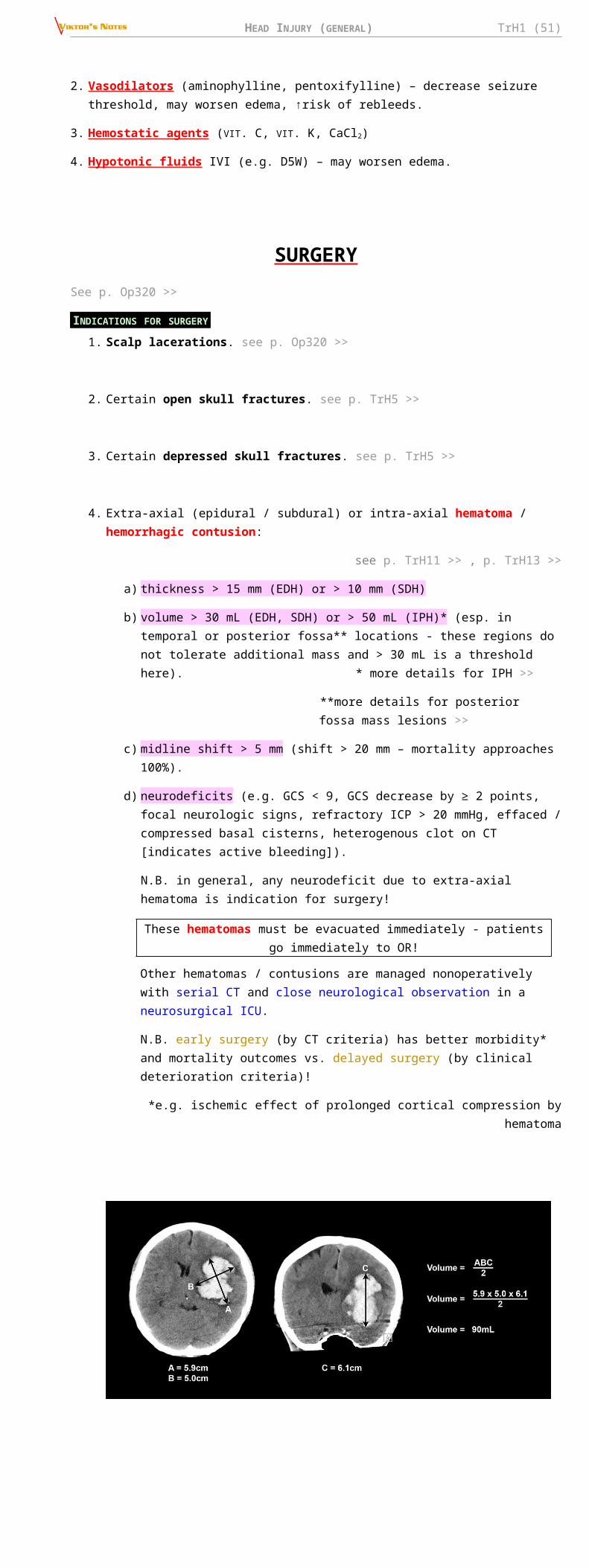
HEAD INJURY (GENERAL) TrH1 (51)
Notably, of these five major predictors, only the hypotension is amenable to medical manipulation!
1. TBI degree (as measured by initial postresuscitation GCS score) - early predictor of patient's overall outcome!
Up to 85% patients with GCS 3-4 die 24 h after injury, yet number of patients with poor initial prognosis (incl. absent pupillary light responses) survive, suggesting that aggressive management is justified in virtually all patients!
If the initial* GCS score is reliably obtained and not tainted by prehospital medications or intubation, approximately 20% of the patients with the worst initial GCS score will survive and 8%-10% will have a functional survival (GOS 4-5).
*the optimal time after injury for determining the initial GCS is the key issue for further research
2. Increasing age – independent predictor of poor outcome!
the critical age threshold for worsening prognosis appears to be above 60 in a review of Class I and II studies (this may be an artifact of the age grouping used by various authors in converting continuous data into categorical data); some studies report outcome as a continuous function of age without threshold values.
aged brain:
1) decreasing capacity for repair after TBI
2) increasing frequency of intracranial hematomas with the largest intracerebral hematomas observed in the oldest groups.
type of injury that occurs frequently in each age group (e.g. increasing proportion of falls and pedestrian accidents with advancing age).
decline in health as one ages may predispose the aged to systemic complications after TBI.
children outcome is better than that of adults (patients < 20 yrs are > 3 times more likely to survive than those > 60 yrs).
3. Fixed dilated pupil; see criteria for pupillary exam >>
% Vegetative/Dead (Glasgow Outcome Scale Score [GOS] 1, 2):
% Good Recovery/Moderate Disability (GOS 4, 5)
EDH vs. SDH
in EDH with bilateral fixed pupils mortality is only 56% compared to an average of 88% in SDH
Phonprasert C, Suwanwela C, Hongsaprabhas C: Extradural hematoma: analysis of 138 cases. J Trauma 20: 679-683, 1980.
Phuenpathom N, Choomuang M, Ratanalert S: Outcome and outcome prediction in acute subdural hematoma. Surg Neurol 40:22-5, 1993.
patients operated on for EDH with bilaterally fixed pupils - 18% had a poor outcome (GOS 1-2) vs. 64% in SDH with bilaterally fixed pupils; delay of > 3 hours in evacuating a traumatic intracranial hematoma with bilateral fixed pupils increased the chance of a poor outcome from 40% to 63%.
Rivas J et al.: Extradural hematoma: analysis of factors influencing the courses of 161 patients. Neurosurg 23:44-51, 1988.
Sakas DE, Bullock R, Teasdale G: One-year outcome following craniotomy for traumatic hematoma in patients with fixed dilated pupils. J Neurosurg 82:961- 965, 1995.
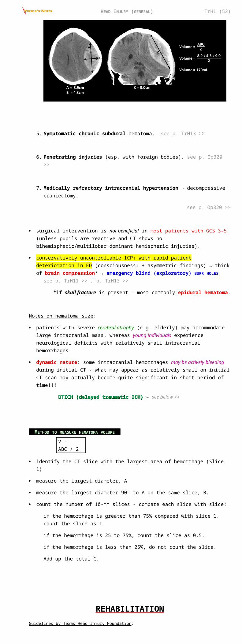
HEAD INJURY (GENERAL) TrH1 (52)
4. Hypotension - single measurement of SBP < 90 mm Hg.
hypotension has 67% PPV for poor outcome (79% PPV when combined with hypoxia).
in one study, logistic regression modeling revealed that early hypotension (injury ÷ resuscitation) was responsible for a 15-fold excess mortality and late hypotension (intensive care unit) for an 11-fold excess mortality in severe TBI.
Chesnut RM, Marshall SB, Piek J, et al.: Early and late systemic hypotension as a frequent and fundamental source of cerebral ischemia following severe brain injury in the Traumatic Coma Data Bank. Acta Neurochirurgica (Suppl) 59:121-5, 1993
influence of iatrogenic hypotensive episodes in patients with severe TBI who had not otherwise been hypotensive - patients with intraoperative hypotension had significantly worse neurologic outcomes than those without.
Pietropaoli JA et al. The deleterious effects of intraoperative hypotension on outcome in patients with severe head injuries. J Trauma 33:403-7, 1992
5. Abnormal head CT (any abnormality noted on CT, consistent with TBI) - within 12 hours of injury; present in 68-94% of patients with severe TBI:
1) compressed or absent basal cisterns measured at the midbrain level (i.e. perimesencephalic cisterns) – the strongest independent prognostic factor.
– mortality when cisterns are absent - 77% (39% when cisterns are compressed; and only 22% when cisterns are open).
Toutant SM et al.: Absent or compressed basal cisterns on first CT scan: ominous predictors of outcome in severe head injury. J Neurosurg 61:691-694, 1984
– Teasdale et al. report that the third ventricle usually becomes obliterated before the basal cisterns. vs. in the study by Lang et al. on 118 patients with diffuse traumatic brain swelling, however, no direct relation was seen between the status of the third ventricle and that of the basal cisterns.
2) tSAH (in the basal cisterns, over the convexity) – the second strongest independent prognostic factor.
Presence of tSAH = 2-fold increase in the risk of dying
– class I evidence: 69-72% PPV for unfavorable outcome with tSAH in the suprasellar or ambient cisterns (61% PPV with convexital SAH).
– Fisher’s Grade 3 → 62% PPV toward unfavorable outcome; Fisher’s Grade 4 → 79% PPV.
3) midline shift at the level of foramen of Monroe.
– PPV of 70% for mortality with midline shift ≥ 15 mm (class II evidence).
– PPV of 78% to poor outcome with shift > 5 mm in patients > 45 yrs (class I evidence).
– prognosis is even worse with midline shift out of proportion to the extent of intracranial hemorrhage.
4) presence and type of intracranial lesions.
– PPV of 79% to poor outcome (dead/vegetative) in the presence of lesions > 15 ml in patients > 45 (class I evidence).
Vollmer DG, et al.: Age and outcome following traumatic coma: why do older patients fare worse? J Neurosurg (Suppl)75:37-49, 1991
– mortality is higher in acute SDH than in EDH.
– in comparison to diffuse injuries:
outcome is more favorable in EDH
outcome is less favorable in acute SDH.
– hematoma volume is correlated to outcome.
if CT is normal on admission, outcome is primarily related to concomitant extracranial injuries.
the full extent of intracranial pathology may not be disclosed on early CT (the absence of abnormalities on CT at admission does not preclude the occurrence of raised ICP, and significant new lesions may develop in 40% of patients).
CT Classification and Outcome on Discharge (Marshall et al. 1991) Marshall classification >>
OTHER PROGNOSTIC FACTORS
1. Penetrating TBI (vs. closed TBI – mortality 2.5 times lower), esp. gunshot TBI.
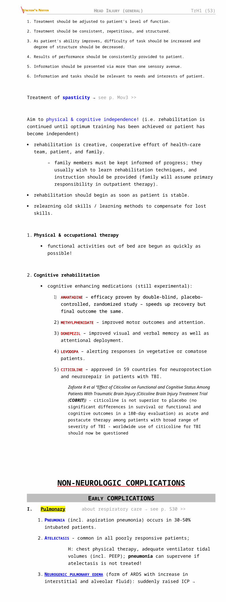
HEAD INJURY (GENERAL) TrH1 (53)
2. Multiple organ injuries - influence on the outcome of severe TBI is primarily mediated through hypotension.
3. Anticoagulants / antiplatelets - raise risk of intracranial bleeding with even trivial TBI.
Novel oral anticoagulants and traumaNovel oral anticoagulants and trauma
Kobayashi, L et al. Novel oral anticoagulants and trauma: The results of a prospective American Association for the Surgery of Trauma Multi-Institutional Trial. Journal of Trauma and Acute Care Surgery: May 2017 - Volume 82 - Issue 5 - p 827–835
prospective observational trial across 16 trauma centers, 1,847 patients on antiplatelets / anticoagulants
blunt trauma accounted for 99% of patients.
50% of patients were on antiplatelet agents, 33% on warfarin, 10% on NOAs (dabigatran, rivaroxaban, or apixaban), and 7% on combination therapy or subcutaneous agents.
patients taking NOAs were not at higher risk for ICH on univariate (24% vs. 31%) or multivariate analysis (incidence rate ratio, 0.78; confidence interval 0.61–1.01, p = 0.05).
compared with all other agents, aspirin (90%, 81 mg; 10%, 325 mg) gave the highest rate (35%) and risk (incidence rate ratio, 1.27; confidence interval, 1.13–1.43; p < 0.001) of ICH.
progression of ICH occurred in 17% of patients and was not different between medication groups.
mortality was 7% and was not significantly different between medication groups.
4. Presence of even one of APOE4 alleles - 14-fold greater likelihood of poor outcome.
Penetrating TBI
Military vs. civilian PBI – see p. Op320 >>
PBI versus TBI outcome distribution (on GOS scale – “J” shape vs. “U” shape):
among survivors, the percentage of patients with favorable outcome (“moderate disability” plus “good recovery”) is equal: 74%, i.e. the main difference in outcome is the difference in mortality
N.B. in PBI, the outcome distribution differs significantly (from blunt TBI) - overwhelming majority of patients have an unfavorable outcome; mostly, death - appropriate outcome measure for PBI is mortality, not GOS score.
1. Increasing age correlates with increased mortality (Class III).
2. Suicide correlates with a higher rate of mortality than other causes of PBI (Class II); plus, suicide patients are less likely to receive aggressive resuscitation efforts.
N.B. patients who recover from attempted suicide frequently report relief over survival and express gratitude to their caregivers!
3. Perforating injuries correlate with a poorer outcome when compared with either penetrating or tangential brain injuries (Class III).
4. The effect of weapon caliber on outcome, independent of total kinetic energy, was not demonstrated in the published data.
5. Hypotension is associated with increased mortality (Class III).
6. Coagulopathy is associated with increased mortality, particularly at lower levels of the GCS (Class III).
7. Respiratory distress is associated with increased mortality (Class III).
8. In civilian patients, low GCS correlates with higher mortality and unfavorable outcome (Class I). In military injuries, fewer patients have a low GCS score; strong correlation also exists between low GCS score and unfavorable outcome in military series (Class III).
9. The presence of bilateral fixed and dilated pupils is highly predictive of mortality (Class III).
10. High ICP is predictive of higher mortality (Class II). Cisternal effacement PBI is associated with increased mortality (Class I). No relation between midline shift and outcome has been established (Class I).

HEAD INJURY (GENERAL) TrH1 (54)
11. Intraventricular hemorrhage is strongly correlated to increased mortality (Class I) - odds ratio 2.83-96.9; Subarachnoid hemorrhage is also correlated to increased mortality (Class I) - odds ratio 1.44-10.6
12. Trajectory :
Bihemispheric injuries relate to increased mortality (Class II) - odds ratio 1.18-20.05; possible exception may be bifrontal injuries
Multilobar (> 1 lobe) injuries are strongly associated with mortality (Class III) - odds ratio 3.27-84.4, negative predictive value for mortality 77-98%.
Perhaps it should be better stated that the presence of unilobar damage is strongly correlated to survival.
Injuries with ventricular involvement have an increased mortality (Class III) - odds ratio 3.35-27.5
SCALES OF OUTCOMEFUNCTIONAL INDEPENDENCE MEASURE (FIM) (one of most widely used measures of function) - level of independence for mobility, self-care, and cognition (all these are often spared in mild TBI - FIM score is inadequate outcome measure for these patients):
Self-care
A. Eating
B. Grooming
C. Bathing
D. Dressing - upper body
E. Dressing - lower body
F. Toileting
Sphincter controlG. Bladder management
H. Bowel management
Transfers
I. Bed, chair, wheelchair
J. Toilet
K. Tub, shower
LocomotionL. Walking, wheelchair
M. Stairs
Motor subtotal score:
CommunicationN. Comprehension
O. Expression
Social interaction
P. Expression
Q. Problem solving
R. Memory
Cognitive subtotal score:
TOTAL FIM SCORE:
GLASGOW OUTCOME SCALE (GOS)
Jennett B , Bond M . Assessment of outcome after severe brain damage. Lancet 1975 ; 1 : 480 – 484
5 – GOOD RECOVERY - normal life despite minor deficits
4 - MODERATE DISABILITY - disabled but independent; can work in sheltered setting
3 - SEVERE DISABILITY - conscious but disabled; dependent on others for daily support
2 - VEGETATIVE - no evidence of meaningful response
1 - DEAD
GOS can be divided further into:
good outcomes (good, moderate disability, independent): 5 and 4
poor outcomes (severe disability, vegetative, dead): 1-3
DISABILITY RATING SCALE (DRS) Rappaport et al. (1982)
CATEGORY ITEM INSTRUCTIONS
Arousability, Awareness and Responsivity
(i.e. GCS)
Eye Opening 0 = spontaneous 1 = to speech 2 = to pain 3 = none
Communication Ability
0 = oriented 1 = confused 2 = inappropriate 3 = incomprehensible 4 = none
Motor Response 0 = obeying 1 = localizing 2 = withdrawing 3 = flexing 4 = extending 5 = none
Cognitive Ability Feeding 0 = complete 1 = partial 2 = minimal 3 = none

HEAD INJURY (GENERAL) TrH1 (55)
for Self Care Activities
Toileting 0 = complete 1 = partial 2 = minimal 3 = none
Grooming 0 = complete 1 = partial 2 = minimal 3 = none
Dependence on Others
Level of Functioning
0 = completely independent 1 = independent in special environment 2 = mildly dependent 3 = moderately dependent 4 = markedly dependent 5 = totally dependent
Psychosocial Adaptability
Employability0 = not restricted 1 = selected jobs 2 = sheltered workshop (non-competitive) 3 = not employable
Disability Categories:Total DRS Score Level of Disability
0 None
1 Mild
2-3 Partial
4-6 Moderate
7-11 Moderately Severe
12-16 Severe
17-21 Extremely Severe
22-24 Vegetative State
25-29 Extreme Vegetative State
PREVENTION Motor vehicle-related TBI:
1) helmets by cyclists
2) automobile seatbelts and child restraints
3) airbags (children < 12 years should ride in back seat of car away from airbag)
4) enforcement of drunk driving laws
SPECIAL ASPECTS
POSTERIOR FOSSA MASS LESIONSMostly – EDH, ICH.
parturitional hemorrhages in neonates primarily (48%) involve posterior fossa.
patients can undergo rapid life-threatening clinical deterioration - limited size of the posterior fossa → brainstem compression.
Indications for surgery:
a) ICH diameter > 2.5-3.0 cm
b) mass effect on CT (distortion / dislocation / obliteration of the 4th ventricle; compression of the basal cisterns; obstructive hydrocephalus)
c) neurological dysfunction / deterioration referable to the lesion.
Surgical treatment: suboccipital craniectomy emergently is the recommended method for evacuation of posterior fossa mass lesions.
ALCOHOL advantages - neuroprotection - alcohol use at time of injury decreases likelihood of poor outcome
(alcohol impedes excitotoxicity).
disadvantages :
1) alcohol raises risk of incurring (repeat) TBI (positive blood alcohol level is detected in 25-60% patients) – risky behavior, coordination↓, motor reaction time↑.
2) obscured real level of consciousness.
3) patients often aggressive, asocial.
4) bleeding risk↑ (thrombocytopathy, coagulopathy, brain atrophy)
5) cardiorespiratory depression
6) increased risk of seizures.

HEAD INJURY (GENERAL) TrH1 (56)
LONG-BONE FRACTURES (CONCURRENT WITH TBI) femur fractures are common among TBI patients.
TIMING OF INTERNAL FIXATION
internal fixation with intramedullary nailing is the ideal method of treatment; however, there is no consensus regarding the optimal timing for internal fixation.
EAST guidelines (2014)EAST guidelines (2014)
Gandhi, Rajesh R et al. Optimal timing of femur fracture stabilization in polytrauma patients: A practice management guideline from the Eastern Association for the Surgery of Trauma. Journal of Trauma and Acute Care Surgery. November 2014, Volume 77 (5), p 787–795
Early vs. late fixation does NOT decrease mortality, infection, or VTE. Early (< 24 hr) fixation is suggested but is conditional and surgical decision should be individualized. The optimal timing remains controversial and guidelines do not replace clinical judgement.
no significant reduction in mortality was associated with early stabilization (RR of 0.74 [95% CI, 0.50–1.08]), quality of evidence was rated as “low.”
no significant reduction in infection (RR, 0.4; 95% CI, 0.10–1.6) or VTE (RR, 0.63; 95% CI, 0.37–1.07) was associated with early stabilization; quality of evidence was rated “low.”
in the absence of a clear contraindication to surgery or anesthesia, the recommendation of this review, although conditional, should prompt early fixation. However, the surgical decision must be individualized to each patient’s needs. Delayed treatment has been associated with a reduction in adverse outcomes in patients with multiple injuries. In addition, delayed stabilization (24–48 hours) may be safer than stabilization within 12 hours for severely injured patients.
CONCLUSION - in trauma patients with open or closed femur fractures, we suggest early (< 24 hours) open reduction and internal fracture fixation. This recommendation is conditional because the strength of the evidence is low. Early stabilization of femur fractures shows a trend (statistically insignificant) toward lower risk of infection, mortality, and VTE. Therefore, the panel concludes the desirable effects of early femur fracture stabilization probably outweigh the undesirable effects in most patients.
N.B. 2014 version of guidelines does not comment on the subgroup of patients with TBI.
2000 version of guidelines states: “Level II recommendation: There is no compelling evidence that early long bone stabilization in mild, moderate, or severe TBI either enhances or worsens outcome. The timing of long bone stabilization should be individualized according to the patient's clinical condition.”
C. Michael Dunham et al. Practice Management Guidelines for the Optimal Timing of Long Bone Fracture Stabilization in Polytrauma Patients: The EAST Practice Management Guidelines Work Group. 2000 Eastern Association For The Surgery of Trauma.
American College of Surgeons (ACS) Trauma Quality Improvement Program (TQIP) best American College of Surgeons (ACS) Trauma Quality Improvement Program (TQIP) best practices in the management of orthopedic traumapractices in the management of orthopedic trauma
special consideration should be given to patients with concomitant orthopedic and TBI. The management of fractures in patients with TBI represent a particular challenge in that early surgical fixation may complicate the acute management of TBI. The goals of acute TBI management are to maintain adequate cerebral perfusion, prevent hypotension, provide adequate oxygenation, avoid hypo- and hypercarbia, and maintain normothermia. Efforts should be made to adhere to each of these goals. Intraoperative monitoring of ICP should be considered to support cerebral perfusion pressure. In the context of a stable ICP and MAP, definitive fixation can be considered in the resuscitated patient, as these patients are less apt to experience transient episodes of hypotension in the operating room. The under-resuscitated patient or those patients whose ICP and CPP have not yet stabilized are best served with damage control procedures or traction.
open fractures: historically, dogma has led orthopedists to treat open fractures with surgical irrigation and debridement within six hours of the injury or risk increased rates of infection. This practice has come to be known as the “six hour rule” in orthopedic surgery. However, it has been disproven in recent years by several high quality studies demonstrating that delaying surgical irrigation and debridement up to 24 hours does not increase infectious complications for open fractures. Based on the best available evidence, the panel does not endorse the “six hour rule.”
SPORTSCHRONIC TRAUMATIC ENCEPHALOPATHY (S. DEMENTIA
PUGILISTICA, PUNCH-DRUNK SYNDROME) professional boxers are especially at risk (only sport with goal of deliberately injuring brain of
opponent).
cause - repeated head trauma during protracted period.
clinical features :
1. Parkinsonism and other extrapyramidal features (tremor, ataxia, cerebellar signs).
2. Progressive dementia.
3. Behavioral abnormalities (morbid jealousy, rage reactions).
manifestations begin 6-40 (average 16) years after starting boxing career.
pathology : hydrocephalus, thinning of corpus callosum, diffuse axonal injury, hypothalamic anomalies, degeneration of substantia nigra, neurofibrillary tangles (mainly in medial temporal areas), diffuse Aβ-positive plaques (≈ as in Alzheimer disease), scarring of cerebellar folia.
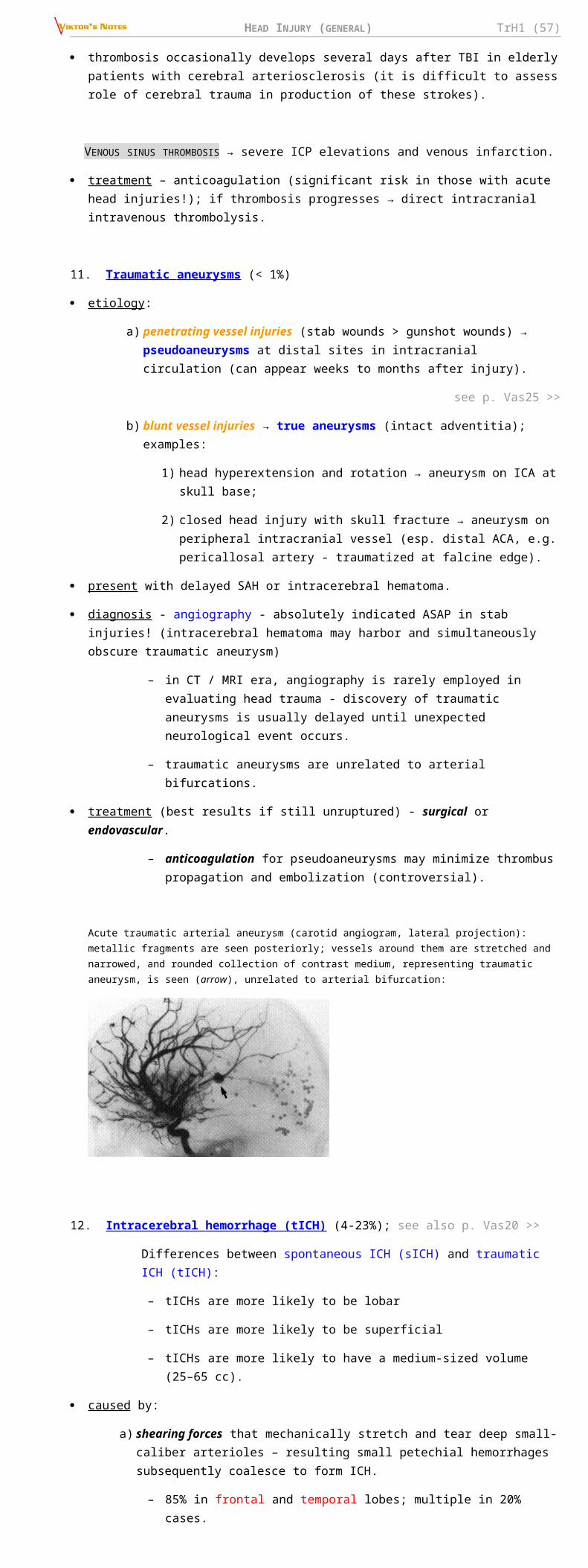
HEAD INJURY (GENERAL) TrH1 (57)
no therapy is actually effective.
prevention :
1) better protection for boxers (head protection, different gloves)
2) physician must stop match when there is evidence of brain injury (i.e. sportsman who sustained concussions, requires physician's statement to return to previous level of involvement). see below
MANAGEMENT OF CONCUSSION IN SPORTS SIDELINE EVALUATION :
Mental status testing:
1. Orientation: Digits backward
3--1--7
4--6--8--2
5--9--3--7--4
2. Months of year in reverse order
3. Memory: names of teams in prior contest
4. President, governor, mayor, recent newsworthy events, three words and three objects at 0 and 5 minutes.
5. Details of contest (plays, moves, strategies, etc.)
Exertional provocative tests:
1. 40 yard sprint
2. 5 push-ups
3. 5 sit-ups
4. 5 knee bends
(any appearance of associated symptoms is abnormal, e.g., headache, dizziness, nausea, unsteadiness, photophobia, blurred or double vision, emotional lability, mental status changes)
Neurologic tests:
1. Pupils: symmetry and reaction
2. Coordination: finger-nose-finger and tandem gait
3. Sensation: finger-nose (eyes closed) and Romberg
GRADING SCALE and GUIDELINES FOR RETURN to competition:
Grade Features Actions
Grade 1 no loss of consciousness, confusion without amnesia
remove from contest → examine immediately and q5min for development of amnesia or postconcussive symptoms at rest and with exertion → may return to contest if no amnesia & no symptoms appear for at least 20 minutes.
Grade 2 no loss of consciousness, confusion with amnesia
remove from contest and disallow return → examine frequently for signs of evolving intracranial pathologic conditions → reexamine next day.
Grade 3 loss of consciousness transport from field by ambulance (with cervical spine immobilization if indicated) to trauma hospital → emergent thorough neurological evaluation:
a) signs of pathologic conditions → hospitalization
b) normal findings → instructions to family for overnight observation.
WHEN TO RETURN TO PLAY after removal from contest:
Grade of Concussion Time Until Return to Play*
Multiple Grade 1 concussions 1 week
Grade 2 concussion 1 week
Multiple Grade 2 concussions 2 weeks
Grade 3 - brief loss of consciousness (seconds) 1 week
Grade 3 - prolonged loss of consciousness (minutes) 2 weeks
Multiple Grade 3 concussions ≥ 1 month (based on decision of physician)
*only after being asymptomatic with normal neurological assessment at rest and with exercise.
SECOND IMPACT SYNDROME
- malignant (fatal) cerebral edema.
at least 35 cases occurred among US football participants in 1980-1993.
cause – one minor HI followed in short order by second minor HI in athletes who are still symptomatic from first injury.

HEAD INJURY (GENERAL) TrH1 (58)
mechanism - impaired cerebral autoregulation → vascular congestion → brain edema → herniation → sudden death.
prevention - postponed return to play for increasing lengths of time depending on concussion severity. see above
BIBLIOGRAPHY for ch. “Head Trauma” → follow this LINK >>
Viktor’s Notes℠ for the Neurosurgery Resident
Please visit website at www.NeurosurgeryResident.net


![Empreendedorismo - conteudo-ava.s3.amazonaws.comconteudo-ava.s3.amazonaws.com/UNIFICADO/[1070103]TRH1... · O empreendedorismo no país é muito alicerçado na necessidade, ou seja,](https://img.pdfslide.net/doc/110x75/5be6f6d909d3f23a518db7ce/empreendedorismo-conteudo-avas3-1070103trh1-o-empreendedorismo-no-pais.jpg)