Embed Size (px)
Citation preview

��������������� ������������������������
������������������������ ������������
�����������������
�������� ����� � ������������������ � ��������������� ��� ��������
�� ����������������� � �������� �� ����������� ���������������
��� � ������������
Two new fossil micromammal localities of Middle Miocene age (Pico del Fraile 2, PF2 and Sancho Abarca 5, SA5) fromthe Tudela Formation (northeastern Ebro Basin) are described. PF2 contains rodents and insectivores of Aragonian age(local zone Dc). The rodent assemblage from the locality SA5 is very scarce and probably of Middle Aragonian age, likePF2. The micromammal fauna from the locality PF2 is very similar to that from Valdemoros 3B (VA3B)(Calatayud-Daroca Basin), including Microdyromys cf. remmerti, a species until now only described from the Mioceneof the Daroca-Villafeliche area. Among the fauna recorded in PF2, a form of Democricetodon is described. The sedi-mentary record of the Pico del Fraile and Sancho Abarca sections and the mammalian findings extend the stratigraphicand paleontological knowledge of this part of the Ebro Basin, and allow its study in a continuous stratigraphic context.• Key words: rodents, insectivores, Democricetodon, magnetostratigraphy, biostratigraphy, Ebro Basin, Aragonian.
RUIZ-SÁNCHEZ, F.J., MURELAGA, X., FREUDENTHAL, M., LARRASOAÑA, J.C., FURIÓ, M., GARCÉS, M.,GONZÁLEZ-PARDOS, M. & SUÁREZ-HERNANDO, O. 2013. Micromammalian faunas from the Middle Miocene (MiddleAragonian) of the Tudela Formation (Ebro Basin, Spain). Bulletin of Geosciences 88(1), 131–152 (7 figures, 4 tables).Czech Geological Survey, Prague. ISSN 1214-1119. Manuscript received April 23, 2012; accepted in revised form Oc-tober 10, 2012; published online November 30, 2012; issued December 6, 2012.
Francisco Javier Ruiz-Sánchez (corresponding author), Area de Paleontología, Universitat de València, Dr. Moliners/n, 46100 Burjassot, Spain; [email protected] • Xabier Murelaga, Departamento de Estratigrafía y Paleontología,Universidad del País Vasco (UPV/EHU), Bilbao Aptdo. 644, E-48080, Spain; [email protected] • MatthijsFreudenthal, Departamento de Estratigrafía y Paleontología, Universidad de Granada, Spain, and Netherlands Centrefor Biodiversity, Naturalis, Leiden, The Netherlands; [email protected] • Juan C. Larrasoaña, Instituto Geológico yMinero de España, Unidad de Proyectos, 50006 Zaragoza, Spain; [email protected] • Marc Furió, Institut Català dePaleontologia M. Crusafont, Edifici ICP, Universitat Autònoma de Barcelona, Cerdanyola del Vallès, 08193 Barce-lona, Spain; [email protected] • Miguel Garcés, Grup de Geodinàmica i Anàlisi de Conques, Universitat de Barce-lona, Zona Universitaria de Pedralbes, 08028 Barcelona, Spain; [email protected] • Mar Gonzáles-Pardos, Area dePaleontología, Universitat de València, Dr. Moliner s/n, 46100 Burjassot, Spain; [email protected] • OierSuárez-Hernando, Departamento de Estratigrafía y Paleontología, Universidad del País Vasco, Bilbao Aptdo. 644,E-48080, Spain; [email protected]
The deposits of the Tudela Formation crop out in the cen-tral part of the Western sector of the Ebro Basin (Spain) inthe so-called Bardenas Reales de Navarra. The sedimento-logical environment during the deposition of the TudelaFormation favours the presence of a great number ofmicro- and macromammal sites in the stratigraphic sequ-ence of this formation. In the past two decades our team hasperformed an extensive paleontological and magnetostra-tigraphical investigation that has increased the paleontolo-gical knowledge and established a detailed chronology ofthe fossiliferous sites in the span between Late Agenian
and Late Ramblian (Murelaga 2000; Murelaga et al. 2002;Murelaga et al. 2004a, 2004b; Larrasoaña et al. 2006;Ruiz-Sánchez et al. 2010a, 2010b; Ruiz-Sánchez et al.2012a, 2012b, 2012c; Figs 1, 2). So far, the younger depo-sits of the Tudela Formation had not been studied. In thiscontribution we present a complete study of the rodent andinsectivore faunas of two new sites found at the top of theTudela Formation, which permit to increase the biostrati-graphical data of the formation, and to compare this withthe fossil contents of other areas of the Ebro Basin of simi-lar age. PF2 contains three genera of cricetids (Eumyarion,
! ��� "�! #"$%&''�()*+,-� !./

Megacricetodon and Democricetodon), four glirids (twospecies of Microdyromys, Pseudodryomys and Vasseuro-mys), one sciurid (Spermophilinus) and three insectivores(Galerix, Miosorex and Myxomygale). SA5 contains twocricetids (Megacricetodon and Democricetodon or Fahl-buschia), one sciurid (Spermophilinus) and one glirid(Vasseuromys). Moreover, the biostratigraphical informa-tion is correlated with the existing magnetostratigraphic in-formation in the area (Larrasoaña et al. 2006).
���������������
The fossil localities PF2 and SA5 are located in the higheststratigraphic levels of the Pico del Fraile and Sancho
Abarca sections, respectively (Figs 1, 2). These sections in-clude sediments of the upper part of the Tudela Formation,including the youngest sediments preserved in the westernpart of the Ebro Basin in the region of the Bardenas Realesde Navarra (Larrasoaña et al. 2006). The Tudela Formationis divided into five lithostratigraphic units according to thepredominance of distal alluvial (Units 1 and 4), palustrine(Unit 3), and lacustrine (Units 2 and 5) facies (Larrasoañaet al. 2006). The Pico del Fraile section spans the upper-most part of Unit 3, Unit 4, and the lowermost part of Unit5, whereas the Sancho Abarca spans the uppermost part ofUnit 4 and the entire Unit 5 (Larrasoaña et al. 2006; Fig. 2).Unit 5 is made up of grey and ochre mudstones and greyand beige limestones. The mudstone packages are massiveand range from a few centimetres to several metres in thick-
!/
����������� ������ �������������
��� ��� ! Geological map of the Tudela Formation in the Bardenas Reales of Navarra, with the situation of the localities presented in this work.PF2 – Pico de Fraile 2, SA5 – Sancho Abarca 5. The situation of other localities previously studied in the lower part of the Tudela Formation is shown aswell (Murelaga 2000; Murelaga et al. 2004a, b).

ness. The limestone beds are up to 2 metres thick, oftenmassive and bioturbated, and contain abundant gastropods,ostracods, charophytes, fish bones and other fossil frag-ments. The limestones indicate deposition in a stablefresh-water lacustrine system, whereas the mudstones weredeposited under palustrine conditions. These facies are si-milar to those described from the Lower and Middle Mio-cene of the central part of the Ebro Basin, when the latterformed an endorrheic depression at the foothills of the Py-renees and the Iberian and Catalan Coastal ranges (Arenas& Pardo 1999, Alonso Zarza et al. 2002). Magnetostrati-graphic data indicate that fossil localities PF2 and SA5 arelocated within chron C5Br of the Langhian (Middle Mio-cene – Larrasoaña et al. 2006; Fig. 2) at an approximate ageof 15.8 and 15.5 Ma, respectively, in the ATNTS2004 timescale (Lourens et al. 2004).
������������������
The fossils were collected during the campaigns of 2004 to2010, and are deposited in the “Departamento de Estratigra-fía y Paleontología de la Universidad del País Vasco”(UPV/EHU) and Museo Aragonés de Paleontología (Funda-ción Conjunto Paleontológico de Teruel). The nomencla-ture used in the description of the teeth and the measure-ment methods are taken from Mein & Freudenthal (1971) –Cricetidae, Freudenthal (2004) – Gliridae, Cuenca (1988) –Sciuridae, Prieto & Rummel (2009) and Prieto et al. (2010)– Erinaceidae, Reumer (1984) – Soricidae and Rümke(1985) –Talpidae. The measurements are given in units of0.1 mm, and were taken on a binocular microscope LeicaMZ75, by means of displacement of a mechanical stage, con-nected to a Sony Magnescale measuring equipment.
Lower teeth are coded as d4, p4, m1, m2, m3, upperteeth as P4, M1, M2 and M3.
We use the MN units (European Neogene land mam-mal units) or the local zones defined by Daams & Freu-denthal (1988), Daams et al. (1999) and/or Van der Meulenet al. (2012).
Abbreviations. – BU – Buñol; CMVI – Can Martí Vell I;PF2 – Pico del Fraile 2; SA5 – Sancho Abarca 5; VA3B –Valdemoros 3B; VL2A – Villafeliche 2A; L – Length; W –Width; UPV/EHU – Universidad del País Vasco/EuskalHerriko Unibertsitatea; SCSIE (UV) – Servei Central deSuport a l’Investigació Experimental de la Universitat deValència.
�"������������������"
Order Rodentia Bowdich, 1821Family Cricetidae Fischer, 1814
Genus Eumyarion Thaler, 1966
Type species. – Eumyarion medius (Lartet, 1851).
Eumyarion cf. weinfurteri (Schaub & Zapfe, 1953)Figure 3A
Locality. – Pico del Fraile 2 (PF2).
Material and measurements (L × W). – 1 m1 (PF2-1)(> 17.7 × 11.5); 1 m2 (PF2-13) (> 15.2 × 12.1).
Description. –m1. The broken tooth does not conserve the anteroconid.On the lingual side the metaconid extends forward to theanteroconid. The anterolophulid is short, interrupted andconnected to the protoconid. Metalophulid double, with along anterior branch connected to the anterolophulid and aposterior very short branch that runs towards to the lingualpart of the protoconid, but ends freely. Posterior arm of themetaconid connected to the mesolophid. The mesolophidis long and double. The ectomesolophid reaches the labialborder of tooth. The hypolophulid is transverse and con-nected to the ectolophid, just in front of the hypoconid. Thesinusid is transverse. There is a long oblique hypoconidhind arm, connected to the posterolophid.
m2. The mesolophid is long and connected to the posteriorarm of the metaconid. The ectomesolophid is short. The
!!
������ ������������ !��"�� ����� �#��$����%���%�%%�� ��"�&�'�����%����
��� ���#! Stratigraphy of the Tudela Formation in the studied sections,with the localities presented in this work, and with the paleomagnetic se-quence of the section of Pico de Fraile and of the section of Sancho Abarca(see Larrasoaña et al. 2006), correlated with the ATNTS time scale ofLourens et al. (2004).
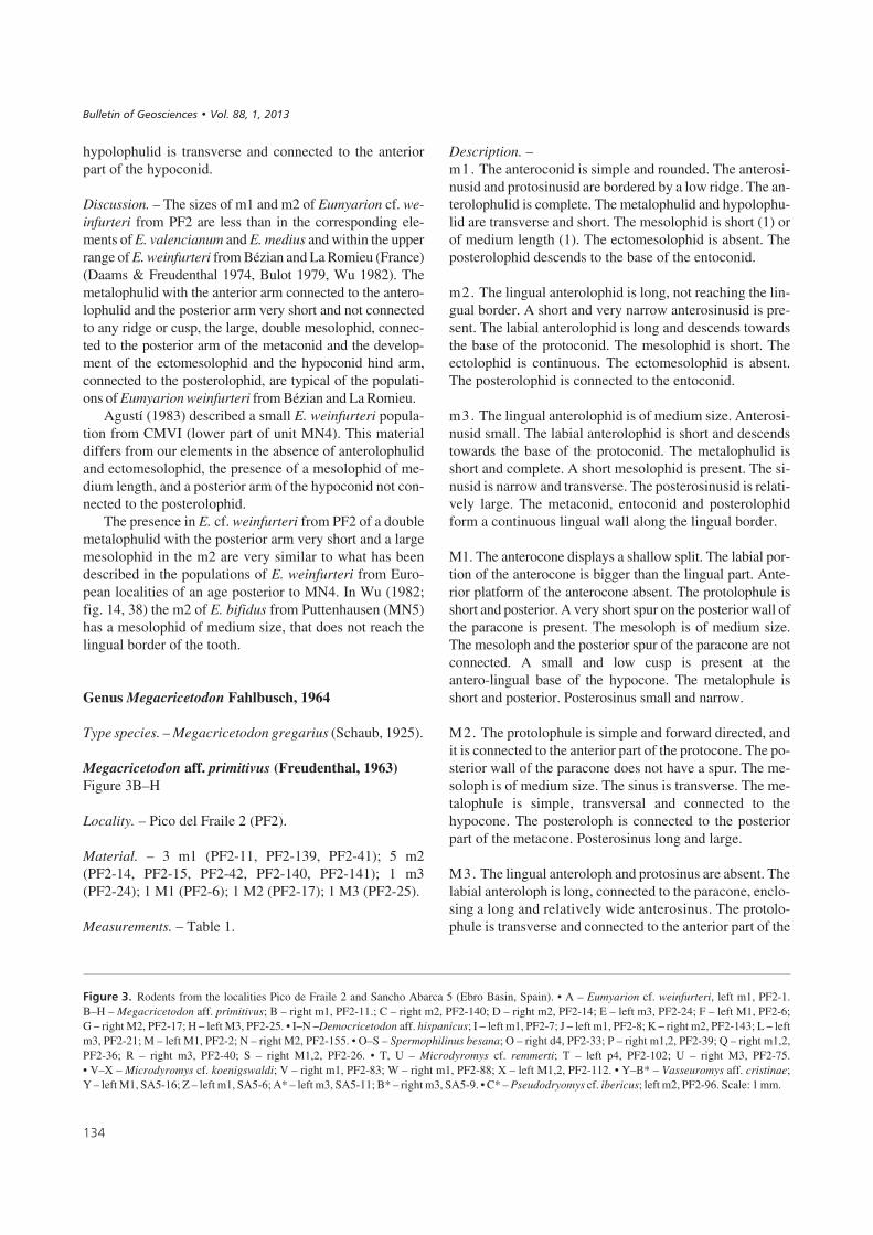
hypolophulid is transverse and connected to the anteriorpart of the hypoconid.
Discussion. – The sizes of m1 and m2 of Eumyarion cf. we-infurteri from PF2 are less than in the corresponding ele-ments of E. valencianum and E. medius and within the upperrange of E. weinfurteri from Bézian and La Romieu (France)(Daams & Freudenthal 1974, Bulot 1979, Wu 1982). Themetalophulid with the anterior arm connected to the antero-lophulid and the posterior arm very short and not connectedto any ridge or cusp, the large, double mesolophid, connec-ted to the posterior arm of the metaconid and the develop-ment of the ectomesolophid and the hypoconid hind arm,connected to the posterolophid, are typical of the populati-ons of Eumyarion weinfurteri from Bézian and La Romieu.
Agustí (1983) described a small E. weinfurteri popula-tion from CMVI (lower part of unit MN4). This materialdiffers from our elements in the absence of anterolophulidand ectomesolophid, the presence of a mesolophid of me-dium length, and a posterior arm of the hypoconid not con-nected to the posterolophid.
The presence in E. cf. weinfurteri from PF2 of a doublemetalophulid with the posterior arm very short and a largemesolophid in the m2 are very similar to what has beendescribed in the populations of E. weinfurteri from Euro-pean localities of an age posterior to MN4. In Wu (1982;fig. 14, 38) the m2 of E. bifidus from Puttenhausen (MN5)has a mesolophid of medium size, that does not reach thelingual border of the tooth.
Genus Megacricetodon Fahlbusch, 1964
Type species. – Megacricetodon gregarius (Schaub, 1925).
Megacricetodon aff. primitivus (Freudenthal, 1963)Figure 3B–H
Locality. – Pico del Fraile 2 (PF2).
Material. – 3 m1 (PF2-11, PF2-139, PF2-41); 5 m2(PF2-14, PF2-15, PF2-42, PF2-140, PF2-141); 1 m3(PF2-24); 1 M1 (PF2-6); 1 M2 (PF2-17); 1 M3 (PF2-25).
Measurements. – Table 1.
Description. –m1. The anteroconid is simple and rounded. The anterosi-nusid and protosinusid are bordered by a low ridge. The an-terolophulid is complete. The metalophulid and hypolophu-lid are transverse and short. The mesolophid is short (1) orof medium length (1). The ectomesolophid is absent. Theposterolophid descends to the base of the entoconid.
m2. The lingual anterolophid is long, not reaching the lin-gual border. A short and very narrow anterosinusid is pre-sent. The labial anterolophid is long and descends towardsthe base of the protoconid. The mesolophid is short. Theectolophid is continuous. The ectomesolophid is absent.The posterolophid is connected to the entoconid.
m3. The lingual anterolophid is of medium size. Anterosi-nusid small. The labial anterolophid is short and descendstowards the base of the protoconid. The metalophulid isshort and complete. A short mesolophid is present. The si-nusid is narrow and transverse. The posterosinusid is relati-vely large. The metaconid, entoconid and posterolophidform a continuous lingual wall along the lingual border.
M1. The anterocone displays a shallow split. The labial por-tion of the anterocone is bigger than the lingual part. Ante-rior platform of the anterocone absent. The protolophule isshort and posterior. A very short spur on the posterior wall ofthe paracone is present. The mesoloph is of medium size.The mesoloph and the posterior spur of the paracone are notconnected. A small and low cusp is present at theantero-lingual base of the hypocone. The metalophule isshort and posterior. Posterosinus small and narrow.
M2. The protolophule is simple and forward directed, andit is connected to the anterior part of the protocone. The po-sterior wall of the paracone does not have a spur. The me-soloph is of medium size. The sinus is transverse. The me-talophule is simple, transversal and connected to thehypocone. The posteroloph is connected to the posteriorpart of the metacone. Posterosinus long and large.
M3. The lingual anteroloph and protosinus are absent. Thelabial anteroloph is long, connected to the paracone, enclo-sing a long and relatively wide anterosinus. The protolo-phule is transverse and connected to the anterior part of the
!#
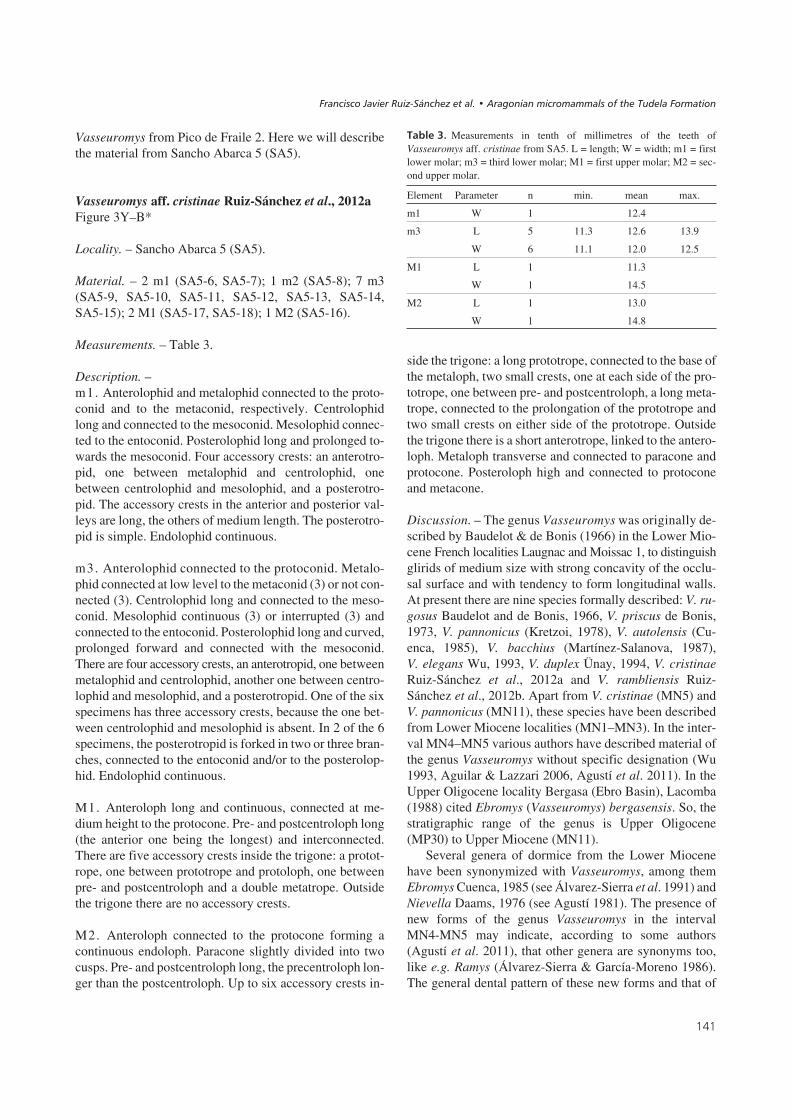
��� ���$! Rodents from the localities Pico de Fraile 2 and Sancho Abarca 5 (Ebro Basin, Spain). • A – Eumyarion cf. weinfurteri, left m1, PF2-1.B–H – Megacricetodon aff. primitivus; B – right m1, PF2-11.; C – right m2, PF2-140; D – right m2, PF2-14; E – left m3, PF2-24; F – left M1, PF2-6;G – right M2, PF2-17; H – left M3, PF2-25. • I–N –Democricetodon aff. hispanicus; I – left m1, PF2-7; J – left m1, PF2-8; K – right m2, PF2-143; L – leftm3, PF2-21; M – left M1, PF2-2; N – right M2, PF2-155. • O–S – Spermophilinus besana; O – right d4, PF2-33; P – right m1,2, PF2-39; Q – right m1,2,PF2-36; R – right m3, PF2-40; S – right M1,2, PF2-26. • T, U – Microdyromys cf. remmerti; T – left p4, PF2-102; U – right M3, PF2-75.• V–X – Microdyromys cf. koenigswaldi; V – right m1, PF2-83; W – right m1, PF2-88; X – left M1,2, PF2-112. • Y–B* – Vasseuromys aff. cristinae;Y – left M1, SA5-16; Z – left m1, SA5-6; A* – left m3, SA5-11; B* – right m3, SA5-9. • C* – Pseudodryomys cf. ibericus; left m2, PF2-96. Scale: 1 mm.
����������� ������ �������������

!0
������ ������������ !��"�� ����� �#��$����%���%�%%�� ��"�&�'�����%����
�
%&
�
��
'
()
*
+
�,
-
�
./ �
�
01 2 3
4
5 �6 �6 %6
7
1m
m

protocone. The sinus is very shallow. Neo-entoloph highand connected to protocone and hypocone. Axioloph short.Centroloph long. The posteroloph is long and curved, for-ming a labial wall with the metacone and the posterior wallof the paracone.
Discussion. – In several morphological features, the mor-phology of Megacricetodon from PF2 is very similar towhat has been described in Valtorres, type locality of Me-gacricetodon primitivus. This species has been describedwith a wide range of morphological variation (Daams &Freudenthal 1988, Oliver-Pérez et al. 2008). This causes aconfusing situation that has led several authors to considerthe possible existence of more than one species in the po-pulations adscribed to this taxon (Ruiz-Sánchez 1999,Ruiz-Sánchez et al. 2003, Oliver-Pérez et al. 2008).
Some of the morphological features that have been de-scribed as typical of M. primitivus are present in the Mega-cricetodon of PF2. The presence of a simple anteroconid inthe m1, a mesolophid in the m3 and a single and anteriorprotolophule without posterior spur in the M2, are verysimilar to what has been described in the type population ofM. primitivus. However, the material from PF2 showssome important differences. The most significant is thebetter development of the mesoloph/ids in PF2.
On the other hand, the Megacricetodon from PF2 isclearly distinguishable from M. collongensis, characteris-tic species from the Middle Aragonian of the Iberian Penin-sula (Daams & Freudenthal 1988, Daams et al. 1999,Ruiz-Sánchez 1999, Ruiz-Sánchez et al. 2003). In this lat-ter species, a high percentage of m1 has a divided ante-roconid, the mesolophid of the m3 is practically absent andthe M2 usually has double protolophules with a posteriorspur on the paracone.
The material of Megacricetodon from PF2 differs fromM. primitivus and M. collongensis and coincides greatlywith the description by Ruiz-Sánchez (1999) of Mega-cricetodon nov. sp. 3 from Morteral 22. Therefore, and un-til more material is available in PF2, we classify this as-semblage as Megacricetodon aff. primitivus.
Megacricetodon sp.
Locality. – Sancho Abarca 5 (SA5).
Material and measurements (L × W). – 1 m1 (SA5-1)(> 12.8 × 9.5); 1 m2 (SA5-3) (– × 9.9); 1 M3 (SA5-4)(8.1 × 8.4).
Description. –m1. An incomplete specimen is available, preserving only theposterior part. The protosinusid is bordered by a low ridge.The mesolophid is of medium length. The ectomesolophid isabsent. The hypolophulid is short and transverse. The poste-rolophid descends reaching the base of the entoconid.
m2. An incomplete specimen is available. Only the mostposterior part of the tooth is preserved. The posterolophiddescends and nearly reaches the entoconid base.
M3. The lingual anteroloph is much reduced. The labialanteroloph is long and is connected to the paracone. A shortand relatively wide anterosinus is present. The protosinusis absent. The protolophule is a slightly oblique, connectedto the anterior part of the protocone. The paracone does nothave a posterior spur and the mesosinus is open to the labialside. The lingual portion of the mesosinus is nearly open tothe lingual side. The sinus is relatively narrow. Theneo-entoloph is very low and connected protocone and hy-pocone. The axioloph is long and low. The centroloph is ofmedium size. The metalophule is long, and is connected tohypocone and metacone. The posteroloph descends alongthe posterior border of the tooth, connecting to the meta-cone. The posterosinus is narrow and deep.
Discussion. – Some of the striking morphological charac-ters of this material are the medium-sized mesolophid ofm1, the height of the neo-entoloph and the opening of themesosinus to the labial side of the M3. The medium-sizedmesolophid is very common in populations of Megacrice-todon in the time span between the Early and Middle Mio-cene (MN4 and MN5, local zones C and D).
On the other hand, the M3 is a very interesting elementin the taxonomy of cricetids (Freudenthal & Daams 1988).According to several authors (Freudenthal & Daams 1988,Ruiz-Sánchez 1999), some of the characters of the M3, asthe height of the neo-entoloph and the size of the sinus,
!.
����� ! Measurements in tenth of millimetres of the teeth of Mega-cricetodon aff. primitivus from PF2. L = length; W = width; p4 = fourthpremolar; m1 = first lower molar; m2 = second lower molar; m3 = thirdlower molar; M1 = first upper molar; M2 = second upper molar; M3 =third upper molar.
Element Parameter n min. mean max
m1 L 2 14.0 14.3 14.6
W 2 8.7 8.8 8.9
m2 L 3 11.3 11.7 11.9
W 3 9.6 10.0 10.3
m3 L 1 9.6
W 1 7.6
M1 L 1 15.4
W 1 9.9
M2 L 1 10.6
W 1 9.7
M3 L 1 8.1
W 1 7.8
����������� ������ �������������

would permit the distinction among the species of the ge-nus Megacricetodon. According to Freudenthal & Daams(1988), there seems to exist an evolutionary tendency to theclosing of the neo-entoloph and reduction of the sinus.Based on these criteria, the only M3 from SA5 would cor-respond to an old representative of the genus Megacri-cetodon. However, the material is too poor and, until morematerial is available, we classify it as Megacricetodon sp.
Genus Democricetodon Fahlbusch, 1964
Type species. – Democricetodon crassus Freudenthal,1969.
Democricetodon aff. hispanicus Freudenthal, 1967Figures 3I–N, 4A
Locality. – Pico de Fraile 2 (PF2).
Material. – 5 m1 (PF2-7, PF2-8, PF2-9, PF2-10, PF2-142);1 m2 (PF2-143); 4 m3 (PF2-19, PF2-21, PF2-22, PF2-23);4 M1 (PF2-2, PF2-3, PF2-4, PF2-5); 2 M2 (PF2-16,PF2-155).
Measurements. – Table 2.
Description. –m1. The length/width ratio varies between 1.45 and 1.51,with a mean of 1.49. The anteroconid is small and triangu-lar. From the anteroconid, an anterolophid descends alongthe border of the molar, sometimes reaching the protoconidbase. A small lingual cingulum ridge from the anteroconidtowards the metaconid base is present (2) or not (2). Themetalophulid is transverse or slightly directed forward (3)or anterior (1). In the former specimens the metalophulidconnects to the posterior end of the anterolophulid near theprotoconid. In the other specimen the metalophulid con-nects to the middle of the anterolophulid. Anterosinusidwide (3) (Fig. 3J) or narrow (1) (Fig. 3I). The protosinusidis large. A small posterior fold of the metaconid is present(3), running towards the mesolophid without reaching it.The mesolophid is long, but does not reach the edge of themolars. A very short ectomesolophid is present in 3 out of 4specimens (Fig. 3J). The sinusid points obliquely forward.The hypolophulid is very short and slightly curved and itpoints forward. A posterior fold of the hypolophulid is pre-sent in 2 out of 4 specimens, either of medium length (1) orlarge (1), connected to the posterolophid in the latter case(Fig. 4A). The posterolophid closes the posterosinusid.
m2. A small labial cingulum ridge is present. Mesolophidof medium length or long. The posterolophid closes the po-sterosinusid.
m3. The lingual anterolophid is very short or absent. Thelabial anterolophid reaches the base of the protoconid.The mesolophid is absent. The sinusid is transverseand shallow. The hypolophulid is short. The posterolop-hid closes a posterosinusid that is smaller than the mesosi-nusid.
M1. The anterocone is a broad transverse ridge. A labialspur of the anterolophule is present in most specimens. In3 out of 4 specimens it is directed towards the base of theparacone, forming an anterior protolophule. So, 3 out of 4specimens have a double protolophule. The mesoloph isof medium length to long, not reaching the labial border.The sinus is transverse. A small and low cusp is present atthe antero-lingual base of the hypocone. The metalophuleis single and posterior, connected to the posteroloph. Theposterosinus is narrow and relatively long, closed bythe connection posteroloph-metacone. Anterosinus andmesosinus are closed by a low ridge that runs along the la-bial side.
!1
�����#! Measurements in tenth of millimetres of the teeth of Demo-cricetodon aff. hispanicus from PF2. L = length; W = width; m1 = firstlower molar; m3 = third lower molar; M1 = first upper molar; M2 = sec-ond upper molar.
Element Parameter n min. mean max.
m1 L 3 14.0 14.4 14.7
W 3 9.2 9.7 10.1
m3 L 3 10.3 10.6 10.8
W 3 8.6 9.0 9.5
M1 L 4 16.6 16.5 17.5
W 4 10.1 11.0 11.8
M2 L 2 10.8 11.3 11.9
W 2 10.3 10.4 10.5
��� ���8! Democricetodon aff. hispanicus from Pico de Fraile 2 (PF2).A – m1 sin. (PF2-9). Scale: 1 mm.
������ ������������ !��"�� ����� �#��$����%���%�%%�� ��"�&�'�����%����
1 mm

M2. The protolophule is double with the anterior branchinterrupted. The mesoloph is long, reaching the border ofthe tooth. The sinus is transverse. A small and low cusp ispresent at the antero-lingual base of the hypocone. The me-talophule is transverse. The posteroloph connects to themetacone. The posterosinus is relatively large.
Discussion. – Van der Meulen et al. (2004) revised the ma-terial of medium-sized Cricetidae from the Miocene of theDaroca-Villafeliche area. In that paper they synonymyzedFahlbuschia, Pseudofahlbuschia and Renzimys with De-mocricetodon, as well as some species of these genera. In asubsequent paper, Freudenthal (2005) discussed this workand ratified the original status of these cricetid genera. Thisquestion is still open to discussion and until it is resolved,we prefer to use the cricetid systematics from before 2004.Nevertheless, some data of the former paper are of great in-terest for our present work.
Figure 6 shows the measurement values of several pop-ulations of the genera Democricetodon and Fahlbuschiafrom the local zones B and C (MN4) and D (MN5) of theAragonian (Iberian Peninsula) and from Erkertshofen(Eichstätt i. B., Germany, type locality of Democricetodonfranconicus). The size of the Democricetodon from PF2 isclearly smaller than the species F. koenigswaldi (Freu-denthal, 1963), P. jordensi Daams & Freudenthal, 1988,D. moralesi Van der Meulen et al., 2004 and D. cf. affinissensu Freudenthal (2005) from Valdemoros 3B (VA3B); itis smaller or coincides with the lower range of F. sace-doniensis Freudenthal, 2005 and F. decipiens Freudenthal& Daams, 1988; it coincides with the mean values ofD. franconicus sensu Van der Meulen et al. (2004) and oftrue D. franconicus Fahlbusch, 1966 from Erkertshofen;lastly, the upper molars coincide with the lower range, andthe lower molars with the upper range of D. hispanicussensu Freudenthal & Daams (1988).
Morphologically, the Democricetodon from PF2 isvery different from Fahlbuschia sacedoniensis and F. de-cipiens. The poor development of the mesolophids ofm1,2 and the absence of an anterior protolophule in theM1 of F. sacedoniensis is very different from what wehave described in the population of Democricetodon fromPF2. The degree of development of mesolophs andmesolophids in the two first molars and of doubleprotolophules in the M1,2 of F. decipiens is similar to thatof Democricetodon from PF2, though slightly less inF. decipiens. However, there are significant differencesbetween the material of these species, like the develop-ment of the posterior branch of the hypolophulid, onlypresent in PF2.
Some of the features described in the Democricetodonfrom PF2 are rare or unknown in any of the populations ofthis genus. Especially, the presence of a posterior fold ofthe metaconid or a double metalophulid of the m1 is absent
in the “lineage” D. hispanicus – D. lacombai (sensu Vander Meulen et al. 2004) and is rare in the “lineage”D. franconicus – D. crusafonti. It was mentioned by Fahl-busch (1966) for D. franconicus from Erkertshofen and itmay be observed in at least one specimen of “D. franco-nicus” from La Col-D (see Van der Meulen et al. 2004,pl. 2, fig. 3). Some of our specimens present an ecto-mesolophid, which is absent (or at least not mentioned byVan der Meulen et al. 2004) in both D. hispanicus and“D. franconicus” from the Daroca-Villafeliche area, andoccasionally present in true D. franconicus from Erkert-shofen (Fahlbusch 1966). Even more important is the dif-ference in the morphology of what we might call doublehypolophulid of the m1 in the Democricetodon from PF2.In 2 of the 4 specimens of the Democricetodon from PF2there is a posterior hypolophulid in the posterosinusidwhich may almost connect (1) or connect (1) to the pos-terolophid. The development of a complete doublehypolophulid has not been described in any of the popula-tions of the genus Democricetodon. However, Freudenthal& Daams (1988, pl. 4, fig. 8) figured a m1, of what thoseauthors called Democricetodon cf. affinis from the localityVA3B, in which the entoconid extends into theposterosinusid without connecting to the posterolophid.That morphology is similar to some m1 from PF2. Apartfrom that, the general morphology of the Democricetodonfrom VA3B is very similar to the material from PF2: longmesoloph(id)s, protolophule of the M1,2 double, etc.
Fahlbusch (1966) mentioned that the only differencebetween D. hispanicus and D. franconicus is the longermesoloph(id)s in the latter. Van der Meulen et al. (2004)add, as a second difference, between these species, thepresence of up to 20% of M1 with a double protolophule inD. franconicus from Erkertshofen. However, in D. his-panicus from the localities of the Daroca-Villafeliche area,Van der Meulen et al. (2004; table 16) cited the presence ofa double protolophule of the M1 of: 5% in San Roque 1(SR1), 21% in San Marcos (SAM), 16% in San Roque 2(SR2), 25% in San Roque 5 (SR5) and 26% in Villafeliche2A (VL2A). So, for this feature, there is no difference be-tween D. franconicus from Erkertshofen and D. hispanicusfrom the localities of the Daroca-Villafeliche area. On theother hand, the Democricetodon from PF2 presents a dou-ble protolophule in 3 of the 4 M1 and the single M1 fromVA3B has a double protolophule too. Though our materialis poor, the double protolophule in the M1 from PF2 seemsto be dominant, contrary to the situation in D. hispanicusand D. franconicus.
We think our material represents a new species, but re-frain from naming it because the material is poor, and call itprovisionally Democricetodon aff. hispanicus. Maybeit would have been better to call it D. aff. franconicus,but unfortunately that may create confusion: Freudenthal(2005) argued that Van der Meulen et al. (2004) incorrectly
!2
����������� ������ �������������

assigned the material from VA3B to D. franconicus. Whatthey called D. franconicus is different from the type popula-tion from Erkertshofen, the material from VA3B should beexcluded from it because it is too big, and the supposed line-ages are not supported by the data. Our material shows somesimilarities with D. franconicus sensu Fahlbusch, 1966, butcalling it D. aff. franconicus might lead to confusion withD. franconicus sensu Van der Meulen et al. (2004). It maybe related to the Democricetodon from VA3B, but not iden-tical, because the latter is larger.
Democricetodon sp. or Fahlbuschia sp.
Locality. – Sancho Abarca 5 (SA5).
Material and measurements (L × W). – 1 m2 (SA5-2)(– × 11.7).
Description. –m2. The specimen is broken. Only the posterior side of thetooth can be observed. A broken and not measurable meso-lophid is present. The sinus is transverse. The posterolop-hid is curved and connected to the posterior side of the me-tacone. The posterosinus is narrow, long and curved.
Discussion. – The general morphology of the specimen ag-rees with both Democricetodon and Fahlbuschia. On thebasis of this damaged specimen, its affinity cannot be de-termined any closer.
Family Gliridae Muirhead, 1819
Genus Microdyromys De Bruijn, 1966
Type species. – Microdyromys koenigswaldi De Bruijn,1966.
Microdyromys cf. koenigswaldi De Bruijn, 1966Figure 3V–X
Locality. – Pico de Fraile 2 (PF2).
Material and measurements (L × W). – 1 d4 (PF2-105)(7.5 × 6.6); 2 m1 (PF2-83, PF2-88) (10.2 × 9.5; 9.4 × 9.0);1 M1,2 (PF2-112) (8.1 × > 8.0).
Description. –d4. Anterior part narrower than posterior. Anterior valleyclosed, with two small crests that are connected at its lin-gual border. The posterior valley is closed, without poste-rotropid. From the center of the mesolophid a small spurextends into the posterior valley.
m1. Anterolophid continuous and fused to the metalophid.Anterotropid long. Metalophid fused to the metaconid or not.In one of the two specimens, a spur connects the labial part ofthe metalophid with the mesolophid. Centrolophid long andcontinuous or not connected to the metaconid. In the latterspecimen the centrolophid sends a longitudinal spur to themesolophid. In one of the two specimens there is a short ac-cessory crest between metalophid and centrolophid. Meso-lophid long and continuous. The posterior valley has a longposterotropid, which in one of the specimens presents twolongitudinal spurs that are connected to the posterolophid. En-dolophid continuous, only interrupted at the the central valley.
M1,2. Endoloph continuous. Anteroloph connected to theparacone. Precentroloph longer than the postcentroloph.Prototrope long. The postcentroloph joins the metacone ata low level. Metaloph and posteroloph continuous. The po-steroloph fused to the metacone. The prototrope is the onlyaccessory crest.
Discussion. – The morphology and the size of the materialassigned to Microdyromys cf. koenigswaldi are very simi-lar to those described for that species. However, the pre-sence of a small accessory crest between the metalophidand the centrolophid has not been described in M. koenigs-waldi, but is found in one of the two m1 from PF2. Thatmorphology appears in other species of the genus, likeM. complicatus De Bruijn, 1966, and M. remmerti García-Paredes et al., 2010. The difference between the materialfrom PF2 and that of M. complicatus is that in the latterthere are always well-developed accessory crests outsidethe trigone in the upper molars (García-Paredes et al.2010), whereas the single M1,2 of M. cf. koenigswaldifrom PF2 does not present that morphology. On the otherhand, M. cf. koenigswaldi from PF2 is smaller and has asimpler pattern of accessory crests than M. remmerti. Apartfrom M. cf. koenigswaldi we describe in this paper somespecimens that by size and morphology are reminiscent ofM. remmerti. García-Paredes et al. (2010, p. 1607) con-clude that in the Miocene of Spain the species M. remmertiand M. koenigswaldi always occur together.
Microdyromys cf. remmerti García-Paredes,Peláez-Campomanes & Álvarez-Sierra, 2010Figure 3T, U
Locality. – Pico de Fraile 2 (PF2).
Material and measurements (L × W). – 1 p4 (PF2-102)(10.5 × 9.4); 1 M3 (PF2-75) (10.2 × 12.3).
Description. –p4. Suboval tooth with concave occlusal surface. Main
!3
������ ������������ !��"�� ����� �#��$����%���%�%%�� ��"�&�'�����%����

crests thicker than the accessory crests. There is a small ac-cessory crest in the anterior valley. Mesolophid long, labial-ly connected to the complex of anterior crests and linguallynot connected to the posterolophid. Between the mesolophidand the complex of crests in the anterior valley there is a verysmall accessory crest. Posterolophid crescent-shaped. Be-tween the posterolophid and the mesolophid there is a longposterotropid, connected at its ends to the mesolophid.
M3. Contour of a “Spanish fan”. Endoloph continuous,partially interrupted at the lingual border of the posteriorvalley. Labial border rounded and of greater length than thelingual border. Anteroloph not connected to the paracone.Precentroloph connected at medium height with the labialend of the protoloph. Postcentroloph connected to the me-tacone. Precentroloph of medium length, longer than thepostcentroloph. Precentroloph and postcentroloph forma Y; the postcentroloph is interrupted. Posteroloph connec-ted at medium height to the metacone. There are no acces-sory crests, neither inside nor outside the trigone.
Discussion. – The general morphology and the size of thetwo specimens is within the range of M. remmerti. Themorphology of the p4 from PF2 is very similar to that of theoldest populations of the species found in the type area ofthe Aragonian, near Daroca-Villafeliche. One of the dia-gnostic characters of this material is the number of acces-sory crests. The single p4 from Pico de Fraile 2 (PF2-102)has 6 crests, in M. remmerti the number of crests variesbetween 6 and 8 (García-Paredes et al. 2010). The morpho-logy of the M3 of M. cf. remmerti from PF2 is somewhatsimpler than in the populations of M. remmerti from thetype area. Whereas the M3 of M. remmerti has between 7and 12 crests, the specimen from PF2 has 6 crests, withoutaccessory crests inside or outside the trigone. In spite ofthat difference, the size, thickness of the crests and the pre-sence of up to three accessory crests in the p4 from PF2lead us to classify this material as M. cf. remmerti.
Genus Pseudodryomys De Bruijn, 1966
Type species. – Pseudodryomys ibericus De Bruijn, 1966.
Pseudodryomys cf. ibericus De Bruijn, 1966Figure 3C*
Locality. – Pico de Fraile 2 (PF2).
Material and measurements (L × W). – 1 m2 (PF2-96)(13.5 × 13.9).
Description. –m2. Subquadrangular outline. Anterolophid not connected
to the protoconid. Metalophid moderately curved and notconnected to the metaconid. Centrolophid long, connectedat low level to the posterior part of the metalophid. Meso-lophid and posterolophid long and continuous, connectedto the entoconid. Posterotropid of medium length. Anteriorvalley open towards anterior. Central and posterior valleyslabially open.
Discussion. – The morphology of the m2 is very similar tomorphotype L (Daams 1974) of the m1,2 of the genusPseudodryomys. The presence of a long centrolophid, of asingle accessory crest in the posterior valley and of a meta-lophid that is little curved and not connected to the metaco-nid characterize this morphotype. The m2 from PF2 isclearly smaller than P. granatensis Agustí, 1993, inMartín-Suárez et al. 1993 from the locality of Murchas(MN5, Martín-Suárez et al. 1993) and P. rex García-Moreno, 1986, in Álvarez-Sierra & García-Moreno, 1986from Torremormojón 6b (MN5-MN7/8, Álvarez-Sierra &García-Moreno 1986), and similar in size to the populati-ons of P. ibericus. Morphologically it is very similar toP. ibericus and clearly different from P. granatensis andP. rex. Whereas in the m2 from PF2 there is only one acces-sory crest in the posterior valley, in P. granatensis there areno accessory crests (Martín-Suárez et al. 1993) and inP. rex there are two, one in the posterior valley and anotherone between metalophid and centrolophid (Álvarez-Sierra& García-Moreno 1986). We classify this specimen asP. cf. ibericus.
Genus Vasseuromys Baudelot & de Bonis, 1966
Type species. – Vasseuromys rugosus Baudelot & de Bo-nis, 1966.
Vasseuromys cristinae Ruiz-Sánchez et al., 2012a
Locality. – Pico de Fraile 2 (PF2).
Material and measurements (L × W). – See Ruiz-Sánchezet al. (2012a).
Diagnosis. – (From Ruiz-Sánchez et al. 2012a.) Medium-sized Vasseuromys. Lower molars with four extra ridges:anterotropid, extra ridge between metalophid and centro-lophid, second centrolophid and posterotropid; metalophidmostly connected to the metaconid and mesolophid to en-toconid; posterotropid connected to posterolophid; M1,2with incomplete endoloph. M1,2 without extra ridges out-side the trigone and three inside (prototrope, metatrope andmedium-sized and elongated mesostyle between pre-andpostcentroloph).
Ruiz-Sánchez et al. (2012a) described the population of
#"
����������� ������ �������������
Vasseuromys from Pico de Fraile 2. Here we will describethe material from Sancho Abarca 5 (SA5).
Vasseuromys aff. cristinae Ruiz-Sánchez et al., 2012aFigure 3Y–B*
Locality. – Sancho Abarca 5 (SA5).
Material. – 2 m1 (SA5-6, SA5-7); 1 m2 (SA5-8); 7 m3(SA5-9, SA5-10, SA5-11, SA5-12, SA5-13, SA5-14,SA5-15); 2 M1 (SA5-17, SA5-18); 1 M2 (SA5-16).
Measurements. – Table 3.
Description. –m1. Anterolophid and metalophid connected to the proto-conid and to the metaconid, respectively. Centrolophidlong and connected to the mesoconid. Mesolophid connec-ted to the entoconid. Posterolophid long and prolonged to-wards the mesoconid. Four accessory crests: an anterotro-pid, one between metalophid and centrolophid, onebetween centrolophid and mesolophid, and a posterotro-pid. The accessory crests in the anterior and posterior val-leys are long, the others of medium length. The posterotro-pid is simple. Endolophid continuous.
m3. Anterolophid connected to the protoconid. Metalo-phid connected at low level to the metaconid (3) or not con-nected (3). Centrolophid long and connected to the meso-conid. Mesolophid continuous (3) or interrupted (3) andconnected to the entoconid. Posterolophid long and curved,prolonged forward and connected with the mesoconid.There are four accessory crests, an anterotropid, one betweenmetalophid and centrolophid, another one between centro-lophid and mesolophid, and a posterotropid. One of the sixspecimens has three accessory crests, because the one bet-ween centrolophid and mesolophid is absent. In 2 of the 6specimens, the posterotropid is forked in two or three bran-ches, connected to the entoconid and/or to the posterolop-hid. Endolophid continuous.
M1. Anteroloph long and continuous, connected at me-dium height to the protocone. Pre- and postcentroloph long(the anterior one being the longest) and interconnected.There are five accessory crests inside the trigone: a protot-rope, one between prototrope and protoloph, one betweenpre- and postcentroloph and a double metatrope. Outsidethe trigone there are no accessory crests.
M2. Anteroloph connected to the protocone forming acontinuous endoloph. Paracone slightly divided into twocusps. Pre- and postcentroloph long, the precentroloph lon-ger than the postcentroloph. Up to six accessory crests in-
side the trigone: a long prototrope, connected to the base ofthe metaloph, two small crests, one at each side of the pro-totrope, one between pre- and postcentroloph, a long meta-trope, connected to the prolongation of the prototrope andtwo small crests on either side of the prototrope. Outsidethe trigone there is a short anterotrope, linked to the antero-loph. Metaloph transverse and connected to paracone andprotocone. Posteroloph high and connected to protoconeand metacone.
Discussion. – The genus Vasseuromys was originally de-scribed by Baudelot & de Bonis (1966) in the Lower Mio-cene French localities Laugnac and Moissac 1, to distinguishglirids of medium size with strong concavity of the occlu-sal surface and with tendency to form longitudinal walls.At present there are nine species formally described: V. ru-gosus Baudelot and de Bonis, 1966, V. priscus de Bonis,1973, V. pannonicus (Kretzoi, 1978), V. autolensis (Cu-enca, 1985), V. bacchius (Martínez-Salanova, 1987),V. elegans Wu, 1993, V. duplex Ünay, 1994, V. cristinaeRuiz-Sánchez et al., 2012a and V. rambliensis Ruiz-Sánchez et al., 2012b. Apart from V. cristinae (MN5) andV. pannonicus (MN11), these species have been describedfrom Lower Miocene localities (MN1–MN3). In the inter-val MN4–MN5 various authors have described material ofthe genus Vasseuromys without specific designation (Wu1993, Aguilar & Lazzari 2006, Agustí et al. 2011). In theUpper Oligocene locality Bergasa (Ebro Basin), Lacomba(1988) cited Ebromys (Vasseuromys) bergasensis. So, thestratigraphic range of the genus is Upper Oligocene(MP30) to Upper Miocene (MN11).
Several genera of dormice from the Lower Miocenehave been synonymized with Vasseuromys, among themEbromys Cuenca, 1985 (see Álvarez-Sierra et al. 1991) andNievella Daams, 1976 (see Agustí 1981). The presence ofnew forms of the genus Vasseuromys in the intervalMN4-MN5 may indicate, according to some authors(Agustí et al. 2011), that other genera are synonyms too,like e.g. Ramys (Álvarez-Sierra & García-Moreno 1986).The general dental pattern of these new forms and that of
#
�����$! Measurements in tenth of millimetres of the teeth ofVasseuromys aff. cristinae from SA5. L = length; W = width; m1 = firstlower molar; m3 = third lower molar; M1 = first upper molar; M2 = sec-ond upper molar.
Element Parameter n min. mean max.
m1 W 1 12.4
m3 L 5 11.3 12.6 13.9
W 6 11.1 12.0 12.5
M1 L 1 11.3
W 1 14.5
M2 L 1 13.0
W 1 14.8
������ ������������ !��"�� ����� �#��$����%���%�%%�� ��"�&�'�����%����
Ramys, seem to confirm the hypothesis pointed at byAgustí et al. (2011).
By size and morphology, the material from SA5 is verysimilar to the material described by Agustí et al. (2011)from SC 109 as V. aff. multicrestatus. The presence of acontinuous endoloph in the M2 and almost continuous (theanteroloph connects at medium height with the protocone)in the M1, as well as a continuous lingual endolophid in them1, 3 of the Vasseuromys from SA5 are very similar to thematerial from SC 109. The presence of a more developedlingual endolophid (complete) in the lower molars fromSA5 and of a continuous endoloph in the M1,2, are differ-ences as compared with the material of V. cristinae from itstype locality, Pico de Fraile 2. Therefore, and hoping to ob-tain more material from this locality, we classify it as V. aff.cristinae.
Family Sciuridae Fischer, 1814
Genus Spermophilinus De Bruijn & Mein, 1968
Type species. – Sciurus bredai Von Meyer, 1848
Spermophilinus besana Cuenca, 1988Figure 3O–S
Locality. – Pico de Fraile 2 (PF2).
Material. – 3 d4 (PF2-32, PF2-33, PF2-34); 1 p4 (PF2-35);4 m1,2 (PF2-36, PF2-37, PF2-38, PF2-39); 2 m3 (PF2-40,PF2-144); 1 P4 (PF2-29); 5 M1,2 (PF2-26, PF2-27,PF2-28, PF2-30, PF2-31).
Measurements. – Table 4.
Remarks. – Several authors changed besana to besanusthinking it is an adjective. However, it is a Spanish substan-tive (besana = furrow) used by Cuenca (1988) in apposi-tion and therefore besana is the correct form.
Kretzoi & Fejfar (2004) considered Spermophilinus tobe a junior synonym of Csakvaromys Kretzoi, 1951. DeBruijn & Bosma (2012) preferred Spermophilinus for thesake of nomenclatorial stability. We refrain from taking adecision on this question, which is not fundamental in thecontext of this paper.
Description. –d4. Outline triangular-cuneiforme. The posterior part isclearly broader than the anterior part. Protoconid, metaco-nid and hypoconid well developed and prominent. The en-toconid does not stand out in the posterolophid (2), or it isnothing but a mere bulge (1). Anteroconulid visible andconnected by a small crest to the protoconid. Of the trans-verse crests the posterolophid is the major one. The meta-lophid is reduced because protoconid and metaconid arevery close. Ectolophid low. Sinusid very reduced.
p4. Ouline subtrapezoidal. Posterior part broader than theanterior part. Protoconid, metaconid and hypoconid are thebest developed and prominent cuspids. The entoconid is aslight thickening of the posterolophid. Metalophid short,connecting protoconid and metaconid. Ectolophid some-what higher than in the d4, bordering a sinusid that is deep-er than in the d4. Mesoconid absent. Posterolophid highand complete.
m1,2. Ouline rhomboidal, with a rounded posterolingualcorner. Protoconid, metaconid and hypoconid are the bestdeveloped and most prominent cuspids. Entoconid notthickened. Metalophid complete (2) or interrupted near thebase of the metaconid (1). Ectolophid of medium height,with a rounded, not much developed, mesoconid. Postero-lophid high and complete.
m3. Narrowed posteriorly. Protoconid, metaconid andhypoconid are the best developed and most prominentcuspids. The entoconid is a weak thickening of the poste-rolophid. Anteroconulid very small (1) and connected tothe base of the protoconid, or absent (1). The anterolophidis straight, connected to the base of the protoconid. Meta-lophid short. Ectolophid of medium height, with a very ob-vious mesoconid that delimits a shallow, forked sinusid.Posterolophid high and complete.
P4. Outline subtriangular-subrounded. Anteroloph shortand isolated, not connected to the protocone. Protocone,paracone and metacone are the only cusps. Protoloph
#/
�����8! Measurements in tenth of millimetres of the teeth of Sper-mophilinus besana from PF2. L = length; W = width; dp4 = fourth lowerpremolar-decidual; p4 = fourth lower premolar; m1,2 = first or secondlower molar; m3 = third lower molar; P4 = fourth upper premolar;M1,2 = first or second upper molar.
Element Parameter n min. mean max.
d4 L 3 11.8 13.2 14.3
W 3 10.0 11.5 12.4
p4 L 1 16.4
W 2 13.9 14.3 14.8
m1,2 L 0 – – –
W 2 14.2 15.7 17.3
m3 L 2 18.6 19.9 21.2
W 2 17.9 18.1 18.3
P4 L 0 – – –
W 1 12.1
M1,2 L 4 13.0 14.7 15.6
W 4 16.9 18.6 20.0
����������� ������ �������������

slightly oblique, metaloph and posteroloph strongly obli-que. The degree of wear impedes to observe more details.
M1,2. Outline subrectangular. There are three maincusps: protocone, paracone and metacone. There are noconules, except for one specimen in which the end of themetaloph is inflated. The four transverse crests (antero-loph, protoloph, metaloph and posteroloph) are continu-ous. Anteroloph and posteroloph are low; the anterolophlower than the posteroloph. Anteroloph low connected (2)or at medium height (1) to the base of the protocone. Proto-loph slightly anterior and oblique. Metaloph oblique. Inone of the specimens there is a small mesostyle.
Spermophilinus sp.
Locality. – Sancho Abarca 5 (SA5).
Material. – 1 lower molar (SA5-22).
Description. –Lower molar . Only the central part is preserved. Thecentral valley is formed by a big basin surrounded by conti-nuous walls. In spite of being only a fragment, the protoco-nid, metaconid and hypoconid may be observed. The ento-conid is not more than a reduced thickening of theposterolophid. Ectolophid high.
Discussion. – The genus Spermophilinus contains four spe-cies: S. besana Cuenca, 1988, S. bredai (von Meyer, 1848),S. giganteus De Bruijn et al., 1970 and S. turolensis DeBruijn & Mein, 1968. The first two species are known fromthe Lower and Middle Miocene (S. besana in MN4 and thelower part of MN5 and S. bredai from the upper part ofMN5 to MN8) (Cuenca 1988); the other two are foundin the Upper Miocene (De Bruijn 1999). The distinctionbetween the two Aragonian species (S. besana and S. bre-dai) is difficult, and is linked to the size and to small mor-phological differences (Cuenca 1988).
The similarities and differences between S. besana andS. bredai were listed by Cuenca (1988, p. 86). Ziegler(2005) has shown that these morphological differences aredifficult to use. Comparing with Cuenca (1988), by sizeand morphology, the material from PF2, resembles mostlyCuenca’s S. besana, especially in the development of themesoconid in the lower teeth. Whereas the mesoconid in alllower teeth of S. bredai is more or less thick and delimits aforked sinusid, in S. besana from PF2, some elements (p4)do not have a mesoconid, and in others (m1,2) the develop-ment of that accessory cusp is less than in S. bredai fromthe Miocene of Calatayud-Montalbán. However, the pres-ence of an anteroloph, which is connected, either low or atmedium height, to the protocone in the M1,2, is a morphol-
ogy that is characteristic of S. bredai according to Cuenca(1988).
The morphological and biometrical analysis of thematerial of the Aragonian populations of the genus Sper-mophilinus seems to demonstrate gradual changes in themorphology and size of the two species (Cuenca 1988).Those changes are a general size increase, a reduction ofthe length of the crests in the upper molars (anterolophand posteroloph) and an increase of the complexity of thedental pattern of the lower molars (mesoconid larger,forked sinusid, metalophid longer). The size of the mate-rial of Spermophilinus from PF2 allow us to tentativelyassign the material to S. besana, although the develop-ment of the anteroloph in the upper molars and of themetalophid in the lower molars indicate a morphologi-cally more derived population than those from the LowerAragonian.
The specimen from SA5 is characterized by the pres-ence of a big central basin with smooth surface, surroundedby a continuous wall. That distinguishes Spermophilinusfrom the other Sciuridae (Cuenca 1988), but this fragmentcannot be classified more precisely.
Order Erinaceomorpha Gregory, 1910Family Erinaceidae Fischer, 1814
Genus Galerix Pomel, 1848
Type species. – Galerix exilis (de Blainville, 1839).
Galerix cf. exilisFigure 5A–D
Locality. – Pico del Fraile 2 (PF2).
Material and measurements (L × W1 × W2 for M1; L × Wfor the rest). – 1 p2 (PF2-46) (16.0 × 8.5); 1 p3 (PF2-47)(14.3 × 8.6); 2 M1 (PF2-43, PF2-44) (22.5 × 26.3 x 29.1;– × 26.8 × –).
Description. –p2. The tooth is labiolingually compressed with a singlecentral cusp. The occlusal outline is rather elliptical.There is no basal cingulum. Although only the crown ispreserved, it is evident that the tooth was originallydouble-rooted. At the posterior extreme, there is a tinyelevation of the surface. There is no elevation at the ante-rior margin.
p3. The tooth is quite elliptical in occlusal view, except forthe posterior margin, which is rather straight. The heel atthis posterior side bears a tiny cusp. The main cusp occu-pies a central position. No roots are preserved.
#!
������ ������������ !��"�� ����� �#��$����%���%�%%�� ��"�&�'�����%����

M1. The tooth has a rather rectangular occlusal outline,but the posterolabial corner is somewhat elongated. Theanterior, labial and posterior margins of the base are sur-rounded by a well-defined cingulum. The metacone, theparacone and the protocone have a similar height. In theless worn specimen (PF2-44) it is evident that these maincusps are quite elevated. There is a short postprotocrestaconnecting the protocone with the metaconule. The meta-conule is not connected to the posterolabial corner, andconsequently it does not interrupt the posterior basal cingu-lum. The anterior arm of the metaconule is connected to theanterior part of the metacone base. The protoconule is anelevation of the preprotocresta, separated from the para-cone by a narrow notch. The lingual root is broad, with anoval section. The two labial roots have narrower circularsections.
Discussion. – The ascription of the material to the genusGalerix is based on the morphology of the M1, with a redu-ced transversal elongation and the posterior arm of the me-taconule not reaching the posterolabial corner of the tooth(Van den Hoek Ostende 2001). The protocone connectedto the metaconule adds some extra weight to the generic as-cription. However, it must be noticed that some Miocenegalericini combine this trait with an elongated paralophidof the p4, typical of Parasorex and Schizogalerix (Prieto etal. 2012), so this character is not always relevant. The p3 isshorter than p2, different to the condition found in Paraso-rex, Schizogalerix and Deinogalerix (Van den Hoek Os-tende 2001), but similar to the equivalent elements of Gale-rix sp. from Petersbuch 68 (Prieto & Rummel 2009,fig. 2.E). However, this character does not seem to be somuch recurrent in Galerix, as evidenced by G. exilis fromSansan (Engesser 2009, fig. 37a, b).
The ascription at species level is somewhat more com-plicated due to the high morphological variation within thegenus Galerix (Van den Hoek Ostende 2003).
Unfortunately, most of the diagnostic elements(P3, M2, p4, m1 and m2) are missing in PF2. Thus, thespecific identification of this material is only based onthe two available M1’s. The measurements indicate thatthe species present in PF2 is clearly smaller thanG. remmerti from Ramblar 1 (Van den Hoek Ostende2003) and G. exilis from most of the Aragonian localitiesof the Calatayud-Teruel Basin (De Jong 1988). Only LasPlanas 5B has provided material of G. exilis of similarsize in both length and width of the M1. According toVan den Hoek Ostende & Doukas (2003), G. symeo-nidisi is on average smaller than G. exilis. However, inthis species often lacks the protocone-metaconule con-nection, a trait present in both M1’s from PF2. Thus, wetentatively ascribe this material to G. cf. exilis. The pres-ence of G. symeonidisi in this locality, however, cannotto be completely discarded.
Order Soricomorpha Gregory, 1910Family Soricidae Fischer, 1814Subfamily Crocidosoricinae Reumer, 1987Tribe Myosoricini Kretzoi, 1965
Genus Miosorex Kretzoi, 1959
Type species. – Miosorex grivensis (Deperet, 1892).
Miosorex sp.Figure 5E–I
Locality. – Pico del Fraile 2 (PF2).
Material and measurements. – 1 I1 (PF2-149) (L: 15.0; LT:8.4; H: 9.7), 1 P4 (PF2-54) (W: 10.8; PE: 8.3; LL: 8.6; BL:11.5), 1 M1 (PF2-150) (AW: 11.9; PE: 9.3; BL: 11.3), 1 i1(PF2-51) (L: 29.0), 1 m2 (PF2-48) (L: 13.8; TRW: 7.0;TAW: 7.3).
Description. –I1. The root is shorter than the crown. The apex is neitherpointed nor fissident. The talon is well developed. The dor-sal margin is rather curved. The posterior margin of thecrown is not preserved in the only specimen available.
P4. The tooth has a mesio-distally compressed aspect. Theparacone is the most prominent cusp of the tooth. The pa-rastyle is connected to the protocone by means of a conti-nuous ridge. The hypocone is not discernible as a singlecusp, but there is a lingual ridge in its place connecting withthe posterior cingulum. The posterior emargination is notmuch pronounced.
M1. The only available specimen is broken, and it lacksthe posterolingual part. Thus, the size and the morphologyof the hypoconal flange and the hypocone are unknown.The general aspect in occlusal view is rather quadrangular.The ectoloph is quite asymmetric. The metacone is higherthan the paracone. The protocone is not connected to anyother cusp. The anterior arm of the protocone vanishes im-mediately in front of the base of the paracone. The poste-rior arm of the protocone neither reaches the base of themetacone nor the hypocone. The labial half of the posteriormargin is occupied by a well-defined cingulum.
i1. The lower incisor is tricuspulate. There is a thick curvedcingulum at the base of the crown. The root is not preservedin the only specimen found. The crown is rather long.
m2. The trigonid and the talonid have a similar size. Theprotoconid is the highest cup. The metaconid is a slightlylower than the protoconid. There is a prominent buccal cin-gulum at the base of the paralophid. The lingual opening of
##
����������� ������ �������������
the trigonid valley is not much developed. The entoconid ismore developed than the entostylid. There is a small notchbetween the entoconid and the entostylid. The oblique cris-tid finishes right in the middle of the posterior face of thetrigonid.
Discussion. – Unlike the Plio-Pleistocene shrews, most ofthe soricids from the Late Oligocene to the Early-MiddleMiocene show very similar molar patterns. Due to the con-servative morphology of their posterior dentition, the maindifferences within this group are usually concentrated inthe antemolars and incisors, and thus complete mandiblesor maxillaries are needed for the identification of the spe-cies. In the assemblage from PF2, only a few disarticulateddental elements were recovered. Consequently, the numberof upper and lower antemolars, one of the most diagnosticcharacters, is unknown. Although the soricid dental ele-ments might indeed belong to more than one species, in theabsence of repeated elements and their relative size, we
have decided to include them all under the same identifica-tion.
The morphology of the I1 is typical of the crocido-soricines (Furió et al. 2007). The morphology of the P4,mesiodistally compressed and without hypocone, differen-tiates the genus Miosorex from Lartetium (Hugueney et al.2012). In fact, the m2 recovered is very similar in size tothat of M. desnoyersianus from Sansan (Baudelot 1972)and the i1 is also tricuspulate, different from the bicus-pulate i1 of Lartetium prevostianum, according toEngesser (2009). However, the crown of the lower incisorfrom PF2 is longer than that of those species and itscuspules are not tilted anteriorly as in M. desnoyersianus.Actually, this lower incisor is extremely similar in bothlength and morphology to that of Miosorex sp. 1 fromSandelzhausen (MN5) figured in Ziegler (2000, fig. 103).The morphology of the broken M1 does not provide muchmore information because the hypoconal flange is missing.Thus, the taxonomic identification is better left in open
#0
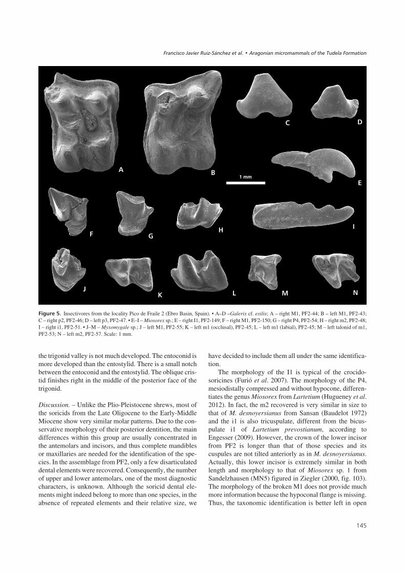
��� ���9! Insectivores from the locality Pico de Fraile 2 (Ebro Basin, Spain). • A–D –Galerix cf. exilis; A – right M1, PF2-44; B – left M1, PF2-43;C – right p2, PF2-46; D – left p3, PF2-47. • E–I – Miosorex sp.; E – right I1, PF2-149; F – right M1, PF2-150; G – right P4, PF2-54; H – right m2, PF2-48;I – right i1, PF2-51. • J–M – Myxomygale sp.; J – left M1, PF2-55; K – left m1 (occlusal), PF2-45; L – left m1 (labial), PF2-45; M – left talonid of m1,PF2-53; N – left m2, PF2-57. Scale: 1 mm.
������ ������������ !��"�� ����� �#��$����%���%�%%�� ��"�&�'�����%����
�
% &
�
� �'
(
�
)* + � ,
1 mm
nomenclature as Miosorex sp. pending further materialfrom the nearby area.
Family Talpidae Fischer, 1814Subfamily Talpinae Fischer, 1814Tribe Urotrichini Dobson, 1883
Genus Myxomygale Filhol, 1890
Type species. – Myxomygale antiqua Filhol, 1890.
Myxomygale sp.Figure 5J–M
Locality. – Pico del Fraile 2 (PF2).
Material and measurements. – 1 p2? (PF2-52), 2 m1(PF2-45) (13.9 × 8.0 × 9.7) (PF2-53) (– × – × 10.4), 2 m2(PF2-57) (– × 10.0 × –) (PF2-153), 2 M1? (PF2-55)(PF2-151), 1 M2? (PF2-154), 1 M3 (PF2-56).
Description. –p2?. The tooth is labiolingually compressed. There is acentral undulated cristid crossing the tooth from its anteriortill its posterior extreme. The lingual face is concave whereasthe labial one is convex. There is a strong cingulum for-ming the posterolingual base of the tooth. The tooth hastwo roots.
m1. There is only one complete m1 (PF2-45). The otherspecimen (PF2-53) preserves only the talonid. In the com-plete specimen, the talonid is much wider than the trigonid.The protoconid is the highest cusp. Paraconid, metaconidand entoconid are placed in a straight line at the lingualmargin of the tooth. The hypolophid is also completelystraight and perpendicular to the lingual face. The proto-lophid makes a slight inflexion, so the metaconid is so-mewhat less advanced than the protoconid. The re-entrantvalley is not much pronounced. There is an intermediatecuspule at the oblique cristid with a small branch orientedtowards the talonid basin. The oblique cristid is displacedlabially with respect to the middle of the posterior face ofthe trigonid. There is a basal cingulum on the labial face,from beneath the paraconid until below the middle of theoblique cristid. The entostylid is less protruding in thecomplete specimen (PF2-45) than in the other one(PF2-53).
m2. Only two incomplete specimens have been found(PF2-57, PF2-153). The trigonid is mesiodistally compres-sed. The paraconid and the metaconid are closer to each ot-her than they are to the protoconid. There is a metacristid,posterior to the metaconid. A small secondary ridge is dis-
cernible midway the oblique cristid, oriented towards thecenter of the talonid basin. The oblique cristid ends lingu-ally, close to the base of the metaconid. The re-entrant la-bial valley is much pronounced. A well-defined labial cin-gulum forms the base of the tooth from below theparaconid till beneath the reentrant valley. The entostylidprotrudes over the occlusal outline posteriorly to the ento-conid.
M1?. There are no complete specimens, and only two lin-gual fragments (PF2-55, PF2-151) are preserved. The en-doloph is asymmetric. The protocone is the highest lingualcusp. There is a small protoconule anteriorly to the proto-cone. Due to the wear of both specimens, no other lingualcusp is discernible. The anterior arm of the protoconuledoes not connect to the base of the paracone, but it extendsuntil the anterior base of the tooth. Neither the mesostylenor any other labial cusp is discernible in either specimen.
M2?. Only the lingual part is preserved in PF2-154. Theprotocone is the highest cusp. Apparently, there is no pro-toconule. The hypocone is discernible as a slight elevationof the endoloph posterior to the protocone.
M3. The only specimen available (PF2-56) lacks the ante-rolabial corner. The paracone is the highest cusp. The me-tacone is somewhat lower than the paracone, but higherthan the protocone. The protocone is the only discerniblelingual cusp. The endoloph extends from the basal midpo-int of the anterior margin until the base of the metacone.The mesostyle is not divided.
Discussion. – The material of Talpidae from PF2 is certa-inly scanty. However, the similar and proportional size ofdifferent dental elements, and the presence of a repeatedpeculiar structure in the lower molars lead to assume thatthey all belong to the same species of talpid. The diversityof Early and Middle Miocene Talpidae in Spain is ratherpoor compared with countries like Germany or Austria(Ziegler 2006, Furió et al. 2011). Up to date, only five dif-ferent genera of moles have been reported from the Ram-blian and Aragonian (Early-Middle Miocene) sedimentsfrom Spain: Desmanella, Desmanodon, Myxomygale, Pro-scapanus and Talpa (Van den Hoek Ostende & Furió2005). In the following lines we carry out a thorough analy-sis of the material from PF2 to elucidate which of these ge-nera can be ruled out and which others cannot.
The most frequent talpid in the Early and early MiddleMiocene in Spain is the genus Desmanodon, which hasbeen found in several localities of the Daroca-Calamochaarea (Van den Hoek Ostende 1997). The ascription of thematerial from PF2 to Desmanodon is discarded becausein the latter genus the endolophs of the upper molars areconnected to the base of the paracone, in contrast with the
#.
����������� ������ �������������

#1
��� ���:! Ranges of variation of length and width (tenths of millimeters) of the lower (m1, m3) and upper molars (M1, M2) of Democricetodonhispanicus from VL2A and Fahlbuschia decipiens from BU sensu Freudenthal & Daams (1988), Democricetodon franconicus from Erkertshofen sensuFahlbusch (1966), Democricetodon franconicus-moralesi-jordensis-koenigswaldi from Daroca-Villafeliche, Democricetodon decipiens from Artesilla 1and Democricetodon from VA3B sensu Van der Meulen et al. (2004), Fahlbuschia sacedoniensis from Córcoles sensu Freudenthal (2005) andDemocricetodon aff. hispanicus from PF2 (this paper).
������ ������������ !��"�� ����� �#��$����%���%�%%�� ��"�&�'�����%����

condition observed in the material studied here, where thisridge extends till the midpoint of the anterior margin. Fur-thermore, the oblique cristid in Desmanodon is connectedto the central point of the posterior face of the trigonid,whereas in the m2 from PF2 it reaches the metaconid. Sim-ilarly, Desmanella usually shows inflated lingual cusps inthe upper molars and the re-entrant labial valley is less pro-nounced than it is in the material from PF2, so its presencein this locality is also rejected. The presence of a meta-cristid in the m2 pleads against its ascription to the genusTalpa. Finally, Proscapanus shows similar oblique cristidsin its second lower molars, but not in its first lower ones,where this element extends lingually to join the metacristid(Ziegler 2003). Moreover, in its upper molars only theprotocone is discernible, whereas in PF2, the labial frag-ments of the upper molars show more than one cusp.Finally, the m1 (the only measurable element found inPF2), is smaller than in any other species of Proscapanusknown so far.
Consequently, we find in the genus Myxomygale theonly candidate combining all the characters mentionedabove: 1 – discernible lingual cusps in the upper molars;2 – endoloph of the upper molars reaching the midpoint ofthe anterior margin; 3 – undivided mesostyle in M3;4 – oblique cristid connected to the metacone in m2 butending more labially in m1; 5 – presence of a metacristidin m2; 6 – small size. With respect to the last character,only the m1 is comparable, because no other talpid elementfrom PF2 is complete. Unfortunately, no m1’s of M. en-gesseri were recovered from the Greek locality of Aliveri(Doukas 1986), so this species cannot be compared with.Within the rest of Miocene species of Myxomygale, the m1from PF2 is just a bit smaller than that of M. gracilis fromPetersbuch 10 (Ziegler 2003) and those of M. hutchisonifrom the German localities of Petersbuch 2 andSandelzhausen (Ziegler 1985, 2000). However, the m1from PF2 is extremely similar in size to M. hutchisoni fromOberdorf 4 (Ziegler 1998) and M. minor from Merkur-Nord (Van den Hoek Ostende & Fejfar 2006). It must benoticed that the first lower molars from PF2 resemble thesetwo latter cases also in morphology by presenting a smallridge towards the talonid basin in their oblique cristids(Ziegler 1998, fig. 3.1; Van den Hoek Ostende & Fejfar2006, fig. 5.4), a character never reported in any other talpidspecies as far as we know. The ascription to the speciesM. minor could get some more weight considering the possi-bly double-rooted p2, something hitherto only documentedin this species within the genus (Van den Hoek Ostende &Fejfar 2006). The identification of this element from PF2,however, is premilinar and the exact number of roots inlower premolars is actually unknown in several species ofMyxomygale. Therefore, the identification has been left atthe genus level because the sample is too small and the mea-surements are not enough for the specific discrimination.
��������������"
Figure 7 contains information on the stratigraphical distri-bution of the taxons described in the localities PF2 andSA5 and of related taxons. The locality PF2 contains Eu-myarion cf. weinfurteri, Megacricetodon aff. primitivus,Democricetodon aff. hispanicus, Microdyromys cf. koenig-swaldi, Microdyromys cf. remmerti, Pseudodryomys cf.ibericus, Spermophilinus besana, Galerix cf. exilis, Mioso-rex sp. and Myxomygale sp. SA5 contains Megacricetodonsp., Democricetodon or Fahlbuschia sp., Spermophilinussp. and Vasseuromys aff. cristinae.
The presence in both localities of a representative of thegenus Megacricetodon permits to place them in the Ara-gonian or in the Lower Vallesian. The Megacricetodonfrom PF2 is similar to the one described by Ruiz-Sánchez(1999) from Morteral 22, of which the age has been estab-lished at the limit between units MN4 and MN5(Ruiz-Sánchez et al. 2009).
The presence of Megacricetodon serves to considergrosso modo the age of the studied localities, and the presenceof the accompanying taxons in PF2 permits a more detailedassignment. In this respect, the relevant taxons are Spermo-philinus besana and especially Microdyromys cf. remmerti.
By size and morphology the population of Spermo-philinus besana from PF2 may be placed in the youngerpart of the time interval of that species. Cuenca (1988) de-scribed the presence of this taxon in the localitiesVillafeliche 2A (local zone B), Vargas 1A (local zone C),Olmo Redondo 5 (local zone C), Olmo Redondo 8 (localzone C) and Olmo Redondo 9 (local zone D), or, in otherwords, a range of distribution between the base of theLower Aragonian (local zone B) and the middle part of theMiddle Aragonian (local zone D). The biometrical andmorphological details of the population from PF2 fit bestwith what Cuenca (1988) described in the populations fromlocal zone D of the Middle Aragonian.
The presence of Microdyromys cf. remmerti delimits theposition of PF2 even more. Microdyromys remmerti was de-scribed by García-Paredes et al. (2010) from the localitiesVillafeliche 4A, Villafeliche 4B, Valdemoros 3D, Valde-moros 3B, Vargas 7, Vargas 8B, Vargas 8C, Valdemoros 6A,Valdemoros 3E and Valdemoros 3F. The range of these local-ities comprises the local zones Dc and Dd. The morphologydescribed by García-Paredes et al. (2010) in the material fromthe localities of the type area of the Aragonian presents a cer-tain degree of variation that permits these authors to distin-guish the age of the localities in question. The morphology ofthe p4 of Microdyromys cf. remmerti from PF2, is very simi-lar to that of the oldest populations of the species, and sug-gests that PF2 may be placed in the local zone Dc.
Regarding to the insectivores, in Spain, the latest part ofMN4 and the beginning of MN5 is characterized by a grad-ual replacement of G. symeonidisi by G. exilis according to
#2
����������� ������ �������������

Van den Hoek Ostende & Doukas (2003). The extinctionof G. symeonidisi in the Daroca-Calamocha area seems tooccur close to the Db-Dc (Van der Meulen et al. 2012) lo-cal zonation boundary (Van den Hoek Ostende & Doukas2003), G. exilis thus being the only Galerix species presentin the younger Aragonian small mammal assemblages.However, this genus is really scarce or nearly absent in thelocalities of the local zone Dc from Daroca-Calamocha, theinterval represented in Pico del Fraile 2. Thus, the presenceof G. symeonidisi in this locality cannot to be completelyruled out. At the genus level, the association of theerinaceid Galerix and the soricid Miosorex is very charac-teristic in the Middle Miocene small mammal assemblagesfrom Spain, according to the data compiled by Van denHoek Ostende & Furió (2005).
It is worth mentioning that the presence of the talpidMyxomygale in Spain had been reported only once beforein the literature, from the MN3 locality of Ramblar 1 (Vanden Hoek Ostende 2003). However, the occurrence in PF2is much younger. In the German fossil record, Myxomygalehas a rather continuous record (at least once occurrence per
MN Unit), ranging from MN 1 to MN 5 (Ziegler 2006).Therefore, the absence of Myxomygale in most of theSpanish localities in between is more likely indicative of atransient genus that only reached southern latitudes whenenvironmental conditions were favourable.
The situation of SA5 is much more difficult to establishthan PF2. However, on the basis of the stratigraphy andpaleomagnetic data, SA5 is likely similar in age to PF2, beingthe record of Vasseuromys in line with correlation to zone Dc.
The location of the fossil localities PF2 and SA5 in themiddle part of chron C5Br is entirely consistent with theirbiostratigraphic attribution to local zone Dc, keeping in mindthe magnetobiostratigraphic correlation of the Aragonian inthe Calatayud-Daroca Basin (Daams et al. 1999).
%�� ����
In the Unit 5 of the Tudela Formation two new localities(PF2 and SA5) have been located, whose fauna of rodentsand insectivores permits an assignment to the Aragonian.
#3
��� ���;! Distribution chart of the rodent studied in this paper and of related species according to the revision from Sesé (2006), and Cuenca (1988) andDe Bruijn (1999) (Spermophilinus), Freudenthal & Daams (1988) (Democricetodon, Fahlbuschia and Pseudofahlbuschia), Daams & Freudenthal (1988)(Megacricetodon), Van der Meulen et al. (2004) (Democricetodon hispanicus-franconicus-moralesi) and Kälin (1999) (Eumyarion). Sensu VDM et al. –according to Van der Meulen et al. (2004).
������ ������������ !��"�� ����� �#��$����%���%�%%�� ��"�&�'�����%����

Regarding the rodents, in PF2 we found three represen-tatives of the family Cricetidae, four of the Gliridae andone of the Sciuridae. Moreover, three species of insecti-vores are present, too. In SA5 we found two Cricetidae andone Sciuridae.
The Megacricetodon from PF2 presents morphologyintermediate between the populations of the genus of theLower and the Middle Aragonian.
The association of the erinaceid Galerix and the soricidMiosorex is characteristic from the Middle and early LateMiocene.
The presence of the genus Myxomygale in the localityPF2 represents the first occurrence of this taxon in Ara-gonian deposits from Spain.
The presence, in PF2, of Microdyromys cf. remmertiand Spermophilinus besana permits a detailed biostrati-graphic assignment to local zone Dc; for SA5 this is moredifficult, due to the poor material available.
��<�=��������
We acknowledge the assistance during the sampling campaigns byAlejandro Urmeneta and Rubén Arcos of the Comunidad of theBardenas Reales of Navarra and of Eliseo Martínez and SalvadorGarcía of the Aula Paleontológica of Cenicero. We are also gratefulfor the efficient work of the technicians of the SCSIE of the UV, P.Gómez, E. Navarro and A.J. Ibáñez. We thank Jérôme Prieto, Larsvan den Hoek Ostende and Executive Editor, Šárka Doležalová, fortheir successful comments and criticism. This investigación has beenfinancied through the projects BTE2003-7252, CGL2004-0780,CGL2007/66431/C02-02, GVPRE/2008/320, CGL2011-2868,PIPH-2009 SGR 754, Generalitat de Catalunya and a fieldworkgrant (DGPC-DGA-120/2010) from the Aragón Government.
/��������
AGUILAR, J.P. & LAZZARI, V. 2006. Nouvelles espèces de gliridésdu gisement karstique de Blanquatère 1 (Miocène moyen, sudde la France). Geodiversitas 28, 277–295.
AGUSTÍ, J. 1981. Cladistics and Paleomastology: Application tothe Phylogeny of Rodents. I. Neogene Gliridae from Europe.International Symposium: Concept and Method in Paleontol-ogy (Barcelona), 103–110.
AGUSTÍ, J. 1983. Roedores (Mammalia) del Mioceno inferiorde Can Martí Vell (Vallès – Penedès, Cataluña, España).Estudios Geológicos 39, 417–430.
AGUSTÍ, J., PÉREZ-RIVARÉS, F.J., CABRERA, L., GARCÉS, M., PARDO,G. & ARENAS, C. 2011. The Ramblian-Aragonian boundary andits significance for the European Neogene continental chronol-ogy. Contributions from the Ebro Basin record (NE Spain).Geobios 44, 121–134. DOI 10.1016/j.geobios.2011.01.001
ALONSO-ZARZA, A.M., ARMENTEROS, A., BRAGA, J.C., MUÑOZ, A.,PUJALTE, V., RAMOS, E., AGUIRRE, J., ALONSO-GAVILÁN, G.,ARENAS, C., BACETA, J.I., CARBALLEIRA, J., CALVO, J.P.,CORROCHANO, A., FORNÓS, J.J., GONZÁLEZ, A., LUZÓN, A.,
MARTÍN, J.M., PARDO, G., PAYROS, A., PÉREZ, A., POMAR, L.,RODRÍGUEZ, J.M. & VILLENA, J. 2002. Tertiary, 293–334. InGIBBONS, W. & MORENO, T. (eds) The Geology of Spain. Geo-logical Society, London.
ÁLVAREZ-SIERRA, M.A., DAAMS, R., LACOMBA, J.I., LÓPEZ
MARTÍNEZ, N., MEULEN, A.J. VAN DER, SESÉ, C. & DE VISSER, J.1991. Palaeontology and biostratigraphy (micromammals) ofthe continental Oligocene-Miocene deposits of the North-Central Ebro Basin (Huesca, Spain). Scripta Geologica 94,1–77.
ÁLVAREZ-SIERRA, M.A. & GARCÍA-MORENO, E. 1986. NewGliridae and Cricetidae from the Middle and Upper Mioceneof the Duero basin, Spain. Studia Geologica Salmanticensia22, 145–189.
ARENAS, C. & PARDO, G. 1999. Latest Oligocene-late Miocenelacustrine systems of the north-central part of the Ebro Basin(Spain): sedimentary facies model and paleogeographicsíntesis. Palaeogeography, Palaeoclimatology, Palaeoecol-ogy 151, 127–148. DOI 10.1016/S0031-0182(99)00025-5
BAUDELOT, S. 1972. Etude des Chiroptères, Insectivores etRongeurs du Miocène de Sansan (Gers). 364 pp. Ph.D. thesis,Université Paul Sabatier, Toulouse, France.
BAUDELOT, S. & BONIS, L. DE 1966. Nouveaux Gliridés (Rodentia)de l’Aquitanien du basin d’Aquitaine. Comptes Rendus Som-maires Societé Geologique de la France 9, 342–343.
BRUIJN, H. de 1999. Superfamily Sciuroidea, 271–280. In RÖSS-
NER, G.E. & HEISSIG, K. (eds) Land Mammals of Europe.Verlag Dr. Friedrich Pfeil, München.
BRUIJN, H. DE & BOSMA, A.A. 2012. Spermophilinus andCsakvaromys, two names for the same genus of ground squir-rel (Tamiini, Sciuridae, Rodentia, Mammalia) from the Neo-gene of Europe. Annalen des Naturhistorischen Museum inWien, Serie A 114, 317–320.
BULOT, C. 1979. Un nouvel Eumyarion (Rodentia, Mammalia)du Miocène de Bézian près de La Romieu (Gers). Bulletin dela Société d’Histoire Naturelle de Toulouse 114(3–4),373–381.
CUENCA, G. 1988. Revisión de los Sciuridae del Aragoniense y delRambliense en la fosa de Calatayud-Montalbán, 1–116. InFREUDENTHAL, M. (ed.) Biostratigraphy and paleoecology ofthe Neogene micromammalian faunas from the Calatayud-Teruel Basin (Spain), Scripta Geologica Special Issue 2.
DAAMS, R. 1974. Pseudodryomys (Gliridae, Rodentia, Mam-malia) from Early and Middle Miocene deposits in Spain. Pro-ceedings Koninklijke Nederlandse Akademie van Weten-schappen 77, 201–240.
DAAMS, R. & FREUDENTHAL, M. 1974. Early Miocene Cricetidae(Rodentia, Mammalia) from Buñol (Prov. Valencia, Spain).Scripta Geologica 24, 1–16.DOI 10.1016/S0012-821X(98)00273-8
DAAMS, R. & FREUDENTHAL, M. 1988. Synopsis of theDutch-Spanish collaboration program in the Aragonian typearea, 1975–1986, 3–18. In FREUDENTHAL, M. (ed.) Biostrati-graphy and paleoecology of the Neogene micromammalianfaunas from the Calatayud-Teruel Basin (Spain), ScriptaGeologica Special Issue 1.
DAAMS, R., MEULEN, A.J. VAN DER, ÁLVAREZ SIERRA, M.A.,PELÁEZ CAMPOMANES, P. & KRIJGSMAN, W. 1999. Aragonianstratigraphy reconsidered, and a re-evaluation of the middle
0"
����������� ������ �������������

Miocene mammal biochronology in Europe. Earth and Plane-tary Science Letters 165, 287–294.
DOUKAS, C.S. 1986. The mammals from the Lower Miocene ofAliveri (Island of Evia, Greece). Part 5. The insectivores. Pro-ceedings of the Koninklijke Nederlandse Akademie vanWetenschappen 89(1), 15–38.
ENGESSER, B. 2009. The Insectivores (Mammalia) from Sansan(Middle Miocene, south-western France). SchweizerischePaläontologische Abhandlungen 128, 1–91.
FAHLBUSCH, V. 1966. Cricetidae (Rodentia, Mamm.) aus dermittelmiozänen Spaltenfüllung Erkertshofen bei Eichstätt.Mitteilungen der Bayerischen Staatssammlung für Paläon-tologie und Historische Geologie 6, 109–131.
FREUDENTHAL, M. 2004. Gliridae (Rodentia, Mammalia) fromthe Eocene and Oligocene of the Sierra Palomera (Teruel,Spain). Treballs del Museu de Geologia de Barcelona 12,97–173.
FREUDENTHAL, M. 2005. The status of Democricetodon, Fahl-buschia, Pseudofahlbuschia and Renzimys (Cricetidae,Mammalia). A reply to Van der Meulen et al. (2004). Geobios39, 43–55. DOI 10.1016/j.geobios.2004.08.005
FREUDENTHAL, M. & DAAMS, R. 1988. Cricetidae (Rodentia) fromthe type Aragonian; the genera Democricetodon, Pseudofahl-buschia nov. gen. and Renzimys, 133–252. In FREUDENTHAL,M. (ed.) Biostratigraphy and paleoecology of the Neogenemicromammalian faunas from the Calatayud-Teruel Basin(Spain), Scripta Geologica Special Issue 1.
FURIÓ, M., CASANOVAS-VILAR, I. & HOEK OSTENDE, L.W. VAN DEN
2011. Predictable structure of Miocene insectivore(Lipotyphla) faunas in Western Europe along a latitudinal gra-dient. Palaeogeography, Palaeoclimatology, Palaeoecology304, 219–229. DOI 10.1016/j.palaeo.2010.01.039
FURIÓ, M., SANTOS-CUBEDO, A., MINWER-BARAKAT, R. & AGUSTÍ,J. 2007. Evolutionary history of the African soricid Myosorex(Insectivora, Mammalia) out of Africa. Journal of VertebratePaleontology 27, 1018–1032.DOI 10.1671/0272-4634(2007)27[1018:EHOTAS]2.0.CO;2
GARCÍA-PAREDES, I., PELÁEZ-CAMPOMANES, P. & ÁLVAREZ-SIERRA,M.A. 2010. Microdyromys remmerti, sp. nov., a new Gliridae(Rodentia, Mammalia) from the Aragonian type area (Mio-cene, Calatayud-Montalbán Basin, Spain). Journal of Verte-brate Paleontology 30, 1594–1609.DOI 10.1080/02724634.2010.501453
HOEK OSTENDE, L.W. VAN DEN 1997. Insectivore faunas from theLower Miocene of Anatolia. Part 4: The genus Desmanodon(Talpidae) with the description of a new species from theLower Miocene of Spain. Proceedings of the KoninklijkeNederlandse Akademie van Wetenschapen 100, 27–65.
HOEK OSTENDE, L.W. VAN DEN 2001. A revised generic classifica-tion of the Galericini (Insectivora, Mammalia) with some re-marks on their palaeobiogeography and phylogeny. Geobios34, 681–695. DOI 10.1016/S0016-6995(01)80029-2
HOEK OSTENDE, L.W. VAN DEN 2003. Insectivores (Erinaceo-morpha, Soricomorpha, Mammalia) from the Ramblian of theDaroca-Calamocha area. Coloquios de PaleontologíaVolumen Extraordinario 1, 281–310.
HOEK OSTENDE, L.W. VAN DEN & DOUKAS, C.S. 2003. The distri-bution of Galerix symeonidisi Doukas, 1986, 287–303. InREUMER, J.W.F. & WESSELS, W. (eds) Distribution and Migra-
tion of Tertiary Mammals in Eurasia. A volume in honour ofHans de Bruijn, Deinsea 10.
HOEK OSTENDE, L.W. VAN DEN & FEJFAR, O. 2006. Erinaceidaeand Talpidae (Erinaceomorpha, Soricomorpha, Mammalia)from the Lower Miocene of Merkur-Nord (Czech Republic,MN 3). Beiträge zur Paläontologie 30, 175–203.
HOEK OSTENDE, L.W. VAN DEN & FURIÓ, M. 2005. Spain, 149–284.In HOEK OSTENDE, L.W. VAN DEN, DOUKAS, C.S. & REUMER,J.W.F. (eds) The Fossil Record of the Eurasian Neogene In-sectivores (Erinaceomorpha, Soricomorpha, Mammalia),Part I, Scripta Geologica Special Issue 5.
HUGUENEY, M., MEIN, P. & MARIDET, O. 2012. Revision and newdata on the Early and Middle Miocene soricids (Sorico-morpha, Mammalia) from Central and South-Eastern France.Swiss Journal of Palaeontology 131, 23–49.DOI 10.1007/s13358-011-0036-1
JONG, F. DE 1988. Insectivora from the Upper Aragonian and theLower Vallesian of the Daroca-Villafeliche area in the Cala-tayud-Teruel Basin (Spain), 253–286. In FREUDENTHAL, M.(ed.) Biostratigraphy and paleoecology of the Neogene micro-mammalian faunas from the Calatayud-Teruel Basin (Spain),Scripta Geologica Special Issue 1.
KÄLIN, D. 1999. Tribe Cricetini, 373–387. In RÖSSNER, G.E. &HEISSIG, K. (eds) Land Mammals of Europe. Verlag Dr.Friedrich Pfeil, München.
KRETZOI, M. & FEJFAR, O. 2004. Sciurids and Cricetids (Mam-malia, Rodentia) from Rudabánya. Palaeontographia Italica90, 113–148.
LACOMBA, J.I. 1988. Estudio de las faunas de micromamíferos delOligoceno superior y Mioceno inferior en las cuencas deLoranca, Ebro riojano y Ebro aragonés. Aspectos paleo-ecológicos. 389 pp. Ph.D. thesis, Universidad Complutense,Madrid, Spain.
LARRASOAÑA, J.C., MURELAGA, X. & GARCÉS, M. 2006. Magne-tobiochronology of Lower Miocene (Ramblian) continentalsediments from the Tudela Formation (western Ebro basin,Spain). Earth and Planetary Science Letters 243, 409–423.DOI 10.1016/j.epsl.2006.01.034
LOURENS, L.J., HILGEN, F.J., SHACKLETON, N.J., LASKAR, J. & WIL-
SON, D.S. 2004. The Neogene period, 409–440. In GRADSTEIN,F.N., OGG, J.G. & SMITH, A.G. (eds) A Geologic Time Scale2004. Cambridge University Press, Cambridge.
MARTÍN-SUÁREZ, E., FREUDENTHAL, M. & AGUSTÍ, J. 1993. Micro-mammals from the Middle Miocene of the Granada basin(Spain). Geobios 26(3), 377–387.DOI 10.1016/S0016-6995(93)80028-P
MEIN, P. & FREUDENTHAL, M. 1971. Les Cricetidae (Mammalia,Rodentia) du Néogène Moyen de Vieux-Collonges. Partie 1:Le genre Cricetodon Lartet, 1851. Scripta Geologica 5, 1–51.
MEULEN, A.J. VAN DER, GARCÍA-PAREDES, I., ÁLVAREZ-SIERRA,M.A., HOEK OSTENDE, L.W. VAN DEN, HORDIJK, K., OLIVER, A. &PELÁEZ-CAMPOMANES, P. 2012. Updated Aragonian biostrati-graphy: Small Mammal distribution and its implications for theMiocene European Chronology. Geologica Acta 10(2), 1–24.
MEULEN, A.J. VAN DER, PELÁEZ-CAMPOMANES, P. & DAAMS, R.2004. Revision of medium-sized Cricetidae from the Mioceneof the Daroca-Villafeliche area in The Calatayud-Teruel basin(Zaragoza, Spain). Coloquios de Paleontología, VolumenExtraordinario 1, 385–441.
0
������ ������������ !��"�� ����� �#��$����%���%�%%�� ��"�&�'�����%����

MURELAGA, X. 2000. Estudio de las faunas de vertebrados delMioceno inferior de las Bardenas Reales de Navarra y áreascolindantes (Cuenca del Ebro, Navarra). 363 pp. Ph.D. thesis,Universidad del País Vasco, Bilbao, Spain.
MURELAGA, X., ASTIBIA, H., SESÉ, C., SORIA, D. & PEREDA-SUBERBIOLA, X. 2004a. Mamíferos del Mioceno inferior de lasBardenas Reales de Navarra (Cuenca del Ebro, PenínsulaIbérica). Munibe 55, 7–102.
MURELAGA, X., LARRASOAÑA, J.C. & GARCÉS, M. 2004b. Nuevalocalidad fosilífera en el Mioceno inferior de las Bardenas Re-ales de Navarra (Cuenca del Ebro, Península Ibérica). Geo-gaceta 36, 179–182.
MURELAGA, X., PEREDA-SUBERBIOLA, X., LAPPARENT DE BROIN,F., RAGE, J.C., DUFFAUD, S., ASTIBIA, H. & BADIOLA, A. 2002.Reptiles and amphibians from the Lower Miocene of theBardenas Reales of Navarra (Ebro basin, Iberian Peninsula).Geobios 35, 347–365.DOI 10.1016/S0016-6995(02)00031-1
OLIVER-PÉREZ, A., LÓPEZ-GUERRERO, P. & PELÁEZ-CAMPOMANES,P. 2008. Primer representante del género Megacricetodon dela Cuenca de Calatayud-Daroca (Zaragoza, España). Palae-ontologica Nova SEPAZ 8, 317–329.
PRIETO, J., GROSS, M., BÖHMER, C. & BÖHME, M. 2010. Insecti-vores and bat (Mammalia) from the late Middle Miocene ofGratkorn (Austria): biostratigraphic and ecologic implica-tions. Neues Jahrbuch für Geologie und Palaeontologie, Ab-handlungen 258, 107–119.DOI 10.1127/0077-7749/2010/0088
PRIETO, J., HOEK OSTENDE, L.W. VAN DEN & HÍR, J. 2012. TheMiddle Miocene insectivores from Sámsonháza 3 (Hungary,Nógrád County): Biostratigraphical and palaeoenvironmentalnotes near to the Middle Miocene Cooling. Bulletin of Geo-sciences 87(2), 227–240. DOI 10.3140/bull.geosci.1296
PRIETO, J. & RUMMEL, M. 2009. Erinaceidae (Mammalia, Erina-ceomorpha) from the Middle Miocene fissure filling Peters-buch 68 (southern Germany). Zitteliana 48/49, 103–111.
REUMER, J.W.F. 1984. Ruscinian and early Pleistocene Soricidae(Insectivora, Mammalia) from Tegelen (The Netherlands) andHungary. Scripta Geologica 73, 1–173.
RUIZ-SÁNCHEZ, F.J. 1999. Estudio paleontológico de los roedoresfósiles del Mioceno inferior de la cuenca del río Magro (Va-lencia). 395 pp. Ph.D. thesis, University of Valencia, Valen-cia, Spain.
RUIZ-SÁNCHEZ, F.J., LÁZARO-CALATAYUD, B. & FREUDENTHAL, M.2009. Eomyops noeliae sp. nov., a new Eomyidae (Mammalia,Rodentia) from the Aragonian of Spain. Comptes RendusPalevol 8, 375–384. DOI 10.1016/j.crpv.2008.12.002
RUIZ-SÁNCHEZ, F.J., MURELAGA, X., FREUDENTHAL, M., LARRA-
SOAÑA, J.C. & GARCÉS, M. 2010a. Presencia del géneroVasseuromys (Gliridae, Rodentia) en la sección de Pico delFraile (Bardenas Reales de Navarra, Cuenca del Ebro,España). Publicaciones del Seminario de Paleontología deZaragoza 9, 277–280.
RUIZ-SÁNCHEZ, F.J., MURELAGA, X., FREUDENTHAL, M., LARRA-
SOAÑA, J.C. & GARCÉS, M. 2012a. A new Vasseuromys speciesfrom the Aragonian of Ebro basin (Spain). Acta Palaeon-
tologica Polonica 57(2), 225–239.DOI 10.4202/app.2010.0081
RUIZ-SÁNCHEZ, F.J., MURELAGA, X., FREUDENTHAL, M., LARRA-
SOAÑA, J.C. & GARCÉS, M. 2012b. Vasseuromys rambliensissp. nov. (Gliridae, Mammalia) from the Ramblian (LowerMiocene) of the Tudela Formation (Ebro basin, Spain).Palaeontologia Electronica 15(1), 1–16.
RUIZ-SÁNCHEZ, F.J., MURELAGA, X., FREUDENTHAL, M., LARRA-
SOAÑA, J.C. & GARCÉS, M. 2012c. Hypsodont Myomiminae(Gliridae, Rodentia) from the Lower Miocene Tudela Forma-tion (Bardenas Reales, Ebro Basin, Spain) and their bearing onthe age of the Agenian-Ramblian boundary. Geodiversitas.
RUIZ-SÁNCHEZ, F.J., MURELAGA, X., FREUDENTHAL, M., LARRA-
SOAÑA, J.C., GARCÉS, M., CRESPO-ROURES, V.D., MANSINO-PARIS, S. & GONZÁLEZ-PARDOS, M. 2010b. Pico del Fraile 2(PF2), una nueva localidad de edad Aragoniense en la Cuencadel Ebro (Bardenas Reales de Navarra). Publicaciones delSeminario de Paleontología de Zaragoza 9, 281–284.
RUIZ-SÁNCHEZ, F.J., SANTISTEBAN, C. DE & LACOMBA, J.I. 2003.Nuevas faunas de roedores fósiles (Mammalia, Rodentia) deedad Aragoniense inferior y medio en la serie del Barranco deMorteral (cuenca del río Magro, prov. de Valencia, España).Coloquios de Paleontología 1, 579–594.
RÜMKE, C.G. 1985. A review of fossil and recent Desmaninae(Talpidae, Insectivora). Utrecht Micropaleontological Bulle-tins Special Publication 4, 1–241.
SESÉ, C. 2006. Los roedores y lagomorfos del Neógeno deEspaña. Estudios Geológicos 62, 429–480.DOI 10.3989/egeol.0662138
WU, W. 1982. Die Cricetiden (Mammalia, Rodentia) aus derOberen Süsswasser-Molasse von Puttenhausen (Nieder-bayern). Zitteliana 9, 37–80.
WU, W. 1993. Neue Gliridae (Rodentia, Mammalia) aus unter-miozänen (orleanischen) Spaltenfüllungen Süddeutschlands.Documenta Naturae 81, 1–149.
ZIEGLER, R. 1985. Talpiden (Mammalia, Insectivora) aus demOrleanium und Astaracium Bayerns. Mitteilungen der Bayeri-schen Staatssammlung für Paläontologie und HistorischeGeologie 25, 131–175.
ZIEGLER, R. 1998. Wirbeltiere aus dem Unter-Miozän des Lignit-Tagerbaues Oberdorf (Weststeirisches Becken, Österreich): 5.Marsupialia, Insectivora und Chiroptera (Mammalia).Annalen des Naturhistorischen Museums in Wien 99, 43–97.
ZIEGLER, R. 2000. The Miocene Fossil-Lagerstätte Sandelz-hausen, 17. Marsupialia, Lipotyphla and Chiroptera (Mam-malia). Senckenbergiana lethaea 80(1), 81–127.
ZIEGLER, R. 2003. Moles (Talpidae) from the late Middle Mio-cene of South Germany. Acta Palaeontologica Polonica48(4), 617–648.
ZIEGLER, R. 2005. The squirrels (Sciuridae, Mammalia) of theMiocene Fossil-Lagerstaette Sandelzhausen (Bavaria, S Ger-many). Neues Jahrbuch für Geologie und Palaeontologie,Abhandlungen 237(2), 273–312.
ZIEGLER, R. 2006. Miocene insectivores from Austria and Ger-many – An overview. Beiträge zur Paläontologie 30,481–494.
0/
����������� ������ �������������


![Limestone, Chert, or Limestone? [2013225_fieldtrip]](https://img.pdfslide.net/doc/110x75/558421bfd8b42a86478b4731/limestone-chert-or-limestone-2013225fieldtrip.jpg)