Embed Size (px)
Citation preview
CHEMICAL ENGINEERING TRANSACTIONS
VOL. 64, 2018
A publication of
The Italian Associationof Chemical Engineering
Online at www.aidic.it/cet
Guest Editors: Enrico Bardone, Antonio Marzocchella, Tajalli KeshavarzCopyright © 2018, AIDIC Servizi S.r.l.ISBN 978-88-95608- 56-3; ISSN 2283-9216
Kefiran-based Scaffolds For Biomedical ApplicationsMauro Toscanoa, Francesco Carfì Paviac, Gioacchino Conoscentic, Maria A. Sabatinob, Vincenzo La Carrubbac, Clelia Dispenzab, Valerio Brucatoc
aDICAM, University of Palermo, Viale delle Scienze building 8, 90128 PalermobDIID, University of Palermo, Viale delle Scienze building 8, 90128 PalermocDICAM & UdR INSTM, University of Palermo, Viale delle Scienze building 8, 90128 [email protected]
Kefiran is an exopolysaccharide produced by microorganisms present in kefir grains, with several health promoting properties. A optimized protocol was developed for the separation of kefiran from kefir grains, allowing to reach a yield 4÷5 % without using toxic or expensive chemicals. The capability of kefiran to produce scaffold via Thermally Induced Phase Separation (TIPS) technique was investigated and porous scaffolds structure was obtained. Separated kefiran and scaffolds were analyzed via DSC and different thermal properties between purified kefiran and scaffold were revealed. XRD analysis revealed different structure between kefiran and scaffolds. The porous scaffold structure can be modulated by modifiying the thermal path of the solution during the phase separation. The citotoxicity of kefiran was evaluated with cell viability test and a significant faster cell proliferation in the treated colture medium with kefiran was evidenced.
1. IntroductionThe exopolysaccharides (EPSs) are extracellular polymers which are produced by bacteria colonies, fungi and blue-green algae during their metabolism (Amjres et al. 2015). The EPSs can be either capsular polysaccharide (CPS) if it forms a capsular structure around cell, or exopolysaccharide if it is weakly attached or totally secreted into the medium during cell growth (Yang et al. 2010). In food industry EPSs are a valid alternative to commercial stabilizers because of their enhancement of rheology, structure and due to organoleptic characteristics of fermented products (Ahmed et al. 2013). Among them Kefiran has gained significant attention from scientific and practical point of view beacuse of its biocompatibility and biodegradability. It is produced by Lactobacillus kefiranofaciens, a bacterium that is trapped in gelatinous irregular masses (grains) which are the starter for obtaining the fermented milk know as Kefir. Those grains contain lactic acid bacteria, acetic acid bacteria and yeast involved in fermentation (Abraham and De Antoni 1999). Kefiran is an high molecular weight polymer of approximately 106 Da (Piermaria et al. 2008), made by equal amount of D-glucose and D-galactose (Cheirsilp et al. 2001). It is quite resistant to hydrolysis and forms gels in aqueous solutions containing ethanol (Micheli et al. 1999). This polymer has ben widely used as a thickener, stabiliser, emulsifier, fat substitute and gelling agent and also exhibits antibacterial, antifungal and antitumour activities (Welman and Maddox 2003; Tada et al. 2007). The use of biopolymers has steadily increased along the years owing to the potential to extend shelf life and increase quality of food while reducing environmental impact with coating films from renewable resources (Garcia et al. 1998; Sorrentino et al. 2007). Another very relevant application for biopolymers concerns using them to produce 3D porous supports (scaffold) for biomedical applications. Among the all possible scaffold production techniques Thermally Induced Phase Separation (TIPS) is one of the most promising ones (Ghersi et al. 2016): in this case, a temperature decrease causes a variation of the polymer solubility, inducing a mechanism of nucleation and growth of droplets of polymer poor phase that will form the porous structure (Mannella et al. 2014, van de Witte et al. 1996, Carfì Pavia et al. 2009). The aim of this work is to develop and optimize a separation protocol of kefiran and to characterize kefiran scaffolds produced via Thermally Induced Phase Separation (TIPS) in order to evaluate their potential application in biomedical field. With this respect, Kefiran is able not
only to offer environmentally friendly solutions, but also to produce scaffolds without managing toxic chemicals.
2. Materials and Methods2.1 Isolation and purification of kefiran
A separation protocol was adapted and optimised based on the procedue reported by Piermaria et al. (2008). A weighed amount of kefir grains was dissolved in water (1:10 w/v) with continuous stirring until a homogeneous mixture was obatined. The mixture was centrifuged at 10,000g for 20 min at 20°C (Thermo Scientific SL40R Centrifuge). Then, two volumes of ethanol were added to the obtained supernatant; the as-prepared mixture was stored at -20 C overnight. Finally, the mixture was centrifuged at 10,000g for 20 min at 4°C. The pellets gained from the centrifugation were re-dissolved in boiling water and the whole procedure was repeated twice. The pellets collected at the end of the process were finally dissolved in water and freeze-dried.
2.2 Thermally induced phae separation (TIPS) scaffold-forming protocol
A weighed amount of kefiran was dissolved in a solution of water/ethanol ratio 80/20 wt/wt at 80°C. The polymer concentration selected for the scaffold-forming solution was 4% wt. The pre-heated solution was poured in a aluminium disk-shaped sample-holder, with a diameter of 12 mm and a thickness of 2 mm. The filled sample-holder was then pool-immersed into a thermostatic bath at fixed temperature (25°C, 30°C, 35°C) for 10 min, in order to investigate the influence of the demixing temperature on the scaffold morphology. Finally, a rapid quench in an ethanol bath at -20°C for 10 min was carried out in order to freeze the as-obtained structure. In order to understand the most suitable technique to dry the scaffolds, two methods were tested: drying at 30°C under vacuum and freeze-drying.
2.3 Scanning Electron Microscopy (SEM)
Microstructural analysis of the surface and cross-sections of the scaffolds was conducted via scanning electron microscopy by using a SEM Phenom Pro-X apparatus (Phenom-World, Netherlands). The scaffolds were fractured in liquid nitrogen, mounted on aluminium stubs using a double-sided adhesive tape. All samples were examined using an accelerating voltage of 10.0 kV. Samples were examined at 90° with respect to the surface, in order to allow observation of the scaffold cross-sections.
2.4 Differential Scanning Calorimetry (DSC)
The thermal properties of the scaffold were evaluated by using Differential Scanning Calorimetry analysis (DSC evo 131 SETARAM). Samples were scanned at heating rate of 10°C/min in the temperature range from -20°C and 210°C. The melting point (Tm) was calculated as the temperature where the peak of the endotherm occurs. The melting enthalpy H was determined as the area over the endothermic peak. All these properties were determined in triplicates and the results were averaged.
2.5 X-rays diffraction (XRD)
The X-ray diffractograms of scaffold produced from 4% kefiran solution were obtained in a Panalytical X-Pert Powder apparatus. The samples were exposed to an X-ray beam, the X-ray generator running with radiation Cu-K (=154 nm, 40 kV and 30mA). The relative intensity was recorded at ambient temperature over an angular range (2) of 5-70° and 2503 scan points were acquired.
2.6 Cell viability test
0.4 g of kefiran were solubilised in 20 ml of complete M199 culture medium at 37°C for 7 days. Successively, the medium was filtered with 0.45 μm mesh. In order to test scaffold cytotoxicity, cell viability assays were performed. 104 ECV cells were seeded into wells of a 24-well culture plate and incubated with 500 μl of treated medium. As a control, cells grown with normal medium were utilised. Cell viability was evaluated by Cell Counting Kit 8 (CCK8) assay after 24 and 48 incubation hours from seeding. Briefly, at each time-point, the medium in the well was replaced with 500 μl of a CCK8/medium solution (1/10 v/v) for 3 hours. At the end of the incubation time, the solution was transferred into wells of a 96 multiwell and read with a spectrophotometer (BIOTEK, Synergy HT) at a wavelength of 450 nm.
3. Results and Discussion3.1 Optimization of the purification protocol
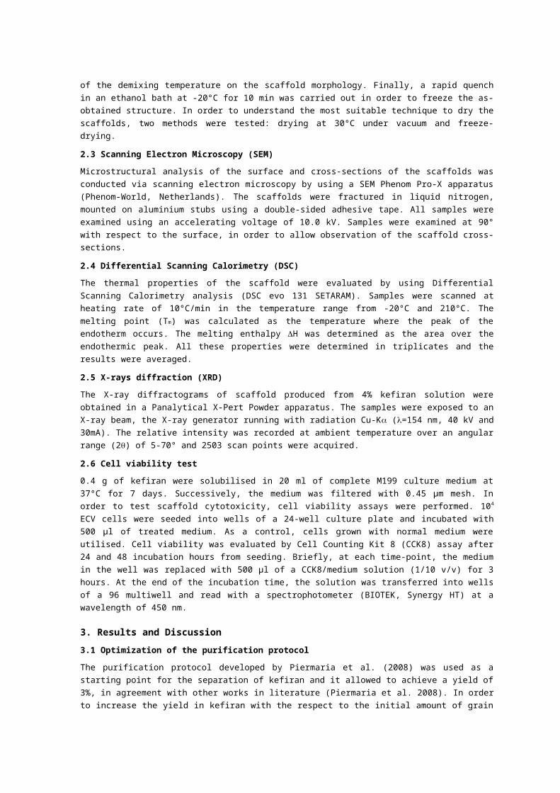
The purification protocol developed by Piermaria et al. (2008) was used as a starting point for the separation of kefiran and it allowed to achieve a yield of 3%, in agreement with other works in literature (Piermaria et al. 2008). In order to increase the yield in kefiran with the respect to the initial amount of grain a modification of the protocol was necessary. The optimized protocol, described in paragraph 2.1, ensured a significant increasing yield between 4 and 5 % as shown in tables 1-2.
Table 1: Yields of the application of Piermaria et al. (2008) protocol
Piermaria et al. ProtocolGrain weight Kefiran obtained Yield
21.475g 0.651g 3.03%45.191g 1.355g 2.99%
Table 2: Yields of the custom optimized protocol
Optimized ProtocolGrain weight Kefiran obtained Yield
2.534g 0.107g 4.22%2.525g 0.141g 5.58%66.81g 2.89g 4,32%
3.2 Scaffold production
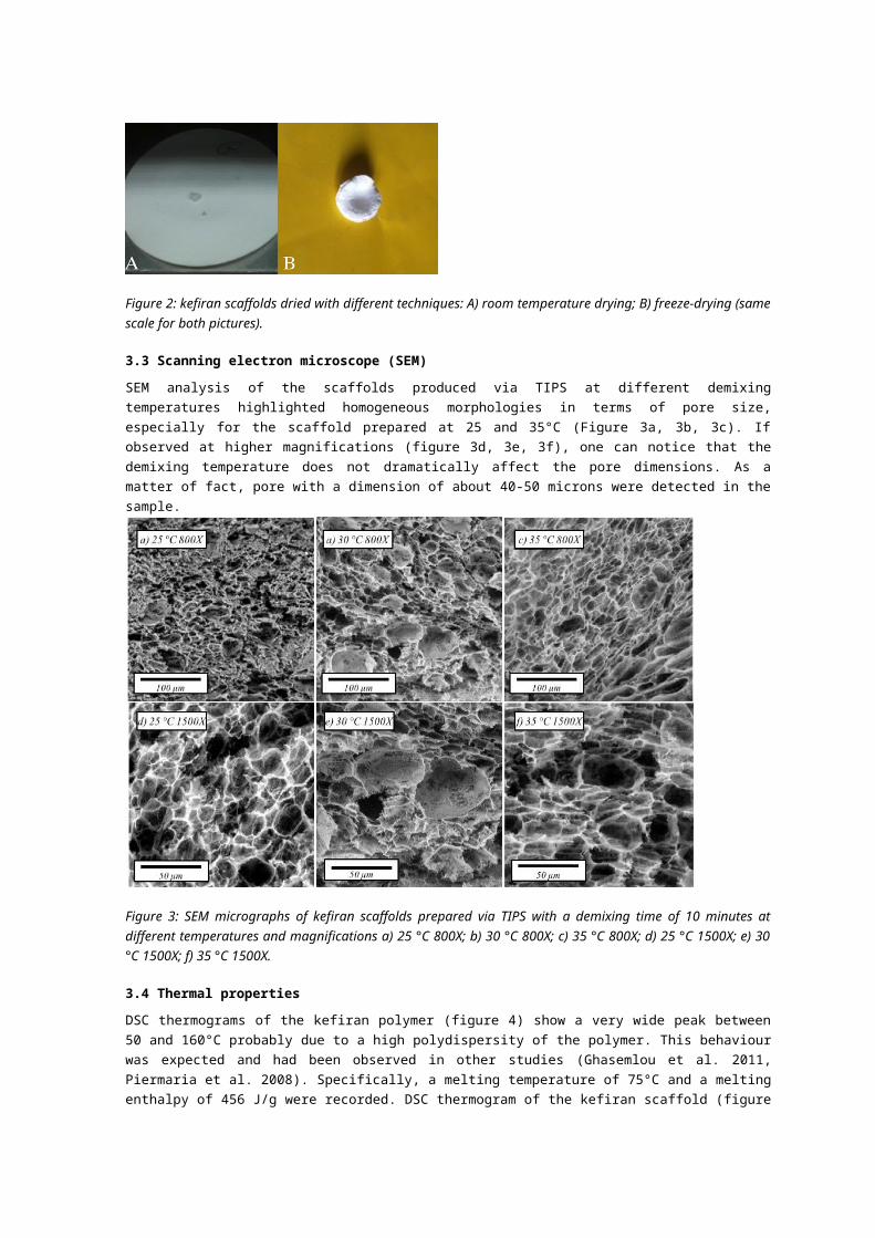
Once extracted form the sample holder, the scaffold were placed in ethanol. From Figure 1 it is possible to observe that the cylindrical shape and the dimensions (both diameter and thickness), provided by the geometry of the sample holder were preserved. In order to characterize the as-obtained sample, a desiccation step was mandatory. The first trial was a simple drying in an oven at 30°C under vacuum for 12h. Unfortunately, at the end of the process a dramatic reduction of size of the scaffold was observed (Figure 2A). This behaviour can be surely ascribed to the weak mechanical strength of the scaffold, which shrinks during solvent removal due to drying. In other words, the ethanol removal could not be beared by the structure, leading to a collapse. Conversely, when the solvent is removed in the solid state via a freeze-drying process (Figure 2), carried out at -20°C, the scaffold maintained its structure and dimensions.
Figure 1: kefiran scaffolds prepared via TIPS.
In order to investigate the influence of the demixing temperature on the internal morphology of the scaffolds, three different demixing temperatures (25, 30 and 35°C) were tested, keeping constant the demixing time at 10 min.
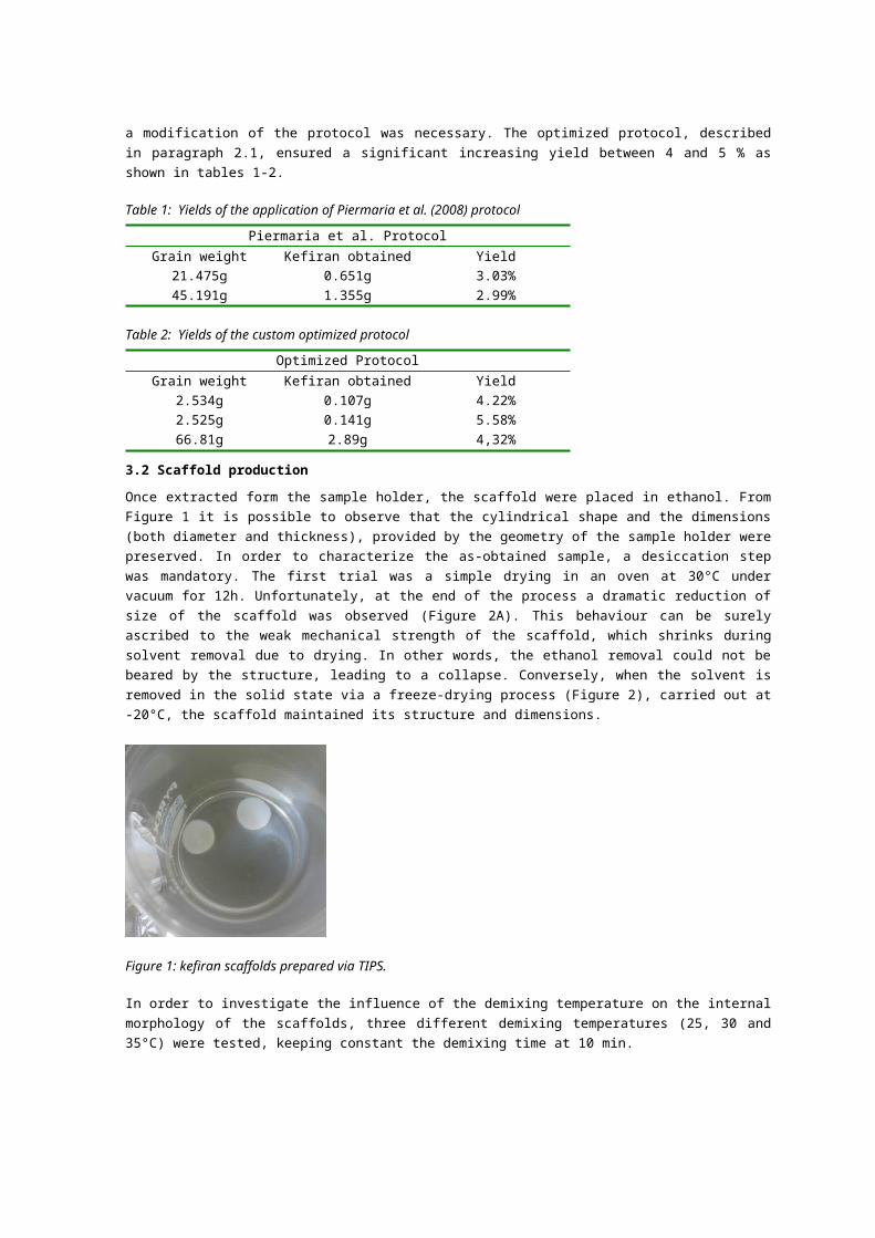
Figure 2: kefiran scaffolds dried with different techniques: A) room temperature drying; B) freeze-drying (same scale for both pictures).
3.3 Scanning electron microscope (SEM)
SEM analysis of the scaffolds produced via TIPS at different demixing temperatures highlighted homogeneous morphologies in terms of pore size, especially for the scaffold prepared at 25 and 35°C (Figure 3a, 3b, 3c). If observed at higher magnifications (figure 3d, 3e, 3f), one can notice that the demixing temperature does not dramatically affect the pore dimensions. As a matter of fact, pore with a dimension of about 40-50 microns were detected in the sample.
Figure 3: SEM micrographs of kefiran scaffolds prepared via TIPS with a demixing time of 10 minutes at different temperatures and magnifications a) 25 °C 800X; b) 30 °C 800X; c) 35 °C 800X; d) 25 °C 1500X; e) 30 °C 1500X; f) 35 °C 1500X.
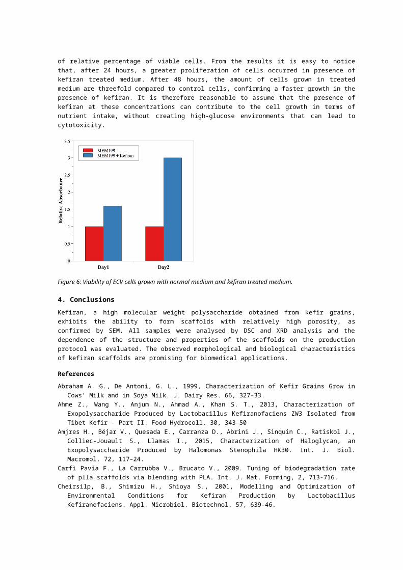
3.4 Thermal properties
DSC thermograms of the kefiran polymer (figure 4) show a very wide peak between 50 and 160°C probably due to a high polydispersity of the polymer. This behaviour was expected and had been observed in other studies (Ghasemlou et al. 2011, Piermaria et al. 2008). Specifically, a melting temperature of 75°C and a melting enthalpy of 456 J/g were recorded. DSC thermogram of the kefiran scaffold (figure 4) is quite different showing a wider peak between 60 and 170°C, a melting temperature of 106°C and a melting enthalpy of 405
J/g. The difference could be attributed to the different structure of kefiran polymer and kefiran scaffold.
Figure 4: DSC thermogram of kefiran polymer and 4% kefiran scaffold.
3.5 X-ray diffraction (XRD)
The XRD analysis was performed on the scaffold to investigate whether there is an influence of the material production process. Kefiran polymer and 4% scaffold were analysed. Figure 5 reports in blue colour the XRD spectrum of kefiran polymer, while in green the spectrum of kefiran scaffold is shown. The scaffold presents the only presence of a peak centered at 15°, while polymer shows the presence of two peaks, the first centered at 7.5° and the second at 15°. Therefore, from the obtained data, it was possible to confirm the conclusions drawn from DSC analyses, i. e. the existence of a different structure between purified kefiran and the scaffold, with a relative different level of crystallinity (larger in the case of polymer powder with respect to the scaffold).
Figure 5: XRD spectrum of a 4% kefiran scaffold
3.6 Cell viability test
The graph in Figure 6 shows the amount of viable cells grown in normal medium (control) and in the medium treated with kefiran. The data are reported in terms of relative percentage of viable cells. From the results it is easy to notice that, after 24 hours, a greater proliferation of cells occurred in presence of kefiran treated medium. After 48 hours, the amount of cells grown in treated medium are threefold compared to control cells, confirming a faster growth in the presence of kefiran. It is therefore reasonable to assume that the presence of kefiran at these concentrations can contribute to the cell growth in terms of nutrient intake, without creating high-glucose environments that can lead to cytotoxicity.
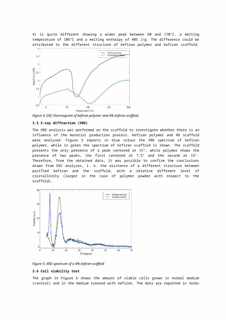
Figure 6: Viability of ECV cells grown with normal medium and kefiran treated medium.
4. ConclusionsKefiran, a high molecular weight polysaccharide obtained from kefir grains, exhibits the ability to form scaffolds with relatively high porosity, as confirmed by SEM. All samples were analysed by DSC and XRD analysis and the dependence of the structure and properties of the scaffolds on the production protocol was evaluated. The observed morphological and biological characteristics of kefiran scaffolds are promising for biomedical applications.
References
Abraham A. G., De Antoni, G. L., 1999, Characterization of Kefir Grains Grow in Cows’ Milk and in Soya Milk. J. Dairy Res. 66, 327–33.
Ahme Z., Wang Y., Anjum N., Ahmad A., Khan S. T., 2013, Characterization of Exopolysaccharide Produced by Lactobacillus Kefiranofaciens ZW3 Isolated from Tibet Kefir - Part II. Food Hydrocoll. 30, 343–50
Amjres H., Béjar V., Quesada E., Carranza D., Abrini J., Sinquin C., Ratiskol J., Colliec-Jouault S., Llamas I., 2015, Characterization of Haloglycan, an Exopolysaccharide Produced by Halomonas Stenophila HK30. Int. J. Biol. Macromol. 72, 117–24.
Carfì Pavia F., La Carrubba V., Brucato V., 2009. Tuning of biodegradation rate of plla scaffolds via blending with PLA. Int. J. Mat. Forming, 2, 713-716.
Cheirsilp, B., Shimizu H., Shioya S., 2001, Modelling and Optimization of Environmental Conditions for Kefiran Production by Lactobacillus Kefiranofaciens. Appl. Microbiol. Biotechnol. 57, 639–46.
Garcia, M., Martino M. N., Zaritzky N. E., 1998, Plasticized Starch-Based Coatings to Improve Strawberry (Fragaria X Ananassa) Quality and Stability. J. Agric. Food Chem. 46, 3758–67.
Ghasemlou, M., Khodaiyan F., Oromiehie A., Yarmand M. S., 2011, Development and Characterisation of a New Biodegradable Edible Film Made from Kefiran, an Exopolysaccharide Obtained from Kefir Grains. Food Chem. 127, 1496–1502.
Ghersi G., Carfì Pavia F., Conoscenti G., Mannella G. A., Greco S., Rigogliuso S., La Carrubba V., Brucato V., 2016, Plla scaffold for bone tissue engineering, Chemical Engineering Transactions, 49, 301-306 DOI: 10.3303/CET1649051
Maeda H., Zhu X., Omura K., Suzuki S., Kitamura S., 2004, Effects of an Exopolysaccharide (Kefiran) on Lipids, Blood Pressure, Blood Glucose, and Constipation. Biofactors 22, 197–200.
Mannella G. A., Carfì Pavia F., Conoscenti G., La Carrubba V., Brucato V., 2014, Evidence of Mechanisms Occurring in Thermally Induced Phase Separation of Polymeric Systems. J. Polym. Sci. B. Polym. Phys. 52, 979-83.
Micheli L., Uccelletti D., Palleschi C., Crescenzi V., 1999, Isolation and Characterisation of a Ropy Lactobacillus Strain Producing the Exopolysaccharide Kefiran. Appl. Microbiol. Biotechnol. 53, 69–74.
Piermaria J. A., de la Canal M. L., Abraham A. G., 2008, Gelling Properties of Kefiran, a Food-Grade Polysaccharide Obtained from Kefir Grain. Food Hydrocoll. 22, 1520–27.
Sorrentino A., Gorrasi G., Vittoria V., 2007, Potential Perspectives of Bio-Nanocomposites for Food Packaging Applications. Trends Food Sci. Technol. 18, 84–95.
Tada S., Katakura Y., Ninomiya K., Shioya S., 2007, Fed-Batch coculture of Lactobacillus kefiranofaciens with Saccharomyces cerevisiae for effective production of kefiran. J Biosci Bioeng. 103, 557–562
van de Witte, P., Dijkstra P. J., van den Berg J. W. A., Feijen, J., 1996, Phase Separation Processes in Polymer Solutions in Relation to Membrane Formation. J. Memb. Sci. 117, 1–31.
Welman A. D., Maddox I. S., 2003, Exopolysaccharides from lactic acid bacteria: Perspectives and challenges. Trends Biotechnol. 21, 269–274.
Yang Z., Li S., Zhang X., Zeng X., Li D., Zhao Y., Zhang J., 2010, Capsular and Slime-Polysaccharide Production by Lactobacillus Rhamnosus JAAS8 Isolated from Chinese Sauerkraut: Potential Application in Fermented Milk Products. J. Biosci. Bioeng. 110, 53–57.