Embed Size (px)
Citation preview
TemplateTreatment/Comparative Therapeutics
Title: Hepatic Encephalopathy
Author: Adam G. Gow, BVM&S PhD DSAM DipECVIM-CA MRCVSContact Details: Head of Small Animal Medicine, Hospital for Small Animals, Royal (Dick) School of Veterinary Studies, The University of Edinburgh, Easter Bush, Edinburgh, EH25 9RG, Scotland. Email: [email protected]
AbstractThis article reviews hepatic encephalopathy (HE) in companion animals. Clinical signs as well as categories of hepatic disease likely to cause HE are discussed. Ammonia has a key role in pathogenesis and current concepts in body ammonia metabolism are reviewed. Inflammation and manganese accumulation are also thought to be important in pathogenesis. Treatment of HE in both acute and chronic cases is discussed along with the rationale for current treatment recommendations. Potential avenues for new treatments and human treatments, which may be transferable to companion animals are reviewed.
Key Points Clinicians should be aware that hepatic encephalopathy signs can be subtle and intermittent Ammonia is not the only factor that drives HE, there is strong evidence for inflammation also
playing a key role. Knowledge of these other factors and precipitants will improve management of these cases.
Dietary protein restriction is no longer the cornerstone of HE management
OutlineIntroductionClinical SignsType of hepatic encephalopathy
Acute Chronic
Factors implicated in pathogenesisAmmonia
Hepatic ammonia detoxificationRenal ammonia metabolismSkeletal muscle ammonia metabolismCNS ammonia metabolism
InflammationAmmonia and innate immune dysfunction
ManganeseAcid/Base and Electrolyte disturbances
Neurological consequences of astrocyte dysfunctionDiagnosisTreatment
Removal of underlying cause
Key Words: portosystemic shunting, ammonia, inflammation, manganese, neurotransmitter

Assessment and treatment of precipitating factorsAcute HE due to hepatic failureChronic HE
Intravenous fluidsEnemasLactulose/LactitolDiet Antibiotics
Future treatment optionsIncreasing ammonia metabolismFecal microbiome modulationReducing pro-inflammatory cytokines/inflammation
Summary
Introduction
Hepatic encephalopathy (HE) is most usefully defined in veterinary medicine as neurological
dysfunction as a result of hepatic disease and/or portosystemic shunting. This definition
allows the diverse consequences of liver disease on the CNS to be encompassed. Broad
categories of hepatic disease resulting in HE have been defined in human medicine and are
applicable to veterinary medicine (Table 1).1 In veterinary medicine, although acute
hepatopathy occurs not infrequently, as a result of the reserve capacity of the liver, acute liver
failure (category A) is a relatively uncommon cause of HE in dogs and cats. Category B is
the most common cause of HE seen in veterinary practice due to the high prevalence of
congenital portosystemic shunts (CPSS). For companion animals it has been suggested that
category C should be widened to encompass all chronic intrinsic liver disease, rather than
purely the end-stage of cirrhosis.2
HE Type
Disease process Subcategory Subdivision Veterinary Modification
A Acute liver failureB Portosystemic bypass with no
intrinsic hepatocellular diseaseC Cirrhosis and portal Episodic Precipitated Intrinsic

hypertension or portosystemic shunts
Persistent
Minimal
Spontaneous Recurrent
MildSevere
hepatocellular disease leading to portal hypertension and portosystemic shunting
Table 1: Category of hepatic disease process resulting in hepatic encephalopathy with suggested veterinary modification.
Clinical Signs
The spectrum of clinical signs varies from subtle behavioral abnormalities through to coma.
Seizures can occur with HE although these would be part of a constellation of clinical signs
rather than in isolation. A grading scale for humans has been modified for veterinary patients
(Table 2; Video 1: Dog exhibiting Grade II HE).3, 4 In human medicine, a more subtle grade
is recognized; minimal HE where individuals appear neurologically normal but cognitive
deficits are revealed during psychometric testing. It has been suggested that the human scale
undergo revision, combining minimal and grade I, partly due to the subjectivity in accurately
defining Grade I.5, 6 These would be combined into “covert HE” and grade II-IV termed
“overt HE.” It is unlikely, beyond research into veterinary HE, that there is a benefit in
detecting minimal HE in our patients. It is likely however that detecting Grade I suffers from
increased subjectivity in veterinary medicine; within the confines of a consultation, decreased
mental alertness is a relative term and co-morbidities e.g. degenerative joint disease in the
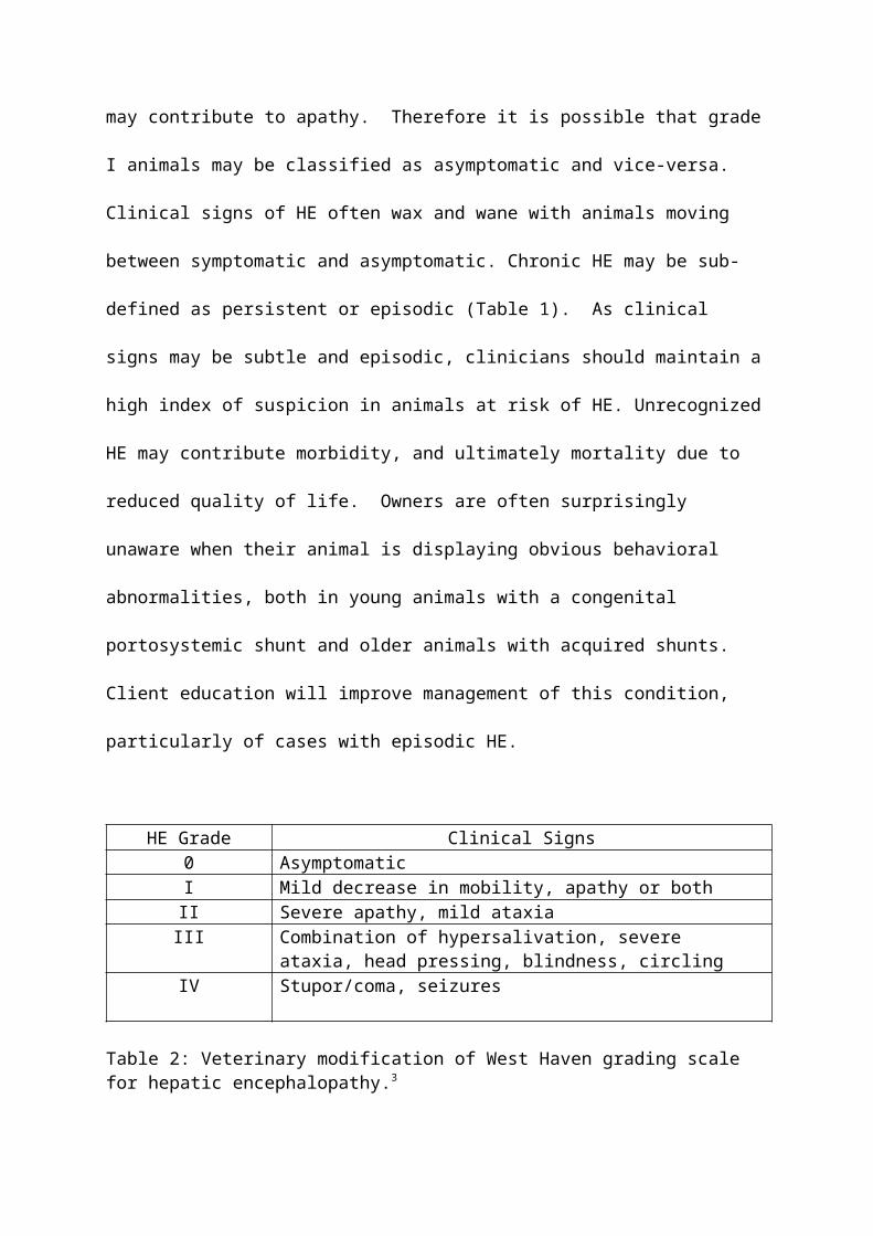
dog with chronic hepatitis, may contribute to apathy. Therefore it is possible that grade I
animals may be classified as asymptomatic and vice-versa. Clinical signs of HE often wax
and wane with animals moving between symptomatic and asymptomatic. Chronic HE may be
sub-defined as persistent or episodic (Table 1). As clinical signs may be subtle and episodic,
clinicians should maintain a high index of suspicion in animals at risk of HE. Unrecognized
HE may contribute morbidity, and ultimately mortality due to reduced quality of life.

Owners are often surprisingly unaware when their animal is displaying obvious behavioral
abnormalities, both in young animals with a congenital portosystemic shunt and older
animals with acquired shunts. Client education will improve management of this condition,
particularly of cases with episodic HE.
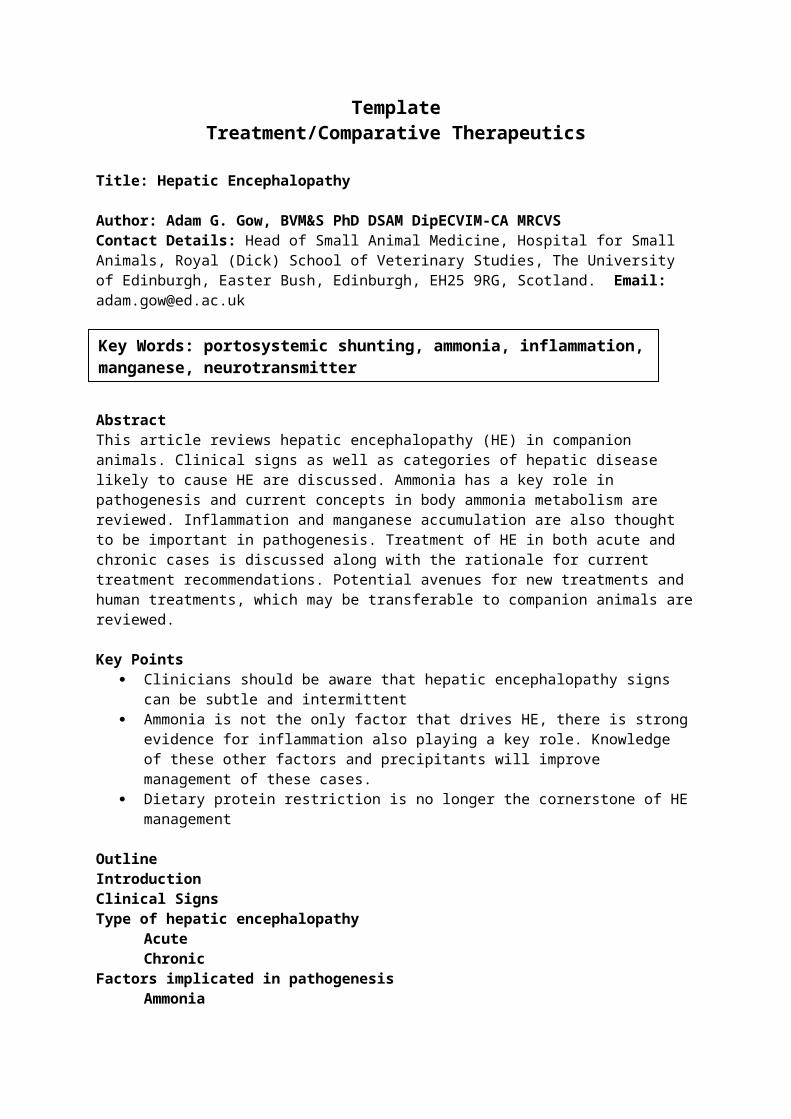
HE Grade Clinical Signs0 AsymptomaticI Mild decrease in mobility, apathy or bothII Severe apathy, mild ataxiaIII Combination of hypersalivation, severe ataxia, head pressing,
blindness, circlingIV Stupor/coma, seizures
Table 2: Veterinary modification of West Haven grading scale for hepatic encephalopathy.3
Type of HE
Acute HE
This is seen in acute liver failure and is part of a constellation of clinical signs, which are
rapid and progressive (see Chapter 4: Acute Liver Injury). Animals often have multiple
metabolic/biochemical derangements such as electrolyte, or acid/base status disorders, which
contribute to their clinical presentation as well as their hyperammonemia. Hepatic
encephalopathy is severe with animals often presenting in stupor or coma (Grade III/IV HE).
These patients require intensive management (see treatment of acute HE) and the prognosis is
grave.7
Chronic HE
This is the most common presentation seen in veterinary medicine, due to congenital or
acquired portosystemic shunting. Although HE has been described in cases of chronic liver
disease where a shunting vessel has not been identified, this is most likely due to a lack of
testing sensitivity for these vessels as the liver has such a large reserve capacity that chronic
intrinsic hepatic failure as a cause of HE in isolation is unlikely.
Factors implicated in the pathogenesis of HE
It can be appreciated that many metabolic abnormalities that can occur as a consequence of
liver disease may have an impact upon the CNS. It has been over 100 years since HE was
described and its pathogenesis investigated in the dog by Nencki and Pavlov using surgically
created porto-systemic shunts.8 Their groundbreaking work demonstrated that the liver
converted ammonia to urea and delivery of ammonia to the systemic circulation and
ultimately the CNS produced neurological abnormalities.
It is unarguable that ammonia has a central role in HE pathogenesis, however it has long been
recognized both in human and veterinary medicine that although in a population of
individuals with chronic HE there is correlation between ammonia and HE grade, ammonia
concentrations are a poor predictor of HE in the individual.9, 10 In human medicine, HE is a
diagnosis of exclusion rather than relying on ammonia measurement.11 This has led to clinical
and experimental investigations to understand what other factors are important in HE
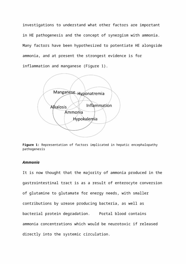
pathogenesis and the concept of synergism with ammonia. Many factors have been
hypothesized to potentiate HE alongside ammonia, and at present the strongest evidence is
for inflammation and manganese (Figure 1).

Figure 1: Representation of factors implicated in hepatic encephalopathy pathogenesis
Ammonia
It is now thought that the majority of ammonia produced in the gastrointestinal tract is as a
result of enterocyte conversion of glutamine to glutamate for energy needs, with smaller
contributions by urease producing bacteria, as well as bacterial protein degradation. Portal
blood contains ammonia concentrations which would be neurotoxic if released directly into
the systemic circulation.
Hepatic ammonia detoxification
In healthy animals the liver is the major point of ammonia detoxification. This is achieved by
two main mechanisms.12 Periportal hepatocytes convert ammonia to urea via the urea cycle.
Urea is substantially less toxic and is excreted by the kidney. Some urea undergoes
enterohepatic recirculation via saliva, bile or direct diffusion into the gastrointestinal tract
where it is degraded by urease bacteria. As blood flows towards the centrilobular vein, there
is switching of ammonia detoxification to conversion of glutamate to glutamine, via the
enzyme glutamine synthetase: consuming ammonia in the process. This switching is gradual
through the liver lobule such that perivenous hepatocytes mainly use the glutamate/glutamine

pathway. Hepatic ammonia detoxification is extremely efficient therefore blood released into
the systemic circulation contains low concentrations of ammonia. There is a large reserve
capacity and even in the face of severe hepatic insufficiency, ammonia detoxification is often
maintained. In portosystemic shunting, dependent on the shunting fraction, variable
proportions of the high ammonia concentrations in the portal vasculature directly reach the
systemic circulation.
Renal ammonia metabolism
The kidney plays a crucial role in the defense of hyperammonemia via urea and ammonia
excretion. The majority of ammonia that is excreted in the urine is generated in the kidney.13
The kidney contains glutaminase with glutamine degradation producing NH4+ and HCO3
-.
This mechanism is important in acid/base homeostasis; generated ammonia can either be
released into the systemic circulation or excreted in urine. Metabolic acidosis increases
urinary ammonia excretion, whereas alkalosis and also hypokalemia increase ammonia
production in the kidney, potentiating the development of HE. There is evidence that renal
production and urinary excretion of ammonia can increase in hyperammonemic conditions.14
Skeletal muscle ammonia metabolism
Skeletal myocytes contain glutamine synthetase and due to the large mass, have significant
ability to remove ammonia by conversion to glutamine. This sequestration of ammonia is
temporary however as glutamine is released into the systemic circulation where glutaminases
regenerate ammonia and glutamate. 15 Nonetheless this process can act as an important buffer
to ammonia challenges.
Central nervous system ammonia metabolism and HE pathogenesis
Astrocytes are the most numerous cell in the brain making up between 25-50% of total brain
volume. The key to HE pathogenesis is astrocyte dysfunction. These contain glutamine
synthetase and act as a buffer to increasing CNS ammonia concentrations.16 As a result of
buffering, glutamate concentrations decrease and glutamine increase. Glutamate is an
excitatory neurotransmitter while glutamine is inhibitory. These neurotransmitters cycle
between astrocyte and neurons (Figure 2). Glutamine is released from the astrocyte, taken up
by neurons and converted to glutamate by the enzyme glutaminase. Glutamate is released as a
neurotransmitter at the synaptic cleft. Astrocytes rapidly re-uptake released glutamate. High
ammonia concentrations increase extracellular glutamine inhibiting further release from the
astrocyte causing increased intracellular concentrations. Ammonia also inhibits key enzymes
in the TCA cycle, reducing the astrocytes ability for aerobic respiration; causing a switch to
anaerobic respiration and lactate production. Astrocytic mitochondria split glutamine,
releasing ammonia, causing oxidative stress and mitochondrial dysfunction.17 These
metabolic effects result in astrocyte swelling. As astrocytes account for a significant
proportion of brain volume, in acute HE this results in edema and cerebral hypertension.
Disturbances in control of cerebral blood flow also contribute to this.18 This can rapidly lead
to herniation, brainstem compression and death.19
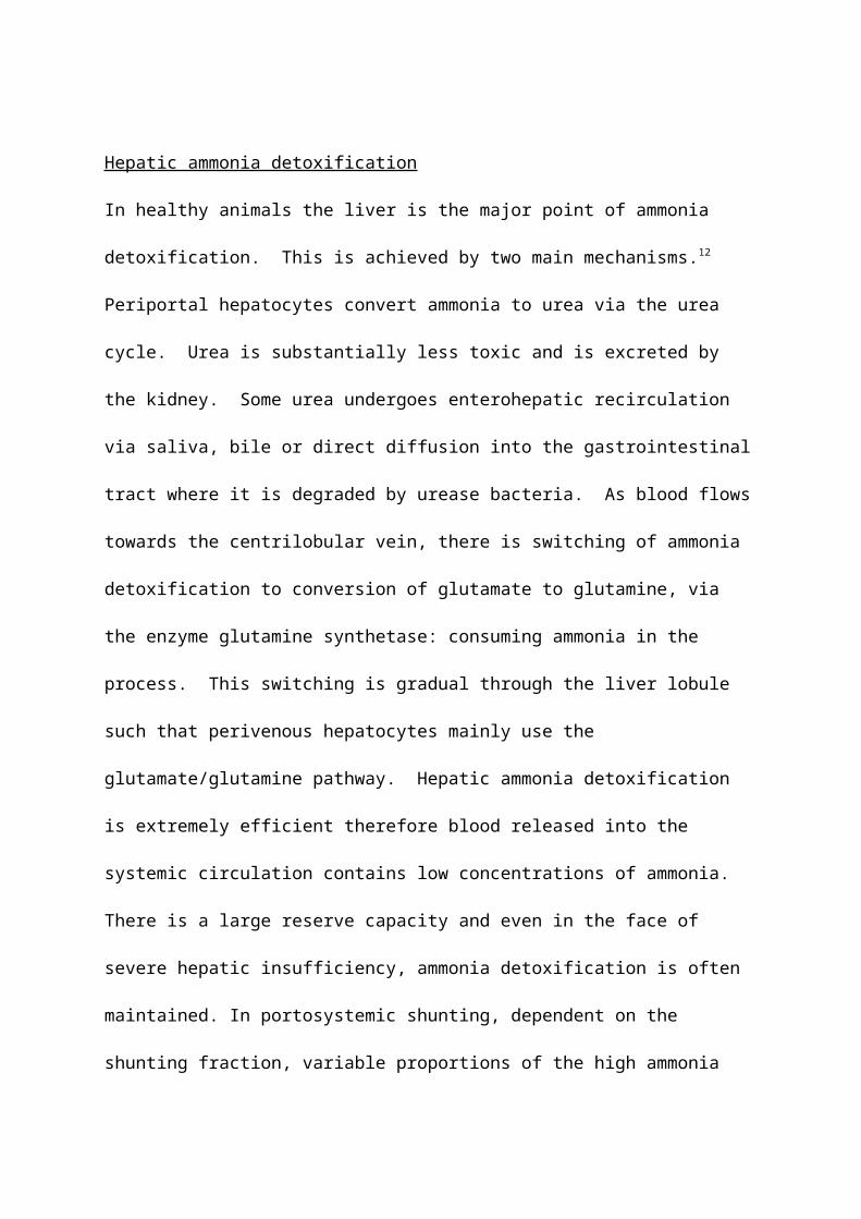
Figure 2: Representation of ammonia metabolism in astrocyte and neuron. In the face of hyperammonemia, glutamine increases intracellularly, leading to oxidative damage to mitochondria and ammonia inhibiting the TCA cysle. This disrupts cell metabolism, leading to cell swelling. GLN. Glutamine; GLU, glutamate; GSynthetase, glutamine synthetase; ROS, reactive oxygen species.
In chronic HE, astrocyte swelling is less dramatic but still present, giving rise to Alzheimer
type II astrocytes. It is these cases, more commonly seen in veterinary medicine, where
correlation with ammonia concentrations is less pronounced and other factors become more
significant.
Inflammation
There is now compelling evidence in human medicine that inflammation is an important
potentiator of HE.20 In support of this, rats with experimentally induced HE have been shown
to have restored learning ability after administration of NSAID’s.21 In dogs with CPSS, C-
reactive protein has been shown to be increased in those exhibiting signs of HE and that both
ammonia and systemic inflammatory response syndrome (SIRS) predicts the presence of
HE.10, 22 Any process causing a systemic inflammatory response has the potential to
precipitate HE. Congential portosystemic shunt dogs have higher endotoxin in both their

portal and peripheral circulations, suggesting that the gastrointestinal tract and microbiome
may be an important driver and in support of this, shunt attenuation causes a reduction in
inflammatory markers.23, 24
Pro-inflammatory cytokines increase cerebral blood flow as well as compromising the blood
brain barrier; increasing permeability to ammonia. The blood-brain barrier (BBB) prevents
direct transfer of systemic cytokines into the CNS. However, cytokines can affect the CNS,
possibly through afferent nerves, active transport across the BBB, or via areas lacking the
BBB, such as the circumventricular organs.25 Both inflammatory mediators and ammonia
cause microglial activation and neuro-inflammation.20 In humans, it has been demonstrated
that inflammation potentiates ammonia in precipitating HE and that systemic inflammation
and not ammonia is associated with severe encephalopathy.9, 26
Ammonia and innate immune dysfunction
In vitro and in vivo studies have shown that neutrophil phagocytic ability is reduced and
spontaneous oxidative burst activity increases on exposure to ammonia.27 Reduced
phagocytic ability in the face of hyperammonemia also occurs in canine neutrophils in vitro
[Gow, unpublished data]. This has important implications in that infection may be more
likely in the face of high ammonia concentrations and that increased spontaneous oxidative
burst activity, potentiating the inflammatory response syndrome.
Manganese (Mn)
Manganese is an essential trace element with the potential to be neurotoxic, causing
psychiatric disturbances and cognitive defects. The majority (98%) of Mn ingested and
absorbed is efficiently removed by the liver and excreted in bile.28 Portosystemic shunting
allows high Mn concentration blood in the splanchnic circulation to reach the systemic

circulation. Astrocytes have a high-affinity Mn transport mechanism and concentrations can
be 50 times higher than in surrounding cells.29 This causes oxidative damage and
mitochondrial dysfunction; Mn has been shown to produce Alzheimer’s type II astrocytosis,
identical to that of hyperammonemia.30 In addition, Mn stimulates microglia to release
inflammatory cytokines and reactive oxygen species.31 A direct relationship has been
demonstrated between blood Mn, MRI hyperintensity consistent with Mn deposition, and HE
severity in humans.32, 33 Dogs with CPSS and chronic liver disease have higher blood Mn
compared to controls.34, 35 MRI of dogs and cats with CPSS detected CNS lesions consistent
with Mn deposition and there is one report of a CPSS dog, which had increased CNS Mn
concentrations on post-mortem examination. 36, 37
Acid/base and electrolyte disturbances
Hyponatremia, hypokalemia and alkalosis are all recognized as potentiating HE in humans.38
Hyponatremia potentiates cerebral edema, hypokalemia increases renal ammoniagenesis, and
reduced renal excretion of ammonia. Although it is likely that these would also potentiate
HE in our patients, the evidence for this is lacking studies to date finding no association.10, 39
This may reflect the low prevalence of these abnormalities in our patients, partly due to the
differing main etiology of HE (CPSS versus chronic hepatitis) and severity of the disease
process; in cases of chronic hepatitis, our patients may be euthanized before these
abnormalities occur.40
Neurological consequence of astrocyte dysfunction
In acute HE the major pathological consequence is cerebral edema and raised intra-cranial
pressure due to astrocyte swelling. In chronic HE the major consequence of all these factors
is neurotransmitter dysregulation. In addition to glutamate/glutamine imbalance, increased

“GABAergic tone” has been long been recognized in cases of HE. GABA is the main
inhibitory neurotransmitter, reducing excitability. Initially this increase in tone was
hypothesized to be due to increased GABA synthesis, however canine studies in dogs with
experimentally induced biliary cirrhosis and dogs with experimentally created portocaval
shunts found no increase in CNS GABA or GABA receptors.41 This led to a search for
alternative ligands. It is now known that GABA receptors can be activated by a group of
compounds known as “neurosteroids” which include allopregnanolone (ALLO) and
tetrahydrodeoxycorticosterone (THDOC). These compounds have been shown to be
dramatically increased in human patients with HE.42 Neurosteroids are synthesized in
response to activation of a translocator protein (TSPO) in astrocyte mitochondria (formerly
known peripheral-type benzodiazepine receptors). These receptors are increased in humans
with cirrhosis. In-vitro studies have demonstrated that astrocyte TSPO is highly upregulated
by both ammonia and also manganese.43, 44
Diagnosis of HE
As stated above ammonia concentrations are an unreliable marker of HE. A high index of
suspicion should be maintained in animals with hepatic disease or those that may have a
portosystemic shunt. A high ammonia concentration makes HE likely, however normal
values do not exclude the diagnosis. Careful sampling handling for ammonia measurement is
important to avoid in vitro increases.45 Routine clinical pathology allows exclusion of other
metabolic causes of neurological disease as well as providing support for hepatic
disease/insufficiency. Imaging provides support for acquired liver disease as well as
investigating for portosystemic shunting.
Treatment of HE

Removal of the underlying cause
In animals with a CPSS, vessel attenuation often allows a rapid increase in hepatic mass and
function.46 Medical management prior to shunt attenuation is recommended. Also some
cases with CPSS may not be candidates for attenuation due to the client’s financial resources,
patient co-morbidities, or shunt anatomy. Where possible the inciting cause of acute hepatic
failure should be addressed; for example, patients with acetaminophen toxicity should
undergo gastrointestinal decontamination and replenishment of glutathione, which may help
ameliorate ongoing hepatotoxicity.47
Assessment and treatment of precipitating factors
Many factors have been implicated in precipitating HE, as described above. Cases should be
examined for an inflammatory focus/sepsis and their electrolyte and acid/base status should
also be assessed. There are many factors implicated in increasing blood ammonia
concentrations including: dehydration, high protein meals, gastrointestinal hemorrhage due to
hemoglobin digestion (also an inflammatory focus), uremia and constipation. If any are
identified then these should be addressed.
Acute HE due to hepatic failure
These cases usually have multiple severe co-morbidities, most notably coagulopathy and
organ dysfunction, all of which require intensive management (See Chapter 4: Acute Liver
Injury).7 Cases are often stuporous/comatose, requiring intubation to protect the airway and in
some cases mechanical ventilation. Hypoglycemia is common and cases should be monitored
for this regularly during hospitalization. Of great concern is cerebral edema as a result of
hyperammonemia leading to increased intracranial pressure. To minimize this risk, the
patient’s head should be raised to a 30o angle and jugular venous drainage should not be

compromised (i.e. no jugular blood sampling, removal of any collars). Intravenous fluids
should be used to maintain a normovolemic state. If cerebral hypertension is suspected, then
intravenous mannitol is recommended at a dose of 0.5-1g/kg IV given over 20 minutes. This
can then be repeated after 4 hours but due to the osmotic diuretic effect, serum/plasma
electrolytes should be monitored. Assisted ventilation to maintain a PaCO2 between 30-40
mmHg is recommended. Overly aggressive reductions in pCO2 will reduce cerebral perfusion
and oxygenation and therefore must be prevented. Ammonia-lowering measures outlined
below should be instituted. In human medicine, these interventions alongside therapeutic
hypothermia, temporary hepatectomy (removing a source of necrosis and inflammation) and
ultimately liver transplantation are recommended management strategies.48 In veterinary
medicine, these advanced options are unavailable and as a result of limited treatment options,
the prognosis for dogs with fulminant hepatic failure is grave.7
Chronic HE
Any animal with an exacerbation of clinical signs should be thoroughly investigated for
precipitating factors (Figure 3). Animals with signs of sepsis require intravenous empiric
antibiotic therapy whilst awaiting culture and sensitivity results. Infection or inflammatory
foci should be managed aggressively. Concurrent medications should be reviewed for the
potential to cause dehydration, electrolyte disturbances or hepatotoxicity. Hemorrhage from
the upper gastrointestinal tract is often managed with omeprazole and sucralfate alongside
other supportive measures. The level of intervention depends on the severity of the clinical
signs, grade III/IV animals may require intubation as above to protect against aspiration
(Figure 4).
Figure 3: Suggested approach to patient with signs of hepatic encephalopathy

Intravenous fluids
Many animals with HE are dehydrated and/or hypovolemic due to obtundation reducing
water intake and precipitating causes e.g. diarrhea/ GI hemorrhage. Restoring euvolemia
reduces ammonia concentrations by dilution and improves urinary excretion of ammonia and
urea, as well as preventing ammoniagenesis via urease producing bacteria in the colon.
Diuresis will further improve renal excretion. Some clinicians avoid lactate-containing fluids
in animals with hepatic failure as this additive requires hepatic metabolism.
Enemas
These physically remove colonic contents and therefore a source of nitrogen from urease
producing bacteria. Physical removal of hemoglobin from GI bleeding prevents bacterial
degradation of hemoglobin and ammoniagenesis. Cleansing enemas with warm water or
isotonic fluids are used until clear fluid is evacuated. Retention enemas are then used;
options include warm water, neomycin, povidone iodine, or non-absorbable disaccharides
(lactulose and lactitol). There are no studies assessing the efficacy of retention enemas in
veterinary patients, one human study found that lactulose and lactitol containing enemas were
more effective than warm water.49 The author uses 1 part lactulose to 3 parts warm water
given via a Foley catheter at 10 mL/kg, retained for 30 minutes – 1 hour.
Lactulose/Lactitol
These are non-absorbable disaccharides, which are digested by colonic bacteria to form
volatile fatty acids including, acetic acid, lactic acid, and butyric acid. Acidification of the
colon favors formation of ammonium ions (NH4+) which are less able to move through cell
membranes, thus providing a colonic ammonia “trap.” Lactulose solution in itself is acidic

and this may be the reason that lactulose enemas (see above) are effective in improving
clinical signs.50 These volatile fatty acids also act as osmotic laxatives, removing trapped
ammonia from the body as well as nitrogen. Longer term, these products act as probiotics,
favoring non-urease bacteria reducing ammonia production from the large intestine. The
dose is titrated to produce 2-3 soft stools per day, starting at a dose of 0.5 mL/kg PO twice
daily. Lactitol is a powder and, where available may be used in animals which do not accept
lactulose. Overdose of these agents causes osmotic diarrhea potentially producing
hypernatremia as well as reduced renal excretion of ammonia and urea.
Diet
Although feeding a low protein diet and thus reducing the potential for ammonia production
would seem intuitive, there are potentially deleterious effects to this approach. Failure to
feed adequate protein causes tissue catabolism with a reduction in muscle mass. As well
leading to the release of ammonia, this reduces the animal’s ability to buffer via glutamine
synthetase in skeletal muscle. In addition, with acquired shunting due to portal hypertension,
any reduction in serum albumin decreases oncotic pressure, potentiating ascites. As the bulk
of ammoniagenesis is thought to be due to enterocyte metabolism converting glutamine to
glutamate, it is unclear how significant dietary protein is in driving ammonia production.
Protein restriction is no longer recommended in humans with HE and dogs with CPSS have
identical dietary protein requirements to control animals.51, 52 A highly digestible, high
biological value protein source is recommended but ideally protein content of the diet should
be appropriate for the age and growth phase of the animal. The exceptions to this are during
the initial stabilization period to control clinical signs or if despite other methods of
management, clinical signs recur on a standard protein ration. An approximate level of
protein restriction of 4 g protein/100 kcal/day would be the starting point upon which protein

can be titrated upwards in the dog. Veterinary commercial canine hepatic diets vary but are
generally around this level. Commercial hepatic diets have much to recommend them;
restricted in copper to prevent secondary copper accumulation in chronic hepatitis,
supplemented with zinc; an antioxidant and required for optimal urea cycle function as well
as containing protein of high digestibility and biological value. However commercial diets
are often overly protein restricted for long-term management, especially for young animals.
Supplementation with another high quality protein source such as cottage cheese or tofu is
required. There is evidence that vegetable protein sources may improve control compared to
meat-based diets.3, 53 As ammoniogenesis in the gastrointestinal tract will occur during
digestion, feeding small frequent meals is recommended. Monitoring weight, muscle
condition score and serum albumin is recommended to ensure that calorie and protein
requirements are being met for animals on long-term medical management. When in doubt
clinicians are advised to consult with a board-certified veterinary nutritionist.
Antibiotics
Antibiotics are used to reduce the numbers of urease-producing bacteria in the
gastrointestinal tract. By this rationale, non-absorbable antibiotics would be a sensible
choice. Neomycin was previously recommended, however some systemic absorption can
occur producing renal and ototoxicity.54 In human medicine, rifaximin-α, a semi-synthetic
non-absorbable antibiotic which is effective at treating and preventing HE, is widely used.55
Pharmacokinetic data is available in the dog which confirms, at least in healthy animals, no
systemic uptake.56 The major hurdle to this drugs use in veterinary medicine is at present the
cost.

As a result of the above problems, systemic antibiotics are often used including:
metronidazole, ampicillin, and potentiated amoxicillin. Metronidazole undergoes hepatic
metabolism therefore a dose reduction to 7.5mg/kg every 8-12 hours is recommended. In
view of the increasing concern regarding antibiotic resistance, it is prudent that antibiotics are
used short-term or when animals are experiencing an exacerbation. Despite this, there are
cases which appear to be clinically improved whilst on antibiotic treatment.
Future treatment options
Increasing ammonia metabolism
L-ornithine L-aspartate (LOLA) is a mixture of two amino acids which are substrates for the
urea cycle and also stimulates glutamine production in the liver and skeletal muscle. It is
delivered by intravenous infusion and effective in humans with overt HE, however the effects
are short-lived in that on discontinuing the infusion, ammonia concentrations rebound.57
There is one case series reporting its use in dogs, although no statement can be made as to its
efficacy.58 Like LOLA, ornithine phenylacetate (OP) increases glutamine synthetase activity
however glutamine is then bound to phenylacetate, and excreted by the kidney preventing
rebound ammonia. This compound is currently undergoing clinical trials in humans.57 An
alternative compound with an identical end-product is glycerol phenylbutyrate, which also
appears effective in clinical trials.59 There is no information on their use in companion
animals.
Fecal microbiome modulation
The potential benefits of this are: reducing urease-producing bacteria, and producing a less
pro-inflammatory microbiome thus reducing endotoxin and inflammatory cytokine release.
Current management with lactulose and vegetable protein may act in this manner. In humans

with cirrhosis, intestinal dysbiosis has been demonstrated in humans with HE.60 There is
interest in other prebiotics and probiotics as well as fecal microbiota transplant to control HE
in human medicine.57, 61 It is unknown at present if dysbiosis occurs in companion animals
with HE.
Reducing pro-inflammatory cytokines/ inflammation
As one source of inflammation is the gut microbiome, modulation of this may improve
control. An engineered carbon adsorbent (AST-120) given orally is thought to adsorb
ammonia, endotoxin and TNFα, although an initial study did not demonstrate an
improvement in humans with covert HE.62 Other options are minocycline which reduces
activation of the microglia, as well as etanercept and infliximab, which neutralise TNFα, all
of which are under investigation to manage HE in human medicine.31
Summary
At present, all long-term management strategies to control HE in companion animals are
directed towards lowering ammonia concentrations. As a result of intensive research, it is
clear that ammonia is not the only factor and that previously held beliefs as to the source of
ammonia, and also the mechanisms of action of the current interventions have changed. Due
to the fact that liver disease and as a consequence, HE is increasing in the human population,
research into therapeutics is continuing apace. From veterinary research carried out to date, it
appears that the pathogenesis of HE in dogs is similar to that in humans and it is hoped that
with the necessary validation, advances in human HE management will be transferable to
veterinary medicine.
References

1. Ferenci P, Lockwood A, Mullen K, et al. Hepatic encephalopathy--definition, nomenclature, diagnosis, and quantification: final report of the working party at the 11th World Congresses of Gastroenterology, Vienna, 1998. Hepatology 2002;35(3):716-721.
2. Lidbury JA, Cook AK, Steiner JM. Hepatic encephalopathy in dogs and cats. J Vet Emerg Crit Care (San Antonio) 2016;26(4):471-87.
3. Proot S, Biourge V, Teske E, et al. Soy protein isolate versus meat-based low-protein diet for dogs with congenital portosystemic shunts. J Vet Intern Med 2009;23(4):794-800.
4. Rothuizen J. Important clinical syndromes associated with liver disease. The Veterinary clinics of North America Small animal practice 2009;39(3):419-437.
5. Schomerus H, Hamster W. Quality of life in cirrhotics with minimal hepatic encephalopathy. Metab Brain Dis 2001;16(1-2):37-41.
6. Bajaj JS, Cordoba J, Mullen KD, et al. Review article: the design of clinical trials in hepatic encephalopathy--an International Society for Hepatic Encephalopathy and Nitrogen Metabolism (ISHEN) consensus statement. Aliment Pharmacol Ther 2011;33(7):739-747.
7. Lester C, Cooper J, Peters RM, et al. Retrospective evaluation of acute liver failure in dogs (1995-2012): 49 cases. J Vet Emerg Crit Care (San Antonio) 2016; 26(4):559-67.
8. Nencki M, Pawlow JP, Zaleski J. Ueber den Ammoniakgehalt des Blutes und der Organe und die Harnstoffbildung bei den Säugethieren. Archiv für experimentelle Pathologie und Pharmakologie 1895;37(1):26-51.
9. Shawcross DL, Sharifi Y, Canavan JB, et al. Infection and systemic inflammation, not ammonia, are associated with Grade 3/4 hepatic encephalopathy, but not mortality in cirrhosis. Journal of hepatology 2011;54(4):640-649.
10. Tivers MS, Handel I, Gow AG, et al. Hyperammonemia and systemic inflammatory response syndrome predicts presence of hepatic encephalopathy in dogs with congenital portosystemic shunts. PLoS One 2014;9(1):e82303.
11. Jawaro T, Yang A, Dixit D, et al. Management of Hepatic Encephalopathy: A Primer. Ann Pharmacother 2016;50(7):569-577.
12. Haussinger D, Lamers WH, Moorman AF. Hepatocyte heterogeneity in the metabolism of amino acids and ammonia. Enzyme 1992;46(1-3):72-93.
13. Weiner ID, Verlander JW. Renal ammonia metabolism and transport. Comprehensive Physiology 2013;3(1):201-220.
14. Dejong CHC, Deutz NEP, Soeters PB. Metabolic adaptation of the kidney to hyperammonemia during chronic liver insufficiency in the rat. Hepatology 1993;18(4):890-902.
15. Olde Damink SW, Jalan R, Redhead DN, et al. Interorgan ammonia and amino acid metabolism in metabolically stable patients with cirrhosis and a TIPSS. Hepatology 2002;36(5):1163-1171.
16. Belanger M, Magistretti PJ. The role of astroglia in neuroprotection. Dialogues Clin Neurosci 2009;11(3):281-295.
17. Scott TR, Kronsten VT, Hughes RD, et al. Pathophysiology of cerebral oedema in acute liver failure. World J Gastroenterol 2013;19(48):9240-9255.
18. Jalan R, Olde Damink SW, Deutz NE, et al. Restoration of cerebral blood flow autoregulation and reactivity to carbon dioxide in acute liver failure by moderate hypothermia. Hepatology 2001;34(1):50-54.
19. Scott TR, Kronsten VT, Hughes RD, et al. Pathophysiology of cerebral oedema in acute liver failure. World Journal of Gastroenterology : WJG 2013;19(48):9240-9255.

20. Aldridge DR, Tranah EJ, Shawcross DL. Pathogenesis of Hepatic Encephalopathy: Role of Ammonia and Systemic Inflammation. Journal of Clinical and Experimental Hepatology 2015;5, Supplement 1:S7-S20.
21. Cauli O, Rodrigo R, Piedrafita B, et al. Inflammation and hepatic encephalopathy: ibuprofen restores learning ability in rats with portacaval shunts. Hepatology 2007;46(2):514-519.
22. Gow AG, Marques AI, Yool DA, et al. Dogs with congenital porto-systemic shunting (cPSS) and hepatic encephalopathy have higher serum concentrations of C-reactive protein than asymptomatic dogs with cPSS. Metab Brain Dis 2012;27(2):227-229.
23. Tivers MS, Lipscomb VJ, Smith KC, et al. Lipopolysaccharide and toll-like receptor 4 in dogs with congenital portosystemic shunts. Vet J 2015;206(3):404-413.
24. Tivers MS, Handel I, Gow AG, et al. Attenuation of congenital portosystemic shunt reduces inflammation in dogs. PLoS One 2015;10(2):e0117557.
25. Licinio J, Wong ML. Pathways and mechanisms for cytokine signaling of the central nervous system. The Journal of clinical investigation 1997;100(12):2941-2947.
26. Shawcross DL, Davies NA, Williams R, et al. Systemic inflammatory response exacerbates the neuropsychological effects of induced hyperammonemia in cirrhosis. Journal of hepatology 2004;40(2):247-254.
27. Shawcross DL, Wright GA, Stadlbauer V, et al. Ammonia impairs neutrophil phagocytic function in liver disease. Hepatology 2008;48(4):1202-1212.
28. Davis CD, Zech L, Greger JL. Manganese metabolism in rats: an improved methodology for assessing gut endogenous losses. Proceedings of the Society for Experimental Biology and Medicine Society for Experimental Biology and Medicine 1993;202(1):103-108.
29. Aschner M, Gannon M, Kimelberg HK. Manganese uptake and efflux in cultured rat astrocytes. J Neurochem 1992;58(2):730-735.
30. Hazell AS, Normandin L, Norenberg MD, et al. Alzheimer type II astrocytic changes following sub-acute exposure to manganese and its prevention by antioxidant treatment. Neurosci Lett 2006;396(3):167-171.
31. Butterworth RF. The liver-brain axis in liver failure: neuroinflammation and encephalopathy. Nat Rev Gastroenterol Hepatol 2013;10(9):522-528.
32. Spahr L, Butterworth RF, Fontaine S, et al. Increased blood manganese in cirrhotic patients: relationship to pallidal magnetic resonance signal hyperintensity and neurological symptoms. Hepatology 1996;24(5):1116-1120.
33. Layrargues GP, Rose C, Spahr L, et al. Role of manganese in the pathogenesis of portal-systemic encephalopathy. Metab Brain Dis 1998;13(4):311-317.
34. Gow AG, Marques AI, Yool DA, et al. Whole blood manganese concentrations in dogs with congenital portosystemic shunts. J Vet Intern Med 2010;24(1):90-96.
35. Kilpatrick S, Jacinto A, Foale RD, et al. Whole blood manganese concentrations in dogs with primary hepatitis. J Small Anim Pract 2014;55(5):241-246.
36. Torisu S, Washizu M, Hasegawa D, et al. Measurement of brain trace elements in a dog with a portosystemic shunt: relation between hyperintensity on T1-weighted magnetic resonance images in lentiform nuclei and brain trace elements. The Journal of veterinary medical science / the Japanese Society of Veterinary Science 2008;70(12):1391-1393.
37. Torisu S, Washizu M, Hasegawa D, et al. Brain magnetic resonance imaging characteristics in dogs and cats with congenital portosystemic shunts. Vet Radiol Ultrasound 2005;46(6):447-451.
38. Frederick RT. Current concepts in the pathophysiology and management of hepatic encephalopathy. Gastroenterol Hepatol (N Y) 2011;7(4):222-233.

39. Lidbury JA, Ivanek R, Suchodolski JS, et al. Putative precipitating factors for hepatic encephalopathy in dogs: 118 cases (1991-2014). J Am Vet Med Assoc 2015;247(2):176-183.
40. Kilpatrick S, Dreistadt M, Frowde P, et al. Presence of Systemic Inflammatory Response Syndrome Predicts a Poor Clinical Outcome in Dogs with a Primary Hepatitis. PLoS One 2016;11(1):e0146560.
41. Roy S, Pomier-Layrargues G, Butterworth RF, et al. Hepatic encephalopathy in cirrhotic and portacaval shunted dogs: lack of changes in brain GABA uptake, brain GABA levels, brain glutamic acid decarboxylase activity and brain postsynaptic GABA receptors. Hepatology 1988;8(4):845-849.
42. Butterworth RF. Neurosteroids in hepatic encephalopathy: Novel insights and new therapeutic opportunities. J Steroid Biochem Mol Biol 2016;160:94-97.
43. Hazell AS, Desjardins P, Butterworth RF. Chronic exposure of rat primary astrocyte cultures to manganese results in increased binding sites for the 'peripheral-type' benzodiazepine receptor ligand 3H-PK 11195. Neurosci Lett 1999;271(1):5-8.
44. Itzhak Y, Norenberg MD. Ammonia-induced upregulation of peripheral-type benzodiazepine receptors in cultured astrocytes labeled with [3H]PK 11195. Neurosci Lett 1994;177(1-2):35-38.
45. Willard MD, Tvedten H. Small animal clinical diagnosis by laboratory methods. 5th ed. St. Louis, Mo.: Elsevier; 2012.
46. Kummeling A, Vrakking DJ, Rothuizen J, et al. Hepatic volume measurements in dogs with extrahepatic congenital portosystemic shunts before and after surgical attenuation. J Vet Intern Med 2010;24(1):114-119.
47. Bates N, Rawson-Harris P, Edwards N. Common questions in veterinary toxicology. J Small Anim Pract 2015;56(5):298-306.
48. Jalan R. Intracranial hypertension in acute liver failure: pathophysiological basis of rational management. Semin Liver Dis 2003;23(3):271-282.
49. Uribe M, Campollo O, Vargas F, et al. Acidifying enemas (lactitol and lactose) vs. nonacidifying enemas (tap water) to treat acute portal-systemic encephalopathy: a double-blind, randomized clinical trial. Hepatology 1987;7(4):639-643.
50. Elkington SG. Lactulose. Gut 1970;11(12):1043-1048.51. Abdelsayed GG. Diets in Encephalopathy. Clin Liver Dis 2015;19(3):497-505.52. Laflamme DP, Allen SW, Huber TL. Apparent dietary protein requirement of dogs
with portosystemic shunt. American journal of veterinary research 1993;54(5):719-723.
53. Amodio P, Bemeur C, Butterworth R, et al. The nutritional management of hepatic encephalopathy in patients with cirrhosis: International Society for Hepatic Encephalopathy and Nitrogen Metabolism Consensus. Hepatology 2013;58(1):325-336.
54. Greenberg LH, Momary H. AUdiotoxicity and nephrotoxicity due to orally administered neomycin. JAMA : the journal of the American Medical Association 1965;194(7):827-828.
55. Kimer N, Krag A, Moller S, et al. Systematic review with meta-analysis: the effects of rifaximin in hepatic encephalopathy. Aliment Pharmacol Ther 2014;40(2):123-132.
56. Venturini AP. Pharmacokinetics of L/105, a new rifamycin, in rats and dogs, after oral administration. Chemotherapy 1983;29(1):1-3.
57. Hadjihambi A, Jalan R. Hepatic encephalopathy: New treatments. Clinical Liver Disease 2015;5(5):109-111.

58. Ahn JO, Li Q, Lee YH, et al. Hyperammonemic hepatic encephalopathy management through L-ornithin-L-aspartate administration in dogs. Journal of veterinary science 2015.
59. Rockey DC, Vierling JM, Mantry P, et al. Randomized, double-blind, controlled study of glycerol phenylbutyrate in hepatic encephalopathy. Hepatology 2014;59(3):1073-1083.
60. Rai R, Saraswat VA, Dhiman RK. Gut microbiota: its role in hepatic encephalopathy. J Clin Exp Hepatol 2015;5(Suppl 1):S29-36.
61. Kao D, Roach B, Park H, et al. Fecal microbiota transplantation in the management of hepatic encephalopathy. Hepatology 2016;63(1):339-340.
62. Bajaj JS, Sheikh MY, Chojkier M, et al. Su1685 AST-120 (Spherical Carbon Adsorbent) in Covert Hepatic Encephalopathy: Results of the Astute Trial. Gastroenterology;144(5):S-997.
Figure Legends
Figure 1: Representation of factors implicated in hepatic encephalopathy pathogenesis.
Figure 2: Representation of ammonia metabolism in astrocyte and neuron. GLU: glutamate, GLN: glutamine, GSynthetase: glutamine synthetase, ROS: reactive oxygen species. In the face of hyperammonemia, glutamine increases intracellularly, leading to oxidative damage to the mitochondria as well as ammonia inhibiting the TCA cycle. This disrupts cell metabolism, leading to cell swelling.
Figure 3: Suggested approach to patient with signs of hepatic encephalopathy
Figure 4: Samoyed with a congenital portosystemic shunt exhibiting grade III hepatic encephalopathy due to gastrointestinal hemorrhage. This patient could be roused with stimuli.
Video 1: Schnauzer with a congenital portosystemic shunt exhibiting grade III hepatic encephalopathy: circling, poor vision, head pressing. This exacerbation of clinical signs coincided with developing a urinary tract infection. Ammonia concentrations were only modestly increased. Control of the infectious process resulted in resolution of the hepatic encephalopathy.