Embed Size (px)
Citation preview
© 2007 OEPP/EPPO,
Bulletin OEPP/EPPO Bulletin
37
, 543–553
543
Blackwell Publishing Ltd
European and Mediterranean Plant Protection Organization PM 7/80 (1)Organisation Européenne et Méditerranéenne pour la Protection des Plantes
DiagnosticsDiagnostic
Xanthomonas oryzae
Specific scope
This standard describes a diagnostic protocol for
Xanthomonasoryzae
pathovars
oryzae
and
oryzicola.
Specific approval and amendment
Approved in 2007-09.
Introduction
The species
Xanthomonas oryzae
includes two pathovars,namely,
oryzae
and
oryzicola
(Swings
et al.
, 1990). Thesebacterial pathogens are closely related organisms and wereearlier named as pathovars of
Xanthomonas campestris
. Rice isthe main host for both pathogens, which are seed-borne andseed-transmitted.
Bacterial leaf blight of rice (BLB)
was first reported inFukuoka Prefecture, Japan, during 1884 in rice affected by
X.oryzae
pv.
oryzae
. This disease is considered one of the mostserious rice diseases worldwide although it has declined inincidence in Japan since the mid 1970s. Nevertheless it is stillprevalent worldwide. The disease was reported in South EastAsia in the early 1960s, where it is currently widespread, and itstill affects the rice crop in its severe form (Goto, 1992). It hasalso been reported in several African countries, in Australia,North America (Lousiana and Texas, US), Central and SouthAmerica (OEPP/EPPO, 2006a) but it is only of economicimportance in Asia. Other reported hosts include wild or minorcultivated Poaceae:
Brachiaria mutica, Cenchrus ciliaris,Cyperus difformis, C. rotundus, Cynodon dactylon, Echinochloacrus-galli, Leersia
spp. (
Leersia hexandra, L. oryzoides),Leptochloa chinensis, Oryza
spp.,
Panicum maximum, Paspalumscrobiculatum, Zizania aquatica, Z. latifolia, Z. palustris,
and
Zoysia japonica
(Li
et al
., 1985; Bradbury, 1986; Valluva-paridasan & Mariappan, 1989; EPPO/CABI, 1997; Saddler,2002a). Cyperaceae (sedges) that are naturally infected include
Cyperus difformis
and
C. rotundus.
Variation in virulencehas been observed between isolates, and many races have beendescribed; however, the different races have not been clearlydefined with specific reactions being assigned to each variety(Mew, 1987). The diversity of the pathogen has been analysedbased on virulence and PCR-based DNA fingerprinting(George
et al.
, 1994). Low virulence strains have been reported
in the United States and India (Jones
et al
., 1989; Gnanamanickam
et al
., 1993).
Bacterial leaf streak of rice (BLS)
is caused by
X. oryzae
pv
.oryzicola
. The disease is present in Tropical Asia, West Africaand Australia (OEPP/EPPO, 2006b).
X. oryzae
pv.
oryzicola
has reached epidemic proportions in recent years in China.Other hosts affected by the pathogen are:
Oryza
spp.,
Leersia
spp.,
Leptochloa filiformis
,
Paspalum orbiculare
,
Zizaniaaquatica, Z. palustris
and
Zoysia japonica
. No races have beenrecorded, however, differences in virulence of strains have beenobserved in many countries (Vera Cruz
et al.,
1984; Adhikari &Mew, 1985; Saddler, 2002b).
Further information on the biology and ecology of thespecies can be found in the EPPO data sheet on
X. oryzae
(OEPP/EPPO, 1997)
Identity
Taxonomic position
: Kingdom: Bacteria; Phylum: Proteobacteria;Class: Gammaproteobacteria; Order: Xanthomonodales; Family:Xanthomonodaceae; Genus: Xanthomonas (Dowson, 1939)
Name
:
Xanthomonas oryzae
pv.
oryzae
(Ishiyama 1922)Swings
et al
. (1990)
Synonyms
:
Xanthomonas campestris
pv.
oryzae
(Ishiyama1922) Dye 1978;
Xanthomonas oryzae
(Ishiyama, 1921)Dowson 1943; Other synonyms are
Xanthomonas itoana,Xanthomonas kresek
,
Xanthomonas translucens
f. sp.
oryzae
EPPO computer code
: XANTOR
Phytosanitary categorization
: EPPO A1 list no. 2, EU annexII/A1 as
Xanthomonas campestris
pv.
oryzae.
Xanthomonas oryzae
pv.
oryzicola
Name
:
Xanthomonas oryzae
pv.
oryzicola
(Fang
et al
., 1956)Swings
et al.
(1990)
544 Diagnostics
© 2007 OEPP/EPPO,
Bulletin OEPP/EPPO Bulletin
37
, 543–553
Synonyms
:
Xanthomonas campestris
pv.
oryzicola
(Fang
et al.,
1956) Dye 1978;
Xanthomonas oryzicola
(Fang
et al
.,1956) Dowson 1943;
Xanthomonas translucens
f. sp.
oryzicola
(Fang
et al.
, 1956) Bradbury 1971
EPPO computer code
: XANTTO
Phytosanitary categorization
: EPPO A1 list no. 3, EU annexII/A1 as
Xanthomonas campestris
pv.
oryzicola.
Detection
Disease symptoms
X. oryzae
pv.
oryzae
and
X. oryzae
pv.
oryzicola
can be clearlydistinguished by symptoms, which reflect the differences intheir modes of infection.
Bacterial leaf blight of rice (BLB)X. oryzae
pv.
oryzae
enters either through wounds or hydathodes,multiplies in the epitheme and moves to the xylem vesselswhere active multiplication results in blight on the leaves. Thesymptoms of the disease include leaf blight, wilting (kresek),(Fig. 1) and pale yellow leaves. Leaf blight is characterized bywavy elongated lesions, which develop along the leaf margins.
They start as small water-soaked stripes from the tips wherewater pores are found and rapidly enlarge in length and width,forming a yellow lesion with a wavy margin along the leadedges. Later on, diseased areas turn white to grey. These lesionscan develop on one or both sides of the leaf and occasionallyalong the midribs, and leaf blight symptoms generally occurfrom maximum tillering stage and onwards. In young lesions,drops of bacterial ooze can be observed early in the morning.On panicles the disease causes grey to light brown lesions onglumes that result in infertility and low quality of the grains.Kresek is the result of systemic infection that is common in thetropics in young plants and during the tillering stage of susceptiblecultivars. Leaves of infected plants wilt, roll up, turn grey-greenand whither, and entire plants finally die. Surviving plants lookstunted and yellowish. Yellow or pale yellow leaves are due tosystemic infections that appear at tillering stage; the youngestleaves become uniformly pale yellow or show a broad yellowstripe, and bacteria are found in the internodes and crowns ofaffected stems, but not in the leaf itself (Ou, 1985; Goto, 1992).
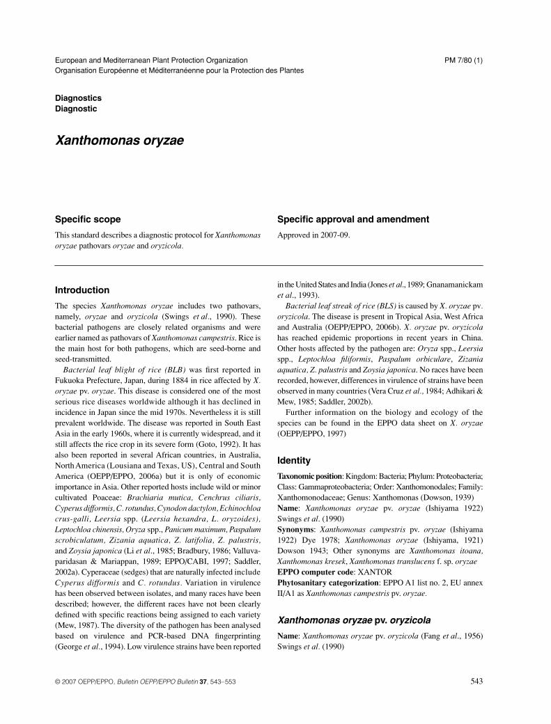
Bacterial leaf streak of rice (BLS)X. oryzae
pv.
oryzicola
is a foliar disease that appears at anygrowth stage of the host. Cells of
X. oryzae
pv.
oryzicola
enter
Fig. 1 Symptoms of X. oryzae pv. oryzae (bacterial leaf blight). (a) Rice field plants with X. oryzae pv. oryzae symptoms; (b) X. oryzae pv. oryzae symptoms in rice nursery beds; (c) Rice seedlings showing X. oryzae pv. oryzae symptoms; (d) Wilting of transplanted seedlings; (e) Kresek symptoms in field plants; (f) Progressive development of bacterial leaf blight in rice plants grown from infected seed.
Xanthomonas oryzae
545
© 2007 OEPP/EPPO,
Bulletin OEPP/EPPO Bulletin
37
, 543–553
through the stomata and multiply in the parenchyma tissues ofthe leaves.
X. oryzae
pv.
oryzicola
infects mainly the parenchymaof the cells of the leaves, but is not systemic. Initial symptomsare small water-soaked, transparent interveinal streaks (Fig. 2),which may elongate and darken. The transparent streaksdifferentiate leaf streak lesions from those of
X. oryzae
pv.
oryzae
that are opaque against the light. Bacterial exudates can
be observed as tiny yellow beads. The narrow, long, translucentlesions may coalesce, forming large patches, and severelyaffected fields appear burnt. It is at this stage that leaves wither,turn brown and eventually die, and the disease can be difficultto distinguish from bacterial leaf blight (Ou, 1985).
Isolation
Symptomatic samples are processed individually or in smallgroups. Precautions are made to avoid cross contaminationswhen collecting the samples and during the extraction process.The samples should be processed as soon as possible aftercollection and conserved at 4–8
°
C until use. Freshly preparedsample extracts are necessary for a successful isolation. Severalprocedures have been used for the isolation of
X. oryzae
pv.
oryzae
from symptomatic and asymptomatic plant parts,including seeds. Most of the procedures described for theisolation of
X. oryzae
pv.
oryzae
from rice plants, can also beapplied for the isolation of
X. oryzae
pv.
oryzicola
. However, itis known that both bacteria grow slowly on the isolating mediaand can be overgrown by fast growing contaminants. Often thesecontaminants are of yellow colour (e.g.
Pantoea agglomerans
and
Xanthomonas
-like saprophytic bacteria) thus makingdifficult the observations of colonies of the target organisms and
Fig. 1 Continued
Fig. 2 Symptoms of bacterial leaf streak in rice plants.
546 Diagnostics
© 2007 OEPP/EPPO,
Bulletin OEPP/EPPO Bulletin
37
, 543–553
are often visually undistinguishable in colony morphology,growth, and colour from strains of both pathogens. Culturingmay fail from advanced stages of infection due to competitionor being overgrown by saprophytic organisms and also fromseeds with low levels of inoculum or under bad conditions ofstorage. If disease symptoms are typical but isolation is negative,the isolation step should be repeated.
Direct plating from symptomatic and asymptomatic leaves collected from the field
Bacterial leaf blight in temperate regions can usually beobserved during the latter part of the seed bed stage (Ou, 1985).The bacteria can be detected from the upper part of infectedleaves before symptoms appear (Goto, 1965; Misukami &Wakimoto, 1969; Sakthivel
et al.
, 2001). Tabei (1967) reportedthat symptomless rice seedlings may carry the pest.
The isolation of
Xanthomonas
from symptomatic material ispreferable and can be performed using Peptone sucrose agar(PSA), Nutrient Broth Yeast Extract agar medium (NBY),Growth Factor (GF) agar (Agarwal
et al.
, 1989; Sakthivel
et al
.,2001). (Appendix 1). The bacteria can also be isolated onnutrient agar (NA) but with a very slow growth; semi-selectiveagar substrates like modified XOS agar medium (mXOS)(Di
et al
., 1991; Gnanamanickam
et al.
, 1994).
X. oryzae
pv.
oryzicola
colonies are also isolated on modified Wakimoto’sagar (Mew & Mistra, 1994) developed for the detection of
X.oryzae
pv.
oryzae,
but with the omission of ferrous sulfate.Surface disinfection with 70% alcohol for 15 s followed by
two to three times rinsing with sterile water is used for isolationfrom field plants. Plant parts showing fresh symptoms and withexudates are selected whenever possible. Sections (2 mm
×
7 mm) from infected tissue and preferably from the advancingportion of lesions are selected. The exudates can be processedseparately in 1 mL of sterile water. Narrow sections of infectedtissue are cut with a sterilized razor blade or scalpels, placed ina drop of sterile water and covered with a cover slip before theobservation under the compound microscope for bacterialstreaming, an indication of bacterial presence. Drops of leafextract are streaked onto the selected media plates andincubated at 27 ± 2°C (Agarwal et al., 1989; Mew & Mistra,1994). Characteristics of the colonies are described in the section:general characteristics (Fig. 3). These selected colonies aretransferred to NBY, PSA or Nutrient Agar (NA) plates for 1–2days for purification and further identification. Isolates aremaintained for longer periods at −80°C, e.g. in bacterialpreservers (i.e. Protect, Technical Service ConsultantsLtd., GB).
Direct isolation from seedsX. oryzae pv. oryzae and X. oryzae pv. oryzicola are found inthe glumes and occasionally within the endosperm of seedcollected from heavily infected fields (Fang et al., 1956;Srivastava & Rao, 1964).
Samples of 400 seeds are crushed to coarse flour that issuspended in 200 mL of sterile sodium chloride (0.85%)(sterile saline solution) (Agarwal et al., 1989; Mortensen et al.,
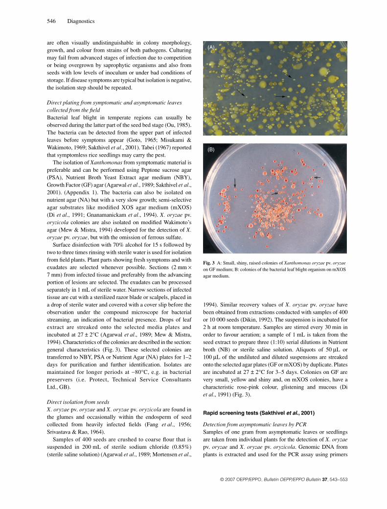
1994). Similar recovery values of X. oryzae pv. oryzae havebeen obtained from extractions conducted with samples of 400or 10 000 seeds (Dikin, 1992). The suspension is incubated for2 h at room temperature. Samples are stirred every 30 min inorder to favour aeration; a sample of 1 mL is taken from theseed extract to prepare three (1:10) serial dilutions in Nutrientbroth (NB) or sterile saline solution. Aliquots of 50 μL or100 μL of the undiluted and diluted suspensions are streakedonto the selected agar plates (GF or mXOS) by duplicate. Platesare incubated at 27 ± 2°C for 3–5 days. Colonies on GF arevery small, yellow and shiny and, on mXOS colonies, have acharacteristic rose-pink colour, glistening and mucous (Diet al., 1991) (Fig. 3).
Rapid screening tests (Sakthivel et al., 2001)
Detection from asymptomatic leaves by PCRSamples of one gram from asymptomatic leaves or seedlingsare taken from individual plants for the detection of X. oryzaepv. oryzae and X. oryzae pv. oryzicola. Genomic DNA fromplants is extracted and used for the PCR assay using primers
Fig. 3 A: Small, shiny, raised colonies of Xanthomonas oryzae pv. oryzae on GF medium; B: colonies of the bacterial leaf blight organism on mXOS agar medium.
Xanthomonas oryzae 547
© 2007 OEPP/EPPO, Bulletin OEPP/EPPO Bulletin 37, 543–553
TXT and TXT4R (Sakthivel et al., 2001) described inAppendix 2. These primers amplify a 964-bp fragment of aninsertion squence (IS1113). Suspensions of 108 CFU mL−1 (0.1OD600) of X. oryzae pv. oryzae and X. oryzae pv. oryzicolashould be used for positive controls, and a healthy plant extractand a sample of the ultra pure water (PCR reagent) for negativecontrols.
Direct detection from seeds by BIO-PCRThe main difference between this method and the method ofdirect isolation from seeds is that the incubation time is shorterand no morphological identification is required. In addition, alarger number of seeds can be tested.
BIO-PCR is an enrichment in solid medium combined withPCR and is recommended with primers TXT and TXT4R(Appendix 2). 500 g of seeds are soaked overnight at 4°C in750 mL of 0.01% Tween 20 in sterile water. Sub-samples of100 μL of seed extract are plated onto PSA in duplicates andincubated at 27 ± 2°C for 2 days or until the appearance ofpin-point colonies. Plates are then washed three times each with1 mL of sterile distilled water and 35 μL of the washings areused for the PCR assays (see Appendix 2). Suspensions of 107
CFU/mL of X. oryzae pv. oryzae and X. oryzae pv. oryzicolashould be used as positive controls and a sample of the ultrapure water (PCR reagent) as negative control.
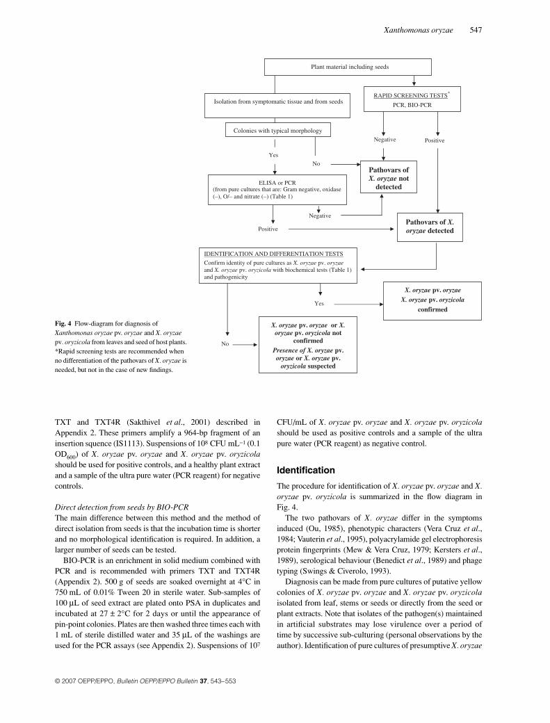
Identification
The procedure for identification of X. oryzae pv. oryzae and X.oryzae pv. oryzicola is summarized in the flow diagram inFig. 4.
The two pathovars of X. oryzae differ in the symptomsinduced (Ou, 1985), phenotypic characters (Vera Cruz et al.,1984; Vauterin et al., 1995), polyacrylamide gel electrophoresisprotein fingerprints (Mew & Vera Cruz, 1979; Kersters et al.,1989), serological behaviour (Benedict et al., 1989) and phagetyping (Swings & Civerolo, 1993).
Diagnosis can be made from pure cultures of putative yellowcolonies of X. oryzae pv. oryzae and X. oryzae pv. oryzicolaisolated from leaf, stems or seeds or directly from the seed orplant extracts. Note that isolates of the pathogen(s) maintainedin artificial substrates may lose virulence over a period oftime by successive sub-culturing (personal observations by theauthor). Identification of pure cultures of presumptive X. oryzae
Fig. 4 Flow-diagram for diagnosis of Xanthomonas oryzae pv. oryzae and X. oryzae pv. oryzicola from leaves and seed of host plants. *Rapid screening tests are recommended when no differentiation of the pathovars of X. oryzae is needed, but not in the case of new findings.
548 Diagnostics
© 2007 OEPP/EPPO, Bulletin OEPP/EPPO Bulletin 37, 543–553
isolates is conducted using at least two different tests of thepathogens (nutritional profile, serological or molecular). Anappropriate host test is used as final confirmation of pathogenicity.If this test is negative, the presence of X. oryzae pv. oryzae orX. oryzae pv. oryzicola is suspected but cannot be confirmed.Reference strains and negative test controls are used whereappropriate for performing the tests.
For confirmation of diagnosis of isolated bacteria (see Fig. 4),follow the tests recommended in the identification section andconduct a pathogenicity test in rice plants. The biochemicaltests results and pathogenicity tests observations are used in thedifferentiation of pathovars of Xanthomonas oryzae. Rapidscreening tests can be recommended when no differentiation ofthe pathovars of X. oryzae is needed. Additional information onthese tests can be found in the literature (Vera Cruz et al., 1984;Lelliott & Stead, 1987; Mew & Mistra, 1994; Schaad et al.,2001).
General characteristics
X. oryzae pv. oryzae and X. oryzae pv. oryzicola are Gramnegative, rods (0.4–0.6 × 1.1–2.0 μm), frequently capsulated,occurring singly, rarely in pairs, but not in chains, motile witha single polar flagellum. Colony morphology is examined onthe third day or when growth has appeared on the selected agarmedium. Colonies of X. oryzae pv. oryzae grow more slowlyon NA than those of X. oryzae pv. oryzicola. After 3–4 dayscolonies of X. oryzae pv. oryzae on NA are circular, entire,smooth, convex, opaque, and pale yellow at first, straw yellowcolour later. Colonies reach 1–2 mm after 5–7 days for X.oryzae pv. oryzae, and their survival on solid media is short.Colony formation from a single cell is poor and frequently failsto grow in many media, but can be improved by the addition ofbeef extract, methionine, or glutamic acid. On potato, sucrose
agar growth is faster, reaching 2 mm in 3–4 days, honey yellow,and longer-lived. X. oryzae pv. oryzicola grows faster than X.oryzae pv. oryzae on NA, producing smooth, opaque, glistening,circular, convex and entire colonies; whitish at first, becomingstraw to pale yellow later; about 1 mm in diameter in three days.Colonies of both pathogens on NBY are pale yellow, circular,raised and mucoid; on PSA, colonies are pale yellow, mucoidand shiny; on GF, colonies are very small, yellow and shiny. OnmXOS, colonies have a characteristic rose pink colour, mucoid,raised and glistening after 3–5 days (Fig. 3).
Optimum temperature for growth is 25–30°C for X. oryzaepv. oryzae and the growth range is 5–40°C while for X. oryzaepv. oryzicola is 25–28°C and with a growth range from8–38°C. For further details of the strains, see Vera Cruz et al.(1984) and IMI Descriptions of Fungi and Bacteria No. 1457and 1458 (Saddler, 2002a). Diagnosis is confirmed withpathogenicity tests on 4–6 weeks-old rice plants. For pre-liminary identification of isolates follow the tests recommendedin Table 1.
Fatty acid profiles
Fatty acid profiles allow identification at genus level only(Swings et al., 1990) so it is not recommended as a diagnosticmethod. This is given by the presence of 11:0 iso3OH, 13:0iso3OH, 12:0 iso3OH. The fatty acid profiles of X. oryzae pv.oryzae and X. oryzae pv. oryzicola are very similar, but withdifferences in the relative amount of 12:0 iso3OH, 12:0 3OH,and 15:0 anteiso fatty acids.
PCR
Presumptive X. oryzae colonies from agar platings are used toprepare bacterial suspensions of approximately 108 CFU mL−1
in molecular grade sterile water. The PCR test allowsidentification at species level. Appropriate PCR proceduresshould be applied without DNA extraction. (see Appendix 2).Bacterial suspensions of 25 μL are used in the PCR tests.Pathovar-specific primers have been described in the literaturebut they have not been tested in Europe. They are presented forreference in Appendix 2.
ELISA
Presumptive X. oryzae colonies (yellow, raised, nitrate, oxidase,gelatine and starch negative), are tested by an indirect-ELISAprocedure. A commercial Indirect ELISA kit based on X.oryzae monoclonal antibodies (Agdia Inc.) is available and canbe used in the confirmatory identification of the isolates. Forconducting the tests follow guidelines and procedures describedby the supplier of the kit.
Pathogenicity tests
Isolates are tested on susceptible rice cultivars. For X. oryzaepv. oryzae use 30–45 days old IR24 or IR8 (International Rice
Table 1 Discriminating bacteriological tests for preliminary identification and differentiation of pathovars of Xanthomonas oryzae
Testsb
X. o. pv.oryzae
X. o. pv. oryzicola
Gram staining − −Oxidase test −a −a
2-ketogluconate production − −Fluorescence on King’s B medium − −Nitrate reduction − −Oxidation-fermentation of glucose O/− O/−Gelatin hydrolysisc −/v +/v Starch hydrolysisc − +Sensitivity to 0.001% cupric nitrate + −Growth on L-alanine as carbon source − +/vAcetoin productiond − +Growth on 0.2% vitamin-free casamino acidsd − +
+ = positive; − = negative; O = oxidative; v = variable.aWeak positive reactions can be observed.bMew & Misra (1994); cBradbury (1986); dVera Cruz et al. (1984).
Xanthomonas oryzae 549
© 2007 OEPP/EPPO, Bulletin OEPP/EPPO Bulletin 37, 543–553
Research Institute, Manila, Philippines) or TN1 (Taichungnative 1) rice plants; for X. oryzae pv. oryzicola IR24 or IR50are recommended. Local popular rice varieties from theEuropean region with known susceptibility to the disease arerecommended to be included in the tests (Mew & Mistra, 1994).Pathogenicity grouping of isolates is based in several countrieson the reaction of specific differential cultivars. For additionalinformation consult Ou (1985). The leaf clipping method iswidely used for the inoculation tests with X. oryzae pv. oryzaestrains and spray inoculations for X. oryzae pv. oryzicola. Forall inoculations grow the plants in potting mixture with aweekly fertilization of 1–2 g urea L−1. Keep inoculated plantsunder high moisture conditions with 12 h light/dark cycle at anoptimum temperature of 28–32°C/22°C. Include a negativecontrol (e.g. plants inoculated with sterile saline solution alone)and a positive control to monitor false negative reactions caused(e.g. by technical failure).
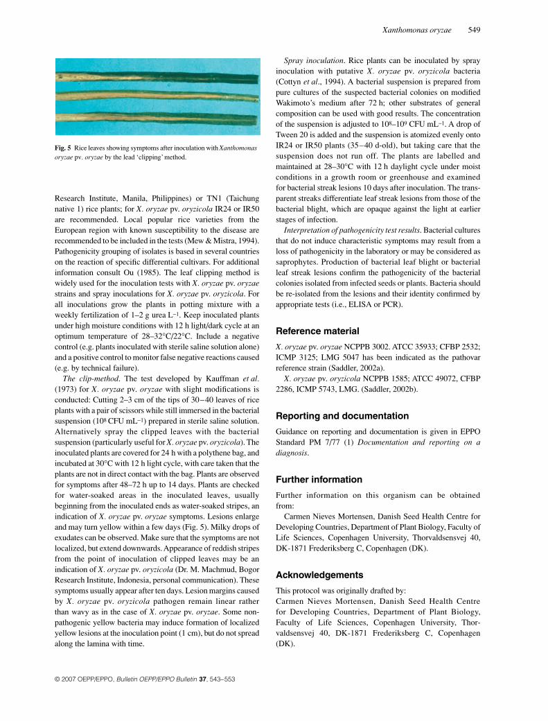
The clip-method. The test developed by Kauffman et al.(1973) for X. oryzae pv. oryzae with slight modifications isconducted: Cutting 2–3 cm of the tips of 30–40 leaves of riceplants with a pair of scissors while still immersed in the bacterialsuspension (108 CFU mL−1) prepared in sterile saline solution.Alternatively spray the clipped leaves with the bacterialsuspension (particularly useful for X. oryzae pv. oryzicola). Theinoculated plants are covered for 24 h with a polythene bag, andincubated at 30°C with 12 h light cycle, with care taken that theplants are not in direct contact with the bag. Plants are observedfor symptoms after 48–72 h up to 14 days. Plants are checkedfor water-soaked areas in the inoculated leaves, usuallybeginning from the inoculated ends as water-soaked stripes, anindication of X. oryzae pv. oryzae symptoms. Lesions enlargeand may turn yellow within a few days (Fig. 5). Milky drops ofexudates can be observed. Make sure that the symptoms are notlocalized, but extend downwards. Appearance of reddish stripesfrom the point of inoculation of clipped leaves may be anindication of X. oryzae pv. oryzicola (Dr. M. Machmud, BogorResearch Institute, Indonesia, personal communication). Thesesymptoms usually appear after ten days. Lesion margins causedby X. oryzae pv. oryzicola pathogen remain linear ratherthan wavy as in the case of X. oryzae pv. oryzae. Some non-pathogenic yellow bacteria may induce formation of localizedyellow lesions at the inoculation point (1 cm), but do not spreadalong the lamina with time.
Spray inoculation. Rice plants can be inoculated by sprayinoculation with putative X. oryzae pv. oryzicola bacteria(Cottyn et al., 1994). A bacterial suspension is prepared frompure cultures of the suspected bacterial colonies on modifiedWakimoto’s medium after 72 h; other substrates of generalcomposition can be used with good results. The concentrationof the suspension is adjusted to 108–109 CFU mL−1. A drop ofTween 20 is added and the suspension is atomized evenly ontoIR24 or IR50 plants (35–40 d-old), but taking care that thesuspension does not run off. The plants are labelled andmaintained at 28–30°C with 12 h daylight cycle under moistconditions in a growth room or greenhouse and examinedfor bacterial streak lesions 10 days after inoculation. The trans-parent streaks differentiate leaf streak lesions from those of thebacterial blight, which are opaque against the light at earlierstages of infection.
Interpretation of pathogenicity test results. Bacterial culturesthat do not induce characteristic symptoms may result from aloss of pathogenicity in the laboratory or may be considered assaprophytes. Production of bacterial leaf blight or bacterialleaf streak lesions confirm the pathogenicity of the bacterialcolonies isolated from infected seeds or plants. Bacteria shouldbe re-isolated from the lesions and their identity confirmed byappropriate tests (i.e., ELISA or PCR).
Reference material
X. oryzae pv. oryzae NCPPB 3002. ATCC 35933; CFBP 2532;ICMP 3125; LMG 5047 has been indicated as the pathovarreference strain (Saddler, 2002a).
X. oryzae pv. oryzicola NCPPB 1585; ATCC 49072, CFBP2286, ICMP 5743, LMG. (Saddler, 2002b).
Reporting and documentation
Guidance on reporting and documentation is given in EPPOStandard PM 7/77 (1) Documentation and reporting on adiagnosis.
Further information
Further information on this organism can be obtainedfrom:
Carmen Nieves Mortensen, Danish Seed Health Centre forDeveloping Countries, Department of Plant Biology, Faculty ofLife Sciences, Copenhagen University, Thorvaldsensvej 40,DK-1871 Frederiksberg C, Copenhagen (DK).
Acknowledgements
This protocol was originally drafted by:Carmen Nieves Mortensen, Danish Seed Health Centrefor Developing Countries, Department of Plant Biology,Faculty of Life Sciences, Copenhagen University, Thor-valdsensvej 40, DK-1871 Frederiksberg C, Copenhagen(DK).
Fig. 5 Rice leaves showing symptoms after inoculation with Xanthomonas oryzae pv. oryzae by the lead ‘clipping’ method.
550 Diagnostics
© 2007 OEPP/EPPO, Bulletin OEPP/EPPO Bulletin 37, 543–553
It was further revised by Elena G. Biosca, Dpt. Microbiologíay Ecología, Universidad de Valencia, 46100 Burjassot,Valencia (ES).
References
Adachi N & Oku T (2000) PCR-mediated detection of Xanthomonas oryzaepv. oryzae by amplification of the 16S-23S rDNA spacer region sequence.Journal of General Plant Pathology 66, 303–309.
Adhikari TB & Mew TW (1985) Antibiotic sensitivity of Xanthomonascampestris pv. oryzicola in vitro. International Rice Research Newsletter10, 19.
Agarwal PC, Mortensen CN & Mathur SB (1989) Seed-borne diseases andseed health testing of rice. Technical Bulletin No. 3/PhytopathologicalPapers No. 30. Danish Government Institute of Seed Pathology for Develop-ing Countries & CAB International Mycological Institute (DK/GB).
Benedict AA, Alvarez AM, Berestecky J, Imanaka W, Mizumoto CY,Pollard LW, Mew TW & Gonzalez CF (1989) Pathovar-specific monoclonalantibodies for Xanthomonas campestris pv. oryzae and for Xanthomonascampestris pv. oryzicola. Phytopathology 79, 322–328.
Bradbury, JF (1986). Guide to Plant Pathogenic Bacteria. CAB InternationalMycological Institute, Kew, Surrey (GB).
Cottyn B, Cerez MT & Mew TW (1994) Chapter 7: Bacteria. In: A Manualof Rice Seed Health Testing (eds Mew TW & Mistra JK), pp. 322–328.IRRI, Manila (PH).
Di M, Ye HZ, Schaad NW & Roth DA (1991) Selective recovery ofXanthomonas spp. from rice seeds. Phytopathology 81, 1358–1363.
Dikin A (1992) Studies on Bacterial Blight of Rice: Development of SeedHealth Testing Methods, Seed Transmission, Survival of BacterialPathogens in the Seed. Danish Government Institute of Seed Pathologyfor Developing Countries, Hellerup (DK).
EPPO/CABI (1997) Xanthomonas oryzae. Quarantine Pests for Europe,2nd edition, pp. 1129–1136. CAB International, Wallingford (GB).
Fang CR, Lin CF & Chu CL (1956) A preliminary study on the disease cycle ofthe bacterial leaf blight of rice. Acta Phytotaxonomica Sinica 2, 173–185.
George MLC, Cruz WT & Nelson RJ (1994) DNA fingerprinting ofXanthomonas oryzae pv. oryzae by ligation-mediated polymerase chainreaction. International Rice Research Notes 19, 29–30.
Gnanamanickam SS, Alvarez AM, Benedict AA, Sakthivel N & Leach JE(1993) Characterization of weakly virulent bacterial strains associatedwith the bacterial blight (BB) or leaf streak (BLS) in southern India.International Rice Research Institute Newsletter 18, 5–16.
Gnanamanickam SS, Shigaki T, Medalla ES, Mew TW & Alvarez AM(1994) Problems in detection of Xanthomonas oryzae pv. oryzae in riceseed and potential for improvement using monoclonal antibodies. PlantDisease 78, 173–178.
Goto M (1965) A technique for detecting the infected area of bacterial blightof rice caused by Xanthomonas oryzae before symptom appearance.Annals of the Phytopathological Society of Japan 30, 37–41.
Goto M (1992) Fundamentals of Bacterial Plant Pathology. Academic PressInc., San Diego, California (US).
Jones, RK, Barnes, LW, Gonzales CF, Leach JE & Benedict AA (1989)Identification of low-virulence strains of Xanthomonas campestris pv.oryzae from rice in the United States. Phytopathology 79, 984–990.
Kauffman HE, Reddy APK, Hsieh SPY & Merca SD (1973) An improvedtechnique for evaluating resistance of rice varieties to Xanthomonasoryzae. Plant Disease Reporter 57, 537–541.
Kersters K, Pot B, Hoste B, Gillis M, & De Ley J (1989) Protein electrophoresisand DNA-DNA hybridizations of xanthomonads from grasses and cereals.Bulletin OEPP/EPPO Bulletin 19, 51–55.
Kim HM & Song WY (1996) Characterization of ribosomal RNA intergenicspacer region of several seedborne bacterial pathogens of rice. SeedScience and Technology 24, 571–580.
Lelliott RA & Stead DE (1987) Methods for the Diagnosis of BacterialDiseases of Plants, Methods in Plant Pathology Volume 2. BlackwellScientific Publications, Oxford (GB).
Li ZZ, Zhao H & Ying, XD (1985) The weed carriers of bacterial leaf blightof rice. Acta Phytopathologica Sinica 15, 246–248.
Mew, T.W. (1987) Current status and future prospects of research on bacterialblight of rice. Annual Review of Phytopathology 25, 359–382.
Mew TW & Mistra JK (1994) A Manual of Rice Seed Health Testing. IRRI,Manila (PH).
Mew TW & Vera Cruz CM (1979) Variability of Xanthomonas oryzae:specificity in infection of rice differentials. Phytopathology 69, 152–155.
Misukami T & Wakimoto S (1969) Epidemiology and control of bacterialblight of rice. Annual Review of Phytopathology 7, 51–72.
Mortensen CN, Manandhar HK, Cahyaniati & Haryanti SE (1994) Pathogenicbacteria associated with rice seed samples from Indonesia and Nepal.Plant Pathogenic Bacteria, Versailles (France), 8th International Conference,June 9–12, 1992. Les colloques, no. 66. INRA, Paris (FR).
OEPP/EPPO (2006a) Distribution Maps of quarantine pests for EuropeXanthomonas oryzae pv. oryzae. http://www.eppo.org/QUARANTINE/bacteria/Xanthomonas_oryzicola/XANTTO_map.htm [accessed on 2 May2007]
OEPP/EPPO (2006b) Distribution Maps of quarantine pests for EuropeXanthomonas oryzae pv. oryzicola. http://www.eppo.org/QUARANTINE/bacteria/Xanthomonas_oryzae/XANTOR_map.htm
Ou SH (1985) Rice Diseases. 2nd edition. Commonwealth MycologicalInstitute, Kew, Surrey (GB).
Saddler GS (2002a) Xanthomonas oryzae pv. oryzae. IMI Descriptions ofFungi and Bacteria 1457. CAB International, Kew, Surrey (GB).
Saddler GS (2002b) Xanthomonas oryzae pv. oryzicola. IMI Descriptions ofFungi and Bacteria 1458. CAB International, Kew, Surry (GB).
Sakthivel N, Mortensen CN & Mathur SB (2001) Detection of Xanthomonasoryzae pv. oryzae in artificially and naturally infected rice seeds andplants by molecular techniques. Applied Microbiology and Biotechnology56, 435–441.
Sambrook J, Fritsch EF & Maniatis T (1989) Molecular Cloning. ALaboratory Manual, 2nd edition. CSH, Cold Spring Harbor LaboratoryPress, Cold Spring Harbor (US).
Schaad NW, Jones JB & Chun W (2001) Laboratory Guide for identificationof Plant Pathogenic Bacteria, 3rd edition. APS Press, St Paul (US).
Srivastava DN & Rao YP (1964) Seed transmission and epidemiology ofbacterial blight disease of rice in North India. Indian Phytopathology 17,77–78.
Swings J & Civerolo EL (1993) Xanthomonas. Chapman & Hall, London (GB).Swings J, Van Den Mooter M, Vauterin L, Hoste B, Gillis M, Mew TW et al.
(1990) Reclassification of the causal agents of bacterial blight (Xan-thomonas campestris pv. oryzae) and bacterial leaf streak (Xanthomonascampestris pv. oryzicola) of rice as pathovars of Xanthomonas oryzae (exIsiyama 1922) sp. nov., nom. rev. International Journal of SystematicBacteriology 40, 301–311.
Tabei H (1967) Anatomical studies of rice plant affected by bacterial leafblight, with special reference to stomatal infection at the coleoptile andthe foliage leaf sheath of rice seedling. Annals of the PhytopathologicalSociety of Japan 33, 12–16.
Valluvaparidasan V & Mariappan V (1989) Alternate hosts of rice bacterialblight (BB) pathogen Xanthomonas campestris pv. oryzae. InternationalRice Research Newletter 14(5), 27–28.
Vauterin L, Hoste B, Kersters K & Swings J (1995) Reclassification of Xan-thomonas. International Journal of Systematic Bacteriology 45, 472–489.
Vera Cruz CM, Gossele F, Kersters K, Segers P, Van Den Mooter M, SwingsJ et al. (1984) Differentiation between Xanthomonas campetris pv.oryzae, Xanthomonas campestris pv. oryzicola and the bacterial ‘brownblotch’ pathogen on rice by numerical analysis of phenotypic features andprotein gel electrophoregrams. Journal of General Microbiology 130,2983–2999.
Xanthomonas oryzae 551
© 2007 OEPP/EPPO, Bulletin OEPP/EPPO Bulletin 37, 543–553
Appendix 1
Growing media and buffers
Agar mediaThe following media are used for the isolation and purificationof X. oryzae pv. oryzae and X. oryzae pv. oryzicola from seedsand leaves. GF, PSA, and XOS agar media are useful for thepreliminary isolation from seeds or leaves. NBY, PSA,Wakimoto’s agar and NA are useful in the general isolationfrom leaves and for purification of strains for identification.YDC is often used in the determination of pigment productionof the strains. However, some strains of the bacterial blightorganism may not grow well on this media, which has oftenbeen used for the short-term storage of the bacterial cultures.
Growth Factor (GF) Agar: KH2PO4 0.4 g; MgSO4. 7H2O0.05 g; NaCl 0.1 g; NH4H2PO4 0.5 g; FeCl3 0.01 g; yeastextract 0.01 g; agar 18.0 g; distilled water 1000.0 mL. Dissolveby heating and autoclave for 15 min at 121°C.
Nutrient Broth (NB): Peptone 5.0 g; beef extract 3.0 g;distilled water 1000 mL. Dissolve, distribute into test tubes andautoclave for 15 min at 121°C. Can be purchased in dehydratedform from Difco.
Nutrient Broth Yeast Extract Agar Medium (NBY): Sol. 1.nutrient broth 8.0 g; yeast extract 2.0 g; K2 HPO4 2.0 g; KH2
PO4 0.5 g; agar 18.0 g; distilled water 950.0 mL. Sol. 2. glucose5.0 g; distilled water 50.0 mL. Sol. 3. MgSO4. 7H2O 1.0 mL of1 M solution* (*Make 1 M sol. by dissolving 2.46 g in 10 mLdistilled water). Dissolve and autoclave (121°C for 15 min)the three solutions separately and mix well when the temperaturehas reached 45–50°C four pouring plates.
Peptone Sucrose Agar (PSA): sucrose 10.0 g; peptone 10.0 g;Na-glutamate 1.0 g; agar 17.0 g; distilled water 1000.0 mL. pHis adjusted 6.8–7.0, mix, heat to dissolve and autoclave at121°C for 15 min.
Yeast Dextrose Chalk Agar (YDC): Sol. 1. yeast extract10.0 g; precipitated chalk (CaCO3), light powder 20.0 g; agar18.0 g; distilled water 950.0 mL. Sol. 2. dextrosa (L-glucose)20.0 g; distilled water 50.0 mL. Mix, dissolve and autoclave thetwo solutions separately for 15 min at 121°C; mix well whenthe temperature of the medium is about 40–50°C for pouringplates.
Modified XOS agar medium (mXOS): sucrose 20.0 g;peptone 2.0 g; monosodium glutamate 5.0 g; Ca (NO3)2 0.2 g;K2 HPO4 2.0 g; Fe (EDTA) 1.0 mg; agar 18.0 g; distilled water1000.0 mL. Mix and dissolve, adjust pH of the medium to 6.8–7.0 and add, after autoclaving (15 min at 121°C), the followingfilter sterilised solutions: tetrazolium chloride (tetrazolium red)10 mg (1 mL of a 1% stock solution); cyloheximide (Actidione)100 mg (1 mL of a 100 mg mL−1 solution dissolved in 75%ethanol); cephalexin (20 mg) 20 mg (2 mL of a 10 mg mL−1
aqueous solution); kasugamycin 20 mg (2 mL of a 10 mg mL−1
aqueous solution); methyl violet 2B 0.3 mg (3 mL of a0.01 g/100 mL aqueous solution).
Modified Wakimoto’s medium: ferrous sulphate 0.05 g;calcium nitrate 0.5 g; sodium phosphate 0.82 g; bacto peptone
5.0 g; agar 17 g; sucrose 20 g; distilled water 1000 mL.Dissolve by heating and autoclave for 15 min at 121°C.
Potato sucrose agar: Cook 200 g of diced potatoes in 0.5 Lof water for 10 min.
Filter through cheesecloth and add water to filtrate to 1.0 L. AddAgar (20.0 g) Sucrose (20.0 g). Autoclave at 121°C for 15 min.
DNA Extraction reagentsTE-buffer: Made from stock solutions of 1 M Tris-HCl, pH 8.0and 0.5 M EDTA
10 mL 1 M Tris HCl, 2 mL 0.5 M EDTA. Bring up to1000 mL with H2 O.
10% SDS: (Lauryl Sulfate / Sodium dodecyl sulfate): 10 g in100 mL H2O.
CTAB: (Hexadecyltrimethylammonium Bromide): 10 g in100 mL 0.7 M NaCl. (0.7 M NaCl: 4.1 g NaCl in 100 mL H2O).
5M NaCl: 29.2 g NaCl in 100 mL H2O.Chloroform:Isoamylalcohol 24:1: 24 mL Chloroform, 1 mL
Isoamylalcohol. Mix well and store in poison cabin.Protease: 10 mg mL−1.
Buffer for electrophoresis5X TBE buffer: 54 g Tris base, 27.5 g Boric Acid, 20 mL of0.5M EDTA, pH 8.0, 2.5 mg EtBr = 2.5 mL of 10 mg mL−1
stock (optional), and adjust volume to 1 L distilled water andstore in glass. Discard any buffers that develop a precipitate.Use 0.5X for agarose gels.
Loading buffer: 0.025 bromothymol blue, 3 g glycol, 10 mLdistilled water.
Buffer formulations for indirect ELISA (Agdia Inc.)PBS (PBS – phosphate buffered saline). Dissolve the follow-
ing ingredients in 930 mL distilled water in the order listed.Add sodium phosphate slowly. 1.15 g Sodium phosphate,dibasic (anhydrous), 0.2 g potassium chloride, 0.2 g potassiumphosphate, monobasic (anhydrous), 8.0 g sodium chloride, 0.2 gsodium azide; adjust pH to 7.4. Adjust final volume to 1000 mLwith distilled water.
Coating buffer. Dissolve in distilled water to 1000 mL: 1.59 gsodium carbonate (anhydrous), 2.93 g sodium bicarbonate,0.2 g sodium azide. Adjust pH to 9.6. Store at 4°C.
PBSTween (wash buffer). Dissolve in distilled water to1000 mL: 8.0 g sodium chloride, 1.15 g sodium phosphate, dibasic(anhydrous), 0.2 g Potassium phosphate, monobasic (anhydrous),0.2 g potassium chloride, 0.5 g Tween-20. Adjust pH to 7.4.
PNP buffer. Dissolve in 800 mL distilled water: 0.1 gmagnesium chloride hexahydrate, 0.2 g sodium azide, 97 mLDiethanolamine. Adjust pH to 9.8 with hydrochloric acid. Adjustfinal volume to 1000 mL with distilled water. Store at 4°C.
Appendix 2
PCR tests
DNA extraction:DNA extraction reagent composition are presented inAppendix 1.

552 Diagnostics
© 2007 OEPP/EPPO, Bulletin OEPP/EPPO Bulletin 37, 543–553
Tissue samples from leaves are homogenized into emulsionin a small sterile mortar (normal glass or porcelain mortar withpestle, top diameter 5 cm) with 500 μL extraction buffer andtransferred into a 1.5 mL centrifuge tube. The remaining stepsare the same as that for normal scale DNA extraction.
From pure cultures of bacteria isolated from leaves or seeds,the strains are grown in nutrient broth (NB) overnight withshaking at 30°C and 1.5 mL is dispensed into an Eppendorftube.
Suspensions from tissue macerate or bacterial suspensionfrom pure cultures in NB are centrifuged at 12 000 rpm ina microcentrifuge for 10 min at room-temperature. Thesupernatant is discarded and the pellet is re-suspended in500 μL TE-buffer and 75 μL of 10% SDS and 10 μL of10 mg mL−1 protease is added. The suspension is gently shakenat 37°C for 1 h.
A volume of 120 μL 5M NaCl is added and the suspensionis thoroughly mixed by inverting the tubes several times;100 μL CTAB (10% C-TAB in 0.7 M NaCl) are added, mixedthoroughly and incubated at 65°C for 20 min.
An equal volume (approx. 700 μL) of chloroform: isoamy-lalcohol (24:1) is added and the sample is mixed and vigorouslyshaken for 30 min. Tubes are centriguged for 30 min at15 000 rpm at room temperature and the aqueous phase istransferred to a clean tube.
An equal volume of isopropanol is added (centrifugation isomitted) and the precipitate is transferred to 750 μL of 70%ethanol. (Consider leaving in freezer overnight before washingin ethanol.)
The sample is centrifuged for 8 min at 14 000 rpm in amicrocentrifuge, the supernatant is discarded and the precipitateis allowed to air dry. The pellet is dissolved in 50 μL TE and theresulting DNA is placed at 70°C to hasten the dissolution. Theextracted DNA is stored at 4°C for the PCR assay. Samples of200 ng are used for the PCR tests.
Alternatively several commercially available DNA extractionkit can be used (e.g. Easy-DNA isolation kit from Invitrogen orDNAeasy Plant kit from Qiagen).
PrimersTXT (5-GTCAAGCCAACTGTGTA-3) and TXT4R (5-CGTTCGGCACAGTTG-3)
PCR master mix (15 µL) (Table 2):
Sample volume for BIO-PCR: add 35 μL sample to 15 μLmaster mix
Sample volume for pure culture bacterial DNA: 4 μL of50 ng μL−1 (total 200 ng/reaction) to 15 μL of mastermix; add31 μL water.
PCR reaction conditionsThe reaction conditions are: initial denaturation step of 95°Cfor 5 min followed by 30 cycles of 95°C for 1 min, 56°C for1 min, 72°C for 1 min and final extension of 10 min at 72°C.The amplification conditions are optimized for the PerkinElmer 2400 themocycler, but reproducible results are obtainedwith other machines such as the Eppendorf mastercyclergradient, MJ thermocycler and Hybaid PCR expressthermocycler (Cycling conditions should be adapted usingPeltier-type thermocyclers.) Taq polymerase enzymes suppliedby other manufacturers (Promega, Finnzyme, Bioline USA andBangalore Geni) also yield similar PCR results.
After PCR, analyse samples of 10 μL on a 1% agarose gelusing standard procedures (Sambrook et al., 1989).
Interpretation of the PCR resultsA PCR product of 964 bp indicates the presence of X. oryzae pv.oryzae or X. oryzae pv. oryzicola in the sample. The PCR isnegative if the X. oryzae pv. oryzae and X. oryzae pv. oryzicolaspecific amplicon of expected size is not detected in the samplein question but detected for all positive controls. If one of thenegative controls shows a band of the expected size, the PCRtest is repeated with a new mix and several negative controls areincluded to monitor the contaminations. Inhibition of the PCRmay be suspected if the expected amplicon is obtained from thepositive controls of X. oryzae pv. oryzae and X. oryzae pv.oryzicola, but negative from positive controls with suspensionsof X. oryzae pv. oryzae or X. oryzae pv. oryzicola prepared inhealthy plant extracts.
For the differentiation of X. oryzae pv. oryzae and X. oryzaepv. oryzicola strains Kim & Song (1996) used PCR primersR16-1 (CTTGTACACACCGCCGTCA) and R23-2R(TCCG-GGTACTTAGATGTTTC) targeted towards the ribosomalintergenic spacer region (16S and 23S). All Korean strains ofX. oryzae pv. oryzae and X. oryzae pv. oryzicola testedshowed a species-specific fragment of 880 bp. Both pathovarsof X. oryzae were differentiated from each other based on
Table 2 PCR master mix (15 µL)
Reagent Initial concentration Volume Final concentration (in 50 µL)
Water (PCR grade) 5.9 μLDNA buffer (Promega) 10× 5 μL 1×MgCl2 25 mM 3 μL 1.5 mMdNTPs 25 mM each 0.4 μL 200 μMPrimer TXT 100 pmol μL−1 0.25 μL 0.5 pm μL−1
Primer TXT4R 100 pmol μL−1 0.25 μL 0.5 pm μL−1
Taq polymerase [Promega]a 5 units 0.2 μL 1 U
aA regular Taq polymerase.

Xanthomonas oryzae 553
© 2007 OEPP/EPPO, Bulletin OEPP/EPPO Bulletin 37, 543–553
differences in a secondary fragment of 430 bp or 460 bp forX. oryzae pv. oryzae strains and of 440 and 370 bp. Other non-pathogenic Xanthomonas-like strains had a primary fragmentof 860 bp and a secondary fragment of 460 bp. Adachi & Oku(2000) designed a set of primers from the spacer regionbetween the 16S and 23S rDNA for the identification of purecultures of X. oryzae pv. oryzae. The specific primers, XOR-F (5′-GCATGACGTCATCGTCCTGT-3′) and XOR-R2 (5′-CTCGGAGCTATATGCCGTGC-3′) amplified a 470 bpfragment from X. oryzae pv. oryzae strains isolated fromJapan.
The PCR assays developed by Kim & Song (1996) appearto be useful in the differentiation of the strains, but are stillto be evaluated with more strains of different geographicalorigin. Similarly the PCR studies for the specific identificationand detection of X. oryzae pv. oryzae (Adachi & Oku, 2000)were based on Japanese strains alone. The PCR assaysdescribed in this protocol (Sakthivel et al., 2001) do not dif-ferentiate between the pathovars of Xanthomonas oryzae,however, they can be applied in conjunction with a selected setof tests (PCR, serological test, biochemical characters) andhost plant inoculation tests.
Corrigendum
For the Diagnostic Standard PM 7/80 (1) Xanthomonas oryzae, it has been brought to the attention of the EPPO Secretariat
that the primer TXT4R for the PCR test in the Appendix 2 based on Sakthivel et al. (2001) had been incorrectly reproduced
in the EPPO Standard.
The details of the primer should therefore be replaced
Old version
TXT4R (5-CGTTCGGCACAGTTG-3)
Corrected version
TXT4R (5-CGTTCGCGCCACAGTTG-3)
The EPPO Secretariat apologies for this error.
References
EPPO (2007) PM 7/80 (1) Xanthomonas oryzae. Bulletin OEPP/EPPO Bulletin 37, 543–553.Sakthivel N, Mortensen CN & Mathur CB (2001) Detection of Xanthomonas oryzae pv. oryzae in artificially inoculated and naturally infected rice
seeds and plants by molecular techniques. Applied Microbiology and Biotechnology 56, 435–441.
Bulletin OEPP/EPPO Bulletin (2018) 48 (2), 318 ISSN 0250-8052. DOI: 10.1111/epp.12474
318 ª 2018 The Authors. Journal compilation ª 2018 OEPP/EPPO, EPPO Bulletin 48, 318