Embed Size (px)
Citation preview

Accepted by E. Bernard: 24 Dec. 2015; published: ?? Month 2016
ZOOTAXA
ISSN 1175-5326 (print edition)
ISSN 1175-5334 (online edition)Copyright © 2016 Magnolia Press
Zootaxa 0000 (0): 000–000
http://www.mapress.com/j/zt/
Article
1
http://doi.org/10.11646/zootaxa.0000.0.0
http://zoobank.org/urn:lsid:zoobank.org:pub:00000000-0000-0000-0000-00000000000
The earwig fauna (Insecta: Dermaptera) of Penang Island, Malaysia,
with descriptions of two new species
YOSHITAKA KAMIMURA1,2,4
, MASARU NISHIKAWA3
& CHOW-YANG LEE2
1
Department of Biology, Keio University, 4-1-1 Hiyoshi, Yokohama 223-8521, Japan
2
Urban Entomology Laboratory, Vector Control Research Unit, School of Biological Sciences, Universiti Sains Malaysia, Minden
11800, Penang, Malaysia
3
Entomological Laboratory, Faculty of Agriculture, Ehime University, Matsuyama, 790-8566, Japan
4
Corresponding author. E-mail: [email protected]
Abstract
The earwig (Dermaptera) fauna of Penang Island, Malaysia, was evaluated by means of an extensive field survey together
with revision of the few published data. Based on the results of the field survey, 31 species are recognized (2 Diplatyidae,
3 Pygidicranidae, 5 Anisolabididae, 2 Labiduridae, 14 Spongiphoridae, 4 Chelisochidae, 1 Forficulidae). Fifteen of these
taxa are new to Peninsular Malaysia (=West Malaysia): Diplatys annandalei Burr, 1911, Diplatys mutiara n. sp., Euborel-
lia philippinensis Srivastava, 1979, Metisolabis punctata (Dubrony, 1879), Pseudovostox brindlei Srivastava, 2003,
Chaetospania anderssoni Brindle, 1971, Chaetospania javana Borelli, 1926, Chaetospania huisiangi n. sp., Paralabel-
lula boettcheri (Borelli, 1923), Paralabellula rotundifrons (Hincks, 1954), Nesogaster amoenus (Stål, 1855), Hamaxas
crassus Borelli, 1926, Proreus coalescens (Borelli, 1927), Hypurgus humeralis (Kirby, 1891), and an unidentified Echi-
nosoma sp. Species composition of the island are compared with the dermapteran fauna of Thailand. Descriptions of fe-
males (or female genitalia) are given for some species for the first time.
Key words: Chaetospania huisiangi, Diplatys mutiara, semi-urban environments, Southeast Asia, species catalog, spe-
cies diversity
Introduction
Dermaptera (earwigs) is a polyneopteran insect order with ~2200 described species from mainly tropical and warm
temperate regions (Popham 2000; Grimaldi & Engel 2005, Haas et al. 2012). Although several species are
considered pests in gardens and in agriculture, or act as pest control agents, most earwig species inhabit natural and
semi-natural environments with no direct relationship to human activities (Costa 2006). Although many
dermapteran families show circumtropical distributions (Popham 2000), faunal, taxonomic, and ecological studies
are especially scarce for tropical earwigs. For the Malaysian earwig fauna, no comprehensive review or checklist
has been reported for Peninsular Malaysia (=West Malaysia) or Borneo (Sabah and Sarawak) in recent years. To
begin filling this gap, we conducted an intensive faunal study of the earwigs of Penang Island, Peninsular Malaysia.
Penang Island (Pulau Pinang) is a 299-km2
island of Penang state, located in the Straits of Malacca, approximately
5 km from the western coast of the mainland of Peninsular Malaysia (Fig. 1). It has a typical tropical climate with
average temperatures throughout the year of 27‒30°C and 22‒24°C during the day and night, respectively, and with
a mean annual rainfall of 2,670 mm (Gardner et al. 2011). Although Penang Island has become increasingly
urbanized, there are still large natural forests and plantation areas in the central and northeastern parts of the island
(Gardner et al. 2011). Furthermore, recent studies on other insect groups have found many insect species new to
Malaysia or new to science on this small island (e.g., Lee et al. 2004, Disney et al. 2009, Smith et al. 2011).
The aim of this study was to examine the contemporary dermapteran fauna of the island, which was almost
unknown, by means of an extensive field survey. In total, 31 species are listed and discussed, and 2 new species are
described. Including the new species and an undescribed species, 15 species are recorded for the first time in

KAMIMURA ET AL.2 · Zootaxa 0000 (0) © 2016 Magnolia Press
Peninsular Malaysia. Taxonomists rarely include the morphology of female genitalia in descriptions of earwig
species. However, female genital characteristics are being increasingly recognized to contain important
information for phylogenetic studies of earwigs (Klass 2001, 2003, Schneider & Klass 2013, Kamimura & Lee
2014b). For some species with sufficient materials, female genital structures, especially the structure of the
spermatheca, are described for the first time.
Material and methods
Earwigs were collected from various places on Penang Island during March 2012 to March 2013, for a total of 67
days. At each field survey (usually 1‒5 hours during daytime), insect samples were collected by hand-sorting of
decaying logs, fallen fruits, leaf litter, and loose tree bark, or by sweeping shrubs with an insect net. Nymphs were
reared to adulthood in the laboratory for identification. The type material of new species and some representative
samples collected in this study will be deposited in the Lee Kong Chian Natural History Museum (LKCNHM),
Singapore, and the Osaka Museum of Natural History (OMNH) and the Ehime University Museum (EUMJ),
Japan. A few additional records are reported from collections made in 2014.
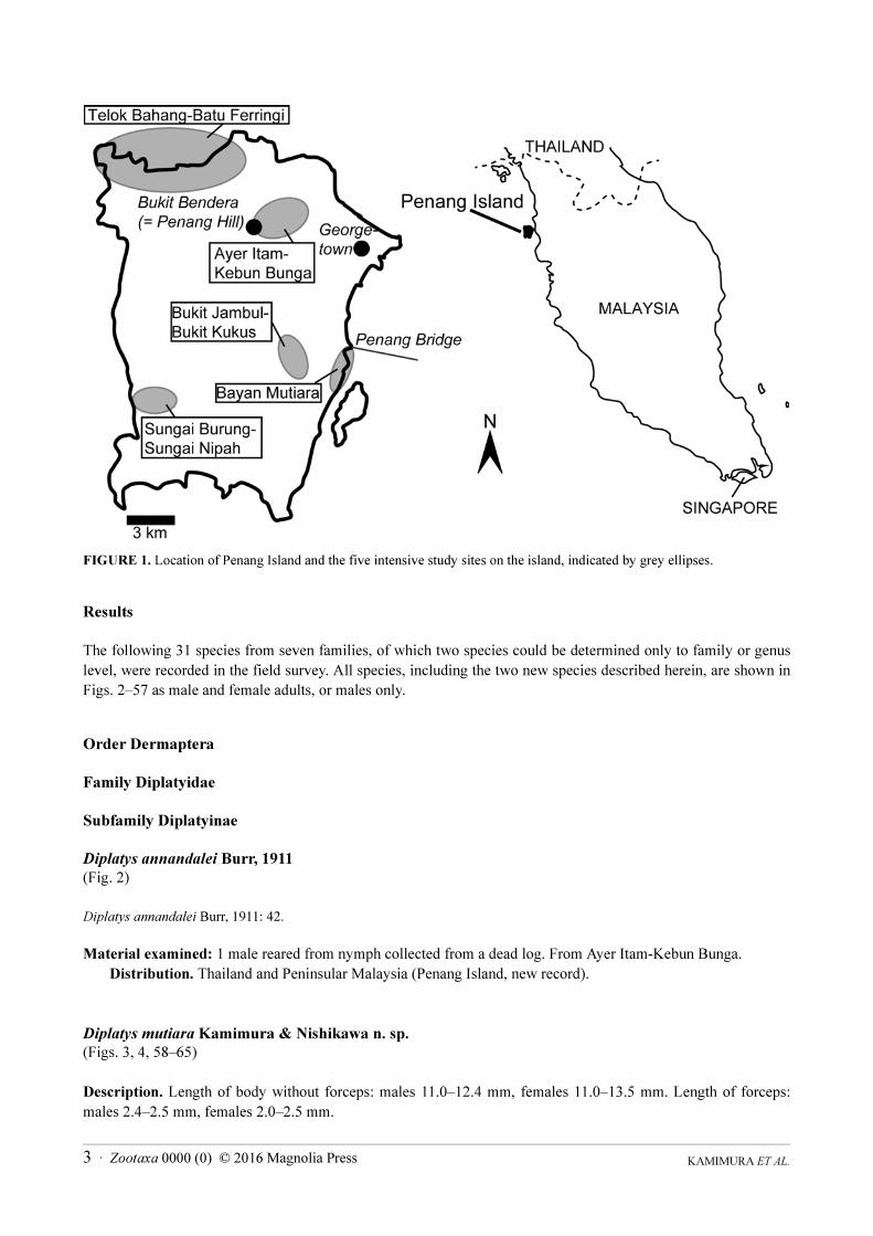
The following five areas were selected as intensive study sites, with more than 10 days of surveys at each site
(Table 1, Fig. 1): Telok Bahang-Batu Ferringi, Ayer Itam-Kebun Bunga, Bukit Jambul-Bukit Kukus, Bayan
Mutiara, and Sungai Burung-Sungai Nipah areas. Collectively, these sites included almost all environmental and
vegetation types on the island (coastal environments, grasslands, park gardens, fruit plantations, rubber plantations,
rice fields, secondary forests, and natural forests), from the coastline to mountainous regions with elevations of up
to 700 m (Table 1). We also reviewed critically the literature records of Dermaptera from Penang Island and the
distribution of species found in the field survey. Monographs or compilations by Sakai (1985, 1987, 1990, 1991,
1992, 1993, 1994, 1995a, 1995b, 1995c, 1995d, 1996) and Steinmann (1986, 1989a, 1989b, 1990, 1993) were used
extensively as primary references.
TABLE 1. Penang Island environments of the five intensive study sites plotted in Fig. 1.
The suprageneric classification followed in the present work follows Srivastava (2003b), excepting the family
Pygidicranidae. Srivastava (2003b) placed the subfamily Diplatyinae in the family Pygidicranidae. In the present
work, Diplatyidae at the level of family and Diplatyinae were used according to the classification proposed by
Sakai (1982). The generic classification of Diplatyidae + Pygidicranidae, Anisolabididae and Spongiphoridae
basically follows Srivastava (1992, 1999, 1995), respectively, unless otherwise noted. For several species, the
reproductive biology and morphology have already been reported elsewhere based on samples collected during the
field survey (Kamimura & Lee 2014a, b; Kamimura et al. in press).
Study sites, GPS coordinates Environment
Telok Bahang-Batu Ferringi area
5.464946N,100.222778E
Sandy beaches, coastal forests, and natural forest areas including dipterocarp forests
in Penang National Park (or Pantai Ache Forest Reserve), elevation to about 400 m.
Ayer Itam-Kebun Bunga area
5.433503N,100.280113E
Plantations of durian and other fruits, natural forests at high elevation on Bukit
Bendera (= Penang Hill), recreational parks and gardens including Kebun Bunga
(= Penang Botanical Garden), elevation to 730 m.
Bukit Jambul-Bukit Kukus area
5.348821N,100.285692E
Plantations of rubber, durian, banana and other fruit trees, surrounded by secondary
forests, elevation up to about 400 m.
Sungai Burung-Sungai Nipah area
5.332071N,100.199776E
Paddy fields and banana plantations in a coastal wetland, elevation less than 10 m.
Bayan Mutiara
5.342241N,100.310497E
Sandy beach with grasslands, elevation less than 10 m.
KAMIMURA ET AL.3 · Zootaxa 0000 (0) © 2016 Magnolia Press
FIGURE 1. Location of Penang Island and the five intensive study sites on the island, indicated by grey ellipses.
Results
The following 31 species from seven families, of which two species could be determined only to family or genus
level, were recorded in the field survey. All species, including the two new species described herein, are shown in
Figs. 2‒57 as male and female adults, or males only.
Order Dermaptera
Family Diplatyidae
Subfamily Diplatyinae
Diplatys annandalei Burr, 1911
(Fig. 2)
Diplatys annandalei Burr, 1911: 42.
Material examined: 1 male reared from nymph collected from a dead log. From Ayer Itam-Kebun Bunga.
Distribution. Thailand and Peninsular Malaysia (Penang Island, new record).
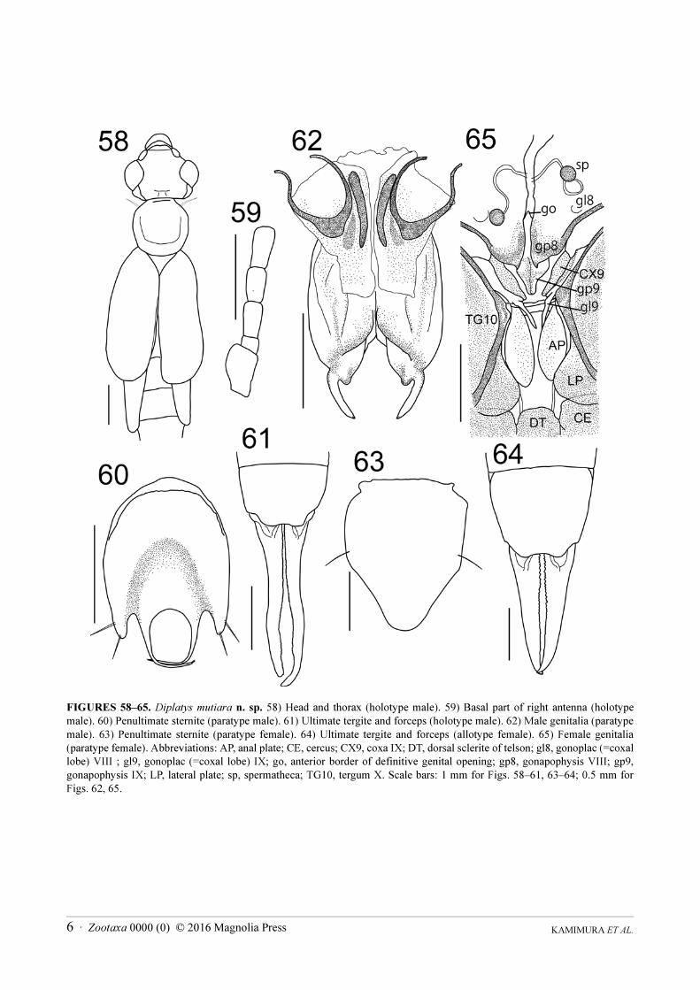
Diplatys mutiara Kamimura & Nishikawa n. sp.
(Figs. 3, 4, 58‒65)
Description. Length of body without forceps: males 11.0‒12.4 mm, females 11.0‒13.5 mm. Length of forceps:
males 2.4‒2.5 mm, females 2.0‒2.5 mm.

KAMIMURA ET AL.4 · Zootaxa 0000 (0) © 2016 Magnolia Press
FIGURES 2‒57. Thirty earwig species from Penang Island. Scale bars = 3 mm. 2) Diplatys annandalei male. 3, 4) Diplatys
mutiara n. sp., male, female. 5, 6) Echinosoma denticulatum male, female. 7, 8) Echinosoma sumatranum male, female. 9, 10)
Echinosoma sp. male, female. 11, 12) Euborellia annulata male, female. 13‒16) Euborellia philippinensis male, female, fully
winged form male, fully winged form female. 17, 18) Gonolabis electa male, female. 19, 20) Metisolabis punctata male,
female. 21, 22) Platylabia major male, female. 23) Allostethus indicum male. 24, 25) Labidura riparia male, female. 26, 27)
Pseudovostox brindlei male, female. 28, 29) Chaetospania anderssoni male, female. 30, 31) Chaetospania javana male,
female. 32, 33) Chaetospania huisiangi n. sp., male, female. 34) Chaetospania thoracica male; 35) Paralabellula boettcheri
male. 36, 37) Paralabellula curvicauda male, female. 38, 39) Paralabellula rotundifrons male, female. 40, 41) Spirolabia
pilicornis male, female. 42, 43) Nesogaster amoenus male, female. 44, 45) Marava arachidis male, female. 46, 47)
Spongovostox mucronatus male, female. 48) Spongovostox semiflavus male. 49, 50) Chelisoches morio male, female. 51, 52)
Hamaxas crassus male, female. 53, 54) Proreus coalescens male, female. 55) Proreus ludekingi male. 56, 57) Hypurgus
humeralis male, female.

KAMIMURA ET AL.5 · Zootaxa 0000 (0) © 2016 Magnolia Press
Male (Fig. 3) generally dark brown; mouth parts, antennae, pronotum, tegmina at base, scutellum and legs light
yellowish brown, except foreleg with tarsus, tibia and apical two thirds of femur dark brown. Head (Fig. 58)
sparsely pubescent, about as long as broad, widest in the region of eyes; frons tumid and occiput depressed;
transverse suture obsolete, median suture distinct; hind margin weakly emarginated in middle; post-ocular carina
sharp, curved, running from middle of the internal margin of eyes to the hind margin of the head. Antennae (Fig.
59) partly broken in holotype; first segment stout, slightly expanded apically, length about half of the distance
between the antennal bases; second segment short, transverse; third segment long and slender, fourth only slightly
longer than broad, shorter and stouter than third; fifth segment nearly equal to third, remaining segments gradually
lengthening and narrowing distally. Eyes prominent, longer than post-ocular length. Pronotum (Fig. 58) smooth,
about as long as broad, octagonal; anterior and posterior margins almost straight; sides rounded; fore and hind
angles weakly rounded; median sulcus distinct; prozona weakly raised and metazona depressed. Tegmina and
wings (Fig. 58) well-developed, smooth, pubescent; small triangular scutellum visible. Legs long, slender; hind
tarsi with first segment 2.5‒3.0 times longer than third, second segment about half as long as third, claw with
arolium. Abdomen long, cylindrical, smooth, sparsely pubescent, segments eight and nine slightly enlarged.
Penultimate sternite (Fig. 60) caudal margin deeply trisinuate; horseshoe-shaped depression present on posterior
half originating from the lateral sides of the median sinuation, the sides of the median sinuation taking the form of
two long, prominent projections that curve inward with several strong setae only at the apices. Ultimate tergite
(Fig. 61) smooth, about as long as broad, gradually sloping backwards; lateral margins oblique; hind margin with a
pair of swellings at the bases of forceps. Forceps (Fig. 61) pubescent, slender, tapering apically, undulate; apices
pointed and gently hooked (apex of right branch broken in holotype); inner margin covered with row of small black
denticles. Genitalia (Fig. 62) with virga dividing in apical third of lobe, with tips protruding from penis lobes;
parameres (= external parameres) comparatively short, base wide, inner margin with large rounded tooth
proximally.
Female (Fig. 4) coloration similar to males but with dark brownish markings on middle and hind femora from
apical one-third to slightly before apex. Third segment of hind tarsi minute. Eyes smaller, about as long as
postocular length. Penultimate sternite (Fig. 63), simple, almost triangular in posterior half. Forceps (Fig. 64)
straight, tapering, with inner margin crenulate. Genitalia (Fig. 65) with well-developed ovipositor components,
with pair of spermathecae each with brownish capsule at the distal end.
Specimens examined. All specimens listed below were collected by Y. Kamimura. Holotype male, Jalan
Kebun Bunga, Penang Island, Peninsular Malaysia, collected as nymph 4.XI.2012, adult 20.XI.2012 (OMNH).
Allotype female, same locality as holotype, collected as nymph 18.X.2012, adult 30.X.2012 (OMNH). Paratypes
from same locality as holotype: 1 male (genitalia mounted between two cover slips and attached to pin of
specimen) collected as nymph 18.X.2012, adult 30.X.2012 (OMNH); 1 female (genitalia, ultimate tergite, and
forceps mounted between two cover slips and attached to pin of specimen), collected as nymph 15.X.2012, adult
5.XI.2012 (OMNH); 1 male collected as nymph 18.X.2012, adult 11.XI.2012 (LKCNHM); 1 female, collected as
nymph 4.XI.2012, adult 17.XI.2012 (LKCNHM); 1 male collected as nymph 18.X.2012, adult 2.XI.2012 (EUMJ);
1 female collected as nymph 18.X.2012, adult 30.X.2012 (EUMJ); 1 female collected as nymph 1.XII.2012, adult
17.XII.2012 (EUMJ); 1 male collected as nymph 6.III.2014, adult 5.V.2014 (EUMJ); 1 female, collected as nymph
6.III.2014, adult 5.IV.2014(EUMJ); Paratypes from Ayer Itam, Penang Island, Peninsular Malaysia: 1 female
collected as nymph 6.XII.2012, adult 23.XII.2012 (LKCNHM); 1 male collected as nymph 6.XII.2012, adult
23.XII.2012 (EUMJ).
Additional material examined, same locality as holotype: 1 female collected as nymph 4.XI.2012, adult
20.XI.2012; 1 female collected as nymph 18.X.2012, adult 30.X.2012.
Etymology. The specific epithet refers to the type locality, Penang Island, which is often called the “Pearl of
the Orient.” “Mutiara” means pearl in the Malay language.
Distribution. Penang Island, Malaysia.
Remarks. Nymphs are common under fallen leaves near small mountain streams and also found from Telok
Bahang-Batu Ferringi and Bukit Jambul-Bukit Kukus areas. Adult males can be distinguished readily from all
other species of the family by the combination of the characteristic penultimate sternite that bears a horseshoe-
shaped depression and a pair of prominent projections that curve inward with several strong setae only at the apices
(Fig. 60), and parameres with a wide base and a large rounded tooth proximally at the inner margin (Fig. 62).
KAMIMURA ET AL.6 · Zootaxa 0000 (0) © 2016 Magnolia Press
FIGURES 58‒65. Diplatys mutiara n. sp. 58) Head and thorax (holotype male). 59) Basal part of right antenna (holotype
male). 60) Penultimate sternite (paratype male). 61) Ultimate tergite and forceps (holotype male). 62) Male genitalia (paratype
male). 63) Penultimate sternite (paratype female). 64) Ultimate tergite and forceps (allotype female). 65) Female genitalia
(paratype female). Abbreviations: AP, anal plate; CE, cercus; CX9, coxa IX; DT, dorsal sclerite of telson; gl8, gonoplac (=coxal
lobe) VIII ; gl9, gonoplac (=coxal lobe) IX; go, anterior border of definitive genital opening; gp8, gonapophysis VIII; gp9,
gonapophysis IX; LP, lateral plate; sp, spermatheca; TG10, tergum X. Scale bars: 1 mm for Figs. 58‒61, 63‒64; 0.5 mm for
Figs. 62, 65.
KAMIMURA ET AL.7 · Zootaxa 0000 (0) © 2016 Magnolia Press
Family Pygidicranidae
Subfamily Echinosomatinae
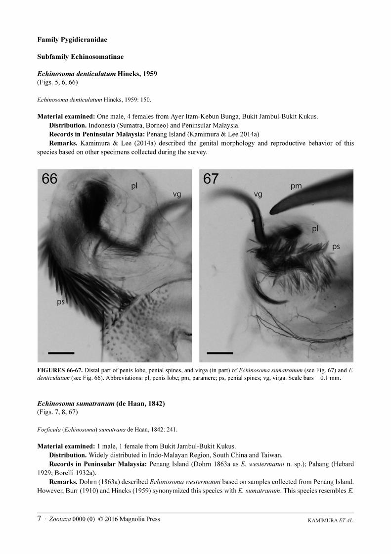
Echinosoma denticulatum Hincks, 1959
(Figs. 5, 6, 66)
Echinosoma denticulatum Hincks, 1959: 150.
Material examined: One male, 4 females from Ayer Itam-Kebun Bunga, Bukit Jambul-Bukit Kukus.
Distribution. Indonesia (Sumatra, Borneo) and Peninsular Malaysia.
Records in Peninsular Malaysia: Penang Island (Kamimura & Lee 2014a)
Remarks. Kamimura & Lee (2014a) described the genital morphology and reproductive behavior of this
species based on other specimens collected during the survey.
FIGURES 66-67. Distal part of penis lobe, penial spines, and virga (in part) of Echinosoma sumatranum (see Fig. 67) and E.
denticulatum (see Fig. 66). Abbreviations: pl, penis lobe; pm, paramere; ps, penial spines; vg, virga. Scale bars = 0.1 mm.
Echinosoma sumatranum (de Haan, 1842)
(Figs. 7, 8, 67)
Forficula (Echinosoma) sumatrana de Haan, 1842: 241.
Material examined: 1 male, 1 female from Bukit Jambul-Bukit Kukus.
Distribution. Widely distributed in Indo-Malayan Region, South China and Taiwan.
Records in Peninsular Malaysia: Penang Island (Dohrn 1863a as E. westermanni n. sp.); Pahang (Hebard
1929; Borelli 1932a).
Remarks. Dohrn (1863a) described Echinosoma westermanni based on samples collected from Penang Island.
However, Burr (1910) and Hincks (1959) synonymized this species with E. sumatranum. This species resembles E.

KAMIMURA ET AL.8 · Zootaxa 0000 (0) © 2016 Magnolia Press
denticulatum, but can be distinguished by the darker coloration of the body (especially the basal part of femora),
the different shape of the female pygidium (see Hincks 1959), and less-developed penial spines in the male
genitalia (Figs. 66, 67).
Echinosoma sp.
(Figs. 9, 10)
Material examined: 1 male, 1 female from Bukit Jambul-Bukit Kukus.
Remarks. These specimens possibly represent an undescribed species.
Family Anisolabididae
Subfamily Anisolabidinae
Euborellia annulata (Fabricius, 1793)
Forficula annulata Fabricius, 1793: 4.
(Figs. 11, 12, 68A)
Material examined: 5 males, 10 females from Telok Bahang-Batu Ferringi, Ayer Itam-Kebun Bunga, Bayan
Mutiara, Sungai Burung-Sungai Nipah
Distribution. Almost worldwide in distribution, especially in warmer parts.
Record in Peninsular Malaysia: Pahang (Borelli (1932a) as Euborellia stali).
Remarks. The identification of the samples is only tentative, because of the many similar species in the genus
that areinadequately described. As in other examined Euborella spp, (Hudson 1973; Kamimura 2000), the
spermatheca is a very long, thin, blind duct without a capsule at the distal end (Fig. 68A). In one laboratory-raised
male the tegmina were well-developed but hindwings were totally lacking.
Euborellia philippinensis Srivastava, 1979
(Figs. 13−16, 68B)
Euborellia philippinensis Srivastava, 1979: 49.
Material examined: 10 males, 11 females from Sungai Burung-Sungai Nipah.
Distribution. Philippines (Luzon), and Peninsular Malaysia (Penang Island, new record).
Remarks. The brachypterous specimens possessed tegmina as narrow ovate flaps. The external morphology
and male genitalia of this species agree with previous descriptions of E. philippinensis, and thus we assign our
specimens to this species. Field-collected adults were all brachypterous, but laboratory-reared adults showed
remarkable wing dimorphism. In the fully winged form adults, both tegmina and hindwings are fully developed
(Figs. 15, 16). This form is reported here for the first time for E. philippinensis. The fully winged form of our
specimens cannot be distinguished from those of Euborellia plebeja (Dohrn, 1863) (Dohrn 1863b; Srivastava
2003a), warranting further studies of this group. Similar to its close relatives (Hudson 1973; Kamimura 2000), the
spermatheca is a very long, thin, blind duct without a capsule at the distal end (Fig. 68B).
Gonolabis electa Burr, 1910
(Figs. 17, 18)
Gonolabis electa Burr, 1910: 79.

KAMIMURA ET AL.9 · Zootaxa 0000 (0) © 2016 Magnolia Press
Material examined: 2 males, 1 female from Ayer Itam-Kebun Bunga, Bukit Jambul-Bukit Kukus.
Distribution. Oriental Region, adventive in Africa (São Tomé and west African coast).
Records in Peninsular Malaysia: Negeri Sembilan (Nishikawa 1973); Ulu Gombak, Selangor (Steghaus-
Kovac & Maschwitz 1993).
Remarks. The male shown in Fig. 17 was bleached by long-term preservation in alcohol, but in life the color
of males is similar to that of females (Fig. 18).
FIGURE 68. Spermatheca of several species of earwigs from Penang Island. A) Euborellia annulata. B) Euborellia
philippinensis. C) Metisolabis punctata. D) Platylabia major. E) Chaetospania anderssoni. F) Spirolabia pilicornis. G)
Nesogaster amoenus. H) Spongovostox mucronatus. I) Proreus coalescens. J) Hypurgus humeralis. For P. major (D) and S.
mucronatus (H), structures near the spermatheca are also shown. Abbreviations: CP8, coxopodite VIII; CP9, coxopodite IX;
gp8, gonapophysis VIII; LC9?, laterocoxa IX?; si8, spiracle VIII; so, spermathecal opening; SO, sclerotization around
spermathecal opening; sp, spermatheca; TG8-9, tergum VIII-X. Scale bars = 0.1 mm.

KAMIMURA ET AL.10 · Zootaxa 0000 (0) © 2016 Magnolia Press
Subfamily Brachylabidinae
Metisolabis punctata (Dubrony, 1879)
(Figs. 19, 20, 68C)
Brachylabis punctata Dubrony, 1879: 357.
Material examined: 1 male, 3 females from Telok Bahang-Batu Ferringi, Ayer Itam-Kebun Bunga, Bukit Jambul-
Bukit Kukus.
Distribution. Oriental Region, New Guinea, North Australia. Peninsular Malaysia (Penang Island, new
record).
Remarks. The spermatheca is a very long, thin, blind duct without a capsule at the distal end (Fig. 68C).
Subfamily Platylabinae (= Palicinae Engel & Haas 2007, = Palexinae Kočárek 2010)
Platylabia major Dohrn, 1867
(Figs. 21, 22, 68D)
Platylabia major Dohrn, 1867: 347.
Material examined: 12 males, 16 females from Ayer Itam-Kebun Bunga, Bukit Jambul-Bukit Kukus, Bukit
Gedung.
Distribution. Widely distributed in the Oriental Region: South India (?), Sri Lanka, Peninsular Malaysia,
Myanmar, China (Guangxi, Hainan), Thailand, Laos, Vietnam, Indonesia (Java, Sumatra, Celebes), and
Philippines.
Records in Peninsular Malaysia: Pulo Penang (= Penang Island, Bormans 1900, as Platylabia sparattoides);
Selangor (Srivastava & Kovac 1993).
Remarks. The spermatheca of this species consists of a weakly pigmented capsule at the end of an elongated
duct (Fig. 68D). The opening region of the spermatheca is located between a pair of triangular lobes (Fig. 68D),
which are apparently homologous to the eighth-segment gonapophyses (gp8) of some labidurids (Kamimura & Lee
2014b). The outer margins of gp8 are heavily sclerotized and well-pigmented, and continue to a laterally adjoining
sclerite with many muscles (Fig. 68D). This sclerite likely represents the ninth-segment laterocoxa (LC9; see
Kamimura & Lee 2014b). These remarks constitute the first report of gp8 and LC9 for a member of
Anisolabididae.
Family Labiduridae
Subfamily Allostethinae
Allostethus indicum (Burmeister, 1838)
(Figs. 23)
Forficula indica Burmeister, 1838: 751.
Material examined: 3 males, 2 females from Ayer Itam-Kebun Bunga, Bukit Jambul-Bukit Kukus.
Distribution. Indonesia (Java, Sumatra, Borneo, Sulawesi, Batu Island), Malaysia (Malay Peninsula),
Thailand (Trang), Philippine Islands.
Records in Peninsular Malaysia: Selangor and Pahang (Hebard 1929); Perak (Burr 1912; Borelli 1932a);
Pahang (Borelli 1932a); Penang Island (Dubrony 1879; Kamimura & Lee 2014b); Malacca (Burr 1912).
Remarks. Kamimura & Lee (2014b) described the genital morphology and reproductive behavior of this
species based on other specimens collected during the survey.

KAMIMURA ET AL.11 · Zootaxa 0000 (0) © 2016 Magnolia Press
Subfamily Labidurinae
Labidura riparia (Pallas, 1773)
(Figs. 24, 25)
Forficula riparia Pallas, 1773: 727.
Material examined: 4 males, 2 females from Bayan Mutiara
Distribution. Cosmopolitan.
Record in Peninsular Malaysia: Negri Sembilan and Selangor (Borelli 1932a); Penang Island (Kamimura &
Lee 2014b).
Remarks. Kamimura & Lee (2014b) described the female genital morphology of this species based on other
specimens collected during the survey.
Family Spongiphoridae
Subfamily Geracinae
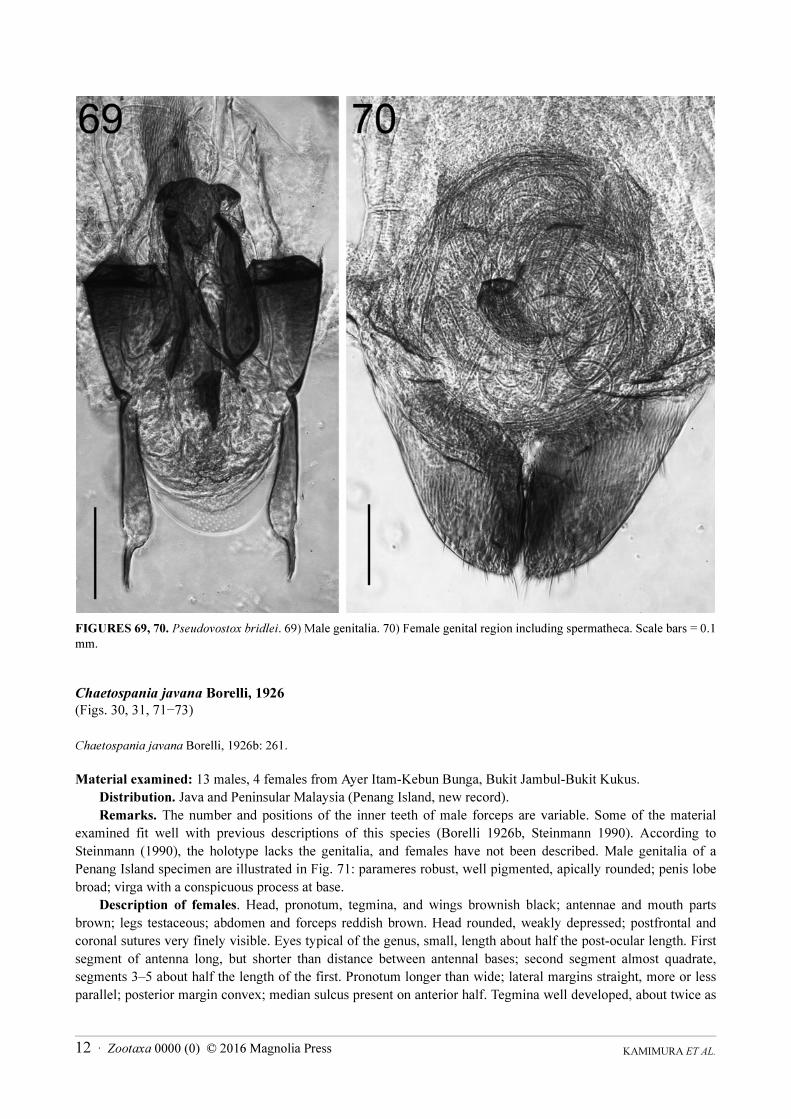
Pseudovostox brindlei Srivastava, 2003
(Figs. 26, 27, 69, 70)
Pseudovostox brindlei Srivastava, 2003b: 45.
Material examined: 10 males, 10 females from Ayer Itam-Kebun Bunga, Bukit Jambul-Bukit Kukus.
Distribution. Vietnam and Peninsular Malaysia (Penang Island, new record).
Remarks. Previously known from Vietnam. The body coloration of the samples collected from Penang Island
differed slightly from those of the original description: Srivastava (2003b) described the head, pronotum, tegmina,
and hindwings of this species as blackish brown, except for yellow makings on the pronotum, tegmina and
hindwings. Specimens from Penang Island have the head reddish-brown, pronotum and tegmina bases mostly
yellowish-brown, and distal parts of tegmina and hindwings purplish brown with white markings (Figs. 26, 27).
Except for the body coloration, our material agrees well with previous descriptions of the morphology of the
species, including male genital morphology (Fig. 69), and thus the Penang Island specimens are considered to be P.
brindlei. The female genitalia of this genus are described here for the first time. A pair of well-developed lobes
were found posterior to the spermatheca, which is a long, thin duct (Fig. 70). The morphological identity of the
lobes is presently unknown, but they represent the first report of ovipositor-like structures for females of the
Eudermaptera (Schneider & Klass 2013).
This species may be myrmecophilous, and is frequently found together with ants of the genus Crematogaster,
which show similar body coloration.
Subfamily Labiinae
Chaetospania anderssoni Brindle, 1971
(Figs. 28, 29)
Chaetospania anderssoni Brindle, 1971: 227.
Material examined: 6 males, 4 females from Sungai Burung-Sungai Nipah.
Distribution. Sri Lanka and Peninsular Malaysia (Penang Island, new record).
Remarks. The spermatheca of this species consists of an elongated thin tube with a well-pigmented sclerotized
capsule at the distal end (Fig. 68E). Kočárek (2009) reported ovoviviparity for Chaetospania borneensis (Dubrony,
1879), but we observed that female C. anderssoni deposits eggs as is usual in forficuline earwigs.
KAMIMURA ET AL.12 · Zootaxa 0000 (0) © 2016 Magnolia Press
FIGURES 69, 70. Pseudovostox bridlei. 69) Male genitalia. 70) Female genital region including spermatheca. Scale bars = 0.1
mm.
Chaetospania javana Borelli, 1926
(Figs. 30, 31, 71−73)
Chaetospania javana Borelli, 1926b: 261.
Material examined: 13 males, 4 females from Ayer Itam-Kebun Bunga, Bukit Jambul-Bukit Kukus.
Distribution. Java and Peninsular Malaysia (Penang Island, new record).
Remarks. The number and positions of the inner teeth of male forceps are variable. Some of the material
examined fit well with previous descriptions of this species (Borelli 1926b, Steinmann 1990). According to
Steinmann (1990), the holotype lacks the genitalia, and females have not been described. Male genitalia of a
Penang Island specimen are illustrated in Fig. 71: parameres robust, well pigmented, apically rounded; penis lobe
broad; virga with a conspicuous process at base.
Description of females. Head, pronotum, tegmina, and wings brownish black; antennae and mouth parts
brown; legs testaceous; abdomen and forceps reddish brown. Head rounded, weakly depressed; postfrontal and
coronal sutures very finely visible. Eyes typical of the genus, small, length about half the post-ocular length. First
segment of antenna long, but shorter than distance between antennal bases; second segment almost quadrate,
segments 3‒5 about half the length of the first. Pronotum longer than wide; lateral margins straight, more or less
parallel; posterior margin convex; median sulcus present on anterior half. Tegmina well developed, about twice as
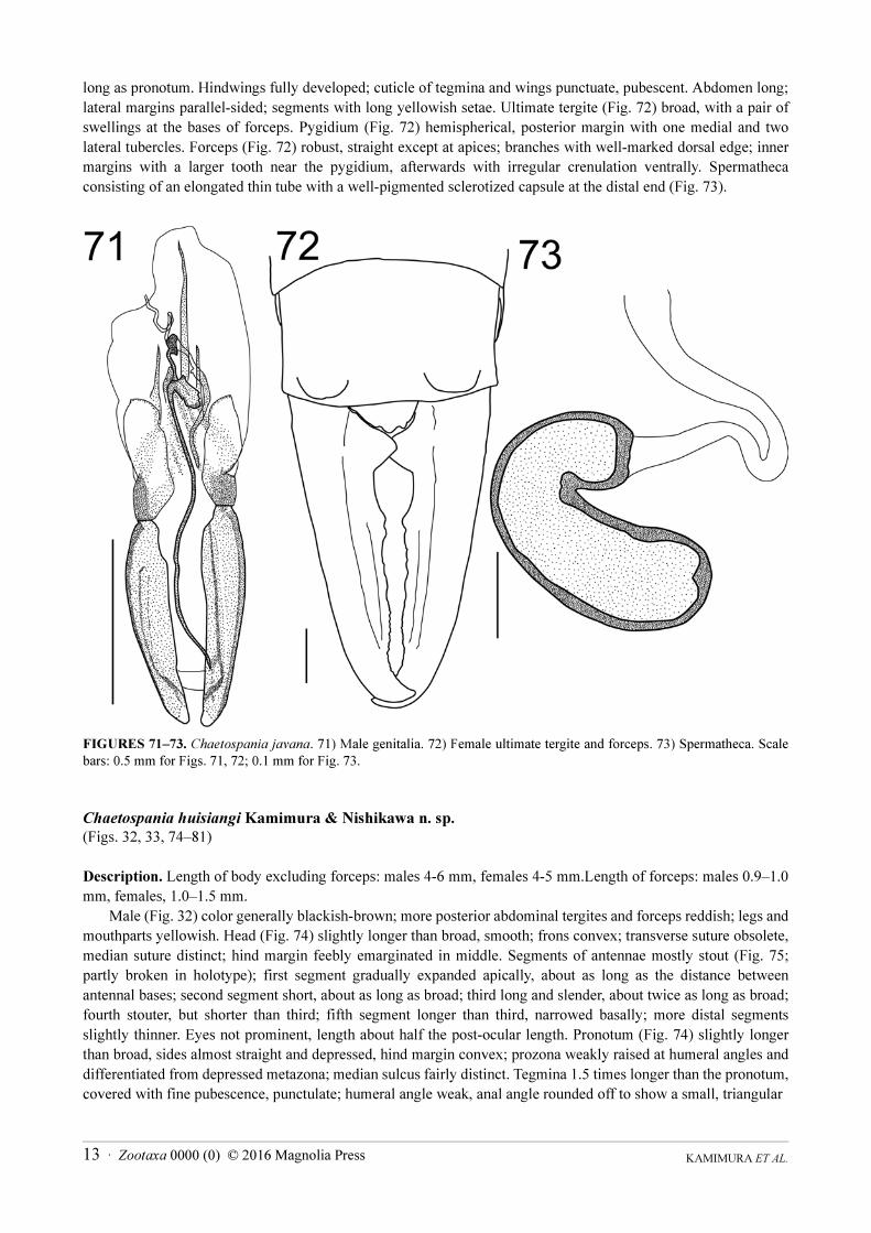
KAMIMURA ET AL.13 · Zootaxa 0000 (0) © 2016 Magnolia Press
long as pronotum. Hindwings fully developed; cuticle of tegmina and wings punctuate, pubescent. Abdomen long;
lateral margins parallel-sided; segments with long yellowish setae. Ultimate tergite (Fig. 72) broad, with a pair of
swellings at the bases of forceps. Pygidium (Fig. 72) hemispherical, posterior margin with one medial and two
lateral tubercles. Forceps (Fig. 72) robust, straight except at apices; branches with well-marked dorsal edge; inner
margins with a larger tooth near the pygidium, afterwards with irregular crenulation ventrally. Spermatheca
consisting of an elongated thin tube with a well-pigmented sclerotized capsule at the distal end (Fig. 73).
FIGURES 71‒73. Chaetospania javana. 71) Male genitalia. 72) Female ultimate tergite and forceps. 73) Spermatheca. Scale
bars: 0.5 mm for Figs. 71, 72; 0.1 mm for Fig. 73.
Chaetospania huisiangi Kamimura & Nishikawa n. sp.
(Figs. 32, 33, 74‒81)
Description. Length of body excluding forceps: males 4-6 mm, females 4-5 mm.Length of forceps: males 0.9‒1.0
mm, females, 1.0‒1.5 mm.
Male (Fig. 32) color generally blackish-brown; more posterior abdominal tergites and forceps reddish; legs and
mouthparts yellowish. Head (Fig. 74) slightly longer than broad, smooth; frons convex; transverse suture obsolete,
median suture distinct; hind margin feebly emarginated in middle. Segments of antennae mostly stout (Fig. 75;
partly broken in holotype); first segment gradually expanded apically, about as long as the distance between
antennal bases; second segment short, about as long as broad; third long and slender, about twice as long as broad;
fourth stouter, but shorter than third; fifth segment longer than third, narrowed basally; more distal segments
slightly thinner. Eyes not prominent, length about half the post-ocular length. Pronotum (Fig. 74) slightly longer
than broad, sides almost straight and depressed, hind margin convex; prozona weakly raised at humeral angles and
differentiated from depressed metazona; median sulcus fairly distinct. Tegmina 1.5 times longer than the pronotum,
covered with fine pubescence, punctulate; humeral angle weak, anal angle rounded off to show a small, triangular

KAMIMURA ET AL.14 · Zootaxa 0000 (0) © 2016 Magnolia Press
FIGURES 74‒81. Chaetospania huisiangi n. sp. 74) Head and thorax (holotype male). 75) Basal part of left antenna (holotype
male). 76) Penultimate sternite and forceps (paratype male). 77) Ultimate tergite and forceps (holotype male). 78) Male
genitalia (paratype male). 79) Penultimate sternite and forceps (paratype female). 80) Ultimate tergite and forceps (allotype
female). 81) Female genitalia (paratype female). Scale bars: 0.5 mm for Figs. 74‒80, 0.1 mm for Fig. 81.

KAMIMURA ET AL.15 · Zootaxa 0000 (0) © 2016 Magnolia Press
scutellum; hind margin oblique. Wings well developed, punctate, pubescent. Femora swollen; first tarsal segment
of hind leg almost equal to third; second segment short, about as long as broad. Abdomen strongly depressed, long,
more or less parallel-sided, except first two segments narrowed; segments with long yellowish setae on sides;
lateral tubercules on third and fourth tergites inconspicuous; sides of segments broadly convex. Penultimate
sternite (Fig. 76) transverse, obscurely punctulate, with fine and long pubescence; hind margin rounded with slight
emargination in middle. Ultimate tergite (Fig. 77) smooth, rectangular, transverse, moderately depressed; hind
margin nearly truncate with small rounded swellings above the base of forceps. Pygidium large, with dorsal and
ventral part; dorsal part rounded, tumid; ventral part flattened, armed with one pair of large lateral tubercles
separated by deep emargination. Forceps (Figs. 76, 77) strongly setose, strongly curving in posterior half,
depressed, tapering apically; internal margin with a large triangular tooth at middle directed inwards. Genitalia
(Fig. 78) with parameres broader in basal two-thirds, apically narrowed with acute tip; penis lobe provided with
several sclerotized plates and rows of fine chitinous teeth; virga short, tubular, with vesicle at base.
Female (Fig. 33) similar to male, but hind margin of penultimate sternite (Fig. 79) without emargination in
middle. Pygidium (Fig. 80) transverse, broad basally, narrowed apically; lateral margins weakly raised; posterior
margin weakly emarginated in middle. Forceps (Figs. 79-80) stout and straight; at base with large ventral tooth,
otherwise irregularly dentate dorsally as well as ventrally. Spermatheca consists of an elongated thin tube with a
characteristic pea pod-shaped sclerotized capsule at the distal end (Fig. 81).
Specimens examined. All specimens listed below were collected by Y. Kamimura at Bukit Jambul. Penang
Island, Peninsular Malaysia. Holotype male, offspring of a female collected 22.XI.2012 (OMNH). Allotype
female, same data as for holotype (OMNH).
Paratypes, same locality data as holotype: 1 male (genitalia mounted between two cover slips and attached to
pin of specimen), same data as for holotype (OMNH); 1 female (spermatheca mounted between two cover slips and
attached to pin of specimen), 7.XII.2012 (OMNH); 2 males, 7.XII.2012 (LKCNHM); 1 male, 2.III.2013
(LKCNHM); 1 female (LKCNHM); 2 males, 30.XI.2012 (EUMJ); 1 female (EUMJ).
Additional material, same locality data as holotype: 1 male, 26.X.2012; 2 males, 22.XI.2012; 1 male,
2.III.2013; 8 males, 5 females, 22.XI.2012; 1 female, 30.XI.2012.
Etymology. Named in honor of Hui-Siang Tee, Universiti Sains Malaysia, who assisted in our collection and
rearing of insects during the survey on Penang Island.
Distribution. Penang Island, Malaysia.
Remarks. Adults and nymphs of this species were found under bark of dead logs in rubber plantations. The
general appearance of this species closely resembles that of C. minuta Borelli, 1921, which has been recorded from
Borneo (the type locality), Langkawi Island, and the Philippines (Mindanao) (Borelli 1921, 1926a, 1932a, 1932b).
However, the shape of male pygidium is distinctively different; the posterior margin is convex in C. minuta
(Srivastava 1983) but deeply incised in C. huisiangi n. sp. (Fig. 77). Females of the two species cannot be
distinguished. The record of C. minuta from Langkawi Island, located about 90 km north of Penang Island, is based
on a female sample (Borelli 1932a: 86), and thus it might be belong to C. huisiangi n. sp.. This new species also
resembles C. brunneri (Bormans, 1883) in many external features, but the shapes of the male genitalia and female
spermatheca are markedly different (Hincks 1949, Hudson 1973, Steinmann 1990 (as Paraspania brunneri)).
Chaetospania thoracica (Dohrn, 1867)
(Fig. 34)
Platylabia thoracica Dohrn, 1867: 348.
Material examined: 4 males, 1 female from Ayer Itam-Kebun Bunga, Bukit Jambul-Bukit Kukus.
Distribution. Oriental Region, New Guinea, Seychelles.
Records in Peninsular Malaysia: Selangor (Borelli 1932a; Srivastava & Kovac 1993); Perak (Borelli 1932a);
Malacca (Brindle 1968); Penang (Dohrn 1867 as Platylabia thoracica).
Remarks. Penang Island is the type locality of this species (Dohrn 1867).

KAMIMURA ET AL.16 · Zootaxa 0000 (0) © 2016 Magnolia Press
Paralabellula boettcheri (Borelli, 1923)
(Fig. 35)
Labia boettcheri Borelli, 1923: 7.
Material examined: 1 male from Bukit Jambul-Bukit Kukus.
Distribution. Philippines and Peninsular Malaysia (Penang Island, new record).
Paralabellula curvicauda (Motschulsky, 1863)
(Figs. 36, 37)
Forfiscelia curvicauda Motschulsky, 1863: 2.
Material examined: 13 males, 7 females from Telok Bahang-Batu Ferringi, Ayer Itam-Kebun Bunga, Bukit
Jambul-Bukit Kukus, Sungai Burung-Sungai Nipah, Bukit Gedung
Distribution. Cosmopolitan.
Records in Peninsular Malaysia: Penang Island (Dubrony 1879 as Labia curvicauda); Pahang (Borelli 1932a
as Labia curvicauda); Selangor (Borelli 1932a as Labia curvicauda; Srivastava & Kovac 1993 as Paralabella
curvicauda).
Remarks. Dubrony (1879) reported this species (as Labia curvicauda) from “Pulo Penang” (= Pulau Pinang,
Penang Island) based on one male. The spermathecal morphology has been described by Hudson (1973).
Paralabellula rotundifrons (Hincks, 1954)
(Figs. 38, 39, 82−84)
Labia rotundifrons Hincks, 1954: 20.
FIGURES 82‒84. Paralabellula rotundifrons. 82) Male genitalia. 83) Female ultimate tergite and forceps. 84) Spermatheca.
Scale bars: 0.1 mm for Figs. 82, 84; 0.5 mm for Fig. 83.

KAMIMURA ET AL.17 · Zootaxa 0000 (0) © 2016 Magnolia Press
Material examined: 5 males, 6 females from Telok Bahang-Batu Ferringi, Bukit Jambul-Bukit Kukus.
Distribution. Sri Lanka, and Peninsular Malaysia (Penang Island, new record).
Remarks. The male genitalia have a well-sclerotized plate in the penis lobe (Fig. 82). Females are described
here for the first time.
Description of female. Body colour fuscous, antennae and legs lighter; abdominal segments of distal part and
forceps dark reddish-brown. Cuticle finely punctate and pubescent. Head broad, tumid; postfrontal and coronal
sutures indistinct; lateral margins behind eyes with postero-lateral angles rounded; posterior margin concave. Eyes
small, length about half of post-ocular length. First antennal segment shorter than distance between antennal bases;
second segment wider than long, third longer than fourth. Pronotum longer than wide; lateral margins straight;
posterior margin convex. Tegmina and hindwings normally developed. Abdomen fusiform, slightly widened
medially. Ultimate tergite (Fig. 83) transverse, smooth, simple; posterior margin with a pair of swellings at the base
of forceps. Pygidium (Fig. 83) transverse. Forceps trigonal; inner margins serrated both ventrally and dorsally.
Spermatheca (Fig. 84) resembles that of P. curvicauda, but capsule less pigmented.
Spirolabia pilicornis (Motschulsky, 1863)
(Figs. 40, 41, 68F)
Forfiscelia pilicornis Motschulsky, 1863: 2.
Material examined: 6 males, 12 females from Bukit Jambul-Bukit Kukus, Sungai Burung-Sungai Nipah.
Distribution. Widely distributed in circumtropical regions.
Records in Peninsular Malaysia: Selangor (Borelli 1932a as Labia pilicornis); Malacca (Burr 1912 as Labia
pilicornis).
Remarks. The spermatheca of this species consists of an elongated thin tube with a sclerotized capsule at the
distal end (Fig. 68F).
Subfamily Nesogastrinae
Nesogaster amoenus (Stål, 1855)
(Figs. 42, 43, 68G)
Forficula amoena Stål, 1855: 350.
Material examined: 12 males, 20 females from Telok Bahang-Batu Ferringi, Ayer Itam-Kebun Bunga, Bukit
Jambul-Bukit Kukus, Bukit Gedung.
Distribution. Philippines (Mindanao, Luzon), Peninsular Malaysia (Penang Island, new record), Indonesia
(Sumatra, Mentawei, Java, Borneo, Celebes), New Guinea, and Australia.
Remarks. The spermatheca of this species consists of an elongated thin tube with a weakly pigmented capsule
at the distal end (Fig. 68G).
Subfamily Spongiphorinae
Marava arachidis (Yersin, 1860)
(Figs. 44, 45)
Forficula arachidis Yersin, 1860: 509.
Material examined: 203 males, 432 females from Minden (Universiti Sains Malaysia)
Distribution. Almost all faunal Regions.
Record in Peninsular Malaysia: Malacca (Brindle 1968); Penang Island (Kamimura et al. in press).
KAMIMURA ET AL.18 · Zootaxa 0000 (0) © 2016 Magnolia Press
Remarks. A detailed description of the female genital structures is given in Schneider & Klass (2013).
Kamimura et al. (in press) described the reproductive biology of this ovoviviparous species based on other
specimens collected during the survey.
Spongovostox mucronatus (Stål, 1860)
(Figs. 46, 47, 68H)
Forficula mucronata Stål, 1860: 303.
Material examined: 3 males, 2 females from Bukit Jambul-Bukit Kukus.
Distribution. Widely distributed in the Oriental Region: India, Sri Lanka, Myanmar, Peninsular Malaysia,
Indonesia (Sumatra, Java, East Sumba, West Flores), and Philippines; also recorded from Mauritius, New Guinea
and Ambon Island.
Records in Peninsular Malaysia: Selangor (Borelli 1932a, as Labia mucronata; Srivastava & Kovac 1993).
Remarks. Srivastava (2013) transferred this species to the genus Paratages (of the subfamily Homotaginae)
on the basis of tarsal morphology. However, well-developed sclerotization around the spermathecal opening of this
species (Fig. 68H) suggests a close affinity to S. semiflavus and M. arachidis, for which the female genitalia were
described by Schneider & Klass (2013).
Spongovostox semiflavus (Bormans, 1894)
(Fig. 48)
Spongophora semi-flava de Bormans, 1894: 385.
Material examined: 2 males from Bukit Jambul-Bukit Kukus.
Distribution. Widely distributed in the Oriental Region: India, Sri Lanka, Bhutan, Myanmar, China (Yunnan),
Laos, Thailand, Vietnam, Malaysia (Sarawak; Peninsular Malaysia), Indonesia (Sumatra, Java, Simalur, Borneo,
Sumba), Philippines (Palawan, Mindanao, Luzon), and Taiwan; also recorded from Bismarck Island.
Record in Peninsular Malaysia: Kedah (Borelli 1932a).
Remarks. Detailed description of the female genital structures is given in Schneider & Klass (2013).
Spongiphoridae gen. et sp. indet.
Material examined: 1 female from Bukit Jambul-Bukit Kukus.
Remarks. Possibly a female Irdex sp. (Irdexinae), but the identity is uncertain because of the poor condition of
the sample.
Family Chelisochidae
Subfamily Chelisochinae
Chelisoches morio (Fabricius, 1775)
(Figs. 49, 50)
Forficula morio Fabricius, 1775: 270.
Material examined: 1 male, 2 females from Bukit Jambul-Bukit Kukus, Sungai Burung-Sungai Nipah.
Distribution. Widely distributed in Oriental, Australian and Afrotropic Regions, and USA.
Records in Peninsular Malaysia: Malay Peninsula (Burr 1910); Selangor (Borelli 1932a; Hoshiba et al.
KAMIMURA ET AL.19 · Zootaxa 0000 (0) © 2016 Magnolia Press
1988); Kuala Lumpur (McClure et al. 1967); Penang Island (Dohrn 1865, as Lobophora morio; Dubrony 1879, as
Labophora morio; Burr 1901).
Remarks. Detailed descriptions of the female genital structures are given in Hudson (1973) and Schneider &
Klass (2013).
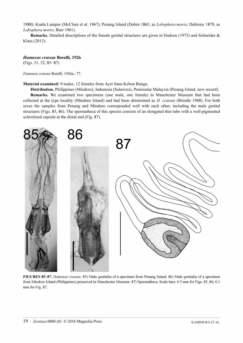
Hamaxas crassus Borelli, 1926
(Figs. 51, 52, 85−87)
Hamaxas crassus Borelli, 1926a.: 77.
Material examined: 9 males, 12 females from Ayer Itam-Kebun Bunga.
Distribution. Philippines (Mindoro), Indonesia (Sulawesi), Peninsular Malaysia (Penang Island, new record).
Remarks. We examined two specimens (one male, one female) in Manchester Museum that had been
collected at the type locality (Mindoro Island) and had been determined as H. crassus (Brindle 1968). For both
sexes the samples from Penang and Mindoro corresponded well with each other, including the male genital
structures (Figs. 85, 86). The spermatheca of this species consists of an elongated thin tube with a well-pigmented
sclerotized capsule at the distal end (Fig. 87).
FIGURES 85‒87. Hamaxas crassus. 85) Male genitalia of a specimen from Penang Island. 86) Male genitalia of a specimen
from Mindoro Island (Philippines) preserved in Manchester Museum. 87) Spermatheca. Scale bars: 0.5 mm for Figs. 85, 86; 0.1
mm for Fig. 87.

KAMIMURA ET AL.20 · Zootaxa 0000 (0) © 2016 Magnolia Press
Proreus coalescens (Borelli, 1927)
(Figs. 53, 54, 68I)
Chelisoches coalescens Borelli, 1927: 75.
Material examined: 7 males, 3 females from Bukit Jambul-Bukit Kukus.
Distribution. Philippines (Luzon), Indonesia (Sumatra) and Peninsular Malaysia (Penang Island, new record).
Remarks. The spermatheca of this species consists of an elongated thin tube with a well pigmented sclerotized
capsule at the distal end (Fig. 68I).
Proreus ludekingi (Dohrn, 1865)
(Fig. 55)
Lobophora ludekingi Dohrn, 1865: 73.
Material examined: 1 male from Bukit Jambul-Bukit Kukus.
Distribution. Oriental Region.
Records in Peninsular Malaysia: Kedah, Pahang and Selangor (Borelli 1932a); Langkawi Island (Tworzydło
et al. 2010, as ”Malaysia”).
Family Forficulidae
Subfamily Opisthocosminae
Hypurgus humeralis (Kirby, 1891)
(Fig. 56, 57, 68J)
Opisthocosmia humeralis Kirby, 1891: 523.
Material examined: 8 males, 15 females from Ayer Itam-Kebun Bunga
Distribution. India, Nepal, Sri Lanka, Myanmar, China (Yunnan), Thailand, Vietnam, Borneo, and Peninsular
Malaysia (Penang Island, new record).
Remarks. The spermatheca of this species consists of an elongated thin tube with a weakly pigmented capsule
at the distal end (Fig. 68J). Srivastava (2013) provided a synonym list for this species.
Discussion
We collected 31 species of Dermaptera during these field surveys, of which two species were new to science.
Including the new species and an undescribed species, fifteen of the 31 are new records for Peninsular Malaysia.
Echinosoma denticulatum, for which the reproductive biology has already been reported elsewhere (Kamimura and
Lee 2014a), was also recorded for the first time from Peninsular Malaysia during the field survey. Some of these
have been recorded previously from an adjacent country: Thailand (1 species), Indonesia (2), and Vietnam (1). Two
other species (Chaetospania anderssoni, Paralabellula rotundifrons) were previously known only from Sri Lanka.
Penang Island is located at almost the same latitude as Sri Lanka at the opposite side of the Bay of Bengal,
suggesting spreading by ocean currents via rafting. Similarly, Euborellia philippinensis and Paralabellula
boettcheri were known only from the Philippines. On Penang Island, both species occur in semi-disturbed habitats
(banana and rubber plantations, respectively), suggesting possible human-assisted migration.
The female genital morphologies described herein provide insights into the phylogenetic relationships of
earwigs. The finding of the eighth-segment gonapophyses (gp8) and the ninth-segment laterocoxa (LC9) in female
Platylabia major (Fig. 68D) suggests its possible affinity to the family Labiduridae (Kamimura & Lee 2014b).
KAMIMURA ET AL.21 · Zootaxa 0000 (0) © 2016 Magnolia Press
Srivastava (1995) treated the genera Paralabella Steinmann, 1990 (later synonymized with Paralabellula Kevan,
1997; Kevan & Vickery 1997) and Spirolabia Steinmann, 1987 as a synonym of Circolabia Steinmann, 1987.
Steinmann (1987, 1990) distinguished these three genera by the shape and arrangement of the virga, while
Srivastava (1995) considered these characters unstable for generic classification. Considering the differences in the
shapes of the spermathecae of Paralabellula and Spirolabia spp. (Figs. 68F, 84) revealed in this study, we have
followed Steinmann’s (1990) view in this paper.
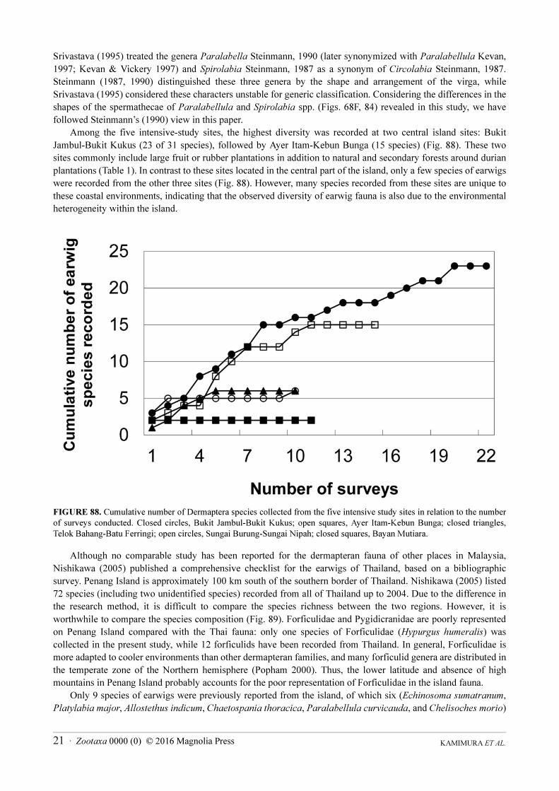
Among the five intensive-study sites, the highest diversity was recorded at two central island sites: Bukit
Jambul-Bukit Kukus (23 of 31 species), followed by Ayer Itam-Kebun Bunga (15 species) (Fig. 88). These two
sites commonly include large fruit or rubber plantations in addition to natural and secondary forests around durian
plantations (Table 1). In contrast to these sites located in the central part of the island, only a few species of earwigs
were recorded from the other three sites (Fig. 88). However, many species recorded from these sites are unique to
these coastal environments, indicating that the observed diversity of earwig fauna is also due to the environmental
heterogeneity within the island.
FIGURE 88. Cumulative number of Dermaptera species collected from the five intensive study sites in relation to the number
of surveys conducted. Closed circles, Bukit Jambul-Bukit Kukus; open squares, Ayer Itam-Kebun Bunga; closed triangles,
Telok Bahang-Batu Ferringi; open circles, Sungai Burung-Sungai Nipah; closed squares, Bayan Mutiara.
Although no comparable study has been reported for the dermapteran fauna of other places in Malaysia,
Nishikawa (2005) published a comprehensive checklist for the earwigs of Thailand, based on a bibliographic
survey. Penang Island is approximately 100 km south of the southern border of Thailand. Nishikawa (2005) listed
72 species (including two unidentified species) recorded from all of Thailand up to 2004. Due to the difference in
the research method, it is difficult to compare the species richness between the two regions. However, it is
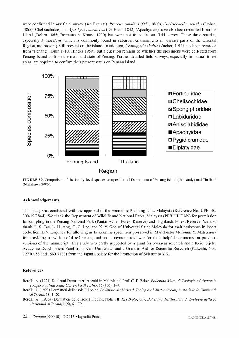
worthwhile to compare the species composition (Fig. 89). Forficulidae and Pygidicranidae are poorly represented
on Penang Island compared with the Thai fauna: only one species of Forficulidae (Hypurgus humeralis) was
collected in the present study, while 12 forficulids have been recorded from Thailand. In general, Forficulidae is
more adapted to cooler environments than other dermapteran families, and many forficulid genera are distributed in
the temperate zone of the Northern hemisphere (Popham 2000). Thus, the lower latitude and absence of high
mountains in Penang Island probably accounts for the poor representation of Forficulidae in the island fauna.
Only 9 species of earwigs were previously reported from the island, of which six (Echinosoma sumatranum,
Platylabia major, Allostethus indicum, Chaetospania thoracica, Paralabellula curvicauda, and Chelisoches morio)
KAMIMURA ET AL.22 · Zootaxa 0000 (0) © 2016 Magnolia Press
were confirmed in our field survey (see Results). Proreus simulans (Stål, 1860), Chelisochella superba (Dohrn,
1865) (Chelisochidae) and Apachyus chartaceus (De Haan, 1842) (Apachyidae) have also been recorded from the
island (Dohrn 1865; Bormans & Krauss 1900) but were not found in our field survey. These three species,
especially P. simulans, which is commonly found in suburban environments in warmer parts of the Oriental
Region, are possibly still present on the island. In addition, Cranopygia similis (Zacher, 1911) has been recorded
from “Penang” (Burr 1910; Hincks 1959), but a question remains of whether the specimens were collected from
Penang Island or from the mainland state of Penang. Further detailed field surveys, especially in natural forest
areas, are required to confirm their present status on Penang Island.
FIGURE 89. Comparison of the family-level species composition of Dermaptera of Penang Island (this study) and Thailand
(Nishikawa 2005).
Acknowledgements
This study was conducted with the approval of the Economic Planning Unit, Malaysia (Reference No. UPE: 40/
200/19/2844). We thank the Department of Wildlife and National Parks, Malaysia (PERHILITAN) for permission
for sampling in the Penang National Park (Pantai Acheh Forest Reserve) and Highlands Forest Reserve. We also
thank H.-S. Tee, L.-H. Ang, C.-C. Lee, and X.-Y. Goh of Universiti Sains Malaysia for their assistance in insect
collection, D.V. Logunov for allowing us to examine specimens preserved in Manchester Museum, Y. Matsumura
for providing us with useful references, and an anonymous reviewer for their helpful comments on previous
versions of the manuscript. This study was partly supported by a grant for overseas research and a Keio Gijuku
Academic Development Fund from Keio University, and a Grant-in-Aid for Scientific Research (Kakenhi, Nos.
22770058 and 15K07133) from the Japan Society for the Promotion of Science to Y.K.
References
Borelli, A. (1921) Di alcuni Dermateteri raccolti in Malesia dal Prof. C. F. Baker. Bollettino Musei di Zoologia ed Anatomia
comparata della Reale Università di Torino, 35 (736), 1–9.
Borelli, A. (1923) Dermatteri delle isole Filippine. Bollettino dei Musei di Zoologia ed Anatomia comparata della R. Università
di Torino, 38, 1–20.
Borelli, A. (1926a) Dermatteri delle Isole Filippine, Nota VII. Res Biologicae, Bollettino dell’Instituto di Zoologia della R.
Università di Torino, 1 (5), 61–79.
KAMIMURA ET AL.23 · Zootaxa 0000 (0) © 2016 Magnolia Press
Borelli, A. (1926b) Dermaptères de Java, Sumatra et îles Voisines. Treubia, 8 (3–4), 248–273.
Borelli, A. (1927) Fauna sumatrensis (Beitrag Nr. 36): Dermaptera. Supplementa Entomologica, 15, 69–80.
Borelli, A. (1932a) Dermapteres de la Presqu’ile Malaise. Bulletin of the Raffles Museum, Singapore, Straits Settlemnts, 7,
80–95.
Borelli, A. (1932b) Dermapteres de Borneo. Journal of the Federated Malay States Museiums, 17 (1), 179–190.
Bormans, A. de. (1883) Etude sur quelques Forficulaires nouveaux ou peu connus précédé d un tableau synoptique des genres
de cette famille. Annales de la Société Entomologique de Belgique, 27, 59–90.
Bormans, A. de. (1894) Viaggio di Leorardo Fea in Birmania e regioni vicine: LXI. Dermaptères. Annali del Museo Civico di
Storia Naturale Genova, 34, 371–409.
Bormans, A. de (1900) Quelques Dermaptères du Musée civique de Gênes. Annali del Musco Civico di Storia Naturale di
Genova, Serie 2.a, 20, 441–467.
Bormans, A. de & Krauss, H. (1900) Forficulidae und Hemimeridae. In: Das Tierreich, 11. Verlag von R. Friedländer und
Sohn, Berlin, 142 pp. http://dx.doi.org/10.5962/bhl.title.69336
Brindle, A. (1968) The Dermaptera of the Naturhistoriska Riksmuseum, Stockholm, Part III. Arkiv for Zoologi, Serie 2, 20 (25),
533–552.
Brindle, A. (1971) The Dermaptera of Ceylon. Entomologica scandinavica Supplements, 1, 208–226.
Burmeister, H. (1838) Dermatoptera. Handbuch der Entomologie, Bd2, 2 (1), 743–756.
Burr, M. (1901) The earwigs of Ceylon. Journal of the Bombay Natural History Society, 14 (2), 316–336.
Burr, M. (1910) Fauna of British India, including Ceylon and Burma. Dermaptera (earwigs). Taylor and Francis, London, xviii
+ 217 pp., 10 pls. http://dx.doi.org/10.5962/bhl.title.100789
Burr, M. (1911) A Revision of the Genus Diplatys, serv. (Dermaptera). Transactions of the Entomological Society of London,
1911, 21–46, pls. 7–8.
Burr, M. (1912) Die Dermapteren des k. k. naturhistorischen Hofmuseums in Wien. Annalen des k. k. Naturhistorischen
Hofmuseums, 26, 63–108.
Costa, J.T. (2006) The other insect societies. Harvard University Press, Cambridge, 767 pp.
Disney, R.H.L., Neoh, K.-B & Lee, C.-Y. (2009) A new species of scuttle fly (Diptera: Phoridae) parasitizing a termite
(Isoptera: Termitidae) in Malaysia. Sociobiology, 54, 89–94.
Dohrn, H. (1863a) Versuch einer Monographie der Dermapteren. Entomologische Zeitung (= Stettiner entomologische zeitung),
24, 35–66.
Dohrn, H. (1863b) Versuch einer Monographie der Dermapteren. Entomologische Zeitung (= Stettiner entomologische zeitung),
24, 309–322.
Dohrn, H. (1865) Versuch einer Monographie der Dermapteren. Entomologische Zeitung (= Stettiner entomologische zeitung),
26 (1–3), 68–99.
Dohrn, H. (1867) Neue und bisher nicht genugend bekannte Forficulinen. Entomologische Zeitung (= Stettiner entomologische
zeitung), 28, 343–349.
Dubrony, A. (1879) Enumeration des Orthoptères rapportés par MM. J. Doria, O. Beccari, et L. M. d’Albertis des regions
Indienne et Austro-Malaise. Annali del Museo civico di storia natural di Genova, 14, 348–383.
Engel, M.S. & Haas, F. (2007) Family-group names for earwigs (Dermaptera). American Museum Novitates, 3567, 1‒20. http://dx.doi.org/10.1206/0003-0082(2007)539[1:FNFED]2.0.CO;2
Fabricius, J.C. (1775) Systema Entomologiae, sistens insectorum classes, ordines, genera, species, adiectis synonymis, locis,
descriptionibus, observationibus. Officina Libraria Kortii, Flensburg and Leipzig, 832 pp. http://dx.doi.org/10.5962/bhl.title.36510
Fabricius, J.C. (1793) Entomologia systematica emendata et aucta. Secundum classes, ordines, genera, species adjectis
synonimis, locis, observationibus, descriptionibus. Tome 2. Christ. Gottl. Proft, Hafniae, 519 pp.
Gardner, S., Sidisunthorn, P. & May, L.-E. (2011) Heritage trees of Penang. Areca Books, Penang, Malaysia, 397 pp.
Grimaldi, D. & Engel, M.S. (2005) Evolution of the Insects. Cambridge University Press, Cambridge, New York, 772 pp.
Haan, W. de (1842) Bijdragen tot de kennis der Orthoptera. In: Temminck, K.J. (Ed.), Verhandelingen over de Natuurlijke
Geschiedenis der Nederlandsche Overzeesche Bezittingen. Natuurkuundige Commissie in Indie, Leiden, pp. 12–243.
Haas, F., Hwen, J.T.C. & Tang, H.B. (2012) New evidence on the mechanics of wing unfolding in Dermaptera. Arthropod
Systematics & Phylogeny, 70, 95–105.
Hebard, M. (1929) Notes on Malayan and Philippine Dermaptera, with the description of two new species. Transactions of the
American Entomological Society, 55 (4), 335–344.
Hincks, W.D. (1949) Some earwigs (Dermaptera) from New Zealand. Proceedings of the Royal Entomological Society, London
(B), 18 (11–12), 201–206. http://dx.doi.org/10.1111/j.1365-3113.1949.tb01410.x
Hincks, W.D. (1954) The Dermaptera of Sumba and F1ores. Verhandlungen der Naturforschenden Gesellschaft in Basel, 65
(1), 9–23.
Hincks, W.D. (1959) A Systematic Monograph of the Dermaptera of the World. Part II. Pygidicranidae excluding Diplatyinae.
British Museum (Natural History), London, 218 pp.
KAMIMURA ET AL.24 · Zootaxa 0000 (0) © 2016 Magnolia Press
Hoshiba, H., Sakai, S. & Imanishi, M. (1988) Chromosome studies on the two chelisochid earwigs with special reference to the
46 dermapteran species. Proceedings of the Japanese Academy, Series B, 64, 303–306. http://dx.doi.org/10.2183/pjab.64.303
Hudson, L. (1973) A systematic revision of the New Zealand Dermaptera. Journal of the Royal Society of New Zealand, 3,
219–254. http://dx.doi.org/10.1080/03036758.1973.10430603
Kamimura, Y. (2000) Possible removal of rival sperm by the elongated genitalia of the earwig, Euborellia plebeja. Zoological
Science, 17, 667–672.
Kamimura, Y. & Lee, C.-Y. (2014a) Mating and genital coupling in the primitive earwig species Echinosoma denticulatum
(Pygidicranidae): implications for genital evolution in dermapteran phylogeny. Arthropod Systematics & Phylogeny, 72
(1), 11–21.
Kamimura, Y. & Lee, C.-Y. (2014b) Genital morphology and mating behavior of Allostethus (Insecta: Dermaptera), an earwig
genus of enigmatic phylogenetic position. Arthropod Systematics & Phylogeny, 72 (3), 331–343.
Kamimura, Y., Tee, H.-S. & Lee, C.-Y. (2016) Ovoviviparity and genital evolution: a lesson from an earwig species with
coercive traumatic mating and accidental breakage of elongated intromittent organs. Biological Journal of the Linnean
Society. [in press]
Kevan, D.K. McE. & Vickery, V.R. (1977) An annotated provisional list of non-saltatorial orthopteroid insects of Micronesia,
compiled mainly from the literature. Micronesia, 30, 269–353.
Kirby, W.F. (1891) A revision of the Forficulidae, with descriptions of new species in the British Museum. The Journal of the
Linnean Society of London, Zoology, 23, 502–531.
Klass, K.-D. (2001) The female abdomen of the viviparous earwig Hemimerus vosseleri (Insecta: Dermaptera: Hemimeridae),
with a discussion of the postgenital abdomen of Insecta. Zoological Journal of the Linnean Society, 131, 251–307. http://dx.doi.org/10.1111/j.1096-3642.2001.tb02239.x
Klass, K.-D. (2003) The female genitalic region in basal earwigs (Insecta: Dermaptera: Pygidicranidae s.l.). Entomologische
Abhandlungen, 61, 173–225.
Kočárek, P. (2009) A case of viviparity in a tropical non-parasitizing earwig (Dermaptera Spongiphoridae). Tropical Zoology,
22, 237–241.
Kočárek, P. (2010) Case 3522. Palicinae Burr, 1910 (Dermaptera, Spongiphoridae): proposed emendation of the current
spelling to Palexinae to remove homonymy with Palicidae Bouvier, 1898 (Crustacea, Decapoda). Bulletin of Zoological
Nomenclature, 67, 211–212.
Lee, C.-Y., Ngee, P.-S., Lee, L.-C. & Na, J.P.-S. (2004) Survey of termite diversity in Pantai Acheh Forest Reserve, Penang
Island, Malaysia. Jurnal Biosains, 15, 91–99.
McClure, H. E., Lim, B.-L. & Winn, S.E. (1967) Fauna of the dark cave, Batu Caves, Kuala Lumpur, Malaysia. Pacific Insects,
9 (3), 399–728.
Motschulsky, V. de. (1863) D’un catalogue des insects de l’ile Ceylan. Bulletin de la Société Impériale des naturalistes de
Moscou, 36, 1–153.
Nishikawa, M. (1973) Dermaptera of Malay Peninsula taken by the Japanese IBP Team. Nature and Life in Southeast Asia, 7,
317–322.
Nishikawa, M. (2005) A checklist of the Dermaptera recorded from Thailand. Thailand Natural History Museum Journal, 1,
149–164.
Pallas, P.S. (1773) Anhang zum zweyten Theil. Descriptiones Animalium. Reise durch verschiedene Provinzen des Russischen
Reiches, 2, 701–732.
Popham, E.J. (2000) The geographical distribution of the Dermaptera (Insecta) with reference to continental drift. Journal of
Natural History, 34, 2007–2027. http://dx.doi.org/10.1080/00222930050144837
Sakai, S. (1982) A new proposed classification of the Dermaptera with special reference to the check-list of the Dermaptera of
the world. Bulletin of Daito Bunka University, 20, 1–108.
Sakai, S. (1985) Dermapterorum Catalogus XVI-XVIII: Iconographia I-III. Pygidicranidae and Diplatyidae. Daito Bunka
University, Tokyo, 1080 pp.
Sakai, S. (1987) Dermapterorum Catalogus XIX-XX: Iconographia IV-V. Chelisochidae and Anisolabididae. Daito Bunka
University, Tokyo, 1567 pp.
Sakai, S. (1990) Dermapterorum Catalogus XXI-XXII: Iconographia VI-VII. Labiduridae and Apachyidae. Daito Bunka
University, Tokyo, xxii + 889 pp.
Sakai, S. (1991) Dermapterorum Catalogus XXIII: Iconographia VIII. Spongiphoridae I. Daito Bunka University, Tokyo, 708
pp.
Sakai, S. (1992) Dermapterorum Catalogus XXIV: Iconographia VIII. Spongiphoridae II. Daito Bunka University, Tokyo, 757
pp.
Sakai, S. (1993) Dermapterorum Catalogus XXV: Iconographia IX. Spongiphoridae III. Daito Bunka University, Tokyo, 595
pp.
Sakai, S. (1994) Dermapterorum Catalogus XXVI: Iconographia X. Forficulidae. Daito Bunka University, Tokyo, 889 pp.
Sakai, S. (1995a) Dermapterorum Catalogus XXVII: Iconographia XI. Forficulidae. Daito Bunka University, Tokyo, 926 pp.
KAMIMURA ET AL.25 · Zootaxa 0000 (0) © 2016 Magnolia Press
Sakai, S. (1995b) Dermapterorum Catalogus XXVIII: Iconographia XII. Forficulidae. Daito Bunka University, Tokyo, 712 pp.
Sakai, S. (1995c) Dermapterorum Catalogus XXIX: Iconographia XIII. Forficulidae. Daito Bunka University, Tokyo, 970 pp.
Sakai, S. (1995d) Dermapterorum Catalogus XXX: Iconographia XIV. Forficulidae. Daito Bunka University, Tokyo, 748 pp.
Sakai, S. (1996) Dermapterorum Catalogus XXXI: Notes on contemporary classification of Dermaptera and recent references
of Dermaptera. Daito Bunka University, Tokyo, 588 pp.
Schneider, K. & Klass, K.-D. (2013) The female genitalic region in Eudermaptera (Insecta: Dermaptera). Zoologischer
Anzeiger, 252, 183–203. http://dx.doi.org/10.1016/j.jcz.2012.05.004
Smith, G.B., Veera Singham, G., Kuah, M.-K. & Lee, C.-Y. (2011) Two new inquiline silverfish (Zygentoma: Ateluridae,
Lepismatidae) from Malaysia. Sociobiology, 57, 19–34.
Srivastava, G.K. (1979) On a new species of genus Euborellia Burr (Dermaptera : Carcinophoridae) from Philippines. Bulletin
of the Zoological Survey of India, 2 (1), 49–51.
Srivastava, G.K. (1983) Notes on some Borelli’s types of Dermaptera (Ins.) Bollettino del Museo Regionale di Scienze Naturali
Torino, 1 (2), 227–242.
Srivastava, G.K. (1992) Taxonomic status of certain genera of Pygidicranidae (Dermaptera). Records of the Zoological Survey
of India, 92 (1–4), 41–52.
Srivastava, G.K. (1995) On the classification of Spongiphoridae (= Labiidae) with a list of species. Records of the Zoological
Survey of India, 95, 71–105.
Srivastava, G.K. (1999) On the higher classification of Anisolabididae (Insecta: Dermaptera) with a check-list of genera and
species. Records of the Zoological Survey of India, 97, 73–100.
Srivastava, G.K. (2003a) Fauna of India and the adjacent Countries, Dermaptera Part II: Anisolaboidea. Zoological Survey of
India, Kolkata, 235 pp.
Srivastava, G.K. (2003b) Studies on Oriental Dermaptera preserved in the B. P. Museum, Hawaii, U. S. A. In: Records of the
Zoological Survey of India, Occasional Paper, No. 210. Zoological Survey of India, Kolkata, pp. 1–72.
Srivastava, G.K. (2013) Fauna of India and the adjacent countries, Dermaptera Part III: Apachyoidea and Forficuloidea.
Zoological Survey of India, Kolkata, 469 pp.
Srivastava, G.K. & Kovac, D. (1993) Notes on some Dermaptera from Malaya with description of two new species. Records of
the Zoological Survey of India, 93 (1–2), 253–266.
Stål, C. (1855) Entomologiska Notiser. Öfversigt af Kongliga Vetenskaps-Akademiens Förhandlingar, 12, 343–353.
Stål, C. (1860) Orthptera, species novas descripsit. Kongliga Svenska Fregatten Eugenies resa omkring jorden, Vetenskaplica
Iakttagelser, II (Zoologi), 1 (Insecta), 299–348.
Steghaus-Kovac, S. & Maschwitz, U. (1993) Predation on earwigs: A novel diet specialization within the genus Leptogenys
(Formicidae: Ponerinae). Insectes Sociaux, 40 (3), 337–340. http://dx.doi.org/10.1007/BF01242370
Steinmann, H. (1986) Dermaptera. Catadermaptera I. In: Das Tierreich. 102. Walter de Gruyter, Berlin, pp. 1–343.
Steinmann, H. (1987) Two new genera and species for the subfamily Labiinae (Dermaptera: Labiidae). Acta Zoologica
Hungarica, 33 (1–2), 177–186.
Steinmann, H. (1989a) Dermaptera. Catadermaptera II. In: Das Tierreich. 105. Walter de Gruyter, Berlin, pp. 1–504.
Steinmann, H. (1989b) World Catalogue of Dermaptera. Kluwer Academic Publishers, Dordrecht-Boston-London, 934 pp.
Steinmann, H. (1990) Dermaptera. Eudermaptera I. In: Das Tierreich. 106. Walter de Gruyter, Berlin, pp. 1–558.
Steinmann, H. (1993) Dermaptera. Eudermaptera II. In: Das Tierreich. 108. Walter de Gruyter, Berlin, pp. 1–711.
Tworzydło, W., Biliński, S.M., Kočárek, P. & Haas, F. (2010) Ovaries and germline cysts and their evolution in Dermaptera
(Insecta). Arthropod Structure & Development 39, 360–368. http://dx.doi.org/10.1016/j.asd.2010.05.004
Yersin, M.A. (1860) Note sur quelques Orthoptères nouveaux ou peu connus d’Europe. Annales de la Société Entomologique
de France, 3 (8), 509–537.
Zacher, F. (1911) Studien über die System der Protodermaptera. Zoologische Jahrbücher Systematik, 30, 303–340.