Embed Size (px)
Citation preview

270 J Can Chiropr Assoc 2005; 49(4)
0008-3194/2005/270–296/$2.00/©JCCA 2005
Evidence-based protocol forstructural rehabilitation of the spine and posture: review of clinical biomechanics of posture (CBP®) publicationsPaul A. Oakley, DC, MSc*Donald D. Harrison, PhD, DC, MSE **Deed E. Harrison, DC†Jason W. Haas, DC††
b a c k g r o u n d : Although practice protocols exist for SMT and functional rehabilitation, no practice protocols exist for structural rehabilitation. Traditional chiropractic practice guidelines have been limited to acute and chronic pain treatment, with limited inclusion of functional and exclusion of structural rehabilitation procedures.o b j e c t i v e : (1) To derive an evidence-based practice protocol for structural rehabilitation from publications on Clinical Biomechanics of Posture (CBP®) methods, and (2) to compare the evidence for Diversified, SMT, and CBP®.m e t h o d s : Clinical control trials utilizing CBP® methods and spinal manipulative therapy (SMT) were obtained from searches in Mantis, CINAHL, and Index Medicus. Using data from SMT review articles, evidence for Diversified Technique (as taught in chiropractic colleges), SMT, and CBP® were rated and compared.r e s u lt s : From the evidence from Clinical Control Trials on SMT and CBP®, there is very little evidence support for Diversified (our rating = 18), as taught in chiropractic colleges, for the treatment of pain subjects, while CBP® (our rating = 46) and SMT for neck pain (rating = 58) and low back pain (our rating = 202) have evidence-based support.c o n c l u s i o n s : While CBP® Technique has approximately as much evidence-based support as SMT for neck pain, CBP® has more evidence to support its methods than the Diversified technique taught in chiropractic colleges, but not as much as SMT for low
a n t é c é d e n t s : Quoi qu’il y ait des protocoles d’exercice établis pour la manipulation vertébrale et la réadaptation fonctionnelle, il n’existe aucun protocole pour la réadaptation structurale. Les directives d’exercices chiropratiques traditionnels se limitent à des traitements pour la douleur aigue et chronique avec une inclusion limitée des procédures de réadaptation fonctionnelle et une exclusion des procédures de réadaptation stucturale.o b j e c t i f : (1) Faire dévier des publications sur les procédures biomécaniques cliniques de la posture (BCP) un protocole d’exercice, avec preuve à l’appui, pour la réadaptation structurale, et (2) Comparer la preuve pour la techniques diversifiée, la manipulation vertébrale et la procédure biomécanique de la posture (BCP).p r o c é d u r e s : Des essais de contrôle clinique avec l’utilisation des procédures biomécaniques cliniques et la manipulation vertébrale ont été obtenus, suite à des recherches dans Mantis, CINAHL et Index Medicus. En utilisant, les données des rapports de synthèse sur la manipulation vertébrale pour la technique diversifiée (tel qu’enseigné dans les collèges de chiropractie), la manipulation vertébrale et les procédures BCP ont été évaluées et comparées.r é s u ltat s : Fondés sur la preuve des essais de contrôle clinique sur la manipulation vertébrale et les procédures BCP, il existe peu de preuve pour soutenir la technique diversifiée (notre taux = 18), tel qu’enseigné dans les collèges de chiropractie, pour le traitement de la douleur des sujets, contrairement aux procédures BCP
** Corresponding author: Paul A. Oakley, DC, MSc, 11C-1100 Gorham Street, Newmarket, Ontario L3Y 8Y8, Phone 905-868-9090 Email [email protected]** PO BOX 1590, Evanston, Wyoming, USA, 82931.†† 123 Second Street, Elko, NV, USA 89801.†† 1180 Main Street, Suite 7, Windsor, CO, USA 80550. © JCCA 2005.
PA Oakley, DD Harrison, DE Harrison, JW Haas
J Can Chiropr Assoc 2005; 49(4) 271
IntroductionRecently, the buzzwords “evidence-based,” “evidence-based medicine” (EBM), and “evidence-based practice”(EBP) have appeared in clinical practice protocols. EBPis defined as clinical decision-making based on (1),sound external research evidence combined with individ-ual clinical expertise and (2), the needs of the individualpatient.1,2 EBP protocols have recently been written forseveral conditions.2–9
The goal of EBP is to improve patient outcomes, qual-ity of care, and provide some standardization of treat-ment. Systematic reviews of available published evidenceare required. The value of these literature reviews, how-ever, depends on the quality of the review (selection biasby those doing the review) and the quality of the publica-tions.10 To have “evidence” on aspects of all treatmentmethods is nearly impossible in any healthcare discipline,including medicine11 and chiropractic.12
The highest form of “evidence” would seem to be reli-
ability studies, validity studies, and randomized clinicalcontrol trials (RCTs). Critics of a certain healthcaremethod often condemn that method if RCTs have notbeen published. This might seem unreasonable as theremay be more agreement amongst researchers for othertypes of evidence. There are other types of clinical (non-randomized clinical trial, cohort, case report, etc.) andbasic scientific studies that can provide “evidence” that acertain type or method of care might be reasonable, suffi-cient, or standard. In fact, the RCT may not be the bestsource of evidence for the clinical practice of chiroprac-tic.1,13,14
The exact question being debated is “what does andwhat does not provide evidence in EBM.15–17 In 2001,Bolton1 discussed the reliance on RCTs in EBP proto-cols. Although the first few published RCTs on spinalmanipulative therapy (SMT) were important for thechiropractic profession, RCTs are so narrow in methodol-ogy as to not often be useful in clinical chiropractic prac-
back pain. The evolution of chiropractic specialization has occurred, and doctors providing structural-based chiropractic care require protocol guidelines for patient quality assurance and standardization. A structural rehabilitation protocol was developed based on evidence from CBP® publications.(JCCA 2005, 49(4):270–296)
key words: chiropractic, spinal manipulation, rehabilitation.
(notre taux = 46) et la manipulation vertébrale pour la cervicalgie (taux = 58) et le lumbago (notre taux = 202) qui sont soutenues par la preuve.c o n c l u s i o n : Tandis que la procédure BCP possède approximativement, autant de preuve à l’appui que la manipulation vertébrale pour la cervicalgie, la méthode BCP possède davantage de preuves à l’appui, pour soutenir ses procédures que la méthode diversifiée, enseignée dans les collèges de chiropractie, mais pas autant que la manipulation vertébrale pour le lumbago. L’évolution de la spécialisation de la chiropractie est devenue une réalité et les médecins qui fournissent des soins de chiropractie, à base structurale, nécessitent des directives de protocole pour offrir aux patients une assurance de la qualité des soins et une normalisation. Un protocole de réadaptation structurale a été élaboré, fondé sur la preuve des publications, reliées aux procédures BCP.(JACC 2005; 49(4):270–296)
mots clés : chiropractie, manipulation vertébrale, réadaptation.

Structural rehab protocol
272 J Can Chiropr Assoc 2005; 49(4)
tice.1,14 Although the RCT is unarguably the best re-search design, “randomization and controlled conditionsplay no part in everyday clinical practice”1 and thus, evi-dence for effectiveness in that arena cannot be collectedby RCTs. The strengths and limitations of the RCT havebeen discussed elsewhere.18
Given the limitations of the RCT in evaluating chiro-practic treatment,14 it has been stated that it does notmake sense to exclusively pursue the RCT in the future.13
Other research designs such as qualitative and outcomesresearch are now being recognized as very meaningfulways of providing the evidence in EBP.1 Additionally, itis known today that well-done case studies most oftendemonstrate findings consistent with that of the RCT.19,20
Since 1975, the chiropractic profession has enjoyedimproved political support in a number of countries dueto some published analyses favorable to chiropracticcare.21–25 Unfortunately, because early evidence (RCTs)for manipulation had been on the effectiveness for treat-ing acute and chronic pain complaints (i.e. LBP), the en-suing guidelines have primarily been based on acute/chronic pain care. Consequently, in North America,chiropractic’s inclusion in government health insuranceand private insurance programs has coincided with re-strictions in frequency and duration of the care permitted.In response, some chiropractic organizations have writtentheir own guidelines in an attempt to obtain fairness inthese systems.26–28
Chiropractic treatment protocols could ideally be cate-gorized into either: (a) Acute pain care, (b) Chronic paincare, (c) Functional rehabilitation care, or (d) Structuralrehabilitation care. Recently, the profession has shownrenewed interest in structural rehabilitative care. In a sur-vey of 108 participating North American practices, Hawket al.29 (their Table 10, page 167) reported Clinical Bio-mechanics of Posture (CBP®) technique to be the 7thmost utilized technique in chiropractic practices. Prob-lematically, there is only one manuscript detailing pro-posed guidelines for structural rehabilitation of the spine;this was based on a few preliminary studies.30
Our objectives are two-fold: (1) Compare the evidencefor Diversified, General Spinal Manipulative Therapy(SMT), and CBP® for reduction in neck and low backpain intensity; (2) Derive an evidence-based practice pro-tocol for structural rehabilitation from publications onClinical Biomechanics of Posture (CBP®) methods. We
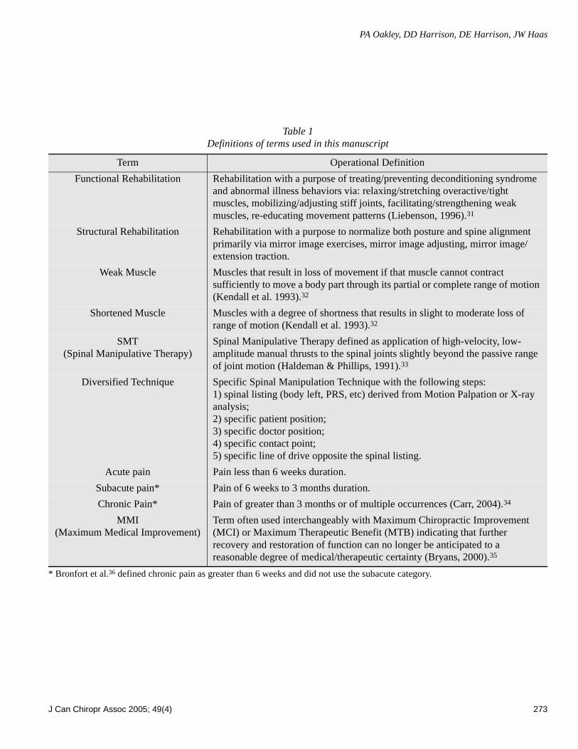
will use some of Bolton’s1 ideas of evidence for EBPguidelines when discussing recent published researchconcerning CBP® structural rehabilitation procedures. Toinsure communication with the reader, a table of defini-tions is presented31–35 (Table 1).
MethodsIn order to compare the evidence for Diversified, SMT,and CBP®, literature searches were conducted in Mantis,CINAHL, and Index Medicus for Clinical Control Trialson SMT, Diversified, and CBP® methods.
We identified 73 RCTs on SMT, including only twoclinical control trials on Diversified technique (as definedin Table 1), and 6 non-randomized clinical control trialson CBP® technique methods. In 2004, Bronfort et al.36
had identified 69 of the RCTs on SMT and performed ameta-analysis. This manuscript will adapt the Bronfort etal.36 analysis of the 69 RCTs identified before February2003, and apply this analysis to two additional RCTs lo-cated since February 2003.37–38 Additionally, there havebeen two other review articles discussing SMT.39–40
In these RCT papers’ methods, we were looking for(1) any pain scales, (2) any disability scores, (3) thenumber of subjects, (4) whether SMT was actually usedor only PT Mobilization, (5) if Diversified technique wasused (as defined in Table 1), and (6) if DCs, MDs, or PTsperformed the treatment, and (7) the number of treat-ments (visits).
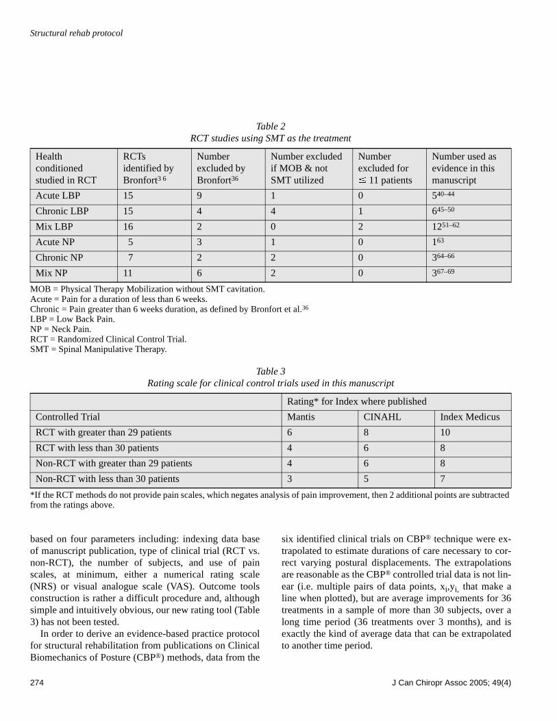
Bronfort et al.36 deleted RCTs with 10 subjects or less,while in this manuscript, we deleted any RCTs if (1)there were 11 subjects or less (there were 3 such RCTs)and (2) if instead of SMT, authors utilized Physical Ther-apist’s mobilization (MOB) techniques. Table 2 providesour adapted analysis from Bronfort et al.,36 who catego-rized RCTs into acute pain, chronic pain, and mixed painin each of the neck and low back regions, and our numberof RCTs excluded and included for analysis. In 2004, twoadditional RCTs were published.70–71 While Hurwitz etal.’s 2004 study71 is a re-look at previous 2002 data,59 the2004 Haas et al. study70 had less than 10 subjects in thetreatment groups. We compared pain scale data in the 29remaining RCTs41–69 and the two RCTs published sinceFebruary 200337–38 to the CBP® published clinical con-trol trials.72–77
Table 3 provides the authors’ rating scale for ClinicalControl Trials used in this manuscript. This rating scale is
PA Oakley, DD Harrison, DE Harrison, JW Haas
J Can Chiropr Assoc 2005; 49(4) 273
Table 1Definitions of terms used in this manuscript
* Bronfort et al.36 defined chronic pain as greater than 6 weeks and did not use the subacute category.
Term Operational Definition
Functional Rehabilitation Rehabilitation with a purpose of treating/preventing deconditioning syndrome and abnormal illness behaviors via: relaxing/stretching overactive/tight muscles, mobilizing/adjusting stiff joints, facilitating/strengthening weak muscles, re-educating movement patterns (Liebenson, 1996).31
Structural Rehabilitation Rehabilitation with a purpose to normalize both posture and spine alignment primarily via mirror image exercises, mirror image adjusting, mirror image/extension traction.
Weak Muscle Muscles that result in loss of movement if that muscle cannot contract sufficiently to move a body part through its partial or complete range of motion (Kendall et al. 1993).32
Shortened Muscle Muscles with a degree of shortness that results in slight to moderate loss of range of motion (Kendall et al. 1993).32
SMT(Spinal Manipulative Therapy)
Spinal Manipulative Therapy defined as application of high-velocity, low-amplitude manual thrusts to the spinal joints slightly beyond the passive range of joint motion (Haldeman & Phillips, 1991).33
Diversified Technique Specific Spinal Manipulation Technique with the following steps:1) spinal listing (body left, PRS, etc) derived from Motion Palpation or X-ray analysis; 2) specific patient position; 3) specific doctor position; 4) specific contact point; 5) specific line of drive opposite the spinal listing.
Acute pain Pain less than 6 weeks duration.
Subacute pain* Pain of 6 weeks to 3 months duration.
Chronic Pain* Pain of greater than 3 months or of multiple occurrences (Carr, 2004).34
MMI(Maximum Medical Improvement)
Term often used interchangeably with Maximum Chiropractic Improvement (MCI) or Maximum Therapeutic Benefit (MTB) indicating that further recovery and restoration of function can no longer be anticipated to a reasonable degree of medical/therapeutic certainty (Bryans, 2000).35
Structural rehab protocol
274 J Can Chiropr Assoc 2005; 49(4)
based on four parameters including: indexing data baseof manuscript publication, type of clinical trial (RCT vs.non-RCT), the number of subjects, and use of painscales, at minimum, either a numerical rating scale(NRS) or visual analogue scale (VAS). Outcome toolsconstruction is rather a difficult procedure and, althoughsimple and intuitively obvious, our new rating tool (Table3) has not been tested.
In order to derive an evidence-based practice protocolfor structural rehabilitation from publications on ClinicalBiomechanics of Posture (CBP®) methods, data from the
six identified clinical trials on CBP® technique were ex-trapolated to estimate durations of care necessary to cor-rect varying postural displacements. The extrapolationsare reasonable as the CBP® controlled trial data is not lin-ear (i.e. multiple pairs of data points, xi,yi, that make aline when plotted), but are average improvements for 36treatments in a sample of more than 30 subjects, over along time period (36 treatments over 3 months), and isexactly the kind of average data that can be extrapolatedto another time period.
Table 2RCT studies using SMT as the treatment
MOB = Physical Therapy Mobilization without SMT cavitation.Acute = Pain for a duration of less than 6 weeks.Chronic = Pain greater than 6 weeks duration, as defined by Bronfort et al.36
LBP = Low Back Pain.NP = Neck Pain.RCT = Randomized Clinical Control Trial.SMT = Spinal Manipulative Therapy.
Table 3Rating scale for clinical control trials used in this manuscript
*If the RCT methods do not provide pain scales, which negates analysis of pain improvement, then 2 additional points are subtracted from the ratings above.
Healthconditioned studied in RCT
RCTs identified by Bronfort3 6
Number excluded by Bronfort36
Number excluded if MOB & not SMT utilized
Number excluded for� 11 patients
Number used as evidence in this manuscript
Acute LBP 15 9 1 0 540–44
Chronic LBP 15 4 4 1 645–50
Mix LBP 16 2 0 2 1251–62
Acute NP 5 3 1 0 163
Chronic NP 7 2 2 0 364–66
Mix NP 11 6 2 0 367–69
Rating* for Index where published
Controlled Trial Mantis CINAHL Index Medicus
RCT with greater than 29 patients 6 8 10
RCT with less than 30 patients 4 6 8
Non-RCT with greater than 29 patients 4 6 8
Non-RCT with less than 30 patients 3 5 7
PA Oakley, DD Harrison, DE Harrison, JW Haas
J Can Chiropr Assoc 2005; 49(4) 275
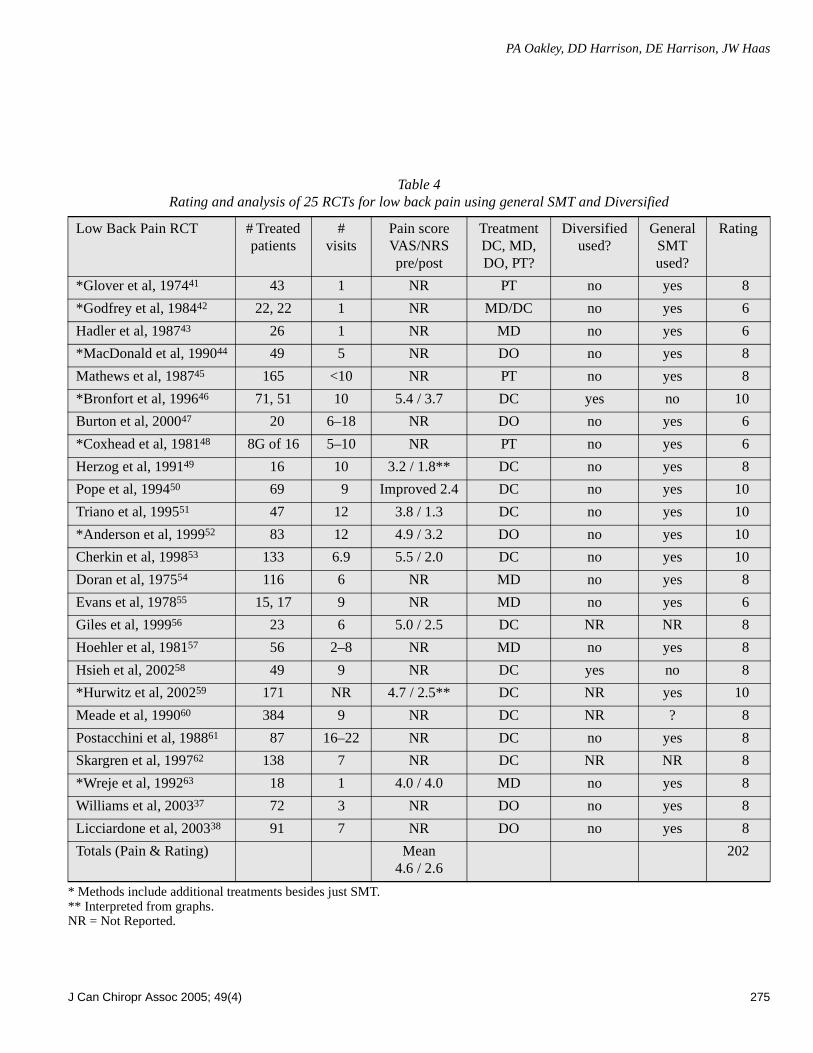
Table 4Rating and analysis of 25 RCTs for low back pain using general SMT and Diversified
* Methods include additional treatments besides just SMT.** Interpreted from graphs.NR = Not Reported.
Low Back Pain RCT # Treated patients
#visits
Pain score VAS/NRSpre/post
TreatmentDC, MD, DO, PT?
Diversified used?
General SMT used?
Rating
*Glover et al, 197441 43 1 NR PT no yes 8
*Godfrey et al, 198442 22, 22 1 NR MD/DC no yes 6
Hadler et al, 198743 26 1 NR MD no yes 6
*MacDonald et al, 199044 49 5 NR DO no yes 8
Mathews et al, 198745 165 <10 NR PT no yes 8
*Bronfort et al, 199646 71, 51 10 5.4 / 3.7 DC yes no 10
Burton et al, 200047 20 6–18 NR DO no yes 6
*Coxhead et al, 198148 8G of 16 5–10 NR PT no yes 6
Herzog et al, 199149 16 10 3.2 / 1.8** DC no yes 8
Pope et al, 199450 69 9 Improved 2.4 DC no yes 10
Triano et al, 199551 47 12 3.8 / 1.3 DC no yes 10
*Anderson et al, 199952 83 12 4.9 / 3.2 DO no yes 10
Cherkin et al, 199853 133 6.9 5.5 / 2.0 DC no yes 10
Doran et al, 197554 116 6 NR MD no yes 8
Evans et al, 197855 15, 17 9 NR MD no yes 6
Giles et al, 199956 23 6 5.0 / 2.5 DC NR NR 8
Hoehler et al, 198157 56 2–8 NR MD no yes 8
Hsieh et al, 200258 49 9 NR DC yes no 8
*Hurwitz et al, 200259 171 NR 4.7 / 2.5** DC NR yes 10
Meade et al, 199060 384 9 NR DC NR ? 8
Postacchini et al, 198861 87 16–22 NR DC no yes 8
Skargren et al, 199762 138 7 NR DC NR NR 8
*Wreje et al, 199263 18 1 4.0 / 4.0 MD no yes 8
Williams et al, 200337 72 3 NR DO no yes 8
Licciardone et al, 200338 91 7 NR DO no yes 8
Totals (Pain & Rating) Mean4.6 / 2.6
202

Structural rehab protocol
276 J Can Chiropr Assoc 2005; 49(4)
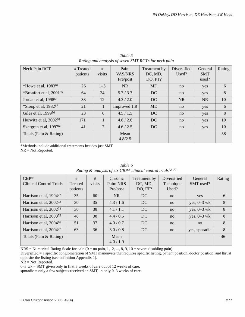
ResultsOverall, there is a considerable amount of evidence sup-porting the use of SMT for low back (our rating = 202)and neck pain (our rating = 58), as well as support forCBP® technique for pain (our rating = 46). Surprisingly,there is little data existing on Diversified technique (asdefined in Table 1) for either low back (our rating = 18)or neck pain (our rating = 0). Tables 4–6 summarize thisdata.
Using reported VAS (divided by 10) or NRS scores inTables 4–6, an average pain reduction can be computedfor the clinical control trials for low back pain using SMTtechniques, neck pain SMT techniques, and CBP® meth-ods. This comparison shows that the average reported nu-merical rating of initial (I) and follow-up (F) pain forNeck SMT studies is I/F = 4.8 / 2.5, the average for LowBack SMT studies is 4.6 / 2.6, while the average forCBP® studies is 4.0 /1.0. Thus, while the average pain re-duction (NRS) in SMT studies is approximately 48%, theaverage for CBP® studies is 75%.
Using only the papers that reported numerical paindata in Tables 4–6, ending clinical pain can be assessed.Simple pain outcome data (i.e. NRS, VAS) comparingpre-to-post treatment results indicate that, although par-tial reductions in pain levels are achieved for SMT trials,post-treatment pain levels are reported to be at significantclinical levels, e.g., an average of ending NRS = 2.6 inTable 4 and ending NRS = 2.5 in Table 5. The same paindata comparison found for the CBP® clinical trials indi-cate minimal-to-negligible pain levels in the post-treat-ment groups (average ending NRS = 1.0 in Table 6).
A similar analysis to numerical pain using disabilityscores (not shown) from the RCTs in Tables 4 and 5 indi-cates that treatment subjects, on average, have significantdisability at follow-up in these SMT studies (using thosepapers which report disability with NDI, SF-36, Roland-Morris, and/or Oswestry). For example, the reader is re-ferred to the recent study by Leboeuf-Yde.78
Extrapolated CBP® clinical trial average data (Table 7)could be easily interpreted to estimate hypothetical treat-ment durations based on the magnitude of postural devia-tions (millimeters/degrees) as starting points in patientcare (see Table 8).
DiscussionFrom the analysis presented on RCTs with the treatment
of SMT in Tables 4–6, there is considerable evidence forthe treatment of general spinal manipulation to be uti-lized for neck pain (our rating = 58) and back pain (ourrating = 202). However, there is very little evidence forDiversified Technique, as defined in Table 1 (rating =18). Thus, there is more evidence for CBP methods (ourrating = 46) than for the Diversified technique (rating =18), which is the technique method mandated by theCouncil on Chiropractic Education (CCE-USA and CCE-Canada) to be taught in all Chiropractic College curriculain the USA and Canada.
According to our review of studies on SMT and reduc-tion in chronic pain intensity, it is readily apparent thatthe average neck pain subject is left with a NRS/VAS =2.5 and the average for low back pain subject is left witha NRS/VAS = 2.6 (review Table 5). We note, the defini-tion of a 2 = Constant Minimal to Intermittent Slight Painand a 3 = Constant Slight Pain with some handicap. Incontrast, the same pain data comparison found for theCBP® clinical trials indicate an ending value of NRS/VAS = 1 = Minimal Pain or annoyance (Table 6). There-fore, it is obvious that while short-term usage of SMT re-duces chronic pain intensity, it does not relieve it and infact these subjects would not be described as MMI. Therecent studies by Haas et al.71 and Leboeuf-Yde et al.78
are good examples of this. Thus, practice protocols basedon pain in SMT studies are incomplete. Also, such prac-tice protocols must include more visits than 12 becausetreated subjects, in published RCTs, were left in chronicpain (NRS = 2.6) after up to 12 treatments of SMT.
In a pilot RCT with a small number of subjects (n = 8in each group), Haas et al.71 found that an increasednumber of treatments, up to 12, was associated withgreater improvement of headache pain. However, even inthe 12 treatment group, the headache pain was still lessthan 50% improved.71 Likewise, in a recent large multi-center trial, after 4 treatments of SMT for lower backpain, Leboeuf-Yde et al. found that subjects were leftwith a NRS of 2.6 (12 month follow up data). Leboeuf-Yde et al.78 also reported that these subjects still had sig-nificant levels of disability on the Oswestry scale (35-Moderate Disability down to 22.2-Moderate Disability).
Recent publications have found that health relatedquality of life and functional disability measures (ShortForm-36, Oswestry, Neck Disability questionnaires, etc....) are more sensitive and important than simple pain in-
PA Oakley, DD Harrison, DE Harrison, JW Haas
J Can Chiropr Assoc 2005; 49(4) 277
Table 5Rating and analysis of seven SMT RCTs for neck pain
*Methods include additional treatments besides just SMT.NR = Not Reported.
Table 6Rating & analysis of six CBP® clinical control trials72–77
NRS = Numerical Rating Scale for pain (0 = no pain, 1, 2, ..., 8, 9, 10 = severe disabling pain).Diversified = a specific conglomeration of SMT maneuvers that requires specific listing, patient position, doctor position, and thrust opposite the listing (see definition Appendix 1).NR = Not Reported.0–3 wk = SMT given only in first 3 weeks of care out of 12 weeks of care.sporadic = only a few subjects received an SMT, in only 0–3 weeks of care.
Neck Pain RCT # Treated patients
#visits
Pain:VAS/NRSPre/post
Treatment byDC, MD, DO, PT?
Diversified Used?
General SMT used?
Rating
*Howe et al, 198364 26 1–3 NR MD no yes 6
*Bronfort et al, 200165 64 24 5.7 / 3.7 DC no yes 8
Jordan et al, 199866 33 12 4.3 / 2.0 DC NR NR 10
*Sloop et al, 198267 21 1 Improved 1.8 MD no yes 6
Giles et al, 199956 23 6 4.5 / 1.5 DC no yes 8
Hurwitz et al, 200268 171 1 4.8 / 2.6 DC no yes 10
Skargren et al, 199769 41 7 4.6 / 2.5 DC no yes 10
Totals (Pain & Rating) Mean4.8/2.5
58
CBP®
Clinical Control Trials#
Treated patients
#visits
Chronic Pain: NRSPre/post
Treatment byDC, MD, DO, PT?
Diversified Technique
Used?
General SMT used?
Rating
Harrison et al, 199472 35 60 NR DC no yes 6
Harrison et al, 200273 30 35 4.3 / 1.6 DC no yes, 0–3 wk 8
Harrison et al, 200274 30 38 4.1 / 1.1 DC no yes, 0–3 wk 8
Harrison et al, 200375 48 38 4.4 / 0.6 DC no yes, 0–3 wk 8
Harrison et al, 200476 51 37 4.0 / 0.7 DC no no 8
Harrison et al, 200477 63 36 3.0 / 0.8 DC no yes, sporadic 8
Totals (Pain & Rating) Mean4.0 / 1.0
46
Structural rehab protocol
278 J Can Chiropr Assoc 2005; 49(4)
tensity outcomes.79 It is of interest that recent publica-tions have found strong correlations between alteredsagittal spinal alignment (specifically loss of the distallumbar lordosis), health quality of life, and physical func-tion as measured with the Short Form-36 questionnaire.80
Therefore, we recommend that clinicians utilize healthstatus and disability questionnaires at all initial and fol-low-up examinations. In this manner, strong evidence canbe used to support the need and outcome effects of CBP®
treatment methods (or any other chiropractic care) pastthe 8–12 visit “bench mark” of traditional SMT basedstudies.
Thus, CBP® multi-modal methods can be utilized toachieve health and disability improvements. But im-provements in structural alignment are the primary inter-vention goal. Before deriving a protocol for structuralrehabilitation from CBP® publications, a brief review isgiven for an appreciation of the clinical relevance ofstructural rehabilitation of the spine and posture.
Clinical relevance for structural rehabilitationIt is known that spinal function is directly related to spinalstructure, as has been proven for the cervical and lumbarspinal regions.80–84 With mal-alignment in neutral posture,static and especially dynamic function from this mal-alignment dictates altered stress/strain relationships of as-sociated spinal structures, including the bones,85–87 in-tervertebral discs,88–91 facet joints,92 musculotendinoustissues,93 ligamentous tissues,94 and neural elements.95–100
Postural alterations are known to be associated with aplethora of human afflictions from general pain syn-dromes,101–109 to problems with specific joints such as thehip110–111 and the knee,112 to problems with specific spi-nal regions such as the flat-back syndrome,113 and cervi-cal kyphosis,109 to local organ ailments such as uterineprolapse,114–115 gastric herniation,116 and respiratoryfunction,117–119 to thinking,118,120 and even to morbidityand mortality.121–125 Improved posture alignment hasbeen one of the most sought-after goals in the treatmentof human ailments for ages; this continues today in allmedical arenas, such as dentistry, physiotherapy, physia-try, surgery, and chiropractic.126–134
Since traditional SMT has not been found to be associ-ated with routine improvement in spinal alignment, itstherapeutic effects are thought to be in reducing pain andfacilitating increased spinal motion.135 Of interest, how-
ever, many monotherapies, have been found to have ei-ther limited effectiveness or complete lack of success intreating chronic low back pain. Bogduk136 has discussedthat these monotherapies include analgesics, NSAIDs,muscle relaxants, antidepressants, physiotherapy, sur-gery, and manipulative therapy. In contrast, Gross et al.137
have reported that the multi-modal care of exercise com-bined with cervical manipulation provides better resultsthan either procedure used alone. The criticism of mono-therapies is taken into consideration by CBP® techniqueas it uses a multi-modality care regimen of SMT as wellas mirror image® exercises, mirror image® adjusting andmirror image®/extension spinal traction procedures,other stretching procedures, and ergonomic counseling.
LimitationsLimitations of using data from RCTs on SMT and CBP®
Clinical Control Trials in this review study are the sameas those expressed by Bronfort et al.36 The interestedreader is referred to page 350 of their study.
Goals in structural rehabilitationIf we categorize chiropractic treatment into: (a) acutepain care, (b) chronic pain care, (c) functional rehabilita-tion care, and (d) structural rehabilitation care, the goalsof care in structural rehabilitation may encompass thefirst two (acute and chronic pain care). Considering therelationship between spinal function and structure,80–84 itis probable that functional rehab and structural rehab areattempts to treat the same patient dysfunction; albeit withdifferent approaches.
For example, a patient presenting with acute or chronicneck pain with 50 mm of anterior head translation, ini-tially would be treated with a trial of traditional SMT in-cluding any ancillary procedures (heat, ice, massage,stretching, etc. ...) for about 2–4 weeks to attempt to im-prove pain levels and spinal motion. Following this, func-tional rehabilitation would seek to improve strength inweakened muscles and flexibility in shortened or tight-ened muscles; muscle dysfunction is considered to be thecause of the displacement.32 In the case of anterior headtranslation it has been found that the neck flexors havedecreased endurance and maximal isometric contractionstrength.138
In contrast, structural rehabilitation procedures wouldseek to improve the postural abnormality by exercising

PA Oakley, DD Harrison, DE Harrison, JW Haas
J Can Chiropr Assoc 2005; 49(4) 279
Figure 1. Algorithm for Structural Rehabilitation
Structural rehab protocol
280 J Can Chiropr Assoc 2005; 49(4)
the head in the exact opposite displacement (i.e., posteri-or head translation). Posterior head translation causescontraction of the upper neck flexors and stretches theupper neck extensors.139 Therefore, it is apparent the twoapproaches (functional vs. structural) are attempting totreat the same disorder by different means.
Figure 1 provides an algorithm that outlines the proto-cols, procedures, and time frames for a structural rehabil-itation program of chiropractic care. This structuralrehabilitation algorithm will be first supported and thendetailed in the duration of the manuscript. The unique-ness of structural rehabilitation is the goal of normalizingboth posture and spine alignment to evidence based val-ues. This goal requires (1) a precise definition of normalposture; (2) reliability and validity of postural measure-ment; (3) a precise definition (model) of the normalspine; and (4) reliability and validity of spinal measure-ments.
Overview of CBP® Procedures
1) Radiographic ProceduresRadiographic line drawing analysis has been shown to beone of the most reliable tools in clinical practice.134 TheHarrison posterior tangent method for measurement ofsagittal spinal curves and the modified Riser-Fergusonmethod for measurement of frontal plane spinal displace-ments have both been studied for reliability. These twomethods have been found to have good to excellent interand intra-examiner reliability with small mean absolutevalues of observer differences for both angles and dis-tances.140–143
The validity of radiographic analysis is supported byknowledge of posture and coupling, termed main motion(posture) and coupled motion (spinal segmental move-ments) in the literature.144–148 The radiographic spinalalignment (spinal coupling) can be compared to the ini-tial posture of the patient (main motion) to determine ifthe coupling patterns are the same as published move-ments in the literature. This is valid, however, only if theclinician utilizes the same radiographic positioning meth-ods as that used in the published studies;149 in fact, theCBP® standardized x-ray positioning procedures havebeen studied for their repeatability. Harrison et al.149 pre-sented data on pre-post lateral and AP spinal x-rays as-certained in six different control groups and compared
their results to twenty other manuscripts in the literature.Their149 results were similar to previous investigators,namely that x-ray positioning is highly repeatable evenwhen taken by different examiners.
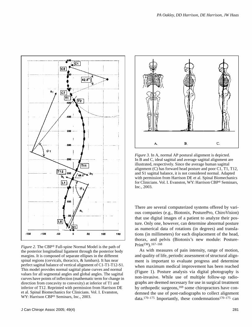
2) Ideal and Average Spinal ModelTo determine abnormality of radiographic spinal align-ment, the radiographic measurements are compared to apublished normal spinal model that provides ideal andaverage alignment values (Figure 2). This normal spinalmodel is based, in part, on average values from normalsubjects and has been published in orthopedic and chiro-practic journals.150–156 Normal values have been pub-lished for each sagittal vertebral segmental angle (RRA =relative rotation angle, e.g., T8–T9) and for sagittal re-gional global angles (ARA = absolute rotation angle, e.g.,C2–C7, T3–T10, and L1–L5). This model is “evidence-based.” In fact, the CBP® sagittal lumbar model153 andthe sagittal cervical model156 were found to have discrim-inative validity in as much as they can distinguish be-tween normal subjects, acute pain subjects, and chronicpain subjects.153,156
Further validity for an optimum upright spinal positioncomes from an analysis of loads and stresses based onminimum energy expenditure.89,121,157–159 Also, statisticalanalyses have derived an average normal spine.160,161 A bi-omechanical analysis of spinal loads dictates a verticalspine in the antero-posterior view (see AP view in Figure3A), while more work must be done to derive a normalpostural position in the sagittal view. Both ideal and aver-age sagittal human postural alignments have been dis-cussed.134,160–163 Figures 3B and 3C illustrate the ideal andaverage sagittal postures, respectively. The average sagit-tal posture in Figure 3C has a forward head posture andpoor sagittal balance of C1, T1, T12, and S1; recent pub-lications negate this position as normal due to increasedmuscle and disc loads, and tissue stresses.89,109,121,128,158,
163,164
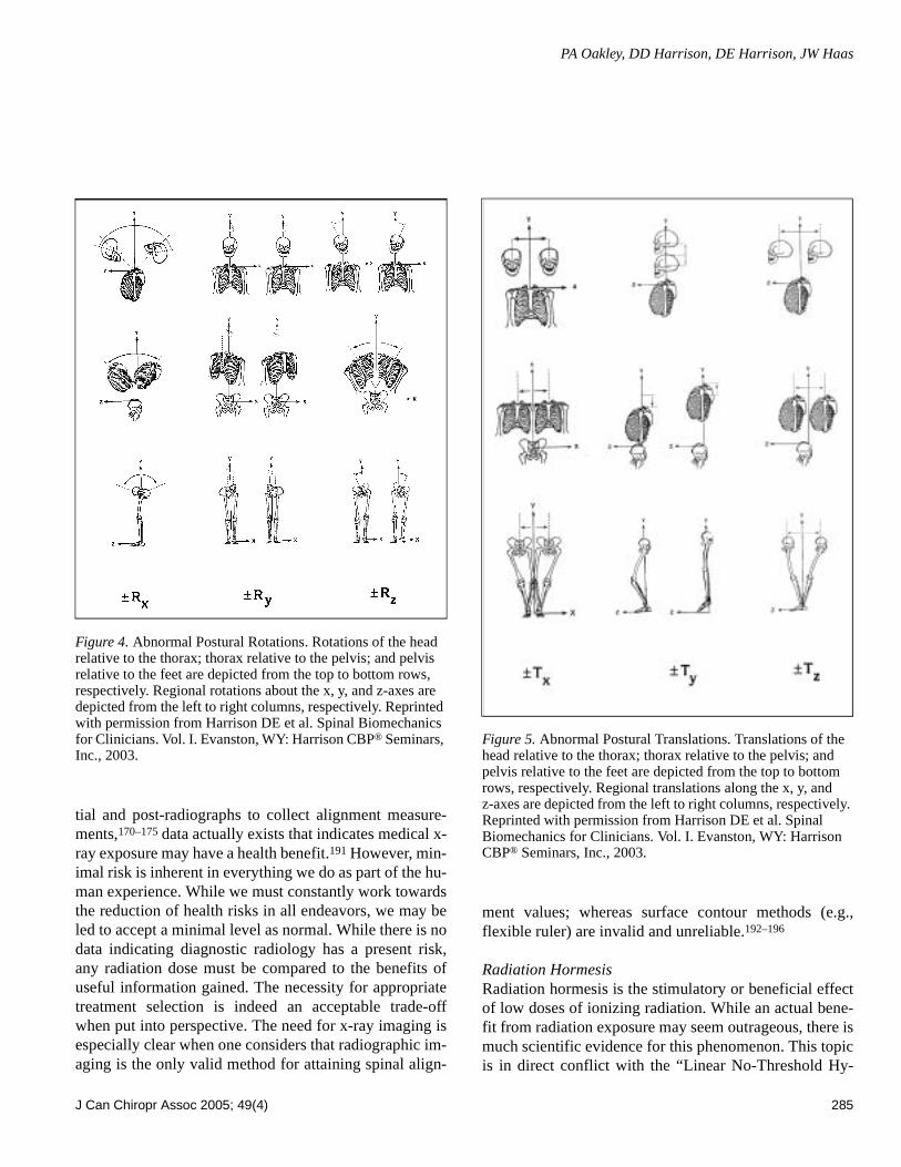
With normal posture precisely described (Figures 3Aand B), abnormal posture can be determined. Using bio-mechanical concepts, abnormal posture has been de-scribed as rotations and translations of the head, rib cage,and pelvis from normal position in a 3-dimensional coor-dinate system (Figures 4 and 5).165,166 The last item nec-essary to complete the goals of structural rehabilitationprotocols is the measurement of standing human posture.
PA Oakley, DD Harrison, DE Harrison, JW Haas
J Can Chiropr Assoc 2005; 49(4) 281
There are several computerized systems offered by vari-ous companies (e.g., Biotonix, PosturePro, ChiroVision)that use digital images of a patient to analyze their pos-ture. Only one, however, can determine abnormal postureas numerical data of rotations (in degrees) and transla-tions (in millimeters) for each displacement of the head,thorax, and pelvis (Biotonix’s new module: Posture-PrintTM).167–168
As with measures of pain intensity, range of motion,and quality of life, periodic assessment of structural align-ment is important to evaluate progress and determinewhen maximum medical improvement has been reached(Figure 1). Posture analysis via digital photography isnon-invasive. While use of multiple follow-up radio-graphs are deemed necessary for use in surgical treatmentby orthopedic surgeons,169 some chiropractors have con-demned the use of post-radiographs to collect alignmentdata.170–175 Importantly, these condemnations170–175 can
Figure 2. The CBP® Full-spine Normal Model is the path of the posterior longitudinal ligament through the posterior body margins. It is composed of separate ellipses in the different spinal regions (cervicals, thoracics, & lumbars). It has near perfect sagittal balance of vertical alignment of C1-T1-T12-S1. This model provides normal sagittal plane curves and normal values for all segmental angles and global angles. The sagittal curves have points of inflection (mathematic term for change in direction from concavity to convexity) at inferior of T1 and inferior of T12. Reprinted with permission from Harrison DE et al. Spinal Biomechanics for Clinicians. Vol. I. Evanston, WY: Harrison CBP® Seminars, Inc., 2003.
Figure 3. In A, normal AP postural alignment is depicted. In B and C, ideal sagittal and average sagittal alignment are illustrated, respectively. Since the average human sagittal alignment (C) has forward head posture and poor C1, T1, T12, and S1 sagittal balance, it is not considered normal. Adapted with permission from Harrison DE et al. Spinal Biomechanics for Clinicians. Vol. I. Evanston, WY: Harrison CBP® Seminars, Inc., 2003.
Structural rehab protocol
282 J Can Chiropr Assoc 2005; 49(4)
be considered expert opinion evidence only, without sup-porting data. In contrast, there is data to show that the useof medical x-rays constitutes a very minor health risk.176–
185 In fact, Cohen (University of Pittsburg) has written ex-tensively on the over-exaggeration of exposure from med-ical x-rays.171,181–184 The following section will review therelative health risks of spinal radiography.
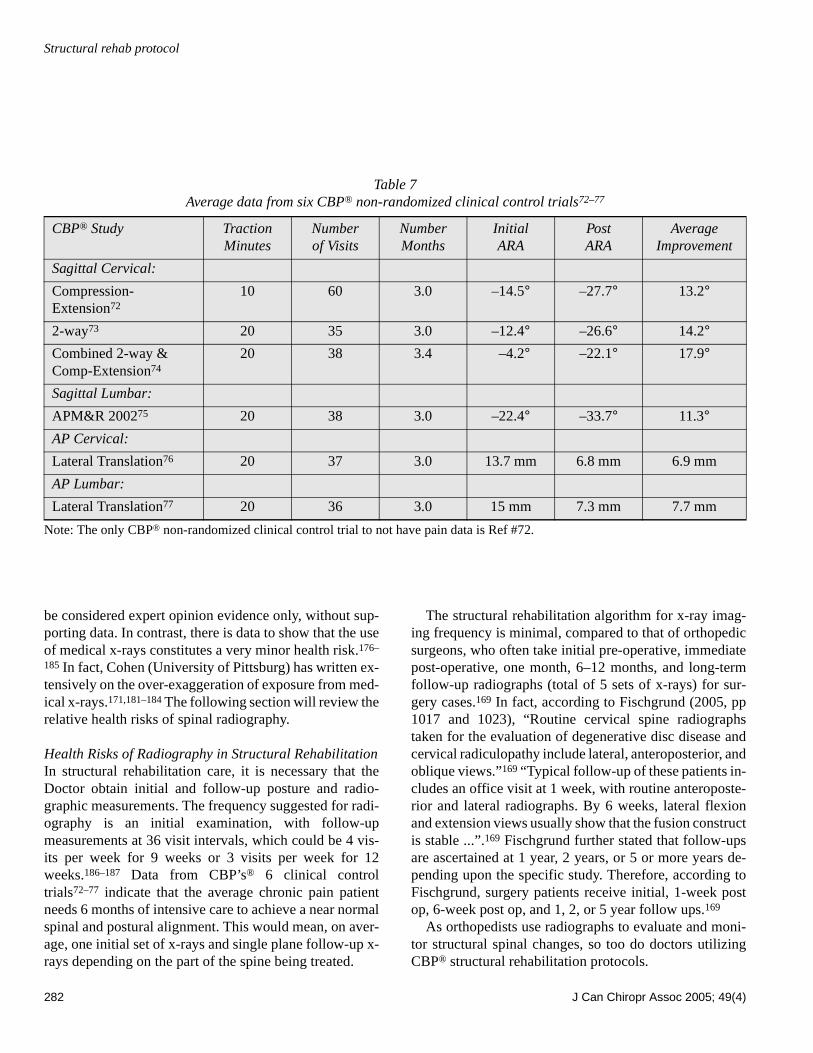
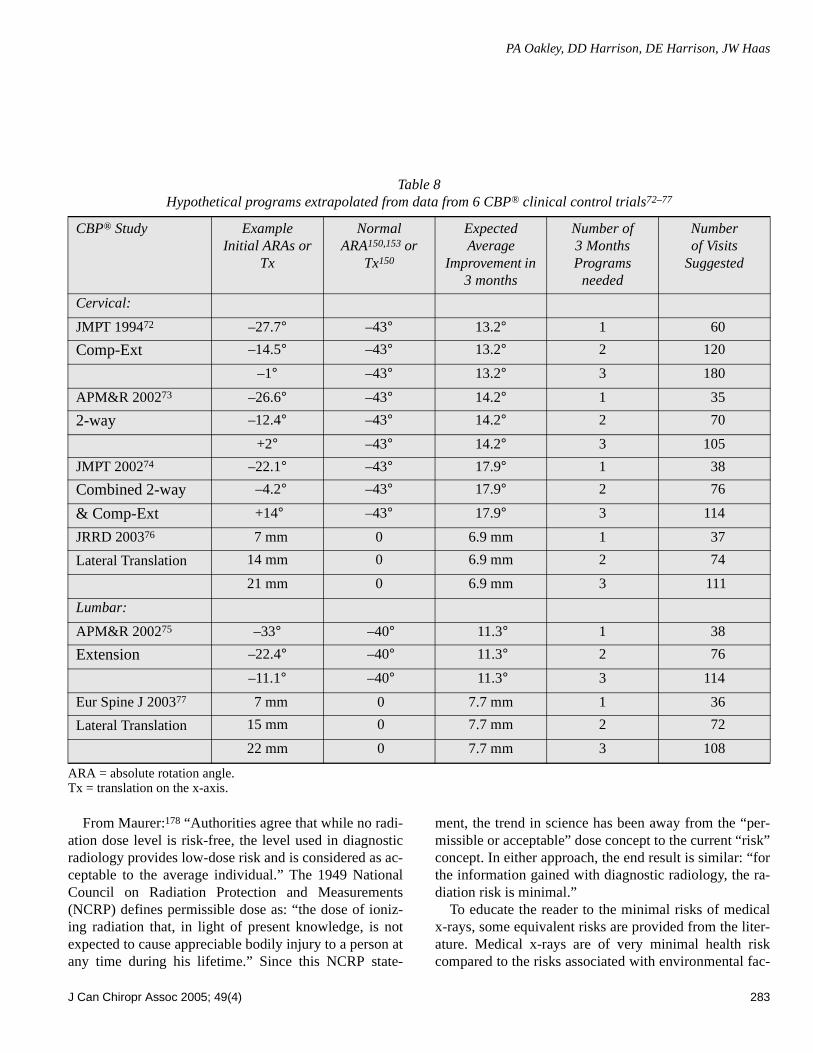
Health Risks of Radiography in Structural RehabilitationIn structural rehabilitation care, it is necessary that theDoctor obtain initial and follow-up posture and radio-graphic measurements. The frequency suggested for radi-ography is an initial examination, with follow-upmeasurements at 36 visit intervals, which could be 4 vis-its per week for 9 weeks or 3 visits per week for 12weeks.186–187 Data from CBP’s® 6 clinical controltrials72–77 indicate that the average chronic pain patientneeds 6 months of intensive care to achieve a near normalspinal and postural alignment. This would mean, on aver-age, one initial set of x-rays and single plane follow-up x-rays depending on the part of the spine being treated.
The structural rehabilitation algorithm for x-ray imag-ing frequency is minimal, compared to that of orthopedicsurgeons, who often take initial pre-operative, immediatepost-operative, one month, 6–12 months, and long-termfollow-up radiographs (total of 5 sets of x-rays) for sur-gery cases.169 In fact, according to Fischgrund (2005, pp1017 and 1023), “Routine cervical spine radiographstaken for the evaluation of degenerative disc disease andcervical radiculopathy include lateral, anteroposterior, andoblique views.”169 “Typical follow-up of these patients in-cludes an office visit at 1 week, with routine anteroposte-rior and lateral radiographs. By 6 weeks, lateral flexionand extension views usually show that the fusion constructis stable ...”.169 Fischgrund further stated that follow-upsare ascertained at 1 year, 2 years, or 5 or more years de-pending upon the specific study. Therefore, according toFischgrund, surgery patients receive initial, 1-week postop, 6-week post op, and 1, 2, or 5 year follow ups.169
As orthopedists use radiographs to evaluate and moni-tor structural spinal changes, so too do doctors utilizingCBP® structural rehabilitation protocols.
Table 7Average data from six CBP® non-randomized clinical control trials72–77
Note: The only CBP® non-randomized clinical control trial to not have pain data is Ref #72.
CBP® Study TractionMinutes
Numberof Visits
Number Months
InitialARA
PostARA
AverageImprovement
Sagittal Cervical:
Compression-Extension72
10 60 3.0 –14.5° –27.7° 13.2°
2-way73 20 35 3.0 –12.4° –26.6° 14.2°Combined 2-way & Comp-Extension74
20 38 3.4 –4.2° –22.1° 17.9°
Sagittal Lumbar:
APM&R 200275 20 38 3.0 –22.4° –33.7° 11.3°AP Cervical:
Lateral Translation76 20 37 3.0 13.7 mm 6.8 mm 6.9 mm
AP Lumbar:
Lateral Translation77 20 36 3.0 15 mm 7.3 mm 7.7 mm
PA Oakley, DD Harrison, DE Harrison, JW Haas
J Can Chiropr Assoc 2005; 49(4) 283
From Maurer:178 “Authorities agree that while no radi-ation dose level is risk-free, the level used in diagnosticradiology provides low-dose risk and is considered as ac-ceptable to the average individual.” The 1949 NationalCouncil on Radiation Protection and Measurements(NCRP) defines permissible dose as: “the dose of ioniz-ing radiation that, in light of present knowledge, is notexpected to cause appreciable bodily injury to a person atany time during his lifetime.” Since this NCRP state-
ment, the trend in science has been away from the “per-missible or acceptable” dose concept to the current “risk”concept. In either approach, the end result is similar: “forthe information gained with diagnostic radiology, the ra-diation risk is minimal.”
To educate the reader to the minimal risks of medicalx-rays, some equivalent risks are provided from the liter-ature. Medical x-rays are of very minimal health riskcompared to the risks associated with environmental fac-
Table 8Hypothetical programs extrapolated from data from 6 CBP® clinical control trials72–77
ARA = absolute rotation angle.Tx = translation on the x-axis.
CBP® Study ExampleInitial ARAs or
Tx
Normal ARA150,153 or
Tx150
Expected Average
Improvement in 3 months
Number of 3 Months Programs
needed
Numberof Visits
Suggested
Cervical:
JMPT 199472 –27.7° –43° 13.2° 1 60
Comp-Ext –14.5° –43° 13.2° 2 120
–1° –43° 13.2° 3 180
APM&R 200273 –26.6° –43° 14.2° 1 35
2-way –12.4° –43° 14.2° 2 70
+2° –43° 14.2° 3 105
JMPT 200274 –22.1° –43° 17.9° 1 38
Combined 2-way –4.2° –43° 17.9° 2 76
& Comp-Ext +14° –43° 17.9° 3 114
JRRD 200376 7 mm 0 6.9 mm 1 37
Lateral Translation 14 mm 0 6.9 mm 2 74
21 mm 0 6.9 mm 3 111
Lumbar:
APM&R 200275 –33° –40° 11.3° 1 38
Extension –22.4° –40° 11.3° 2 76
–11.1° –40° 11.3° 3 114
Eur Spine J 200377 7 mm 0 7.7 mm 1 36
Lateral Translation 15 mm 0 7.7 mm 2 72
22 mm 0 7.7 mm 3 108
Structural rehab protocol
284 J Can Chiropr Assoc 2005; 49(4)
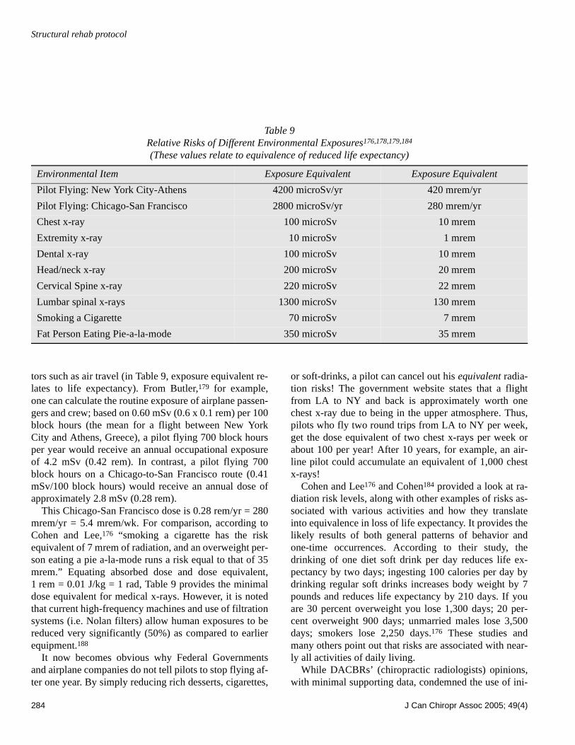
tors such as air travel (in Table 9, exposure equivalent re-lates to life expectancy). From Butler,179 for example,one can calculate the routine exposure of airplane passen-gers and crew; based on 0.60 mSv (0.6 x 0.1 rem) per 100block hours (the mean for a flight between New YorkCity and Athens, Greece), a pilot flying 700 block hoursper year would receive an annual occupational exposureof 4.2 mSv (0.42 rem). In contrast, a pilot flying 700block hours on a Chicago-to-San Francisco route (0.41mSv/100 block hours) would receive an annual dose ofapproximately 2.8 mSv (0.28 rem).
This Chicago-San Francisco dose is 0.28 rem/yr = 280mrem/yr = 5.4 mrem/wk. For comparison, according toCohen and Lee,176 “smoking a cigarette has the riskequivalent of 7 mrem of radiation, and an overweight per-son eating a pie a-la-mode runs a risk equal to that of 35mrem.” Equating absorbed dose and dose equivalent,1 rem = 0.01 J/kg = 1 rad, Table 9 provides the minimaldose equivalent for medical x-rays. However, it is notedthat current high-frequency machines and use of filtrationsystems (i.e. Nolan filters) allow human exposures to bereduced very significantly (50%) as compared to earlierequipment.188
It now becomes obvious why Federal Governmentsand airplane companies do not tell pilots to stop flying af-ter one year. By simply reducing rich desserts, cigarettes,
or soft-drinks, a pilot can cancel out his equivalent radia-tion risks! The government website states that a flightfrom LA to NY and back is approximately worth onechest x-ray due to being in the upper atmosphere. Thus,pilots who fly two round trips from LA to NY per week,get the dose equivalent of two chest x-rays per week orabout 100 per year! After 10 years, for example, an air-line pilot could accumulate an equivalent of 1,000 chestx-rays!
Cohen and Lee176 and Cohen184 provided a look at ra-diation risk levels, along with other examples of risks as-sociated with various activities and how they translateinto equivalence in loss of life expectancy. It provides thelikely results of both general patterns of behavior andone-time occurrences. According to their study, thedrinking of one diet soft drink per day reduces life ex-pectancy by two days; ingesting 100 calories per day bydrinking regular soft drinks increases body weight by 7pounds and reduces life expectancy by 210 days. If youare 30 percent overweight you lose 1,300 days; 20 per-cent overweight 900 days; unmarried males lose 3,500days; smokers lose 2,250 days.176 These studies andmany others point out that risks are associated with near-ly all activities of daily living.
While DACBRs’ (chiropractic radiologists) opinions,with minimal supporting data, condemned the use of ini-
Table 9Relative Risks of Different Environmental Exposures176,178,179,184
(These values relate to equivalence of reduced life expectancy)
Environmental Item Exposure Equivalent Exposure Equivalent
Pilot Flying: New York City-Athens 4200 microSv/yr 420 mrem/yr
Pilot Flying: Chicago-San Francisco 2800 microSv/yr 280 mrem/yr
Chest x-ray 100 microSv 10 mrem
Extremity x-ray 10 microSv 1 mrem
Dental x-ray 100 microSv 10 mrem
Head/neck x-ray 200 microSv 20 mrem
Cervical Spine x-ray 220 microSv 22 mrem
Lumbar spinal x-rays 1300 microSv 130 mrem
Smoking a Cigarette 70 microSv 7 mrem
Fat Person Eating Pie-a-la-mode 350 microSv 35 mrem
PA Oakley, DD Harrison, DE Harrison, JW Haas
J Can Chiropr Assoc 2005; 49(4) 285
tial and post-radiographs to collect alignment measure-ments,170–175 data actually exists that indicates medical x-ray exposure may have a health benefit.191 However, min-imal risk is inherent in everything we do as part of the hu-man experience. While we must constantly work towardsthe reduction of health risks in all endeavors, we may beled to accept a minimal level as normal. While there is nodata indicating diagnostic radiology has a present risk,any radiation dose must be compared to the benefits ofuseful information gained. The necessity for appropriatetreatment selection is indeed an acceptable trade-offwhen put into perspective. The need for x-ray imaging isespecially clear when one considers that radiographic im-aging is the only valid method for attaining spinal align-
ment values; whereas surface contour methods (e.g.,flexible ruler) are invalid and unreliable.192–196
Radiation HormesisRadiation hormesis is the stimulatory or beneficial effectof low doses of ionizing radiation. While an actual bene-fit from radiation exposure may seem outrageous, there ismuch scientific evidence for this phenomenon. This topicis in direct conflict with the “Linear No-Threshold Hy-
Figure 4. Abnormal Postural Rotations. Rotations of the head relative to the thorax; thorax relative to the pelvis; and pelvis relative to the feet are depicted from the top to bottom rows, respectively. Regional rotations about the x, y, and z-axes are depicted from the left to right columns, respectively. Reprinted with permission from Harrison DE et al. Spinal Biomechanics for Clinicians. Vol. I. Evanston, WY: Harrison CBP® Seminars, Inc., 2003.
Figure 5. Abnormal Postural Translations. Translations of the head relative to the thorax; thorax relative to the pelvis; and pelvis relative to the feet are depicted from the top to bottom rows, respectively. Regional translations along the x, y, and z-axes are depicted from the left to right columns, respectively. Reprinted with permission from Harrison DE et al. Spinal Biomechanics for Clinicians. Vol. I. Evanston, WY: Harrison CBP® Seminars, Inc., 2003.
Structural rehab protocol
286 J Can Chiropr Assoc 2005; 49(4)
pothesis” (LNT), which has been assumed to be true formore than 50 years. This LNT model comes from esti-mating the risks at lower doses of radiation, in the ab-sence of data, by extrapolating in a linear model fromlarge doses of radiation from atomic bombs dropped onJapan in the 1940s.
This LNT model has been used to set limits of radia-tion exposure by all official and governmental associa-tions.185 Recently in 2003, Kauffman180 reiterated thatauthors critical of exposure from diagnostic radiation al-ways use the LNT model. This use of the LNT model in-cludes the recent 2005 report by the USA NationalResearch Council.197 This report stated, “there will besome risk, even at low doses (100 mSv or less), althoughthe risk is small” and “there is no direct evidence of in-creased risk of non-cancer diseases at low doses.”197 This2005 report ignored and contradicted an earlier 2003 re-view by Kant et al.198
For a comparison of exposures, USA citizens are ex-posed to an average annual natural background radiationlevel of 3 mSv, while exposure from a chest x-ray is ap-proximately 0.1 mSv and exposure from a whole bodycomputerized tomography (CT) scan is approximately 10mSv.197 Also it is noted that 10mSv = 1,000mrem, whichequates to about 46 cervical series or 8 lumbar series (seeTable 9).
The LNT model has been questioned for its applicationto low levels of exposure by many researchers.183,189,199–
202 Actually, below a certain level of exposure, there arebeneficial health effects, (termed radiation hormesis),which do not follow from extrapolation of the high-doseportion of the curve.182,189–191,203–217
Structural Rehabilitation ProtocolsWhile other methods may provide evidence for structuralrehabilitation, we discuss only recent research outcomesin CBP® Technique. CBP® is unique in chiropractic, inthat it utilizes a “mirror image®” concept applied to hu-man posture; this basic tenet has a sound foundation inLinear Algebra, an area of study common to both en-gineering and mathematics.218 CBP® multi-modal careconsists of three primary procedures: mirror image® ex-ercises, mirror image® adjustments, and mirror image®/extension traction. These mirror image® posture posi-tions are the rotation and translation pairs in or abouteach coordinate axis (Figures 4 and 5).
The reason for postural mirror image® exercises, ad-justments, and traction procedures is to address all thetissues involved in spine and posture alignment. Al-though strength and conditioning exercise has not provento correct posture,219 mirror image® exercises haveshown initial promise in the reduction of posture and spi-nal displacements.220–224 These exercises are performedto stretch shortened muscles and to strengthen those mus-cles that have weakened in areas where postural muscleshave adapted to asymmetric or ill-positioned postures.
Postural adjustments as performed with drop table,hand-held instrument, or even mirror image® manipula-tion procedures, are performed for resetting the nervoussystem regulation of postural muscle balance.225,226 Pos-tural mirror image® extension traction provides sus-tained loading periods of 10–20 minutes and is necessaryto cause visco-elastic deformation to the resting length ofthe spinal muscles, ligaments, and discs.227
From 1994–2004, CBP® has completed seven casestudies,228–234 and has completed six non-randomizedclinical control trials.72–77 While case studies are rankedas the lowest level of clinical studies evidence on the tra-ditional scientific evidence hierarchy, non-randomizedcontrol trials are the 2nd highest type of evidence; rankedsecond only to the RCT.1,14 Recalling that RCTs are inad-equate for evaluating multi modal chiropractic care regi-mens,1 these seven CBP® case studies228–234 and sixCBP® control trials72–77 provide a growing clinical evi-dence base to support the need for CBP® structural careprograms of sufficient durations to provide as near nor-mal as possible posture and spine structural rehabilita-tion/re-alignment. From CBP’s® six clinical controltrials, 72–77 Table 7 presents the average total number ofvisits, frequency, duration of care, and the amount of spi-nal alignment improvement found for the cervical andlumbar lordoses and cervical/lumbar frontal plane align-ments, respectively.
Table 8 presents actual and extrapolated data estimat-ing durations of care necessary for correcting hypotheti-cal sagittal plane displacements; we note that a negativesign means lordotic curvature and a positive sign indi-cates kyphotic curvature for measurements in the cervicaland lumbar regions.140–142 Table 8 also gives average andextrapolated data estimating the duration of care neces-sary to reduce/correct a head and trunk list (side shiftposture) in the AP cervico-thoracic and lumbar radio-

PA Oakley, DD Harrison, DE Harrison, JW Haas
J Can Chiropr Assoc 2005; 49(4) 287
graphs as measured appropriately (i.e. measured as a hor-izontal displacement of: 1) Mid-C2 dens compared tovertical line up from estimated center of mass (CM) ofThoracic #4 in an AP cervico-thoracic analysis;144 2) Es-timated CM of T12 compared to a vertical line up fromthe S2 tubercle in an AP lumbar analysis.143,235
From Table 8 one can estimate structural care dura-tions based on extrapolations from the CBP® clinicaltrials. It should be mentioned that these are merely aver-ages; and are in fact extrapolations from the current clini-cal trial data. Therefore, individual patients may changein shorter or longer times; thus, follow-up radiologicalexams provide the clinician with valuable insight into in-dividual patient response to treatment. The followingprovide examples of estimating average durations of carehypothetically necessary to restore a patient’s spine toideal spinal alignment:
a) If a patient had a kyphotic cervical curve ARA (C2 toC7) measuring +12°, then 3 programs (treatmentblocks of 38 sessions) of 2-way extension-compres-sion traction would hypothetically correct the mis-alignment. That is, three increments of averageimprovement of –17.9°, results in an extrapolated av-erage correction of 3x(–17.9) +12° = –41.7°, whichapproximates the ideal cervical lordosis of –43°.150
b) If a patient had a hypolordotic cervical ARA (C2 toC7) measuring –29°, then one program of 35 visits ofCBP® 2-way traction would be expected to correct themisalignment: –29° + 1x(–14.2°) = –43.2° (ideal nor-mal).150
c) If a patient had a hypolordotic lumbar curve ARA (L1to L5) of –18°, then two programs of CBP® lumbarextension traction of 38 visits would hypotheticallycorrect the misalignment. That is, two increments ofaverage improvement of –11.3°, results in an extrapo-lated correction of 2x(–11.3°) + –18° = –40.6°, whichapproximates the average/ideal normal lumbar lordo-sis value of –40°153 (see Table 8).
Criticisms of CBP MethodsIn the past, criticisms of CBP® methods and x-ray proto-cols have been based on the 1999 Commentary by Haas etal.170 These criticisms often neglect to provide the Harri-son rebuttal written in 2000,134 which reported that Haas etal.170 misrepresented references, misinterpreted referenc-
es, misquoted references, and performed a selective liter-ature review.134 In fact, this Harrison-Haas debate was aseries of three publications.134,170,236 Additionally, the uni-formed often state that the extension position in CBP® cer-vical traction methods are dangerous.
Numerous articles from the literature lead to the con-clusion that this is definitely not the case. In fact, in a1999 thorough review of the literature on varying posi-tions of the head associated with vertebral and basilarartery blood flow and dissection, Haldeman et al.237 con-cluded that “examination of the data fails to show a con-sistent position or movement of the neck that could beconsidered particularly dangerous.”
In addition, Thiel et al.238 found no occlusion of verte-bral artery blood flow during various head and neck posi-tioning tests on the patient, including head extension.
Inaccurate personal opinions about the dangers of ex-tension come from “Beauty Parlor Stroke.” There hasbeen anecdotal criticism of the hyper-extension head po-sition at Beauty Parlors. Much of this criticism seems tobe based on several letters to editors and case reports inthe Index Medicus literature concerning “beauty parlorstroke.”239–243 The positions referred to were prolonged(1-hour or more) hyper-extension combined with axialrotation,239–243 although Endo et al.243 did not discuss anyrotation of the head. In 1992 and 1993, Weintraub239–241
reported on seven cases of “Beauty Parlor Stroke” inwhich clients at beauty parlors had symptoms of nystag-mus, ataxia, slurred speech, facial weakness, nausea,vomiting, vertigo, and dysarthria after having their hairshampooed. Six of these seven individuals were olderthan 75 years and one was 54-years-old. The 54-year-oldsubject had been left in a position of cervical hyper-ex-tension over the edge of a shampoo bowl in excess of twohours. In 1995, Stratigos242 reported on the condition ofhis mother after a trip to a beauty parlor.
All four of these articles discussed in detail that themechanism of vertebrobasilar injury is associated withcervical axial rotation while in hyper-extension. In 2000,Endo et al.243 reported a single case of a woman aged 62who suffered a “beauty parlor stroke.” There was nomention of the duration of shampoo treatment or a de-tailed explanation of the position of the head.
Unlike beauty parlor employees, individuals employ-ing CBP® spinal traction methods are trained physicians,who do screening examinations on patients for tolerance
Structural rehab protocol
288 J Can Chiropr Assoc 2005; 49(4)
to head extension. Using our cervical traction protocol,patients are screened and then monitored while tractiontime periods are increased at only a few minutes per visit,starting at 3–5 minutes, over a period of many visits to amaximum of 20 minutes. These traction methods are alsonot used or modified for those of advanced age and donot involve axial rotation in the extended position. Whileany induced stroke symptoms would be unacceptable,these “beauty parlor strokes” should not be applied toCBP® cervical extension traction methods, when used bytrained physicians.
ConclusionsBesides the RCT, other forms of scientific evidence, ifexisting, may be more than adequate to create goal-ori-ented clinical guidelines.1,13,14,244 CBP® studies providethe typical type of chiropractic care, as several proce-dures are provided to the patient on each visit. At present,there is evidence for SMT for neck and low back pain.From published research for the treatment of chronicneck and back pain at this time, CBP® Technique hasmore supporting evidence than Diversified Technique, astaught in all Chiropractic Colleges.
This paper has presented guidelines as a clinical toolfor the practice of structural rehabilitation by CBP® tech-nique methods. CBP® is unique, in that, unlike mostchiropractic techniques,245 CBP® has laid a solid founda-tion of basic science research (spine modeling, x-ray linedrawing reliability, x-ray positioning repeatability, pos-ture reliability, biomechanical stress analysis), clinical re-search (case studies, clinical trials), and educationalresearch (reviews, position papers).
Because traditional practice protocols in chiropractichave considered only acute and chronic pain conditions,limited inclusion of functional rehabilitation, and a totalneglect of structural rehabilitation, there is a need to havepublished protocols for structural rehabilitation of thespine and posture. This manuscript has proposed structur-al protocol guidelines based on clinical evidence from asignificant quantity and quality of CBP® technique publi-cations. Tables 6–8 are based on CBP® mirror image®methods. The use of multiple clinical methodologies inthese CBP® studies is consistent with Bolton’s ideas13 ofclinical applications in EBP. Because of the focused liter-ature herein, this guideline serves as a tool only for thedoctor practicing structural rehabilitation utilizing CBP®
mirror image® exercise, adjusting, and traction proce-dures.
AcknowledgementThe authors acknowledge the assistance of Dr. DennisMizel DC with this manuscript.
References1 Bolton JE. The evidence in evidence-based practice: what
counts and what doesn’t count? J Manipulative Physiol Ther 2001; 24:362–366.
2 Sackett DL, Richardson WS, Rosenberg W, Haynes RB. Evidence-based medicine: How to practice and teach EBM. New York: Churchill-Livingstone, 1997.
3 Lipman G. Evidence-based pain management and palliative care. J Pain Palliat Care Pharmacother 2002; 16:1–3.
4 Fishbain DA, Cutler RB, Rosomoff HL, Rosomoff RS. Can patients taking opioids drive safely? A structured evidence-based review. J Pain Palliat Care Pharmacother 2002; 16:9–28.
5 Caramanica L, Cousino JA, Peterson S. Four elements of a successful quality program. alignment, collaboration, evidence-based practice, and excellence. Nurs Adm Q 2003; 27:336–343.
6 Jordan KM, Arden NK, Doherty M, et al. EULAR recommendations 2003: an evidenced based approach to the management of knee osteoarthritis: Report of a task force of the standing committee for the International Clinical Studies Including Therapeutic Trials. Ann Rheum Dis 2003; 62:1145–1155.
7 Wassem RA, Stillion-Allen KA. Evidence-based management of the fibromyalgia patient. In search of optimal functioning. Adv Nurs Prac 2003; 11:34–38,41–43.
8 Hasenfeld R, Shekelle PG. Is the methodological quality of guidelines declining in the US? Comparison of the quality of US Agency for Health Care Policy and Research (AHCPR) guidelines with those published subsequently. Qual Saf Health Care 2003; 12:428–434.
9 Sackett DL. Evidence-based medicine [editorial]. Spine 1998; 23:1085–1086.
10 Davidson KW, Goldstein M, Kaplan RM. Evidence-based behavioral medicine: What is it and how do we achieve it? Ann Behav Med 2003; 26:161–171.
11 Smith R. Where is the wisdom ...? The poverty of medical evidence. Br Med J 1991; 303:798–799.
12 Brouwers M, Charette M. Evaluation of clinical practice guidelines in chiropractic care: A comparison of North American guideline reports. J Can Chiropr Asso 2001; 45:141–153.
13 Bolton JE. Whence the evidence from evidence-based practice? Br J Chiropr 2000; 4:2–3.
PA Oakley, DD Harrison, DE Harrison, JW Haas
J Can Chiropr Assoc 2005; 49(4) 289
14 Hawk C. Chiropractic clinic research: where are we looking for the key? J Neuromusculoskeletal System 1999; 7:150–155.
15 Miles A, Polychronis A, Grey J. Evidence-based medicine: Why all the fuss? This is why. J Eval Clin Pract 1997; 3:83–86.
16 Fahey T. Applying the results of clinical trials to patients in general practice: perceived problems, strengths, assumptions, and challenges for the future. Br J Gen Pract 1998; 1:1173–1178.
17 McKee M, Britton A, Black N, et al. Interpreting the evidence: choosing between randomized and non-randomized studies. Br Med J 1999; 319:312–315.
18 Rosenfeld RM. Meaningful outcomes research. In: Isenberg SF, ed. Managed care, outcomes and quality. New York: Thieme, 1998:99–115.
19 Venning GR. Validity of anecdotal reports of suspected adverse drug reactions: The problem of false alarms. BMJ 1982; 284:249–252.
20 Benson K, Hartz AJ. A comparison of observational studies and randomized controlled trials. N Engl J Med 2000; 342:1878–1886.
21 Chiropractic in New Zealand: report of the commission of inquiry. Wellington, NZ:P.D. Hasselberg, Government Printer, 1979.
22 Manga P, Angus D, Papadopoulos C, Swan W. The effectiveness and cost-effectiveness of chiropractic management of low back pain. Ottawa:Kenilworth Publishing, 1993.
23 Meade TW, Dyer S, Browne W, Frank AO. Randomized comparison of chiropractic and hospital outpatient management for low back pain: results from extended follow up. Br Med J 1995; 311:349–351.
24 Shekelle, P. G., Adams, A. H., Chassin, M. R., Hurwitz, E. L., Phillips, R. B., and Brook, R. H. The appropriateness of spinal manipulation for low back pain. Project overview and literature review. Santa Monica, CA, RAND, 1991.
25 Bigos S, Bowyer O, Braen G, et al. Acute low back pain problems in adults. Clinical practice guideline No. 14. AHCPR Publication No. 95–0642. Rockville, MD: Dept. of Health and Human Services (US), Agency for Health Care Policy and Research, Public Health Service:1994.
26 Michigan Chiropractic Society. Chiropractic Care and Utilization Review Guidelines. Lansing, MI:MCS, 1991.
27 Standards of Practice. Minnesota Chiropractic Association. MN:MCA, 1991.
28 Ohio State Chiropractic Association. The chiropractic manual for insurance claims personnel. Columbus, OH:OSCA, 1990.
29 Hawk C, Long CR, Boulanger KT. Prevalence of non-musculoskeletal complaints in chiropractic: report from a practice-based research program. J Manipulative Physiol Thera 2001; 24:157–169.
30 Troyanovich SJ, Harrison DE, Harrison DD. Structural rehabilitation of the spine and posture: Rationale for treatment beyond the resolution of symptoms. J Manipulative Physiol Ther 1998; 21:37–50.
31 Liebenson C. Rehabilitation of the spine: a practicioner’s manual. New York: Lippincott Williams & Wilkins, 1996.
32 Kendall FP, McCreary EK, Provance PG. Muscles Testing and Function. (4th ed.). Baltimore: Williams & Wilkins, 1993.
33 Haldeman S, Phillips RB. Spinal manipulative therapy in the management of low back pain. In: Frymoyer, J.W., Ducker, T.B., Hadler, N.M., Kostuik, J.P., Weinstein, J.N., Whitecloud, T.S. editors. The adult spine: principles and practice. New York: Raven Press, Ltd. 1991:1581–1605.
34 Carr DB, Goudas LC, Denman WT, Brookoff D, Staats PS, Brennen L, Green G, Albin R, Hamilton D, Rogers MC, Firestone L, Lavin PT, Mermelstein. Safety and efficacy of intranasal ketamine for the treatment of breakthrough pain in patients with chronic pain: a randomized, double-blind, placebo-controlled, crossover study. Pain. 2004; 108(1–2):17–27.
35 Bryans R. (editor). Whiplash: A practitioner’s guide to understanding Whiplash Associated Disorders (WAD). Canadian Chiropractic Association, 2000.
36 Bronfort G, Haas M, Evans R, Bouter LM. Efficacy of spinal manipulation and mobilization for low back pain and neck pain: a systematic review and best evidence synthesis. Spine J 2004; 4:335–356.
37 Williams NH, Wilkinson C, Russell I, Edwards RT, Hibbs R, Linck P, et al. Randomized osteopathic manipulation study (ROMANS): pragmatic trial for spinal pain in primary care. Fam Pract 2003; 20:662–669.
38 Licciardone JC, Stoll ST, Fulda KG, Russo DP, Siu J, Winn W, et al. Osteopathic manipulative treatment for chronic low back pain: a randomized controlled trial. Spine 2003; 28:1355–1362.
39 Assendelft WJ, Morton SC, Yu EI, Suttorp MJ, Shekelle PG. Spinal manipulative therapy for low back pain. Cochrane database Syst Rev 2004; (4): CD000447.
40 Cherkin DC, Sherman KJ, Deyo RA, Shekelle PG. A review of the evidence for the effectiveness, safety, and cost of acupuncture, massage therapy, and spinal manipulation for low back pain. Ann Intern Med 2003; 138:898–906.
41 Glover JR, Morris JG, Khosla T. Back pain: a controlled clinical trial of rotational manipulation of the trunk. Br J Industr Med 1974; 31:59–64.
42 Godfrey CM, Morgan PP, Schatzker J. A randomized trial of manipulation for low-back pain in a medical setting. Spine 1984; 9:301–304.
43 Hadler NM, Curtis P, Gillings DB, Stinnet S. A benefit of spinal manipulation as adjunctive therapy for acute low-back pain: a stratified controlled trial. Spine 1987; 12:703–706.
Structural rehab protocol
290 J Can Chiropr Assoc 2005; 49(4)
44 MacDonald RS, Bell CMJ. An open controlled assessment of osteopathic manipulation in nonspecific low-back pain. Spine 1990; 15:364–370.
45 Mathews JA, Mills SB, Jenkins VM et al. Back pain and sciatica: controlled trials of manipulation, traction, sclerosant and epidural injections. Br J Rheumatol 1987; 26:416–423.
46 Bronfort G, Goldsmith CH, Nelson CF, Boline PD, Anderson AV. Trunk exercise combined with spinal manipulation or NSAID therapy for chronic low back pain: a randomized, observer-blinded clinical trial. J Manipulative Physiol Ther 1996; 19:570–582.
47 Burton AK, Tillotson KM, Cleary J. Single-blind randomized controlled trial of chemonucleolysis and manipulation in the treatment of symptomatic lumbar disc herniation. Eur Spine J 2000; 9:202–207.
48 Coxhead CE, Inskip H, Meade TW, North WR, Troop JD. Multicentre trial of physiotherapy in the management of sciatic symptoms. Lancet 1981; 1:1065–1068.
49 Herzog W, Conway PJ, Wilcox BJ. Effects of different treatment modalities on gait symmetry and clinical measures for sacroiliac joint patients. J Manipulative Physiol Ther 1991; 14:104–109.
50 Pope MH, Phillips RB, Haugh LD, Hsich CY, MacDonald L, Haldeman S. A prospective randomized three-week trial of spinal manipulation, transcutaneous muscle stimulation, massage and corset in the treatment of subacute low back pain. Spine 1994; 19:2571–2577.
51 Triano JJ, McGregor M, Hondras MA, Brennen PC. Manipulative therapy versus education programs in chronic low back pain. Spine 1995; 20:948–955.
52 Anderson GB, Lucente T, Davis AM, Kappler RE, Lipton JA, Leurgans S. A comparison of osteopathic spinal manipulation with standard care for patients with low back pain. N Engl J Med 1999; 341:1426–1431.
53 Cherkin DC, Deyo RA, Battie M, Street J, Barlow, W. A comparison of physical therapy, chiropractic manipulation, and provision of an educational booklet for the treatment of patients with low back pain. N Engl J Med 1998; 339:1021–1029.
54 Doran DM, Newell DJ. Manipulation in treatment of low back pain: a multicentre study. BMJ 1975; 2:161–164.
55 Evans DP, Burke MS, Lloyd KN, Roberts EE, Roberts GM. Lumbar spinal manipulation on trials. Part I: Clinical Assessment Rheumatol Rehabil 1978; 17:46–53.
56 Giles LGF, Muller R. Chronic spinal pain syndromes : a clinical pilot trial comparing acupuncture, a nonsteroidal anti-inflammatory drug, and spinal manipulation. J Manipulative Physiol Ther 1999; 22:376–381.
57 Hoehler FK, Tobis JS, Buerger AA. Spinal manipulation for low back pain. JAMA 1981; 245:1835–1838.
58 Hsieh CY, Adams AH, Tobis J, et al. Effectiveness of four conservative treatments for subacute low back pain: a randomized clinical trial. Spine 2002; 27:1142–1148.
59 Hurwitz EL, Morgenstern H, Harper P, et al. A randomized trial of medical care with and without physical therapy and chiropractic care with and without physical modalities for patients with low back pain: 6-month follow-up outcomes from the UCLA low back pain study. Spine 2002; 27:2193–2204.
60 Meade TW, Dyer S, Browne W, Townsend J, Frank AQ. Low back pain of mechanical origin: randomized comparison of chiropractic and hospital outpatient treatments. BMJ 1990; 300:1431–1437.
61 Postacchini F, Facchini M, Palieri P. Efficacy of various forms of conservative treatment in low back pain. A comparative study. Neuro Orthop 1988; 6; 28–35.
62 Skargren EI, Oberg BE, Carlsson PG, Gade M. Cost and effectiveness analysis of Chiropractic and physiotherapy treatment for low back and neck pain. Six-months follow-up. Spine 1997; 22:2167–2177.
63 Wreje U, Nordgren B, Aberg H. Treatment of pelvic joint dysfunction in primary care- a controlled study. Scand J Prim Health Care 1992; 10:310–315.
64 Howe DH, Newcombe RG, Wade MT. Manipulation of the cervical spine. A pilot study. J R Coll Gen Pract 1983; 33:574–579.
65 Bronfort G, Evans R, Nelson B, Aker P, Goldsmith C, Vernon H. A randomized clinical trial of exercise and spinal manipulation for patients with chronic neck pain. Spine 2001; 26:788–799.
66 Jordan A, Bendix T, Nielsen H, Hansen FR, Host D, Winkel A. Intensive training, physical therapy, or manipulation for patients with chronic neck pain. A prospective single-blinded randomized clinical trial. Spine 1998; 23:311–319.
67 Sloop PR, Smith DS, Goldberg E, Dore C. Manipulation for chronic neck pain. A double-blinded controlled trial. Spine 1982; 7:532–535.
68 Hurwitz EL, Morgenstern H, Harber P, Kominski GF, Yu F, Adams AH. A randomized trial of chiropractic manipulation and mobilization for patients with neck pain: clinical outcomes from the UCLA neck-pain study. Am J Public Health 2002; 92:1634–1641.
69 Skargren EI, Oberg BE, Carlsson PG, Gade M. Cost and effectiveness analysis of Chiropractic and physiotherapy treatment for low back and neck pain. Six-months follow-up. Spine 1997; 22:2167–2177.
70 Hurwitz EL, Morgenstern H, Vassilaki M, Chiang LM. Adverse reactions to chiropractic treatment and their effects on satisfaction and clinical outcomes among patients enrolled in the UCLA Neck Pain Study. J Manipulative Physiol Ther 2004; 27:16–25.
71 Haas M, Groupp E, Aicki M, Fairweather A, Ganger B, Attwood M, Cummins C, Baffes L. Dose response for chiropractic care of chronic cervicogenic headache and asssociated neck pain: a randomized pilot study. J Manipulative Physiol Ther 2004; 27(9):547–553.
PA Oakley, DD Harrison, DE Harrison, JW Haas
J Can Chiropr Assoc 2005; 49(4) 291
72 Harrison DD, Jackson BL, Troyanovich SJ, Robertson G, De George D, Barker WF. The efficacy of cervical extension-compression traction combined with diversified manipulation and drop table adjustments in the rehabilitation of cervical lordosis: a pilot study. J Manipulative Physiol Ther 1994; 17:454–464.
73 Harrison DE, Cailliet R, Harrison DD, Janik TJ, Holland B. A new 3-point bending traction method for restoring cervical lordosis and cervical manipulation: A nonrandomized clinical controlled trial. Arch Phys Med Rehab 2002; 83:447–453.
74 Harrison DE, Harrison DD, Betz J, Colloca CJ, Janik TJ, Holland B. Increasing the cervical lordosis with seated combined extension-compression and transverse load cervical traction with cervical manipulation: Nonrandomized clinical control trial. J Manipulative Physiol Ther 2003; 26:139–151.
75 Harrison DE, Cailliet R, Harrison DD, Janik TJ, Holland B. Changes in sagittal lumbar configurations with a new method of extension traction: Nonrandomized clinical controlled trial. Arch Phys Med Rehab 2002; 83:1585–1591.
76 Harrison DE, Cailliet R, Betz J, et al. Conservative methods for reducing lateral translation postures of the head: A non-randomized clinical control trial. J Rehab Res Dev 2004; 41(4): 631–640.
77 Harrison DE, Cailliet R, Betz JW, Harrison DD, Haas JW, Janik TJ, Holland B. A non-randomized clinical control trial of Harrison mirror image methods for correcting trunk list (lateral translations of the thoracic cage) in patients with chronic low back pain. Eur Spine J 2005; 14(2):155–162.
78 Leboeuf-Yde C, Gronstvedt A, Borge JA, Lothe J, Magnesen E, Nilsson O, Rosok G, Stig LC, Larsen K. The Nordic back pain subpopulation program: A 1-year prospective multicenter study of outcomes of persistent low-back pain in chiropractic patients. J Manipulative Physiol Ther 2005; 28(2):90–96.
79 Horng YS, Hwang YH, Wu HC, Liang HW, Jang Y, Twu FC, Wang JD. Predicting health-related quality of life in patients with low back pain. Spine 2005; 30:551–555.
80 Korovessis P, Dimas A, Lambiris E. The significance of correlation of radiographic variables and MOS short-form health survey for clinical decision in symptomatic low back pain patients. Stud Health Technol Inform 2002; 91:325–331.
81 Walmsley RP, Kimber P, Culham E. The effect of initial head position on active cervical axial rotation range of motion in two age populations. Spine 1996; 21:2435–2442.
82 Takeshima T, Omokawa S, Takaoka T, Araki M, Ueda Y, Takakura Y. Sagittal alignment of cervical flexion and extension: Lateral radiographic analysis. Spine 2002; 27:E348–355.
83 Hirose D, Ishida K, Nagano Y, Takahashi T, Yamamoto H. Posture of the trunk in the sagittal plane is associated with gait in community-dwelling elderly population. Clin Biomech 2004; 19(1):57–63.
84 Edmondston SJ, Henne SE, Loh W, Ostvoid E. Influence of cranio-cervical posture on three-dimensional motion of the cervical spine. Man Ther 2005; 10(1):44–51.
85 Stokes I, Spence H, Aronsson D, Kilmer N. Mechanical modulation of vertebral body growth: implications for scoliosis progression. Spine 1996; 21:1162–1167.
86 Bowman SM, Keaveny TM, Gibson LJ, Hayes WC, McMahon TA. Compressive creep behavior of bovine trabecular bone. J Biomechanics 1994; 27:301–310.
87 Mente PL, Aronsson DD, Stokes IAF, Iatridis JC. Mechanical modulation of growth for the correction of vertebral wedge deformities. J Orthop Res 1999; 17:518–524.
88 Brickley-Parsons D, Glimcher MJ. Is the chemistry of collagen in intervertebral discs an expression of Wolff’s Law? A study of the human lumbar spine. Spine 1983; 9:148–163.
89 Harrison DE, Colloca CJ, Harrison DD, Janik TJ, Haas JW, Keller TS. Anterior thoracic posture increases thoracolumbar disc loading. Eur Spine J 2005; 14(3):234–242.
90 Kaigle A, Ekstrom L, Holm S, Rostedt M, Hansson T. In vivo dynamic stiffness of the porcine lumbar spine exposed to cyclic loading: influence of load and degeneration. J Spinal Disord 1998; 11:65–70.
91 Lotz JC, Chin JR. Intervertebral disc cell death is dependant on the magnitude and duration of spinal loading. Spine 2000; 25:1477–1483.
92 Frechede B, et al. Risk of injury of the human neck during impact: role of geometrical and mechanical parameters. Euro CSRS, Porto, Portugal, May 30-June 5 , Paper A29, 2004.
93 Mayoux-Benhamou MA, et al. Influence of head position on dorsal neck muscle efficiency. Electromyogr Clin Neurophysiol 1993; 33:161–166.
94 Kumaresan S, Yoganandan N, Pintar FA. Finite element analysis of the cervical spine: A material property sensitivity study. Clinical Biomechanics 1999; 14:41–53.
95 Smith CG. Changes in length and position of the segments of the spinal cord with changes in posture in the monkey. Radiology 1956; 66:259–265.
96 Reid JD. Effects of flexion-extension movements of the head and spine upon the spinal cord and nerve roots. J Neurol Neurosurg Psychiatry 1960; 23:214–221.
97 Adams CBT, Logue V. Studies in cervical spondylotic myelopathy I: Movement of the cervical roots, dura and cord, and their relationship to the course of the extrathecal roots. Brain 1971; 94:557–568.
Structural rehab protocol
292 J Can Chiropr Assoc 2005; 49(4)
98 Breig A. Pathological stress in the pons-cord tissue tract and its alleviation by neurosurgical means. In: Congress of Neurological Surgeons, ed. Clinical neurosurgery. Baltimore: Williams & Wilkins, 1973:87–94.
99 Breig A, Marions O. Biomechanics of the lumbosacral nerve roots. Acta Radiologica Diagnosis 1963; 1:1141–1160.
100 Breig A. Adverse mechanical tension in the central nervous system: An analysis of cause and effect: Relief by functional neurosurgery. Stockholm: Almqvist and Wiksell International, 1978.
101 Cailliet R. Low back pain syndrome. Philadelphia: F.A. Davis, 1981.
102 Harber P, Bloswick D, Beck J, Pena L, Baker D, Lee J. Supermarket checker motions and cumulative trauma risk. J Occup Med 1993; 35:805–811.
103 Genaidy AM, Karwowski W. The effects of neural posture deviations on perceived joint discomfort ratings in sitting and standing postures. Ergonomics 1993; 36:785–792.
104 Rys M, Konz S. Standing. Ergonomics 1994; 37:677–687.105 Powers C, Maffucci R, Hampton S. Rear foot posture in
subjects with patello-femoral pain. J Orthop Sports Phys Ther 1995; 22:155–160.
106 Eklund J, Odenrick P, Zettergren S, Johansson H. Head posture measurements among work vehicle drivers and implications for work and workplace design. Ergonomics 1994; 37:623–639.
107 Watson A. Sports injuries in footballers related to defects of posture and body mechanics. J Sports Med Phys Fitness 1995; 35:289–294.
108 Beaman DN, Graziano GP, Glover RA, Wojtys EM, Chang V. Substance P innervation of lumbar spine facet joints. Spine 1993; 18:1044–1049.
109 Ganju A, Ondra SL, Shaffrey CI. Cervical kyphosis. Techniques in Orthopaedics 2003; 17:345–354.
110 Davies JW. Man’s assumption of the erect posture: Its effect on the position of the pelvis. Am J Obstet Gynecol 1955; 70:1012–1020.
111 Offierski CM, Macnab I. Hip-spine syndrome. Spine 1983; 8:316–321.
112 Tsuji T, Matsuyama Y, Goto M, et al. Knee-spine syndrome: correlation between sacral inclination and patello-femoral joint pain. J Orthop Sci 2002; 519–523.
113 Lee CS, Lee CK, Kim YT, Hong YM, Yoo JH. Dynamic sagittal imbalance of the spine in degenerative flat back. significance of pelvic tilt in surgical treatment. Spine 2001; 26:2029–2035.
114 Lind LR, Lucente V, Kohn N. Thoracic kyphosis and the prevalence of advanced uterine prolapse. Obstet Gynecol 1996; 87:605–609.
115 Mattox TF, Lucente V, McIntyre P, Miklos JR, Tomezsko J. Abnormal spinal curvature and its relationship to pelvic organ prolapse. Am J Obstet Gynecol 2000; 183:1381–1384.
116 Winans HM. Anemia in the aged (Syndrome of kyphosis, gastric hernia, and anemia). Texas J Med 1938; 34:422–423.
117 Hansson KG. Body mechanics in geriatrics. J Am Geriatrics Soc 1954; 2:429–433.
118 Lennon J, Shealy N, Cady RK, Matta W, Cox R, Simpson WF. Postural and respiratory modulation of autonomic function, pain, and health. Am J Pain Manag 1994; 4:36–39.
119 Weiss HR. The effect of an exercise program on vital capacity and rib mobility in patients with idiopathic scoliosis. Spine 1991; 16:88–93.
120 Riskind J, Gotay C. Physical posture: Could it have regulatory of feedback effects on motivation and emotion? Motivation Emotion 1982; 6:273–298.
121 Keller TS, Harrison DE, Colloca CJ, Harrison DD, Janik TJ. Prediction of osteoporotic spinal deformity. Spine 2003; 28:455–462.
122 Freeman JT. Posture in the aging and aged body. JAMA 1957; 165:843–846.
123 Milne JS, Williamson J. A longitudinal study of kyphosis in older people. Age Ageing 1983; 12:225–233.
124 Cutler WB, Friedmann E, Genovese-Stone E. Prevalence of kyphosis in a healthy sample of pre- and postmenopausal women. Am J Phys Med Rehab 1993; 72:219–225
125 Anderson F, Cowan NR. Survival of healthy older people. Br J Prev Soc Med 1976; 30:231–232.
126 Festa F, Tecco S, Dolci M, Ciufolo F, et al. Relationship between cervical lordosis and facial morphology in Caucasian women with a skeletal class II malocclusion: a cross-sectional study. Cranio 2003; 21:121–129.
127 Garrett TR, Youdas JW, Madson TJ. Reliability of measuring forward head posture in a clinical setting. JOSPT 1993; 17:155–160.
128 Hanten WP, Lucio RM, Russell JL, Brunt D. Assessment of total head excursion and resting head posture. Arch Phys Med Rehab 1991; 72:877–880.
129 Carr EK, Kenney FD, Wilson-Barnett J, et al. Inter-rater reliability of postural observation after stroke. Clin Rehabil 1999; 13:229–242.
130 Muto T, Takeda S, Kanazawa M, Yamazaki A, Fujiwara Y, Mizoguchi I. The effect of head posture on the pharyngeal airway space (PAS). Int J Oral Maxillofac Surg 2002; 31:579–583.
131 Taylor AR. The mechanism of injury to the spinal cord in the neck without damage to the vertebral column. J Bone Joint Surg 1951; 33B:543–547.
132 Lazennec J-Y, Ramare S, Arafati N, Laudet CG, et al. Sagittal alignment in lumbosacral fusion: relations between radiological parameters and pain. Euro Spine J 2000; 9:47–55.
PA Oakley, DD Harrison, DE Harrison, JW Haas
J Can Chiropr Assoc 2005; 49(4) 293
133 Akumaru T, Kawahara N, Yoon T, Minamide A, et al. Adjacent segment motion after a simulated lumbar fusion in different sagittal alignments. Spine 2003; 28:1560–1566.
134 Harrison DE, Harrison DD, Troyanovich SJ, Harmon S. A normal spinal position: It’s time to accept the evidence. J Manipulative Physiol Ther 2000; 23:623–644.
135 Hurwitz EL, Aker PD, Adams AH, Meeker WC, Shekelle PG. Manipulation and mobilization of the cervical spine: a systematic review of the literature. Spine 1996; 21:1746–1759.
136 Bogduk N. Management of chronic low back pain. Med J Aust 2004; 180:79–83.
137 Gross AR, Kay TM, Kennedy C, et al. Clinical practice guideline on the use of manipulation or mobilization in the treatment of adults with mechanical neck disorders. Man Ther 2002; 7:193–205.
138 Watson DH, Trott PH. Cervical headache: An investigation of natural head posture and upper cervical flexor muscle performance. Cephalgia 1993; 13:272–284.
139 Ordway NR, Seymour R, Donelson RG, Hojnowski L, Lee E, Edwards WT. Cervical sagittal range-of-motion analysis using three methods. cervical range-of-motion device, 3space, and radiograph. Spine 1997; 22:501–508.
140 Harrison DE, Harrison DD, Cailliet R. Cobb method or Harrison posterior tangent method: which to choose for lateral cervical radiographic analysis. Spine 2000; 25:2072–2078.
141 Harrison DE, Cailliet R, Harrison DD, Janik TJ, Holland B. Reliability of Centroid, Cobb, and Harrison Posterior Tangent Methods: Which to choose for analysis of thoracic kyphosis. Spine 2001; 26:E227-E234.
142 Harrison DE, Harrison DD, Cailliet R, Janik TJ, Holland B. Radiographic analysis of lumbar lordosis: Centroid, Cobb, TRALL, and Harrison Posterior Tangent Methods. Spine 2001; 26:E235–E242.
143 Harrison DE, Holland B, Harrison DD, Janik TJ. Further reliability analysis of the Harrison radiographic line drawing methods: Crossed ICCs for lateral posterior tangents and AP Modified-Riser Ferguson. J Manipulative Physiol Ther 2002; 25:93–98.
144 Harrison DE, Harrison DD, Troyanovich SJ. Three-dimensional spinal coupling mechanics: part I. a review of the literature. J Manipulative Physiol Ther 1998; 21:101–113.
145 Harrison DE, Harrison DD, Troyanovich SJ. Three-dimensional spinal coupling mechanics: Part II. Implications for chiropractic theories and practice. J Manipulative Physiol Ther 1998; 21:177–186.
146 Harrison DE, Cailliet R, Harrison DD, Janik TJ, Troyanovich SJ, Coleman RR. Lumbar coupling during lateral translation of the thoracic cage relative to a fixed pelvis. Clin Biomech 1999; 14:704–709.
147 Harrison DE, Harrison DD, Cailliet R, Janik TJ, Troyanovich SJ. Cervical coupling during lateral head translation creates an S-configuration. Clin Biomech 2000; 15:436–440.
148 Harrison DE, Cailliet R, Harrison DD, Janik TJ. How do anterior/posterior translations of the thoracic cage affect the sagittal lumbar spine, pelvic tilt, and thoracic kyphosis? Euro Spine J 2001; 11:287–293.
149 Harrison DE, Harrison DD, Colloca CJ, Betz J, Janik TJ, Holland B. Repeatability over time of posture, radiograph positioning, and radiograph line drawing: An analysis of six control groups. J Manipulative Physiol Ther 2003; 26:87–98.
150 Harrison DD, Janik TJ, Troyanovich SJ, Holland B. Comparisons of lordotic cervical spine curvatures to a theoretical ideal model of the static sagittal cervical spine. Spine 1996; 21:667–675.
151 Harrison DD, Janik TJ, Troyanovich SJ, Harrison DE, Colloca CJ. Evaluation of the assumptions used to derive an ideal normal cervical spinal model. J Manipulative Physiol Ther 1997; 20:246–256.
152 Janik TJ, Harrison DD, Cailliet R, Troyanovich SJ, Harrison DE. Can the sagittal lumbar curvature be closely approximated by an ellipse? J Orthop Res 1998; 16:766–770.
153 Harrison DD, Cailliet R, Janik TJ, Troyanovich SJ, Harrison DE, Holland B. Elliptical modeling of the sagittal lumbar lordosis and segmental rotation angles as a method to discriminate between normal and low back pain subjects. J Spinal Disord 1998; 11:430–439.
154 Harrison DE, Janik TJ, Harrison DD, Cailliet R, Harmon S. Can the thoracic kyphosis be modeled with a simple geometric shape? The results of circular and elliptical modeling in 80 asymptomatic patients. J Spinal Disord Tech 2002; 15:213–220.
155 Harrison DD, Harrison DE, Janik TJ, Cailliet R, Hass JW. Do alterations in vertebral and disc dimensions affect an elliptical model of thoracic kyphosis? Spine 2003; 28:463–469.
156 Harrison DD, Harrison DE, Janik TJ, et al. Modeling of the sagittal cervical spine as a method to discriminate hypo-lordosis: results of elliptical and circular modeling in 72 asymptomatic subjects, 52 acute neck pain subjects, and 70 chronic neck pain subjects. Spine 2004; 29(22):2485–2492.
157 Farfan HF. Form and function of the musculoskeletal system as revealed by mathematical analysis of the lumbar spine. Spine 1995; 20:1462–1474.
158 Harrison DE, Harrison DD, Janik TJ, Jones EW, Cailliet R, Normand M. Comparison of axial flexural stresses in lordosis and three buckled configurations of the cervical spine. Clin Biomech 2001; 16:276–284.

Structural rehab protocol
294 J Can Chiropr Assoc 2005; 49(4)
159 Harrison DE, Jones EW, Janik TJ, Harrison DD. Evaluation of axial and flexural stresses in the vertebral body cortex and trabecular bone in lordosis and two sagittal cervical translation configurations with an elliptical shell model. J Manipulative Physiol Ther 2002; 26:391–401.
160 Beck A, Killus J. Normal posture of spine determined by mathematical and statistical methods. Aerospace Medicine 1973; 44:1277–1281.
161 Beck A, Killus J. Analyse par conputer de la statique du rachis [Computer analysis of spinal measurements]. J Radiol Electrol Med Nucl 1975; 56:402–403.
162 Kuchera M. Gravitational stress, musculoligamentous strain, and postural alignment. Spine: State of the Art Reviews 1995; 9:463–489.
163 Jackson RP, McManus AC. Radiographic analysis of sagittal plane alignment and balance in standing volunteers and patients with low back pain matched for age, sex, and size. Spine 1994; 19:1611–1618.
164 Kiefer A, Shirazi-Adl A, Parnianpour M. Synergy of the human spine in neutral postures. Euro Spine J 1998; 7:471–479.
165 Harrison DD. CBP Technique: The physics of spinal correction. National Library of Medicine #WE 725 4318C, 1982–1997, 1982.
166 Harrison DD. Abnormal postural permutations calculated as rotations and translations from an ideal normal upright static spine. In: Sweere JJ, ed. Chiropractic Family Practice. Gaithersburg: Aspen Publishers, 1992:1–22.
167 Harrison DE, Janik TJ, Cailliet R, Harrison DD, Normand MC, Black P, Perron DL. Validation of an algorithm to estimate 3-D rotations and translations of the rib cage in upright posture from three 2-D digital images. 8th Biennial Congress of the World Federation of Chiropractic, International Conference on Chiropractic Research, Sydney, Australia, June 16–18, 2005:328–329.
168 Harrison DE, Janik TJ, Cailliet R, Harrison DD, Normand MC, Black P, Perron DL. Validation of an algorithm to estimate 3-D rotations and translations of the head in upright posture from three 2-D images. 8th Biennial Congress of the World Federation of Chiropractic, International Conference on Chiropractic Research, Sydney, Australia, June 16–18, 2005:332–333.
169 Fischgrund JS. Surgical Management of Cervical Radiculopathy: Anterior Procedures. Part A. Anterior Cervical Discectomy and Fusion. Ch. 75. In: The Cervical Spine. 4th ed. Clark CR editor. Lippincott Williams and Wilkins, Philadelphia, 2005. page 1017,1023.
170 Haas M, Taylor JAM, Gillette RG. The routine use of radiographic spinal displacement analysis: a dissent. J Manipulative Physiol Ther 1999; 22:254–259.
171 Taylor JAM. The use of radiography in evaluating subluxation. In: Gatterman MI, ed. Foundations of chiropractic subluxation. St. Louis: Mosby, 1995:68–85.
172 Hariman DG. Letter to Editor [Harrison et al. The efficacy of cervical extension-compression traction combined with diversified manipulation and drop table adjustments in the rehabilitation of cervical lordosis]. J Manipulative Physiol Ther 1995; 18:42–44.
173 Schultz GD, Bassano JM. Is radiography appropriate for detecting subluxations? Topics in Clinical Chiro 1997; 4:1–8.
174 Phillips RB. Plain film radiography in chiropractic. J Manipulative Physiol Ther 1992; 15:47–50.
175 Taylor JAM. Full-spine radiography: a review. J Manipulative Physiol Ther 1993; 16:460–474.
176 Cohen BL, Lee IS. A catalog of risks. Health Physics 1979; 36:707–722.
177 International Atomic Energy Agency. Facts about low-level radiation. American Nuclear Society, 1982.
178 Maurer EL. Risk perspective. Dynamic Chiropractic 1990 (http://www.chiroweb.com/archives/08-14/11.html).
179 Butler G. Inflight occupational exposures to cosmic radiation and magnetic fields. Air Line Pilot 2000; 1:30+.
180 Kauffman JM. Diagnostic Radiation: Are the risks exaggerated? J Amer Phys Surg 2003; 8(2):54–55.
181 Cohen BL. Test of the linear-no threshold theory of radiation carcinogenesis for inhaled radon decay products. Health Physics 1995:68(2):157–174.
182 Cohen BL. The cancer risk from low level radiation: a review of recent evidence. Med Sent 2000; 5:128–131.
183 Cohen BL. Cancer risk from low-level radiation. AJR 2002; 179(5):1137–43.
184 Cohen BL. Catalog of risks extended and updated. Health Physics 1991; 61(3):317–335.
185 Walker JS. Permissible dose: A history of radiation protection in the 20th century. Berkeley, Calif: University of California Press, Berkeley, 2000.
186 Harrison DE, Harrison DD, Hass JW. Structural rehabilitation of the cervical spine. Evanston, WY: Harrison CBP® Seminars, Inc., 2002.
187 Harrison DE, Harrison DD, Haas JW, Betz J, Oakley PA. CBP® structural rehabilitation of the lumbar spine. Evanston, WY: Harrison CBP® Seminars, Inc., 2005.
188 Regano LJ, Sutton RA. Radiation dose reduction in diagnostic X-ray procedures. Phys Med Biol 1992; 37(9):1773–1788.
189 Luckey TD. Radiation Hormesis. Boca Raton: CRC Press, 1991; 5:228–230.
190 Luckey TD. Nurture with ionizing radiation: a provocative hypothesis. Nutrition Cancer 1999; 34(1):1–11.
191 Sherwood T. 100 years’ observation of risks from radiation for British male radiologists. Lancet 2001; 358:604.
192 Stokes IAF, Bevins TM, Lunn RA. Back surface curvature and measurement of lumbar spinal motion. Spine 1987; 12:355–361.
PA Oakley, DD Harrison, DE Harrison, JW Haas
J Can Chiropr Assoc 2005; 49(4) 295
193 Johnson GM. The correlation between surface measurement of head and neck posture and the anatomic position of the upper cervical vertebrae. Spine 1998; 23:921–927.
194 Refshauge KM, Goodsell M, Lee M. The relationship between surface contour and vertebral body measures of upper spine curvature. Spine 1994; 19:2180–2185.
195 Mosner EA, Bryan JM, Stull MA, Shippee R. A comparison of actual and apparent lumbar lordosis in black and white adult females. Spine 1989; 14:310–331.
196 Harrison DE, Haas JW, Cailliet R, Harrison DD, Holland B, Janik TJ. Concurrent validity of the flexicurve instrument measurements: sagittal skin contour of the cervical spine compared to lateral cervical radiographic measurements. J Manipulative Physiol Ther 2005; in Press.
197 Committee to Assess Heath Risks from Exposure to Low Levels of Ionizing Radiation. Health risks from exposure to low levels of ionizing radiation: BEIR VII Phase 2. National Research Council. Washington DC: National Academies Press, 2005.
198 Kant K, Chauhan RP, Sharma GS, Chakarvarti SK. Hormesis in animals exposed to low-level ionizing radiation. Inter J Low Rad 2003; 1(1):76–87.
199 Feinendegen LE. Evidence for beneficial low level radiation effects and radiation hormesis. Br J Radiol 2005; 78:3–7.
200 Parsons PA. Hormesis: an adaptive expectation with emphasis on ionizing radiation. J Appl Tox 2000; 20:103–112.
201 Pollycove M. The issue of the decade: hormesis. Eur J Nucl Med 1995; 22:399–401.
202 Pollycove M. Nonlinearity of radiation health effects. Env Health Persp 1998; 106:363–368.
203 Luckey TD. Hormesis with Ionizing Radiation. CRC Press, Boca Raton, FL, 1980:
204 Cohen BL. Problems in the radon vs. lung cancer test of the linear no-threshold theory and a procedure for resolving them. Health Phys 1997; 72:623–628.
205 Cohen BL. Updates and extensions to tests of the linear no-threshold theory. Technology 2000; 7:657–672.
206 Frigerio NA, Eckerman KF, Stowe RS. Carcinogenic hazard from low-level, low-rate radiation, Part I. Rep.ANL/ES-26.Argonne Nat.Lab 1973.
207 Jagger J. Natural background radiation and cancer death rate in Rocky Mountain and Gulf Coast States. Health Phys 1998; 75:428–434.
208 Kostyuchenko VA, Krestina L. Long-term irradiation effects in the population evacuated from the east-Urals radioactive trace area. Sci Total Environ 1994; 142:119–125.
209 Matanoski, G. M. Health effect of low level radiation in shipyard workers: final report. report no. DOE DE-AC02–79 EV10095. 1991. Washington, DC, US Dept of Energy.
210 Hickey RJ, Bowers EJ, Clelland RC. Radiation hormesis, public health, and public policy: a commentary. Health Phys 1983; 44:207–219.
211 Tubiana M. The carcinogenic effect of low doses: the validity of the linear no-threshold relationship. Int J Low Rad 2003; 1:1–33.
212 Cameron JR. Moderate doae rate ionizing radiation increases longevity. Br J Radiol 2005; 78:11–13.
213 Cameron JR. Radiation increased the longevity of British radiologists. Br J Radiol 2002; 75:637–640.
214 Cameron JR. Longevity is the most appropriate measure of health effects of radiation. Radiol 2003; 229:14–15.
215 Cameron JR. Is radiation an essential trace energy? Physics and Society 2001; Oct:http://www.aps.org/units/fps/newsletters/2001/october/a50ct01.html
216 Parsons PA. Radiation hormesis: an ecological and energetic perspective. Med Hypoth 2000; 57:277–279.
217 Luckey TD. Radiation hormesis in cancer mortality. Chi Med J 1994; 107:627–630.
218 Harrison DD, Janik TJ, Harrison GR, Troyanovich SJ, Harrison DE, Harrison SO. Chiropractic biophysics technique: A linear algebra approach to posture in chiropractic. J Manipulative Physiol Ther 1996; 19:525–535.
219 Hrysomallis C, Goodman C. A review of resistance exercise and posture realignment. J Strength Cond Res 2001; 15:385–390.
220 Den Boer WA, Anderson PG, Limbeek JV, Kooijman MA. Treatment of idiopathic scoliosis with side shift therapy. an initial comparison with a brace treatment historical cohort. Euro Spine J 1999; 8:406–410.
221 Weiss HR. Influence of an in-patient exercise program on scoliotic curve. Italian Journal of Orthopaedics & Traumatology 1992; 18:395–406.
222 Weiss HR, Lohschmidt K, el-Obeich N, Verres C. Preliminary results and worst-case analysis of inpatient scoliosis rehabilitation. Pediatr Rehabil 1997; 1:35–40.
223 Weiss HR. The Schroth scoliosis-specific back school-Initial results of a prospective follow-up study. Orthop Ihre Grenzgeb 1995; 133:114–117.
224 Pearson ND, Walmsley RP. Trial into the effects of repeated neck retractions in normal subjects. Spine 1995; 20:1245–1251.
225 DeVocht JW, Pickar JG, Wilder DG. EMG activity levels of paraspinal muscles during spinal manipulation. Proceedings of International Society for the Study of the Lumbar Spine, Cleveland, Ohio, May 14–18, 2002.
226 Harrison DD. Spinal Biomechanics: A Chiropractic Perspective. National Library of Medicine #WE 725 4318C, 1982–1997.
227 Oliver MJ, Twomey LT. Extension creep in the lumbar spine. Clin Biomech 1995; 10:363–368.
Structural rehab protocol
296 J Can Chiropr Assoc 2005; 49(4)
228 Paulk GP, Bennett DL, Harrison DE. Management of a chronic lumbar disk herniation with CBP methods following failed chiropractic manipulative intervention. J Manipulative Physiol Ther 2004; 27(9): 579e1–579e7.
229 Bastecki A, Harrison DE, Haas JW. ADHD: A CBP case study. J Manipulative Physiol Ther 2004; 27(8): 525e1–525e5.
230 Ferrantelli JR, Harrison DE, Harrison DD, Steward D. Conservative management of previously unresponsive whiplash associated disorders with CBP methods: a case report. J Manipulative Physiol Ther 2005; 28(3): 205e1–205e8.
231 Haas JW, Harrison DE, Harrison DD, Bymers B. Conservative treatment of a patient with syringomyelia using chiropractic biophysics protocols. J Manipulative Physiol Ther 2005; 28(6):452e1–452e7.
232 Berry RH, Oakley PA, Harrison DE. A structural approach to the post-surgical laminectomy. J Chiropractic Education 2005; 19(1):44.
233 Harrison DE, Bula JB, Harrison DD. Non-operative correction of flat back using lumbar extension traction: A case study of three. J Chiropractic Education 2003; 17(1):13–14.
234 Oakley PA, Harrison DE. Use of Clinical Biomechanics of Posture (CBP®) protocol in a postsurgical C4-C7 total fusion patient: A case study. J Chiropractic Education 2005; 19(1):66.
235 Troyanovich SJ, Harrison SO, Harrison DD, et al. Chiropractic Biophysics digitized radiographic mensuration analysis of the anteroposterior lumbar view: A reliability study. Journal of Manipulative and Physiological Therapeutics 1999; 22:309–315.
236 Harrison DE, Harrison DD, Troyanovich SJ. Reliability of spinal displacement analysis on plane x-rays: A review of commonly accepted facts and fallacies with implications for chiropractic education and technique. J Manipulative Physiol Ther 1998; 21(4):252–66.
237 Haldeman S, Kohlbeck FJ, Mcgregor M. Risk factors and precipitating neck movements causing vertebrobasilar artery dissection after trauma and spinal manipulation. Spine 1999; 24:785–794.
238 Thiel H, Wallace K, Donat J, Yong-Hing K. Effect of various head and neck positions on vertebral artery blood flow. Clin Biomech 1994; 9:105–110.
239 Weintraub M. Beauty parlor stroke syndrome: a report of two cases. Neurology 1992; 42 (suppl): 340.
240 Weintraub M. Beauty parlor stroke syndrome: a report of five cases. [Letter to editor] JAMA 1993; 269:2085–2086.
241 Weintraub M. Beauty parlor stroke: When a beautician becomes a physician. [Reply to Letter to Editor] JAMA 1993; 270: 1198–1199.
242 Stratigos J. Vertebrobasilar disease and beauty parlor stroke syndrome. Am Family Phys 1995; 52:1287 &1290.
243 Endo K, Ichimaru K, Shimura H, Imakiire A. Cervical vertigo after hair shampoo treatment at a hairdressing salon: a case report. Spine 2000; 25(5): 632–634.
244 Kaptchuk TJ. The double-blind, randomized, placebo-controlled trial: gold standard or golden calf? J Clin Epidemiol 2001; 54:541–549
245 Keating Jr.JC. Several pathways in the evolution of chiropractic manipulation. J Manipulative Physiol Ther 2003; 26:300–321.
Support Chiropractic Research
Your gift will transform chiropractic
Become a member of theCanadian Chiropractic Research Foundation and help us establishuniversity based Chiropractic Research Chairs in every province
Contact Dr. Allan Gotlib
Tel: 416-781-5656 Fax: 416-781-0923 Email: [email protected]
![2015-2016 Bill 3194: Campaign contribution records - South …3194-1] South Carolina General Assembly. 121st Session, 2015-2016. H. 3194. STATUS INFORMATION. General Bill. ... \council\bills\ggs\22691zw15.docx](https://img.pdfslide.net/doc/110x75/5aeca2a97f8b9ab24d904952/2015-2016-bill-3194-campaign-contribution-records-south-3194-1-south-carolina.jpg)