Embed Size (px)
Citation preview
1 | P a g e
1) Diseases of the Pericardium - Dr. Jawad
Pericarditis
• Pericardial inflammation may be due to infection, immunological reaction, trauma or neoplasm and sometime
remained unexplained.
• Pericarditis and Myocarditis often coexist.
• Causes :
1. Common: Acute myocardial infarction ,viral ( e.g.Coxsacki B)
2. Less common: Uremia, malignant dis, Trauma & connective tissue dis.
3. Rare: Bacterial infection, rheumatic fever & Tuberculosis.
Clinical features
• Pain is retrosternal with radiation to the shoulders and neck and typically aggravated by deep breathing,
movement, a change of position, exercise and swallowing.
• Low grade fever is common.
• A pericardial rub is a high pitched sound its diagnostic, often heard in systole and may be in diastole. (To –and –
fro) quality.
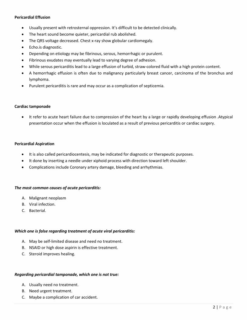
• Investigations: The ECG show ST elevation with upward concavity over the affected area .Later may be T
inversion, particularly if there is associated myocarditis.
Management
• The pain can be relieved by aspirin high dose but a more potent anti-inflammatory agent such as indomethacin
may be required.
• Corticosteroid may suppress symptoms but not healing.
• In viral pericarditis recovery usually occurs within a few days or weeks. But there may be recurrence.
• Purulent pericarditis require treatment with antimicrobial therapy, paracentesis and if necessary surgical
drainage.
2 | P a g e
Pericardial Effusion
• Usually present with retrosternal oppression. It’s difficult to be detected clinically.
• The heart sound become quieter, pericardial rub abolished.
• The QRS voltage decreased. Chest x-ray show globular cardiomegaly.
• Echo.is diagnostic.
• Depending on etiology may be fibrinous, serous, hemorrhagic or purulent.
• Fibrinous exudates may eventually lead to varying degree of adhesion.
• While serous pericarditis lead to a large effusion of turbid, straw-colored fluid with a high protein content.
• A hemorrhagic effusion is often due to malignancy particularly breast cancer, carcinoma of the bronchus and
lymphoma.
• Purulent pericarditis is rare and may occur as a complication of septicemia.
Cardiac tamponade
• It refer to acute heart failure due to compression of the heart by a large or rapidly developing effusion .Atypical
presentation occur when the effusion is loculated as a result of previous pericarditis or cardiac surgery.
Pericardial Aspiration
• It is also called pericardiocentesis, may be indicated for diagnostic or therapeutic purposes.
• It done by inserting a needle under xiphoid process with direction toward left shoulder.
• Complications include Coronary artery damage, bleeding and arrhythmias.
The most common causes of acute pericarditis:
A. Malignant neoplasm
B. Viral infection.
C. Bacterial.
Which one is false regarding treatment of acute viral pericarditis:
A. May be self-limited disease and need no treatment.
B. NSAID or high dose aspirin is effective treatment.
C. Steroid improves healing.
Regarding pericardial tamponade, which one is not true:
A. Usually need no treatment.
B. Need urgent treatment.
C. Maybe a complication of car accident.

3 | P a g e
Tuberculosis Pericarditis
• May complicate pulmonary TB, but may be the first manifestation of the disease.
• In Africa tuberculous effusion is a common manifestation of AIDS.
• The condition typically present with chronic malaise, weight loss and low grade fever.
• An effusion usually develops and the pericardium may become thick and unyielding, leading to pericardial
constriction or tamponade, an associated pleural effusion is often present.
Management
• The diagnosis may be confirmed by aspiration of the fluid and direct examination or culture for tubercle bacilli.
• Treatment requires specific anti TB, in addition, a 3 month course of prednisolone has been shown to improve
outcome.
Chronic Constrictive Pericarditis
• Is due to progressive thickening, fibrosis & calcification of the pericardium.
• In effect the heart encased in a solid shell & cannot work properly; the calcification may extend to the
myocardium, so affect the myocardial function.
Possible Causes
1) Tuberculous pericarditis
2) Hemopericardium
3) Viral pericarditis
4) Rheumatoid arthritis
5) Purulent pericarditis
Clinical Features
• Fatique.
• Rapid low volume pulse.
• Pulsus paradoxicus.
• Elevated JVP (rapid y descent).
• Kussmaul sign.
• Loud early third sound (pericardial knock).
• Hepatomegaly.
• Ascites & peripheral edema.
• The condition should be suspected in any patient with unexplained right sided failure and a small heart.
• CXR show pericardial calcification, echocardiography, CT and MRI help for diagnosis.
• The differentiation of chronic constrictive pericarditis from restrictive cardiomyopathy is difficult and need
complex echo-doppler studies and cardiac catheterization.
Management
• Surgical resection of the diseased pericardium can lead to dramatic improvement in up to 50 % of cases.