Embed Size (px)
Citation preview
02.16.11Lecture 12 - The actin cytoskeleton
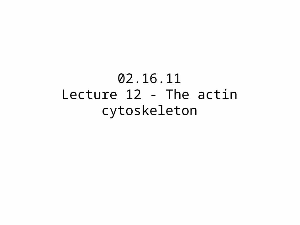
Actin filaments allow cells to adopt different shapes and perform different functions
Villi Contractilebundles
Sheet-like &Finger-like protrusions
Contractilering
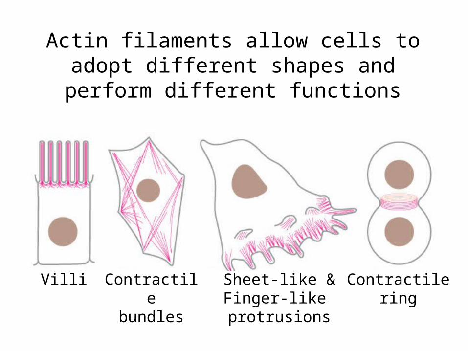
Actin filaments are thin and flexible
• 7 nm in diameter
• Less rigid than microtubules
• Plus end - fast growing
• Minus end - slow growing
• Monomers polymerize into a helical chain
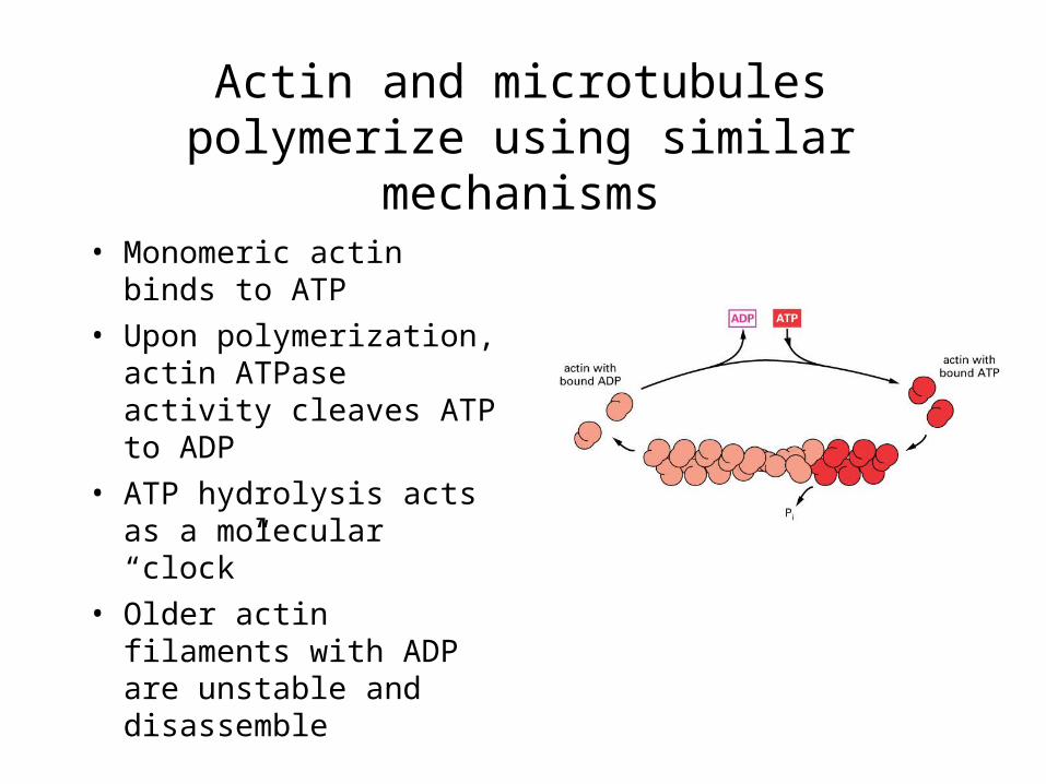
Actin and microtubules polymerize using similar mechanisms
• Monomeric actin binds to ATP
• Upon polymerization, actin ATPase activity cleaves ATP to ADP
• ATP hydrolysis acts as a molecular “clock”
• Older actin filaments with ADP are unstable and disassemble
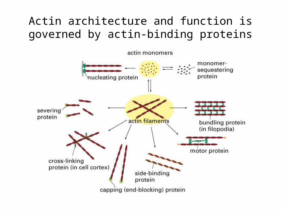
Actin architecture and function is governed by actin-binding proteins
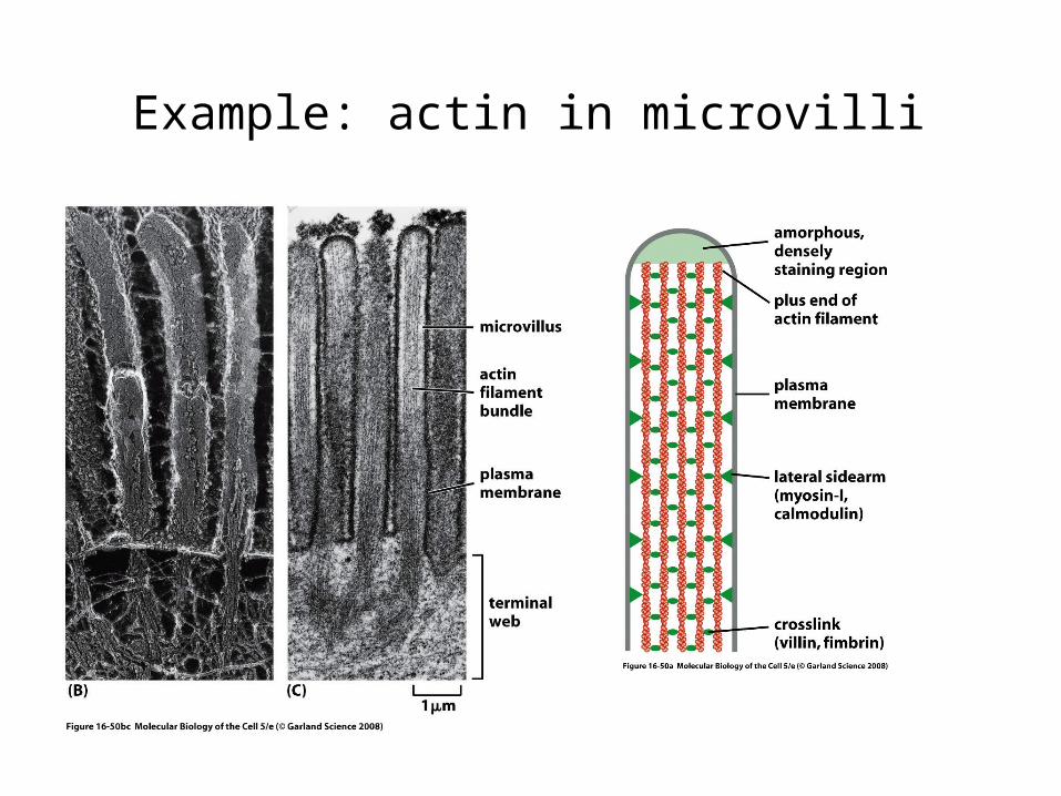
Example: actin in microvilli
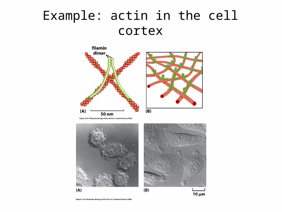
Example: actin in the cell cortex
Actin polymerization can produce “pushing” forces
• Polymerization at the front of a cell pushes the leading edge forward
• Phagocytosis - formation of pseudopods• Intracellular movement and cell-to-cell
spreading of pathogens
During cell migration, actin polymerization pushes the leading edge forward
QuickTime™ and aYUV420 codec decompressor
are needed to see this picture.
Actin polymerization drives protrusion of the cell membrane
QuickTime™ and aTIFF decompressor
are needed to see this picture.
QuickTime™ and aTIFF decompressor
are needed to see this picture.
Lamellipodia Filopodia
11
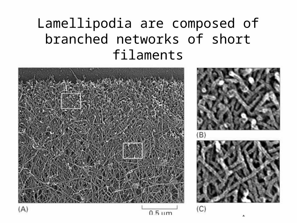
Lamellipodia are composed of branched networks of short filaments
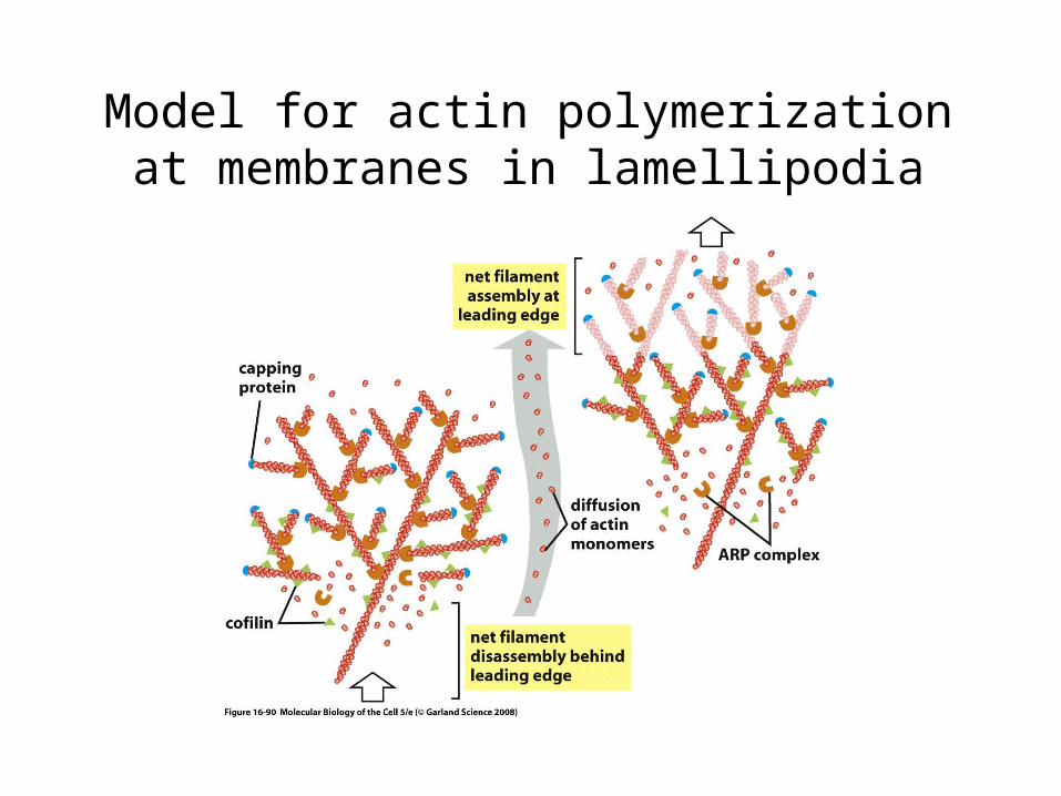
Model for actin polymerization at membranes in lamellipodia
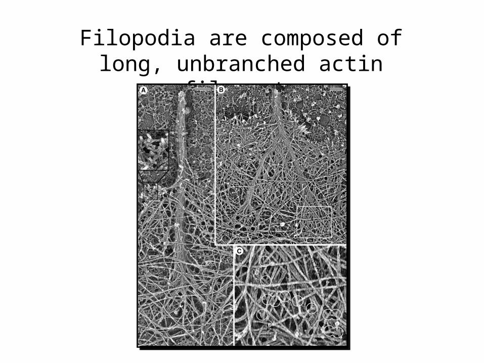
Filopodia are composed of long, unbranched actin filaments
Actin polymerization powers engulfment during phagocytosis
QuickTime™ and aMPEG-4 Video decompressor
are needed to see this picture.
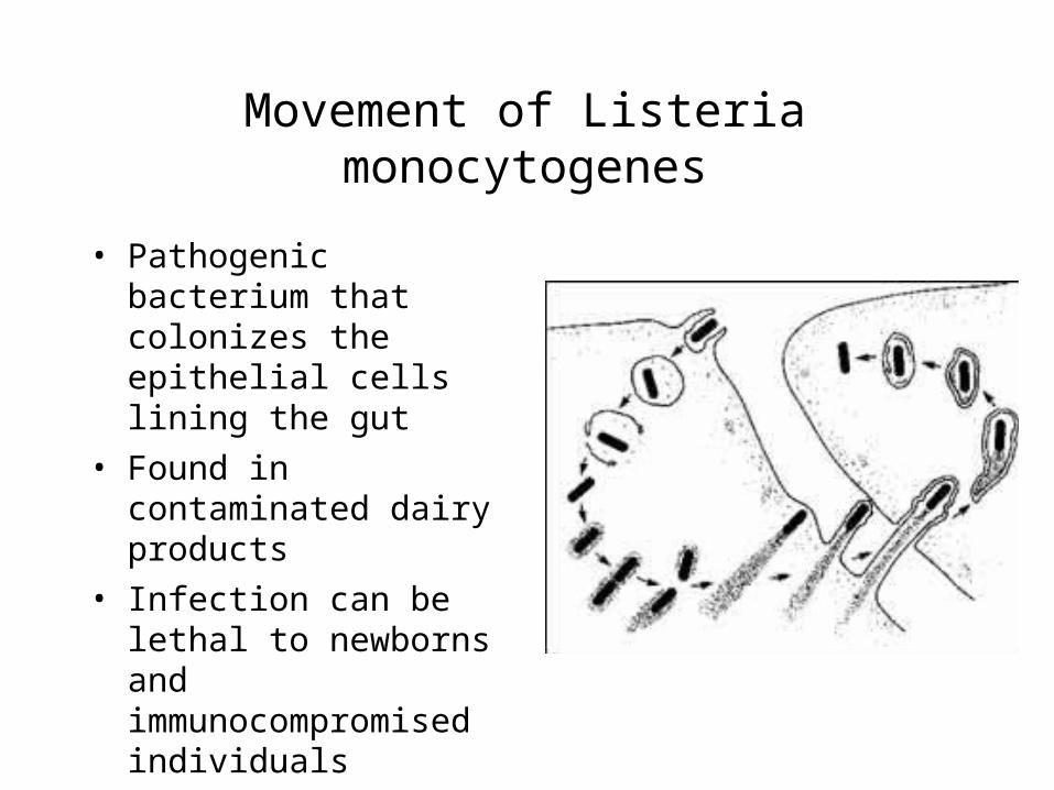
Movement of Listeria monocytogenes
• Pathogenic bacterium that colonizes the epithelial cells lining the gut
• Found in contaminated dairy products
• Infection can be lethal to newborns and immunocompromised individuals
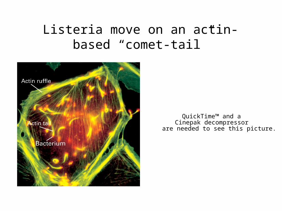
Listeria move on an actin-based “comet-tail”
QuickTime™ and aCinepak decompressor
are needed to see this picture.
Myosins are actin-based motor proteins
• Myosins convert ATP hydrolysis into movement along actin filaments
• Many different classes of myosins (>30 in humans)
• Some myosins move cargoes, other myosins slide actin (as in muscles)
• Actin & ATP binding sites in N-terminal head domain

Myosins “walk” along actin filaments
QuickTime™ and aVideo decompressor
are needed to see this picture.
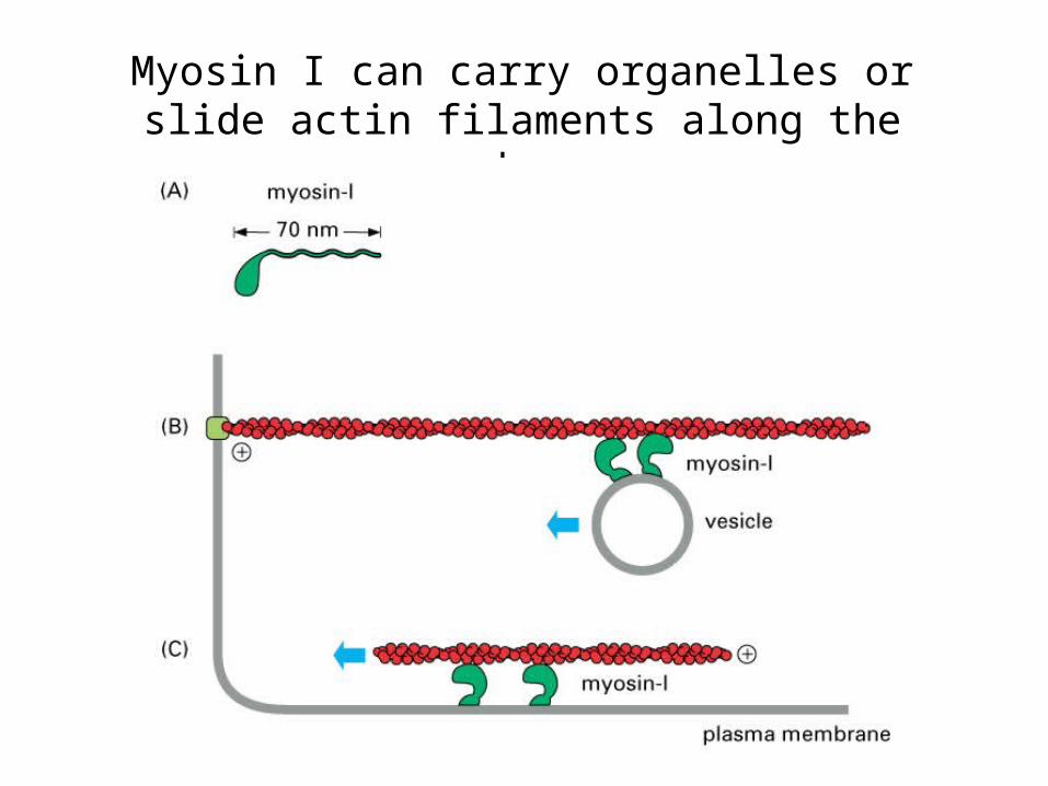
Myosin I can carry organelles or slide actin filaments along the membrane
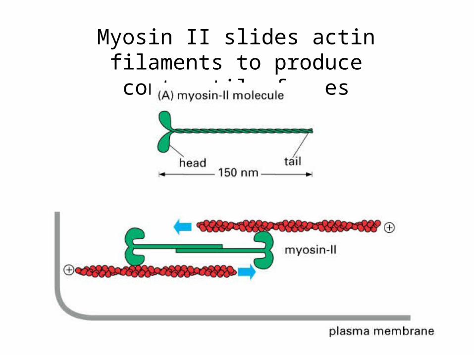
Myosin II slides actin filaments to produce contractile forces
Myosin-based contraction drives cytokinesis
QuickTime™ and aVideo decompressor
are needed to see this picture. QuickTime™ and aPhoto - JPEG decompressor
are needed to see this picture.
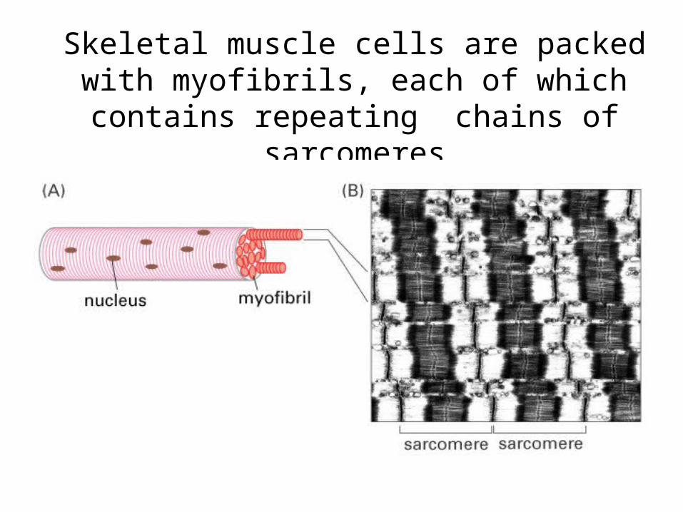
Skeletal muscle cells are packed with myofibrils, each of which contains repeating
chains of sarcomeres
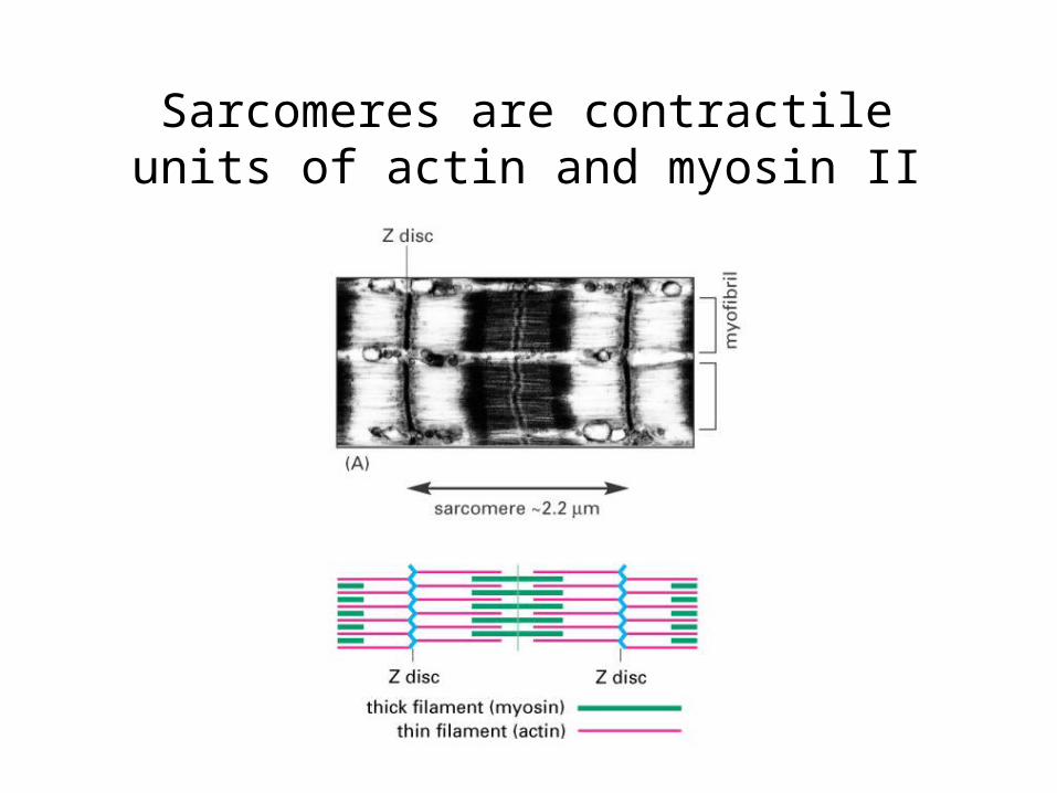
Sarcomeres are contractile units of actin and myosin II
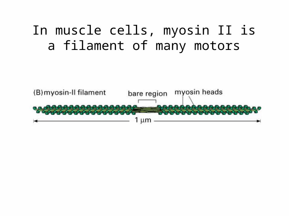
In muscle cells, myosin II is a filament of many motors
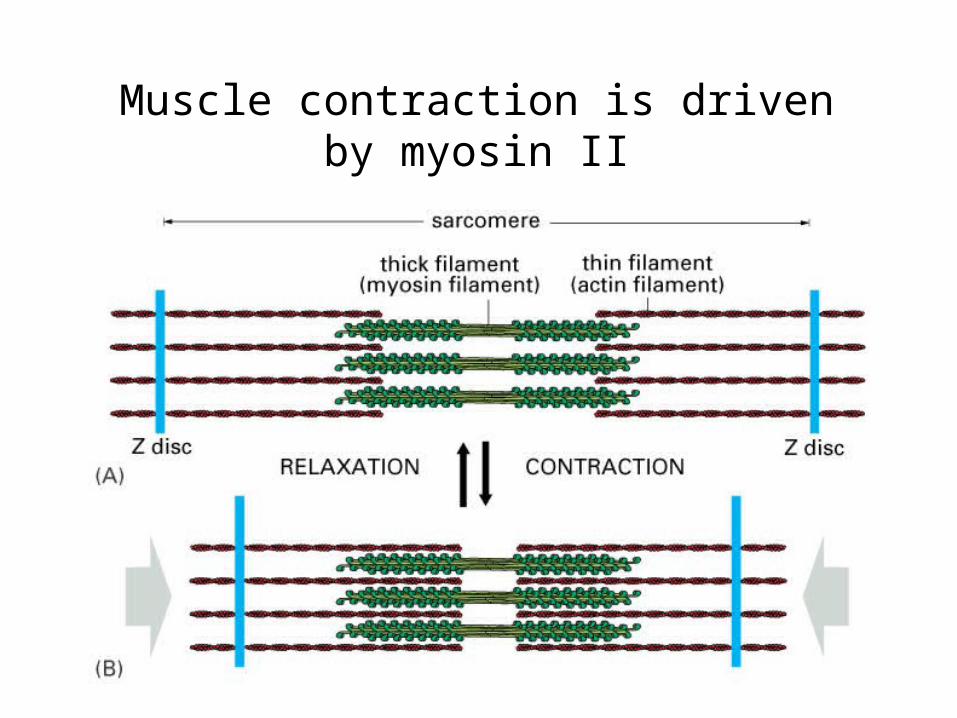
Muscle contraction is driven by myosin II
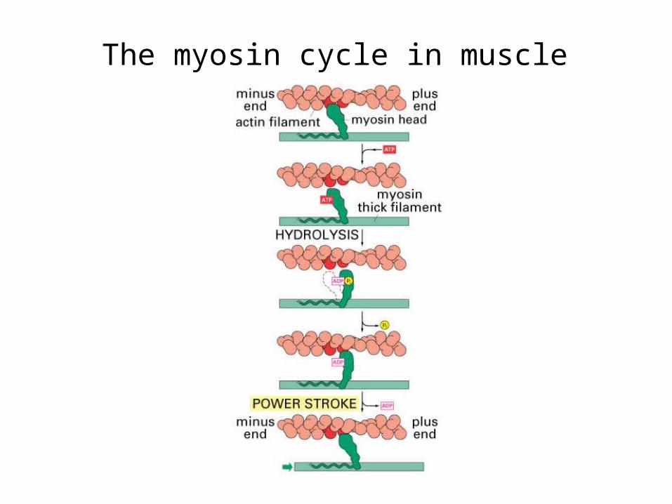
The myosin cycle in muscle
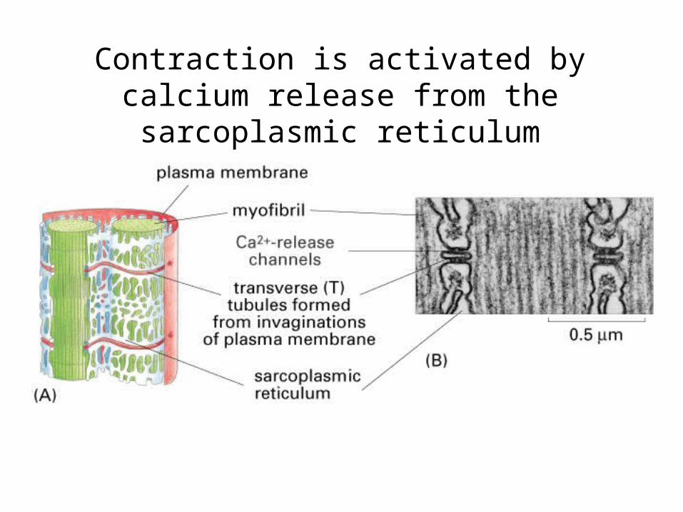
Contraction is activated by calcium release from the sarcoplasmic reticulum
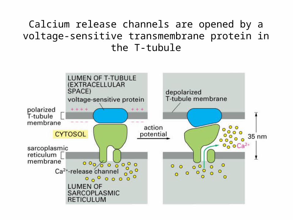
Calcium release channels are opened by a voltage-sensitive transmembrane protein in the T-tubule
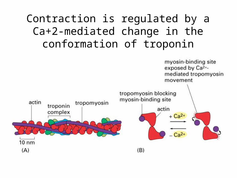
Contraction is regulated by a Ca+2-mediated change in the conformation of troponin
Muscle contraction
QuickTime™ and aAnimation decompressor
are needed to see this picture.