Embed Size (px)
Citation preview
1
Supplementary information
An integrated microfluidic system for rapid detection and
multiple subtyping of influenza A viruses by using glycan-
coated magnetic beads and RT-PCR
Kao-Mai Shena, Narayana Murthy Sabbavarapub, Chien-Yu Fua, Jia-Tsrong Janb, Jen-
Ren Wangc, Shang-Cheng Hungb,d* and Gwo-Bin Leea,e,f*
aDepartment of Power Mechanical Engineering, National Tsing Hua University,
Hsinchu 30013, Taiwan
bGenomics Research Center, Academia Sinica, Taipei 11529, Taiwan
cDepartment of Medical Laboratory Science and Biotechnology, College of Medicine,
National Cheng Kung University, Tainan 701, Taiwan
dDepartment of Applied Science, National Taitung University, 369, Section 2,
University Road, Taitung 95092, Taiwan
eInstitute of Biomedical Engineering, National Tsing Hua University, Hsinchu 30013,
Taiwan
fInstitute of NanoEngineering and Microsystems, National Tsing Hua University,
Hsinchu 30013, Taiwan
Electronic Supplementary Material (ESI) for Lab on a Chip.This journal is © The Royal Society of Chemistry 2019
2
Supplementary material table of contents
I. Working principle of the diagnostic assay.……………………………... 4
Supplementary table 1 Experimental procedures on the microfluidic system...…...............................................................
4
II. Influenza virus strains and their preparation……………………...….. 5
Supplementary table 2 The initial titers of influenza virus stocks............... 5
Supplementary fig. 1 The real-time PCR standard curves for quantification of virus plasmid copy numbers.…....
5
III. Glycans and magnetic bead coating.…………………………………..... 6
Supplementary table 3 The chemical structure of the glycans SCH-42, SNM-01-139, and DJR-03-99................................
6
Supplementary fig. 2 Capture abilities of three glycan-coated magnetic beads (SNM-01-139, SCH-42, and DJR-03-99) for the InfA/ H1N1 virus on-chip and with a benchtop protocol……..…......................................
7
Supplementary fig. 3 Capture abilities of two kinds of SNM-01-139-coated magnetic beads (Dynabeads MyOne™ Streptavidin C1 and T1) with 10-fold serial dilutions of InfA/H1N1 virus stocks…...................................................................
7
IV. Chip design, fabrication, and characterization……………………......... 8
4.1.1 Pumping rate........................................................... 84.1.2 Pumping precision................................................... 94.1.3 Mixing index............................................................ 10
V. One-step RT-PCR protocol............................................................................. 12
Supplementary table 4 Sequences of primers used in this study ................. 12Supplementary fig. 4 Specificity tests of the subtyping primers for viral RNA
of InfA/H1N1, InfA/H3N2, InfA/H5N1, InfA/H5N2, InfA/H7N9, InfB/Victoria and InfB/ Yamagata viruses, respectively, by performing benchtop one-step RT-PCR
and gel electrophoresis..................................
13
VI. Sensitivity tests............................................................................................ 14
3
Supplementary fig. 5 The LOD results of this microfluidic system with SNM-01-139-coated beads and the HA-specific and NA-specific subtyping primers.........................
VII. Supplementary references............................................................................ 17
4
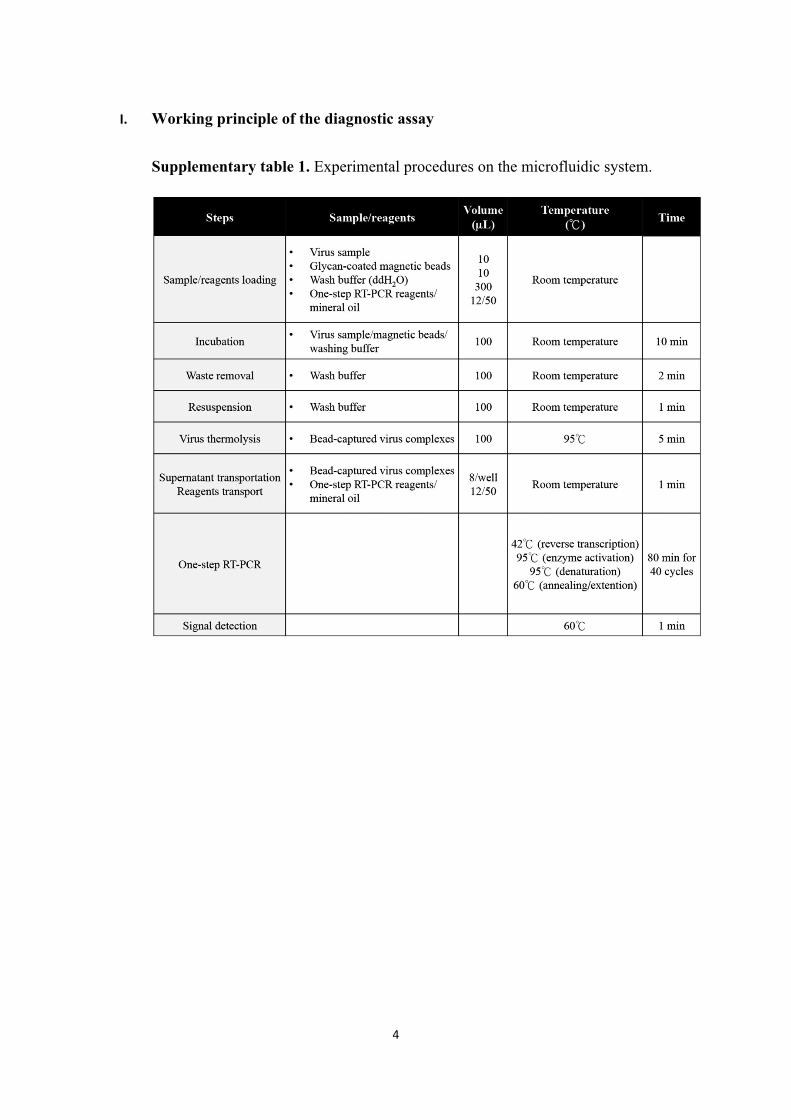
I. Working principle of the diagnostic assay
Supplementary table 1. Experimental procedures on the microfluidic system.
5
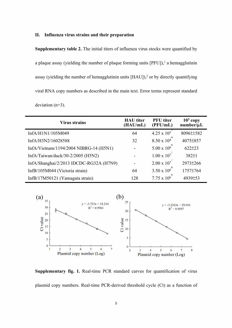
II. Influenza virus strains and their preparation
Supplementary table 2. The initial titers of influenza virus stocks were quantified by
a plaque assay (yielding the number of plaque forming units [PFU]),1 a hemagglutinin
assay (yielding the number of hemagglutinin units [HAU]),2 or by directly quantifying
viral RNA copy numbers as described in the main text. Error terms represent standard
deviation (n=3).
Virus strains HAU titer(HAU/mL)
PFU titer (PFU/mL)
103 copy number/L
InfA/H1N1/105M049 64 4.25 x 1055
8096±1582InfA/H3N2/16028588 32 8.50 x 10⁴
44075±857
InfA/Vietnam/1194/2004 NIBRG-14 (H5N1) - 5.00 x 1066
622±23InfA/Taiwan/duck/30-2/2005 (H5N2) - 1.00 x 10⁷
7382±1
InfA/Shanghai/2/2013 IDCDC-RG32A (H7N9) - 2.00 x 1077
2973±266InfB/105M044 (Victoria strain) 64 3.50 x 10⁶
61757±764
InfB/17M50121 (Yamagata strain) 128 7.75 x 1066
4939±53
Supplementary fig. 1. Real-time PCR standard curves for quantification of virus
plasmid copy numbers. Real-time PCR-derived threshold cycle (Ct) as a function of
6
InfA (a) and InfB (b) plasmid copy number (n=2; standard deviation error bars are
generally not visible due to high technical precision.).
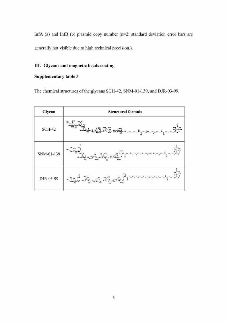
III. Glycans and magnetic beads coating
Supplementary table 3
The chemical structures of the glycans SCH-42, SNM-01-139, and DJR-03-99.
Glycan Structural formula
SCH-42
SNM-01-139
DJR-03-99
7
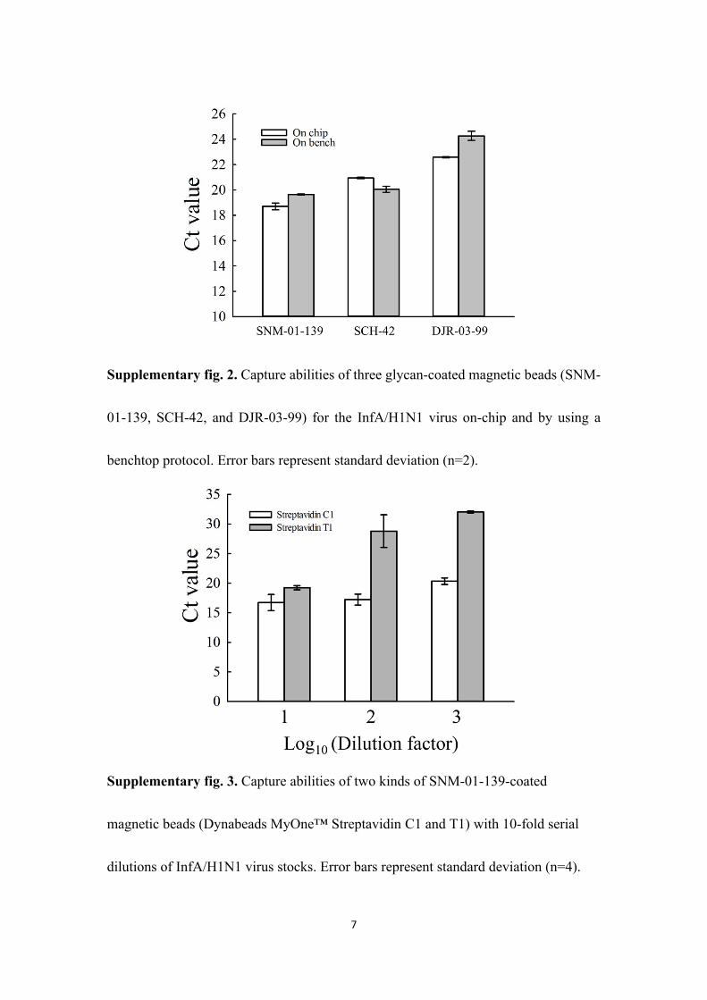
Supplementary fig. 2. Capture abilities of three glycan-coated magnetic beads (SNM-
01-139, SCH-42, and DJR-03-99) for the InfA/H1N1 virus on-chip and by using a
benchtop protocol. Error bars represent standard deviation (n=2).
Supplementary fig. 3. Capture abilities of two kinds of SNM-01-139-coated
magnetic beads (Dynabeads MyOne™ Streptavidin C1 and T1) with 10-fold serial
dilutions of InfA/H1N1 virus stocks. Error bars represent standard deviation (n=4).
8
IV. Chip design, fabrication, and characterization
4.1 Characterization of the chip’s performance
4.1.1 Pumping rate
The chip’s pneumatic micropumps with several pneumatic normally-closed
microvalves were precisely controlled by the custom-made control system described in
the main text. In order to evaluate the transport efficiency of the consecutive
micropumps, the pumping rate was measured under different operating conditions as
described in the main text. Water (ddH2O) was used as the test liquid, and it was
transported from the RT-PCR reagent loading chambers to the middle RT-PCR reaction
chambers by the consecutive micropumps. The pumping rates were measured at three
operating frequencies (0.5, 1.0, and 2.0 Hz) at each of six applied negative gauge
pressures (-6.7, -13.3, -26.7, -40.0, -53.3, and -66.7 kPa) at a constantly applied positive
gauge pressure (13.3 kPa). Moreover, the working time of the three steps of the
microvalves was set to 3 s, so the total operating times for each round of fluid transport
enacted by the micropumps at 0.5, 1.0, and 2.0 Hz were 5, 4, and 3.5 s, respectively.
9
4.1.2 Pumping precision
In order to evaluate the fluid transport precision of the chip’s micropumps, the
pumping uniformity was measured under different operating conditions. Water
(ddH2O) was used as the test liquid, and it was transported from the right micromixer
to the middle reaction chambers by the arrayed micropumps. The pumping precision
was measured at four applied negative gauge pressures (-40.0, -53.3, -66.7, and -80.0
kPa) at a constant applied positive gauge pressure (13.3 kPa). Moreover, the working
times of the three steps of the microvalves and the one step of the consecutive
micropumps were set to 3 and 5 s, respectively; this resulted in a total operating time
for each round of transport of 8 s.
10
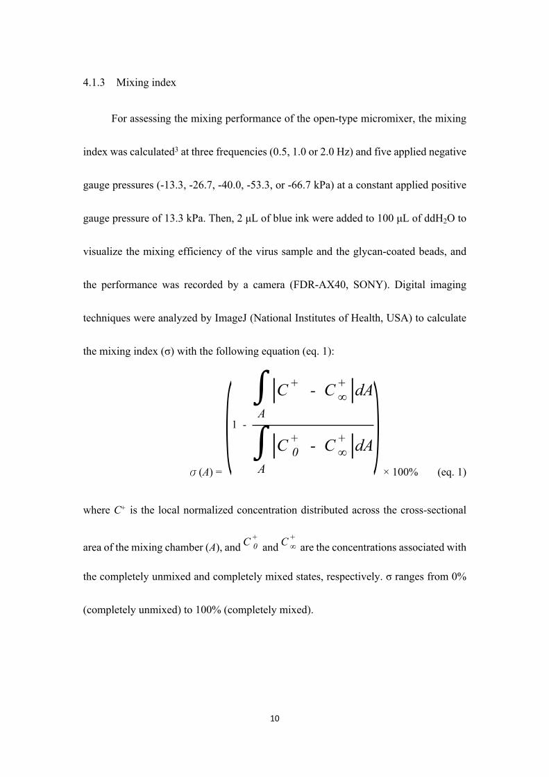
4.1.3 Mixing index
For assessing the mixing performance of the open-type micromixer, the mixing
index was calculated3 at three frequencies (0.5, 1.0 or 2.0 Hz) and five applied negative
gauge pressures (-13.3, -26.7, -40.0, -53.3, or -66.7 kPa) at a constant applied positive
gauge pressure of 13.3 kPa. Then, 2 μL of blue ink were added to 100 μL of ddH2O to
visualize the mixing efficiency of the virus sample and the glycan-coated beads, and
the performance was recorded by a camera (FDR-AX40, SONY). Digital imaging
techniques were analyzed by ImageJ (National Institutes of Health, USA) to calculate
the mixing index (σ) with the following equation (eq. 1):
σ(A) = × 100% (eq. 1)
(1 -
∫A
|C + - C +∞ |dA
∫A
|C +0 - C +
∞ |dA)where C+ is the local normalized concentration distributed across the cross-sectional
area of the mixing chamber (A), and and are the concentrations associated with C +0 C +
∞
the completely unmixed and completely mixed states, respectively. σ ranges from 0%
(completely unmixed) to 100% (completely mixed).
11
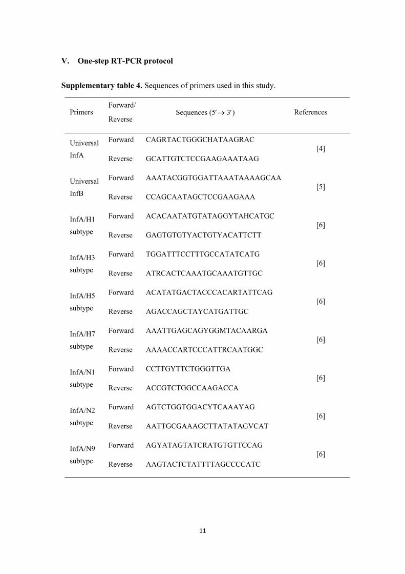
V. One-step RT-PCR protocol
Supplementary table 4. Sequences of primers used in this study.
PrimersForward/
ReverseSequences (5 3) References
Universal InfA
Forward
Reverse
CAGRTACTGGGCHATAAGRAC
GCATTGTCTCCGAAGAAATAAG[4]
Universal InfB
Forward
Reverse
AAATACGGTGGATTAAATAAAAGCAA
CCAGCAATAGCTCCGAAGAAA[5]
InfA/H1 subtype
Forward
Reverse
ACACAATATGTATAGGYTAHCATGC
GAGTGTGTYACTGTYACATTCTT[6]
InfA/H3 subtype
Forward
Reverse
TGGATTTCCTTTGCCATATCATG
ATRCACTCAAATGCAAATGTTGC[6]
InfA/H5 subtype
Forward
Reverse
ACATATGACTACCCACARTATTCAG
AGACCAGCTAYCATGATTGC[6]
InfA/H7 subtype
Forward
Reverse
AAATTGAGCAGYGGMTACAARGA
AAAACCARTCCCATTRCAATGGC[6]
InfA/N1 subtype
Forward
Reverse
CCTTGYTTCTGGGTTGA
ACCGTCTGGCCAAGACCA[6]
InfA/N2 subtype
Forward
Reverse
AGTCTGGTGGACYTCAAAYAG
AATTGCGAAAGCTTATATAGVCAT[6]
InfA/N9 subtype
Forward
Reverse
AGYATAGTATCRATGTGTTCCAG
AAGTACTCTATTTTAGCCCCATC[6]
12
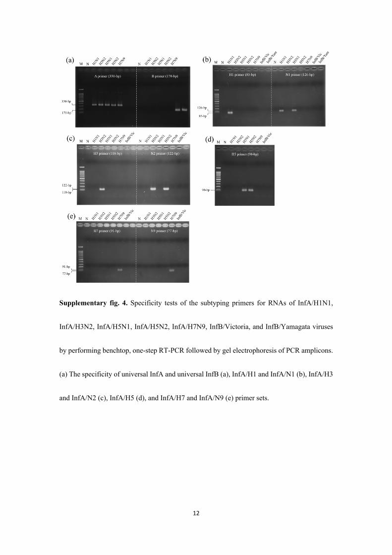
Supplementary fig. 4. Specificity tests of the subtyping primers for RNAs of InfA/H1N1,
InfA/H3N2, InfA/H5N1, InfA/H5N2, InfA/H7N9, InfB/Victoria, and InfB/Yamagata viruses
by performing benchtop, one-step RT-PCR followed by gel electrophoresis of PCR amplicons.
(a) The specificity of universal InfA and universal InfB (a), InfA/H1 and InfA/N1 (b), InfA/H3
and InfA/N2 (c), InfA/H5 (d), and InfA/H7 and InfA/N9 (e) primer sets.
13
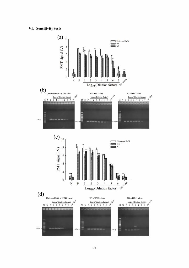
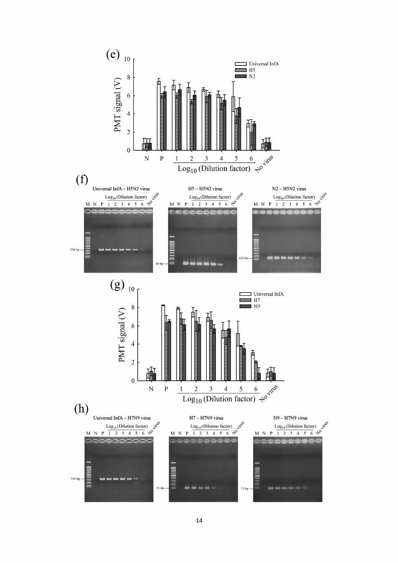
VI. Sensitivity tests
14
15
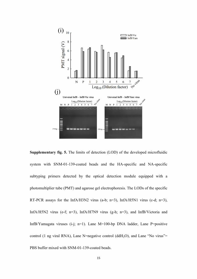
Supplementary fig. 5. The limits of detection (LOD) of the developed microfluidic
system with SNM-01-139-coated beads and the HA-specific and NA-specific
subtyping primers detected by the optical detection module equipped with a
photomultiplier tube (PMT) and agarose gel electrophoresis. The LODs of the specific
RT-PCR assays for the InfA/H3N2 virus (a-b; n=3), InfA/H5N1 virus (c-d; n=3),
InfA/H5N2 virus (e-f; n=3), InfA/H7N9 virus (g-h; n=3), and InfB/Victoria and
InfB/Yamagata viruses (i-j; n=1). Lane M=100-bp DNA ladder, Lane P=positive
control (1 ng viral RNA), Lane N=negative control (ddH2O), and Lane “No virus”=
PBS buffer mixed with SNM-01-139-coated beads.
16
Supplementary references
1. Cooper, P.D., 1961. The plaque assay of animal viruses. Adv Virus Res 8, 319-378.
2. Stuart-Harris, C., 1979. Epidemiology of influenza in man. Brit Med Bull 35(1), 3-
8.
3. Yang, S.Y., Lin, J.L., Lee, G.B., 2009. A vortex-type micromixer utilizing
pneumatically driven membranes. J Micromechanics Microengineering 19(3).
4. Tsukamoto, K., Ashizawa, H., Nakanishi, K., Kaji, N., Suzuki, K., Okamatsu, M.,
Yamaguchi, S., Mase, M., 2008. Subtyping of avian influenza viruses H1 to H15 on
the basis of hemagglutinin genes by PCR assay and molecular determination of
pathogenic potential. J Clin Microbiol 46(9), 3048-3055.
5. Wang, C.H., Lien, K.Y., Hung, L.Y., Lei, H.Y., Lee, G.B., 2012. Integrated
microfluidic system for the identification and multiple subtyping of influenza viruses
by using a molecular diagnostic approach. Microfluid Nanofluid 13(1), 113-123.
6. Hoffmann, B., Hoffmann, D., Henritzi, D., Beer, M., Harder, T.C., 2016. Riems
influenza a typing array (RITA): An RT-qPCR-based low density array for
subtyping avian and mammalian influenza A viruses. Sci Rep 6, 27211.


![SD servicio nombres docu examen2 - UPMlaurel.datsi.fi.upm.es/_media/.../sd/...examen-4pp.pdfd urrw vhuyhuv qhw =rqd d qlf hv =rqd hv hlqvwhlq ffxsp xsp hv =rqd xsp hv ]dsh il xsp hv](https://img.pdfslide.net/doc/110x75/5fdd3be9af48e220dc67f7d6/sd-servicio-nombres-docu-examen2-d-urrw-vhuyhuv-qhw-rqd-d-qlf-hv-rqd-hv-hlqvwhlq.jpg)


![$UL]RQD %DVLF )LQDQFLDO 6WDWHPHQWV](https://img.pdfslide.net/doc/110x75/620410a8bd58765e0f795408/ulrqd-dvlf-lqdqfldo-6wdwhphqwv-.jpg)


![6WDWH RI $UL]RQD %XGJHW 5HTXHVW](https://img.pdfslide.net/doc/110x75/62040c13aef71220db78f5c7/6wdwh-ri-ulrqd-xgjhw-5htxhvw.jpg)


![ZZZ EHO]RQD FRP](https://img.pdfslide.net/doc/110x75/62040d0dedb3a75b68040498/zzz-ehorqd-frp.jpg)


![RI $UL]RQD 3URJUDP](https://img.pdfslide.net/doc/110x75/62040cd03248a67d524c97a8/ri-ulrqd-3urjudp.jpg)